nutrition journal - centro di nutrizione dott. bruni significant effec… · 1 no significant...
TRANSCRIPT

This Provisional PDF corresponds to the article as it appeared upon acceptance. Fully formattedPDF and full text (HTML) versions will be made available soon.
No significant effect on bone mineral density by high doses of vitamin D3 givento overweight subjects for one year
Nutrition Journal 2010, 9:1 doi:10.1186/1475-2891-9-1
Rolf Jorde ([email protected])Monica Sneve ([email protected])
Peter A Torjesen ([email protected])Yngve Figenschau ([email protected])
John-Bjarne Hansen ([email protected])Guri Grimnes ([email protected])
ISSN 1475-2891
Article type Research
Submission date 12 September 2009
Acceptance date 7 January 2010
Publication date 7 January 2010
Article URL http://www.nutritionj.com/content/9/1/1
This peer-reviewed article was published immediately upon acceptance. It can be downloaded,printed and distributed freely for any purposes (see copyright notice below).
Articles in Nutrition Journal are listed in PubMed and archived at PubMed Central.
For information about publishing your research in Nutrition Journal or any BioMed Central journal, goto
http://www.nutritionj.com/info/instructions/
For information about other BioMed Central publications go to
http://www.biomedcentral.com/
Nutrition Journal
© 2010 Jorde et al. , licensee BioMed Central Ltd.This is an open access article distributed under the terms of the Creative Commons Attribution License (http://creativecommons.org/licenses/by/2.0),
which permits unrestricted use, distribution, and reproduction in any medium, provided the original work is properly cited.

1
No significant effect on bone mineral density by high doses
of vitamin D3 given to overweight subjects for one year
Rolf Jorde1, Monica Sneve
2, Peter A Torjesen
3, Yngve Figenschau
4, John-Bjarne
Hansen5 and Guri Grimnes
1
Address: 1
Institute of Clinical Medicine, University of Tromsø, and Medical Clinic,
University Hospital of North Norway, Tromsø, Norway; 2 Department of Ophthalmology and
Neurosurgery, Division of Ophthalmology, University Hospital of North Norway, Tromsø,
Norway; 3
Hormone Laboratory, Aker University Hospital, Oslo, Norway; 4
Department of
Medical Biochemistry, University Hospital of North Norway, and Institute of
Medical Biology, University of Tromsø, Tromsø, Norway; 5
Center for Atherothrombotic
Research in Tromsø (CART), Institute of Clinical Medicine, University of Tromsø, Norway
Email: Rolf Jorde – [email protected]; Monica Sneve – [email protected]; Peter A
Torjesen - [email protected]; Yngve Figenschau –
[email protected]; John-Bjarne Hansen – [email protected]; Guri Grimnes
Corresponding author: Rolf Jorde, Medical Clinic, University Hospital of North Norway,
9038 Tromsø, Norway. Telephone: 47 776 26000 Fax: 47 776 26863
E-mail: [email protected]

2
Abstract
Background: In meta-analyses supplementation with vitamin D appears to reduce incidence
of fractures, and in cross-sectional studies there is a positive association between serum 25-
hydroxyvitamin D (25(OH)D) levels and bone mineral density (BMD). However, the effect of
supplementation with high doses of vitamin D on BMD is more uncertain and could in theory
have both positive and negative effects.
Methods: The study was a one year, double blind placebo-controlled intervention trial
performed at the University Hospital of North Norway. 421 subjects, 21 – 70 years old,
were included and 312 completed the study. The subjects were randomized to vitamin D3
40.000 IU per week (DD group), vitamin D3 20.000 IU per week (DP group), or placebo
(PP group). All subjects were given 500 mg calcium daily. Serum 25(OH)D,
osteoprotegrin (OPG), receptoractivator of nuclear factor-kappaB ligand (RANKL), and
BMD at the lumbar spine and the hip were measured before and at the end of the study.
Results: At baseline the mean serum 25(OH)D levels were 58 nmol/L (all subjects) and
increased to 141 and 100 nmol/L in the DD and DP groups, respectively. After one year,
no significant differences were found between the three groups regarding change in BMD,
serum OPG or RANKL.
Conclusions: Supplementation with high doses of vitamin D for one year does not appear
to have a negative effect on BMD in healthy subjects. In order to disclose a positive
effect, subjects with low BMD and/or low serum 25(OH)D levels need to be studied.
Trial registration: The trial was registered at ClinicalTrials.gov (NCT00243256).
3
Background
A sufficient intake of vitamin D, or sun exposure for production of vitamin D in the skin, is of
vital importance for skeletal health, and patients with severe vitamin D deficiency exhibit
hypocalcemia and rickets or osteomalacia [1].
The skeleton is constantly remodelled. This occurs in small bone remodelling units where the
balance between bone resorption by the osteoclasts and bone matrix synthesis by the
osteoblasts determines the volume and quality of the bone [2]. It has been known for many
years that the activity of the osteoclasts is influenced by the osteoblast, which was clarified by
the recent discovery of the osteoprotegrin (OPG)/ receptoractivator of nuclear factor-kappaB
(RANK)/ RANK ligand (RANKL) system [2-6]. Thus, the osteoblasts and the bone marrow
stromal cells produce RANKL which activates the osteoclasts by binding to its RANK
receptor. The osteoblasts also produce OPG which functions as a decoy receptor and binds
RANKL and thereby prevents it from binding to RANK [2,7].
The classical effect of 1,25-dihydoxyvitamin D (1,25(OH)2D), which is the active form of
vitamin D, is to increase the intestinal calcium absorption and thereby have an indirect effect
on bone formation [1]. In addition, 1,25(OH)2D has direct effects on bone, and receptors for
1,25(OH)2D (VDR) have been found in osteoclasts [8] as well as in osteoblasts [9].
Traditionally, 1,25(OH)2D has been considered a bone-resorbing hormone. This was based on
its potent stimulation of bone resorption in tissue cultures [10,11] which was further
substantiated by the finding that 1,25(OH)2D increased the RANKL expression [5,11].
However, active vitamin D metabolites have also been reported to inhibit bone resorption [12]
and may therefore have a dual effect on bone formation [11].

4
From these in vitro experiments it is hard to predict the effect of vitamin D supplementation
on bone in humans provided there is not a severe vitamin D deficiency. If the effect is
positive, one would expect that supplementation with vitamin D would prevent osteoporotic
fractures which was demonstrated in a recent meta-analysis by Bischoff-Ferrari et al. [13]. A
dose dependency was also reported, and no effect was seen with 400 IU per day. Similarly, in
a large cross-sectional study there was a positive relation between serum 25(OH)D, which is
the storage form of the vitamin and the one used to evaluate a subject’s vitamin D status, and
bone mineral density (BMD) [14]. However, for subjects above the age of 50 years, the BMD
reached a plateau at serum 25(OH)D levels of 90 – 100 nmol/L, and thereafter appeared to
decline. If this reflects a causal relation, vitamin D given in high doses may have a negative
effect on bone. Given the focus on the need for higher doses of vitamin D [15] this is an
important issue to settle, but so far, most intervention studies have used vitamin D in doses of
800 IU per day or less [13].
We have recently performed a one year placebo-controlled intervention study with vitamin D
in doses of 20.000 IU and 40.000 IU per week with change in weight as primary end point
[16]. In addition, bone densitometry was performed and serum OPG and RANKL measured
before and at the end of the study. This gave us the opportunity to address the issue of skeletal
effects of high dose vitamin D supplementation.
Methods
The protocol has previously been described in detail [16]. The subjects were recruited by
advertisements in local newspapers and from our outpatient clinic. The subjects were initially
screened at our outpatient clinic at the Department of Internal Medicine at the University
Hospital of North Norway. All potential participants were told that they would be part of a

5
weight loss study assessing the effect of high doses of vitamin D on body weight. Males and
females 21 to 70 years old, with BMI between 28.0 – 47.0 kg/m2 were included. Subjects with
diabetes or a history of coronary infarction, angina pectoris, stroke, renal stone disease, or
sarcoidosis were excluded. Subjects with serum calcium > 2.55 mmol/L, males with serum
creatinine > 129 µmol/L and females with serum creatinine > 104 µmol/L, and subjects using
bisphosphonates or oestrogen (for contraception or replacement) were not included.
At baseline fasting blood samples were drawn and any previous supplements with calcium
and vitamin D (including cod liver oil) were discontinued. All subjects were given a daily
supplement with 500 mg calcium (Nycoplus Calcium®
, Nycomed, Norway) throughout the
one year intervention period. The participants were given oral information and written
recommendations on healthy diet and physical activity. The study was a randomized, double
blind clinical trial. Using block-randomization, the subjects were randomized into one of three
groups, stratified by gender and smoking status: one group was to take two capsules of
vitamin D3 (20 000 IU cholecalciferol per capsule (Decristol®
, Jenapharm, Jena, Germany))
per week; one group one capsule of vitamin D3 and one placebo capsule per week; and one
group two placebo capsules per week. The subjects were supplied with new medication every
third month. Unused calcium tablets and capsules were returned and counted. The subjects
were classified as current smokers or current non-smokers. Non-fasting blood samples for
serum calcium analysis were drawn after 3, 6, and 9 months to disclose development of
hypercalcemia.
Measurements
Height and weight were measured wearing light clothing and no shoes. Serum calcium and
parathyroid hormone (PTH) were measured as previously described [17]. Serum levels of 25-

6
hydroxyvitamin D (25(OH)D) were measured by radioimmunoassay (DiaSorin, Stillwater,
MN, USA). This assay measures both 25(OH)D3 and 25(OH)D2, and the intra- and total assay
coefficients of variation (CVs) are 6 % and 14 %, respectively [18]. BMD was determined by
anterior-posterior dual-energy X-ray absorptiometry (DEXA) scans at the lumbar spine and
hip according to the manufacturer (GE Lunar Prodigy, LUNAR Corporation, Madison, WI,
USA). The mean of L2-L4 and the mean of the right and left total hip values were used in the
analyses (CV 3.6 %). Serum OPG was measured as previously described with an intra-assay
CV of 3.2 % and inter-assay CV of 11.1 % [19]. The concentration of free RANKL was
measured by a new, highly sensitive ELISA assay for free RANKL with a detection limit of
0.02 pmol/l, an intra-assay CV of 9.6 % and an inter-assay CV of 15.3 % (ampli sRANKL
human, Biomedica, Vienna, Austria). The analysis was performed according to the
manufacturer’s instruction. The analyses of serum OPG and RANKL were performed on
coded samples without knowledge of clinical status by the person performing the assays. All
samples were analyzed in duplicate and the mean value is used in this report. OPG and
RANKL were measured in baseline sera in all subjects who completed the study and in sera
from the end of the study in a random sample of subjects in the DD and PP groups as appear
in the tables.
Statistical analyses
Normal distribution was evaluated with visual inspection of histograms with normal curve,
and determination of skewness and kurtosis. All dependent variables except serum OPG and
RANKL were considered normally distributed at baseline. After log transformation OPG
attained normal distribution and was used as such when parametric statistics were applied.
Because of several 0-values (not detectable) RANKL could not be log transformed and
therefore was evaluated with non-parametric statistics. The delta values for OPG and RANKL

7
were also not normally distributed and could not be log transformed because of several 0-
values, and were therefore evaluated with non-parametric statistics. The other delta values
were normally distributed. A multiple linear regression model with age, gender, BMI,
smoking status, serum 25(OH)D, serum PTH and serum calcium as covariates was used to
evaluate individual predictor of BMD L2-L4, BMD total hip and serum OPG. Correlations
were evaluated with Pearson’s or Spearman’s correlation coefficients as appropriate.
The intervention study was analysed per protocol. Comparisons between the groups at
baseline and between their delta values (value at end of the study minus value at baseline)
were performed with ANOVA, the Kruskall Wallis test or the Chi-square test. The Bonferroni
correction was used where multiple comparisons were performed. Unless otherwise stated,
data are expressed as mean ± SD. All tests were done two-sided, and P-value < 0.05 was
considered statistically significant. The Statistical Package for Social Sciences version 15.0
was used for all statistical analyses (SPSS Inc., Chicago, Ill., USA).
The power calculation was performed according to the primary end point (weight loss) to
disclose a clinically significant difference of 6 kg with or without vitamin D supplementation
[16].
Ethics
The study was approved by the Regional Ethics Committee. All participants gave written
informed consent prior to the study.
Results
Baseline
8
The inclusion period started in November 2005 and the last person was included in October
2006. Of 626 subjects initially screened by telephone interview, 421 (156 men and 265
women) met the inclusion criteria and had complete datasets (Figure 1). The baseline
characteristics of these subjects are shown in Table 1. Among the women, 114 were
postmenopausal. The age distribution among the men and women are shown in Figure 2. The
mean serum 25(OH)D levels was 57.7 ± 20.7 nmol/L and the distribution is shown in Figure
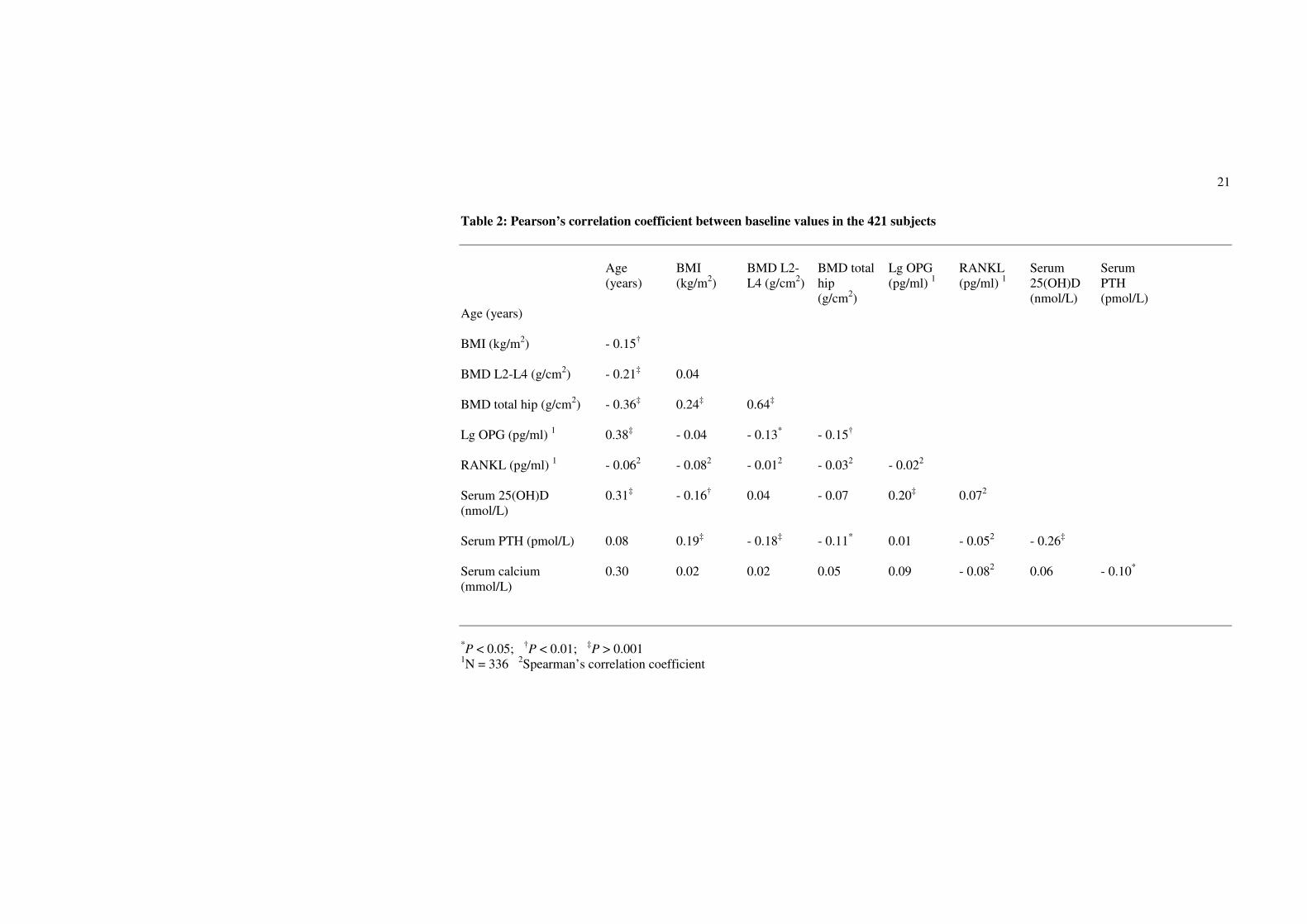
3. As expected there were a number of significant univariate correlations between the
parameters measured (Table 2). However, after adjusting for confounders in the multiple
linear regression model only a few of these relations remained significant. In particular, serum
PTH remained as a significant and negative predictor of BMD both for L2-L4 and the hip, and
female gender and high age were associated with Lg OPG (Table 3). Serum OPG was
negatively correlated with BMD L2-L4 and BMD at the hip, but not after adjusting for the
other covariates (data not shown). Serum RANKL was below the detection limit in 141 of the
336 subjects with RANKL measurement at baseline, and no association was found with any
of the other parameters measured.
Intervention study
One hundred and forty-seven, 132 and 142 subjects were randomized to the DD, DP and PP
groups, respectively. 110 subjects in the DD group, 97 subjects in the DP group, and 105
subjects in the PP group completed the study (Figure 1). The reasons for the dropouts (37
subjects in the DD group, 35 subjects in the DP group and 37 subjects in the PP group) have
previously been described in detail [16]. However, most of those who dropped out withdrew
their consent for participation without stating a specific reason. The compliance rate in the
DD, DP and PP groups were for the vitamin D / placebo capsules 95 %, 96 %, 96 %, and for
the calcium tablets 82 %, 84 %, and 83 %, respectively. At baseline, those in the DP group

9
had BMI 1.5 kg/m2 lower than those in the PP group (P < 0.05). Apart from this difference,
the DD, DP and PP groups were similar (Table 4).
At the end of the study serum 25(OH)D levels were 140.9 ± 34.7 nmol/L, 99.7 ± 20.3 nmol/L,
and 57.9 ± 20.4 nmol/L in the DD, DP and PP groups respectively. There was a highly
significant positive correlation between baseline and 12 months serum 25(OH)D in both the
DD and DP groups (r = 0.40 and r = 0.69, respectively (P < 0.001)), and a significant negative
correlation between baseline and delta serum 25(OH)D (r = - 0.57 and r = - 0.49, respectively
(P < 0.001)). Accordingly, in those given vitamin D, those with the lowest baseline serum
25(OH)D levels had the highest increase in serum 25(OH)D, but not high enough to even out
the differences seen at baseline. There was a significant decrease in the serum PTH levels in
the DD and DP groups, but no significant changes in weight or serum calcium. The three
groups did not differ significantly in delta values for BMD L2-L4, BMD hip, serum OPG or
serum RANKL (Table 4), not even when the DD and DP groups were combined to one
vitamin D group and compared with the PP group, or when only subjects with low serum
25(OH)D values (< the 25th
percentile (< 45.0 nmol/L)) were evaluated separately (data not
shown).
One subject in the PP group had an increase of 11.3 % and one subject in the DD group had a
decrease of 11.5 % in BMD L2-L4. Apart from these two subjects none had an increment or a
decrement outside the “least significant change” (2.8 x CV) regarding the bone densitometry.
Among the 66 women in the DD group, 58 women in the DP group, and 64 women in the PP
group, 29, 28 and 35 women, respectively, were postmenopausal. However, the delta values
for these women did not differ significantly between the DD, DP and PP groups, being for
10
BMD L2-L4 0.004 ± 0.03 g/cm2, - 0.006 ± 0.035 g/cm
2, and - 0.006 ± 0.035 g/cm
2,
respectively; and for BMD hip 0.003 ± 0.013 g/cm2, 0.008 ± 0.015 g/cm
2, and 0.003 ± 0.014
g/cm2, respectively.
Adverse events
As previously described in detail [16], no serious adverse events were seen and there were no
significant differences between the treatment groups regarding adverse events. Two subjects
were diagnosed as having primary hyperparathyroidism during the study, and one had an
increase in serum calcium to 2.62 mmol/L, and all three were excluded from the study. Four
subjects had transient increases in serum calcium > 2.59 mmol/L and remained in the study.
Discussion
In the present study we found no significant associations for serum 25(OH)D with BMD,
serum OPG or serum RANKL at baseline after adjustment for confounders, nor did vitamin D
supplementation for one year differ form placebo regarding change in BMD, serum OPG or
serum RANKL. On the other hand, a high serum PTH level was associated with reduced
BMD in the spine and the hip.
Regarding the negative association between PTH and BMD this is in line with previous
publications from large cross-sectional studies [20,21], whereas the lack of association
between 25(OH)D and BMD differs from the report by Bischoff-Ferrari et al. [14], and was
most likely due to selection and number of subjects in our study. Apart from the expected
significant increase in serum OPG with age and the higher levels in females [22], OPG or
RANKL were not significantly associated with any of the other variables included in the
study. In particular, there was after adjustment for confounders no significant relation

11
between OPG and BMD, which is similar to that reported by Indridason et al. in a study
including 1630 subjects [23].
In the intervention study, our vitamin D doses of 20.000 IU and 40.000 IU per week were
substantially higher than those usually given in osteoporosis studies. This resulted in serum
25(OH)D levels in the high physiological range [24], but no significant change in BMD was
found. However, a positive effect of vitamin D on BMD cannot be ruled out from our study as
most of the subjects had normal BMD at baseline, the study only lasted 12 months, and an
increase in BMD would therefore be hard to disclose. On the other hand, supplementation
with vitamin D could in theory also have a negative effect on BMD as the production of
1,25(OH)2D from 25(OH)D is substrate dependent [25] and 1,25(OH)2D may induce
osteoclastogenesis [5,11]. Our result is therefore of importance as it indicates that high doses
of vitamin D, at least when given for a short period of time to healthy subjects, do not have
serious adverse effects on bone. However, it must be emphasised that a direct and negative
effect of vitamin D could be masked in our study by the concomitant fall in serum PTH, as a
fall in serum PTH may have a beneficial effect on BMD [21,26].
Although 1,25(OH)2D has been demonstrated in in-vitro studies to increase RANKL
expression [5] and reduce the OPG expression [9], supplementation with vitamin D in our
study did not significantly affect their serum levels. In other studies where serum OPG and
RANKL have been measured after therapy, the results have varied [27]. Thus, both serum
OPG and RANKL have been found to decrease after treatment with oestrogen in
postmenopausal women in one study [28], whereas serum OPG but not RANKL increased
after oral contraceptives in another study [29]. No effect on OPG by bisphosphonate therapy
has been reported in subjects with osteoporosis or rheumatoid arthritis [30,31], whereas a

12
decrease in OPG after bisphosphonates has been found in subjects with Paget’s disease [30].
Most likely oestrogen, as well as bisphosphonates, influence the production of OPG and
RANKL as demonstrated in in-vitro studies [32,33]. Therefore, the lack of corresponding
changes in serum levels indicate that the circulating levels of OPG and RANKL do not reflect
the concentrations in the local tissues [27].
Our study has several limitations, and the results should be evaluated with caution. Thus, the
effect of vitamin D on BMD, serum OPG and RANKL were only secondary endpoints, most
of the included subjects were not vitamin D deficient, and serum 1,25(OH)2D was not
measured. All subjects were given calcium supplementation and the results should therefore
be interpreted as vitamin D plus calcium versus calcium alone. On the other hand, our study is
of importance as we used high vitamin D doses resulting in serum 25(OH)D levels in the high
physiological range.
Conclusion
Supplementation with high doses of vitamin D for one year did not have a negative effect on
BMD at the spine or hip in healthy overweight or obese subjects, and a relation between
serum levels of vitamin D and OPG and RANKL could not be demonstrated.
However, in order to disclose a positive effect on BMD, subjects with low BMD and/or
low serum 25(OH)D levels need to be studied for a longer period of time.
Competing interests
The authors declare that they have no competing interest

13
Authors’ contributions
RJ: study design, data gathering, preparation manuscript. MS: data gathering, preparation
manuscript. PAT: vitamin D analyses, preparation manuscript. YF: biochemical analyses,
preparation manuscript. JBH: OPG and RANKL analyses, preparation manuscript. GG:
statistical analyses, preparation manuscript. All authors have read and approved the final
manuscript.
Acknowledgements
The present study was supported by a grant from The Northern Norway Regional Health
Authority. The superb assistance by the staff at the Clinical Research Unit and by Inger
Myrnes and Astrid Lindvall at the Department of Medical Biochemistry, University Hospital
of North Norway, is gratefully acknowledged. We are grateful for the generous supply of
calcium tablets from Nycomed Norway.
References
1. DeLuca HF: Overview of general physiologic features and functions of vitamin D.
Am J Clin Nutr 2004, 80:1689S-1696S.
2. Vega D, Maalouf NM, Sakhaee K: Clinical Review: the role of receptor activator of
nuclear factor-kappaB (RANK)/RANK ligand/osteoprotegerin: clinical
implications. J Clin Endocrinol Metab 2007, 92:4514-4521.
3. Tsuda E, Goto M, Mochizuki S, Yano K, Kobayashi F, Morinaga T, Higashio K:
Isolation of a novel cytokine from human fibroblasts that specifically inhibits
osteoclastogenesis. Biochem Biophys Res Commun 1997, 234:137-142.
14
4. Simonet WS, Lacey DL, Dunstan CR, Kelley M, Chang MS, Lüthy R, Nguyen HQ,
Wooden S, Bennett L, Boone T, Shimamoto G, DeRose M, Elliott R, Colombero A,
Tan HL, Trail G, Sullivan J, Davy E, Bucay N, Renshaw-Gegg L, Hughes TM, Hill D,
Pattison W, Campbell P, Sander S, Van G, Tarpley J, Derby P, Lee R, Boyle WJ:
Osteoprotegerin: a novel secreted protein involved in the regulation of bone
density. Cell 1997, 89:309-319.
5. Yasuda H, Shima N, Nakagawa N, Yamaguchi K, Kinosaki M, Mochizuki S,
Tomoyasu A, Yano K, Goto M, Murakami A, Tsuda E, Morinaga T, Higashio K,
Udagawa N, Takahashi N, Suda T: Osteoclast differentiation factor is a ligand for
osteoprotegerin/osteoclastogenesis-inhibitory factor and is identical to
TRANCE/RANKL. Proc Natl Acad Sci 1998, 95:3597-3602.
6. Lacey DL, Timms E, Tan HL, Kelley MJ, Dunstan CR, Burgess T, Elliott R,
Colombero A, Elliott G, Scully S, Hsu H, Sullivan J, Hawkins N, Davy E, Capparelli
C, Eli A, Qian YX, Kaufman S, Sarosi I, Shalhoub V, Senaldi G, Guo J, Delaney J,
Boyle WJ: Osteoprotegerin ligand is a cytokine that regulates osteoclast
differentiation and activation. Cell 1998, 93:165-176.
7. Boyce BF, Xing L: Functions of RANKL/RANK/OPG in bone modeling and
remodeling. Arch Biochem Biophys 2008, 473:139-146.
8. Takasu H, Sugita A, Uchiyama Y, Katagiri N, Okazaki M, Ogata E, Ikeda K: c-Fos
protein as a target of anti-osteoclastogenic action of vitamin D, and synthesis of
new analogs. J Clin Invest 2006, 116:528-535.
9. Baldock PA, Thomas GP, Hodge JM, Baker SU, Dressel U, O'Loughlin PD,
Nicholson GC, Briffa KH, Eisman JA, Gardiner EM: Vitamin D action and
regulation of bone remodeling: suppression of osteoclastogenesis by the mature
osteoblast. J Bone Miner Res 2006, 21:1618-1626.

15
10. Raisz LG, Trummel CL, Holick MF, DeLuca HF: 1,25-dihydroxycholecalciferol: a
potent stimulator of bone resorption in tissue culture. Science 1972 175 768-769.
11. Ikeda K: Vitamin D, osteoclastogenesis and bone resorption: from mechanistic
insight to the development of new analogs. Endocr J 2007, 54:1-6.
12. Shiraishi A, Takeda S, Masaki T, Higuchi Y, Uchiyama Y, Kubodera N, Sato K, Ikeda
K, Nakamura T, Matsumoto T, Ogata E: Alfacalcidol inhibits bone resorption and
stimulates formation in an ovariectomized rat model of osteoporosis: distinct
actions from estrogen. J Bone Miner Res 2000, 15:770-779.
13. Bischoff-Ferrari HA, Willett WC, Wong JB, Stuck AE, Staehelin HB, Orav EJ,
Thoma A, Kiel DP, Henschkowski J: Prevention of nonvertebral fractures with
oral vitamin D and dose dependency: a meta-analysis of randomized controlled
trials. Arch Intern Med 2009, 169:551-561.
14. Bischoff-Ferrari HA, Dietrich T, Orav EJ, Dawson-Hughes B: Positive association
between 25-hydroxy vitamin D levels and bone mineral density: a population-
based study of younger and older adults. Am J Med 2004, 116:634-639.
15. Vieth R, Bischoff-Ferrari H, Boucher BJ, Dawson-Hughes B, Garland CF, Heaney
RP, Holick MF, Hollis BW, Lamberg-Allardt C, McGrath JJ, Norman AW, Scragg R,
Whiting SJ, Willett WC, Zittermann A: The urgent need to recommend an intake of
vitamin D that is effective. Am J Clin Nutr 2007, 85:649-650.
16. Sneve M, Figenschau Y, Jorde R: Supplementation with cholecalciferol does not
result in weight reduction in overweight and obese subjects. Eur J Endocrinol
2008, 159:675-684.
17. Kamycheva E, Jorde R, Figenschau Y, Haug E: Insulin sensitivity in subjects with
secondary hyperparathyroidism and the effect of a low serum 25-hydroxyvitamin
D level on insulin sensitivity. J Endocrinol Invest 2007, 30:126-132.

16
18. Osmancevic A, Nilsen LT, Landin-Wilhelmsen K, Søyland E, Abusdal Torjesen P,
Hagve TA, Nenseter MS, Krogstad AL: Effect of climate therapy at Gran Canaria
on vitamin D production, blood glucose and lipids in patients with psoriasis. J Eur
Acad Dermatol Venereol 2009, 23:1133-1140.
19. Vik A, Mathiesen EB, Notø AT, Sveinbjørnsson B, Brox J, Hansen JB: Serum
osteoprotegerin is inversely associated with carotid plaque echogenicity in
humans. Atherosclerosis 2007, 191:128-134.
20. von Mühlen D, Greendale G, Garland C, Wan L, Barrett-Connor E: Vitamin D,
parathyroid hormone levels and bone mineral density in community-dwelling
older women: The Rancho Bernardo Study. Osteoporosis International 2005,
16:1721-1726.
21. Sneve M, Emaus N, Joakimsen RM, Jorde R: The association between serum
parathyroid hormone and bone mineral density, and the impact of smoking: the
Tromso Study. Eur J Endocrinol 2008, 158:401-409.
22. Kudlacek S, Schneider B, Woloszczuk W, Pietschmann P, Willvonseder R; Austrian
Study Group on Normative Values of Bone Metabolism: Serum levels of
osteoprotegerin increase with age in a healthy adult population. Bone 2003,
32:681-686.
23. Indridason OS, Franzson L, Sigurdsson G: Serum osteoprotegerin and its
relationship with bone mineral density and markers of bone turnover. Osteoporos
Int 2005, 16:417-423.
24. Bischoff-Ferrari HA, Giovannucci E, Willett WC, Dietrich T, Dawson-Hughes B:
Estimation of optimal serum concentrations of 25-hydroxyvitamin D for multiple
health outcomes. Am J Clin Nutr 2006, 84:18-28.

17
25. Zittermann A: Vitamin D and disease prevention with special reference to
cardiovascular disease. Progress in Biophysics and Molecular Biology 2006, 92:39-
48.
26. Lee SK, Lorenzo JA: Parathyroid hormone stimulates TRANCE and inhibits
osteoprotegerin messenger ribonucleic acid expression in murine bone marrow
cultures: correlation with osteoclast-like cell formation. Endocrinology 1999,
140:3552-3561.
27. Rogers A, Eastell R: Circulating osteoprotegerin and receptor activator for
nuclear factor kappaB ligand: clinical utility in metabolic bone disease
assessment. J Clin Endocrinol Metab 2005, 90:6323-6331.
28. Crisafulli A, Altavilla D, Squadrito G, Romeo A, Adamo EB, Marini R, Inferrera MA,
Marini H, Bitto A, D'Anna R, Corrado F, Bartolone S, Frisina N, Squadrito F: Effects
of the phytoestrogen genistein on the circulating soluble receptor activator of
nuclear factor kappaB ligand-osteoprotegerin system in early postmenopausal
women. J Clin Endocrinol Metab 2004, 89:188-192.
29. Hofbauer LC, Schoppet M, Schüller P, Viereck V, Christ M: Effects of oral
contraceptives on circulating osteoprotegerin and soluble RANK ligand serum
levels in healthy young women. Clin Endocrinol 2004, 60:214-219.
30. Alvarez L, Peris P, Guañabens N, Vidal S, Ros I, Pons F, Filella X, Monegal A,
Muñoz-Gomez J, Ballesta AM: Serum osteoprotegerin and its ligand in Paget's
disease of bone: relationship to disease activity and effect of treatment with
bisphosphonates. Arthritis Rheum 2003, 48:824-828.
31. Valleala H, Mandelin J, Laasonen L, Koivula MK, Risteli J, Konttinen YT: Effect of
cyclical intermittent etidronate therapy on circulating osteoprotegerin levels in
patients with rheumatoid arthritis. Eur J Endocrinol 2003, 148:527-530.

18
32. Hofbauer LC, Khosla S, Dunstan CR, Lacey DL, Spelsberg TC, Riggs BL: Estrogen
stimulates gene expression and protein production of osteoprotegerin in human
osteoblastic cells. Endocrinology 1999, 140:4367-4370.
33. Viereck V, Emons G, Lauck V, Frosch KH, Blaschke S, Gründker C, Hofbauer LC:
Bisphosphonates pamidronate and zoledronic acid stimulate osteoprotegerin
production by primary human osteoblasts. Biochem Biophys Res Commun 2002,
291:680-686.

19
Legends to figures
Figure 1. Study consort diagram
Figure 2. Age distribution in the 156 males and 265 females at baseline
Figure 3. Distribution of serum 25(OH)D in the 421 subjects at baseline

20
Table 1: Baseline characteristics of all the 421 subjects included in the study
Males (%) 37.1
Age (years) 47.1 ± 11.4
BMI (kg/m2) 34.8 ± 3.9
Smokers (%) 22.3
BMD L2-L4 (g/cm2) 1.256 ± 0.163
BMD total hip (g/cm2) 1.096 ± 0.136
OPG (pg/ml) 1 1970 ± 672
RANKL (pg/ml) 1 0.08 ± 0.17
Serum 25(OH)D (nmol/L) 57.7 ± 20.7
Serum PTH (pmol/L) 5.41 ± 1.87
Serum calcium (mmol/L) 2.31 ± 0.11
1N = 336
21
Table 2: Pearson’s correlation coefficient between baseline values in the 421 subjects
Age
(years)
BMI
(kg/m2)
BMD L2-
L4 (g/cm2)
BMD total
hip
(g/cm2)
Lg OPG
(pg/ml) 1
RANKL
(pg/ml) 1
Serum
25(OH)D
(nmol/L)
Serum
PTH
(pmol/L)
Age (years)
BMI (kg/m2)
- 0.15†
BMD L2-L4 (g/cm2)
- 0.21‡ 0.04
BMD total hip (g/cm2)
- 0.36‡ 0.24
‡ 0.64
‡
Lg OPG (pg/ml) 1
0.38‡ - 0.04 - 0.13
* - 0.15
†
RANKL (pg/ml) 1
- 0.062 - 0.08
2 - 0.01
2 - 0.03
2 - 0.02
2
Serum 25(OH)D
(nmol/L)
0.31‡ - 0.16
† 0.04 - 0.07 0.20
‡ 0.07
2
Serum PTH (pmol/L)
0.08 0.19‡ - 0.18
‡ - 0.11
* 0.01 - 0.05
2 - 0.26
‡
Serum calcium
(mmol/L)
0.30 0.02 0.02 0.05 0.09 - 0.082 0.06 - 0.10
*
*P < 0.05;
†P < 0.01;
‡P > 0.001
1N = 336
2Spearman’s correlation coefficient

22
Table 3: Baseline relations evaluated with multiple linear regression in the 421 subjects. Values are standardized β-coefficients.
Dependent variables
_____________________
R2 Independent variables in the model
________________________________________________________________________________
Age (years) BMI
(kg/m2)
Gender1 Smoking
status2
Serum
25(OH)D
(nmol/L)
Serum PTH
(pmol/L)
Serum
calcium
(mmol/L)
BMD L2-L4 (g/cm2)
0.10 - 0.22‡ 0.06 - 0.13
† 0.07 0.06 - 0.17
‡ 0.01
BMD total hip (g/cm2)
0.25 - 0.34‡ 0.24
‡ - 0.25
‡ 0.05 0.03 - 0.13
† 0.04
Lg OPG (pg/ml) 3
0.17 0.36‡ 0.04 0.11
* 0.07 0.07 0.01 0.09
*P < 0.05;
†P < 0.01;
‡P > 0.001
1Males = 1, females = 2;
2Smokers = 1, non-smokers = 2;
3N = 336

23
Table 4: Baseline and delta values in those who completed the study
Baseline values
__________________________________________
Delta values (value at end of study minus baseline)
__________________________________________
DD group DP group PP group DD group DP group PP group
N 110 97 105
Males (%) 40.0 40.2 39.0
Age (years) 47.3 ± 11.1 47.7 ± 11.6 50.8 ± 10.7
BMI (kg/m2) 34.4 ± 3.9 33.7 ± 3.5
* 35.2 ± 3.9 0.01 ± 1.33 0.13 ± 1.10 0.09 ± 1.35
Smokers (%) 20.9 20.6 17.1
BMD L2-L4 (g/cm2) 1.270 ± 0.155 1.235 ± 0.161 1.251 ± 0.170 0.008 ± 0.036 0.008 ± 0.039 0.007 ± 0.042
BMD total hip (g/cm2) 1.107 ± 0.133 1.067 ± 0.128 1.092 ± 0.130 0.008 ± 0.014 0.011 ± 0.014 0.009 ± 0.017
OPG (pg/ml) 1875 ± 509 1961 ± 600 2092 ± 650 56 ± 3061 - 34 ± 472
2
RANKL (pg/ml) 0.09 ± 0.15 0.10 ± 0.27 0.05 ± 0.10 - 0.01 ± 0.101 0.00 ± 0.06
2
Serum 25(OH)D (nmol/L) 61.3 ± 20.7 58.3 ± 21.2 60.1 ± 22.3 79.9 ± 31.3† 41.7 ± 22.8
† - 2.2 ± 16.8
Serum PTH (pmol/L) 5.1 ± 1.6 5.4 ± 1.8 5.7 ± 1.7 - 0.9 ± 1.5† - 0.7 ± 1.4
* - 0.2 ± 1.6
Serum calcium (mmol/L) 2.30 ± 0.11 2.32 ± 0.11 2.31 ± 0.10 - 0.01 ± 0.12 - 0.02 ± 0.12 - 0.01 ± 0.11
*P < 0.05;
†P < 0.001 versus delta values in the PP grooup
1N = 76;
2N = 81

626 subjects screened by
telephone interview
545 invited to screening
73 subjects not eligible
472 invited to baseline
51 subjects not included;
421 randomized and included
147 randomized to DD 132 randomized to DP 142 randomized to PP
110 randomized to DD
completed the study
97 randomized to DP
completed the study
105 randomized to PP
completed the study
37 subjects lost to
follow-up or
discontinued
intervention
35 subjects lost to
follow-up or
discontinued
intervention
37 subjects lost to
follow-up or
discontinued
intervention
81 subjects not eligible;
Figure 1

0
5
10
15
20
25
30
35
40
45
50
<35 35-39 40-44 45-49 50-54 55-59 60-64 65-
Age (years)
Nu
mb
er
of
su
bje
cts
Males
Females
Figure 2

0
10
20
30
40
50
60
70
80
90
<30 30-39 40-49 50-59 60-69 70-79 80-89 90-99 100-
Serum 25(OH)D (nmol/L)
Nu
mb
er
of
su
bje
cts
Figure 3