nutrition and metabolism in acute renal failure - atlas of diseases
TRANSCRIPT

18
Nutrition and Metabolismin Acute Renal Failure
Adequate nutritional support is necessary to maintain proteinstores and to correct pre-existing or disease-related deficits inlean body mass. The objectives for nutritional support for
patients with acute renal failure (ARF) are not much different fromthose with other catabolic conditions. The principles of nutritional sup-port for ARF, however, differ from those for patients with chronic renalfailure (CRF), because diets or infusions that satisfy minimal require-ments in CRF are not necessarily sufficient for patients with ARF.
In patients with ARF modern nutritional therapy must include atailored regimen designed to provide substrate requirements with var-ious degrees of stress and hypercatabolism. If nutrition is provided toa patient with ARF the composition of the dietary program must bespecifically designed because there are complex metabolic abnormali-ties that affect not only water, electrolyte, and acid-base-balance butalso carbohydrate, lipid, and protein and amino acid utilization.
In patients with ARF the main determinants of nutrient require-ments (and outcome) are not renal dysfunction per se but the degree ofhypercatabolism caused by the disease associated with ARF, the nutri-tional state, and the type and frequency of dialysis therapy. Pre-exist-ing or hospital-acquired malnutrition has been identified as an impor-tant contributor to the persisting high mortality in critically ill persons.
Thus, with modern nutritional support requirements must be met forall nutrients necessary for preservation of lean body mass, immunocom-petence, and wound healing for a patient who has acquired ARF—inmay instances among other complications. At the same time the spe-cific metabolic alterations and demands in ARF and the impairedexcretory renal function must be respected to limit uremic toxicity.
In this chapter the multiple metabolic alterations associated withARF are reviewed, methods for estimating nutrient requirements arediscussed and, current concepts for the type and composition of nutri-tional programs are summarized. This information is relevant fordesigning nutritional support in an individual patient with ARF.
Wilfred Druml
C H A P T E R

18.2 Acute Renal Failure
FIGURE 18-1
Nutritional goals in patients with acute renal failure (ARF). Thegoals of nutritional intervention in ARF differ from those inpatients with chronic renal failure (CRF): One should not providea minimal intake of nutrients (to minimize uremic toxicity or toretard progression of renal failure, as recommended for CRF) butrather an optimal amount of nutrients should be provided for cor-rection and prevention of nutrient deficiencies and for stimulationof immunocompetence and wound healing in the mostly hypercata-bolic patients with ARF [1].
NUTRITION IN ACUTE RENAL FAILURE
Goals
Preservation of lean body mass
Stimulation of wound healing and reparatory functions
Stimulation of immunocompetence
Acceleration of renal recovery (?)
But not (in contrast to stable CRF)
Minimization of uremic toxicity (perform hemodialysis and CRRT as required)
Retardation of progression of renal failure
Thus, provision of optimal but not minimal amounts of substrates
METABOLIC PERTURBATIONS IN ACUTE RENAL FAILURE
Determined by
Renal dysfunction (acute uremic state)
Underlying illness
The acute disease state, such as systemic inflammatory response syndrome (SIRS)
Associated complications (such as infections)
Plus
Specific effects of renal replacement therapy
Nonspecific effects of extracorporeal circulation (bioincompatibility)
FIGURE 18-2
Metabolic perturbations in acute renal failure (ARF). In mostinstances ARF is a complication of sepsis, trauma, or multipleorgan failure, so it is difficult to ascribe specific metabolic alter-ations to ARF. Metabolic derangements will be determined by theacute uremic state plus the underlying disease process or by com-plications such as severe infections and organ dysfunctions and,last but not least by the type and frequency of renal replacementtherapy [1, 2]. Nevertheless, ARF does not affect only water, electrolyte, and acid
base metabolism: it induces a global change of the metabolic envi-ronment with specific alterations in protein and amino acid, carbo-hydrate, and lipid metabolism [2].
FIGURE 18-3
Energy metabolism in acute renal failure (ARF). In experimental ani-mals ARF decreases oxygen consumption even when hypothermiaand acidosis are corrected (uremic hypometabolism) [3]. In contrast,in the clinical setting oxygen consumption of patients with variousform of renal failure is remarkably little changed [4]. In subjects with chronic renal failure (CRF), advanced uremia (UA), patients on regular hemodialysis therapy (HD) but also in patients with un-complicated ARF (ARFNS) resting energy expenditure (REE) wascomparable to that seen in controls (N). However, in patients withARF and sepsis (ARFS) REE is increased by approximately 20%.
Thus, energy expenditure of patients with ARF is more deter-mined by the underlying disease than acute uremic state and takentogether these data indicate that when uremia is well-controlled byhemodialysis or hemofiltration there is little if any change in energymetabolism in ARF. In contrast to many other acute disease process-es ARF might rather decrease than increase REE because in multipleorgan dysfunction syndrome oxygen consumption was significantlyhigher in patients without impairment of renal function than inthose with ARF [5]. (From Schneeweiss [4]; with permission.)
Energy metabolism
Metabolic Alterations in Acute Renal Failure

18.3Nutrition and Metabolism in Acute Renal Failure
ESTIMATION OF ENERGY REQUIREMENTS
Calculation of resting energy expenditure (REE) (Harris Benedict equation):
Males: 66.47 � (13.75 � BW) � (5 � height) � (6.76 � age)
Females: 655.1 � (9.56 � BW) � (1.85 � height) � (4.67 � age)
The average REE is approximately 25 kcal/kg BW/day
Stress factors to correct calculated energy requirement for hypermetabolism:
Postoperative (no complications) 1.0
Long bone fracture 1.15–1.30
Cancer 1.10–1.30
Peritonitis/sepsis 1.20–1.30
Severe infection/polytrauma 1.20–1.40
Burns (� approxim. REE � % burned body surface area) 1.20–2.00
Corrected energy requirements (kcal/d) � REE � stress factor
FIGURE 18-4
Estimation of energy requirements. Energy requirements ofpatients with acute renal failure (ARF) have been grossly over-estimated in the past and energy intakes of more than 50 kcal/kgof body weight (BW) per day (ie, about 100% above restingenergy expenditure (REE) haven been advocated [6]. Adverseeffects of overfeeding have been extensively documented duringthe last decades, and it should be noted that energy intake mustnot exceed the actual energy consumption. Energy requirementscan be calculated with sufficient accuracy by standard formulassuch as the Harris Benedict equation. Calculated REE should bemultiplied with a stress factor to correct for hypermetabolic disease; however, even in hypercatabolic conditions such as sepsisor multiple organ dysfunction syndrome, energy requirementsrarely exceed 1.3 times calculated REE [1].
FIGURE 18-5
Protein metabolism in acute renal failure (ARF): activation ofprotein catabolism. Protein synthesis and degradation rates inacutely uremic and sham-operated rats. The hallmark of metabol-ic alterations in ARF is activation of protein catabolism withexcessive release of amino acids from skeletal muscle and sus-tained negative nitrogen balance [7, 8]. Not only is protein break-down accelerated, but there also is defective muscle utilization ofamino acids for protein synthesis. In muscle, the maximal rate ofinsulin-stimulated protein synthesis is depressed by ARF and pro-tein degradation is increased, even in the presence of insulin [9].(From [8]; with permission.)
Protein metabolism

18.4 Acute Renal Failure
FIGURE 18-6
Protein metabolism in acute renal failure (ARF): impairment ofcellular amino acid transport. A, Amino acid transport into skele-tal muscle is impaired in ARF [10]. Transmembranous uptake ofthe amino acid analogue methyl-amino-isobutyrate (MAIB) isreduced in uremic tissue in response to insulin (muscle tissuefrom uremic animals, black circles, and from sham-operated ani-mals, open circles, respectively). Thus, insulin responsiveness isreduced in ARF tissue, but, as can be seen from the parallel shift
of the curves, insulin sensitivity is maintained (see also Fig. 18-14).This abnormality can be linked both to insulin resistance and to a generalized defect in ion transport in uremia; both the activityand receptor density of the sodium pump are abnormal in adi-pose cells and muscle tissue [11]. B, The impairment of rubidiumuptake (Rb) as a measure of Na-K-ATPase activity is tightly cor-related to the reduction in amino acid transport. (From [10,11];with permission.)
FIGURE 18-7
Protein catabolism in acute renal failure (ARF). Amino acids areredistributed from muscle tissue to the liver. Hepatic extraction ofamino acids from the circulation—hepatic gluconeogenesis, A, andureagenesis, B, from amino acids all are increased in ARF [12].The dominant mediator of protein catabolism in ARF is this accel-
erated hepatic gluconeogenesis, which cannot be suppressed byexogenous substrate infusions (see Fig. 18-15). In the liver, proteinsynthesis and secretion of acute phase proteins are also stimulated.Circles—livers from acutely uremic rats; squares—livers from shamoperated rats. (From Fröhlich [12]; with permission.).

18.5Nutrition and Metabolism in Acute Renal Failure
FIGURE 18-8
Protein catabolism in acute renal failure (ARF): contributing factors.The causes of hypercatabolism in ARF are complex and multifoldand present a combination of nonspecific mechanisms induced bythe acute disease process and underlying illness and associated com-plications, specific effects induced by the acute loss of renal function,and, finally, the type and intensity of renal replacement therapy.
CONTRIBUTING FACTORS TO PROTEIN CATABOLISM IN ACUTE RENAL FAILURE
Impairment of metabolic functions by uremia toxins
Endocrine factors
Insulin resistance
Increased secretion of catabolic hormones (catecholamines, glucagon, glucocorticoids)
Hyperparathyroidism
Suppression of release or resistance to growth factors
Acidosis
Systemic inflammatory response syndrome (activation of cytokine network)
Release of proteases
Inadequate supply of nutritional substrates
Loss of nutritional substrates (renal replacement therapy)
A major stimulus of muscle protein catabolism in ARF is insulinresistance. In muscle, the maximal rate of insulin-stimulated protein synthesis is depressed by ARF and protein degradation isincreased even in the presence of insulin [9].
Acidosis was identified as an important factor in muscle proteinbreakdown. Metabolic acidosis activates the catabolism of proteinand oxidation of amino acids independently of azotemia, andnitrogen balance can be improved by correcting the metabolic acidosis [13]. These findings were not uniformly confirmed forARF in animal experiments [14].
Several additional catabolic factors are operative in ARF. Thesecretion of catabolic hormones (catecholamines, glucagon, glucocorticoids), hyperparathyroidism which is also present in ARF(see Fig. 18-22), suppression of or decreased sensitivity to growthfactors, the release of proteases from activated leukocytes—all canstimulate protein breakdown. Moreover, the release of inflammato-ry mediators such as tumor necrosis factor and interleukins havebeen shown to mediate hypercatabolism in acute disease [1, 2].
The type and frequency of renal replacement therapy can alsoaffect protein balance. Aggravation of protein catabolism, certainly,is mediated in part by the loss of nutritional substrates, but somefindings suggest that, in addition, both activation of protein breakdown and inhibition of muscular protein synthesis areinduced by hemodialysis [15].
Last (but not least), of major relevance for the clinical situationis the fact that inadequate nutrition contributes to the loss of leanbody mass in ARF. In experimental animals, starvation potentiatesthe catabolic response of ARF [7].
FIGURE 18-9
Amino acid pools and amino acid utilization in acute renal failure(ARF). As a consequence of these metabolic alterations, imbal-ances in amino acid pools in plasma and in the intracellular com-partment occur in ARF. A typical plasma amino acid pattern isseen [16]. Plasma concentrations of cysteine (CYS), taurine (TAU),methionine (MET), and phenylalanine (PHE) are elevated, where-as plasma levels of valine (VAL) and leucine (LEU) are decreased.
Moreover, elimination of amino acids from the intravascularspace is altered. As expected from the stimulation of hepatic
extraction of amino acids observed in animal experiments, overall amino acid clearance and clearance of most glucoplasticamino acids is enhanced. In contrast, clearances of PHE, proline(PRO), and, remarkably, VAL are decreased [16, 17]. ALA—alanine; ARG—arginine; ASN—asparagine; ASP—aspartate;CIT—citrulline; GLN—glutamine; GLU—glutamate; GLY—glycine; HIS—histidine; ORN—ornithine; PRO—proline; SER—serine; THR—threonine; TRP—tryptophan; TYR—tyrosine.(From Druml et al. [16]; with permission.)
18.6 Acute Renal Failure
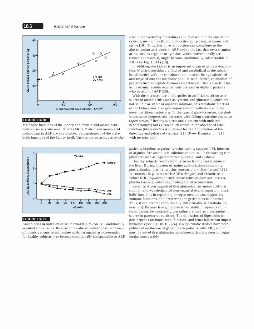
FIGURE 18-10
Metabolic functions of the kidney and protein and amino acidmetabolism in acute renal failure (ARF). Protein and amino acidmetabolism in ARF are also affected by impairment of the meta-bolic functions of the kidney itself. Various amino acids are synthe-
FIGURE 18-11
Amino acids in nutrition of acute renal failure (ARF): Conditionallyessential amino acids. Because of the altered metabolic environmentof uremic patients certain amino acids designated as nonessentialfor healthy subjects may become conditionally indispensable to ARF
sized or converted by the kidneys and released into the circulation:cysteine, methionine (from homocysteine), tyrosine, arginine, andserine [18]. Thus, loss of renal function can contribute to thealtered amino acid pools in ARF and to the fact that several aminoacids, such as arginine or tyrosine, which conventionally aretermed nonessential, might become conditionally indispensable inARF (see Fig. 18-11) [19].
In addition, the kidney is an important organ of protein degrada-tion. Multiple peptides are filtered and catabolized at the tubularbrush border, with the constituent amino acids being reabsorbedand recycled into the metabolic pool. In renal failure, catabolism ofpeptides such as peptide hormones is retarded. This is also true foracute uremia: insulin requirements decrease in diabetic patientswho develop of ARF [20].
With the increased use of dipeptides in artificial nutrition as asource of amino acids (such as tyrosine and glutamine) which arenot soluble or stable in aqueous solutions, this metabolic functionof the kidney may also gain importance for utilization of thesenovel nutritional substrates. In the case of glycyl-tyrosine, metabol-ic clearance progressively decreases with falling creatinine clearance(open circles, 7 healthy subjects and a patient with unilateralnephrectomy*) but extrarenal clearance in the absence of renalfunction (black circles) is sufficient for rapid utilization of thedipeptide and release of tyrosine [21]. (From Druml et al. [21];with permission.)
patients: histidine, arginine, tyrosine, serine, cysteine [19]. Infusionof arginine-free amino acid solutions can cause life-threatening com-plications such as hyperammonemia, coma, and acidosis.
Healthy subjects readily form tyrosine from phenylalanine in the liver: During infusion of amino acid solutions containingphenylalanine, plasma tyrosine concentration rises (circles) [22]. In contrast, in patients with ARF (triangles) and chronic renal failure (CRF, squares) phenylalanine infusion does not increaseplasma tyrosine, indicating inadequate interconversion.
Recently, it was suggested that glutamine, an amino acid that traditionally was designated non-essential exerts important meta-bolic functions in regulating nitrogen metabolism, supportingimmune functions, and preserving the gastrointestinal barrier.Thus, it can become conditionally indispensable in catabolic ill-ness [23]. Because free glutamine is not stable in aqueous solu-tions, dipeptides containing glutamine are used as a glutaminesource in parenteral nutrition. The utilization of dipeptides in part depends on intact renal function, and renal failure can impairhydrolysis (see Fig. 18-10) [24]. No systematic studies have beenpublished on the use of glutamine in patients with ARF, and itmust be noted that glutamine supplementation increases nitrogenintake considerably.

18.7Nutrition and Metabolism in Acute Renal Failure
FIGURE 18-12
Estimation of protein catabolism and nitrogen balance. The extentof protein catabolism can be assessed by calculating the urea nitro-gen appearance rate (UNA), because virtually all nitrogen arisingfrom amino acids liberated during protein degradation is convertedto urea. Besides urea in urine (UUN), nitrogen losses in other bodyfluids (eg, gastrointestinal, choledochal) must be added to anychange in the urea pool. When the UNA rate is multiplied by 6.25,it can be converted to protein equivalents. With known nitrogenintake from the parenteral or enteral nutrition, nitrogen balancecan be estimated from the UNA calculation.
Protein requirements
ESTIMATING THE EXTENT OF PROTEIN CATABOLISM
Urea nitrogen appearance (UNA) (g/d)
� Urinary urea nitrogen (UUN) excretion
� Change in urea nitrogen pool
� (UUN � V) � (BUN2 � BUN1) 0.006 � BW
� (BW2 � BW1) � BUN2/100
If there are substantial gastrointestinal losses, add urea nitrogen in secretions:
� volume of secretions � BUN2
Net protein breakdown (g/d) � UNA � 6.25
Muscle loss (g/d) �UNA � 6.25 � 5
V is urine volume; BUN1 and BUN2 are BUN in mg/dL on days 1 and 2
BW1 and BW2 are body weights in kg on days 1 and 2
FIGURE 18-13
Amino acid and protein requirements of patients with acute renalfailure (ARF). The optimal intake of protein or amino acids isaffected more by the nature of the underlying cause of ARF andthe extent of protein catabolism and type and frequency of dialysisthan by kidney dysfunction per se. Unfortunately, only a few stud-ies have attempted to define the optimal requirements for proteinor amino acids in ARF:
In nonhypercatabolic patients, during the polyuric phase of ARFprotein intake of 0.97 g/kg body weight per day was required toachieve a positive nitrogen balance [25]. A similar number (1.03g/kg
body weight per day) was derived from a study in which, unfortu-nately, energy intake was not kept constant [6]. In the polyuricrecovery phase in patients with sepsis-induced ARF, a nitrogen intakeof 15 g/day (averaging an amino acid intake of 1.3 g/kg body weightper day) as compared to 4.4 g/kg per day (about 0.3 g/kg aminoacids) was superior in ameliorating nitrogen balance [26].
Several recent studies have tried to evaluate protein and aminoacid requirements of critically ill patients with ARF. Kierdorf andassociates found that, in these hypercatabolic patients receiving continuous hemofiltration therapy, the provision of amino acids 1.5g /kg body weight per day was more effective in reducing nitrogenloss than infusion of 0.7 g (�3.4 versus �8.1 g nitrogen per day)[27]. An increase of amino acid intake to 1. 74 g/kg per day did notfurther ameliorate nitrogen balance.
Chima and coworkers measured a mean PCR of 1.7 g kg bodyweight per day in 19 critically ill ARF patients and concluded thatprotein needs in these patients range between 1.4 and 1.7 g/kg perday [28]. Similarly, Marcias and coworkers have obtained a proteincatabolic rate (PCR) of 1.4 g/kg per day and found an inverse relationship between protein and energy provision and PCR andagain recommended protein intake of 1.5 to 1.8 g/kg per day [29].Similar conclusions were drawn by Ikitzler in evaluating ARFpatients on intermittent hemodialysis therapy [30]. (From Kierdorfet al. [27]; with permission.)
18.8 Acute Renal Failure
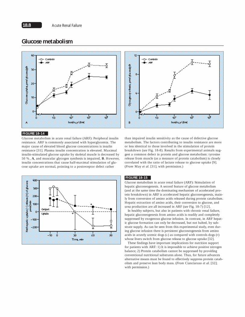
FIGURE 18-14
Glucose metabolism in acute renal failure (ARF): Peripheral insulinresistance. ARF is commonly associated with hyperglycemia. Themajor cause of elevated blood glucose concentrations is insulinresistance [31]. Plasma insulin concentration is elevated. Maximalinsulin-stimulated glucose uptake by skeletal muscle is decreased by50 %, A, and muscular glycogen synthesis is impaired, B. However,insulin concentrations that cause half-maximal stimulation of glu-cose uptake are normal, pointing to a postreceptor defect rather
Glucose metabolism
than impaired insulin sensitivity as the cause of defective glucosemetabolism. The factors contributing to insulin resistance are moreor less identical to those involved in the stimulation of proteinbreakdown (see Fig. 18-8). Results from experimental animals sug-gest a common defect in protein and glucose metabolism: tyrosinerelease from muscle (as a measure of protein catabolism) is closelycorrelated with the ratio of lactate release to glucose uptake [9].(From May et al. [31]; with permission.)
FIGURE 18-15
Glucose metabolism in acute renal failure (ARF): Stimulation ofhepatic gluconeogenesis. A second feature of glucose metabolism (and at the same time the dominating mechanism of accelerated pro-tein breakdown) in ARF is accelerated hepatic gluconeogenesis, main-ly from conversion of amino acids released during protein catabolism.Hepatic extraction of amino acids, their conversion to glucose, andurea production are all increased in ARF (see Fig. 18-7) [12].
In healthy subjects, but also in patients with chronic renal failure,hepatic gluconeogenesis from amino acids is readily and completelysuppressed by exogenous glucose infusion. In contrast, in ARF hepat-ic glucose formation can only be decreased, but not halted, by sub-strate supply. As can be seen from this experimental study, even dur-ing glucose infusion there is persistent gluconeogenesis from aminoacids in acutely uremic dogs (•) as compared with controls dogs (o)whose livers switch from glucose release to glucose uptake [32].
These findings have important implications for nutrition supportfor patients with ARF: 1) It is impossible to achieve positive nitrogenbalance; 2) Protein catabolism cannot be suppressed by providingconventional nutritional substrates alone. Thus, for future advancesalternative means must be found to effectively suppress protein catab-olism and preserve lean body mass. (From Cianciaruso et al. [32];with permission.)

18.9Nutrition and Metabolism in Acute Renal Failure
FIGURE 18-16
Lipid metabolism in acute renal failure (ARF). Profound alterationsof lipid metabolism occur in patients with ARF. The triglyceride con-tent of plasma lipoproteins, especially very low-density (VLDL) andlow-density ones (LDL) is increased, while total cholesterol and inparticular high-density lipoprotein (HDL) cholesterol are decreased[33,34]. The major cause of lipid abnormalities in ARF is impair-ment of lipolysis. The activities of both lipolytic systems, peripherallipoprotein lipase and hepatic triglyceride lipase are decreased inpatients with ARF to less than 50% of normal [35].
Maximal postheparin lipolytic activity (PHLA), hepatic triglyceridelipase (HTGL), and peripheral lipoprotein lipase (LPL) in 10 controls(open bars) and eight subjects with ARF (black bars). However, incontrast to this impairment of lipolysis, oxidation of fatty acids isnot affected by ARF. During infusion of labeled long-chain fattyacids, carbon dioxide production from lipid was comparablebetween healthy subjects and patients with ARF [36]. FFA—freefatty acids. (Adapted from Druml et al. [35]; with permission.)
Lipid metabolism
FIGURE 18-17
Impairment of lipolysis and elimination of artificial lipid emulsionsin acute renal failure (ARF). Fat particles of artificial fat emulsionsfor parenteral nutrition are degraded as endogenous very low-den-sity lipoprotein is. Thus, the nutritional consequence of theimpaired lipolysis in ARF is delayed elimination of intravenouslyinfused lipid emulsions [33, 34]. The increase in plasma triglyc-erides during infusion of a lipid emulsion is doubled in patientswith ARF (N=7) as compared with healthy subjects (N=6). Theclearance of fat emulsions is reduced by more than 50% in ARF.The impairment of lipolysis in ARF cannot be bypassed by usingmedium-chain triglycerides (MCT); the elimination of fat emul-sions containing long chain triglycerides (LCT) or MCT is equallyretarded in ARF [34]. Nevertheless, the oxydation of free fatty acidreleased from triglycerides is not inpaired in patients with ARF[36]. (From Druml et al. [34]; with permission.)

18.10 Acute Renal Failure
FIGURE 18-19
Electrolytes in acute renal failure (ARF): hypophosphatemia andhypokalemia. It must be noted that a considerable number ofpatients with ARF do not present with hyperkalemia or hyperphos-phatemia, but at least 5% have low serum potassium and morethan 12% have decreased plasma phosphate on admission [38].Nutritional support, especially parenteral nutrition with low elec-trolyte content, can cause hypophosphatemia and hypokalemia in as many as 50% and 19% of patients respectively [39,40].
In the case of phosphate, phosphate-free artificial nutrition causeshypophosphatemia within a few days, even if the patient was hyper-phosphatemic on admission (black circles) [41]. Supplementation of5 mmol per day was effective in maintaining normal plasma phos-phate concentrations (open squares), whereas infusion of more than10 mmol per day resulted in hyperphosphatemia, even if the patientshad decreased phosphate levels on admission (open circles).
Potassium or phosphate depletion increases the risk of developingARF and retards recovery of renal function. With modern nutrition-al support, hyperkalemia is the leading indication for initiation ofextracorporeal therapy in fewer than 5% of patients [38]. (Adaptedfrom Kleinberger et al. [41]; with permission.)
FIGURE 18-20
Micronutrients in acute renal failure (ARF): water-soluble vitamins.Balance studies on micronutrients (vitamins, trace elements) are notavailable for ARF. Because of losses associated with renal replace-ment therapy, requirements for water-soluble vitamins are expectedto be increased also in patients with ARF. Malnutrition with deple-tion of vitamin body stores and associated hypercatabolic underly-ing disease in ARF can further increase the need for vitamins.Depletion of thiamine (vitamin B1) during continuous hemofiltra-tion and inadequate intake can result in lactic acidosis and heartfailure [42]. This figure depicts the evolution of plasma lactate con-centration before and after administration of 600 mg thiamine intwo patients. Infusion of 600 mg of thiamine reversed the metabol-ic abnormality within a few hours. An exception to this approachto treatment is ascorbic acid (vitamin C); as a precursor of oxalicacid the intake should be kept below 200 mg per day because anyexcessive supply may precipitate secondary oxalosis [43]. (FromMadl et al. [42]; with permission.)
FIGURE 18-18
Electrolytes in acute renal failure (ARF): causes of hyperkalemiaand hyperphosphatemia. ARF frequently is associated with hyper-kalemia and hyperphosphatemia. Causes are not only impairedrenal excretion of electrolytes but release during catabolism, altereddistribution in intracellular and extracellular spaces, impaired cellular uptake, and acidosis. Thus, the type of underlying diseaseand degree of hypercatabolism also determine the occurrence andseverity of electrolyte abnormalities. Either hypophosphatemia orhyperphosphatemia can predispose to the development and maintenance of ARF [37].
Electrolytes and micronutrients
CAUSES OF ELECTROLYTE DERANGEMENTS IN ACUTE RENAL FAILURE
Hyperkalemia
Decreased renal elimination
Increased release during catabolism
2.38 mEq/g nitrogen
0.36 mEq/g glycogen
Decreased cellular uptake/increased release
Metabolic acidosis: 0.6 mmol/L rise/0.1decrease in pH
Hyperphosphatemia
Decreased renal elimination
Increased release from bone
Increased release during catabolism: 2 mmol/g nitrogen
Decreased cellular uptake/utilizationand/or increased release from cells
18.11Nutrition and Metabolism in Acute Renal Failure
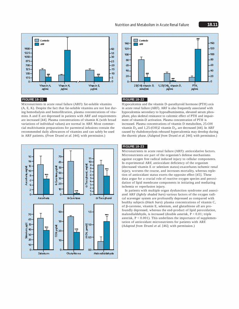
FIGURE 18-21
Micronutrients in acute renal failure (ARF): fat-soluble vitamins (A, E, K). Despite the fact that fat-soluble vitamins are not lost dur-ing hemodialysis and hemofiltration, plasma concentrations of vita-mins A and E are depressed in patients with ARF and requirementsare increased [44]. Plasma concentrations of vitamin K (with broadvariations of individual values) are normal in ARF. Most commer-cial multivitamin preparations for parenteral infusions contain therecommended daily allowances of vitamins and can safely be usedin ARF patients. (From Druml et al. [44]; with permission.)
FIGURE 18-22
Hypocalcemia and the vitamin D–parathyroid hormone (PTH) axis in acute renal failure (ARF). ARF is also frequently associated withhypocalcemia secondary to hypoalbuminemia, elevated serum phos-phate, plus skeletal resistance to calcemic effect of PTH and impair-ment of vitamin-D activation. Plasma concentration of PTH isincreased. Plasma concentrations of vitamin D metabolites, 25-OH vitamin D3 and 1,25-(OH)2 vitamin D3, are decreased [44]. In ARFcaused by rhabdomyolysis rebound hypercalcemia may develop duringthe diuretic phase. (Adapted from Druml et al. [44]; with permission.)
FIGURE 18-23
Micronutrients in acute renal failure (ARF): antioxidative factors.Micronutrients are part of the organism’s defense mechanismsagainst oxygen free radical induced injury to cellular components.In experimental ARF, antioxidant deficiency of the organism(decreased vitamin E or selenium status) exacerbates ischemic renalinjury, worsens the course, and increases mortality, whereas reple-tion of antioxidant status exerts the opposite effect [45]. Thesedata argue for a crucial role of reactive oxygen species and peroxi-dation of lipid membrane components in initiating and mediatingischemia or reperfusion injury.
In patients with multiple organ dysfunction syndrome and associ-ated ARF (lightly shaded bars) various factors of the oxygen radi-cal scavenger system are profoundly depressed as compared withhealthy subjects (black bars): plasma concentrations of vitamin C,of �-carotene, vitamin E, selenium, and glutathione all are pro-foundly depressed, whereas the end-product of lipid peroxidation,malondialdehyde, is increased (double asterisk, P < 0.01; tripleasterisk, P < 0.001). This underlines the importance of supplemen-tation of antioxidant micronutrients for patients with ARF.(Adapted from Druml et al. [46]; with permission.)

18.12 Acute Renal Failure
(arteriovenous) hemofiltration (CHF) and continuous hemodialysishave gained wide popularity. CRRTs are associated with multiplemetabolic effects in addition to “renal replacement” [47].
By cooling of the extracorporeal circuit and infusion of cooledsubstitution fluids, CHF may induce considerable heat loss (350 to700 kcal per day). On the other hand, hemofiltration fluids containlactate as anions, oxidation of which in part compensates for theheat loss. This lactate load can result in hyperlactemia in the pres-ence of liver dysfunction or increased endogenous lactate formationsuch as in circulatory shock.
Several nutrients with low protein binding and small molecularweight (sieving coefficient 0.8 to 1.0), such as vitamins or aminoacids are eliminated during therapy. Amino acid losses can be esti-mated from the volume of the filtrate and average plasma concen-tration, and usually this accounts for a loss of approximately 0.2g/L of filtrate and, depending on the filtered volume, 5 to 10 g ofamino acid per day, respectively, representing about 10 % of aminoacid input, but it can be even higher during continuous hemodiafil-tration [48].
With the large molecular size cut-off of membranes used in hemofil-tration, small proteins such as peptide hormones are filtered. In viewof their short plasma half-life hormone losses are minimal and proba-bly not of pathophysiologic importance. Quantitatively relevant elimi-nation of mediators by CRRT has not yet been proven. On the otherhand, prolonged blood-membrane interactions can induce conse-quences of bioincompatibility and activation of various endogenouscascade systems.
Metabolic Impact of Renal Replacement Therapy
METABOLIC EFFECTS OF CONTINUOUS RENAL REPLACEMENT THERAPY
Amelioration of uremia intoxication (renal replacement)
Plus
Heat loss
Excessive load of substrates (eg, lactate, glucose)
Loss of nutrients (eg, amino acids, vitamins)
Elimination of short-chain proteins (hormones, mediators?)
Induction or activation of mediator cascades
Stimulation of protein catabolism?
FIGURE 18-25
A, B, Impact of nutritional interventions on renal function andcourse of acute renal failure (ARF). Starvation accelerates proteinbreakdown and impairs protein synthesis in the kidney, whereasrefeeding exerts the opposite effects [49]. In experimental animals, provision of amino acids or total parenteral nutrition acceleratestissue repair and recovery of renal function [50]. In patients, however, this has been much more difficult to prove, and only onestudy has reported on a positive effect of TPN on the resolutionof ARF [51].
FIGURE 18-24
Metabolic impact of extracorporeal therapy. The impact of hemodial-ysis therapy on metabolism is multifactorial. Amino acid and proteinmetabolism are altered not only by substrate losses but also by activa-tion of protein breakdown mediated by release of leukocyte-derivedproteases, of inflammatory mediators (interleukins and tumor necro-sis factor) induced by blood-membrane interactions or endotoxin.Dialysis can also induce inhibition of muscle protein synthesis [15].
In the management of patients with acute renal failure (ARF), con-tinuous renal replacement therapies (CRRT), such as continuous
Nutrition, Renal Function, and Recovery
Infusion of amino acids raised renal cortical protein synthesis as evaluated by 14C-leucine incorporation and depressed protein breakdownin rats with mercuric chloride–induced ARF [49]. On the other hand, in asimilar model of ARF, infusions of varying quantities of essential aminoacids (EAA) and nonessential amino acids (NEAA) did not provide anyprotection of renal function and in fact increased mortality [52]. However,in balance available evidence suggests that provision of substrates mayenhance tissue regeneration and wound healing, and potentially, also renal tubular repair [49]. (From Toback et al. [50]; with permission.)

18.13Nutrition and Metabolism in Acute Renal Failure
FIGURE 18-26
Impact of nutritional interventions on renal function in acute renalfailure (ARF). Amino acid infused before or during ischemia ornephrotoxicity may enhance tubule damage and accelerate loss ofrenal function in rat models of ARF. In part, this therapeutic para-dox [53] from amino acid alimentation in ARF is related to theincrease in metabolic work for transport processes when oxygensupply is limited, which may aggravate ischemic injury [54].Similar observations have been made with excess glucose infusionduring renal ischemia. Amino acids may as well exert a protectiveeffect on renal function. Glycine, and to a lesser degree alanine,limit tubular injury in ischemic and nephrotoxic models of ARF[55]. Arginine (possibly by producing nitric oxide) reportedly actsto preserve renal perfusion and tubular function in both nephro-toxic and ischemic models of ARF, whereas inhibitors of nitricoxide synthase exert an opposite effect [56,57]. In myoglobin-induced ARF the drop in renal blood flow (black circles, ARF con-trols) is prevented by L-arginine infusion (black triangles) [57].(From Wakabayashi et al. [57]; with permission.)
ischemic ARF, A, but also reduces the increase in BUN andimproves nitrogen balance, B, [58]. (open circles) ARF plus vehi-cle; (black circles, sham-operated rats plus vehicle; open squares,ARF plus rhIGF-I; black squares, sham operated rats plus rhIGF-I.) Unfortunately, efficacy of these interventions was not uniform-ly confirmed in clinical studies [59, 60]. (From Ding et al. [58];with permission.)
FIGURE 18-27
Impact of endocrine-metabolic interventions on renal function andcourse of acute renal failure (ARF). Various other endocrine-meta-bolic interventions (eg, thyroxine, human growth hormone [HGH],epidermal growth factor, insulin-like growth factor 1 [IGF-1]) havebeen shown to accelerate regeneration after experimental ARF[51]. In a rat model of postischemic ARF, treatment with IGF-1starting 5 hours after induction of ARF accelerates recovery from
18.14 Acute Renal Failure
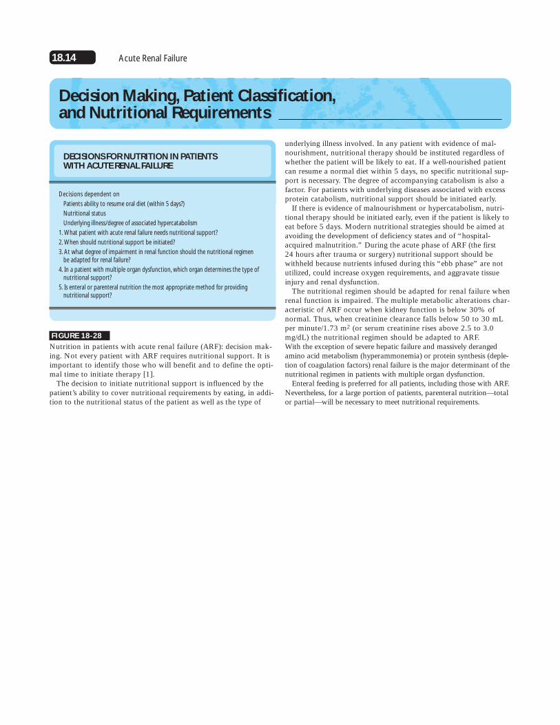
underlying illness involved. In any patient with evidence of mal-nourishment, nutritional therapy should be instituted regardless ofwhether the patient will be likely to eat. If a well-nourished patientcan resume a normal diet within 5 days, no specific nutritional sup-port is necessary. The degree of accompanying catabolism is also afactor. For patients with underlying diseases associated with excessprotein catabolism, nutritional support should be initiated early.
If there is evidence of malnourishment or hypercatabolism, nutri-tional therapy should be initiated early, even if the patient is likely toeat before 5 days. Modern nutritional strategies should be aimed atavoiding the development of deficiency states and of “hospital-acquired malnutrition.” During the acute phase of ARF (the first 24 hours after trauma or surgery) nutritional support should bewithheld because nutrients infused during this “ebb phase” are notutilized, could increase oxygen requirements, and aggravate tissueinjury and renal dysfunction.
The nutritional regimen should be adapted for renal failure whenrenal function is impaired. The multiple metabolic alterations char-acteristic of ARF occur when kidney function is below 30% ofnormal. Thus, when creatinine clearance falls below 50 to 30 mLper minute/1.73 m2 (or serum creatinine rises above 2.5 to 3.0mg/dL) the nutritional regimen should be adapted to ARF.With the exception of severe hepatic failure and massively derangedamino acid metabolism (hyperammonemia) or protein synthesis (deple-tion of coagulation factors) renal failure is the major determinant of thenutritional regimen in patients with multiple organ dysfunction.
Enteral feeding is preferred for all patients, including those with ARF.Nevertheless, for a large portion of patients, parenteral nutrition—totalor partial—will be necessary to meet nutritional requirements.
Decision Making, Patient Classification, and Nutritional Requirements
DECISIONS FOR NUTRITION IN PATIENTS WITH ACUTE RENAL FAILURE
Decisions dependent on
Patients ability to resume oral diet (within 5 days?)
Nutritional status
Underlying illness/degree of associated hypercatabolism
1. What patient with acute renal failure needs nutritional support?
2. When should nutritional support be initiated?
3. At what degree of impairment in renal function should the nutritional regimen be adapted for renal failure?
4. In a patient with multiple organ dysfunction, which organ determines the type ofnutritional support?
5. Is enteral or parenteral nutrition the most appropriate method for providing nutritional support?
FIGURE 18-28
Nutrition in patients with acute renal failure (ARF): decision mak-ing. Not every patient with ARF requires nutritional support. It isimportant to identify those who will benefit and to define the opti-mal time to initiate therapy [1].
The decision to initiate nutritional support is influenced by thepatient’s ability to cover nutritional requirements by eating, in addi-tion to the nutritional status of the patient as well as the type of
18.15Nutrition and Metabolism in Acute Renal Failure
PATIENT CLASSIFICATION AND SUBSTRATE REQUIREMENTS IN PATIENTS WITH ACUTE RENAL FAILURE
Excess urea appearance (above nitrogen intake)
Clinical setting (examples)
Mortality
Dialysis or hemofiltration frequency
Route of nutrient administration
Energy recommendations (kcal/kg BW/d)
Energy substrates
Glucose (g/kg BW/d)
Fat (g/kg BW/d)
Amino acids/protein (g/kg/d)
Nutrients used
Mild
>6 g
Drug toxicity
20 %
Rare
Oral
25
Glucose
3.0–5.0
0.6–1.0
EAA (�NEAA)
Foods
Moderate
6–12 g
Elective surgery ± infection
60%
As needed
Enteral or parenteral
25–30
Glucose + fat
3.0–5.0
0.5–1.0
0.8–1.2
EAA � NEAA
Enteral formulas
Glucose 50%–70% �fat emulsions 10% or 20%
Severe
>12 g
Severe injury or sepsis
>80%
Frequent
Enteral or parenteral
25–35
Glucose � fat
3.0–5.0 (max. 7.0)
0.8–1.5
1.0–1.5
EAA � NEAA
Enteral formulas
Glucose 50%–70% +fat emulsions 10%or 20%
BW—body weight; EAA—essential amino acids; NEAA—nonessential amino acids.
Extent of Catabolism
FIGURE 18-29
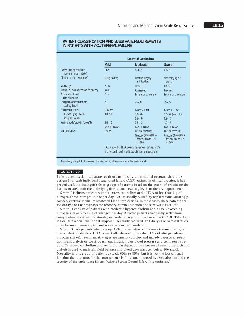
Patient classification: substrate requirements. Ideally, a nutritional program should bedesigned for each individual acute renal failure (ARF) patient. In clinical practice, it hasproved useful to distinguish three groups of patients based on the extent of protein catabo-lism associated with the underlying disease and resulting levels of dietary requirements.
Group I includes patients without excess catabolism and a UNA of less than 6 g ofnitrogen above nitrogen intake per day. ARF is usually caused by nephrotoxins (aminogly-cosides, contrast media, mismatched blood transfusion). In most cases, these patients arefed orally and the prognosis for recovery of renal function and survival is excellent.
Group II consists of patients with moderate hypercatabolism and a UNA exceedingnitrogen intake 6 to 12 g of nitrogen per day. Affected patients frequently suffer fromcomplicating infections, peritonitis, or moderate injury in association with ARF. Tube feed-ing or intravenous nutritional support is generally required, and dialysis or hemofiltrationoften becomes necessary to limit waste product accumulation.
Group III are patients who develop ARF in association with severe trauma, burns, oroverwhelming infection. UNA is markedly elevated (more than 12 g of nitrogen abovenitrogen intake). Treatment strategies are usually complex and include parenteral nutri-tion, hemodialysis or continuous hemofiltration plus blood pressure and ventilatory sup-port. To reduce catabolism and avoid protein depletion nutrient requirements are high anddialysis is used to maintain fluid balance and blood urea nitrogen below 100 mg/dL.Mortality in this group of patients exceeds 60% to 80%, but it is not the loss of renalfunction that accounts for the poor prognosis. It is superimposed hypercatabolism and theseverity of the underlying illness. (Adapted from Druml [1]; with permission.)
EAA + specific NEAA solutions (general or “nephro”)
Multivitamin and multitrace element preparations
18.16 Acute Renal Failure
Enteral Nutrition
FIGURE 18-30
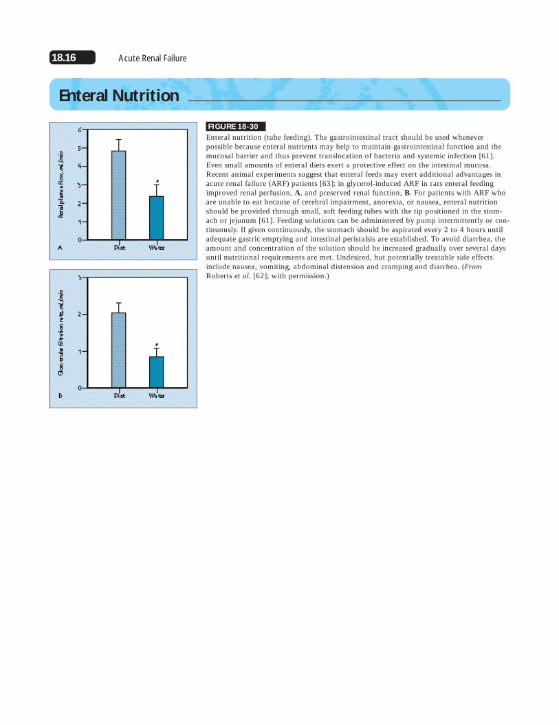
Enteral nutrition (tube feeding). The gastrointestinal tract should be used whenever possible because enteral nutrients may help to maintain gastrointestinal function and themucosal barrier and thus prevent translocation of bacteria and systemic infection [61].Even small amounts of enteral diets exert a protective effect on the intestinal mucosa.Recent animal experiments suggest that enteral feeds may exert additional advantages inacute renal failure (ARF) patients [63]: in glycerol-induced ARF in rats enteral feedingimproved renal perfusion, A, and preserved renal function, B. For patients with ARF whoare unable to eat because of cerebral impairment, anorexia, or nausea, enteral nutritionshould be provided through small, soft feeding tubes with the tip positioned in the stom-ach or jejunum [61]. Feeding solutions can be administered by pump intermittently or con-tinuously. If given continuously, the stomach should be aspirated every 2 to 4 hours untiladequate gastric emptying and intestinal peristalsis are established. To avoid diarrhea, theamount and concentration of the solution should be increased gradually over several daysuntil nutritional requirements are met. Undesired, but potentially treatable side effectsinclude nausea, vomiting, abdominal distension and cramping and diarrhea. (FromRoberts et al. [62]; with permission.)

18.17Nutrition and Metabolism in Acute Renal Failure
FIGURE 18-31
Enteral feeding formulas. There are standardized tube feeding for-mulas designed for subjects with normal renal function that canalso be given to patients with acute renal failure (ARF).Unfortunately, the fixed composition of nutrients, including pro-teins and high content of electrolytes (especially potassium andphosphate) often limits their use for ARF.
Alternatively, enteral feeding formulas designed for nutritionaltherapy of patients with chronic renal failure (CRF) can be used.The preparations listed here may have advantages also for patients
SPECIFIC ENTERAL FORMULAS FOR NUTRITIONAL SUPPORT OF PATIENTS WITH RENAL FAILURE
Volume (mL)
Calories (kcal)
(cal/mL)
Energy distribution
Protein:fat:carbohydrates (%)
kcal/g N
Proteins (g)
EAA (%)
NEAA (%)
Hydrolysate (%)
Full protein (%)
Nitrogen (g)
Carbohydrates (g)
Monodisaccharides (%)
Oligosaccharides (%)
Polysaccharides (%)
Fat (g)
LCT (%)
Essential GA (%)
MCT (%)
Nonprotein (cal/g N)
Osmol (mOsm/kg)
Sodium (mmol/L)
Potassium (mmol/L)
Phosphate (mmol)
Vitamins
Minerals
Amin-Aid
750
1467
1.96
4:21:75
832:1
14.6
100
—
—
—
1.76
274
100
—
—
34.6
502
1095
11
—
—
b
b
Travasorb renal*
1050
1400
1.35
7:12:81
389:1
24.0
60
30
—
—
3.6
284
100
—
—
18.6
30
18
70
363
590
—
—
16.1
a
b
Salvipeptide nephro†
500
1000
2.00
8:22:70
313:1
20.0
23
20
23
34
3.2
175
3
28
69
24
50
31
50
288
507
7.2
1.5
6.13
a
a
Survimed renal‡
1000
1320
1.32
6:10:84
398:1
20.8
100
—
3.32
276
88
15.2
52
30
374
600
15.2
8
6.4
a
a
* 3 bags � 810 mL � 1050 mL† component I � component II � 350 mL = 500 mL‡ 4 bags � 800 mL � 1000 mL§ Liquid formula, cans 8 fl oz (�237.5 mL), supplemented with carnitine, taurine with a low-protein (Suplena) or moderate-protein content (Nepro)
a 2000 kcal/d meets RDA for most vitamins/trace elements
b Must be added
EAA—essential amino acids; FA—fatty acids; LCT—long-chain triglycerides; MCT—medium-chain triglycerides; N—nitrogen; NEAA—non-essential amino acids.
Suplena§
500
1000
2.00
6:43:51
418:1
15.0
100
2.4
128
10
48
100
22
0
393
635
32
27.0
11.0
a
a
Nepro§
500
1000
2.00
14:43:43
179:1
35
100
5.6
108
12
90
47.8
100
0
154
615
34.0
28.5
11.0
a
a
with ARF. The protein content is lower and is confined to high-quality proteins (in part as oligopeptides and free amino acids), theelectrolyte concentrations are restricted. Most formulations containrecommended allowances of vitamins and minerals.
In part, these enteral formulas are made up of components thatincrease the flexibility in nutritional prescription and enable adapta-tion to individual needs. The diets can be supplemented with addi-tional electrolytes, protein, and lipids as required. Recently, ready-to-use liquid diets have also become available for renal failure patients.
18.18 Acute Renal Failure
FIGURE 18-32
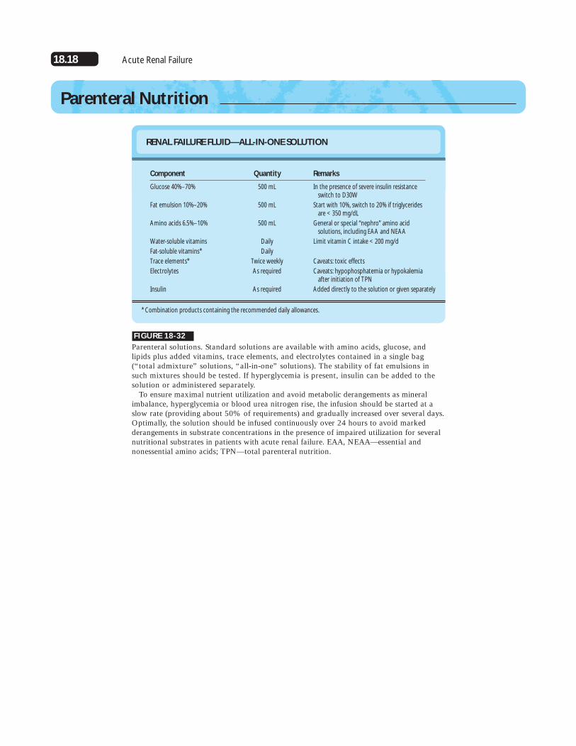
Parenteral solutions. Standard solutions are available with amino acids, glucose, and lipids plus added vitamins, trace elements, and electrolytes contained in a single bag(“total admixture” solutions, “all-in-one” solutions). The stability of fat emulsions insuch mixtures should be tested. If hyperglycemia is present, insulin can be added to thesolution or administered separately.
To ensure maximal nutrient utilization and avoid metabolic derangements as mineralimbalance, hyperglycemia or blood urea nitrogen rise, the infusion should be started at aslow rate (providing about 50% of requirements) and gradually increased over several days.Optimally, the solution should be infused continuously over 24 hours to avoid markedderangements in substrate concentrations in the presence of impaired utilization for severalnutritional substrates in patients with acute renal failure. EAA, NEAA—essential andnonessential amino acids; TPN—total parenteral nutrition.
Parenteral Nutrition
RENAL FAILURE FLUID—ALL-IN-ONE SOLUTION
Component
Glucose 40%–70%
Fat emulsion 10%–20%
Amino acids 6.5%–10%
Water-soluble vitamins
Fat-soluble vitamins*
Trace elements*
Electrolytes
Insulin
Quantity
500 mL
500 mL
500 mL
Daily
Daily
Twice weekly
As required
As required
Remarks
In the presence of severe insulin resistance switch to D30W
Start with 10%, switch to 20% if triglycerides are < 350 mg/dL
General or special “nephro” amino acid solutions, including EAA and NEAA
Limit vitamin C intake < 200 mg/d
Caveats: toxic effects
Caveats: hypophosphatemia or hypokalemia after initiation of TPN
Added directly to the solution or given separately
* Combination products containing the recommended daily allowances.

18.19Nutrition and Metabolism in Acute Renal Failure
FIGURE 18-33
Amino acid (AA) solutions for parenteral nutrition in acute renalfailure (ARF). The most controversial choice regards the type ofamino acid solution to be used: either essential amino acids (EAAs)exclusively, solutions of EAA plus nonessential amino acids(NEAAs), or specially designed “nephro” solutions of differentproportions of EAA and specific NEAA that might become “condi-tionally essential” for ARF (see Fig. 18-11).
Use of solutions of EAA alone is based on principles established fortreating chronic renal failure (CRF) with a low-protein diet and anEAA supplement. This may be inappropriate as the metabolic adapta-tions to low-protein diets in response to CRF may not have occurredin patients with ARF. Plus, there are fundamental differences in thegoals of nutritional therapy in the two groups of patients, and conse-quently, infusion solutions of EAA may be sub-optimal.
Thus, a solution should be chosen that includes both essentialand nonessential amino acids (EAA, NEAA) in standard propor-
AMINO ACID SOLUTIONS FOR THE TREATMENT OF ACUTE RENAL FAILURE (“NEPHRO” SOLUTIONS)
Amino acids (g/L)(� g/%)
Volume (mL)
(mOsm/L)
Nitrogen (g/L)
Essential amino acids (g/L)Isoleucine
Leucine
Lysine acetate/HCl
Methionine
Phenylalanine
Threonine
Tryptophan
Valine
Nonessential amino acids (g/L)Alanine
Arginine
Glycine
Histidine
Proline
Serine
Tyrosine
Cysteine
Rose-Requirements
1.40
2.20
1.60
2.20
2.20
1.00
0.50
1.60
RenAmin (Clintec)
65
6.5
500
600
10
5.00
6.00
4.50
5.00
4.90
3.80
1.60
8.20
5.60
6.30
3.00
4.20
3.50
3.00
0.40
Aminess (Clintec)
52
5.2
400
416
8.3
5.25
8.25
6.00
8.25
8.25
3.75
1.88
6.00
4.12
Aminosyn RF (Abbott)
52
5.2
1000
475
8.3
4.62
7.26
5.35
7.26
7.26
3.30
1.60
5.20
6.00
4.29
* Glycine is a componenet of the dipeptide.† Tyrosine is included as dipeptide (glycyl-L-tyrosine).
NephrAmine(McGaw)
54
5.4
1000
435
6.5
5.60
8.80
6.40
8.80
8.80
4.00
2.00
6.40
2.50
0.20
Nephrotect(Fresenius)
100
10
500
908
16.3
5.80
12.80
12.00
2.00
3.50
8.20
3.00
8.70
6.20
8.20
6.30*
9.80
3.00
7.60
3.00†
0.40
tions or in special proportions designed to counteract the metabolic changes of renal failure (“nephro” solutions), includ-ing the amino acids that might become conditionally essential in ARF.
Because of the relative insolubility of tyrosine in water, dipep-tides containing tyrosine (such as glycyl-tyrosine) are contained inmodern nephro solutions as the tyrosine source [22, 23]. Oneshould be aware of the fact that the amino acid analogue N-acetyltyrosine, which previously was used frequently as a tyrosinesource, cannot be converted into tyrosine in humans and mighteven stimulate protein catabolism [21].
Despite considerable investigation, there is no persuasive evi-dence that amino acid solutions enriched in branched-chain aminoacids exert a clinically significant anticatabolic effect. Systematicstudies using glutamine supplementation for patients with ARF arelacking (see Fig. 18-11).

18.20 Acute Renal Failure
FIGURE 18-34
Energy substrates in total parenteral nutrition (TPN) in acute renal failure (ARF): glucose and lipids. Because of the well-docu-mented effects of overfeeding, energy intake of patients with ARFmust not exceed their actual energy expenditure (ie, in most cases100% to 130% of resting energy expenditure [REE]; see Figs. 18-3and 18-4) [2].
Glucose should be the principal energy substrate because it can beutilized by all organs, even under hypoxic conditions, and has thepotential for nitrogen sparing. Since ARF impairs glucose tolerance,insulin is frequently necessary to maintain normoglycemia. Anyhyperglycemia must be avoided because of the untoward associatedside effects—such as aggravation of tissue injury, glycation of pro-teins, activation of protein catabolism, among others [2]. Whenintake is increased above 5 g/kg of body weight per day infused glu-cose will not be oxidized but will promote lipogenesis with fattyinfiltration of the liver and excessive carbon dioxide production andhypercarbia. Often, energy requirements cannot be met by glucoseinfusion without adding large amounts of insulin, so a portion ofthe energy should be supplied by lipid emulsions [2].
The most suitable means of providing the energy substrates forparenteral nutrition for patients with ARF is not glucose or lipids,but glucose and lipids [2]. In experimental uremia in rats, TPNwith 30% of nonprotein energy as fat promoted weight gain andameliorated the uremic state and survival [63]. (From Wennberg et al. [63]; with permission.)
FIGURE 18-35
Energy substrates in parenteral nutrition: lipid emulsions. Advantages of intravenous lipidsinclude high specific energy content, low osmolality, provision of essential fatty acids andphospholipids to prevent deficiency syndromes, fewer hepatic side effects (such as steato-sis, hyperbilirubinemia), and reduced carbon dioxide production, especially relevant forpatients with respiratory failure.
Changes in lipid metabolism associated with acute renal failure (ARF) should not pre-vent the use of lipid emulsions. Instead, the amount infused should be adjusted to meet thepatient’s capacity to utilize lipids. Usually, 1 g/kg of body weight per day of fat will notincrease plasma triglycerides substantially, so about 20% to 25% of energy requirementscan be met [1]. Lipids should not be administered to patients with hyperlipidemia (ie, plas-ma triglycerides above 350 mg/dL) activated intravascular coagulation, acidosis (pH below7.25), impaired circulation or hypoxemia.
Parenteral lipid emulsions usually contain long-chain triglycerides (LCT), most derivedfrom soybean oil. Recently, fat emulsions containing a mixture of LCT and medium-chaintriglycerides (MCT) have been introduced for intravenous use. Proposed advantagesinclude faster elimination from the plasma owing to higher affinity to the lipoproteinlipase enzyme, complete, rapid, and carnitine-independent metabolism, and a triglyceride-lowering effect; however, use of MCT does not promote lipolysis, and elimination oftriglycerides of both types of fat emulsions is equally retarded in ARF [34]. (Adapted from[34]; with permission.)
18.21Nutrition and Metabolism in Acute Renal Failure
FIGURE 18-36
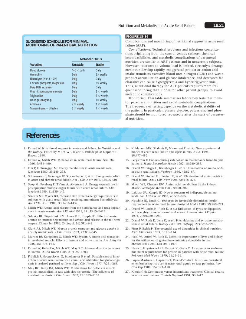
Complications and monitoring of nutritional support in acute renalfailure (ARF).
Complications: Technical problems and infectious complica-tions originating from the central venous catheter, chemicalincompatibilities, and metabolic complications of parenteralnutrition are similar in ARF patients and in nonuremic subjects.However, tolerance to volume load is limited, electrolyte derange-ments can develop rapidly, exaggerated protein or amino acidintake stimulates excessive blood urea nitrogen (BUN) and wasteproduct accumulation and glucose intolerance, and decreased fatclearance can cause hyperglycemia and hypertriglyceridemia.Thus, nutritional therapy for ARF patients requires more fre-quent monitoring than it does for other patient groups, to avoidmetabolic complications.
Monitoring: This table summarizes laboratory tests that moni-tor parenteral nutrition and avoid metabolic complications. The frequency of testing depends on the metabolic stability ofthe patient. In particular, plasma glucose, potassium, and phos-phate should be monitored repeatedly after the start of parenter-al nutrition.
SUGGESTED SCHEDULE FOR MINIMAL MONITORING OF PARENTERAL NUTRITION
Variables
Blood glucose
Osmolality
Electrolytes (Na+, K+, Cl+)
Calcium, phosphate, magnesium
Daily BUN increment
Urea nitrogen appearance rate
Triglycerides
Blood gas analysis, pH
Ammonia
Transaminases � bilirubin
Unstable
1–6 � daily
Daily
Daily
Daily
Daily
Daily
Daily
Daily
2 � weekly
2 � weekly
Stable
Daily
2� weekly
Daily
3� weekly
Daily
2 � weekly
2 � weekly
1� weekly
1 � weekly
1 � weekly
Metabolic Status
References
1. Druml W: Nutritional support in acute renal failure. In Nutrition andthe Kidney. Edited by Mitch WE, Klahr S. Philadelphia: Lippincott-Raven, 1998.
2. Druml W, Mitch WE: Metabolism in acute renal failure. Sem Dial1996, 9:484–490.
3. Om P, Hohenegger M: Energy metabolism in acute uremic rats.Nephron 1980, 25:249–253.
4. Schneeweiss B, Graninger W, Stockenhuber F, et al.: Energy metabolismin acute and chronic renal failure. Am J Clin Nutr 1990, 52:596–601.
5. Soop M, Forsberg E, Thˆrne A, Alvestrand A: Energy expenditure inpostoperative multiple organ failure with acute renal failure. ClinNephrol 1989, 31:139–145.
6. Spreiter SC, Myers BD, Swenson RS: Protein-energy requirements insubjects with acute renal failure receiving intermittent hemodialysis.Am J Clin Nutr 1980, 33:1433–1437.
7. Mitch WE: Amino acid release from the hindquarter and urea appear-ance in acute uremia. Am J Physiol 1981, 241:E415–E419.
8. Salusky IB, Flügel-Link RM, Jones MR, Kopple JD: Effect of acuteuremia on protein degradation and amino acid release in the rat hemi-corpus. Kidney Int 1983, 24(Suppl. 16):S41–S42.
9. Clark AS, Mitch WE: Muscle protein turnover and glucose uptake inacutely uremic rats. J Clin Invest 1983, 72:836–845.
10. Maroni BJ, Karapanos G, Mitch WE: System A amino acid transportin incubated muscle: Effects of insulin and acute uremia. Am J Physiol1986, 251:F74–F80.
11. Druml W, Kelly RA, Mitch WE, May RC: Abnormal cation transportin uremia. J Clin Invest 1988, 81:1197–1203.
12. Fröhlich J, Hoppe-Seyler G, Schollmeyer P, et al.: Possible sites of inter-action of acute renal failure with amino acid utilization for gluconeoge-nesis in isolated perfused rat liver. Eur J Clin Invest 1977, 7:261–268.
13. May RC, Kelly RA, Mitch WE: Mechanisms for defects in muscleprotein metabolism in rats with chronic uremia: The influence ofmetabolic acidosis. J Clin Invest 1987; 79:1099–1103.
14. Kuhlmann MK, Shahmir E, Maasarani E, et al.: New experimentalmodel of acute renal failure and sepsis in rats. JPEN 1994,18:477–485.
15. Bergström J: Factors causing catabolism in maintenance hemodialysispatients. Miner Electrolyte Metab 1992, 18:280–283.
16. Druml W, Bürger U, Kleinberger G, et al.: Elimination of amino acidsin acute renal failure. Nephron 1986, 42:62–67.
17. Druml W, Fischer M, Liebisch B, et al.: Elimination of amino acids inrenal failure. Am J Clin Nutr 1994, 60:418–423.
18. Mitch WE, Chesney RW: Amino acid metabolism by the kidney.Miner Electrolyte Metab 1983, 9:190–202.
19. Laidlaw SA, Kopple JD: Newer concepts of indispensable aminoacids. Am J Clin Nutr 1987, 46:593–605.
20. Naschitz JE, Barak C, Yeshurun D: Reversible diminished insulinrequirement in acute renal failure. Postgrad Med J 1983, 59:269–271.
21. Druml W, Lochs H, Roth E, et al.: Utilisation of tyrosine dipeptidesand acetyl-tyrosine in normal and uremic humans. Am J Physiol1991, 260:E280–E285.
22. Druml W, Roth E, Lenz K, et al.: Phenylalanine and tyrosine metabo-lism in renal failure. Kidney Int 1989, 36(Suppl 27):S282–S286.
23. Fürst P. Stehle P: The potential use of dipeptides in clinical nutrition.Nutr Clin Pract 1993, 8:106–114.
24. Hübl W, Druml W, Roth E, Lochs H: Importance of liver and kidneyfor the utilization of glutamine-containing dipeptides in man.Metabolism 1994, 43:1104–1107.
25. Hasik J, Hryniewiecki L, Baczyk K, Grala T: An attempt to evaluateminimum requirements for protein in patients with acute renal failure.Pol Arch Med Wewn 1979, 61:29–36.
26. Lopez-Martinez J, Caparros T, Perez-Picouto F: Nutrition parenteralen enfermos septicos con fracaso renal agudo en fase poliurica. RevClin Esp 1980, 157:171–178.
27. Kierdorf H: Continuous versus intermittent treatment: Clinical resultsin acute renal failure. Contrib Nephrol 1991, 93:1–12.

18.22 Acute Renal Failure
28. Chima CS, Meyer L, Hummell AC, et al.: Protein catabolic rate inpatients with acute renal failure on continuous arteriovenous hemofil-tration and total parenteral nutrition. J Am Soc Nephrol 1993,3:1516–1521.
29. Macias WL, Alaka KJ, Murphy MH, et al.: Impact of nutritional regi-men on protein catabolism and nitrogen balance in patients withacute renal failure. JPEN 1996, 20:56–62.
30. Ikizler TA, Greene JH, Wingard RL, Hakim RM: Nitrogen balance inacute renal failure patients. J Am Soc Nephrol 1995, 6:466A.
31. May RC, Clark AS, Goheer MA, Mitch WE: Specific defects ininsulin-mediated muscle metabolism in acute uremia. Kidney Int1985, 28:490–497.
32. Cianciaruso B, Bellizzi V, Napoli R, et al.: Hepatic uptake and releaseof glucose, lactate and amino acids in acutely uremic dogs.Metabolism 1991, 40:261–290.
33. Druml W, Laggner A, Widhalm K, et al.: Lipid metabolism in acuterenal failure. Kidney Int 1983, 24(Suppl 16):S139–S142.
34. Druml W, Fischer M, Sertl S, et al.: Fat elimination in acute renal fail-ure: Long-chain versus medium-chain triglycerides. Am J Clin Nutr1992, 55:468–472.
35. Druml W, Zechner R, Magometschnigg D, et al.: Post-heparin lipolyt-ic activity in acute renal failure. Clin Nephrol 1985, 23:289–293.
36. Adolph M, Eckart J, Metges C, et al.: Oxidative utilization of lipidemulsions in septic patients with and without acute renal failure. ClinNutr 1995, 14(Suppl 2):35A.
37. Dobyan DC, Bulger RE, Eknoyan G: The role of phosphate in thepotentiation and amelioration of acute renal failure. Miner ElectrolyteMetab 1991, 17:112–115.
38. Druml W, Lax F, Grimm G, et al.: Acute renal failure in the elderly—1975–1990. Clin Nephrol 1994, 41:342–349.
39. Kurtin P, Kouba J: Profound hypophosphatemia in the course of acuterenal failure. Am J Kidney Dis 1987, 10:346–349.
40. Marik PE, Bedigian MK: Refeeding hypophosphatemia in critically illpatients in an intensive care unit. Arch Surg 1996, 131:1043–1047.
41. Kleinberger G, Gabl F, Gassner A, et al.: Hypophosphatemia duringparenteral nutrition in patients with renal failure. Wien KlinWochenschr 1978, 90:169–172.
42. Madl Ch, Kranz A, Liebisch B, et al.: Lactic acidosis in thiamine defi-ciency. Clin Nutr 1993, 12:108–111.
43. Friedman AL, Chesney RW, Gilbert EF, et al.: Secondary oxalosis as acomplication of parenteral alimentation in acute renal failure. Am JNephrol 1983, 3:248–252.
44. Druml W, Schwarzenhofer M, Apsner R, Hörl WH: Fat soluble vita-mins in acute renal failure. Miner Electrolyte Metab 1998, 24:220–226.
45. Zurovsky Y, Gispaan I: Antioxidants attenuate endotoxin-inducedacute renal failure in rats. Am J Kidney Dis 1995, 25:51–57.
46. Druml W, Bartens C, Stelzer H, et al.: Impact of acute renal failure onantioxidant status in multiple organ failure syndrome. JASN 1993,4:314A.
47. Druml W: Impact of continuous renal replacement therapies onmetabolism. Int J Artif Organs 1996, 19:118–120.
48. Frankenfeld DC, Badellino MM, Reynolds HN, et al.: Amino acidloss and plasma concentration during continuous hemodiafiltration.JPEN 1993, 17:551–561.
49. Toback FG: Regeneration after acute tubular necrosis. Kidney Int1992, 41:226–246.
50. Toback FG, Dodd RC, Maier ER, Havener LJ: Amino acid adminis-tration enhances renal protein metabolism after acute tubular necro-sis. Nephron 1983, 33:238–243.
51. Abel RM, Beck CH, Abbott WM, et al.: Improved survival from acuterenal failure after treatment with intravenuous essential amino acidsand glucose: Results of a prospective double-blind study. N Engl JMed 1973, 288:695–699.
52. Oken DE, Sprinkel M, Kirschbaum BB, Landwehr DM: Amino acidtherapy in the treatment of experimental acute renal failure in the rat.Kidney Int 1980, 17:14–23.
53. Zager RA, Venkatachalam MA: Potentiation of ischemic renal injuryby amino acid infusion. Kidney Int 1983, 24:620–625.
54. Brezis M, Rosen S, Spokes K, et al.: Transport-dependent anoxic cellinjury in the isolated perfused rat kidney. Am J Pathol 1984,116:327–341.
55. Heyman SN, Rosen S, Silva P, et al.: Protective action of glycine incisplatin nephrotoxicity. Kidney Int 1991, 40:273–279.
56. Schramm L, Heidbreder E, Lopau K, et al.: Influence of nitric oxideon renal function in toxic renal failure in the rat. Miner ElectrolyteMetab 1996, 22:168–177.
57. Wakabayashi Y, Kikawada R: Effect of L-arginine on myoglobin-induced acute renal failure in the rabbit. Am J Physiol 1996,270:F784–F789.
58. Ding H, Kopple JD, Cohen A, Hirschberg R: Recombinant humaninsulin-like growth factor-1 accelerates recovery and reduces catabo-lism in rats with ischemic acute renal failure. J Clin Invest 1993,91:2281–2287.
59. Franklin SC, Moulton M, Sicard GA, et al.: Insulin-like growth factor1 preserves renal function postoperatively. Am J Physiol 1997,272:F257–F259.
60. Hirschberg R, Kopple JD, Guler HP, Pike M: Recombinant humaninsulin-like growth factor-1 does not alter the course of acute renalfailure in patients. 8th Int. Congress Nutr Metabol Renal Disease,Naples 1996.
61. Druml W, Mitch WE: Enteral nutrition in renal disease. In Enteraland Tube Feeding. Edited by Rombeau JL, Rolandelli RH.Philadelphia: WB Saunders, 1997:439–461.
62. Roberts PR, Black KW, Zaloga GP: Enteral feeding improves outcomeand protects against glycerol-induced acute renal failure in the rat.Am J Respir Crit Care Med 1997, 156:1265–1269.
63. Wennberg A, Norbeck HE, Sterner G, Lundholm K: Effects of intra-venous nutrition on lipoprotein metabolism, body composition,weight gain and uremic state in experimental uremia in rats. J Nutr1991, 121:1439–1446.