nursing management for patients with cardiovascular ... 10/nursing-manageme… · tricuspid...
TRANSCRIPT

Nursing Management for Patients
with Cardiovascular Disorders.
Dr. Hakima Sh. Hassan
Adult Nursing Department
2017

Female in mid 60’s
•Brought in by ambulance (BIBA) to emergency department (ED) with
–2hour history of intermittent discomfort in jaw and heaviness in both forearms, which developed into constant discomfort
–Pale, clammy, nauseated
–IV access in ambulance, 10mg IV Morphine on route, Aspirin 300mg chewed, Glytrin spray x 3 and ECG showing ST elevation
Describe common

OBJECTIVES
1-Main symptoms and signs of heart diseases
2-Health assessment of the heart
3-Cardiac Investigations and Procedures
4-Coronary Artery Disease
5- Atherosclerosis
6-Myocardial Infarction
7-Congestive Heart Failure
8-Hypertension
9-Cardiac Rehabilitation

Main symptoms and signs of heart
diseasesChest Pain or Discomfort history is very important
although a cardinal manifestation of heart disease, also
originates from Non-cardiac intrathoracic structures
aorta, pulmonary artery, bronchopulmonary tree, pleura,
mediastinum, oesophagus and diaphragm tissues of the
neck and thoracic wall skin, thoracic muscles,
cervicodorsal spine
Chest Pain Points to note in the history location
radiation character aggravating factors relieving factors
time relationships duration, frequency and pattern of
occurrence setting in which it occurs associated factors

Breathlessness(dyspnoea) abnormally uncomfortable
awareness of breathing regarded as abnormal only when
it occurs at rest or at level of physical activity not
expected to cause it associated with diseases of heart
lungs chest wall respiratory muscles also associated
with anxiety

Breathlessness(dyspnoea) Exertional dyspnoea Comes
on during exertion and subsides with rest Commonly
due to HF or lung disease Orthopnoea breathlessness
on lying flat A symptom of left ventricular failure due to
redistribution of fluid from the lower extremities to the
lungs

Breathlessness(dyspnoea) Paroxysmal Nocturnal
dyspnoea a variant of orthopnoea patient awakes from
sleep

severely breathless persistent cough, may have white
frothy sputum a manifestation of left ventricular failure

Oedema Peripheral Oedema a feature of chronic heart
failure due to excessive salt and water retention In
patients found in the ankles, legs, thighs and lower
abdomen and over the sacrum associated with other
features of heart failure
Oedema Causes of peripheral oedema cardiac failure
Chronic venous insufficiency Hypoalbuminaemia –
nephrotic syndrome, liver disease, protein losing Drugs
Palpitations definition rapid beating of the heart caused
by disorders of cardiac rhythm and rate history in
palpitation beginning, rapid heart rate with regular or
irregular rhythm

Palpitations associated with drug use tobacco, coffee,
tea, alcohol epinephrine, aminophylline, associated with
anxiety state
Syncope definition sudden temporary loss of
consciousness associated with loss of postural tone
with spontaneous recovery not requiring electrical or
chemical cardioversion due to sudden vasodilation or
sudden fall in cardiac output

Cough defined as cough is a sudden, usually involuntary,
expulsion of air from the lungs with a characteristic and
easily recognizable sound

Cough the nature of the sputum is often helpful pink
frothy sputum - pulmonary oedema clear white mucoid
sputum –viral infection or longstanding bronchial
irritation thick, yellowish sputum – infection rusty
sputum – pneumococcal pneumonia blood streaked
sputum – tuberculosis, bronchiectasis, Ca lung or
pulmonary infarction

fatigue non-specific common in patients with impaired
cardiovascular function consequent to a reduced
cardiac output associated with muscular weakness may
be caused by drugs e.g. β -blockers may also result for
excessive blood pressure reduction in patients with
hypertension or heart failure caused by excessive
diuresis or diuretic induced hypokalaemia
Other symptoms Nocturia common in early heart
failure Anorexia Abdominal fullness right upper quadrant
abdominal discomfort weight loss

Health Assessment of The Heart
Physical Examination General examination pallor
indicate anaemia cyanosis: bluish discolouration of the
mucous and skin due to arterial hypoxaemia central
cyanosis poor gaseous exchange in the lungs–
pulmonary disease or pulmonary oedema right to left
shunt in congenital heart disease peripheral cyanosis
obesity associated with hyperlipidaemia and diabetes

CVS examination Pulse Rate bradycardia tachycardia
Rhythm regular irregular regular with dropped beats
completely irregular sinus arrhythmia (speeds up in
inspiration and slows with expiration) Volume depend
on the cardiac stroke volume

. Jugular venous pulse observed from the right internal
jugular vein usually examined with patient at 45 °
measurement of the JVP height above the sternal angle –
usually < 4cm Abdomino-jugular reflux seen in right
heart failure Causes of raised JVP Rt heart failure
Tricuspid incompetence Pericardial effusion ,pericarditis
Tricuspid stenosis

Apical impulses palpation apex beat lowermost and outermost
point of cardiac impulse normally in the 5LICS at the mid-
clavicular line when displaced suggests cardiac enlargement
heaving apex – LVH mitral stenosis
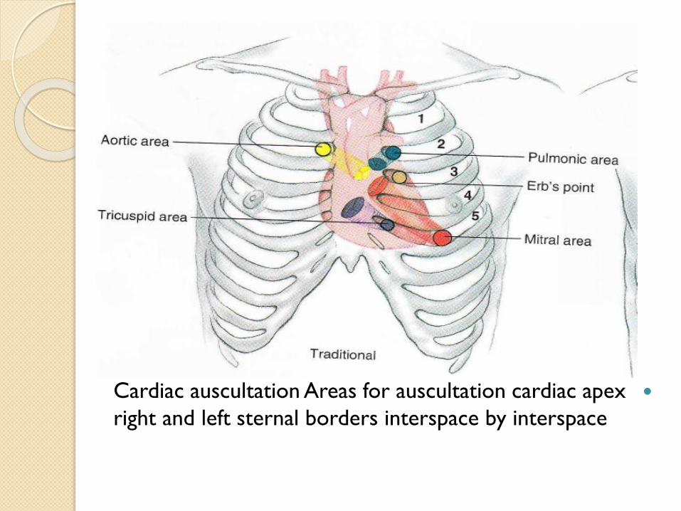
Cardiac auscultation Areas for auscultation cardiac apex
right and left sternal borders interspace by interspace

Cardiac Investigations and Procedures
Electrocardiogram (ECG) Chest X-Ray (CXR)
Echocardiography Trans- oesophagealEchocardiography
(TEE) Treadmill Testing

Cardiac Investigations and Procedures Other New
Imaging Techniques Cardiac Catherization Percutanous
Transluminal Coronary Angioplasty (PTCA)

Cardiac Investigations and Procedures
Electrophysiological Study of the Heart (EPS) and
Radio-frequency Ablation (RF) Permanent Pacemaker
Implantation (PPM) Automatic Implantable Cardiovertor
-Defibrillator (AICD) Automated External Defibrillators
( AEDs )


Coronary Artery Disease
Atherosclerosis
◦Define: thickness and hardening of the arteries caused by deposits of fat and fibrin which harden.
◦Leads to decreased lumen and decreased blood flow and ischemia and death of the tissue

AtherosclerosisDefine: thickness and hardening of the arteries caused by deposits of fat and fibrin which harden.Leads to decreased lumen and decreased blood flow and ischemia and death of the tissue

Signs and Symptoms
Pain usual symptom but may experience
dyspnea
May have irregular heart rate
N/V may also accompany the other symptoms
Called angina
◦Unstable – persistent, even at rest
◦Prinzmetal's – variant, and may occur without
atherosclerosis

Risk Factors:
1. Modifiable risk factors:
- Cholesterol levels
- Cigarette smoking
- Hypertension
- Diabetes mellitus
2. Nonmodifiable risk factors:
- Age
- Gender
- Family history
- Race

Medical Treatment
Decrease risk factors
◦Diet
◦Control cholesterol/triglycerides
◦Exercise
◦Smoking
◦Hypertension
Drugs
◦Calcium channel blockers
◦Nitroglycerin
Surgery

Myocardial Infarction
Myocardial infarction is the necrosis of an area of
cardiac tissue as a result of obstruction of blood flow
through a coronary artery or one of its branches
The myocardial tissue dies as a result of the occlusion

Signs/Symptoms:
1. Chest pain, substernally with radiation to arm, neck,
jaw, or back; and unrelieved by rest or nitrates.
2. Diaphoresis and cool, clammy, pale skin.
3. Nausea and vomiting.
4. Dyspnea.
5. Palpitations or syncope.
6. Restlessness and anxiety.
7. Tachycardia or bradycardia.
8. Decreased.

Assessment for Chest Pain
Subjection
◦Tightness, heaviness, squeezing, or crushing pain in the
substernal area, which can radiate to the jaw, neck, left
arm, or shoulder
◦Determine if pain is precipitated by an event
(exercise, stress or exertion)
◦Is the pain relieved by rest or drugs?
◦Is there any predisposing factors?
◦Patient may experience anxiety and feeling of doom

Objective
◦Dyspnea
◦Profuse diaphoresis
◦Adventurous breath sounds
◦Tachycardia, decreased B/P, ^ temp
◦Elevation of cardiac enzymes (CPK, CPK-MB,
LDH, Troponin)
◦EKG changes
Medical Treatment
Early treatment is important
Nitroglycerin
◦Dilates coronary arteries
Morphine sulfate – 2-4 mg titrated for pain relief
◦decreases blood return to the heart
◦decreases anxiety
◦relaxes smooth muscle in the lungs
◦has analgesic effect

Oxygen at 2-4 L/min
Thrombolytic therapy – must meet criteria
◦Streptokinase
◦Heparin
Lidocaine, Calcium channel blockers, Digoxin, Beta blockers, Dopamine, Dobutamine
Angioplasty/Stent placement
Coronary Artery Bypass Grafting

Nursing Management:Provide quiet, calm environment
Keep client on bedrest for 24-48 hours
Give medications as ordered –analgesics, O2, Nitroglycerin
Elevate head of bed
Watch for any more chest pain
Maintain IV line
Monitor for signs of CHF, cardiogenic shock, and pulmonary edema
Evaluate signs of MI◦Skin color, and temperature
◦Monitor vitals
◦Observe EKG for dysrhythmias
◦Monitor fluid volume levels
◦Check labs

Home care
◦Teach about medications
◦Include follow-up with physician
◦May need to teach about CAD
◦Teach modification of risk factors –weight, diet, smoking, exercise, etc.
◦Notify of any chest pain

SB, 60-year-old male is a retiree and was admitted to the
hospital accompanied by his daughter. that he was
overweight. When admitted, patient was complained of
shortness of breath for 2 weeks and was worsening on the
day of admission. Besides, he also experienced orthopnea,
fatigue, paroxysmal nocturnal dyspnea and leg swelling up to
his thigh. Mr. SB was admitted to the hospital for to the same
problem last year.Mr. SB had known case of heart failure
since 3 years ago and he had also diagnosed with
hypertension for 5 years. Before admitted to the hospital,
patient was taking frusemide 40mg, aspirin 150mg,
metoprolol 50mg, amlodipine 10mg, and simvastatin 40mg for
his hypertension and heart failure. diuretics, digitalis,
anticoagulants, vasodilators.
His family history revealed that his father had died of
ischemic heart disease 4 years ago while his brother has
hypertension. As for his social history, he smokes 2-3
cigarettes a day for 35 years and the calculated smoking
pack years was 5 pack years. Besides, Mr. SB also drinks
occasionally.

Congestive Heart Failure
CHF is inability of the heart to pump adequate amount
of blood to all vital organs.
CHF Classification:
Left- sided (or left ventricular)
Right- sided (or right ventricular)

Left-Sided Heart Failure
Signs/Symptoms:
1. Dyspnea upon exertion, paroxysmal nocturnal
dyspnea or orthopnea.
2. Pale, cool extremities.
3. Decreased peripheral pulses.
4. Tachycardia.
5. Oliguria(<30 ml/hour)
6. Insomnia and restlessness.

Right-Sided Heart Failure
Signs/Symptoms:
1. Dependent pitting edema.
2. Jugular vein distention.
3. Hepatomegaly.
4. Ascites.
5. Weakness, anorexia, and nausea.
6. Weight gain.

Nursing Management:
Administer prescribed medications, diuretics, digitalis, anticoagulants, vasodilators.
2. Check intake and output.
3. Weigh daily.
4. Provide a low- sodium diet.
5. Auscultate lung sounds.
6. Determine degree of JVD.
7. Assess dependent edema.
8. Monitor vital signs.
9. Administer oxygen as prescribed.
10 Psychological support.

Hypertension
Hypertension is intermittent or sustained elevation in systolic or diastolic blood pressure.
There are two major types, primary (essential) hypertension and secondary hypertension.
Etiology:1. Primary hypertension.
a. Non modifiable risk factors.
- Family history.
- Gender. Men ˃ women.
- Age.
- Race.

. Modifiable risk factors.
- Stress.
- Obesity.
- High dietary intake of sodium or saturated fats.
- Excessive caffeine, alcohol, or cigarette smoking.
- Oral contraceptives use.
2. Secondary hypertension.
- Renal vascular diseases.
- Coarctation of aorta.
- Primary hyperaldosteronism.
- Hyperthyroidism.
- Medications, such as estrogen, antidepressants,
steroids.

Signs/Symptoms:
1.Usually asymptomatic.
2. May cause headache, dizziness, blurred vision.
Nursing Management:
1. Administer medications as prescribed, such as diuretics,
antihypertensive…etc
2. Provide patient and family teaching.
- Advise the patient to reduce weight.
- Instruct the patient to restrict sodium alcohol and
caffeine intake.

- Smoking cessation.
- Discuss the importance of regular blood pressure
monitoring.
- Discuss the importance of lifelong medical follow up
examination.

Cardiac Rehab defined:
A progressive program with a goal of helping patients
restore and maintain optimal health while helping to
reduce the risk of future heart problems.

Phase I- (inpatient) assessment and mobilization,
education on risk factors and a discharge plan
Phase II- (outpatient) exercise, risk factor reduction,
four to six weeks. It focuses on health education and
resumption of physical activity,
Phase IIIThe duration of Phase 3 may vary from six to
12 weeks with patients required to attend a CR unit
two to three times weeklyfor structured exercise and
other lifestyle interventions
Structure of Cardiac Rehabilitation

IV- maintenance program
constitutes the components of long-term maintenance
of lifestyle changes and professional monitoring of
clinical status

Mr …, 28-year-old, student, normotensive, nondiabetic, nonsmoker,
presented with fever for 1 month, which is low grade, continued,
sometimes associated with chills and rigor, also with profuse
sweating, subsides only with paracetamol, highest recorded
temperature was 101F. He also complains of central chest pain, does
not aggravate by cough or movement of the chest. He also experiences
occasional palpitation, associated with difficulty in breathing after mild to
moderate exertion for the last few months, the patient also experiences
malaise, generalized weakness, arthralgia, myalgia, anorexia and loss
of weight. hematuria or loin pain.Splinter hemorrhages in nail beds
◦Petechiae,Osler’s nodes on fingers or toes,Janeway’s lesions on
palms or soles,Roth’s spots (retinal hemorrhages),clubbing,
splinter hemorrhage, cardiac murmur,
. He does not give any history of dental procedures or cardiac or other
surgery or instrumental procedure (catheterization, colonoscopy,
cannula, etc.) or any history of intravenous drug abuse.He has been
suffering from some valvular heart disease for several years.

Infective Endocarditis
Infection of the inner layer of the heart that usually
affects the cardiac valves

Layers of the Heart

Causative Organisms
Causative organism more virulent Streptococcus and Staphylococcus are most common
bacterial
Viruses
Fungi

Clinical Manifestations
May be nonspecific
Fever occurs in 90% of patients
Chills
Weakness
Malaise
Fatigue
Anorexia
Clinical Manifestations
May manifest signs and symptoms of heart failure
Clinical Manifestations
Subacute form
◦Arthralgias
◦Myalgias
◦Back pain
◦Abdominal discomfort
◦Weight loss
◦Headache
◦Clubbing of fingers

Clinical Manifestations
Vascular manifestations
◦Splinter hemorrhages in nail beds
◦Petechiae
◦Osler’s nodes on fingers or toes
◦Janeway’s lesions on palms or soles
◦Roth’s spots (retinal hemorrhages)

Clinical Manifestations
Murmur in most patients
Heart failure in up to 80% with aortic valve
endocarditis
Manifestations secondary to embolism

What investigations should be done to diagnose SBE
•CBC ;
•Blood culture.
•Echocardiography (to see vegetation, valvular lesion or congenital
anomaly).
•Urine (hematuria and proteinuria may be present).
•CXR P/A view (may show cardiomegaly or evidence of cardiac failure).
•ECG: may show prolong PR interval (AV block due to aortic root abscess
formation) and occasionally infarction (due to emboli).
•Urea and creatinine.
predisposing factors are as follows:
•Rheumatic valve lesion (e.g. AR, MR, etc.).
•Congenital heart disease (VSD, PDA, bicuspid aortic valve,
coarctation of aorta, TOF. SBE is rare in ASD, PS, MS, AS).
•Prosthetic valve.
•Dental extraction.
•Instrumentation (catheterization, sigmoidoscopy, cystoscopy,
endoscopy, cannulation).
•Cardiac surgery or cardiac catheterization.
•IV drug abuse (right sided endocarditis is more common,
especially involves tricuspid valve).

Collaborative Care
Antibiotic administration
◦Weeks to months of antibiotics required
◦Monitor antibiotic serum levels
◦Subsequent blood cultures
◦Renal/hepatic function monitored

Collaborative Care
Fungal and prosthetic valve endocarditis
◦Responds poorly to antibiotics
◦Valve replacement is adjunct procedure

Nursing Implementation
Assessment of nonspecific manifestations
Monitor laboratory data
Monitor signs/symptoms of superinfection related to antibiotics
Monitor patency of IV
Hygiene
Nutrition