nurse practitioner wound management clinical protocols
TRANSCRIPT

Sir Charles Gairdner Hospital – Nurse Practitioner Wound Management Clinical Protocols
Nurse Practitioner Clinical Protocols
for
Wound Management Services
Prepared by: Pam Morey, RN BN STN, MN(NP) MRCNA
March 2007
Sir Charles Gairdner Hospital North Metropolitan Health Service
Acknowledgement: L. MacLellan, G. Gardner, A. Gardner, Canberra Hospital T. Swanson, J.Smart, S.Morrison, South West Healthcare, Warnambool, Victoria M. Asimus, Hunter New England Health (the Maitland Hospital), New South Wales D. Angel, Royal Perth Hospital, Perth Western Australia Issued: 2007 Review: 2009
i
Sir Charles Gairdner Hospital – Nurse Practitioner Wound Management Clinical Protocols
Wound Diagnostics and Treatment Clinical Protocol 1.
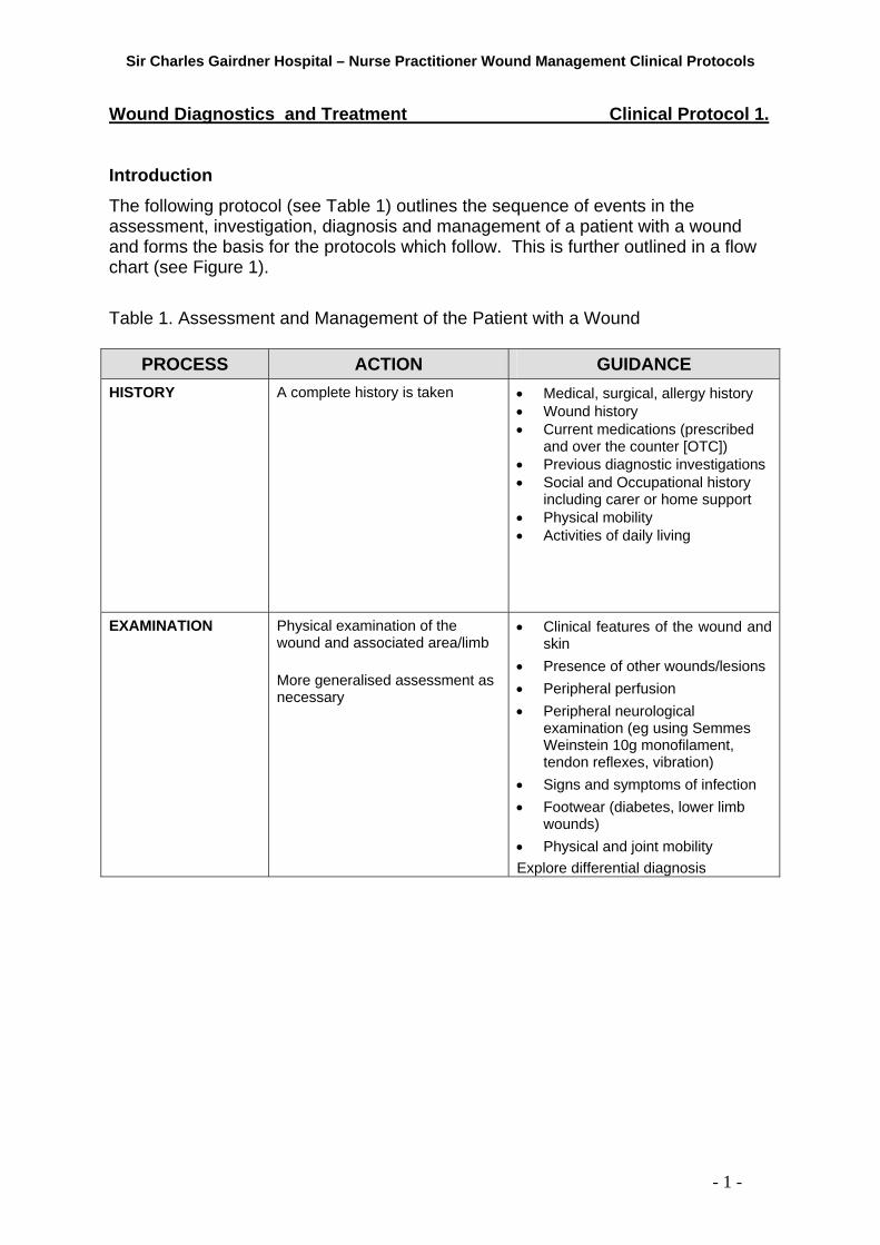
Introduction The following protocol (see Table 1) outlines the sequence of events in the assessment, investigation, diagnosis and management of a patient with a wound and forms the basis for the protocols which follow. This is further outlined in a flow chart (see Figure 1). Table 1. Assessment and Management of the Patient with a Wound
PROCESS ACTION GUIDANCE HISTORY A complete history is taken • Medical, surgical, allergy history
• Wound history • Current medications (prescribed
and over the counter [OTC]) • Previous diagnostic investigations • Social and Occupational history
including carer or home support • Physical mobility • Activities of daily living
EXAMINATION Physical examination of the wound and associated area/limb More generalised assessment as necessary
• Clinical features of the wound and skin
• Presence of other wounds/lesions • Peripheral perfusion • Peripheral neurological
examination (eg using Semmes Weinstein 10g monofilament, tendon reflexes, vibration)
• Signs and symptoms of infection • Footwear (diabetes, lower limb
wounds) • Physical and joint mobility Explore differential diagnosis
- 1 -

Sir Charles Gairdner Hospital – Nurse Practitioner Wound Management Clinical Protocols
PROCESS ACTION GUIDANCE INVESTIGATIONS Determine which investigations
may be required to assist in a diagnosis or provide a baseline of nutrition and health Referral for: Arterial Duplex Scan: To determine presence and/or severity or arterial disease in the lower limb Venous Duplex Scan: To determine disease or impairment of superficial, deep, and perforating veins and valves.
Pathology Haematology
• FBP, ESR, CRP, INR Biochemistry
• U & Es • LFT, (Total Protein, Albumin),
Pre-albumin • Glucose, HbA1C • Lipids • Thyroid function
Microbiology and Histology
• Wound fluid/swabs - microscopy, culture and sensitivity (MC&S)
• Wound/tissue biopsy – MCS and histopathology
• Skin Scraping, Immunofluorescence
Biopsy This may be required if the wound has been non-healing, despite optimal treatment, for greater than 4 weeks; or the duration is greater than 6 months; and/or is assessed as atypical. Radiology / Medical Imaging
• Ankle Brachial Pressure Index (ABPI)
• Toe Doppler Pressures/Index • Duplex Scan (Arterial &
Venous) • Photoplethysmography (PPG) • X-Ray • MRI
ABPI performed on all outpatient leg ulcer patients. If the ABPI does not complement the clinical assessment or is inconclusive then further diagnostic investigation may be required.
Arterial/Venous Duplex Scan Non-invasive investigation is recommended for initial diagnosis X-Ray If there is a suspicion of osteomyelitis, sinus, fistula, significant undermining or foreign body, then an x-ray may be ordered. Bone Scan / MRI If there is a suspicion of osteomyelitis, then a Bone Scan or MRI may be ordered following consultation with an Infectious Diseases physician or Vascular Surgeon.
- 2 -

Sir Charles Gairdner Hospital – Nurse Practitioner Wound Management Clinical Protocols
PROCESS ACTION GUIDANCE DIAGNOSIS Make provisional diagnosis On clinical picture, available
assessment data and results of investigations.
MANAGEMENT Urgent Referrals: • Life/limb threatening
infection • Abnormal test results that
require medical intervention • Treatment required outside
the NP scope of practice • Acute DVT • New patient with a ABI <0.7
or ankle systolic <80 mmHg • Patient that requires surgical
intervention • Ulcers on the plantar aspect
of the foot to have immediate Podiatry referral
• Significant deterioration in wound since last review
Nurse Practitioner: Non-pharmacological treatment Patient education for self care Pharmacological treatment - Based on diagnostic investigations, clinical assessment, and Therapeutic Guidelines Refer to other Clinical Practice Guidelines
Referrals If the wound fails to heal despite optimal therapy then consultation with other health care practitioners and further investigations may be required at that time. Non-pharmacological treatment • Appropriate dressings and/or
compression bandages based on diagnosis and patient lifestyle preferences
• Cleansing and debridement of wound
Patient /Carer education for self care• Hygiene (cleansing self and
waterproofing as required) • Diet (the importance of essential
vitamins and minerals as required). • Indications to seek medical help • Bandaging/dressing techniques • Exercise regimes • Lifestyle changes • Disease process and health
maintenance • Prevention of recurrence • Pain management • Medication (Includes relevant consumer handouts)
Pharmacological treatment
Analgesics Topical antimicrobials/antifungals Local anaesthetics Topical corticosteroids Oral antibiotics Moisturisers Barrier ointments, creams and wipes Skin cleansers
Lower leg ulcer Minor surgical procedures (further protocols to follow)
- 3 -

Sir Charles Gairdner Hospital – Nurse Practitioner Wound Management Clinical Protocols
PROCESS ACTION GUIDANCE MANAGEMENT PARTNERSHIPS
Appropriate referrals to assist in overall management
Other Health Professionals as required: Medical:
• Vascular Surgeon • Plastic Surgeon • Infectious Diseases Physician • Dermatologist • Endocrinologist • Pain Management • Palliative Care • General Practitioner
Allied Health:
• Dietitian • Podiatrist • Diabetic Educator • Occupational Therapist • Physiotherapist • Pharmacist
Community care providers:
• Silver Chain Nursing • Other home care providers (eg
Stanhope)
ONGOING MANAGEMENT
Follow-up Review as appropriate: • Test results • Monitor progress • Maintenance of wound • Review treatment plan in
accordance with investigative results
SEPARATION Discharge from service As appropriate:
• Wound healing achieved • Referral to community services
for long term management • Referral for Specialist care.
- 4 -

Sir Charles Gairdner Hospital – Nurse Practitioner Wound Management Clinical Protocols
Figure 1. Wound Diagnostics and Treatment: Flow Chart
- 5 -
1.Assessment
Patient History • Medical history/Co-morbidities • Wound History • Current medications • Social and occupational history • Activities of daily living
Physical examination • Clinical features of wound and skin • Presence of other wounds/lesions • Peripheral perfusion • Neurological examination eg tendon reflexes and
monofilament testing
Investigations – as indicated • Full blood examination • Urea & Electrolytes • Blood glucose levels and HBA1C, other
haematology, biochemical tests • Ankle Brachial Index/Doppler signal characteristics • Toe Doppler index • Duplex scan • X-Ray • Wound swab/s • Wound biopsy
2. Diagnosis
!! Consider conditions for urgent referral e.g. • Ischaemic limb/s • Serious infection e.g.
wet gangrene • Diabetic foot infections
3a. Conditions for specialist referral
• Urgent conditions as indicated above
• Treatment outside of NP scope of practice e.g. ischaemia, severe infection
3b. Treatment options / Conditions for NP treatment
• All wound conditions outside urgent treatment
→ Management as per specific protocols
3c. Integrated management of co-morbidities Includes diabetes, autoimmune disease, cardiac failure
• Endocrinologist • Vascular surgeon • Dermatolologist • Infectious diseases physician • Plastic surgeon • Pain Management • Palliative Care • General Practitioner • Diabetic Educator • Dietitian • Podiatrist/Orthotist • Pharmacist • Other health professionals as
required
Non-pharmalogical approaches • Cleansing and debridement
of wound • Appropriate
dressings/bandages • Skin care and moisturisers
4. Follow-up • Review as appropriate• Test results • Monitor progress • Maintenance of healed
wound
Pharmacological agents • Antibiotics • Antifungals • Analgesics • Topical agents (eg
corticosteroids, antimicrobials)
Patient education for self care • Hygiene • Diet • Exercise • Dressing/bandaging regimes • Disease process and health
maintenance • Medication
Adapted from MacLellan, L., Gardner, G., Gardner, A (2002) Designing the future in wound care: The role of the nurse practitioner. Primary Intention 10(3): 97-112 © ACT Government, reprinted with permission.

Sir Charles Gairdner Hospital – Nurse Practitioner Wound Management Clinical Protocols
Minor Surgical Procedures Clinical Protocol 2. Introduction To provide appropriate management of wounds, there are occasions where either
wound biopsy or sharp debridement procedures are required. Both of these are
minor surgical procedures. The flow chart demonstrates the protocol (see Figure 2)
Biopsy Skin and wound biopsy are used for diagnosis and may identify the presence (or
absence) or various skin conditions or diseases. In addition, biopsy may be
performed for semi-quantitative bacteriology where surface swabs are inadequate.
Wounds considered for biopsy may include long-standing wounds, those that are
atypical in location or appearance, or those that have not responded to treatment
(Trent, Federman, & Kirsner, 2003). Biopsy may be considered for lesions
suspicious of malignancy where there is increase in size; malodour and pain; have
excess granulation tissue, bleeding, or drainage; are exophytic; or have an irregular
base or margin. The procedure involves prior assessment of the wound or lesion;
preparation of the patient including explanation of the procedure; cleansing of the
area, and anaesthetisation with local anaesthetic. There are various methods of
biopsy and in this instance the preferred options are incisional or punch biopsy.
Particular wound conditions that may be identified include squamous cell carcinoma,
basal cell carcinoma, vasculitis, and calciphylaxis.
Debridement Debridement is the removal of devitalised (non-viable) tissue, particulate matter and
foreign material (Fowler & van Risjwijk, 1995). Conservative sharp wound
debridement (CSWD) is a procedure used to debride non-viable tissue from a
wound down to non-bleeding tissue using sharp instruments (eg scalpel, scissors
and forceps). Debridement may be undertaken to remove contaminated, dead and
damaged tissue that may inhibit healing or contribute to infection in the wound . It
may also be undertaken to prepare a wound for skin grafting, application of skin
substitutes, or topical negative pressure therapy (e.g. VAC – Vacuum Assisted
Closure). Sharp debridement may be necessary in either acute wounds (eg skin tear
with non-viable tissue), or chronic wounds (eg pressure ulcers), and the decision
about this type of procedure being undertaken requires consideration of both local
and systemic factors. These include but are not limited to knowledge of underlying
- 6 -

Sir Charles Gairdner Hospital – Nurse Practitioner Wound Management Clinical Protocols
anatomical structures, local tissue perfusion, the presence of impaired clotting or the
use of anticoagulant medication, and the presence of malignancy (Carville, 2005).
The outline of assessment process, investigations and management are outlined in
Table 2.
Table 2 Assessment and Management: Minor Surgical Procedures.
PROCESS ACTION GUIDANCE HISTORY A complete history is taken • Medical, surgical, allergy
history/comorbidities • Wound history • Current medications
(prescribed and OTC) • Previous diagnostic
investigations • Social and Occupational
history including carer or home support
• Physical mobility • Activities of daily living •
EXAMINATION Physical examination of the wound and associated area/limb More generalised assessment as necessary
Findings from assessment of complex, infected wounds, leg ulcers, and diabetic foot ulcers Abnormal clinical presentation: • Raised/unusual clinical
features • Suspicion of neoplastic
disease • Senescent tissue • Hypergranulation tissue • Non healing despite optimal
treatment Presence of: • Infection not responding to
antibiotic treatment • Contaminated/non-viable
material • Foreign bodies
- 7 -

Sir Charles Gairdner Hospital – Nurse Practitioner Wound Management Clinical Protocols
PROCESS ACTION GUIDANCE INVESTIGATIONS Biopsy of wound for histology
and/or microbiology
Histology • To confirm wound aetiology Microbiology
To identify organisms (semi-quantitative) and sensitivities
DIAGNOSIS Make provisional diagnosis On clinical picture, available assessment data and results of investigations.
MANAGEMENT Urgent Referrals:
• Life/limb threatening infection
• Abnormal test results that require medical intervention
• Treatment required outside the NP scope of practice
• Significant deterioration in wound since last review
Nurse Practitioner: Non-pharmacological treatment Patient education for self care Pharmacological treatment - Based on diagnostic
investigations, clinical assessment, and Therapeutic Guidelines
Conservative Sharp Surgical Debridement
Notify medical practitioners of investigations ordered and referrals organised If the wound fails to heal despite optimal therapy then consultation with other health care practitioners and further investigations may be required at that time. Non-pharmacological treatment • Appropriate dressings/bandaging
based on diagnosis and patient lifestyle preferences
• Cleansing and debridement of wound
Patient / Carer education for self
care • Hygiene (cleansing self and wound
waterproofing as required) • Diet (the importance of essential
vitamins and minerals as required). • Signs and symptoms of
complications • Bandaging/dressing techniques • Exercise regimes • Lifestyle changes • Disease process and health
maintenance • Prevention of recurrence • Pain management • Medication (Includes relevant consumer handouts)
Pharmacological treatment
Analgesics Topical antimicrobials/antifungals Local anaesthetics Topical corticosteroids Oral antibiotics
Conservative Sharp Surgical Debridement To remove: • Contaminated material • Foreign bodies
- 8 -

Sir Charles Gairdner Hospital – Nurse Practitioner Wound Management Clinical Protocols
• Non-viable tissue To prepare the wound environment for: • Topical Negative Pressure Therapy
(VAC) • Skin grafts • Substitutes to accelerate the healing
process
MANAGEMENT PARTNERSHIPS
Appropriate referrals to assist in overall management
Other Health Professionals as required: Medical:
• General Practitioner • Plastic Surgeon • Dermatologist • Infectious Diseases Physician
Consultation with the medical practitioner if required for further treatment and investigation.
Allied Health:
• Dietitian • Podiatrist • Diabetic Educator • Occupational Therapist • Physiotherapist • Pharmacist
Community care providers:
• Silver Chain Nursing • Other home care providers
ONGOING MANAGEMENT
Follow-up Review as appropriate: • Test results • Monitor progress • Maintenance of wound • Review treatment plan in
accordance with investigative results
SEPARATION Discharge from service As appropriate:
• Wound healing achieved • Referral to community services
for long term management • Referral for Specialist care.
- 9 -

Sir Charles Gairdner Hospital – Nurse Practitioner Wound Management Clinical Protocols
Figure 2. Minor Surgical Procedures for Diagnosis and Treatment in Wound Care.
Contraindications e.g. severe ischaemia (refer to specialist)
1. Findings from assessment of:• Complex wounds • Lower leg ulcers • Infected wounds • Diabetic foot wounds
1a. Abnormal clinical presentations e.g. • Raised/unusual features e.g.
irregular raised edges, purpura, vasculitic nodules
• Suspicion of neoplastic disease • Senescent tissue • Hypergranulation tissue • Non healing despite optimal
treatment
1c. Presence of: • Contaminated dead material
e.g. slough or necrotic tissue • Foreign bodies e.g. sutures
2c. Sharp surgical debridement/curette to: • Remove:
- Contaminated material - Foreign bodies e.g. sutures - Non-viable tissue
• Prepare for grafting of skin or skin substitutes;
• Prepare for application of topical negative pressure therapy (VAC)
• Accelerate healing process
2a. Biopsy of wound Histological examination to confirm wound aetiology
Pharmacological agents • Analgesics – paracetamol,
codeine, tramadol • Local anaesthetics –
Lignocaine/Prilocaine, lignocaine hydrochloride
2b. Biopsy of wound Microbiological examination to identify organisms and sensitivities treatment
3a. Conditions for specialist referral Biopsy results indicates • Neoplastic disease – refer to Plastic
surgeon/Dermatologist/Vascular surgeon as appropriate
• Biopsy findings inconclusive or suggestive of vasculitis or other dermatological conditions beyond the scope of practice of the NP– refer to Dermatologist/Vascular surgeon as appropriate
4. Follow up • Review as appropriate • Test results • Monitor progress • Maintenance of healed wound
3b. Conditions for specialist referral Biopsy results indicate • Infection – refer to Infectious
Diseases physician
1b. Presence of infection not responding to antibiotic treatment
- 10 -Adapted from MacLellan, L., Gardner, G., Gardner, A (2002) Designing the future in wound care: The role of the nurse practitioner. Primary Intention 10(3): 97-112 © ACT Government, reprinted with permission.

Sir Charles Gairdner Hospital – Nurse Practitioner Wound Management Clinical Protocols
Lower Leg Ulcers (Vascular) Clinical Protocol 3
Introduction This protocol has been designed to guide and facilitate the Nurse Practitioner (NP)
in diagnosing and providing appropriate care of clients with leg ulcers.
Assessment A thorough assessment of the individual will follow as per the Protocol 1., ‘Wound
Diagnostics and Management’. The following information outlines in more detail the
specific process for the NP in managing patients with lower leg ulcers (see Table 3)
using evidence gradings outlined on page 32-33. A flow chart outlining the Lower
Leg Ulcer Clinical Protocol is shown in Figure 3, and the Guidelines for
Compression Bandaging are represented in Figure 4. An explanation of
compression bandaging components is outlined in Appendix 1.
- 11 -

Sir Charles Gairdner Hospital – Nurse Practitioner Wound Management Clinical Protocols
Table 3 Assessment and Management of Lower Leg Ulcers. PROCESS ACTION LEVEL OF
EVIDENCE – GUIDANCE
HISTORY • A complete history is taken: medical, surgical, allergy history
• Wound history • Current medications (prescribed and OTC) • Previous diagnostic investigations • Social and Occupational history including carer
or home support • Physical mobility • Activities of daily living Assess history of ulcers, duration of current ulcer, mechanism of injury and previous methods of treatment Assess for venous insufficiency:
• Family history of venous disease; • Patient history of DVT; • Lower leg fracture or
other major leg injury; • Previous vein surgery; • Prior history of ulceration - with or without
compression bandaging. Assess for arterial insufficiency:
• History of intermittent claudication or rest pain
• Hypertension. • Heart disease; • Diabetes, • Ischaemic stroke • Smoking (or stopped < 5 years),
In the presence of mixed disease (arterial + venous), patients may present with both. Assess for diabetes, rheumatoid arthritis and systemic vasculitis (Specialist assessment/referral should be considered). Assess for correctable factors that may delay healing, including smoking, anaemia, and evidence of malnutrition or poor nutrition. Assess for pain and formulate plans that involve exercise (including ankle exercises) and leg elevation for venous ulcers and adequate analgesia irrespective of aetiology.
C C B A B C C
- 12 -

Sir Charles Gairdner Hospital – Nurse Practitioner Wound Management Clinical Protocols
PROCESS ACTION EVIDENCE / GUIDANCE
EXAMINATION Physical examination of the wound and associated area/limb Conduct lower limb examination of both legs Eg the presence varicose veins in venous disease Examine for signs of arterial insufficiency: • Lower skin temperature, auscultation of
femoral bruit and pulses (weak or absent). Unilateral signs may be present where there is acute deterioration.
Assess for malignancy – can be a cause and may be a sequel of leg ulceration. Signs suggestive of malignancy are: irregular nodular appearance of the surface of the ulcer, raised or rolled edge, raised granulation tissue above the ulcer base, failure to respond to treatment, rapid increase in ulcer size and abnormal pigmentation.. Assess the wound and surrounding tissue:
• The surface area of ulcers should be measured at regular intervals to monitor progress
• Venous ulcers are generally shallow,
moist and appear on the gaiter area of the leg; eczema, haemosiderin pigmentation, ankle oedema and ankle flare are often present; varicose veins, atrophie blanche & lipodermatosclerosis may be present.
• Arterial ulcers have a punched out
appearance, a poorly perfused base and are pale and dry; surrounding skin is shiny and taut; dependent rubor present.
Lower limb pulses – palpable pulses alone are insufficient to rule out arterial disease More generalised assessment as necessary • Clinical features of the wound and skin • Presence of other wounds/lesions • Peripheral perfusion • Neurological examination (eg using Semmes
Weinstein 10g monofilament) • Signs and symptoms of infection • Footwear (diabetes, lower limb wounds) • Physical and joint mobility
Explore differential diagnosis
B A B C B C C
- 13 -

Sir Charles Gairdner Hospital – Nurse Practitioner Wound Management Clinical Protocols
PROCESS ACTION EVIDENCE / GUIDANCE
INVESTIGATIONS ABPI performed on all outpatient leg ulcer patients. If the ABPI does not complement the clinical assessment or is inconclusive then further diagnostic investigation may be required. Measurement of ABPI by handheld Doppler • Ankle Brachial Pressure Index (ABPI). Normal 0.9 –
1.2. • A ratio of < 0.8 indicates the presence of peripheral
arterial disease (PAD). • Further tests should be considered prior to initiating
compression bandaging if a patient has an ABPI >0.8 in the presence of signs and symptoms of PAD, rheumatoid arthritis, systemic vasculitis or diabetes mellitus.
• Doppler determination of ABPI should not be used in isolation from clinical assessment.
• Repeat measurement of ABPI when an ulcer deteriorates; is not fully healed by 3/12; or when patient presents with recurrence (of whichever leg)
• Toe Doppler Pressures/Index and arterial Photophlethysmography (PPG) are adjunct tests to ascertain arterial insufficiency particularly where diabetes, incompressible vessels or calcification are present.
• Venous PPG will provide information on venous refilling time as an assessment of venous insufficiency
Determine which investigations may be required to assist in a diagnosis or provide a baseline of nutrition and health Pathology Haematology
• FBP, CRP Biochemistry
• U & Es • LFT, Total Protein, Albumin, • Glucose, HbA1C • Lipids • Thyroid function
Microbiology and Histology
• Wound fluid/swabs - microscopy, culture and sensitivity (MC&S)
• Wound/tissue biopsy – MCS and histopathology • Skin Scraping, Immunofluorescence
Note: Routine bacteriological swabs are unnecessary unless there is evidence of clinical infection Biopsy This may be required if the wound has been non-healing for 4 – 6 weeks with optimal treatment; is assessed as atypical, or has been present greater than 6 months.
A B A C A B B
- 14 -

Sir Charles Gairdner Hospital – Nurse Practitioner Wound Management Clinical Protocols
Radiology / Medical Imaging • Duplex Scan (Arterial & Venous) • X-Ray
Arterial/Venous Duplex Scan Non-invasive investigation is recommended for initial diagnosis Arterial Duplex Scan: To determine presence and/or severity or arterial disease in the lower limb Venous Duplex Scan: To determine disease or impairment of superficial, deep, and perforating veins and valves X-Ray If there is a suspicion of osteomyelitis, sinus, significant undermining or foreign body, then an x-ray may be ordered.
DIAGNOSIS Make provisional diagnosis On clinical picture, available assessment data and results of investigations
- 15 -

Sir Charles Gairdner Hospital – Nurse Practitioner Wound Management Clinical Protocols
PROCESS ACTION EVIDENCE / GUIDANCE
MANAGEMENT Urgent Referrals: • Life/limb threatening infection • Abnormal test results that require medical intervention • Treatment required outside the NP scope of practice • DVT • New patient with a ABI <0.7 or ankle systolic <80
mmHg • Patient that requires surgical intervention • Ulcers on the plantar aspect of the foot or other areas
of the foot subject to pressure from weight-bearing or footwear, to have immediate Podiatry referral
• Significant deterioration in wound since last review Nurse Practitioner: Non-pharmacological treatment • Compression bandaging should be applied when
venous insufficiency is present, and should be based on the ABI and interpretation of clinical signs and additional investigative data. (Figure 2.)
• Compression bandaging (elastic and inelastic) has been demonstrated to be effective in the healing of venous leg ulcers.
• Reduced compression may be effective in selected patients with mixed disease (venous + arterial components) where the ABI is 0.6 -0.8 however these patient should be closely monitored for signs of reduced circulation/ischaemia in a specialised clinic.
• Dressing techniques should be clean and aimed at preventing cross-infection – strict asepsis is not necessary.
• Ulcers can be cleansed with either potable water or sterile saline. Ulcerated legs can be washed normally in potable water.
• Wound debridement may be undertaken where necrotic tissue is present. There is no evidence to favour any one method of debridement, whether mechanical, surgical, biosurgical, autolytic, chemical or enzymatic and choice would be based on patient assessment (Also see Minor procedures protocol).
Patient / Carer education for self care • Hygiene (cleansing self and waterproofing as
required) • Diet (the importance of essential vitamins and
minerals as required, in particular Vitamin C and Zinc).
• Signs and symptoms of complications • Bandaging/dressing techniques • Exercise regimes
Exercise programmes can improve calf muscle function, walking distances and pain for people with intermittent claudication • Lifestyle changes • Disease process and health maintenance
Referrals If the wound fails to heal despite optimal therapy then consultation with other health care practitioners and further investigations may be required at that time. A B C C C C A
- 16 -

Sir Charles Gairdner Hospital – Nurse Practitioner Wound Management Clinical Protocols
• Prevention of recurrence • Pain management • Medication
(Includes relevant consumer handouts) Pharmacological treatment - Based on diagnostic investigations, clinical assessment, and Therapeutic Guidelines Pharmacological treatment
Analgesics Oral antibiotics Topical antimicrobials Topical anti-fungals Topical corticosteroids Local anaesthetics Moisturisers Barrier ointments, creams and wipes Skin cleansers
Note: Patients can be sensitised to treatments at any time. Products which commonly cause sensitivity such as those containing lanolin, cetyl alcohol or topical antibiotics, are best avoided. Associated Clinical Practice Guidelines:
• Wound management and diagnostics • Minor surgical procedures • Diabetic foot ulcer* • Infected wound* • Complex Wound*
*undergoing formulation.
B
- 17 -

Sir Charles Gairdner Hospital – Nurse Practitioner Wound Management Clinical Protocols
MANAGEMENT PARTNERSHIPS
Appropriate referrals to, or liaison with other health professionals to assist in overall management Medical:
• Vascular Surgeon • Plastic Surgeon • Infectious Diseases Physician • Endocrinologist • Pain Management • General Practitioner • Dermatologist
Note: Patients with dermatitis that does not resolve following removal of common sensitisers and treatment with a moderate topical steroid should be considered for referral to a Dermatologist. Venous surgery followed by graduated compression is an option for consideration in patients with superficial venous insufficiency. Allied Health:
• Dietitian • Podiatrist • Diabetic Educator • Occupational Therapist • Physiotherapist • Pharmacist
Community care providers:
• Silver Chain Nursing • Homelink / Hospital in the Home • Residential care agencies • Other home care providers
C B
ONGOING MANAGEMENT
Review as appropriate**: • Test results • Monitor progress • Maintenance of wound • Prophylactic review (eg 6/12 review for patient
with healed venous ulcers – prescription of graduated compression stockings)
** Patient reviews will be determined according to a number of factors. This will include whether or not the client is new to the service, whether compression therapy is initiated, their access to transport and availability for appointments; partnerships in care in place, and patient and wound factors. For a patient commencing compression therapy for the first time, review is usually within 1-2 weeks. Ongoing review may vary from 4 – 12 weeks. In the case of the patient with a healed venous ulcer who is wearing maintenance compression therapy, reviews are usually 6 monthly. As with all patient related visits, findings of the review and treatment plans will be documented in the patient’s integrated medical record. Review treatment plan in accordance with response to treatment and investigative results.
- 18 -

Sir Charles Gairdner Hospital – Nurse Practitioner Wound Management Clinical Protocols
SEPARATION Discharge from service
As appropriate: • Wound healing achieved • Referral to community services for long term
management • Referral for Specialist care.
- 19 -

Sir Charles Gairdner Hospital – Nurse Practitioner Wound Management Clinical Protocols
Figure 3. Flow Chart - Lower Leg Ulcer Clinical Protocol
- 20 -
1.Assessment
Patient History • Medical history/Co-morbidities • Wound History • Current medications • Social and occupational history • Activities of daily living
Physical examination • General health assessment • Clinical features of wound and skin • Presence of other wounds/lesions • Examination of peripheral pulses • Signs of autoimmune disease eg Rh Arthritis, SLE • Neurological examination • Signs of presence of infection eg lymphangitis,,
lymphadenopathy • Exclude neoplastic disease
Investigations – as indicated • ABPI/Doppler signal characteristics/Toe pressures • Duplex scan • Photoplethysmography (PPG) • Full blood examination • Blood glucose levels, HBA1C, other haematology,
biochemical tests • Wound swab • Wound biopsy (see ‘Minor surgical procedures’
protocol)
2. Diagnosis
!! Consider conditions for urgent referral e.g.
• Ischaemic limb/s • Serious infection • Diabetic foot
infection
3a. Conditions for specialist referral
• Urgent conditions as indicated above
• Treatment outside of NP scope of practice e.g. cellulitis, surgical intervention required
• ABPI < 0.7
3b. Treatment options / Conditions for NP treatment
• All wounds outside urgent treatment
3c. Integrated management of co-morbidities Includes diabetes, autoimmune disease, cardiac failure Medical:
• General Practitioner • Vascular surgeon • Dermatologist • Infectious diseases physician • Plastic surgeon • Pain Management • Endocrinologist
Allied Health: • Dietitian • Podiatrist/Orthotist • Pharmacist • Other health professionals as
required
Non-pharmalogical approaches • Appropriate dressings and
graduated compression therapy Refer to Figure 2
• Debridement (see ‘Minor surgical procedures’ protocol)
4. Follow-up • Review as appropriate• Test results • Monitor progress • Maintenance of healed
wound
Adapted from MacLellan, L., Gardner, G., Gardner, A (2002) Designing the future in wound care: The role of the nurse practitioner. Primary Intention 10(3): 97-112 © ACT Government, reprinted with permission.
Pharmacological agents as indicated • Analgesics • Antibiotics • Topical antimicrobials • Local Anaesthetic • Topical corticosteroids
Patient education for self care • Hygiene • Diet • Foot inspection (diabetes) • Dressings/Bandaging/Compression • Exercise regimes
Differential diagnosis • Venous • Mixed • Arterial • Vasculitic, Neuropathic,
Infective, Neoplastic, Other

Sir Charles Gairdner Hospital – Nurse Practitioner Wound Management Clinical Protocols
Figure 3. NP Management Pathway for Lower Leg Ulcers:
- 21 -
Venous ulcer ABPI >0.8:
Compression therapy - Multilayer (Elastic or inelastic) - Reduced compression - Graduated compression stockings - Intermittent Pneumatic
compression (IPC) Medical and surgical treatment as necessary (referral) Local ulcer treatment*
Mixed arterial and venous ulcer
(ABPI 0.5-0.8)
- Reduced compression therapy (15 – 25 mmHg) - Local ulcer treatment* - Refer to Vascular surgeon if ABPI
< 0.7 or patient symptomatic (eg intermittent claudication)
Arterial Ulcer - Refer to Vascular
surgeon - Local ulcer treatment* - NO compression
Immobile/fixed ankle patient 1st line therapy: - Multilayer compression (elastic) 2nd line therapy: - Multilayer compression (elastic +
IPC
Active/mobile patient 1st line therapy: - Multilayer compression (elastic or inelastic) 2nd line therapy - Elastic stockings
Healed ulcer - Prevention of recurrence including
below the knee compression stocking
- Evaluation for surgical correction - Education - Review
Assessment Patient history Examination
o Investigations
Other - Disease specific
treatment/referral - Local ulcer treatment* - Appropriate compression
for oedema control based on ABPI
Patient presents with lower leg ulcer
Reasons for referral: - Allergy - Unable to tolerate compression - Uncontrolled pain - No reduction in ulcer size after
4-6/52 - Cellulitis unresponsive to
treatment - Frequent recurrence
Mixed arterial & venous ulcer
Severe arterial insufficiency ABPI <0.5 - Refer to Vascular surgeon - Local ulcer treatment* - NO compression
* Local ulcer treatment (Dressing) based on: - Assessment of wound and surrounding skin - Allergies / Skin sensitivities - Product availability
Ulcer fails to heal (no reduction in size in 6/52) - Refer to specialist - Re-evaluation including
reassessment and diagnosis - Evaluation for surgical correction
or skin grafting
Diagnosis
Treat concurrent issues:- Pain eg analgesics - Infection eg antibiotics
(see Protocol 3) - Dermatitis/Eczema
Consider cause (eg venous or contact) and treatment: o Corticosteroids o Zinc paste bandages o Avoid sensitisers
Education - Foot and skin care - Exercise - Nutrition - Cessation of smoking - Weight loss if obese - Disease aetiology - Dressings and
bandage/compression regimes
- Medications
Adapted from International Leg Ulcer Advisory Board cited in Stacey, M., Falanga, V., Marston, W., Moffat, C., Phillips, T., Sibbald, R.G., Vanscheldt, W., & Lindholm. C. (2002) The use of compression therapy in the treatment of venous leg ulcers: a recommended management pathway. EWMA Journal 2(1)

Sir Charles Gairdner Hospital – Nurse Practitioner Wound Management Clinical Protocols
- 22 -
COMPRESSION BANDAGING SYSTEMS
Multi-layer Layers - usually 3-4 layers and may include either elastic or inelastic compression bandages, cohesive/adhesive bandages, crepe bandages and/or padding layers. Zinc bandages – elastic and rigid varieties. Wound & skin contact layer. Underpadding - cotton or synthetic padding to protect the skin/ bony prominences from bandage trauma and may have additional absorbent capacity. Used as base layer under most compression bandage systems Wraps - eg Webril, Sofban, Kerlix Tubular knitted padding – eg Dynacast, Tubular Plus, Samme Frotte
Compression bandages, - elastic with various degrees of elasticity - inelastic
Cohesive elastic wraps eg Coban, CoPlus or elastic tubular support eg Tubigrip, Versagrip, Tensogrip
Single layer – eg Setopress, Surepress, Tensopress
Multilayer systems: - eg Profore system, Veno 4, Proguide
Elastic
May be used across range of mobile and immobile patients but particularly indicated for immobile patients or those with reduced ankle mobility/fixed ankle deformity where calf muscle contraction is limited.
Provide sustained compression with minor variations during walking.
Multilayer – light (reduced) compression (15-25 mm Hg versus High compression 35-45 mm Hg, @ ankle)
eg Profore Light, Lastodur Light
Inelastic
Suitable for actively mobile clients where the bandage reinforces or supports the action of the calf muscle pump.
They provide high pressure on moving and low resting pressures. May be more effective in patients with extensive deep vein reflux (Marston & Vowden, 2003).
Number of layers according to ABPI, full compression usually 2 layers – sub-bandage pressures will vary according to a number of factors including wear-time and oedema
eg Comprilan, Lastolan, Tensolan
Note: The degree of compression in governed by La Place’s law P = pressure exerted by bandage where sub-bandage pressure is demonstrated thus: N = number of layers P is proportional to : N x T T = bandage tension (elasticity) C x W C = circumference of limb
W= bandage width

Sir Charles Gairdner Hospital – Nurse Practitioner Wound Management Clinical Protocols
Drug Formulary Wound Management
Classification Drug Dosage
Analgesic Paracetomol 500-1000 mg 4-6 hourly
Analgesic Paracetamol/Codeine 500mg/8-30 mg 4-6 hourly
Analgesic Tramadol 50-100mg 4-8 hourly
Antibiotic Amoxycillin Clavulanate 500/125 – 875/125 mg 12 hourly
Antibiotic Cephalexin 250 – 500 mg 6 hourly
Antibiotic Dicloxacillin 250 – 500 mg 6 hourly
Antibiotic Flucloxacillin 250 – 500 mg 6 hourly
Antibiotic Ciprofloxacin 250 – 500 mg twice daily
Antibiotic Clindamycin 150 – 450 mg 8 hourly
Antibiotic Metronidazole 200 – 400 mg 8-12 hourly
Topical Antibiotic Metronidazole 0.5% twice daily
Topical Antibiotic Silver Sulphadiazine Chlorhexidine digluconate
1%, 0.2% 1-2 x /day
Topical Antifungal Clotrimazole 1% 3 x /day
Topical Antifungal Terbinafine 1% 1-2 x /day
Topical Antifungal Nystatin 100,000units/g 2-3 x /day
Topical Antiseptic, Anti-infective
Mupirocin 2% 3 x /day
Topical corticosteroid + antimicrobial
Triamcinilone/Neomycin/Nystatin/ Gramicidin
0.1%, 0.25%, 100,000U/g, 0.025%
2-3 x /day
Topical corticosteroid Hydrocortisone 0.5-1% 1-2 x /day
Topical corticosteroid Hydrocortisone acetate 0.5-1% 1-2 x /day
Topical corticosteroid Triamcinolone Acetonide 0.02% 1-2 x /day
Topical corticosteroid Betamethasone valerate 0.02-0.05% 1-2 x /day
Topical corticosteroid Betamethasone dipropionate 0.05% 1-2 x /day
Topical Anaesthetic Lignocaine 0.05-1% Pre-procedure
Local Anaesthetic Lignocaine with Adrenaline 0.05-1% Pre-procedure
Local Anaesthetic Lignocaine with Prilocaine 0.05-1% Pre-procedure
- 23 -

Sir Charles Gairdner Hospital – Nurse Practitioner Wound Management Clinical Protocols
For more severe pain, review causative factors and refer to appropriate specialist (eg Pain Team, Vascular Surgeon).
Analgesics Mild Pain:
• Paracetamol 1000mg 4-6 hourly maximum daily dose 4000mg
Mild to moderate pain:
• Paracetamol with Codeine 500/8-30mg 1-2 tablets 4 to 6 hourly maximum daily dose 4000mg paracetamol
OR
• Tramadol 50 mg to 100mg 4 to 8 hourly maximum daily dose 400mg daily (300mg maximum dose for elderly)
(Therapeutic Guidleines: Analgesics, 2002)
Antibiotics (topical)
Localised skin infections, critical colonisation of wounds (eg leg ulcers and pressure ulcers), and minor burn prophylaxis • Silver sulfadiazine (SSD) 1% +
chlorhexidine 0.2% cream topically, once or twice daily.
(contraindicated if sulpha or chlorhexidine allergy) Impetigo, infected small skin lesions(mild or localised infections) and elimination of Staph.aureus carriage • Mupirocin 2% topically, following skin
cleansing 3 times per day for up to 10 days
Cancerous malodourous wounds • Metronidazole gel 0.75% topically
• Silver sulfadiazine (SSD) 1% + chlorhexidine 0.2% cream topically, once or twice daily.
(Sibbals, Orsted, Schultz et al., 2003; Therapeutic Guidelines: Antibiotic, 2006; Therapeutic Guidelines: Dermatology, 2002)
Alternatives to consider include silver, povidone-iodine, and cadexomer iodine dressing products.
- 24 -

Sir Charles Gairdner Hospital – Nurse Practitioner Wound Management Clinical Protocols
Antibiotics (systemic)
Skin and soft tissue infection3 Common infecting organisms may include: Staphylococcus aureus, MRSA, Streptococcus species, Pseudomonas aeruginosa, and anaerobes Empirical antibiotics to be commenced whilst waiting for sensitivities For mild to moderate infection with surrounding cellulitis, use:
• Di/flucloxacillin 500 mg orally 6-hourly for at least 5 days
For patients hypersensitive to penicillin (excluding immediate hypersensitivity), use:
• Cephalexin 500mg 6-hourly for at least 5 days
Diarrhoea is a common adverse effect and the patient should be told to seek medical attention should this occur.
Alternatively, if Gram-negative organisms are suspected or known to be involved, use:
• Amoxycillin+clavulanate 875+125mg orally, 12 hourly for 5 days
Gram-negative organisms often colonise ulcers. Therefore for less severe infections, antibiotics against gram positive organism should be used initially. If the infection is not responding then broadening to include gram-negative cover can then be considered. (Therapeutic Guidelines: Antibiotic, 2006)
The routine use of antibiotics is not advocated in chronic wounds. Antibiotics to be commenced only when there is clinical evidence of infection (e.g. localised erythema, localised pain, localised heat, cellulitis, and oedema). If no clinical improvement within one week (next visit) or worsening of symptoms, for medical review. For more severe infections, particularly where systemic symptoms are present, and for, intravenous antibiotics, medical review will be required. Antibiotic susceptibilities of gram negative organisms should be reviewed and advice obtained from a clinical Microbiologist or ID Physician for organisms resistant to amoxycilllin + clavulanate.
- 25 -

Sir Charles Gairdner Hospital – Nurse Practitioner Wound Management Clinical Protocols
Diabetic foot infections:
For mild to moderate infection with no evidence of osteomyelitis or septic arthritis, use:
• Amoxycillin+clavulanate 875+125mg orally, 12-hourly for at least five days
Or
• Cephalexin 500mg orally, 6-hourly for at least five days
Plus
• Metronidazole 400mg orally, 12-hourly for at least five days
Inform patients that nausea, diarrhoea and metallic taste an adverse effect whilst taking metronidazole. To seek medical attention for nausea and diarrhoea.
For patients with penicillin hypersensitivity, use:
• Ciprofloxacin 500mg orally, 12 hourly for at least 5 days
To seek medical attention if the patient develops a rash, nausea, vomiting, diarrhoea, abdominal pain, and/or dyspepsia.
Plus
• Clindamycin 300 mg to 450 mg orally, t.d.s. for at least 5 days
Patients must be informed of the adverse effect of diarrhoea with a risk of pseudomembranous colitis, whilst taking clindamycin. Patients must be told to report these side effects and seek medical attention. (Therapeutic Guidelines: Antibiotic, 2006)
For severe limb- or life-threatening infection (systemic toxicity/septic shock, bacteraemia, marked necrosis or gangrene, ulceration to deep tissues, severe cellulitis, presence of osteomyelitis) medical review is required. Approval is required from a Clinical Microbiologist or ID Physician for Ciprofloxacin use.
- 26 -

Sir Charles Gairdner Hospital – Nurse Practitioner Wound Management Clinical Protocols
Topical Antifungal
Tinea (body, limbs, face and interdigital) • Terbinafine 1% topically, daily for 7
days Or an imidazole: • Clotrimazole 1% topically, 2 to 3 times
daily for 2 to 4 weeks, continued for 14 days after symptoms resolve.
Cutaneous candidiasis • Clotrimazole 1% topically, 2 to 3 times
daily for 2 to 4 weeks, continued for 14 days after symptoms resolve.
Or
• Nystatin 100,000 units/g cream topically, 2 to 3 times per day. Continue treatment for 2 weeks after symptoms resolve.
If necessary for inflammation, add
• Hydrocortisone cream1% topically, 2 to 3 times daily
(Therapeutic Guidelines: Dermatology, 2004)
Diagnosis of fungal infection can be confirmed via microscopy and culture of skin scrapings, subungal debris, nails or plucked hair. If poor response, or topical treatment is impracticable, refer to Microbiologist or dermatologist for consideration of systemic treatment.
- 27 -

Sir Charles Gairdner Hospital – Nurse Practitioner Wound Management Clinical Protocols
Topical Corticosteroid
Stasis/contact dermatitis 2
Mild • Hydrocortisone cream1% topically, 2
to 3 times daily Or • Hydrocortisone acetate 1% cream or
ointment 30g. Apply once or twice daily
Moderate • Betamethasone valerate 0.02% -
0.05% cream or ointment topically, once or twice daily
Or • Triamcinolone acetonide 0.02%
topically once or twice daily
Severe Betamethasone dipropionate cream or ointment 0.05%, topically once or twice daily (use sparingly, and for as short a period of time as possible, due to potency and potential local adverse effects).
(Therapeutic Guidelines: Dermatology, 2004)
Uncomplicated stasis dermatitis is common in chronic leg ulcers. Stasis dermatitis is frequently complicated by allergic contact dermatitis, which usually resolves with the removal of the sensitising agents (frequently encountered in many dressing products) and treatment with a mild/moderate topical corticosteroid.
If poor response, refer to a dermatologist.
Topical corticosteroid + antimicrobial
Skin infections and small areas of overgranulation
• Triamcinolone 0.1% / Neomycin 0.25% / Nystatin 100,000U/g / Gramicidin 0.025% cream topically 2 to 3 times per day for up to one week.
Local anaesthetic
Biopsy • Lignocaine (7mg/kg) with Adrenaline
(5 micrograms/ml). Lignocaine%, adrenaline 1:100 000, 5mL.
• Lignocaine 1%, 5mL Local wound debridement (pre procedure) where appropriate
• Lignocaine with Prilocaine 0.05-1% topically.
(Rossi [Ed], 2005; Therapeutic Guidelines: Dermatology, 2004).
Lignocaine with adrenaline should not be used on an extremity, such as a digit, especially in the presence of PAD, to avoid potential necrosis. For infiltration 1-2 mL is sufficient to provide anaesthesia and will not distort the histology
- 28 -