numerical analysis of acoustophoretic discrete particle focusing … · 2020. 8. 25. · 1...
TRANSCRIPT

Numerical Analysis of Acoustophoretic Discrete Particle Focusing in Microchannels
Ioannis H. Karampelas1,2, Jenifer Gómez-Pastora3, Michael J Cowan2, Eugenio Bringas3, Inmaculada Ortiz3 and Edward P.
Furlani2,4
1Flow Science, Inc. (USA),
2Dept. Chemical and Biological Engineering, State University of New York at Buffalo (USA), 3Dept. of Chemical and Biomolecular Engineering, University of Cantabria (Spain), 4Dept. of Electrical Engineering, State
University of New York at Buffalo (USA)
Office: (505) 982-0088, Fax: (505) 982-5551, [email protected]
ABSTRACT
Acoustophoretic particle focusing is a modern and very
attractive method of removing a variety of objects from
solutions in a microfluidic channel. The process is inherently
general and can be readily extended to multiple types of
applications such as healthcare (e.g. malignant cell removal),
academic research (e.g. nanoparticle separation), industrial
(e.g. reclaiming of rare earths) and environmental
applications (e.g. sequestration of suspended solids).
According to classical understanding, particle separation is
realized by the acoustic radiation force. Traditionally, the
wavelength of the acoustic wave has to be fine-tuned to the
channel width and specified to λ = 2Lchannel. This creates a
constant pressure node in the middle of the channel that
forces particle motion towards it. Particle motion is theorized
to be caused by differences in the pressure gradient acting on
the partciles and/or wave scattering. This article also
demonstrates that particle focusing can also be achieved
under different conditions, provided that the amplitude of the
oscillations is sufficiently high. In this study, results from
numerical simulations performed in FLOW-3D help
promote understanding of acoustophoretic particle focusing
by closely examining the forces that act on the particles. To
the best of our knowledge, this is the first explicit study of
this phenomenon that focuses on the different force
contributions This computational analysis can be generally
applied to a multitude of acoustophoretic processes and
should prove useful in promoting understanding of the
phenomena involved in particle focusing.
Keywords: Acoustophoresis, standing acoustic wave, SAW,
λ/2 channels, microchannel oscillations, nanoparticles,
particle focusing
1 INTRODUCTION
The use of acoustic standing wave technology has been
increased in the last 10 years for the manipulation, separation
or concentration of particulate matter in complex media due
to its many advantages, such as non-invasiveness, versatility,
simple fabrication, easy operation, and convenient
integration with other on-chip units[1]. This “label free”
method has allowed the separation of micrometer biological
cells from complex biofluids depending on the specific sizes,
densities and compressibilities. Among its many advantages,
the use of high flow rates (up to L/h) in microfluidic devices
makes acoustophoresis more convenient than other sorting
methods. For instance, Abdallah et al. [2] were able to
transfer particles between different fluids at much higher
flow rates (x10) by using acoustophoresis than
dielectrophoresis. Also, it was also tested in processing
whole human blood and proved capable of transferring blood
cells from undiluted whole human blood with an efficiency
of 95% at 1 L/h and 82% at 2 L/h [3]. Although it has been
employed for the manipulation of micron-sized particles,
Laurell’s group has recently achieved the separation of
submicron particles as small as 500 nm by using two-
dimensional acoustic focusing (i.e. focusing of the sub-
micrometer particles both horizontally and vertically in the
cross section of a microchannel) [4]. Furthermore, the
combination of multiple streams flowing under laminar
conditions in microchannels, ligand complexed microbeads
and acoustic standing wave technology offers a new brand of
applications that were not previously possible. For example,
Augustsson et al. [5] developed an acoustophoresis-based
microfluidic flow-chip as a novel platform to facilitate
analysis of proteins and peptides loosely bound to the surface
of beads or cells. In this work, the authors demonstrated the
elution of surface bound peptides from beads and human
spermatozoa by using buffers with different pH levels.
Apart from the wealth of experimental studies, various
theoretical and numerical investigations of the process have
been reported. For example, Nama et al.[6] have studied the
acoustophoresis of particles in PDMS channels. In their
work, the system is acoustically actuated via two counter-
propagating surface acoustic waves that form a standing
wave in a piezoelectric material interfacing the liquid
channel, and the particle motion is governed by the viscous
drag force from the acoustic streaming as well as the direct
acoustic radiation force due to scattering of sound waves on
the particles. For their specific model parameters, (600 μm
wavelength, 6.65 MHz actuation frequency, and polystyrene
particles suspended in water), an approximate critical
particle size of 10 μm was obtained for which the particle
motion goes from being streaming-drag dominated to being
radiation dominated. The numerical study developed by
Büyükkocak et al. [7] is also of particular interest since they
used a finite element approach for modelling the particle
separation under the influence of ultrasonic waves. In their
article, the authors solved the fluid flow in a microchannel
174 TechConnect Briefs 2017, TechConnect.org, ISBN 978-0-9988782-0-1

numerically, whereas analytical relations were employed for
the calculation of ultrasonic radiation forces (which were
coupled to the fluidic analysis). Furthermore, they simulated
the cell washing process by taking into account the diffusion
of contaminants between colaminar streams flowing side by
side inside the channel, and the results were compared with
experimental works showing good agreement. In all of the
studies listed above, the authors used an analytical
expression to for the radiation force coupled with the CFD
simulations. To the best of our knowledge, this is the first
study that examines particle focusing as a direct result of
flow parameters. Explicitly modeling the acoustophoretic
process provides additional insight that may prove useful in
the development of novel acoustophoretic applications.
2 THEORY
Acoustophoresis can be defined as the movement of
particles using acoustic radiation pressure from resonant
sound waves. The phenomenon has shown promising
applications in microfluidics, namely for the separation of
free flowing particles [8]. In microfluidics, acoustophoresis
is commonly achieved by using a piezoelectric actuator to
create a standing ultrasound wave lateral to the direction of
the channel. In general, a standing wave can be described as:
𝑦 = 𝐴 ∗ cos (2𝜋𝑥
𝜆+
𝜙
2) ∗ sin (
2𝜋𝑡
𝑇+
𝜙
2) (1)
where y is the displacement of a point in the wave at position
x and time t, A is the amplitude, λ is the wavelength, T is the
period, and ϕ is the phase difference. For the majority of
microfluidics applications, the wavelength is set to be twice
the width of the channel. This creates a pressure node in the
center of the channel with antinodes of fluctuating pressure
on either side. The scenario allows for Eq. 1 to also be
defined in terms of pressure. It can now be written as:
𝑝 = 𝑝0 ∗ sin (2𝜋
𝜆𝑥) ∗ cos (
2𝜋
𝑇𝑡) (2)
where 𝑝 is the pressure at distance 𝑥 from the pressure node
at time 𝑡, 𝑝0 is the pressure amplitude, 𝜆 is the wavelength,
and 𝑇 is the period. Since there is only one standing wave in
this situation, the phase difference terms could be removed.
Based on Eq. 2, one can see that the pressure gradient and,
more specifically, the differences in pressure δp within the
medium play a pivotal role in the acoustophoresis particle
focusing phenomenon [9]. Hence, the radiation force Frad on
suspended body with surface S can be calculated using the
following surface integral:
𝑭𝒓𝒂𝒅 = ∫ (−𝒏)𝛿𝑝𝑑𝛼𝑆
(3)
where n is the vector normal to the surface [10]. As it can be
readily observed from the above derivations, particle motion
is possible in the presence of a pressure field.
3 RESULTS AND DISCUSSION
In the following sections, we model particle motion
under the influence of an acoustic wave using a CFD analysis
performed in FLOW-3D. In section 3.1, we demonstrate
discrete particle motion caused solely by the pressure
gradient. Whereas, in section 3.2, it is shown that particle
focusing may also be possible due to oscillations of the entire
microchannel at off-resonance frequencies.
(a) (b) Figure 1. Numerical λ/2 channel stable pressure field analysis at φ=0 (solid lined) and at φ=π (dashed line). (a) Normalized
pressure vs normalized channel length, (b) Normalized pressure gradient at the direction of wave propagation vs
normalized length.
-1
-0.8
-0.6
-0.4
-0.2
0
0.2
0.4
0.6
0.8
1
0 0.1 0.2 0.3 0.4 0.5 0.6 0.7 0.8 0.9 1
Nor
mal
ized
Pre
ssur
e
Normalized Length
-1
-0.8
-0.6
-0.4
-0.2
0
0.2
0.4
0.6
0.8
1
0 0.1 0.2 0.3 0.4 0.5 0.6 0.7 0.8 0.9 1
Nor
mal
ized
Pre
ssur
e G
radi
ent
Normalized Length
175Biotech, Biomaterials and Biomedical: TechConnect Briefs 2017
3.1 Half Wavelength (λ/2) Channel Pressure
Field Analysis
We perform a 3D numerical analysis of the impact of an
oscillating pressure field to the motion of particle suspended
in fluid. For the purposes of this analysis, an elastic
membrane is placed at the bottom of the computational
domain (Normalized Length = 0) where it follows a
prescribed oscillating motion which results in the generation
of a standing acoustic wave. The oscillation frequency is
carefully chosen so that the wavelength is twice the length of
the channel. The force amplitude for the membrane’s
oscillation is set arbitrarily high so that a pressure field
maximum of the order of 1 Atm can be observed. A wall
boundary condition that allows for total wave reflection is
placed at the top of the domain (Normalized Length = 1).
Symmetry boundary conditions are placed on all the
remaining boundaries. Initially, the fluid is at rest and its
pressure is initialized at 0 Atm. At the onset of the
simulation, the membrane begins to oscillate, generating
amplifying pressure waves. It was observed that the pressure
extrema reach stable values after approximately 1000
oscillations. Figure 1 shows the pressure field and pressure
gradient once these stable operating conditions have been
reached. As it can be observed from Fig 1a., the simulation
results verify the existence of a pressure node at the middle
of the channel. It is important to note that the absolute values
of the pressure maxima at φ=0 and φ=π are higher that the
absolute values of their respective minima. Therefore, since
the pressure waves do not cancel each other out, particle
focusing is possible. Moreover, the pressure gradient along
the direction of propagation, shown in Fig 1b, reaches a peak
value at the location of the pressure node A probe particle
was also placed at a random location in the computational
domain. The particle appears to move, in phase, with each
membrane oscillation while approaching the center of the
domain. This simulation required 14 hours on a standard 24-
core workstation for a total of 10000 oscillations.
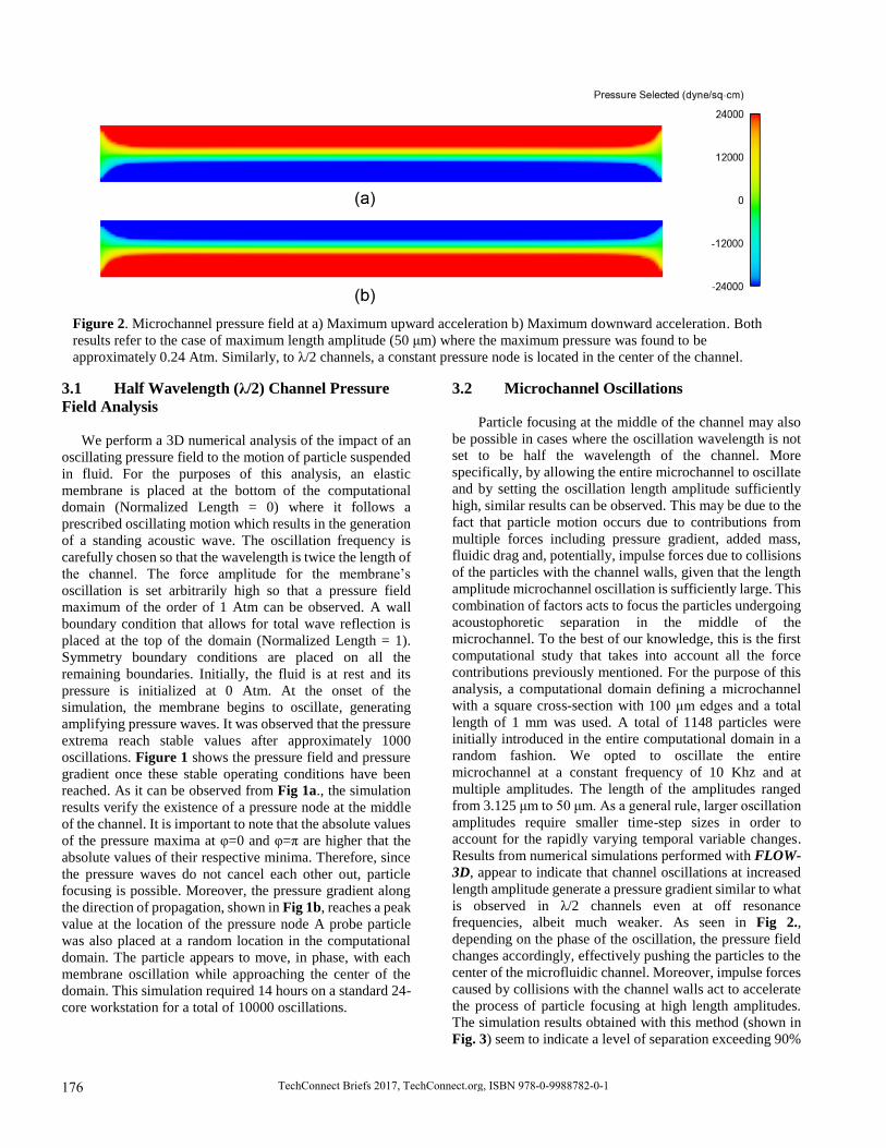
3.2 Microchannel Oscillations
Particle focusing at the middle of the channel may also
be possible in cases where the oscillation wavelength is not
set to be half the wavelength of the channel. More
specifically, by allowing the entire microchannel to oscillate
and by setting the oscillation length amplitude sufficiently
high, similar results can be observed. This may be due to the
fact that particle motion occurs due to contributions from
multiple forces including pressure gradient, added mass,
fluidic drag and, potentially, impulse forces due to collisions
of the particles with the channel walls, given that the length
amplitude microchannel oscillation is sufficiently large. This
combination of factors acts to focus the particles undergoing
acoustophoretic separation in the middle of the
microchannel. To the best of our knowledge, this is the first
computational study that takes into account all the force
contributions previously mentioned. For the purpose of this
analysis, a computational domain defining a microchannel
with a square cross-section with 100 μm edges and a total
length of 1 mm was used. A total of 1148 particles were
initially introduced in the entire computational domain in a
random fashion. We opted to oscillate the entire
microchannel at a constant frequency of 10 Khz and at
multiple amplitudes. The length of the amplitudes ranged
from 3.125 μm to 50 μm. As a general rule, larger oscillation
amplitudes require smaller time-step sizes in order to
account for the rapidly varying temporal variable changes.
Results from numerical simulations performed with FLOW-
3D, appear to indicate that channel oscillations at increased
length amplitude generate a pressure gradient similar to what
is observed in λ/2 channels even at off resonance
frequencies, albeit much weaker. As seen in Fig 2.,
depending on the phase of the oscillation, the pressure field
changes accordingly, effectively pushing the particles to the
center of the microfluidic channel. Moreover, impulse forces
caused by collisions with the channel walls act to accelerate
the process of particle focusing at high length amplitudes.
The simulation results obtained with this method (shown in
Fig. 3) seem to indicate a level of separation exceeding 90%
Figure 2. Microchannel pressure field at a) Maximum upward acceleration b) Maximum downward acceleration. Both
results refer to the case of maximum length amplitude (50 μm) where the maximum pressure was found to be
approximately 0.24 Atm. Similarly, to λ/2 channels, a constant pressure node is located in the center of the channel.
176 TechConnect Briefs 2017, TechConnect.org, ISBN 978-0-9988782-0-1

for an overall process time of less than 4 ms. However, one
drawback of this process is that particle sorting may not be
as selective as in λ/2 channels, especially so, if large
oscillations are preferred. This is due to the fact that impulse
forces due to collisions are more dominant at larger
oscillations, effectively forcing all particles to focus in the
center of the domain.
4 CONCLUSIONS
In this article, we used CFD simulations to model the
acoustophoretic process. Particle motion under the influence
of acoustic waves was modeled for the case of λ/2 channels
and for cases where the amplitude is sufficiently large to
cause oscillations of the entire domain. To the best of our
knowledge, this is the first numerical investigation of
acoustophoresis where the forces acting on the particles are
a direct result of the flow characteristics such as pressure
gradient and drag force instead of coupling CFD with
analytical predictions of the acoustic radiation force. It was
demonstrated that particle focusing can be made possible due
to the pressure field created by a standing acoustic wave and
also by impulse forces caused oscillations of the entire
domain.
REFERENCES
1. Lin, S.-C.S., X. Mao, and T.J. Huang, Surface
acoustic wave (SAW) acoustophoresis: now and
beyond. Lab on a chip, 2012. 12(16): p. 2766-2770.
2. Abdallah, A., et al., Microfluidic Device for
Acoustophoresis and Dielectrophoresis Assisted
Particle and Cell Transfer between Different
Fluidic Media. Procedia Engineering, 2015. 120: p.
691-694.
3. Adams, J.D., et al., High-throughput, temperature-
controlled microchannel acoustophoresis device
made with rapid prototyping. Journal of
Micromechanics and Microengineering, 2012.
22(7): p. 075017.
4. Antfolk, M., et al., Focusing of sub-micrometer
particles and bacteria enabled by two-dimensional
acoustophoresis. Lab on a Chip, 2014. 14(15): p.
2791-2799.
5. Augustsson, P. and T. Laurell, Acoustofluidics 11:
Affinity specific extraction and sample
decomplexing using continuous flow
acoustophoresis. Lab on a Chip, 2012. 12(10): p.
1742-1752.
6. Nama, N., et al., Numerical study of
acoustophoretic motion of particles in a PDMS
microchannel driven by surface acoustic waves.
Lab on a Chip, 2015. 15(12): p. 2700-2709.
7. Büyükkoçak, S., M.B. Özer, and B. Çetin,
Numerical modeling of ultrasonic particle
manipulation for microfluidic applications.
Microfluidics and nanofluidics, 2014. 17(6): p.
1025-1037.
8. Nilsson, A., et al., Acoustic control of suspended
particles in micro fluidic chips. Lab on a Chip,
2004. 4(2): p. 131-135.
9. Petersson, F., et al., Free flow acoustophoresis:
microfluidic-based mode of particle and cell
separation. Analytical chemistry, 2007. 79(14): p.
5117-5123.
10. Bruus, H., Theoretical microfluidics. 2008. New
York: Oxford University Press. p. 268-270.
Figure 3. Acoustophoretic particle focusing accelerated by wall collisions: (a) Initial particle distribution (b) Final particle
locations
177Biotech, Biomaterials and Biomedical: TechConnect Briefs 2017