nuclebtide sequence of a crf-1, -...
TRANSCRIPT

NUCLEBTIDE SEQUENCE OF A cDNA FOR crf-1,
A STEROID HORMONE RECEPTOR HOMOLOGUE GENE IN
CAENORHABDITIS ELEGANS
by
Rebecca M. Eagen
Hon.B.Sc., University of Western Ontario, 1988
THESIS SUBMITTED IN PARTIAL FULFILLMENT OF
THE REQUIREMENTS FOR THE DEGREE OF
MASTER OF SCIENCE
in the Department
of
Biological Sciences
Rebecca M. Eagen 1991
SIMON FRASER UNIVERSITY
October 199 1
All rights reserved. This work may not be
reproduced in whole or in part, by photocopy or other means, without permission of
the author.

APPROVAL
Name :
Degree:
REBECCA EAGEN
Master of Science
Title of Thesis:
NUCLEOTIDE SEQUENCE OF AcDNA FOR crf-1, A STEROID HORMONE RECEPTOR HOMOLOGUE IN CAENORHABDITIS ELEGANS
Examining Committee:
Chairman : Dr. A.T. Beckenbach
Dr. B. Honda, Associate Professor, Senior Supervisor, Dept. Biological Sciences SFU
Dr. D. aill lie, Professor, Dept. BioloHsal ... ~cye~ces, SFU
Dr. M. Smk-th, Professor, Dept. Bio1,qgical Sci$mces, ,SFU
Dr. B. Brandhorst, Professor, Director, IMBB, SFU, Public Examiner
Date Approved L L / r
.' PART ldL COPYRIGHT L l CENSE , . . **;.: . .. : I $ 'i~..
I hereby grant to Slmon.Fraser Unlverslty the right. to lend
my thesis,,proJect or extended essay'(the I*ltle of whlch Is shown below)
to users of the Slmon Fraser Unlversl ty L I brhy, and to m k e part la l or
single coples only for such users or In response to a request from the . .
1 i brary of any other unlverslty, or other educat lona l lnst I tut Ion, on its'own behalf or for one of Its users. I further agree that permi.ssion
for multlple copying of fhls work for scholarly purposes may be granted
by me or the Dean of Graduate Studles, It Is understood that,copylng
or publfcatlon of thls work for flnanclal galn,shall not be allowod
wlthout my wrltten permlsslon.
Author : (signature)
(date)
iii
ABSTRACT
The purpose of this work was to obtain sequence of a cDNA for a putative
steroid hormone receptor homologue in Caenorhabditis elegans and to elucidate its ,
pattern of expression during development.
In previous work, two oligonucleotides that share identity with the DNA
binding domain of a variety of human steroid hormone receptors had been used to
isolate genomic and cDNA clones of a C. elegans steroid receptor homologue, cv-1
(C elegans receptor finger). Nested deletions of a 2.2 kb cDNA had also been
created to allow DNA sequencing.
The nucleotide sequence of the cDNA was determined and a putative
protein of 440 amino acids and 50 147 Daltons was derived. A portion of this
protein sequence shares identity with the DNA binding domain of other steroid
hormone receptors and transcription factors. There are sites for phosphorylation,
recognition of a hormone response element and intron splice junctions within the
cDNA and protein product of crj-1 that are characteristic of steroid hormone
receptors. The putative ligand binding region shows no apparent homology to any
previously reported genes.
Northern blots of stage specific RNA from C. elegans were probed with the
c@-1 cDNA to reveal a possible 2.2 kb message expressed during all stages.
ACKNOWLEDGEMENTS
I would like to thank Dr. B. Honda for his support throughout this thesis. I
also thank my supervisory committee, Dr. D. Baillie and Dr. M. Smith, for their ,
helpful suggestions and encouragement. I'd like to also thank my fellow students; H.
Vahidi, L. Willis and R. Liming who contributed valuable advice, F. Ouellette for
his tireless computer consultation and J. Thomlinson , A. Griffiths, K. Wendt and B.
Kuchinka for their invaluable support.

TABLE OF CONTENTS
Title.................. .................................. i Approval ................................................. ii Abstract.............. ................................... iii Acknowledgments .......................................... iv List of Figures .......................................... vii Introduction ...........>.................................. 1
Materials and Methods
I. DNA Sequencing
Materials.......... ................................. 8 Oligonucleotides .................................... 8 Isolation of plasmid DNA............................g
Restriction Digests ................................. 9 Agarose Gel Electrophoresis ......................... 9 Electroelution... ................................... 10 Sequencing of DNA...................................lO
Protein sequence alignment and phylogeny of crf-1
and other steroid receptors ......................... 12 11. Organization and Expression of crf-1
Labelling of Probes.................................12
Northern Blotting ................................... 13 Southern Blotting ................................... 14
Results
I. Sequence Analysis of crf-1
Sequence Analysis ................................... 16 Protein sequence alignment and phylogeny of crf-1
and other steroid receptors ......................... 20
........ Hydropathy Plot of Putative Protein Sequence 24
11 . organization and Expression of crf-1 ........................ Chromosomal Location of crf-l 24
Northern Analysis .................................... 24 Southern Analysis .................................... 28
Discussion
I . Sequence Analysis of crf-1
Sequence Analysis ................................... 32 I1 . Organization and Expression of crf-1
Northern Analysis ................................... 38 Southern Analysis ................................... 38
I11 . Conclusions ......................................... 40 Proposals for future research ............................ 42 Appendix
.......................................... Subcloning 43
Transformation ...................................... 44 Generation of Nested Deletions for Sequencing ....... 44 Genomic Sequence derived from phage 9aSRI ........... 45
References ............................................... 47
vii
LIST OF FIGURES
Figure 1 . Figure 2 . Figure 3 . Figure 4 . Figure 5 . Figure 6 . Figure 7 . Figure 8 . Figure 9 . Figure 10 . Figure 11 . Figure 12 . Figure 13 . Figure 14 . Figure 15 .
Diagram of steroid receptor peptide .......... 3 Deletion clones of cDNA ...................... 17 cDNA sequence ................................ 18
........................ Zinc finger structure 19
.............. Alignment of DNA binding domain 21
Alignment of CI region ....................... 22 Phylogenetic tree of CI region ............... 23 Hydropathy plot of crf-1. err1 and 2 ......... 25 Chromosomal location of crf-1 ................ 26 Northern blot of crf-1 ....................... 27 Northern blot of hsp70A ...................... 29
...... Southern blot of the 3 ' end of the cDNA 30
...... Southern blot of the 5' end of the cDNA 31
.... Introns. oligos within DNA binding domain 34
........ Genomic sequence of 9aSR10.7 kb EcoR1 46

INTRODUCTION
Steroid hormone receptors are a class of regulatory proteins that are
activated by the binding of a hormone. The binding of the ligand activates the
DNA binding domain (Gehring, 1987). This area then interacts with cis regulatory
elements of specific genes (Yamamoto, 1985). This type of interaction is one
mechanism of regulating gene expression. In eukaryotes, transcriptional regulation
is thought to be the primary mechanism of control (Chambon et al, 1984).
Therefore, steroid hormone receptors are a unique group of proteins, the study of
which allows one to elucidate details concerning very fundamental elements of the
control of growth and differentiation. Steroids are derived from cholesterol and
make ideal intercellular messages because they are small, stable and lipophilic
(Yamamoto, 1985). These molecules are important for mediating a wide variety of
physiological responses from stress to differentiation.
The purpose of this study was to characterize a previously isolated steroid
receptor homologue in Caenorhabditis elegans. It has been shown that in
Drosophila, a steroid hormone receptor svp is involved in photoreceptor
differentiation during neuronal development (Ritchie et al, 1990). Consequently if
the paradigm of hormone messenger-activated receptor-gene control is a universal
system then even if a different class of message is involved the mechanism of
interaction should be more or less standard. All eukaryotes including plants and
invertebrates as well as some prokaryotes have the enzymatic components capable
of synthesizing and metabolizing steroids (Y amamoto, 1985).
Why choose the organism C. elegans? It is an invertebrate but hormonal <
function is not as well characterized as in the insects. C. elegans is a well suited
organism since it has been extensively characterized in terms of genetics, anatomy,
development and behavior (Emrnons, 1988 and Kenyon, 1988). In addition there
exists an ordered cosmid library which will eventually allow researchers to map the
genome and the ability to create transgenic animals (Wood, 1988). However, much
is still unknown about gene regulation in this organism.
Steroid hormone receptors, although they can range in size from 400-1000
amino acids in length, can be strictly divided into four functional domains. There is
a hypervariable region with respect to size and composition, located at the amino
terminus. The ultimate function is unknown; however, deletions within this region
can diminish but not abolish receptor activity (Evans, 1988). The DNA binding
domain, also called region 'C', lies immediately to the carboq side of the
hypervariable region (see Figure 1). This is a highly conserved region of
approximately seventy amino acid residues. There are eight conserved cysteine
residues which complex with zinc ions to form two fingers. This region may also
interact with associated proteins and play a role in nuclear translocation (Beato,
1989). Immediately following this region, less well conserved and of unknown
function is the 'Dl region (Green and Chambon, 1988). The carboxy terminus
contains the 'E' region which is responsible for ligand binding and also possibly
involved in transactivation, nuclear translocation and dimerization (Walhi and
Martinez, 1991). Binding of the steroid hormone by the receptor in the cytoplasm of
the cell causes an allosteric change in the receptor structure. The actual events
involved in this process are unknown at present but may involve the dissociation of
receptor associated components (hsp90) (Green and Chambon, 1988). The DNA
binding site becomes active and the receptor moves into the nucleus to regulate
transcription by associating with hormone response elements flanking steroid
Figure 1 A schem .atic diagram of th e general peptide structure of a steroid hormone
receptor. The peptide domains A-E commonly share the characteristics listed below
each domain.
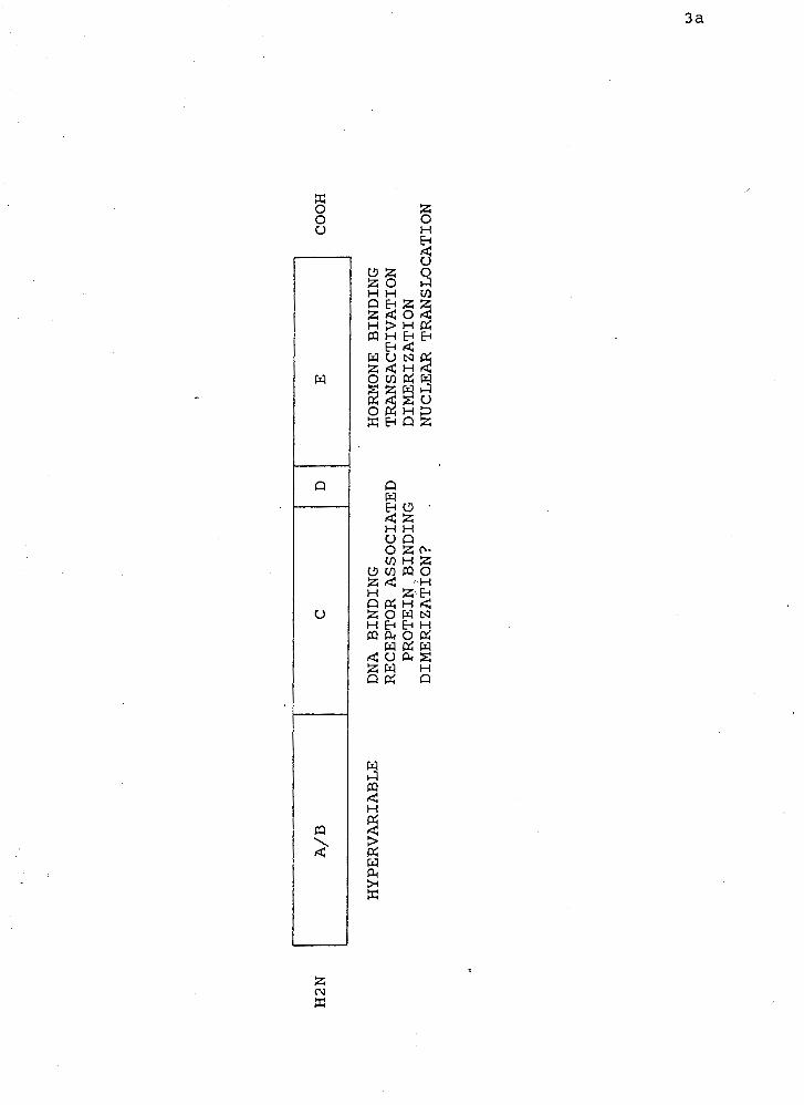
HYPERVARIABLE
DNA BINDING
HORMONE BINDING
RECEPTOR ASSOCIATED
TRANSACTIVATION
PROTEIN,BINDING
DIMERIZATION
DIMERIZATION?
NUCLEAR TRANSLOCATION
H2N
A/B
C
D
E
COOH

sensitive genes (Gehring, 1987).
Phosphorylation of steroid hormone receptors may play a role in ligand
binding (Beato, 1987) but also may be important in mediating the biological actions
of steroid hormones (Moudgil, 1990). For progesterone receptors the kinases
involved phosphorylate mostly serine residues and the sites of phosphorylation lie
within the amino terminus of the receptor. These multiple sites may suggest that
multiple kinases are involved. For the estrogen receptor, the sites are found in the
carboxy terminus and tyrosine appears to act as the major substrate of the kinase.
The role of phosphorylation in steroid hormone receptor function could be diverse.
This post translational modification could modulate ligand binding, it could be
critical for DNA binding to the hormone response elements and it could also be
involved in receptor regulation (Moudgil, 1990). The receptor itself could
autophosphorylate or act as a kinase, these two possibilities have not been
eliminated. It is however known that under physiological conditions, hormone
treatment leads to amplification of phosphorylation and a coincident change in the
expression of hormone sensitive genes.
The DNA binding fingers (see Figure 2) formed by the tetrahedral
association of the sulphur atoms of two pairs of highly conserved cysteine residues
with zinc ions are thought to be responsible for interacting with the DNA and
consequently altering gene expression (Beato, 1991). The DNA sequence that these
- fingers interact with have been called the hormone response element. This cis
acting element has 'enhancer-like' properties in that it functions in a position and
orientation independent fashion (Beato, 1987). Commonly, these hormone
response elements are comprised of 13-15 base pairs which include two conserved
blocks with their centers 10 base pairs apart separated by three nonconserved

nucleotides. They are palindromic with a central axis of dyad symmetry (Beato,
1991). There is a general consensus sequence for the element and depending upon
the nucleotide substituted at two locations, three subgroups have been identified.
The overall consensus sequence is S'AGGNxCAN0.5TGNyCCT 3' where the ,
Nx=T and the Ny =A for the estrogens and Nx=A and Ny = T for the
glucocorticoids and Nx=T or A for the thyroid response elements (Danielsen et al,
1989; Glass and Holloway, 1990; Wahli and Martinez, 1991). The first zinc finger
has been implicated as being responsible for the sequence specific interaction
(Green and Chambon, 1988; Luisi et al, 1991). Precisely three amino acids (marked
with asterisks in Figure 2), whose mutation has been shown to abolish this
interaction, occur between the third and fourth cysteine and directly after it (Mader
et al, 1989). These residues lie within what is thought to be an alpha-helical region
(Walhi and Martinez, 1991). For the estrogen group the sequence of residues is
CEGCKAIG and for the glucocorticoid group it is CGSCKV in which the second,
third and fifth residues are the ones previously referred to. A receptor dimer
binding to one response element is the current model of the mechanism of
interaction (Green et al, 1988). The two zinc fingers of the glucocorticoid receptor
form a globular domain. Then the amino terminus of the first finger with its tertiary
structure of antiparallel beta-pleated sheet is responsible for orientating the
residues that make contact with the phosphate backbone of DNA (Luisi et al, 1991).
The carboxy terminus of the finger is a distorted alpha-helix of which the residue
side chains form contacts with the bases in the major groove. The entire carboxy
terminus of the first and second finger are involved in dimer contacts and positive
transcriptional regulation. The actual DNA binding domain lacks the ability to
dimerize and it is only after binding to the hormone response element that this
event occurs (Luisi et al, 1991).
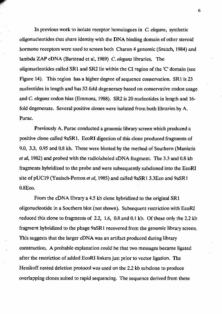
In previous work to isolate receptor homologues in C. elegans, synthetic
oligonucleotides that share identity with the DNA binding domain of other steroid
hormone receptors were used to screen both Charon 4 genomic (Snutch, 1984) and
lambda ZAP cDNA (Barstead et al, 1989) C. elegans libraries. The ,
oligonucleotides called SR1 and SR2 lie within the CI region of the 'C' domain (see
Figure 14). This region has a higher degree of sequence conservation. SR1 is 23
nucleotides in length and has 32-fold degeneracy based on conservative codon usage
and C. elegans codon bias (Emrnons, 1988). SR2 is 20 nucleotides in length and 16-
fold degenerate. Several positive clones were isolated from both libraries by A.
Purac.
Previously A. Purac conducted a genomic library screen which produced a
positive clone called 9aSR1. EcoRI digestion of this clone produced fragments of
9.0, 3.3, 0.95 and 0.8 kb. These were blotted by the method of Southern (Maniatis
et al, 1982) and probed with the radiolabeled cDNA fragment. The 3.3 and 0.8 kb
fragments hybridized to the probe and were subsequently subcloned into the EcoRI
site of pUC19 (Yanisch-Perron et al, 1985) and called 9aSR13.3Eco and 9aSR1
0.8Eco.
From the cDNA library a 4.5 kb clone hybridized to the original SR1
oligonucleotide in a Southern blot (not shown). Subsequent restriction with EcoRI
reduced this clone to fragments of 2.2, 1.6, 0.8 and 0.1 kb. Of these only the 2.2 kb
fragment hybridized to the phage 9aSR1 recovered from the genomic library screen.
This suggests that the larger cDNA was an artifact produced during library
construction. A probable explanation could be that two messages became ligated
after the restriction of added EcoRI linkers just prior to vector ligation. The
Henikoff nested deletion protocol was used on the 2.2 kb subclone to produce
overlapping clones suited to rapid sequencing. The sequence derived from these
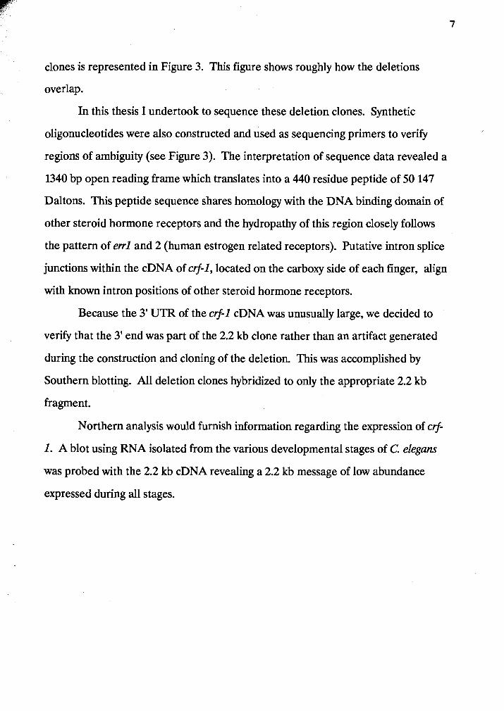
clones is represented in Figure 3. This figure shows roughly how the deletions
overlap.
In this thesis I undertook to sequence these deletion clones. Synthetic
oligonucleotides were also constructed and used as sequencing primers to verify
regions of ambiguity (see Figure 3). The interpretation of sequence data revealed a
1340 bp open reading frame which translates into a 440 residue peptide of 50 147
Daltons. This peptide sequence shares homology with the DNA binding domain of
other steroid hormone receptors and the hydropathy of this region closely follows
the pattern of err1 and 2 (human estrogen related receptors). Putative intron splice
junctions within the cDNA of crf-1, located on the carboxy side of each finger, align
with known intron positions of other steroid hormone receptors.
Because the 3' UTR of the crf-1 cDNA was unusually large, we decided to
verify that the 3' end was part of the 2.2 kb clone rather than an artifact generated
during the construction and cloning of the deletion. This was accomplished by
Southern blotting. All deletion clones hybridized to only the appropriate 2.2 kb
fragment.
Northern analysis would furnish information regarding the expression of crf-
1. A blot using RNA isolated from the various developmental stages of C. elegans
was probed with the 2.2 kb cDNA revealing a 2.2 kb message of low abundance
expressed during all stages.
MATERIALS AND METHODS
I. DNA SEQUENCING
MATERIALS
Caenorhabditis elegans var. Bristol, strain N2 was the organism used in this
study. The nematodes were maintained on NGM plates supplemented with
Escherichia coli strain Op50 as a food source (Brenner, 1974). For large scale DNA
and RNA preparations, worms were cultured on high peptone plates seeded with a
lawn of E. coli strain B (Rose et al, 1982).
Restriction endonucleases used throughout this research were obtained from
Pharmacia and BRL. All radioisotopes were obtained from NEN.
OLIGONUCLEOTIDES
Oligonucleotides were used as sequencing primers to verify regions of
ambiguity (see figure 3). They were on average 20 nucleotides in length and were
obtained from either Tom Atkinson at the Department of Medical Genetics, UBC
or Bruce Brandhorst of the Institute of Molecular Biology and Biochemistry, SFU.
The oligonucleotides were manufactured using an ABI 391 DNA synthesizer
according to the phosphoamidite method of oligonucleotide synthesis. The
oligonucleotides were purified by filtration through Millipore Sep-pack C10
cartridges prepared in the following manner. The cartridge was first equilibrated
with HPLC grade acetonitrile and washed with distilled water. The oligonucleotide,
dissolved in 1.5 rnls of ammonium acetate, was loaded into the cartridge and washed
with distilled water. It was eluted from the column with 20% acetonitrile. This
fraction was evaporated to dryness using a Savant Speed Vac Concentrator. The
oligonucleotidess were rehydrated with sterile distilled water and the concentration
was measured by spectrophotometry. Distilled water was used to make the
appropriate dilutions and the stocks were frozen in small aliquots at -20•‹c for
future use.
ISOLATION OF PLASMID DNA
Small scale plasmid DNA was purified from E. coli JM83 by Miniprep Spun
Column kit purchased from Pharmacia. This procedure is based on the alkaline
lysis method (Maniatis et al, 1982) with the following modification. If the plasmid
DNA was to be used for sequencing it was resuspended in 50 ul of 1X TE (10 mM
Tris, 1 mM EDTA at pH 8.0), denatured for 10 minutes at room temperature in 2 M
sodium hydroxide and then purified by loading and spinning the columns for 2
minutes at 1000 RPM.
RESTRICTION DIGESTS
Restriction enzymes were used with the appropriate buffers under
manufacturers recommended reaction conditions. These conditions include
limiting the enzyme concentration to less than or equal to 5% of the total reaction
volume and maintaining equivalent enzyme and buffer volumes. To ensure that
genomic DNA was fully digested, 5 ug was incubated in excess enzyme for extended
periods of up to 4 hours at 37O~.
AGAROSE GEL ELECTROPHORESIS
Agarose was dissolved most often to the concentration of 0.7% in 1X TBE
(89 mM Tris, 89 mM borate, 2.5 mM EDTA). This is the concentration most
effective for resolving DNA fragments of 0.1 to 10 kb by size (Maniatis et al, 1982).
Ethidium bromide was added to a concentration of 0.2 ug/uls in order to visualize
the fragments under UV light. Loading buffer was added to the DNA to a 1X final
concentration (10X LB = 25% Ficoll400,0.25% bromophenol blue, 0.25% xylene
cyan01 in 1X TBE buffer). Gels were run in 1X TBE at 20-100 volts for 2-12 hours.
Under short wave ultraviolet light of 302 to 365 rim they were photographed using
Kodak Plus X Pan film.
ELECTROELUTION
Recovery of restriction digested DNA fragments from agarose gels was
conducted by dissecting the fragment of interest from the gel and placing it in
dialysis tubing covered with 600 ul of 1X TE3E and sealed using removable clips
(Maniatis et al, 1982). A current of 20-100 volts was then applied for 2-12 hours
such that the DNA in the gel positioned closest to the cathode would be
electroeluted into the buffer toward the anode. Electroelution was complete when
the ethidium bromide intercalated into the DNA fragment could be visualized
under UV light in the buffer fraction. The polarity of the electrodes was then briefly
reversed to free up the DNA from contact with the dialysis tubing. The buffer was
collected and spun for 15 minutes at 4 ' ~ in a microcentrifuge in order to precipitate
the residual agarose. The supernatant containing the DNA was ethanol precipitated
in two volumes of 95% ethanol and 1/10 volume of 5 M ammonium acetate at - 20•‹C, washed with 70% ethanol and vacuum desiccated. The pellet was then
resuspended in 1X TE.
SEQUENCING OF DNA
Denatured plasmid template DNA was obtained as previously described.
The amount of DNA required per sequencing reaction was 3-5 ug. The sequencing
protocol followed was according to Sequenase Version 2.0 Kit (USB) which
contained all reagents except radionucleotides which were supplied by NEN. This
protocol is based on the dideoxy method of Sanger et a1 (1977) but slightly altered
according to Tabor and Richardson (1984). The denatured plasmid was added to ,
0.5 pmol of appropriate primer (T3 forward, T7 reverse, MI3 reverse or -40) and
reaction buffer and denatured for 2 minutes at 65'~. The reaction was cooled for
15 minutes at room temperature to allow the primer to anneal to the template. This
mixture is added to 0.1 M D m , 7.5 uM of dGTP, dCTP and d m , 5 uCi 3 5 ~ - d ~ ~ ~
and 1 unit of recombinant T7 DNA polymerase and incubated 2-5 minutes at room
temperature. This reaction mixture is equally aliquoted into four prewarmed
termination mixtures of G, A, T, or C which consist of deoxy/dideoxynucleotides.
These are incubated for 5 minutes at 37OC. A stop solution of 95% formamide, 20
mM EDTA, 0.05% bromophenol blue and 0.05% xylene cyan01 FF is added and the
tubes are heated to 85OC for 2 minutes and put on ice just prior to loading.
Samples were electrophoresed on 6% acrylamide, 8 M urea gels at 50 amps
for 6 hours in 1X TBE in the top buffer chamber and 1 M ammonium acetate in 1X
TBE in the lower chamber. The gels were then transferred on to 3MM Whatman
chromatography paper and covered with Saran wrap to be dried at 80•‹c for 20
minutes under vacuum. Afterwards, the gel was placed directly on Kodak XAR-5
film for 12 hours.
An amended protocol for sequencing was used to resolve DNA regions which
displayed high secondary structure (Terry Snutch, personal communication). This
method was used in conjunction with the Sequenase 2.0 Kit (USB). The primer at
2.5 pmol concentration is first annealed to 2-4 ug of denatured double stranded
template DNA in the presence of 1 ul of DMSO at a higher temperature of 9S0c
for 3 minutes. The reaction mixture is frozen on dry ice and ethanol for 5 minutes,
quickly thawed just prior to the addition of sequencing buffer and incubated at room
temperature for 5 minutes. The labelling mix which consists of D m , dilute
labelling mix, 3 5 ~ - d ~ ~ ~ , dilute Sequenase 2.0 enzyme and 0.5 ul DMSO is added
and the mixture incubated for another 5 minutes at room temperature. Aliquots are
added to each dideoxy termination mix, incubated for 5 minutes at 3 7 O ~ and 4 ul of /
stop mix is added and the tubes are denatured for 5 minutes at 9 5 - 1 0 0 ~ ~ just prior
to loading on the gel.
PROTEIN SEQUENCE ALIGNMENT AND PHYLOGENY OF crf-1 AND
OTHER STEROID RECEPTORS AND TRANSCRIPTION FACTORS
The DNA binding domain of crf-1 was aligned with the corresponding region
of other steroid hormone receptors and transcription factors (Figure 5 and 6). The
other sequences were obtained from the sources cited in the Figure 5 legend. The
computer program Clustal4 compares amino acid identity in order to construct a
phylogenetic tree. Clustal uses a distance matrix based on operational taxonomic
units for sequence comparison. This program was used on the CI alignment and is
shown in Figure 7. See page 35 for further details.
11. ORGANIZATION AND EXPRESSION OF crf-1
LABELING OF PROBES
DNA probes used for library screening, Southern or Northerns were labeled
by nick translation (Maniatis et al, 1985), random priming with oligonucleotides ('I7
Quick Prime Kit, Pharmacia) or end labeled by kinase (Maniatis ef al, 1982).
The nick translation reaction requires approximately 500 ng of DNA to be
incubated with alpha 3 2 ~ - d ~ ~ ~ (80 Ci/mmole) as well as unlabeled nucleotides
and alpha 32~-dTTF (2.6 uM) in the presence of DNase and DNA polymerase I for
2 hours at 15OC. Unincorporated nucleotides were removed by chromatography
through Sephadex G-25.
The T7 Quick Prime Kit protocol based on the method of Feinberg and
Vogelstein (1983) requires greater than or equal to 50 ng of denatured DNA and
alpha 3 2 ~ - d ~ ~ ~ (3000 Ci/mmole). This is incubated with a reagent mix of
unlabeled nucleotides and T7 DNA polymerase for 15 minutes at 37'~. The
reaction product of the T7 Quick Prime Kit was acid precipitated in TCA prior to
counting.
End labeling of the 5' end with kinase requires DNA which lacks a 5'
phosphate as in the case of synthetic oligonucleotides. This protocol involves
incubation with T4 polynucleotide kinase, 100 mM magnesium chloride, 100 mM
DTT and alpha 3 2 ~ - d ~ ~ ~ (50 uCi) for 15 minutes at 37'~. This method also
requires the removal of unincorporated nucleotides by chromatography through
Sephadex G-25.
In all methods the specific activity of the labeled DNA was measured by
7 Cerenkov counting in a scintillation counter. A specific activity of 1x10 cpm/ug
could be achieved for probes labeled by these methods.
NORTHERN BLOTTING
Total RNA of mixed stages or stage specific total RNA was isolated from C.
e l egm by Terry Snutch according to the method of Chirgwin et a1 (1979). RNA at a
concentration of 30 ug was denatured in 50% formamide, 1 mM EDTA, 5 mM
sodium acetate, 10X MOPS at pH 7.0 for 15 minutes at 60•‹c. Loading buffer was
added to a concentration of 1X and 2 ul of ethidium bromide (10 mg/ml) just prior
to electrophoresis through 1.1% agarose gel containing 1X MOPS and 2.2 mM
formaldehyde for 4-5 hours at 80 volts in 1X MOPS running buffer. An 8 ul aliquot

of BRL 0.24 - 9.5 kb RNA ladder was treated like a sample and used as a marker.
The gel was photographed under UV light prior to transfer.
In preparation for transfer, the gel was soaked twice for 15 minutes in 0.025
M potassium phosphate at pH 7.5 and once in distilled water for 15 minutes.
Genescreen (Dupont), the membrane for transfer, was soaked for 15 minutes in
0.025 M monobasic and dibasic sodium phosphate at pH 6.5. The blotting
procedure was according to Maniatis et a1 (1982). The transfer was conducted for at
least 12 hours. Upon disassembly, the membrane was rinsed in buffer to remove
residual agarose, cross-linked at 254 nm for 40 seconds in a Stratagene cross-linker
and baked for 2 hours at 80•‹C under vacuum.
The membrane was prehybridized for at least 16 hours at 4 2 ' ~ in 50%
deionized formamide, 5X Denhardts, 5X SSC, 1% SDS and 100 uglml herring
sperm DNA (Denhardt, 1966). Hybridization conditions were of moderate
stringency using fresh prehybridization solution containing the denatured probe
(lxlo7 cpmlug) for 16-48 hours at 42'~. Two washes under each of the following
conditions were performed: 2X SSC at room temperature for 5 minutes, 2X SSC
and 1% SDS at 6 5 ' ~ for 30 minutes and 0.1X SSC for 30 minutes at room
temperature. The membrane was sealed in plastic and exposed to preflashed Kodak
XARd film at -70 '~ with an intensifying screen for over 48 hours.
SOUTHERN BLOTTING
DNA was prepared as previously described and 2-30 ug were restriction
digested and size fractionated by electrophoresis on 0.7% agarose gels in 1X TBE
for 12 hours at 20 volts. A photograph of the gel was taken. The excess gel was
trimmed off and the remainder soaked in 0.25 M hydrochloric acid for 10 minutes at
room temperature then briefly rinsed in distilled water. The gel was soaked three
times each for 20 minutes at room temperature in 25 mM monobasic and dibasic
membrane (Genescreen, Dupont) and 3MM Whatman chromatography paper were
cut to the size of the gel and soaked for 5 minutes at room temperature in 25 mM
transfer buffer. Transfer was set up according to Maniatis et a1 (1982) for 12 hours
in transfer buffer.
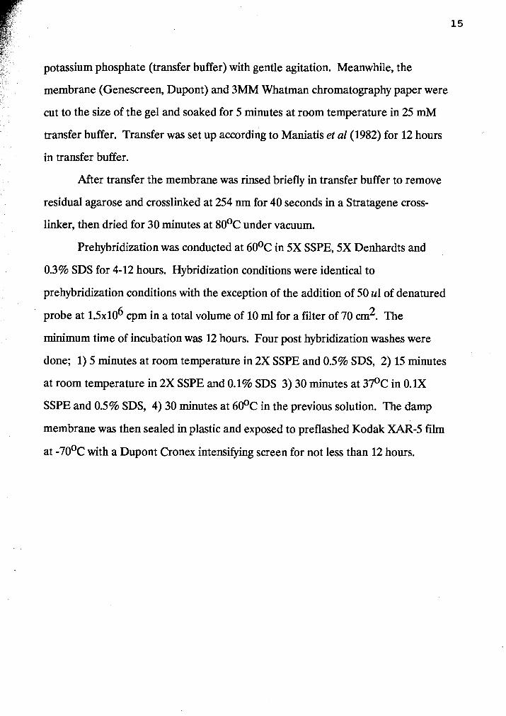
After transfer the membrane was rinsed briefly in transfer buffer to remove
residual agarose and crosslinked at 254 nm for 40 seconds in a Stratagene cross-
linker, then dried for 30 minutes at 80•‹C under vacuum.
Prehybridization was conducted at 6 0 ' ~ in 5X SSPE, 5X Denhardts and
0.3% SDS for 4-12 hours. Hybridization conditions were identical to
prehybridization conditions with the exception of the addition of 50 ul of denatured
probe at 15x10~ cpm in a total volume of 10 ml for a filter of 70 cm2. The
minimum time of incubation was 12 hours. Four post hybridization washes were
done; 1) 5 minutes at room temperature in 2X SSPE and 0.5% SDS, 2) 15 minutes
at room temperature in 2X SSPE and 0.1% SDS 3) 30 minutes at 3 7 ' ~ in 0.1X
SSPE and 0.5% SDS, 4) 30 minutes at 60•‹C in the previous solution. The damp
membrane was then sealed in plastic and exposed to preflashed Kodak XAR-5 film
at -70•‹c with a Dupont Cronex intensifymg screen for not less than 12 hours.
RESULTS
I. SEQUENCE ANALYSIS OF THE of-1 cDNA
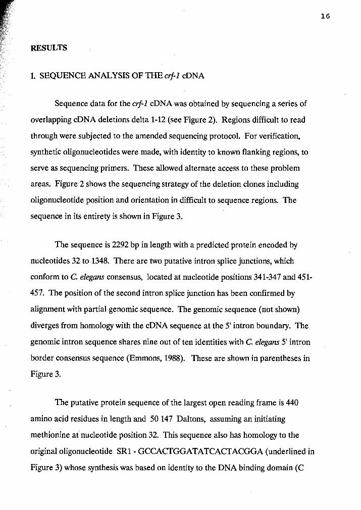
Sequence data for the ctf-1 cDNA was obtained by sequencing a series of
overlapping cDNA deletions delta 1-12 (see Figure 2). Regions difficult to read
through were subjected to the amended sequencing protocol. For verification,
synthetic oligonucleotides were made, with identity to known flanking regions, to
serve as sequencing primers. These allowed alternate access to these problem
areas. Figure 2 shows the sequencing strategy of the deletion clones including
oligonucleotide position and orientation in difficult to sequence regions. The
sequence in its entirety is shown in Figure 3.
The sequence is 2292 bp in length with a predicted protein encoded by
nucleotides 32 to 1348. There are two putative intron splice junctions, which
conform to C. elegam consensus, located at nucleotide positions 341-347 and 451-
457. The position of the second intron splice junction has been confirmed by
alignment with partial genomic sequence. The genomic sequence (not shown)
diverges from homology with the cDNA sequence at the 5' intron boundary. The
genomic intron sequence shares nine out of ten identities with C. elegans 5' intron
border consensus sequence (Ernmons, 1988). These are shown in parentheses in
Figure 3.
The putative protein sequence of the largest open reading frame is 440
amino acid residues in length and 50 147 Daltons, assuming an initiating
methionine at nucleotide position 32. This sequence also has homology to the
original oligonucleotide SR1- GCCACTGGATATCACTACGGA (underlined in
Figure 3) whose synthesis was based on identity to the DNA binding domain (C

Figure 2 Representation of the amount of sequence obtained from the deletion
clones of crf-1. The sequence of the oligonucleotides used as sequencing primers is
shown as well as their position and orientation with respect to the deletion clones.
Numbers at the right end of the boxes indicate the number of nucleotides sequenced
from each particular deletion clone. The hatched boxes indicate sequence obtained
from oligonucleotide primers.
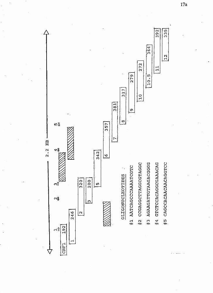
OLIGONUCLEOTIDES
.
AATCAGCCCAAAATCGTC
CCGAGGCTTAGGCGTAGGC
AGAAGATTGTAAGACGGGG
GTCTCCACAGGCGAAACAG
CAGCCACAACAACAGGTCC
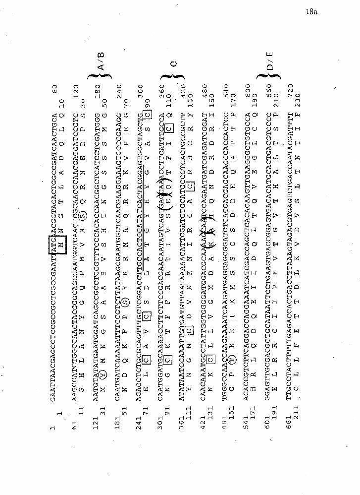
Figure 3 The complete nucleotide sequence of crf-1. The putative protein product
is indicated below the nucleotide sequence. Circled amino acid residues indicate
putative phosphorylation sites. The domains corresponding to known steroid
hormone receptors are indicated in the right margin. The boxed residues are the ,
conserved cysteines in the DNA binding region. The underlined region indicates
the sequence and position of oligonucleotides originally used to isolate this clone.
The boxed methioine represents the putative start codon. The two set of brackets
denote putative intron splice junctions (Fields, 1989). The asterisk within the
protein sequence indicates a stop codon.
GAATTAACGAGCCTCCGCCGCTCGCCGAAT ATG
CGGTACACTGGCCGATCAACTGCA
G T
LA
D Q
LO10 60
1
61
ASIGCCATCTGGCCAACTACGGGCAGCCAATGGTCAACTCGCAACGCAACGAGGATCCGTC
120
11
SH
LA
NY
GQ
PM
VN
@Q
RN
ED
PS
30
\
121
AATGT TATGAATGGATCAGCCGCCTCCGTTTCCCACACCAACGGCTCATCCTCGATGGG
31
M&
)M
NG
SA
AS
VS
HT
NG
SS
SM
G5
0
181
CA
AT
GA
TC
AA
AA
AT
TT
CC
GT
CT
TA
TA
AG
CG
MT
GG
CT
CC
G
240
51
ND
QK
FP
@Y
KR
MA
QR
RK
VP
EG
70
241
AGAGCTGTGCGCAGTTTGCTCGGACCTTGCCACTGGATATCACTACGGAGTGGCTAGCTG
300
71
La
A V
mS
D L
A
G Y
H Y
G'vA
301
CA
AT
GG
AT
GC
AA
AA
CC
TT
CT
TC
CG
AC
GA
AC
AA
TA
GT
CA
G
A ~~CCTTCATTTGCCA
91
NG
~K
TF
FR
RT
IV
SE
QT
FI
~Q
11
0
421
CAACAAATGCCTATTGGTGGGGATGGACGC
CCAGAATGATCGAGATCGGAT
480
131
NK
RL
LV
GM
DA
T$q Q
N D
R D
R 1150
481
TGGGCCAACGMGAAAATCAAGATGATGAGCAGCGGATCTGACGACGAGCAAGCCACCACTCC
540
151
GP
@K
KI
KM
SS
GS
DD
EQ
AT
TP
17
0
541
ACACCGTCTTCAGGACCAGGAAATCATCGACCAGCTCACACAAGTTGAAGGGCTGTGCCA
600
171
HR
LQ
DQ
EI
ID
QL
TQ
VE
GL
CQ
19
0,
, 6
0 1
GCAGTTGCGACGCTGCATAATTCCTGAAGTGACCGGAGTGACACATGCACTGACGTCCC
6 6
0
J D /
E 191
EL
RR
CI
IP
EV
TG
VT
HA
LT
SP
21
0
661
TT
GC
CT
AC
TT
TT
TG
AG
AC
CA
CT
GA
CC
TT
AA
AG
TA
GA
CG
TG
AG
TC
TG
AC
CA
AT
AC
GA
TT
TT
720
211
CL
LF
ET
TD
LK
VD
VS
LT
NT
IF
23
0

721
CAAAGAGCTCTTCCCGGCGTCGATGAACGACATTCGTATGTGGAACATTCGCGAGATGCG
780
231
KE
LF
PA
SM
ND
IR
MW
NI
RE
MR
25
0
781
CA
TC
TG
CA
TC
GA
GT
GG
GC
AA
AG
AC
GT
TC
GA
CG
TT
CG
AT
GT
TT
AC
CA
GA
GG
CT
CA
AC
CT
TT
TT
GA
TC
A
840
251
IC
IE
WA
KT
FD
VY
QR
LN
LF
DQ
27
0
841
GT
TT
GC
CC
TC
GT
CC
GA
AA
CT
TT
GC
CT
TC
GC
AT
TT
AA
TC
TG
CT
CA
AC
AG
AG
TG
TT
CT
AC
TC
900
271
FA
LV
RN
FA
FA
FN
LL
NR
VF
YS
29
0
901
GCCGGACCATGGACCAGATAAGATTGTCTTCCAAAATGGAGCCTTCATCATGAGACAGCC
960
291
PD
HG
PD
KI
VF
QN
GA
FI
MR
QP
31
0
961
ACAACAACAGGTCCMCTCTCGGGATGCCGGCCTATCTACACTCGACTGGACGAGAT
1020
311
QV
QL
SG
CR
PI
YT
RQ
MD
EI
33
0
1021 TATGATTCCATTCCGCAAATTGCAGTTGAGCGTGGCCGAGTTTGCCACCTTCGCCGC 1080
331
MI
PF
RK
LQ
LS
VA
EF
AT
FK
AA
35
0
1081
TT
TG
TT
TT
TT
AA
TC
CA
GA
CG
CT
TT
GG
AT
TT
GT
CT
CC
AC
AG
GC
GA
AA
CA
GG
AA
GT
TT
TC
GA
1140
351
LF
FN
PD
AL
DL
SP
QA
KQ
EV
FE
37
0
1141 GGAGAGAAACAAATATTTGGGTGGACTTTTTACCTGTATCACACAGTTGGTCCC 1200
371
ER
NK
YL
GG
LF
TC
I@
QK
IG
IP
39
0
1201 GACTGGAGTTCAGAAATATGGMGTCTTTTAATGATGACCGCTAGTATACAGAATATTCT
1260
391
TG
VQ
KY
GS
LL
MM
TA
SI
QN
IL
41
0
1262
GGCGCWTGAGGAAAATATGCAAGTGATGGAATTGTTCCTGGGAAGTGGATCC 1320
411
AQ
NE
EN
MQ
VM
EL
FK
NW
EV
DP
43
0

ATTGAAGTTTTTTATGTTTAACTTTTTAAGACATTAGTTGCCTTCAGATCTCTCACCAAGT
TT
TG
AG
CW
TT
GT
TT
CT
CC
CT
TA
TC
AT
TA
TT
TT
AT
TA
TT
AT
TT
GA
CT
TT
TG
AC
TT
CG
CT
GG
GC
CC
AA
TA
CT
TT
TT
TT
CT
GT
G-C
CA
AC
TA
GG
AC
AC
GT
CA
TA
GA
TT
C
CCCCGTTCCACACTCACAAACAGTTCCCCGTCTTACAATCTTCTAGTTCTGCTGTATATA
TT
TG
TG
AT
WT
AA
TA
TA
AT
GT
TC
AG
TT
TT
GT
ccT
TA
TT
TcT
TT
Tcc
cAcc
AG
GT
TT
TT
TG
TG
CC
AT
CG
CT
GM
TT
AG
TT
TG
AG
CA
CA
CT
TT
TT
TA
T-T
TA
GC
TG
AG
GT
TT
AT
GC
T
TAGGTTTAGGCCGAGGCTTAGGCGTAGGCTTGGATCTAGGATCTTTTAGATCTCAGTTTT
AGGCTCAGCTTTAGGCTTAGGCTTAACCTTCCCCATGCGCTCCTCAAAAAGCCTGTTAAT
GG
CC
GG
TT
GC
CC
TT
AC
CT
GU
TT
CA
CT
~T
TC
CT
CC
CG
TT
TT
CA
CA
AG
CC
AG
TT
TT
TA
AACCCTTTCCCAAATCCAACCMCAACCTCGCCCAGTTCTGATCCAACCTTCCCACCTCTT
CC
TC
TT
CC
AA
GC
AT
TG
TC
AT
CG
AT
TG
CA
AT
TT
TT
TC
mT
TA
TT
CA
TG
TT
TC
AA
CA
AA
AA
ATGTTTCCTCTATAAAAACCCCTATTATTTACTGGTATCAATTCTTTTATTTTCTCCCTA
CC
AC
GA
TT
TT
GG
GC
TG
AT
TT
AT
CT
TT
TT
TT
CC
CA
AA
TT
TT
TT
TA
AC
CA
GT
WT
TG
G
TT
TG
GT
AT
TT
TG
AT
TT
CC
GA
TT
GC
CC
CT
TA
CW
TT
CT
TG
AT
GT
TC
AA
GT
TT
AT
TT
TC
C
CC
CA
CT
TT
TT
TC
TT
TT
GT
CA
TT
CA
TT
TT
TT
TT
AA
AT
TT
TT
TT
TT
CT
CC
CT
CA
AT
MT
CT
T
CA
TT
TC
TT
W

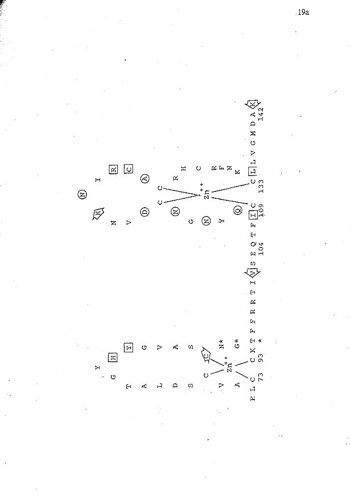
Figure 4 A schematic diagram of the putative protein sequence of the DNA binding
domain of crf-1. Zinc ions are shown interacting with eight highly conserved
cysteine residues. The positions of oligo SR1 and SR2 used to screen the libraries
are indicated by the lines. The regions enclosed by brackets indicate putative alpha- '
helical tertiary structure. Circled amino acids indicate those that are thought to be
involved in dimer contacts. Boxed residues are thought to interact with phosphates
of the DNA backbone. The residues marked by asterisk denote amino acids
important in recognizing hormone response elements. The arrows indicate intron
and putative intron positions of various steroid hormone receptors (Mader et al,
1989; Ritchie et al, 1990; Luisi et al, 199 1).

domain) of various known steroid hormone receptors proteins (Beato, 1989). This
identity spans nucleotides 241-291. The putative secondary structure of the DNA
binding domain of crf l is illustrated in Figure 4. The A/B domain could
presumably span from nucleotides 9-241. The hinge region and the ligand binding
domain (domains D and E) could occur after nucleotide 446. Homology to other
proteins was investigated for all domains outside of the 'C' region. However,
nothing significant was revealed. These homologies were calculated by a Fasta
(Wilbur and Lipman, 1983) search of the Swiss protein database.
Putative b a s e C and tyrosine kinase phosphorylation sites as predicted by
Prosite (PC Gene) are indicated as circled residues in Figure 3.
PROTEIN SEQUENCE ALIGNMENT AND PHYLOGENY OF crf-1 AND
OTHER STEROID RECEPTORS AND TRANSCRIPTION FACTORS
An alignment of the DNA binding regions of the peptide product of crf-I and
other steroid receptors and trans-cription factors is shown in Figure 5. These
receptors were chosen because preliminary analysis indicated homology to the
progesterone family of receptors. In addition several invertebrate receptors were
included. Identical residues are denoted by asterisks and conservative changes are
indicated by dots. Figure 6 is an alignment of just the CI region of the DNA binding
. domain. This alignment was used as data by the computer program Clustal4 to
construct a phylogenetic tree (Figurs . This tree indicates that the crf-I protein
product is most homologous to the CI region of estrogen and the estrogen related
receptors. The next closest group includes; MTH, svp, COUP, E754 crf-2 and tll.
Crf-2 shares homology with E75A. On the other branch

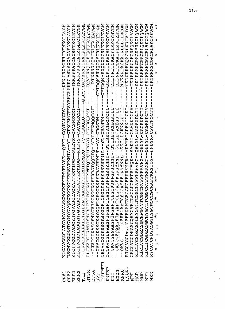
Figure 5 An alignment of the DNA binding domain of a variety of steroid receptors.
The program Clustal4 was used with the following parameters: ktup = 1, window
size = 10, filtering level =2-5 and gap penalty =3. The asterisks denote residues
which are identical and the dots denote conservative amino acid substitutions.
HMR - human mineralcorticoid, Err1 and 2 - human estrogen related receptors,
COUPTFl - chicken ovalbumin upstream transcription factor (Wang et al, 1989),
HGR - human glucocorticoid, HPR - human progesterone, HER - human estrogen,
HT3R - human thyroid, HVD3R - human vitamin D3 (Schwabe and Rhodes, 1991),
crf-1 and 2 - C. eleguizs receptor finger (R. Eagen and A. Sluder, personal
communication), tll - Drosophila tailless, E75A - Drosophila ecdyson transcription
factor, svp - Drosphila seven up, KNIRP, KNI and KNRL - Drosophila Knirp
related, egon - Drosophila embryonic gonad (reviewed by Pignoni et ul, 1990).
CRFl
CRF2
ERR1
ERR2
TLL
HT3R
E75A
SVP
COUPTFI
KNIRP
KNI
EGON
KNRL
HVDR
MTH
HGR
HMR
HPR
HER
ELCAVCSDLATGYHYGVASCNGCKTFFRRTIVSE--QTFI-CQYNGNCDV------- NKNIRCACRHCRFNKCLLVGM
DRCMVCGDNSTGYHYGVQSCEGCKGFFRRSVHKNIA--YL-CTKGENCTFSYENCAANRGVRTRCQACAFAKCLAVGM
RLCLVCGDVASGYHYGVASCEACKAFFKRTIQG--SIEYS-CPASNECEI------- TKRRRKACQACRFTKCLRVGM
RLCLVCGDIASGYHYGVASCEACKAFFKRTIQG--NIEYS-CPATNECEI------- TKRRRKSCQACRFMKCLIWGM
--CKVCRDHSSGKKYGIYACDGCAGFFKRSIRRSRQ--YV-CKSQKQ----- GLCWDKTHRNQCRACRLRKCFEVGM
ELCWCGDKATGYHYRCITCEGCKGFFRRTIQKNLHPSYS-CKYEGKCVI------- DKVTRNQCQECRFKKCIYVGM
--CRVCGDKASGFHYGVHSCKGCKGFFRRSIQQKIQ--YRPCTKNQQCSIL------- RI NRNRCQYCRLKKCIAVGM
--CWCGDKSSGKHYGQFTCEGCKSFFKRSVRRNLT--YS-CRGSRN-------
CPIDQHHRNQCQYCRLKKCLK-GM
IECWCGDKSSGXHYGQFTCEGCKSFFKRSVRRNLT--YT-CMNRN------- CPIDQH-RNQCQYCRLKKCLKVGM
QTCKVCGEPAAGFHFGAFTCAGCKSFFGRSYNNI--STISECKNEGKCII------- DKKNRTTCKACRLRKCYNVGM
--CKVCGEPAAGFHFGAFTCEGCKSFFGRSYNNI--STISECKNEGKCII------- DKKNRTTCKACRLRKCYNVGM
--CKVCGEPAAGFHFGAFTCEGCKSFFGRSYNNI--STISECKNEGKCII------- DKKNRTTCKACRLRKCYNVGM
--CKVC.
GFHFGAFTCEGCKSFFGRSYNNL--SSISDCKNNGECII------- NKKNRTACKACRLLLCLMCGM
RICGVCGbhh 'GFHFNAMTCEGCKGFFRRSMKR---KAMFTCPFNGDCKI------- TKDNRRHCQACRLKRCVDIGM
GRCAVCGDNASCQHYGVRTCAGCKGFFKRTVQK--SAKYI-CLANKDCPV------- DKRRRNRCGFCRFQKCLAVGM
KLCLVCSDEASGCHYGVLTCGSCKVFFKRAVEG--QHNYL-CAGRNDCII------- DKI RRXNCPACRYRXCLQAGM
KICLVCGDEASGCHWGWTCGSCKVFFKRAVEG--QHNYL-CAGRNDCII------- DKIRRKNCPACRLQKCLQAGM
KICLICGDEASGCHYGVLTCGSCKVFFKRAMEG--QHNYL-CAGRNDCIV------- DKIRRKNCPACRLRKCCQAGM
RYCAVCNDYASGYHYGVWSCEGCWiFFKRSIQG--HNDYM-CPATNQCTI------- DKNRRKSCQACRLRKCYEVGM
* .*
. .
. *.
,*
.*
** *
. *
,*
**
*
**
Figure 6 An alignment of the CI region of the DNA binding domain of a variety of
steroid receptors and transcription factors. This alignment was calculated by the
same program and under the same parameters as those in Figure 5. Sequence
sources are identical to those in Figure 5.
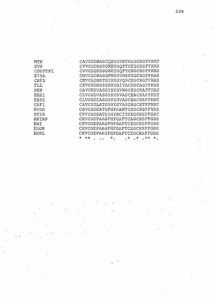
MTH SVP COUPTFI E75A CRF2 TLL HER ERR1 ERR2 CRFl HVDR HT3R KNIRP KNI EGON KNRL
CAVCGDNASCQHYGVRTCAGCKGFFKRT CWCGDKSSGKHYGQFTCEGCKSFFKRS CWCGDKSSGKHYGQFTCEGCKSFFKRS CRVCGDKASGFHYGVHSCKGCKGFFFCRS CMVCGDNSTGYHYGVQSCEGCKGFFRRS CKVCRDHSSGXHYGIYACDGCAGFFKRS CAVCNDYASGYHYGVWSCEGCKAFFKRS CLVCGDVASGYHYGVASCEACKAFFKRT CLVCGDIASGYHYGVASCEACKAFFKRT CAVCSDLATGYHYGVASCNGCKTFFRRT CGVCGDRATGFHFNAMTCEGCKGFFRRS CWCGDKATGYHYRCITCEGCXGFFRRT CXVCGEPAAGFHFGAFTCAGCKSFFGRS CKVCGEPAAGFHFGAFTCEGCKSFFGRS CKVCGEPAAGFHFGAFTCEGCKSFFGRS CKVCGEPAAGFHFGAFTCEGCKSFFGRS * * * . .. *. . * . * . * * *.
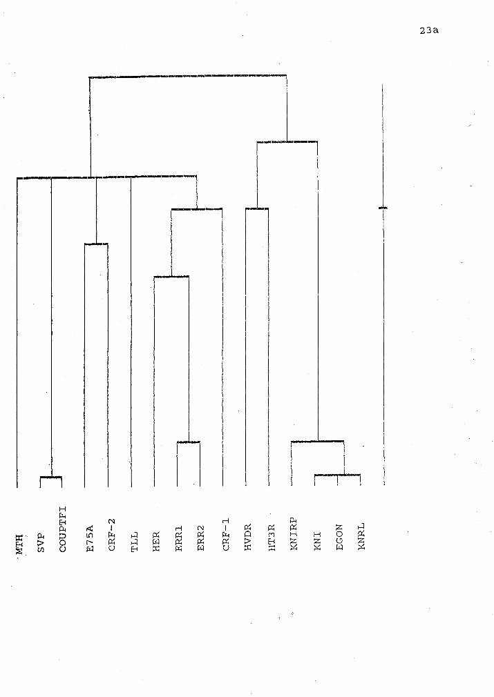
Figure 7 A phylogenetic tree of the DNA binding domain of various steroid
receptors and transcription factors. This tree was constructed using the alignments
in Figure 6 and the parameters of gap penalty (fixed) = 10 and gap penalty
(varying) = 10. The vertical axis represents relative time since divergence.
n F I C\1 P; P; P; PC; W W
lie HVDR, HT3R, and the Drosophila KNIRP, KNI, egon and KNRL.
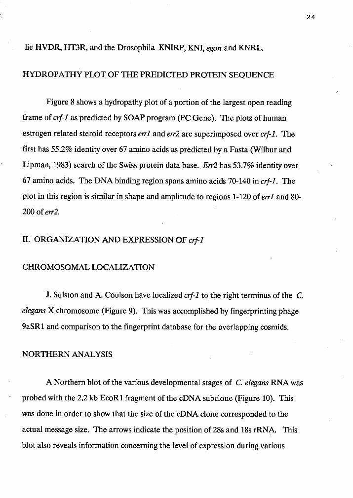
HYDROPATHY PLOT OF THE PREDICTED PROTEIN SEQUENCE
Figure 8 shows a hydropathy plot of a portion of the largest open reading
frame of crf-1 as predicted by SOAP program (PC Gene). The plots of human
estrogen related steroid receptors errl and err2 are superimposed over crf-1. The
first has 55.2% identity over 67 amino acids as predicted by a Fasta (Wilbur and
Lipman, 1983) search of the Swiss protein data base. Err2 has 53.7% identity over
67 amino acids. The DNA binding region spans amino acids 70-140 in crf-1. The
plot in this region is similar in shape and amplitude to regions 1-120 of errl and 80-
200 of err2.
11. ORGANIZATION AND EXPRESSION OF c#-l
CHROMOSOMAL LOCALIZATION
J. Sulston and A. Coulson have localized crf-1 to the right terminus of the C.
elegans X chromosome (Figure 9). This was accomplished by fingerprinting phage
9aSR1 and comparison to the fingerprint database for the overlapping cosmids.
NORTHERN ANALYSIS
A Northern blot of the various developmental stages of C. elegans RNA was
- probed with the 2.2 kb EcoRl fragment of the cDNA subclone (Figure 10). This
was done in order to show that the size of the cDNA clone corresponded to the
actual message size. The arrows indicate the position of 28s and 18s rRNA. This
blot also reveals information concerning the level of expression during various
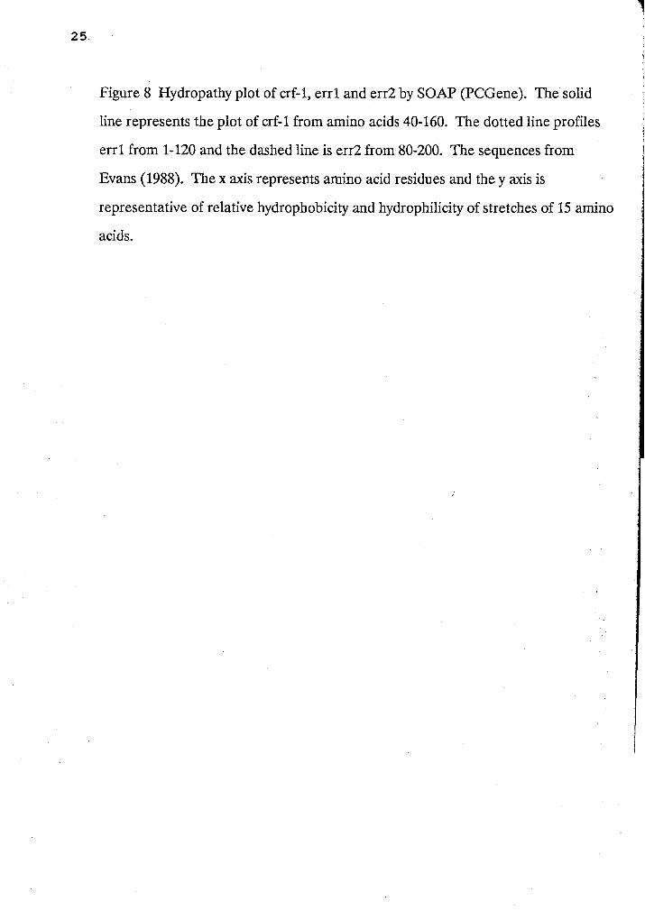
Figure 8 Hydropathy plot of crf-1, errl and err2 by SOAP (PCGene). The solid
line represents the plot of crf-1 from amino acids 40-160. The dotted line profiles
errl from 1-120 and the dashed line is err2 from 80-200. The sequences from
Evans (1988). The x axis represents amino acid residues and the y axis is
representative of relative hydrophobicity and hydrophilicity of stretches of 15 amino
acids.
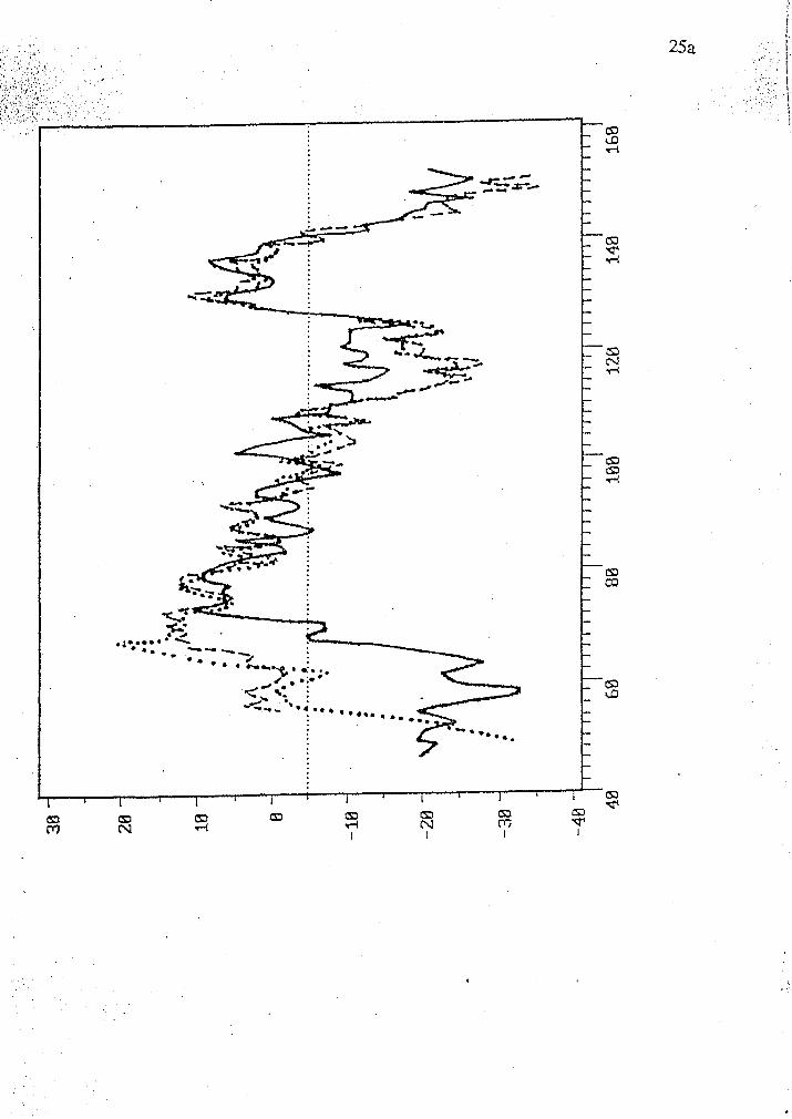
Figure 9 Chromosome maps of the right end of the X chromosome of C. elegans.
The symbols indicate the positions of three markers.
a. The genomic clone called BN#SR19A which hybridizes to crf-1 occurs at the
extreme right end of YAC Y52G2.
b. This diagram indicates by symbols the relative positions of several genetic
markers.
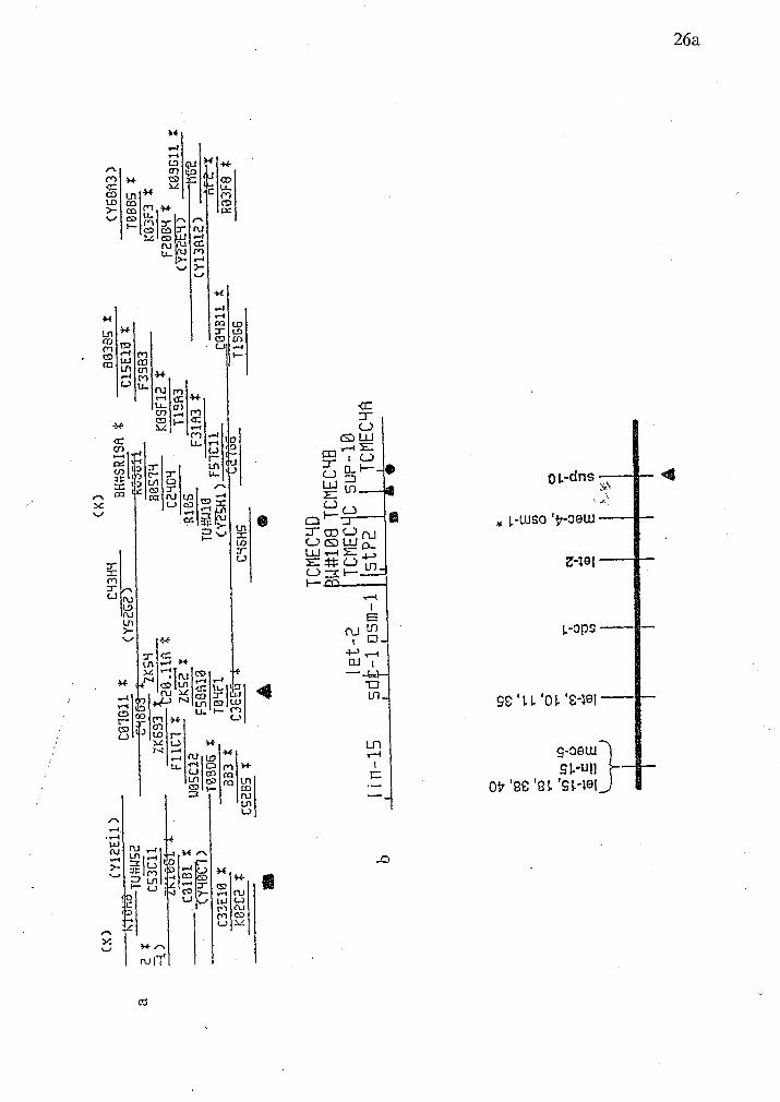
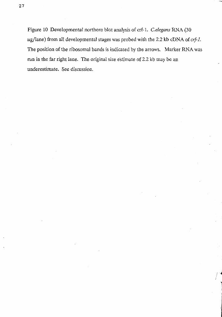
Figure 10 Developmental northern blot analysis of crf-1. Celegans RNA (30
ugllane) from all developmental stages was probed with the 2.2 kb cDNA of @-I.
The position of the ribosomal bands is indicated by the arrows. Marker RNA was
run in the far right lane. The original size estimate of 2.2 kb may be an
underestimate. See discussion.


developmental stages. A potential band is visible in all lanes at the 2.2 kb size and
the intensity of hybridization is similar. Figure 11 shows the same Northern blot
stripped and reprobed with hsp70A (Heschl, 1988) a control whose message is 2.4
kb in size. The position of 28s and 18s rRNA is indicated by arrows. The expression
of this message is low during L1 but increases throughout L2, L3/4 and adult
(Prasad and Baillie, 1989).
SOUTHERN ANALYSIS
A Southern blot of the EcoRl restricted original cDNA clone called cD3A
4.5Eco was probed with either end of the EcoRl fragment of the 2.2 kb cDNA to
verify that the 2.2 kb deletion subclones were not artifacts of the deletion protocol.
The blot, probed with oligonucleotide #1 located within the 3' end, hybridizes with a
2.2 kb band (Figure 12). The same blot probed with a PstI/AflIII fragment of 115
bp from the deletion clone #12 is shown is Figure 13. A 2.2 kb band is visible.
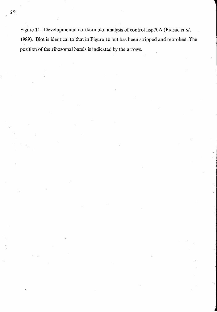
Figure 11 Developmental northern blot analysis of control hsp70A (Prasad et al,
1989). Blot is identical to that in Figure 10 but has been stripped and reprobed. The
position of the ribosomal bands is indicated by the arrows.

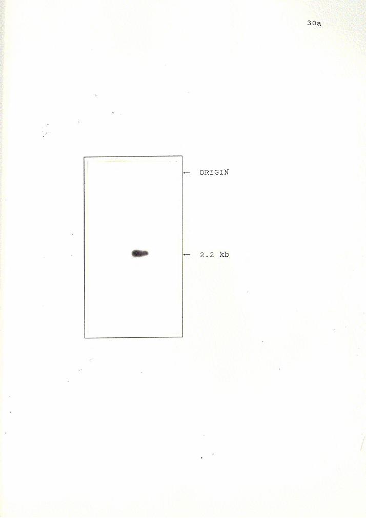
Figure 12 Southern blot analysis of Eco RI digested original 4.5 kb cDNA clone
probed with oligo #I located at the 3' end of the 2.2 kb cDNA clone. This was
carried out to verify that the deletion clones from the 3' end were not artifacts of
construction. This confirmed that the 3' sequence obtained did indeed originate
from the 3' end of the 2.2 kb clone.
ORIGIN
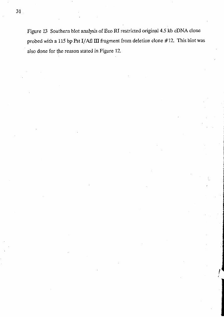
Figure 13 Southern blot analysis of Eco RI restricted original 4.5 kb cDNA clone
probed with a 115 bp Pst I /M 111 fragment from deletion clone #12. This blot was
also done for the reason stated in Figure 12.

+- ORIGIN
DISCUSSION
I. SEQUENCE ANALYSIS OF THE crf-1 cDNA
The sequence data obtained for the crf-1 cDNA spans 2292 nucleotides and
has a large open reading frame of 1340 nucleotides (Figure 3). It is within this ORF
that peptide consensus to the DNA binding region occurs (domain C). This reading
frame has several features which seem to indicate that it may code for a mRNA
which could be translated into a protein. The first feature is an AUG start codon at
position 32. The putative ribosome binding site has the sequence GAATI'ATGA
and an identity occurs in eight out of ten positions with the C. elegans consensus
sequence (M.D.Perry, personal communication). If we assume that this codon is the
initiating methionine for translation and proceed to translate the remaining
sequence in this reading frame several other features are observed. The size of crf-1
protein is predicted to be 50 147 Daltons for its 440 amino acids. The most obvious
feature is the identity with the DNA binding site of other steroid hormone
receptors. This identity spans nucleotides 269-291. Figure 5 shows an alignment of
the protein sequence of this region. Crf-1 protein sequence has been aligned with
the C domain of other steroid receptors. The DNA binding region consists of two
fingers which contain ten highly conserved cysteine residues that are thought to
interact with zinc ions allowing the tertiary formation of two "zinc fingers" (Green
and Chambon, 1988) which in turn interact with sequence specific areas of the
hormone response element that allow interaction with other factors that regulate
transcription (Beato, 1991). In all cases these two fingers are encoded by separate
exons (Green et al, 1988) except for COUP-TF1 where both fingers are found in the
first exon (Ritchie et al, 1990). This finger motif was originally detected in the
Xenopus protein TFIIIA (Miller et al, 1985; Evans et al, 1988). Since the two fingers

of steroid hormone receptors are similar one might speculate that they have arisen
by duplication, however there are amino acid substitutions that indicate they have
since diverged significantly. The first exon called CI is closest to the amino terminal
of the peptide and contains four of the conserved cysteine residues and the
remainder of the residues are hydrophobic (Wahli and Martinez, 1991). In the crf-1
protein the conserved cysteine residues lie at peptide positions 248, 257, 299 and
308 ( Figure 3). Nonpolar amino acids comprise 50% of the residues in this area.
The second finger called CII contains six conserved cysteines and several basic
amino acids. These residues can all be found within the putative second finger of
the crf-1 protein. The splice junction following the first finger in other steroid
hormone receptors seems to occur at one of three positions with respect to the last
conserved cysteine residue of this finger. The erb-A junction is located at the second
amino acid following the last conserved cysteine residue of the first finger. VD3R
and NGF1-B junctions are found at the fifth amino acid following the same cysteine,
while AR, PR, and ER are at the tenth amino acid. The junction as indicated by the
sequence of the crf-1 cDNA lies between the eleventh and twelfth amino acid. The
remainder of an intron splice junction consensus sequence is located at nucleotides
341-346 in crf-1 cDNA sequence. This putative splice junction contains remnant C.
elegans 5' donor sequence RAG in juxtaposition to 3' consensus NNR (C. Fields,
personal communication). Figure 14 shows the positions of the exonlintron splice
junctions for several other steroid hormones. For this particular sequence, this
intron remnant sequence is GAGICAA. There is also another potential intron
splice junction with the sequence AAG/CAA at position 451-457 (see Figure 3).
The position of an intron in crf-1 has been confirmed by comparison of cDNA
sequence with partial genomic sequence. This was obtained from a 0.7 kb Accl
fragment derived from the genomic phage 9aSR1 (see end of appendix section).
Figure 15 shows that the genomic sequence is identical to the cDNA nucleotide

Figure 14 This is the protein sequence of the DNA binding domain of crf-1. The
positions of the oligonucleotides SR1 and SR2 used to screen the libraries are
indicated by parentheses. The arrows indicate intron and putative intron positions
of the various steroid hormone receptors (Luisi et al, 1991)

.:
A
R , PR
ER, V
D3R
N
GF
~-B
, crf
-1
erb-A, COUP
u u
8 4
ELCAVCSDL(ATGYHYG)VASCNG(CKTFFRR)TIVSEQTFICQYNGNCDW~IRCACNCRFNKCLLVGMDAK
SR1
SR
2
If A
R
CI
PR
CII
Sp
ec
ific
DN
A
Bin
din
g
ER
N
on
-sp
ec
ific
D
NA
B
ind
ing
(d
irn
eri
za
tio
n?
)
positions 389 to 456. The genomic sequence shares nine out of ten positions with
C. elegans consensus sequence for a 5' intron border sequence (Emmons, 1988).
This directly follows the second zinc finger and corresponds in position exactly to
the junction position of ER, AR, PR, NGF1-B, VD3R, erb-A and COUP (Green
and Chambon, 1988; Ritchie et al, 1990). This position is the seventh amino acid
following the last conserved cysteine of the second finger. This appears to be a
highly conserved location (Ritchie et al, 1990).
A Fasta (Wilbur and Lipman, 1983) search of the Swiss protein database was
conducted on the putative protein of crf-1 in regions flanking the DNA binding
domain thought to correspond to A/B, D and E. Homologies to known proteins
were not revealed. This is not unusual since other putative transcription factors
have been identified that have unknown ligand activators such as COUP-TF (Power
et al, 1991).
The alignment of the protein sequence of the CI region of the DNA binding
region of crf-1 and other steroid receptors and transcription factors is shown in
Figure 5. The CI region is most highly conserved between receptors. This
alignment served as data for the Clustal4 program which predicts a phylogenetic '
tree based on UPGMA scores (unweighted pair group method with arithmatic
mean). This is shown in figure 7. This tree shows that crf-1 protein shares
homology with HER and err1 and 2. Also within this branch but diverging much
earlier are MTH, tll, svp and COUP, E75A and crf-2. An even earlier divergence
separates this group from HVDR, HT3R, KNIRP, KNI, ego4 and KNRL proteins.
. In a search of the Swiss protein database with the DNA binding domain of the crf-1
protein, the Fasta program predicted that it shared identity with err1 and 2. The
Fasta program uses the amino acid replaceme& matrix of Dayhoff et a1 (1978) while
Clustal uses a distance matrix based on operational taxonomic units (OTU's) for
sequence comparison. OTU's are pairs of sequences grouped for comparison.
The large 3' end of the crf-l cDNA which lacks a contiguous open reading
frame from nucleotides 1350-2292 is not unique among C. elegans transcripts. Fem-
3, a sex determining gene of C. elegans, has a 268 nucleotide 3' untranslated region
(3' UTR). This 3' UTR is thought to play a role in the regulation of fem-3 ,
expression during development by binding a factor that inhibits translation
(Ahringer and Kimble, 1991 and Lawson et al, 1991). This may provide an
explanation for the existence of such a large 3' UTR in the crf-1 cDNA sequence.
Trans-splicing is a phenomenon well documented in the mammalian parasite
Trypanosoma brucei (Hannon et al, 1990) and nematodes such as Ascaris
lumbridicoides (Maroney et al, 1990) and C. elegans (Blumenthal and Thomas, 1988
and Nilsen, 1989 and Thomas et al, 1988). The trans-splicing status of this C.
elegans cDNA is unknown without more of the 5' sequence.
Phosphorylation of steroid hormone receptors is at present a confusing issue.
There seems to be a lack of consistency with regards to site and substrate between
different types of hormone receptors as well as a lack of agreement and evidence
regarding the function of phosphorylation and also when this reversible post-
translational modification occurs. In the crf-1 protein there are four sites which
conform to the consensus sequence of substrate requirements for protein kinase C
(Prosite, PC Gene). The substrate residue is a serine or threonine. These sites are
indicated in Figure 4. They span nucleotides 0-1182 and lie within the amino half of
the putative protein product of crf-1. Two sites at positions 99 and 201 fall within
the putative A/B domains while the remaining three sites occur within the putative
DIE domains. These features resemble progesterone receptors in that the kinase
acts on the identical substrate and known sites occur near the amino terminus
(Moudgil, 1990). There is only one putative tyrosine kinase phosphorylation site at
nucleotide position 126, very close to the amino terminus. This conforms to a

pattern found in estrogen receptors in that usually tyrosine kinase is involved,
however the target sites usually fall near the carboxy terminus (Moudgil, 1990).
Sequence homology within the DNA binding domain tentatively indicates
that crj-I is more closely related to the estrogen receptor family. If one examines
the amino acid residues that are thought to interact with the hormone response
element we see that this relationship is recapitulated. The peptide sequence of crj-1
on the carboxy side of the first finger at residue position 91-95 is NGCKT. This
sequence shares identity at three of five positions with the estrogen receptor
sequence of E/DGCKX and only two of five sites with glucocorticoid receptor
whose sequence is GSCKV (Danielsen et al, 1989; Mader et al, 1989). The fact that
this sequence is not 100% conserved in the crj-I protein suggests that it may interact
with unique response elements. Its function may prove to be quite interesting.
The sequence of the crf-2 protein was obtained by A. Sluder, Harvard
Medical School, Boston. She employed a similar strategy using oligonucleotides to
screen two cosmid libraries to recover nuclear hormone receptors in C. elegam. She
recovered a clone that is most closely related to mouse thyroid hormone receptor.
The clone was called crj-2 and has been localized to chromosome I. This gene has a
1.6 kb transcript and is present in embryos but not in larva and only at low levels in
early adults (A. Sluder, personal communication). The crj-2 protein product is
thought to.be involved in some aspect of embryogenesis.
The function of crj-1 is unknown at present yet a steroid receptor in C.
elegans has been characterized. Daf-12 is a dauer inducing hormone receptor
(W.H.Yeh, personal communication). This receptor is thought to transcriptionally
activate genes in the dauer larva formation pathway (W.H.Yeh, personal
communication). There is a variety of steroid receptor homologues in Drosophila
rnelanogaster that have well defined developmentally important functions. The
product of the gene tailless (tll) plays a role in establishing nonmetameric domains
at the anterior and posterior poles of embryos (Pignoni et al, 1990). A receptor
protein product of E75A and its ligand 20-hydroxyecdysone are important in the
regulation of larval molting (Feigl et al, 1989). A retinoic acid receptor homologue
a product of usp, is also involved in pattern formation in Drosophila (Oro et al,
1990).
11. ORGANIZATION AND EXPRESSION OF crf-1
NORTHERN ANALYSIS
The size of mRNA was originally estimated to be 2.2 kb. However, closer
examination of figures 10 and 11 indicate crf-1 mRNA may in fact be larger based
on slower mobitity than the known heat shock mRNA (2.4 kb). A potential RNA
band, visible in figure 10, may correspond to the expression of a crf-1 message. This
message appears to occur throughout all stages of development. The position of 18s
and 28s rRNA has been indicated in figure 10. It is also possible that the signal of
the 2.2 kb message may simply reflect artifactual hybridization to rRNA. Further
work with a more pure source of polyAt RNA would be required to verify that this
is a real message. Equivalent amounts of RNA were run from each developmental
stage and the intensity of hybridization is approximately constant. When one
compares the expression profile of crf-1 to hsp70A (Heschl, 1990) the differences in
the intensity of hybridization between the crf-1 probe and the hsp70A probe indicate
that the expression of crf-1 occurs at lower levels and the message is not as abundant
as hsp7OA. The hsp7OA transcript is 2.4 kb in size and the abundance of the
message changes throughout development (figure 11). The expression of hsp70A is
low during L1 but increases significantly through out L2, L3/4 and adult (Prasad
and Baillie, 1989).

SOUTHERN ANALYSIS
Southern blotting was used to verify that the deletion clones spanning the 3'
UTR were not artifacts of deletion construction. In Figures 12 and 13 both the 3'
and 5' ends of the sequenced cDNA clone crf-1 hybridize to only the 2.2 EcoRI
fragment of the larger 4.5 EcoRI original cDNA clone.
111. CONCLUSIONS
Crj-I cDNA appears to code for a peptide that has an initiating methionine
at position 32 and is 440 amino acid residues in length. Its predicted size is 50 147
Daltons. This protein product shares identity with the DNA binding domain of
other steroid hormone receptors and transcription factors especially err I and 2 and
also crj-2, Drosophila tll, E75A, svp, human thyroid and COUP..
There is no apparent homology outside of the DNA binding region according
to a FASTA protein database search. There are other proteins with unknown ligand
activators. The crj-1 protein product is most homologous to estrogen and estrogen
related receptors according to a phylogenetic tree. This tree was predicted by the
Clustal program using an alignment of the CI region of the DNA binding domain.
The cDNA sequence of crf-I has a large 3' untranslated region that lacks a
contiguous reading frame. This region may be a 3' UTR and could have regulatory
capabilities.
There are a variety of sites which could serve as substrates for cellular
kinases in the putative protein sequence of crf-1. These sites are found in other
steroid hormone receptors but their function and role are as yet unknown. The
weak similarities do not contribute to resolving the identity of crf-1 as a member of
either family of steroid hormone receptors.
Amino acid residues of the crf-l protein important for HRE interaction share
identity with the estrogen family. Lack of total conservation suggests that a unique
response element may be involved.
The location of splice junctions following the DNA binding fingers supports
identity of cqf-I as that belonging to the superfamily of steroid/thyroid hormone
receptors.
One cannot draw definite conclusions about trans-splicing with respect to the
cDNA sequence of crj-1.
Northern blot analysis reveals that the crf-1 message could be expressed
during all stages of C. elegans development. The potential transcript is 2.2 kb in size
and abundance is relatively low.

PROPOSALS FOR FUTURE RESEARCH
There are many observations surrounding crj-1 that require further
investigation and clarification. Is the 3' end really untranslated, and if so, is it ,
involved in regulation?
The genomic clones 9aSR13.3Eco and 9aSR10.8Eco should be sequenced to
provide information about genomic organization. They could also be used to probe
a Southern blot of genomic C. elegam. This same blot probed with the 2.2 kb cDNA
should be repeated for clarification.
One could investigate the temporal and spatial pattern of regulation of crj-1
by in situ hybridization. An alternative method would be to subcloning the cDNA
sequence linked to a reporter gene into a transformation vector and use it to
generate transgenic worms (A. Sluder, personal communication). The expression of
ctf-1 as indicated by the presence of the reporter gene product could be monitored
closely since anatomy and the process of development in C. elegans are well
characterized.
The protein product of crf-1 could be amplified in an expression vector and
characterized biochemically.
The finger swap assay could be used to identify possible ligands of ctf-I. This
could be accomplished by constructing a hybrid of the DNA binding domain of
various receptors with known response elements linked to crj-1 ligand binding
domain. Subsequent trials would reveal which ligand would activate transcription.
One could also test if crj-1 can rescue adjacent chromosomal deficiencies or
mutations.
APPENDIX
ORIGIN OF THE CLONES
The original genomic clone phage 9aSR1 was isolated by A. Purac from
Charon 4 genomic C. elegans library (Snutch, 1984). This phage was EcoRI digested
to yield fragments of 9.0, 3.3, 0.95 and 0.8 kb. These were subcloned, by A. Purac,
into the EcoRI site of vector pUC19 (Yanisch-Perron et al, 1985). The fragments of
interest were 3.3 and 0.8 kb and were called 9aSR13.3Eco and 9aSR10.8Eco.
The 4.5 kb cDNA clone isolated from the lambda ZAP cDNA (Barstead, )
was EcoRI restricted and fragments of 2.2, 1.6, 0.8 and 0.1 kb were obtained. The
2.2 kb fragment hybridized to the genomic phage 9aSR1 on a Southern blot (not
shown) and was subsequently subcloned into the EcoRI site of pBluescript KS
(Stratagene). This clone is referred to as cDNA 2.2Eco and nested deletions were
made to allow DNA sequencing.
SUBCLONING
All phage clones were subcloned into pBluescript KS (Stratagene) and
pUC19I The fragment to be cloned is cut with enzymes corresponding to unique
restriction endonuclease sites within the polylinker region of the vector. The vector
is cut with the same enzymes and purified by phenol/chloroform extraction and
ethanol precipitation. To ligate vector to insert, reactions were set up in as small a
volume as possible in a 1:3 molar ratio. The appropriate buffer and T4 DNA Ligase
were added and the mixture incubated overnight at 15OC (Maniatis et al, 1982).
Once ligation was complete, a fraction of the mixture was used to transform
competent cells. To select for recombinant plasmids the bacteria was grown on
NZYM plates with ampicillin (100 uglml) and IPTG (160 uglml) and XGAL (40
uglml). These conditions allow one to distinguish non-recombinants with functional
B galactosidase (B gal) activity from recombinants which lack the functional gene
due to insertional inactivation by the fragment.
TRANSFORMATION
Competent E. coli JM83 cells (Mandel and Higa, 1970) were incubated with
100 ng of plasmid DNA on ice for 15 minutes then heat shocked at 4 2 ' ~ for 2
minutes (Hanahan, 1983). The cells were then plated on NZYM plates (Maniatis et
al, 1982) to which 100 uglml of ampicillin was added in order to allow for selection
of transformants carrying the resistance gene on the plasrnid. Positive colonies were
isolated and grown up overnight at 37OC in liquid NZYM and ampicillin and a
fraction of the culture was frozen at -70•‹c in 50% glycerol.
GENERATION OF NESTED DELETIONS FOR DNA SEQUENCING
Nested deletions for sequencing were generated according to the protocol of
Heriikoff (1984). This procedure requires two restriction enzyme sites within the
polylinker of the vector. The cuts should result in a blunt ended or 5' overhang and
in a four base 3' overhang. The enzymes used were KpnI and XhoI or SstI and XhoI
on 5 ug of the 2.2 kb cDNA clone of crf-1 in pBluescript KS vector. The restriction
digest was extracted with phenol, then chloroform and precipitated in 2.5 volumes of
95% ethanol and 0.2 N sodium chloride. The DNA was washed in 70% ethanol.
This was lyophilized and resuspended in a buffer of 66 mM Tris-HC1 pH 8.0 and
0.66 mM magnesium chloride. Five hundred units of the enzyme exonuclease I11
was added and 2.5 ul fractions of the reaction were collected every 30 seconds and
blunted with S1 nuclease for 30 minutes at room temperature. These products were ,
run on a 0.7% agarose gel to ensure that the reaction had worked. Following
ethanol precipitation in 5 M ammonium acetate, the pellet was dissolved in 1X TE
and subjected to Klenow DNA polymerase for 10 minutes at 3 7 ' ~ to ensure that
blunt ended products were produced. These products were then recircularized with
T4 DNA ligase at 1 5 ' ~ overnight. The plasmids were then transformed as
previously described and the plasmid DNA was extracted in preparation for
sequencing according to the Miniprep protocol. The plasmids were cut with PvuII
and XhoI or XbaI and size fractionated on 0.7% agarose gels to ensure that size
staggered subclones resulted.
GENOMIC SEQUENCE DERIVED FROM PHAGE 9aSRI
A 3.3 kb EcoRI subclone derived from phage 9aSRI was digested with AccI
and three smaller fragments (9aSRI2.0AccI,9aSRI0.7AccI, 9aSRIOSAccI) were
isolated and subcloned. The ends were sequenced by the dideoxy method as
previously described, and sequence derived from the end of the 0.7 kb clone
9aSRIO.7AccI is shown in figure 15. Sequence from the other ends failed to match
- any cDNA sequences.

Figure 15
a. Genomic sequence of a 0.7 kb Accl subclone of 9aSR1. The nucleotide
positions refer to the cDNA sequence in figure 3. The dots indicate identity with a
5' intron border consensus sequence. ,
b. Map of the Accl subclones of the genomic clone 9aSR13.3EcoRl. (The order is
unknown).

REFERENCES
Ahringer, J. and J. Kimble. 1991. Control of the s em-oocyte switch in
Nature. 349: 346-348. R Caenorhabditis elegans hermaphrodites by t e fern3 3' untranslated region.
/
Albertson, D. G. 1985. Mapping muscle protein genes by in situ hybridization using biotin-labeled probes. European Molecular Biology, J. 4: 2439-2498.
Barstead, R.J. and R. H. Waterston. 1989. The basal component of the nematode dense-body is vinculin. J. Biol. Chem. 264 (17): 10177-10185.
Beato, M. 1987. Induction of transcription by steroid hormones. Biochemie Biophys Acta. 910: 95-102.
Beato, M, 1989. Gene regulation by steroid hormones. Cell. 56: 335-344.
Beato, M. 1991. Transcriptional control by nuclear receptors. FASEB. 5: 2044- 205 1.
Blumenthal, T and J. Thomas. 1988. Cis and trans mRNA splicing in C.elegans. Trends in Genetics. 4 (11): 305-308.
Brenner, S. 1974. The genetics of Caenorhabditis elegans. Genetics 77: 71-94.
Chambon, P., A. Dierrich, M. Gaub, S. Jakowlev, J. Jongstra, A. Krust, J. LePennec, P. Oudet and T. Reudelhuber. 1984. Promoter elements of genes coding for proteins and modulation of transcription by estrogen and progesteron. Recent Progress in Hormone Research. 40: 1-39
Chirgwin, J.M., A. E. Przybyla, R .J. MacDonald and W. J. Rutler. 1979. Isolation of biologically active ribonucleic acid for sources enriched in ribonuclease. Biochem. 18: 5294-5299.
Danielsen, M., L. Hinck and G. M. Ringold. 1989. Two amino acids within the knuckle of the first zinc finger specific DNA response element activation by the glucocorticoid receptor. Cell. 57: 113 1-1 138.
Dayhoff, M., R. M. Schwarty and B. C. Orcutt. 1978. "Atlas of protein sequence and structure". Vol. 5, Suppl. 3. (Ed. M. Dayhoff) pp. 345. National Biomedical Research Foundation. Silver Spring, Maryland.
Denhardt, D. T. 1966. Biochem. Biophys. Res. Comrnun. 23: 641.
Emmons, S.W. 1988. The Genome in The nematode Caenorhabditis elegans. (Ed. W.B. Wood) pp. 56,59,61. Cold Spring Harbor Laboratory.
Evans, R.M. 1988. The steroid and thyroid hormone receptor superfamily. Science. 240: 889-895.

Evans, R.M. and S. M. Hollenberg. 1988. Zinc fingers: guilt by association. Cell. 52: 1-3.
Feigl, G., M. Gram and 0. Pongs. 1989. A member of the steroid hormone receptor gene family is expressed in the 20-OH-ecd sone inducible puff 75B in Drosophila melanogaster. Nucleic Acids Researc g . 17 (18): 7167-7179.
Feinberg, A.P. and B. Vogelstein. 1983. A technique for radiolabeling DNA ,
restriction endonuclease fragments to high specific activity. Anal Biochem. 132: 6-13.
Gehring, U. 1987. Steroid hormone receptors: biochemistry, genetics and molecular biology. Trends in Genetics. 12: 399-402
Glass, C.K. and J. M. Holloway. 1990. Regulation of gene expression by the thyroid hormone receptor. Biochemie Biophys Acta. 1032: 157-176.
Green, S. and P. Chambon. 1988. Nuclear receptors enhance our understanding of transcription regulation. Trends in Genetics. 4: 309-314.
Green, S., V. Kumar, I. Theulaz, W. Wahli and P. Chambon. 1988. The N terminal DNA binding 'zinc finger' of the estrogen and glucocorticoid receptors determines target gene specificity. European Molecular Biology, J. 7 (10): 3037-3044.
Hanahan, D. 1983. Studies on transformation of Escherichia coli with plasmids. J. Mol. Biol. 166: 557-580.
Hannon, G.J., P. A. Maroney, J. A. Denker, and T. W. Nilsen. 1990. Trans splicing of nematode pre-messenger RNA in vitro. Cell. 61: 1247-1255.
Henikoff, S. 1984. Unidirectional digestion with exonuclease I11 in DNA sequence analysis. Gene. 28: 351-59.
Heschl, M. F. P. 1988. Characterization of the hsp70 multigene subfamilies from C. elegans. Ph.D. thesis. SFU.
Kenyon, C. 1988. The nematode Caenorhabditis elegans. Science. 240: 1448-1453.
Lawson, D.N., T. Rosenquist and J. Kimble. 1991. Post-transcriptional regulation of fern-3. in Caenorhabditis elegans 1991 Meeting Abstracts. pp.31.
Luisi, B. F., W. X. Xu, Z. Otwinowski, L. P. Freedman, K. R. Yamamoto, P. B. Sigler. 1991. Crystallographic analysis of the interactions of the glucocorticoid receptor with DNA. Nature. 352: 497-505.
Mader, S., V. Kumar, H. deverneuil and P. Chambon. 1989. Three amino acids of the estrogen receptor are essential to its ability to distinguish an estrogen response element from a glucocorticoid response element. Nature. 338: 271- 273.
~ania t i s , T., E. F. Fritsch and J. Sambrook. 1982. Molecular cloning, a laboratory manual. Cold Spring Harbor Laboratory, New York.
Maroney, P. A., G. J. Hannon, J. A. Denker, and T. W. Nilsen. 1990. The nematode spliced leader RNA participates in bans-splicing as an Sm s n ~ ~ p . European Molecular Biology, J. 9 (11): 3667-3673.
Miller, J., A. D. McLachlan and A. Khg. 1985. Repetitive zinc binding domains in /
the protein transcription factor IIIA from Xenopus oocytes. European Molecular Biology, J. 4: 1609-1614.
Moudgil, V.K. 1990. Phosphorylation of steroid hormone receptors. Biochemie Biophys Acta. 1055: 243-258.
Nilsen, T.W. 1989. Trans-splicing in nematodes. Experimental Parasitology. 69: 4 13-416.
Oro, A. E., M. McKeown and R .M. Evans. 1990. Relationship between the product of the Drosphila ultraspiracle locus and the vertebrate retinoid X receptor. Nature. 347: 298-301.
Pignoni, F., R. M. Baldarelli, E. Steingrimsson, R. J. Diaz, A. Patapoutian, J. R. Merriam and J. A. Lengyel. 1990. The Drosophila gene tailless is expressed at the embryonic termini and is a member of the steroid receptor superfamily. Cell. 62: 15 1-163.
Power, R. F., J. P. Lydon, 0. M. Connelly, B. W. O'Malley. 1991. Dopamine activation of an orphan of the steroid receptor superfamily. Science. 252: 1546-1548.
Prasad, S. and D. L. Baillie. 1989. Evolutionary conserved coding sequences in the dpy-20-unc-22 region of C. elegans. Genomics. 5: 185- 198.
PC Gene @1988-1990 Amos Bairoch. Dept. Biochimie Medicale, University of Geneva, Switzerland.
Ritchie, H. H., L. Wang, S. Tsai, B. W. O'Malley and M. Tsai. 1990. Coup TF gene: a structure uni ue for steroid/thyroid receptor superfamily. Nucleic Acids Research. 181 723): 6857-6862.
Rose, A. M., D. L. Baillie, E. P. M. Candido, K. A. Beckenbach and D. Nelson. 1982. The linkage mapping of cloned restriction fragment length differences in Caenorhabditk elegans. Mol. Gen. Genet. 188: 286-291.
Rothe, M., U. Nauber and H. Jackle. 1989. Three hormone receptor-like Drosphila genes encode an identical DNA-binding finger. European Molecular Biology, J, 8 (10): 3087-3094.
Sanger, R., S. Nicklen and A. R. Coulson, 1977. DNA sequencing with chain terminating inhibitors. Proc. Natl. Acad. Sci. USA. 74: 5463-5467.
Schwabe, J. W. R., and D. Rhodes. 1991. Beyond zinc fingers: steroid hormone receptors have a novel structural motif for DNA recognition. Trends in Biochemistry. 16: 291-296.
Snutch, T. 1984. A molecular and genetic analysis of the heat shock response of C. elegans. Ph.D. thesis. SFU.
Tabor, S. and C. C. Richardson. 1984. DNA sequence analysis with a modified bacteriophage T7 DNA polymerase. Proc. Natl. Acad. Sci. USA. 84 (14): 4767-4771.
Thomas, J. D., R. C. Conrad and T. Blumenthal. 1988. The C. elegans trans-spliced leader RNA is bound to Sm and has a trimethylguanosine cap. Cell. 54: 533- 539.
Wang, L., S. Y. Tsai, R. G. Cook, W. G. Beattie, M.Tsai and B. W. OMalley. 1989. COUP transcription factor is a member of steroid receptor superfamily. Nature. 340: 163-166.
Wahli, W. and E. Martinez. 1991. Superfamily of steroid nuclear receptors: positive and negative regulators of gene expression. FASEB. 5: 2243-2249.
Wilbur, W. J. and D. J. Lipman. 1983. Fasta computer based sequence data searches. Proc. Natn. Acad. Sci. USA. 80: 726.
Wood, W. B. 1988. The genome of Caenorhabditis elegans. (ed. W.B.Wood) pp.8- 12. Cold Spring Harbour Laboratories.
Yamamoto, K.R. 1985. Steroid receptor regulated transcription of specific genes and gene networks. Ann. Rev. Genet. 19: 209-252.