nuclear receptor nr4a1 modulates both regulatory t-cell...
TRANSCRIPT
Nuclear receptor Nr4a1 modulates both regulatoryT-cell (Treg) differentiation and clonal deletionMarlys S. Fassett, Wenyu Jiang, Anna Morena D’Alise, Diane Mathis1, and Christophe Benoist1
Department of Microbiology and Immunobiology, Division of Immunology, Harvard Medical School, Boston, MA 02115
Contributed by Christophe Benoist, January 3, 2012 (sent for review June 28, 2011)
Immature thymocytes expressing autoreactive T-cell receptors (TCR)can adopt differing cell fates: clonal deletion by apoptosis ordeviation into alternative lineages such as FoxP3+ regulatory T cells(Treg). We revisited the role of the transcription factor Nr4a1(Nur77), an immediate-early response gene induced by TCR engage-ment. Nr4a1KO mice show clear quantitative defects in antigen-in-duced clonal deletion. The impact of the Nr4a1 deletion is notenhanced by deletion of the proapoptotic factor Bim. In addition,Nr4a1 curtails initial differentiation into the Treg lineage in TCRtransgenicmice and in nontransgenicmice. Transcriptional profilingof Nr4a1KO thymocytes under selection conditions reveals thatNr4a1 activates the transcription of several targets, consistent withthese diverse actions: (i) Nr4a1 partakes in the induction of Bim afterTCR triggering; (ii) perhaps paradoxically, Nr4a1 positively controlsseveral transcripts of the Treg signature, in particular Ikzf2 andTnfrsf9; (iii) consistent with its prosurvival and metabolic role inthe liver, Nr4a1 is also required for the induction by TCR of a coor-dinated set of enzymes of the glycolytic and Krebs cycle pathways,whichweproposemay antagonize Treg selection as does activationof mTOR/Akt. Thus, Nr4a1 appears to act as a balancing molecule infate determination at a critical juncture of T-cell differentiation.
glycolysis | immune tolerance | thymus | nuclear receptor
Ahallmark of the adaptive immune system is its ability to re-spond to the infinite repertoire of potential pathogens to
which it might someday be exposed, but the undirected processof antigen receptor rearrangement in thymocytes generatesreceptors that are reactive to self and hence are potentiallydangerous. Fledgling T cells carrying a nascent T-cell receptor(TCR) must undergo a rigorous process of negative selection,during which cells expressing a self-reactive TCR are eliminatedby induced apoptosis or deviated to alternative differentiationpathways in which self-reactivity is defused [NKT, CD8αα, orFoxP3+ regulatory T cells (Treg) (1)].Triggering of mitochondrial apoptosis by signaling cascades
downstream from the TCR is clearly established. Bim and Bax/Bak, proapoptotic members of the mitochondrial apoptosis path-way, are essential for efficient negative selection (2, 3), but thismaynot be the sole mechanism. Early experiments involving blockadeof RNA or protein synthesis suggested that negative selection isboth transcription and translation dependent (4). Indeed, thetranscription factor Nr4a1 has been implicated in negative selec-tion (5, 6). An orphanmember of the nuclear receptor superfamilyand the Nr4a1/Nr4a2/Nr4a3 subfamily, Nr4a1 is a transcriptionalactivator, but can also promote mitochondrial apoptosis. In bothtumor cell lines and thymocytes undergoing negative selection,Nr4a1 undergoes phosphorylation-dependent export from thenucleus to the mitochondria, where it conformationally alters Bcl-2 to promote apoptosis (7–10). Nr4a1 is associated with apoptosisin a number of physiological and tumor systems (11–14) andinduces apoptosis upon transgenic overexpression (5, 15). Para-doxically, however, it acts as a survival factor in other instances—itwas originally described as a growth-factor–inducible gene—isoverexpressed in several tumors, and can protect against ceramideor TNF-induced death (16, 17).Nr4a1 is activated as part of the immediate-early response
downstream of TCR signals (18), and can serve as an indicator forstrength of TCR signals (19). Nr4a1 transcripts are differentially
induced in double-positive (DP) and single-positive (SP) thymo-cytes (20) and in B6 vs. non-obese diabetic (NOD) thymocytesduring negative selection, where lower levels of Nr4a1 mRNAcorrelate with the apparently impaired clonal deletion of NODmice (21, 22). Nr4a1 transcripts also appear as part of the proa-poptotic program in T lymphocytes during times of cellular stresssuch as steroid treatment or oxygen deprivation (12, 13). Winotoand colleagues first reported an association of Nr4a1 with nega-tive selection (5): mice expressing a dominant-negative mutant ofNr4a1 in T cells showed a profound defect in thymocyte apo-ptosis. This hypothesis was undercut by the absence of a negativeselection phenotype in a Nr4a1 knockout mouse (23). Re-dundancy within theNr4a1 family may account for these disparatefindings because Nr4a1, Nr4a2, and Nr4a3 have partially over-lapping expression patterns and can share DNA-binding motifswithin the promoter regions of target genes (24). To date, how-ever, this compensatory activity has not been demonstrated in thecontext of thymocyte apoptosis, so the relative contributions ofNr4a1, Nor-1, andNurr1 to the negative selection pathway remainundefined.Clonal deletion is now appreciated to be closely intertwined
with commitment to alternative lineages, which allow self-reactivethymocytes to escape apoptosis by adopting phenotypes in whichTCR-mediated signals are mitigated (CD8αα cells) or that aremore resistant to TCR-induced apoptosis (FoxP3+ Tregs, NKTcells). In this light, we revisited the fate of autoreactive T cellsin relation to Nr4a1 with respect to clonal deletion and clonaldeviation.
ResultsNr4a1 Affects Clonal Deletion of BDC2.5 and OTII Thymocytes. Wefirst re-explored the involvement of Nr4a1 in negative selection inthe context of the BDC2.5 TCR transgenic model, in which ourprevious analyses had shown Nr4a1 to be differentially induced inthe NOD and B6.H2g7 (B6g7) backgrounds in response to nega-tive selection cues (22). This system also allows an analysis ofapoptosis induction in response to graded signals, perhaps lessstringent than the H-Y and AND systems used initially (23).Clonal deletion was examined in fetal thymic organ culture(FTOC), where embryonic day 15 (E15) thymi were culturedin vitro in the presence of a mimotope peptide [BDCmi (25)] thattriggers clonal deletion of BDC2.5+ thymocytes at the immatureCD4+CD8+ DP stage. We crossed the Nr4a1 KO allele ontoBDC2.5/B6g7 mice and tested for clonal deletion after 3 d inFTOC with agonist BDCmi peptide. More BDC2.5 DP thymo-cytes survived in Nr4a1KO FTOC over a range of BDCmi doses(Fig. 1), although the protective effect of the knockout wasovercome at high peptide doses. This was true whether DP cellswere estimated as a proportion or as absolute numbers per FTOC(Fig. 1B). FTOCs from the Nr4a1KO background always behaved
Author contributions: M.S.F., W.J., A.M.D., D.M., and C.B. designed research; M.S.F., W.J.,and A.M.D. performed research; M.S.F., W.J., A.M.D., D.M., and C.B. analyzed data; andM.S.F., D.M., and C.B. wrote the paper.
The authors declare no conflict of interest.1To whom correspondence may be addressed. E-mail: [email protected] or [email protected].
This article contains supporting information online at www.pnas.org/lookup/suppl/doi:10.1073/pnas.1200090109/-/DCSupplemental.
www.pnas.org/cgi/doi/10.1073/pnas.1200090109 PNAS Early Edition | 1 of 6
IMMUNOLO
GY
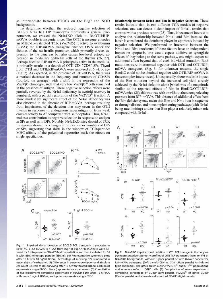
as intermediate between FTOCs on the B6g7 and NODbackgrounds.To determine whether the reduced negative selection of
BDC2.5 Nr4a1KO DP thymocytes represents a general phe-nomenon, we crossed the Nr4a1KO allele to B6.OTII/RIP-mOVA double-transgenic mice. The OTII transgene encodesan MHC-II–restricted TCR (Vα2Vβ5) reactive to ovalbumin(OVA); the RIP-mOVA transgene encodes OVA under thedictates of the rat insulin promoter, which primarily directs ex-pression to the pancreas but also causes low-level ectopic ex-pression in medullary epithelial cells of the thymus (26, 27).Perhaps because RIP-mOVA is principally active in the medulla,it primarily results in a dearth of OTII CD4+CD8− SPs. Thymifrom OTII and OTII/RIP-mOVA were analyzed at 6 wk of age(Fig. 2). As expected, in the presence of RIP-mOVA, there wasa marked decrease in the frequency and numbers of CD4SPs(fourfold on average) with a shift in the expression of theVα2Vβ5 clonotype, such that very few Vα2Vβ5hi cells remainedin the presence of antigen. These negative selection effects werepartially reversed by the Nr4a1 deficiency (a twofold recovery innumbers), with a partial restoration of the Vα2Vβ5hi fraction. Amore modest yet significant effect of the Nr4a1 deficiency wasalso observed in the absence of RIP-mOVA, perhaps resultingfrom impairment of the deletion that may occur in the OTIIthymus in response to endogenous superantigen or from weakcross-reactivity to Ab complexed with self-peptides. Thus, Nr4a1makes a contribution to negative selection in response to antigenin SPs as well as in DPs. Notably, Nr4a1KO mice devoid of TCRtransgenes showed no changes in proportion or numbers of DPsor SPs, suggesting that shifts in the window of TCR/peptide/MHC affinity of the polyclonal repertoire mask the effects onsingle specificities.
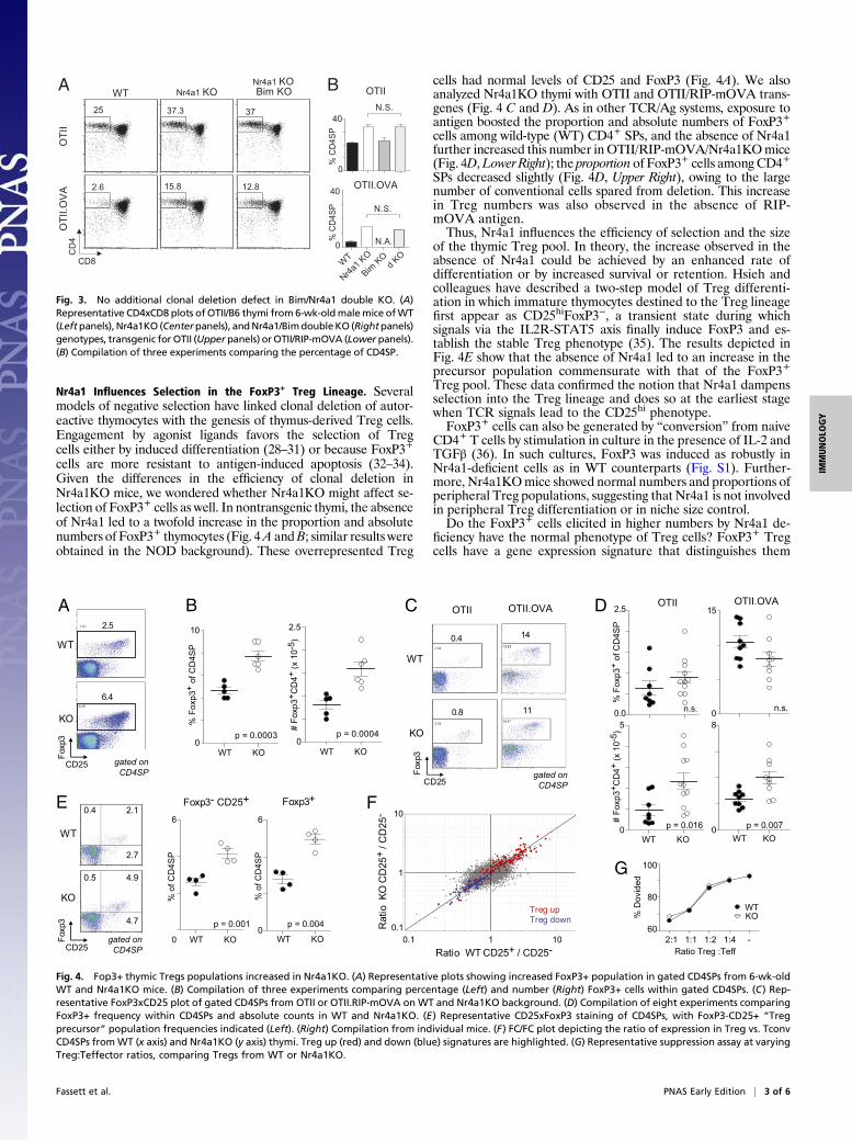
Relationship Between Nr4a1 and Bim in Negative Selection. Theseresults indicate that, in two different TCR models of negativeselection, one can detect a clear impact of Nr4a1, results thatcontrast with a previous report (23). Thus, it became of interest toanalyze the relationship between Nr4a1 and Bim because thelatter is considered the dominant player in apoptosis induced bynegative selection. We performed an intercross between theNr4a1 and Bim knockouts; if these factors have an independentimpact on apoptosis, one would expect additive or synergisticeffects; if they belong to the same pathway, one might expect noadditional effect beyond that of each individual mutation. Bothmutations were intercrossed together with OTII and OTII/RIP-mOVA transgenes (Fig. 3; for unknown reasons, the singleBimKO could not be obtained together with OTII/RIP-mOVA inthese complex intercrosses). Unexpectedly, there was little impactof the Bim mutation beyond the increased cell yield alreadyachieved by the Nr4a1 deletion alone [which was of a magnitudesimilar to the reported effects of Bim in BimKO/OTII.RIP-mOVAmice (2)]; this was true with or without the strong selectingpressure fromRIP-mOVA. This absence of additional effect fromthe Bim deficiency may mean that Bim and Nr4a1 act in sequenceor through distinct and noncomplementing pathways (with Nr4a1being rate limiting) and/or that Bim plays a relatively minor rolecompared with Nr4a1.
A
CD8
CD
4
BDC2.5/WT BDC2.5/KO
no p
eptid
eB
DC
mi
(10n
g/m
l)
39 49
18 26
B
0
100
% D
P
Nr4a1WT
0 1 3 100
8
BDCmi (ng/ml)
# D
P (x
10-5
)
no peptide
0
100
% D
P
C 3 ng/ml BDCmi
0
100
% D
P
Nr4a1 KOWT Nr4a1 KOWT
p=0.007 p=0.001
Fig. 1. Impaired clonal deletion of BDC2.5 TCR transgenic thymocytes inNr4a1KO. E15.5 BDC2.5-tg FTOCs from B6g7 or B6g7.Nr4a1KO mice were cul-tured for 3 d to promote CD4+CD8+ differentiation and then incubated for 16h with BDC mimotope peptide (BDCmi). (A) Representative cytometry plotsafter 16 h with 10 ng/mL BDCmi. Percentage of surviving DPs is indicated inupper right of each panel. (B) Differences in percentage (Upper) and absolutecell count (Lower) of DPs surviving after 16 h with titrated BDCmi; each pointrepresents a single FTOC culture (representative experiment). (C) Compilationof five experiments comparing percentage of surviving DPs after 16 h FTOCwith no or 3 ng/mL BDCmi; each point represents a single FTOC.
B
0
15
p < 0.0001
0
60
p = 0.0008
0
10
p = 0.07
0
50
p < 0.00010
80
p = 0.04
p = 0.030
20
CD4SP x10-6
A KO32 49
11 1
49 38
9 1
4 66
19 2
1163
15 2
46 62
2 11
CD8
CD
4
KOWT KOWT KOWT
KOWT KOWT KOWT
% CD4
WT KOWT
OTI
I.OV
AO
TII
OTI
I.OV
AO
TII
Fig. 2. Nr4a1KO impairs clonal deletion of OTII TCR transgenic thymocytes.(A) Representative cytometry profiles of OTII TCR transgenic thymi on WT orNr4a1KO backgrounds, without (Upper panels) or with (Lower panels) theRIP-mOVA transgene. (Left panels) CD4 vs. CD8. (Right panels) Anti-clono-typic antibodies. The gates drawn outline the OTIIhi and OTIIlow populations,and numbers refer to OTIIhi cells. (B) Compilation of seven experimentscomparing percentage of CD4SP (Left panels), Vα2Vβ5hi of gated CD4SP(Center panels), and absolute cell count of CD4SP (Right panels).
2 of 6 | www.pnas.org/cgi/doi/10.1073/pnas.1200090109 Fassett et al.
Nr4a1 Influences Selection in the FoxP3+ Treg Lineage. Severalmodels of negative selection have linked clonal deletion of autor-eactive thymocytes with the genesis of thymus-derived Treg cells.Engagement by agonist ligands favors the selection of Tregcells either by induced differentiation (28–31) or because FoxP3+
cells are more resistant to antigen-induced apoptosis (32–34).Given the differences in the efficiency of clonal deletion inNr4a1KO mice, we wondered whether Nr4a1KO might affect se-lection of FoxP3+ cells as well. In nontransgenic thymi, the absenceof Nr4a1 led to a twofold increase in the proportion and absolutenumbers of FoxP3+ thymocytes (Fig. 4A andB; similar results wereobtained in the NOD background). These overrepresented Treg
cells had normal levels of CD25 and FoxP3 (Fig. 4A). We alsoanalyzed Nr4a1KO thymi with OTII and OTII/RIP-mOVA trans-genes (Fig. 4 C and D). As in other TCR/Ag systems, exposure toantigen boosted the proportion and absolute numbers of FoxP3+cells among wild-type (WT) CD4+ SPs, and the absence of Nr4a1further increased this number inOTII/RIP-mOVA/Nr4a1KOmice(Fig. 4D,Lower Right); the proportion of FoxP3+ cells among CD4+SPs decreased slightly (Fig. 4D, Upper Right), owing to the largenumber of conventional cells spared from deletion. This increasein Treg numbers was also observed in the absence of RIP-mOVA antigen.Thus, Nr4a1 influences the efficiency of selection and the size
of the thymic Treg pool. In theory, the increase observed in theabsence of Nr4a1 could be achieved by an enhanced rate ofdifferentiation or by increased survival or retention. Hsieh andcolleagues have described a two-step model of Treg differenti-ation in which immature thymocytes destined to the Treg lineagefirst appear as CD25hiFoxP3−, a transient state during whichsignals via the IL2R-STAT5 axis finally induce FoxP3 and es-tablish the stable Treg phenotype (35). The results depicted inFig. 4E show that the absence of Nr4a1 led to an increase in theprecursor population commensurate with that of the FoxP3+Treg pool. These data confirmed the notion that Nr4a1 dampensselection into the Treg lineage and does so at the earliest stagewhen TCR signals lead to the CD25hi phenotype.FoxP3+ cells can also be generated by “conversion” from naive
CD4+ T cells by stimulation in culture in the presence of IL-2 andTGFβ (36). In such cultures, FoxP3 was induced as robustly inNr4a1-deficient cells as in WT counterparts (Fig. S1). Further-more, Nr4a1KOmice showed normal numbers and proportions ofperipheral Treg populations, suggesting that Nr4a1 is not involvedin peripheral Treg differentiation or in niche size control.Do the FoxP3+ cells elicited in higher numbers by Nr4a1 de-
ficiency have the normal phenotype of Treg cells? FoxP3+ Tregcells have a gene expression signature that distinguishes them
WT Nr4a1 KONr4a1 KOBim KOA
CD8
CD
4
WT
Nr4a1 K
O
Bim KO
d KO
OTII.OVA
OTII
N.S.
N.S.
B
0
40
% C
D4S
P
0
40
% C
D4S
P
N.A.
OTI
I.OV
AO
TII
25 37.3 37
2.6 15.8 12.8
Fig. 3. No additional clonal deletion defect in Bim/Nr4a1 double KO. (A)Representative CD4xCD8 plots ofOTII/B6 thymi from6-wk-oldmalemice ofWT(Leftpanels), Nr4a1KO (Centerpanels), andNr4a1/BimdoubleKO (Rightpanels)genotypes, transgenic for OTII (Upperpanels) orOTII/RIP-mOVA (Lower panels).(B) Compilation of three experiments comparing the percentage of CD4SP.
F
G
Ratio WT CD25+ / CD25-
Rat
io K
O C
D25
+ / C
D25
-
B
p = 0.00040
10
p = 0.0003
% F
oxp3
+ of
CD
4SP
0
2.5
# Fo
xp3+
CD
4+ (x
10-
5 )
WT KO WT KO
60
80
100
Ratio Treg :Teff
A
WT
KO
2.5
6.4
gated on
CD4SP
WT
KO
0.4
0.8
14
11
C
CD25
Foxp
3
gated on
CD4SP
0
8
p = 0.0070
5
p = 0.016
D
0.0
2.5
n.s.
% F
oxp3
+ of
CD
4SP
0
15
n.s.
WT KOWT KO
Foxp3+Foxp3- CD25+
CD25
Foxp
3
E
0
6
% o
f CD
4SP
0
6
% o
f CD
4SP
WT
KO
gated on
CD4SP
2:1 1:1 1:2 1:4 -
% D
ovid
ed
WT KOWT KO
WTKO
# Fo
xp3+
CD
4+ (x
10-
5 )
1
10
0.11 100.1
p = 0.004p = 0.001
CD25
Foxp
3
2.7
4.7
2.1
4.9
0.4
0.5
Treg upTreg down
OTII.OVAOTIIOTII.OVAOTII
Fig. 4. Fop3+ thymic Tregs populations increased in Nr4a1KO. (A) Representative plots showing increased FoxP3+ population in gated CD4SPs from 6-wk-oldWT and Nr4a1KO mice. (B) Compilation of three experiments comparing percentage (Left) and number (Right) FoxP3+ cells within gated CD4SPs. (C) Rep-resentative FoxP3xCD25 plot of gated CD4SPs from OTII or OTII.RIP-mOVA on WT and Nr4a1KO background. (D) Compilation of eight experiments comparingFoxP3+ frequency within CD4SPs and absolute counts in WT and Nr4a1KO. (E) Representative CD25xFoxP3 staining of CD4SPs, with FoxP3-CD25+ “Tregprecursor” population frequencies indicated (Left). (Right) Compilation from individual mice. (F) FC/FC plot depicting the ratio of expression in Treg vs. TconvCD4SPs fromWT (x axis) and Nr4a1KO (y axis) thymi. Treg up (red) and down (blue) signatures are highlighted. (G) Representative suppression assay at varyingTreg:Teffector ratios, comparing Tregs from WT or Nr4a1KO.
Fassett et al. PNAS Early Edition | 3 of 6
IMMUNOLO
GY

from conventional CD4+ cells (37); Tregs from Nr4a1KO thymiappeared very similar to their WT counterparts in this respect,with the expected distribution of signature genes on expressionprofiles (Fig. 4F). In addition, Treg cells from Nr4a1KO micewere as effective as WT in the classic in vitro suppression assay(Fig. 4G). Thus, within the informativity of these assays, the en-hanced clonal deviation to the Treg lineage in Nr4a1KO micegenerates phenotypically and functionally normal Treg cells.
Nr4a1 Transcriptional Footprint in Developing Thymocytes. Weturned to transcriptional profiling for clues about Nr4a1’s mech-anism of action in both apoptosis and Treg differentiation. Pro-files from WT or Nr4a1-deficient thymocytes were comparedprimarily in two different settings: (i) DPs from age/sex-matchedWT or Nr4a1KO stimulated in vitro for 3 h with plate-bound anti-TCRβ/CD28/CD2, conditions used by others to mimic negativeselection and under which Nr4a1 and Bim are rapidly induced(20); this short response time should bring forth Nr4a1’s directtranscriptional footprint; (ii) DP and CD4+CD25−Vα2hiVβ5hi SPthymocytes from adult OTII and OTII/RIP-mOVA transgenicmice for a footprint of Nr4a1 in vivo under steady-state conditionsof antigen-induced clonal deletion. The latter in vivo datasetsshould show, under more physiological conditions, the more in-tegrated (direct and indirect) influence of Nr4a1 in cells thatsurvive apoptosis.We focused first on theNr4a1 target genes differentially induced
in DP thymocytes by short-term stimulation. In unstimulated cells,the absence of Nr4a1 had very little effect (Fig. 5A, Left, in thesame range as the experimental background estimated by MonteCarlo randomization). However, a clear difference between WTand Nr4a1KO emerged after 3 h (Fig. 5A, Right). These Nr4a1-dependent transcripts (listed in Table S1) were not affected by the
Nr4a1 deficiency before stimulation. They included genes be-longing to several different pathways, but the most notable can-didates in this context were Bcl2l11 (Bim) and the Treg signaturegenes Tnfrsf9 (4.1BB) and Ikzf2 (Helios).We analyzed more directly the effect of Nr4a1 on the ex-
pression of Bim in the three conditions analyzed (red highlight inFig. 5B). Bim was represented in all contexts, particularly in DPs,suggesting that Nr4a1 does have a transcriptional impact on Bim.It should be pointed out, however, that the impact of Nr4a1 isonly partial (1.5- to 2-fold) relative to the strong induction ofBim by TCR ligands (8.7-fold in in vitro DPs, 4.7-fold in OTIISPs, 2.3-fold in OTII DPs).We also noted, among the genes most clearly influenced by
Nr4a1, the glycolytic enzyme Enolase 3, which was reported to bedirectly regulated by Nr4a1 in an analysis of metabolic control inthe liver (38). Enolase 3 catalyzes an important step in the gly-colytic pathway (39), so we asked whether Eno3 was part ofa broader set of metabolic-control transcripts influenced byNr4a1. Indeed, transcripts encoding glycolytic enzymes were es-sentially all under-represented in Nr4a1-deficient cells, whetherin short-term activatedDPs or in long-term antigen-exposed OTIIDPs and SPs (Fig. 5B and Table S2). Nr4a1 similarly impactedtranscripts encoding enzymes of the Krebs cycle (blue). Thus,Nr4a1 effects a global activation of the energy-producing meta-bolic pathways in thymocytes, which can have a profound effect oncell survival and proliferation (40), consistent with effects inskeletal muscle and adipose tissue in addition to liver (reviewedin ref. 41).The absence of Nr4a1 leads to an enhanced efficiency of Treg
selection. We thus asked whether we could distinguish, amongNr4a1-controlled genes, transcripts that might reflect this effecton selection (e.g., a repression of key controlling genes in the
Expression value (AU)
A
B
WT
/ KO
1
0
50
1
0.1
10
2
0.5
10 100 1,000 10,000
Nr4al
Bcl2l11
Tnfrsf9
Nr4a1
Expression value (AU)
294
22
50
1
0.1
10
2
0.5
10 100 1,000 10,000
Eno3
Ikzf2
Bcl2l11
Sox1
Trio
WT
/ KO
Resting DP 3h anti-TCR DP
3h anti-TCR DPD
Ratio WT/ KO (in OTII.OVA 4SP)
1
10
0.11 100.1
S100a6
Itgae
Myo1e
Cst7
Elk3
Nt5e
Pdcd1
Tnfrsf9
Ikzf2
Treg upTreg down
1
10
0.11 100.1
Itgae
Tnfrsf9
Ikzf2
Gadd45b
Ahrgap20
Tnfrsf4
Pdcd1
OTII 4SP
C
Treg upTreg down
Elk3 S100a6
Cst7
Smpde3a
Lrrc32
Eno3
Ratio +/- OVA (in WT)
Rat
io +
/- O
VA
(in
KO
) R
atio
WT/
KO
(in
3h a
nti-T
CR
DP
)
Ratio WT / KO Ratio WT / KO Ratio WT / KO
p-va
lue
WT
vs K
O
OTII.OVA DP OTII.OVA SP1
10-3
10-7
10-5
0.3 1 7
Eno3
Glycolysis Krebs cycle Bcl2l11 (Bim)
0.3 1 7 0.3 1 7
Fig. 5. Nr4a1-dependent transcripts identified by microarray profiling. (A) FoldChange vs. expression plots for WT vs. Nr4a1KO in resting DP thymocytes(Left) or DP thymocytes stimulated in vitro for 3 h with plate-bound anti-CD2/CD28/TCRβ. Transcripts highlighted indicate ratio of expression >2× (orange) or<0.5× (aqua) in WT/Nr4a1KO. (B) Volcano plots comparing WT and KO thymocytes (FoldChange: x axis; t test P value: y axis) for DPs stimulated 3 h with plate-bound anti-CD2/CD28/TCRβ (Left), DPs from OTII.OVA (Center), or CD4SPs from OTII.OVA (Right). (C) FC/FC plot showing the ratio of expression in CD4 SPsfrom OTII vs. OTII.OVA thymi from WT or Nr4a1KO mice. Treg up (red) and down (blue) signatures are highlighted. (D) FC/FC plot depicting the WT vs. KOratio of expression in OTII.OVA CD4SPs (x axis) or in 3-h anti-TCR–stimulated DPs (y axis). Highlights as in C.
4 of 6 | www.pnas.org/cgi/doi/10.1073/pnas.1200090109 Fassett et al.
Treg signature. We examined the relative expression of Tregsignature genes in the CD4SP OTII samples, which were gen-erated from CD25− thymocytes, thereby excluding most thymicTregs and Treg precursor cells. The effect of Nr4a1 on the set ofgenes induced by the presence of OVA in WT and KO thymi isdisplayed on the FoldChange/FoldChange (FC/FC) plots of Fig.5C. Transcripts along the diagonal represent genes equally up- ordown-regulated by OVA in WT and Nr4a1KO, whereas tran-scripts biased toward the horizontal are those whose inductionrequires Nr4a1 (listed in Table S3). A general transcriptionalactivation and repression of “Treg up” and “Treg down” signa-ture genes were induced by OVA, likely because some bona fideFoxP3+ Treg cells are CD25− and thus partake in this bias. Anumber of transcripts of the Treg up signature were less induced(Ikzf2 and Tnfrsf9) or even uninduced (Itgae, Cst7, Elk3) in theabsence of Nr4a1. On the other hand, there was no influence ofNr4a1 on the Treg down signature. Thus, a marked effect ofNr4a1 was observed, but an unexpected one: instead of medi-ating a generalized repression, Nr4a1 turned out to activatea subset of Treg signature transcripts.The FC/FC plot of Fig. 5D addresses the relation between
Nr4a1’s immediate target genes relative to its longer-term impactin CD4SPs by representing the variation between WT andNr4a1KO cells for both antigen-stimulated OTII CD4SPs andin vitro-stimulated DPs. Here, transcripts lining the diagonalwould reflect target genes that Nurr77 affects immediately andpersistently, whereas transcripts along the x axis reflect delayedeffects (because they are active only in SPs, because they areNr4a1-dependent but late responders, or because they are in-directly affected). A few transcripts showed the constant imprintof Nr4a1 (notably Ikzf2 and Tnfrsf9, previously noted), but theplot also delineated a set of Treg transcripts uniquely influencedin the steady-state population (Nd5e, Pdcd1, Itgae). As expected,no Nr4a1-related differences were observed in OTII SPs fromOVA-negative mice. Nr4a1’s footprint seemed mainly inductive,with little or no repressive signature, suggesting that it comesinto play as a positive transcriptional inducer, of both theproapoptotic Bim and, paradoxically, some Treg signature genes.
DiscussionWe have observed an important role for the transcription factorNr4a1 in modulating two key aspects of tolerance to self duringdifferentiation of immature T cells in the thymus: clonal deletionof self-reactive thymocytes and commitment to the Treg lineage.Nr4a1 expression in immature thymocytes, at a stage where self-recognition can elicit either death by clonal deletion or deviationinto the Treg lineage, is at the crux of this important cell-lineagedetermination. The early literature on Nr4a1 and T cells hingeson its molecular description as a transcription factor, with de-fined transcriptional activation domains and DNA-binding motif,and on its functional description as a proapoptotic factor, whoseoverexpression in cell lines and in transgenic mice promotes T-cell apoptosis. In subsequent reviews, this description has beenabbreviated to “proapoptotic transcription factor” Nr4a1. How-ever, a growing literature also describes Nr4a1 as a survivalfactor and activator of metabolic pathways in a variety of celltypes (12, 41, 42), and one should consider both facets inattempting to integrate Nr4a1’s role in T-cell differentiation.Clonal deletion results, obtained in two TCR transgenic
mouse systems, are conceptually concordant with the effect ofdominant-negative Nr4a1 transgenic mice, which showed defectsin negative selection of TCR transgenic thymocytes (5, 6). Onthe other hand, they disagree with a previous report that foundno detectable phenotype in Nr4a1KO thymus (23). Why did thisoriginal study not find any defect in negative selection? DifferentTCR transgenes were used (H-Y and AND), both of which leadto strong negative selection at the DP stage, whereas the OT-IIand BDC2.5 systems show only partial and/or late negative se-lection. The BDC2.5 TCR stems from a diabetogenic T-cellclone that had, by definition, escaped clonal deletion in NODmice; the OT-II TCR responds to OVA peptides presented by
medullary epithelial cells in RIP-mOVA transgenic mice witha late (medullary SP stage only) and incomplete negative selec-tion. Whatever the explanation, it is clear that Nr4a1 can havea dominant impact, which is not redundant with its Nor-1 andNurr1 cousins.How does Nr4a1 effect clonal deletion? An open question is
whether Nr4a1 induces deletion through transcriptional means byinducing proapoptotic molecules or through its direct interactionswith Bcl-2 (7–10). Initially thought to be transcriptional (15, 24),the phenotype of the early Nr4a1 mutants that led to this con-clusion may have reflected perturbed nuclear export signals con-tained within the transcriptional activation domain (8). We findthat Nr4a1 is necessary for the full induction of Bim transcripts inall three conditions tested, including a very early time point, whichsuggests direct transcriptional regulation (consistent with thedouble-KO phenotype). However, the share of Bim induction thatcan be imparted to Nr4a1 matches only a fraction of the Biminduction during negative selection. Nr4a1 cannot be the solecontroller of Bim. No other clear proapoptotic candidate wasreadily identified among Nr4a1-controlled genes.In addition to the negative-selection defect, we find that Nr4a1
expression negatively impacts differentiation of thymic Tregcells. There are no apparent differences in Treg phenotypesbetween WT and Nr4a1KO mice, suggesting that Nr4a1’s role inTregs is a matter of differentiation and not of function. Indeed,peripheral homeostasic mechanisms restore the peripheral Tregpopulation to normal numbers, as Treg over-representation inNr4a1KO is seen only in the thymus, not in secondary lymphoidorgans. Mechanisms that promote Treg differentiation are in-completely understood, but likely are controlled by the balanceof signals along the NF-κB vs. Akt-mTOR pathways. We did testthe hypothesis that Akt/mTOR might act directly through Nr4a1,which proved false as the inhibitory effects of activated Akt werestill observed in Nr4a1KO cells.Importantly and quite paradoxically, Nr4a1 had a positive ef-
fect on a fraction of the Treg signature (but not on FoxP3 itself,consistent with ref. 43). Thus, Nr4a1 biases Treg transcriptionaldifferentiation even while restricting their numbers. One mightspeculate that Nr4a1, by promoting survival, boosts negativefeedback mechanisms that control the efficacy of Treg selectionand/or thymic Treg niche size. Or perhaps, in a cell-autonomousfashion, the transcripts that it induces are actually negativefeedback controls on Tregs themselves.In addition, Sekiya et al. (43) recently reported that trans-
duction of Nr4a2 induces FoxP3 in vitro; Nr4a1 and Nr4a3 didnot, the difference mapping to the N-terminal transactivationdomain. In Nr4a2-deficient mice, thymic selection of Treg cellswas normal, but there was reduced FoxP3 induction in responseto IL-2/TGFβ and differential Treg cell stability (43). BecauseNr4a family members have conserved DNA-binding motifs andcan heterodimerize, it is possible that some of the present resultsreflect competition or cooperation of Nr4a1 with Nr4a2. Wenote, however, that Nurr1 is expressed at low levels in both Tregand conventional thymocyte populations relative to Nr4a1 andNr4a3 (Fig. S2).Within this context, can the Nr4a1-regulated transcripts
identified here be interpreted for their potential involvement atthe clonal deletion vs. clonal deviation branch point? Genes ofthe costimulatory family, including PD-1 and 4.1BB, are differ-entially expressed and relevant to both fates. All of these aredifferentially expressed in Treg cells, and several studies haveassociated PD-1 with negative selection (44). A previous studyalso identified both Pdcd1 and Ctla4 as genes induced by Nr4a1upon transgenic over-expression in thymocytes (15). One mightspeculate that, by dampening the signals associated with theTCR (Ctla4) or by modulating its downstream footprint (PD-1,4.1BB), the costimulatory molecules controlled by Nr4a1 mayaffect the balance of survival and lineage commitment.In addition, the transcriptional profiles clearly denote a pro-
survival effect of Nr4a1: the whole group of glycolytic and TCAenzymes behave as early response genes and Nr4a1 participates in
Fassett et al. PNAS Early Edition | 5 of 6
IMMUNOLO
GY
their activation, thus boosting the whole cascade of energy pro-duction in the cell. This is most obvious for Eno3, which Kasleret al. identified as a Nr4a1-transcriptional target in D011.10 cellsoverexpressing Nr4a1 (45) and in analyses of hepatic glucosemetabolism (38). Nr4a1 binds directly to a response element inthe Eno3 promoter (38). Enhanced activation of metabolism inNr4a1-expressing thymocytes may blunt differentiation into theTreg lineage, similar to that recently described in peripheral Tcells in which transcriptional activation of glycolysis preferentiallypromotes Th17 rather than Treg differentiation via a HIF1α-dependent mechanism (46). In that study, as here, genetic inhibi-tion of glycolysis increased Treg cell numbers. (We attempted todirectly test the role of glycolysis in Treg differentiation byinhibition with 2-DG during the FTOC, but the results wereconfounded by toxicity of 2-DG on immature thymocytes). Wenote that activation of the Akt/mTOR axis, which boosts glucosemetabolism, also results in lower Treg differentiation.One might propose, then, that the induction of Nr4a1, through
its different biochemical activities, forces decisions in relation tothymocyte fate: promoting clonal deletion viaNr4a1’s proapoptoticactivity at the mitochondrial or transcriptional levels and facilitat-ing cell survival by boosting ATP generation, a condition that maypromote commitment away from clonal deviation.
Materials and MethodsMice. C57BL/6.Nr4a1−/− [Nr4a1KO (23)] mice, a gift from J. Milbrandt(Washington University, St Louis, MO), were crossed with the transgeniclines BDC2.5/B6g7 (47), OT-II (26), and RIP-mOVA (27). C57BL/6.Bim−/−
[BimKO (48)] mice were from The Jackson Laboratory. Mice were bred in anspecific-pathogen-free barrier facility.
Antigen Exposure. Fetal thymus lobes from E15.5 embryos were cultured asdescribed (22). On culture day 3, BDC-specific mimotope peptide (BDCmi,peptides 1,040–1,063) (25) was added to culture media, and lobes wereanalyzed 16 h later. For in vitro thymocyte stimulation, single-cell thymocytesuspensions were incubated at 5 × 10⁶ cells/mL for 3 h at 37 °C with platesprecoated with anti-TCRβ (10 μg/mL), anti-CD2 (10 μg/mL), and anti-CD28 (50μg/mL) as described (20). Cell sorting for microarray RNA preparation andhybridization to Affymetrix MoGene 1.0ST and RMA normalization wereperformed as described (49).
ACKNOWLEDGMENTS. We thank K. Hattori, J. LaVecchio, G. Buruzula, andS. Davis for help with mice, cytometry, and computational analysis. This workwas supported by a grant from the Juvenile Diabetes Research Foundation(4-2007-1057) and by Grant AI051530 from the National Institute of Allergyand Infectious Diseases (to C.B. and D.M.). M.S.F. was supported by a NationalInstitutes of Health Training Grant (T32 DK7260) and the Harvard-MITMedical Scientist Training Program.
1. Hogquist KA, Baldwin TA, Jameson SC (2005) Central tolerance: Learning self-controlin the thymus. Nat Rev Immunol 5:772–782.
2. Bouillet P, et al. (2002) BH3-only Bcl-2 family member Bim is required for apoptosis ofautoreactive thymocytes. Nature 415:922–926.
3. Rathmell JC, Lindsten T, Zong WX, Cinalli RM, Thompson CB (2002) Deficiency in Bakand Bax perturbs thymic selection and lymphoid homeostasis.Nat Immunol 3:932–939.
4. D’Adamio L, Clayton LK, Awad KM, Reinherz EL (1992) Negative selection of thy-mocytes. A novel polymerase chain reaction-based molecular analysis detects re-quirements for macromolecular synthesis. J Immunol 149:3550–3553.
5. Calnan BJ, Szychowski S, Chan FK, Cado D, Winoto A (1995) A role for the orphansteroid receptor Nur77 in apoptosis accompanying antigen-induced negative selec-tion. Immunity 3:273–282.
6. Zhou T, et al. (1996) Inhibition of Nur77/Nurr1 leads to inefficient clonal deletion ofself-reactive T cells. J Exp Med 183:1879–1892.
7. Lin B, et al. (2004) Conversion of Bcl-2 from protector to killer by interaction withnuclear orphan receptor Nur77/TR3. Cell 116:527–540.
8. Thompson J, Winoto A (2008) During negative selection, Nur77 family proteinstranslocate to mitochondria where they associate with Bcl-2 and expose its proa-poptotic BH3 domain. J Exp Med 205:1029–1036.
9. Wang A, Rud J, Olson CM, Jr., Anguita J, Osborne BA (2009) Phosphorylation of Nur77by the MEK-ERK-RSK cascade induces mitochondrial translocation and apoptosis in Tcells. J Immunol 183:3268–3277.
10. Li H, et al. (2000) Cytochrome c release and apoptosis induced by mitochondrialtargeting of nuclear orphan receptor TR3. Science 289:1159–1164.
11. Liu ZG, Smith SW, McLaughlin KA, Schwartz LM, Osborne BA (1994) Apoptotic signalsdelivered through the T-cell receptor of a T-cell hybrid require the immediate-earlygene nur77. Nature 367:281–284.
12. Moll UM, Marchenko N, Zhang XK (2006) p53 and Nur77/TR3: Transcription factorsthat directly target mitochondria for cell death induction. Oncogene 25:4725–4743.
13. Winoto A, Littman DR (2002) Nuclear hormone receptors in T lymphocytes. Cell 109(Suppl):S57–S66.
14. Woronicz JD, Calnan B, Ngo V, Winoto A (1994) Requirement for the orphan steroidreceptor Nur77 in apoptosis of T-cell hybridomas. Nature 367:277–281.
15. Rajpal A, et al. (2003) Transcriptional activation of known and novel apoptoticpathways by Nur77 orphan steroid receptor. EMBO J 22:6526–6536.
16. Brás A, Albar JP, Leonardo E, de Buitrago GG, Martínez-A C (2000) Ceramide-inducedcell death is independent of the Fas/Fas ligand pathway and is prevented by Nur77overexpression in A20 B cells. Cell Death Differ 7:262–271.
17. Suzuki S, et al. (2003) Nur77 as a survival factor in tumor necrosis factor signaling.Proc Natl Acad Sci USA 100:8276–8280.
18. Osborne BA, et al. (1994) Identification of genes induced during apoptosis in T lym-phocytes. Immunol Rev 142:301–320.
19. Moran AE, et al. (2011) T cell receptor signal strength in Treg and iNKT cell devel-opmentdemonstratedbyanovelfluorescent reportermouse. J ExpMed208:1279–1289.
20. Cunningham NR, et al. (2006) Immature CD4+CD8+ thymocytes and mature T cellsregulate Nur77 distinctly in response to TCR stimulation. J Immunol 177:6660–6666.
21. Liston A, et al. (2004) Generalized resistance to thymic deletion in the NOD mouse;a polygenic trait characterized by defective induction of Bim. Immunity 21:817–830.
22. Zucchelli S, et al. (2005) Defective central tolerance induction in NOD mice: genomicsand genetics. Immunity 22:385–396.
23. Lee SL, et al. (1995) Unimpaired thymic and peripheral T cell death in mice lacking thenuclear receptor NGFI-B (Nur77). Science 269:532–535.
24. Cheng LE, Chan FK, Cado D, Winoto A (1997) Functional redundancy of the Nur77 andNor-1 orphan steroid receptors in T-cell apoptosis. EMBO J 16:1865–1875.
25. Judkowski V, et al. (2001) Identification of MHC class II-restricted peptide ligands,including a glutamic acid decarboxylase 65 sequence, that stimulate diabetogenic Tcells from transgenic BDC2.5 nonobese diabetic mice. J Immunol 166:908–917.
26. Barnden MJ, Allison J, Heath WR, Carbone FR (1998) Defective TCR expression intransgenic mice constructed using cDNA-based alpha- and beta-chain genes underthe control of heterologous regulatory elements. Immunol Cell Biol 76:34–40.
27. Kurts C, et al. (1996) Constitutive class I-restricted exogenous presentation of selfantigens in vivo. J Exp Med 184:923–930.
28. Apostolou I, Sarukhan A, Klein L, von Boehmer H (2002) Origin of regulatory T cellswith known specificity for antigen. Nat Immunol 3:756–763.
29. Jordan MS, et al. (2001) Thymic selection of CD4+CD25+ regulatory T cells induced byan agonist self-peptide. Nat Immunol 2:283–284.
30. Kawahata K, et al. (2002) Generation of CD4(+)CD25(+) regulatory T cells from au-toreactive T cells simultaneously with their negative selection in the thymus and fromnonautoreactive T cells by endogenous TCR expression. J Immunol 168:4399–4405.
31. Walker LS, Chodos A, Eggena M, Dooms H, Abbas AK (2003) Antigen-dependentproliferation of CD4+ CD25+ regulatory T cells in vivo. J Exp Med 198:249–258.
32. Bonasio R, et al. (2006) Clonal deletion of thymocytes by circulating dendritic cellshoming to the thymus. Nat Immunol 7:1092–1100.
33. Liston A, Lesage S, Wilson J, Peltonen L, Goodnow CC (2003) Aire regulates negativeselection of organ-specific T cells. Nat Immunol 4:350–354.
34. Smith KA (2004) The quantal theory of how the immune system discriminates be-tween “self and non-self.” Med Immunol 3:3.
35. Burchill MA, et al. (2008) Linked T cell receptor and cytokine signaling govern thedevelopment of the regulatory T cell repertoire. Immunity 28:112–121.
36. ChenW, et al. (2003) Conversion of peripheral CD4+CD25− naive T cells to CD4+CD25+regulatory T cells by TGF-beta induction of transcription factor Foxp3. J Exp Med 198:1875–1886.
37. Hill JA, et al. (2007) Foxp3 transcription-factor-dependent and -independent regula-tion of the regulatory T cell transcriptional signature. Immunity 27:786–800.
38. Pei L, et al. (2006) NR4A orphan nuclear receptors are transcriptional regulators ofhepatic glucose metabolism. Nat Med 12:1048–1055.
39. Warburg O, Christian W (1941) Isolation and crystallization of fermentation enzymeenolase. Biochem Z 310:384–421.
40. Vander Heiden MG, Cantley LC, Thompson CB (2009) Understanding the Warburgeffect: The metabolic requirements of cell proliferation. Science 324:1029–1033.
41. Pearen MA, Muscat GE (2010) Minireview: Nuclear hormone receptor 4A signaling:Implications for metabolic disease. Mol Endocrinol 24:1891–1903.
42. Pols TW, Bonta PI, de Vries CJ (2007) NR4A nuclear orphan receptors: Protective invascular disease? Curr Opin Lipidol 18:515–520.
43. Sekiya T, et al. (2011) The nuclear orphan receptor Nr4a2 induces Foxp3 and regulatesdifferentiation of CD4+ T cells. Nat Commun 2:269.
44. Baldwin TA, Hogquist KA (2007) Transcriptional analysis of clonal deletion in vivo.J Immunol 179:837–844.
45. Kasler HG, Verdin E (2007) Histone deacetylase 7 functions as a key regulator of genes in-volved in both positive and negative selection of thymocytes.Mol Cell Biol 27:5184–5200.
46. Shi LZ, et al. (2011) HIF1alpha-dependent glycolytic pathway orchestrates a metaboliccheckpoint for the differentiation of TH17 and Treg cells. J Exp Med 208:1367–1376.
47. Katz JD, Wang B, Haskins K, Benoist C, Mathis D (1993) Following a diabetogenic Tcell from genesis through pathogenesis. Cell 74:1089–1100.
48. Bouillet P, et al. (1999) Proapoptotic Bcl-2 relative Bim required for certain apoptotic re-sponses, leukocyte homeostasis, and to preclude autoimmunity. Science 286:1735–1738.
49. Yamagata T, Mathis D, Benoist C (2004) Self-reactivity in thymic double-positive cellscommits cells to a CD8 alpha alpha lineage with characteristics of innate immunecells. Nat Immunol 5:597–605.
6 of 6 | www.pnas.org/cgi/doi/10.1073/pnas.1200090109 Fassett et al.