nuclear medicine and its applications
TRANSCRIPT

NUCLEAR MEDICINE AND ITS APPLICATIONS
PRESENTED BY:- SARITA RAI MRT12UGBBM010

Nuclear Medicine
Nuclear medicine is a medical speciality that involves giving a patient a small amount of radioactive medication, called a radiopharmaceutical. This makes the body slightly radioactive for a short time. A special nuclear medicine camera detects the radiation, which is emitted (released) from the body, and takes images or pictures of how the inside of the body is working. Many different organs can be imaged depending on the type of radioactive medication used.

The radioactive medication is most commonly injected into the blood stream through a vein, but might be given in different ways, including:
•Swallowed;•Injected directly into the tissue beneath the skin;•Injected into A shunt;•Injected into A joint; or•Inhaled (breathed in).
Only a very small amount of radiopharmaceutical is given to keep the radiation dose to a minimum.
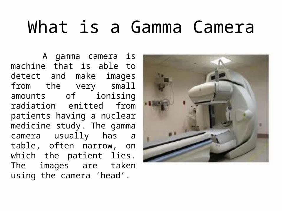
What is a Gamma Camera
A gamma camera is machine that is able to detect and make images from the very small amounts of ionising radiation emitted from patients having a nuclear medicine study. The gamma camera usually has a table, often narrow, on which the patient lies. The images are taken using the camera ‘head’.

GAMMA CAMERA

Imaging techniques using gamma cameras
Scintigraphy ("scint") is the use of gamma cameras to capture emitted radiation from internal radioisotopes to create two-dimensional images.
SPECT (single photon emission computed tomography) imaging, as used in nuclear cardiac stress testing, is performed using gamma cameras. Usually one, two or three detectors or heads, are slowly rotated around the patient's torso.
Multi-headed gamma cameras can also be used for Positron emission tomography scanning, provided that their hardware and software can be configured to detect "coincidences" (near simultaneous events on 2 different heads).

How is Nuclear Medicine different from normal X-ray and CT examinations?
An X-ray or CT image is formed from ionising radiation (X-rays) that passes through the body, but does not arise from the body; whereas a nuclear medicine image is formed from the ionising radiation (usually gamma rays) emitted from within the body. A gamma ray has similar properties to an X-ray, but it arises from the nucleus of an atom, whereas an X-ray arises from the electron shell of an atom.Another way that nuclear medicine is different from X-ray and CT examinations is that an X-ray study shows what something looks like. In nuclear medicine studies, the radiopharmaceutical usually only goes to the part of the body or organ system if it has some function and so shows how it is working.

Single Photon Emission Computed Tomography (SPECT) This SPECT technique uses a gamma camera to record images at a series of angles around the patient. These images are then subjected to a form of digital image processing called Image Reconstruction in order to compute images of slices through the patient.

SPECT The gamma camera is typically rotated around the
patient in order to acquire the images. Modern gamma cameras which are designed specifically for SPECT scanning can consist of two camera heads mounted parallel to each other with the patient in between. The time required to produce images is therefore reduced by a factor of about two. In addition some SPECT gamma cameras designed for brain scanning have three camera heads mounted in a triangular arrangement.

SPECT machine performing a total body bone scan.

Applications SPECT can be used to complement any gamma
imaging study, where a true 3D representation can be helpful, e.g.:-
• Tumor imaging,• Infection imaging,• Thyroid imaging Because SPECT permits accurate localisation in
3D space, it can be used to provide information about localised function in internal organs, such as functional cardiac or brain imaging.

Positron emission tomography In modern PET-CT scanners, three dimensional imaging is
often accomplished with the aid of a CT X-ray scan performed on the patient during the same session, in the same machine.
If the biologically active molecule chosen for PET is fluorodeoxyglucose (FDG), an analogue of glucose, the concentrations of tracer imaged will indicate tissue metabolic activity by virtue of the regional glucose uptake. Use of this tracer to explore the possibility of cancer metastasis (i.e., spreading to other sites) is the most common type of PET scan in standard medical care (90% of current scans). However, on a minority basis, many other radioactive tracers are used in PET to image the tissue concentration of other types of molecules of interest.

PET Acquisition Process

Applications:-
PET is both a medical and research tool.• It is used heavily in clinical oncology (
medical imaging of tumors and the search for metastases).
• PET is also an important research tool to map normal human brain and heart function, and support drug development.