nuclear localization of the de novo thymidylate ... · nuclear localization of the de novo...
TRANSCRIPT

1
NUCLEAR LOCALIZATION OF THE DE NOVO THYMIDYLATE BIOSYNTHESIS PATHWAY IS REQUIRED TO PREVENT URACIL ACCUMULATION IN DNA*
Amanda J. MacFarlane1†, Donald D. Anderson2, Per Flodby1#, Cheryll A. Perry1, Robert H. Allen3, Sally P. Stabler3, and Patrick J. Stover1,2
1Division of Nutritional Sciences, Cornell University, Ithaca, New York 14853 2Graduate Field of Biochemistry, Molecular and Cell Biology, Cornell University, Ithaca, New York 14853
3Department of Medicine and Division of Hematology, University of Colorado School of Medicine, Aurora, CO 80045 *Running title: Nuclear thymidylate synthesis is required to prevent uracil incorporation in DNA
Present address: †Nutrition Research Division, Food Directorate, Health Products and Food Branch, Health Canada, Ottawa, Ontario K1A 0K9, Canada; # Division of Pulmonary & Critical Care Medicine, Keck School of Medicine, University of Southern California, 2011 Zonal Avenue, HMR 911, Los Angeles, California 90033
Address correspondence to: Patrick J. Stover, 315 Savage Hall, Division of Nutritional Sciences, Cornell University, Ithaca NY 14853; Tel. (607)255-9751; Fax (607)255-1033; E-mail: [email protected] Keywords: Cytoplasmic serine hydroxymethyltransferase, thymidylate synthesis, folate, uracil
Background: S-phase nuclei contain the thymidylate synthesis pathway. Results: Mice overexpressing a Shmt1 transgene exhibit elevated expression of SHMT1 and TYMS, impaired nuclear localization of the thymidylate biosynthesis pathway and elevated uracil in DNA. Conclusion: SHMT1 and TYMS localization to the nucleus is essential to prevent uracil accumulation in DNA. Significance: SHMT1-mediated nuclear de novo thymidylate synthesis is critical for maintaining DNA integrity.
SUMMARY
Uracil accumulates in DNA as a result of impaired folate-dependent de novo thymidylate biosynthesis, a pathway composed of the enzymes serine hydroxymethyltransferase (SHMT), thymidylate synthase (TYMS) and dihydrofolate reductase (DHFR). In G1, this pathway is present in the cytoplasm and at S phase undergoes SUMO-dependent translocation to the nucleus. It is not known if this pathway functions in the cytoplasm, nucleus or both in vivo. SHMT1 generates 5,10-methylenetetrahydrofolate for de novo thymidylate biosynthesis, a limiting step in the pathway, but also tightly binds 5-methyltetrahydrofolate in the cytoplasm, a required cofactor for homocysteine remethylation. Overexpression of SHMT1 in cell cultures inhibits folate-dependent homocysteine remethylation and enhances thymidylate biosynthesis. In this study, the impact of increased Shmt1 expression on
folate-mediated one-carbon metabolism was determined in mice that over-express the Shmt1 cDNA (Shmt1tg+ mice). Compared to wildtype mice, Shmt1tg+ mice exhibited elevated SHMT1 and TYMS protein levels in tissues and evidence for impaired homocysteine remethylation, but surprisingly exhibited depressed levels of nuclear SHMT1 and TYMS, lower rates of nuclear de novo thymidylate biosynthesis and a nearly 10-fold increase in uracil content in hepatic nuclear DNA when fed a folate and choline deficient diet. These results demonstrate that SHMT1 and TYMS localization to the nucleus is essential to prevent uracil accumulation in nuclear DNA, and indicate that SHMT1-mediated nuclear de novo thymidylate synthesis is critical for maintaining DNA integrity.
Depletion of de novo dTMP synthesis due to folate deficiency, antifolate inhibitors or genetic disruption of the pathway results in deoxyuridine misincorporation into nuclear DNA leading to genome instability (1-3). De novo thymidylate biosynthesis is distinct from the other nucleotide synthesis pathways in that it is compartmentalized at the sites of DNA replication, namely the mitochondria (4) and nucleus (5,6). The de novo thymidylate biosynthesis pathway requires three enzymatic activities: serine hydroxymethyltransferase (SHMT), which catalyzes the conversion of serine and tetrahydrofolate (THF) to form glycine and methyleneTHF; thymidylate synthase (TYMS), which catalyzes the conversion of methyleneTHF and dUMP to dTMP and dihydrofolate (DHF). The cycle
http://www.jbc.org/cgi/doi/10.1074/jbc.M111.307629The latest version is at JBC Papers in Press. Published on November 4, 2011 as Manuscript M111.307629
Copyright 2011 by The American Society for Biochemistry and Molecular Biology, Inc.
by guest on March 10, 2019
http://ww
w.jbc.org/
Dow
nloaded from

2
is completed by the conversion of DHF to THF in a NADPH-requiring reaction catalyzed by dihydrofolate reductase (DHFR) (Fig. 1). In mitochondria, the pathway is encoded by SHMT2, TYMS, and DHFR1 (4). Disruption of the pathway in Chinese hamster ovary cell mitochondria results in a glycine auxotrophy and elevated levels of uracil in mitochondrial DNA (mtDNA) (4). In the nucleus, the de novo thymidylate biosynthesis pathway is encoded by SHMT1 and, to a lesser extent SHMT2, which are functionally redundant in supplying methyleneTHF, TYMS and DHFR. TYMS is the only gene that is common to and essential for mitochondrial and nuclear thymidylate biosynthesis, and is essential for early embryonic development (7).
Nuclear thymidylate biosynthesis is cell cycle regulated through the Small Ubiquitin-like Modifier (SUMO)-dependent nuclear translocation of SHMT1, TYMS and DHFR during S phase (5,6,8). Isolated intact nuclei exhibit de novo thymidylate synthesis activity, but this activity is lost in sonicated nuclei suggesting that this pathway functions through a multienzyme complex and requires compartmentalization in an intact nucleus (8). It is not known if thymidylate biosynthesis occurs in the cytoplasm. Shmt1-/- mice are viable and fertile (3) and retain about 25% de novo thymidylate biosynthesis capacity in isolated hepatic nuclei due to redundant function of the enzyme SHMT2α, encoded by Shmt2 through alternative promoter usage (8). Despite this functional redundancy between SHMT1 and SHMT2α in the cytoplasm and nucleus, SHMT1 is the major provider of methyleneTHF; mice are sensitive to reductions in SHMT1 expression, as indicated by elevated levels of uracil in nuclear DNA in Shmt1+/- mice (3,9), and their increased susceptibility to neural tube defects (10) and Apcmin/+-mediated intestinal tumors (9). Interestingly, a common SHMT1 human variant, L474F, is not an effective substrate for UBC-9-catalyzed sumoylation in vitro and exhibits impaired nuclear translocation at S phase (8) suggesting that its association with cardiovascular disease (11,12) and lung cancer (13) risk may be due to impaired nuclear de novo thymidylate synthesis.
SHMT1 activity can be limiting for nuclear de novo thymidylate biosynthesis and is also involved in homocysteine remethylation in the cytoplasm. Expression of the SHMT1 cDNA in cell cultures increases rates of de novo thymidylate biosynthesis, while impairing homocysteine remethylation (14).
SHMT1 impairs folate-dependent homocysteine remethylation in the cytoplasm by binding and sequestering 5-methylTHF, the substrate for methionine synthase, thereby making it unavailable for the homocysteine remethylation cycle (Fig. 1)(3,14). SHMT1 expression is regulated over a wide dynamic range in cell culture models by zinc chelators (15), ferritin (16), UV radiation (17) and vitamin A (18), but the effects of increased SHMT1 expression on one-carbon metabolism have not been examined in an animal model. Here, we have generated a transgenic mouse model that over-expresses SHMT1, Shmt1tg+ mice, to determine the impact of elevated SHMT1 expression on homocysteine remethylation and de novo thymidylate synthesis in vivo. In this mouse model, increased SHMT1 expression impaired nuclear localization of the de novo dTMP synthesis pathway and markedly increased uracil accumulation in nuclear DNA, indicating an essential role for nuclear thymidylate biosynthesis in the maintenance of DNA integrity and suggests that de novo thymidylate biosynthesis does not occur in the cytoplasm at rates sufficient to prevent uracil misincorporation into DNA.
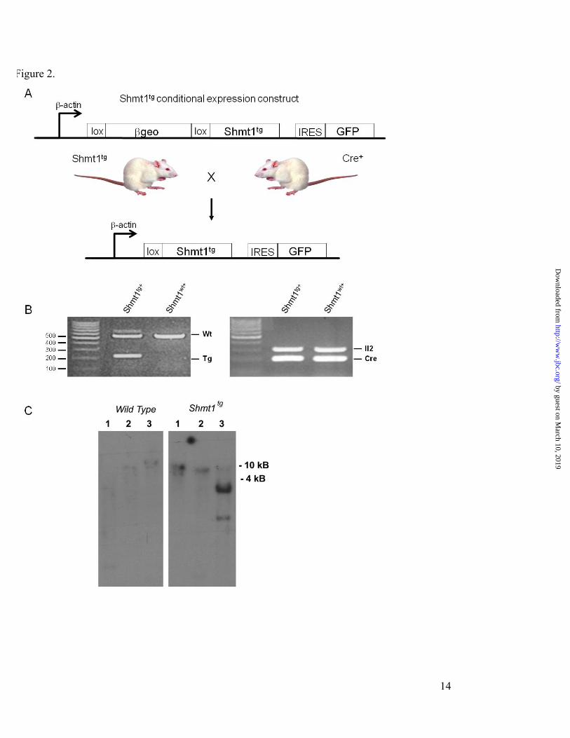
EXPERIMENTAL PROCEDURES Shmt1 Expression Vector - The Shmt1 Z/EG vector (Fig. 2, A) was designed to express the βgeo reporter cassette under the control of the chicken β-actin promoter. The Z/EG vector contains the chicken β-actin promoter with an upstream cytomegalovirus enhancer that drives the expression of a loxP-flanked βgeo cassette (lacZ-neomycin resistance fusion) and an internal ribosome-entry sequence (IRES) that enables the translation of an enhanced green fluorescent protein (EGFP) (19). The murine Shmt1 cDNA (20) was cloned 3ʹ′ of the second loxP site and 5ʹ′ of the IRES-EGFP into the XhoI site (Fig. 2, A). Following Cre-mediated excision, the murine Shmt1 cDNA is located directly 3ʹ′ of the β-actin promoter.
Generation of Shmt1tg Mice – Mice were maintained under specific-pathogen free conditions in accordance with standard use protocols and animal welfare regulations. All study protocols were approved by the Institutional Animal Care and Use Committee of Cornell University and conform to the NIH Guide for the Care and Use of Laboratory Animals. The Z/EG vector containing the Shmt1 cDNA was linearized with SfiI, which cuts 3ʹ′ of the EGFP cDNA. Shmt1tg mice were generated by
by guest on March 10, 2019
http://ww
w.jbc.org/
Dow
nloaded from

3
pronuclear injection of the linearized construct into FVB/N embryos at the Cornell University Transgenic Mouse Facility. Single site integration of the vector was confirmed in Shmt1tg founder mice by southern blot analysis of purified tail DNA (Qiagen) (Fig. 2, C). Total nuclear DNA was digested with: BglII, which does not cut within the Shmt1 cDNA or the vector; EcoR5, which cuts at nt 418 of the Shmt1 cDNA but not in the vector; or, XhoI which was the cloning site for the Shmt1 cDNA insertion into the vector. The digests were run on a 1.5% agarose gel, transferred to nitrocellulose and hybridized with a 32P-labeled probe generated from nt 443-1233 of the Shmt1 cDNA using protocols described elsewhere (21). DNA isolated from the FVB/N-Tg(ACTB-LacZ/Shmt1)B1Stov (Shmt1tg) founder exhibited a high molecular band when digested with BglII or EcoR5. Digestion with XhoI resulted in a prominent band 1.6 kB and a weaker smaller band, which were not observed in DNA isolated from wildtype mice (Fig. 2, C). The major 1.6 kB band approximates the size of the Shmt1 cDNA insert whereas smaller minor band likely reflects XhoI star activity. Southerns were repeated over 3 generations without changes in the Southern banding pattern, indicating a single integration site. A single male founder was used to generate the colony. Thereafter, Shmt1tg mice were genotyped by PCR using purified tail nuclear DNA (Qiagen) using the forward primer 5ʹ′-GATCCCAAGACTGGCAAAGAGACTT-3' and the reverse primer 5ʹ′-GATGCACTCACAGAGCTAGGCTACAAA-3', which correspond to exon 9 and 10, respectively, of the Shmt1 gene, which amplify a 215 and a 542 bp products representing the Shmt1 transgene and wildtype alleles (Fig. 2, B).
To achieve cDNA activation, loxP sites were placed both 5ʹ′ and 3ʹ′ of the βgeo cassette. The Shmt1 transgene was activated by crossing male FVB/N-Tg(ACTB-LacZ/Shmt1)B1Stov (Shmt1tg) mice with female BALB/c-Tg(CMV-Cre)1Cgn/J (Cre; The Jackson Laboratory, Bar Harbor, ME) mice that were homozygous for the Cre transgene. Presence of the Cre allele was determined by PCR using the forward primer 5ʹ′-ACCAGCCAGCTATCAACTCG-3ʹ′ and the reverse primer 5ʹ′-TTACATTGGTCCAGCCACC-3ʹ′, as described (The Jackson Laboratory). The forward and reverse primers 5ʹ′-CTAGGCCACAGAATTGAAAGATCT-3ʹ′ and 5ʹ′-GTAGGTGGAAATTCTAGCATCATCC-3ʹ′, respectively, were used for detection of the Il2
internal control gene, as described (The Jackson Laboratory) (Fig. 2, B).
Diets – Mice used for characterization of protein over-expression and the nuclear dTMP synthesis assays were fed a standard rodent chow (Harlan Teklad LM-485) from weaning. Mice on the controlled diet study were randomly weaned at three weeks of age to a control diet (C, AIN93G containing 2 mg folic acid/kg and 2.5 g choline bitartrate/kg, Dyets, Inc., Bethlehem, PA) or a folate/choline deficient diet (FCD, AIN-93G diet lacking folic acid and choline, Dyets, Inc.). Mice were maintained on the controlled diet for 5 or 32 weeks (until 8 or 35 weeks of age), as indicated in the results.
Determination of AdoMet and AdoHcy Concentrations—The animal feeding cycle was synchronized prior to tissue harvest to ensure AdoMet concentrations reflected homocysteine remethylation capacity with minimal contributions from dietary methionine. Food was removed 24 h prior to killing the animals. After 12 h, each animal was given one food pellet, and the animals were killed 12 h later by cervical dislocation. Tissues were harvested and immediately flash-frozen and stored at –80 °C until analysis. Frozen tissues were sonicated in 500 µl of 0.1 M NaAcO buffer (pH 6), and protein was precipitated by adding 312 µl of 10% perchloric acid to each sample. After vortexing, samples were centrifuged at 2000 × g for 10 min at 4 °C. AdoMet and AdoHcy were determined as described previously (14). AdoMet and AdoHcy values were normalized to total protein (22).
Immunoblotting— For tissues, total protein was extracted and quantified by the Lowry-Bensadoun assay (22). Tissue lysis was achieved by sonication in lysis buffer (2% SDS, 100 mM, dithiothreitol, 60 mM Tris (pH 6.8)). For analysis of liver nuclei, pelleted nuclei, purified as described below, were disrupted by boiling in SDS-PAGE loading buffer for 10 min and protein concentrations quantified.
Proteins (40 µg/well for tissues, 20 µg/well for nuclei) were separated on a 10% (nuclei) or 12% (tissue) SDS-PAGE gel. Proteins were transferred at 4 °C to an Immobilon-P polyvinylidene difluoride membrane (Millipore Corp.) using a Transblot apparatus (Bio-Rad). Following transfer, membranes were blocked in 5% (w/v) nonfat skim milk in phosphate-buffered saline (PBS) for 1 h followed by overnight incubation in primary antibody at 4 °C. The membranes were washed with PBS containing 0.1%
by guest on March 10, 2019
http://ww
w.jbc.org/
Dow
nloaded from

4
Tween 20 and then incubated overnight with the appropriate horseradish peroxidase-conjugated secondary antibody (below). The membranes were visualized using the SuperSignal® West Pico chemiluminescent substrate system (Pierce).
For SHMT1 detection, sheep anti-mouse SHMT1 antibody (20) was diluted 1:10,000, and rabbit anti-sheep IgG secondary antibody (Pierce) was diluted 1:20,000. For TYMS detection, affinity-purified sheep anti-human TS antibody (Abcam) was diluted 1:5000, and rabbit anti-sheep IgG secondary antibody (Pierce) was diluted 1:10,000. For DHFR detection, affinity-purified goat anti-human DHFR antibody (Santa Cruz Biotechnology) was diluted 1:1000, and mouse anti-goat IgG secondary antibody (Pierce) was diluted 1:5000. For lamin A detection, rabbit anti-lamin A (Santa Cruz Biotechnology) was diluted 1:500, and goat anti-rabbit IgG secondary antibody (Pierce) was diluted 1:20000. For detection of the loading control GAPDH, mouse anti-human GAPDH antibody (Novus Biologicals) was diluted 1:100,000 dilution, and goat anti-mouse IgG secondary antibody (Pierce) was diluted 1:10000.
Plasma and Tissue Folate Concentration—Folate concentration in plasma and liver was quantified using the Lactobacillus casei microbiological assay as described (14).
For quantification of nuclear folate levels, four livers were isolated from Shmt1wt+ and Shmt1tg+ mice and placed immediately in cold PBS at 5°C containing 200mM β -mercaptoethanol (Sigma) and 2% (w/v) sodium ascorbate (Sigma). Liver nuclei from four age-matched males for each genotype were combined and prepared as described in the Nuclear de novo thymidylate synthesis section below. Nuclear folate content was determined in two independent experiments.
Uracil Content in Nuclear DNA—Nuclear DNA was extracted from 25 to 50 mg of tissue using DNeasy Tissue and Blood Kit (Qiagen), including an incubation with RNase A (Sigma) and RNase T1 (Ambion) for 30 min at 37 °C. 10 µg of DNA was treated with 1 unit of uracil DNA glycosylase (Epicenter) for 1 h at 37 °C. Immediately following incubation, 10 pg of [15N2]uracil (Cambridge Isotopes) was added to each sample as an internal standard, and the sample was dried completely in a speed vacuum. 50 µl of acetonitrile, 10 µl of triethylamine, and 1 µl of 3,5-bis(trifluoromethyl) benzyl bromide were added to each sample and incubated for 25 min at 30 °C with shaking at 500
rpm. 50 µl water followed by 100 µl of isooctane were added to each sample. Samples were vortexed and centrifuged. Organic extraction of derived uracil was completed by the removal of the aqueous phase and analysis of the organic phase.
Analysis of uracil-3,5-bis(trifluoromethyl) benzyl bromide was carried out on a Shimadzu QP2010. 1 µl of sample or standard was analyzed in the splitless mode with a purge activation time of 1 min and split vent flow of 50 ml/min with an injection port temperature of 280°C. Ultrapurity helium gas was used as carrier gas with a linear velocity of 55 ml/min. Separation of derived uracil was obtained by using an XTI-5, 30 m, 0.25 mm inner diameter, 0.25 µm column (Restek), using the following temperature cycles for the oven: 100°C for 1 min, ramping to 280°C at 25°C/min, holding for 5 min, ramping to 300°C at 5°C/min, and holding for 5 min. The interface temperature was held at 300°C with an ion source temperature of 260°C. Ionization was achieved using the NCI mode using methane as the reagent gas and monitoring for ions 337 m/z for uracil and 339 m/z for [15N2]uracil.
Nuclear de novo Thymidylate Synthesis -Livers from age-matched male Shmt1wt and Shmt1tg mice fed a standard rodent chow diet (4-6 months of age, n=12 per genotype) were dissected and placed immediately in cold PBS at 5°C. Liver extracts for each genotype group were combined for nuclei purification. Nuclei were prepared using an iodixanol gradient as previously described (8). De novo dTMP reactions were completed as previously described (8). In short, purified nuclei were suspended in 500 µL of nuclear assay buffer containing 5 mM NADPH (Sigma), 100 mM β -mercaptoethanol, 25 mM HEPES, pH 7.5, 50 mM sucrose, 5 mM MgCl2, 25 mM KCl, and 1 mM dUMP (Sigma) and quantified using a hemocytometer. 125 µl of assay buffer containing equal numbers of suspended nuclei were aliquoted into four 1.5 ml plastic tubes and 8 µCi of [2,3-3H]-L-serine (Moravek Biochemicals) was added to each sample. The assay was conducted under three different experimental conditions: lysed nuclei, nuclei were lysed with sonication (Branson Sonifier 150) at 5°C using two 10 sec pulses at 10 watts separated by a 10 sec resting interval; intact nuclei; or intact nuclei with SHMT1 inhibitor, intact nuclei incubated with aminomethylphosphonate (Sigma) added to a final concentration of 100 mM. Reactions were incubated for 12 h at 37°C with shaking at 300 rpm. Nuclei were pelleted by centrifugation at 2000 rpm for 5 min
by guest on March 10, 2019
http://ww
w.jbc.org/
Dow
nloaded from

5
and the supernatant was collected and analyzed for radiolabeled thymidylate by HPLC. Sample preparation and HPLC was performed as previously described (23,24). Fractions were collected and tritium quantified with a scintillation counter. The retention times of [2,3-3H]-L-serine (9 min) and 3H-thymidine (17 min) (Moravek Biochemicals) were verified prior to separation of the reaction mixtures. Data were normalized to the number of nuclei. All experiments were performed in duplicate.
Metabolite Profile from Plasma—Total homocysteine, cystathionine, total cysteine, methionine, glycine, serine, α -aminobutyric acid, N,N-dimethylglycine, and N-methylglycine were assayed in mouse plasma by stable isotope dilution capillary gas chromatography-mass spectrometry as described previously (25,26).
Statistical Analyses—Differences in genotype distribution were analyzed by the χ2 test. Differences between two groups were determined by Student's t test analysis. Differences among more than two genotypes were analyzed by two-way ANOVA and Dunnett's post hoc test using the wild type genotype as the control group. Diet × genotype effects were analyzed by two-way ANOVA and Tukey's HSD post hoc test. Groups were considered significantly different when the p value ≤ 0.05. Data are presented as mean ± SEM. All statistics were performed using JMP IN software, release 5.1.2.
RESULTS Shmt1tg+ Mice Are Viable —Shmt1 transgene expression was activated by crossing Shmt1tg mice to Cre-expressing mice, which results in deletion of the βgeo cassette and re-location of the Shmt1 intronless transgene 3′ to the β-actin promoter, generating Shmt1tg+ mice (Fig. 2, A; + denotes Cre positive). PCR-based genotyping results in the generation of a 542-bp PCR product for the wildtype Shmt1 allele, and a 215-bp PCR product for the transgene (Fig. 2, B).
To determine whether Shmt1tg mice were viable upon activation of the transgene, the Shmt1tg+ genotype distribution was determined from crosses between BALB/c CMV-Cre homozygous male mice and FVB/N Shmt1tg female mice carrying one copy of the transgenic allele (Fig. 2, C). A total of 226 F1 pups from 39 litters were examined (Table 1). The mean litter size was 5.8 pups, which approximates observed litter sizes for inbred FVB/N and BALB/c mice. The Shmt1 transgene was distributed as
expected for Mendelian inheritance with a ratio of Shmt1wt+ to Shmt1tg+ mice of 97:129 and both sexes were found at the expected frequency. Shmt1tg+ mice were not bred so the effect of SHMT1 over-expression on fertility remains to be determined.
SHMT1 Protein Over-Expression in Shmt1tg+
Mice—Shmt1tg+ mice demonstrated variable tissue-dependent SHMT1 over-expression, as detected by immunoblotting (Fig. 3). The kidney, colon and ileum of Shmt1tg+ mice exhibited a 3-4 fold increase whereas liver demonstrated a 2-fold increase in SHMT1 protein levels in comparison with Shmt1wt, Shmtwt+ and Shmt1tg (not Cre activated) mice. Endogenous SHMT1 is undetectable in the brain of wildtype mice by immunoblotting, but a significant amount of SHMT1 can be detected in the brain of Shmttg+ mice. Note that the lower band on the brain immunoblot is non-specific, as it is also observed in Shmt1 null mice.
SHMT1 and Folate Status – SHMT1 over-expression did not impact plasma or liver folate status in mice fed the control or folate/choline deficient diet (Table 2). As expected, the folate/choline deficient diet resulted in a significant decrease in plasma and liver folate after 32 weeks on diet (Table 2).
SHMT1, de novo Thymidylate Synthesis and Uracil Misincorporation in Nuclear DNA –SHMT1 preferentially partitions one-carbon units to thymidylate synthesis through the SUMO-mediated localization of the thymidylate synthesis pathway to the nucleus during S phase (5,6). Furthermore, TYMS protein levels respond to changes in SHMT1 expression; Shmt1+/- mice exhibit decreased SHMT1 and TYMS protein, decreased capacity for de novo dTMP synthesis and elevated uracil in nuclear DNA (9) . Therefore, we hypothesized that over-expression of SHMT1 would be associated with increased TYMS expression and increased nuclear de novo thymidylate synthesis capacity with a consequent decrease in uracil incorporation in nuclear DNA. Unexpectedly, Shmt1tg+ mice fed the control diet demonstrated a significant 2-fold increase in uracil content in hepatic nuclear DNA (Table 2) despite an almost 4-fold increase in total liver SHMT1 (3.9 ± 0.3) and TYMS (3.7 ± 0.4) protein content in comparison with Shmt1wt+ mice (1.0 ± 0.3 and 1.0 ± 0.3, respectively) (Fig. 4, A). When placed on a folate and choline deficient diet, Shmt1tg+ mice exhibited a 10-fold increase in nuclear uracil content compared to a 2-fold increase observed in wildtype mice on the same deficient diet (Table 2). These data
by guest on March 10, 2019
http://ww
w.jbc.org/
Dow
nloaded from

6
indicate that elevated SHMT1 and TYMS expression is not sufficient to increase capacity for de novo thymidylate synthesis.
To explore this unexpected finding, we sought to clarify the specific effect of SHMT1 over-expression on nuclear de novo thymidylate synthesis. Isolated hepatic nuclei from Shmt1tg+ mice fed the folate-replete diet contained 75% less SHMT1 and TYMS protein compared with their wildtype counterparts (Fig. 4, C and D), whereas SHMT1 over-expression had no effect on DHFR localization to the nucleus (Fig. 4, D). The decrease in nuclear SHMT1 and TYMS observed in Shmt1tg+ mice was associated with an approximate 50% decrease in nuclear de novo thymidylate biosynthesis capacity in purified hepatic nuclei (Fig. 4, B). The decrease in de novo thymidylate synthesis was not due to decreased hepatic nuclear folate content as we did not observe significant differences in nuclear folate concentrations between Shmt1wt+ and Shmt1tg+ mice fed a standard rodent chow (9.5 ± 0.4 vs. 10.7 ± 2.5, respectively).
Impact of SHMT1 Over-expression on Homocysteine Remethylation - SHMT1 binds 5-methylTHF in the cytoplasm making it unavailable for the remethylation of homocysteine in cultured MCF-7 cells (14). Therefore increased SHMT1 expression is expected to result in decreased AdoMet levels and elevated homocysteine. Consistent with this expectation, hepatic AdoMet and AdoMet:AdoHcy ratio, which is indicative of the cellular methylation capacity, were signficiantly lower in Shmt1tg+ mice at 32 weeks post-weaning (Table 2). AdoHcy was unaffected by SHMT1 over-expression. We did not observe any genotype x diet effects on AdoMet, AdoHcy or the AdoMet: AdoHcy ratio.
The impact of SHMT1 over expression on homocysteine- and folate-related metabolites in the plasma of male and female mice was determined at 5 weeks post-weaning. We did not observe a significant effect of SHMT1-over expression on any of the metabolites queried due to insufficient power to detect significant differences. A significant sex effect on plasma homocysteine, methionine, cysteine and cystathionine was observed. Female mice exhibited increased plasma homocysteine and cysteine and decreased cystathionine and methionine relative to male mice. Plasma serine tended to be decreased in Shmt1tg+ mice, an effect driven by samples from male mice (Table 3). The folate/choline deficient diet was
associated with increased plasma homocysteine and cysteine, and decreased methylglycine (Table 3). We did not observe any significant genotype x diet effects on any of the queried metabolites.
DISCUSSION Our study demonstrates that nuclear localization of the de novo dTMP synthesis pathway is essential to prevent uracil accumulation in nuclear DNA and thereby maintain genome stability, and confirms previous studies demonstrating that SHMT1 expression is an important determinant of uracil accumulation in DNA. The results demonstrate unequivocally that restricting the de novo dTMP synthesis pathway to the cytoplasm results in elevated uracil accumulation into nuclear DNA.
Shmt1tg+ mice demonstrated a 2-4-fold increase in SHMT1 and TYMS protein in tissues that normally express SHMT1, namely the liver, kidney and gastrointestinal tract (Fig. 3). No significant differences in genotype distribution among Cre-activated F1 wildtype or transgenic pups were observed (Table 1), indicating that the achieved level of SHMT1 over-expression had no impact on embryo survival in dams fed a standard rodent chow diet. It will be of interest to determine the effect of a folate deficient diet on Shmt1tg+ embryo development, as we have demonstrated folate-responsive neural tube defects in Shmt1 heterozygous null mice, which was attributed to reduced de novo thymidylate synthesis (10).
Shmt1+/- mice have been reported to exhibit increased uracil content in hepatic nuclear DNA, reduced levels of nuclear SHMT1 and TYMS and consequent decreased capacity for de novo thymidylate synthesis (3,9). In this study, contrary to expectations, uracil content in hepatic nuclear DNA was increased in Shmt1tg+ mice compared to wild type mice, which was exacerbated markedly by a folate and choline deficient diet. Indeed, hepatic nuclear DNA uracil content in Shmt1tg+ mice fed the control diet was comparable to that observed in Shmt1+/- mice (3,9). Compared to wild type mice, Shmt1tg+ mice exhibited a 4-fold increase in total hepatic SHMT1 and TYMS protein expression, while nuclear localization of SHMT1 and TYMS was reduced by approximately 75% resulting in a 50% reduction in dTMP synthesis in isolated nuclei from folate-replete mice (Fig. 4). Although Shmt1tg+ have several fold increased expression of SHMT1 and TYMS compared to Shmt+/- mice, they both exhibit
by guest on March 10, 2019
http://ww
w.jbc.org/
Dow
nloaded from

7
decreased levels of nuclear SHMT1 and TYMS protein, reduced nuclear thymidylate synthesis and elevated uracil in DNA compared to wild type mice. Collectively, these results indicate that nuclear localization of the de novo thymidylate biosynthesis pathway is essential for maintaining sufficient thymidine nucleotide pools for DNA replication.
The mechanism by which SHMT1 and TYMS accumulation in the nucleus is inhibited in Shmt1tg+ mice is not known. Elevated expression of SHMT1 in the cytoplasm may impair sumoylation of SHMT1 and/or TYMS, or prevent their translocation into the nucleus. Interestingly, the accumulation of SHMT1 and TYMS, but not DHFR, in the nucleus is coordinated and dependent on SHMT1 expression.
Similar to our previous studies, in which SHMT1-dependent expression of TYMS was observed in colon and embryonic tissue (9,10), the present study provides additional evidence for the co-regulation of these two proteins. Shmt1-/+ mice were shown to have approximately 50% SHMT1 protein content, which was concomitant with a reduction in TYMS (27). Interestingly, SHMT1, TYMS and thymidine kinase 1 protein expression were also responsive to folate deficiency as demonstrated by their increased expression in mice fed the FCD diet (27). Microarray analysis did not indicate significant transcriptional changes to TYMS expression in Shmt1-/+ suggesting that their co-regulation occurs post-transcriptionally (27). We also have not queried the effect of SHMT1 expression on sumoylation of
itself or TYMS, a mode by which SHMT1 could influence nuclear translocation. The mechanism by which SHMT1 expression influences TYMS levels is actively being investigated.
This study also confirms that increased SHMT1 expression plays a modest role in the regulation of homocysteine methylation in the cytoplasm. SHMT1 is a 5-methylTHF binding protein (28), and therefore over-expression of SHMT1 was anticipated to reduce hepatic methionine and AdoMet synthesis by sequestering cytoplasmic 5-methylTHF. Consistent with this expectation, SHMT1 overexpression decreased hepatic AdoMet, which resulted in a reduced AdoMet: AdoHcy ratio (Table 2). SHMT1 over-expression did not have a major impact on serum one-carbon metabolites, as our study was underpowered to detect significant differences.
We previously demonstrated that the common human L474F variant impairs the UBC9-SHMT1 interaction and consequently SHMT1 sumoylation and translocation to the nucleus (5). The impact of this SHMT1 variant on TYMS nuclear localization and nuclear de novo thymidylate biosynthesis capacity is unknown. The reduction of nuclear de novo dTMP synthesis capacity in the Shmt1tg+ mouse provides a unique experimental model of the human L474F SHMT1 variant, and permits mechanistic studies that investigate and validate reported epidemiological associations of this variant with lung cancer and cardiovascular risk (11-13).
REFERENCES
1. Goulian, M., Bleile, B., and Tseng, B. Y. (1980) Proc Natl Acad Sci U S A 77, 1956-1960 2. Blount, B. C., Mack, M. M., Wehr, C. M., MacGregor, J. T., Hiatt, R. A., Wang, G.,
Wickramasinghe, S. N., Everson, R. B., and Ames, B. N. (1997) Proc Natl Acad Sci U S A 94, 3290-3295
3. MacFarlane, A. J., Liu, X., Perry, C. A., Flodby, P., Allen, R. H., Stabler, S. P., and Stover, P. J. (2008) J Biol Chem 283, 25846-25853
4. Anderson, D. D., Quintero, C. M., and Stover, P. J. (2011) Proc Natl Acad Sci U S A 108, 15163-15168
5. Woeller, C. F., Anderson, D. D., Szebenyi, D. M., and Stover, P. J. (2007) J Biol Chem 282, 17623-17631
6. Anderson, D. D., Woeller, C. F., and Stover, P. J. (2007) Clin Chem Lab Med 45, 1760-1763
by guest on March 10, 2019
http://ww
w.jbc.org/
Dow
nloaded from

8
7. Ching, Y. H., Munroe, R. J., Moran, J. L., Barker, A. K., Mauceli, E., Fennell, T., Dipalma, F., Lindblad-Toh, K., Abcunas, L. M., Gilmour, J. F., Harris, T. P., Kloet, S. L., Luo, Y., McElwee, J. L., Mu, W., Park, H. K., Rogal, D. L., Schimenti, K. J., Shen, L., Shindo, M., Shou, J. Y., Stenson, E. K., Stover, P. J., and Schimenti, J. C. (2010) BMC Genet 11, 106
8. Anderson, D. D., and Stover, P. J. (2009) PLoS One 4, e5839 9. MacFarlane, A. J., Perry, C. A., McEntee, M. F., Lin, D. M., and Stover, P. J. (2011) Cancer Res
71, 2098-2107 10. Beaudin, A. E., Abarinov, E. V., Noden, D. M., Perry, C. A., Chu, S., Stabler, S. P., Allen, R. H.,
and Stover, P. J. (2011) Am J Clin Nutr 93, 789-798 11. Lim, U., Peng, K., Shane, B., Stover, P. J., Litonjua, A. A., Weiss, S. T., Gaziano, J. M.,
Strawderman, R. L., Raiszadeh, F., Selhub, J., Tucker, K. L., and Cassano, P. A. (2005) J Nutr 135, 1989-1994
12. Wernimont, S. M., Raiszadeh, F., Stover, P. J., Rimm, E. B., Hunter, D. J., Tang, W., and Cassano, P. A. (2011) J Nutr 141, 255-260
13. Piskac-Collier, A. L., Monroy, C., Lopez, M. S., Cortes, A., Etzel, C. J., Greisinger, A. J., Spitz, M. R., and El-Zein, R. A. (2011) Genes Chromosomes Cancer 50, 1-12
14. Herbig, K., Chiang, E. P., Lee, L. R., Hills, J., Shane, B., and Stover, P. J. (2002) J Biol Chem 277, 38381-38389
15. Perry, C., Sastry, R., Nasrallah, I. M., and Stover, P. J. (2005) J Biol Chem 280, 396-400 16. Woeller, C. F., Fox, J. T., Perry, C., and Stover, P. J. (2007) J Biol Chem 282, 29927-29935 17. Fox, J. T., Shin, W. K., Caudill, M. A., and Stover, P. J. (2009) J Biol Chem 284, 31097-31108 18. Nakshatri, H., Bouillet, P., Bhat-Nakshatri, P., and Chambon, P. (1996) Gene 174, 79-84 19. Novak, A., Guo, C., Yang, W., Nagy, A., and Lobe, C. G. (2000) Genesis 28, 147-155 20. Liu, X., Szebenyi, D. M., Anguera, M. C., Thiel, D. J., and Stover, P. J. (2001) Biochemistry 40,
4932-4939 21. Stover, P. J., Chen, L. H., Suh, J. R., Stover, D. M., Keyomarsi, K., and Shane, B. (1997) J Biol
Chem 272, 1842-1848 22. Bensadoun, A., and Weinstein, D. (1976) Anal Biochem 70, 241-250 23. Field, M. S., Szebenyi, D. M., and Stover, P. J. (2006) J Biol Chem 281, 4215-4221 24. Friso, S., Choi, S. W., Dolnikowski, G. G., and Selhub, J. (2002) Anal Chem 74, 4526-4531 25. Stabler, S. P., Lindenbaum, J., Savage, D. G., and Allen, R. H. (1993) Blood 81, 3404-3413 26. Allen, R. H., Stabler, S. P., and Lindenbaum, J. (1993) Metabolism 42, 1448-1460 27. Macfarlane, A. J., Perry, C. A., McEntee, M. F., Lin, D. M., and Stover, P. J. (2011) Cancer Res
71, 2098-2107 28. Stover, P., and Schirch, V. (1991) J Biol Chem 266, 1543-1550
Acknowledgements - We would like to acknowledge Martha Field, Anna Beaudin, Sylvia Allen and Rachel Slater for technical assistance.
FOOTNOTES *This work was supported by Public Health Service grant DK58144. Address correspondence to: Patrick J. Stover, 315 Savage Hall, Division of Nutritional Sciences, Cornell University, Ithaca NY 14853; Tel. (607)255-9751; Fax (607)255-1033; E-mail: [email protected] 1Division of Nutritional Sciences, Cornell University, Ithaca, New York 14853 2Graduate Field of Biochemistry, Molecular and Cell Biology, Cornell University, Ithaca, New York 14853
by guest on March 10, 2019
http://ww
w.jbc.org/
Dow
nloaded from

9
3Department of Medicine and Division of Hematology, University of Colorado School of Medicine, Aurora, CO 80045 Present address: †Nutrition Research Division, Food Directorate, Health Products and Food Branch, Health Canada, Ottawa, Ontario K1A 0K9, Canada; # Division of Pulmonary & Critical Care Medicine, Keck School of Medicine, University of Southern California, 2011 Zonal Avenue, HMR 911, Los Angeles, California 90033 4Abbreviations: AdoHcy, S- adenosylhomocysteine; AdoMet, S-adenosylmethionine; AMPA, aminomethyl phosphonate; C, control AIN-93G diet; DHF, dihydrofolate; DHFR, DHF reductase; EGFP, enhanced green fluorescent protein; FCD, folate and choline deficient modified AIN-93G diet; IRES, internal ribosome-entry sequence; mtDNA, mitochondrial DNA; SHMT, serine hydroxymethyltransferase; SUMO, small ubiquitin-like modifier; THF, tetrahydrofolate; TYMS, thymidylate synthase.
FIGURE LEGENDS Figure 1. Folate-dependent de novo thymidylate biosynthesis. The de novo thymidylate biosynthesis pathway is comprised of SHMT1, Serine Hydroxymethyltransferase 1; Serine Hydroxymethyltransferase 2α; SHMT2α, TYMS, Thymidylate Synthase; and DHFR, Dihydrofolate Reductase. During S-phase, these enzymes are SUMOylated by Ubc9 which serves as a signal for nuclear import.
Figure 2. Generation of SHMT1 over-expressing mice. A, An inducible βgeo/Shmt1 expression vector was created to allow for Cre/lox activation of the Shmt1 transgene. The βgeo/ Shmt1 expression cassette allows for detection and localization of the transgene promoter activity via the lacZ reporter gene and for conditional over-expression of functional SHMT1. Transgene activation is achieved when Shmt1tg mice are mated to Cre-expressing mice, in this case, Cre expression was controlled by the CMV promoter. Cre expression results in the deletion of the βgeo reporter gene while simultaneously repositioning the Shmt1 transgene 3′ of the chicken β -actin promoter; we refer to mice expressing Cre and the transgene as Shmt1tg+. B, Shmt1tg+ genotyping in Cre+ Shmt1 wildtype mice and Cre+ Shmt1tg+ SHMT1 over-expressing mice. The Shmt1 transgene is detected as a 215-bp PCR product, whereas the wildtype (wt) allele is detected as a 542-bp PCR product. The presence of the Cre transgene was detected as a 119-bp PCR product and the Il2 internal control gene was detected as a 324-bp PCR product. C, Southern blot of genomic DNA demonstrating a single insertion of the Shmt1 transgene. Total nuclear DNA from tailsnips was isolated from PCR verified Shmt1wt and Shmt1tg mice. The DNA was digested with BglII (lane 1. Does not cut within the Z/EG vector), EcoR5 (lane 2, does not cut the Z/EG vector but cuts at position 418nt of the Shmt1 cDNA), or XhoI (lane 3, the site used for cloning which cuts 5′ and 3′ of the Shmt1 cDNA insert within the vector). Shmt1wt+, Cre+ Shmt1 wildtype mice; Shmt1tg+, Cre+ Shmt1tg mice. Figure 3. SHMT1 protein expression in liver, brain, colon, ileum and kidney of non-Cre-activated Shmt1 wildtype (wt) and transgenic (tg) mice (lane 1 and 2, respectively), and Cre-activated Shmt1 wildtype (wt+, lanes 3 and 5) and transgenic (tg+, lanes 4 and 6) mice. Immunoblots of tissue lysates were probed with polyclonal anti-SHMT1 and polyclonal anti-GAPDH antibodies. GAPDH served as a loading control. Figure 4. Hepatic nuclear SHMT1, TYMS and DHFR protein content and de novo dTMP biosynthesis in SHMT1 over-expressing mice. A, Immunoblotting for SHMT1, TYMS and actin (loading control) from whole liver extracts from Cre-activated Shmt1 wildtype (Shmt1wt+) and Shmt1tg+ mice. B, Nuclei were isolated from Shmt1wt+ and Shmt1tg+ mouse liver and capacity to convert dUMP and [2,3-3H]-L-serine to [3H]dTMP was determined in reactions that contained: sonicated nuclei; intact nuclei; or intact nuclei incubated with 100 mM aminomethyl phosphonate (AMPA), an SHMT1 inhibitor (8). De novo thymidylate biosynthesis activity was normalized to that of Shmt1wt+ intact nuclei, which was assigned an arbitrary value of 1.0. Reactions were performed in duplicate and the experiment was repeated twice. We did not observe a run-dependent difference; therefore data from the two replicate experiments were combined. Data are presented as mean±SE. C and D, Immunoblotting for SHMT1, TYMS and DHFR was performed on hepatic nuclei isolated from Shmt1 null (panel C only, (3)), and Shmt1wt +
by guest on March 10, 2019
http://ww
w.jbc.org/
Dow
nloaded from
10
and Shmt1tg+ mice. Lamin A was used as a nucleus-specific loading control. E, Immunoblotting of cytoplasm-restricted GAPDH confirmed the purity of hepatic nuclei.
by guest on March 10, 2019
http://ww
w.jbc.org/
Dow
nloaded from

11
TABLES Table 1. SHMT1 over-expressing mice are viable. Shmt1tg mice were crossed with Cre-expressing mice to activate expression of the Shmt1 transgene and their progeny were genotyped. The expected genotype distribution was calculated based on a Mendelian distribution. Differences between observed and expected genotype distributions were analyzed by Chi square analysis. P values ≤ 0.05 were considered significantly different. Shmt1wt+, Cre+ Shmt1 wildtype mice; Shmt1tg+, Cre+ Shmt1tg mice.
Observed Genotype Distribution Expected Genotype Distribution Genotype Male Female Total Male Female Total Shmt1wt+ 55 42 97 56.5 56.5 113 Shmt1tg+ 70 59 129 56.5 56.6 113 Total 125 101 226 113 113 226 Number of litters observed 39 Mean litter size (Mean ± SEM) 5.8 ± 0.4 P value, observed vs. expected genotype distribution ns P value, observed vs. expected sex distribution ns ns, not significant
by guest on March 10, 2019
http://ww
w.jbc.org/
Dow
nloaded from

10
Table 2. Plasma and liver folate, liver S-Adenosyl-methionine, S-adenosyl-homocysteine, S-adenosyl-methionine:S-adenosyl-homocysteine ratio and uracil content in nuclear DNA in SHMT1-overexpressing mice at 32 weeks post-weaning. Differences between genotypes and diets were analyzed by Student’s t-test. Genotype x diet effects were analyzed by two-way ANOVA using Tukey’s HSD post-hoc analysis. Data represent mean ± SEM values. P values ≤ 0.05 were considered significantly different. n=4-7 per group. Shmt1wt+, Cre+ Shmt1 wildtype mice; Shmt1tg+, Cre+ Shmt1tg mice.
Diet Shmt1 Genotype
Plasma folate (ng/ml)
Liver folate (fmol/µg protein)
AdoMet (pmol/µg protein)
AdoHcy (pmol/µg protein)
AdoMet:AdoHcy Liver uracil
(pg uracil/ug DNA)
AIN-93G
Shmt1wt+ 36.3 ± 7.7 43.1 ± 2.3 0.7 ± 0.2 0.4 ± 0.1 2.1 ± 0.4 0.1 ± 0.0
Shmt1tg+ 46.8 ± 5.8 51.3 ± 3.4 0.3 ± 0.2 0.2 ± 0.2 1.2 ± 0.0 0.2 ± 0.0
AIN-93G minus folate &
choline
Shmt1wt+ 7.4 ± 0.2 36.1 ± 2.6 0.7 ± 0.1 0.6 ± 0.1 1.2 ± 0.2 0.3 ± 0.0
Shmt1tg+ 5.5 ± 1.5 34.0 ± 5.8 0.4 ± 0.0 0.6 ± 0.1 0.9 ± 0.3 2.3 ± 0.7
P value, diet effect <0.0001 <0.0001 ns 0.006 0.04 0.01
P value, genotype effect ns ns 0.02 ns 0.04 0.02
P value, Diet x genotype effect ns ns ns ns ns 0.031
ns, not significant 1 folate/choline deficient Shmt1tg+ mice are significantly different than control and folate/choline deficient Shmt1wt+ and control Shmt1tg+ mice, p<0.05, as analyzed by two-way ANOVA and Tukey’s HSD post-hoc test
by guest on March 10, 2019
http://ww
w.jbc.org/
Dow
nloaded from

11
Table 3. Plasma metabolic profile of SHMT1 over-expressing mice at five weeks post-weaning. Differences between sexes, diets and genotypes were analyzed by Student’s t-test. Genotype x diet effects were analyzed by two-way ANOVA using Tukey’s HSD post-hoc analysis. Data are presented as the mean ± SEM values. P values ≤ 0.05 were considered significantly different. n = 3 males and n = 3 females per diet/genotype group. Shmt1wt+, Cre+ Shmt1 wildtype mice; Shmt1tg+, Cre+ Shmt1tg mice.
Genotype Shmt1wt+ Shmt1tg+ P value of model effect
Metabolite Sex C FCD C FCD Sex Diet Genotype Diet x Genotype
Homocysteine (µM)
Both 5.6 ± 0.7 7.0 ± 0.7 7.8 ± 1.3 9.9 ± 1.0 0.009 0.007 0.06 ns Male 4.8 ± 0.7 5.5 ± 0.2 6.6 ± 2.3 8.4 ± 0.6 - 0.10 Ns ns
Female 6.4 ± 1.0 8.4 ± 0.3 9.0 ± 1.1 11.4 ± 1.6 - 0.03 0.08 ns
Cystathionine (nM)
Both 1697 ± 237 1687 ± 334 1552 ± 189 1555 ± 70 0.002 ns Ns ns Male 1979 ± 399 2235 ± 483 1931 ± 181 1655 ± 83 - ns Ns ns
Female 1414 ± 202 1140 ± 159 1173 ± 54 1455 ± 89 - ns Ns 0.08
Cysteine (µM)
Both 171 ± 29 200 ± 20 214 ± 19 219 ± 15 <0.0001 0.006 0.10 ns Male 109 ± 21 158 ± 8 178 ± 15 192 ± 14 - 0.01 0.07 ns
Female 232 ± 4 242 ± 12 251 ± 18 246 ± 12 - ns Ns ns
Methionine (µM)
Both 39.6 ± 5.0 29.3 ± 3.9 40.9 ± 8.8 31.4 ± 1.3 0.03 ns 0.07 ns Male 49.9 ± 3.7 34.6 ± 5.8 48.1 ± 18.2 32.4 ± 1.6 - ns Ns ns
Female 29.3 ± 2.2 24.1 ± 3.7 33.7 ± 1.1 30.4 ± 2.2 - 0.07 Ns ns
α-Aminobutyric Acid (µM)
Both 6.8 ± 1.7 5.0 ± 0.5 4.5 ± 0.6 4.2 ± 0.3 ns ns Ns ns Male 5.8 ± 1.4 4.6 ± 0.4 5.0 ± 1.2 4.4 ± 0.5 - ns Ns ns
Female 7.8 ± 3.3 5.4 ± 1.0 4.0 ± 0.3 4.0 ± 0.3 - ns Ns ns
Glycine (µM)
Both 312 ± 42 286 ± 22 270 ± 17 289 ± 20 0.0006 ns Ns ns Male 383 ± 59 322 ± 32 291 ± 23 327 ± 6 - ns Ns ns
Female 240 ± 12 249 ± 9 249 ± 22 250 ± 20 - ns Ns ns
Serine (µM)
Both 161 ± 21 136 ± 11 146 ± 18 134 ± 9 <0.0001 ns 0.07 ns Male 203 ± 17 158 ± 8 174 ± 25 151 ± 6 - ns 0.07 ns
Female 119 ± 10 113 ± 8 119 ± 16 117 ± 9 - ns Ns ns
Dimethylglycine (µM)
Both 7.8 ± 1.7 7.1 ± 0.7 6.0 ± 0.7 6.2 ± 0.8 0.0004 0.10 Ns ns Male 5.0 ± 0.1 5.6 ± 0.4 5.0 ± 0.7 4.6 ± 0.2 - ns Ns ns
Female 10.5 ± 2.6 8.6 ± 0.6 7.0 ± 1.1 7.8 ± 0.5 - ns Ns ns
by guest on March 10, 2019
http://ww
w.jbc.org/
Dow
nloaded from

12
Methylglycine (µM)
Both 2.8 ± 0.5 2.1 ± 0.2 1.4 ± 0.1 2.0 ± 0.3 ns 0.04 Ns 0.06 Male 2.3 ± 0.5 2.3 ± 0.3 1.4 ± 0.1 1.6 ± 0.1 - 0.02 Ns ns
Female 3.3 ± 1.0 1.8 ± 0.2 1.3 ± 0.1 2.4 ± 0.7 - ns Ns 0.07 ns, not significant
by guest on March 10, 2019
http://ww
w.jbc.org/
Dow
nloaded from

13
FIGURES Figure 1.
by guest on March 10, 2019
http://ww
w.jbc.org/
Dow
nloaded from


Allan, Sally P. Stabler and Patrick J. StoverAmanda J. MacFarlane, Donald D. Anderson, Per Flodby, Cheryll A. Perry, Robert H.
prevent uracil accumulation in DNANuclear localization of the De Novo thymidylate biosynthesis pathway is required to
published online November 4, 2011J. Biol. Chem.
10.1074/jbc.M111.307629Access the most updated version of this article at doi:
Alerts:
When a correction for this article is posted•
When this article is cited•
to choose from all of JBC's e-mail alertsClick here
by guest on March 10, 2019
http://ww
w.jbc.org/
Dow
nloaded from