npc natural product communications · natural product communications 2012 volume 7, number 12...
TRANSCRIPT


INFORMATION FOR AUTHORS Full details of how to submit a manuscript for publication in Natural Product Communications are given in Information for Authors on our Web site http://www.naturalproduct.us. Authors may reproduce/republish portions of their published contribution without seeking permission from NPC, provided that any such republication is accompanied by an acknowledgment (original citation)-Reproduced by permission of Natural Product Communications. Any unauthorized reproduction, transmission or storage may result in either civil or criminal liability. The publication of each of the articles contained herein is protected by copyright. Except as allowed under national “fair use” laws, copying is not permitted by any means or for any purpose, such as for distribution to any third party (whether by sale, loan, gift, or otherwise); as agent (express or implied) of any third party; for purposes of advertising or promotion; or to create collective or derivative works. Such permission requests, or other inquiries, should be addressed to the Natural Product Inc. (NPI). A photocopy license is available from the NPI for institutional subscribers that need to make multiple copies of single articles for internal study or research purposes. To Subscribe: Natural Product Communications is a journal published monthly. 2012 subscription price: US$1,995 (Print, ISSN# 1934-578X); US$1,995 (Web edition, ISSN# 1555-9475); US$2,495 (Print + single site online); US$595 (Personal online). Orders should be addressed to Subscription Department, Natural Product Communications, Natural Product Inc., 7963 Anderson Park Lane, Westerville, Ohio 43081, USA. Subscriptions are renewed on an annual basis. Claims for nonreceipt of issues will be honored if made within three months of publication of the issue. All issues are dispatched by airmail throughout the world, excluding the USA and Canada.
NPC Natural Product Communications
EDITOR-IN-CHIEF
DR. PAWAN K AGRAWAL Natural Product Inc. 7963, Anderson Park Lane, Westerville, Ohio 43081, USA [email protected]
EDITORS
PROFESSOR ALEJANDRO F. BARRERO Department of Organic Chemistry, University of Granada, Campus de Fuente Nueva, s/n, 18071, Granada, Spain [email protected]
PROFESSOR ALESSANDRA BRACA Dipartimento di Chimica Bioorganicae Biofarmacia, Universita di Pisa, via Bonanno 33, 56126 Pisa, Italy [email protected]
PROFESSOR DEAN GUO State Key Laboratory of Natural and Biomimetic Drugs, School of Pharmaceutical Sciences, Peking University, Beijing 100083, China [email protected]
PROFESSOR YOSHIHIRO MIMAKI School of Pharmacy, Tokyo University of Pharmacy and Life Sciences, Horinouchi 1432-1, Hachioji, Tokyo 192-0392, Japan [email protected]
PROFESSOR STEPHEN G. PYNE Department of Chemistry University of Wollongong Wollongong, New South Wales, 2522, Australia [email protected]
PROFESSOR MANFRED G. REINECKE Department of Chemistry, Texas Christian University, Forts Worth, TX 76129, USA [email protected]
PROFESSOR WILLIAM N. SETZER Department of Chemistry The University of Alabama in Huntsville Huntsville, AL 35809, USA [email protected]
PROFESSOR YASUHIRO TEZUKA Institute of Natural Medicine Institute of Natural Medicine, University of Toyama, 2630-Sugitani, Toyama 930-0194, Japan [email protected]
PROFESSOR DAVID E. THURSTON Department of Pharmaceutical and Biological Chemistry, The School of Pharmacy, University of London, 29-39 Brunswick Square, London WC1N 1AX, UK [email protected]
ADVISORY BOARD Prof. Berhanu M. Abegaz Gaborone, Botswana
Prof. Viqar Uddin Ahmad Karachi, Pakistan
Prof. Øyvind M. Andersen Bergen, Norway
Prof. Giovanni Appendino Novara, Italy
Prof. Yoshinori Asakawa Tokushima, Japan
Prof. Lee Banting Portsmouth, U.K.
Prof. Julie Banerji Kolkata, India
Prof. Anna R. Bilia Florence, Italy
Prof. Maurizio Bruno Palermo, Italy
Prof. César A. N. Catalán Tucumán, Argentina
Prof. Josep Coll Barcelona, Spain
Prof. Geoffrey Cordell Chicago, IL, USA
Prof. Ana Cristina Figueiredo Lisbon, Portugal
Prof. Cristina Gracia-Viguera Murcia, Spain
Prof. Duvvuru Gunasekar Tirupati, India
Prof. Kurt Hostettmann Lausanne, Switzerland
Prof. Martin A. Iglesias Arteaga Mexico, D. F, Mexico
Prof. Leopold Jirovetz Vienna, Austria
Prof. Karsten Krohn Paderborn, Germany
Prof. Hartmut Laatsch Gottingen, Germany
Prof. Marie Lacaille-Dubois Dijon, France
Prof. Shoei-Sheng Lee Taipei, Taiwan
Prof. Francisco Macias Cadiz, Spain
Prof. Imre Mathe Szeged, Hungary
Prof. Joseph Michael Johannesburg, South Africa
Prof. Ermino Murano Trieste, Italy
Prof. M. Soledade C. Pedras Saskatoon, Canada
Prof. Luc Pieters Antwerp, Belgium
Prof. Peter Proksch Düsseldorf, Germany
Prof. Phila Raharivelomanana Tahiti, French Polynesia
Prof. Luca Rastrelli Fisciano, Italy
Prof. Monique Simmonds Richmond, UK
Prof. John L. Sorensen Manitoba, Canada
Prof. Valentin Stonik Vladivostok, Russia
Prof. Winston F. Tinto Barbados, West Indies
Prof. Sylvia Urban Melbourne, Australia
Prof. Karen Valant-Vetschera Vienna, Austria
HONORARY EDITOR
PROFESSOR GERALD BLUNDEN The School of Pharmacy & Biomedical Sciences,
University of Portsmouth, Portsmouth, PO1 2DT U.K.

Natural Product Communications 2012
Volume 7, Number 12
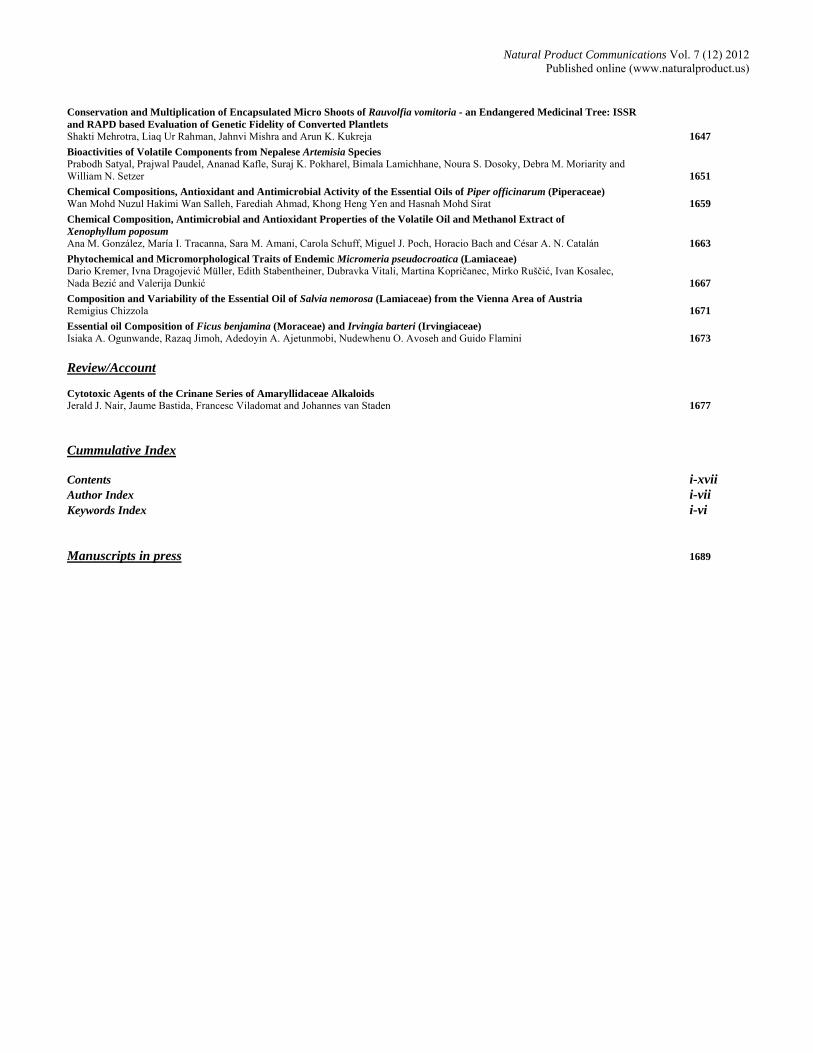
Contents
Original Paper Page
A Comparative Study of the Antioxidant/Prooxidant Effects of Carvacrol and Thymol at Various Concentrations on Membrane and DNA of Parental and Drug Resistant H1299 Cells Aysun Ozkan and Ayse Erdogan 1557
New Secoiridoid Glycosides from the Buds of Lonicera macranthoides Jiang Liu, Jing Zhang, Feng Wang and Xingfu Chen 1561
Chemical Constituents from the Aerial Parts of Gynura bicolor Jian Chen, Sven Mangelinckx, An Adams, Wei-lin Li, Zheng-tao Wang and Norbert De Kimpe 1563
Chemical Constituents of Ligularia nelumbifolia and L. subspicata Hybrid Collected in Shangrila County, Yunnan Province of China Ryo Hanai, Hiroka Yamada, Yurika Suzuki, Hajime Nagano, Takayuki Kawahara, Jiao-Jun Yu, Xun Gong and Chiaki Kuroda 1565
A Practical, Enantiospecific Synthesis of (S)-trans-γ-Monocyclofarnesol Stefano Serra 1569
ACAT Inhibitory Activity of Exudates from Calocedrus macrolepis var. formosana Yu-Hsin Hsieh, Kuan-Jung Chen, Shih-Chang Chien, Wen-Ling Cheng, Jun-Hong Xiao and Sheng-Yang Wang 1573
Cucurbitane-Type Triterpenoids from the Fruit Pulp of Momordica charantia Yun-Wen Liao, Chiy-Rong Chen, Yueh-Hsiung Kuo, Jue-Liang Hsu, Wen-Ling Shih, Hsueh-Ling Cheng, Tzou-Chi Huang and Chi-I Chang 1575
Two Novel Phenethylamine Alkaloids from Streptomyces sp. YIM10049 Xueqiong Yang, Guangwei He, Lixing Zhao, Yabin Yang, Yun Liu, Lihua Xu and Zhongtao Ding 1579
Alkaloids from Cinnamomum philippinense Hsing-Tan Li, Wei-Jen Li, Hui-Ming Wu and Chung-Yi Chen 1581
Conversional Synthesis of Heteratisine Ling Wang, Qi-Feng Chen and Feng-Peng Wang 1583
Flavonoids and Triterpenes from the leaves of Artocarpus fulvicortex Shajarahtunnur Jamil, Muhammad Taher, Hasnah M. Sirat and Nur Azlin Othman 1587
Dihydrospinochalcone-A and Epi-flemistrictin-B, Natural Isocordoin Derivatives from the Root Extract of Lonchocarpus xuul Fabiola Escalante-Erosa, Brenda González-Morales, Ramiro F. Quijano-Quiñones, Gumersindo Miron-López and Luis M. Peña-Rodríguez 1589
Phenolic Compounds, Including Novel C-glycosylflavone, from the Flowers of the Tall Bearded Iris Cultivar ‘Victoria Falls’ Takayuki Mizuno, Tsutomu Yabuya, Nobuhiro Sasaki and Tsukasa Iwashina 1591
Two New Biisoflavonoids from the Roots of Daphne oleoides Shazia Yasmeen, Muhammad Aijaz Anwar, Sadia Ferheen, Nighat Afza, Abdul Malik and Lubna Iqbal 1595
Biflavonoids from the Unripe Fruits of Clusia paralicola and their Antioxidant Activity Rafaela Ferreira Oliveira, Celso Amorim Camara, Maria de Fátima Agra and Tania Maria Sarmento Silva 1597
Ochnaflavone and Ochnaflavone 7-O-methyl Ether two Antibacterial Biflavonoids from Ochna pretoriensis (Ochnaceae) Tshepiso J. Makhafola, Babatunde B. Samuel, Esameldin E. Elgorashi and Jacobus N. Eloff 1601
Factors Affecting the Separation and Bioactivity of Red Clover (Trifolium pratense) Extracts Assayed against Clostridium sticklandii, a Ruminal Hyper Ammonia-producing Bacterium Isabelle A. Kagan and Michael D. Flythe 1605
Exploitation of the Antioxidant Potential of Geranium macrorrhizum (Geraniaceae): Hepatoprotective and Antimicrobial Activities Niko S. Radulović, Milan B. Stojković, Snežana S. Mitić, Pavle J. Randjelović, Ivan R. Ilić, Nikola M. Stojanović and Zorica Z. Stojanović-Radić 1609
Differential Expression of Benzophenone Synthase and Chalcone Synthase in Hypericum sampsonii Lili Huang, Hong Wang, Hechun Ye, Zhigao Du, Yansheng Zhang, Ludger Beerhues and Benye Liu 1615
Efficient Preparation of Hangekobokuto (Banxia-Houpo-Tang) Decoction by Adding Perilla Herb before Decoction is Finished Megumi Sumino, Yuko Saito, Fumio Ikegami, Yoshiro Hirasaki and Takao Namiki 1619
Anti-neutrophilic Inflammatory Secondary Metabolites from the Traditional Chinese Medicine, Tiankuizi Chia-Lin Lee, Tsong-Long Hwang, Chieh-Yu Peng, Chao-Jung Chen, Yuan-Shiun Chang and Yang-Chang Wu 1623
DART MS Based Chemical Profiling for Therapeutic Potential of Piper betle Landraces Vikas Bajpai, Renu Pandey, Mahendra Pal Singh Negi, Nikhil Kumar and Brijesh Kumar 1627
Total Phenolic Content and Antioxidative Properties of Commercial Tinctures Obtained from Some Lamiaceae Plants Adam Kowalczyk, Izabela Biskup and Izabela Fecka 1631
Synthesis, Antifungal Activity and Structure-Activity Relationships of Vanillin oxime-N-O-alkanoates Vivek Ahluwalia, Nandini Garg, Birendra Kumar, Suresh Walia and Om P. Sati 1635
Cinnamic Acid Derivatives Acting Against Aspergillus Fungi. Taq Polymerase I a Potential Molecular Target Lucas J. Gutierrez, Maria L. Mascotti, Marcela Kurina-Sanz, Carlos R. Pungitore, Ricardo D. Enriz and Fernando A. Giannini 1639
Insecticidal Effects of Acetogenins from Rollinia occidentalis Seed Extract Diego Tolosa, Olga Álvarez Colom, Alicia Bardón and Adriana Neske 1645
Continued Overleaf
Natural Product Communications Vol. 7 (12) 2012 Published online (www.naturalproduct.us)
Conservation and Multiplication of Encapsulated Micro Shoots of Rauvolfia vomitoria - an Endangered Medicinal Tree: ISSR and RAPD based Evaluation of Genetic Fidelity of Converted Plantlets Shakti Mehrotra, Liaq Ur Rahman, Jahnvi Mishra and Arun K. Kukreja 1647
Bioactivities of Volatile Components from Nepalese Artemisia Species Prabodh Satyal, Prajwal Paudel, Ananad Kafle, Suraj K. Pokharel, Bimala Lamichhane, Noura S. Dosoky, Debra M. Moriarity and William N. Setzer 1651
Chemical Compositions, Antioxidant and Antimicrobial Activity of the Essential Oils of Piper officinarum (Piperaceae) Wan Mohd Nuzul Hakimi Wan Salleh, Farediah Ahmad, Khong Heng Yen and Hasnah Mohd Sirat 1659
Chemical Composition, Antimicrobial and Antioxidant Properties of the Volatile Oil and Methanol Extract of Xenophyllum poposum Ana M. González, María I. Tracanna, Sara M. Amani, Carola Schuff, Miguel J. Poch, Horacio Bach and César A. N. Catalán 1663
Phytochemical and Micromorphological Traits of Endemic Micromeria pseudocroatica (Lamiaceae) Dario Kremer, Ivna Dragojević Müller, Edith Stabentheiner, Dubravka Vitali, Martina Kopričanec, Mirko Ruščić, Ivan Kosalec, Nada Bezić and Valerija Dunkić 1667
Composition and Variability of the Essential Oil of Salvia nemorosa (Lamiaceae) from the Vienna Area of Austria Remigius Chizzola 1671
Essential oil Composition of Ficus benjamina (Moraceae) and Irvingia barteri (Irvingiaceae) Isiaka A. Ogunwande, Razaq Jimoh, Adedoyin A. Ajetunmobi, Nudewhenu O. Avoseh and Guido Flamini 1673
Review/Account Cytotoxic Agents of the Crinane Series of Amaryllidaceae Alkaloids Jerald J. Nair, Jaume Bastida, Francesc Viladomat and Johannes van Staden 1677
Cummulative Index Contents i-xvii Author Index i-vii Keywords Index i-vi Manuscripts in press 1689

Natural Product Communications Vol. 7 (12) 2012 Published online (www.naturalproduct.us)
LIST OF AUTHORS Adams, A ............................. 1563 Afza, N ................................ 1595 Agra,MF .............................. 1597 Ahluwalia, V ....................... 1635 Ahmad, F ............................. 1659 Ajetunmobi, AA .................. 1673 Amani, SM .......................... 1663 Anwar, MA .......................... 1595 Avoseh, NO ......................... 1673 Bach, H ................................ 1663 Bajpai, V .............................. 1627 Bardón, A ............................ 1645 Bastida, J ............................. 1677 Beerhues, L .......................... 1615 Bezić, N ............................... 1667 Biskup, I .............................. 1631 Camara, CA ......................... 1597 Catalán, CAN ...................... 1663 Chang, CI ............................ 1575
Chang, YS ........................... 1623 Chen, CJ .............................. 1623 Chen, CR ............................. 1575 Chen, CY ............................. 1581 Chen, J ................................. 1563 Chen, KJ .............................. 1573 Chen, QF ............................. 1583 Chen, X ................................ 1561 Cheng, HL ........................... 1575 Cheng, WL .......................... 1573 Chien, SC ............................ 1573 Chizzola, R .......................... 1671 Colom, OÁ .......................... 1645 De Kimpe, N ....................... 1563 Ding, Z ................................ 1579 Du, Z .................................... 1615 Dunkić, V ............................ 1667 Elgorashi, EE ....................... 1601 Eloff, JN .............................. 1601 Enriz, RD ............................. 1639 Erdogan, A .......................... 1557 Escalante-Erosa, F ............... 1589
Fecka, I .................................1631 Ferheen, S .............................1595 Flamini, G ............................1673 Flythe, MD ...........................1605 Garg, N .................................1635 Giannini, FA .........................1639 Gong, X ................................1565 González, AM ......................1663 González-Morales, B ...........1589 Gutierrez, LJ .........................1639 Hanai, R ................................1565 He, G ....................................1575 Hirasaki, Y ...........................1619 Hsieh, YH .............................1573 Hsu, JL .................................1575 Huang, L ...............................1615 Huang, TC ............................1575 Hwang, TL ...........................1623 Ikegami, F ............................1619 Ilić, IR...................................1609 Iqbal, L .................................1595 Iwashina, T ...........................1591 Jamil, S .................................1587 Jimoh, R ...............................1673 Kagan, IA .............................1605 Kawahara, T ........................1565 Kopričanec, M ......................1667 Kosalec, I ..............................1667 Kowalczyk, A .......................1631 Kremer, D .............................1667 Kukreja, AK .........................1647 Kumar, B .................... 1627,1635 Kumar, N ..............................1627 Kuo, YH ...............................1575 Kurina-Sanz, M ....................1639 Kuroda, C .............................1565 Lee, CL .................................1623 Li, HT ...................................1581 Li, W .....................................1563 Li, WJ ...................................1581
Liao, YW .............................. 1575 Liu, B .................................... 1615 Liu, J ..................................... 1561 Liu, Y ................................... 1579 Makhafola, TJ ...................... 1601 Malik, A ............................... 1595 Mangelinckx, S .................... 1563 Mascotti, ML ........................ 1639 Mehrotra, S ........................... 1647 Miron-López, G ................... 1589 Mishra, J ............................... 1647 Mitić, SS ............................... 1609 Mizuno, T ............................. 1591 Müller, ID ............................. 1667 Nagano, H ............................ 1565 Nair, JJ .................................. 1677 Namiki, T ............................. 1619 Negi, MPS ............................ 1627 Neske, A ............................... 1645 Ogunwande, IA .................... 1673 Oliveira, RF .......................... 1597 Othman, NA ......................... 1587 Ozkan, A .............................. 1557 Pandey, R ............................. 1627 Peña-Rodríguez, LM ............ 1589 Peng, CY ............................... 1623 Poch, MJ ............................... 1663 Pungitore, CR ....................... 1639 Quijano-Quiñones, RF ......... 1589 Radulović, NS ...................... 1609 Rahman, LU ......................... 1647 Randjelović, PJ.................... 1609 Ruščić, M ............................. 1667 Saito, Y ................................. 1619 Salleh, WMNHW ................. 1659 Samuel, BB .......................... 1601 Sasaki, N .............................. 1591 Sati, OP ................................ 1635 Schuff, C .............................. 1663
Serra, S ................................. 1569 Setzer, WN ........................... 1651 Shih, WL .............................. 1575 Silva, TMS ........................... 1597 Sirat, HM .............................. 1587 Sirat, HM .............................. 1659 Stabentheiner, E ................... 1667 Stojanović, NM .................... 1609 Stojanović-Radić, ZZ ........... 1609 Stojković, NB ....................... 1609 Sumino, M ............................ 1619 Suzuki, Y ............................. 1565 Taher, M ............................... 1587 Tolosa, D .............................. 1645 Tracanna, MI ........................ 1663 van Staden, J ........................ 1677 Viladomat, F ........................ 1677 Vitali, D ................................ 1667 Walia, S ................................ 1635 Wang, F ................................ 1561 Wang, FP .............................. 1583 Wang, H ............................... 1615 Wang, L ................................ 1583 Wang, SY ............................ 1573 Wang, Z ................................ 1563 Wu, HM ............................... 1581 Wu, YC ................................ 1623 Xiao, JH ............................... 1573 Xu, L .................................... 1579 Yabuya, T ............................. 1591 Yamada, H ........................... 1565 Yang, X ................................ 1575 Yang, Y ................................ 1575 Yasmeen, S .......................... 1595 Ye, H .................................... 1615 Yen, KH ............................... 1659 Yu, J ..................................... 1565 Zhang, J ................................ 1561 Zhang, Y............................... 1615 Zhao, L ................................. 1575


A Comparative Study of the Antioxidant/Prooxidant Effects of Carvacrol and Thymol at Various Concentrations on Membrane and DNA of Parental and Drug Resistant H1299 Cells Aysun Ozkan* and Ayse Erdogan
aDepartment of Biology, Faculty of Science, Akdeniz University, 07058, Antalya, Turkey [email protected]
Received: March 8th, 2012; Accepted: October 16th, 2012
Carvacrol and thymol, both used as flavor agents in cosmetic and food products, have prooxidant and antioxidant activities. To clarify the mechanisms of their cytotoxicity and the factors affecting their antioxidant/prooxidant activities, we investigated cell membrane and DNA damage induced by carvacrol and thymol in parental and drug-resistant human lung cancer cell lines. After 24 and 48 hour incubation periods, the cytotoxicity of carvacrol (IC50 380 and 244 µM) was found to be higher than that of thymol (IC50 497 and 266 µM) in parental cells. However, thymol showed higher cytotoxic effects in drug resistant H1299 cells for three incubation periods. Also, carvacrol and thymol, at higher concentrations, increased malondealdehyde (MDA) levels causing membrane damage and 8-hydroxy deoxyguanozine (8-OHdG) levels, causing DNA damage to both parental and drug resistant cells. On the other hand, carvacrol and thymol protected the cells against H2O2-induced cytotoxicity, and membrane and DNA damage when the cells were preincubated with these two compounds at lower concentration (<IC50) before H2O2 incubation. These findings suggest that carvacrol and thymol exhibit protective/damaging effects depending on cell resistance, concentration and time. Keywords: Carvacrol, Thymol, DNA, Membrane, Anticancer, Antioxidant.
Monoterpenes are highly hydrophobic substances that exert a wide spectrum of biological actions of great importance in many different areas [1,2]. Thymol (5-methyl-2-(1-methylethyl) phenol) is an isomer of carvacrol (5-isopropyl-2-methyl phenol), having the hydroxyl group at a different location on the phenolic ring. The hydrophobic nature of carvacrol and thymol enables them to react with the lipids of the cell membrane and mitochondria, rendering them permeable and leading to leakage of cell components [3]. Natural antioxidants are considered useful agents for the prevention of diseases [4-7]. Many studies have shown that phenolic compounds in plant essential oils display antioxidant activity as a result of their capacity to scavenge free radicals [8-10]. On the other hand, effects of antioxidant concentrations on oxidation reactions depend on many factors such as structure of the antioxidant, oxidation conditions and changing of the oxidized structure. Phenolic antioxidants lose their antioxidant effects at higher concentrations and gain a prooxidant structure. They can either protect DNA and membranes against oxidants as an antioxidant at lower concentrations or damage DNA and membranes as a prooxidant at higher concentrations. Recent studies reveal that anti-/pro-oxidant and toxic properties of these molecules change depending on concentration, the kind of cells and organism, and so are not safe for human being [11]. Tumors are heterogeneous in many respects, including chemotherapeutic susceptibility [12]. Resistance to chemo-therapeutic agents is a major problem in the treatment of patients with small cell (SCLC) and non-small cell lung cancer (NSCLC). Acquired multidrug resistance is the main obstacle for the cure of SCLC. A group of drug resistance cells can occur in tumors during chemotherapy. The development of drug resistance and detoxifying mechanisms in human and mice microsome enzymes CYP3A4 used eucalyptol as a substrate [13,14]. We tried to prove the ability of carvacrol and thymol in preventing cytotoxicity, membrane damage
and DNA damage induced by the oxidative agent H2O2, while they also have cytotoxic and damaging effects in parental and drug resistant H1299 cells because many phenolic components have shown various protective/damaging activities in different biological systems dependant on concentration. Thymol and carvacrol showed cytotoxic effects on parental and drug resistance H1299 cells (Figure 1 and 2). After 24 and 48 hour incubation periods, the cytotoxicity of carvacrol was found to be higher {380 and 244 µM (IC50) concentrations} than that of thymol (IC50 497 and 266 µM) in parental cells. However, thymol showed higher cytotoxic effects in drug resistant H1299 cells for three incubation periods. The viability of cells decreased between 25-1000 μM carvacrol and 10-1000 μM thymol concentrations. The cytotoxic effect was close to that of the control until 25 μM concentrations for carvacrol and 5 μM concentrations for thymol. So, neither compound had either an effective cytotoxic or antitumor effect on cancer cells at lower concentrations. Carvacrol and thymol exhibited dose- and incubation time-dependent cytotoxic effects on parental and drug resistant H1299 cells. Also, the effects changed depending on the drug resistance capacity of the target cells. In one study, carvacol and thymol had a dose-dependent antiproliferative effects on Hep G2 cells, which make them potentially interesting for adjuvant experimental cancer treatments. Both compounds induced membrane damage and cytotoxicity in hepatoma G2 at relatively higher concentrations than those that mediate its anticancer activities. The induction of cytotoxic cell death can be accompanied by membrane damage [2]. Koparal and Zeytinoglu [15] also observed that carvacrol was a very potent inhibitor of cell growth in the A549 cancer cell line. In another study, carvacrol and thymol had dose-dependent antiproliferative effects on human uterine carcinoma cells [16]. Also, carvacrol and thymol differed in their cytotoxic effects on K562 cells [17]. All these studies support our results.
NPC Natural Product Communications 2012 Vol. 7 No. 12
1557 - 1560

1558 Natural Product Communications Vol. 7 (12) 2012 Ozkan & Erdogan
0102030405060708090
100
0 600 1200 1800
Carvacrol concentrations (micromolar)
Cel
l via
bilit
y (%
)
Parental H1299 24 h.Parental H1299 48 h.Parental H1299 72 h.Resistant H1299 24 h.Resistant H1299 48 h.Resistant H1299 72 h.
Figure 1: Cytotoxic effects of carvacrol on parental and drug-resistant H1299 cells.
0102030405060708090
100
0 500 1000 1500 2000
Thymol concentrations (micromolar)
Cel
l via
bilit
y (
%)
Parental H1299 24 h.Parental H1299 48 h.Parental H1299 72 h.Resistant H1299 24 h.Resistant H1299 48 h.Resistant H1299 72 h.
Figure 2: Cytotoxic effects of thymol on parental and drug-resistant H1299 cells.
Figure 3: Cytoprotective effects of carvacrol against H2O2 cytotoxicity on parental and drug-resistant H1299 cells.
We measured the cytoprotective (antioxidant) effect of carvacrol and thymol against the cytotoxicity of the strong oxidant H2O2 in parental and drug-resistant H1299 cells. Figures 3 and 4 show the levels of H2O2-induced cytotoxicity in the cells pre-incubated with different concentrations of carvacrol and thymol. Carvacrol had a strong antioxidant effect at an IC30 concentration for parental cells and an IC10 concentration for resistant cells against H2O2
cytotoxicity (Figure 3). Also, thymol decreased H2O2 cytotoxicity in both cells. The maximum antioxidant effect of thymol was found at an IC20 concentration for parental cells and IC10 concentration for resistant cells (Figure 4). So, resistant cells can protect themselves with lower concentrations of thymol and carvacrol against H2O2
cytotoxicity than parental cells. We can assume that drug resistant cells show more resistance to cytotoxicity than parental cells. In this study, carvacrol and thymol increased malondealdehyde (MDA) and 8-OHdG levels in both parental and drug resistant cells at different concentrations (Table 1). While increasing MDA amounts cause membrane damage, increases in 8-OHdG show the DNA damaging effect of carvacrol and thymol on the cells. The membrane and DNA damaging effect of the compounds on both
Figure 4: Cytoprotective effects of thymol against H2O2 cytotoxicity on parental and drug-resistant H1299 cells.
Table 1: Membrane and DNA damaging effects of carvacrol and thymol on parental and drug resistant H1299 cells
Concentrations MDA (nmol/mg protein) X SE
8-OHdG (ng/mL) X SE
IC10 Carvacrol (P) 0.35 0.02 a 0.09 0.03 a IC50 Carvacrol (P) 0.60 0.03 ab 0.10 0.04 a IC70 Carvacrol (P) 1.67 0.07 bc 0.15 0.03 ab IC10 Carvacrol (R) 0.31 0.05 a 0.08 0.03 a IC50 Carvacrol (R) 0.56 0.08 ab 0.09 0.02 a IC70 Carvacrol (R) 1.12 0.12 b 0.12 0.09 ab IC10 Thymol (P) 0.70 0.13 ab 0.08 0.04 a IC50 Thymol (P) 1.90 0.21 bc 0.08 0.03 a IC70 Thymol (P) 2.71 0.44 cd 0.10 0.02 a IC10 Thymol (R) 0.62 0.45 ab 0.08 0.04 a IC50 Thymol (R) 1.14 0.47 b 0.09 0.06 a IC70 Thymol (R) 2.70 0.85 cd 0.15 0.11 ab Control 0.31 0.02 a 0.08 0.04 a 0.5% DMSO 0.33 0.03 a 0.08 0.03 a
Results are means of five different experiments. Values that are followed by different letters within each column are significantly different (p≤ 0.05). df1=2, df2=95, F=11.96. SE: Standard Error. P; parental, R; resistant cells
cells was found to be close to that of the control at IC10
concentrations. The differences between the data are given in Table 1 (p≤ 0.05). The higher concentration of carvacrol and thymol caused statistically important membrane and DNA damage to the cells (p≤ 0.05). Parental cells were found to be more sensitive than resistant cells to the membrane damaging effects of both compounds. They induced membrane and DNA damage and cytotoxicity in H1299 cells at relatively higher concentrations than those that mediate its anticancer activities. The induction of cytotoxic cell death can be accompanied by membrane and DNA damage. Koparal and Zeytinoglu [15] also observed that carvacrol was a very potent inhibitor of cell growth in the A549 cancer cell line. In another study, carvacrol and thymol had dose-dependent antiproliferative effects on human uterine carcinoma cells [16]. The present studies show that phenolic compounds have antioxidant/prooxidant properties under different conditions. Carvacrol and thymol significantly decreased membrane and DNA damage in H2O2 treated H1299 cells (Table 2). The selected protective concentrations are those that showed the highest protective effect against H2O2 cytotoxicity. In the cytoprotective study, while carvacrol had a strong antioxidant effect at IC30 concentration for parental cells and IC10 concentration for resistant cell, thymol showed maximum antioxidant effect at IC20 concentration for parental cells and IC10 concentration for resistant cells against H2O2 cytotoxicity (Figures 3, 4). Also, at these cytoprotective concentrations, carvacrol and thymol showed different membrane and DNA protective effects against H2O2
oxidation (Table 2). The most effective membrane protective effect of three concentrations (IC10, IC50, and IC70) was found at IC10 for

Antioxidant/prooxidant effects of thymol and carvacrol Natural Product Communications Vol. 7 (12) 2012 1559
Table 2: Protective effects of carvacrol and thymol against H2O2 membrane and DNA damaging effects on parental and drug resistant H1299 cells
Concentrations MDA (nmol/mg prot.) X SE
8-OHdG (ng/ml) X SE
IC20 Thymol + IC10 H2O2 (P) 0.86 0.03 ab 3.09 0.44 d IC20 Thymol + IC50 H2O2 (P) 0.90 0.08 ab 8.00 0.56 hi IC20 Thymol + IC70 H2O2 (P) 1.10 0.65 b 11.70 0.98 lm IC10 Thymol + IC10 H2O2 (R) 0.95 0.13 ab 3.45 0.22 d IC10 Thymol + IC50 H2O2 (R) 1.14 0.82 b 7.60 0.32 hi IC10 Thymol + IC70 H2O2 (R) 1.78 0.96 bc 10.76 0.67 kl IC10 Carvacrol+ IC10 H2O2 (R) 0.56 0.09 ab 2.35 0.24 c IC10 Carvacrol+ IC50 H2O2 (R) 0.68 0.07 ab 7.10 0.34 h IC10 Carvacrol+ IC70 H2O2 (R) 0.96 0.08 ab 10.45 0.99 k IC30 Carvacrol + IC10 H2O2 (P) 0.93 0.08 ab 2.60 0.14 cd IC30 Carvacrol + IC50 H2O2 (P) 1.10 0.32 b 8.00 0.36 hi IC30 Carvacrol + IC70 H2O2 (P) 1.70 0.51 bc 12.00 0.35 lm IC10 H2O2 (Control P) 1.30 0.03 b 2.62 0.03 cd IC50 H2O2 (Control P) 1.80 0.12 bc 7.79 0.50 hi IC70 H2O2 (Control P) 2.40 0.14 c 11.97 0.41 lm IC10 H2O2 (Control R) 1.20 0.23 b 2.51 0.14 cd IC50 H2O2 (Control R) 1.60 0.34 bc 6.65 0.31 gh IC70 H2O2 (Control R) 2.10 0.98 c 10.55 0.82 kl Control 0.31 0.05 a 0.10 0.01 a 0.5% DMSO 0.32 0.03 a 0.11 0.01 a
Results are means of five different experiments. Values that are fallowed by different letters within each column are significantly different (p≤ 0.05). df1=2, df2=95, F=11.96. SE: Standard Error. P; parental, R; resistant cells.
carvacrol in resistant cells against H2O2 damage. Also, the IC10 concentration of carvacrol had the highest DNA protective effect in resistant cells against H2O2 damage. This means that resistant cells have more membrane protective ability than parental cells. Carvacrol and thymol have effective DNA protective effects in both cells at different concentrations. Aydin et al. [18] also observed that these compounds, at concentrations below 0.2 and 0.1 mM, respectively, significantly reduced the oxidative damage in human lymphocytes. In another study, the incubation of Hep G2 and Caco-2 cells in the presence of a range of concentrations of either carvacrol or thymol led, in both cases, to a significant protection of the cells studied from DNA strand breaks induced by the potent oxidant hydrogen peroxide [19]. Epigallocatechin-3-gallate (1 μM), a polyphenol abundant in tea, was shown to significantly reduce MDA production due to H2O2/Fe2+ exposure, indicating a protection of cells from oxidative stress [20]. The malondialdehyde level increased in H2O2 exposed (IC50 and IC70) hepatoma G2 cells, but decreased in these cells preincubated with carvacrol and thymol before H2O2 exposure [2]. Carvacrol and thymol differed in their cytotoxic and genotoxic effects on K562 cells, and reduced the level of DNA damage induced by the strong oxidation of H2O2 [17]. Further understanding of the underlying mechanism of their protective effects in reducing intracellular oxygen radicals in H1299 cell death may lead to the development of new therapeutic treatments for cancer, since carvacrol and thymol provide protection against H2O2 insult. Their lung protective effects against H2O2
toxicity might be of importance and may contribute in part to their clinical efficacy for the treatment of lung carcinoma. These results suggest that carvacrol and thymol may be potentially valuable sources of natural therapeutic agents. Thus, it is becoming increasingly evident that certain phytochemicals, particularly those included in our daily diet, have important cancer chemopreventive properties. In the present study, carvacrol and thymol induced DNA and membrane damage, and cytotoxicity in H1299 cells at relatively higher concentrations than those that mediate its anticancer activities. These findings suggest that carvacrol and thymol exhibit anticancer/antioxidant effects dependant on cell resistance, concentration and time.
Experimental
Cancer cell culture: The H1299 cell line was purchased from the American Type Culture Collection (Rockville, MD). Cells were routinely cultured in RPMI 1640 medium supplemented with10% fetal calf serum, 1% antibiotic-antimycotic solution in a humidified atmosphere containing 5% CO2 at 37°C. For subculturing, cells were harvested after trypsin/EDTA treatment at 37◦C. Cells were used when monolayer confluence had reached 75%. The drug resistant (Epirubicin-resistant) H1299 tumor cells were derived from the parental line by stepwise selection in increasing concentrations of Epirubicin until the cells were capable of propagating in 220 ng/mL drug, as described previously [21,22]. Cell viability assay: The cancer cells (10,000 cells/well, monolayer) were plated in a 96-well plate. The next day, the cells were treated with different concentrations of carvacrol and thymol in the medium for 24, 48 and 72 h. At the end of the incubation periods, the cytotoxicity of thymol and carvacrol on cancer cells was determined by the the cell titer-blue-cell viability assay. The assay is based on the ability of living cells to convert a redox dye (resazurin) into a fluorescent end product (resorufin). Nonviable cells rapidly lose metabolic capacity and thus do not generate a fluorescent signal [23]. Following cellular reduction, fluorescence is recorded at 560 nm excitation / 590 nm emission. The data were expressed as average values obtained from 8 wells for each concentration. The IC50 value was calculated from the equation of the graph. H2O2 cytotoxicity on cancer cells was measured in the same way. For measuring the antioxidant effect of carvacrol and thymol against H2O2 cytotoxicity, the cells were preincubated with them at different concentrations (10–150 μg/mL) for 1 h, before hydrogen peroxide treatment for 24 h. Determination of malondealdehyde level: The cells were plated at a density 5–10 x 105 cells/100 mm dish. The cells were preincubated with maximum cytoprotective concentrations of carvacrol and thymol for 1 h, before hydrogen peroxide treatment (IC10, IC50 and IC70) for 24 h. Cells were scraped off the culture plates with culture medium and centrifuged at 400 × g for 10 min. The cell pellets were washed with PBS and then sonicated (3 × 15 sec) in 50 mM potassium phosphate, pH 7.2, containing 1 mM PMSF (Sigma) and 1 µg/mL of leupeptin (Sigma) and centrifuged at 150,000 × g for 1 h. The supernatant was used for the determination of malondealdehyde level, as described by Wasowicz et al. [24]. This fluorometric method for measuring thiobarbituric acid-reactive substances (TBARS) in supernatant is based on the reaction between malondialdehyde and thiobarbituric acid. The product of this reaction was extracted into n-butanol and measured spectrofluorometrically at 525 nm (excitation) and 547 nm (emission). Protein was determined by the Bradford method [25] with bovine serum albumin as a standard. Determination of 8-OHdG level: After DNA purification [26] from the cultured cells (Genomic DNA Mini Kit, Invitrogen), the genomic DNA samples were used to determine 8-OHdG with a competitive ELISA kit (Highly sensitive 8-OHdG Check New, Japan Institute for Control of Aging, Fukuroi, Shizuoka, Japan). Micro titer ELISA plates were precoated with 8-OHdG. Fifty µL of the sample and primary antibody were added to each well and incubated at 4°C overnight. The wells were washed 3 times. Then 100 µL secondary antibody was added to each well and incubated for 1 h at room temperature. The wells were again washed 3 times. After that, enzyme substrate solution was added and the wells incubated at room temperature for 15 min. Terminating solution stopped the reaction. The absorbance was read at a wavelength of 450 nm [27,28].

1560 Natural Product Communications Vol. 7 (12) 2012 Ozkan & Erdogan
Data analysis: The results of the replicates were pooled and expressed as mean ± standard error. Analysis of variance and Student’s t-test were carried out. Significance was accepted at p≤ 0.05 [29].
Acknowledgements - This work was supported by the Scientific Research Projects of the Administration Unit of Akdeniz University (2008.01.0105.011). The authors wish to thank Akdeniz University Scientific Research Projects Unit for financial support of this work.
References
[1] Ozbek T, Gulluce M, Sahin F, Ozkan H, Sevsay S, Baris O. (2008) Investigation of the antimutagenic potentials of the methanol extract of Origanum vulgare L. subsp vulgare in the Eastern Anatolia region of Turkey. Turkish Journal of Biology, 32, 271–276.
[2] Ozkan A, Erdogan A. (2011) A comparative evaluation of antioxidant and anticancer activity of essential oil from Origanum onites (Lamiaceae) and its two major phenolic components. Turkish Journal of Biology, 35, 735-742.
[3] Lambert RJV, Skandamis PN, Coote PJ, Nychas GJE. (2001) A study of the minimum inhibitory concentration and mode of action of oregano essential oil, thymol and carvacrol. Journal of Applied Microbiology, 91, 453-462.
[4] Duthie GG, Brown KM. (1998) Reducing the risk of cardiovascular disease. In Functional Foods: Designer Foods, Pharmafoods, Nutraceuticals. Goldberg I. (Ed.), Chapman & Hall, New York, NY, USA, 19-38.
[5] Milner JA. (1998) Reducing the risk of cancer. In Functional Foods: Designer Foods, Pharmafoods, Nutraceuticals. Goldberg I. (Ed.). Chapman & Hall, New York, NY, USA, 39-70 pp.
[6] Gulçin I, Kireçci E, Akkemik E, Topal F, Hisar O. (2010) Antioxidant, antibacterial, and anticandidal activities of an aquatic plant: duckweed (Lemna minor L., Lemnaceae). Turkish Journal of Biology, 34, 175-188.
[7] Kutlu T, Durmaz G, Ateş B, Yılmaz I, Cetin MS. (2011) Antioxidant properties of different extracts of black mulberry (Morus nigra L.). Turkish Journal of Biology, 35, 103-110.
[8] Seyoum A, Asres K, El-Fiky FK. (2006) Structure-radical scavenging activity relationship of flavonoids. Phytochemistry, 67, 2058-2070. [9] Ozkan A, Erdogan A, Sokmen M, Tugrulay S, Unal O. (2010) Antitumoral and antioxidant effect of essential oils and in vitro antioxidant
properties of essential oils and aqueous extracts from Salvia pisidica. Biologia, 65, 990-996. [10] Ozkan A, Gubbuk H, Gunes E, Erdogan A. (2011) Antioxidant capacity of juice from different papaya (Carica papaya L.) cultivars grown under
greenhouse conditions in Turkey. Turkish Journal of Biology, 35, 619-625. [11] Moteki H, Hibasami H, Yamada Y, Katsuzaki H, Imai K, Komiya T. (2002) Specific induction of apoptosis by 1,8-cineole in two human leukemia
cell lines, but not in human stomach cancer cell line. Oncology Reports, 9, 757-760. [12] Rihova B, Strohalm J, Kobackova K. (2002) Acquired and specific immunological mechanisms co-responsible for efficacy of polymer-bound
drugs. Journal of Controlled Release, 78, 97-114. [13] Miyazawa M, Shindo M. (2001) Biotransformation of 1,8-cineole by human liver microsomes. Natural Product Letters, 15, 49-53. [14] Duisken M, Sandner F, Blomeke B, Hollender J. (2005) Metabolism of 1,8-cineole by human cytochrome P450 enzymes: identification of a new
hydroxylated metabolite. Biochimica et Biophysica Acta, 1722, 304–311. [15] Koparal AT, Zeytinoglu M. (2003) Effects of carvacrol on a human nonsmall cell lung cancer (NSCLC) cell line, A549. Cytotechnology, 43,
149-154. [16] Mastelic J, Jerkovic I, Blazevic I, Poljak-Blazi M, Borovic S, Ivancic-Bace I, Smrecki V, Zarkovic N, Brcic-Kostic K, Vikic-Topic D, Muller N.
(2008) Comparative study on the antioxidant and biological activities of carvacrol, thymol, and eugenol derivatives. Journal of Agricultural and Food Chemistry, 56, 3989-3996.
[17] Horvathova E, Turcaniova V, Slamenova D. (2007) Comparative study of DNA-damaging and DNA-protective effects of selected components of essential plant oils in human leukemic cells K562. Neoplasma, 54, 478-483.
[18] Aydin S, Basaran AA, Basaran N. (2005) Modulating effects of thyme and its major ingredients on oxidative DNA damage in human lymphocytes. Journal of Agricultural and Food Chemistry, 53, 1299-1305.
[19] Slamenova D, Horvathova E, Sramkova M, Marsalkova L. (2007) DNA-protective effects of two components of essential plant oils carvacrol and thymol on mammalian cells cultured in vitro. Neoplasma, 54, 108-112.
[20] Peng IW, Kuo SM. (2003) Flavonoid structure affects the inhibition of lipid peroxidation in Caco-2 intestinal cells at physiological concentrations. Journal of Nutrition, 133, 2184-2187.
[21] Jonsson O, Motlagh PB, Persson M, Henriksson R, Grankvist K. (1999) Increase in doxorubicin cytotoxicity by carvedilol inhibition of P-glycoprotein activity. Biochemical Pharmacology, 58, 1801-1806.
[22] Ozkan A. (2007) Lymphokine-activated killer cell susceptibility in epirubicin resistant and parental human non-small cell lung cancer (NSCLC). Biologia, 62, 232-237.
[23] Gloeckner H, Jonuleit T, Lemke HD. (2001) Monitoring of cell viability and cell growth in a hollow-fiber bioreactor by use of the dye Alamar Blue (TM). Journal of Immunological Methods, 252, 131-138.
[24] Wasowicz W, Neve J, Peretz A. (1993) Optimized steps in fluorometric determination of thiobarbituric acid-reactive substances in serum; importance of extraction pH and influence of sample preservation and storage. Clinical Chemistry, 39, 2522-2526.
[25] Bradford MM. (1976) A rapid and sensitive method for the quantitation of microgram quantities of protein utilizing the principle of protein dye binding. Analytical Biochemistry, 72, 248-254.
[26] www.invitrogen.com [27] Toyokuni S, Tanaka T, Hattori Y, Nishiyama Y, Yoshida A, Uchida K, Hiai H, Oci H, Osawa T. (1997) Quantitative immunohistochemical
determination of 8-hydroxy-2’-deoxyguanosine by a monoclonal antibody N45. 1: its application to ferric nitrilotriacetate-induced renal carcinogenesis model. Laboratory Investigation, 76, 365-374.
[28] Garçon G, Dagher Z, Zerimech F, Ledoux F, Courcot D, Aboukais A, Puskarıc E, Shirali P. (2006) Dunkerque city air pollution particulate matter-induced cytotoxicity, oxidative stress and inflammation in human epithelial lung cells (L132) in culture. Toxicology in Vitro, 20, 519–528.
[29] Kirkman TW. (1996) Statistics to use [Online]. Available:http://www.physics.csbsju.edu/stats/1996 [17 August 2008].

New Secoiridoid Glycosides from the Buds of Lonicera macranthoides Jiang Liua, Jing Zhangb, Feng Wanga and Xingfu Chena,* aDivision of Pharmaceutical Botany, College of Agronomy, Sichuan Agricultural University, Chengdu 611130, P. R. China
bCollege of Horticulture, Sichuan Agricultural University, Ya’an 625014, P. R. China [email protected]
Received: October 18th, 2012; Accepted: November 6th, 2012
Two new secoiridoid glycosides, named ethyl secologanoside (1) and 6'-O-α-L-arabinopyranosyl demethylsecologanol (2), together with three known ones, secologanoside (3), secoxyloganin (4), and loniceroside (5), were isolated from the dried buds of Lonicera macranthoides. The structures of the new compounds were determined on the basis of detailed spectroscopic analyses and acidic hydrolysis. Keywords: Lonicera macranthoides, Caprifoliaceae, Secoiridoid glycosides, Ethyl secologanoside, 6'-O-α-L-arabinopyranosyl demethylsecologanol. Lonicera is one of the most important genera in the Caprifoliaceae family. L. macranthoides Hand.-Mazz. has long been traditionally used in China and south-east Asia to treat acute fever, headache, pharyngodynia, respiratory infection, pyocutaneous disease and epidemic disease [1]. Earlier chemical studies on L. macranthoides led to the isolation of a series of phenolic acids, iridoids and saponins [2]. In this article, we present the isolation and structure elucidation of two new secoiridoid glycosides (1) and (2), as well as three known compounds: Secologanoside (3) [3a], secoxyloganin (4) [3a], and loniceroside (5) [3b] from the methanol extract of the buds of L. macranthoides (Figure 1a)
In the positive-ion FAB-MS of ethyl secologanoside (1), quasi-molecular ion peaks were observed at m/z 419 (M+H)+ and m/z 441 (M+Na)+, consistent with the molecular formula C18H26O11. The 1H NMR and 13C NMR spectra (Table 1) showed signals that indicated the presence of the olefinic proton of β-alkoxyacrylic acid at δ 7.45 (d, J=2.0 Hz, H-3), a set of three protons on the vinyl group at δ 5.63 (ddd, J=17.4, 9.7, 9.7 Hz, H-8), 5.23 (d, J=9.7 Hz, H-10A) and 5.28 (d, J=17.4 Hz, H-10B), an acetal proton at δ 5.47 (d, J=4.1 Hz, H-1), and one sugar unit containing a β-linked anomeric proton at δ 4.64 (d, J=8.3 Hz, H-1'), which were characteristic of secoiridoid-type monoterpene glycosides. Furthermore, DEPT, HMQC and HMBC experiments (Figure 1b) revealed the 18 carbons, including two carboxyl carbons (δ 176.4, C-7 and δ 168.5, C-11), four alkenyl carbons [δ 154.2 (C-3) and δ 110.4 (C-4) due to the β-alkoxyacrylic acid group, and δ 134.5 (C-8) and δ 120.5 (C-10) due to the vinyl group], one anomeric carbon (δ 99.9, C-1'), one acetal carbon (δ 97.5, C-1), and two hydroxymethyl carbons (δ 61.2, C-12 and δ 62.7, C-6'). The above data indicated that 1 was a lactone open form of iridoid. The 13C NMR spectra of 1, and 3 and 4 were also in good agreement, except for the signal arising from the ethyl group (δ 61.2, C-12 and 14.5, C-13) of the carbethoxy function at C-11. The anomeric proton H-1' (δ 4.64, d, J=8.3 Hz) and the splitting pattern of the other sugar protons gave evidence of a β-glucose unit. Finally, the absolute configuration of glucose was determined to be of the D-series by HPLC analysis of the hydrolyzate of 1 using an optical rotation detector. On the basis of this evidence, the structure of compound 1 was elucidated to be 2H-pyran-4-acetic acid, 3-ethenyl-2-(β-D-glucopyranosyloxy)-3,4-dihydro-5-(ethoxycarbonyl), and named as ethyl secologanoside.
(a) (b)
Figure 1: (a) Structures of compounds 1-5; (b) Selected HMBC and DQF correlations of compounds 1 and 2.
Table 1: 1H NMR and 13C NMR data for 1 in methanol-d4 δH δC
1 5.47 (d, J=4.1) 97.5 3 7.45 (d, J=2.0) 154.2 4 110.4 5 3.20 (m) 28.6 6 A 2.25 (dd, J=8.9, 16.5) 35.2
B 2.92 (dd, J=4.8, 16.5) 7 176.4 8 5.63 (ddd, J=17.4, 9.7, 9.7) 134.5 9 2.80 m 45.3 10 A 5.23 (d, J= 9.7)
120.5 B 5.28 (d, J= 17.4)
11 168.5 12 4.12 (m) 61.2 13 1.25 (t, J= 6.9) 14.5 Glu- 1' 4.64 (d, J=8.3) 99.9 2' 3.20 (t, J=8.3) 74.6 3' 3.34 (t, J=8.3) 78.0 4' 3.28 (overlap) 71.5 5' 3.28 (overlap) 78.4 6' A 3.65 (dd, 5.5, 11.7) 62.7
B 3.88 (dd, 2.0, 11.7)
6'-O-α-L-arabinopyranosyl demethylsecologanol (2), an amorphous powder, exhibited quasi-molecular ion peaks at m/z 509 (M+H)+ and m/z 531 (M+Na) +, consistent with C21H32O14. The NMR spectra (Table 2) and HMQC, HMBC, and DQF data (Figure 1b) showed signals that indicated the presence of β-alkoxyacrylic acid, acetal, a vinyl group, and two sugar units, which were also characteristic of secoiridoid-type monoterpene glycosides, like compound 1. Furthermore, the 1H NMR and 13C NMR spectra of 2 were quite similar to those of demethylsecologanol [3c], except for a set of signals due to an additional α-arabinopyranoside unit, The downfield shift of the glucosyl C-6' signal (δ 68.9) suggested that the arabino-pyranosyl unit was at the C-6' position of the inner glucose, which was confirmed by the HMBC correlations between
NPC Natural Product Communications 2012 Vol. 7 No. 12
1561 - 1562

1562 Natural Product Communications Vol. 7 (12) 2012 Liu et al.
Table 2: 1H NMR and 13C NMR data for 2 in methanol-d4.
No. 2 δH δC
1 5.51 (d, J=6.2) 97.6 3 7.42 (brs) 152.8 4 110.8 5 2.85 (m) 31.0 6 A 1.75 (m)
30.9 B 2.07 (m)
7 A 3.66 (dd, J=5.5, 11.7) 62.8 B 3.89 (overlap)
8 5.76 (ddd, J=17.2, 10.3, 10.3) 136.0 9 2.64 (m) 45.4 10 A 5.23 (d, J= 10.3)
119.3 B 5.27 (d, J= 17.2)
11 170.2 Glu- 1' 4.67 (d, J=7.6) 100.0 2' 3.19 (t, J=7.6) 74.7 3' 3.36 (t, J=8.3) 77.9 4' 3.26 (t, J=9.6) 71.6 5' 3.29 (overlap) 78.4 6' A 3.50 (overlap) 68.9
B 3.89 (overlap) Ara- 1'' 4.16 (d, J=6.9) 104.9 2'' 3.50 (overlap) 72.4 3'' 3.50 (overlap) 74.2 4'' 3.79 (brs) 69.5 5'' A 3.50 (overlap) 66.6
B 3.82 (dd, 2.8, 12.4)
the anomeric proton of the terminal arabinose at δ H 4.16 (H-1'') and the C-6' of the inner glucose. Finally, the absolute configuration of glucose and arabinose were determined as D and L, respectively on HPLC analysis of the hydrolyzate of 2 using an optical rotation detector. Accordingly, the structure of compound 2 was elucidated to be 2H-pyran-5-carboxylic acid, 3-ethenyl-2-(α-L-arabinopyrano-syl(1-6')-β-D-glucopyranosyloxy)-3,4-dihydro-4-(2-hydroxyethyl), and named as 6'-O-α-L-arabinopyranosyl demethylsecologanol.
Experimental
General: The following instruments were used to obtain physical data: JEOL spectrometer (600 MHz for 1H NMR, 150 MHz for 13C NMR); FAB-MS, JEOL JMS-SX 102A mass spectrometer; CC, Diaion HP-20; TLC: pre–coated silica gel 60F264.
Plant material: The buds of Lonicera macranthoides were collected from Suining, Sichuan province, China in 2011, and identified by Prof. Xingfu Chen (Sichuan Agricultural University, China). A voucher specimen of this plant was deposited with the College of Agronomy, Sichuan agricultural University. Extraction and isolation: The buds of L. macranthoides (2 Kg) were extracted 3 times with MeOH under reflux for 3 h. Evaporation of the solvent under reduced pressure provided the MeOH extract (202.1 g), which was fractionated by Diaion HP-20 CC, sequentially eluted with H2O and MeOH to give H2O-eluted (80.5 g) and MeOH-eluted fractions (120.5 g). The MeOH-eluted
fraction (120.5 g) was chromatographed on ODS columns using a gradient of MeOH: H2O (20: 80→40: 60→60: 40→80: 20)→ MeOH to give 15 fractions (1-15). Fraction 1 (2.0 g) was repeatedly chromatographed on RP-C18 [MeOH: H2O (30: 70)] to give 2 (16.0 mg), 3 (12.3 mg) and 4 (10.3 mg). Fraction 4 (1.0 g) was repeatedly chromatographed on RP-C18 [MeOH: H2O (35: 65)] to give 5 (7.7 mg). Fraction 6 (2.0 g) was repeatedly chromatographed on Sephadex LH-20 columns (MeOH) and RP-C18 [MeOH: H2O (40: 60)] to give 1 (28.9 mg).
Ethyl secologanoside (1) White amorphous powder. [α]D
30: –130.83 (c 0.29, MeOH). IR (film, MeOH): 3413, 1684, 1617, 1617, 1268, 1075 cm-l. UV (MeOH) λmax (log ε): 228 (4.17) nm. 1H and 13C NMR: Table 1. HR-FAB-MS: 441.1571 ([M+Na]+, C18H26O11Na+; calc. 441.1568).
6'-O-α-L-Arabinopyranosyl demethylsecologanol (2)
White amorphous powder. [α]D
30: –79.21 (c 0.12, MeOH). IR (film, MeOH): 3413, 1739, 1693, 1251, 1073, 592 cm-l. UV (MeOH) λmax (log ε): 227 (4.16) nm. 1H and 13C NMR: Table 2. HR-FAB-MS: 531.1870 ([M+Na]+, C18H26O11Na+; calc. 531.1867).
Acid hydrolysis of 1 and 2: Solutions of 1 (3.0 mg) and 2 (3.0 mg) in 5% H2SO4–1,4-dioxane (1:1, v/v, 1.0 mL) were heated under reflux for 1 h. After cooling, the reaction mixture was neutralized with Amberlite IRA-400 (OH—form), and the resin removed by filtration. On removal of the solvent from the filtrate under reduced pressure, the residue was partitioned in an EtOAc–H2O (1:1, v/v) mixture, and the solvent removed in vacuo from the EtOAc-soluble fraction. The aqueous layer was subjected to HPLC analysis under the following conditions: HPLC column, Kaseisorb LC NH2-60-5, 4.6 mm i.d.250 mm (Tokyo Kasei Co., Ltd., Tokyo, Japan); detection, optical rotation [Shodex OR-2 (Showa Denko Co., Ltd., Tokyo, Japan); mobile phase, CH3CN–H2O (17:3, v/v); flow rate 1.0 mL/min]. Identification of D-glucose from 1 and 2, and L-arabinose from 2 present in the aqueous layer was carried out by comparison of their retention times and optical rotations with those of authentic samples. tR: 7.1 min (L-arabinose, positive optical rotation), 11.6 min (D-glucose, positive optical rotation), respectively. Acknowledgments - This study was funded by a grant from Sichuan Provincial Crop breeding research project Application in the 12th Five-Year Period (No. 2011NZ0098—12—01).
References [1] (a) Chen M, Wu W, Shen G., Luo S, Li H. (1994) Chemical constituents of Lonicera macranthoides Hand.-Mazz part V. Isolation and structures of
macranthoin F and G. Yaoxue Xuebao, 29, 617-620; (b) Mao Q, Cao D, Jia X. (1993) Studies on the chemical constituents of Lonicera macranthoides Hand.-Mazz. Yao Xue Xue Bao, 28, 273-281; (c) Shi J, Chen X, Wan L. (1999) Hepatoprotective effects of some constituents of Lonicera fulvotomentosa Hsu et S.C. Cheng, and L. macranthoides Hand.-Mazz. against carbon tetrachloride- and D-galactosamine-induced liver injuries in mice and rats. Zhongguo Zhongyao Zazhi, 24, 363-364; (d) Wang J, Zhao X, Qi Q, Tao L, Zhao Q. (2009) Macranthoside B, a hederagenin saponin extracted from Lonicera macranthoides and its anti-tumor activities in vitro and in vivo. Food and Chemical Toxicology, 47, 1716-1721.
[2] (a) Chen C, Qi L, Li H, Li P, Yi L, Ma H. (2007) Simultaneous determination of iridoids, phenolic acids, flavonoids, and saponins in Flos Lonicerae and Flos Lonicerae Japonicae by HPLC-DAD-ELSD coupled with principal component analysis. Journal of Separation Science, 30, 3181-3192; (b) Chen Y, Zhao Y, Wang M, Sun H, Dong Y, Feng X. (2012) The first chlorogenic acid ester saponin from Lonicera macranthoides. Chemistry of Natural Compounds, 47, 940-943; (c) Chen Y, Shan Y, Zhao Y, Wang Q, Wang M, Feng X, Liang J. (2012) Two new triterpenoid saponins from Lonicera macranthoides. Chinese Chemical Letters, 23, 325-328; (d) Chen Y, Feng X, Wang M, Jia X, Zhao Y, Dong Y. (2009) Triterpene glycosides from Lonicera. II. Isolation and structural determination of glycosides from flower buds of Lonicera macranthoides. Chemistry of Natural Compounds, 45, 514-518; (e) Chen M, Luo S, Li H. (1990) Chemical constituents of Lonicera macranthoides Hand.-Mazz. I. Structure of macranthoiside I. Chinese Chemical Letters, 1, 219-220; (f) Chai X, Li S, Li P. (2005) Quality evaluation of Flos Lonicerae through a simultaneous determination of seven saponins by HPLC with ELSD. Journal of Chromatography A, 1070, 43-48.
[3] (a) Calis I, Sticher O. (1984) Secoiridoid glucosides from Lonicera periclymenum. Phytochemistry, 23, 2539-2540; (b) Einaggar LJ, Beal JL. (1980) Iridoids. A review. Journal of Natural Products, 43, 649-707; (c) Kitajima M, Fujii N, Yoshino F, Sudo H, Saito K, Aimi N, Takayama H. (2005) Camptothecins and two new monoterpene glucosides from Ophiorrhiza liukiuensis. Chemical and Pharmaceutical Bulletin, 53, 1355-1358.

Chemical Constituents from the Aerial Parts of Gynura bicolor
Jian Chena,b,c, Sven Mangelinckxb, An Adamsb, Wei-lin Lic, Zheng-tao Wanga* and Norbert De Kimpeb* aDepartment of Pharmacognosy, China Pharmaceutical University, Nanjing 210009, China bDepartment of Sustainable Organic Chemistry and Technology, Faculty of Bioscience Engineering, Ghent University, Coupure links 653, B-9000 Ghent, Belgium cInstitute of Botany, Jiangsu Province and Chinese Academy of Sciences, Nanjing 210014, China [email protected]; [email protected] Received: September 18th, 2012; Accepted: October 3rd, 2012
Gynura bicolor (Willd.) DC., is used in folk recipes for the treatment of diabetes mellitus in Jiangsu, Zhejiang and Sichuan province in the south of China. A previous pharmacological study proved that the plant showed significant hypoglycemic activity on normal and alloxan-diabetic mice. In this study, two terpenes, four megastigmane-type norisoprenoids and two glycosides were isolated from the aqueous ethanolic extract of the aerial parts of Gynura bicolor and characterized mainly by NMR spectroscopy and mass spectrometry. These compounds were isolated for the first time from this plant, and no evidence could be found for the previous reported presence of megastigmane-type norisoprenoids in the genus Gynura. Keywords: Gynura bicolor, Terpenes, Norisoprenoids, Glycosides, Diabetes mellitus. The genus Gynura belongs to the family Asteraceae, comprising approximately 40 species mainly distributed in Asia, Africa and Australia, of which 10 species were recorded in the south of China [1]. In the literature, the presence of some interesting components in plants of the Gynura genus, such as volatiles [2,3], phenolics [4a], chromanes [4b], chromanones [4c,d], coumarins [4e], steroids [4d,f] and cerebrosides [5-7a], together with pyrrolizidine alkaloids [7b-10], which are hepatotoxic and could cause hepatic veno-occlusive disease [11a], is reported.
Gynura bicolor, which has been cultivated as a popular vegetable, is not only known to be nutritive but is also used for the treatment of diabetes in the south of China. Our previous pharmacological tests proved that the ethyl acetate and n-butanol extracts of aerial parts of the plant had significant effects on lowering blood glucose level in normal and alloxan-diabetic mice [11b]. To our knowledge, only three studies investigated the volatiles [3a-c] and one research reported on some anthocyanins [11c] from this plant. In the present study, two terpenes, four megastigmane-type norisoprenoids and two glycosides were obtained by combined chromatographic methods from the aqueous ethanolic extract of the aerial parts of Gynura bicolor. By comparison of their 1H, 13C NMR and ESI-MS spectral data with those reported, these isolated compounds (1-8) were unambiguously identified as: ficusic acid (1) [12a], loliolide (2) [12b], dehydrovomifoliol (3) [12c], vomifoliol (4) [13a], boscialin (5) [13b], (6S,9S)-roseoside (6) [13c], benzyl-β-D-glucopyranoside (7) [14a-b], 2-phenylethyl-β-D-glucopyranoside (8) [14b]. Noteworthy, the 1H and 13C NMR spectral data of compound 2, which were in correspondence with loliolide [12b], were also nearly identical with those of a compound, named pubinernoid A, which was isolated from Schisandra pubescens var. pubinervis [14c]. The structure proposal for pubinernoid A, with the same molecular formula as loliolide, was (4S*,6R*)-4-hydroxy-4,8,8-trimethyl-9-oxabicyclo[4.2.1]non-1-en-3-one, with an atypical value of 183.2 ppm for the chemical shift of the keto group. However, upon comparing GC-MS spectral data with data available in the WILEY6N database, compound 2 was identified as loliolide.
Chemotaxonomic significance: Pyrrolizidine alkaloids are widely found and are characteristic secondary metabolites in the tribe
H
COOH
O
1
OHO
O
2 OOH
O
3 OOH
OHH
4 HO
OH
O
5
O
OH
OH
O
HO OHOHOH
6
O O
OH
OH
HOHO
7
O O
OH
OH
HOHO
8
Figure 1: Structures of isolated compounds (1-8). Senecioneae, belonging to the Asteraceae family [14d]. In this study, chemical constituents of G. bicolor from the same tribe are assigned as terpenes (1-2), megastigmane-type norisoprenoids (3-6) and glycosides (6-8). It should be noted that all these compounds were isolated for the first time from this plant, and no evidence could be found of the previous reported presence of megastigmane-type norisoprenoids in the genus Gynura. Roseoside (6) was firstly isolated from Vinca rosea in 1974 [14e], and together with their stereoisomers, they were found in many plant sources. It is interesting to note that (6S,9R)-roseoside, which is an isomer of (6S,9S)-roseoside (6) currently isolated, has previously been shown to increase insulin secretion from INS-1 β-cells [14f]. Vomifoliol (4), the aglycone of an isomer of (6S,9S)-roseoside (6), and its analogue, dehydrovomifoliol (3), were identified for the first time in the fruits from Vitis vinifera [15]. Boscialin (5) can be regarded as a reduced analogue of dehydrovomifoliol (3). The occurrence of C13-type norisoprenoids in Gynura bicolor, as demonstrated in this paper, is noteworthy. Experimental
Plant material: The aerial parts of Gynura bicolor (Willd.) DC. were collected in June 2010 in Nanjing Botanical Garden Memorial Sun Yat-Sen, in the south of the Zijin Mountain, Nanjing, China. The plant (voucher specimen No. 510310-1) was identified by Professor Guo Rong-lin of Institute of Botany, Jiangsu Province and Chinese Academy of Sciences. Extraction and isolation: Dry aerial parts of G. bicolor (2.9 kg) were extracted with 80% aqueous ethanol at 70 ˚C twice to afford 295 g of crude extract after evaporation in vacuo of the solvent. The
NPC Natural Product Communications 2012 Vol. 7 No. 12
1563 - 1564

1564 Natural Product Communications Vol. 7 (12) 2012 Chen et al.
extract was coated on silica gel, and then successively extracted with n-hexane, CH2Cl2, EtOAc and MeOH to give four fractions. The CH2Cl2 extract (3.4 g) was further fractionated by column chromatography over silica gel, eluted with CH2Cl2 containing increasing concentrations of MeOH to obtain seven fractions (Fr. B1-B7). Fr. B2 (600 mg) was fractionated with an automatic flash chromatography system on a reversed-phase column (C-18) eluted by a gradient of water and MeOH (50-100%) to get four fractions (Fr. B2-1~4). Fr. B2-1 (248 mg) and B2-2 (175 mg) were further subjected to column chromatography over MCI gel CHP20P eluted by a gradient of water and MeOH (20-100%). Upon further silica gel column chromatography (CH2Cl2/MeOH, 100:1 to 10:1) and prep-HPLC (H2O/CH3CN, 75:25), ficusic acid 1 (2.2 mg) was obtained. Following silica gel column chromatography (petroleum ether/EtOAc, 10:1 to 1:1) and prep-HPLC (H2O/CH3CN, 75:25), loliolide 2 (4.0 mg) and dehydrovomifoliol 3 (0.5 mg) were isolated. Fr. B3 (655 mg) was successively fractionated by automatic flash chromatography (C-18, H2O/MeOH, 50:50 to 0:100), MCI gel column chromatography (H2O/MeOH, 80:20 to 0:100), prep-HPLC (H2O/MeCN, 75:25) to give vomifoliol 4 (1.2 mg) and boscialin 5 (0.8 mg). The EtOAc extract (7.7 g) was fractionated by chromatography on a C-18 column, eluted with a gradient of water and MeOH (20-100%), to give five fractions (Fr.
C1-C5). Fr. C2 (724 mg) was further separated with automatic flash chromatography on a reverse phase column (C-18) eluted with a gradient of water and MeOH (20-100%). Four fractions (Fr. C2-1~4) were obtained and Fr. C2-2 (510 mg) was subjected to chromatography on a silica gel column eluted with a CHCl3/MeOH gradient (100:1 to 6:1) to give six fractions (C2-2-1~6). The C2-2-3 fraction (12 mg) and C2-2-4 fraction (20 mg) were finally purified by prep-HPLC eluted with MeOH/H2O (36:64) to yield pure (6S,9S)-roseoside 6 (2.2 mg) and benzyl-β-D-glucopyranoside 7 (1.3 mg). Fr. C3 (2.1 g) was purified successively via automatic flash chromatography (C-18, H2O/MeOH 80:20 to 0:100), silica gel column chromatography (CH2Cl2/MeOH 10:1 to 4:1) and prep-HPLC (MeOH/H2O 35:65) to afford 2-phenylethyl-β-D-glucopyranoside 8 (1.1 mg). Acknowledgments - This study was supported by a grant from the National Natural Science Foundation of China (No. 30900123) and a grant from Jiangsu Center for Research & Development of Medicinal Plants (No. 200902). The authors are also indebted to the Special Research Fund (BOF-UGent) for funding for a joint doctoral study of Jian Chen, and to the Research Foundation-Flanders (FWO-Vlaanderen) for a postdoctoral fellowship for Sven Mangelinckx and An Adams.
References
[1] Chen YL. (1999) Flora of China. Science Press, Beijing, 309. [2] Rana VS, Blazquez MA. (2007) Chemical constituents of Gynura cusimbua aerial parts. Journal of Essential Oil Research, 19, 21-22. [3] (a) Shimizu Y, Imayoshi Y, Kato M, Maeda K, Iwabuchi H, Shimomura K. (2009) Volatiles from leaves of field-grown plants and shoot cultures of
Gynura bicolor DC. Flavour and Fragrance Journal, 24, 251-258; (b) Shimizu Y, Imayoshi Y, Kato M, Maeda K, Iwabuchi H, Shimomura K. (2011) New eudesmane-type sesquiterpenoids and other volatile constituents from the roots of Gynura bicolor DC. Flavour and Fragrance Journal, 26, 55-64; (c) Chen J, Adams A, Mangelinckx S, Ren BR, Li WL, Wang ZT, De Kimpe N. (2012) Investigation of the volatile constituents of different Gynura species from two Chinese origins by SPME/GC-MS. Natural Product Communications, 7, 655-657.
[4] (a) Wan CP, Yu YY, Zhou SR, Tian SG, Cao SW. (2011) Isolation and identification of phenolic compounds from Gynura divaricata leaves. Pharmacognosy Magazine, 7, 101-108; (b) Lin WY, Teng CM, Tsai IL, Chen IS. (2000) Anti-platelet aggregation constituents from Gynura elliptica. Phytochemistry, 53, 833-836; (c) Jong TT, Chou-Hwang JY. (1997) An optically active chromanone from Gynura formosana. Phytochemistry, 44, 553-554; (d) Lin WY, Kuo YH, Chang YL, Teng CM, Wang EC, Ishikawa T. (2003) Anti-platelet aggregation and chemical constituents from the rhizome of Gynura japonica. Planta Medica, 69, 757-764; (e) Bohlmann F, Zdero C. (1977) Gynuron, ein neues terpen-cumarin-derivat aus Gynura crepioides. Phytochemistry, 16, 494-495; (f) Takahira M, Kondo Y, Kusano G, Nozoe S. (1977) Four new 3α-hydroxyspirost-5-ene derivatives from Gynura japonica Makino. Tetrahedron Letters, 41, 3647-3650.
[5] Chen L, Wang JJ, Zhang GG, Song HT, Qin LP. (2009) A new cerebroside from Gynura divaricata. Natural Product Research, 23, 1330-1336. [6] (a) Chen L, Li HQ, Song HT, Zhang GG. (2009) A new cerebroside from Gynura divaricata. Fitoterapia, 80, 517-520; (b) Chen L, Wang JJ, Song
HT, Zhang GG, Qin LP. (2009) New cytotoxic cerebroside from Gynura divaricata. Chinese Chemical Letters, 20, 1091-1093. [7] (a) Lin WY, Yen MH, Teng CM, Tsai IL, Chen IS. (2004) Cerebrosides from the rhizomes of Gynura japonica. Journal of the Chinese Chemical
Society, 51, 1429-1434; (b) Liang X T, Roeder E. (1984) Senecionine from Gynura segetum. Planta Medica, 50, 362. [8] Roeder E, Eckert A, Wiedenfeld H. (1996) Pyrrolizidine alkaloids from Gynura divaricata. Planta Medica, 62, 386. [9] Wiedenfeld H. (1982) Two new hepatotoxic pyrrolizidine alkaloids from Gynura scandens. Planta Medica, 45, 136-137. [10] Wiedenfeld H. (1982) Two pyrrolizidine alkaloids from Gynura scandens. Phytochemistry, 21, 2767-2768. [11] (a) Schoental R. (1968) Toxicology and carcinogenic action of pyrrolizidine alkaloids. Cancer Research, 28, 2237-2246; (b) Li WL, Ren BR, Zhuo
M, Hu Y, Lu CG, Wu JL, Chen J, Sun S. (2009) The anti-hyperglycemic effect of plants in genus Gynura Cass. American Journal of Chinese Medicine, 37, 961-966; (c) Shimizu Y, Imada T, Zhang HL, Tanaka R, Ohno T, Shimomura K. (2010) Identification of novel poly-acylated anthocyanins from Gynura bicolor leaves and their antioxidative activity. Food Science and Technology Research, 16, 479-486.
[12] (a) Wang CY, Liu X, Guo LM, Shao CL, Fang YC, Wei YX, Zheng CJ, Gu QQ, Zhu WM, Guan HS. (2010) Two new natural keto-acid derivatives from Sargassum pallidum. Chemistry of Natural Compounds, 46, 292-294; (b) Chen BN, Yang GE, Li JK, Du HJ, Li QS, Zhang ZM. (2009) Cytotoxic constituents from Viscum coloratum. Chemistry of Natural Compounds, 45, 547-549; (c) Kisiel W, Michalska K, Szneler E. (2004) Norisoprenoids from aerial parts of Cichorium pumilum. Biochemical Systematics and Ecology, 32, 343-346.
[13] (a) Hammami S, Jannet HB, Bergaoui A, Ciavatta L, Cimino G, Mighri Z. (2004) Isolation and structure elucidation of a flavanone, a flavanone glycoside and vomifoliol from Echiochilon Fruticosum growing in Tunisia. Molecules, 9, 602-608; (b) Pauli N, Sequin U, Walter A. (1990) Boscialin and boscialin 4′-O-glucoside, two new compounds isolated from the leaves of Boscia salicifolia OLIV. Helvetica Chimica Acta, 73, 578-582; (c) Yajima A, Oono Y, Nakagawa R, Nukada T, Yabuta G. (2009) A simple synthesis of four stereoisomers of roseoside and their inhibitory activity on leukotriene release from mice bone marrow-derived cultured mast cells. Bioorganic & Medicinal Chemistry, 17, 189-194.
[14] (a) Deffieux D, Natangelo A, Malik G, Pouysegu L, Charris J, Quideau S. (2011) First and biomimetic total synthesis of a member of the C-glucosidic subclass of ellagitannins, 5-O-desgalloylepipunicacortein A. Chemical Communications, 47, 1628-1630; (b) Yoneda Y, Krainz K, Liebner F, Potthast A, Rosenau T, Karakawa M, Nakatsubo F. (2008) “Furan endwise peeling” of celluloses: mechanistic studies and application perspectives of a novel reaction. European Journal of Organic Chemistry, 3, 475-484; (c) Huang SX, Yang J, Xiao WL, Zhu YL, Li RT, Li LM, Pu JX, Li X, Li SH, Sun HD. (2006) Three novel terpenoids from Schisandra pubescens var. pubinervis. Helvetica Chimica Acta, 89, 1169-1175; (d) Langel D, Ober D, Pelser PB. (2011) The evolution of pyrrolizidine alkaloid biosynthesis and diversity in the Senecioneae. Phytochemistry Reviews, 10, 3-74; (e) Bhakuni DS, Joshi PP, Uprety H, Kapil RS. (1974) Roseoside-A C13 glycoside from Vinca rosea. Phytochemistry, 13, 2541-2543; (f) Frankish N, de Sousa Menezes F, Mills C, Sheridan H. (2010) Enhancement of insulin release from the beta-cell line INS-1 by an ethanolic extract of Bauhinia variegata and its major constituent roseoside. Planta Medica, 76, 995-997.
[15] Strauss CR, Wilson B, Williams PJ. (1987) 3-Oxo-α-ionol, vomifoliol and roseoside in Vitis vinifera fruit. Phytochemistry, 26, 1995-1997.
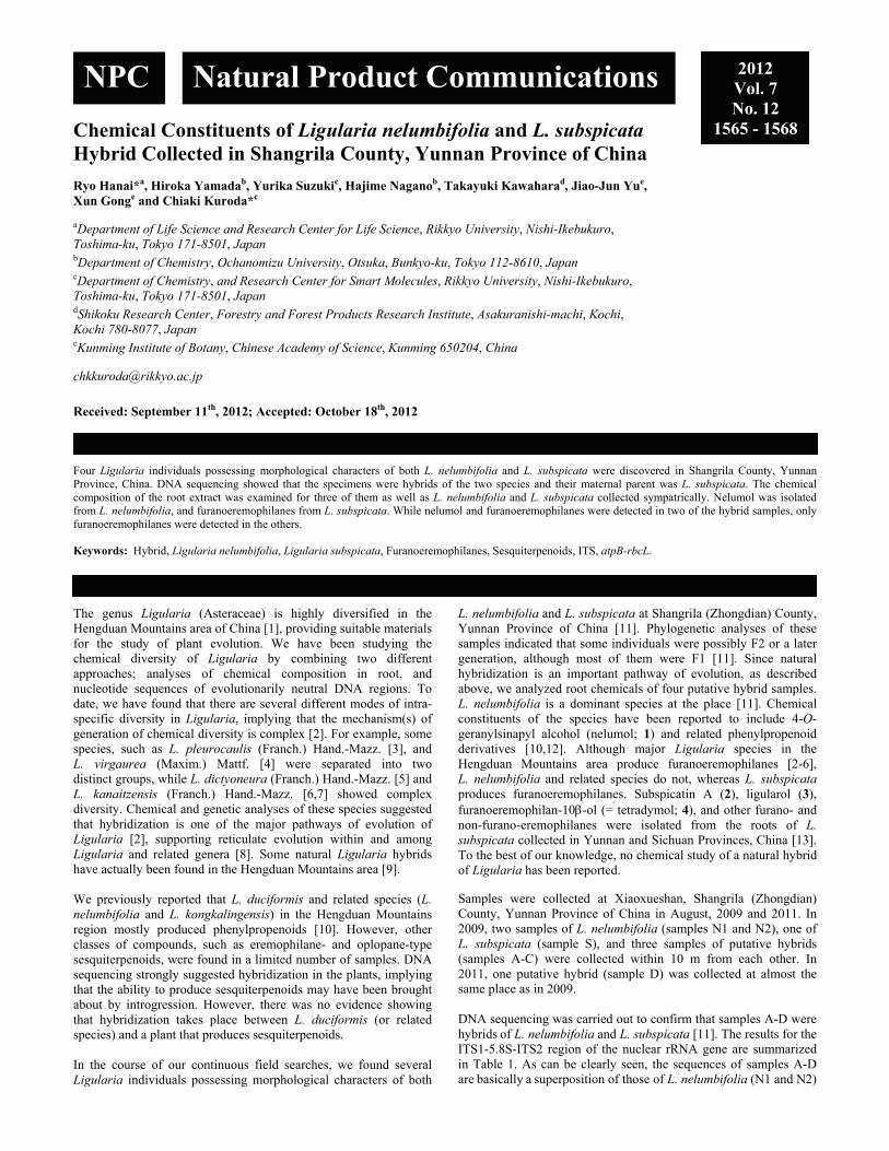
Chemical Constituents of Ligularia nelumbifolia and L. subspicata Hybrid Collected in Shangrila County, Yunnan Province of China Ryo Hanai*a, Hiroka Yamadab, Yurika Suzukic, Hajime Naganob, Takayuki Kawaharad, Jiao-Jun Yue, Xun Gonge and Chiaki Kuroda*c aDepartment of Life Science and Research Center for Life Science, Rikkyo University, Nishi-Ikebukuro, Toshima-ku, Tokyo 171-8501, Japan
bDepartment of Chemistry, Ochanomizu University, Otsuka, Bunkyo-ku, Tokyo 112-8610, Japan
cDepartment of Chemistry, and Research Center for Smart Molecules, Rikkyo University, Nishi-Ikebukuro, Toshima-ku, Tokyo 171-8501, Japan
dShikoku Research Center, Forestry and Forest Products Research Institute, Asakuranishi-machi, Kochi, Kochi 780-8077, Japan
eKunming Institute of Botany, Chinese Academy of Science, Kunming 650204, China [email protected]
Received: September 11th, 2012; Accepted: October 18th, 2012
Four Ligularia individuals possessing morphological characters of both L. nelumbifolia and L. subspicata were discovered in Shangrila County, Yunnan Province, China. DNA sequencing showed that the specimens were hybrids of the two species and their maternal parent was L. subspicata. The chemical composition of the root extract was examined for three of them as well as L. nelumbifolia and L. subspicata collected sympatrically. Nelumol was isolated from L. nelumbifolia, and furanoeremophilanes from L. subspicata. While nelumol and furanoeremophilanes were detected in two of the hybrid samples, only furanoeremophilanes were detected in the others. Keywords: Hybrid, Ligularia nelumbifolia, Ligularia subspicata, Furanoeremophilanes, Sesquiterpenoids, ITS, atpB-rbcL. The genus Ligularia (Asteraceae) is highly diversified in the Hengduan Mountains area of China [1], providing suitable materials for the study of plant evolution. We have been studying the chemical diversity of Ligularia by combining two different approaches; analyses of chemical composition in root, and nucleotide sequences of evolutionarily neutral DNA regions. To date, we have found that there are several different modes of intra-specific diversity in Ligularia, implying that the mechanism(s) of generation of chemical diversity is complex [2]. For example, some species, such as L. pleurocaulis (Franch.) Hand.-Mazz. [3], and L. virgaurea (Maxim.) Mattf. [4] were separated into two distinct groups, while L. dictyoneura (Franch.) Hand.-Mazz. [5] and L. kanaitzensis (Franch.) Hand.-Mazz. [6,7] showed complex diversity. Chemical and genetic analyses of these species suggested that hybridization is one of the major pathways of evolution of Ligularia [2], supporting reticulate evolution within and among Ligularia and related genera [8]. Some natural Ligularia hybrids have actually been found in the Hengduan Mountains area [9]. We previously reported that L. duciformis and related species (L. nelumbifolia and L. kongkalingensis) in the Hengduan Mountains region mostly produced phenylpropenoids [10]. However, other classes of compounds, such as eremophilane- and oplopane-type sesquiterpenoids, were found in a limited number of samples. DNA sequencing strongly suggested hybridization in the plants, implying that the ability to produce sesquiterpenoids may have been brought about by introgression. However, there was no evidence showing that hybridization takes place between L. duciformis (or related species) and a plant that produces sesquiterpenoids. In the course of our continuous field searches, we found several Ligularia individuals possessing morphological characters of both
L. nelumbifolia and L. subspicata at Shangrila (Zhongdian) County, Yunnan Province of China [11]. Phylogenetic analyses of these samples indicated that some individuals were possibly F2 or a later generation, although most of them were F1 [11]. Since natural hybridization is an important pathway of evolution, as described above, we analyzed root chemicals of four putative hybrid samples. L. nelumbifolia is a dominant species at the place [11]. Chemical constituents of the species have been reported to include 4-O-geranylsinapyl alcohol (nelumol; 1) and related phenylpropenoid derivatives [10,12]. Although major Ligularia species in the Hengduan Mountains area produce furanoeremophilanes [2-6], L. nelumbifolia and related species do not, whereas L. subspicata produces furanoeremophilanes. Subspicatin A (2), ligularol (3), furanoeremophilan-10-ol (= tetradymol; 4), and other furano- and non-furano-eremophilanes were isolated from the roots of L. subspicata collected in Yunnan and Sichuan Provinces, China [13]. To the best of our knowledge, no chemical study of a natural hybrid of Ligularia has been reported. Samples were collected at Xiaoxueshan, Shangrila (Zhongdian) County, Yunnan Province of China in August, 2009 and 2011. In 2009, two samples of L. nelumbifolia (samples N1 and N2), one of L. subspicata (sample S), and three samples of putative hybrids (samples A-C) were collected within 10 m from each other. In 2011, one putative hybrid (sample D) was collected at almost the same place as in 2009. DNA sequencing was carried out to confirm that samples A-D were hybrids of L. nelumbifolia and L. subspicata [11]. The results for the ITS1-5.8S-ITS2 region of the nuclear rRNA gene are summarized in Table 1. As can be clearly seen, the sequences of samples A-D are basically a superposition of those of L. nelumbifolia (N1 and N2)
NPC Natural Product Communications 2012 Vol. 7 No. 12
1565 - 1568

1566 Natural Product Communications Vol. 7 (12) 2012 Hanai et al.
Table 1: ITS1-5.8S-ITS2 sequences of L. nelumbifolia, L. subspicata, and putative hybridsa.
no. Species
ITS1 5.8S ITS2
1 1 1 1 1 1 1 2 2 2 2 2 2 1 1 1 1 1 1 1 1 1 2
1 4 5 6 7 8 0 2 2 2 3 5 8 1 1 2 2 2 4 3 1 2 3 4 7 0 0 1 4 7 8 9 9 0
3 4 4 8 7 3 8 5 6 7 2 4 4 7 8 1 2 4 0 3 3 5 1 7 3 6 6 0 4 6 9 4 6 1 9 3
N1 L. nelumbifolia T T Y C R R A C T A T C C G T G C C T T Y C C C A C G T C GA C R M Y T
N2 L. nelumbifolia T T Y C T R A C T A T C C G T G C C T T C C C C A C G T C GA C R M Y T
S L. subspicata A A C Y T G G T C C C A T C G T - T C C C Y T Y G T T C YGG T G C C C
A Hybrid W W Y Y T R R Y Y M Y M Y S K K b Y Y Y C Y Y C R Y KY Y R R Y R M YY
B Hybrid W W Y C T R R Y Y M Y M Y S K K b Y Y Y C C Y C R Y KY C R R Y R M YY
C Hybrid W W Y Y T R R Y Y M Y M Y S K K b Y Y Y C C Y C R Y KY YG R Y R M YY
D Hybrid W W Y Y T R R Y Y M Y M Y S K K b Y Y Y C C Y C R Y KY YG R Y R M YY
a Only the differences among the samples are shown. The base numbering is for L. nelumbifolia. K=G+T; M=A+C; R=A+G; S=G+C; W=A+T; Y=C+T. b Because of the presence of an indel, the sequence in the upstream part of ITS1 and the downstream part were determined separately only for either strand. The bases shown in Table 1 are as read from upstream and downstream toward the 222nd base position.
and L. subspicata (S), showing that samples A-D were very likely to be hybrids between the two species. In order to determine the maternal parent of samples A-D, sequences of rpl32-trnL and atpB-rbcL intergenic regions of the plastid genome were determined [11,14]. The rpl32-trnL sequences of the present L. nelumbifolia and L. subspicata samples were exactly the same as determined previously (database accession JF767242 for L. nelumbifolia and JF767237 for L. subspicata) [11]. Samples A-D had the L. subspicata sequence. The atpB-rbcL sequence was A-A9, 344G for the L. nelumbifolia samples (N1 and N2) and G-A9, 409T for the L. subspicata sample (S) under our nomenclature [10,13,14]. The sequence was G-A9, 409T for samples A-D. Thus, the maternal parent was deduced to be L. subspicata. For a rough examination of the composition of root chemicals in each sample, a small amount of fresh root was extracted with EtOH and compounds therein were analyzed by Ehrlich’s test on TLC [15], which has been used as a powerful method to detect furan compounds in our previous works [3-7, 13, 14]. L. subspicata (sample S) showed many Ehrlich-positive spots, showing that the sample was furanoeremophilane-producing type [13]. All hybrid samples were also Ehrlich-positive. In contrast, two L. nelumbifolia samples were negative to the test, suggesting the absence of furanoeremophilanes. Isolation and structure determination of major constituents in each sample was carried out using air-dried roots. Each sample was extracted with EtOH at room temperature and the extract was submitted to silica gel column chromatography. From L. subspicata (sample S), three furanoeremophilanes, subspicatin A (2) [13], ligularol (3) [16], and 6-ethoxyfuranoeremophilan-10-ol (5) [6], were isolated. Compound 5 may be an artifact generated from the corresponding diol during EtOH extraction [6]. From the EtOH extract of L. nelumbifolia (samples N1 and N2), nelumol (1) [17] was obtained as the major component. Furanoeremophilanes were found in the extracts of all four hybrid samples. The EtOH extract of sample B yielded subspicatin A (2) and tetradymol (4) [18], together with nelumol (1). In contrast, sample C yielded only furanoeremophilanes, i.e., subspicatin A (2) and ligularol (3). The chemical composition of sample D was very similar to that of sample C, yielding subspicatin A (2) and ligularol (3). Although isolation of chemical components from sample A was attempted, this was not successful (see experimental section). The chemical composition of sample A was deduced by TLC. Ligularol
(3) was detected by Ehrlich’s test as a pink spot at Rf 0.63 (n-hexane-EtOAc 7:3). Nelumol (1) was also detected at Rf 0.15 when visualized by UV and phosphomolybdic acid. The results of the chemical analysis are summarized in Table 2. Table 2: Compounds in L. nelumbifolia, L. subspicata, and putative hybridsa
Sample (species) 1 2 3 4 5N1 (L. nelumbifolia) + N2 (L. nelumbifolia) +
S (L. subspicata) + + + A (hybrid) (+) (+) B (hybrid) + + + C (hybrid) + + D (hybrid) + +
a +, isolated; (+), detected on TLC It is obvious that the ability to produce furanoeremophilanes originated from the L. subspicata parent, because L. nelumbifolia at this place did not produce them, and because subspicatin A, found in hybrid samples B, C, and D, was a compound characteristic of L. subspicata. In contrast, nelumol (1), the major component of L. nelumbifolia, was detected only in samples A and B, and the compound or a related phenylpropenoid was not detected in the extracts of samples C and D. These data show that samples A and B produced root chemicals of both parents, while the samples C and D produced mainly L. subspicata components. Such chemical variety among offspring had been reported previously [19]. At any rate, the present results show that hybridization between L. nelumbifolia and an eremophilane-producing Ligularia is possible and that offspring can produce eremophilanes. The composition of furanoeremophilanes in sample B was noteworthy; namely, tetradymol (4) was included as a major component in the sample, but was absent in the other three hybrid samples. Interestingly, tetradymol (4) was not detected in the L. subspicata sample collected sympatrically, although the compound had been isolated as a minor component of the species collected at a different place [13]. We previously proposed a hypothesis that the production of furanoeremophilanes was ecologically advantageous [2]. Among a number of furanoeremophilane-producing Ligularia plants, tetradymol-producing plants were ecologically more predominant [7]. If a furanoeremophilane-producing hybrid can backcross with L. nelumbifolia (or related species), the advantage would select furanoeremophilane-producing offspring, thereby introgressing the ability to produce furanoeremophilanes into L. nelumbifolia. Experimental
General: NMR, JEOL AL 400 or JEOL ECX-400 spectrometer (400 MHz for 1H; 100 MHz for 13C); IR, JASCO FT/IR-230 spectrometer; Mass spectra (MS) (EI+), JEOL JMS-700 MStation. Column chromatography was carried out on silica gel (either Kanto silica gel 60 N (spherical neutral) or Wakogel C-200 or C-300). Analytical TLC was carried out on Merck Kieselgel 60 F254 (layer thickness 0.2 or 0.25 mm) using either Ehrlich’s reagent (p-dimethylaminobenzaldehyde and HCl) [15] or p-anisaldehyde/

Chemical constituents of Ligularia nelumbifolia and L. subspicata hybrid Natural Product Communications Vol. 7 (12) 2012 1567
AcOH/H2SO4 as visualizing agents. HPLC was carried out on a Shimadzu LC-20AT pump, SPD-20A Prominence UV/VIS detector, Kanto Mightysil Si60 (10 × 250 mm) ODS column. Purification of DNA from dried leaves, amplification of the ITS1-5.8S-ITS2 and the atpB-rbcL intergenic regions by polymerase chain reaction, and DNA sequencing of the regions were carried out as described earlier [13]. Amplification and sequencing of the rpl32-trnL intergenic region was carried out with rpL32-F and trnL(UAG) primers [20]. Plant materials: Samples were collected at Xiaoxueshan (Mt. Xiaoxue), Shangrila County, Yunnan Province of China. Each sample was identified by XG (author). Voucher specimens were deposited in the Herbarium of Kunming Institute of Botany (No. 2009-07 (sample N1, L. nelumbifolia), 2009-12 (sample N2, L. nelumbifolia), 2009-08 (sample S, L. subspicata), 2009-09 (hybrid A), 2009-10 (hybrid B), 2009-11 (hybrid C), and 2011-113 (hybrid D)). Extraction and purification: Dried roots of sample S (L. subspicata) (4.6 g) was extracted with EtOH at room temperature and the extract concentrated under reduced pressure to give an oily product (113 mg). Part of this (45.6 mg) was submitted to CC (SiO2 3 g; eluent: n-hexane-EtOAc 20:1 to 5:1) to give subspicatin A (2) (6.1 mg, 13%) and ligularol (3) (7.3 mg, 16%). From less polar fractions, 6-ethoxyfuranoeremophilan-10-ol (5) (1.5 mg, 3.3%, Rf 0.80) was isolated after further chromatography (SiO2 1 g; eluent: n-hexane-EtOAc 100:1). Dried root samples of N1 (L. nelumbifolia; 39.1 g) and N2 (L. nelumbifolia; 21.3 g) were extracted and the extracts concentrated as described above to give oily products (251 mg and 826 mg, respectively). Part of these (122 mg and 102 mg) were submitted to
CC (SiO2 10 g; eluent: n-hexane-EtOAc 20:1 to 5:1) to give nelumol (1) (12.1 mg, 9.9%; and 4.7 mg, 4.7%, respectively). Dried roots of sample A (hybrid) (3.1 g) was also extracted with EtOH at room temperature and the extract concentrated under reduced pressure to give an oily product (99 mg). Isolation of chemical components was attempted using silica gel CC, but was not successful. Although TLC of the extract showed two spots (see text), isolation of the products, presumably ligularol (3) and nelumol (1), was not successful due to the paucity of the component and severe contamination by some other components. Dried roots of sample B (hybrid) (12.6 g) was extracted with EtOH at room temperature and the extract concentrated under reduced pressure to give an oily product (257 mg). Part of this (166 mg) was submitted to CC (SiO2 15 g; eluent: n-hexane-EtOAc 20:1 to 1:1) to give tetradymol (4) (11.4 mg, 6.9%), subspicatin A (2) (9.9 mg, 6.0%), and nelumol (1) (4.2 mg, 2.5%). Dried root samples of C and D (hybrids) (25.5 g, 12.3 g, respectively) were separately extracted and the extracts concentrated as described above to give oily products (582 mg and 1050 mg, respectively). Part of the C extract (182 mg) was submitted to CC (SiO2 10 g; eluent: n-hexane-EtOAc 20:1 to 10:1) to give ligularol (3) (19.4 mg, 11%) and subspicatin A (2) (5.1 mg, 2.8%). The D extract was submitted to CC (SiO2 20 g; eluent: n-hexane-EtOAc 0:1 to 1:0) to give ligularol (3) (2.0 mg, 0.2%) and subspicatin A (2) (3.3 mg, 0.3%). Acknowledgments - We thank Mrs Guowen Hu of Kunming Institute of Botany for research coordination. This work was partly supported by a Grant-in-Aid for Scientific Research from JSPS (No. 21404009).
References
[1] (a) Liu SW. (1989) In Flora Reipublicae Popularis Sinicae, Yu B, Zeng GF. (Eds). Science Press (Beijing), Vol. 77, pp 4-115; (b) Liu S, Illarionova ID (2011). In Flora of China, Wu ZY, Raven PH, Hong DY. (Eds). Science Press (Beijing) and Missouri Botanical Garden Press (St. Louis), Vol. 20-21, pp 376-415.
[2] Kuroda C, Hanai R, Nagano H, Tori M, Gong X. (2012) Diversity of furanoeremophilanes in major Ligularia species in the Hengduan Mountains. Natural Product Communications, 7, 539-548.
[3] Nagano H, Iwazaki Y, Gong X, Shen Y, Kuroda C, Hanai R. (2006) Chemical and genetic differentiation of Ligularia pleurocaulis in northwestern Yunnan and southwestern Sichuan Provinces of China. Bulletin of the Chemical Society of Japan, 79, 300-304.
[4] Tori M, Honda K, Nakamizo H, Okamoto Y, Sakaoku M, Takaoka S, Gong X, Shen Y, Kuroda C, Hanai R. (2006) Chemical constituents of Ligularia virgaurea and its diversity in southwestern Sichuan of China. Tetrahedron, 62, 4988-4995.
[5] Nagano H, Iwazaki Y, Matsushima M, Sato M, Gong X, Shen Y, Hirota H, Kuroda C, Hanai R. (2007) High diversity of Ligularia dictyoneura in chemical composition and DNA sequence. Chemistry and Biodiversity, 4, 2874-2888.
[6] Tori M, Watanabe A, Matsuo S, Okamoto Y, Tachikawa K, Takaoka S, Gong X, Kuroda C, Hanai R. (2008) Diversity of Ligularia kanaitzensis in sesquiterpenoid composition and neutral DNA sequences. Tetrahedron, 64, 4486-4495.
[7] Shimizu A, Suzuki Y, Torihata A, Hanai R, Saito Y, Tori M, Gong X, Kuroda C. (2012) Complex diversity in Ligularia kanaitzensis. Natural Product Communications, 7, 431-434.
[8] (a) Liu JQ, Wang YJ, Wang AL, Ohba H, Abbott RJ. (2006) Radiation and diversification within the Ligularia-Cremanthodium-Parasenecio complex (Asteraceae) triggered by uplift on the Qinghai-Tibetan plateau. Molecular Phylogenetics and Evolution, 38, 31-49; (b) Riesberg LH, Carney SE. (1998) Plant hybridization. New Phytologist, 140, 599-624.
[9] (a) Pan Y, Shi S, Gong X, Kuroda C. (2008) A natural hybrid between Ligularia paradoxa and L. duciformis (Asteraceae, Senecioneae) from Yunnan, China. Annals of the Missouri Botanical Garden, 95, 487-494; (b) Zhou R, Gong X, Boufford D, Wu CI, Shi S. (2008) Testing a hypothesis of unidirectional hybridization in plants: Observations on Sonneratia, Bruguiera, and Ligularia. BMC Evolutionary Biology, 8, 149.
[10] Nagano H, Hanai R, Yamada H, Matsushima M, Miura Y, Hoya T, Ozawa M, Fujiwara M, Kodama H, Torihata A, Onuki H, Nezu Y, Kawai S, Yamazaki M, Hirota H, Saito Y, Tori M, Ohsaki A, Gong X, Kuroda C. (2012) Chemical and genetic study of Ligularia duciformis and related species in Sichuan and Yunnan Provinces of China. Chemistry and Biodiversity, 9, 789-805.
[11] Yu JJ, Kuroda C, Gong X. (2011) Natural hybridization and introgression in sympatric Ligularia species (Asteraceae, Senecioneae). Journal of Systematics and Evolution, 49, 438-448.
[12] (a) Peng HR, Jia ZJ, Yang L, Zhao Y. (1995) A new sesquiterpene from Ligularia nelumbifolia. Chinese Chemical Letters, 6, 583-584; (b) Zhao Y, Peng HR, Jia ZJ. (1995) Six sinapyl alcohol derivatives from Ligularia nelumbifolia. Chinese Chemical Letters, 6, 387-390; (c) Zhao Y, Jia Z, Yang L. (1994) Sinapyl alcohol derivatives and other constituents from Ligularia nelumbifolia. Phytochemistry, 37, 1149-1152; (d) Zhao Y, Jia ZJ, Yang L, Wang JG. (1993) Sinapyl alcohol derivatives and other constituents from the roots of Ligularia nelumbifolia. Chinese Chemical Letters, 4, 895-898; (e) Zhao Y, Hao X, Lu W, Cai J, Yu H, Sevénet T, Guéritte F. (2002) Synthesis of two cytotoxic sinapyl alcohol derivatives and isolation of four new related compounds from Ligularia nelumbifolia. Journal of Natural Products, 65, 902-908.

1568 Natural Product Communications Vol. 7 (12) 2012 Hanai et al.
[13] (a) Tori M, Okamoto Y, Tachikawa K, Mihara K, Watanabe A, Sakaoku M, Takaoka S, Tanaka M, Gong X, Kuroda C, Hattori M, Hanai R. (2008) Isolation of new eremophilane-type sesquiterpenoids, subspicatins A-D and subspicatolide from Ligularia subspicata, and chemical and genetic diversity of the species. Tetrahedron, 64, 9136-9142; (b) Saito Y, Hattori M, Iwamoto Y, Takashima Y, Mihara K, Sasaki Y, Fujiwara M, Sakaoku M, Shimizu A, Chao X, Kuroda C, Gong X, Hanai R, Tori M. (2011) Overlapping chemical and genetic diversity in Ligularia lamarum and Ligularia subspicata. Isolation of ten new eremophilanes and a new seco-bakkane compound. Tetrahedron, 67, 2220-2231.
[14] Hanai R, Gong X, Tori M, Kondo S, Otose K, Okamoto Y, Nishihama T, Murota A, Shen Y, Wu S, Kuroda C. (2005) Chemical and genetic study of Ligularia tongolensis, Ligularia cymbulifera, and Ligularia atroviolacea in the Hengduan Mountains of China. Bulletin of the Chemical Society of Japan, 78, 1302-1308.
[15] (a) Kuroda C, Ueshino T, Nagano H. (2004) Ehrlich’s reaction of furanoeremophilanes. Bulletin of the Chemical Society of Japan, 77, 1737-1740; (b) Kuroda C, Nishio E. (2007) Substituent effect in color of Ehrlich’s test of tetrahydrobenzofuran, Natural Product Communications, 2, 581-585.
[16] Yamakawa K, Satoh T. (1979) Studies on terpenoids and related alicyclic compounds. XVIII. Stereoselective synthesis of (±)-furanofukinol and (±)-petasalbin. Chemical and Pharmaceutical Bulletin, 27, 1747-1752.
[17] Bohlmann F, Grenz M, Gupta RK, Dhar AK, Ahmed M, King RM, Robinson H. (1980) Eudesmane derivatives from Verbesina species. Phytochemistry, 19, 2391-2397.
[18] Jennings PW, Reeder SK, Hurley JC, Caughlan CN, Smith GD (1974) Isolation and structure determination of one of the toxic constituents from Tetradymia glabrata. Journal of Organic Chemistry, 39, 3392-3398.
[19] Harborne JB, Turner BL. (1984) Plant Chemosystematics. Academic Press, London. [20] Shaw J, Lickey EB, Schilling EE, Small RL. (2007) Comparison of whole chloroplast genome sequences to choose noncoding regions for
phylogenetic studies in angiosperms: the tortoise and the hare III. American Journal of Botany, 94, 275-288.

A Practical, Enantiospecific Synthesis of (S)-trans-γ-Monocyclofarnesol Stefano Serra C.N.R., Istituto di Chimica del Riconoscimento Molecolare, Via Mancinelli, 7 I-20131 Milano, Italy
[email protected]; [email protected]
Received: August 28th, 2012; Accepted: October 3rd, 2012
An expedient and concise synthesis of (S)-trans-γ-monocyclofarnesol is here described. The aforementioned sesquiterpene was prepared starting from enantioenriched (S)-γ-dihydroionone, which was in turn obtained from racemic -ionone through the combined use of two previously developed processes. Key steps of the presented synthesis are the stereoselective Horner-Wadsworth-Emmons reaction between triethyl phosphonoacetate and γ-dihydroionone and the effective fractional crystallization of the γ-monocyclofarnesol-3,5-dinitrobenzoate esters. By these means the target compound was obtained in good yield and with very high stereoisomeric purity. Keywords: Sesquiterpenes, Stereoselective synthesis, Enzyme-mediated resolution, Horner-Wadsworth-Emmons reaction, Fractional crystallization.
The general monocyclofarnesyl framework 1 (Figure 1) occurs in a large number of terpenic natural products.
Figure 1: The (S)-trans-γ-monocyclofarnesyl framework 1 and some relevant natural products featuring this structure. The simplest compound belonging to this class is trans-γ-monocyclofarnesol 1a (R=OH), which was first identified [1] among the minor metabolites formed by the mycelia of the fungus Helminthosporium siccans in the biosynthetic process that affords the antibiotic siccanin. More than twenty years after, the same alcohol was extracted from the liverwort Diplophyllum serrulatum [2], confirming definitively its natural occurrence, albeit from two natural sources only. Beside the rarity of 1a, a large number of its homologues (e.g. 1b-f) were isolated [3-7], and the biological activity of some of these
compounds [6-10] greatly increased the synthetic interest for the γ-monocyclofarnesyl structure. It is noteworthy that all the aforementioned products are usually present in their corresponding natural sources as a single stereoisomeric form. More specifically, they show an enantiocentre of (S) configuration and a (E)-trisubstituted double bond. Since their biological activity is strictly related to their stereochemistry, chemical syntheses providing a high level of stereocontrol were particularly sought-after. For such purposes, (S)-trans-γ-monocyclofarnesol is the most suitable chiral building block, having all the steric requirements already present in the molecular scaffold and allowing the preparation of higher homologues by exploiting the chemical reactivity of the hydroxy group. Such a kind of approach was successfully employed for the synthesis of the alkaloid phenazinomycin 1e [9]. In this process the key intermediate 1a was prepared by means of an elegant, highly stereospecific, synthesis. Unfortunately, this alcohol was obtained through a very lengthy path, thus hampering the general use of this approach. Two different syntheses [11,12] afforded either 1a or the corresponding alkyl bromide starting from the enantiopure (S)-γ-dihydroionone, which was in turn obtained by degradation of the natural triterpenes ambreine and sclareol, respectively. In these procedures the (E)-trisubstituted double bond was built up stereoselectively, although close to a minority amount of the (Z)-isomer. Recently, we have described [13] a very useful chemo-enzymatic synthesis of the enantioenriched (4R,6S)-acetoxy-γ-ionone (2) starting from racemic ionone alpha (Figure 2). The following palladium-catalysed reductive elimination of the acetate functional group allows the preparation of (S)-γ-ionone (3), both in high enantiomeric and stereoisomeric purity. In addition, we have previously demonstrated that enantiopure γ-ionone isomers could be selectively reduced to the corresponding γ-dihydroionone derivatives by means of Bu3SnH and catalytic (Ph3P)2PdCl2 [14]. The combination of these findings offers a reliable synthetic access to (S)-γ-dihydroionone (4), not strictly depending upon natural sources.
NPC Natural Product Communications 2012 Vol. 7 No. 12
1569 - 1572
(S)-1
.
R
O
OH1b Trixagol
1d Luffarin W
N N
O
1e Phenazinomycin
1c Dehydroambliol-AO
O
1f Agelasine E
N
NN
N
NH2
Cl-
R= OH
R= R=
1a MonocyclofarnesolR=
R=R=

1570 Natural Product Communications Vol. 7 (12) 2012 Serra
With this chiral building block in hand, we looked for an effective and stereoselective C-2 homologation process.
Figure 2: Synthesis of (S)-trans-γ-monocyclofarnesol (+)-1a starting from (4R,6S)-acetoxy-γ-ionone. Reagents and conditions: a) Et3N, HCO2H, (Ph3P)2PdCl2 cat., THF reflux; b) Bu3SnH, NH4Cl, H2O, (Ph3P)2PdCl2 cat., THF at rt; c) (EtO)2POCH2CO2Et, NaH, THF, from 0° to 50° C; d). DIBAH in toluene, THF, 10° C; e) 3,5-dinitrobenzoyl chloride, pyridine, DMAP cat., CH2Cl2; f) three crystallizations from n-hexane; g) NaOH, MeOH, rt. It was previously demonstrated [11] that Horner-Wadsworth-Emmons reaction between methyl diethylphosphonoacetate and γ-dihydroionone affords a 63:27 mixture of the (E)- and (Z)-γ-monocyclofarnesyl methyl esters, respectively, which could be reduced to the corresponding γ-monocyclofarnesol isomers. Since neither the latter alcohols nor the starting esters are separable by chromatography, the fractional distillation of the isomeric mixture of the esters was employed in the stereoselective preparation of 1a. Otherwise, we found that the latter tricky separation can be avoided by combining an improvement in the condensation stereoselectivity with the direct purification of the obtained E/Z isomeric mixture of alcohols 1a, through fractional crystallization of their 3,5-dinitrobenzoate esters. More specifically, we observed a considerable increasing in the E/Z selectivity when the Horner-Wadsworth-Emmons reaction was performed both by using an excess of triethylphosphonoacetate instead of methyl diethylphosphonoacetate and by a carefully control of the reaction temperature (see experimental). According to our protocol, (+)-4 was converted into an 83:17 mixture of the γ-monocyclofarnesyl ethyl esters (E)-5 and (Z)-5, respectively, in 94% overall yield. The latter isomeric mixture was then reduced with DIBAH to give the corresponding allylic alcohols, which were directly treated with 3,5-dinitrobenzoyl chloride and pyridine. Thus, the obtained esters mixture was crystallized three times from n-hexane. The collected crystals were obtained in 58% overall yield and turned out to be free of the (Z)-isomer. Indeed, neither the NMR analysis of (+)-6 nor the GC analysis of the alcohol 1a obtained from the aforementioned dinitrobenzoate ester revealed the presence of the unwanted isomer.
Furthermore, the purity of the γ-isomer increased from 96% to 98%, thus indicating that the fractional crystallization is in many respects a very effective purification procedure. As a final point, the basic hydrolysis of the ester (+)-6 afforded pure alcohol (+)-1a whose analytical data agree both with those reported for the natural product and with those described for the synthetic material. Noteworthy is that the reported overall procedure was tested on a small scale (starting from a few hundred milligrams of γ-dihydroionone) and on a multi-gram scale, and analogous results were obtained. This flexibility corroborates the reliability of the process. The exploitation of the obtained isomerically pure (S)-trans-γ-monocyclofarnesol for the synthesis of further natural products is ongoing and the results will be presented in due course. Experimental
General: All moisture-sensitive reactions were carried out under a static atmosphere of nitrogen. All reagents were of commercial quality. TLC: Merck silica gel 60 F254 plates. Column chromatography: silica gel. GC-MS analyses: HP-6890 gas chromatograph equipped with a 5973 mass detector, using a HP-5MS column (30 m 0.25 mm, 0.25 m firm thickness; Hewlett Packard) with the following temp. program: 60° (1 min) 6°/min 150° (1 min) 12°/min 280° (5 min); carrier gas, He; constant flow 1mL/min; split ratio, 1/30; tR given in min: tR(1a) 21.79, tR(4) 16.00, tR(E-5) 22.71, tR(Z-5) 21.93; mass spectra: m/z (rel.%). Mass spectrum of compound (+)-6 was recorded on a Bruker ESQUIRE 3000 PLUS spectrometer (ESI detector). Optical rotations: Jasco-DIP-181 digital polarimeter, measured at 20°C. 1H and 13C Spectra: CDCl3 solns. at rt; Bruker-AC-400 spectrometer at 400 and 100 MHz, respectively; chemical shifts in ppm rel to internal SiMe4 (=0 ppm), J values in Hz. (S)-γ-Dihydroionone (+)-4: Compound (+)-4 (98% chemical purity; 96% ee; isomers ratio ::β = 96:3.5:0.5) was obtained by reduction of (S)-γ-ionone, in turn prepared from (4R,6S)-acetoxy-γ-ionone, according to our previously reported procedures [13, 14]. []D = +16.5° (c = 1, CHCl3); Lit. [14]: []D = +19.4° (c = 1.2, CHCl3) 1H NMR, 13C NMR, MS in accordance with those previously reported [14]. (S)-Ethyl 5-(2,2-dimethyl-6-methylenecyclohexyl)-3-methylpent-2-enoate 5: Triethyl phosphonoacetate (7 g, 31.2 mmol) was added dropwise under nitrogen over a period of 1 h to a stirred suspension of NaH (60% in mineral oil; 1.25 g, 31.2 mmol) in dry THF (25 mL) at 0°C. To the resulting mixture was slowly added a solution of (+)-4 (2 g, 10.3 mmol) in dry THF (5 mL). When the addition was complete, the reaction was slowly heated at 50°C, stirring until the starting ketone was no longer detectable by TLC analysis (3 h). After cooling, the reaction was poured onto a mixture of crushed ice and a saturated solution of NH4Cl aq. (100 mL). The mixture was extracted with diethyl ether (2 × 100 mL). The organic phase was washed with brine, dried (Na2SO4), and concentrated under reduced pressure. The residue was chromatographed using n-hexane/diethyl ether (95:5–9:1) as eluent to afford pure 5 (2.55 g, 94% yield) as an E/Z mixture of isomers (E/Z ratio 5:1, by NMR analysis). (E)-5: 1H NMR (400 MHz, CDCl3): 0.84 (3H, s), 0.92 (3H, s), 1.18-1.27 (1H, m), 1.28 (3H, t, J = 7.2 Hz), 1.41-1.58 (4H, m), 1.58-1.69 (1H, m), 1.70 (1H, dd, J = 11.4, 3.3 Hz), 1.85-1.96 (1H, m), 1.97-2.16 (3H, m), 2.15 (3H, d, J = 1.3 Hz), 4.15 (2H, q, J = 7.2 Hz), 4.55 (1H, m), 4.78 (1H, br s), 5.65 (1H, br q, J = 1.3 Hz).
(+)-4(S)--dihydroionone96% ee; isomer 96%
CO2Et EtO2C
O
O
NO2
NO2
O
OAc
a
d, e, f
g
(+)-1a
racemic-ionone
O
b
c
(+)-2 (+)-3
(E)-5 (Z)-5 E/Z = 5:1
(+)-6E >99%, isomer 98%
O
94%
58%
95%

Synthesis of (S)-trans-γ-monocyclofarnesol Natural Product Communications Vol. 7 (12) 2012 1571
13C NMR (100 MHz, CDCl3): 14.3 (Me), 18.9 (Me), 23.6 (CH2), 24.4 (CH2), 26.2 (Me), 28.3 (Me), 32.4 (CH2), 34.9 (C), 36.2 (CH2), 39.6 (CH2), 53.7 (CH), 59.4 (CH2), 109.3 (CH2), 115.4 (CH), 148.9 (C), 160.6 (C), 166.9 (C). GC-MS (EI): m/z (%) = 264 [M+] (3), 249 (25), 219 (8), 203 (14), 191 (9), 176 (100), 161 (69), 141 (39), 128 (76), 121 (31), 109 (99), 95 (66), 81 (87), 69 (69), 55 (28). (Z)-5: 1H NMR (400 MHz, CDCl3): 0.84 (3H, s), 0.93 (3H, s), 1.18-2.52 (11H, m), 1.26 (3H, t, J = 7.2 Hz), 1.87 (3H, d, J = 1.3 Hz), 4.13 (2H, q, J = 7.2 Hz), 4.60 (1H, m), 4.78 (1H, br s), 5.63 (1H, br s). 13C NMR (100 MHz, CDCl3): 14.3 (Me), 23.6 (CH2), 25.1 (CH2), 25.3 (Me), 26.4 (Me), 28.3 (Me), 32.2 (CH2), 32.6 (CH2), 34.9 (C), 36.1 (CH2), 54.5 (CH), 59.3 (CH2), 109.2 (CH2), 115.9 (CH), 149.0 (C), 160.8 (C), 166.2 (C). GC-MS (EI): m/z (%) = 264 [M+] (3), 249 (23), 218 (7), 203 (10), 191 (7), 176 (76), 161 (47), 137 (78), 128 (100), 121 (30), 109 (72), 95 (64), 81 (82), 69 (58), 55 (27). (S)-(E)-5-(2,2-dimethyl-6-methylenecyclohexyl)-3-methylpent-2-enyl 3,5-dinitrobenzoate (+)-6: DIBAH (14.5 mL of 1.7 M solution in toluene) was added dropwise under nitrogen to a stirred solution of ester 5 (E/Z ratio 5:1, 2.64 g, 10 mmol) in dry THF (40 mL) at 10 °C. The reaction was stirred at 0°C for 1 h, then diluted with diethyl ether (100 mL) and quenched with a saturated solution of NH4Cl aq. (50 mL). The mixture was acidified with diluted HCl aq. and extracted with diethyl ether (2 × 100 mL). The combined organic phases were washed with brine, dried (Na2SO4) and concentrated under reduced pressure. The residue was dissolved in CH2Cl2 (10 mL) and treated with pyridine (2 mL), DMAP (15 mg, 0.1 mmol) and a solution of 3,5-dinitrobenzoyl chloride (3 g, 13 mmol) in CH2Cl2 (5 mL). After complete transformation of the starting alcohol the mixture was diluted with water (200 mL) and extracted with diethyl ether (2x150 mL). The combined organic phases were washed with aq. NaHCO3 (5% solution), brine and then dried (Na2SO4). Concentration at reduced pressure gave an oil that was purified by CC (n-hexane/Et2O 9:1) and crystallized 3 times from n-hexane. The ester (+)-6 (2.4 g, 58% yield) was obtained as colorless crystals showing 99% chemical purity, free of Z-isomer and showing a γ/α isomeric ratio of 98:2 (by NMR analysis). MP: 82-83ºC. [α]D: +10.5 (c 2.1, CHCl3).
1H NMR (400 MHz, CDCl3): 0.85 (3H, s), 0.92 (3H, s), 1.18-1.28 (1H, m), 1.42-1.58 (4H, m), 1.57-1.67 (1H, m), 1.71 (1H, dd, J = 11.4, 3.3 Hz), 1.80 (3H, s), 1.87 (1H, ddd, J = 14.3, 10.5, 6.1 Hz), 1.95-2.14 (3H, m), 4.55 (1H, m), 4.78 (1H, br s), 4.97 (2H, d, J = 7.2 Hz), 5.48 (1H, br t, J = 7.2 Hz), 9.16 (2H, d, J = 2.1 Hz), 9.21 (1H, t, J = 2.1 Hz). 13C NMR (100 MHz, CDCl3): 16.7 (Me), 23.6 (CH2), 24.3 (CH2), 26.3 (Me), 28.3 (Me), 32.4 (CH2), 34.9 (C), 36.2 (CH2), 38.1 (CH2), 53.6 (CH), 63.7 (CH2), 109.1 (CH2), 116.7 (CH), 122.2 (CH), 129.4 (CH), 134.3 (C), 145.1 (C), 148.6 (C), 149.0 (C), 162.5 (C). MS (ESI): m/z (positive ion) = 439 [M+ + Na]. (S)-Trans-γ-monocyclofarnesol = (S,E)-5-(2,2-dimethyl-6-methylenecyclohexyl)-3-methylpent-2-en-1-ol (+)-1a: The 3,5-dinitrobenzoate (+)-6 (0.5 g, 1.2 mmol) was dissolved in methanol (5 mL) and treated with NaOH (1 g, 25 mmol) in methanol (5 mL) stirring at rt until the starting ester was no longer detected by TLC analysis. The mixture was diluted with water (50 mL) and extracted with diethyl ether (2 x 50 mL). The organic phase was washed with brine, dried (Na2SO4) and concentrated in vacuo. The residue was roughly chromatographed (n-hexane/Et2O 9:1) to give (+)-1a (255 mg, 95% yield) as a colorless oil showing 97% chemical purity (by GC) and a γ/α isomeric ratio of 98:2 (by NMR analysis). [α]D: +17.2 (c 2.3, CHCl3). The recorded optical rotation value is slightly inferior to those previously reported: [α]D: +18.2 (c 0.3, CHCl3) [2]; [α]D: +21.1 (CHCl3) [11]. Since the (S)--ionone isomers usually show a higher optical rotation value and opposite sign than their corresponding γ-isomers, the amount of about 2% of the (S)--monocyclofarnesol might take into account the discrepancy observed. 1H NMR (400 MHz, CDCl3): 0.84 (3H, s), 0.92 (3H, s), 1.17-1.26 (1H, m), 1.36-1.64 (6H, m), 1.67 (3H, s), 1.69 (1H, dd, J = 11.4, 3.2 Hz), 1.78 (1H, ddd, J = 14.3, 10.8, 6.2 Hz), 1.93-2.12 (3H, m), 4.14 (2H, d, J = 6.9 Hz), 4.54 (1H, m), 4.76 (1H, br s), 5.40 (1H, tq, J = 6.9, 1.3 Hz). 13C NMR (100 MHz, CDCl3): 16.3 (Me), 23.6 (CH2), 24.5 (CH2), 26.2 (Me), 28.3 (Me), 32.4 (CH2), 34.8 (C), 36.2 (CH2), 38.0 (CH2), 53.7 (CH), 59.3 (CH2), 108.9 (CH2), 123.0 (CH), 140.3 (C), 149.1 (C). GC-MS (EI): m/z (%) = 222 [M+] (1), 207 (22), 204 (11), 189 (47), 177 (9), 161 (14), 148 (13), 133 (23), 121 (31), 109 (100), 95 (49), 81 (79), 69 (57), 55 (30). Supplementary data: Copies of the 1H NMR, 13C NMR and DEPT NMR spectra of the (S)-trans-γ-monocyclofarnesol 1a are available.
References
[1] Suzuki KT, Suzuki N, Nozoe S. (1971) Isolation and enzymic formation of trans-γ-monocyclofarnesol. Journal of the Chemical Society D: Chemical Communications, 527.
[2] Toyota M, Ooiso Y, Kusuyama T, Asakawa Y. (1994) Drimane-type sesquiterpenoids from the liverwort Diplophyllum serrulatum. Phytochemistry, 35, 1263-1265.
[3] De Pascual Teresa J, Caballero E, Caballero C, Medarde M, Barrero AF, Grande M. (1978) Trixagol, natural γ-cyclogeranyl-geraniol from Bellardia trixago (L.) All. Tetrahedron Letters, 19, 3491-3494.
[4] Walker RP, Faulkner DJ. (1981) Diterpenes from the sponge Dysidea amblia. Journal of Organic Chemistry, 46, 1098-1102. [5] Butler M, Capon R. (1992) The Luffarins (A-Z), novel terpenes from an Australian marine sponge, Luffariella geometrica. Australian Journal of
Chemistry, 45, 1705-1743. [6] Funayama S, Eda S, Komiyama K, Ohmura S, Tokunaga T. (1989) Structure of phenazinomycin, a novel antitumor antibiotic. Tetrahedron Letters,
30, 3151-3154. [7] Wu H, Nakamura H, Kobayashi J, Ohizumi Y, Hirata Y. (1984) Agelasine-E and -F, novel monocyclic diterpenoids with 9-methyladeninium unit
possessing inhibitory effects on Na,K-ATPase isolated from the Okinawan sea sponge Agelas nakamurai Hoshino. Tetrahedron Letters, 25, 3719-3722.
[8] Bakkestuen AK, Gundersen L-L. (2003) Synthesis of (+)-trixagol and its enantiomer, the terpenoid side chain of (−)-agelasine E. Tetrahedron, 59, 115-121.
[9] Kinoshita Y, Kitahara T. (1997) Total synthesis of phenazinomycin and its enantiomer via high-pressure reaction. Tetrahedron Letters, 38, 4993-4996.

1572 Natural Product Communications Vol. 7 (12) 2012 Serra
[10] Bakkestuen AK, Gundersen L-L, Petersen D, Utenova BT, Vik A. (2005) Synthesis and antimycobacterial activity of agelasine E and analogs. Organic & Biomolecular Chemistry, 3, 1025-1033.
[11] Schulte-Elte K. Muller BL, Ohloff G. (1978) Photosensitized oxygenation of (E)- and (Z)-(+)-γ-monocyclofarnesol. An access to (S)-(+)-ambra-aldehyde. Nouveau Journal de Chimie, 2, 427-430.
[12] Barrero AF, Alvarez-Manzaneda EJ, Chahboun R, Cuerva JM, Segovia A. (2000) Synthetic applications of the thermal rearrangement of ozonides: First enantiospecific synthesis of marine metabolite Luffarin W. Synlett, 1269-1272.
[13] Serra S, Fuganti C, Brenna E. (2006) Synthesis, olfactory evaluation, and determination of the absolute configuration of the 3,4-didehydroionone stereoisomers. Helvetica Chimica Acta, 89, 1110-1122.
[14] Fuganti C, Serra S, Zenoni A. (2000) Synthesis and olfactory evaluation of (+)- and (−)-gamma-ionone. Helvetica Chimica Acta, 83, 2761-2768.

ACAT Inhibitory Activity of Exudates from Calocedrus macrolepis var. formosana Yu-Hsin Hsieha,c, Kuan-Jung Chenb, Shih-Chang Chienf, Wen-Ling Cheng b, Jun-Hong Xiaob and Sheng-Yang Wanga,b,d,e,f,g,* aMolecular and Biological Agricultural Sciences Program, Taiwan International Graduate Program, National Chung-Hsing University and Academia Sinica, Taiwan
bDepartment of Forestry, National Chung-Hsing University, Taichung, Taiwan
cGraduate Institute of Biotechnology, National Chung-Hsing University, Taichung, Taiwan
dAgricultural Biotechnology Research Center, Academia Sinica, Taipei, Taiwan
eAgricultural Biotechnology Center, National Chung-Hsing University, Taichung, Taiwan fExperimental Forest Management Office, National Chung-Hsing University, Taichung, Taiwan
Received: May 28th, 2012; Accepted: October 17th, 2012
Cholesterol acyltransferase (ACAT) is an enzyme controlling cholesterol esterification in cells. Large amounts of cholesterol esters accumulate in macrophages and smooth muscle cells of blood vessel walls resulting in the initial stages of atherosclerosis. Thus, atherosclerosis might be inhibited through inhibition of the activity of ACAT. In the present study, we identified by spectral analysis and chromatographic quantification that ferruginol was the most abundant component of exudates secreted from the wounding site of Calocedrus macrolepis Kurz var. formosana. Results obtained from the cholesterol absorption assay revealed that ferruginol exhibited a significant inhibitory activity on cholesterol absorption in mice macrophages (RAW 264.7 cell). Based on the results from analyzing the ratio of cholesterol esterification, ferruginol dose-dependently suppressed cholesterol esterification and the IC50 value was 2.0 μg/mL. In conclusion, ferruginol revealed strong inhibitory activities that retarded the absorption and esterification of cholesterol in cells. Our finding indicates that ferruginol might possess a potential for development as a pharmaceutical product for preventing arteriosclerosis. Keywords: Ferruginol, Arteriosclerosis, Acyl-CoA, Cholesterol acyltransferase, Calocedrus macrolepis Kurz var. formosana. Calocedrus macrolepis Kurz var. formosana (Cupressaceae) is an endemic conifer in Taiwan. More than 100 compounds have been isolated from the plant, including monoterpenoids, diterpenoids, lignans and steroids [1-6]. The resin produced by plants, consisting mainly of susquiterpenoids and diterpenoids, is a vital defense system against insect and pathogen attacks [7]. In this study, we collected exudates from the bark surface of 20-year-old C. macrolepis and 7 diterpenoids, including ferruginol [8], trans-communic acid [9], isopimaric acid [10], isopimarol [11], 6-hydroxysugiol, sugiol, and secoabietane dialdehyde [5] were identified using chromatographic separation and spectroscopic analyses. Ferruginol (Figure 1) was the most abundant compound identified in the exudates, forming approximately 77%, w/w, of the total. ACAT is an enzyme which catalyzes the conversion of free cholesterol and fatty acyl CoA to a storage form of cholesterol, cholesteryl ester (CE) [12]. Atherosclerosis results from an excess accumulation of cholesterol and macrophages that deposit on the blood vessel wall and eventually narrow the artery [13]. In the present study, we utilized NBD-cholesterol, a fluorescent sterol analog, to mimic the absorption and esterification of native cholesterol in cultured cells [14]. RAW 264.7 cells pre-treated with ferruginol for 1 h significantly suppressed cholesterol absorption in a dose-dependent manner (Table 1). The inhibitory activity was 62.5% at a concentration of 20 g/mL and the IC50 value was 9.5 g/mL. Sandoz 58-035, a known inhibitor of cholesterol acyltransferase [15], showed 45.8% inhibition. Our data revealed that ferruginol has a higher potential
H
OH
Figure 1: The dominant compound, ferruginol, in the exudates secreted from Calocedrus macrolepis var. formosana.
Table 1: Inhibition effect of ferruginol on cholesterol absorption
RAW 264.7 cells were incubated in medium containing ferruginol at indicated dosages for 1 h and further treated with 1 g/mL NBD-cholesterol for 6 h. 10 g/mL Sandoz 58-035 (Sigma St. Louis MO) was used as a reference compound. The inhibitory effect was compound-treated group compared with vehicle control (DMSO).
for suppressing cholesterol absorption than Sandoz 58-035 and the working mechanism is worth further investigation. To decipher whether the inhibitory effect of cholesterol absorption by ferruginol is correlated with ACAT activity, we isolated the lipid contents from ferruginol treated cells and detected the levels of NDB-cholesterol and NBD-cholesterol ester, respectively, using high performance liquid chromatography (HPLC). Our data showed that ferruginol dose-dependently suppressed the level of NBD-cholesterol ester, which dramatically decreased from 25.3% to 1.4%
NPC Natural Product Communications 2012 Vol. 7 No. 12
1573 - 1574

1574 Natural Product Communications Vol. 7 (12) 2012 Hsieh et al.
at a concentration of 10 g/mL (Table 2). By comparison with control, cells treated with 10 g/mL ferruginol suppressed 94.4% of cholesterol esterification. From these findings we suggest that ferruginol efficiently inhibits the ACAT activity of converting free cholesterol to CE by macrophages. In conclusion, ferruginol revealed a strong retarding activity for the absorption and esterification of cholesterol in cells. Our finding indicates that ferruginol might possess the potential to be developed as a pharmaceutical product for preventing arteriosclerosis.
Table 2: Inhibitory activity of cholesterol esterification by ferruginol.
RAW 264.7 cells were incubated in medium containing ferruginol at indicated dosages for 1 h and further treated with 1 g/mL NBD-cholesterol for 6 h. Isolated lipid contents and detected the levels of NBD-CE and NBD-cholesterol respectively using HPLC. The inhibitory effect was ferruginol-treated group compared with control.
Experimental
Materials: The exudates secreted from the wounding site of 20-year-old C. macrolepis were dissolved in ethyl acetate and separated by HPLC using a Luna silica column (250 × 10 mm; 5 m, Phenomenex, Torrance, CA) eluted with n-hex/EtOAc (n-hex/EtOAc = 88/12, flow rate = 2.5 mL/min). Compounds 1 to 7 were obtained at the retention times of 10.8, 16.0, 19.3, 21.6, 24.6, 33.5, and 38.3 min, respectively. The structures of the compounds were elucidated using spectroscopic analysis. The amount of the major compound (ferruginol) in exudates was further analyzed by
the same HPLC system. The peak area of ferruginol in the chromatogram of the exudates (with known loading concentration) was then defined and its content in the exudates calculated based on the quantity calibrated from the standard calibration curve. Cell culture: RAW264.7 cells were maintained in Dulbecco’s modified Eagle’s medium supplemented with 100 units/mL penicillin, 100 μg/mL streptomycin and 10% fetal bovine serum (GIBCO Carlsbad, CA) at 37°C in a 5% CO2 humidified incubator. Cholesterol absorption assay: RAW 264.7 cells were plated in 24-well culture plates at a density of 1 106 cells/well and allowed to recover for 24 h. Cells were incubated in medium containing ferruginol at various concentrations (5, 10 and 20 μg/mL) for 1 h and further treated with 1 g/mL NBD-cholesterol for 6 h. 10 g/mL Sandoz 58-035 (Sigma St. Louis MO) was used as a reference compound. After incubation, the cells were washed thrice with phosphate buffered saline (PBS). Fluorescence was detected using a Chameleon V Multilabel Microplate Reader (Hidex) with 485 nm excitation and 535 nm emission filters [16]. Cholesterol esterification assay: RAW 264.7 cells were plated in 6 cm culture dishes at a density of 5 106 cells/well and allowed to recover for 24 h. Cells were incubated in medium containing ferruginol at various concentrations (1, 2.5, 5 and 10 μg/mL) for 1 h and further treated with 1 g/mL NBD-cholesterol for 6 h. Cells were collected and the lipid contents isolated using n-hex/2-propanol (3:2) solution. The ratio of NDB-cholesterol and NBD-cholesterol ester was determined from the absorbance at 443 nm using HPLC (Luna Silica 250 mm × 10 mm, 5 m).
References
[1] Fang JM, Hsu KC, Cheng YS. (1989) Terpenoids from leaves of Calocedrus formosana. Phytochemistry, 28, 1173-1175. [2] Fang JM, Hsu KC, Cheng YS. (1989) Lignans from leaves of Calocedrus formosana. Phytochemistry, 28, 3553-3555. [3] Fang JM, Liu MY, Cheng YS. (1990) Lignans from wood of Calocedrus formosana. Phytochemistry, 29, 3048-3049. [4] Chien SC, Liu HK, Kuo YH. (2004) Two new compounds from the leaves of Calocedrus macrolepsis var. formosana. Chemical and
Pharmaceutical Bulletin, 52, 762-763. [5] Wang SY, Wu JH, Cheng SS, Lo CP, Chang HN, Shyur LF, Chang ST. (2004) Antioxidant activity of extracts from Calocedrus formosana leaf,
bark and heartwood. Journal of Wood Science, 50, 422-426. [6] Hsieh CL, Tseng MH, Shao YY, Chang JY, Kuo CC, Chang CY, Kuo YH. (2006) C-35 terpenoids from the bark of Calocedrus macrolepis var.
formosana with activity against human cancer cell lines. Journal of Natural Products, 69, 1611-1613. [7] Trapp S, Croteau R. (2001) Defensive resin biosynthesis in conifers. Annual Review of Plant Physiology and Plant Molecular Biology, 52, 689-724. [8] Chang ST, Wang SY, Wu CL, Su YC, Kuo YH. (1999) Antifungal compounds in the ethyl acetate soluble fraction of the extractives of Taiwania
(Taiwania cryptomerioides Hayata) heartwood. Holzforschung, 53, 487-490. [9] Hanari N, Yamamoto H, Ooi T, Kusumi T, Kuroda K. (2001) A new diterpene dimer from the bark of Chamaecyparis obtusa. Journal of Wood
Science, 47, 36-40. [10] Wenkert E, Buckwalter BL. (1972) Carbon-13 nuclear magnetic resonance spectroscopy of naturally occurring substances X. Pimaradienes.
Journal of American Chemical Society, 94, 4367–4369. [11] Kofujita H, Fujino Y, Ota M, Takahashi K. (2006) Antifungal diterpenes from the bark of Cryptomeria japonica D. Don. Holzforschung, 60, 20-23. [12] Chang TY, Chang CCY, Cheng D. (1997) Acyl-coenzyme A: cholesterol acyltransferase. Annual Review of Biochemistry. 66, 613- 638. [13] Ross R. (1999) Atherosclerosis-an inflammatory disease. New England Journal of Medicine, 340, 115-126. [14] Sparrow CP, Patel S, Baffic J, Chao YS, Hernandez M, Lam MH, Montenegro J, Wright SD, Detmers PA. (1999) A fluorescent cholesterol analog
traces cholesterol absorption in hamsters and is esterified in vivo and in vitro. Journal of Lipid Research, 40, 1747–1757. [15] Ross AC, Go KJ, Heider JG, Rothblat GH. (1984) Selective inhibition of acyl coenzyme A: cholesterol acyltransferase by compound 58-035.
Journal of Biological Chemistry, 259, 815-819 [16] Lada AT, Davis M, Kent C, Chapman J, Tomoda H, Omura S, Rudel LL. (2004) Identification of ACAT1- and ACAT2-specific inhibitors using a
novel, cell-based fluorescence assay: individual ACAT uniqueness. Journal of Lipid Research, 45, 378-386

Cucurbitane-Type Triterpenoids from the Fruit Pulp of Momordica charantia Yun-Wen Liaoa,#, Chiy-Rong Chenb,#, Yueh-Hsiung Kuoc, Jue-Liang Hsud, Wen-Ling Shihd, Hsueh-Ling Chengd,
Tzou-Chi Huanga,d,*and Chi-I Changd,* aDepartment of Food Science, National Pingtung University of Science and Technology, Pingtung 91201, Taiwan
bDepartment of Life Science, National Taitung University, Taitung 95002, Taiwan
cTsuzuki Institute for Traditional Medicine, College of Pharmacy, China Medical University, Taichung 40402, Taiwan
dDepartment of Biological Science and Technology, National Pingtung University of Science and Technology, Pingtung 91201, Taiwan #equal contribution to this article [email protected]
Received: September 26th, 2012; Accepted: October 4th, 2012
Three new cucurbitane-type triterpenoids, 5β,19-epoxy-23(R)-methoxycucurbita-6,24-dien-3β-ol (1), 5β,19-epoxy-23(S)-methoxycucurbita-6,24-dien-3β-ol (2), and 3β-hydroxy-23(R)-methoxycucurbita-6,24-dien-5β,19-olide (3), were isolated from the fruit pulp of Momordica charantia. Their structures were established on the basis of extensive NMR (1H, 13C, COSY, HMQC, HMBC, and NOESY) and EI-MS studies. Compound 1 exhibited cytotoxic activity against the SK-Hep 1 cell line. Keywords: Momordica charantia, Cucurbitaceae, Fruit, Triterpenoid, Cucurbitane. Momordica charantia L. (Cucurbitaceae), commonly known as bitter melon, is a slender-stemmed tendril climber and is cultivated throughout the world as a vegetable crop. Tissues of this plant have extensively been used in folk medicine as a remedy for diabetes in Asia. Previous investigations have shown that the extracts and compounds of tissues of M. charantia possess cytotoxic, anti-diabetic, and anti-inflammatory activities [1-4] and more than seventy cucurbitane-type triterpenes have been isolated from the fruits [4-13], seeds [14,15], root [16], and leaves and vines [17,18] of M. charantia. In the course of our research for bioactive secondary metabolites from Taiwanese M. charantia, we have reported the isolation and structure elucidation of twenty-five cucurbitane-type triterpenoids from the MeOH extract of the stems of this plant [19-23]. In the continuing phytochemical investigation of M. charantia, we further identified three new cucurbitane-type triterpenoids, 5β,19-epoxy-23(R)-methoxycucurbita-6,24-dien-3β-ol (1), 5β,19-epoxy-23(S)-methoxycucurbita-6,24-dien-3β-ol (2), and 3β-hydroxy-23(R)-methoxycucurbita-6,24-dien-5β,19-olide (3) from the fruit pulp of M. charantia. In this paper, we report the extraction, isolation, purification, and structure elucidation of compounds 1-3 and the cytotoxic activity of compound 1. The MeOH extract of the fruit pulp of M. charantia was suspended in water and then successively partitioned with EtOAc and n-BuOH. The combined n-BuOH soluble layer was subjected to Diaion HP-20 and silica gel column chromatography and further purification by normal phase semi-preparative HPLC to yield compounds 1-3. The structures of the three new cucurbitane-type triterpenoids were elucidated as follows.
O
HO
R1
1 R1 = OCH3, R2 = H2 R1 = H, R2 = OCH3
R2
H H
3
O
O
HO
OCH3H H
1
45 7
910
11 13
15
17
18
19
2021 22
23 25
26
27
2829
303
Figure 1: Structures of compounds 1-3 from M. charantia.
The molecular formula of compound 1 was assigned as C31H50O3 based on the molecular ion at m/z 470.3767 in the HR-EI-MS. The IR spectrum showed absorption for a hydroxyl group (3463 cm-1). The 1H and 13C NMR spectra of 1 (Tables 1 and 2) displayed signals characteristic of the presence of four methyl singlets [δH
0.83, 0.86, 0.87, 1.18 (3H each, s)], one methyl doublet [δH 0.91 (3H, d, J = 6.4 Hz)], two vinylic methyls [H 1.65 (3H, d, J = 1.2 Hz), 1.71 (3H, d, J = 1.2 Hz)], a methoxy [δH 3.19 (3H, s)], an oxymethylene [δH 3.48 (1H, d, J = 8.4 Hz), 3.64 (1H, d, J = 8.4 Hz); δC 80.1 (t)], one allylic oxymethine [δH 3.93 (1H, dt, J = 3.2, 9.2 Hz)], one olefinic methine [δH 5.00 (1H, dt, J = 1.2, 7.6 Hz)], and an allylic ABX system of cis-oriented cyclohexene [δH 6.00 (1H, dd, J = 2.4, 10.0 Hz, H-6), 5.61 (1H, dd, J = 3.6, 10.0 Hz, H-7), 2.32 (1H, brt, J = 3.6 Hz, H-8); δC 131.8 (d), 131.9 (d), 52.2 (d)]. The 13C NMR spectrum of 1 revealed 31 carbon signals, which were assigned by DEPT experiments as seven aliphatic methyl, one methoxy, seven aliphatic methylene, four aliphatic methine, four aliphatic quaternary, three olefinic methine, one quaternary olefinic, one secondary oxygenated, two tertiary oxygenated, and one quaternary oxygenated carbons. The spectroscopic characteristics were similar to the known compound (23E)-5β,19-epoxycucurbita-6,23-diene-3β,25-diol [19], except for the signals of C-20-C-27.
NPC Natural Product Communications 2012 Vol. 7 No. 12
1575 - 1578
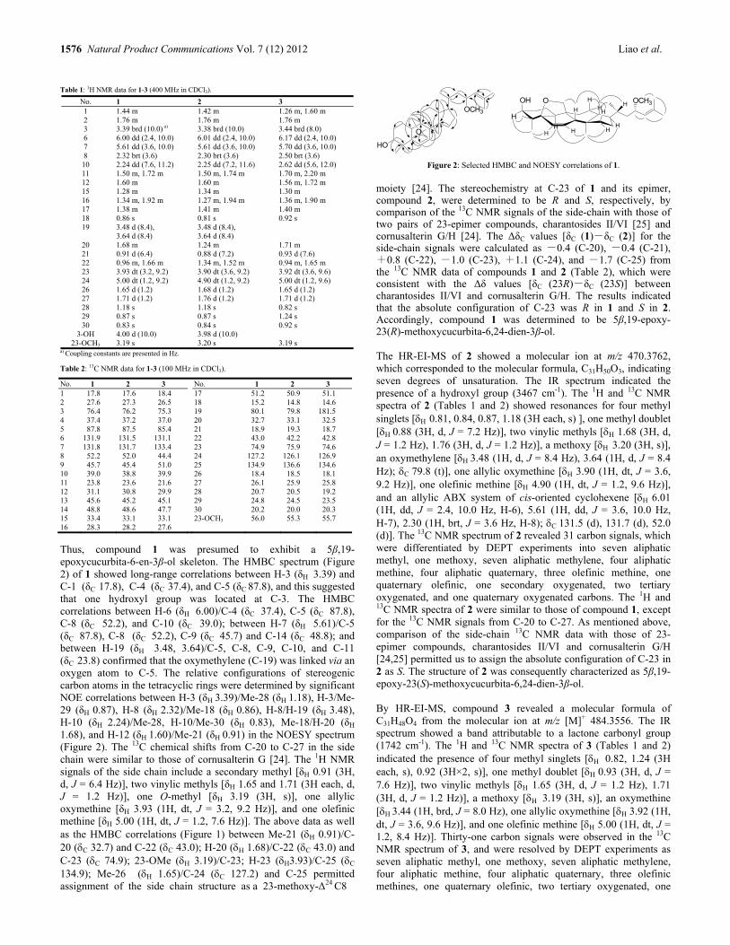
1576 Natural Product Communications Vol. 7 (12) 2012 Liao et al.
Table 1: 1H NMR data for 1-3 (400 MHz in CDCl3).
No. 1 2 3 1 1.44 m 1.42 m 1.26 m, 1.60 m 2 1.76 m 1.76 m 1.76 m 3 3.39 brd (10.0) a) 3.38 brd (10.0) 3.44 brd (8.0) 6 6.00 dd (2.4, 10.0) 6.01 dd (2.4, 10.0) 6.17 dd (2.4, 10.0) 7 5.61 dd (3.6, 10.0) 5.61 dd (3.6, 10.0) 5.70 dd (3.6, 10.0) 8 2.32 brt (3.6) 2.30 brt (3.6) 2.50 brt (3.6)
10 2.24 dd (7.6, 11.2) 2.25 dd (7.2, 11.6) 2.62 dd (5.6, 12.0) 11 1.50 m, 1.72 m 1.50 m, 1.74 m 1.70 m, 2.20 m 12 1.60 m 1.60 m 1.56 m, 1.72 m 15 1.28 m 1.34 m 1.30 m 16 1.34 m, 1.92 m 1.27 m, 1.94 m 1.36 m, 1.90 m 17 1.38 m 1.41 m 1.40 m 18 0.86 s 0.81 s 0.92 s 19 3.48 d (8.4),
3.64 d (8.4) 3.48 d (8.4), 3.64 d (8.4)
20 1.68 m 1.24 m 1.71 m 21 0.91 d (6.4) 0.88 d (7.2) 0.93 d (7.6) 22 0.96 m, 1.66 m 1.34 m, 1.52 m 0.94 m, 1.65 m 23 3.93 dt (3.2, 9.2) 3.90 dt (3.6, 9.2) 3.92 dt (3.6, 9.6) 24 5.00 dt (1.2, 9.2) 4.90 dt (1.2, 9.2) 5.00 dt (1.2, 9.6) 26 1.65 d (1.2) 1.68 d (1.2) 1.65 d (1.2) 27 1.71 d (1.2) 1.76 d (1.2) 1.71 d (1.2) 28 1.18 s 1.18 s 0.82 s 29 0.87 s 0.87 s 1.24 s 30 0.83 s 0.84 s 0.92 s
3-OH 4.00 d (10.0) 3.98 d (10.0) 23-OCH3 3.19 s 3.20 s 3.19 s
a) Coupling constants are presented in Hz.
Table 2: 13C NMR data for 1-3 (100 MHz in CDCl3).
Thus, compound 1 was presumed to exhibit a 5β,19-epoxycucurbita-6-en-3β-ol skeleton. The HMBC spectrum (Figure 2) of 1 showed long-range correlations between H-3 (δH 3.39) and C-1 (δC 17.8), C-4 (δC 37.4), and C-5 (δC 87.8), and this suggested that one hydroxyl group was located at C-3. The HMBC correlations between H-6 (δH 6.00)/C-4 (δC 37.4), C-5 (δC 87.8), C-8 (δC 52.2), and C-10 (δC 39.0); between H-7 (δH 5.61)/C-5 (δC 87.8), C-8 (δC 52.2), C-9 (δC 45.7) and C-14 (δC 48.8); and between H-19 (δH 3.48, 3.64)/C-5, C-8, C-9, C-10, and C-11 (δC 23.8) confirmed that the oxymethylene (C-19) was linked via an oxygen atom to C-5. The relative configurations of stereogenic carbon atoms in the tetracyclic rings were determined by significant NOE correlations between H-3 (δH 3.39)/Me-28 (δH 1.18), H-3/Me-29 (δH 0.87), H-8 (δH 2.32)/Me-18 (δH 0.86), H-8/H-19 (δH 3.48), H-10 (δH 2.24)/Me-28, H-10/Me-30 (δH 0.83), Me-18/H-20 (δH
1.68), and H-12 (δH 1.60)/Me-21 (δH 0.91) in the NOESY spectrum (Figure 2). The 13C chemical shifts from C-20 to C-27 in the side chain were similar to those of cornusalterin G [24]. The 1H NMR signals of the side chain include a secondary methyl [δH 0.91 (3H, d, J = 6.4 Hz)], two vinylic methyls [δH 1.65 and 1.71 (3H each, d, J = 1.2 Hz)], one O-methyl [δH 3.19 (3H, s)], one allylic oxymethine [δH 3.93 (1H, dt, J = 3.2, 9.2 Hz)], and one olefinic methine [δH 5.00 (1H, dt, J = 1.2, 7.6 Hz)]. The above data as well as the HMBC correlations (Figure 1) between Me-21 (H 0.91)/C-20 (C 32.7) and C-22 (C 43.0); H-20 (H 1.68)/C-22 (C 43.0) and C-23 (C 74.9); 23-OMe (H 3.19)/C-23; H-23 (H3.93)/C-25 (C 134.9); Me-26 (H 1.65)/C-24 (C 127.2) and C-25 permitted assignment of the side chain structure as a 23-methoxy-Δ24 C8
O
HO
OCH3H
H
H
HOH
H
H
H
HH
H
HO OCH3
Figure 2: Selected HMBC and NOESY correlations of 1.
moiety [24]. The stereochemistry at C-23 of 1 and its epimer, compound 2, were determined to be R and S, respectively, by comparison of the 13C NMR signals of the side-chain with those of two pairs of 23-epimer compounds, charantosides II/VI [25] and cornusalterin G/H [24]. The ΔδC values [δC (1)-δC (2)] for the side-chain signals were calculated as -0.4 (C-20), -0.4 (C-21), +0.8 (C-22), -1.0 (C-23), +1.1 (C-24), and -1.7 (C-25) from the 13C NMR data of compounds 1 and 2 (Table 2), which were consistent with the Δδ values [δC (23R)-δC (23S)] between charantosides II/VI and cornusalterin G/H. The results indicated that the absolute configuration of C-23 was R in 1 and S in 2. Accordingly, compound 1 was determined to be 5β,19-epoxy-23(R)-methoxycucurbita-6,24-dien-3β-ol.
The HR-EI-MS of 2 showed a molecular ion at m/z 470.3762, which corresponded to the molecular formula, C31H50O3, indicating seven degrees of unsaturation. The IR spectrum indicated the presence of a hydroxyl group (3467 cm-1). The 1H and 13C NMR spectra of 2 (Tables 1 and 2) showed resonances for four methyl singlets [H 0.81, 0.84, 0.87, 1.18 (3H each, s) ], one methyl doublet [H 0.88 (3H, d, J = 7.2 Hz)], two vinylic methyls [H 1.68 (3H, d, J = 1.2 Hz), 1.76 (3H, d, J = 1.2 Hz)], a methoxy [H 3.20 (3H, s)], an oxymethylene [H 3.48 (1H, d, J = 8.4 Hz), 3.64 (1H, d, J = 8.4 Hz); C 79.8 (t)], one allylic oxymethine [H 3.90 (1H, dt, J = 3.6, 9.2 Hz)], one olefinic methine [H 4.90 (1H, dt, J = 1.2, 9.6 Hz)], and an allylic ABX system of cis-oriented cyclohexene [H 6.01 (1H, dd, J = 2.4, 10.0 Hz, H-6), 5.61 (1H, dd, J = 3.6, 10.0 Hz, H-7), 2.30 (1H, brt, J = 3.6 Hz, H-8); C 131.5 (d), 131.7 (d), 52.0 (d)]. The 13C NMR spectrum of 2 revealed 31 carbon signals, which were differentiated by DEPT experiments into seven aliphatic methyl, one methoxy, seven aliphatic methylene, four aliphatic methine, four aliphatic quaternary, three olefinic methine, one quaternary olefinic, one secondary oxygenated, two tertiary oxygenated, and one quaternary oxygenated carbons. The 1H and 13C NMR spectra of 2 were similar to those of compound 1, except for the 13C NMR signals from C-20 to C-27. As mentioned above, comparison of the side-chain 13C NMR data with those of 23-epimer compounds, charantosides II/VI and cornusalterin G/H [24,25] permitted us to assign the absolute configuration of C-23 in 2 as S. The structure of 2 was consequently characterized as 5β,19-epoxy-23(S)-methoxycucurbita-6,24-dien-3β-ol. By HR-EI-MS, compound 3 revealed a molecular formula of C31H48O4 from the molecular ion at m/z [M]+ 484.3556. The IR spectrum showed a band attributable to a lactone carbonyl group (1742 cm-1). The 1H and 13C NMR spectra of 3 (Tables 1 and 2) indicated the presence of four methyl singlets [H 0.82, 1.24 (3H each, s), 0.92 (3H×2, s)], one methyl doublet [H 0.93 (3H, d, J = 7.6 Hz)], two vinylic methyls [H 1.65 (3H, d, J = 1.2 Hz), 1.71 (3H, d, J = 1.2 Hz)], a methoxy [H 3.19 (3H, s)], an oxymethine [H 3.44 (1H, brd, J = 8.0 Hz), one allylic oxymethine [H 3.92 (1H, dt, J = 3.6, 9.6 Hz)], and one olefinic methine [H 5.00 (1H, dt, J = 1.2, 8.4 Hz)]. Thirty-one carbon signals were observed in the 13C NMR spectrum of 3, and were resolved by DEPT experiments as seven aliphatic methyl, one methoxy, seven aliphatic methylene, four aliphatic methine, four aliphatic quaternary, three olefinic methines, one quaternary olefinic, two tertiary oxygenated, one
No. 1 2 3 No. 1 2 3 1 17.8 17.6 18.4 17 51.2 50.9 51.1 2 27.6 27.3 26.5 18 15.2 14.8 14.6 3 76.4 76.2 75.3 19 80.1 79.8 181.5 4 37.4 37.2 37.0 20 32.7 33.1 32.5 5 87.8 87.5 85.4 21 18.9 19.3 18.7 6 131.9 131.5 131.1 22 43.0 42.2 42.8 7 131.8 131.7 133.4 23 74.9 75.9 74.6 8 52.2 52.0 44.4 24 127.2 126.1 126.9 9 45.7 45.4 51.0 25 134.9 136.6 134.6 10 39.0 38.8 39.9 26 18.4 18.5 18.1 11 23.8 23.6 21.6 27 26.1 25.9 25.8 12 31.1 30.8 29.9 28 20.7 20.5 19.2 13 45.6 45.2 45.1 29 24.8 24.5 23.5 14 48.8 48.6 47.7 30 20.2 20.0 20.3 15 33.4 33.1 33.1 23-OCH3 56.0 55.3 55.7 16 28.3 28.2 27.6

Triterpenoids from Momordica charantia Natural Product Communications Vol. 7 (12) 2012 1577
quaternary oxygenated, and one ketone carbonyl carbons. Comparison of the 1H and 13C NMR spectra of 3 with those of 1 revealed that the two compounds are structurally very similar in rings A, C, and D and the side chain. The only difference was that the oxymethylene [H 3.48 (1H, d, J = 8.4 Hz), 3.64 (1H, d, J = 8.4 Hz); C 80.1 (t)] in 1 was replaced by a lactone carbonyl [C 181.5 (s)] [21]. This proposed structure was further suggested by the fragment ion peak at m/z 440 [M-CO2]
+ in the EI-MS of 3 and the HMBC correlations between H-10 (H 2.62)/C-19 (C 181.5 s) and H-11 (H 2.20)/C-19. Thus, compound 3 was formulated as 3β-hydroxy-23(R)-methoxycucurbita-6,24-dien-5β,19-olide. Compound 1 was evaluated for cytotoxic activity against human hepatoma SK-Hep-1 cells with an IC50 value of 26.6 μM. 5-FU (Fluorouracil) was used as positive control (IC50 = 1.0 M). Experimental
General experimental procedures: Optical rotations were measured using a JASCO DIP-180 digital spectropolarimeter. The IR spectra were obtained on a Nicolet 510P FT-IR spectrometer. The NMR spectra were recorded in CDCl3 at room temperature on a Varian Mercury plus 400 NMR spectrometer, and the solvent resonance was used as internal shift reference (TMS as standard). The 2D NMR spectra were recorded using standard pulse sequences. EI-MS and HR-EI-MS were recorded on a Finnigan TSQ-700 and a JEOL SX-102A spectrometer, respectively. TLC was performed using silica gel 60 F254 plates (200 μm, Merck). Diaion HP-20 (Mitsubishi) and silica gel (230-400 mesh ASTM, Merck) were used for column chromatography. HPLC was performed on a Hitachi L-7000 chromatograph with a Lichrosorb Si gel 60 column (5μm, 250 × 10 mm). Plant material: The fruits of Momordica charantia were collected in Pingtung County, Taiwan, in July 2006. The plant material was identified by Prof. Sheng-Zehn Yang, Curator of the Herbarium, National Pingtung University of Science and Technology. A voucher specimen (no. BT114) was deposited in the Department of Biological Science and Technology, National Pingtung University of Science and Technology, Pingtung, Taiwan. Extraction and isolation: Oven-dried pieces of fruit pulp of Momordica charantia (35.7 kg) were extracted 3 times with methanol (100 L) at room temperature (7 days each). The MeOH extract was evaporated in vacuo to afford a black residue, which was suspended in H2O (6 L), and then partitioned sequentially using EtOAc and n-BuOH (4L × 3). The n-BuOH fraction (937 g) was chromatographed over Diaion HP-20, using mixtures of water and MeOH of reducing polarity as eluents. Twenty-five fractions were collected as follows: fr. 1 [6000 mL, water-MeOH (40:10)], fr. 2 [4000 mL, water-MeOH (38:12)], fr. 3 [3000 mL, water-MeOH (36:14)], fr. 4 [5000 mL, water-MeOH (34:16)], fr. 5 [5000 mL, water-MeOH (32:18)], fr. 6 [3000 mL, water-MeOH (30:20)], fr. 7 [4000 mL, water-MeOH (28:22)], fr. 8 [6000 mL, water-MeOH (26:24)], fr. 9 [4000 mL, water-MeOH (24:26)], fr. 10 [5000 mL, water-MeOH (22:28)], fr. 11 [3000 mL, water-MeOH (20:30)], fr. 12 [3000 mL, water-MeOH (18:32)], fr. 13 [5000 mL, water-MeOH (16:34)], fr. 14 [4000 mL, water-MeOH (14:36)], fr. 15 [4000 mL, water-MeOH (12:38)], fr. 16 [2000 mL, water-MeOH (10:40)], fr. 17 [3000 mL, water-MeOH (9:41)], fr. 18 [3000 mL, water-MeOH (8:42)], fr. 19 [3000 mL, water-MeOH (7:43)], fr. 20 [2000 mL, water-MeOH (6:44)], fr. 21 [3000 mL, water-MeOH (5:45)], fr.
22 [3000 mL, water-MeOH (4:46)], fr. 23 [4000 mL, water-MeOH (3:47)], fr. 24 [3000 mL, water-MeOH (1:49)], fr. 25 [8000 mL, MeOH]. Fraction 16 was further chromatographed on a silica gel column (5 × 45 cm), eluted with CH2Cl2-MeOH (20:1 to 0:l), to obtain 5 fractions (each about 800 mL), 16A-16E. Fraction 16D was separated by semi-preparative HPLC on a Merck Lichrosorb Si 60 column (5 μm, 250 × 10 mn) with CH2Cl2-isopropylalcohol (20:1) as eluent, 2 mL/min, to yield 1 (13.5mg, tR
= 23.8 min) and 2 (1.1 mg, tR = 24.8 min). Fraction 20 was further purified through a silica gel column (5 × 45 cm), eluted with CH2Cl2-MeOH (20:1 to 0:1) to obtain 6 fractions (each about 600 mL), 20A-20F. Fraction 20A was separated by a silica gel column (2 × 40 cm), eluted with n-hexane-acetone (10:1) to give 5 sub-fractions 20Aa-20Ae. 20Ab was further purified by semi-preparative HPLC on a Merck Lichrosorb Si 60 column (5 μm, 250 × 10 mn) with CH2Cl2-lsopropylalcohol (20:1) as eluent, 2 mL/min, to yield 3 (1.8 mg, tR = 21.4 min). Cytotoxicity assay: The cytotoxicity of compound 1 was evaluated by the MTT [3-(4,5-dimethylthiazol-2-yl)-2,5-diphenyltetrazolium bromide] colorimetric method based on the described procedures [26]. SK-Hep 1 cell lines were maintained in Dulbecco’s modified Eagle’s medium (DMEM) supplemented with 10% (v/v) fetal bovine serum, L-glutamine 2 mM, 1% penicillin/streptomycin (penicillin 10000 U/mL and streptomycin 10 mg/mL) in a humidified atmosphere of 5% CO2 at 37 °C. A volume of SK-Hep 1 cells 100 μL at a density of 1 × 10⁵ cells/mL was incubated under the same conditions for 24 h in a 96-well flat-bottomed microplate. Test samples dissolved in DMSO were added to the cultures. After a 48 h incubation period, the wells were incubated with the MTT (100 μL/well concentrated at 5 mg/mL) at 37°C for 4 h. 200 μL of DMSO was added to redissolve the formazan crystals after removing the supernatant. The absorbance of the resulting formazan was measured by an enzyme-linked immunosorbent assay plate reader at 550 nm. The results were assayed in triplicate. The ratio of cell viability (%) was calculated by using the following formula: [(experimental absorbance - background absorbance)/(control absorbance - background absorbance)] × 100. The activity is shown as IC50 value, which the concentration (μM) of the tested sample that results in 50% inhibition of cell growth. 5β,19-Epoxy-23(R)-methoxycucurbita-6,24-dien-3β-ol (1) Amorphous white powder. [α]25
D: -37.0 (c 0.68, CHCl3). IR (KBr) νmax: 3463, 3026, 2970, 2937, 2873, 2814, 1663, 1645, 1466, 1415, 1376, 1106, 1080, 1055, 999, 948, 911, 771 cm-l. 1H NMR: Table 1. 13C NMR: Table 2. EI-MS (70 eV) m/z (rel. int.): 470 [M]+ (3), 421 (2), 391 (1), 354 (1), 323 (4), 281 (2), 255 (1), 171 (4), 109 (4), 99 (100), 69 (3). HR-EI-MS: m/z 470.3767 (calcd for C31H50O3 470.3762, [M]+). 5β,19-Epoxy-23(S)-methoxycucurbita-6,24-dien-3β-ol (2) Amorphous white powder. [α]25
D: -48.6 (c 0.10, CHCl3). IR (KBr) νmax: 3467, 3030, 2988, 2962, 2924, 2873, 2809, 1665, 1648, 1449, 1376, 1106, 1084, 940, 915, 775, 741 cm-l. 1H NMR: Table 1. 13C NMR: Table 2. EI-MS (70 eV) m/z (rel. int.): 470 [M]+ (4), 438 (10), 421 (2), 408 (3), 390 (15), 354 (1), 323 (6), 309 (21), 281 (23), 255 (1), 239 (7), 211 (7), 185 (12), 171 (21), 109 (48), 99 (100), 81 (42), 69 (3), 55 (38). HR-EI-MS: m/z 470.3762 (calcd for C31H50O3 470.3762, [M]+).
1578 Natural Product Communications Vol. 7 (12) 2012 Liao et al.
3β-Hydroxy-23(R)-methoxycucurbita-6,24-dien-5β,19-olide (3) Amorphous white powder. [α]25
D: -64.5 (c 0.12, CHCl3). IR (KBr) νmax: 3573, 3505, 2965, 2940, 2936, 2877, 2814, 1742, 1470, 1450, 1372, 1280, 1187, 1163, 1090, 1041, 939, 900, 749 cm-l. 1H NMR: Table 1. 13C NMR: Table 2. EI-MS (70 eV) m/z (rel. int.): 484 [M]+
(3), 440 (3), 408 (1), 375 (1), 309 (3), 281 (2), 241 (1), 213 (1), 171 (4), 109 (6), 99 (100), 55 (7). HR-EI-MS: m/z 484.3556 (calcd for C31H48O4 484.3554, [M]+).
Acknowledgments - This research was supported by grants from the National Science Council of the Republic of China (NSC 99-2622-B-020-004-CC1 and NSC 100-2622-B-020-001-CC1) and in part by Taiwan Department of Health Clinical Trial and Research Center of Excellence (DOH100-TD-B-111-004). We thank Ms Shu-Yun Sun and Ms Lih-Mei Sheu for the EI-MS and HR-EI-MS measurements in the Instrumentation Center of the College of Science, National Taiwan University and National Chung Hsing University. We are also grateful to the National Center for High-performance computing for computer time and facilities.
References
[1] Wang X, Sun W, Cao J, Qu H, Bi X, Zhao Y. (2012) Structures of new triterpenoids and cytotoxicity activities of the isolated major compounds from the fruit of Momordica charantia L. Journal of Agricultural and Food Chemistry, 60, 3927–3933.
[2] Rathi SS, Grover JK, Vats V. (2002) The effect of Momordica charantia and Mucuna pruriens in experimental diabetes and their effect on key metabolic enzymes involved in carbohydrate metabolism. Phytotherapy Research, 16, 236–243.
[3] Kobori M, Nakayama H, Fukushima K, Ohnishi-Kameyama M, Ono H, Fukushima T, Akimoto Y, Masumoto S, Yukizaki C, Hoshi Y. Deguchi T, Yoshida M. (2008) Bitter gourd suppresses lipopolysaccharide-induced inflammatory responses. Journal of Agricultural and Food Chemistry, 56, 4004–4011.
[4] Harinantenaina L, Tanaka M, Takaoka S, Oda M, Mogami O, Uchida M, Asakawa Y. (2006) Momordica charantia constituents and antidiabetic screening of the isolated major compounds. Chemical & Pharmaceutical Bulletin, 54, 1017–1021.
[5] Liu Y, Ali Z, Khan IA. (2008) Cucurbitane-type triterpene glycosides from the fruits of Momordica charantia. Planta Medica, 74, 1291–1294. [6] Kimura Y, Akihisa T, Yuasa N, Ukiya M, Suzuki T, Toriyama M, Motohashi S, Tokuda H. (2 [7] 005) Cucurbitane-type triterpenoids from the fruit of Momordica charantia. Journal of Natural Products, 68, 807–809. [8] Akihisa T, Higo N, Tokuda H, Ukiya M, Akazawa H, Tochigi Y, Kimura Y, Suzuki T, Nishino H. (2007) Cucurbitane-type triterpenoids from the
fruits of Momordica charantia and their cancer chemopreventive effects. Journal of Natural Products, 70, 1233–1239. [9] Li QY, Chen HB, Liu ZM, Wang B, Zhao YY. (2007) Cucurbitane triterpenoids from Momordica charantia. Magnetic Resonance in Chemistry, 45,
451–456. [10] Matsuda H, Nakamura S, Murakami T, Yoshikawa M. (2007) Structures of new cucurbitane-type triterpenes and glycosides, karavilagenins D and
E, and karavilosides VI, VII, VIII, IX, X, and XI, from the fruit of Momordica charantia. Heterocycles, 71, 331–341. [11] Nakamura S, Murakami T, Nakamura J, Kobayashi H, Matsuda H, Yoshikawa M. (2006) Structures of new cucurbitane-type triterpenes and
glycosides, karavilagenins and karavilosides, from the dried fruit of Momordica charantia L. in Sri Lanka. Chemical & Pharmaceutical Bulletin, 54, 1545–1550.
[12] Fatope MO, Takeda Y, Yamashita H, Okabe H, Yamauchi T. (1990) New cucurbitane triterpenoids from Momordica charantia. Journal of Natural Products, 53, 1491–1497.
[13] Murakami T, Emoto A, Matsuda H, Yoshikawa M. (2001) Medicinal foodstuffs. XXI. Structures of new cucurbitane-type triterpene glycosides, goyaglycosides-a, -b, -c, -d, -e, -f, -g, and -h, and new oleanane-type triterpene saponins, goyasaponins I, II, and III, from the fresh fruit of Japanese Momordica charantia L. Chemical & Pharmaceutical Bulletin, 49, 54–63.
[14] Begum S, Ahmed M, Siddiqui BS, Khan A, Saify ZS, Arif M. (1997) Triterpenes, a sterol and a monocyclic alcohol from Momordica charantia. Phytochemistry, 44, 1313–1320.
[15] Miyahara Y, Okabe H, Yamauchi T. (1981) Studies on the constituents of Momordica charantia L. II. Isolation and characterization of minor seed glycosides, momordicosides C, D and E. Chemical & Pharmaceutical Bulletin, 29, 1561–1566.
[16] Okabe H, Miyahara Y, Yamauchi T, Miyahara K, Kawasaki T. (1980) Studies on the constituents of Momordica charantia L. I. Isolation and characterization of momordicosides A and B, glycosides of a pentahydroxy-cucurbitane triterpene. Chemical & Pharmaceutical Bulletin, 28, 2753–2762.
[17] Chen J, Tian R, Qiu M, Lu L, Zheng Y, Zhang Z. (2008) Trinorcucurbitane and cucurbitane triterpenoids from the roots of Momordica charantia. Phytochemistry, 69, 1043–1048.
[18] Yasuda M, Iwamoto M, Okabe H, Yamauchi T. (1984) Structures of momordicines I, II and III, the bitter principles in the leaves and vines of Momordica charantia L. Chemical & Pharmaceutical Bulletin, 32, 2044–2047.
[19] Chen JC, Liu WQ, Lu L, Qiu MH, Zheng YT, Yang LM, Zhang XM, Zhou L, Li ZR. (2009) Kuguacins F–S, cucurbitane triterpenoids from Momordica charantia. Phytochemistry, 70, 133–140.
[20] Chang CI, Chen CR, Liao YW, Cheng HL, Chen YC, Chou CH. (2006) Cucurbitane-type triterpenoids from Momordica charantia. Journal of Natural Products, 69, 1168–1171.
[21] Chang CI, Chen CR, Liao YW, Cheng HL, Chen YC, Chou CH. (2008) Cucurbitane-type triterpenoids from the stems of Momordica charantia. Journal of Natural Products, 71, 1327–1330.
[22] Chang CI, Chen CR, Liao YW, Shih WL, Cheng HL, Tzeng CY, Li JW, Kung MT. (2010) Octanorcucurbitane triterpenoids protect against t-BHP-induced hepatotoxicity from the stems of Momordica charantia. Chemical & Pharmaceutical Bulletin, 58, 225–229.
[23] Chen CR, Liao YW, Shih WL, Tzeng CY, Chang CI. (2010) Triterpenoids from the stems of Momordica charantia. Helvetica Chimica Acta, 93, 1355–1361.
[24] Chen CR, Liao YW, Wang L, Kuo YH, Liu HJ, Shih WL, Cheng HL, Chang CI. (2010) Cucurbitane triterpenoids from Momordica charantia and their cytoprotective activity in tert-butyl hydroperoxide-induced hepatotoxicity of HepG2 cells. Chemical & Pharmaceutical Bulletin, 58, 1639–1642.
[25] Kim KG, Choi SU, Kim YC, Lee KR. (2011) Tirucallane triterpenoids from Cornus walteri. Journal of Natural Products, 74, 54–59. [26] Akihisa T, Higo N, Tokuda H, Ukiya M, Akazawa H, Tochigi Y, Kimura Y, Suzuki T, Nishino H. (2007) Cucurbitane-type triterpenoids from the
fruits of Momordica charantia and their cancer chemopreventive effects. Journal of Natural Products, 70, 1233–1239. [27] Kuo YH, Li SY, Shen YC, Huang HC, Hsu YW, Tseng RJ, Ou JC, Chen CF. (2001) Sesquiterpene polyol esters and triterpenes from Celastrus
punctatus. The Chinese Pharmaceutical Journal, 53, 257–268.

Two Novel Phenethylamine Alkaloids from Streptomyces sp. YIM10049
Xueqiong Yanga, 1, Guangwei Hea, 1, Lixing Zhaob, Yabin Yanga, Yun Liua, Lihua Xub and Zhongtao Dinga, * aKey Laboratory of Medicinal Chemistry for Natural Resource, Ministry of Education, School of Chemical Science and Technology, Yunnan University, Kunming 650091, China
bYunnan Institute of Microbiology, Yunnan University, Kunming 650091, China 1The authors contributed equally to this paper
Received: August 15th, 2012; Accepted: October 17th, 2012
Two novel phenethylamine alkaloids were isolated from Streptomyces sp. YIM 10049. On the basis of spectral data, their structures were determined as (S)-N-(-phenylethyl)-2 -hydroxyl-acrylimine (1) and (S)-N-nitroso-1-amino-p-hydroxy phenylethanol (2). Three known compounds, indole-3-carboxylic acid (3), cyclo(L-Ala-L-Tyr)(4), and bis(2-ethylhexyl) phthalate (5), were also isolated and characterized. Compound 1 is a rare enol tautomer, and compound 2 an unusual phenethylamine alkaloid with a N-NO group. Keywords: Streptomyces sp., Alkaloid, Phenethylamine.
The genus Streptomyces is recognized as the most important bacterial source of clinically used therapeutic agents, including lifesaving antibiotics and antifungal, anticancer, and immunosuppressive agents [1a]. The alkaloids are reported to possess broad spectrum antimicrobial, antiradical, antioxidant, antiplasmodial, anticancer and antimutagenic activities [1b]. Phenethylamines (PEA) are well known for their psychotropic effects and have been isolated from a variety of organisms, including octocorals, the edible mushroom Laetiporus sulphureus, Streptomyces sp., Enterococcus sp. and other species of cyanobacteria [1c]. Streptomyces sp. YIM 10049 was isolated from the excrement of alpaca, and a primary bioactivity screening indicated that this strain showed some cytotoxicity to A549 and Raji cell lines, and antibacterial activity to Fusarium oxysporum and F. solani. As a part of our persistent search for new and active natural products from actinomycetes, the chemical investigation of Streptomyces sp. YIM 10049 led to the isolation of two novel phenethylamines, (S)-N-(-phenylethyl)-2-hydroxyl-acrylimine (1) and (S)-N-nitroso-1-amino-p-hydroxy phenylethanol (2) (Figure 1), together with three known compounds, indole-3-carboxylic acid (3) [2a], cyclo(L-Ala-L-Tyr) (4) [2b], and bis(2-ethylhexyl) phthalate (5) [3]. Compound 1 had a molecular formula of C11H13NO according to HRESIMS analysis. The 13C NMR and DEPT spectra of 1 showed 11 carbon signals for one methyl, one olefinic methylene, seven methines (six for an olefinic group), and two quaternary carbons. The 2D NMR spectrum identified monosubstituted phenethyl, allyl units (Figure 2). That C-7 connected to C-1 was elucidated by the HMBC correlations from H-7 and H-8 to C-1, and H-7 to C-6. The connection of C-2 to N-1 and C-3 was elucidated by the correlations from H-3, H-4 and H-7 to C-2 in the HMBC. Because of the overlap of signals for H-3, 4, and 7 in the 1H NMR spectrum in C5D5N, the 2D NMR spectrum was measured again in MeOD, and the results were consistent. The structure of 1 was similar to that of N-(-phenylethyl)-acrylamide (1a), but its IR spectrum showed no obvious CO absorption peak. In order to identify the structure of 1, 1a was synthesized (Figure 3). The NMR spectroscopic data of 1 and 1a were similar, but 1a showed, in its
N
OH
HN
O HO
OH
NH
NO
1 1a 2
Figure 1: Structures of compounds 1, 2 and 1a Table 1: 13C NMR (125M Hz) and 1H NMR (500 MHz) data of compound 1 in C5D5N.
Position H C
OH 9.31 (1H, brs) 2 165.8 3 6.55 (1H, m,) 133.3 4 5.53 (1H, m), 6.55 (1H, m) 126.4 1 145.9 2 7.48 (1H, d, 7.6Hz) 127.6 3 7.30 (1H, t,7.6Hz) 129.7 4 7.22 (1H, d, 7.4Hz) 128.1 5 7.30 (1H, t, 7.6Hz) 129.7 6 7.48 (1H, d, 7.6Hz) 127.6 7 5.53 (1H, m) 49.8 8 1.46 (3H, d, 7.0Hz) 23.1
N
OH1H-1H COSY
HMBC
1'
2'
3'4'
5'
6'
7'
8'
2
43
NOESY
1
Figure 2: 1H-1H COSY, HMBC and NOESY correlations for 1.
NH2
TEA
CH2Cl2
+
O
ClNH
O
Figure 3: Synthesis of compound 1a.
IR spectrum, carbonyl and amino signals at 1660 and 3286 cm-1, but 1 a hydroxyl signal at 3434 cm-1. The two compounds also had different TLC chromatographic characteristics [e.g. color development reaction, Rf value: 0.2 for 1 and 0.4 for 1a with CHCl3CH3OH (3:1) as developing solvent]. Therefore, compound 1 was determined as N-(-phenylethyl)-2-hydroxyl-acrylimine, the enol tautomer of 1a. The cis configuration between N-1 and C-2 was determined by NOESY from 2-OH to H-8. The stereochemistry of compound 1 was confirmed as S ([]25
D -218.0) by comparison with the optical rotation of 1a ([]23
D -207) [4].
NPC Natural Product Communications 2012 Vol. 7 No. 12
1579 - 1580

1580 Natural Product Communications Vol. 7 (12) 2012 Yang et al.
Compound 2 had a molecular formula of C8H10N2O3. The 13C NMR and DEPT spectra of 2 showed eight carbon signals for one methylene, five methines (4 olefinic) and two quaternary carbons, respectively. Four aromatic protons [δ 6.72 (2H, d, 7.6 Hz), 7.10 (2H, d, 7.6 Hz)] were identified, one a para-substituted phenyl, which was confirmed by the 2D NMR spectrum (Figure 4). The HMBC correlations from H-2 to C-1 and COSY correlation between H-1 and H-2 indicated that C-2 was connected to C-1, and C-1 to C-2. The 1H NMR spectrum in C5D5N exhibited a NH signal at 7.39, and the IR spectrum showed a nitroso signal at 1567 cm-1. So, compound 2 was determined as N-nitroso-1-amino-p-hydroxy phenylethanol. The configuration of 2 was determined as S ([]D: -4.2) by comparison with the optical rotation of the analogue, (S)-2-hydroxy-3-(4-hydroxyphenyl) propionic acid (2a) ([]
D: -10) [5]. A novel protein tyrosine phosphatase inhibitor, 2-(N-methyl-N-nitroso) hydroquinone, with a N-nitroso phenyl group like compound 2 was also isolated from a Streptomyces [6]. Table 2: 13C NMR and 1H NMR data of compound 2 in MeOD.
Position H C 1 4.28 (1H, brs) 73.72 2.84, 3.02 (2H, m) 41.3 1 130.2 2 7.10 (1H, d, 7.6 Hz) 132.0 3 6.72 (1H, d, 7.6 Hz) 116.5 4 157.5 5 6.72 (1H, d, 7.6 Hz) 116.5 6 7.10 (1H, d, 7.6 Hz) 132.0
HONH NO
OH
12
1'
2'3'
4'
5' 6'
COSY
HMBC Figure 4: 1H-1H COSY, HMBC correlations for 2
Experimental
General experimental procedures: Optical rotations, Jasco P-1020; 1D and 2D NMR, Bruker DRX-500 MHz; MS, G3250AA and AutoSpec Premier P776 spectrometer; IR, Nicolet magna-IR 550; silica gel (Qingdao Marine Chemical Group Co.) and Sephadex LH-20 (GE Healthcare Co) were used for column chromatography.
Biological material and cultivation of actinomycete strain: The bacterial strain, isolated from the excrement of alpaca from a zoo in Yunnan province, China, was identified as Streptomyces sp. (YIM 10049) by complete 16S rRNA gene sequence. This bacterium was cultivated on a 40 L scale using 1L Erlenmeyer flasks containing 250 mL of seed medium (yeast extract 0.4%, glucose 0.4%, malt extract 0.5%, decavitamin 0.01%, pH 7) and the fermentation medium (soluble starch 0.5%, glucose 2%, soybean powder 0.2%, peptone 0.2%, NaCl 0.4%, CaCO3 0.2%, K2HPO3 0.05%, MgSO4 0.05%, pH 7) at 28C for 10 days on rotary shakers (250 rpm).
Antibacterial and antitumor cell assays: The cytotoxicity of this strain towards A549 and Raji cells was determined in vitro by the MTT method for an inhibition ratio at 40%60%, while cisplatin was used as a positive control. The primary antibacterial activity of the culture of this strain to Fusarium oxysporum and F. solani was measured using the agar diffusion method. Inhibition was measured by the size of zones of inhibition (10 and 12 mm). Extraction and isolation of compounds: The fermentation broth (40 L) for the Streptomyces sp. YIM 10049 was filtered, the filtrate extracted with ethyl acetate, and the solvent removed under vacuum. The resulting extract was separated into 4 fractions by silica gel CC, eluting with CHCl3 /CH3OH (100%-0%). The first part (CHCl3) was separated by repeated CC on silica gel and Sephadex LH-20, to yield compounds 1 (2.5 mg), 4 (6.7 mg), and 5 (4.7 mg). The third part (CHCl3 /CH3OH =3:1) was separated by repeated CC on silica gel and Sephadex LH-20, to yield compounds 2 (2.4 mg), and 3 (4.5 mg). Compound 1 White needles (CH3OH). MP: 99.4 -101.7C []25
D: -218.0 (c 0.02,C5H5N). IR (KBr): 3434 (OH), 2925 (CH), 2354, 1557 (C=N) cm-1; 1H NMR and 13C NMR: Table 1. HR-ESI m/z: [M+H]+ Calcd for C11H14NO 176.1075, Found: m/z 176.1054.
Compound 1a White needles (CH3OH). MP: 92.3-94.6C IR (KBr): 3286(NH), 2355, 1660(CO), 1544 cm-1. 13C NMR (CHCl3, 75MHz) : 164.7, 143.1, 131.0, 128.6, 127.3, 126.4, 126.2, 48.8 , 21.7. ESI m/z: 176[M+H]+. Compound 2 White needles (CH3OH). MP: 94.7-96.3C []25
D: -4.2 (c 0.05, CH3OH). IR (KBr): 3409 (OH), 2351, 1567 cm-1. 1H NMR and 13C NMR: Table 2. HR-ESI m/z: [M-H]- Calcd for C8H9N2O3 181.0613; found: m/z 181.0616. Acknowledgments - This project was supported by the National Natural Science Foundation of China (No. U0932601; No. 81060263).
References
[1] (a) Lewis K, Epstein S, D'Onofrio A, Ling L. (2010) Uncultured microorganisms as a source of secondary metabolites. The Journal of Antibiotics, 63, 468-476; (b) Kiplimo JJ, Islam MS, Koorbanally NA. (2011) A novel flavonoid and furoquinoline alkaloids from Vepris glomerata and their antioxidant activity. Natural Product Communications, 6, 1847-1850; (c) Malloy KL, Suyama TL, Engene N, Debonsi H, Cao Z, Matainaho T, Spadafora C, Murray TF, Gerwick WH. (2012) Credneramides A and B: neuromodulatory phenethylamine and isopentylamine derivatives of a vinyl chloride-containing fatty acid from cf. Trichodesmium sp. nov. Journal of Natural Products, 75, 60−66.
[2] (a) Tan J, Bednarek P, Liu J, Schneider B, Svatoš A, Hahlbrock K. (2004) Universally occurring phenylpropanoid and species-specific indolic metabolites in infected and uninfected Arabidopsis thaliana roots and leaves. Phytochemistry, 65, 691-699; (b) Chen G, Lin Y, Vrijmoed LLP, Fong WF. (2006) A new isochroman from the marine endophytic fungus 1893#. Chemistry of Natural Compounds, 42, 138-141.
[3] Amade P, Mallea M, Bouaïcha N. (1994) Isolation, structural identification and biological activity of two metabolites produced by Penicillium olsonii bainier and sartory. The Journal of Antibiotics, 47, 201-207.
[4] Blaschke G, Donow F. (1975) Untersuchung chromatographischer racemattrennungen, IV Trennwirkung optisch aktiven poly [N-((S)-1-phenyläthyl) acrylamids] in abhängigkeit vom polymerisationsverfahren. European Journal of Inorganic Chemistry, 108, 1188-1197.
[5] Valls N, Vallribera M, López-Canet M, Bonjoch J. (2002) Synthesis of microcin SF608. The Journal of Organic Chemistry, 67, 4945-4950. [6] Kakeya H, Imoto M, Takahashi Y, Naganawa H, Takeuchi T, Umezawa K. (1993) Dephostatin, a novel protein tyrosine phosphatase inhibitor
produced by Streptomyces II. structure determination. The Journal of Antibiotics, 46, 1716-1719.

Alkaloids from Cinnamomum philippinense Hsing-Tan Li, Wei-Jen Li*, Hui-Ming Wu* and Chung-Yi Chen* School of Medical and Health Sciences, Fooyin University, Kaohsiung 83102, Taiwan [email protected]; [email protected]; [email protected]
Received: October 5th, 2012; Accepted: October 19th, 2012
A new pyridine, 2-(4-hydroxypyridin-3-yl)-acetic acid (1), along with five known alkaloids, cinnaretamine (2), crykonisine (3), corydaldine (4), glaziovine (5) and zenkerine (6), were isolated from the roots of Cinnamomum philippinense (Lauraceae). Their structures were characterized and identified by spectral analysis. Keywords: Cinnamomum philippinense, Lauraceae, Pyridine. Cinnamomum philippinense (Merr.) Chang (Lauraceae) is a medium-sized tree distributed widely in the Philippines and the southern part of Taiwan. The chemical constituents of this species have been studied by Wu et al. [1]. In the course of screening for biologically and chemically novel agents from Formosan plants in the family Lauraceae [2a-2q], C. philippinense was chosen for further phytochemical investigation. In this study, the MeOH extract of its roots was subjected to chromatographic separation to afford six alkaloids, including a pyridine, 2-(4-hydroxypyridin-3-yl)-acetic acid (1), an amide, cinnaretamine (2) [2g], a benzylisoquinoline, crykonisine (3) [3a], an isoquinolone, corydaldine (4) [3b], a proaporphine, glaziovine (5) [3c] and an aporphine, zenkerine (6) [3d]. Among them, 1 was a new compound and all of these compounds were isolated for the first time from this species.
N
OH
O
HO 12
2'
4'5'
6'N
OH
O
HO
Figure 1: COSY (▬), HMBC (→) and NOESY (↔) Correlations of 1.
2-(4-Hydroxypyridin-3-yl)-acetic acid (1) was obtained as a white amorphous powder and its molecular formula was deduced as C7H7NO3 by HRESIMS (m/z 177.0401 [M + Na + H]+; calc. 177.0402). The UV spectrum of 1 showed intense absorption bands at 220, 265 and 280 nm, which were typical of a pyridine skeleton [4]. The IR spectrum of 1 exhibited absorption bands at λ 3100 and 1660 cm-1, indicating hydroxyl and carbonyl groups, respectively.
The structure was confirmed from the 1H NMR spectrum, which contained signals at 6.38 (1H, d, J = 6.0 Hz, H-5), 7.78 (1H, d, J = 6.0 Hz, H-6) and 7.87 (1H, s, H-2) on the pyridine ring, and 3.34 (2H, s, H-2). The 13C NMR and DEPT experiments for 1 showed seven resonance lines consisting of one methylene, three methines, and three quaternary carbons (including a carbonyl signal at 172.2). The structure of 1 was also confirmed by 2D NMR experiments. A COSY correlation was observed between H-5 and H-6 (Figure 1). The HETCOR experiment showed that the carbon signals at 116.5 for C-5, 154.4 for C-2, 156.0 for C-6 and 30.9 for C-2 were correlated to the proton signals at 6.38 for H-5,
7.87 for H-2, 7.78 for H-6 and 3.34 for H-2, respectively. The NOESY correlations between H-5 and H-6 and between H-2 and H-2 established the connective sites as shown in structure 1 (Figure 1). Thus, 1 is a new pyridine alkaloid, which was further confirmed by HMBC experiment (Figure 1). Experimental
General: IR, Hitachi 260-30 spectrophotometer; 1D and 2D NMR, Varian (Unity Plus) NMR spectrometer; Low-resolution ESI-MS, API 3000 (Applied Biosystems); High-resolution ESI-MS, Bruker Daltonics APEX II 30e spectrometer; Silica gel 60 for CC and precoated silica gel plates (Merck) were used for TLC, visualized with 10% H2SO4. Plant material: The roots of C. philippinense were collected from Taipei County, Taiwan in May 2008 and the plant material was identified by Dr Yen-Ray Hsui (Chungpu Research Center, Taiwan Forestry Research Institute). A voucher specimen (Cinnamo. 8) was deposited at the School of Medical and Health Sciences, Fooyin University, Kaohsiung, Taiwan. Extraction and isolation: The air-dried roots of C. philippinense (3.7 kg) were extracted with MeOH (5 L x 6) at room temperature. A MeOH extract (54.3 g) was obtained by concentration under reduced pressure. The MeOH extract, suspended in H2O (1 L), was partitioned with CHCl3 (2 L x 5) to give fractions soluble in CHCl3 (34.4 g) and H2O (8.8 g). The CHCl3-soluble fraction (34.4 g) was chromatographed over silica gel (950 g, 70-230 mesh) using n-hexane-EtOAc-MeOH as eluent to produce 6 further fractions. Part of fraction 6 (6.23 g) was subjected to silica gel CC, eluting with CH2Cl2–MeOH (40:1) and enriched gradually with MeOH, to obtain 5 fractions (6-1-6-5). Fraction 6-1 (0.33 g) was further separated by silica gel CC using the same solvent system and purified by preparative TLC to yield cinnaretamine (2) (4 mg). Fraction 6-2 (0.42 g) was further separated by silica gel CC, and purified by preparative TLC (CH2Cl2–MeOH, 30:1) to gave crykonisine (3) (12 mg) and corydaldine (4) (17 mg), respectively. Fraction 6-3 (0.29 g) was further separated by silica gel CC using the same solvent system and purified by preparative TLC (CH2Cl2–MeOH, 50:1) to yield 2-(4-hydroxypyridin-3-yl)-acetic acid (1) (6 mg). Fraction 6-4 (0.52 g) was further separated by silica gel CC, and purified by preparative TLC (CH2Cl2–MeOH, 20:1) to give glaziovine (5) (5 mg) and zenkerine (6) (4 mg), respectively.
NPC Natural Product Communications 2012 Vol. 7 No. 12
1581 - 1582

1582 Natural Product Communications Vol. 7 (12) 2012 Li et al.
2-(4-Hydroxypyridin-3-yl)-acetic acid (1)
White amorphous powder. IR (neat) max: 3100 (br, OH), 1660 (C=O), 1300, 950 cm-1. UV/Vis (CH3CN): max (log ε): 280 (3.52), 265 (2.54), 220 (2.21) nm. 1H NMR (500 MHz, CDCl3): 3.34 (2H, s, H-2), 6.38 (1H, d, J = 6.0 Hz, H-5), 7.78 (1H, d, J = 6.0 Hz, H-6), 7.87 (1H, s, H-2).
13C NMR (125 MHz, CDCl3): 30.9 (C-2, CH2), 116.5 (C-3, CH), 124.0 (C-3, C), 154.4 (C-2, CH), 156.0 (C-6, CH), 172.2 (C-1, C=O), 178.4 (C-4, C). MS (ESI): m/z (%): 177 [M + Na + H]+ HRMS-ESI: m/z [M + Na + H]+ calcd for C7H8O3NNa: 177.0402.1422; found: 177.0401. Acknowledgments - This investigation was supported by a grant from the Fooyin University.
References
[1] Wu TS, Leu YL, Chan YY, Yu SM, Teng CM, Su JD. (1994) Lignans and an aromatic acid from Cinnamomum philippinense. Phytochemistry, 36, 785788.
[2] (a) Chen CY. (2006) Butanolides from the stem of Cinnamomum kotoense. Natural Product Communications, 1, 453455; (b) Hsieh TJ, Chen CH, Lo WL, Chen CY. (2006) Lignans from the stem of Cinnamomum camphora. Natural Product Communications, 1, 2125; (c) Chen CY, Chen CH, Wong CH, Liu YW, Lin YS, Wang YD, Hsui YR. (2007) Cytotoxic constituents of the stems of Cinnamomum subavenium. Journal of Natural Products, 70, 103106; (d) Lin RJ, Lo WL, Wang YD, Chen CY. (2008) A novel cytotoxic monoterpenoid from the leaves of Cinnamomum subavenium. Natural Product Research, 22, 10551059; (e) Lin RJ, Cheng MJ, Huang JC, Lo WL, Yeh YT, Yeh CM, Lu CM, Chen CY. (2009) Cytotoxic compounds from the stems of Cinnamomum tenuifolium. Journal of Natural Products, 72, 18161824; (f) Cheng KC, Hsueh MC, Chang HC, Lee AY, Wang HM, Chen CY. (2010) Antioxidants from the leaves of Cinnamomum kotoense. Natural Product Communications, 5, 911912; (g) Chen CY, Yeh HC. (2011) A new amide from the stems of Cinnamomum reticulatum Hay. Natural Product Research, 25, 2630; (h) Cheng MJ, Yeh YT, Wang CJ, Chen CY. (2011) Isolation of a nitrobenzoate from the leaves of Cinnamomum tenuifolium. Natural Product Research, 25, 118122; (i) Hong ZL, Huang JC, Kuo SY, Chen CY. (2011) Amides from the stems of Cinnamomum burmannii. Natural Product Communications, 6, 12971298; (j) Lin IJ, Yeh HC, Cham TM, Chen CY. (2011) A new butanolide from the leaves of Cinnamomum reticulatum. Chemistry of Natural Compounds, 47, 4345; (k) Chen CY, Wang YD. (2011) A novel sesquiterpenoid from the leaves of Cinnamomum subavenium. Chemistry of Natural Compounds, 47, 215217; (l) Chen CY, Hong ZL. (2011) Chemical constituents from the fruits of Cinnamomum kotoense. Chemistry of Natural Compounds, 47, 450451; (m) Chen CY, Yeh YT. Hsui YR. (2011) A new lignin from the roots of Cinnamomum philippinense. Chemistry of Natural Compounds, 47, 519520; (n) Yang WL, Guo YC. Lin HH, Hong HC, Chen CY. (2011) Chemical constituents from the twigs of Cinnamomum macrostemon. Chemistry of Natural Compounds, 47, 10301031; (o) Chen CY, Hong ZL, Yang WL, Wu MH, Huang JC, Lee JY. (2012) A novel homosesquiterpenoid from the stems of Cinnamomum burmanii. Natural Product Research, 26, 12181223; (p) Cheng MJ, Yeh YT, Wang CJ, Chen CY. (2012) A new 3,4-dihydronaphthalen-1(2H)-one from the leaves of Cinnamomum philippinense. Natural Product Research, 26, 14331435; (q) Chen HL, Kuo SY, Li YP, Kang YF, Yeh YT, Huang JC, Chen CY. (2012) A new benzodioxocinone from the leaves of Cinnamomum tenuifolium. Natural Product Research, 26, 18811886.
[3] (a) Lu ST. (1967) Studies on the alkaloids of formosan lauraceous plants. Yakugaku Zasshi, 68, 1278–1280; (b) Krane BD, Shamma M. (1982) The isoquinolone alkaloids. Journal of Natural Products, 45, 377384; (c) Sonnet PE, Jacobson M. (1971) Tumor inhibitor II. Cytotoxic alkaloids from Annona purpurea. Journal of Pharmaceutical Sciences, 60, 12541256; (d) Helene G, Michel L, Ander C. (1979) Aporphine alkaloids II. Journal of Natural Products, 42, 325360.
[4] Chen CY, Chang FR, Teng CM, Wu CY. (1999) Cheritamine, a new N-fatty acyl tryptamine and other constituents from the stems of Annona cherimola. Journal of the Chinese Chemical Society, 46, 7786.

Conversional Synthesis of Heteratisine Ling Wang, Qi-Feng Chen and Feng-Peng Wang* Department of Chemistry of Medicinal Natural Products, West China College of Pharmacy, Sichuan University, Chengdu, Sichuan, PR China, 610041 [email protected]
Received: October 19th, 2012; Accepted: October 27th, 2012
The first conversional synthesis of heteratisine has been accomplished in 14 steps and 3.2% overall yield from deltaline, mainly including deoxygenation at C-10, removal of the dioxymethylene moiety, O-demethylation, as well as Baeyer-Villiger oxidation. Keywords: C19 –Diterpenoid Alkaloids, Deltaline, Heteratisine. Heteratisine (1) was first isolated by Jacobs and Craig from Aconitum heterophyllum Wall. (Ranunculaeae) in 1942 [1]. The structure and absolute configuration of 1 were established by Pelletier’s Group [2a-2b] and Przybylska’s Group [2c-2d]. As far as we know, besides Aconitum heterophyllum Wall., heteratisine was isolated from only four other plants, A. tanguticum (Maxim.) Stapf [3a], A. palmatum Don. [3b], A. zeravschanicum Steinb.[3c], and A. pulchellum Hand.-Mazz. [3d]. In 1996, Dzhakhangirov et al. reported that heteratisine and its analogues had pronounced antiarrhythmia activity [4]. However, although the antiarrhythmia index of these alkaloids is quite high, the activity of these phytoagents is insufficient for their use in medicine because of their limited availability [5]. Recently, our research group has successfully completed the conversional synthesis of the lactone-type alkaloid 3 from the aconitine-type C19-diterpenoid alkaloid 2 via Baeyer-Villiger oxidation (Scheme 1) [6]. In this paper, we wish to report the conversional synthesis of heteratisine (1) , using deltaline (4) as starting material, which is abundant in Delphinium bonvalotii Franch. (Ranunculaeae).
NOH
OH
OMeO
O
1 Heteratisine
H
NOH
OMe
OMe O
NOH
OMe
OMeO
O
2 3
HCOOH-H2O2(1:1)
OMe
OMe
MeO MeO
Scheme 1.
H H
Compound 5 was prepared from deltaline (4) by the literature method [7]. Hydrogenation of 5 yielded reduction product 6. O-Demethylation of 6 at C-14 with 6.5% HBr-HOAc produced 7. Hydrolysis of 7 with 5% NaOH-MeOH generated 8. Proctection of 8 with TBSOTf, catalyzed by 2,6-lutidine and DMAP in CH2Cl2 afforded 9. Compound 10 was obtained by acetylation of 9 with Ac2O/TsOH. Deprotection of 10 with TBAF, followed by oxidation with Jones reagent generated the ketone 12. Treatment of 12 with formic acid - hydrogen peroxide led to 6-acetylheteratisine (13) in 46% yield. Finally, hydrolysis of 13 with ammonium hydroxide afforded the target compoud heteratisine (1) (Scheme 2). The NMR spectra for 13 and 1 were identical with those of the natural alkaloids respectively, as reported in the literature [8].
In conclusion, the first conversional synthesis of heteratisine (1) is reported in 14 steps and 3.2% overall yield from deltaline (4), which makes it a useful, practical method for the synthesis of heteratisine and its analogues. Experimental
General methods: Optical rotations, PE-314 polarimeter; IR, Nicolet 200 SXV spectrometer; ESIMS, VG-Autospec-3000 mass spectrometer; HRMS, Bruker BioTOFQ mass spectrometer; NMR, Varian INOVA-400/54 spectrometer; silica gel GF254 and H (10–40 µm, Qingdao, China) were used for TLC and CC. Preparation of compound 6: To a solution of compound 5 (140 mg, 0.36 mmol) in 95% EtOH/EtOAc (1:1, 10 mL) were subsequently added acetic acid (3 mL) and 10% Pd/C (50 mg), and the reaction was allowed to proceed with stirring in the presence of hydrogen at 50 °C for 24 h. The insoluble material was filtered off, and the filtrate was concentrated to give a residue, which was diluted with water (5 mL). The subsequent mixture was basified with conc. ammonium hydroxide to pH>9 and extracted with dichloromethane (5 mL×3). The combined extracts were dried over anhydrous sodium sulfate, and concentrated in vacuo to generate a reduced product 6 (white amorphous powder, 138 mg, 98%).
[α] 20D = +6.4 (c 0.50, CHCl3).
IR (KBr): 3444, 3390, 2934, 2817, 2193, 1459, 1377, 1193, 1164, 1120, 1074, 1030 cm-1. 1H NMR (400 MHz, CDCl3): 0.95 (3H, s, H-18), 1.04 (3H, t, J = 7.2 Hz, H-22), 3.08 (1H, dd, J = 10.8, 6.8 Hz, H-1), 3.30, 3.36 (each 3H, s, OCH3 × 2), 3.57 (1H, d, J = 2.0 Hz, H-17), 3.58 (1H, t, J = 5.2 Hz, H-14β), 4.33 (1H, d, J = 7.2 Hz, H-6α). 13C NMR (100 MHz, CDCl3): Table 1. HRMS-ESI: m/z [M + H]+calcd for C23H38NO4: 392.2801; found: 392.2801. Preparation of compound 7: Compound 6 (300 mg, 0.77 mmol) was dissolved in a solution of 6.5% HBr in acetic acid (10 mL), and the reaction solution was kept stirring at 80 °C for 19 h prior to being poured into ice water (10 mL). The subsequent mixture was basified with conc. ammonium hydroxide to pH > 9, and extracted with dichloromethane (10 mL×3). The combined extracts were dried over anhydrous sodium sulfate, and concentrated under reduced pressure to furnish a residue. Column chromatography of
NPC Natural Product Communications 2012 Vol. 7 No. 12
1583 - 1586

1584 Natural Product Communications Vol. 7 (12) 2012 Wang et al.
NOH
OH
OMeOMe
NOAc
OAc
OAcOMe
NOH
OH
OHOMe
NOH
OH
OTBSOMe
NOH
OAc
OTBSOMe
NOH
OAc
OHOMe
NOH
OAc
OMe O
NOH
OAc
OMeO
O
6
7
85%
90%
80% 93%
13 6-Acetylheteratisine (46%)
+
1 Heteratisine (38%)
Pd-C,H2
6.5%HBr-HOAc
5%NaOH-MeOHTBSOTf,2,6-lutidine,
DMAP,80%
Ac2O,TsOH
TBAF,THF Jones reagent
1)HCOOH-H2O2(1:1)2) NH3 H2O
Scheme 2.
N
O
O
OAc
OH
OMe
OMeOMe
NOH
OH
OMeOMe
4 Deltaline 5
Ref. 7
over 5 steps, 14%~100%
60%
8
NOH
OH
OMeO
O
H H H
H H H
H H H
H H
9
10 11 12
NH3 H2O
~100%
Table 1: 13C NMR data for compounds 1, 6-13 (100 MHz, CDCl3).
Position 6 7 8 9 10 11 12 13 1 1 86.0 d 84.5 d 85.9 d 86.2 d 85.8 d 85.6 d 85.3 d 82.2 d 83.4 d 2 22.8 t 25.2 t 22.7 t 23.2 t 22.8 t 22.2 t 25.8 t 26.7 t 26.7 t 3 37.4 t 31.0 t 37.3 t 37.4 t 37.2 t 37.1 t 36.9 t 36.3 t 36.5 t 4 37.8 s 34.2 s 34.8 s 34.9 s 34.8 s 34.9 s 34.7 s 34.7 s 34.6 s 5 58.4 d 56.6 d 58.8 d 58.4 d 55.6 d 55.9 d 55.6 d 55.4 d 58.1 d 6 73.0 d 73.9 d 73.4 d 73.0 d 74.2 d 74.5 d 74.0 d 74.0 d 72.7 d 7 49.3 d 45.6 d 50.4 d 49.3 d 48.0 d 49.7 d 49.5 d 49.6 d 50.6 d 8 77.2 s 87.3 s 77.2 s 77.5 s 75.2 s 76.2 s 84.9 s 75.1 s 75.6 s 9 44.9 d 40.5 d 45.8 d 45.5 d 45.2 d 44.2 d 44.1 d 48.5 d 49.3 d 10 31.5 d 32.4 d 34.9 d 35.2 d 35.1 d 34.8 d 42.7 d 42.7 d 42.6 d 11 49.0 s 48.4 s 49.0 s 49.0 s 48.6 s 48.5 s 48.7 s 48.6 s 49.1 s 12 29.9 t 26.3 t 29.1 t 30.0 t 29.5 t 29.5 t 30.7 t 29.3 t 29.0 t 13 45.8 d 45.2 d 46.0 d 46.3 d 45.7 d 46.0 d 54.0 d 75.2 d 75.7 d 14 83.6 d 75.7 d 74.7 d 75.8 d 75.9 d 74.6 d 221.0 s 174.0 s 176.1 s 15 30.9 t 29.5 t 29.9 t 31.0 t 31.1 t 30.9 t 33.4 t 35.7 t 32.9 t 16 25.9 t 26.1 t 25.8 t 26.0 t 25.9 t 25.8 t 26.6 t 28.8 t 28.9 t 17 64.3 d 64.7 d 64.3 d 64.4 d 64.6 d 65.0 d 65.1d 62.4 d 62.1 d 18 25.8 q 25.7 q 25.9 q 25.8 q 25.6 q 25.7 q 25.7 q 25.9 q 26.1 q 19 58.2 t 57.2 t 58.0 t 58.2 t 57.7 t 57.7 t 57.6 t 57.2 t 57.7 t 21 49.4 t 49.2 t 49.4 t 49.4 t 49.2 t 49.3 t 49.1 t 48.7 t 48.9 t 22 13.7 q 13.4 q 13.6 q 13.7 q 13.6 q 13.6 q 13.5 q 13.4 q 13.5 q 1-OCH3 56.4 q 56.0 q 56.4 q 56.4 q 56.3 q 56.3 q 56.1 q 54.9 q 55.2 q 14-OCH3 57.1 q — — — — — — — — 6-OAc — 170.9 s — — 170.8 s 171.1 s 171.0 s 170.9 s — 22.7 q 21.6 q 21.7 q 21.7 q 21.7 q 8- OAc — 170.8 s — — — — — — — 21.6 q 14-OAc — 169.4 s — — — — — — — 21.3 q 14-OTBS — — — 18.0 s 17.9 s — — — — 25.8 q
×3 25.7 q ×3
-4.8 q -4.9 q -5.3 q -5.3 q

Preparation of heteratisine Natural Product Communications Vol. 7 (12) 2012 1585
the residue over silica gel, employing petroleum ether/acetone (30:1) as eluent, gave compound 7 (white amorphous powder, 232 mg, 60%).
[α] 20D :-35.0 (c 0.50, CHCl3).
IR (KBr): 2936, 2818, 1737, 1370, 1260, 1241, 1086 cm-1. 1H NMR (400 MHz, CDCl3): 0.80 (3H, s, H-18), 1.04 (3H, t, J = 7.2 Hz, H-22), 1.89, 2.03, 2.05 (each 3H, s, OAc× 3), 3.08 (1H, dd, J = 10.0, 6.8 Hz, H-1), 3.27 (3H, s, OCH3), 3.50 (1H, br s, H-17), 3.56 (1H, t, J = 5.6 Hz, H-9), 3.61 (1H, d, J = 7.2 Hz, H-7), 4.63 (1H, t, J = 4.8 Hz, H-14β), 5.08 (1H, d, J = 7.2 Hz, H-6α). 13C NMR (100 MHz, CDCl3): Table 1. HRMS-ESI: m/z [M + H]+calcd for C28H42NO7: 504.2961; found: 504.2962. Preparation of compound 8: A solution of compound 7 (232 mg, 0.46 mmol) in of 5% NaOH (7 mL) in methanol was stirred at room temperature for 5h. After removal of methanol under reduced pressure, the residue was suspended with water (10 mL), and the subsequent mixture was extracted with dichloromethane (10 mL×3). The combined extracts were dried over anhydrous sodium sulfate, and concentrated under reduced pressure to give a residue. Column chloromatography of the residue, using petroleum ether/acetone (5:1) as eluent, afforded 8 (a white amorphous powder, 147mg, 85%).
[α] 20D : -5.8 (c 0.50, CHCl3).
IR (KBr): 3371, 2929, 2809, 1453, 1202, 1164, 1099 cm-1. 1H NMR (400 MHz, CDCl3): 0.91 (3H, s, H-18), 1.03 (3H, t, J = 7.2 Hz, H-22), 3.06 (1H, overlapped, H-1), 3.28 (3H, s, OCH3), 3.55 (1H, d, J = 2.0 Hz, H-17), 3.98 (1H, t, J = 5.2 Hz, H-14β), 4.38 (1H, d, J = 7.2 Hz, H-6α). 13C NMR (100 MHz, CDCl3): Table 1. HRMS-ESI: m/z [M + H]+calcd for C22H36NO4: 378.2644; found: 378.2643. Preparation of compound 9: To a solution of 8 (100 mg, 0.26 mmol) in CH2Cl2 (2 mL) under argon, 2,6-lutidine (57 mg, 0.53 mmol), DMAP (7 mg, 0.05 mmol), and TBSOTf (140 mg, 0.53 mmol) were added. The resulting mixture was stirred for 45 min at ambient temperature. The reaction was quenched with methanol (0.5 mL), diluted with water (5 mL), and extracted with CH2Cl2 (5 mL×3). The combined organic layers were dried (Na2SO4), and evaporated in vacuo to yield a residue. Column chromatography of the residue, using cyclohexane/acetone (20:1) as eluent, afforded 9 (a white amorphous powder, 102 mg, 80%).
[α] 20D : -6.4 (c 0.50, CHCl3).
IR (KBr): 3454, 2930, 2860, 2815, 1460, 1254, 1097 cm-1. 1H NMR (400 MHz, CDCl3): 0.09, 0.10 (each 3H, s, TBS), 0.91 (9H, s, TBS), 0.95 (3H, s, H-18), 1.04 (3H, t, J = 7.2 Hz, H-22), 3.06 (1H, dd, J = 10.4, 6.4 Hz, H-1), 3.28 (3H, s, OCH3), 3.54 (1H, br s, H-17), 4.02 (1H, t, J = 5.2 Hz, H-14β), 4.32 (1H, d, J = 7.2 Hz, H-6α). 13C NMR (100 MHz, CDCl3): Table 1. HRMS-ESI: m/z [M + H]+calcd for C28H50NO4Si: 492.3509; found: 492.3503. Preparation of compound 10: A solution of compound 9 (102 mg, 0.21 mmol) and TsOH (19 mg, 0.11 mmol) in Ac2O (2 mL) was heated at 50 °C for 2 h prior to being poured into ice water (10 mL). The mixture was basified with conc. ammonium hydroxide to pH > 9 and extracted with dichloromethane (10 mL × 3). The extracts were dried over anhydrous sodium sulfate and concentrated. Column chloromatography of the residue, using petroleum ether/acetone (30:1) as eluent, afforded 10 (a white amorphous powder, 100 mg, 90%).
[α] 20D : -5.0 (c 0.50, CHCl3).
IR (KBr): 3535, 2931, 2859, 2818, 1741, 1466, 1363, 1251, 1099 cm-1. 1H NMR (400 MHz, CDCl3): 0.09, 0.11 (each 3H, s, TBS), 0.83 (3H, s, H-18), 0.91 (9H, s, TBS), 1.05 (3H, t, J = 7.6 Hz, H-22), 1.99 (3H, s, OAc), 3.08 (1H, dd, J = 10.4, 6.4 Hz, H-1), 3.30 (3H, s, OCH3), 3.66 (1H, br s, H-17), 4.00 (1H, t, J = 5.2 Hz, H-14β), 5.19 (1H, d, J = 6.8 Hz, H-6α). 13C NMR (100 MHz, CDCl3): Table 1. HRMS-ESI m/z [M + H]+calcd for C30H52NO5Si: 534.3615; found: 534.3619. Preparation of compound 11: A solution of compound 10 (100 mg, 0.19 mmol) and tetrabutylammonium fluoride trihydrate (151 mg, 0.48 mmol) in THF (2 mL) was heated at 50 °C for 17 h. The mixture was diluted with water (5 mL), basified with conc. ammonium hydroxide to pH > 9 and extracted with dichloromethane (5 mL × 3). The extracts were dried over anhydrous sodium sulfate and concentrated. Column chloromatography of the residue, using cyclohexane/acetone (9:1) as eluent, afforded 11 (a white amorphous powder, 64 mg, 80%).
[α] 20D : -1.5 (c 0.50, CHCl3).
IR (KBr): 3418, 2930, 2876, 2823, 1735, 1644, 1256, 1094 cm-1. 1H NMR (400 MHz, CDCl3): 0.85 (3H, s, H-18), 1.05 (3H, t, J = 7.2 Hz, H-22), 2.05 (3H, s, OAc), 3.09 (1H, dd, J = 10.4, 6.4 Hz, H-1), 3.29 (3H, s, OCH3), 3.68 (1H, d, J = 2.0 Hz, H-17), 3.97 (1H, t, J = 4.0 Hz, H-14β), 5.24 (1H, d, J = 6.8 Hz, H-6α). 13C NMR (100 MHz, CDCl3): Table 1. HRMS-ESI: m/z [M + H]+calcd for C24H38NO5: 420.2750; found: 420.2751. Preparation of compound 12: To a solution of 11 (37 mg, 0.09 mmol) in acetone (3 mL) was added Jones reagent (0.1 mL) and the mixture was kept stirring at 0 °C for 15 min before being quenched with saturated Na2SO3 solution. The insoluble material was filtered off, and the acetone was removed from the filtrate under reduced pressure. The concentrated filtrate was diluted with water (5 mL), basified with conc. ammonium hydroxide to pH > 9, and extracted with dichloromethane (3 × 5 mL). The combined extracts were dried over anhydrous sodium sulfate and concentrated to yield 12 (a white amorphous powder, 35 mg, 93%).
[α] 20D : +8.5 (c 0.50, CHCl3).
IR (KBr): 3425, 2927, 1738, 1464, 1367, 1248, 1095 cm-1. 1H NMR (400 MHz, CDCl3): 0.87 (3H, s, H-18), 1.08 (3H, t, J = 7.2 Hz, H-22), 2.08 (3H, s, OAc), 3.20 (1H, dd, J = 10.4, 6.4 Hz, H-1), 3.33 (3H, s, OCH3), 3.92 (1H, d, J = 2.0 Hz, H-17), 5.32 (1H, d, J = 6.8 Hz, H-6α). 13C NMR (100 MHz, CDCl3): Table 1. HRMS-ESI: m/z [M + H]+calcd for C24H36NO5: 418.2593; found: 418.2597. Preparation of compounds 13 and 1: Compound 13 (26 mg, 0.06 mmol) was dissolved in a solution of HCOOH-H2O2 (1:1), and the reaction solution was kept stirring at room temperature for 30 min prior to being poured into ice water (5 mL). The subsequent mixture was basified with conc. ammonium hydroxide to pH > 9, and extracted with dichloromethane (5 mL×3). The combined extracts were rinsed with brine, dried over anhydrous sodium sulfate, and concentrated under reduced pressure to give a residue. Column chromatography of the residue, using cyclohexane/acetone (10:1) as eluent, gave compounds 13 (a white amorphous powder, 12 mg, 46%) and 1 (a white amorphous powder, 9 mg, 38%).

1586 Natural Product Communications Vol. 7 (12) 2012 Wang et al.
Compound 13
[α] 20D : +8.7 (c 0.50, CHCl3).
IR (KBr): 3452, 2928, 2872, 1742, 1715, 1266, 1213, 1085, 1053 cm-1. 1H NMR (400 MHz, CDCl3): 0.87 (3H, s, H-18), 1.05 (3H, t, J = 7.2 Hz, H-22), 1.44 (1H, br s, H-5), 2.09 (3H, s, OAc), 3.15 (1H, t, J = 9.2 Hz, H-1), 3.27 (3H, s, OCH3), 3.58 (1H, br s, H-17), 3.82 (1H, d, J = 8.0 Hz, H-9), 4.74 (1H, m, H-13), 5.31 (1H, d, J = 7.6 Hz, H-6α). 13C NMR (100 MHz, CDCl3): Table 1. HRMS-ESI: m/z [M + H]+calcd for C24H36NO6: 434.2543; found: 434.2541. Compound 1
[α] 20D : +12.8 (c 0.50, CHCl3).
IR (KBr): 3456, 3405, 2965, 2917, 2985, 2788, 2559, 2525, 1738, 1443, 1216, 1068, 975 cm-1. 1H NMR (400 MHz, CDCl3): 0.96 (3H, s, H-18), 1.04 (3H, t, J = 7.2 Hz, H-22), 1.35 (1H, br s, H-5), 2.09 (3H, s, OAc), 3.14 (1H, dd, J = 9.6, 7.6 Hz, H-1), 3.27 (3H, s, OCH3), 3.50 (1H, br s, H-17), 3.97 (1H, d, J = 7.6 Hz, H-9), 4.48 (1H, m, H-6α), 4.74 (1H, m, H-13). 13C NMR (100 MHz, CDCl3): Table 1. HRMS-ESI: m/z [M + H]+calcd for C22H34NO5: 392.2437; found: 392.2435. Acknowledgments - We are grateful for the National Science Foundation of China (No. 81273387) for financial support of this research.
References
[1] Jacobs WA, Craig LC. (1943) The aconitine alkaloids: IX. The isolation of two new alkaloids from Aconitum heterophllyum, heteratisine and hetisine.The Journal of Biological Chemistry, 143, 605-609.
[2] (a) Aneja R, Pelletier SW. (1964) The diterpene alkaloids. The structure of heteratisine. Tetrahedron Letters, 5, 669-677. (b) Aneja R, Pelletier SW. (1965) The diterpene alkaloids: the pyrolysis and absolute configuration of heteratisine. Tetrahedron Letters, 6, 215-223. (c) Przybylska M. (1963) The molecular structure of heteratisine hydrobromide monohydrate. Canadian Journal of Chemistry, 41, 2911-2912. (d) Przybylska M. (1965) The crystal structure of heteratisine hydrobromide monohydrate. Acta Crystallographica, 18, 536-540.
[3] (a) Chen DH, Song WL. (1985) Alkaloids of tangut monkshood (Aconitum tanguticum). Zhongcaoyao, 16, 338-342. (b) Jiang QP, Pelletier SW. (1988) Four new diterpenoid alkaloids from Aconitum palmatum Don. Tetrahedron Letters, 29, 1875-1878. (c) Salimov BT. (1993) Atidine, and atisine azomethine from Aconitum zeravschanicum. Chemistry of Natural Compounds, 29, 70-72. (d) Chen PQ, He L, Ding LS, Zhang GL, Li BG. (1997) Lactone-type norditerpenoid alkaloids from Aconitum pulchellum. Tianran Chanwu Yanjiu Yu Kaifa, 9, 1-3.
[4] Salimov BT, Kuzibaeva ZK, Dzhakhangirov FN. (1996) Study of structure–activity relationships in a series of alkaloids of Aconitum zeravschanicum and their analogs. Chemistry of Natural Compounds, 32, 366-368.
[5] Yunusov MS. (2011) Antiarrhythmic agents based on diterpenoid alkaloids. Russian Chemical Bulletin, International Edition, 60, 633-638. [6] Zhu M, Liu XY, Chen QH, Wang FP. (2012) Biomimetic conversion of aconitine-type C19 –diterpenoid alkaloids to lactone-type alkaloids. Journal
of Asian Natural Products Research, 14, 441-449. [7] Wang L, Chen QH, Wang FP. (2012) Unusual reactions of a 7,17-seco-type C19 –diterpenoid alkaloids. Natural Product Communications, 7,
721-724. [8] Desai HK, Pelletier SW. (1993) 13C-NMR assignments for lactone-type norditerpenoid alkaloids. Journal of Natural Products, 56, 2193-2197.
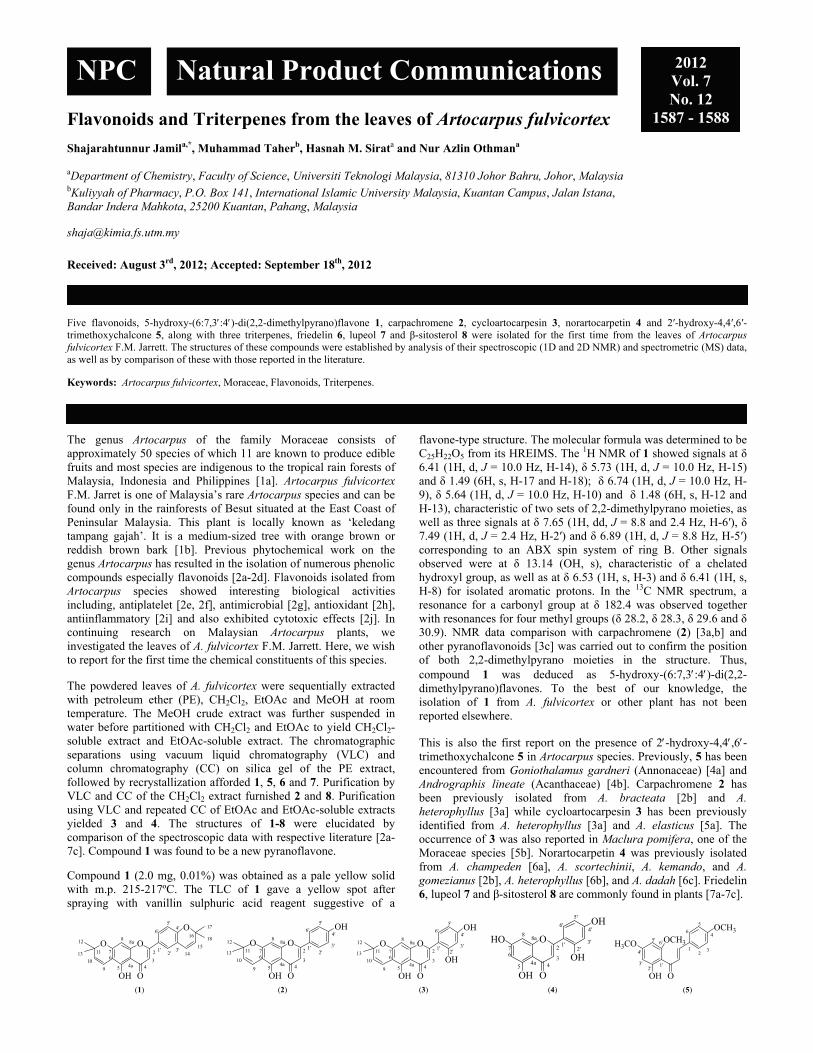
Flavonoids and Triterpenes from the leaves of Artocarpus fulvicortex Shajarahtunnur Jamila,*, Muhammad Taherb, Hasnah M. Sirata and Nur Azlin Othmana aDepartment of Chemistry, Faculty of Science, Universiti Teknologi Malaysia, 81310 Johor Bahru, Johor, Malaysia
bKuliyyah of Pharmacy, P.O. Box 141, International Islamic University Malaysia, Kuantan Campus, Jalan Istana, Bandar Indera Mahkota, 25200 Kuantan, Pahang, Malaysia
Received: August 3rd, 2012; Accepted: September 18th, 2012
Five flavonoids, 5-hydroxy-(6:7,3:4)-di(2,2-dimethylpyrano)flavone 1, carpachromene 2, cycloartocarpesin 3, norartocarpetin 4 and 2′-hydroxy-4,4′,6′-trimethoxychalcone 5, along with three triterpenes, friedelin 6, lupeol 7 and β-sitosterol 8 were isolated for the first time from the leaves of Artocarpus fulvicortex F.M. Jarrett. The structures of these compounds were established by analysis of their spectroscopic (1D and 2D NMR) and spectrometric (MS) data, as well as by comparison of these with those reported in the literature. Keywords: Artocarpus fulvicortex, Moraceae, Flavonoids, Triterpenes. The genus Artocarpus of the family Moraceae consists of approximately 50 species of which 11 are known to produce edible fruits and most species are indigenous to the tropical rain forests of Malaysia, Indonesia and Philippines [1a]. Artocarpus fulvicortex F.M. Jarret is one of Malaysia’s rare Artocarpus species and can be found only in the rainforests of Besut situated at the East Coast of Peninsular Malaysia. This plant is locally known as ‘keledang tampang gajah’. It is a medium-sized tree with orange brown or reddish brown bark [1b]. Previous phytochemical work on the genus Artocarpus has resulted in the isolation of numerous phenolic compounds especially flavonoids [2a-2d]. Flavonoids isolated from Artocarpus species showed interesting biological activities including, antiplatelet [2e, 2f], antimicrobial [2g], antioxidant [2h], antiinflammatory [2i] and also exhibited cytotoxic effects [2j]. In continuing research on Malaysian Artocarpus plants, we investigated the leaves of A. fulvicortex F.M. Jarrett. Here, we wish to report for the first time the chemical constituents of this species. The powdered leaves of A. fulvicortex were sequentially extracted with petroleum ether (PE), CH2Cl2, EtOAc and MeOH at room temperature. The MeOH crude extract was further suspended in water before partitioned with CH2Cl2 and EtOAc to yield CH2Cl2-soluble extract and EtOAc-soluble extract. The chromatographic separations using vacuum liquid chromatography (VLC) and column chromatography (CC) on silica gel of the PE extract, followed by recrystallization afforded 1, 5, 6 and 7. Purification by VLC and CC of the CH2Cl2 extract furnished 2 and 8. Purification using VLC and repeated CC of EtOAc and EtOAc-soluble extracts yielded 3 and 4. The structures of 1-8 were elucidated by comparison of the spectroscopic data with respective literature [2a-7c]. Compound 1 was found to be a new pyranoflavone.
Compound 1 (2.0 mg, 0.01%) was obtained as a pale yellow solid with m.p. 215-217ºC. The TLC of 1 gave a yellow spot after spraying with vanillin sulphuric acid reagent suggestive of a
flavone-type structure. The molecular formula was determined to be C25H22O5 from its HREIMS. The 1H NMR of 1 showed signals at δ 6.41 (1H, d, J = 10.0 Hz, H-14), δ 5.73 (1H, d, J = 10.0 Hz, H-15) and δ 1.49 (6H, s, H-17 and H-18); δ 6.74 (1H, d, J = 10.0 Hz, H-9), δ 5.64 (1H, d, J = 10.0 Hz, H-10) and δ 1.48 (6H, s, H-12 and H-13), characteristic of two sets of 2,2-dimethylpyrano moieties, as well as three signals at δ 7.65 (1H, dd, J = 8.8 and 2.4 Hz, H-6′), δ 7.49 (1H, d, J = 2.4 Hz, H-2′) and δ 6.89 (1H, d, J = 8.8 Hz, H-5′) corresponding to an ABX spin system of ring B. Other signals observed were at δ 13.14 (OH, s), characteristic of a chelated hydroxyl group, as well as at δ 6.53 (1H, s, H-3) and δ 6.41 (1H, s, H-8) for isolated aromatic protons. In the 13C NMR spectrum, a resonance for a carbonyl group at δ 182.4 was observed together with resonances for four methyl groups (δ 28.2, δ 28.3, δ 29.6 and δ 30.9). NMR data comparison with carpachromene (2) [3a,b] and other pyranoflavonoids [3c] was carried out to confirm the position of both 2,2-dimethylpyrano moieties in the structure. Thus, compound 1 was deduced as 5-hydroxy-(6:7,3:4)-di(2,2-dimethylpyrano)flavones. To the best of our knowledge, the isolation of 1 from A. fulvicortex or other plant has not been reported elsewhere. This is also the first report on the presence of 2-hydroxy-4,4,6-trimethoxychalcone 5 in Artocarpus species. Previously, 5 has been encountered from Goniothalamus gardneri (Annonaceae) [4a] and Andrographis lineate (Acanthaceae) [4b]. Carpachromene 2 has been previously isolated from A. bracteata [2b] and A. heterophyllus [3a] while cycloartocarpesin 3 has been previously identified from A. heterophyllus [3a] and A. elasticus [5a]. The occurrence of 3 was also reported in Maclura pomifera, one of the Moraceae species [5b]. Norartocarpetin 4 was previously isolated from A. champeden [6a], A. scortechinii, A. kemando, and A. gomezianus [2b], A. heterophyllus [6b], and A. dadah [6c]. Friedelin 6, lupeol 7 and β-sitosterol 8 are commonly found in plants [7a-7c].
O
O
OOH
12
13
9
10
8a
4a3
2'3'
4'5'
6'
1'14
15
17
1816
2
45
7
8
11
O
6
O O
OH O
OH
12
13 11
10
9
67
88a
54a 4
3
2 1'2'
3'
4'
5'
6'
O O
OH O
OH
12
13 11
10
9
67
88a
54a 4
3
2 1'2'
3'
4'
5'
6'
OH
O
OH O
OH
67
88a
54a 4
3
2 1' 3'
4'
5'
6'
OH2'
HO
H3CO
OCH3
OOH
3'
23
4
5
6
14'
5' 6'
1'2'
OCH3
(1) (2) (3) (4) (5)
NPC Natural Product Communications 2012 Vol. 7 No. 12
1587 - 1588

1588 Natural Product Communications Vol. 7 (12) 2012 Jamil et al.
Experimental
Plant material: The leaves of Artocarpus fulvicortex J.M. Jarrett were collected from Besut, Terengganu in September 2007. A voucher specimen (HTBP 962) has been deposited at Putrajaya Botanical Garden Herbarium, Malaysia.
Isolation procedure: Sequential extraction of the dried powdered leaves (1.5 kg) of A. fulvicortex at room temperature using different polarity of organic solvents for 48 h each afforded the PE extract (16.1 g, 1.1%), the CH2Cl2 extract (18.0 g, 1.2%), the EtOAc extract (17.9 g, 1.2%), and the MeOH extract (78.5 g, 4.2%). The MeOH extract was further suspended in water, then sequentially partitioned with CH2Cl2 and then EtOAc to yield the CH2Cl2-soluble extract (AFLMD) (1.1 g, 1.4%) and the EtOAc-soluble extract (AFLME) (1.2 g, 1.5%). Fractionation of the leaves PE extract (15.0 g) by silica gel VLC afforded twenty five fractions of 100 mL each. Fractions with a similar TLC profile were combined to give six major fractions AFLP 1 - AFLP 6. Repeated CC of AFLP 3 (1.7 g), followed by PLC with hexane/Et2O (7:3) as the solvent system gave 1 (2.0 mg, 0.01%) as a pale yellow solid. Successive purification of AFLP 4 (3.4 g) using silica gel CC yielded 6 (115.8 mg, 0.72%) as white needles; 7 (23.6 mg, 0.15%) as a white powder and 5 (9.3 mg, 0.06%) as a yellow solid. Fractionation of the leaves CH2Cl2 extract (12.0 g) by VLC yielded seven major fractions. Purification of fraction 5 (1.6 g) by silica gel CC gave 446 fractions. Fractions 187-264 were combined and washed with hexane to afford 8 (112.2 mg, 0.62%) as white crystalline needles. Purification of fraction 6 (2.8 g) by silica gel CC yielded carpachromene 2 (99.2 mg, 0.55%) as pale yellow needles. Fractionation of the EtOAc extract (10.0 g) by
silica gel VLC afforded twenty fractions which were combined to yield seven major fractions (AFLE 1 - AFLE 7). Purification of AFLE 3 (0.9 g) by silica gel CC afforded 3 (21.2 mg, 0.12%) as pale yellow needles. Purification of the EtOAc-soluble fraction of the MeOH extract (1.0 g) using silica gel CC and hexane/EtOAc (7:3) as the solvent yielded 5 (13.6 mg, 0.14%) as a yellow solid. 5-Hydroxy-(6:7,3:4)-di(2,2-dimethylpyrano)flavone (1) MP: 215-217ºC. IR (KBr): 3441, 2923, 1655, 1606, 1127 cm-1. 1H NMR (400 MHz, CDCl3): 1.49 (6H, s, H-17 and H-18), 1.48 (6H, s, H-12 and H-13), 5.64 (1H, d, J = 10.0 Hz, H-10), 5.73 (1H, d, J = 10.0Hz, H-15), 6.41 (1H, d, J = 10.0 Hz, H-14), 6.41 (1H, s, H-8), 6.53 (1H, s, H-3), 6.74 (1H, d, J = 10.0 Hz, H-9), 6.89 (1H, d, J = 8.8 Hz, H-5'), 7.49 (1H, d, J = 2.4 Hz, H-2'), 7.65 (1H, dd, J = 8.8 Hz, 2.4 Hz, H-6'), 13.14 (1H, s, 5-OH). 13C NMR (CDCl3, 100 MHz): δ 28.2 (CH3), 28.3 (CH3), 29.6 (CH3), 30.9 (CH3), 72.6 (C), 77.9 (C), 94.9 (CH), 104.0 (CH), 105.4 (C), 105.5 (C), 109.3 (C), 115.5 (CH), 116.9 (CH), 121.5 (CH), 123.5 (C), 124.3 (CH), 127.4 (CH), 128.0 (CH), 131.6 (CH), 157.4 (C), 156.4 (C), 159.7 (C), 161.5 (C), 163.8 (C), 182.4 (C=O). MS (EI, 70 eV): m/z (%) 402 (27) [M+], 387 (100), 219 (29), 203 (13), 186 (32), 131 (12); HREIMS: m/z [M+] calcd. for C25H22O5: 402.14574; found 402.14618. Acknowledgments - The authors wish to thank the Ministry of Science, Technology and Innovations (MOSTI) for the financial support and the Dept. of Chemistry, Faculty of Science, Universiti Teknologi Malaysia for the facilities.
References
[1] (a) Burkhill IH. (1966) A dictionary of the economic products of the Malay Peninsula, Ministry of Agriculture and Co-operatives, Kuala Lumpur, 2nd. Edn., vol. 1, 249260. (b) Whitmore TC. (1978) Tree flora of Malaya, Longman, Kuala Lumpur, vol. 3, 119167.
[2] (a) Nomura T, Hano Y, Aida M. (1998) Isoprenoid-substituted flavonoids from Artocarpus plants (Moraceae). Heterocycles, 47, 11791205; (b) Hakim EH, Achmad SA, Juliawaty LD, Makmur L, Syah YM, Aimi N, Kitajima M, Takayama H, Ghisalberti EL. (2006) Prenylated flavonoids and related compounds of the Indonesian Artocarpus (Moraceae). Journal of Natural Medicine, 60, 161184; (c) Jamil S, Sirat HM, Jantan I, Aimi N, Kitajima M. (2008) A new prenylated dihydrochalcone from the leaves of Artocarpus lowii. Journal of Natural Medicine, 62, 321324; (d) Musthapa I, Latip J, Takayama H, Juliawaty LD, Hakim EH, Syah YM. (2009) Prenylated flavones from Artocarpus lanceifolius and their cytotoxic properties against P-388 cells. Natural Product Communications, 4, 927-930; (e) Weng JR, Chan SC, Lu YH, Lin HC, Ko HH, Lin CN (2006) Antiplatelet prenylflavonoids from Artocarpus communis. Phytochemistry, 67:824829; (f) Jantan I, Mohd Yasin YH, Jamil S, Sirat HM, Basar N. (2010) Effect of prenylated flavonoids and chalcones isolated from Artocarpus species on platelet aggregation in human whole blood. Journal of Natural Medicine, 64, 365369; (g) Kuete V, Ango PY, Fotso GW, Kapche GDWF, Dzoyem JP, Wouking AG, Ngadjui BT, Abegaz BM (2011) Antimicrobial activities of the methanol extract and compounds from Artocarpus communis (Moraceae). BMC Complementary and Alternative Medicine, 11, 4246; (h) Jayasinghe L, Balasooriya BAIS, Padmini WC, Hara N, Fujimoto Y. (2004) Geranyl chalcone derivatives with antifungal and radical scavenging properties from the leaves of Artocarpus nobilis. Phytochemistry, 65, 12871290. (i) Wei BL, Weng JR, Chiu PH, Hung CF, Wang JP, Lin CN (2005) Anti-inflammatory flavonoids from Artocarpus heterophyllus and Artocarpus communis. Journal of Agricultural and Food Chemistry, 53, 38673871. (j) Hsu C-Lin, Shyu M-H, Lin J-A, Yen G-C, Fang S-C (2011) Cytotoxic effects of geranyl flavonoid derivatives from the fruit of Artocarpus communis in SK-Hep-1 human hepatocellular carcinoma cells. Food Chemistry, 127, 127134.
[3] (a) Zheng ZP, Cheng K-W, To JT-K, Li H, Wang M (2008) Isolation of tyrosinase Inhibitors from Artocarpus heterophyllus and use of its extract as antibrowning agent. Molecular Nutrition and Food Research, 52, 15301538; (b) Jain AC, Khazanchi R, Kumar A (1978) Synthesis of Carpachromene. Chemistry Letters, 9, 991994; (c) Hano Y, Itoh N, Hanaoka A, Nomura T (1995) Paratocarpins F-L, seven new isoprenoid-substituted flavonoids from Paratocarpus venenosa Zoll. Heterocycles, 41, 23132326.
[4] (a) Seidel V, Bailleu, F, Waterman PG. (2000) (Rel)-1b,2a-di-(2,4-dihydroxy-6-methoxybenzoyl)-3b,4a-di-(4-methoxyphenyl)-cyclobutane and other flavonoids from the aerial parts of Goniothalamus gardneri and Goniothalamus thwaitesii. Phytochemistry, 55, 439446. (b) Kishore PH, Reddy MVB, Reddy MK, Gunasekar D, Caux C, Bodo B. (2003) Flavonoids from Andrographis lineate Phytochemistry, 63, 457461.
[5] (a) Kijjoa A, Cidade HM, Pinto MMM, Gonzalez MJTG, Anantachoke C, Gedris TE, Herz W. (1996) Prenylflavonoids from Artocarpus elasticus Phytochemistry, 43, 691694. (b) Monache DM, Rosa MCD, Scurria R, Vitali A, Cuteri A, Monacelli B, Pasqua G, Botta B (1995) Comparison between metabolite productions in cell culture and in whole plant of Maclura pomifera. Phytochemistry, 39, 575580.
[6] (a) Hakim EH, Juliawaty LD, Syah YM, Achmad SA (2005) Molecular diversity of Artocarpus champeden (Moraceae): A species endemic to Indonesia. Molecular Diversity, 9, 149158; (b) Lin C-N, Lu C-M, Huang P-L (1995) Flavonoids from Artocarpus heterophyllus. Phytochemistry, 39, 14471451. (c) Su B-N, Cuendet M, Hawthorne ME, Kardono LBS, Riswan S, Fong HHS, Mehta RG, Pezzuto JM, Kinghorn AD (2002) Constituents of the barks and twigs of Artocarpus dadah with cyclooxygenase inhibitory activity. Journal of Natural Products, 65, 163169.
[7] (a) Agarwal RB, Rangari VD (2003) Antiinflammatory and antiarthritic activities of lupeol isolated from Strobilanthus callosus and Strobilanthus ixiocephala Roots. Indian Journal of Pharmacology, 35, 384387. (b) Queiroga CL, Silva GF, Dias PC, Possenti A, Carvalho JE (2000) Evaluation on the antiulcerogenic activity of friedelin-3β-ol and friedelin isolated from Maytenus ilicifolia (Celastraceae). Journal of Ethnopharmacology, 72, 465468. (c) Dhakal RC, Rajbhandari M, Kalauni, SK, Awale S, Gewali MB (2008) Phytochemical constituents of the barks of Vitex negundo L. Journal of Nepal Chemical Society, 23, 8992.

Dihydrospinochalcone-A and Epi-flemistrictin-B, Natural Isocordoin Derivatives from the Root Extract of Lonchocarpus xuul Fabiola Escalante-Erosaa, Brenda González-Moralesa, Ramiro F. Quijano-Quiñonesb, Gumersindo Miron-Lópezc and Luis M. Peña-Rodríguez*,a aLaboratorio de Química Orgánica, Unidad de Biotecnología, Centro de Investigación Científica de Yucatán, Calle 43 No. 130, Colonia Chuburná, Mérida, Yucatán, México
bLaboratorio de Química Farmacéutica and c Laboratorio de Resonancia Magnética Nuclear, Facultad de Química, Universidad Autónoma de Yucatán, Calle 41 No. 421 Col. Industrial, C.P. 97150, Mérida, Yucatán, México [email protected]
Received: December 26th, 2011; Accepted: July 15th, 2012
Purification of the root extract of Lonchocarpus xuul resulted in the isolation and identification of dihydrospinochalcone A (1) and epi-flemistrictin B (2) as additional natural isocordoin derivatives. Identification was based on the analysis of their spectroscopic data and by comparing these with those of previously reported metabolites. Keywords: Lonchocarpus xuul, Leguminosae, Chalcone, Isocordoin, Dihydroisocordoin, Dihydrospinochalcone A, Epi-flemistrictin B.
Lonchocarpus xuul Lundell (Leguminosae) is a tree endemic to the Yucatan Peninsula where it is known as “xuul”, “kanxuul” or “yaax-xuul” [1]. Previous phytochemical investigations of the root extract resulted in the isolation and identification of a number of flavonoids [2a-2c], some with antiprotozoal and cytotoxic activity [3]. In continuing our search for bioactive metabolites from the Yucatan native flora, we report herein the isolation and identification of dihydroespinochalcone A (1), and epi-flemistrictin B (2), as two additional natural isocordoin derivatives from the root extract of L. xuul. Concentration of the n-hexane extract of the roots of L. xuul yielded a pure metabolite whose 1H and 13C NMR spectra showed a number of similarities with those of dihydroisorcordoin (3), a previously reported chalcone from L. xuul [1]; the similarities included signals for a monosubstituted aromatic ring B and for a saturated ketone. However, a two vinylic-proton multiplet at 5.26, together with four signals for methyl groups bonded to quaternary sp2 carbons ( 1.69, 1.77, 1.78, and 1.82), and an isolated aromatic proton singlet at 7.35, indicated the presence of two isoprenyl groups in a pentasubstituted ring A. These findings were confirmed by both the parent ion peak (m/z 378) and the suggested molecular formula (C25H30O3) obtained from the mass spectrum of the purified metabolite. On the basis of these data, and by comparison with those reported in the literature, the isolated metabolite was identified as dihydrospinochalcone A (1), a new natural chalcone whose unsaturated relative, spinochalcone A, has been previously reported from the roots of Tephrosia spinosa (Leguminosae) [4]. The second metabolite showed a protonated parent ion peak at m/z 325 in its mass spectrum, indicating a molecular formula of C20H20O4 for the parent metabolite, and suggesting it having a structure with eleven unsaturation sites. The 1H and 13C NMR data of the purified metabolite proved to be very similar to those reported for flemistrictin B (4), a natural derivative of isocordoin first isolated from the leaves of Flemingia stricta [5] and recently reported from the roots of L. xuul [1]. However, while the 1H NMR spectrum of 4 shows the H -2” signal as a doublet of doublets
(2) (3) (1) (4)
(J= 8.4, 9.6 Hz) at 4.78 [1], the same proton appears as an AB quartet (J= 5.2 and 17.4 Hz) centered at 2.98 in the 1H NMR spectrum of the new metabolite. These differences in both chemical shift and coupling pattern suggested that the two metabolites were epimeric in their C-2” stereochemistry and, on the basis of these results, we propose the name of epi-flemistrictin B for the new metabolite.
(a) (b)
Figure 1: Suggested stereochemistry at C-2’’ for epiflemistrictin B (a) and flemistrictin B (b). The fact that the stereochemistry at the C-2’’position of flemistrictin B (3) has not yet been established has been attributed to the possibility of its occurring naturally as a racemic mixture, resulting from the non-stereospecific cyclization between the phenolic hydroxyl group at C-4’ and the epoxidated prenylated chain at C-3’ [1]. In order to define the stereochemistry at the C-2’’ position of the epimeric chalcones, a theoretical calculation of the H-2’’ chemical shift was carried out using DFT [B3LYP/6-311+G(d,p)] and GIAO methodologies. The results obtained confirmed the significant differences in the chemical shift value for H-2’’ in each of the epimeric structures (Figures 1a and 1b), and showed that the H -2” in the alpha orientation (Figure 1a) appears at a significantly higher field (δ 3.41) when compared to that of the H-2’’ in the beta orientation (δ 4.72; Figure 1b). On the basis of
NPC Natural Product Communications 2012 Vol. 7 No. 12
1589 - 1590
O
HO
OHO
O
HO
OHO
HO
OH
H
HOO
OH
1´
2´3´
4´5´
6´
1´´
2´´3´´
O
1
2
3
4
5
6
H
HOO
OH
R
1´
2´3´
4´5´
6´
1´´
2´´3´´
H
HOO
OH
R
1´
2´3´
4´5´
6´
1´´
2´´3´´
R= C9H7O

1590 Natural Product Communications Vol. 7 (12) 2012 Erosa et al.
these calculations, the stereochemistry at the C-2’’ position of the epimeric chalcones could be assigned as those shown in Figures 1a and 1b for epi-flemistrictin B and flemistrictin B, respectively, and the structure of epi-flemistrictin B could be established as that depicted in 2. Experimental
General: IR, FT-IR Nicolet Magna Protégé 460 instrument; NMR, Bruker AMX-400 NMR spectrometer; GC-MS, Agilent Technologies 6890 N gas chromatograph coupled to a 5975B mass selective detector. Theoretical calculations: All quantum chemical calculations were performed using the Spartan’06 program [6]. Calculations were carried out and geometries were optimized without geometry constraints within the DFT [7] methodology using the B3LYP [8] functional. The triplet split valence polarized and diffuse 6-311+G(d,p) basis set was used for geometry optimization and frequency calculations. In addition, the stationary points were characterized with frequency calculations [8]; no imaginary frequencies were present in final geometries. Calculations of the isotropic shielding constant (σ) for the corresponding equilibrium geometries were carried out at the same level of calculation within the GIAO [9] methodology. Finally, the J couplings were calculated using the PC model software [10]. Plant material: Roots of L. xuul were collected in December 2008 from plants growing in a field located at Sierra Papacal in Yucatán, Mexico. The plant material was washed with tap water and dried, first for a week at room temperature, and then for 72 h in an oven at 55°C. Extraction and isolation: Dry and ground roots (4 Kg) of L. xuul were extracted 3 times first with n-hexane (each 14 L) and then with ethanol (each 14 L). Evaporation of the combined n-hexane extracts under reduced pressure produced 18.34 g (0.46 %) of crude extract and 1.6 g (0.04 %) of a yellow precipitate identified as dihydrospinochalcone A (1). Similarly, evaporation of the combined ethanol extracts yielded 166.95 g (5.22%) of crude extract, which was suspended in 1.25 L of a 3:2 water-methanol mixture and subjected to a liquid-liquid partition procedure with n-hexane (3 times; 2:1, 1:1, 1:1; v:v) to produce 5.1 g of a low-polarity fraction. Successive purifications of the low-polarity
fraction using VLC (column diameter: 7 cm; height: 7 cm), eluting with increasing amounts of acetone in n-hexane, followed by open column chromatography eluting with a mixture of n-hexane/acetone 8:2, resulted in the isolation of epi-flemistrictin B (2, 47.4 mg) in pure form. Dihydrospinochalcone A (1) Yellow powder. Rf: 0.34 (n-hexane-ethyl acetate 8:2); GC: tR 20.78 min. IR v max (film): 3395 (OH), 2929 (C-H), and 1618 (C=O) cm-1. 1H NMR (400 MHz, CDCl3): 6.77 (2H, d, H-2/H-6), 7.10 (3H, m, H-3,4,5), 7.35 (1H, s, H-6’), 3.42 (2H, d, J= 7.2 Hz, H-1’’), 3.25 (2H, d, J= 6.8 Hz, H-1’´’), 5.20 (2H, m, H-2’’/H-2´´´), 1.69 (3H, s, CH3), 1.78 (3H, s, CH3), 1.82 (3H, s, CH3), 1.77 (3H, s, CH3), 3.17 (2H, t, J= 7.6 Hz ), 2.97 (2H, t, J= 7.8 Hz), 6.20 (1H, s, OH-4’), 12.99 (1H, s, OH-2’). 13C NMR (100 MHz, CDCl3): 133.14 (C-1), 115.35 (C-2/C-6), 129.50 (C-3/C-4/C-5), 112.88 (C-1’), 161.17 (C-2’), 118.90 (C-3’), 159.95 (C-4’), 114.26 (C-5´), 128.70 (C-6´), 21.80 (C-1’’), 121.74 (C-2’’), 135.14 (C-3’’), 28.90 (C-1´´´), 121.30 (C-2´´´), 134.70 (C-3´´´), 17.88 (CH3), 17.90 (CH3), 25.80 (CH3/CH3) 40.01 (C-), 29.80 (C-), 203.91 (C=O). MS: m/z 378 [M+]. Epi-flemistrictin B (3) Yellow oil. Rf: 0.34 (n-hexane-ethyl acetate 8:2); GC: tR 22.81 min. 1H NMR (400 MHz, CDCl3): 7.65 (2H, m, H-2/H-6), 7.42 (3H, m, H-3,4,5), 6.44 (1H, d, J= 8.9 Hz, H-5’), 7.73 (1H,d, J= 9 Hz, H-6’), 3.89 (1H, t, H-2’’), 2.98 (2H,qAB, J= 5.2 Hz, 17.36, H-1’’), 1.35 (3H, s, CH3),1.49 (3H, s, CH3), 7.59 (1H, d, J= 15.48 Hz), 7.88 (1H, d, J= 15.48 Hz), 13.81 (1H, s, OH-2’). 13C NMR (100 MHz, CDCl3): 134.94 (C-1), 128.6 (C-2/C-6), 129.1 (C-3/C-5), 130.7 (C-4), 120.4 (C-1’), 164.5 (C-2’), 107.3 (C-3’), 159.7 (C-4’), 109.14 (C-5’), 129.1 ( C-6’), 69.01 (C-2’’), 25.8 (C-1’’), 78.3 (C-3’’), 22.2 (CH3), 24.9 (CH3), 120.4 (C-), 144.3 (C-), 192.0 (C=O). MS: m/z 324 [M+]. Acknowledgments - We wish to thank Carlos A. Cruz-Cruz, J. Alejandro Yam-Puc and Paulino Simá-Polanco for assistance in collecting the plant material.
References
[1] Yam-Puc A, Peña-Rodríguez LM. (2009) Isocordoin derivaties from the root exract of Lonchocarpus xuul. Journal of the Mexican Chemical Society, 53, 12-14.
[2] (a) Borges-Argáez R, Peña-Rodríguez LM, Waterman PG. (2002) Flavonoids from two Lonchocarpus species of the Yucatán Península. Phytochemistry, 60, 533-540; (b) Borges-Argáez R, Peña-Rodríguez LM, Waterman PG. (2000) Flavonoids from the stem bark of Lonchocarpus xuul. Phytochemistry, 54, 611-614; (c) Borges-Argáe R, Poot-Díaz ME, Waterman PG, Peña-Rodríguez LM. (2005) Additional flavonoids from Lonchocarpus xuul. Journalof the Brazilian Chemical Society, 16, 1078-1081.
[3] Borges-Argáez R, Balnbury L, Flowers A, Giménez-Turba A, Ruiz E, Waterman PG, Peña-Rodríguez LM. (2007) Cytotoxic and antiprotozoal activity of flavonoids from Lonchocarpus xuul. .Phytomedicine, 14, 530-533.
[4] Venkata Rao, Prasad R. (1992) The chalcones from Tephrosia spinosa. Phytochemistry, 31, 2121-2122 [5] Subrahmanyam K, Madhusudhana-Rao J, Vemuri VSS, Sivaram-Bacu S, Roy CP, Jagannadha Rao KV. (1982) New chalcones from leaves of
Flemingia stricta. Indian Journal of Chemistry, 21, 895-897. [6] Wavefunction, Inc. Irvine, CA. Spartan’06 [7] Kohn W, Sham LJ. (1965) Self-consistent equations including exchange and correlation effects. Physical Review, 140A, 1133-1138. [8] Lee C, Yang W, Parr RG. (1988) Development of the Colle-Salvetti conelation energy formula into a functional of the electron density. Physical
Review, 37B, 785-789. [9] Wolinski K, Hinton JF. Pulay P. (1990) Efficient implementation of the gauge-independent atomic orbital method for NMR chemical shift
calculations. Journal of the American Chemical Society, 112, 8251-8260. [10] PC model for Windows V7.00, Serena Software.
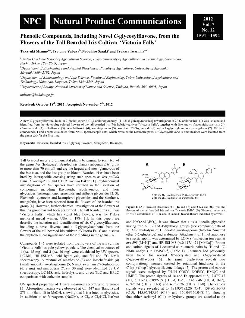
Phenolic Compounds, Including Novel C-glycosylflavone, from the Flowers of the Tall Bearded Iris Cultivar ‘Victoria Falls’ Takayuki Mizunoa*, Tsutomu Yabuyab, Nobuhiro Sasakic and Tsukasa Iwashinaa,d aUnited Graduate School of Agricultural Science, Tokyo University of Agriculture and Technology, Saiwai-cho, Fuchu, Tokyo 183−8509, Japan
bDepartment of Biochemistry and Applied Biosciences, Faculty of Agriculture, University of Miyazaki, Miyazaki 889−2192, Japan
cDepartment of Biotechnology and Life Science, Faculty of Engineering, Tokyo University of Agriculture and Technology, Naka-cho, Koganei, Tokyo 184−8588, Japan
dDepartment of Botany, National Museum of Nature and Science, Tsukuba, Ibaraki 305−0005, Japan [email protected]
Received: October 18th, 2012; Accepted: November 7th, 2012
A new C-glycosylflavone, luteolin 7-methyl ether 6-C-[β-arabinopyranosyl-(1→2)-β-glucopyranoside] (swertiajaponin 2″-O-arabinoside) (1) was isolated and identified from the violet blue colored flowers of the tall bearded iris (Iris hybrid) cultivar ‘Victoria Falls’, together with five known flavonoids, swertisin 2″-O-arabinoside (2), schaftoside (3), isoschaftoside (4), swertiajaponin (5), swertisin 2″-O-glucoside (6) and a C-glycosylxanthone, mangiferin (7). Of these compounds, 1 and 2 were elucidated from NMR spectroscopic data, which revealed the rotameric pairs. C-Glycosylflavone O-arabinosides were isolated from the genus Iris for the first time. Keywords: Iridaceae, Bearded iris, C-glycosylflavones, Mangiferin, Rotamers. Tall bearded irises are ornamental plants belonging to sect. Iris of the genus Iris (Iridaceae). Bearded iris plants (subgenus Iris) grow to more than 70 cm tall and are the largest and most glamorous of the Iris taxa, and the last group to bloom. Bearded irises have been bred by interspecific crossing using such species as Iris pallida Lam., I. variegata L. and I. kashimiriana Baker. [1]. Phytochemical investigations of Iris species have resulted in the isolation of compounds including flavonoids, isoflavonoids and their glycosides, benzoquinone, terpenoids and stilbene glycosides [2, 3]. Flavonols, quercetin and kaempferol glycosides and the xanthone, mangiferin, have been reported from the flowers of the bearded iris group [4]. However, further chemical investigation of the flowers of this iris group has not been performed. The tall bearded iris cultivar ‘Victoria Falls’, which has violet blue flowers, was the Dykes memorial medal winner, USA in 1984 [1]. In this paper, we describe the isolation and identification of six C-glycosylflavones, including a novel flavone, and a C-glycosylxanthone from the flowers of the tall bearded iris cultivar ‘Victoria Falls’ and discuss the phytochemical significance of these findings in the genus Iris. Compounds 1−7 were isolated from the flowers of the iris cultivar ‘Victoria Falls’ as pale yellow powders. The chemical structures of 1 (ca. 15 mg) and 2 (ca. 40 mg) were elucidated by UV spectra, LC-MS, HR-ESI-MS, acid hydrolysis, and 1H and 13C NMR spectroscopy. A mixture of schaftoside (3) and isoschaftoside (4) (small amount), swertiajaponin (5, 6 mg), swertisin 2″-O-glucoside (6, 8 mg) and mangiferin (7, ca. 30 mg) were identified by UV spectroscopy, LC-MS, acid hydrolysis, and direct TLC and HPLC comparisons with authentic samples. UV spectral properties of 1 were measured according to reference [5]. Absorption maxima were observed at λmax 347 nm (Band I) and 271 nm (Band II) in MeOH, showing that 1 is a luteolin glycoside. In addition to shift reagents (NaOMe, AlCl3, AlCl3/HCl, NaOAc
Figure 1: (A) Chemical structures of 1 (1a and 1b) and 2 (2a and 2b) from the flowers of the tall bearded iris cultivar ‘Victoria Falls’. (B) Observed important NOESY correlations of 1 (1a and 1b) and 2 (2a and 2b) are indicated by arrows. and NaOAc/H3BO3), it was shown that 1 is a luteolin glycoside having free 5-, 3′- and 4′-hydroxyl groups (see compound data of 1). Acid hydrolysis of 1 liberated swertiajaponin (luteolin 7-methyl ether 6-C-glucoside) and arabinose. Attachment of 1 mol arabinose to swertiajaponin was determined by LC-MS (molecular ion peak at m/z 595 [M+H]+) and HR-ESI-MS (m/z 617.1471 [M+Na]+). Proton and carbon signals of 1 occurred as rotameric pairs by 1H and 13C NMR analysis in DMSO-d6 (Table 1). Rotamers had previously been found for several X″-acetylated and O-glycosylated C-glycosylflavones [6]. The signal duplication reveals two conformational isomers created by rotational hindrance at the C (sp3)-C (sp2) glycosylflavone linkage [7]. The proton and carbon signals were assigned by 1H-1H COSY, NOESY, HMQC and HMBC. The proton signals of 1a and 1b appeared at δH 7.47/7.47 (1H, d, H-2′), 6.89/6.89 (1H, d, H-5′), 7.46/7.46 (1H, d, H-6′), 6.74/6.74 (1H, s, H-3) and 6.75/6.76 (1H, s, H-8). The carbon signals were revealed at δC 181.95/182.20 (C-4), 159.80/160.95 (C-5), 145.95/145.95 (C-3′) and 150.04/150.04(C-4′), showing that either carbonyl (C-4) or hydroxy groups are attached to the
NPC Natural Product Communications 2012 Vol. 7 No. 12
1591 - 1594

1592 Natural Product Communications Vol. 7 (12) 2012 Mizuno et al.
Table 1: 1H (500 MHz) and 13C NMR (125 MHz) spectral data (DMSO-d6) of 1a, 1b*, 2a and 2b* from the flowers of the tall bearded iris cultivar ‘Victoria Falls’.
Position 1a 1b* 2a 2b*
δ 1H (J in Hz) δ 13C δ 1H (J in Hz) δ 13C δ 1H (J in Hz) δ 13C δ 1H (J in Hz) δ 13C
Aglycone 2 164.18 165.46 165.59 164.09 3 6.74 (s) 103.18 6.74 (s) 103.12 6.87 (s) 103.28 6.85 (s) 103.22 4 181.95 182.20 182.18 182.43 5 159.80 160.95 159.90 161.05 6 108.71 108.76 108.82 108.88 7 164.03 163.90 164.03 163.96 8 6.75 (s) 90.66 6.76 (s) 90.12 6.80 (s) 90.91 6.81 (s) 90.36 9 157.10 156.99 157.24 157.12 10 104.15 104.43 104.27 104.56 1′ 121.37 121.37 121.93 121.93 2′ 7.47 (d, 2.3) 113.64 7.47 (d, 2.3) 113.59 7.97 (d, 9.2) 128.81 7.97 (d, 9.2) 128.78 3′ 145.95 145.95 6.90 (d, 9.2) 116.21 6.90 (d, 9.2) 116.21 4′ 150.04 150.04 161.53 161.53 5′ 6.89 (d, 8.5) 116.11 6.85 (d, 8.5) 116.11 6.90 (d, 9.2) 116.12 6.90 (d, 9.2) 116.12 6′ 7.46 (d, 8.5) 119.16 7.46 (d, 8.5) 119.21 7.97 (d, 9.2) 128.81 7.97 (d, 9.2) 128.78 7-OMe 3.87 (s) 56.29 3.88 (s) 56.61 3.87 (s) 56.43 3.88 (s) 56.74 6-C-Glucose
1 4.67 (d, 11.0) 70.81 4.64 (d, 10.0) 71.19 4.67 (d, 9.7) 70.93 4.65 (d, 9.9) 71.31 2 4.43 (t, 19.5) 81.39 4.25 (t, 19.5) 80.89 4.41 (t, 18.5) 81.50 4.25 (t, 18.0) 81.00 3 3.40 (m) 78.84 3.40 (m) 78.39 3.40 (m) 78.96 3.40 (m) 78.51 4 3.13 (m) 70.49 3.13 (m) 70.46 3.14 (m) 70.56 3.14 (m) 70.59 5 3.14 (m) 82.15 3.14 (m) 81.83 3.14 (m) 82.28 3.14 (m) 81.95 6 3.67 (d, 11.5) 61.55 3.67 (d, 11.5) 61.60 3.68 (d, 12.2) 61.66 3.68 (d, 12.2) 61.69 3.33 (m) 3.33 (m) 3.35 (m) 3.35 (m) 2″-O-Arabinose 1 4.15 (d, 7.5) 106.10 4.10 (d, 8.0) 106.16 4.15 (d, 7.6) 106.21 4.10 (d, 7.6) 106.28 2 2.82 (m) 74.22 2.82 (m) 74.17 2.83 (m) 74.29 2.83 (m) 74.34 3 2.98 (m) 76.44 2.98 (m) 76.27 2.98 (m) 76.56 2.98 (m) 76.39 4 3.01 (m) 69.35 3.01 (m) 69.41 3.00 (m) 69.51 3.00 (m) 69.47 5 3.22 (m) 65.91 3.22 (m) 65.74 3.22 (dd, 4.5, 11.5) 66.03 3.15 (m) 65.86 3.01 (m) 3.01 (m) 2.73 (m) 2.51 (m)
* Rotameric forms of 1 and 2. respective position. The attachment of a methoxyl group to the 7-position was shown by the occurrence of crosspeaks at 3.87/3.88 (3H, s, OMe) and 164.03/163.90 (C-7) in the long-range 1H-13C HMBC spectrum. Thus, 1a and 1b were shown to be swertiajaponin, glycosylated with arabinose. The C-C linkage between the aglycone and glucose at the 6-position was confirmed by the occurrence of crosspeaks at δH 4.67/4.64 (1H, d, Glc-1) and δC 108.71/108.76 (C-6), 164.03/163.90 (C-7) and 159.80/160.95 (C-5) in the HMBC spectrum. Since the anomeric protons of C-glucose at δH 4.67/4.64 exhibited large coupling constants (J = 11.0/10.0 Hz), the sugars were of the β-pyranose type. The 3JH,C correlations between δH 4.67/4.64 (Glc-1) and δC 82.15/81.83 (Glc-5) supported the pyranose structure of the C-glucosyl group. The β-pyranose structures of arabinose were shown by the 3JH,C correlation of δH 4.15/4.10 (1H, d, Ara-1) with δC 65.91/65.74 (Ara-5) and the large coupling constants (J = 7.5/8.0 Hz) of the anomeric protons of 1a and 1b, respectively. Moreover, HMBC correlation between the Ara-1 proton and Glc-2 carbon at δC 81.39/80.89 showed that the glucose-arabinose linkages are 1→2. Thus, 1 (1a and 1b) was identified as luteolin 7-methyl ether 6-C-[β-arabinopyranosyl-(1→2)-β-glucopyranoside] (swertiajaponin 2″-O-arabinoside), which is a new natural flavonoid [8]. The chemical structure of 1 (1a and 1b) and significant NOESY signals are shown in Figure 1. The absorption maxima of 2 at λmax 334 nm (Band I) and 272 nm (Band II) in MeOH, showed that 2 is an apigenin glycoside. In addition to shift reagents, it was shown that 2 is an apigenin glycoside with free 5- and 4′-hydroxyl groups (see compound data
of 2). Furthermore, swertisin (apigenin 7-methyl ether 6-C-glucoside) and arabinose were liberated by acid hydrolysis. Attachment of 1 mol arabinose to swertisin was confirmed by LC-MS (molecular ion peak at m/z 579 [M+H]+) and HR-ESI-MS (m/z 601.1535 [M+Na]+). These data suggested that 2 is swertisin X″-O-arabinoside. The proton and carbon signals of 2 (2a and 2b) appeared as rotameric pairs and were assigned by 1H−1H COSY, NOESY, HMQC and HMBC. Since the anomeric protons of C-glucose in the axial position occurred as a doublet with a large coupling constant (J = 9.7/9.9 Hz), the C-glucosyl groups were determined to be in the β-pyranose form. The 3JH,C correlations between δH 4.67/4.65 (Glc-1) and δC 82.28/81.95 (Glc-5) supported the pyranose structure of the C-glucosyl group of 2a and 2b. The 3JH,C correlations of δH 4.15/4.10 (1H, d, Ara-1) with δC 66.03/65.86 (Ara-5) and the large coupling constant (J = 7.6/7.6 Hz) of the Ara-1 protons of 2a and 2b showed that the arabinose had β-pyranose structures. HMBC correlations of δH 4.15/4.10 (Ara-1) to δC 81.50/81.00 (Glc-2) confirmed the arabinosyl-(1→2)-glucosyl linkages. Finally, the chemical structure of 2 (2a and 2b) was determined as apigenin 7-methyl ether 6-C-[β-arabinopyranosyl-(1→2)-β-glucopyranoside] (swertisin 2″-O-arabinoside), which has only been reported from the leaves of Achillea fragrantissima (Forssk) Sch. Bip (Asteraceae) [9]. However, since chemical data of the compound have not been reported, 1H and 13C NMR spectroscopic characteristics are recorded in Table 1. The chemical structure of 2 (2a and 2b) and significant NOESY signals are shown in Figure 1.
Phenolic compounds from Iris hybrid cultivar ‘Victoria Falls’ Natural Product Communications Vol. 7 (12) 2012 1593
Four known C-glycosylflavones and a C-glycosylxanthone were also isolated from the flowers. They were identified as schaftoside (apigenin 6-C-glucosyl-8-C-arabinoside, 3), isoschaftoside (apigenin 6-C-arabinosyl-8-C-glucoside, 4), swertiajaponin (5), swertisin 2″-O-glucoside (6) and mangiferin (7). Characterization was achieved by UV spectroscopy, LC-MS, acid hydrolysis and direct TLC and HPLC comparisons with authentic samples. Although another two xanthones, xanthone C-glycoside and mangiferin X″-O-glucoside, were also isolated, they could not be characterized because of the very small amounts obtained. To the best our knowledge, this is the first report of the isolation of luteolin 7-methyl ether 6-C-[β-arabinopyranosyl-(1→2)-β-glucopyranoside] in nature. Five known C-glycosylflavones, swertisin 2″-O-arabinoside, schaftoside, isoschaftoside, swertiajaponin and swertisin 2″-O-glucoside and a xanthone, mangiferin, were also found in the flowers of the tall bearded iris cultivar ‘Victoria Falls’. Flavones and xanthones are known as co-pigment substances with anthocyanins [10]. We presumed that the isolated flavones and xanthone contribute to the flower color of the tall bearded iris cultivar ‘Victoria Falls’. The distribution of flavonoids in the genus Iris has been reviewed [11]. More recently, flavonoids, including new ones, were reported from a few Iris species [2,12]. However, C-glycosylflavone O-arabinosides have not been found previously in the genus Iris. In this study, we isolated and identified two C-glycosylflavone O-arabinosides (1 and 2) from the flowers of the tall bearded iris for the first time. These C-glycosylflavone O-arabinosides are presumed to be contained only in the bearded iris group, e.g. Iris pallida and I. variegata, but further flavonoid investigations are necessary. Experimental
General experimental procedures: The isolated flavones and xanthone were identified by UV spectral survey, LC-MS, HR-ESI-MS, acid hydrolysis and direct TLC using: BAW (n−BuOH/HOAc/H2O = 4:1:5, upper phase), 15% HOAc, and BEW (n−BuOH/EtOH/H2O = 4:1:2.2), and HPLC comparisons with authentic samples. UV spectra were measured on a Shimadzu MPS−2000 multipurpose recording spectrophotometer (220−500 nm) according to Mabry et al. [5]. LC-MS were measured by a Shimadzu LC-MS system using a L−column 2 ODS (I.D. 2.1 × 100 mm, Chemical Evaluation and Research Institute, Japan), at a flow-rate of 0.2 mL min−1, a detection wavelength of 350 and 300 nm, and FMW (HCOOH/MeCN/H2O = 1:12:87) as eluant, ESI+ 4.5 kV, ESI− 3.5 kV, 250°C. Compounds 1 and 2 were analyzed by HR-MS using a microTOF-Q system (Bruker Daltonic, Bremen, Germany) in electrospray ionization (ESI) mode. Acid hydrolysis was performed in 12% aq. HCl for 30 min at 100°C. After cooling in water, the solution was shaken with diethyl ether. The aglycone and sugars were identified by HPLC (flavones) and PC (sugars) by comparison with authentic samples using BBPW (n−BuOH/benzene/pyridine/H2O = 5:1:3:3) and BTPW (n−BuOH/toluene/pyridine/H2O = 5:1:3:3) for sugars; 3−5 and 7 could not be hydrolyzed. Authentic samples of schaftoside, isoschaftoside, swertiajaponin, swertisin and mangiferin were obtained from the flowers and leaves of Iris rossii Baker [12] and swertisin 2″-O-glucoside (flavoayamenin) from the flowers of I. setosa Pallas var. nasuensis Hara [13]. HPLC comparisons with authentic specimens were performed using a Shimadzu HPLC system with a L−column2 ODS (I.D. 6.0×150 mm, Chemical Evaluation and Research Institute), at a flow-rate of 1.0 mL min−1, detection: 190−400 nm and eluent: MeCN/H2O/H3PO4 (12:88:0.2). NMR spectra (1H and 13C NMR, 1H-1H COSY, NOESY, HMBC
and HMQC) were measured on a JEOL JNM−ECA500 spectrometer (JEOL Ltd., Tokyo, Japan). Chemical shifts (δ) of 1H (500 MHz) and 13C (125 MHz) NMR are given in parts per million (ppm) relative to δH 2.49 /δC 39.6 for DMSO-d6.
Plant material: The tall bearded iris (Iris hybrid) cultivar ‘Victoria Falls’ was cultivated at the nursery of the Faculty of Agriculture, University of Miyazaki, Miyazaki Pref., Japan. Voucher specimen was deposited in the herbarium of the National Museum of Nature and Science, Japan (TNS). Extraction and isolation: The dried perianths were extracted with 0.1% HCl in MeOH. After concentration, the extracts were separated by preparative paper chromatography using as development systems: BAW, 15% HOAc and BEW. The isolated flavonoids and xanthone were purified by Sephadex LH-20 CC using 70% MeOH, and were further purified by preparative HPLC using Inertsil ODS-4 (I.D. 10×250 mm, GL-science Inc., Japan) at a flow-rate of 3.0 mL/min, injection of 300−350 μL, detection wavelength 340 nm, and an eluent of FMW2 (HCOOH/MeCN/H2O=1:18:81). Luteolin 7-methyl ether 6-C-[β-arabinopyranosyl-(1→2)-β-glucopyranoside] (swertiajaponin 2″-O-arabinoside) (1)
TLC: Rf 0.59 (BAW), 0.72 (15% HOAc), 0.51 (BEW); Color UV (365 nm)−dark purple, UV/NH3−yellow. HPLC: Rt 25.1 min. UV λmax (nm): MeOH 244, 258, 271, 347; +NaOMe 271, 396 (inc.); +AlCl3 277, 425; +AlCl3/HCl 260sh, 279, 295sh, 363, 390sh; +NaOAc 268, 402; +NaOAc/H3BO3 265, 374. 1H and 13C NMR: Table1. LC-MS: m/z 595 [M+H]+, 593 [M−H]−. HR-ESI-MS: m/z 617.1471 [M+Na]+ (Calcd for C27H30O15Na = 617.1477). Apigenin 7-methyl ether 6-C-[β-arabinopyranosyl-(1→2)-β-glucopyranoside] (swertisin 2″-O-arabinoside) (2)
TLC: Rf 0.49 (BAW), 0.54 (15% HOAc), 0.40 (BEW); Color UV (365 nm)−dark purple, UV/NH3−dark yellow. HPLC: Rt 49.7 min. UV λmax (nm): MeOH 272, 334; +NaOMe 275, 387 (inc.); +AlCl3 280, 303, 354, 380sh; +AlCl3/HCl 281, 302, 350, 370sh; +NaOAc 270, 390; +NaOAc/H3BO3 272, 340. 1H and 13C NMR: Table 1. LC-MS: m/z 579 [M+H]+ and 577 [M−H]−. HR-ESI-MS: m/z 601.1535 [M+Na]+ (Calcd for C27H30O14Na = 601.1528). Mixture of schaftoside and isoschaftoside (3 and 4)
TLC: Rf 0.29 (BAW), 0.33 and 0.44 (15% HOAc), 0.23 (BEW); Color UV (365 nm)−dark purple, UV/NH3−dark yellow. HPLC: Rt 19.8 (schaftoside) and 20.9 (isoschaftoside) min. UV λ max (nm): MeOH 273, 332; +NaOMe 283, 333, 400 (inc.); +AlCl3 278, 305, 353, 380sh; +AlCl3/HCl 278, 305, 351, 375sh; +NaOAc 282, 396; +NaOAc/H3BO3 275sh, 285, 321, 350sh, 405. LC-MS: m/z 565 [M+H]+ and 563 [M−H]−. Swertiajaponin (5)
TLC: Rf 0.47 (BAW), 0.41 (15% HOAc), 0.51 (BEW); Color UV (365 nm)−dark purple, UV/NH3−yellow. HPLC: Rt 23.7 min. UV λ max (nm): MeOH 243, 260sh, 270, 347; +NaOMe 274, 399 (inc.); +AlCl3 274, 422; +AlCl3/HCl 260sh, 277, 295sh, 364, 384sh; +NaOAc 266, 401; +NaOAc/H3BO3 266, 374. LC-MS: m/z 463 [M+H]+ and 461 [M−H]−.

1594 Natural Product Communications Vol. 7 (12) 2012 Mizuno et al.
Swertisin 2″-O-glucoside (6)
TLC: Rf 0.41 (BAW), 0.85 (15% HOAc), 0.54 (BEW); Color UV (365 nm)−dark purple, UV/NH3−dark yellow. HPLC: Rt 39.9 min. UV λ max (nm): MeOH 272, 334; +NaOMe 275, 384 (inc.); +AlCl3 260sh, 280, 301, 351, 380sh; +AlCl3/HCl 260sh, 282, 302, 350, 380sh; +NaOAc 269, 392; +NaOAc/H3BO3 272, 340. LC-MS: m/z 609 [M+H]+ and 607 [M−H]−. Mangiferin (7)
TLC: Rf 0.36 (BAW), 0.37 (15% HOAc), 0.54 (BEW); Color UV (365 nm)−orange, UV/NH3−bright yellow.
HPLC: Rt 10.0 min. UV λ max (nm): MeOH 241, 258, 316, 365; +NaOMe 273, 300sh, 350sh, 394; +AlCl3 268, 285sh, 352, 407; +AlCl3/HCl 263, 310sh, 336, 400; +NaOAc 271, 304, 387; +NaOAc/H3BO3 263, 285sh, 323, 359, 414. LC-MS: m/z 423 [M+H]+ and 421 [M−H]−. Acknowledgments - The authors thank Prof. Dr. Yoshihiro Ozeki, Dr Keiichi Noguchi, Mr Hiroshi Sekiguchi and Mr Motoki Yasunaga (Department of Biotechnology and Life Science, Tokyo University of Agriculture and Technology) for NMR and HR-ESI-MS research support and precious information.
References
[1] Austin C. (2005) Irises: A Gardener’s Encyclopedia. Timber Press, Inc., Oregon. [2] Fang R, Veitch NC, Kite GC, Howes MR, Porter HE, Simmonds MSJ. (2011) Glycosylated constituents of Iris fulva and Iris brevicaulis. Chemical
and Pharmaceutical Bulletin, 59, 124-128. [3] (a) Rahman AU, Nasim S, Baig I, Jahan IA, Sener B, Orhanand I, Choudhary MI. (2002) Isoflavonoid glycosides from the rhizomes of Iris
germanica. Chemical and Pharmaceutical Bulletin, 50, 1100-1102; (b) Singh N, Mahmood U, Kaul VK, Gupta AP, Jirovetz L. (2006) A new alkylated benzoquinone from rhizomes of Iris kumaonensis. Natural Product Research, 20, 75-78; (c) Farag SF, Takaya Y, Niwa M. (2009) Stilbene glucosides from the bulbs of Iris tingitana. Phytochemistry Letters, 2, 148-151.
[4] (a) Bate-Smith EC, Harborne JB. (1963) Mangiferin and other glycophenolics in Iris species. Nature, 198, 1307-1308; (b) Ashtakala SS, Forward DF. (1971) Pigmentation in iris hybrids: occurrence of flavonoid pigments and six cultivars of Iris germanica. Canadian Journal of Botany, 49, 1975-1979.
[5] Mabry TJ, Markham KR, Thomas MB. (1970) The Systematic Identification of Flavonoids. Springer, Berlin. [6] (a) Jay M. (1993) C-Glycosylflavonoids. In The Flavonoids: Advances in Research since 1986. Harborne JB. (Ed.). Chapman and Hall, London.
57-93; (b) Rayyan S, Fossen T, Nateland HS, Andersen ØM. (2005) Isolation and identification of flavonoids, including flavone rotamers, from the herbal drug ‘crataegi folium cum flore’ (hawthorn). Phytochemical Analysis, 16, 334-341; (c) Bjorøy Ø, Rayyan S, Fossen T, Kalberg K, Andersen ØM. (2009) C-glycosylanthocyanidins synthesized from C-glycosylflavones. Phytochemistry, 70, 278-287.
[7] Rayyan S, Fossen T, Andersen ØM. (2010) Flavone C-glycosides from seeds of fenugreek, Trigonella foenum-graecum L. Journal of Agricultural and Food Chemistry, 58, 7211-7217.
[8] (a) Harborne JB, Baxter H. (1999) Handbook of Natural Flavonoids. Vol. 1. Wiley, Chichester; (b) Andersen ØM, Markham KR. (2006) Flavonoids: Chemistry, Biochemistry, and Applications. Taylor & Francis, Boca Raton; (c) Veitch NC, Grayer RJ. (2008) Flavonoids and their glycosides, including anthocyanins. Natural Product Reports, 25, 555-611; (d) Veitch NC, Grayer RJ. (2011) Flavonoids and their glycosides, including anthocyanins. Natural Product Reports, 28, 1625-1695.
[9] Ahmed AA, Shalaby AM, Melek FR, Mabry TJ. (1988) Swertisin 2″-O-arabinoside, a new C-glycosylflavone from Achillea fragrantissima. Journal of Natural Products, 51, 971-972.
[10] Asen S, Stewart RN, Norris KH. (1972) Co-pigmentation of anthocyanins in plant tissues and its effect on color. Phytochemistry, 11, 1139-1144. [11] (a) Iwashina T, Ootani S. (1998) Flavonoids of the genus Iris: structures, distribution and function (review). Annals of the Tsukuba Botanical
Garden, 17, 147-183; (b) Wang H, Cui Y, Zhao C. (2010) Flavonoids of genus Iris (Iridaceae). Mini Reviews in Medicinal Chemistry, 10, 643-661. [12] Mizuno T, Okuyama Y, Iwashina T. (2012) Phenolic compounds from Iris rossii, and their chemotaxonomic and systematic significance.
Biochemical Systematics and Ecology, 44, 157-160. [13] Hayashi K, Ootani S, Iwashina T. (1989) Comparative analysis of the flavonoids and related compounds occurring in three varieties of Iris setosa in
the flora of Japan. Science Report of the Research Institute of Evolutionary Biology, 6, 30-60.
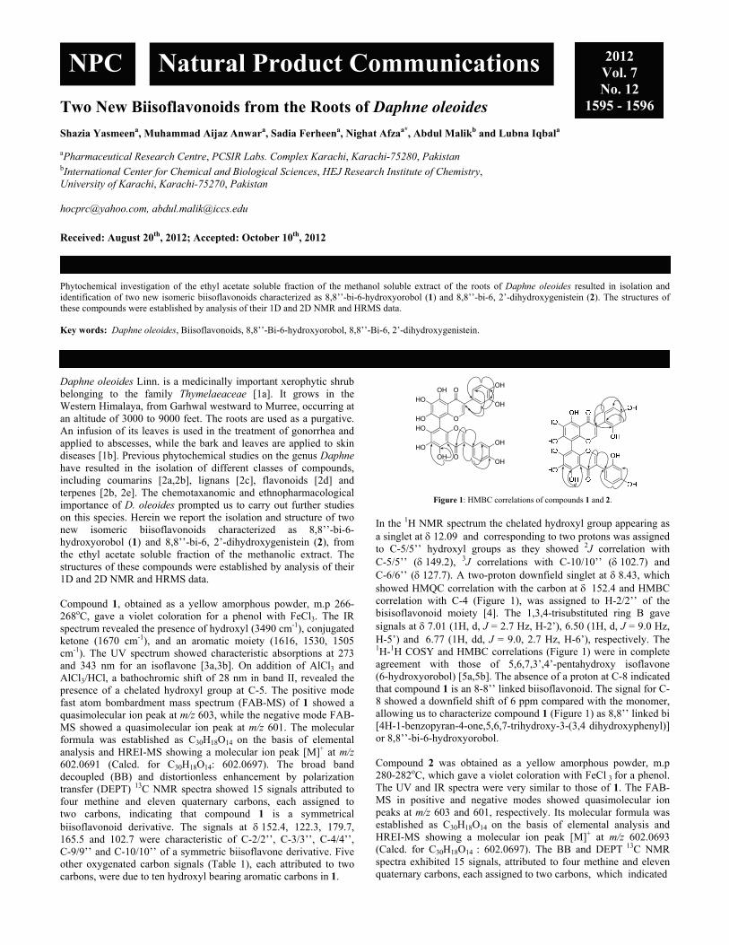
Two New Biisoflavonoids from the Roots of Daphne oleoides Shazia Yasmeena, Muhammad Aijaz Anwara, Sadia Ferheena, Nighat Afzaa*, Abdul Malikb and Lubna Iqbala aPharmaceutical Research Centre, PCSIR Labs. Complex Karachi, Karachi-75280, Pakistan
bInternational Center for Chemical and Biological Sciences, HEJ Research Institute of Chemistry, University of Karachi, Karachi-75270, Pakistan [email protected], [email protected]
Received: August 20th, 2012; Accepted: October 10th, 2012
Phytochemical investigation of the ethyl acetate soluble fraction of the methanol soluble extract of the roots of Daphne oleoides resulted in isolation and identification of two new isomeric biisoflavonoids characterized as 8,8’’-bi-6-hydroxyorobol (1) and 8,8’’-bi-6, 2’-dihydroxygenistein (2). The structures of these compounds were established by analysis of their 1D and 2D NMR and HRMS data. Key words: Daphne oleoides, Biisoflavonoids, 8,8’’-Bi-6-hydroxyorobol, 8,8’’-Bi-6, 2’-dihydroxygenistein. Daphne oleoides Linn. is a medicinally important xerophytic shrub belonging to the family Thymelaeaceae [1a]. It grows in the Western Himalaya, from Garhwal westward to Murree, occurring at an altitude of 3000 to 9000 feet. The roots are used as a purgative. An infusion of its leaves is used in the treatment of gonorrhea and applied to abscesses, while the bark and leaves are applied to skin diseases [1b]. Previous phytochemical studies on the genus Daphne have resulted in the isolation of different classes of compounds, including coumarins [2a,2b], lignans [2c], flavonoids [2d] and terpenes [2b, 2e]. The chemotaxanomic and ethnopharmacological importance of D. oleoides prompted us to carry out further studies on this species. Herein we report the isolation and structure of two new isomeric biisoflavonoids characterized as 8,8’’-bi-6-hydroxyorobol (1) and 8,8’’-bi-6, 2’-dihydroxygenistein (2), from the ethyl acetate soluble fraction of the methanolic extract. The structures of these compounds were established by analysis of their 1D and 2D NMR and HRMS data. Compound 1, obtained as a yellow amorphous powder, m.p 266-268oC, gave a violet coloration for a phenol with FeCl3. The IR spectrum revealed the presence of hydroxyl (3490 cm-1), conjugated ketone (1670 cm-1), and an aromatic moiety (1616, 1530, 1505 cm-1). The UV spectrum showed characteristic absorptions at 273 and 343 nm for an isoflavone [3a,3b]. On addition of AlCl3 and AlCl3/HCl, a bathochromic shift of 28 nm in band II, revealed the presence of a chelated hydroxyl group at C-5. The positive mode fast atom bombardment mass spectrum (FAB-MS) of 1 showed a quasimolecular ion peak at m/z 603, while the negative mode FAB-MS showed a quasimolecular ion peak at m/z 601. The molecular formula was established as C30H18O14 on the basis of elemental analysis and HREI-MS showing a molecular ion peak [M]+ at m/z 602.0691 (Calcd. for C30H18O14: 602.0697). The broad band decoupled (BB) and distortionless enhancement by polarization transfer (DEPT) 13C NMR spectra showed 15 signals attributed to four methine and eleven quaternary carbons, each assigned to two carbons, indicating that compound 1 is a symmetrical biisoflavonoid derivative. The signals at 152.4, 122.3, 179.7, 165.5 and 102.7 were characteristic of C-2/2’’, C-3/3’’, C-4/4’’, C-9/9’’ and C-10/10’’ of a symmetric biisoflavone derivative. Five other oxygenated carbon signals (Table 1), each attributed to two carbons, were due to ten hydroxyl bearing aromatic carbons in 1.
HO
HO O
O
OHO
HO
O
OH
OH
OH
OH
OH
OH
Figure 1: HMBC correlations of compounds 1 and 2.
In the 1H NMR spectrum the chelated hydroxyl group appearing as a singlet at 12.09 andcorresponding to two protons was assigned to C-5/5’’ hydroxyl groups as they showed 2J correlation with C-5/5’’ (149.2), 3J correlations with C-10/10’’ (102.7) and C-6/6’’ (127.7). A two-proton downfield singlet at 8.43, which showed HMQC correlation with the carbon at 152.4 and HMBC correlation with C-4 (Figure 1), was assigned to H-2/2’’ of the bisisoflavonoid moiety [4]. The 1,3,4-trisubstituted ring B gave signals at 7.01 (1H, d, J = 2.7 Hz, H-2’), 6.50 (1H, d, J = 9.0 Hz, H-5’) and 6.77 (1H, dd, J = 9.0, 2.7 Hz, H-6’), respectively. The 1H-1H COSY and HMBC correlations (Figure 1) were in complete agreement with those of 5,6,7,3’,4’-pentahydroxy isoflavone (6-hydroxyorobol) [5a,5b]. The absence of a proton at C-8 indicated that compound 1 is an 8-8’’ linked biisoflavonoid. The signal for C-8 showed a downfield shift of 6 ppm compared with the monomer, allowing us to characterize compound 1 (Figure 1) as 8,8’’ linked bi [4H-1-benzopyran-4-one,5,6,7-trihydroxy-3-(3,4 dihydroxyphenyl)] or 8,8’’-bi-6-hydroxyorobol. Compound 2 was obtained as a yellow amorphous powder, m.p 280-282oC, which gave a violet coloration with FeCl 3 for a phenol. The UV and IR spectra were very similar to those of 1. The FAB-MS in positive and negative modes showed quasimolecular ion peaks at m/z 603 and 601, respectively. Its molecular formula was established as C30H18O14 on the basis of elemental analysis and HREI-MS showing a molecular ion peak [M]+ at m/z 602.0693 (Calcd. for C30H18O14 : 602.0697). The BB and DEPT 13C NMR spectra exhibited 15 signals, attributed to four methine and eleven quaternary carbons, each assigned to two carbons, which indicated
NPC Natural Product Communications 2012 Vol. 7 No. 12
1595 - 1596

1596 Natural Product Communications Vol. 7 (12) 2012 Yasmeen et al.
Table 1: 1H- and 13C-NMR (DMSO-d6) data of compounds 1 and 2 at 400 and 100
MHz, respectively; in ppm, J in Hz.
position 1 2 J = Hz) C J = Hz) C
2/2’’ 8.43 (s) 152.4 8.59 (s) 151.53/3’’ - 122.3 - 123.1 4/4’’ - 179.7 - 180.7 5/5’’ - 149.2 - 148.7 6/6’’ - 121.7 - 122.5 7/7’’ - 156.1 - 155.7 8/8’’ - 98.3 - 98.4 9/9’’ - 165.5 - 166.3
10/10’’ - 102.7 - 103.1 1’/1’’’ - 124.3 - 113.6 2’/2’’’ 7.01 (d, J = 2.7) 115.3 - 156.2 3’/3’’’ - 145.5 7.16 (d, J = 2.1) 106.3 4’/4’’’ - 143.2 - 160.8 5’/5’’’ 6.50 (d, J = 9.0) 117.5 7.09 (dd, J = 8.7, 2.1) 109.2 6’/6’’’ 6.77 (dd, J = 9.0, 2.7) 120.6 7.81 (d, J = 8.7) 131.4
5/5’’-OH 12.09 (s) - 12.15 (s)
that compound 2 is a symmetrical biisoflavonoid derivative. It showed close similarity to those of 1, except for minor differences in the chemical shifts of the carbons of ring B/ring E. The 1H NMR spectrum of 2 was also very similar to that of 1 except for the notable differences in the chemical shifts of ring B/ring E, as well as HMBC correlations [7.81 (1H, d, J = 8.7 Hz), 7.16 (1H, d, J = 2.1 Hz) and 7.09 (1H, dd, J = 8.7, 2.1 Hz)]. The 1H-1H COSY and HMBC correlations (Figure 1) were in conformity to a 1,2,4-trisubtituted ring B. The spectral data showed close agreement to those of 6-hydroxygenestein and 5,6,7,2’,4’-pentahydroxy isoflavone (6, 2’-dihydroxygenistein) [6] thereby indicating that compound 2 is a regioisomer of compound 1. Compound 2 could therefore be characterized as 8,8’’ linked bi [4H-1-benzopyran-4-one,5,6,7-trihydroxy-3-(2,4-dihydroxyphenyl)] or 8,8’’-bi-6,2’-dihydroxygenistein. Experimental
General: UV measurements were obtained on a Hitachi-UV-3200 spectrophotometer, and IR spectra on a Jasco-302-A spectrophotometer. Aluminum sheets 4.5 x 10 cm, coated with silica gel 60 F254 were used for analytical TLC and compounds were visualized under UV light and detections were made at 254 nm and by spraying with ceric sulfate reagent. Column and flash chromatography were carried out on silica gel (230-400 mesh E. Merck). HPLC analyses were carried out on a JAIGEL using an ODS-M 80 column. Melting points (m.p.) were recorded on a Gallenkamp apparatus and were uncorrected. NMR experiments were performed on a Bruker AMX 400 MHz spectrometer. Chemical shifts ( in ppm) are referenced to the carbon (C 39.52) and residual proton (H 2.50) signals of (CD3)2SO. FAB-MS were recorded on a Jeol-JMS HX-110 mass spectrometer. EI-MS and HREI-MS were conducted on a Jeol JMS-DA-500 mass
spectrometer. Elemental analyses were performed on a Carlo Elba MOD-1106 Elemental Analyzer. Plant material: The roots of Daphne oleoides (15 kg) were collected from Peshawar (Pakistan) in May 2010 and identified by Prof. Manzoor Husain, Plant Taxonomist, Govt. Postgraduate College-1, Abbotabad, KPK, Pakistan, where a voucher specimen has been deposited. Extraction and isolation: Dried and ground roots (15 kg) of Daphne oleoides were extracted with MeOH (3 × 50 L) at room temperature. The combined MeOH extracts were filtered and concentrated under reduced pressure to yield 800 g of gummy residue, which was suspended in H2O and fractionated into n-hexane (40 g), ethyl acetate (EtOAc) (60 g), and n-BuOH (80 g) soluble fractions. The EtOAc soluble fraction was subjected to column chromatography (CC) over silica gel eluting with mixtures of n-hexane and CHCl3 in increasing order of polarity. The fraction obtained from CHCl3-MeOH (9:1) was subjected to recycling HPLC (ODS-M 80 column, 2 mL/min) and eluted with H2O/MeOH (1:1) resulting in the isolation of compounds 1 (10 mg) and 2, (13 mg). 8,8’’-Bi-6-hydroxyorobol (1) Yellow amorphous powder. MP: 266-268°C. IR (KBr) max cm-1: 3490 (OH), 1670 (conjugated ketone), 1616, 1530, 1505 (aromatic C=C). UV (MeOH) (max) (log ε): 273 (4.17), 343 (3.92) (sh) nm 1H and 13C NMR: Table 1 (+)-FAB-MS m/z: 603 [M+H]+; (˗)-FAB-MS m/z: 601 [M-H]- HREI-MS m/z: [M]+ calcd. for C30H18O14: 602.0697; found: 602.0691 Elemental analysis: Calcd. C 59.81, H 3.01, O 37.18; found C 59.79, H 3.04, O 37.09 8,8’’-Bi-6, 2’-dihydroxygenistein (2) Yellow amorphous powder. MP: 280-282°C. IR (KBr) max cm-1: 3510 (OH), 1675 (conjugated ketone), 1610, 1530, 1510 (aromatic C=C). UV (MeOH) (max) (log ε): 275 (4.29), 347 (3.52) (sh) nm 1H and 13C NMR: Table 1 (+)-FAB-MS m/z: 603 [M+H]+; (˗)-FAB-MS m/z: 601 [M-H]- HREI-MS m/z: [M]+ calcd. for C30H18O14: 602.0697; found: 602.0693 Elemental analysis: Calcd. C 59.81, H 3.01, O 37.18; found C 59.76, H 3.07, O 37.15.
References
[1] (a) Baquar SR. (1989) Medicinal and Poisonous Plants of Pakistan, Printas Press Karachi, Pakistan. 161; (b) Ullah N, Ahmed S, Ahmed Z, Khan PM, Malik A. (1999) Dimeric guaianolides from Daphne oleoides. Phytochemistry, 51, 559-562.
[2] (a) Kreher B, Neszmelyi A, Hildebert W (1990) Triumbellin, a tricoumarin rhamnopyranoside from Daphne mezereum. Phytochemistry, 29, 3633-3637; (b) Ullah N, Ahmed S, Malik A. (1999) A dicoumarin glycoside from Daphne oleoides. Phytochemistry, 51, 99-101; (c) Riaz M, Ullah N, Mehmood A, Rabnawaz H, Malik A, Afza N. (2000) Furanoid and furofuranide lignans from Daphne oleoides. Zeitschrift für Naturforschung, 55b, 1216-1220; (d) Taniguchi M, Baba K. (1996) Three biflavonoids from Daphne odora. Phytochemistry, 42, 1447-1453; (e) Ullah N, Ahmed Z. Ahmed S, Khan PM, Malik A. (1999) A pentacyclic triterpene from Daphne oleoides. Phytochemistry, 50, 839-841.
[3] (a) Markham KR. (1982) Techniques of Flavonoids Identification, Academic Press, New York; (b) Anouar EH, Gierschner J, Duroux JL, Trouillas P. (2012) UV/Visible spectra of natural polyphenols: A time dependant density functional theory study. Food Chemistry, 131, 79-89.
[4] Wong SM, Konno C, Oshima Y, Pezzuto JM, Fong HHS, Farnsworth NR. (1987) Irisones A and B: Two new isoflavones from Iris missouriensis, Journal of Natural Products, 50, 178-180.
[5] (a) Horie T, Sasagawa M, Torii F, Kawamura Y, Yamashita K. (1996) Studies of the selective O-alkylation and dealkylation of flavonoids. XX. A convenient method for synthesizing 5,6,7-trihydroxyflavones and 5,6-dihydroxy-7-methoxyisoflavones. Chemical and Pharmaceutical Bulletin, 44, 486-491; (b) Kulling SE, Honig DM, Simat TJ, Metzler M. (2000) Oxidative in vitro metabolism of the soy phytoestrogens daidzein and genistein, Journal of Agricultural and Food Chemistry, 48, 4963-4972.
[6] Markham KR, Mabry TJ, Swift TW. (1968) New isoflavones from the genus Baptisia (Leguminosae). Phytochemistry, 7, 803-808.

Biflavonoids from the Unripe Fruits of Clusia paralicola and their Antioxidant Activity Rafaela Ferreira Oliveiraa, Celso Amorim Camaraa, Maria de Fátima Agrab and Tania Maria Sarmento Silvaa* aDepartamento de Ciencias Moleculares, Universidade Federal Rural de Pernambuco, Recife, Pernambuco, CEP 52171-900, Brazil
bLaboratório de Tecnologia Farmacêutica, Universidade Federal da Paraíba, João Pessoa, Paraiba, CEP 58051-970, Brazil [email protected]
Received: October 27th, 2011; Accepted: October 6th, 2012
Investigation of the green fruits of Clusia paralicola (Clusiaceae) led to the isolation and characterization of two 3,8”-biflavonoids, 2R, 3S, 2”R, 3”R-GB1-7”-O--glucoside (1) and 2R, 3S, 2”R, 3,8”-binaringenin-7”-O--glucoside (2), together with four known compounds: -sitosterol, stigmasterol, -amyrin, and epicatechin. The structures were established from the IR, LC-ESI-MS and NMR spectral data, including 2D-NMR experiments. The absolute configurations of 1 and 2 were determined by CD spectra. The total extract and the biflavonoids demonstrated significant antioxidant activity in DPPH, ABTS, and -carotene/linoleic acid tests. Keywords: Clusiaceae, Clusia paralicola, Biflavonoid glucosides, Antioxidant activity. The genus Clusia (Clusiaceae) occurs in the neotropical region and comprises around 250 species of semi-epiphytes, vines, shrubs and trees [1, 2]. C. paralicola G. Mariz Cunha is native and endemic to northeastern Brazil, such as in the states of Paraiba, Pernambuco, Bahia and Alagoas, in areas of Caatinga and Atlantic Forest [3], and is popularly known as pororoca. Previous studies of C. paralicola showed the presence of xanthone, an uncommon prenylated dihydrophenanthrene, paralycoline A [4, 5] and new biphenyls with DNA strand-scission activity [6]. In the present investigation, fractionation of an extract of the unripe fruits of C. paralicola led to the isolation of compounds 1-6. We report herein the isolation and structure elucidation of biflavonoids 1 and 2. Although a wide variety of compounds, both aromatic and nonaromatic, including biflavonoids of the Garcinia type [4] have been isolated from Clusia species earlier, to our knowledge this is the first report of these biflavonoids from this source. In our continuing search for potent antioxidants from natural sources [7,8], we found that the ethanolic extract, the fractions thereof, and compounds 1 and 2 displayed antioxidant activity. The UV spectra of compounds 1 and 2 were typical of biflavonones with an absorption maximum at 285 nm, followed by a shoulder at 330 nm [9], and showed an [M+H]+ ion at m/z 721 and 705 in its ESI-MS, corresponding to the molecular formula, C36H32O16 and C36H32O15, respectively. In addition, the ESI-MS fragments at m/z 559 and 543 for 1 and 2, revealed the loss of glucose in each case. The structure of 1 was deduced from detailed analysis of 1H and 13C NMR data aided by 2D NMR experiments (1H-1H COSY, HSQC, and HMBC) (Table 1). Duplication of signals (in a ratio 1:0.6) in the 1H NMR spectrum of the optically active 1 in DMSO-d6 suggests the existence of two conformers at room temperature. This observation has been previously reported for the biflavanone GB-1 and could be attributed to the hindered rotation between the flavanone and the flavanonol moieties around the C-3/C-8 axis [10]. For this type of molecules, the existence of rotational isomers at
O
OH O
OH
OH
O
OH O
OH
O
OH
OH
OH
O
OH
R
2
34
5
6
7
8 1'
2'
3'4'
5'
6'
2"
3"4"
5"6"
7"8" 1'"
2'"
3'"4'"
5'"
6'"
1""2""
3""
4""5""
6""
1 R=OH2 R=H
Figure 1: Structures of biflavonoids 1 and 2 from fruits of Clusia paralicola.
ambient temperature is explained by the strong intra- and intermolecular H-bonding that occurs due to the many OH groups present [11]. Biflavonoid 1 (Figure 1) showed singlets for OH protons, two at low fields at H 12.12 and 11.66, attributed to the H-bonded OH-(C5) and OH-(C5”), respectively (Table1). The doublets at H 7.23, 7.18, and 6.68, 6.60 (J=8.5 Hz) could be assigned to the AA’BB’ spin system of the para-substituted B rings H-2”’/6’’’, H-2’/6’, H3”’/5”’, and H-3’/5’, respectively. Two doublets for two protons at H 5.89 and 5.75 (J=2.0 Hz) were accordingly located at H-6 and H-8, respectively. H-6” exhibited a singlet at H 6.21. In the same way, the remaining three doublets at H 5.45 (J=12 Hz), 5.06 (J=12 Hz) and 5.03 (J=11 Hz), and one doublet of doublets at H 4.30 (J=11 and 6 Hz) for one proton each were assigned to H-2, H2”, H-3, and H-3”, respectively. The vicinal coupling constants (11-12 Hz) J [H-C(2), H-C(3)] and J [H-C(2”), H-C(3”)] established the trans-diaxial arrangements of these pairs. The resonances of the β-glucose moiety were observed as follows: an anomeric proton H-1”” at H 4.73 (d, J=7.5 Hz), with methylene protons H-6”” at H 3.44 (m) and 3.70 (d, J=11 Hz) and the other
NPC Natural Product Communications 2012 Vol. 7 No. 12
1597 - 1600

1598 Natural Product Communications Vol. 7 (12) 2012 Oliveira et al.
Table 1: 1H (500 MHz) and 13C NMR (125 MHz) spectroscopic data for 1 and 2 (major conformer) (DMSO-d6, δ in ppm).
1 2 Position C H 2JCH 3JCH C H 2 82.1 5.45 (d, 12.0) 81.8 5.55 (d, 12) 3 46.7 5.06 (d, 12.0) 46.6 5.09 (d, 12) 4 196.9 H-3 H-2 196.9 5 163.6 H-6 163.5 6 95.9 5.89 (d, 2.0) H-8 95.9 6.18 (s) 7 168.9 H-6,
H-8 166.5
8 94.8 5.75 (d, 2.0) 94.9 5.79 (d, 2.0) 9 163.2 H-8 163.1 10 101.1 H-6, H-8 101.3 1’ 127.0 H-3’,
H-5’ 128.4
2’, 6’ 129.8 7.18 (d, 8.5) H-2 128.5 7.25 (d, 8.5) 3’, 5’ 114.8 6.60 (d, 8.5) 115.1 6.60 (d, 8.5) 4’ 157.6 H-3’,
H-5’ H-2’, H-6’
157.9
2” 82.7 5.06 (d, 11.0) 78.4 5.48 (dd; 12.5, 3.0)
3” 72.4 4.30 (dd; 11.0, 6.0)
42.1 3.05 (dd; 17.5, 13.0) 2.75 (dd; 17.5, 3.0)
4” 198.2 H-3 H-2 197.6 5” 161.8 H-6 162.0 6” 96.0 6.21 (s) 95.9 6.18 (s) 7” 162.8 H-6 H-1””,
H-3 162.8
8” 102.7 H-3 H-6” 103.3 9” 158.6 H-3 160.1 10” 102.7 1”’ 127.4 H-2” H-3”,
H-3’”, H-5’”
128.1
2”’, 6”’ 128.3 7.23 (d, 8.5) 128.0 7.19 (d, 8.5) 3”’, 5”’ 114.9 6.68 (d, 8.5) 115.1 6.68 (d, 8.5) 4”’ 157.5 H-3’”,
H-5’” H-2’”, H-6’”
157.6
1”” 99.9 4.73 (d, 7.5) 99.9 4.72 (d, 7.0) 2”” 73.0 3.18 (m) 73.0 3.17 (m) 3”” 77.1 3.66 (m) 77.1 3.37 (m) 4”” 69.6 3.09 (m) 69.6 3.11 (m) 5”” 76.1 3.15 (m) 76.8 3.16 (m) 6”” 60.6 3.70 (m)
3.44 (m) 60.5 3.66 (m)
3.44 (m) OH-C(5)/C(5”)
12.12 (s) 11.66 (s)
12.14 (s)
OH-C(3) 5.85 (d, 6.5)
protons absorbing at H 3.15-3.66. The APT spectrum displayed low field signals at C 198.2 and 196.9, typical of carbonyl groups of flavanone and flavanonol. The signals at C 129.8, 128.3, 114.8, and 114.9 could be assigned to the carbons 2’, 6’, 2”’, 6”’, 3’, 5’, 3”’, and 5”’ to IB and IIB rings in the AA’BB’ system, respectively. The signals at C 72.4 and 46.7 represented the aliphatic C-atoms of the 3-hydroxyflavanone and flavanone moieties, respectively. Similarly, the signals at C 82.7 and 82.1 were assigned to C-2” and C-2, respectively. By comparison with 13C NMR data of other related 3,8”-biflavonoids, I-3,II-8-binaringenin or GB-1a (Garcinia biflavonoid) [10], the measured resonances of 1 could be attributed to a 3,8”-biflavonoid consisting of 3-hydroxyflavonone and flavanone moieties. The connectivity in the molecule, particularly the 3,8”-linkage of the flavonoid, was confirmed by HMBC correlations (Table 1). The glycosidic linkage site of β-D-glucose at C-7 was further verified by the HMBC experiments in which the anomeric proton at H 4.73 showed a cross peak with the C C-7 at 168.9. Detailed analyses of the HSQC and HMBC spectra allowed for the assignments of all the 1H- and 13C-NMR signals for both the glucosyl and aglycone moieties. The 1H- and 13C-NMR data for compound 2 (Figure 1) were similar to those of 1, except for the signals at H 5.48 (H-2”; dd, J=12.5 and 3.0 Hz), H 3.05 (H-3"; dd, J=17.5 and 13.0 Hz), H 2.75 (H-3”; dd, J=17.5 and 3.0 Hz) and C 42.1 (C-3”). The absolute configurations of the two biflavonoids were determined by CD spectra. The CD spectrum of biflavonoid 1 revealed Cotton effects due to the electronic np*
and p* transitions of the two constituent structural moieties: the 3-substituted flavanone upper unit (I) and the flavanonol lower unit (II) (Figure 2). The observed Cotton effects were similar to those observed for the flavonoids ent-naringeninyl-(I-3α,II-8)-4’-O-methylnaringenin [12] and GB1 [13]. The positive Cotton effect near 330 nm for the np* transition and the negative Cotton effect near 300 nm for the p* transition may be assigned to the lower flavanonol unit. Similarly, the negative Cotton effect (shoulder) near 325 nm for the np* transition and the positive Cotton effect near 280 nm for the p* transition may be assigned to the upper flavanone unit. Application of Gaffield’s rule then permits assignment of (2R, 3S) and (2”R, 3”R) absolute configurations for the flavanone and flavanonol moieties, respectively [14]. Therefore, the structure of compound 1 could be established unambiguously as 2R, 3S, 2”R, 3”R-GB1-7”-O--glucoside. The CD spectrum of 2 was similar to biflavanoid 1 (Figure 2) and the absolute configuration was established as 2R, 3S, 2”R, 3,8”-binaringenin-7”-O--glucoside. GB1-7”-O--glucoside was reported to exist in Garcinia kola, but no data for structural identification was published previously. Although 3,8”-binaringenin-7”-O--glucoside was earlier isolated from Garcinia multiflora [16], the 13C NMR and CD presented here are, to our knowledge, new data. 3,8”-Biflavanoids have been isolated mainly from Garcinia, Rheedia, and Allanblakia species (Clusiaceae), and have recently been isolated from Clusia columnaris [17]. Compounds 3 (epicatechin) [18], 4 (-sitosterol) [19], 5 (stigmasterol) [19], and 6 (-amyrin) [20] were identified by comparison with published spectroscopic data.
250 300 3500
20000
40000
60000
80000
100000
-20
-10
0
10
Wavelength [nm]
Figure 2: Upper curves: CD spectra of 1 (dotted line) and 2 (solid line). Lower curves: UV spectra of 1 (dotted line) and 2 (solid line). Table 2: Total phenolics and antioxidant activity of extracts, fractions and biflavonoids from Clusia paralicola.
Sample Total phenolic content (mg GAE/g ± SD)
DPPH (EC50)
a ABTS (EC50) -Carotene
bleaching (% O.I.)b
EtOH extract 132.1±1.3 12.7±0.1 7.4±0.4 65.6±3.8 n-Hexane fraction
42.9±3.2 17.4±0.1 18.6±0.1 68.3±9.8
AcOEt fraction 135.4±3.2 10.8±0.1 6.2±0.3 70.6±3.2 MeOH:H2O fraction
94.8±3.3 13.9±0.2 13.9±0.2 82.4±2.3
Biflavonoids 60.2±1.6 26.7±3.3 53.2±2.7 Ascorbic acid 2.1±0.0 2.8±0.2 Trolox 77.4±1.8
Mean value ± standard deviation: n=3 aConcentration of antioxidant required to reduce the original amount of radicals by 50 %. bOxidation inhibition at 60 min.
The total phenolic content and the antioxidant activity of the EtOH extract, n-hexane, EtOAc, and MeOH: H2O fractions were evaluated by DPPH and ABTS radical scavenging, as well as -carotene/linoleic acid system activity assays and the data were compared with those of the reference antioxidants, ascorbic acid and Trolox (Table 2). The extract and fractions tested displayed antioxidant activity in all of the test methods. Biflavonoids 1 and 2 displayed moderate antioxidant activity in all the assays. The

Biflavonoid glucosides from Clusia paralicola Natural Product Communications Vol. 7 (12) 2012 1599
EtOAc fraction showed activity similar to that of the EtOH extract in all tests. The EtOH extract and fractions showed correlation (Pearson correlation, r, data not shown) with the total phenolic content (Table 2). Experimental
General: Melting points were determined on a Koefler hot stage and are uncorrected. The infrared absorption spectra were recorded in KBr pellets, using a Varian 640 FT-IR spectrophotometer with a PIKE ATR accessory operating in the 4000-400 cm-1 range. The LC-ESI-MS was obtained in positive electrospray mode using an Esquire 3000 Plus (Bruker). Silica gel 60 was used for CC, and silica gel 60 F254 (E. Merck) for TLC plates. Sephadex®LH-20 (Sigma) was employed for gel permeation chromatography. 1H and 13C NMR spectra were obtained using a Bruker DRX 500 (500 MHz for 1H and 125 MHz for 13C) and Bruker DPX300 (300 MHz for 1H and 75 MHz for 13C) in DMSO-d6. The CD was recorded with a Jasco J-515 CD spectrometer. The optical rotation was determined in a KRUESS OPTRONIC spectrometer. All solvents used were of commercial HPLC grade. Plant material: The unripe fruits of Clusia paralicola were collected in Santa Rita city, State of Paraiba, Brazil in May 2010. The plant was identified by Maria de Fátima Agra. Voucher specimen (M. F. Agra et al. 7455) is deposited at the Herbarium Lauro Pires Xavier, Universidade Federal da Paraiba. Extraction and isolation: The plant material (280.0 g) was extracted successively with EtOH to give 40.0 g of dry extract. This was dissolved in MeOH: H2O (1:1) and successively fractionated with n-hexane and AcOEt. The AcOEt fraction (8.0 g) was successively submitted to CC using Sephadex® LH-20 and silica gel to yield 1 (50.0 mg), 2 (20.0 mg) and 3 (25.0 mg), as yellow amorphous solids. The n-hexane fraction (10.0 g) was subjected to CC using Sephadex® LH-20. Fractions 4 and 5 were submitted to CC on silica gel using n-hexane, EtOAc and MeOH to furnish the mixture of 4 and 5 (11.0 mg) and 6 (35.0 mg). 2R, 3S, 2”R, 3”R-GB1-7”-O--glucoside (1) MP: 223-225ºC. D: -11.9 (c 0.23, MeOH).
IR (KBr) max: 3400 (OH), 1640 (C=O), 1600, 1520 (C=C from aromatic rings). 1H and 13C NMR (DMSO-d6): Table 1. LC-ESI-MS (positive mode) mz: 721 [M+H]+ (C36H32O16). 2R, 3S, 2”R, 3,8”-binaringenin-7”-O--glucoside (2) MP: 218-220ºC; D: -35.1 (c 0.67, MeOH). IR (KBr) max: 3400 (OH), 1640 (C=O), 1600, 1520 (C=C from aromatic rings). 1H and 13C NMR (DMSO-d6): Table 1. LC-ESI-MS (positive mode) mz: 705 [M+H]+ (C36H32O15). Determination of total phenolic content: Total soluble phenolic contents of the EtOH extract, and the n-hexane, AcOEt, and MeOH: H2O fractions (1 mg/mL) was determined with the Folin-Ciocalteu reagent according to the method of Slinkard & Singleton [21], with modification by using gallic acid as a standard phenolic compound. DPPH• radical scavenging assay, ABTS+• radical cation decolorisation assay, and antioxidant activity in linoleic acid oxidation: The free radical-scavenger activity (DPPH) [7], the radical cation decolorization assay (ABTS) [22], and antioxidant activity in linoleic acid oxidation [23] of the EtOH extract, and n-hexane, AcOEt, MeOH:H2O fractions and the biflavonoids was determined as described previously. Statistical analysis: All samples were analyzed in triplicate unless stated otherwise and the results are expressed as average ± standard deviation. All statistical analyses were carried out using the Microsoft Excel software package (Microsoft Corp., Redmond, WA). Correlation analysis of antioxidant activity versus the total phenolic content was carried out using the correlation and regression program in Graphpad Prism 5.0 DEMO (GraphPad Software, San Diego, California USA). Acknowledgements - The authors thank CNPq (TMSS, CAC and MFA research fellowships), CAPES (ROF graduate fellowship) and FACEPE (PRONEM APQ-1232.1.06/10 and APQ-0158-1.06/08 grant numbers) for financial support, CENAPESQ and CETENE analytical centers for the recorded data kindly provided by them. ROF also thanks PRPPG-UFRPE for their kind fellowship support. The authors also thank Dr J. Bhattacharyya for his kind suggestions.
References
[1] Bittrich V, Amaral MCE. (1996) Flower morphology and pollination biology of some Clusia species from the Gran Sabana (Venezuela). Kew Bulletin, 51, 681-694.
[2] Bittrich V, Amaral MCE. (1997) Floral biology of some Clusia species from Central Amazonia. Kew Bulletin, 52, 617-635. [3] Forzza RC, Baumgratz JFA, Costa A, Hopkins M, Leitman PM, Lohmann LG, Martinelli G, Morim MP, Coelho MAN, Peixoto AI, Pirani JR,
Queiroz LP, Stehmann JR, Walter BMT, Zappi D. (2010) In Catálogo de plantas e fungos do Brasil: As angiospermas do Brasil. Forza RC. Forza RC, Leitman PM, Costa A, Carvalho JrAA, Peixoto AL, Walter BMT, Bicudo C, Zappi D, Costa DP, Lleras E, Martinelli G, Lima HC, Prado J, Stehmann JR, Baumgratz JFA, Pirani JR, Sylvestre LS, Maia LC (Eds). Jardim Botânico do Rio de Janeiro, Rio de Janeiro. pp 857.
[4] Delle Monache F, Delle Monache G, Botta B, Gacs-Baitz E. (2002) A biphenyl, a dihydrophenanthrene and a xanthone from Clusia paralycola. Heterocycles, 56, 589-597.
[5] Delle Monache F, Delle Monache G, Cavalcanti JF, Pinheiro RM. (1987) An unexpected dihydrophenanthrene from Clusia paralycola. Tetrahedron Letters, 28, 563-566.
[6] Seo EK, Huang L, Wall ME, Wani MC, Navarro H, Mukherjee R, Farnsworth NR, Kinghorn AD. (1999) New biphenyl compounds with DNA strand-scission activity from Clusia paralicola. Journal of Natural Products, 62, 1484-1487.
[7] Silva TMS, Camara CA, Lins ACS, Barbosa JM, Silva EMS, Freitas BM, Santos FAR. (2006) Chemical composition and free radical scavenging activity of pollen loads from stingless bee Melipona subnitida Ducke. Journal of Food Composition and Analysis, 19, 507-511.
[8] Silva TMS, Camara CA, Lins ACS, Agra MF, Silva SEM, Reis IT, Freitas BM. (2009) Chemical composition, botanical evaluation and screening of radical scavenging activity of collected pollen by the stingless bees Melipona rufiventris (Uruçu-amarela). Anais da Academia Brasileira de Ciências, 81, 173-178.
[9] Mabry TJ, Markhan KR, Thomas MB. (1970) The Systematic Identification of Flavonoids. Springer-Verlag, New York. [10] Han QB, Lee SF, Qiao CF, He ZD, Song JZ, Sun HD, Xu HX. (2005) Complete NMR assignments of the antibacterial biflavonoid GB1 from
Garcinia kola. Chemical & Pharmaceutical Bulletin, 53, 1034-1036.

1600 Natural Product Communications Vol. 7 (12) 2012 Oliveira et al.
[11] Jackson B, Locksley HD, Scheinmann F, Wolstenholme WA. (1971) Extractives from Guttiferae. Part XXII. The isolation and structure of four novel biflavanones from the heartwoods of Garcinia buchananii Baker and G. eugeniifolia Wall. Journal of the Chemical Society C, 3791-3804.
[12] Mbwambo ZH, Kapingu MC, Moshi MJ, Machumi F, Apers S, Cos P, Ferreira D, Marais JPJ, Berghe DV, Maes L, Vlietinck A, Pieters L. (2006) Antiparasitic activity of some xanthones and biflavonoids from the root bark of Garcinia livingstonei. Journal of Natural Products, 69, 369-372.
[13] Ferrari J, Terreaux C, Kurtan T, Szikszai-Kiss A, Antus S, Msonthi JD, Hostettmann K. (2003) Isolation and online LC/CD analysis of 3,8′′-linked biflavonoids from Gnidia involucrata. Helvetica Chimica Acta, 86, 2768-2778.
[14] Gaffield W. (1970) Circular dichroism, optical rotatory dispersion and absolute configuration of flavanones, 3-hydroxyflavanones and their glycosides - determination of aglycone chirality in flavanone glycosides. Tetrahedron, 26, 4093-4108.
[15] Okunji CO, Ware TA, Hicks RP, Iwu MM, Skanchy DJ. (2002) Capillary electrophoresis determination of biflavanones from Garcinia kola in three traditional African medicinal formulations. Planta Medica, 68, 440-444.
[16] Chen FC, Lin YM, Hung, JG. (1975) A new biflavanone glucoside from Garcinia multiflora. Phytochemistry, 14, 818-820. [17] Compagnone RS, Suarez AC, Leitao SG, Monache FD. (2008) Flavonoids, benzophenones and a new euphane derivative from Clusia columnaris
Engl. Revista Brasileira de Farmacognosia, 18, 6-10. [18] Agrawal PK. (1989) Carbon-13 NMR of Flavonoids. Elsevier, New York. [19] Goulart MOF, Sant’ana AEG, de Lima RA, Cavalcante SH, de Carvalho MG, Filho RB. (1993) Fitoconstituintes químicos isolados de Jatropha
elliptica. Atribuição dos deslocamentos químicos dos átomos de carbono e hidrogênio dos diterpenos jatrofolanos A e B. Quimica Nova, 16, 95-100.
[20] Maia RM, Barbosa PR, Cruz FG, Roque NF, Fascio M. (2000) Triterpenos da resina de Protium heptaphyllum March (Bourseraceae): caracterização em misturas binárias. Quimica Nova, 23, 623-626.
[21] Slinkard K, Singleton VL (1977) Total phenol analyses: automation and comparison with manual methods. American Journal of Enology and Viticulture, 28, 49-55.
[22] Re R, Pellegrini N, Proteggente A, Pannala A, Yang M, Rice-Evans C. (1999) Antioxidant activity applying an improved ABTS radical cation decolorization assay. Free Radical Biology & Medicine, 26, 1231-1237.
[23] Emmons CL, Peterson DM, Paul GL. (1999) Antioxidant capacity of oat (Avena sativa L.) extracts. 2. In vitro antioxidant activity and contents of phenolic and tocol antioxidants. Journal of Agricultural and Food Chemistry, 47, 4894-4898.

Ochnaflavone and Ochnaflavone 7-O-methyl Ether two Antibacterial Biflavonoids from Ochna pretoriensis (Ochnaceae) Tshepiso J. Makhafola, Babatunde B. Samuel, Esameldin E. Elgorashi and Jacobus N. Eloff* Phytomedicine Programme, Department of Paraclinical Sciences, Faculty of Veterinary Science, University of Pretoria, Private Bag x04, Onderstepoort, 0110, South Africa [email protected] Received: August 18th, 2012; Accepted: October 10th, 2012
The acetone extract of Ochna pretoriensis was evaluated for antibacterial activity using bioautography and serial microplate dilution methods against four nosocomial bacterial pathogens namely: Escherichia coli, Staphylococcus aureus, Enterococcus faecalis and Pseudomonas aeruginosa. A bioass ay-guided fractionation of th e crude extract led to the isolation of two antibacter ial biflavonoids, ochnaflavone and o chnaflavone 7-O-methyl ether. Gram-negative bacteria were more sensitive to t he isolated compounds than the Gram-positive bacteria (MIC values: 31.3 µg/mL for P. aeruginosa and 62.5 µg/mL for S. aureus). In addition, the isolated com pounds were assessed for their pot ential toxic effects in the MTT toxicity assay using monkey kidney vero cells and Ames genotoxicity test using Salmonella typhimurium strain TA98. LC50 values were 125.9 µg/mL for ochnaflavone and 162.0 µg/mL for ochnaflavone 7-O- methyl ether. The isolated co mpounds have selectivity index values r anging from 1.29 to 4.03. Selectivity index values high er than one indicate that test samples are less toxic to the host cells than to the pathogens. The biflavonoids did not have any mutagenic effects in the Salmonella/microsome assay without metabolic activation. Keywords: Ochna pretoriensis, Antibacterial activity, Ochnaflavone, Ochnaflavone 7-O-methyl ether. Antibiotic resistance and the ever increasing development of multi-drug resistant by pathogenic microbes are of concern. This resistance has resulted in an increased incidence of inf ectious diseases leading to high morbidity and mortality [1]. As a result the search for new antimicrob ial/anti-infective drugs (from plants) is imperative to counter resistance development and to avert or del ay the arrival of a pos t-antibiotic era [2] . Plants have been us ed throughout history to cure and prevent infectious diseases and thus present an obvious source of new antimicrobial compounds [3]. The genus Ochna, (Ochnaceae) comprises 86 species of evergreen trees and shrubs and occurs widely in Africa, Asia and Madagascar. Biflavonoids isolated from this genus have diverse biological and pharmacological properties, which include antiviral, antihepatotoxic, anticancer and immune suppressive activities [4-6]. While several pharmacological activities have been reported for members of the genus Ochna, there has been no previous report of the antibacterial activity of m embers of this genus. In a previ ous study, we investigated the an tibacterial potential of several Ochna species from which we iden tified the m ost active ones [7]. This study was an a ttempt, to isolate the antibacterial constituents of O. pretoriensis. Two main antibacterial compounds were isolated using bioassay-guided fractionation and chemically characterised. Moreover, the potential toxic e ffects (cytotoxic and g enotoxic effects) of the isolated compounds were evalu ated using a tetrazolium based colorimetric assay (MTT assay) on Vero monkey kidney cells, and a b acterial based mutagenicity test in S. typhimurium (Ames tests), respectively. Bioassay-guided fractionation of the crude extract of the dried leaves of O. pretoriensis, followed b y column chromatography, yielded two compounds identified as ochnaflavone ( 1) and ochnaflavo ne 7-O-methyl ether ( 2) using 1H and 13C NMR spectroscopic and mass spectrometric analysis. The structures were confirmed by comparison of the NMR data with that in the literature [8].
O
OH
RO
O
OH
O
O
O OH
OH
Figure 1: Chemical structures of the two compounds isolated from O. pretoriensis. 1: R = H= ochnaflavone, 2: R = CH3 = ochnaflavone 7-O-methyl ether. The MIC of the tested crude extracts, different solvent-solvent fractions and the biflavonoids ochnaflavone and ochnaflavone 7-O-methyl ether are presented in Table 1. The most sensitive organism to both biflavonoids was P. aeruginosa with an MIC value of 31.3 µg/mL . E. faecalis was more sensitive to ochnaf lavone. Ochnaflavone had higher activity than its methoxy derivative. The cytotoxic potential of ochnaflavone (1) and ochnaflavone 7-O-methyl ether (2) was tested in the MTT assay using Vero monkey kidney cells. Ochnaflavone and ochnaflavone 7-O-methyl ether had low cytotoxicity compared with berberine . The LC50 values were 125.9 µg/mL for ochnaflavone and 162.0 µg/mL for ochnaflavone 7-O-methyl ether. Berberine, the positive con trol, had a LC 50 of 39.0 µg/mL. To evaluate p otential efficacy, tolerability and therapeutic antibacterial activity of the two compounds, selectivity index (SI) was calculated by dividing the LC50 in mg/mL by MIC in mg/mL. The selectivity Index (SI) values of the isolated compounds are presented in Table 2. The isolated compounds had positive, but low, selectivity indices ranging from 1.29 to 4.03. Results obtained from the mutagenicity and antimutagenicity test of ochnaflavone and ochnaflavon e 7-O-methyl ether using th e Salmonella typhimurium strain TA98, based o n the number of induced revertant colonies, are presented in Table 4. Test samples are considered mutagenic when the number of induced revertant
NPC Natural Product Communications 2012 Vol. 7 No. 12
1601 - 1604

1602 Natural Product Communications Vol. 7 (12) 2012 Makhafola et al.
Table 1: Minimal inhibitory concentrations (MIC) (µg/mL) of the crude extract, solvent-solvent fractions and compounds isolated from O. pretoriensis.
Bacteria Crude extract Chloroform Ethyl acetate 1 2 Gentamycin
E. coli 65 ± 0.002 156 ± 0.00 39 ± 0.00 41.6 ± 0.02 125 ± 0.00 7
P. aeruginosa 104 ± 0.004 78 ± 0.00 39 ± 0.00 31.3 ± 0.00 31.3 ± 0.00 8
S. aureus 104 ± 0.004 39 ± 0.00 39 ± 0.00 62.5 ± 0.00 125 ± 0.00 8.3
E. faecalis 39 ± 0.00 39 ± 0.00 39 ± 0.00 31.3 ± 0.00 62.5 ± 0.00 10
Table 2: Cytotoxic effects against vero cells LC50 (µg/mL) and selectivity index values of compounds isolated from O. pretoriensis.
Compounds LC50 E. coli P. aeruginosa S. aureus E. faecalis Average
1 125.9 ± 0.00 3.02 4.03 2.10 4.02 3.27
2 162.0 ± 0.00 1.29 5.18 1.29 2.59 2.28
Table 3: Mutagenic and antimutagenic activity of compounds isolated from O. pretoriensis in the frame shift of Salmonella typhimurium TA98 ± standard deviation.
MUTAGENICITY ANTIMUTAGENICITY
Compound 1000 µg/mL 500 µg/mL 50 µg/mL 1000 µg/mL 500 µg/mL 50 µg/mL
1 32.0 ± 0.00 37.3 ± 4.9 32.7 ± 3.0 182.7 ± 5.8 167.3 ± 4.9 178.7 ± 4.2
2 30.7 ± 0.00 40.7 ± 2.1 29.0 ± 4.0 166.7 ± 0.0 181.0 ± 3.2 178.3 ± 1.5
Solvent blank 28.8 ± 0.00 4-NQO (2 µg/mL) 210.0± 12.3
colonies is at least double that of the negative control. Both ochnaflavone and ochnaflavone 7-O-methyl ether had no potential mutagenic effect. Neither of the biflavonoids tested significantly modified the mutagenicity of the known mutagen 4-NQO. However, the slight reduction in the number of revertants was not dose dependent and indicative of toxicity. Ochnaflavone had been isolated previously from several members of the Ochnaceae family including O. squarrosa [9], O. pumila [10], and O. obtusata [8] among others while its methyl ether ochnaflavone 7-O-methyl ether had been isolated from O. squarrosa [9]. Ochnaflavones have a wide range of biological activities including inhibition of cyclooxygenase-2 and 5-lipoxygenase in mouse bone marrow derived mast cells [11] and inhibition of arachidonate release by phospholipase A2 from rat peritoneal macrophage [12]. The flavonoid has also antioxidant activity, inhibits lipopolysaccharide-induced iNOS expression lipopolysaccharide-induced nitric oxide production and inhibits TNF-α [6,13]. Although many biflavonoids have been isolated from the family Ochnaceae, this is the first report of the isolation of ochnaflavone and ochnaflavone 7-O-methyl ether from Ochna pretoriensis and it is also the first report of their antibacterial activity. The compounds isolated in this study may have synergistic and/or combined antibacterial activity since on average, the chloroform and ethylacetate fractions in some cases had a higher activity than the pure compounds. The isolated compounds had better antibacterial activity than the crude extract. The assumption that herbal medicine and pure phytocompounds are safer than synthetic compounds is certainly an overstatement, as many natural products are cytotoxic, mutagenic and carcinogenic. Ochnaflavone and ochnaflavone 7-O-methyl ether did not have substantial toxicity towards vero cells. However, they had low specific/selective antibacterial activity with selectivity indices ranging from 1.29 to 4.03. A selectivity index of higher than 10 is preferred to ensure that an overdose does not harm the host. A high selectivity index is an indication of a large safety margin between a beneficial and toxic dose. At these relatively low selectivity indices the compounds would only be useful in topical applications. The first assumption is that cellular toxicity approximates in vivo toxicity, but this may not necessarily be true. Even though the numbers of revertant colonies obtained in the Ames test for each compound at differing concentrations were more
than that in the negative control, the two compounds tested are considered to be non-mutagenic. Test substances are only considered mutagenic when the number of revertant colonies in the test sample is more than twice that in the negative control. More-over, a dose-response should be evident for the various concentrations of the mutagen [14]. At the highest concentration tested for each compound, the number of revertants seemed to decrease as compared with the lower concentrations tested. This may be a result of toxicity to the bacteria since the compounds being tested have proven to have antibacterial activity. Antimutagenic effects of the compounds were less than 25% in each case, thus all the compounds may be considered to have weak or no antimutagenic activity. Verschaeve and Van Staden [14] explained that when the antimutagenic effects of a substance is more than 25%, then the test sample may be considered to have moderate antimutagenic effects and that it can be concluded that a test sample has a strong antimutagenic effect when the percentage of antimutagenicity is higher than 40%. Since the compounds tested in this experiment have demonstrated antibacterial activity, then their toxicity towards the test strain used may mask the genotoxic response. In conclusion, the potential antibacterial activity of O. pretoriensis was demonstrated for the first time and its most active constituents shown to be ochnaflavone and ochnaflavone 7-O- methyl ether. The isolated compounds were not mutagenic in the Ames test and did not have substantial toxicity towards Vero cells. However, given the low selectivity index values of the isolated compounds, herbal preparations from this plant should probably be used only as topical applications for treating external microbial infections. Experimental
Plant material collection: Leaves of Ochna pretoriensis E. Phillips were collected from the National Botanical Garden in Pretoria, South Africa. A voucher specimen (HGWJ Schweichkerdt Herbarium, UP 114801/2) was deposited at the Herbarium of the University of Pretoria. The leaves were dried in the dark at room temperature, pulverized into fine powder and stored in closed glass bottles until use. Extraction and bioassay-guided fractionation of biflavonoids: The powdered leaves (100 g) were extracted with 1 L of acetone overnight. The extract was filtered through Whatman No.1 filter paper and concentrated under reduced pressure to yield 10.46 g

Antibacterial biflavonoids from Ochna pretoriensis Natural Product Communications Vol. 7 (12) 2012 1603
crude extract, which was subjected to solvent-solvent fractionation using n-hexane, carbon tetrachloride, chloroform, ethyl acetate, 70% water in methanol, water and n-butanol [15]. Aliquots of the fractions were separated by TLC and tested in the bioautographic antibacterial assay described below. The most active ethyl acetate fraction (250 mg) was subjected to CC using silica gel 60 (Merck) and eluted with an isocratic system of chloroform and methanol [90:10→0:100] at 10% increments. Fractions were collected and combined based on TLC. All fractions were analysed again for bioactivity using the antibacterial bioautographic method. Fractions 2&3 were the most active (with 6 yellow lanes of bacterial growth inhibition) and were subjected to further purification using a Sephadex LH20 column with ethyl acetate as eluent to give compounds 1 (30 mg) and 2 (10 mg). Qualitative antibacterial assay by bioautography: The antibacterial bioautographic assay was carried out according to the method of Begue and Kline [16]. Briefly, overnight cultures of Enterococcus faecalis (ATCC 29212), Staphylococcus aureus (ATCC 29213), Escherichia coli (ATCC 27853) and Pseudomonas aeruginosa (ATCC 25922), which were grown in Müeller-Hinton broth at 37C, were centrifuged at 3000 x g for 10 min. The pellets were resuspended 10 mL of fresh MH broth and sprayed onto a TLC plate spotted with 100 g of each test sample. The plate was then incubated under 100% humidity conditions at 37°C overnight. Thereafter, the plates were sprayed with 2 mg/mL aqueous solution of р-iodonotrotetrazolium violet (INT, Sigma) solution and incubated for a further 30 min. Clear zones against a pinkish background on the plates indicated inhibition of bacterial growth by antibacterial compounds present in the fractions. Quantitative antibacterial assay by serial microdilution: The serial dilution assay described [17] was used to determine the minimum inhibitory concentrations of the crude extract and the isolated biflavonoids against Enterococcus faecalis (ATCC 29212), Staphylococcus aureus (ATCC 29213), Escherichia coli (ATCC 27853) and Pseudomonas aeruginosa (ATCC 25922). Each compound was dissolved in acetone (1 mg/mL) and serially diluted two-fold in a 96-well microplate with equal volumes of water for
each of the 4 bacteria used. One hundred μL of each bacterial culture were added to each well. Acetone was used as negative control and gentamicin as the positive control. The plates were then incubated overnight at 37°C. To indicate bacterial growth, 40 µL of 0.2 mg/mL INT was added to each well after incubation and the plates incubated further at 37°C for 30 min. The MIC was recorded as the lowest concentration of the compounds at which the bacterial growth was inhibited. The MIC values were also determined after 12 h and 24-36 h incubation with the compounds to determine if they possess either bacteristatic or bactericidal activity. The experiment was performed in triplicate and repeated twice. Tetrazolium-based cytotoxicity test: Cytotoxic effects of the isolated compounds were determined using the tetrazolium-based colorimetric assay (MTT assay) against Vero monkey kidney cells using the methods described by Mosmann, [18]. Briefly, the cells were maintained in minimal essential medium supplemented with 5% fetal calf serum and 0.1% gentamycin. Cell suspensions were prepared from confluent monolayer cultures and plated at a density of 0.5 × 10³ cells into each well of a 96-well microtiter plate. After incubation at 37°C in a 5% CO2 incubator, the subconfluent cells in the microtiter plate were used in the cytotoxicity assay. The test compounds were assayed at concentrations ranging from 10 to 200 µg/mL. Berberine chloride was used as positive control. The intensity of color is directly proportional to the number of surviving cells. Tests were carried out in quadruplicate and each experiment was repeated 3 times. Genotoxicity testing (Ames test): The potential genotoxic effect of 1 and 2 was investigated using the Ames test [19], performed with Salmonella typhimurium test strain TA98. 4-Nitroquinoline 1-oxide (4-NQO) was used as a positive control at a concentration of 2 µg/mL. Acknowledgments - The National Research Foundation (SA) and the University of Pretoria provided financial support. The Pretoria National Botanical Garden allowed the collection of the plant material and Dr Victor Bagla assisted with the cytotoxicity assay.
References
[1] Walsh FM, Amyes SG. (2004) Microbiology and drug resistance mechanisms of fully resistant pathogens. Current Opinion in Microbiology, 7, 439–444.
[2] Leggadrio RJ. (1995) Emerging drug resistant bacteria: the wake-up call has come. Southern Medical Journal, 88, 884-885. [3] Cowan MM. (1999) Plant products as antimicrobial agents. Clinical Microbiology, 12, 564-582. [4] Kim HP, Son KH, Chang HW, Kang SS. (2004) Anti-inflammatory plant flavonoids and cellular action mechanisms. Journal of Pharmacology and
Science, 96, 229-245. [5] Kang SS, Lee JY, Choi YK, Song SS, Kim JS, Jeon SJ, Han YN, Son KH, Han BH. (2005) Neuroprotective effects of naturally occurring
biflavonoids. Bioorganic and. Medical Chemistry Letters, 15, 3588-3591. [6] Suh SJ, Chung TW, Son MJ, Kim SH, Moon TC, Son KH, Kim HP, Chang HW, Kim CH. (2006) The naturally occurring biflavonoid,
ochnaflavone, inhibits LPS induced iNOS expression, which is mediated by ERK1/2 via NF-κB regulation, in RAW 264.7 cells. Archives of Biochemistry and. Biophysics, 447, 136-146.
[7] Makhafola TJ, Eloff JN. (2012) Five Ochna species have high antibacterial activity and more than 10 antibacterial compounds. South African Journal of Science, 108, Art. #689, 6 pages.. http:// dx.doi.org/10.4102/sajs. v108i1/2.689.
[8] Rao KV, Sreeramulu K, Venkata-Rao C, Gunasekar D. (1997) Two new biflavonoids from Ochna obtusata. Journal of Natural Products, 60, 632-634.
[9] Okigawa M, Kawano N, Aqil M, Rahman W. (1976) Ochnaflavone and its derivative: A new series of diflavonyl ethers from Ochna squarrosa Linn. Journal of the Chemical Society, Perkin Transactions, 1, 580-583.
[10] Kamil M, Khan NA, Alam MS, Ilyas M. (1987) A biflavone from Ochna pumila. Phytochemistry, 26, 1171-1173. [11] Min JS, Tae CM, Eun KL, Kun HS, Hyun PK, Sam SK, Jong KS, Seung HL, Hyeun WC. (2006) Naturally occurring biflavonoids, ochnaflavone
inhibits cyclooxygenase-2 and 5-lipoxygenase in mouse bone marrow-derived mast cells. Archives of Pharmacal Research, 29, 282-286. [12] Lee SJ, Son RH, Chang HW, Kang SS, Kim HP. (1997) Inhibition of arachidonate release from rat peritoneal macrophage by biflavonoids.
Archives of Pharmacal Research, 20, 533-538. [13] Cheon BS, Kim YH, Son KH, Chang HW, Kang SS, Kim HP. (2000) Effects of prenylated flavonoids and biflavonoids on lipopolysaccharide-
induced nitric oxide production from the mouse macrophage cell line RAW 264.7. Planta Medica, 66, 596-600. [14] Verschaeve L, van Staden J. (2008) Mutagenic and antimutagenic properties of extracts from South African traditional medicinal plants. Journal of
Ethnopharmacology, 119, 575-587.

1604 Natural Product Communications Vol. 7 (12) 2012 Makhafola et al.
[15] Suffness M, Douros J. (1979) Drugs of plant origin. Methods in Cancer Research, 26, 73-126. [16] Begue WJ, Kline RM. (1972) The use of tetrazolium salts in bioautographic procedures. Journal of Chromatography, 64, 182-184. [17] Eloff JN. (1998) A sensitive and quick microplate method to determine the minimum inhibitory concentration of plants extracts for bacteria. Planta
Medica, 64, 711-713. [18] Mosmann T. (1983) Rapid colorimetric assay for cellular growth and survival: application to proliferation and cytotoxicity assay. Journal of
Immunological Methods, 65, 263-271. [19] Maron DM, Ames BN. (1983) Revised methods for the Salmonella mutagenicity test. Mutation Research, 113, 173–215.

Factors Affecting the Separation and Bioactivity of Red Clover (Trifolium pratense) Extracts Assayed against Clostridium sticklandii, a Ruminal Hyper Ammonia-producing Bacterium Isabelle A. Kagana,b and Michael D. Flythea,c
aUSDA-ARS Forage-Animal Production Research Unit, Lexington, KY 40546, USA
bDepartment of Plant and Soil Sciences, University of Kentucky, Lexington, KY 40546, USA
cDepartment of Animal and Food Sciences, University of Kentucky, Lexington, KY 40546, USA [email protected]
Received: October 5th, 2012; Accepted: November 7th, 2012
Red clover (Trifolium pratense) is rich in phenolic compounds. Both the crude phenolic extract and biochanin A, an isoflavonoid component of the extract, suppress growth of Clostridium sticklandii, a bovine, Gram-positive, ruminal hyper-ammonia producing bacterium (HAB). The purpose of this study was to determine if other components of red clover extract contributed to its anti-HAB activity. Extracts of the Kenland cultivar of red clover, collected 0 h and 24 h after cutting, were separated by normal-phase thin-layer chromatography (TLC) in either ethyl acetate-hexanes (9:1, v/v) or ethyl acetate-methanol (4:1, v/v). Bands on TLC plates were assayed by either overlaying the plates with agar seeded with C. sticklandii, or setting the excised bands face-down onto plates of bacteria-seeded agar. Biochanin A inhibited C. sticklandii growth on TLC plates when as little as 8 nmol was present in the extract. An antimicrobial band, seen in a previous bioassay, was not found, suggesting that this second compound may be more labile than biochanin A. Keywords: Trifolium pratense, Phenolic, Isoflavonoid, Thin-layer chromatography, HAB. The phenolic compounds of red clover (Trifolium pratense L.) have been extensively studied because of their varied roles in human [1,2], plant [3], and animal health. In the last area, some red clover isoflavones have adverse effects on reproduction in sheep [4,5]. However, at least one red clover isoflavone, biochanin A, may promote growth in cattle by decreasing loss of protein nitrogen as ammonia, thus improving assimilation of protein from forages. Both crude red clover phenolic extracts and biochanin A inhibited growth of Clostridium sticklandii SR, a bovine, Gram-positive, ruminal hyper ammonia-producing bacterium, or HAB [6]. Because red clover extracts differing 10-fold in biochanin A concentrations inhibited C. sticklandii, and about 10 peaks were consistently present in high-performance liquid chromatographic (HPLC) separations of extracts [6], it seemed possible that other red clover phenolics might contribute to the observed anti-HAB activity. The purpose of this study was to test that hypothesis. Red clover phenolic extracts from plants collected 24 h after cutting (Figure 1) had an HPLC profile similar to that of red clover collected immediately after cutting: the same peaks were present at both times, as could be ascertained from the minimal shifts in peak retention times (tR; Table 1). However, concentrations of phenolics differed by as much as 15-fold between the 0-h and 24-h sampling periods (Table 1). These differences may reflect the changes occurring in tissue over the 24-h wilting period. In dying plant cells, organelle integrity is destroyed, allowing the soluble phenolics (normally in the vacuole) to be exposed to oxidases and hydrolytic enzymes [7]. The 14-fold decrease in phaselic acid content between 0 and 24 h (Table 1) may indicate oxidation by polyphenol oxidase (PPO) [8]. The decreases in glucoside malonates of formononetin and biochanin A (FGM and BGM) may indicate that they were hydrolyzed to aglycones during the 24-h wilting period, given the 9- to 15-fold increases, respectively, in formononetin and biochanin A (Table 1). Such reactions have been found to occur readily in damaged clover tissue, due to endogenous glucosidases [9].
Table 1: Quantification of compounds a-i (see Figure 1) in crude extracts of red clover tissue collected 0 and 24 h after cutting. Amounts are means ± average absolute deviations of duplicate extractions. Mean retention times are the averages of the four injections (two per timepoint).
Compound tR on HPLC (min), mean ± std deviation (N=4)
nmol/gdw in total sample, mean ± avg. absolute deviation0 h after cutting (N=2)
24 h after cutting (N=2)
a (phaselic acid) 30.8 ± 0.01 7800 ± 760 540 ± 49 b (clovamide) 32.0 ± 0.01 18000 ± 2200 3200 ± 110 c (p-coumaroyl malate)
35.0 ± 0.02 1500 ± 160 790 ± 20
d (ononin) 43.4 ± 0.01 590 ± 120 482 ± 36 e (FGM) 47.0 ± 0.00 10200 ± 1200 5600 ± 210 f (sissotrin) 48.1 ± 0.00 2400 ± 120 1600 ± 49 g (BGM) 50.8 ± 0.01 10300 ± 1100 2400 ± 86 h (formononetin) 53.6 ± 0.01 810 ± 23 7400 ± 230 i (biochanin A) 57.4 ± 0.01 480 ± 34 7400 ± 220
Abbreviations: tR, retention time; avg., average; FGM, formononetin glucoside malonate; BGM, biochanin A glucoside malonate.
Extracts of clover from both timepoints were separated by normal-phase thin-layer chromatography (TLC) and bioassayed, in case antibacterial activity might exist in compounds more abundant at one timepoint than at the other. Separations in EtOAc-MeOH (4:1, v/v) revealed a few prominent clusters of bands on TLC plates viewed under short-wave UV light (254 nm, Figures 2A and 2B). Because bands on TLC plates were eluted in zones, HPLC retention times of most compounds could not be matched to exact retention factor (Rf) values. HPLC analysis of eluates demonstrated that the most polar compounds (phaselic acid, clovamide, p-coumaroyl malate, FGM, and BGM) eluted in a zone ranging from Rf=0 to Rf=0.096 in EtOAc-MeOH (4:1, v/v; zone 6 on Figure 2B) and in a zone from Rf=0 to Rf=0.029 in EtOAc-hexanes (9:1, v/v). The 7-O-glucosides of formononetin and biochanin A (ononin and sissotrin, respectively) migrated to an Rf of 0.37-0.43 in EtOAc-MeOH (4:1, v/v; zone 3 on Figure 2B), although small amounts were detected in eluates from the most polar zone. In EtOAc-hexanes (9:1, v/v), ononin and sissotrin were at highest concentrations in the zone with
NPC Natural Product Communications 2012 Vol. 7 No. 12
1605 - 1608

1606 Natural Product Communications Vol. 7 (12) 2012 Kagan & Flythe
an Rf value of 0-0.029, although small amounts were found in a zone with an Rf value of 0.08-0.31. Other compounds also were eluted from more than one zone: biochanin A (Rf=0.67) and formononetin (Rf=0.63), which formed somewhat diffuse bands in EtOAc-MeOH (4:1, v/v; zone 1 on Figure 2B), formed distinct, well-resolved bands in EtOAc-hexanes (9:1, v/v), with Rf values at 0.48 and 0.44, respectively. However, both were present in eluates expected to contain only one or the other. This result suggests that even on a 0.2-mm layer of silica, compounds may diffuse beyond the zone immediately detectable under UV light.
Table 2: Amounts of compounds a-i (see Figure 1) on bioassayed TLC plates, based on concentrations determined in crude extracts (Table 1).
Compound
nmol compound on bioassayed TLC platesextract 1 (0 h post
cutting) extract 2 (24 h post cutting)
extract 3 (24 h post cutting)
14% loaded 5% loaded 10% loaded a (phaselic acid) 120 3.2 5.9 b (clovamide) 280 19 33 c (p-coumaroyl malate)
23 4.7 8.0
d (ononin) 9.2 2.9 4.4 e (FGM) 160 33 57 f (sissotrin) 37 9.4 16 g (BGM) 160 14 25 h (formononetin) 12 44 76 i (biochanin A) 7.5 44 75
The only TLC plate zones consistently eliciting an antimicrobial response (Figure 2C) were those containing biochanin A, found in zone 1 on Figure 2B. HPLC quantification of peaks in unseparated extracts indicated that antimicrobial zones were formed in response to 7.5-75 nmol biochanin A (Table 2). HPLC analysis of most eluates encompassing all or part of this zone revealed three peaks with retention times of 49.4, 50.4, and 53.2 min, respectively (data not shown). Because these peaks represented 2% to 11% of the biochanin A concentrations, their potential role in bacterial inhibition seemed unlikely. The peak at 49.4 min had a tR very similar to that of genistein, an isoflavone of red clover [10], but 20 nmol genistein had no effect on C. sticklandii growth (data not shown). Formononetin, which was poorly resolved from biochanin A in the TLC plate bioassayed in Figure 2C, had been found previously to lack activity against C. sticklandii [6]. In bioassays of the TLC plate shown in Figure 2B, zone 2 also elicited a small zone of inhibition, which was not consistently present, even in extracts of tissue in which it had been observed previously [6] (data not shown). This second antimicrobial zone
may have been due to the presence of an unstable compound, or to diffusion of biochanin A into that zone, as mentioned previously. Despite high concentrations of conjugates of caffeic acid, formononetin and biochanin A (Table 1), none of these inhibited C. sticklandii. One of the reasons for using EtOAc-MeOH (4:1, v/v) as a TLC solvent was to see if slightly separating the most polar compounds might reveal an active compound previously masked by high concentrations of co-eluting compounds. Incubation of bands face-down onto agar was expected to maximize diffusion of compounds into the medium. The lack of observed activity indicates that C. sticklandii may be relatively insensitive to glycosylated phenolics. The antimicrobial properties of isoflavones are generally associated with aglycones instead of glycosides [11], and the same may be true of other phenolics as well. In summary, the activity of red clover phenolic extracts against C. sticklandii may be due almost entirely to biochanin A, with occasional contributions from less stable compounds. The fact that biochanin A was active over a 10-fold range of concentrations indicates that it could account entirely for the activity against C. sticklandii observed in this study and in previous work [6]. This conclusion is in contrast with effects of biochanin A on other bacteria. Biochanin A inhibited the efflux of ethidium bromide from Mycobacterium smegmatis, which indicates inhibition of one or more drug-efflux pumps [12]. This latter result leads to the hypothesis that biochanin A potentiates the antimicrobial activity of other drugs, and does not necessarily have antimicrobial activity in the absence of other inhibitors. However, it is important to note that mycobacteria and clostridia have distinctly different cell envelopes. Recent research demonstrates that different Gram-positive bacteria, including clostridia, are sensitive to biochanin A, even when other inhibitors are not present [2,13]. Our results support the conclusions of these latter research groups, and indicate that biochanin A has an additional mechanism of action against Firmicutes that have the classical Gram-positive cell envelope, such as clostridia. Experimental
Plant material: Seed from the Kenland cultivar of red clover, an older cultivar that is still grown widely in Kentucky [14], was planted on April 16, 2004, at the University of Kentucky’s Spindletop Farm in Lexington, KY. Plants were cut to a 5-cm
min20 30 40 50
0
25
50
75
100
125
Figure 1: HPLC chromatogram of an extract of Kenland red clover collected 24 h after cutting. The compounds indicated by lowercase letters (a: phaselic acid, b: clovamide, c: p-coumaroyl malate, d: ononin, e: FGM, f: sissotrin, g: BGM, h: formononetin, and i: biochanin A) were found in eluates of zones from TLC plates. General structures are shown within the chromatogram, and relationships of general structures to individual compounds are shown at right.
biochanin A, R1=OH, R2=H sissotrin, R1=OH, R2=glucose BGM, R1=OH, R2=malonylglucose p-coumaroyl malate, R3=H, R4=malate phaselic acid, R3=OH, R4=malate clovamide, R3=OH, R4=3-hydroxy-L-Tyr
Formononetin, R1=R2=H ononin, R1=H, R2=glucose FGM, R1=H, R2=malonylglucose
a
b
c d
e
f
g h i
Time (min)
Res
pon
se, m
illia
bsor
banc
e un
its (
mA
U) O
OOCH3
OR2
R1
R3
HO
C O
R4

Red clover phenolic TLC and bioactivity Natural Product Communications Vol. 7 (12) 2012 1607
Figure 2: TLC plates of red clover extract collected 24 h (A) and 0 h (B) after cutting and developed in EtOAc-MeOH (4:1, v/v). Plates, photographed under short-wave ultraviolet light (254 nm), are aligned at the origin (OR) and solvent front (SF). Five percent and 14% of the 24-h and 0-h extracts were loaded, respectively. Brackets indicate zones that were cut out from both plates and assayed by laying face-down onto agar seeded with C. sticklandii. The zone of inhibition shown (C) was from zone 1 of the 0-h extract, comprising both formononetin (Rf=0.63, 12 nmol) and biochanin A (Rf=0.67, 7.5 nmol). Zone 2 in the 0-h extract gave a small, inconsistent zone of inhibition. No other antimicrobial zones were found in bioassays.
stubble on July 13, 2004. Plants used in the hydrolysis studies described below (part of a separate study in preparation) were cut on September 20, 2004. Some leaves and stems were put on dry ice immediately in order to assess the phenolic profiles at the time of harvest. Other plants were collected 24 h after harvest, in order to assess changes in the phenolic profiles during wilting. All samples were transported on dry ice to freezer storage (-20°C). Frozen material was lyophilized (Model DX48SA freeze-dryer, Botanique Preservation Equipment, Peoria, AZ, USA) and ground to pass a 1-mm mesh (Wiley mill, Thomas Scientific, Swedesboro, NJ, USA). Reagents and chemicals: Biochanin A, formononetin, sissotrin (biochanin A 7-O-glucoside), ononin (formononetin 7-O-glucoside), and genistein were purchased from Indofine Chemical Company (Hillsboro, NJ, USA). Clovamide (caffeoyl-3-hydroxy-L-tyrosine) was purchased from Santa Cruz Biotechnology, Inc. (Santa Cruz, CA, USA). Phaselic acid (caffeoyl malic acid) and p-coumaroyl malic acid were generously provided by Drs Wayne Zeller and Paul Schatz (USDA-ARS Dairy Forage Research Center, Madison, WI, USA). The synthesis of phaselic acid has been described by Zeller [15], and the synthesis of p-coumaroyl malate by Sullivan and Zarnowski [8]. Tetrazolium red and ethyl ether
(Et2O) were purchased from Sigma-Aldrich. Ethyl acetate (EtOAc), acetic acid (HOAc), and hydrochloric acid (HCl) were purchased from Thermo Fisher Scientific (Waltham, MA, USA). Methanol was purchased from EMD Chemicals (Gibbstown, NJ, USA). Phenolic extractions: The extractions were made as described by Kagan [16], with the following changes. After filtration of 6.5 mL of extract (in MeOH-HOAc-water 60:0.35:40), all filtrates were diluted with an equal volume of water and acidified to pH 1-2 with 3 M HCl before being partitioned with 3 × 0.5 volume EtOAc-Et2O (1:1, v/v). Organic phases, after being dried over MgSO4, were evaporated to dryness under reduced pressure at ambient temperature, resuspended in EtOAc-Et2O (1:1, v/v), dried in vials under nitrogen, and stored at -20°C. Separation of phenolic compounds by TLC: Aluminum-backed silica F254 plates 0.2 mm thick (EMD Chemicals, Gibbstown, NJ, USA) were used. These were washed in EtOAc-MeOH (3:1, v/v) and activated for 10-15 min at 100°C prior to use. Extracts were resuspended in 0.25-0.30 mL MeOH, and 5% to 14% was loaded for bioassays. For isolation of bands, 30% to 60% of the extract was loaded, depending on plate size. Plates were developed either once in EtOAc-MeOH (4:1, v/v), or once in EtOAc-hexanes (9:1, v/v). Bands were visualized under ultraviolet light (254 and 365 nm) using a portable 4-Watt lamp (UVP, Upland, CA, USA) in a viewing cabinet (Chromato-Vue model C-10E, UVP). Elution and HPLC of bands: To minimize sample loss from chipped-off silica, TLC plates were cut into zones corresponding to one or more of the bands visualized under long-wave or short-wave UV light. These zones were covered with either MeOH or EtOAc-Et2O (1:1, v/v) and stirred several times while incubating for 1.5 h to overnight (16-23 h). The eluates were gravity-filtered through #1 filter paper (Whatman, Piscataway, NJ, USA), concentrated under reduced pressure (25-35°C for MeOH eluates, ambient for EtOAc-Et2O eluates), dried under nitrogen for long-term storage, and resuspended in 0.2 to 0.5 mL for HPLC. Eluates were mixed with MeOH and 0.86% aqueous acetic acid (3:2, final proportions). A 15 µL aliquot was separated on an HPLC system (model 1100, Agilent Technologies, Wilmington, DE, USA), equipped with a C18 column (LiChrospher RP-18 endcapped, 250 mm length × 4.6 mm i.d.) at 28°C and 0.8 mL/min. Samples were separated by the following program (A=1.5% HOAc in water, B=MeOH): 0-3 min, 8% B; 3-18 min, linear gradient to 21% B; 18-40 min, linear gradient to 54% B; 40-55 min, linear gradient to 79% B; 55-61 min, held at 79% B; 62-75 min, wash at 100% B; 76-104 min, reequilibration at 8% B. Separations were monitored with a single-wavelength detector at 260 or 300 nm (the absorbance maxima of most eluates). For quantification, separations were monitored at 270 nm, the wavelength used for standard curves in concurrent work. Peaks whose spectra resembled those of phenolic acids (maxima at ~260 and ~300 nm) were quantified as equivalents of caffeic acid, and peaks whose spectra resembled those of isoflavones (maximum at 260 nm) were quantified as equivalents of biochanin A. Peak spectra in T. pratense extracts had been obtained previously with a photodiode array detector [16]. Compound identification: Phaselic acid, p-coumaroyl malate, clovamide, ononin, sissotrin, formononetin, and biochanin A were identified by spiking red clover extracts with standards and looking for increases in the corresponding peaks. The glucoside malonates of formononetin and biochanin A were identified by a mild hydrolysis patterned after the method of de Rijke et al. [17]. Extracts, diluted five-fold (0.7 mL volume; 60% MeOH in 0.35% acetic acid), were heated for 75 min at 80-87°C before HPLC
C
A B
OR
SF
1
2
3
6
5
4
1608 Natural Product Communications Vol. 7 (12) 2012 Kagan & Flythe
analysis. Peak areas were compared in heated and unheated samples, to verify that the glucoside malonates had decreased after hydrolysis, and that the glucosides (ononin and sissotrin) had increased by a similar magnitude. Bacterial strains and growth conditions: The strain of Clostridium sticklandii SR, and the basal medium, were the same as those used by Flythe and Kagan [6] and described by Chen and Russell [18]. The basal medium was prepared by adding the following to 1 L deionized water: 600 mg cysteine hydrochloride, 292 mg K2HPO4, 292 mg KH2PO4, 480 mg Na2SO4, 480 mg NaCl, 100 mg MgSO4·7H20, and 64 mg CaCl2.2H20. The pH was adjusted to 6.7 with 20% NaOH. The mixture was autoclaved (121°C, 15 min), and cooled under O2-free CO2. When the solution was at room temperature the following additions were made: 4g Na2CO3, 2 mg pyridoxamine dihydrochloride, 2 mg riboflavin, 2 mg thiamine hydrochloride, 2 mg nicotinamide, 2 mg calcium pantothenate, 2 mg lipoic acid, 0.1 mg p-aminobenzoic acid, 0.5 mg folic acid, 0.5 mg biotin, and 0.5 mg coenzyme Bl2. The broth was anaerobically dispensed into serum bottles with butyl rubber stoppers and aluminum crimp seals. The bottles were autoclaved for sterility (121°C, 15 min).
Bioassays for anti-HAB activity: Bioassays with C. sticklandii were conducted in an anaerobic chamber (95% CO2, 5% H2; Coy, Grass Lake, MI, USA). The bioassays consisted of cutting bands, or clusters of bands, from TLC plates, and laying them face-down onto Petri plates containing basal medium with 0.75% agar and seeded with C. sticklandii. Bacteria were added to molten agar while in early stationary phase or late in exponential phase. After a 24 h anaerobic incubation at 39°C, the TLC bands were removed and the agar was stained with 1% tetrazolium red, which stains living cells [19]. Disclaimer: Mention of trade names or commercial products in the article is solely for the purpose of providing specific information and does not imply recommendation or endorsement by the USDA. Acknowledgments We thank the late Dr Norman L. Taylor (Department of Plant and Soil Sciences, University of Kentucky) for access to his field plots of Kenland red clover, and for many helpful discussions. This study was funded by the U.S. Department of Agriculture.
References
[1] Lipovac M, Chedraui P, Gruenhut C, Gocan A, Stammler M, Imhof M. (2010) Improvement of postmenopausal depressive and anxiety symptoms after treatment with isoflavones derived from red clover extracts. Maturitas, 65, 258-261.
[2] Liu G, Liang J-C, Wang X-L, Li Z-H, Wang W, Guo N, Wu X-P, Shen F-G, Xing M-X, Liu L-H, Li L, Liu M-Y, Yu L. (2011) In vitro synergy of biochanin A and ciprofloxacin against clinical isolates of Staphylococcus aureus. Molecules, 16, 6656-6666.
[3] Higgins VJ, Smith DG. (1972) Separation and identification of two pterocarpanoid phytoalexins produced by red clover leaves. Phytopathology, 62, 235-238.
[4] Braden AWH, Hart NK, Lamberton JA. (1967) The oestrogenic activity and metabolism of certain isoflavones in sheep. Australian Journal of Agricultural Research, 18, 335-348.
[5] Lundh T J-O, Pettersson HI, Martinsson KA. (1990) Comparative levels of free and conjugated plant estrogens in blood plasma of sheep and cattle fed estrogenic silage. Journal of Agricultural and Food Chemistry, 38, 1530-1534.
[6] Flythe M, Kagan I. (2010) Antimicrobial effect of red clover (Trifolium pratense) phenolic extract on the ruminal hyper ammonia-producing bacterium, Clostridium sticklandii. Current Microbiology, 61, 125-131.
[7] Bittner S. (2006) When quinones meet amino acids: chemical, physical and biological consequences. Amino Acids, 30, 205-224. [8] Sullivan ML, Zarnowski R. (2011) Red clover HCT2, a hydroxycinnamoyl-coenzyme A:malate hydroxycinnamoyl transferase, plays a crucial role
in biosynthesis of phaselic acid and other hydroxycinnamoyl-malate esters in vivo. Plant Physiology, 155, 1060-1067. [9] Toebes AHW, de Boer V, Verkleij JAC, Lingeman H, Ernst WHO. (2005) Extraction of isoflavone malonylglucosides from Trifolium pratense L.
Journal of Agricultural and Food Chemistry, 53, 4660-4666. [10] Krenn L, Unterrieder I, Ruprechter R. (2002) Quantification of isoflavones in red clover by high-performance liquid chromatography. Journal of
Chromatography B, 777, 123-128 [11] Naim M, Gestetner B, Zilkah S, Birk Y, Bondi A. (1974) Soybean isoflavones. Characterization, determination, and antifungal activity. Journal of
Agricultural and Food Chemistry, 22, 806-810. [12] Lechner D, Gibbons S, Bucar F. (2008) Plant phenolic compounds as ethidium bromide efflux inhibitors in Mycobacterium smegmatis. Journal of
Antimicrobial Chemotherapy, 62, 345-348. [13] Sklenickova O, Flesar J, Kokoska L, Vlkova E, Halamova K, Malik J. (2010) Selective growth inhibitory effect of biochanin A against intestinal
tract colonizing bacteria. Molecules, 15, 1270-1279. [14] Taylor NL. (2008) A century of clover breeding developments in the United States. Crop Science, 48, 1-13. [15] Zeller WE. (2012) Synthesis of (+) and (-) phaselic acid. Synthetic Communications, doi 10.1080/00397911.2011.633252 [16] Kagan IA. (2011) Effects of pH, sample size, and solvent partitioning on recovery of soluble phenolic acids and isoflavonoids in red clover
(Trifolium pratense cv. Kenland). Natural Product Communications, 6, 1657-1660. [17] De Rijke E, Zafra-Gómez A, Ariese F, Brinkman UA Th, Gooijer C. (2001) Determination of isoflavone glucoside malonates in Trifolium pratense
L. (red clover) extracts: quantification and stability studies. Journal of Chromatography A, 932, 55-64. [18] Chen G, Russell JB. (1989) More monensin-sensitive, ammonia-producing bacteria from the rumen. Applied and Environmental Microbiology, 55,
1052-1057. [19] Lund BM, Lyon GD. (1975) Detection of inhibitors of Erwinia carotovora and E. herbicola on thin-layer chromatograms. Journal of
Chromatography, 110, 193-196.

Exploitation of the Antioxidant Potential of Geranium macrorrhizum (Geraniaceae): Hepatoprotective and Antimicrobial Activities Niko S. Radulovića,*, Milan B. Stojkovića, Snežana S. Mitića, Pavle J. Randjelovićb, Ivan R. Ilićc, Nikola M. Stojanovićd and Zorica Z. Stojanović-Radiće aDepartment of Chemistry, Faculty of Science and Mathematics, University of Niš, Višegradska 33, 18000 Niš, Serbia \
bDepartment of Physiology, Faculty of Medicine, University of Niš, Zorana Đinđića 81, 18000 Niš, Serbia \
cInstitute of Pathology, Faculty of Medicine, University of Niš, Zorana Đinđića 81, 18000 Niš, Serbia
dFaculty of Medicine, University of Niš, Zorana Đinđića 81, 18000 Niš, Serbia \
eDepartment of Biology and Ecology, Faculty of Science and Mathematics, University of Niš, Višegradska 33, 18000 Niš, Serbia [email protected]
Received: October 14th, 2012; Accepted: November 1st, 2012
In this study we evaluated in vitro (radical scavenging) and in vivo (hepatoprotective effect) antioxidant activities and antimicrobial properties of the extracts of the above- and underground parts of Geranium macrorrhizum L. (Geraniaceae), an ethnopharmacologically renowned plant species. The antioxidant activity and total phenol and flavonoid contents of four different solvent extracts were evaluated by seven different methods. The methanol extracts, administered i.p. to rats (120-480 mg/kg), were evaluated for hepatoprotective activity in a CCl4-induced hepatotoxicity model. The same extracts were tested for antimicrobial activity against seven bacterial and two fungal species. The administered methanol extracts with the highest antioxidant potential showed a significant dose-dependent hepatoprotective action against CCl4-induced liver damage in both decreasing the levels of liver transaminases and bilirubin and in reducing the extent of morphological malformations of the liver. The leaf methanol extract displayed a very strong antibacterial activity, especially against Staphylococcus aureus, with low minimal inhibitory and bactericidal concentrations. These results justify the frequent use of this plant species in folk medicine. Besides the known astringent effect, one can expect that the observed antimicrobial activity against several human pathogens contributes to the wound healing properties of this plant. Keywords: Geranium macrorrhizum L., Radical scavenging, CCl4-induced liver damage, Antimicrobial activity, Methanol extract.
Geranium species (Geraniaceae L.), commonly known as cranesbills, are flowering annual to perennial plants that are common throughout temperate regions and used in many different places as popular folk remedies [1]. To a great extent, it is believed that the content of hydrolysable and condensed tannins determines the diuretic, antidiarrheal and haemostatic effects of a number of species of this genus [2]. More recently, various Geranium species have been used as antiinfective agents [1,3], and antioxidant activity has also been demonstrated [4,5]. In Serbia, G. macrorrhizum L. (Zdravac, in Serbian, a word that can be translated into English as ‘giver of health’) is a common plant, possessing a long and large rhizome, which occurs sporadically on calcareous soil, in shaded locations, in mountainous terrain [6]. Both the rhizome and the herb have been used since antiquity for medicinal purposes to treat diarrhea, and externally for wounds, excessive bleeding, and sores [2,7]. Additionally, an ethnobotanical study conducted in Montenegro [8] revealed that G. macrorrhizum is utilized for treating inflammatory conditions of gallbladder and its ducts, kidney, bladder and liver. Similarly, in the Kopaonik region of Serbia, zdravac is used for the treatment of intestinal ailments in animals [9]. The hepatoprotective effect of a related plant species, G. thunberghii Sieb et Zucc., used traditionally in Japan as a remedy for liver disorders, is well known and corroborated in a number of experimental models of hepatic injury in rats [10]. A considerable amount of research has been undertaken on a tannin, geraniin, isolated from this and other Geranium species, which is believed to exert this protective effect [10,11].
Phytochemical studies of G. macrorrhizum have concentrated mostly on volatile constituents (essential oil) and polyphenolic compounds. The oils were shown to possess a very high and selective antibacterial activity against Bacillus subtilis with minimum inhibitory concentrations (MIC) of 0.4–1.0 μg/mL [1]. Polyphenolic compounds such as gallic, ellagic and 4-galloyl quinic acids, the flavonoid quercetin and three of its glycosides, were identified in the extracts of G. macrorrhizum [5]. Due to the antioxidant properties of these compounds, they suggested a new possible use of zdravac extracts in food (sausage) preservation [12]. Bearing all of the above in mind, the aim of this study was to evaluate the possible protective effect of G. macrorrhizum extractable matter in a CCl4-induced hepatotoxicity model in rats by biochemical assays and histopathological observations in the hope of providing a possible explanation for the use of this plant in the traditional medicine of Balkan peoples. To minimize the number of animals used in the in vivo assays, we decided to perform a number of different in vitro antioxidant experiments prior to the in vivo ones to pinpoint the best candidate extract expected to show the highest hepatoprotective effect. HPLC profiles of the best radical scavenger extracts were additionally acquired and the total phenol and flavonoid contents of all extracts were correlated to their antioxidant potential in order to try to corroborate the starting assumption that the polyphenolic compounds are the ones responsible for the observed activities. An additional goal of this work was to verify whether a possible antimicrobial effect of G. macrorrhizum non-volatile extracts on wound healing exists and not only the above mentioned astringent properties.
NPC Natural Product Communications 2012 Vol. 7 No. 12
1609 - 1614

1610 Natural Product Communications Vol. 7 (12) 2012 Radulović et al.
Table 1: Yields, total phenolic and flavonoid contents, and results of antioxidant assays of different solvent extracts of G. macrorrhizum.
solvent % extraction TPa TFb DPPHc ABTSc IRAa FRAPd CUPRACc methanol (leaf) 51.2 ± 0.3 160.2 ± 3.1 44.9 ± 1.1 178.7 ± 1.8 323.3 ± 1.2 84.2 ± 0.2 1347.9 ± 46.7 466.0 ± 4.1ethanol (leaf) 42.7 ± 0.3 109.5 ± 3.8e 30.1 ± 0.2e 71.0 ± 0.5e 205.9 ± 1.0e 17.83 ± 0.3e 936.6 ± 26.3e 355.0 ± 6.5e
acetone (leaf) 9.9 ± 0.2 13.8 ± 0.5e 9.0 ± 0.02e 9.50± 0.09e 11.3 ± 0.09e 4.0 ± 0.3e 79.5 ± 3.7e 30.5 ± 0.4e
ethyl acetate (leaf) 7.9 ± 0.2 6.2 ± 0.4e 4.1 ± 0.05e 4.2 ± 0.09e 5.7 ± 0.08e 2.1 ± 0.03e 30.4 ± 1.3e 21.1 ± 0.3e
methanol (rhizome) 45.8 ± 0.2 85.7 ± 1.3e 27.5 ± 0.7e 106.4 ± 1.8e 169. 5 ± 1.1e 42.4 ± 0.2e 632.1 ± 9.0e 268.9 ± 2.2e
ethanol (rhizome) 21.3 ± 0.2 50.6 ± 2.0e 14.9 ± 0.3e 50.0 ± 0.5e 72.2 ± 1.0e 11.6 ± 0.3e 355.3 ± 4.0e 207.5 ± 3.2e
acetone (rhizome) 6.0 ± 0.1 22.4 ± 0.8e 5.8 ± 0.2e 20.4 ± 0.7e 20.3 ± 0.1e 8.5 ± 0.4e 108.3 ± 6.0e 49.8 ± 0.6e
ethyl acetate (rhizome) 4.8 ± 0.1 5.5 ± 0.06e 2.5 ± 0.03e 4.1 ± 0.2e 4.4 ± 0.05e 2.1 ± 0.07e 24.6 ± 0.8e 14.8 ± 0.08e
amg gallic acid equivalents per g dry weight (mg GAE/g d.w.); bmg catechin equivalents per g dry weight (mg CE/g d.w.); cmg Trolox equivalents per g dry weight (mg TE/g d.w.); dµmol Fe2+ equivalents per g dry weight (µmol Fe2+/g d.w.); esignificantly different from the value for the leaf methanol extract, at p<0.001.
Table 2: Correlation coefficients between the values of antioxidant equivalents obtained in different in vitro assays.
TF DPPH ABTS IRA FRAP CUPRAC TP 0.9922 0.9572 0.9965 0.8928 0.9973 0.9905 TF 1 0.9634 0.9928 0.9045 0.9881 0.9812
DPPH 1 0.9632 0.9761 0.9425 0.9397 ABTS 1 0.9095 0.9958 0.9813 IRA 1 0.8802 0.8530
FRAP 1 0.9855
Thus, in the continuation of our work on the antibacterial and antifungal activity of Geranium spp. [1,13,14], we screened the mentioned extracts in a microdilution assay against 7 bacterial and 2 fungal species. Of the four solvent extracts prepared by ultrasound-aided exhaustive maceration, the best one in turns of yield was the methanol one, followed closely by ethanol, whereas acetone and ethyl acetate gave low extractible values for G. macrorrhizum (Table 1). The extraction efficiency of methanol in combination with ultrasound was extraordinary: 45.8 and 51.2%, w/w, for the rhizome and aboveground parts (mostly leaves), respectively. The leaves proved to be a better source of total extractible matter in comparison with the rhizome with, in some cases, double the yield (Table 1). The total phenolic (TP) content of G. macrorrhizum methanol extracts (Table 1), with 160.2 ± 3.1 and 85.7 ± 1.3 mg of gallic acid equivalents per g of dry plant material (for leaves and rhizomes, respectively), make this plant and these extracts a very rich resource of natural phenolics. Similar results, though not as good as the ones presented here, were reported for G. macrorrhizum by Miliauskas et al. [15]. Ethanol extracts contained a lower, but still significant, amount of phenolics. Extraction with the other solvents, however, did not result in such high values of TP (Table 1). In addition to TP content, the total flavonoid (TF) content was also the highest for the methanol extracts (44.9 ± 1.1 and 27.5 ± 0.7 mg of catechin equivalents per g dry plant material for leaves and rhizomes, respectively). In general, the leaf extracts had a higher TF content than the corresponding rhizome extracts (Table 1). The very high correlation coefficient (r2=0.9922, Table 2) between TP and TF contents suggests that the flavonoid-type compounds are the ones responsible for the total antioxidant capacity (chemical species capable of reducing Mo(VI) to Mo(V) heteropolyacids) of the mentioned extracts. The methanol extracts showed higher scavenging properties towards the stable 1,1-diphenyl-2-picrylhydrazyl (DPPH) radical (Table 1) compared with other solvent extracts, which can be related to the previous two assays (TP and TF). The reductive capacity of the methanol extracts towards DPPH, compared with Trolox, is shown in Figure 1. The leaf extracts had a higher DPPH scavenging capacity than the corresponding rhizome extracts (except for the acetone extract). Again, the methanol extracts had the strongest 2,2’-azino-bis(3-ethylbenzothiazoline-6-sulfonic acid) (ABTS) radical-scavenging capacity: 323.3 ± 1.2 (leaf) and 169. 5 ± 1.1 (rhizome) mg TE per g dry weight (Table 1). Both the rhizome and
leaf extracts possessed a strong activity, as presented in Figure 1. As expected, the value of Trolox equivalents obtained in this assay per g of the same plant material compared with the one from the DPPH test were much higher due to the less stable nature of the ABTS cation radical. These results also agree with those of Miliauskas et al. [15]. The iron(III) to iron(II) reduction assay (IRA) provides further data on the reducing capacity of a sample and may be a significant indicator of its potential antioxidant activity [16]. The reducing power ability of G. macrorrhizum extracts were compared with the gallic acid standard (the methanol extract activity presented in Figure 1). Once again, the leaf extracts had a higher reducing power than the corresponding rhizome extracts (acetone was once more the exception). This assay confirmed that methanol presents the best extraction medium (Table 1). As expected from the notion that the flavonoid-like compounds are responsible for the observed activities, the reducing power of the extracts decreased in order of their polarity decrease: methanol > ethanol > acetone > ethyl acetate. The results of a related method to the previous one, the ferric reducing antioxidant power (FRAP), for the methanol, ethanol, acetone and ethyl acetate extracts of G. macrorrhizum follow a similar trend of this capacity (r2=0.8802, Table 2), and these are presented in Table 1. The methanol extracts yet again had the highest reduction efficiency, 1347.9 ± 46.7 (leaf) and 632.1 ± 9.0 (rhizome) µmol of Fe2+ per g dry weight (Figure 1). As was the case with the previous full spectrum of in vitro antioxidant tests, the final one performed in this work, the cupric ion reducing antioxidant capacity (CUPRAC) method (466.0 ± 4.1 and 268.9 ± 2.2 mg TE per g dry weight, for leaf and rhizome, respectively), also demonstrated the strongest reducing capacity of the methanol extracts (Table 1, Figure 1). The same order of reducing power was observed: methanol > ethanol > acetone > ethyl acetate. High correlation was observed between total phenol content and individual antioxidant assays. The highest correlation of TP was noted with FRAP (r2=0.9973), followed by ABTS (r2=0.9966), CUPRAC (r2=0.9906), and DPPH (r2=0.9572), whereas the lowest one (but still significant) was between TP and IRP (r2=0.8928). This demonstrates that the expected high total phenolic content is directly linked to the in vitro and most probably in vivo antioxidant activities. Since there are high values of r2 (Table 2) between all the performed antioxidant tests covering almost the whole span of radical scavenging activities (stable and reactive radicals) and redox potentials (ferro/ferric and cuprous/cupric, fine tuned by ligation) and the total phenol and flavonoid contents, one can assume that no other compounds but those that are phenolic in nature are responsible for the activity. All the in vitro tests performed point to the methanol extract as the best (showing several fold higher activity than the other ones) in its radical scavenging ability, total content of phenols and flavonoids, and also having the highest yield of extractible matter per g of dry plant material.

Bioactivity of Geranium macrorrhizum Natural Product Communications Vol. 7 (12) 2012 1611
Figure 1: Geranium macrorrhizum methanol extracts antioxidant activity vs. concentration plots from five different in vitro antioxidant assays. For these reasons, we decided to make detailed compositional analyses, as well as determine hepatoprotective and antimicrobial activities of the methanol extracts of the leaf and rhizome of G. macrorrhizum. HPLC analysis of the leaf and rhizome methanol extracts revealed that they mostly consist of glycosidically bound phenolics, most probably possessing gallic and ferulic acid moieties (from UV spectra gathered during these analyses). In order to corroborate this, a portion of the extracts was acid hydrolyzed and analyzed (the chromatographic conditions were optimized for free phenolic acids determination). Both gallic and ferulic acids were identified by co-injection of the pure substances and quantified. Free gallic acid was also present in the rhizome extract, but only in minute amount (c.a. 3 mg per g dry weight, whereas the leaf extract was completely devoid of the free acid). This indicates that the determined total phenolics are for the most part bound. In the present study, the hepatoprotective activity of G. macrorrhizum leaf and rhizome methanol extracts was assessed in a CCl4-induced hepatotoxicity model in rats [17]. The administration of CCl4 significantly increased the levels of AST, ALT, ALP, γ – GT, PCHE and TB in the rat serum when compared with the control group. The increase of enzyme activity is a characteristic of liver injury, especially the rise in ALT levels [18]. Although the serum levels of these enzymes and TB in the groups of animals that were treated with either of the methanol extracts and CCl4 were significantly elevated when compared with the control group, these parameters were significantly decreased when compared with the CCl4 group (Table 3, Figure 2). Increase in total serum bilirubin concentration after CCl4 administration might be attributed to the failure of normal uptake, conjugation and excretion by the damaged hepatic parenchyma and is a very sensitive test to substantiate the
Figure 2: Effect of G. macrorrhizum rhizome (GMR) and leaf (GML) extracts on plasma ALT and AST levels. *p<0.001 vs. Control; #p<0.001 vs. CCl4.
functional integrity of the liver and severity of necrosis [19,20]. Thus, the observed dose-dependent normalization of bilirubin levels in the groups treated with both methanol extracts indicates that these extracts have the potential of restoring liver synthetic function (Table 3). These biochemical analysis results suggest a positive effect of the extracts on the reduction of liver damage. The obtained histological results were in agreement with the measured serum parameters. Microscopic examination of the control group livers (Fig. 3A) showed a normal morphology with the central vein in the centre of the lobule. The liver cells are radially arranged to form sheets. The venous sinusoids converge upon the central vein. Administration of CCl4 induced severe histopathological deformations of the liver. Normal architecture of the liver was completely lost with the appearance of centrilobular necrosis with tiny vacuoles, lymphocytes infiltration of the periportal area and around the central vein, also fatty changes and sinusoidal congestion were observed. Additionally, the histopathological examinations showed diffuse, numerous, ballooning degenerations. These ballooned hepatocytes were of different sizes and much larger than normal hepatocytes and occasionally appeared as confluent areas. Liver histology of the animals treated with CCl4 + leaf or rhizome methanol extract (Figure 3C and 3D) shows a normalization of the hepatic cells, central vein and portal triad. The extract treatments decreased focal necrosis, vacuolation and reduced the lymphocytic infiltration in the liver and presented regenerative effects. This can be considered as an expression of the functional improvement of hepatocytes, which might be due to accelerated regeneration of parenchymal cells or limited damage in the presence of the two G. macrorrhizum extracts. The trichloromethyl radical, which is generated in the presence of CCl4, binds to tissue macromolecules and thus induces peroxidative degradation of membrane lipids and disturbs Ca2+ homeostasis, which leads to hepatocellular injury and/or depletes antioxidant defenses. Thus, the antioxidant activity or the inhibition of free radical formation must be important in the protection against CCl4-induced hepatopathy [21]. However, the reversal of increased serum enzyme levels in CCl4-induced liver damage by these substances may be due to the prevention of the leakage of intracellular enzymes by its membrane stabilizing activity. This is in agreement with the commonly accepted view that serum levels of transaminases return to normal with the healing of hepatic parenchyma and the regeneration of hepatocytes [22].
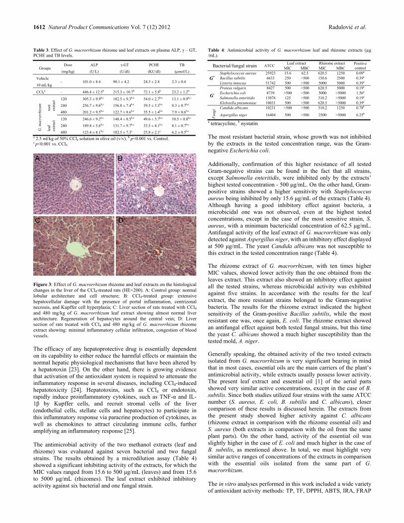
1612 Natural Product Communications Vol. 7 (12) 2012 Radulović et al.
Table 3: Effect of G. macrorrhizum rhizome and leaf extracts on plasma ALP, γ – GT, PCHE and TB levels.
Groups Dose
(mg/kg)
ALP
(U/L)
γ-GT
(U/dl)
PCHE
(KU/dl)
TB
(mol/L)
Vehicle
10 mL/kg - 101.0 ± 8.4 90.1 ± 4.2 24.3 ± 2.8 2.3 ± 0.4
CCl4a - 446.4 ± 12.5b 215.3 ± 10.7b 72.1 ± 5.8b 23.2 ± 1.2b
G. m
acro
rrhi
zum
root
ex
trac
t 120 305.3 ± 9.9b,c 182.5 ± 9.3b,c 54.0 ± 2.7b,c 13.1 ± 0.9b,c
240 256.7 ± 9.6b,c 156.8 ± 7.4b,c 39.5 ± 3.3b,c 8.3 ± 0.7b,c
480 201.2 ± 9.5b,c 122.7 ± 8.6b,c 35.5 ± 1.4b,c 7.9 ± 0.6b,c
leaf
ex
trac
t 120 246.6 ± 9.2b,c 148.4 ± 8.5b,c 49.6 ± 3.7b,c 10.5 ± 0.8b,c
240 189.8 ± 5.6b,c 131.7 ± 9.7b,c 33.5 ± 4.1b,c 8.1 ± 0.7b,c
480 123.4 ± 8.1b,c 102.5 ± 7.3c 25.8 ± 2.1c 6.2 ± 0.5b,c a 2.5 ml/kg of 50% CCl4 solution in olive oil (v/v). b p<0.001 vs. Control. c p<0.001 vs. CCl4
Figure 3: Effect of G. macrorrhizum rhizome and leaf extracts on the histological changes in the liver of the CCl4-treated rats (HE×200). A: Control group: normal lobular architecture and cell structure; B: CCl4-treated group: extensive hepatocellular damage with the presence of portal inflammation, centrizonal necrosis, and Kupffer cell hyperplasia; C: Liver section of rats treated with CCl4 and 480 mg/kg of G. macrorrhizum leaf extract showing almost normal liver architecture. Regeneration of hepatocytes around the central vein; D: Liver section of rats treated with CCl4 and 480 mg/kg of G. macrorrhizum rhizome extract showing: minimal inflammatory cellular infiltration, congestion of blood vessels. The efficacy of any hepatoprotective drug is essentially dependent on its capability to either reduce the harmful effects or maintain the normal hepatic physiological mechanisms that have been altered by a hepatotoxin [23]. On the other hand, there is growing evidence that activation of the antioxidant system is required to attenuate the inflammatory response in several diseases, including CCl4-induced hepatotoxicity [24]. Hepatotoxins, such as CCl4 or endotoxin, rapidly induce proinflammatory cytokines, such as TNF-α and IL-1β̣ by Kupffer cells, and recruit stromal cells of the liver (endothelial cells, stellate cells and hepatocytes) to participate in this inflammatory response via paracrine production of cytokines, as well as chemokines to attract circulating immune cells, further amplifying an inflammatory response [25]. The antimicrobial activity of the two methanol extracts (leaf and rhizome) was evaluated against seven bacterial and two fungal strains. The results obtained by a microdilution assay (Table 4) showed a significant inhibiting activity of the extracts, for which the MIC values ranged from 15.6 to 500 μg/mL (leaves) and from 15.6 to 5000 μg/mL (rhizomes). The leaf extract exhibited inhibitory activity against six bacterial and one fungal strain.
Table 4: Antimicrobial activity of G. macrorrhizum leaf and rhizome extracts (µg /mL).
Bacterial/fungal strain ATCC Leaf extract Rhizome extract Positive
control MIC MBC MIC MBC
G+ Staphylococcus aureus 25923 15.6 62.5 620.5 1250 0.09a Bacillus subtilis 6633 250 >500 150.6 2500 0.39a Listeria innocua 51742 500 >500 5000 5000 0.39a
G-
Proteus vulgaris 8427 500 >500 620.5 5000 0.19a Escherichia coli 8739 >500 >500 5000 >5000 1.56a Salmonella enteritidis 13076 125 >500 310.2 >5000 0.19a Klebsiella pneumoniae 10031 500 >500 620.5 >5000 0.39a
Fun
gi Candida albicans 10231 >500 >500 310.2 1250 0.78b
Aspergillus niger 16404 500 >500 2500 >5000 6.25b
a tetracycline, b nystatin The most resistant bacterial strain, whose growth was not inhibited by the extracts in the tested concentration range, was the Gram-negative Escherichia coli. Additionally, confirmation of this higher resistance of all tested Gram-negative strains can be found in the fact that all strains, except Salmonella enteritidis, were inhibited only by the extracts’ highest tested concentration - 500 μg/mL. On the other hand, Gram-positive strains showed a higher sensitivity with Staphylococcus aureus being inhibited by only 15.6 μg/mL of the extracts (Table 4). Although having a good inhibitory effect against bacteria, a microbicidal one was not observed, even at the highest tested concentrations, except in the case of the most sensitive strain, S. aureus, with a minimum bactericidal concentration of 62.5 μg/mL. Antifungal activity of the leaf extract of G. macrorrhizum was only detected against Aspergillus niger, with an inhibitory effect displayed at 500 μg/mL. The yeast Candida albicans was not susceptible to this extract in the tested concentration range (Table 4). The rhizome extract of G. macrorrhizum, with ten times higher MIC values, showed lower activity than the one obtained from the leaves extract. This extract also showed an inhibitory effect against all the tested strains, whereas microbicidal activity was exhibited against five strains. In accordance with the results for the leaf extract, the more resistant strains belonged to the Gram-negative bacteria. The results for the rhizome extract indicated the highest sensitivity of the Gram-positive Bacillus subtilis, while the most resistant one was, once again, E. coli. The rhizome extract showed an antifungal effect against both tested fungal strains, but this time the yeast C. albicans showed a much higher susceptibility than the tested mold, A. niger. Generally speaking, the obtained activity of the two tested extracts isolated from G. macrorrhizum is very significant bearing in mind that in most cases, essential oils are the main carriers of the plant’s antimicrobial activity, while extracts usually possess lower activity. The present leaf extract and essential oil [1] of the aerial parts showed very similar active concentrations, except in the case of B. subtilis. Since both studies utilized four strains with the same ATCC number (S. aureus, E. coli, B. subtilis and C. albicans), closer comparison of these results is discussed herein. The extracts from the present study showed higher activity against C. albicans (rhizome extract in comparison with the rhizome essential oil) and S. aureus (both extracts in comparison with the oil from the same plant parts). On the other hand, activity of the essential oil was slightly higher in the case of E. coli and much higher in the case of B. subtilis, as mentioned above. In total, we must highlight very similar active ranges of concentrations of the extracts in comparison with the essential oils isolated from the same part of G. macrorrhizum. The in vitro analyses performed in this work included a wide variety of antioxidant activity methods: TP, TF, DPPH, ABTS, IRA, FRAP
Bioactivity of Geranium macrorrhizum Natural Product Communications Vol. 7 (12) 2012 1613
and CUPRAC. The antioxidant activities of G. macrorrhizum extracts presented here were significantly higher than those determined by other authors [5]. Whether this is a consequence of a different extraction method or climatic conditions favoring G. macrorrhizum development is a question that needs further consideration. This study showed that among the methanol, ethanol, acetone and ethyl acetate extracts of G. macrorrhizum, the methanol one of both the leaves and rhizomes demonstrated the highest antioxidant activities. The antioxidant potency in all performed assays decreased in the order of decreasing solvent polarity: methanol > ethanol > acetone > ethyl acetate. Overall, the methanol extracts of G. macrorrhizum were the most potent in scavenging the DPPH and ABTS radicals, as well as in reducing ferric and cupric ions and were for that reason chosen for the in vivo test (hepatoprotective activity) and antimicrobial assays. The last two can be connected to the ethnopharmacological uses of this medicinal plant. The presence of high levels of phenolic compounds in the methanol extracts significantly contributed to the observed antioxidant activities and dose dependent hepatoprotective effect of the extracts in the CCl4-induced liver damage model. There are at least three possible mechanisms of action that can be envisaged: 1) the radical scavenging effect (the formed CCl3
· radicals are quenched by the phenolic groups of G. macrorrhizum constituents), 2) inhibitory action of the extract constituents on the enzymes producing the before mentioned radical from CCl4, and 3) cell membrane strengthening effect and the prevention of cell lysis and leakage of cellular content that can induce further liver damage. These modi operandi have all been established for a compound named geranin isolated from a Japanese Geranium species (G. thunbergii) and may well be operational here, but need confirmation [10,11]. All of the above provides a justification for the popular use of this plant species in folk medicine (the stimulative properties and positive general health effects) and especially for the liver restoring action. One more usage of this plant species is in wound healing, and besides the known astringent effect, we have demonstrated that it also possesses antimicrobial activity against human pathogens that is likewise beneficial in this respect. In addition to the activity of the polar (methanol) extracts of G. macrorrhizum with a high phenol content, the essential oil of this species was also found to be active against a number of bacterial and fungal strains [1]. This gives another argument for the total antibacterial and antifungal effect observed for this plant species extracts. Experimental
General: Extractions were made using an ultrasonic bath (Bandelin SONOREX® Digital 10 P, Sigma, USA). UV measurements were obtained on an Agilent 8453 (Agilent Technologies, USA) spectrophotometer. An Agilent Technologies 1200 HPLC system (Agilent Technologies, USA) equipped with a quaternary pump, autosampler and diode array detector was used for the identification and quantification of phenolic acids and flavonoids. All chromatographic separations were performed on an analytical column ZORBAX Eclipse XDB-C18 (4.6 × 150 mm, 5 μm, Agilent Technologies, USA). A gradient of acetonitrile (A) and deionized water containing 0.1%, v/v, of formic acid (B) mixtures of decreasing polarity were used for elution: 0-15 min 10% A, 15-35 min 10-30% A. The flow rate was set at 0.8 mL/min, column temperature at 30C, and 20 µL of the above prepared solutions was injected. Data were acquired in the spectral range of 200-600 nm and integrated at 260, 280 and 320 nm. Plant material: Aerial and underground parts of G. macrorrhizum L. (Geraniaceae) were collected from the mountain range Svrljiške planine, in the vicinity of the city of Svrljig, south-eastern Serbia.
Preparation of the extracts for in vivo experiments and microbiological assays: Two batches of 100 g of the aerial and underground parts were exhaustively extracted with methanol to afford c.a. 5.0 g and 4.5 g of extractive, respectively, after evaporation of the solvent in a gentle stream of nitrogen. The resulting oily residues were dissolved in physiological saline at a concentration of 300 mg/mL for evaluation of the hepatoprotective effect, and another portion was dissolved in dimethylsulfoxide to give a solution containing 100 mg/mL of extractible matter for the in vitro microdilution microbiological tests. Phytochemical and in vitro antioxidant studies: The total phenolic content of the extracts was determined using Folin-Ciocalteau assay [26a], whereas the total flavonoid content was determined by a colorimetric method [26b]. The antioxidant capacity was studied through the evaluation of the extracts’ free radical-scavenging effect on DPPH [26b] and ABTS [26c] radicals. Iron(III) to iron(II) reduction assay [26d], ferric-reducing antioxidant power assay [26e] and cupric ion reducing antioxidant capacity [26f] were also performed. Evaluation of the hepatoprotective effect: Male Wistar rats weighing 200–250 g, obtained from our own animal facilities, were housed in large spacious cages at 27±2°C with 12 h light and dark cycle throughout the period of the experiment. The animals were provided with a standard laboratory diet and given tap water ad libitum. On the day of the experiment, the animals were divided into 8 experimental groups consisting of 6 animals each. Two groups served as the positive and negative controls and received CCl4 (2.5 mL/kg of 50%, v/v, CCl4 solution in olive oil) and olive oil (10 mL/kg) by intraperitoneal injection (i.p.), respectively. Test groups of rats, 3 groups per extract, were i.p. treated with G. macrorrhizum extracts (leaf and rhizome) prepared as previously mentioned in doses of 120, 240 and 480 mg/kg (body weight). Sixty min after the application of the 2 extracts all animals received 2.5 mL/kg of the 50% CCl4 solution in olive oil. Twenty-four h after the toxin administration, blood samples were withdrawn by cardiac puncture and rats sacrificed with an overdose of ketamine (Ketamidor 10%, Richter Pharma AG, Wels, Austria). Additionally, the liver of each rat was promptly removed and small sections were collected for histological analysis. All experimental procedures with animals were conducted in compliance with The European Council Directive of November 24th, 1986 (86/609/EEC) and approved by the local Ethics Committee (number 01-2857-3) given on 27th April, 2012. Biochemical measurements: The blood was centrifuged at 1500 rpm at 4°C for 15 min to obtain the serum. Aspartate transferase (AST), alanine transaminase (ALT), serum alkaline phosphatase (ALP), -glutamate transpeptidase (γ-GT), pseudocholineesterase (PCHE) and total bilirubin (TB) activities were assayed using an Olympus AU680® Chemistry-Immuno Analyzer (Olympus America Inc, USA). Histopathological observation: The liver tissue specimens separated for histopathological examination were fixed in buffered formaldehyde solution (10%, w/w). The fixed tissue was then dehydrated with different aqueous ethanol solutions from 50–100%, v/v, embedded in paraffin, cut into 4–5 μm thick sections, stained with hematoxylin and eosin, and examined with an Olympus BH2 light microscope (Olympus America INC, USA). Evaluation of antimicrobial activity: G. macrorrhizum extracts (leaf and rhizome) were tested against a panel of microorganisms, see Table 4.

1614 Natural Product Communications Vol. 7 (12) 2012 Radulović et al.
Microdilution antimicrobial assay: Antimicrobial activity was evaluated using a broth microdilution method [1]. Stock solutions of the extracts were made in DMSO (100%) and then serially diluted (the diluting factor 2). The final concentrations of G. macrorrhizum extracts (rhizome and leaf) in the media were in the ranges 2.00-5000 μg/mL and 0.200-500 μg/mL, respectively. All experiments were conducted in triplicate and repeated twice. Statistical analysis: Results were expressed as the mean ± standard deviation. Statistically significant differences were determined by
one-way analysis of variance (ANOVA) followed by Tukey´s post hoc test for multiple comparison (Graph pad Prism version 5.03, San Diego, CA, USA). Probability values (p) less than 0.05 were considered to be statistically significant. Acknowledgments - This research has been supported by the Ministry of Education, Science and Technological Development of Serbia (Project 172061).
References
[1] Radulović N, Dekić M, Stojanović-Radić Z, Zoranić S. (2010) Geranium macrorrhizum L. (Geraniaceae) essential oil: a potent agent against Bacillus subtilis. Chemistry & Biodiversity, 7, 2783–2800.
[2] Genova E, Ivancheva S. (1995) Quantitative analysis of tannin content in Geranium macrorrhizum L. (Geraniaceae) growing in Bulgaria. Phytologia Balcanica, 1, 93–99.
[3] Serkedjieva J, Hay AJ. (1998) In vitro antiinfluenza virus activity of a plant preparation from Geranium sanguineum L. Antiviral Research, 37, 221–230.
[4] Lamaison JL, Petitjean-Freytet C, Carnat A. (1993) Teneurs en polyphenols et activities antioxydantes chez les Geraniaceae françaises. Plantes Medicinales et Phytotherapie, 26, 130–134.
[5] Miliauskas G, Van Beek T, Venskutonis P, Linssen J, Waard P. (2004) Antioxidative activity of Geranium macrorrhizum. European Food Research and Technology, 218, 253–261.
[6] Janković MM. (1973) Flora of Serbia. Srpska akademija nauke i umetnosti, Belgrade. [7] Tucakov J. (1990) Phytotherapy. Rad, Belgrade. [8] Menković N, Savikin K, Tasić S, Zdunić G, Stesević D, Milosavljević S, Vincek D. (2011) Ethnobotanical study on traditional uses of wild
medicinal plants in Prokletije Mountains (Montenegro). Journal of Ethnopharmacology, 133, 97–107. [9] Jarić, S, Popović Z, Mačukanović-Jocić M., Djurdjević L, Mijatović M, Karadžić B, Mitrović M, Pavlović P. (2007) An ethnobotanical study on the
usage of wild medicinal herbs from Kopaonik Mountain (Central Serbia). Journal of Ethnopharmacology, 111, 160–175. [10] Nakanishi Y, Orita M, Okuda T, Abe H. (1998) Effects of geraniin on the liver in rats I - effects of geraniin compared to ellagic acid, and gallic acid
on hepatic injuries induced by CCl4, D-galactosamine, and thioacetamide. Natural Medicines, 52, 396–403. [11] Nakanishi Y, Okuda T, Abe H. (1999) Effects of geraniin on the liver in rats III - correlation between lipid accumulations and liver damage in CCl4-
treated rats. Natural Medicines, 53, 22–26. [12] Miliauskas G, Mulder E, Linssen J, Houben J, Van Beek T, Venskutonis P. (2007) Evaluation of antioxidative properties of Geranium
macrorrhizum and Potentilla fruticosa extracts in Dutch style fermented sausages. Meat Science, 77, 703–708. [13] Radulović N, Dekić M, Stojanović-Radić Z, Palić R. (2011) Chemical composition and antimicrobial activity of the essential oils of Geranium
columbinum L. and G. lucidum L. (Geraniaceae). Turkish Journal of Chemistry, 35, 499–512. [14] Radulović N, Dekić M., Stojanović-Radić Z. (2012) Chemical composition and antimicrobial activity of the volatile oils of Geranium sanguineum
L. and G. robertianum L. (Geraniaceae). Medicinal Chemistry Research, 21, 601–615. [15] Miliauskas G, Venskutonis P, Van Beek T. (2004) Screening of radical scavenging activity of some medicinal and aromatic plant extracts. Food
Chemistry, 85, 231–237. [16] Sacan O, Yanardag R. (2010) Antioxidant and antiacetylcholinesterase activities of chard (Beta vulgaris L. var. cicla). Food and Chemical
Toxicology, 48, 1275–1280. [17] Rao M, Rao C, Pushpangadan P, Shirwaikar A. (2006) Hepatoprotective effects of rubiadin, a major constituent of Rubia cordifolia Linn. Journal
of Ethnopharmacology, 103, 484–490. [18] Lin J, Wang C. (1986) Protection of crocin dyes in the acute hepatic damage induced by aflatoxin B and dimethylnitrosamine in rats. Carcinogenis,
7, 595–599. [19] Rajesh M, Latha M. (2001) Hepatoprotection by Elephantopus scaber Linn. in CCl4-induced liver injury. Indian Journal of Physiology and
Pharmacology, 45, 481–486. [20] Klaassen C, Watkins J. (1984) Mechanisms of bile formation, hepatic uptake, and biliary excretion. Pharmacological Reviews, 36, 1–67. [21] Castro J, Ferrya G, Castro C, Sasame H, Fenos O, Gillete J. (1974) Prevention of carbon tetrachloride induced necrosis by inhibitors of drug
metabolism. Further studies on the mechanism of their action. Biochemical Pharmacology, 23, 295–302. [22] Thabrew M, Joice P. (1987) A comparative study of the efficacy of Pavetta indica and Osbeckia octanda in the treatment of liver dysfunction.
Planta Medica, 53, 239–241. [23] Raghavendran B, Sathivel A, Devaki T. (2003) Hepatoprotective nature of seaweed alcoholic extract on acetaminophen induced oxidative stress.
Journal of Health Science, 50, 42–46. [24] Campo G, Avenoso A, Campo S, Nastasi G, Traina P, D'Ascola A, Rugolo C, Calatroni A. (2008) The antioxidant activity of chondroitin-4-
sulphate, in carbon tetrachloride-induced acute hepatitis in mice, involves NF-kappaB and caspase activation. British Journal of Pharmacology, 155, 945–956.
[25] Simpson K, Lukacs N, Colletti L, Strieter R, Kunkel S. (1997) Cytokines and the liver. Journal of Hepatology, 27, 1120–1132. [26] (a) Mitic M, Obradovic M, Kostic D, Naskovic D, Micic R. (2011) Phenolics content and antioxidant capacity of commercial red fruit juices,
Hemijska industrija, 5, 611–619; (b) Mitic M, Obradovic M, Kostic D, Micic R, Paunovic D. (2011) Phenolic profile and antioxidant capacities of dried red currant from Serbia, extracted with different solvent. Food Science and Biotechnology, 6, 1625–1631; (c) Zhou S, Fang Z, Lü Y, Chen Y, Liu D, Ye XI. (2009) Phenolics and antioxidant properties of bayberry (Myrica rubra Sieb. et Zucc.) pomace. Food Chemistry, 112, 394–399; (d) Dorman H, Peltoketo A, Hiltunen R, Tikkanen M. (2003) Characterisation of the antioxidant properties of de-odourised aqueous extracts from selected Laminaceae herbs. Food Chemistry, 83, 255–262; (e) Maksimovic Z. (2008) In vitro antioxidant activity of ragweed (Ambrosia artemisiifolia L., Asteraceae) herb. Industrial Crops and Products, 28, 356–360; (f) Yıldız L, Baskan K, Tutem E, Apak R. (2008) Combined HPLC-CUPRAC (cupric ion reducing antioxidant capacity) assay of parsley, celery leaves and nettle. Talanta, 77, 304–313.

Differential Expression of Benzophenone Synthase and Chalcone Synthase in Hypericum sampsonii Lili Huanga,d, Hong Wangd, Hechun Yea, Zhigao Dua, Yansheng Zhangc,*, Ludger Beerhuesb and Benye Liua,b,* aKey Laboratory of Plant Molecular Physiology, Institute of Botany, The Chinese Academy of Sciences, Beijing 100093, China
bInstitute of Pharmaceutical Biology, Technische Universität Braunschweig, Braunschweig 38106, Germany
cCAS Key Laboratory of Plant Germplasm Enhancement and Specialty Agriculture, Wuhan Botanical Garden, The Chinese Academy of Science, Wuhan 430074, China
dGraduate University of the Chinese Academy of Sciences, 19A Yuquanlu, Beijing 100049, China [email protected]; [email protected]
Received: September 26th, 2012; Accepted: October 16th, 2012
cDNAs encoding Hypericum sampsonii benzophenone synthase (HsBPS) and chalcone synthase (HsCHS) were isolated and functionally characterized. Differential expressions of HsBPS and HsCHS were monitored using quantitative polymerase chain reaction (PCR). In the vegetative stage, HsBPS was highly expressed in the roots; its transcript level was approx. 100 times higher than that of HsCHS. Relatively high transcript amounts of HsBPS were also detected in older leaves, whereas the youngest leaves contained higher transcript amounts of HsCHS. In the reproductive stage, maximum HsCHS expression was detected in flowers, the transcript level being approx. 5 times higher than that of HsBPS. The inversed situation with a 10-fold difference in the expression levels was observed with fruits. High transcript amounts for both proteins were found in roots. Keywords: Benzophenone synthase, Chalcone synthase, Gene expression, Hypericum sampsonii, Quantitative PCR, Sampsoniones. Hypericum sampsonii Hance is used as a traditional Chinese herbal medicine in the treatment of numerous disorders such as hematemesis, epistaxis, menstrual irregularity, external traumatic injury, snakebite and swellings [1]. In recent years, the plant has aroused further scientific interest for its anticancer activity [2]. Extensive phytochemical studies demonstrated that H. sampsonii contains an array of sampsoniones (A-Q), which are polyprenylated benzophenones characterized by complex caged tetracyclic skeletons (Figure 1). These compounds possess profound cytotoxic activity [3]. Despite their pharmaceutical importance, biosynthesis of these benzophenone derivatives remains poorly understood. The nucleus of sampsoniones is benzophenone, which is biosynthesized by benzophenone synthase (BPS; EC 2.3.1.151), a new member of the type III polyketide synthase (PKS) [4]. The first published BPS (HaBPS) cDNA was from elicitor-treated Hypericum androsaemum cell cultures, which served as a model system for studying benzophenone metabolism [5]. Recently, the second BPS cDNA was cloned and characterized from Garcinia mangostana [6]. However, no information about expression of BPS in planta is so far available. Here we have cloned and functionally characterized a cDNA encoding H. sampsonii benzophenone synthase (HsBPS). The organ-specific expression pattern of HsBPS was analyzed by quantitative polymerase chain reaction (PCR). For comparison, the same studies were carried out for chalcone synthase (HsCHS), which is a well studied ubiquitous type III PKS in all higher plants. The cDNAs for HsBPS and HsCHS were cloned by the homology-based cloning strategy. The ORF of the HsBPS cDNA was 1188 bp long and encoded a 42.7 kDa protein (395 amino acids) with a calculated pI of 5.91. The HsCHS cDNA contained a 1173 bp ORF encoding a 42.7 kDa protein (390 amino acids) with a calculated pI of 6.55. HsBPS and HsCHS shared around 57.0% identity at the
nucleotide as well as at the amino acid sequence level. HsBPS and HsCHS were expressed as N-terminally His6-tagged proteins in Escherichia coli and purified by affinity chromatography. Protein bands of approximately 43 kDa each were observed after SDS-PAGE (Figure 2). Table 1: Substrate specificities of recombinant HsBPS and HsCHS a
Substrate Enzyme activity (% of max)
HaBPS HaCHS Benzoyl-CoA 100 11 2-Hydroxybenzoyl-CoA 5 0 3-Hydroxybenzoyl-CoA 66 0 4-Hydroxybenzoyl-CoA 0 0 Cinnamoyl-CoA 0 65 2-Coumaroyl-CoA 0 0 3-Coumaroyl-CoA 0 0 4-Coumaroyl-CoA 0 100 Acetyl-CoA 0 0 a Data are means of two independent experiments.
HsBPS preferred benzoyl-CoA as a starter substrate (Table 1). The enzymatic product was identified as 2,4,6-trihydroxybenzophenone by liquid chromatography-UV spectroscopy (LC-UV) and liquid chromatography-mass spectrometry (LC-MS) in comparison with a sample of authentic reference compound. The pH and temperature optima were 6.5-7.0 and 40°C, respectively. There were also traces of a side product, 6-phenyl-4-hydroxy-2-pyrone. Beside benzoyl-CoA, HsBPS also accepted 3-hydroxybenzoyl-CoA as starter substrate to form 2,3’,4,6-tetrahydroxybenzophenone and a small amount of 6-(3’-hydroxyphenyl)-4-hydroxy-2-pyrone as a derailment product. 4-Hydroxybenzoyl-CoA, acetyl-CoA, and CoA esters of cinnamic acids were not accepted by HsBPS as starter molecules (Table 1).
NPC Natural Product Communications 2012 Vol. 7 No. 12
1615 - 1618
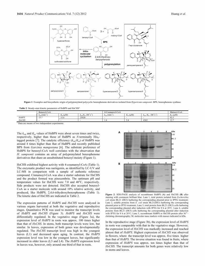
1616 Natural Product Communications Vol. 7 (12) 2012 Huang et al.
Figure 1: Examples and biosynthetic origin of polyprenylated polycyclic benzophenone derivatives isolated from Hypericum sampsonii. BPS, benzophenone synthase.
Table 2: Steady-state kinetic parameters of HsBPS and HsCHSa
Benzoyl-CoA 4-Coumaroyl-CoA Malonyl-CoAkcat (min-1) Km (µM) kcat/Km (M
-1s-1) kcat (min-1) Km (µM) kcat /Km (M-1s-1) Km (µM)
HsBPS 23.7 15.8 25028 - - - 92.5 HsCHS - - - 1.4 5.4 4365 12.7
a Data are means of two independent experiments The kcat and Km values of HsBPS were about seven times and twice, respectively, higher than those of HaBPS as N-terminally His6-tagged protein [7]. The catalytic efficiency (kcat/Km) of HsBPS was around 4 times higher than that of HaBPS and recently published BPS from Garcinia mangostana [6]. The substrate preference of HsBPS for benzoyl-CoA well correlates with the observation that H. sampsonii contains an array of polyprenylated benzophenone derivatives that share an unsubstituted benzoyl moiety (Figure 1). HsCHS exhibited highest activity with 4-coumaroyl-CoA (Table 1). The enzymatic product was naringenin, as identified by LC-UV and LC-MS in comparison with a sample of authentic reference compound. Cinnamoyl-CoA was also a starter substrate for HsCHS and the product formed was pinocembrin. The optimum pH and temperature values for HsCHS were 7.0 and 40°C, respectively. Side products were not detected. HsCHS also accepted benzoyl-CoA as a starter molecule with around 10% relative activity, and produced, like HsBPS, 2,4,6-trihydroxybenzophenone (Table 1). The kinetic data of HsCHS are indicated in Table 2. The expression patterns of HsBPS and HsCHS were analyzed in various organs harvested at both the vegetative and reproductive stages. Quantitative PCR was used to monitor the transcript levels of HsBPS and HsCHS (Figure 3). HsBPS and HsCHS were differentially regulated. In the vegetative stage (Figure 3a), the expression level of HsBPS in roots was approx. 100 times higher than that of HsCHS. In stems, both transcript levels were low, but similar. In leaves, expression of both genes was developmentally regulated. The HsCHS transcript level was high in the youngest leaves (L1) and decreased upon aging. In contrast, the HsBPS expression level was low in the youngest leaves, but significantly increased in older leaves (L3 and L4). The HsBPS expression level in leaves was, however, only around one third of that in roots.
Figure 2: SDS-PAGE analysis of recombinant HsBPS (A) and HsCHS (B) after staining with coomassie brilliant blue. Lane 1, total protein isolated from Escherichia coli strain BL21 (DE3) harboring the corresponding plasmid prior to IPTG treatment; Lane 2, soluble proteins from E. coli strain BL21(DE3) harboring the corresponding plasmid prior to IPTG treatment; Lane 3, total protein from BL21 (DE3) cells harboring the corresponding plasmid after induction with IPTG for 8 h at 28°C. Lane 4, soluble proteins from BL21 (DE3) cells harboring the corresponding plasmid after induction with IPTG for 8 h at 28°C; Lane 5, recombinant HsBPS or HsCHS protein after Ni2+-chelating chromatography; M, molecular mass markers with masses indicated in kDa.
In the reproductive stage (Figure 3b), the expression level of HsBPS in roots was comparable with that in the vegetative stage. However, the expression level of HsCHS was markedly increased and reached almost that of HsBPS. Highest expression of HsCHS was observed in flowers, where the transcript level was approx. five times higher than that of HsBPS. The inverse situation was found in fruits, where expression of HsBPS was approx. ten times higher than that of HsCHS. The transcript amounts for both genes were relatively low in stems and leaves.
kDa
kDa

Differential expressions of HsBPS and HsCHS Natural Product Communications Vol. 7 (12) 2012 1617
Figure 3: Quantitative PCR analysis of the transcript levels of HsBPS and HsCHS in various organs of H. sampsonii at the vegetative stage (A) and the reproductive stage (B). Primers specific for HsBPS and HsCHS were used to measure the transcript levels of the two genes. The H. sampsonii 18s rRNA gene (AY573011.1) was used as an internal standard. Relative quantification used the stem values in the vegetative stage and the leaf values in the reproductive stage as calibrators, which were set to a nominal value of 1. Error bars represent the standard deviations (SD, n=3). R, roots; S, stems; L, the first leaf pair under the inflorescence; L1-4, the first, second, third and fourth leaf pairs from the shoot tip in the vegetative stage; Fl, flowers; Fr, fruits.
In most organs, the expression level of HsBPS was higher than that of HsCHS, which is consistent with the finding that polyprenylated benzophenones and xanthones are typical and dominant secondary products of Hypericaceae plants, whereas the concentration of flavonoids in these plants is lower [3, 8]. HsBPS was most highly expressed in the roots at both the vegetative and reproductive stages, while its expression in the upper parts of the plant was lower. This expression pattern well correlated with the distribution of polyprenylated benzophenones in the plant. The concentration of prenylated benzophenones in the roots is much higher than that in the aerial parts. For example, the 7-epi-clusianone content in the roots of H. sampsonii is about 0.1% of dry weight, which is around 100 times higher than the maximum concentration of sampsoniones in the aerial parts [3]. Another reason for the high HsBPS expression level in roots may be that xanthones also appear to mainly accumulate in the underground organs of many Hypericum species [9]. The HsBPS expression results suggested that roots of H. sampsonii may be the better parts to be used as a herbal remedy because of the high amounts of benzophenone derivatives. The differential expression pattern of HsBPS and HsCHS in H. sampsonii is interesting and should be related to the function of the corresponding end products, benzophenones and flavonoids, respectively. It has been proved that flavonoids serve as flower pigments, UV protectants and signal molecules in plants [10]. The expression pattern of HsCHS can be well explained by these functions, especially, the high transcript level of HsCHS in flowers is likely to be related to pigmentation for insect attraction. Constituents that are widely distributed in species of Hypericaceae are polyprenylated benzophenones and xanthones and their antimicrobial properties suggest that they may also have a defensive
role against pathogens. In H. calycinum, polyprenylated acyl and benzoyl phlorogucinols made up to approx. 20% of the dry weight of anthers and ovarian walls and were presumed to protect the developing seeds against herbivores and microorganisms [11]. In H. perforatum cell cultures, Agrobacterium tumefaciens as a biotic stress factor activated xanthone biosynthesis, which resulted in a rapid up-regulation of BPS expression and a 12-fold increase in the total xanthone concentration [12]. These compounds were supposed to act as both phytoalexins and antioxidants to help the host prevent disease development. It is reasonable to assume a similar function of benzophenone derivatives in H. sampsonii. The differential expression patterns of HsBPS and HsCHS might indicate that benzophenone derivatives serve in a complementary way, in addition to flavonoids, to protect the plant from biotic stress. The results of this work provided the first insight into the in planta expression pattern of HaBPS, which was clearly different from that of HaCHS in H. sampsonii. These data are the solid basis for further deep investigations of the biosynthesis of these polyprenylated benzophenone derivatives. Experimental
Chemicals: All the CoA esters of cinnamic acids and benzoic acids, apart from benzoyl-CoA, were synthesized as described in the literature [13]. Malonyl-CoA, acetyl-CoA, benzoyl-CoA and naringenin were purchased from Sigma-Aldrich (St Louis, MO, USA), 2,4,6-trihydroxybenzophenone from ICN (Meckenheim, Germany), and 6-phenyl-4-hydroxy-2H-pyran-2-one (benzoyl-diacetic acid lactone) from Aurora Fine Chemicals (Graz, Austria). Plant materials: Hypericum sampsonii Hance (Hypericaceae) seeds were kindly provided by Professor Hongfei Lv (Zhejiang Normal University, Jinhua, China). A voucher specimen of this plant (No. 01262860) was deposited in the Herbarium of the Institute of Botany, the Chinese Academy of Sciences, Beijing, China. The seeds were germinated in petri dishes covered with filter paper and grown at 25°C with a photoperiod of 16/8 h (light/dark). Three-week-old seedlings were transferred to 1.5-L pots filled with a mixture of mineral soil and humus in a greenhouse with the same temperature and photoperiod. When plants were 6-8 weeks old (vegetative stage), roots, stems, and leaves were harvested. The leaves were divided into unopened leaf pair (L1), the second leaf pair (L2), the third leaf pair (L3) and the fourth leaf pair (L4). In the reproductive stage, leaf (L) was the first leaf pair under the florescence, the fully opened flower heads (Fl) and green fruits (Fr) were harvested. All the samples were divided into 0.2 g-samples and immediately frozen in liquid nitrogen and stored at -80°C. Cloning of HsBPS and HsCHS cDNAs: Total RNA was isolated from roots and leaves of H. sampsonii using the Universal Plant Total RNA Extraction Kit (Bioteke, Beijing, China). Reverse transcription was carried out at 42°C with M-MLV Reverse Transcriptase (Promega, Madison, WI, USA), and the oligo (dT)-adaptor primer RRP (primer sequences and PCR conditions are provided in supplementary data). Gene specific primers were designed from the 5’ untranslated region (5’ UTR) of HaBPS (Pr-1) and HaCHS (Pr-2) of H. androsaemum [5]. For HsCHS, the full-length cDNA sequence was amplified using primer pair Pr-2 and the adaptor of RRP and the reverse-transcription product of leaf mRNA as template. For HsBPS, we were not able to amplify the full-length HsBPS with primer pair Pr-1 and the adaptor of RRP, but the core cDNA fragment was amplified by PCR using primers Pr-F and Pr-R derived from the ORF of HaBPS [5]. Full-length cDNA was obtained by rapid amplification of cDNA ends (RACE) using the protocol of the SMARTerTM RACE cDNA Amplification kit

1618 Natural Product Communications Vol. 7 (12) 2012 Huang et al.
(Clontech, Beijing, China). The PCR product was purified, cloned into pMD18-T vector (Takara) and sequenced by a commercial sequencing company. Sequence data of this article can be found in the GenBank/EMBL data libraries under the following accession numbers: HsBPS, JQ670939; HsCHS, JQ670940. Heterologous expression and enzyme purification: The ORFs encoding HsBPS and HsCHS were reamplified by PCR using Pfu DNA polymerase. The amplified DNA was cloned to pRSET B (Invitrogen, Carlsbad, CA, USA) with Nhe І/Kpn І site. After sequencing of the ORFs on both strands, the recombinant plasmids were introduced into E. coli BL21-CodonPlus (DE3)-RIL (Stratagene, Amsterdam, Netherlands) for overexpression as N-terminally His6-tagged proteins. HsBPS and HsCHS were purified to homogeneity using Ni-NTA agarose according to the manufacturer’s instructions (Qiagen, Hilden, Germany). The purification efficiency was monitored by SDS-PAGE (Figure 2). Enzyme assay and kinetic data determiation: The standard assay (250 μL) contained 54 μM starter CoA, 324 μM malonyl-CoA, 0.1 M potassium phosphate (pH 7.0) and 2 μg protein. After incubation at 30°C for 10 min, the enzymatic products were extracted and detected as described previously [5]. Kinetic constants were determined using 8 substrate concentrations covering the range of 0.2-12 Km, the concentration at which the second substrate was
saturated. The amount of enzyme added to the assay was 2 µg. The incubation time was limited to 5 min for HsBPS and 10 min for HsCHS at the optimum reaction conditions. Quantitative polymerase chain reaction: Total RNA was isolated from 100 mg of the above mentioned materials using Plant Total RNA Isolation Kit (BioTeck) according to the manufacturer’s instructions. All of the total RNA preparations were treated with DNase I and reverse-transcribed using the M-MLV Reverse Transcriptase (Invitrogen) and the oligo d(T) primers. The quantitative PCRs were performed with the Applied Biosystems StepOne Real-time PCR System using FastStart Universal SYBR Green Master (Roche, Mannheim, Germany) in 3 technical replicates (primer sequences and PCR conditions are provided in supplementary data). Relative quantification used the stem value in the vegetative stages and the leaf value in the reproductive stages as calibrators, which were set to a nominal value of 1. Acknowledgments - The work was financially supported by the Starting Funding of Talent Recruitment of Institute of Botany, The Chinese Academy of Sciences (1103000114), the Grant for One Hundred Talents Program of the Chinese Academy of Sciences, China (Y129441R01) and National Sciences Foundation of China (30870217).
References
[1] Jiangsu New Medical College College. (1997) Dictionary of Chinese Crude Drugs. Shanghai Scientific Technological Publishers. 345-346. [2] Zeng JZ, Sun DF, Wang L, Cao XH, Qi JB, Yang T, Hu CQ, Liu W, Zhang XK. (2006) Hypericum sampsonii induces apoptosis and nuclear export
of retinoid X receptor-alpha. Carcinogenesis, 27, 1991-2000. [3] (a) Xin WB, Jin GL, Mao ZJ, Qin LP. (2011) Two unusual phenolic substances and one new xanthone from Hypericum sampsonii. Helvetica
Chimica Acta, 94, 686-692; (b) Xiao ZY, Mu Q, Shiu WKP, Zeng YH, Gibbons S. (2007) Polyisoprenylated benzoylphloroglucinol derivatives from Hypericum sampsonii. Journal of Natural Products, 70, 1779-1782; (c) Hu LH, Sim KY. (2000) Sampsoniones A-M, a unique family of caged polyprenylated benzoylphloroglucinol derivatives, from Hypericum sampsonii. Tetrahedron, 56, 1379-1386; (d) Xiao ZY, Shiu WKP, Zeng YH, Mu Q, Gibbons S. (2008) A naturally occurring inhibitory agent from Hypericum sampsonii with activity against multidrug-resistant Staphylococcus aureus. Pharmaceutical Biology, 46, 250-253.
[4] Beerhues L, Liu B. (2009) Biosynthesis of biphenyls and benzophenones - Evolution of benzoic acid-specific type III polyketide synthases in plants. Phytochemistry, 70, 1719-1727.
[5] Liu BY, Falkenstein-Paul H, Schmidt W, Beerhues L. (2003) Benzophenone synthase and chalcone synthase from Hypericum androsaemum cell cultures: cDNA cloning, functional expression, and site-directed mutagenesis of two polyketide synthases. The Plant Journal, 34, 847-855.
[6] Nualkaew N, Morita H, Shimokawa Y, Kinjo K, Kushiro T, De-Eknamkul W, Ebizuka Y, Abe I. (2012) Benzophenone synthase from Garcinia mangostana L. pericarps. Phytochemistry, 77, 60-69.
[7] Klundt T, Bocola M, Lutge M, Beuerle T, Liu B, Beerhues L. (2009) A single amino acid substitution converts benzophenone synthase into phenylpyrone synthase. Journal of Biological Chemistry, 284, 30957-30964.
[8] (a) Don MJ, Huang YJ, Huang RL, Lin YL. (2004) New phenolic principles from Hypericum sampsonii. Chemical and Pharmaceutical Bulletin, 52, 866-869; (b) Chen MT, Chen CM. (1985) Constituents of Formosan antitumor folk medicine. Part I. Xanthones from Hypericum sampsonii. Heterocycles, 23, 2543-2548; (c) Hong D, Yin F, Hu LH, Lu P. (2004) Sulfonated xanthones from Hypericum sampsonii. Phytochemistry, 65, 2595-2598.
[9] (a) Sparenberg B. (1993) MAO-inhibitierende Eigenschaften von Hypericum inhaltsstoffen und Untersuchungen zur Analytik und Isolierung von Xanthonen aus Hypericum perforatum L., University of Marburg, Marburg; (b) Nahrstedt A, Butterweck V. (1997) Biologically active and other chemical constituents of the herb of Hypericum perforaturn L. Pharmacopsychiatry, 30, 129-134.
[10] (a) Dhawale S, Souciet G, Kuhn DN. (1989) Increase of chalcone synthase mRNA in pathogen-inoculated soybeans with race-specific resistance is different in leaves and roots. Plant Physiology, 91, 911-916; (b) Ormrod DP, Landry LG, Conklin PL. (1995) Short-term UV-B radiation and ozone exposure effects on aromatic secondary metabolite accumulation and shoot growth of flavonoid-deficient Arabidopsis mutants. Physiologia Plantarum, 93, 602-610; (c) Vanderkrol AR, Lenting PE, Veenstra J, Vandermeer IM, Koes RE, Gerats AGM, Mol JNM, Stuitje AR. (1988) An anti-sense chalcone synthase gene in transgenic plants inhibits flower pigmentation. Nature, 333, 866-869.
[11] Gronquist M, Bezzerides A, Attygalle A, Meinwald J, Eisner M, Eisner T. (2001) Attractive and defensive functions of the ultraviolet pigments of a flower (Hypericum calycinum). Proceedings of the National Academy of Sciences of the United States of America, 98, 13745-13750.
[12] Franklin G, Conceicao LFR, Kombrink E, Dias ACP. (2009) Xanthone biosynthesis in Hypericum perforatum cells provides antioxidant and antimicrobial protection upon biotic stress. Phytochemistry, 70, 60-68.
[13] Beuerle T, Pichersky E. (2002) Enzymatic synthesis and purification of aromatic coenzyme A esters. Analytical Biochemistry, 302, 305-312.
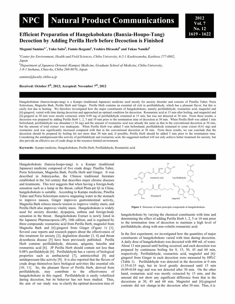
Efficient Preparation of Hangekobokuto (Banxia-Houpo-Tang) Decoction by Adding Perilla Herb before Decoction is Finished Megumi Suminoa,*, Yuko Saitob, Fumio Ikegamia, Yoshiro Hirasakib and Takao Namikib aCenter for Environment, Health and Field Sciences, Chiba University, 6-2-1 Kashiwanoha, Kashiwa 277-0882, Japan
bDepartment of Japanese Oriental (Kampo) Medicine, Graduate School of Medicine, Chiba University, 1-8-1 Inohana, Chuo-ku, Chiba 260-8670, Japan [email protected]
Received: October 5th, 2012; Accepted: November 7th, 2012
Hangekobokuto (banxia-houpo-tang) is a Kampo (traditional Japanese) medicine used mostly for anxiety disorder and consists of Pinellia Tuber, Poria Sclerotium, Magnolia Bark, Perilla Herb and Ginger. Perilla Herb contains an essential oil rich in perillaldehyde, which has a pleasant flavor, but this is easily lost due to heating. We therefore investigated how the major constituents of hangekobokuto, namely perillaldehyde, rosmarinic acid, magnolol and [6]-gingerol, varied with time during decoction and approached an optimal condition for decoction. Rosmarinic acid at 15 min after boiling, and magnolol and [6]-gingerol at 30 min were mostly extracted, while 0.09 mg of perillaldehyde remained at 15 min, but was not detected at 30 min. From these results, a decoction was prepared by adding Perilla Herb 1, 2, 5 and 10 min prior to the termination time of decoction at 30 min. When Perilla Herb was added 1 min beforehand, perillaldehyde was abundant (1.58 mg) and the amount of rosmarinic acid was already the same as that in the conventional decoction at 30 min, but the amount of total extract was inadequate. When Perilla Herb was added 5 min beforehand, perillaldehyde remained to some extent (0.61 mg) and rosmarinic acid was significantly increased compared with that in the conventional decoction at 30 min. From these results, we can conclude that the decoction should be prepared by boiling for not more than 30 min and, if possible, Perilla Herb should be added 5 min prior to the termination time. Considering the antidepressant-like activity of perillaldehyde and rosmarinic acid, the suggested method will not only achieve better treatment for anxiety, but also provide an effective use of crude drugs in the resource-limited environment. Keywords: Kampo medicine, Hangekobokuto, Perilla Herb, Perillaldehyde, Rosmarinic acid. Hangekobokuto (banxia-houpo-tang) is a Kampo (traditional Japanese) medicine composed of five crude drugs: Pinellia Tuber, Poria Sclerotium, Magnolia Bark, Perilla Herb and Ginger. It was described in Jinkuiyaolue, the Chinese traditional literature established in the 3rd century that describes major chronic diseases and treatments. This text suggests that when having a foreign-body sensation such as a lump in the throat, called Plum-pit Qi in China, hangekobokuto is suitable. According to Kampo medicine, Pinellia Tuber and Poria Sclerotium remove stagnating water in the stomach to improve nausea, Ginger improves gastrointestinal activity, Magnolia Bark relaxes muscle tension to improve vitality stasis, and Perilla Herb also improves vitality stasis. Hangekobokuto is widely used for anxiety disorder, dyspepsia, asthma and foreign-body sensation in the throat. Hangekobokuto Extract is newly listed in the Japanese Pharmacopoeia (JP), 16th edition, and is regulated by three compounds: rosmarinic acid from Perilla Herb, magnolol from Magnolia Bark and [6]-gingerol from Ginger (Figure 1) [1]. Several case reports and research papers about the effectiveness of this treatment for anxiety [2], deglutition disorder [3, 4] and throat dysesthesia disease [5] have been previously published. Perilla Herb contains perillaldehyde, shisonin, apigenin, luteolin and rosmarinic acid [6]. JP Perilla Herb should contain not less than 0.08% perillaldehyde [6]. Perillaldehyde has a variety of biological properties such as antibacterial [7], antimicrobial [8] and antidepressant-like activity [9]. It is also reported that the flavors of crude drugs themselves have biological activities like essential oils [10, 11]. So, the pleasant flavor of Perilla Herb, derived from perillaldehyde, may contribute to the effectiveness of hangekobokuto in this regard. Perillaldehyde is easily volatilized during decoction, but its behavior has not been studied. Thus, the aim of our study was to clarify the optimal decoction time of
Figure 1: Structure of main principle compounds in hangekobokuto.
hangekobokuto by varying the chemical constituents with time and determining the effect of adding Perilla Herb 1, 2, 5 or 10 min prior to the termination time of decoction to maintain a high level of perillaldehyde, along with non-volatile rosmarinic acid. In the first experiment, we investigated how the quantities of major constituents of hangekobokuto varied with time during decoction. A daily dose of hangekobokuto was decocted with 400 mL of water. About 13 min passed until boiling occurred, and each decoction was prepared by continuous boiling for 0, 15, 30, 45 and 60 min, respectively. Perillaldehyde, rosmarinic acid, magnolol and [6]-gingerol from Ginger in each decoction were measured by HPLC (Table 1). Perillaldehyde was detected in the decoction at 0 min (1.55±0.35 mg), but its level greatly decreased until 15 min (0.09±0.04 mg) and was not detected after 30 min. On the other hand, rosmarinic acid was mostly extracted by 15 min, and the amount did not show any significant difference from that in the decoctions at 30, 45 and 60 min. Magnolol and [6]-gingerol contents did not change in the decoction after 30 min. Thus, it is
NPC Natural Product Communications 2012 Vol. 7 No. 12
1619 - 1622
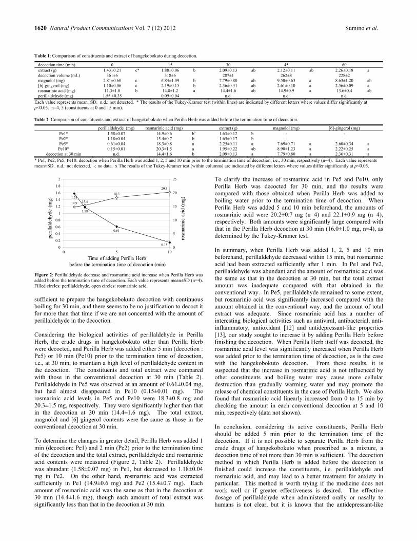
1620 Natural Product Communications Vol. 7 (12) 2012 Sumino et al.
Table 1: Comparison of constituents and extract of hangekobokuto during decoction.
decoction time (min) 0 15 30 45 60 extract (g) 1.43±0.21 c* 1.88±0.06 b 2.09±0.13 ab 2.12±0.11 ab 2.26±0.18 a decoction volume (mL) 361±6 318±6 287±1 262±8 228±2 magnolol (mg) 2.81±0.60 c 6.84±1.09 b 7.79±0.80 ab 9.50±0.63 a 8.63±1.20 ab [6]-gingerol (mg) 1.10±0.06 c 2.19±0.15 b 2.36±0.31 ab 2.61±0.10 a 2.56±0.09 a rosmarinic acid (mg) 11.3±1.0 b 14.8±1.2 a 14.4±1.6 ab 14.9±0.9 a 13.6±0.4 ab perillaldehyde (mg) 1.55 ±0.35 0.09±0.04 n.d. n.d. n.d.
Each value represents mean±SD. n.d.: not detected. * The results of the Tukey-Kramer test (within lines) are indicated by different letters where values differ significantly at p<0.05. n=4, 5 (constituents at 0 and 15 min). Table 2: Comparison of constituents and extract of hangekobokuto when Perilla Herb was added before the termination time of decoction.
perillaldehyde (mg) rosmarinic acid (mg) extract (g) magnolol (mg) [6]-gingerol (mg) Pe1* 1.58±0.07 14.9±0.6 bx 1.63±0.12 b - - Pe2* 1.18±0.04 15.4±0.7 b 1.65±0.17 b - - Pe5* 0.61±0.04 18.3±0.8 a 2.25±0.11 a 7.69±0.71 a 2.60±0.34 a
Pe10* 0.15±0.01 20.3±1.5 a 1.95±0.22 ab 8.90±1.23 a 2.22±0.25 a decoction at 30 min n.d. 14.4±1.6 b 2.09±0.13 a 7.79±0.80 a 2.36±0.31 a
* Pe1, Pe2, Pe5, Pe10: decoction when Perilla Herb was added 1, 2, 5 and 10 min prior to the termination time of decoction, i.e., 30 min, respectively (n=4). Each value represents mean±SD. n.d.: not detected. -: no data. x The results of the Tukey-Kramer test (within columns) are indicated by different letters where values differ significantly at p<0.05.
Figure 2: Perillaldehyde decrease and rosmarinic acid increase when Perilla Herb was added before the termination time of decoction. Each value represents mean±SD (n=4). Filled circles: perillaldehyde, open circles: rosmarinic acid.
sufficient to prepare the hangekobokuto decoction with continuous boiling for 30 min, and there seems to be no justification to decoct it for more than that time if we are not concerned with the amount of perillaldehyde in the decoction. Considering the biological activities of perillaldehyde in Perilla Herb, the crude drugs in hangekobokuto other than Perilla Herb were decocted, and Perilla Herb was added either 5 min (decoction : Pe5) or 10 min (Pe10) prior to the termination time of decoction, i.e., at 30 min, to maintain a high level of perillaldehyde content in the decoction. The constituents and total extract were compared with those in the conventional decoction at 30 min (Table 2). Perillaldehyde in Pe5 was observed at an amount of 0.61±0.04 mg, but had almost disappeared in Pe10 (0.15±0.01 mg). The rosmarinic acid levels in Pe5 and Pe10 were 18.3±0.8 mg and 20.3±1.5 mg, respectively. They were significantly higher than that in the decoction at 30 min (14.4±1.6 mg). The total extract, magnolol and [6]-gingerol contents were the same as those in the conventional decoction at 30 min. To determine the changes in greater detail, Perilla Herb was added 1 min (decoction: Pe1) and 2 min (Pe2) prior to the termination time of the decoction and the total extract, perillaldehyde and rosmarinic acid contents were measured (Figure 2, Table 2). Perillaldehyde was abundant (1.58±0.07 mg) in Pe1, but decreased to 1.18±0.04 mg in Pe2. On the other hand, rosmarinic acid was extracted sufficiently in Pe1 (14.9±0.6 mg) and Pe2 (15.4±0.7 mg). Each amount of rosmarinic acid was the same as that in the decoction at 30 min (14.4±1.6 mg), though each amount of total extract was significantly less than that in the decoction at 30 min.
To clarify the increase of rosmarinic acid in Pe5 and Pe10, only Perilla Herb was decocted for 30 min, and the results were compared with those obtained when Perilla Herb was added to boiling water prior to the termination time of decoction. When Perilla Herb was added 5 and 10 min beforehand, the amounts of rosmarinic acid were 20.2±0.7 mg (n=4) and 22.1±0.9 mg (n=4), respectively. Both amounts were significantly large compared with that in the Perilla Herb decoction at 30 min (16.0±1.0 mg, n=4), as determined by the Tukey-Kramer test. In summary, when Perilla Herb was added 1, 2, 5 and 10 min beforehand, perillaldehyde decreased within 15 min, but rosmarinic acid had been extracted sufficiently after 1 min. In Pe1 and Pe2, perillaldehyde was abundant and the amount of rosmarinic acid was the same as that in the decoction at 30 min, but the total extract amount was inadequate compared with that obtained in the conventional way. In Pe5, perillaldehyde remained to some extent, but rosmarinic acid was significantly increased compared with the amount obtained in the conventional way, and the amount of total extract was adequate. Since rosmarinic acid has a number of interesting biological activities such as antiviral, antibacterial, anti-inflammatory, antioxidant [12] and antidepressant-like properties [13], our study sought to increase it by adding Perilla Herb before finishing the decoction. When Perilla Herb itself was decocted, the rosmarinic acid level was significantly increased when Perilla Herb was added prior to the termination time of decoction, as is the case with the hangekobokuto decoction. From these results, it is suspected that the increase in rosmarinic acid is not influenced by other constituents and boiling water may cause more cellular destruction than gradually warming water and may promote the release of chemical constituents in the case of Perilla Herb. We also found that rosmarinic acid linearly increased from 0 to 15 min by checking the amount in each conventional decoction at 5 and 10 min, respectively (data not shown). In conclusion, considering its active constituents, Perilla Herb should be added 5 min prior to the termination time of the decoction. If it is not possible to separate Perilla Herb from the crude drugs of hangekobokuto when prescribed as a mixture, a decoction time of not more than 30 min is sufficient. The decoction method in which Perilla Herb is added before the decoction is finished could increase the constituents, i.e. perillaldehyde and rosmarinic acid, and may lead to a better treatment for anxiety in particular. This method is worth trying if the medicine does not work well or if greater effectiveness is desired. The effective dosage of perillaldehyde when administered orally or nasally to humans is not clear, but it is known that the antidepressant-like

Preparation of Hangekobokuto decoction by adding Perilla Herb Natural Product Communications Vol. 7 (12) 2012 1621
activity is greater with nasal than with oral administration in mice [9]. Since perillaldehyde easily evaporates during decoction, better treatment can also be expected if patients themselves prepare the decoction and then inhale the vapor or take in the warm decoction by smelling it. In this study, the empirical effect of adding a specific crude drug such as fragrant leaves or flowers before the decoction was finished was experimentally clarified in the case of hangekobokuto. Experimental
Plant materials: The hangekobokuto (daily dose) used in this experiment was composed of 5 crude drugs: Pinellia Tuber (Pinellia ternata Breitenbach, 6 g), Poria Sclerotium (Wolfiporia cocos Ryvarden et Gilbertson, 5 g), Magnolia Bark (Magnolia obovata Thunberg, 3 g), Perilla Herb (Perilla frutescens Britton var. acuta Kudo, 3 g) and Ginger (Zingiber officinale Roscoe, 1 g). Crude drugs were sealed and kept in the refrigerator at 6°C. Pinellia Tuber (Sichuan, China, Lot. 009110012), Poria Sclerotium (Yanbian, China, Lot. 009511014) and Perilla Herb (Zhejiang, China, Lot. 006910004) were purchased from Tochimoto Tenkaido (Osaka, Japan). Magnolia Bark (Nagano, Japan, Lot. A5H0140) and Ginger (Yunnan, China, Lot. OBK0227) were purchased from Uchida Wakanyaku (Tokyo, Japan). The Perilla Herb used in this experiment contained 0.14% perillaldehyde according to the analysis sheet from Tochimoto Tenkaido; that is, 4.2 mg perillaldehyde was contained in 3 g. All crude drugs met the criteria of the JP. Reagents: Authentic specimens of l-perillaldehyde, rosmarinic acid, magnolol, and [6]-gingerol were purchased from Wako Pure Chemical Industries, Ltd. (Osaka, Japan). Phosphoric acid from Wako and acetic acid from Kanto Chemical Co., Inc. (Tokyo, Japan) were used. Acetonitrile of high-performance liquid chromatography (HPLC) grade was purchased from Sigma-Aldrich (Tokyo, Japan). Methanol of HPLC grade was purchased from Wako. Distilled water was prepared by the purification system of Millipore. Tap water was used for preparing decoctions (pH 7.2, Ca2+: 24.7 mg/L, Mg2+: 6.3 mg/L, measured using a polarized Zeeman atomic absorption spectrophotometer, Z-5300, HITACHI). Preparation of decoction: In the first experiment, a daily dose of hangekobokuto and 400 mL of tap water were added to a heat-resistant glass pot with a lid. The pot was heated on a heater
(350W, EK-SA10, Tochimoto Tenkaido, Osaka, Japan). The decoction time (time after boiling) was set at 0, 15, 30, 45 and 60 min. The decoction was filtered through paper, and a portion was freeze-dried and weighed. Another portion was mixed with an equal amount of methanol and filtered through a Millipore filter for HPLC analysis. In the second experiment, the crude drugs other than Perilla Herb were decocted with 400 mL of tap water for 30 min with Perilla Herb then added 1, 2, 5 and 10 min prior to the termination time. HPLC analysis: HPLC analysis was conducted using the following instruments. Column: TSK gel ODS-80TM (4.6 i.d. x 150 mm, TOSOH), Column oven: L-5025 (HITACHI), Pump: L-2130 (HITACHI), UV detector: L-4000 (HITACHI), Integrator: D-2500 Chromato-Integrator (HITACHI). HPLC conditions: The conditions were based on the method of hangekobokuto described in the JP. Rosmarinic acid, magnolol and [6]-gingerol are listed as principal compounds for the quality control of hangekobokuto. Perillaldehyde is a guiding component of Perilla Herb. Rosmarinic acid (mobile phase, flow rate, wave length, column temperature, observed retention time): H2O/CH3CN/H3PO4=800/200/1, 1 mL/min, 330 nm, 30°C, 13.6 min; magnolol: H2O/CH3CN/acetic acid =50/50/1, 1 mL/min, 289 nm, 40°C, 17.0 min; [6]-gingerol : H2O/CH3CN/H3PO4=620/380/1, 1 mL/min, 282 nm, 30°C, 15.9 min; perillaldehyde: H2O/CH3CN=40/60, 1 mL/min, 230 nm, 40°C, 17.6 min. Injection volume was 10 L. All calibration curves showed good linearity (r2>0.999) within the test ranges (rosmarinic acid and magnolol: 1-50 g/mL, [6]-gingerol: 1-20 g/mL, perillaldehyde: 0.1-10 g/mL). Limit of detection (LOD) of perillaldehyde (0.06 g/mL) was calculated according to the formula: 3.3SD/S, based on the standard deviation (SD) of the response and the slope (S) of the calibration curve at a level approximating to the LOD. Statistical analysis: Each value represents the mean ± SD, n=4-5. Differences were considered to be statistically significant when the p value was less than 0.05 according to the Tukey-Kramer test. Acknowledgments - This research received no specific grant from any funding agency in the public, commercial, or not-for-profit sectors. The authors declare that they have no competing interests.
References
[1] Ministry of Health, Labour and Welfare. (2011) Hangekobokuto Extract. Commentary of the Japanese Pharmacopoeia Sixteenth Edition. Hirokawa Publishing Co., Tokyo, Japan, D-688-692.
[2] Luo L, Wang JN, Kong LD, Jiang QG, Tan RX. (2000) Antidepressant effects of Banxia Houpu decoction, a traditional Chinese medicinal empirical formula. Journal of Ethnopharmacology, 73, 277–281.
[3] Iwasaki K, Tei M, Sasaki H. (2002) Effects of Banxia Houpo Tang (Hange-Koboku To) for the treatment of the aspiration pneumonia. Igaku no Ayumi, 203, 1005-1007.
[4] Iwasaki K, Wang Q, Nakagawa T, Suzuki T, Sasaki H. (1999) The Traditional Chinese Medicine Banxia Houpo Tang improves swallowing reflex. Phytomedicine, 6, 103-106.
[5] Inoue Y, Kano K, Matsumoto Y, Shimodaira H, Uchida H, Nishizato Y. (1995) Comparative effect of Hange-koboku-tou and Sai-hoku-tou for patients with esophageal abnormal sensation. Journal of New Remedies & Clinic, 44, 1896-1902.
[6] Ministry of Health, Labour and Welfare. (2011) Perilla Herb. Commentary of the Japanese Pharmacopoeia Sixteenth Edition. Hirokawa Publishing Co., Tokyo, Japan, D-507-512.
[7] Burt S. (2004) Essential oils: their antibacterial properties and potential applications in foods-a review. International Journal of Food Microbiology, 94, 223-253.
[8] Sato K, Krist S, Buchbauer G. (2006) Antimicrobial effect of trans-cinnamaldehyde, (-)-perillaldehyde, (-)-citronellal, citral, eugenol and carvacrol on airborne microbes using an airwasher. Biological & Pharmaceutical Bulletin, 29, 2292-2294.
[9] Ito N, Nagai T, Oikawa T, Yamada H, Hanawa T. (2011) Antidepressant-like effect of l-perillaldehyde in stress-induced depression-like model mice through regulation of the olfactory nervous system. Evidence-Based Complementary and Alternative Medicine, 2011, Article ID 512697, 5 pages doi:10.1093/ecam/nen045.
[10] Ito M. (2009) Relaxing with fragrant vapor of natural medicine: its sedative effects on mice. Aroma Research, 10, 234-241.

1622 Natural Product Communications Vol. 7 (12) 2012 Sumino et al.
[11] Niijima A, Hashimoto K, Sakakibara I, Amagaya S, Aburada M. (2003) Effects of olfactory and gustatory stimulation of Zingiberis rhizoma on the efferent nerve activity of the gastric branch of the vagus nerve and adrenal branch of the sympathetic nerve. Japanese Journal of Taste and Smell Research, 10, 97-102.
[12] Petersen M, Simmonds MSJ. (2003) Rosmarinic acid. Phytochemistry, 62, 121-125. [13] Ito N, Yabe T, Gamo Y, Nagai T, Oikawa T, Yamada H, Hanawa T. (2008) Rosmarinic acid from Perillae Herba produces an antidepressant-like
effect in mice through cell proliferation in the hippocampus. Biological & Pharmaceutical Bulletin, 31, 1376-1380.

Anti-neutrophilic Inflammatory Secondary Metabolites from the Traditional Chinese Medicine, Tiankuizi Chia-Lin Leea,b, Tsong-Long Hwangc, Chieh-Yu Penga,b, Chao-Jung Chend,e, Yuan-Shiun Changf and Yang-Chang Wua,b,g,* aSchool of Chinese Medicine, College of Chinese Medicine, China Medical University, Taichung 404, Taiwan
bNatural Medicinal Products Research Center, China Medical University Hospital, Taichung 404, Taiwan
cGraduate Institute of Natural Products, College of Medicine, Chang Gung University, Taoyuan 333, Taiwan
dGraduate Institute of Integrated Medicine, College of Chinese Medicine, China Medical University, Taichung 404, Taiwan
eProteomics Core Laboratory, Department of Medical Research, China Medical University Hospital, Taichung 404, Taiwan
fSchool of Chinese Pharmaceutical Sciences and Chinese Medicine Resources, College of Pharmacy, China Medical University, Taichung 404, Taiwan
gCenter for Molecular Medicine, China Medical University Hospital, Taichung 404, Taiwan [email protected]
Received: September 25th, 2012; Accepted: October 18th, 2012
Through bioassay-guided fractionation, thirteen compounds (1-13) were isolated from the dry root of Semiaquilegia adoxoides, known as Tiankuizi in traditional Chinese medicine (TCM). Among these, four benzoic acid derivatives (1, 2, 4, 5), one 4,6-dimethoxy-5-methyl-2H-pyran-2-one (10) and one 1,2,3-propanetriol (13) were found for the first time in S. adoxoides. This is the first record of compound 10 from a natural source. 4-Hydroxybenzoic acid (1) and 3,4-dihyroxybenzoic acid (2) showed selective inhibition against elastase release and superoxide anion generation, with IC50 values of 3.20 and 6.21 μg/mL, respectively. Compound 1 had 7-fold better activity than the positive control against elastase release induced by human neutrophils. Overall, our studies demonstrated Tiankuizi (S. adoxoides) as a potential TCM and isolates 1 and 2 as promising lead compounds for neutrophilic inflammatory diseases. Keywords: Semiaquilegia adoxoides, Tiankuizi, Ranunculaceae, Anti-neutrophilic inflammatory activity.
Semiaquilegia adoxoides (DC.) Makino, family Ranunculaceae, distributed mainly in mainland China, Japan and Korea, is the only species in the genus Semiaquilegia. The dry roots of this plant, called Tiankuizi in traditional Chinese medicine (TCM), have been used for their anti-inflammatory, anti-neoplastic, and antibacterial activities, and for the treatment of snakebite in folk medicine [1-6]. Thirty compounds have been reported from S. adoxoides to date, including six cyano-compounds [1,6,7], three alkaloids [1,4,8], three benzoic acid derivatives [4,8], three benzofuranones [1,7], three diterpenes [5,6], three steroids [1,2], three fatty acids [8], two simple benzene derivatives [8], one nitro-compound [2], one flavonoid [1], one amide [9] and one furan [9]. However, it is interesting that the bioactive constituents from this plant have not been evaluated in a systematic manner. Tiankuizi, especially, is often included in many multiple TCM formulas to cure cancer patients. In our preliminary study, a MeOH extract of Tiankuizi was separated into EtOAc-, n-BuOH- and water-soluble fractions, respectively. The three extracts were screened for cytotoxic activity against human lung (A549, H1299), breast (MDA-MB-231), ovarian (SKOV3), colon (HCT116, HT29), pancreas (AsPC-1) and oral (Ca922) cancer cell lines. All were inactive, with IC50 values of > 100 μg/mL. However, during the course of an anti-neutrophilic inflammatory screening program on natural products, it was found that the EtOAc- and n-BuOH-extracts of Tiankuizi were active against superoxide anion generation and elastase release induced by human neutrophils.
Table 1: Effects of Tiankuizi extract on superoxide anion generation and elastase release by human neutrophils in response to FMLP/CB.
Extract Superoxide anion Elastase release
Inh % Inh %SAE 84.8 ± 1.0 *** 106.4 ± 0.2 ***SAB 54.4 ± 1.4 *** 15.9 ± 3.2 * SAW 26.8 ± 1.5 ** 5.5 ± 2.0
SAE: EtOAc-soluble fraction; SAB: n-BuOH-soluble fraction; SAW: water-soluble fraction. Percentage of inhibition (Inh %) at 10 μg/mL concentration. Results are presented as mean ± S.E.M. (n = 2). * p < 0.05; ** p < 0.01; *** p < 0.001compared with the control value. Respiratory system diseases always have the severe problem of mucus hypersecretion, in which superoxide anion and elastase are produced in large amounts by activated neutrophils. In such a situation, neutrophil inflammation will lead to deterioration of the disease and a high mortality rate. Therefore, finding potential new drugs to treat neutrophilic inflammatory diseases has become a necessity. Therefore, the TCM, Tiankuizi, was first selected as a lead to be investigated for anti-neutrophilic inflammatory activity through bioassay-guided fractionation. A MeOH extract of Tiankuizi was partitioned between EtOAc-H2O (1:1), and then the aqueous layer between n-BuOH and H2O (1:1) to obtain EtOAc (SAE), n-BuOH (SAB) and water (SAW) fractions, respectively. As shown in Table 1, the EtOAc- and n-BuOH-soluble fractions possessed inhibitory effects of 84.8% and 54.4% against superoxide anion generation, respectively at the concentration of 10 μg/mL. In addition, only the EtOAc-soluble fraction produced significant inhibition of elastase release. Fractionation of the EtOAc
NPC Natural Product Communications 2012 Vol. 7 No. 12
1623 - 1626

1624 Natural Product Communications Vol. 7 (12) 2012 Lee et al.
HO
COOH
O
O
OCH3H3CO
O
O N+
OCH3
OH
N+
H3CO
HO
HO
H3CO
COOH
O
O
OH
HO
HO
OO
HOOH
OH
H
COOH
O
O
OH
HO
HO
OH
NO2
O
O
O
HO
HO
OH
OHO
OH
HO
CN
HO
OHO
O
HOOH
OH
HO
HO
O
HOH
HO
1 R = H2 R = OH3 R = OCH3
R
4 5 67
8
9
O
O
OH
HO
HO
OHH
10 11 12
OHHO
OH
13
Figure 1: All structures of compounds 1-13 isolated from Tiankuizi.
and n-BuOH extracts led to the isolation of thirteen compounds, including five benzoic acid derivatives, 4-hydroxybenzoic acid (1), 3,4-dihyroxybenzoic acid (2), 3-methoxy-4-hydroxybenzoic acid (3), p-β-D-glucosyloxybenzoic acid (4) [10], monordicophenoide A (5) [11], three alkaloids, thalifendine (6) [8], magnoflorine (7) [1], and 4-hydroxy-1-(2-nitroethyl) benzene 4-O-(6′-O-β-D-xylopyranosyl)-β-D-glucopyranoside (9) [2], one cyano-compound, lithospermoside (8) [1], one 4,6-dimethoxy-5-methyl-2H-pyran-2-one (10) [12,13], one benzofuranone, griffonilide (11) [1], sitosterol-3-β-D-glucose (12) [1], and 1,2,3-propanetriol (13). All structures were identified by NMR spectroscopic and MS data interpretation.
Figure 1: Key correlations of HMBC and NOESY, along with 1H and 13C NMR data for compound 10.
1H, 13C and HSQC NMR spectra of compound 10 indicated three singlet methyl groups (δH 2.32/δC 14.9, δH 3.76/δC 56.8 and δH 3.86/δC 60.2) and one olefinic proton (δH 7.49, s/δC 136.9). In the 13C NMR spectrum, there are four quaternary carbons (144.3, 148.8, 158.5, 170.4) in addition. As Figure 1 shows, the HMBC and NOESY spectra suggested the structure of 10 as 2H-pyran-2-one, 4,6-dimethoxy-5-methyl. This was synthesized in 1989 by Venkataraman and Cha [12,13], but clear spectral data and biological properties of 10 were never reported. Compound 10 is reported by us for the first time as a naturally occurring substance. Of the thirteen compounds isolated from Tiankuizi, the four benzoic acid derivatives (1, 2, 4, 5), and compounds 10 and 13 are reported for the first time for S. adoxoides. Although, the crude extracts of Tiankuizi did not have cytotoxicity, all isolates were tested for cytotoxicity toward eight human cancer cell lines (A549, H1299, MDA-MB-231, SKOV3, HCT116, HT29, AsPC-1 and Ca922), but all were inactive, with IC50 values of > 100 μg/mL. All the isolates, excluding 10, were tested in the anti-neutrophilic inflammation assay based on their effects against superoxide anion generation and elastase release by human neutrophils in response to formyl-L-methionyl-L-leucyl-L-phenylalanine (FMLP)/cytochalasin B (CB) (Table 2). Diphenyleneiodonium (DPI) and phenylmethyl-sulfonyl fluoride (PMSF) were used as the positive controls, respectively. Compounds 1 and 2 showed selective inhibition
Table 2: Effects of compounds isolated from Tiankuizi on superoxide anion generation and elastase release in FMLP/CB-induced human neutrophils.
compd Superoxide anion Elastase release
IC50 (μg/mL)a)
Inh %
IC50 (μg/mL)a)
Inh %
1 >10 32.4 ± 2.1 ***
3.2 ± 0.7 75.1 ± 2.1 ***
2 6.2 ± 1.9 59.2 ± 5.5 *** >10 25.4 ± 4.0 **
3 >10 23.7 ± 3.6 ** >10 19.1 ± 2.4 **
4 >10 13.9 ± 1.6 *** >10 2.4 ± 1.0
5 >10 2.3 ± 0.8 * >10 2.0 ± 1.3
6 >10 34.4 ± 2.0 *** >10 20.1 ± 5.9 *
7 >10 18.6 ± 5.5 * >10 15.8 ± 5.4 *
8 >10 5.0 ± 3.3 >10 7.0 ± 4.7
9 >10 10.7 ± 2.8 ** >10 3.3 ± 0.9 *
10 NTb) NTb) NTb) NTb)
11 >10 6.9 ± 3.9 >10 1.7 ± 2.8
12 >10 29.0 ± 6.9 * >10 3.7 ± 4.8
13 >10 7.9 ± 3.1 >10 8.7 ± 3.1
DPIc) 0.22±0.13
PMSFc) 22.8±5.1
Percentage of inhibition (Inh %) at 10 μg/mL concentration. Results are presented as mean±S.E.M. (n=3 or 4). * p < 0.05, ** p < 0.01, *** p < 0.001 compared with the control value . a) Concentration necessary for 50% inhibition (IC50).
b) NT means not tested. c) Diphenyleneiodonium (DPI) and phenylmethylsulfonyl fluoride (PMSF) were used as positive controls for superoxide anion generation and elastase release, respectively.
against elastase release and superoxide anion generation with IC50 values of 3.2 and 6.2 μg/mL, respectively. Compound 1 had a 7-fold better activity than the positive control, PMSF, against elastase release by human neutrophils. Among the benzoic acid derivatives 1-5, the main differences in their structures are the para-(OH, glucopyranosyl, furanosyl-glucopyranosyl) and meta-substituents (H, OH, OMe). The data for compounds 1, 4, and 5 in Table 2, suggest that the free OH para-substituent of benzoic acid is more favorable. In addition, the potency of a meta-substituent H or OH is better than that of OMe among the p-hydroxy benzoic acids 1-3. The alkaloids and other compounds showed no anti-neutrophilic inflammatory property. Overall, our studies indicated Tiankuizi (S. adoxoides) as a potential natural source and 4-hydroxybenzoic acid (1) and 3,4-dihyroxybenzoic acid (2) as promising compounds for further study of neutrophilic inflammatory diseases, like asthma. Experimental
General: 1D (1H, 13C, DEPT) and 2D (COSY, NOESY, HSQC, HMBC) NMR spectra were measured on Bruker ULTRASHIELDTM 500 and 600 PLUS instruments. Chemical shift (δ) values are given in ppm with C5D5N, CDCl3, CD3OD, and DMSO-d6 as internal standards, and coupling constants (J) are in

Anti-neutrophilic inflammatory constituents from Semiaquilegia adoxoides Natural Product Communications Vol. 7 (12) 2012 1625
Hz. Low-resolution ESIMS were measured on a Bruker Daltonics EsquireHCT ultra high capacity trap mass spectrometer. TLC was performed on either Kieselgel 60 F254 (0.25 mm, Merck) or RP-18 F254S (0.25 mm, Merck) plates, and compounds were detected under UV light at 254 nm and 356 nm before being stained by spraying with 10% H2SO4 and heating on a hot plate. For CC, silica gel (SiliCycle® 70-230 and 230-400 mesh), RP-18 (LiChroprep® 40-63 μm, Merk), SephadexTM LH-20 (GE Healthcare, Sweden) and Diaion® HP-20 (SUPELCOTM) were used. Plant material: TCM Tiankuizi, the dry root of Semiaquilegia adoxoides, was purchased from a Chinese medicine shop in Tainan, Taiwan, in February, 2011 and identified by one author, Prof. Y.-S. Chang. A voucher specimen (SA201102) was deposited at the Natural Medicinal Products Research Center, CMUH, Taiwan. Extraction and isolation: Tiankuizi (260.5 g) was extracted 3 times with MeOH (700 mL each) at room temperature to obtain a crude extract. The MeOH extract (94.7 g) was partitioned between EtOAc and H2O (400 mL/400 mL, 5 times) to give an EtOAc-soluble fraction (4.4 g) and the aqueous phase (89.3 g), which was further partitioned with n-BuOH-H2O (500 mL/500 mL, 3 times) to give n-BuOH- (16.7 g) and H2O-soluble fractions (69.7 g).The EtOAc-soluble fraction (4.4 g) was subjected to open CC on silica gel (70-230 mesh, column: 5 × 19 cm), using gradients of n-hexane-CHCl3-MeOH to give 12 subfractions (SAE1~SAE12). Fraction SAE94 (27.6 mg) was further purified by preparative TLC to obtain 1 (2.4 mg). SAE9 (98.8 mg) was fractionated by Sephadex LH-20 CC (column diameter: 3 cm, length: 29 cm; CHCl3-MeOH, 1:1) to yield 5 fractions (SAE91~ SAE95). Subfraction SAE10 (872.9 mg) was divided into 5 fractions by silica gel CC (230-400 mesh, column: 2.5 × 25 cm) using gradients of CHCl3-MeOH (10:1; 8:1; 6:1; 4:1; 1:1). A precipitate, obtained from subfraction SAE103 (293.3 mg), using MeOH, afforded 12 (60.4 mg). The residue (232.9 mg) of SAE103 was subjected to Sephadex LH-20 CC (column diameter: 3 cm, length: 28.5 cm; CHCl3-MeOH, 1:1) to give 6 subfractions. Subfraction SAE1034 (109.7 mg) was chromatographed on a silica gel column (230-400 mesh, column: 2 × 26 cm), using EtOAc-MeOH (100:0; 50:1; 30:1; 5:1; 0:100). Subfraction SAE10344 (28.8 mg) was purified subsequently by preparative TLC with CHCl3-MeOH (10:1) to obtain 11 (10.5 mg). Subfraction SAE1035 (39.8 mg) was purified by preparative TLC (CHCl3-MeOH, 8:1) to give 2 (8.5 mg). Fraction SAE7 (104.9 mg) was subjected to Sephadex LH-20 CC (column diameter: 3 cm, length: 28 cm; CHCl3-MeOH, 1:1) and subfraction SAE73 (47.9 mg) to preparative TLC (n-Hexane-EtOAc, 1:2) to give 3 (4.6 mg). Fraction SAE3 (48.2 mg) was purified by preparative TLC (n-Hexane-EtOAc, 1:1) to give 10 (2.0 mg). The n-BuOH-soluble fraction (16.7 g) was chromatographed over Diaion® HP-20 (column: 5 × 25.5 cm; H2O-MeOH-Acetone, 100:0:0; 75:25:0; 50:50:0; 25:75:0; 0:100:0; 0:0:100) to give 6 subfractions (SAB1~SAB6). Subfractions SAB4 (110.0 mg) and SAB5 (140.0 mg) were combined and subjected to open CC on silica gel (230-400 mesh, column: 2.5 × 25 cm), using gradients of CHCl3-MeOH (8:1; 6:1; 4:1; 2:1; 0:1) to obtain 8 subfractions (SAB41~SAB48). Subfraction SAB48 (68.0 mg) was separated by RP-18 chromatography (column: 2.5 × 26 cm; MeOH)
and silica chromatography (230-400 mesh, column: 2.5 × 27 cm; CHCl3-MeOH, 8:1; 6:1) to obtain 6 (8.0 mg). Fraction SAB2 (1.4 g) was separated into 7 subfractions by CC on silica gel (column: 2.5 × 25 cm; EtOAc-MeOH, 5:1). Subfraction SAB22 (168.0 mg) was subjected to silica gel chromatography (230-400 mesh, column: 2 × 25 cm; EtOAc-MeOH, 100:0; 10:1; 5:1; 1:1; 0:100) and subfraction SAB229 (30.9 mg) was further purified by preparative TLC (CHCl3-MeOH, 3:1) to give 4 (11.6 mg). Fraction SAB23 (235.0 mg) was purified by Sephadex LH-20 (column diameter: 3 cm, length: 24 cm; MeOH) and silica gel CC (230-400 mesh, column: 2 × 22 cm; CHCl3-MeOH, 3:1) to give 5 (18.7 mg). Compound 8 (3.9 mg) was precipitated and purified from subfraction SAB24 (120.0 mg) using MeOH. Fraction SAB27 (106.0 mg) was divided by RP-18 CC (column: 2.5 × 28 cm; MeOH-H2O, 80:20) into 7 fractions (56.0 mg). Subfraction SAB3 (413.0 mg) was separated by silica gel (230-400 mesh, column: 2.5 × 7 cm; CHCl3-MeOH, 8:1; 1:1; 0:1) and Sephadex LH-20 CC (column diameter: 2.5 cm, length: 26 cm; MeOH) to give subfraction SAB313 (69.7 mg), which was further purified by preparative TLC (CHCl3-MeOH, 3:1; EtOAc-MeOH, 4:1) to give 9 (10.9 mg). 4,6-dimethoxy-5-methyl-2H-Pyran-2-one (10)
1H NMR (600 MHz, CDCl3) δ: 2.32 (3H, s), 3.76 (3H, s), 3.86 (3H, s), 7.49 (1H, s). 13C NMR (150 MHz, CDCl3) δ: 14.9, 56.8, 60.2, 136.9, 144.3, 148.8, 158.5, 170.4. ESI-MS m/z: 170.9 [M+H]+. Cytotoxicity: Compounds were tested against human lung (A549, H1299), breast (MDA-MB-231), ovarian (SKOV3), colon (HCT116, HT29), pancreas (AsPC-1) and oral (Ca922) cancer cell lines using an established colorimetric MTT assay protocol [14].
The absorbance was measured at 550 nm using a microplate reader. The IC50 is the concentration of agent that reduced cell viability by 50% under the experimental conditions. Measurement of superoxide anion generation: The assay of the generation of superoxide anions was carried out according to established protocols by monitoring the reduction of ferricytochrome c that could be inhibited by superoxide dismutase [15]. N-Formyl-Met-Leu-Phe (FMLP, 100 nM)/cytochalasin B (CB, 1 µg/mL) was used as a stimulant to activate neutrophils to produce superoxide anions. The positive control was diphenyleneiodonium (DPI), which is a NADPH oxidase inhibitor. Elastase release assays: Elastase release was measured by degranulation of azurophilic granules, as described previously [15]. Neutrophils were activated by 100 nM FMLP and 0.5 µg/mL CB to release elastase. Phenylmethylsulfonyl fluoride (PMSF), a serine protease inhibitor, was used as the positive control. Acknowledgments - This work was supported by a grant from the China Medical University Hospital, Taiwan awarded to C.L. Lee (DMR-100-174).
References
[1] Han QB, Jiang B, Mei SX, Ding G, Sun HD, Xie JX, Liu YZ. (2001) Constituents from the roots of Semiaquilegia adoxoides. Fitoterapia, 72, 86−88.
[2] Su YF, Zhang ZX, Guo CY. (2004) A new nitroethylphenolic glycoside from Semiaquilegia adoxoides. Fitoterapia, 75, 420−422. [3] Zhang H, Liao ZX, Yue JM. (2004) Cyano- and nitro-containing compounds from the roots of Semiaquilegia adoxoides. Chinese Journal of
Chemistry, 22, 1200−1203. [4] Niu F, Cui Z, Chang HT, Jiang Y, Chen FK, Tu PF. (2006) Constituents from the roots of Semiaquilegia adoxoides. Chinese Journal of Chemistry,
24, 1788−1791.

1626 Natural Product Communications Vol. 7 (12) 2012 Lee et al.
[5] Niu F, Chang HT, Jiang Y, Cui Z, Chen FK, Yuan JZ, Tu PF. (2006) New diterpenoids from Semiaquilegia adoxoides. Journal of Asian Natural Products Research, 8, 87−91.
[6] Niu F, Cui Z, Li Q, Chang H, Jiang Y, Qiao L, Tu PF. (2006) Complete assignments of 1H and 13C NMR spectral data for a novel diterpenoid from Semiaquilegia adoxoides. Magnetic Resonance in Chemistry, 44, 724−726.
[7] Niu F, Niu Z, Xie G, Meng F, Zhang G, Cui Z, Tu PF. (2006) Development of an HPLC fingerprint for quality control of Radix Semiaquilegiae. Chromatographia, 64, 593−597.
[8] Niu F, Xie GB, Cui Z, Chen FK, Tu PF. (2006) Chemical constituents from roots of Semiaquilegia adoxoides. Journal of Chinese Pharmaceutical Sciences, 15, 251−254.
[9] Zou J, Yang J. (2004) Study on chemical constituents isolated from Semiaquilegia adoxoides. Zhongguo Yaoxue Zazhi (Beijing, China), 39, 256−257.
[10] Gaind KN, Chopra KS. (1976) Phytochemical investigation of Abutilon indicum. Planta Medica, 30, 174−185. [11] Li Q, Liang H, Wang, Zhao Y. (2009) Chemical constituents of Momordica charantia L. Yaoxue Xuebao, 44, 1014−1018. [12] Cha JK, Harris TM, Ray JA, Venkataraman H. (1989) Nucleophilic attack on 4,6-dimethoxy-2-pyrones; discovery of a remarkable facile
rearrangement of the pyrones. Tetrahedron Letters, 30, 3505−3508. [13] Venkataraman H, Cha JK. (1989) The scope and mechanism of rearrangement of 4,6-dialkoxy-2-pyrones. Tetrahedron Letters, 30, 3509−3512. [14] Mosmann T. (1983) Rapid colorimetric assay for cellular growth and survival: application to proliferation and cytotoxicity assays. Journal of
Immunological Methods, 65, 55–63. [15] Hwang TL, Wang CC, Kuo YH, Huang HC, Wu YC, Kuo LM, Wu YH. (2010) The hederagenin saponin SMG-1 is a natural FMLP receptor
inhibitor that suppresses human neutrophil activation. Biochemical Pharmacology, 80, 1190−1200.

DART MS Based Chemical Profiling for Therapeutic Potential of Piper betle Landraces Vikas Bajpaia, Renu Pandeya, Mahendra Pal Singh Negib, Nikhil Kumarc and Brijesh Kumara* aSophisticated Analytical Instrument Facility, Central Drug Research Institute (CSIR), Lucknow-226001, India
bBiometry and Statistics Division, Central Drug Research Institute (CSIR), Lucknow-226001, India
cNational Botanical Research Institute (CSIR), Lucknow-226001, India [email protected]
Received: September 26th, 2012; Accepted: November 9th, 2012
Piper betle Linn.leaves are traditionally used as a folk medicine in India and other Asiatic countries. Twenty-one P. betle landraces were analyzed using a Direct Analysis in Real Time (DART) mass spectral technique and evaluated on the basis of molecules detected in the leaves. Clustering of landraces based on three well known biologically active phenols (m/z 151,165,193) showed two broad groups with high and low phenol contents suggesting differences in their therapeutic potential. Findings of this study could be useful in rapid screening of the landraces for determining their medicinal potential and optimum utilization of the bioresource. Keywords: DART-MS, Piper betle, Landraces, Profiling.
Piper betle Linn. (PB) is a pan-Asiatic cultural plant and, according to estimates, nearly 600 million people consume it daily in one form or another [1]. In terms of consumption it ranks next only to coffee and tea. The plant is a native of tropical Asia and grows wild in these regions. Traditional uses of this plant are well known in India and many others countries of South East and Far East Asia. It is widely used in the treatment of coughs and colds, wound healing, oral hygiene and many other ailments. It is also known to have digestive, carminative, anti-inflammatory, and cardiotonic properties. The Indian system of medicine, Ayurveda, recognized the importance of PB and some thirteen properties were ascribed to it [2]. In recent years several properties have been demonstrated in PB like pro-apoptotic, anti-amoebic, anti-oxidant, larvicidal, anti-allergic, anti-fungal, anti-bacterial, anti-inflammatory, hepatoprotective, anti-diabetic, wound healing and suppression of halitosis [3 and references therein]. PB is a shade loving climber and cultivated widely in tropical and some subtropical regions of India. In India more than a hundred PB landraces have been recognized [3]. Biodiversity within a species is represented by landrace and/ or variety. No extensive systematic studies on characterizing these landraces have been made in the past. Some work on the essential oil has shown that most of the PB landraces fall into five broad groups, Bangla, Kapoori, Khasi, Sanchi and Meetha [4]. Studies conducted on molecular taxonomy using random amplified polymorphic DNA (RAPD) using seventy landraces have also shown the existence of major groups as Bangla, Kapoori and others [5,6]. Recently DART-MS was used for studying some PB landraces and the method was found to be useful for screening [7]. Since PB is an important cultural and medicinal plant it is important to explore the available biodiversity for its therapeutic potential. In this communication we have screened twenty-one PB landraces using DART-MS and shown its utility in predicting their therapeutic potential. DART-MS profiles of twenty-one P. betle landraces showed characteristic differences, as illustrated by the spectra shown in Figure 1. The representative spectra of three landraces, namely
Jagarnathi Green, Bangla Mahoba and MeethaPatta, showed intraspecific variations within the landraces (spectra of other landraces are given as supplementary data). The peaks at m/z 193, 207, 235 and 252 were common to all the landraces, which makes these peaks constitutive for PB. Other major peaks seen in the spectra at m/z104, 118,132,135,151,163,165,166,175,177 and 205 were not constitutive making them useful as markers for landraces. The DART mass spectra did not show peaks attributable to terpenes, except for a small peak at m/z 205 corresponding to sesquiterpenes. Peaks were observed at m/z values that corresponded to several reported phenols and their acetates in PB leaf. The peaks at m/z 135, 151, 165, 177, 193, 207 and 235 could be due, respectively, to chavicol, allylpyrocatechol (APC) and/or carvacrol, eugenol and/or chavibetol (CHV), chavicol acetate, allylpyrocatechol acetate, chavibetol acetate and allylpyrocatechol diacetate. Since some of the phenols have the same molecular weight it was not possible to distinguish them from DART mass spectra alone, as in the case of the peaks at m/z 151 and 165. A peak at m/z 135, corresponding to chavicol, was detected only in landraces Saufia and MeethaPatta. From the spectra it can be seen that APC and/or carvacrol was present in all landraces, except Desawari, whereas eugenol and/or CHV was absent in Desawari, Mahoba and Sanchi*. Chavicol acetate was present only in Bangla, MeethaPatta and Saufia, as it could not be detected in other landraces. The structures of the bioactive molecules giving the ions at m/z 151 and 165 are shown in Figure 2. The chemical constituents of betel essential oil consist mainly of terpenes and phenols [4], and the characteristic flavor of betel is due to phenols. The terpenoids include 1,8-cineole,cadinene, camphene, caryophyllene, limonene, and pinene. Chavicol, allylpyrocatechol, carvacrol, safrole, eugenol and chavibetol and their acetates are commonly found in PB. Bioactive molecules reported in the literature are mainly eugenol, methyl eugenol, chavibetol, APC, APC acetate, carvacrol and methyl chavicol. However, APC is the most active constituent [3 and references there in], and its concentration is correlated with biological activity [3,8]. The importance of chavicol [8], and total phenol content has also been
NPC Natural Product Communications 2012 Vol. 7 No. 12
1627 - 1629

1628 Natural Product Communications Vol. 7 (12) 2012 Bajpai et al.
Figure 1: Leaf DART MS of Piper betle landraces Jalesar Green, Bangla Mahoba and MeethaPatta.
O
HO
OH
HO
HO
HO
O
OH
O
O
HO
EugenolC10H12O2
Exact Mass: 164.08
ChavibetolC10H12O2
Exact Mass: 164.08
Allyl pyrocatechol acetateC11H12O3
Exact Mass: 192.07
AllylpyrocatecholC9H10O2
Exact Mass: 150.06
HydroxychavicolC9H10O2
Exact Mass: 150.06
Figure 2: Structures of purified active molecules reported from Piper betle leaf.
reported [10]. Thus the biological activity in PB is mainly due to various phenols [3].The DART mass spectra of PB showed peaks at m/z values corresponding to several of the reported phenols and their acetates in PB leaf; these are in agreement with earlier findings [7]. Peaks corresponding to chavicol, APC, chavibetol, chavicol acetate, APC acetate, chavibetol acetate and allylpyrocatecholdiacetate were detected in PB landraces. Due to the same molecular weight of some of the phenols it was not possible to distinguish them from the DART mass spectra alone, as in the case of the peaks at m/z 151 and 165, which could be respectively due to APC and /or carvacrol and eugenol and /or chavibetol. The percent ionization of the peaks at m/z 151, 165 and 193 is shown in Table 1. Based on total phenolic content (sum of these three peaks) the top four landraces in descending order were Jalesar Green>Kalkatiya>Sirugamani>Deshi. Most of the earlier findings suggested that the biological activity of PB was due to APC [3 and references therein] and possibly its acetate, which was detected unambiguously in the present study. The level of APC and /or carvacrol varied in landraces and the highest value was observed in landrace Mahoba; other notable landraces with high contents were Sirugamani>Jalesar Green >Deshi Bangla. Based on eugenol and /or CHV, the landraces may be graded as Saufia> Bangla Mahoba>MeethaPatta>Deshi Bangla >Deshi>the rest. Most of the studies of PB were made using a crude extract of one or two landraces [3]. Significant differences in total phenol content and antioxidant activity in five PB landraces, mainly BanglaMahoba, Deshi Bangla, Kaker, KapooriVellaikodi and Madras PanKapoori were reported by Tripathi [10]. Our findings based onDART-MS also show marked differences in phenols, which are in agreement with the reported findings [10].
Table 1: Percent ionization of three biologically active phenols allylpyrocatechol /carvacrol (m/z 151), eugenol/chavibetol (m/z 165), andallylpyrocatechol acetate (m/z 193) in the leaf of Piper betle landraces obtained from different locations in India. (Mean ± SD, n=5).
S.No. P. betle landraces Allylpyrocatechol /Carvacrol (m/z 151)
Eugenol/ Chavibetol (m/z 165)
Allylpyrocatechol acetate
(m/z 193) 1 Bangla 11.8 ± 1.5 7.2 ± 1.4 16.5 ± 3.3 2 Bangla Meetha 7.7 ± 1.0 6.5 ± 2.1 12.3 ± 1.5 3Bangla Mahoba 13.3 ± 1.7 13.1 ± 1.0 17.2 ± 0.8 4Deshi Bangla 16.3 ± 1.8 10.4 ± 1.1 18.6 ± 1.08 5Desawari nd nd 20.8 ± 0.9 6 Deshi 14.4 ± 1.6 10.4 ± 1.6 21.1 ± 0.7 7Ganzaam 6.2 ± 1.5 1.6 ± 0.5 10.3 ± 2.0 8Jalesar Green 17.2 ± 0.7 8.1 ± 0.5 23.1 ± 0.6 9Jalesar White 13.2 ± 0.7 4.8 ± 1.4 21.6 ± 1.0 10Jagarnathi Green 6.7 ± 4.0 5.2 ± 0.8 15.0 ± 2.6 11Jagarnathi White 6.5 ± 1.2 3.0 ± 0.5 11.3 ± 1.3 12Kalkatiya 15.5 ± 3.3 7.2 ± 0.6 24.7 ± 0.8 13Maghi White 9.5 ± 2.2 5.2 ± 2.3 15.2 ± 1.6 14Maghi 5.1 ± 2.7 4.6 ± 1.8 8.7 ± 0.6 15Mahoba 20.1 ± 1.5 nd 22.6 ± 1.4 16MaiharDeshi 9.8 ± 1.3 4.8 ± 2.0 12.8 ± 0.3 17MeethaPatta 4.9 ± 1.0 12.7 ± 2.6 6.2 ± 1.1 18 Sanchi * 5.0 ± 0.8 2.5 ± 0.9 15.7 ± 2.7 19 Sanchi** 7.0 ± 2.1 1.9 ± 1.1 16.6 ± 3.2 20Saufia 1.2 ± 0.4 18.2 ± 1.4 6.5 ± 0.8 21Sirugamani 19.7 ± 2.5 5.0 ± 0.2 22.2 ± 0.5
* (V) Varanasi, **(WB) West Bengal; nd- not detected
Figure 3: Tree view of P. betle landraces according to average of three biologically active ions (m/z 151,165 and 193) estimated by DART MS. Upper cluster from Bangla to Sirugamani and the lower cluster from Bangla Meetha to Saufia, respectively, represent higher and lower therapeutic potential of the landraces.
The clustering of twenty-one PB landraces was based on three ions (Table 1) and summarised in Figure 3. The average of the three ions clustered twenty-one landraces into two broad groups. Landraces Bangla, Bangla Mahoba, Deshi Bangla, Deshi, Jalesar Green, Jalesar White, Kalkatiya, Mahoba, and Sirugamani, with higher
DART-MS analysis of Piper betle Natural Product Communications Vol. 7 (12) 2012 1629
APC and/or carvacrol contents, clustered in one group. The other group comprised landraces Bangla Meetha, Desawari, GanzaamUdisa, Jagarnathi Green, Jagarnathi White, Maghi white, Maghi, MaiharDeshi, MeethaPatta, Sanchi*, Sanchi**, and Saufia, with low APC and/or carvacrol or higher eugenol and/or CHV content. Considering the importance of APC in biological activity, groups with high and low APC were, respectively, designated as high and low therapeutic potential groups. Thus, the DART-MS profiles for APC and CHV is a good predictor of the therapeutic potential of P. betle landraces. Percent ionization of bioactive molecules can be taken as a parameter to compare the relative biological activity of landraces based on the assumption that ions are directly related with molecules. Further studies on quantification and biological activity are required to authenticate these findings. It is estimated that in India nearly one hundred PB landraces are in cultivation [3]. Thus, in order to select the most suitable ones for medicinal uses, it is important to screen the available biodiversity by generating their chemical profile. Thus screening of the landraces will not only validate existing information, but will also yield new data on their chemical constituents for predicting biological activity. It will also help in optimizing mass cultivation of the best suited landraces for obtaining drug(s) leading to the best utilization of natural resource. Statistical analysis: Neighbor joining (NJ) tree for PB landraces was generated from the m/z peak data such that only the polymorphic peaks are included in the analysis. The binary state criterion (present/ absent) was used for generating the NJ tree by FREETREE software using the Jaccard coefficient and the NJ method. Four peaks present in all PB landraces screened were not included in this analysis. Cluster analysis was based on three known biologically active ions (m/z 151, m/z 165 and m/z 193) after standardization (mean=0, SD=1). All statistical analyses were performed on STATISTICA windows version 7.0 (StatSoft, Inc., USA).
Experimental
Twenty-one PB Indian landraces, namely Bangla, Bangla Mahoba, Deshi Bangla, Desawari, Deshi, Jalesar Green, Jalesar White, Kalkatiya, Mahoba and Sirugamani (from Lucknow); GanzaamUdisa, Jagarnathi Green, Jagarnathi White, Maghi White, MaiharDeshi, and Sanchi* (from Varanasi); and Bangla Meetha, Maghi, MeethaPatta, Sanchi** and Saufia (from Kolkata); were used in the study. The leaves were collected from different markets and institutions in India and were washed and wiped dry before use. The mass spectrometer (JMS-T100 LC; AccuTof, atmospheric pressure ionization time-of-flight mass spectrometer, Jeol, Tokyo, Japan) fitted with a DART ion source was used in the study. The mass spectrometer was operated in positive-ion mode with a resolving power of 6000 (full-width at half-maximum). The orifice 1 potential was set to 28 V, resulting in minimal fragmentation. The ring lens and orifice 2 potentials were set to 13 and 5 V, respectively. Orifice 1 was set at 100°C. The RF ion guide potential was 300 V. The DART ion source was operated with helium gas flowing at approximately 4.0 L/min. The gas heater was set to 300°C. The potential on the discharge needle electrode of the DART source was set to 3000 V; electrode 1 was 100 V and the grid was at 250 V. Freshly cut pieces of betel leaf were positioned in the gap between the DART source and mass spectrometer for measurements. Data acquisition was from m/z 10 to 1050. Exact mass calibration was accomplished by including a mass spectrum of neat polyethylene glycol (PEG), as well as a 1:1 mixture of PEG 200 and PEG 600 in the data file. m-Nitrobenzyl alcohol was also used for calibration. The mass calibration was accurate to within ± 0.002 m/z and the elemental composition was determined on selected peaks using the Mass Center software. Acknowledgments – Grateful acknowledgement is made to SAIF, CDRI (CSIR), Lucknow, where the mass spectrometric studies were carried out. V B is thankful to CSIR, New Delhi for research grant NWP0045. Thanks are given to Dr SA Ranade, Scientist, National Botanical Research Institute Lucknow for helpful discussion. CDRI Communication No 8361
References
[1] Richard R. (1999)The Lost Civilizations of the Stone Age. The Free Press, New York. USA. [2] Thakur BS, Chunekar,KC. (1972) Glossary of Vegetable Drugs in Brahattrayi.Chowkhamba Sanskrit Series, Varanasi, India. [3] Kumar N, Misra P, Dube A, Bhattacharya S, Dikshit M, Ranade SA. (2010) Piper betle Linn. a maligned Pan-Asiatic plant with an array of
pharmacological activities and prospects for drug discovery. Current Science, 99, 922-932. [4] Balasubrahmanyam VR, Johri JK, Rawat AKS, Tripathi RD, Chaurasia RS. (1995) Betelvine (Piper betle L.), NBRI, Lucknow, India. [5] Ranade SA, Verma A, Gupta M, Kumar N. (2002) RAPD profile analysis of betel vine cultivars. Biologia Plantarum, 45, 523–527. [6] Verma A, Kumar N, Ranade SA. (2004) Genetic diversity amongst landraces of a dioeciousvegetatively propagated plant, betelvine (Piper betle
L.). Journal of Biosciences, 29, 319-328. [7] Bajpai,V, Sharma D, Kumar B, Madhusudanan KP. (2010) Profiling of Piper betle Linn. cultivars by direct analysis in real time mass spectrometric
technique. Biomedical Chromatography, 24, 1283-1286. [8] Mula S, Banerjee D, Patro BS, Bhattacharya S, Barik A, Bandopadhyay SK, Chattopadhyay S. (2008) Inhibitory property of the Piper
betlephenolics against photosensitization-induced biological damages. Bioorganic and Medicinal Chemistry, 16, 2932-2938. [9] Maisuthisakul P, Suttajit M, Pongsawatmanit R. (2007) Assessment of phenolic content and free radical scavenging capacity of some Thai
indigenous plants. Food Chemistry, 100, 1409-1418. [10] Tripathi S. (2008) Chemical investigation of betel vine (Piper betle L.) as antioxidant agent. PhD Thesis,Lucknow University, Lucknow, India.


Total Phenolic Content and Antioxidative Properties of Commercial Tinctures Obtained from Some Lamiaceae Plants Adam Kowalczyka*, Izabela Biskupa and Izabela Feckaa
aDepartment of Pharmacognosy, Wrocław Medical University, Nankiera 1, 50-140 Wrocław, Poland
Received: September 3rd, 2012; Accepted: October 17th, 2012
The antioxidant level of commercial tinctures from three Lamiaceae plants, Salvia officinalis, Mentha piperita, and Melissa officinalis, have been determined by the Folin-Ciocalteu method, the 2.2-diphenyl-2-picrylhydracyl hydrate (DPPH) radical scavenging technique and ABTS assay. Total phenolic content was expressed as GAE (gallic acid equivalent) and ranged from 0.24 to 3.99 mg/mL. Antioxidant activity in the ABTS assay, calculated as TEAC (Trolox equivalent antioxidant capacity), ranged from 23.5 to 35.6 μmol Trolox/mL, while in the DPPH method, the EC50 value ranged from 0.04 to 0.07 mL/assay. Radical scavenging activity was correlated with total phenolic content. Correlations between ABTS and F-C methods, DPPH and F-C methods and ABTS and DPPH methods were calculated. The obtained results can be useful as additional information about the antioxidant activity of galenical preparations. Keywords: Antioxidant activity, DPPH, ABTS, Total phenolic content, Salvia officinalis, Mentha piperita, Melissa officinalis, Galenical preparations.
Natural antioxidants play a very important role in the prevention of different diseases such as cancer, arteriosclerosis and neurodegenerative diseases [1-3]. Therefore, there is an increasing interest in the antioxidant effects of natural compounds from medicinal plants and pharmaceutical products for health [4-8]. In particular, a great number of different species and aromatic herbs have been investigated for their antioxidant activity [9,10]. Lamiaceae plants are widely cultivated and mainly used in medicine and as culinary herbs. Salvia officinalis, one of the best known plants from the family, is rich in flavonoids (luteolin and apigenin glycoside), tannins (e.g., rosmarinic acid, caffeate oligomers) phenolic glycosides and triterpenes [11-18]. Sage is frequently used by people suffering from gastrointestinal problems and excessive perspiration, as well as a gargle for inflammation in the mouth or throat [19]. Peppermint (Mentha × piperita) contains, for example, caffeic acid derivatives, flavonoids, phenolic acids, volatile oil, and terpenoids. Peppermint leaves have spasmolytic, analgesic and carminative activities [20,21]. The leaves of lemon balm (Melissa officinalis), a herb found in southern Europe and the Mediterranean region, contain caffeate oligomers, flavonoids, terpenoids and volatile oil. Lemon balm has spasmolytic and sedative properties and is commonly used in the form of tea infusions [22-24], and in extract form given orally to treat gastrointestinal troubles and nervous disorders One of the easiest forms of pharmaceutical formulation is either an alcoholic extract of leaves or other plant material. It is easy for production and usage by patients, and this, therefore, is a reason of its popularity. The pharmaceutical formulations, e.g. extracts or tinctures, have an impact on the pharmacokinetics, pharmacodynamics and the safety profile of medicaments. The objective of this work was to evaluate the antioxidant activity of commercial alcoholic extracts of Salvia officinalis, Mentha piperita and Melissa officinalis and to correlate these results with their total phenolic content. All products were produced by pharmaceutical companies and can be obtained as OTC preparations in a dispensing pharmacy or herbal shop. This kind of galenical formulation is easily available on the market and is very popular
among patients. Because of this it is worth confirming if it has antioxidative properties and in this way whether it can be of added benefit for health. Total phenolic content and antioxidant activity: The total phenolic content (TPC), determined by the Folin-Ciocalteau method, expressed in gallic acid equivalents (GAE), ranged from 2.12 (±0.03) to 3.00 (±0.03) for lemon balm samples, from 2.02 (±0.02) to 3.99 (±0.03) for sage samples, and from 0.24 (±0.01) to 0.80 (±0.02) for peppermint samples (Table 1). Table 1: Total phenolic contents (TPC) of analyzed samples (n=3). All values are expressed as mean ±SD for three determinations.
Lemon balm samples
TPC,GAE (mg /mL)
Sage samples
TPC, GAE (mg /mL)
Peppermint samples
TPC, GAE(mg /mL)
MoIntr1 3.00±0.03 SoTinc1 3.69±0.05 MpGtt1 0.73±0.02 MoIntr2 2.46±0.04 SoTinc2 2.08±0.05 MpGtt2 0.64±0.01 MoIntr3 2.57±0.04 SoTinc3 2.02±0.02 MpGtt3 0.24±0.01 MoIntr4 2.12±0.03 SoTinc4 3.48±0.01 MpGtt4 0.35±0.01 MoIntr5 2.53±0.02 SoTinc5 3.01±0.03 MpGtt5 0.80±0.02
The average lowest phenol concentration was estimated for extracts from peppermint, while the highest was for sage preparations. Differences in total phenolic content between samples are due to the differences in the plant materials, and also might be caused by the method of production. Intractum is a galenical preparation obtained by extraction of fresh plant material with 70% ethanol in water after its stabilization, while tincture is a 70% ethanol extract produced from the dried plant material. Two methods were used for antioxidant activity estimation. The results from the ABTS method are expressed as TEAC (Trolox equivalents antioxidant capacity) (Table 2). According to this assay, the best antioxidant of all the tested preparations was the lemon balm samples. The lowest antioxidant properties were obtained for peppermint preparations. The antioxidant properties of the tested preparations were correlated with total phenolic content (Figure 1). The highest correlation was obtained for lemon balm (R2 = 0.973), and the lowest for sage(R2 = 0.543).
NPC Natural Product Communications 2012 Vol. 7 No. 12
1631 - 1634

1632 Natural Product Communications Vol. 7 (12) 2012 Kowalczyk et al.
Table 2: Antioxidant capacity of analyzed samples measured in the ABTS radical scavenging assay (n=3).
TEAC (μmol TROLOX/mL)
Sage samples Peppermint samples Lemon balm samples So Tinc 1 46.13 MpGtt 1 5.50 Mo Intr 1 35.59 S oTinc 2 24.09 MpGtt 2 7.83 Mo Intr2 27.47 S oTinc 3 23.87 MpGtt 3 1.91 Mo Intr 3 30.45 S oTinc 4 36.36 MpGtt 4 2.88 Mo Intr4 24.75 S oTinc 5 10.60 MpGtt 5 8.49 Mo Intr 5 28.35 S oTinc 6 24.93 MpGtt65 3.37 Mo Intr 6 23.56
Positive correlations between antioxidant activity and phenol content were found suggesting that the antioxidant capacity of the analyzed products was due, to a great extent, to their polyphenols.
Figure 1: Total phenolic content and ABTS methods correlation
Figure 2a: Radical scavenging activity in DPPH test correlated with total phenolic content in lemon balm samples.
Figure 2b: Radical scavenging activity in DPPH test correlated with total phenolic content in peppermint samples.
Figure 2c: Radical scavenging activity in DPPH test correlated with total phenolic content in sage samples.
Table 3: Antioxidant capacity in analyzed samples measured in DPPH radical scavenging assay (n=3).
EC50 (mL/assay)
Lemon balm samples Sage samples Peppermint samples Mo Intr 1 0.04 S oTinc 1 0.05 MpGutt 1 0.09 Mo Intr 2 0.05 S oTinc 2 0.09 MpGutt2 0.12 Mo Intr 3 0.05 So Tinc 3 0.09 MpGutt3 0.29 Mo Intr 4 0.06 S oTinc 4 0.06 MpGutt4 0.23 Mo Intr 5 0.05 S oTinc 5 0.07 MpGutt5 0.08 Mo Intr 6 0.07 S oTinc 6 0.06 MpGutt6 0.17
The second method used for the estimation of antioxidant properties of the tested preparations was the DPPH radical scavenging assay. The results are expressed as EC50 (mL/assay) (Table 3). The lowest values were obtained for lemon balm and the highest for peppermint, which means that lemon balm preparations were stronger antioxidants in this test compared with the sage and peppermint samples. Radical scavenging activity from this test was also correlated with total phenolic content (Figures 2a,b,c). The highest correlation was observed for peppermint preparations (R2 = 0.9493). Additionally, the correlation between ABTS and DPPH methods was estimated (Table 4). ABTS and DPPH radical scavenging activities correlated very well for lemon balm samples (R2=0.897), while for sage samples this correlation was the lowest (R2=0.364). Table 4: Correlation between two radical scavenging assays: ABTS and DPPH assays.
Sample y R2
Lemon balm y = -532.93x + 57.35 R² = 0.897
Peppermint y = -28.908x + 9.69 R² = 0.767 Sage y = -403.75x + 55.69 R² = 0.364
Galenical preparations, based on traditional recipes, are produced in many countries. The pharmacological activity connected with the plant material can be extended also by their antioxidant properties, which are closely linked with the way of production – either extracts or tinctures. The results obtained in the present study exhibit that high antioxidant and free radical scavenging activities depend both on the plant material and the form of the product. Therefore, studies on pharmaceutical preparations would be of great importance. Pharmaceutical products from Lamiaceae plants are widely used in medicine.Therefore, they seem to have additional benefits for health as antioxidants. Experimental
Chemicals and reagents: Organic solvents and reagents used were of analytical grade. Water was glass-distilled and deionised. Methanol (MeOH) was obtained from Merck (Germany), Folin-Ciocalteu (F-C) reagent (POCH) from Sigma Aldrich, and ABTS (2.2’-azinobis(3-ethylbenzothiazoline-6 sulfonic acid) diammonium salt) and potassium persulfate (di-potassium peroxdisulfate) from Sigma-Aldrich. Standards: Gallic acid was purchased from Koch-Light Laboratories, UK, and Trolox (6-hydroxy-2,5,7,8-tetramethyl-chroman- 2-carboxylic acid) from Hoffman-La Roche. Plant material: The following commercial methanolic extracts, obtained from a dispensing pharmacy as over the counter products, were analyzed: five different extracts (intractum) of lemon balm -MoIntr1, MoIntr2, MoIntr3, MoIntr4, MoIntr5, MoIntr6 - (PhytoPharmKlękaSA,Poland), 10 different tinctures (tinctura)

Antioxidant level of commercial tinctures from three Lamiaceae plants Natural Product Communications Vol. 7 (12) 2012 1633
from peppermint – MpGtt1, MpGtt2, MpGtt3, MpGtt4, MpGtt5, MpGtt6 (Hasco-Lek SA, Wrocław Poland, Aflofarm Sp. z o.o., Poland, “Gemi” Karczew, Poland), and sage SoTinc1, SoTinc2, SoTinc3, SoTinc4, SoTinc5, SoTinc6 (HerbapolKraków S.A., Poland, PhytoPharm, Klęka S.A., Poland). All products differed from one another by serial number and expiry date. Total phenols determination: Total phenolic content was determined spectrophotometrically according to the method of Singleton & Rossi [25]. Briefly, 250 µL of the galenical preparation as methanolic solutions were added to tubes. Next, 250 µL F-C reagent, 500 µL of saturated sodium carbonate and 4 mL of distilled water were added immediately. Samples were vortexed and left for 30 min. incubation at room temperature away from light. After 20 min incubation, the samples were centrifuged for 5 min at 4000 rpm. Supernatants were used for spectrophotometric determination at 725 nm. The assay was calibrated against gallic acid standard solutions of different concentrations (0.025 – 0.15 mg/mL) and the results were expressed as GAE (gallic acid equivalents). Data presented are average of 3 measurements. DPPH radical scavenging activity: The experimental method followed that of Brand-Williams et al. [26]. A methanolic solution of DPPH (concentration 0.25 mg/mL) was prepared 2 h before the analysis and left at room temperature in the dark. 0, 20, 40, 60, 80, 100 μL of methanolic samples of each galenical preparation were added to tubes and filled with methanol to 100 μL. Next, 2 mL of methanol and 0.25 mL DPPH solution were added to all tubes, vortexed and left for 20 min in a dark place; the absorbance was
then taken at 517 nm. Methanol was used as a blank solution. Results are the average of 3 measurements. ABTS radical scavenging assay: Experiments were conducted according to Re at al. [27], with small modifications. Trolox (2.5 mM) was prepared in methanol for use as a stock standard. Working standards were prepared daily on the dilution with methanol. ABTS and potassium persulfate were dissolved in distilled water to a final concentration: 7 mM and 2.45 mM, respectively. These 2 solutions were mixed and kept in the dark at room temperature for 16 h before use in order to produce the ABTS radical (ABTS.+). For the study of phenolic compounds, the ABTS radical solution was diluted with distilled water to an absorbance of 0.700 at 734 nm. 20 μL of methanolic samples of the galenical preparations (diluted 10, 20 or 30 times) or Trolox standards (final concentration 0.2 – 1.757 μmol/mL) were added to 2 mL diluted ABTS•+ solution and the absorbance reading taken after mixing and a 6 min incubation at room temperature. Distilled water was used as blank solution. All determinations were carried out in triplicate. The percentage of inhibition of absorbance at 734 nm was calculated and plotted as a function of concentration of either antioxidants or Trolox. The results were expressed as TEAC (Trolox equivalents). Statistical analysis: All data were the average of triplicate analyses. Mean values, standard deviations (SD), medians, and both minimum and maximum contents of all achieved results have also been determined. Correlation analysis of antioxidant activity was carried out using the correlation and regression program in the Microsoft EXCEL program.
References
[1] Chu YF, Chen Y, Brown PH, Lyle BJ, Black RM, Cheng IH, Ou B, Prior RL. (2012) Bioactivities of crude caffeine: Antioxidant activity, cyclooxygenase-2 inhibition, and enhanced glucose uptake. Food Chemistry, 131, 564-568.
[2] Fernández-Mar MI, Mateos R, García-Parrilla MC, Puertas B, Cantos-Villar E. (2012) Bioactive compounds in wine: Resveratrol, hydroxytyrosol and melatonin: A review. Food Chemistry, 130, 797-813.
[3] Kontou N, PsaltopoulouT, Panagiotakos D, Dimopoulos MA, Linos A. (2011) The Mediterranean diet in cancer prevention: A review. Journal of Medicinal Food, 14, 1065-1078.
[4] Katz DL, Doughty K, Ali A. (2011) Cocoa and chocolate in human health and disease. Antioxidants and Redox Signaling, 15, 2779-2811. [5] Vertuani S, Beghelli E, Scalambra E, Malisardi G, Copetti S, Toso RD, Baldisserotto A, Manfredini S. (2011) Activity and stability studies of
verbascoside, a novel antioxidant, in dermo-cosmetic and pharmaceutical topical formulations. Molecules, 16, 7068-7080. [6] Ramful D, Aumjaud B, Neergheen VS, Soobrattee MA, Googoolye K, Aruoma OI, Bahorun T. (2011) Polyphenolic content and antioxidant
activity of Eugenia pollicina leaf extract in vitro and in model emulsion systems. Food Research International, 44, 1190-1196. [7] Fecka I, Turek S. (2007) Determination of water-soluble polyphenolic compounds in commercial herbal teas from Lamiaceae: peppermint, melissa,
and sage. Journal of Agriculture and Food Chemistry, 55, 10908-10917. [8] Fecka I, Turek S. (2008) Determination of polyphenolic compounds in commercial herbal drugs and spices from Lamiaceae: thyme, wild thyme and
sweet marjoram by chromatographic techniques. Food Chemistry, 108, 1039-1053. [9] Lu Y, Foo LY. (2000) Flavonoid and phenolic glycosides from Salvia officinalis. Phytochemistry, 55, 263-267. [10] Cuvelier ME, Berset C, Richard H. (1994) Antioxidant constituents in sage (Salvia officinalis). Journal of Agriculture and Food Chemistry, 42,
665-669. [11] Lu Y, Foo LY, Wong H. (1999) Sagecoumarin, a novel caffeic acid trimer from Salvia officinalis. Phytochemistry, 52, 1149-1152. [12] Wang M, Shao Y, Li J, Zhu N, Rangarajan M, LaVoie EJ, Ho CT. (1999) Antioxidative phenolic glycosides from sage (Salvia officinalis), Journal
of Natural Products, 62, 454-456. [13] Lu Y, Foo LY. (1999) Rosmarinic acid derivatives from Salvia officinalis. Phytochemistry, 51, 91-94. [14] (a) LuY, Foo LY. (2001) Antioxidant activities of polyphenols from sage (Salvia officinalis). Food Chemistry, 75, 197-202; (b) LuY, Foo LY.
(2000) Flavonoid and phenolic glycosides from Salvia officinalis. Phytochemistry, 55, 263-267. [15] LuY, Foo LY. (2002) Polyphenolics of Salvia—a review. Phytochemistry, 59, 117–140. [16] Koşar M, Damien Dorman HJ, Hüsnü Can Başer K, Hiltunen R. (2010) Salvia officinalis L.: Composition and antioxidant-related activities of a
crude extract and selected sub-fractions. Natural Product Communications, 5, 1453-1456. [17] Areias F, Valentão P, Andrade PB, Ferreres F, Seabra RM. (2000) Flavonoids and phenolic acids of sage: Influence of some agricultural factors.
Journal of Agriculture and Food Chemistry, 48, 6081-6084. [18] Miguel G, Cruz C, Faleiro ML, Simões MTF, Figueiredo AC, Barroso JG, Pedro LG. (2011) Salvia officinalis L. essential oils: effect of
hydrodistillation time on the chemical composition, antioxidant and antimicrobial activities. Natural Product Research, 25, 526-541. [19] Stanojević D, Čomić L, Stefanović O, Solujić-Sukdolak S. (2010) In vitro synergistic antibacterial activity of Salvia officinalis L. and some
preservatives. Archives of Biological Sciences, 62, 167-174. [20] Tyagi AK, Malik A. (2011) Antimicrobial potential and chemical composition of Mentha piperita oil in liquid and vapour phase against food
spoiling microorganisms. Food Control, 22, 1707-1714. [21] Abdolmaleki M, Bahraminejad S, Salari M, Abbasi S, Panjeke N. (2011) Antifungal activity of peppermint (Mentha piperita L.) on
phytopathogenic fungi. Journal of Medicinal Plants, 10, 26-34.

1634 Natural Product Communications Vol. 7 (12) 2012 Kowalczyk et al.
[22] (a) Azar PA, Porgham-Daryasari A, Sharifan A, Saber-Tehrani M, Soleimani M.(2011) Antibacterial activity and chemical constitutions of Melissa officinalis L. Asian Journal of Chemistry, 23,1381-1383; (b) Koksal E, Bursal E, Dikici E, Tozoglu F, Gulcin I. (2011) Antioxidant activity of Melissa officinalis leaves. Journal of Medicinal Plant Research, 5, 217-222.
[23] (a) Suschke U, Sporer F, Schneele J, Heinrich Konrad Geiss HK, Reichling J. (2007) Antibacterial and cytotoxic activity of Nepeta cataria L., N. cataria var. citriodora (Beck.) Balb. and Melissa officinalis L. essential oils. Natural Product Communications, 2, 1277-1286; (b) Uysal B, Sozmen F, Buyuktas BS. (2010) Solvent–free microwave extraction of essential oils from Laurus nobilis and Melissa officinalis: Comparison with conventional hydro-distillation and ultrasound extraction. Natural Product Communications, 5, 111-114.
[24] Moradkhani H, Sargsyan E, Bibak H, Naseri B, Sadat-Hosseini M, Fayazi-Barjin A, Meftahizade H. (2010) Melissa officinalis L., a valuable medicine plant: A review. Journal of Medicinal Plant Research, 4, 2753-2759.
[25] Singleton VL, Rossi JA Jr. (1965) Colorimetry of total phenolics with phosphomolybdic-phosphotungstic acid reagents. American Journal of Enology and Viticulture, 16, 144-158.
[26] Brand-Williams W, Cuvelier ME, Berset C. (1995) Use of free radical method to evaluate antioxidant activity. Lebensmittel-Wissenschaft & Technologie, 28, 28-30.
[27] Re R, Pellegrini N, Proteggente A, Pannala A, Yang M, Rice-Evans C. (1999) Antioxidant activity applying an improved ABTS radical cation decolorization assay. Free Radical Biology and Medicine, 26, 1231-1237.

Synthesis, Antifungal Activity and Structure-Activity Relationships of Vanillin oxime-N-O-alkanoates Vivek Ahluwaliaa,b, Nandini Garga,c, Birendra Kumara, Suresh Waliaa* and Om P. Satib aDivision of Agricultural Chemicals, Indian Agricultural Research Institute, New Delhi 110 012, India
bDepartment of Chemistry, HNB Garhwal University, Srinagar (Garhwal), Uttarakhand 246174, India
cChem-Master International Inc., State University of New York, Stony Brook NY, 11794-3400, USA [email protected]
Received: March 31st, 2012: Accepted: October 22nd, 2012
Vanillin oxime-N-O-alkanoates were synthesized following reaction of vanillin with hydroxylamine hydrochloride, followed by reaction of the resultant oxime with acyl chlorides. The structures of the compounds were confirmed by IR, 1H, 13C NMR and mass spectral data. The test compounds were evaluated for their in vitro antifungal activity against three phytopathogenic fungi Macrophomina phaseolina, Rhizoctonia solani and Sclerotium rolfsii by the poisoned food technique. The moderate antifungal activity of vanillin was slightly increased following its conversion to vanillin oxime, but significantly increased after conversion of the oxime to oxime-N-O-alkanoates. While vanillin oxime-N-O-dodecanoate with an EC50 value 73.1 μg/mL was most active against M. phaseolina, vanillin oxime-N-O-nonanoate with EC50 of value 66.7 μg/mL was most active against R. solani. The activity increased with increases in the acyl chain length and was maximal with an acyl chain length of nine carbons. Keywords: Vanillin, Oxime esters, Structure activity relationships, Antifungal activity, Macrophomina phaseolina, Rhizoctonia solani, Sclerotium rolfsii. Naturally occurring vanillin (4-hydroxy-3-methoxybenzaldehyde) is one of the most abundant aroma ingredients of the Vanilla plant. It contributes a great deal to the typical vanilla flavor and is widely used in the perfume, fragrance and food industry. Vanillin is produced by a multistep curing process of the green pods of V. planifolia, V. pompona and V. tahitiensis [1]. The ethyl acetate extract of leaves and stem of V. fragrans has been reported to be toxic to mosquito larvae [2]. Vanillin has been shown to exhibit moderate antifungal [3,4] and antioxidant activity [5]. It showed minimal inhibitory concentrations (MICs) of 1250 and 738 μg/mL and minimal fungicidal concentrations (MFCs) of 5000 and 1761 μg/mL against Candida albicans and C. neoformans, respectively [6]. Structure activity relationship studies on vanillin and related aldehydes revealed that the aldehyde moiety plays a significant role in imparting antifungal activity [7]. Oxime ether groups of compounds are known for their insecticidal synergistic and insecticidal activity [8]. Among the large number of oximine compounds, piperonal oxime N-O-alkyl ether exhibited insecticide synergistic activity far greater than that of piperonyl butoxide [9]. Several of these also exhibited anti-insect properties against Dysdercus kocnigii [10] and Schistocerca gregaria [11]. Chowdhury et al. synthesized alkoxy-substituted benzaldoxime ethers and evaluated their IGR activity against the desert locust (S. gregaria) [11]. On topical application (at 20 µg/nymph), the most active 3-methoxy-4-ethoxybenzadoxime-N-methyl ether, caused 100% deformity. Very recently, the activity of benzoylphenylureas (BPUs) containing oxime ether and oxime ester groups were found effective against larvae of oriental armyworm and mosquito [12]. In another study, oxime esters of 2-tridecanone exhibited significant nematicidal activity against the root knot nematode Meloidogyne incognita [13]. Essential oil based citral oxime esters also exhibited significant antifungal activity against Rhizoctonia solani and Sclerotium rolfsii [14]. Macrophomina phaseolina, Sclerotium rolfsii and Rhizoctonia solani are major soilborne pathogens of many important crops
OH
OCH3
O
i) NH2OH.HCl
OH
OCH3
N
O R
OVanillin (1)
ii) RCOCl
Vanillin oxime-N-O-alkanoate (3-14)
Figure 1: Synthesis of vanillin oxime-N-O-alkanoates
worldwide. They cause damping-off of seedlings, and rot and collar rot on young and mature plants [15]. Control of these fungi is rather difficult in soil because of the extensive host range, their fast growing capacity, and the production of sclerotia that may persist in the soil for many years.
Table 1: % age yield of synthesized compounds.
HO
ON
OR1
2
34
5
6
78
S. No. Compounds % Yield 2 R = H 98.6 3 R = COCH3 88.5 4 R = COCH2CH3 87.4 5 R = COCH2CH2CH3 84.4 6 R = COCH(CH3)2 82.3 7 R = COCH2(CH2)2CH3 65.7 8 R = COCH2CH(CH3)2 83.7 9 R = COC(CH3)3 79.7 10 R = COCH2(CH2)3CH3 79.2 11 R = COCH2(CH2)4CH3 80.6 12 R = COCH2(CH2)6CH3 89.6 13 R = COCH2(CH2)7CH3 90.3 14 R = COCH2(CH2)9CH3 80.2
Esters of vanillin and vanillin oxime are also known for their fragrance properties [16]. Since naturally occurring vanillin exhibits moderate antifungal activity, efforts were made to enhance the activity by chemical modifications of the lead molecule. The present paper reports the synthesis, antifungal activity and structural
NPC Natural Product Communications 2012 Vol. 7 No. 12
1635 - 1638
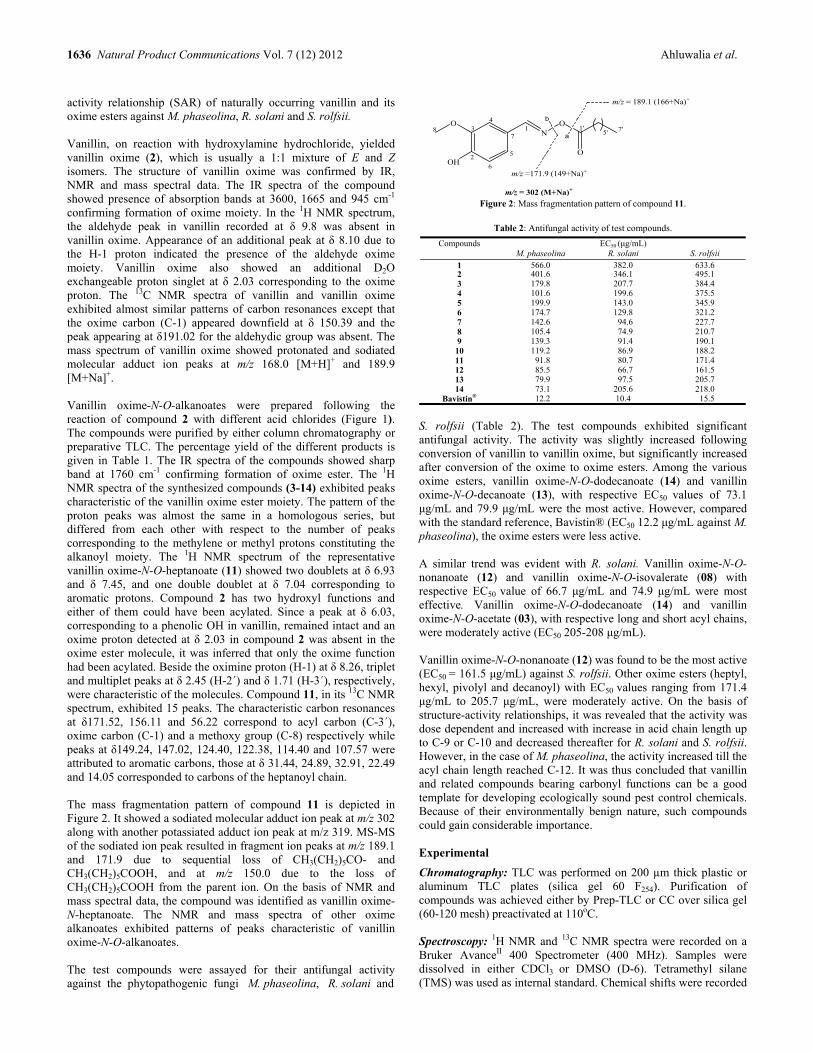
1636 Natural Product Communications Vol. 7 (12) 2012 Ahluwalia et al.
activity relationship (SAR) of naturally occurring vanillin and its oxime esters against M. phaseolina, R. solani and S. rolfsii. Vanillin, on reaction with hydroxylamine hydrochloride, yielded vanillin oxime (2), which is usually a 1:1 mixture of E and Z isomers. The structure of vanillin oxime was confirmed by IR, NMR and mass spectral data. The IR spectra of the compound showed presence of absorption bands at 3600, 1665 and 945 cm-1 confirming formation of oxime moiety. In the 1H NMR spectrum, the aldehyde peak in vanillin recorded at δ 9.8 was absent in vanillin oxime. Appearance of an additional peak at δ 8.10 due to the H-1 proton indicated the presence of the aldehyde oxime moiety. Vanillin oxime also showed an additional D2O exchangeable proton singlet at δ 2.03 corresponding to the oxime proton. The 13C NMR spectra of vanillin and vanillin oxime exhibited almost similar patterns of carbon resonances except that the oxime carbon (C-1) appeared downfield at δ 150.39 and the peak appearing at δ191.02 for the aldehydic group was absent. The mass spectrum of vanillin oxime showed protonated and sodiated molecular adduct ion peaks at m/z 168.0 [M+H]+ and 189.9 [M+Na]+. Vanillin oxime-N-O-alkanoates were prepared following the reaction of compound 2 with different acid chlorides (Figure 1). The compounds were purified by either column chromatography or preparative TLC. The percentage yield of the different products is given in Table 1. The IR spectra of the compounds showed sharp band at 1760 cm-1 confirming formation of oxime ester. The 1H NMR spectra of the synthesized compounds (3-14) exhibited peaks characteristic of the vanillin oxime ester moiety. The pattern of the proton peaks was almost the same in a homologous series, but differed from each other with respect to the number of peaks corresponding to the methylene or methyl protons constituting the alkanoyl moiety. The 1H NMR spectrum of the representative vanillin oxime-N-O-heptanoate (11) showed two doublets at δ 6.93 and δ 7.45, and one double doublet at δ 7.04 corresponding to aromatic protons. Compound 2 has two hydroxyl functions and either of them could have been acylated. Since a peak at δ 6.03, corresponding to a phenolic OH in vanillin, remained intact and an oxime proton detected at δ 2.03 in compound 2 was absent in the oxime ester molecule, it was inferred that only the oxime function had been acylated. Beside the oximine proton (H-1) at δ 8.26, triplet and multiplet peaks at δ 2.45 (H-2΄) and δ 1.71 (H-3΄), respectively, were characteristic of the molecules. Compound 11, in its 13C NMR spectrum, exhibited 15 peaks. The characteristic carbon resonances at δ171.52, 156.11 and 56.22 correspond to acyl carbon (C-3΄), oxime carbon (C-1) and a methoxy group (C-8) respectively while peaks at δ149.24, 147.02, 124.40, 122.38, 114.40 and 107.57 were attributed to aromatic carbons, those at δ 31.44, 24.89, 32.91, 22.49 and 14.05 corresponded to carbons of the heptanoyl chain. The mass fragmentation pattern of compound 11 is depicted in Figure 2. It showed a sodiated molecular adduct ion peak at m/z 302 along with another potassiated adduct ion peak at m/z 319. MS-MS of the sodiated ion peak resulted in fragment ion peaks at m/z 189.1 and 171.9 due to sequential loss of CH3(CH2)5CO- and CH3(CH2)5COOH, and at m/z 150.0 due to the loss of CH3(CH2)5COOH from the parent ion. On the basis of NMR and mass spectral data, the compound was identified as vanillin oxime-N-heptanoate. The NMR and mass spectra of other oxime alkanoates exhibited patterns of peaks characteristic of vanillin oxime-N-O-alkanoates. The test compounds were assayed for their antifungal activity against the phytopathogenic fungi M. phaseolina, R. solani and
m/z = 302 (M+Na)+
m/z =171.9 (149+Na)+
m/z = 189.1 (166+Na)+
OH
ON
O
O
1
2
34
5
6
78 1'
5' 7'a
b
Figure 2: Mass fragmentation pattern of compound 11.
Table 2: Antifungal activity of test compounds.
Compounds M. phaseolina
EC50 (μg/mL) R. solani
S. rolfsii
1 566.0 382.0 633.62 401.6 346.1 495.13 179.8 207.7 384.44 101.6 199.6 375.55 199.9 143.0 345.96 174.7 129.8 321.27 142.6 94.6 227.78 105.4 74.9 210.79 139.3 91.4 190.110 119.2 86.9 188.211 91.8 80.7 171.412 85.5 66.7 161.513 79.9 97.5 205.714 73.1 205.6 218.0
Bavistin® 12.2 10.4 15.5
S. rolfsii (Table 2). The test compounds exhibited significant antifungal activity. The activity was slightly increased following conversion of vanillin to vanillin oxime, but significantly increased after conversion of the oxime to oxime esters. Among the various oxime esters, vanillin oxime-N-O-dodecanoate (14) and vanillin oxime-N-O-decanoate (13), with respective EC50 values of 73.1 μg/mL and 79.9 μg/mL were the most active. However, compared with the standard reference, Bavistin® (EC50 12.2 μg/mL against M. phaseolina), the oxime esters were less active. A similar trend was evident with R. solani. Vanillin oxime-N-O-nonanoate (12) and vanillin oxime-N-O-isovalerate (08) with respective EC50 value of 66.7 μg/mL and 74.9 μg/mL were most effective. Vanillin oxime-N-O-dodecanoate (14) and vanillin oxime-N-O-acetate (03), with respective long and short acyl chains, were moderately active (EC50 205-208 μg/mL). Vanillin oxime-N-O-nonanoate (12) was found to be the most active (EC50 = 161.5 μg/mL) against S. rolfsii. Other oxime esters (heptyl, hexyl, pivolyl and decanoyl) with EC50 values ranging from 171.4 μg/mL to 205.7 μg/mL, were moderately active. On the basis of structure-activity relationships, it was revealed that the activity was dose dependent and increased with increase in acid chain length up to C-9 or C-10 and decreased thereafter for R. solani and S. rolfsii. However, in the case of M. phaseolina, the activity increased till the acyl chain length reached C-12. It was thus concluded that vanillin and related compounds bearing carbonyl functions can be a good template for developing ecologically sound pest control chemicals. Because of their environmentally benign nature, such compounds could gain considerable importance. Experimental
Chromatography: TLC was performed on 200 µm thick plastic or aluminum TLC plates (silica gel 60 F254). Purification of compounds was achieved either by Prep-TLC or CC over silica gel (60-120 mesh) preactivated at 110oC. Spectroscopy: 1H NMR and 13C NMR spectra were recorded on a Bruker AvanceII 400 Spectrometer (400 MHz). Samples were dissolved in either CDCl3 or DMSO (D-6). Tetramethyl silane (TMS) was used as internal standard. Chemical shifts were recorded

Preparation and antifungal activity of vanillin oxime-N-O-alkanoates Natural Product Communications Vol. 7 (12) 2012 1637
in (ppm) values relative to TMS and J values are expressed in Hertz. MS were recorded using a Thermo LC-MS-MS Spectrometer (Thermo Electron Corporation USA) equipped with a P-2000 binary pump. Mass was determined using an ESI source with a Finnigan LCQ tune plus program fitted with a MAX-detector. Xcalibur Software was used for the purpose of identification, quantification and fragmentation of the required masses. IR spectra were recorded in Nujol with a Nicolet Impact 700 FT-infra-red-spectrophotometer. Preparation of vanillin oxime: An equimolecular quantity of vanillin (4 g) in methanol (50 mL), hydroxylamine hydrochloride (1.35 g) and anhydrous potassium carbonate (4 g) was placed in a 250 mL round bottomed flask and the contents refluxed for 1 h. The progress of the reaction was monitored by TLC. After completion of the reaction, the mixture was cooled, filtered and the solvent removed by distillation under vacuum. Ice chilled water (100 mL) was then added and the resultant oxime was extracted with ethyl acetate (3 × 50 mL). The organic phase was passed through anhydrous sodium sulfate to remove traces of water and the solvent evaporated under vacuum to obtain solid vanillin oxime (2). The product crystallized from methanol as fine powder. Compound 2 Rf: 0.4 (n-hexane-acetone, 7:3); IR: 3600 (OH), 3310, 3167, 2890, 1423, 1460, 1665 (C=N), 1250, 945 (N-O), 780 cm-1. 1H NMR (400 MHz, CDCl3): δ 2.03 (s, 1H, NOH-1), 3.90 (s, 3H, H-8), 6.03 (s, 1H, OH-2), 6.99 (d, 1H, H-6, J = 8 Hz), 7.01 (dd, 1H, H-4, J = 8 Hz and 1.8 Hz), 7.21 (d, 1H, H-5, J =1.8 Hz), 8.10 (s, 1H, H-1). 13C NMR (100 MHz, CDCl3): δ 55.98 (C-8), 107.57 (C-7), 114.40 (C-6), 122.38 (C-5), 124.4 (C-4), 146.9 (C-3), 147.63 (C-2), 150.39 (C-1). ESI-MS: m/z 168.0 [(M+H)+, 100%], 189.9 [(M+Na)+, 20%],. MS-MS: m/z 151[168-OH, 100%]. General procedure for the preparation of vanillin oxime-N-O-alkanoates: To a solution of vanillin oxime (2) (0.05 mM) in dichloromethane (30 mL) in a 100 mL round bottomed flask, 2-3 drops of triethylamine (TEA) was added. An equimolar quantity of the respective acid chloride (0.05 mM), dissolved in dichloromethane (1:2), was added drop wise. The reaction mixture was stirred for 3 h with a magnetic stirrer at 0-5oC and the progress of the reaction was monitored by TLC. After completion, cold water (100 mL) was added and the mixture extracted with dichloromethane (3 × 50 mL). The organic phase was then washed with water (2 × 50 mL) and the resultant organic phase passed through anhydrous sodium sulfate to remove traces of water. The solvent was removed under vacuum at a temperature not exceeding 40oC to obtain the desired oxime-N-O-alkanoate. The products were further purified by either CC over silica gel using n-hexane and n-hexane/acetone as the eluting solvent or by prep-TLC. Compound 11 Rf: 0.6 (n-hexane-acetone, 7:3). IR: 3310 (OH), 3167, 2890, 1760 (C=O), 1650 (C=N), 1515, 1460, 1423, 780 cm-1. 1H NMR (400 MHz, CDCl3): δ 0.88 (t, 3H, H-7΄, J = 7.2 Hz), 1.30-1.41 (m, 6H, H-4΄, 5΄, 6΄), 1.71 (m, 2H, H-3΄), 2.45 (t, 2H, H-2΄, J = 7.6 Hz), 3.95 (s, 3H, H-8), 6.03 (s, 1H, OH-2), 6.93 (d, 1H, H-6,
J = 8Hz), 7.04 (dd, 1H, H-4, J = 8 Hz and 1.8 Hz), 7.45 (d, 1H, H-5, J = 1.6 Hz), 8.26 (s, 1H, H-1). 13C NMR (100 MHz, CDCl3): δ 14.05 (C-7΄), 22.49 (C-6΄) 32.91 (C-5΄), 28.82 (C-4΄), 24.89 (C-3΄), 31.44 (C-2΄), 56.22 (C-8), 107.57 (C-7), 114.40 (C-6), 122.38 (C-5), 124.4 (C-4), 147.02 (C-3), 149.24 (C-2), 156.11 (C-1), 171.52 (C-1΄). ESI-MS: m/z 302.0 [(M+Na)+, 100%], 319.0 [(M+H+K)+, 8%]. MS-MS: m/z 189.6 [302.0-CO(CH2)5CH3, 75%], 171.9 [302-CH3(CH2)5COOH, 30%], 150 [280.0- CH3(CH2)5COOH, 20%] Bioassay
Fungicidal activity: Macrophomina phaseolina (ITCC 6267), Rhizoctonia solani (ITCC 4502), and Sclerotium rolfsii (ITCC 6263) were procured from the Indian Type Culture Collection (ITCC) center, Division of Mycology and Plant Pathology, IARI, New Delhi, India. The cultures were maintained on potato dextrose agar (PDA) slants at 27±1°C and were subcultured prior to testing. Antifungal activity of the compounds was carried out employing the poisoned food technique using 4% PDA medium by the macrodilution method [17]. A stock solution of each compound was prepared by dissolving 130 mg in 2 mL acetone. An appropriate quantity of the test compound in acetone was added to molten PDA medium in order to obtain the desired concentration. The medium of each concentration and control was poured into a set of 3 Petri-dishes (3-replications) under aseptic conditions in a laminar flow chamber. The dishes were then kept under UV light in the laminar flow chamber till the medium partially solidified. A 5 mm thick disc of fungus (spore and mycelium) cut from earlier sub-cultured Petri-dishes was put at the centre of the semi solid medium in the test Petri-dishes and the lids of the dishes were fixed. Both treated and control dishes were kept in a BOD incubator at 28±1°C till the fungal growth in the control dishes was complete (4-6 days), after which the readings were taken. The mycelial growth (cm) in both treated (T) and control (C) Petri-dishes was measured diametrically in 3 different directions. From the mean results of the above readings, the percentage inhibition of growth (I) and corrected inhibition (IC) were calculated using Abbott’s formula:
I (%) = [(C-T)/C] × 100 Percent inhibition (I) was converted to corrected per cent inhibition (IC) using the following formula.
IC = {[I(%)-C.F.] / (100-C.F.)} × 100 IC = corrected percent inhibition, CF = correction factor
C.F. (Correction Factor) = [(90-C)/C] × 100 Where, 90 mm is the diameter of the Petri-dishes and C is the growth of the fungus in mm in control. Statistical analysis: EC50 (μg/mL) (effective concentration for 50 percent inhibition of mycelial growth) was calculated from the concentration (μg/mL) and corresponding IC data of each compound, with the help of Statistical Package (GW BASIC) using a personal computer (PC) [18]. Supplementary data: The spectral data of all compounds is included in a "Supplementary Data" section. Acknowledgments- Authors are thankful to Head, Division of Agricultural Chemicals, IARI, New Delhi, India for providing the necessary facilities.
*IARI contribution No. 1108.
References
[1] Walton NJ, Mayer MJ, Narbad A. (2003) Molecules of interest vanillin. Phytochemistry, 63, 505-515. [2] Sun R, Sacalis JN, Chin CK, Still CC. (2001) Bioactive aromatic compounds from leaves and stem of Vanilla fragrance. Journal of Agricultural
and Food Chemistry, 49, 5161-5164.

1638 Natural Product Communications Vol. 7 (12) 2012 Ahluwalia et al.
[3] Cerrutti P, Alzamora SM. (1996) Inhibitory effect of vanillin in some food spoilage yeasts in laboratory media and fruit purees. International Journal of Food Microbiology, 29, 379-386.
[4] Fitzgerald DJ, Stratford M, Gasson MJ, Narbad A. (2004) The potential application of vanillin in preventing yeast spoilage of ready-to-drink beverages. Journal of Food Protection, 67, 391-395.
[5] Burri J, Graf M, Lambelet P, Loliger J. (1989) Vanillin: more than a flavouring agent- a potential antioxidant. Journal of the Science of Food and Agriculture, 48, 49-56.
[6] Boonchird C, Flegel TW. (1982) In vitro antifungal activity of eugenol and vanillin against Candida albicans and Cryptococcus neoformans. Canadian Journal of Microbiology, 28, 1235-1241.
[7] Fitzgerald DJ, Stratford M, Gasson MJ, Narbad A. (2005) Structure-function analysis of the vanillin molecules and its antifungal properties. Journal of Agricultural and Food Chemistry, 53, 1769-1775.
[8] Bull MJ, Davies HJ, Robert JG, Henry AC. (1980) Alkyl aryl ketone oxime-O-ethers, a novel group of pyrethroids. Pesticide Science, 44, 249-256. [9] Walia S, Saxena VS, Mukerjee SK. (1985) Synthesis and synergistic activity of oxime ethers containing a benzo-1,3-dioxole group. Journal of
Agricultural and Food Chemistry, 33, 308-310. [10] Walia S, Gujar GT. (1993) Bioactivity of some substituted benzo-1-3-dioxole oxime ethers against Dysdercus koenigii. Pesticide Research Journal,
5, 205-208. [11] Chowdhury H, Saxena VS, Walia S. (1998) Synthesis and insect growth regulatory activity of alkoxy-substituted benzaldoxime ethers. Journal of
Agricultural and Food Chemistry, 46, 731-736. [12] Sun R, Li Y, Lu M, Xiong L, Wang Q. (2010) Synthesis, larvicidal activity, and SAR studies of new benzoylphenylureas containing oxime ether
and oxime ester group. Bioorganic & Medicinal Chemistry Letters, 20, 4693-4699. [13] Paul R, Walia S, Mondal A, Kumar J, Pankaj. (2009) Synthesis and nematicidal activity of 2-tridecanone and its oxime esters against Meloidogyne
incognita. Annals of Plant Protection Sciences, 17, 453-455. [14] Choudhary G, Walia S, Kumar J. (2007) Synthesis and antifungal activity of citral oxime esters against two phytopathogenic fungi Rhizoctonia
solani and Sclerotium rolfsii. Pesticide Research Journal, 19, 15-19. [15] Abeysinghe A. (2009) Efficacy of combined use of biocontrol agents on control of Sclerotium rolfsii and Rhizoctonia solani of Capsicum annuum.
Archieves of Phytopathology and Plant Protection, 42, 221–227. [16] Dikusar EA, Kozlov NG, Zhukovskaya NA, Moiseichuk KL. (2004) Esters of vanillin and vanilla oximes. Chemistry of Natural Compounds, 40,
180-183. [17] Shukla P, Walia S, Ahluwalia V, Parmar BS, Nair MG. (2012). Activity of alkanediol alkanoates against pathogenic plant fungi Rhizoctonia solani
and Sclerotium rolfsii. Natural Product Communications, 7, 1219-1222 [18] Trevors JT. (1986) A basic programme for estimating LD50 values using the IBM-PC. Bulletin of Environmental Contamination and Toxicology,
37, 18-26.
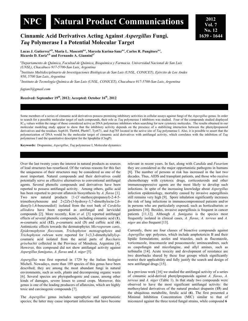
Cinnamic Acid Derivatives Acting Against Aspergillus Fungi. Taq Polymerase I a Potential Molecular Target Lucas J. Gutierreza,b, Maria L. Mascottia,c, Marcela Kurina-Sanza,c, Carlos R. Pungitorea,c, Ricardo D. Enriza,b and Fernando A. Gianninia* aDepartamento de Química, Facultad de Química, Bioquímica y Farmacia. Universidad Nacional de San Luis (UNSL), Chacabuco 917-5700-San Luis, Argentina
bInstituto Multidisciplinario de Investigaciones Biológicas de San Luis (UNSL, CONICET), Ejército de Los Andes 950, 5700 San Luis, Argentina
cInstituto de Tecnología Química de San Luis (UNSL, CONICET), Chacabuco 917-5700-San Luis, Argentina
Received: September 19th, 2012; Accepted: October 16th, 2012
Some members of a series of cinnamic acid derivatives possess promising inhibitory activities in cellular assays against fungi of the Aspergillus genus. In order to search for a possible molecular target of such compounds, their role as Taq polymerase I inhibitors was studied. Four of the compounds studied displayed IC50 values within the range of those considered active as DNA polymerase inhibitors when searching for new cytotoxic molecules. The results obtained in our molecular modeling study appear to show that the inhibitory activity depends on the presence of a stabilizing interaction between the phenylpropanoid derivatives and the residues Asp610, Thr664, Phe667, Tyr671, and Asp785 located in the active site of Taq polymerase I. Also, it is possible to assert that the polymerization of DNA would be the molecular target of cinnamic acid derivatives with antifungal activity, which correlates with the inhibition of Taq polymerase I and the quantitative descriptor for the lipophilia (ClogP). Keywords: Drupamine, Aspergillus, Taq polymerase I, Molecular dynamics.
Over the last twenty years the interest in natural products as sources of lead structures has resurfaced. Of the various reasons for this fact the uniqueness of their structures may be considered as one of the most important. Natural compounds and their derivatives could potentially serve as effective alternatives to conventional antifungal agents. Several phenolic compounds and derivatives have been reported to possess antifungal activity. Among others, gallic acid has been reported to prevent aflatoxin biosynthesis by A. flavus [1]. Two new natural products {1-(3´-methoxypropanoyl)-2-4-5-trimethoxybenzene and 2-(2Z)-(3-hydroxy-3,7-dimethylocta-2,6-dienyl)-1,4-benzenediol} isolated from the root bark of Cordelia alliodora have been reported as antifungal and larvicidal compounds [2]. More recently, Kim et al. [3] reported antifungal effects of several phenolic compounds, including cinnamic acid (1), m-coumaric acid (12), p-coumaric acid (3) and caffeic acid (6). Antimicotic effects towards the dermatophytes Microsporum canis, Epidermophyton floccosum, Trichophyton mentagraphytes and Trichophyton rubrum were reported for 3-(3,3-dimethylallyl)-p-coumaric acid isolated from the aerial parts of Baccharis grisebachii collected in the Province of Mendoza, Argentina [4]. However, this compound did not show antifungal activity against Aspergillus fumigatus, A. flavus and A. niger [5]. Aspergillus was first reported in 1729 by the Italian biologist Micheli. Nowadays, more than 189 species of this genus have been described; they are among the most abundant fungi in natural environments, such as soils, plants and decomposing organic waste [6]. Several species are phytopathogenic and cause, among other kinds of damages, severe losses to cereal crops. Moreover, this genus is one of the leading producers of aflatoxins, which are highly toxic and carcinogenic compounds [7]. The Aspergillus genus includes saprophytic and opportunistic species; the latter may cause important infections that have become
relevant in recent years. In fact, along with Candida and Fusarium they are considered as the major opportunistic pathogens in humans [8]. The number of persons at risk has increased in the last two decades. Thus, AIDS and transplant patients, and those who receive chemotherapy with cytotoxic drugs, corticosteroids and other immunosuppressive agents are the most likely to develop such infections. In spite of the increasing knowledge about Aspergillus infection epidemiology, mortality caused by invasive aspergillosis still remains very high [9]. Spore inhalation significantly increases the risk of lung infections in immunocompromised patients and/or in persons who are particularly exposed, such as horticulturists or gardeners [10]. Besides, invasive aspergillosis is frequent in cancer patients [11,12]. Although A. fumigatus is the species most frequently isolated in clinical cases, A. flavus, A. terreus and A. niger are also frequent [13]. Currently, there are four classes of bioactive compounds against Aspergillus spp: polyenes, which include amphotericin B and their lipidic formulations; azoles and triazoles, such as fluconazole, voriconazole, itraconazole and posaconazole; aminocandines, such as caspofungin and micofungine, and allyl amines, such as terbinafin [14]. Acute toxicity and development of resistance are two drawbacks shared by these four groups which significantly restrict their applicability and fully justify the search and design of new antifungal drugs [15]. In a previous work [16] we studied the antifungal activity of a series of cinnamic acid-derived phenylpropanoids against A. flavus, A. terreus and A. niger (Table 1). In that study two compounds were observed to have the most significant antifungal activity: the methoxylated derivatives of the natural product drupanin (15) and the ubiquitous metabolite, ferulic acid (6). The first presented a Minimal Inhibition Concentration (MIC) similar to that of miconazol against the three tested fungal strains, while compound 6
NPC Natural Product Communications 2012 Vol. 7 No. 12
1639 - 1644
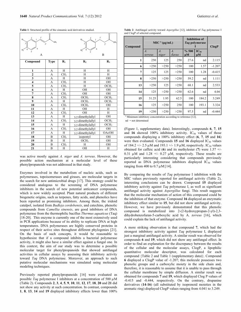
1640 Natural Product Communications Vol. 7 (12) 2012 Gutierrez et al.
Table 1: Structural profile of the cinnamic acid derivatives studied.
OR1
O
R2
R3
1
3
45
6
2 1'
2'
OR1
O
R2
R3
1
3
45
6
2 1'
2'
Compound Type R1 R2 R3
1 A H H H 2 A CH3 H H 3 A H H OH 4 A CH3 H OH 5 A CH3 H OCH3 6 A H OH OH 7 A CH3 OH OH 8 A CH3 OCH3 OCH3 9 A H OCH3 OCH3 10 A CH3 OCH3 OH 11 A H OH H 12 A CH3 OH H 13 A H -dimethylallyl OH 14 A CH3 -dimethylallyl OCH3 15 A H -dimethylallyl OCH3 16 A CH3 -dimethylallyl OH 17 A H -dimethylallyl OAcOH 18 B CH3 isopentyl OH 19 B H OCH3 OH 20 B CH3 H OH 21 B H OH H
was active mostly against A. niger and A. terreus. However, the possible action mechanism at a molecular level of these phenylpropanoids was not addressed in that study. Enzymes involved in the metabolism of nucleic acids, such as polymerases, topoisomerases and girases, are molecular targets in the search for new antimicrobial drugs [17]. This strategy could be considered analogous to the screening of DNA polymerase inhibitors in the search of new potential anticancer compounds, which is now widely accepted. Plant natural products of different biogenetic origins, such as terpenes, fatty acids, and phenolics have been reported as promising inhibitors. Among them, the iridoid catalpol, isolated from Budleja cordobensis, and catechins, phenolic compounds from Camellia sinensis, are good inhibitors of DNA polymerase from the thermophilic bacillus Thermus aquaticus (Taq) [18-20]. This enzyme is currently one of the most extensively used in PCR applications because of its ability to replicate DNA at high temperatures. DNA polymerases are highly conserved proteins in respect of their active sites throughout different phylogenies [21]. On the basis of such concepts, it would be reasonable to hypothesize that if a compound inhibits a bacterial polymerase activity, it might also have a similar effect against a fungal one. In this context, the aim of our study was to determine a possible molecular target for phenylpropanoids that showed antifungal activities in cellular assays by assessing their inhibitory activity toward Taq DNA polymerase. Moreover, an approach to such putative molecular mechanisms was simulated using molecular modeling techniques. Previously reported phenylpropanoids [16] were evaluated as possible Taq polymerase I inhibitors at a concentration of 500 M (Table 2). Compounds 2, 3, 4, 5, 9, 10, 11, 12, 17, 18, 20 and 21 did not show any activity at such concentration. In contrast, compounds 1, 8, 13, 14 and 19 displayed inhibitory effects lower than 100%
Table 2: Antifungal activity toward Aspergillus [12], inhibition of Taq polymerase I and ClogP of selected compound.
Compound MIC a (μg/mL)
Inhibition of Taq polymerase
I ClogP
A. terreus
A. niger
A. flavus
% 500 μM
IC50
μM
1 250 125 250 27.6 nd 2.115
6 >250 >250 >250 100 1.57 -1.207
7 125 125 >250 100 1.28 -0.415
8 >250 >250 >250 59.2 nd 1.111
13 >250 125 >250 68.1 nd 2.533
14 125 >250 >250 62.6 nd 4.04
15 31.25 1.95 62.5 100 184.2 3.249
16 125 >250 250 100 193.1 3.324
19 >250 >250 >250 97.5 nd -0.444a Minimum inhibitory concentration according to reference [12]. nd = not determined
(Figure 1, supplementary data). Interestingly, compounds 6, 7, 15 and 16 showed 100% inhibitory activity. IC50 values of those compounds displaying a 100% inhibitory effect (6, 7, 15 and 16) were then evaluated. Compounds 15 and 16 displayed IC50 values of 184.2 +/- 2.5 M and 193.1 +/- 1.9 M, respectively. IC50 values obtained for caffeic acid (6) and its methylester (7) were 1.57 +/- 0.31 M and 1.28 +/- 0.27 M, respectively. These results are particularly interesting considering that compounds previously reported as DNA polymerase inhibitors displayed IC50 values ranging from 400 to 0.7 M 14. By comparing the results of Taq polymerase I inhibition with the MIC values previously reported for antifungal activity (Table 2), interesting conclusions can be drawn. Compound 15 displayed inhibitory activity against Taq polymerase I, as well as significant antifungal activity against Aspergillus fungi. This result suggests that the molecular mechanism of action of this compound could be the inhibition of that enzyme. Compound 16 displayed an enzymatic inhibitory effect similar to 15, but did not show antifungal activity. However, we have previously demonstrated that this phenolic compound is metabolized into 2-(2-hydroxypropan-2-yl)-2,3-dihydrobenzofuran-5-carboxylic acid by A. terreus [16], which could explain the lack of antifungal activity. A more striking observation is that compound 7, which had the strongest inhibitory activity against Taq polymerase I, displayed just a marginal antifungal activity. A similar result was observed for compounds 6 and 19, which did not show any antifungal effect. In order to find an explanation for the discrepancy between the results of the cellular and the molecular assays, ClogP, a lipophilic quantitative molecular descriptor, was calculated for each compound {Table 2 and Table 1 (supplementary data)}. Compound 6 displayed a ClogP value of -1.207; this molecule possesses two phenolic groups and a carboxylic moiety in the side chain and, therefore, it is reasonable to assume that it is unable to pass through the cellular membrane by simple diffusion. A similar result was obtained for compounds 7 and 19, which displayed Clog P values of -0.415 and -0.444, respectively. On the contrary, drupanine derivatives (14-16) (all substituted by isoprenoid moieties in the aromatic ring) displayed ClogP values ranging from 4.041 to 3.249.
B A

Cinnamic acid dervatives as Taq polymerase I inhibitors Natural Product Communications Vol. 7 (12) 2012 1641
Figure 1: Inhibitor-residue interaction spectra of (a) Taq-6 and (b) Taq-15, according to the MM-GBSA method. The x-axis denotes the residue number of Taq Polymerase I and the y-axis denotes the interaction energy between the inhibitor and specific residues.
To acquire a more-detailed insight into the mechanisms driving the bindings of cinnamic acid derivatives to the active site of Taq polymerase I, the structure-affinity relationship was analysed to complement the experimental results. The information obtained from these calculations is very important for quantitative analyses and is highly useful to the understanding of the binding mechanism. Figure 1 shows the inhibitor-residue interaction spectra calculated by free energy decomposition, which suggests that the interaction spectra of compounds 6 and 15 with Taq-polymerase are almost the same and reflect their similar binding modes (note that the disfavoured interaction of Lys 663 with compound 15 is the only difference obtained when comparing both spectra). Closely related spectra were obtained for compounds 7 and 16 (data not shown), indicating that all the compounds possessing a significant inhibitory effect displayed very similar interaction spectra. For these compounds, the favourable residues can be divided into five groups around Asp610, Thr664, Phe667, Tyr671, and Asp785. The contribution to binding of an individual residue varies in the range of 3.1 to -6.2 kcal/mol. The dominant favourable interactions come from residues Thr664 and Asp785 performing stabilizing hydrogen bonds, as well as from residues Phe667 and Tyr671 making pi stacking interactions with the aromatic ring of the ligand. Figure 2 displays the relative spatial positions of inhibitors 6 and 15 in their
Figure 2: Spatial view of the stabilizing and destabilizing interactions obtained for compounds 6 (a) and 15 (b) interacting with Taq polymerase I. The different amino acids involved in this complex are shown.
lowest-energy structures at the binding complexes extracted from MD simulations. Figure 2 also shows the different stabilizing and destabilizing interactions. It is interesting to note that the interaction spectra obtained for compounds 17 and 18 are different from those observed for compounds possessing an inhibitory effect (Figure 3). In the case of compound 17 (Figure 3a), there are three strong destabilizing interactions, namely with residues Arg573 (5.4 kcal/mol), Lys663 (7.2 kcal/mol) and Gln754 (7.4 kcal/mol). These destabilizing contributions make the binding energy obtained for compound 17 markedly higher than that obtained for compound 6. Figure 4a displays the relative spatial positions of compound 17 in the lowest-energy structure of the binding complexes extracted from MD simulations. The main stabilizing and destabilizing interactions can be well observed in this figure. Although the residues stabilizing the complex are very similar to those observed in the complexes obtained for compounds 6 and 15, in this case the distances between compound 17 and the stabilizing residues are larger and, therefore,
0 100 200 300 400 500 600-8
-6
-4
-2
0
2
4
6
8
Inte
ract
ion
En
erg
y (k
cal/m
ol)
Residue Sequence
293 393 493 593 693 793 893
Gln613 Glu615
Lys663
Thr664
Phe667
Tyr671 Asp785
Arg573
Asp610
293 100 200 300 400 500 831-8
-6
-4
-2
0
2
4
6
8
Inte
ract
ion
En
erg
y (k
cal/m
ol)
Residue Sequence
Thr664
Phe667
Tyr671
Asp785
Glu615 Gln613
Arg573
Asp610
293 393 493 593 693 793 893
a)
b)
HO
HO
OH
O
H3CO
OH
O

1642 Natural Product Communications Vol. 7 (12) 2012 Gutierrez et al.
Figure 3: Inhibitor-residue interaction spectra of (a) Taq-17 and (b) Taq-18 according to the MM-GBSA method. The x-axis denotes the residue number of Taq Polymerase I and the y-axis denotes the interaction energy between the inhibitor and specific residues. the stabilizing energies are weaker (compare Figures 2a and 4a). In addition, it should be noted that in this complex the stabilizing interaction of Thr664 disappears. The strongest destabilizing interaction was obtained for compound 18, namely the interaction between compound 18 and Arg573, which displays about 15.4 kcal/mol (Figures 3b and 4b). This important destabilizing interaction, together with other minor ones, is responsible for the high binding energy obtained for compound 18 in comparison with those for compounds 6 and 15, which possess inhibitory effects. These theoretical results are in a complete agreement with our experimental data. Thus the results obtained in our molecular modelling study make it clear that it is possible to discriminate between compounds possessing significant inhibitory effects (6, 7, 15 and 16) from those which are inactive toward Taq polymerase I (17 and 18). On the basis of our experimental and theoretical results, it is reasonable to assume that the cinnamic acid derivatives here reported are candidates to be leading structures in the design of new Taq polymerase I inhibitors.
Figure 4: Spatial view of the stabilizing and destabilizing interactions obtained for compounds 17 (a) and 18 (b) interacting with Taq polymerase.
Taq polymerase I may be the molecular target for the antifungal activity displayed by the studied derivatives, but it is evident that a ClogP value of about 3 is also necessary to produce the antifungal effect at a cellular level. The strong antifungal activity, the relatively low acute toxicity previously reported, and the molecular mechanism here reported indicate that the methoxylated derivative of drupanin (15) is a very good candidate on which to perform further studies in the search for new agents to control Aspergillus infections. Experimental
Test compounds: Compounds 1, 2, 3, 6, and 11 were purchased from Sigma-Aldrich Co. Drupanin (13) was isolated as colorless crystals (mp 147-148°C) from aerial parts of B. grisebachii Hieron. Its chemical identity was confirmed by spectroscopic data, compared with previous reports [4]. Compounds 4, 5, 6, 7, 8, 9, 10, 12, 14-21 were prepared as previously described by Bisogno and coworkers [16].
293 100 200 300 400 500 831-8
-6
-4
-2
0
2
4
6
8
Inte
ract
ion
En
erg
y (k
cal/m
ol)
Residue Sequence
Phe667 Asp785
Asp610
Arg573 Lys663 Gln754
Glu786
293 393 493 593 693 793 893
293 100 200 300 400 500 831-20
-10
0
10
20
Inte
ract
ion
En
erg
y (k
cal/m
ol)
Residue Sequence
Arg573
Asp610
Asp785
Glu615 His784
Glu786
293 393 493 593 693 793 893
a)
b)
HOOCH2CO
OH
O
HO
OCH3
O

Cinnamic acid dervatives as Taq polymerase I inhibitors Natural Product Communications Vol. 7 (12) 2012 1643
Taq polymerase I inhibition assays: Inhibition of Taq polymerase I from Thermusaquaticus (Sigma-Aldrich) was studied by PCR. The assayed compounds were dissolved in DMSO (1 mg/mL) and inhibition studies were carried out at a screening concentration of 500 M. The PCR master mixture consisted of 1.8 M MgCl2, 0.2 MdNTPs, 1 M primer sense (5´GGATCCATGAGCGGGGACCATCTCCA), and 1 M primer antisense (5´TCAAAACTCGTAGTCCTCATAG). DNA concentration was 0.013 mg/mL and all PCR reactions were made in 20 L. Thermocycling conditions consisted of 35 cycles of denaturation at 94°C for 30s followed by primer annealing at 53°C for 30s and primer extension at 72°C for 1 min 30s. Catapol 100 M was used as positive control. Assays were made in triplicate. Compounds that displayed 100% inhibition at the screening concen-tration were further evaluated at lower concentrations by serial dilution (500 M to 0.95 M) in order to obtain the IC50 value. Analysis of PCR products: Relative intensities of ethidium bromide stained PCR products were analyzed using an optical scanner. The digitized band images were processed using the Image processing software Scion Image, a public domain program. IC50 values were determined by the GraphPad Prism software. Molecular modeling: The coordinate of Taq polymerase I was downloaded from Brookhaven Protein Data Bank (www.rcsb.org). The PDB code entry is 3KTQ. To explore the dynamic stability of the Taq-inhibitor complexes reported here, root mean square deviation (RMSD) values for the protease C atoms during the MD simulations (relative to the docking structure) were calculated. The RMSD values of the Taq-6, Taq-15, Taq-17 and Taq-18 complexes are 1.60±.031, 2.13±0.59, 1.96±0.34 and 1.80±0.31 Å, respectively, showing a deviation lower than 0.6 Å from the mean. These results suggest that the stability of the dynamic equilibriums of these complexes is reliable. Molecular docking: In order to search for the preferable conformation, AutoDock 4.0 software [22] was used for docking calculations, combined with Lamarckian genetic algorithm (LGA). The center of the grid box was the active site and the size of the box was 50 x 45 x 55 grid points, with grid spacing of 0.375 Å. During the docking experiment, the receptor was kept rigid and the ligand was set flexible. The maximum number of energy evaluations was set to 25 x 106; the initial population of trial ligands consisted of 250 individuals; for the rest of the docking parameters the default values were used. Fifty docking runs were performed to find the preferable conformation. The docking results were clustered according to the RMSD of 2Å. Structures with relative lower binding free energy and most cluster members were chosen, which were then subjected to molecular dynamics simulations (MD). Molecular dynamics simulations: To relax the binding complexes and investigate their dynamics, three-nanosecond MD simulations were performed for the different Taq-inhibitor systems using Amber software [23]. The protein force field was taken from ff99SBilnd [24]. The general Amber force field (GAFF) [25] was used to handle small organic molecules and the force field parameters of the inhibitors were produced by the antechamber program in Amber. Each Taq-inhibitor complex was soaked in a truncated octahedral periodic box of TIP3P water molecules. The distance between the
edges of the water box and the closest atom of the solutes was at least 10Å. Na+ ions were added to neutralize the systems charge. The entire system was subjected to energy minimization in two stages to remove bad contacts between the complex and the solvent molecules. First, the water molecules were minimized by holding the solute fixed with harmonic constraints of 100 kcal/molÅ2 strength. Secondly, conjugate gradient energy minimizations were performed repeatedly 4 times using positional restraints to all heavy atoms of the receptor with 15, 10, 5 and 0 kcal/molÅ2. The system was then heated from 0 to 300 K in 300 ps and equilibrated at 300 K for another 200 ps. After minimization and heating, 2.5 ns dynamics simulations were performed at a constant temperature of 300 K and a constant pressure of 1 atm. During minimization and MD simulations, the particle mesh Ewald (PMD) method was employed to treat the long-range electrostatic interactions in a periodic boundary condition. The SHAKE method was used to constrain hydrogen atoms. The time step for all MD is 2 fs, with a direct-space, non-bonded cutoff of 8Å. Initial velocities were assigned from a Maxwellian distribution at the initial temperature. The MM-GBSA method: In MM-GBSA [26], the binding free energy (ΔGbind) resulting from the formation of a RL complex between a ligand (L) and a receptor (R) is calculated as
ΔGbind = ΔEMM + ΔGsol – TΔS (1) ΔEMM = ΔEinternal + ΔEelectrostatic + ΔEvdw (2) ΔGsol = ΔGGB + ΔGSA (3)
where ΔEMM, ΔGsol and -TΔS are the changes of the gas phase MM energy, the solvation free energy, and the conformational entropy upon binding, respectively. ΔEMM includes ΔEinternal (bond, angle, and dihedral energies), ΔEelectrostatic (electrostatic), and ΔEvdw (van der Waals) energies. ΔGsolv is the sum of electrostatic solvation energy (polar contribution), ΔGGB, and the non-electrostatic solvation component (nonpolar contribution), ΔGSA. Polar contribution is calculated using the GB model, while the nonpolar energy is estimated by solvent accessible surface area (SASA). The conformational entropy change, -TΔS, is usually computed by normal-mode analysis, but in this study the entropy contributions were not calculated due to the computational cost involved in such calculations. ClogP calculations: All geometries of the compounds reported here were optimized from DFT calculations using the Becke3-Lee-Yang-Parr [27-29] (B3LYP) functional with the 6-31G(d) basis set. These calculations were carried out using the Gaussian 03 [30] program. Once the different optimized geometries were obtained, ClogP values were calculated using Chem Office Ultra 4.5 (Cambridge- soft. Corp 1988) software. Supplementary data: Theoretical ClogP values for compounds 1-21(Table1). Image of the Taqpolymerase I inhibition assay (Figure1). Acknowledgments - This work was supported by grants from Universidad Nacional de San Luis: PROICO 1412 and 22/Q027. Consejo Nacional de Investigaciones Científicas y Técnicas (CONICET) PIP 6228 and 0357. Agencia Nacional de Promoción Científica y Tecnológica (ANPCyT) PICT 352, 1416 and 1832. LJG and MLM are doctoral CONICET fellows. MKS, CRP and RDE are members of the Research Career of CONICET, Argentina.
References
[1] Mahoney N, Molyneux RJ. (2004) Phytochemical inhibition of aflatoxigenicity in Aspergillusflavus by constituents of walnut (Juglansregia). Journal of Agricultural and Food Chemistry, 52, 1882–1889.

1644 Natural Product Communications Vol. 7 (12) 2012 Gutierrez et al.
[2] Ioset JR, Marston A, Gupta MP, Hostettmann K. (2000) Antifungal and larvicidal compounds from the root bark of Cordia alliodora. Journal of Natural Products, 63, 424–426.
[3] Kim JH, CampbellBC, Mahoney NE, Chan KL, Molyneux RJ. (2004) Identification of phenolics for control of Aspergillus flavus using Saccharomyces cerevisiae in a model target-gene bioassay. Journal of Agricultural and Food Chemistry, 52, 7814–7821.
[4] Gianello JC, Giordano OS. (1987) Constituents from Bacccharis grisebachii. Anales de la Asociación Química Argentina, 75, 1–3. [5] Feresin GE, Tapia A, Gimenez A, Ravelo AG, Zacchino S, Sortino M, Schmeda-Hirschmann G. (2003) Constituents of the Argentinian medicinal
plant Baccharis grisebachii and their antimicrobial activity. Journal of Ethnopharmacology, 89, 73–80. [6] Lass-Flörl C, Perkhofer S. (2008) In vitro susceptibility-testing in Aspergillus species. Mycoses, 51, 437–446. [7] Lu FC. (2003) Assessment of safety/risk vs. public health concerns: Aflatoxins and hepatocarcinoma. Environmental Health and Preventive
Medicine, 7, 235–238. [8] Solé A, Salavert M. (2008) Fungal infections after lung transplantation. Transplantation Reviews (Orlando, Fla.), 22(2), 89–104. [9] Gangneux JP, Camus C, Philippe B. (2010) Epidemiology of invasive aspergillosis and risk factors in non neutropaenic patients. Revue des
Maladies Respiratoires, 27, e34–46. [10] Summerbell RC. (1998) Taxonomy and ecology of Aspergillus species associated with colonizing infections of the respiratory tract. Immunology
and Allergy Clinics of North America, 18, 549–573. [11] Lee LD, Hachem RY, Berkheiser M, Hackett B, Jiang Y, Raad II. (2012) Hospital environment and invasive aspergillosis in patients with
hematologic malignancy. American Journal of Infection Control, 40, 247–249. [12] Nilsson JR, Restrepo CS, Jagirdar J. (2011) Two cases of endobronchial carcinoid masked by superimposed aspergillosis: a review of the literature
of primary lung cancers associated with Aspergillus. Annals of Diagnostic Pathology, in press. [13] Bodey G, Bueltmann B, Duguid W, Gibbs D, Hanak H, Hotchi M, Mall G, Martino P, Meunier F, Milliken S. (1992) Fungal infections in cancer
patients: an international autopsy survey. European Journal of Clinical Microbiology & Infectious Diseases : Official Publication of the European Society of Clinical Microbiology, 11, 99–109.
[14] Qiao J, Liu W, Li R. (2008) Antifungal resistance mechanisms of Aspergillus. Nihon Ishinkin Gakkaizasshi (Japanese Journal of Medical Mycology), 49, 157–63.
[15] Chamilos G, Kontoyiannis DP. (2005) Update on antifungal drug resistance mechanisms of Aspergillus fumigatus. Drug Resistance Updates : Reviews and Commentaries in Antimicrobial and Anticancer Chemotherapy, 8, 344–358.
[16] Bisogno F, Mascoti L, Sanchez C, Garibotto F, Giannini F, Kurina-Sanz M, Enriz R. (2007) Structure-antifungal activity relationship of cinnamic acid derivatives. Journal of Agricultural and Food Chemistry, 55, 10635-10640.
[17] Pungitore CR. (2008) Natural products as inhibitors of DNA related enzymes. Current Enzyme Inhibition, 4, 194-215. [18] Pungitore CR, Ayub MJ, Borkowski EJ, Tonn CE, Ciuffo GM. (2004) Inhibition of Taq DNA polymerase by catalpol. Cellular and Molecular
Biology, 50, 767–772. [19] Jaziri I, Ben Slama M, Mhadhbi H, Urdaci MC, Hamdi M. (2009) Effect of green and black teas (Camellia sinensis L.) on the characteristic
microflora of yogurt during fermentation and refrigerated storage. Food Chemistry, 112, 614–620. [20] Pungitore CR, Ayub MJ, García M, Borkowski EJ, Sosa ME, Ciuffo G, Giordano OS, Tonn CE. (2004) Iridoids as allelochemicals and DNA
polymerase inhibitors. Journal of Natural Products, 67, 357–361. [21] Joyce CM, Steitz T A. (1994) Function and structure relationships in DNA polymerases. Annual Review of Biochemistry, 63, 777–822. [22] Morris GM, Goodsell DS, Huey R, Olson AJ. (1996) Distributed automated docking of flexible ligands to proteins: parallel applications of
AutoDock 2.4. Journal of Computer-aided Molecular Design, 10, 293–304. [23] Case DA, Cheatham TE, Darden T, GohlkeH, Luo R, Merz KM, Onufriev A, Simmerling C, Wang B, Woods RJ. (2005) The Amber biomolecular
simulation programs. Journal of Computational Chemistry, 26, 1668–1688. [24] Lindorff-Larsen K, Piana S, Palmo K, Maragakis P, Klepeis JL, Dror RO, Shaw DE. (2010) Improved side-chain torsion potentials for the Amber
ff99SB protein force field. Proteins, 78, 1950–1958. [25] Cornell W, Cieplak P, Bayly C, Gould I, Merz, K, Ferguson D, Spellmeyer D, Fox T, Caldwell J, Kollman P. (1995) A second generation force
field for the simulation of proteins, nucleic acids, and organic molecules. Journal of the American Chemical Society, 117, 5179–5197. [26] Kollman PA, Massova I, Reyes C, Kuhn B, Huo S, Chong L, Lee M, Lee T, Duan Y, Wang W, Donini O, Cieplak P, Srinivasan J, Case DA,
Cheatham TE. (2000) Calculating structures and free energies of complex molecules: combining molecular mechanics and continuum models. Accounts of Chemical Research, 33, 889–897.
[27] Becke AD. (1988) Density-functional exchange-energy approximation with correct asymptotic behavior. Physical Review A, 38, 3098–3100. [28] Becke AD. (1993) A new mixing of Hartree–Fock and local density-functional theories. The Journal of Chemical Physics, 98, 1372-1377. [29] Lee C, Yang W, Parr RG. (1988) Development of the Colle-Salvetti correlation-energy formula into a functional of the electron density. Physical
Review B, 37, 785–789. [30] Frisch MJ, Trucks GW, Schlege HB, Scuseria GE, Robb MA, Cheeseman JR, Montgomery JA, Vreven JrT, Kudin KN, Burant JC, Millam JM,
Iyengar SS, Tomasi J, Barone V, Mennucci B, Cossi M, Scalmani G, Rega N, Petersson GA, Nakatsuji H, Hada M, Ehara M, Toyota K, Fukuda R, Hasegawa J, Ishida M, Nakajima T, Honda Y, Kitao O, Nakai H, Klene M, Li X, Knox JE, Hratchian HP, Cross JB, Adamo C, Jaramillo J, Gomperts R, Stratmann RE, Yazyev O, Austin A J, Cammi R, Pomelli C, Ochterski JW, Ayala PY, Morokuma K, Voth GA, Salvador P, Dannenberg JJ, Zakrzewski VG, Dapprich S, Daniels AD, Strain MC, Farkas O, Malick DK, Rabuck AD, Raghavachari K, Foresman JB, Ortiz JV, Cui Q, Baboul AG, Clifford S, Cioslowski J, Stefanov BB, Liu G, Liashenko A, Piskorz P, Komaromi I, Martin RL, Fox DJ, Keith T, Al-Laham MA, Peng CY, Nanayakkara A, Challacombe M, Gill PM W, Johnson B, Chen W, Wong MW, Gonzalez C, Pople JA. Gaussian 2003 (2003).

Insecticidal Effects of Acetogenins from Rollinia occidentalis Seed Extract
Diego Tolosaa, Olga Álvarez Coloma, Alicia Bardóna,b and Adriana Neske*a
aInstituto de QuímicaOrgánica, Facultad de Bioquímica, Química y Farmacia, Universidad Nacional de Tucumán, Ayacucho 471, Tucumán 4000, Argentina; bINQUINOA-CONICET, Tucumán, Argentina
Received: September 14th, 2012; Accepted: November 5th, 2012
From the methanol seed extract of Rollinia occidentalis five acetogenins were isolated, sylvaticin (1), rolliniastatin-1 (2), rolliniastatin-2 (3), motrilin (4) and desacetyluvaricin (5), and evaluated for their toxicity and nutritional alterations on Spodoptera frugiperda Smith (Lepidoptera: Noctuidae). All but one of the compounds produced more than 90% mortality on early larval instars at 50 and 100 µg per g of diet. Sylvaticin caused 15 and 60% mortality at 50 and 100 µg/g, respectively, while the methanol seed extract reached 35 and 50% at 100 and 250 µg/g, respectively. The addition of R. occidentalis methanolic seed extract (100 and 250 µg/g) and sylvaticin (100 µg/g) to the larval diet brought about significant changes to the nutritional indices in connection with larvae fed the control diet. This is the first report on the isolation of acetogenins from R. occidentalis and their insecticidal effects on S. frugiperda. Keywords: Plant metabolites, Polyphagous lepidopteran, Toxicity, Nutritional alterations.
Rollinia occidentalis Fries (Annonaceae) is a tree commonly found in northwest Argentina. Characteristic constituents of these plants are the annonaceous acetogenins that possess unique structures and anticancer [1a,b], cytotoxic [2], insecticidal [3a,b], and antiparasitic activities. Previous results from our laboratory indicated that the acetogenins from Annona cherimolia and A. montana have larvicidal effects on the Spodoptera frugiperda (Lepidoptera) corn pest [4a,b], nymphal and adult mortality on the Oncopeltus fasciatus (Hemiptera) cotton pest[4c],and insecticidal activity against Ceratitis capitata Wiedemann (Diptera) [4d].
This is the first report on the isolation and identification of 5 known and structurally relatedbistetrahydrofuran acetogenins from the methanolic seed extract of R. occidentalis: sylvaticin (1) [5], rolliniastatin-1 (2) [6], rolliniastatin-2 (3) [7a], motrilin (4) [7b]and desacetyluvaricin (5)[7c].Continuing with our search for natural insecticides from Annonaceae plants, we evaluated the toxic effects produced by the methanol seed extract and annonaceous acetogenins (ACG) isolated from R. occidentalis on S. frugiperda. Additionally, Consumption Index (CI), Growth (GR), and Efficiency in the Consumption Index (ECI) were also assessed. The "fall armyworm”, S. frugiperda Smith (Lepidoptera: Noctuidae), is a widely distributed pest in America. It causes serious economic damage to many crops even though its host preference is corn. Toxicity was determined by evaluating S. frugiperda larval and pupal mortality and malformations of larvae, pupa and adults. They were fed an artificial diet treated with different ACG at a final concentration of 50 and 100 mg ACG/g of diet. An untreated diet was used as control.
The methanolic seed extract of R. occidentalis at 50 µg/mL did not have a significant toxic effect under the experimental conditions. However, at 100 and 250 µg/mL it had a toxic effect on S. frugiperda with a larval mortality of 45 and 50%, respectively. Sylvaticin (100 µg/mL) produced a significant toxic effect on S. frugiperda with 70% mortality while rolliniastatin-1, rolliniastatin-2, motrilin and desacetyluvaricin caused 100% larval mortality under the experimental conditions. Most of the tested compoundsproduced significant larval and pupal mortality with malformations of larvae, pupa and adults.
(CH2)9
OH
OHOOH
O
OO
(CH2)7CH3
1 sylvaticin
OH
OO
CH3OO
(CH2)10(CH2)9
OH
3 rolliniastatin-2
OHOH
OO
CH3OO
(CH2)10(CH2)9
OH
2 rolliniastatin-1
OHOH
OO
CH3OO
(CH2)12(CH2)9
OH
5 desacetyluvaricin
OH
OO
CH3OO
(CH2)12(CH2)4
OHOH
4 motrilin
(CH2)4
OH
Figure 1: Annonaceous acetogenins from R. occidentalis methanolic seed extract. The addition of methanolic seed extract and sylvaticin to the diet affecting the early instar larvae in their growth and nutrition could not be quantified in the remaining rolliniastatin-1, rolliniastatin-2, motrilin and desacetyluvaricin as larvae mortality occurred within 10 days. Toxicity of annonaceous acetogenins: R. occidentalis methanolic seed extract (50 µg/mL) did not have a significant toxic effect under the experimental conditions; 100 and 250 µg/mL had a slight toxic effect on S. frugiperda with larval mortality of 45 and 50%, respectively. However, sylvaticin (100 µg/mL) produced significant toxic effects on S. frugiperda with 70% mortality and malformed adults (20%), while rolliniastatin-1, rolliniastatin-2, motrilin and desacetyluvaricin were the most toxic (100% larval mortality) under the experimental conditions, as shown in Table 1. Pupal mortality was apparently related to deficient melanization and malformation in wings, legs and antenna cover. Remarkably, every surviving adults were abnormal in abdomen and wings. We analyzed the nutritional effects produced by the addition of the above mentioned extract and ACG to the 2nd instar larval diet of S. frugiperda to assess how treatment induced mortality. Larvae fed with R. occidentalis (50 µg/g) and sylvaticin (50 µg/g) methanolic seed extract presented physiological indices similar to those of control larvae. The addition of R. occidentalis (100 and 250 µg/g) and sylvaticin (100 µg/g) methanolic seed extract to the larval diet caused significant changes to the nutritional indices compared with those of larvae fed the control diet (Table 2).This resulted in an important larval growth decrease and subsequent 100% larval mortality at very early stages in their life cycle and would reveal the
NPC Natural Product Communications 2012 Vol. 7 No. 12
1645 - 1646

1646 Natural Product Communications Vol. 7 (12) 2012 Tolosa et al.
Sylvaticin (50 µg/g) had the lowest intake percentage and the poorest nutrient absorbed conversion. Nutritional indices for rolliniastatin-1, rolliniastatin-2, motrilin and desacetyluvaricin could not be measured as mortality occurred at an early stage.
As shown in Figure 1, all the tested acetogenins carry two THF rings and the THF are adjacent in four of them. Our results indicated that acetogenins2, 3, 4 and 5had killed more than 90% of S. frugiperda larvae at 120 h after the start of the assay, while the remaining acetogenin1, had a lower effect at the doses tested. The most important toxic action was observed on larval instar. In fact, all acetogenins produced more than 70% larval mortality with no dependence on the position of the THF rings or the number and location of the OH groups. The evaluation of food consumption indicated that the tested acetogenins were not antifeedant agents because there were no significant differences related to the control. The high ECI values for sylvaticin (100 µg/g) are due to the very low larval weight even when larvae consume the diet regularly, suggesting that sylvaticin reduces the efficiency to convert food into biomass. The influence of chemical agents and their toxic effects on insects and their nutritional behaviour can be used in the development of environmentally friendly pest control agents.
Experimental
Plant material: R. occidentalis fruits were collected in Tucumán, Argentina, in March 2005. A voucher sample (No. 604639) was deposited at the Herbarium of Instituto Lillo of Tucumán. Acetogenin extraction and purification: Dried and powdered seeds of R. occidentalis were percolated with methanol. Evaporation of the solvent yielded a crude MeOH extract which was further partitioned between CHCl3 and H2O. The solvent was then evaporated from the chloroform extract under reduced pressure and the residue was chromatographed on a silica gel column by using chloroform and increasing amounts of ethyl acetate (0-100%) and finally methanol as eluents. Chromatographic fractions containing acetogenins (TLC detection by Kedde’s reagent) were further processed by high-performance liquid chromatography (HPLC) using a Beckman C 18 column (25 cm x 1 cm i.d., 5 m particle size) and mixtures of methanol and water to yield pure acetogenins. Acetogenins represented around 0.05% of the seed weight.
Table 1: Toxicity produced by R. occidentalis methanolic seed extract and ACG on larvae of S. frugiperda.
Test insects: S. frugiperda larvae were obtained from our laboratory population and maintained with an artificial diet.Toxicity tests were recorded for treatments with all acetogenins (50 and 100 µg/mL) and control experiments [7d].Determination of Consumption (CI), Growth (GR), and Efficiency in the Consumption Index (ECI) were carried out. For comparison purposes, rates are expressed as a relationship between treatment and control; the latter are considered 100%. Values are expressed as (GRT/GRC) 100%, (CIT/CIC) 100% and (ECIT /ECIC) 100% in Table 2 [7d], Table 2: Nutritional alterations produced by R. occidentalis methanolic seed extract and ACG on larvae of S. frugiperda.
Compounds GRT/GRC% CIT/CIC% ECIT/ECIC%
Extract Ro 50 ppm 88 103 102
Extract Ro 100 ppm 62 54 51
Extract Ro 250 ppm 47 50 43
Sylvaticin 50 ppm 89 97 97
Sylvaticin 100 ppm 57 78 78
Acknowledgements – This work was supported by grants from the Agencia Nacional de Promoción Científica y Tecnológica (ANPCyT), Consejo de Investigaciones de la Universidad Nacional de Tucumán (CIUNT) and Concejo Nacional de Investigaciones Científicas y Técnicas (CONICET), República Argentina.
References
[1] (a) Alali F, Liu X, Mc Laughlin JL. (1999) Annonaceous acetogenins: recent progress. Journal of Natural Products, 62, 504-540; (b) Murray B, Leatemia J. (2004) Toxicity and antifeedant activity of crude seed extracts of Annona squamosa (Annonaceae) against lepidopteran pests and natural enemies. International Journal of Tropical Insect Science, 24, 150-158.
[2] Cortes D, Myint S, Dupont B, Davoust D. (1993) Bioactive acetogenins from seeds of Annona cherimolia. Phytochemistry, 32, 1475-1482. [3] (a) Alkofahi A, Rupprecht JK, Anderson JE, Mc Laughlin JL, Mikolajczak K, Scott B. (1989) Search for new pesticides from higher plants. In
Insecticides of Plant Origin. Arnason JT, Philogene B, Morand, A.(Eds).American Chemical Society, Washington DC, 25-43; (b) Cole MD. (1994) Key antifungal, antibacterial and anti-insect assays-a critical review. Biochemical Systematics and Ecology, 22, 837-856.
[4] (a) Álvarez Colom O, Neske A, Popich S, Bardón A. (2007) Toxic effects of annonaceous acetogenins from Annona cherimolia (Magnoliales: Annonaceae) on Spodoptera frugiperda (Lepidoptera: Noctuidae). Journal of Pest Science, 80, 63-67; (b) Di Toto Blessing L, Álvarez Colom O, Popich S, Neske A, Bardón A. (2010) Antifeedant and toxic effects of acetogenins from Annona montana on Spodoptera frugiperda. Journal of Pest Science, 83, 307-310; (c) Álvarez Colom O, Barrachina I, Ayala Mingol I, Gonzalez Mas MC, MoyaSanz P, Neske A, Bardón A. (2008) Toxic effects of annonaceous acetogenins on Oncopeltus fasciatus. Journal of Pest Science, 81, 85-89; (d) Álvarez Colom O, Salvatore A, Willink E, Ordóñez R, Isla MI, Neske A, Bardón A. (2010) Insecticidal, mutagenic and genotoxic evaluation of annonaceous acetogenins. Natural Product Communications, 5, 391-394.
[5] Shi G, Zeng L, Gu Z, MacDougal JM, Mc Laughlin JL. (1995) Absolute stereochemistries of sylvaticin and 12,15-cis-sylvaticin, bioactive C20, 23-cis non-adjacent bis-tetrahydrofuran annonaceous acetogenins, from Rollinia mucosa. Heterocycles, 41, 1785-1796.
[6] Pettit GR, Cragg GM, Polonsky J, Herald DL, Goswami A, Smith CR, Moretti C, Schmidt JM, Weisleder D. (1987) Isolation and structure of rolliniastatin-1 from the South American tree Rollinia mucosa. Canadian Journal of Chemical Engineering, 65, 1433-1435.
[7] (a) Pettit GR, Riensen R, Leet JE, Polonsky J, Smith CR, Schmidt JM, Dufresne C, Schaufelberger D, Moretti C. (1989) Isolation and structure of rolliniastatin-2, a new cell growth inhibitory acetogenin from Rollinia mucosa. Heterocycles,28, 213-217; (b) Cortes D, Myint S, Hocquemiller R. (1991) Molvizarin and motrilin: two novel cytotoxic bis-tetrahydro-furanicγ-lactone acetogenins from Annona cherimolia. Tetrahedron, 47, 8195-8202; (c) Jolad SHD, Hoffmann JJ, Cole JR, Barry CE, Bates RB, Linz GS. (1985) Desacetyluvaricin from Uvaria accuminata, configuration of uvaricin AT C-36.Journal of Natural Products, 48, 644-645; (d) Villafañe E, Tolosa D, Bardón A, Neske A. (2011) Toxic effects of Citrus aurantium and C. lemon essential oils on Spodoptera frugiperda (Lepidoptera: Noctuidae). Natural Product Communications, 6, 1389-1392.
Compounds (%) Larval Mortality (%) Pupal Mortality Malformed Adults (%) Extract Ro 50 ppm 5 30 -
Extract Ro 100 ppm 35 45 20
Extract Ro 250 ppm 50 50 -
Sylvaticin 50 ppm 15 70 15
Sylvaticin 100 ppm 70 10 20
Rolliniastatin-1 50 ppm 90 10 -
Rolliniastatin-1 100 ppm 100 - -
Rolliniastatin-2 50 ppm 100 - -
Rolliniastatin-2 100 ppm 100 - -
Motrilin 50 ppm 100 - -
Motrilin 100 ppm 100 - -
Desacetyluvaricin 50 ppm 100 - -
Desacetyluvaricin 100 ppm 100 - -

Conservation and Multiplication of Encapsulated Micro Shoots of Rauvolfia vomitoria - an Endangered Medicinal Tree: ISSR and RAPD based Evaluation of Genetic Fidelity of Converted Plantlets Shakti Mehrotra*, Liaq Ur Rahman, Jahnvi Mishra and Arun K. Kukreja Plant Biotechnology Department, Central Institute of Medicinal and Aromatic Plants, (Council of Scientific and Industrial Research), PO; CIMAP, Picnic Spot Road, Lucknow-226015, India [email protected]
Received: August 21st, 2012; Accepted: October 3rd, 2012
The in vitro grown axillary micro shoots of Rauvolfia vomitoria were encapsulated in alginate beads. Following 6 months of normal storage at 25 ± 2ºC the re- growth of encapsulated micro shoots, reached 95.2% within 40 days of incubation on MS medium containing 1.0 mg/L BAP and 0.1 mg/L NAA. Among the responding encapsulated explants 69.6% showed emergence of multiple shoots. The developing shoots showed rhizogenesis in two weeks following their transfer to rooting medium. Healthy plants were established in a glass house with 95% survival. Of the 50 RAPD primers tested, 10 produced 23 clear and reproducible amplicons, with an average of 2.3 bands per primer. Eleven ISSR primers produced a total of 42 bands, with a size range of 0.1-1.9 kb. The number of scorable bands for each primer varied from 2 to 6, with an average of 3.81. The similarity matrix, calculated individually from the results obtained from ISSR and RAPD analysis, showed similarity coefficients ranging from 1.0 for RAPD and 0.85 to 1.0 for ISSR. Keywords: Encapsulation, Rauvolfia vomitoria, Germplasm conservation, Genetic fidelity, ISSR, RAPD analysis.
Rauvolfia vomitoria Afzelius, a tree species native to the African continent, is found in wet Guineo-Congolian forests in association with the palms, Trema and Combretum. The medicinal properties of R. vomitoria are attributed to compounds that are predominantly present in the underground parts of the plant [1]. Roots, the natural source of reserpine, are used in the treatment of hypertension, high blood pressure and illness related to the central nervous system. Besides reports showing sedative, aphrodisiac and antispasmodic properties of the roots, a US patent also exists for antidiabetic properties of R. vomitoria root extract [2]. The plant belongs to the endangered category due to unsystematic collection for medicinal uses, as well as the growth of other invasive plant species. In spite of the extensive exploitation of R. vomitoria for the extraction of pharmaceutically active compounds, the biodiversity and conservation aspect of this species have not been passably noticed, and no conventional or unconventional attempt has been made for multiplication and conservation of this important species. Recognizing the loss of the natural germ resource due to hasty and reckless exploitation, biotechnology based conservation and multiplication of R. vomitoria germplasm has drawn the attention of this current research. Synthetic seed technology or alginate encapsulation of explants (embryogenic / non-embryogenic) is a well established practice and has been employed in conjunction with micropropagation to establish in vitro gene banks of various pharmaceutically important species [3-9]. The efficiency of using alginate encapsulated propagules lies in their small size and relative ease of handling and transportation. Besides, increased productive potential and ease of long or short term storage gives additional significance. The main idea behind utilizing synthetic seed technology in the present study is that the technique is endowed with the shared advantages of clonal multiplication and germplasm storage and conservation. However, in response to normal / cryogenic storage, the occurrence of somaclonal variation as a possible snag necessitates assurance
about genetic stability of the upcoming progeny plants. This has drawn attention to PCR based techniques to assess the molecular conformity of plants obtained from encapsulated explants. Though there are many reports available on utilization of synthetic seed technology for mass micro multiplication, along with short and long term storage, only a few recent studies are available on confirmation of the genetic integrity of syn-seed derived plantlets [11-14]. This paper reports, for the first time, that alginate encapsulation provides a secure and stable in vitro clonal repository for the germplasm of R. vomitoria to ensure multiplication, conservation and year round supply of genetically uniform quality plant material. Besides, the genetic integrity of re-grown plants was assessed by RAPD and ISSR based markers. Synthetic seeds of R. vomitoria stored under moist conditions and at 25 ± 2ºC for 6 months showed re-growth within 10 days upon transferring to solidified MS medium supplemented with 0.1 mg/L NAA and 1.0 mg/L BAP (Fig.1a-b). Encapsulated explants exhibited 95.3% re growth frequency in terms of both single and multiple shoots during a 40 day culture period (Table 1). This result was in concurrence with previous studies of Cineraria maritima and Glycyrrhiza glabra, where alginate encapsulated micro shoots showed about 82% and 98% re-growth after six months of storage at room temperature and under moist conditions [14-15]. Among the responding encapsulated micro shoots 69.6% showed emergence of multiple shoots while only 30.4% explants exhibited the formation of a single shoot after 40 days of culture (Figure 1b). No major difference in shoot regeneration frequency was observed in cultures incubated for 20 or 40 days. Multiple shoots from encapsulated single nodal explants may develop due to the formation of more than one lateral primordium from actively dividing meristematic cells after obtaining the desired nutrition from the MS alginate complex of the bead. This is desirable, especially when the intention is not only to preserve the plant material, but also to mass multiply it after storage.
NPC Natural Product Communications 2012 Vol. 7 No. 12
1647 - 1650

1648 Natural Product Communications Vol. 7 (12) 2012 Mehrotra et al.
Figure 1(a-f): Encapsulated micro shoots of R. vomitoria and its response. Micro shoots in calcium alginate beads (a); Single and multiple shoot formation from alginate beads on MS medium containing 0.1 mg/L NAA and 1.0 mg/L BAP (b); Multiple shoot growth from encapsulated beads (c); Rooting in multiple shoots derived from alginate encapsulated micro shoots (d); Complete rooted plant (e); Healthy plants derived from encapsulated micro shoots growing in glass house (f). Table 1: Percentage re-growth response of encapsulated explants after six months of storage
Experimental set
Number of explants
encapsulated
Number of explants re-grown
% Re-growth frequency
Multiple shoot Single shoot I 100 96 68.0±2.0 32.0±1.8 II 100 95 69.7±1.9 30.2±2.0 III 100 95 71.1±2.2 28.9±2.1
Emergence of multiple shoots with increased number of nodes from alginate encapsulated axillary micro cutting after storage could serve as the best option for an integrated conservation procedure. The results also substantiate earlier reports in which alginate-encapsulated shoot tips of Spilanthe sacmella and Eclipta alba showed noteworthy re-growth potential, with multiple shoots after 60 days storage at 4ºC [16,17]. The present findings also corroborate a previous report of encapsulation of shoot tips of R. serpentina and their regeneration followed by storage at 4 ºC for up to 14 weeks [18]. Reports of encapsulation, cryopreservation and germination of micro shoots of some shrubs [19, 20], medicinal herbs [8,21], and trees [22-24] also validate the present results. However, in the current study, encapsulated beads stored under sterile moist conditions at 25 ± 2ºC reduced the overall cost of this protocol and made it easy to handle. This kind of simple low cost method would be especially useful when developing a low temperature or cryostorage method that requires species-based complex protocols and when the test plant is perennial and woody for which in vitro multiplication and rooting is difficult. The developing shoots showed rhizogenesis in two weeks following their transfer to rooting medium (half strength MS containing 2% sucrose and 1.0 mg/L NAA; Fig 1c-e). Approximately 95% plant survival was observed during acclimatization of syn-seed derived plants of R. vomitoria, which again correlates with previous reports [13,15] (Figure 1f). It is relevant to mention that, in the present work, axillary micro cuttings were taken as explants as, of the various methods for plant micropropagation and conservation, these are described as being particularly useful due to their simplicity and the relatively high propagation rates. Furthermore, axillary meristem culture is normally believed to have a low risk of genetic precariousness due to the existence of organized meristems, which are less sensitive to genetic variations that might occur during cell division or differentiation under in vitro conditions. Moreover, the utilization of stem micro cuttings obtained from aseptically
Table 2: Total number and size range of amplified fragments and number of polymorphic fragments generated by RAPD and ISSR analysis of R.vomitoria.
Primer Total no. of amplified products
No. of polymorphic
bands
Size range (kb)
% polymorphic
bands OPJ-07 2 0 0.1-1.4 0 OPJ-10 3 0 0.1-1.9 0 OPT-12 2 0 0.5-1.6 0 OPT-13 2 0 0.1-1.6 0 OPT-15 4 0 0.5-1.4 0 OPT-17 4 1 0.6-1.8 25 OPO-06 1 0 1.7-1.8 0 OPO-09 2 0 0.5-1.8 0 OPO-12 1 0 1.0-1.3 0 OPO-17 2 0 0.5-1.8 0 UBC 810 5 0 0.5-1.7 0 UBC 826 3 0 0.5-1.7 0 UBC 828 5 1 0.6-1.9 20 UBC 841 4 1 0.1-1.1 25 UBC 848 2 0 0.6-1.9 0 UBC 855 4 0 0.6-1.5 0 UBC 845 5 0 0.1-1.6 0 UBC 843 2 0 0.6-1.9 0 UBC807 2 0 0.1-1.5 0 UBC844 6 1 0.6-1.9 16 UBC823 4 0 0.4-1.4 0
Table 3: Summary of RAPD and ISSR amplified products from R. vomitoria.
Description RAPD ISSR Total bands scored 23 42 Number of monomorphic bands 22 39 Number of polymorphic bands 1 3 Percentage of polymorphism 4.34 7.14 Number of primers used 10 11 Average polymorphism per primer 0.1 0.27 Average number of fragments per primer 2.3 3.81 Size range of amplified fragments (kb) 0.1-1.9 0.1-1.9
established cultures provides an excellent source of plant material during storage as they are free from infection. A total of 50 RAPD primers from OPO (01-20), OPT (01-20) and OPJ (1-10) series were tested for the assessment of genetic integrity among syn-seed derived plants, in vitro established shoot cultures, and source plants of R. vomitoria. Amongst the 50, a total of 10 primers produced 23 clear and reproducible bands from genomic DNA (Table 2), out of which 95.7% were monomorphic in nature, whereas only 4.3 were polymorphic obtained from primer OPT 17. A low degree of occurrence of polymorphic bands indicated a higher similarity scale amongst the sampled plants. An average of 2.3 fragments per primer was amplified with 10 RAPD primers, which were in a size range of 0.1-1.9 kb. On the other hand, after an initial screening of 13 ISSR primers, eleven primers amplified and produced a total of 42 amplification bands, out of which 39 monomorphic bands were obtained revealing 92.8% similarities among the tested plants. The number of scorable bands for each primer varied from 2 (UBC 848, 843 and 807) to 6 (UBC 844), with an average of 3.81 bands per primer. The number of bands per primer was greater in ISSR (3.81) than RAPD (2.3) (Table 3).The possible reason for this difference could be the high melting temperature for ISSR primers, which allows much more rigorous annealing conditions and, consequently, more precise and repeatable amplification. Moreover, the ISSRs are widely distributed throughout the genome and make amplification of genomic DNA possible in much larger numbers of fragments per primer. Research reports are available which pragmatically support that ISSR fingerprints sense more polymorphic loci in comparison with RAPD fingerprinting [14,25,26]. A monomorphic pattern was observed in the overall banding profile obtained from RAPD and ISSR. The similarity matrix calculated individually from the results obtained from ISSR and RAPD analysis showed similarity
a
c
b
e d f

Multiplication of encapsulated microshoots of Rauvolfia vomitoria Natural Product Communications Vol. 7 (12) 2012 1649
coefficients of 1.0 for RAPD and 0.85 to 1.0 for ISSR. This strongly indicated a similar genetic stature among sampled and source plants and revealed that the plants derived from the alginate encapsulated micro shoots stored for six months under moist conditions were not affected during storage. The result is in agreement with recent studies where RAPD based markers revealed a higher similarity scale in syn-seed derived plants after moist storage [9-12]. However, in the present study, two PCR-based techniques, RAPD and ISSR, were chosen because of their simplicity and cost-effectiveness. Having the ability to amplify different regions of the genome, both ISSR and RAPD markers allow better analysis of genetic affinity/diversity among the samples. Besides the advantage of higher reproducibility and relatively low cost, the higher amplification efficiency of ISSRs makes the genetic inferences much clearer at inter and even intra specific levels. This paper also signifies the use of ISSR markers for the first time for genetic profiling in Rauvolfia species. In a recent report, the clonal fidelity assessment of synthetic seed derived plants of Glycyrrhiza glabra was also assessed by the use of two types of DNA markers [14]. ISSRs and RAPD assisted assessment of genetic similarity was also reported in the long term forin vitro maintained shoot cultures of Gerbera jamesonii [26], Vanilla planifolia [27] and banana [28]. In conclusion, the reported protocol involving axillary micro shoots of R. vomitoria for alginate encapsulation proves to be a simple, cost effective method for standard storage and further recovery of genetically stable and high quality plants. In addition, it ensures year round supply of genetically uniform germplasm for commercial cultivation without any climatic and geographical limitation. Experimental
Plant material: Nodal segments of Rauvolfia vomitoria Afzelius (field book number 7202 as per CIMAP herbarium) obtained from a 10 year old tree growing in CIMAP farm (26.5° N latitude, 80.5° E longitude and 120 M altitude) at Lucknow, Uttar Pradesh, (India) were used as explants to initiate and establish the aseptic cultures according to the protocol described earlier for R. serpentina [29]. For encapsulation purposes, 3-5 mm micro cuttings were used having apical or axillary buds dissected from 2 month old in vitro shoot cultures maintained on MS medium containing 1.0 mg/L BAP and 0.1 mg/L NAA. Encapsulation and storage of axillary/apical micro cuttings: Encapsulation of micro cuttings having apical or axillary buds was performed by using 4% sodium alginate in MS medium as gelling mixture and 100mM CaCl2 as solution for ion exchange, autoclaved at 120ºC for 18 min under 15 lb pressure. Micro shoots of R. vomitoria were suspended in the gelling mixture and drop wise dispensed to ion exchange solution under continuous shaking with a magnetic stirrer. The resultant beads were allowed to remain in CaCl2 for 20-30 min for ion exchange and the formation of the Ca-alginate complex. Beads were then thoroughly washed with pre sterilized distilled water and immediately kept for storage in sterile Petridishes (10 beads / Petridishes) under moist conditions maintained by placing sterile filter paper lining soaked with sterile distilled water at 25 ± 2ºC. To maintain moist conditions, the filter paper lining was frequently (20 d interval) sprayed with sterile distilled water. Data on re-growth (single or multiple shoot) response of the stored encapsulated explants was recorded after 20
and 40 d of incubation on solidified MS medium supplemented with 0.1 mg/L NAA and 1.0 mg/L BAP. Developing shoots were further transferred to half strength MS medium containing 2% sucrose and 1.0 mg/L NAA for rhizogenesis. The experiment was repeated thrice with 10 Petridishes each time. Hardening of regenerated plants: Synthetic seed derived plantlets were transplanted to earthen pots containing a mixture of sand: soil: FYM in the ratio of 1:2:1. Initially the plants were covered with transparent polythene to maintain humidity (80-90%). Covers were temporarily withdrawn for 2 to 3 h every day after the second week for further acclimatization, and were completely withdrawn after the fourth week. Then these plants were kept in a green house for 1 month to check the survival rate. Assessment of genetic stature of in vitro regenerated plantlets: Three randomly-selected plants, derived from encapsulated micro shoots from each of the 3 sets, 2 month old in vitro established shoot cultures and field grown mother plants were analyzed for their genetic stature through RAPD and ISSR analysis. Approximately 200 mg of leaves was ground and powdered in liquid nitrogen and subjected to DNA isolation. Quantity and quality of DNA was measured using a Nanodrop spectrophotometer (Nanodrop, ND-1000, Nanodrop Technologies, USA), as well as by electrophoresis on 0.8%, w/v, Agarose gel. Fifty RAPD decamer primers from OPJ (01-10), OPO (01-20) and OPT (01-20) series from Operon Tech., Alameda, CA, USA and 13 ISSR primers (UBC-807, UBC-810, UBC-823, UBC-826, UBC-828, UBC-834, UBC-835, UBC-841, UBC-843, UBC-844, UBC-845, UBC-848, and UBC-855) from the University of British Columbia, Vancouver, Canada were used for DNA amplification. Amplification was performed using 25 µL PCR mixture comprised of 2.5 µL buffer, 1.0 µL dNTPs (10 µM: 2.5 µM each of the dNTPs viz. dCTP, dATP, dGTP, and dTTP), 0.2 µL Taq polymerase (all procured from GeNeiTM, Bangalore, India), 1.0 µL DNA (approximate concentration 25 ng/µL), 1.0 µL (5 pM) primer, and 19.2 µL Mili Q water (Millipore, India). Amplifications were carried out in a thermal Cycler (i- cyclerTM; model 4.006, Bio-Rad, USA). The initial denaturation of DNA was at 94ºC for 5 min, followed by 1 min denaturation at 94ºC, 1 min annealing at 35ºC, and 2 min extension at 72ºC. The reaction continued for 35 cycles, followed by a final extension of 3 min at 72ºC. Similar amplification conditions were maintained for ISSR primers, except that the annealing temperature was 52ºC. PCR products were loaded with 5 µL of bromophenol blue, and separated on a 1.2% agarose gel slab (Hi-Media, India) in 1.0% TAE buffer (40 mMTris acetate, pH 8.2; 1 mM EDTA) using a Minipack-250 electrophoresis system (GeNeiTM,) at 50 V for 3 h. A 1 kb λ DNA marker, double-digested with EcoRI and HindIII was used as a molecular standard. Gels were documented using an Image Master VDS, Thermal Imaging System, FTI- 500 (Amersham Pharmacia Biotech., USA). Statistical analysis data scoring: The data obtained on re-growth frequencies of encapsulated micro shoots from repeated experiments were analyzed using the statistical analysis tool box MAT LAB 7.7 version through mean, standard error and one way ANOVA. The significance of results was checked for p< 0.05. The RAPD profiles were analyzed by recording the presence and absence of bands in sampled plants. Similarity indices were generated using Nei and Li’s coefficient [30]. The average similarity matrix was used to generate a tree for cluster analysis by UPGMA (Unweighted Pair Group Method with Arithmetic average) using the NTSYS 2.0j software package.

1650 Natural Product Communications Vol. 7 (12) 2012 Mehrotra et al.
Acknowledgement - The authors wish to express their sincere thanks to the Director, CIMAP (CSIR), for providing the facilities to carry out this research. The financial assistance to the authors
Shakti Mehrotra and Janhvi Mishra from Department of Science and Technology, India is gratefully acknowledged.
References [1] Eluwa MA, Udoaffah MT, Vulley MBG, Ekanem TB, Akpantah AO, Asuquo OA, Ekong MB.(2010) Comparative study of teratogenic potentials
of crude ethanolic root bark and leaf extract of Rauwolfia vomitoria (Apocynaceae) on the fetal heart. North American Journal of Medical Science, 2, 592-595.
[2] Joan CT. (2006) Anti-diabetic extract isolated from Rauvolfia vomitoria and Citrus aurantium, and methods of using same. US patent: WO/2006/046156.
[3] Ara H, Jaiswal U, Jaiswal VS. (2000) Synthetic seed: prospects and limitations. Current Science, 78, 1438-1444. [4] Rao NK.(2004) Plant genetic resources: advancing conservation and use through biotechnology. African Journal of Biotechnology, 3, 136–145. [5] Borner A.(2006) Preservation of plant genetic resources in the biotechnology era. Biotechnology Journal, 1, 1393–1404. [6] Singh AK, Sharma M, Varshney R, Agarwal SS, Bansal KC. (2006) Plant regeneration from alginate-encapsulated shoot tips of Phyllanthus
amarus Schum and Thonn, a medicinally important plant species. In vitro Cell Developmental Biology Plant, 42, 109–113. [7] Singh AK, Sharma M, Varshney R, Agarwal SS, Bansal KC. (2006) Regeneration of plants from alginate-encapsulated shoot tips of Withania
somnifera (L.) Dunal, a medicinally important plant species. Journal of Plant Physiology, 163, 220–223. [8] Lata H, Chandra S, Khan IA, ElSohly MA. (2009) Propagation through alginate encapsulation of axillary buds of Cannabis sativa L.—an
important medicinal plant. Physiology and Molecular Biology of Plants, 15, 79–86. [9] Banerjee S, Singh S, Pandey H, Pandey P, Rahman LU. (2011) Conservation and storage of Curcuma amada Roxb.syn-seeds on Luffa sponge
matrix and RAPD analysis of the converted plantlets. Industrial Crops and Products, 36, 383–388. [10] Ray A, Bhattacharya S. (2010) Storage and conversion of Eclipta alba syn-seeds and RAPD analysis of the converted plantlets. Plant Cell Tissue
and Organ Culture, 54, 547–550. [11] Lata H, Chandra S, Techen N, Khan IA, ElSohly MA. (2011) Molecular analysis of genetic fidelity in Cannabis sativa L. plants grown from
synthetic (encapsulated) seeds following in vitro storage. Biotechnology Letters, 33, 2503-2508. [12] Ramesh M, Vijaya kumar KP, Vijaya kumar A, Pandian SK.(2011) RAPD based genetic stability analysis among micropropagated synthetic seed
derived and hardened plants of Bacopa monnieri (L.): a threatened Indian medicinal herb. Acta Physiologia Plantarum, 33, 163–171. [13] Mishra J, Singh M, Palni LMS, Nandi SK. (2011) Assessment of genetic fidelity of encapsulated micro shoots of Picrorrhiza kurroa. Plant Cell
Tissue and Organ Culture, 104, 181-186. [14] Mehrotra S, Khwaja O, Kukreja AK, Rehman L. (2012) ISSR and RAPD based evaluation of genetic stability of encapsulated micro shoots of
Glycyrrhiza glabra following six months of storage.Molecular Biotechnology. In press DOI 10.1007/s12033-011-9491-6 [15] Srivastava V, Khan SA, Banerjee S. (2009) An evaluation of genetic fidelity of encapsulated microshoots of the medicinal plant: Cineraria
maritima following six months of storage. Plant Cell Tissue and Organ Culture, 99, 193-198. [16] Singh SK, Rai MK, Asthana P, Pandey S, Jaiswal VS, Jaiswal U. (2009) Plant regeneration from alginate-encapsulated shoot tips of Spilanthe
sacmella (L.) Murr, a medicinally important and herbal pesticidal plant species. Acta Physiologia Plantarum, 31, 649-653. [17] Singh SK, Rai MK, Asthana P, Sahoo L. (2010) Alginate-encapsulation of nodal segments for propagation, short-term conservation and germplasm
exchange and distribution of Eclipta alba L. Acta Physiologia Plantarum, 32, 607-610. [18] Ray A, Bhattacharyaa S. (2008) Storage and plant regeneration from encapsulated shoot tips of Rauvolfia serpentina - an effective way of
conservation and mass propagation. South African Journal of Botany, 74, 776–779. [19] Gangopadhyay G, Bandyopadhyay T, Poddar R, Gangopadhyay SB, Mukherjee KK. (2005) Encapsulation of pineapple micro shoots in alginate
beads for temporary storage. Current Science, 88, 972–977. [20] Todd PW, John EP. (2006) Bulk alginate encapsulation of Hibiscus moscheutos nodal segments. Plant Cell Tissue and Organ Culture, 97, 345-351. [21] Mandal J, Pattnaik S, Chand PK.(2001) Growth response of the alginate-encapsulated nodal segments of in vitro-cultured basils. Journal of Herbs,
Spices & Medicinal Plants, 8, 65-74. [22] Anand Y, Bansal YK. (2002) Synthetic seeds: a novel approach of in vitro plantlet formation in Vasaka (Adhatoda vasica. Nees.)Plant
Biotechnology, 19, 159–162. [23] Scocchi A, Faloci M, Medina R, Olmos S, Mroginski L. (2004) Plant recovery of cryopreserved apical meristem-tips of Melia azedarach L. using
encapsulation/dehydration and assessment of their genetic stability. Euphytica, 135, 29–38. [24] Tsvetkov I, Hausman JF. (2005) In vitro regeneration from alginate-encapsulated microcuttings of Quercus sp. Scientia Horticulturae, 103,
503-507. [25] Devarumath RM, Nandy S, Rani V, Marimuthu S, Muraleedharan N.(2002) RAPD, ISSR and RFLP finger printing as useful markers to evaluate
genetic integrity of microprpagated plants of three diploid and triploid elite tea clones representing Camellia sinensis (China type) and C. assamica ssp. Assamica (Assam India type). Plant Cell Report, 21, 166-173.
[26] Bhatia R, Singh KP, Sharma TR, Jhang T. (2011) Evaluation of the genetic fidelity of in vitro propagated gerbera (Gerbera jamesonii Bolus) using DNA based markers. Plant Cell Tissue and Organ Culture, 104, 131-135.
[27] Lakshamanan V, Venkataramareddy SR, Neelwarne B. (2007) Molecular analysis of genetic stability in long term microshoots of banana using RAPD and ISSR markers. Electronic Journal of Biotechnology, 10, 1-8.
[28] Sreedhar RV, Venkatachalam L, Bhagyalakshmi N.(2007) Genetic fidelity of long term micropropagated shoot cultures of vanilla (Vanilla palvifolia Andrews) as assessed by molecular markers. Biotechnology Journal, 2, 1007-1013.
[29] Goel MK, Mehrotra S, Kukreja AK, Shanker K, Khanuja SP.(2009) In vitro propagation of Rauwolfia serpentina using liquid medium, assessment of genetic fidelity of micropropagated plants, and simultaneous quantitation of reserpine, ajmaline, and ajmalicine. In: Jain SM, Saxena PK (Eds.) Methods in Molecular Biology. Protocols for In vitro Cultures and Secondary Metabolite Analysis of Aromatic Plants (Vol) 547, Humana Press Inc.,Springer,USA,pp. 17-33.
[30] Nei M, Li W. (1979) Mathematical model for studying genetic variation in terms of restriction endonucleases. Proceedings of the National Academy of Science, USA, 76, 5269–5273.

Bioactivities of Volatile Components from Nepalese Artemisia Species Prabodh Satyala, Prajwal Paudela, Ananad Kafleb, Suraj K. Pokharelc, Bimala Lamichhaned, Noura S. Dosokye, Debra M. Moriaritye and William N. Setzera,* aDepartment of Chemistry, University of Alabama in Huntsville, Huntsville, AL 35899, USA
bDepartment of Natural Sciences, Kathmandu University, Dhulikhel, Kavre, Nepal
cLaboratory High School, Kirtipur, Kathmandu, Nepal
dTribhuvan University, Central Department of Chemistry, Kirtipur, Nepal
eDepartment of Biological Sciences, University of Alabama in Huntsville, Huntsville, AL 35899, USA [email protected]
Received: September 24th, 2012; Accepted: October 18th, 2012
The essential oils from the leaves of Artemisia dubia, A. indica, and A. vulgaris growing wild in Nepal were obtained by hydrodistillation and analyzed by GC-MS. The major components in A. dubia oil were chrysanthenone (29.0%), coumarin (18.3%), and camphor (16.4%). A. indica oil was dominated by ascaridole (15.4%), isoascaridole (9.9%), trans-p-mentha-2,8-dien-1-ol (9.7%), and trans-verbenol (8.4%). The essential oil of Nepalese A. vulgaris was rich in α-thujone (30.5%), 1,8-cineole (12.4%), and camphor (10.3%). The essential oils were screened for phytotoxic activity against Lactuca sativa (lettuce) and Lolium perenne (perennial ryegrass) using both seed germination and seedling growth, and all three Artemisia oils exhibited notable allelopathic activity. A. dubia oil showed in-vitro cytotoxic activity on MCF-7 cells (100% kill at 100 μg/mL) and was also marginally antifungal against Aspergillus niger (MIC = 313 μg/mL). DFT calculations (B3LYP/6-31G*) revealed thermal decomposition of ascaridole to be energetically accessible at hydrodistillation and GC conditions, but these are spin-forbidden processes. If decomposition does occur, it likely proceeds by way of homolytic peroxide bond cleavage rather than retro-Diels-Alder elimination of molecular oxygen. Keywords: Artemisia dubia, Artemisia indica, Artemisia vulgaris, Allelopathy, Antimicrobial, Cytotoxicity, α-Thujone, 1,8-Cineole, Camphor, Ascaridole, Density functional theory.
The genus Artemisia (Asteraceae) is made up of around 350 species of aromatic shrubs and herbs distributed over the northern hemisphere, many of which are used as traditional herbal medicines [1]. In this work we have examined the essential oil compositions of three species of Artemisia growing wild in Nepal: A. dubia Wall. ex Besser, A. indica Willd. and A. vulgaris L. In Nepal, the leaf juice of A. dubia is used to treat cuts and wounds while the plant paste is used against fever; A. indica leaf juice is used to treat ringworm [2], and A. vulgaris for digestive problems, intestinal worms [3], nose bleeds, neurological disorders [4], and as incense [5]. The composition of the essential oil of A. dubia from China has been previously studied [6]. The major components were 1,8-cineole, α-bergamotene, (E)-caryophyllene, and τ-cadinol. A. dubia extract [7] and essential oil [8] from Nepal have shown antimicrobial activity. A. indica essential oil from China was found to be largely composed of 1,8-cineole, camphor, borneol, germacrene D, (E)-caryophyllene, and caryophyllene oxide [9]. A. vulgaris oils have been extensively studied [10] and show a wide variation in chemical composition. A. vulgaris oils have demonstrated allelopathic [11], insecticidal, repellent [12], antioxidant [13], and antimicrobial activities [10r,14]. Essential oil compositions: Artemisia essential oils were obtained in 0.04%, 0.05%, and 0.8% yield from A. dubia, A. indica, and A. vulgaris, respectively. The chemical compositions of the oils are summarized in Table 1. A. dubia oil from Nepal was dominated by chrysanthenone (29.0%), coumarin (18.3%), and camphor (16.4%), with lesser amounts of verbenone (5.2%) and borneol (3.1%), in marked contrast to A. dubia essential oil from China [6]. A. herba-
alba essential oils from Algeria [15] and Morocco [16] have been characterized as having high concentrations of both chrysanthenone and camphor. Chrysanthenone is known to be a photoisomerization product of verbenone [17] and filifolone has been shown to be a thermal decomposition product of chrysanthenone [18]. Likewise, Nepalese A. indica essential oil was notably different from that from China [9]. The A. indica oil in this present study showed high concentrations of ascaridole (15.4%) and isoascaridole (9.9%), along with trans-p-mentha-2,8-dien-1-ol (9.7%), trans-verbenol (8.4%), cis-p-mentha-1(7),8-dien-2-ol (6.0%), α-terpineol (5.6%), and menthol (5.4%). The essential oil of A. molinieri from France has been reported to contain high levels of ascaridole [19], as did the essential oil of A. persica from Iran [20]. Ascaridole is the active component of Chenopodium ambrosioides oil and is responsible for the antiparasitic effects of that oil [21]. The compound is known to be thermally labile, however, and isomerizes to isoascaridole [22]. Because of the reported heat-sensitivity of ascaridole, possible thermal decomposition reactions were investigated using density functional theory, DFT (see below). Because of the great degree of chemical variability in A. vulgaris oils [23], a numerical cluster analysis based on the 36 most abundant components was carried out (Figure 1). A. vulgaris oils are generally dominated by the monoterpenoids camphor, 1,8-cineole, borneol, α-thujone and β-thujone, as well as the sesquiterpene hydrocarbons α-humulene and (E)-caryophyllene, but the relative abundance of these constituents and the presence and abundance of other components is variable and likely depends on geographical, ecological, and phenological factors. The cluster analysis indicates that the sample from Nepal in this study is more
NPC Natural Product Communications 2012 Vol. 7 No. 12
1651 - 1658

1652 Natural Product Communications Vol. 7 (12) 2012 Satyal et al.
Table 1: Chemical compositions of Artemisia essential oils from Nepal.
RIa Compound Percent Composition
A. dubia A. indica A. vulgaris 869 1-Hexanol trb --- --- 931 Tricyclene --- --- tr 935 α-Thujene --- --- tr 941 α-Pinene --- --- 0.4 952 α Fenchene --- --- 0.6 953 Camphene --- --- 0.9 964 Benzaldehyde tr --- --- 976 Sabinene --- --- 0.6 978 β-Pinene 0.1 --- 0.5 981 1-Octen-3-ol 1.9 --- 0.3 988 3-Octanone --- --- tr 989 6-Methyl-5-hepten-2-one --- 1.7 --- 992 Dehydro-1,8-cineole --- --- tr 994 Mesitylene --- --- tr 996 3-Octanol 0.1 --- 0.1 1004 α-Phellandrene tr --- --- 1016 α-Terpinene --- --- tr 1021 1,2,4-Trimethylbenzene --- --- tr 1024 p-Cymene 0.3 --- 3.7 1028 Limonene tr --- 0.2 1030 1,8-Cineole 1.1 1.8 12.4 1032 Benzyl alcohol tr --- --- 1038 (Z)-β-Ocimene 0.1 --- --- 1048 (E)-β-Ocimene tr --- --- 1058 γ-Terpinene 0.1 --- 0.1 1062 Artemisia ketone --- --- tr 1066 cis-Sabinene hydrate 1.2 2.1 0.4 1071 1-Octanol tr --- --- 1098 trans-Sabinene hydrate 0.6 1.9 0.4 1101 Linalool 0.2 --- --- 1103 Filifolone 0.6 --- --- 1107 α-Thujone --- 3.6 30.5 1110 Isochrysanthenonec 0.8 --- --- 1114 2-Phenethanol 0.1 --- --- 1117 β-Thujone --- 1.1 7.1 1120 Isophorone --- --- 0.5 1121 cis-p-Menth-2-en-1-ol --- --- 0.1 1121 trans-p-Mentha-2,8-dien-1-ol --- 9.7 --- 1123 Unidentified --- 0.9 --- 1126 Chrysanthenone 29.0 --- 3.7 1134 cis-p-Mentha-2,8-dien-1-ol --- 2.7 --- 1135 iso-3-Thujanol --- --- 0.6 1138 trans-Pinocarveol 0.2 1.6 0.5 1139 trans-Sabinol --- 1.0 --- 1140 trans-p-Menth-2-en-1-ol --- --- 0.2 1144 trans-Verbenol --- 8.4 --- 1144 Camphor 16.4 --- 10.3 1155 Isoborneol 0.3 --- 0.2 1157 Sabina ketone --- --- 0.1 1162 Pinocarvone 0.5 0.8 0.1 1164 Borneol 3.1 --- 2.7 1166 δ-Terpineol --- --- 0.1 1167 Lavandulol --- --- 0.1 1172 Menthol --- 5.4 --- 1173 cis-Pinocamphone --- --- 0.1 1176 Terpinen-4-ol 0.5 0.9 0.8 1183 p-Methylacetophenone --- --- 0.1 1184 p-Cymen-8-ol --- --- 0.3 1186 trans-p-Mentha-1(7),8-dien-2-ol --- 3.5 --- 1189 α-Terpineol 1.0 5.6 0.4 1195 Myrtenol 0.8 0.7 0.4 1197 Unidentified --- --- 0.6 1207 Verbenone 5.2 4.3 0.7 1217 trans-Carveol 0.3 1.9 0.2 1218 cis-Sabinene hydrate acetate --- --- 0.1 1225 Bornyl formate --- --- 0.3 1225 m-Cumenol 0.3 --- --- 1225 cis-p-Mentha-1(7),8-dien-2-ol --- 6.0 --- 1228 cis-Carveol 0.2 --- --- 1233 trans-Chrysanthenyl acetate --- --- 0.2 1235 Ascaridole --- 15.4 --- 1236 Cuminaldehyde --- --- 0.1 1238 Unidentified 0.9 --- --- 1240 Carvone --- 2.1 0.2 1246 Carvotanacetone --- --- 0.1 1250 cis-Myrtanol 0.1 --- --- 1252 trans-Myrtanol 0.3 --- --- 1252 Piperitone --- --- tr 1254 Chavicol 0.1 --- --- 1261 cis-Chrysanthenyl acetate --- --- tr 1265 iso-3-Thujanol acetate --- --- tr 1267 trans-Ascaridol glycol --- --- 0.1 1270 Unidentified 0.9 --- tr 1272 Perilla aldehyde 0.4 --- tr 1281 neoiso-3-Thujanol acetate --- --- 0.1 1285 Bornyl acetate 0.2 --- 0.7
RIa Compound Percent Composition
A. dubia A. indica A. vulgaris 1286 Unidentified --- 0.8 --- 1289 p-Cymen-7-ol 0.1 --- 0.2 1291 Lavandulyl acetate 0.8 --- 1.0 1296 Perilla alcohol 0.3 --- tr 1301 Isoascaridole --- 9.9 --- 1301 Carvacrol 0.1 --- 0.1 1317 Unidentified 0.1 --- 0.9 1343 Unidentified 0.5 --- --- 1356 Eugenol 1.2 --- --- 1366 Piperitenone oxide --- 1.1 --- 1375 α-Copaene --- --- 0.1 1379 trans-Soberol --- --- tr 1396 4-Methyl-2-prenylfuranc 0.9 --- --- 1412 cis-3-Hydroxy-p-menth-1-en-6-onec 0.3 --- --- 1419 (E)- β-Caryophyllene 0.9 0.9 0.1 1436 Coumarin 18.3 --- --- 1443 (Z)-β-Farnesene --- --- 0.2 1453 α-Humulene 0.2 --- --- 1460 allo-Aromadendrene 0.1 --- 0.1 1482 Germacrene D 0.3 --- 0.8 1486 β-Selinene --- --- 0.2 1495 epi-Cubebol --- --- 0.1 1501 α-Muurolene --- --- 0.1 1514 γ-Cadinene --- --- 0.2 1524 δ-Cadinene 0.1 --- 0.1 1553 (Z)-Caryophyllene oxidec --- --- 0.5 1568 Palustrol --- --- 0.2 1576 Germacrene D-4-ol 1.6 0.7 --- 1578 Spathulenol --- --- 0.6 1583 Caryophyllene oxide 1.2 --- 3.5 1590 Unidentified --- 1.4 --- 1603 Ledol 0.1 --- 0.4 1609 Humulene epoxide II --- --- 0.5 1628 1-epi-Cubenol --- --- tr 1629 Eremoligenol 0.4 --- --- 1631 γ-Eudesmol + (E)-Sesquilavandulol 1.4 --- --- 1635 Caryophylla-4(12),8(13)-dien-5-ol 0.6 0.8 tr 1642 τ-Muurolol tr --- 0.2 1645 α-Muurolol 0.1 --- 0.1 1650 β-Eudesmol 0.2 --- 0.6 1654 α-Cadinol 0.4 --- 0.4 1669 14-Hydroxy-9-epi-(E)- β-caryophyllene --- --- 0.5 1676 (Z)-α-Santalol --- --- 0.2 1684 epi-α-Bisabolol --- 1.4 --- 1685 Germacra-4(15),5,10(14)-trien-1α-ol tr --- --- 1741 Oplopanone --- --- 0.3 1751 (6S,7R)-Bisabolone --- --- 0.2 1809 Cryptomeridiol 0.1 --- --- 2108 (E)-Phytol 0.1 --- ---
Total Identified 95.9 96.8 93.9 a RI = “Retention Index” with respect to a series of normal alkanes on a HP-5ms column. b tr = “trace” (< 0.05%). c Identification based on MS only. closely related, chemically, to a sample from Turkey [10r] and a sample from India [10d] and represents an α-thujone-rich chemotype (Figure 1). In addition to α-thujone (30.5%), the Nepalese A. vulgaris oil was also rich in 1,8-cineole (12.4%) and camphor (10.3%). Allelopathic activity: The allelopathic potentials of Artemisia essential oils from Nepal have been assessed in terms of inhibition of seed germination as well as inhibition of seedling growth against a representative dicot (lettuce, Lactuca sativa) and a representative monocot (perennial ryegrass, Lolium perenne). The allelopathic activities of A. dubia, A. indica, and A. vulgaris essential oils are summarized in Table 2. Lactuca sativa seed germination is notably inhibited by all three essential oils: A. dubia IC50 = 160 μg/mL, A. indica IC50 = 250 μg/mL, and A. vulgaris IC50 < 125 μg/mL. Lolium perenne was less susceptible to germination inhibition (IC50 = 657, 1015, and 1000 μg/mL, respectively, for A. dubia, A. indica, and A. vulgaris). Both root (radicle) and shoot (hypocotyl) growth of L. sativa and L. perenne were notably inhibited by the three Artemisia essential oils. A. dubia and A. indica oils significantly inhibited growth of both seedlings at concentrations of 250 μg/mL or higher. A. vulgaris oil, however, was somewhat less active in terms of growth inhibition, with significant activity at 500 μg/mL or higher.
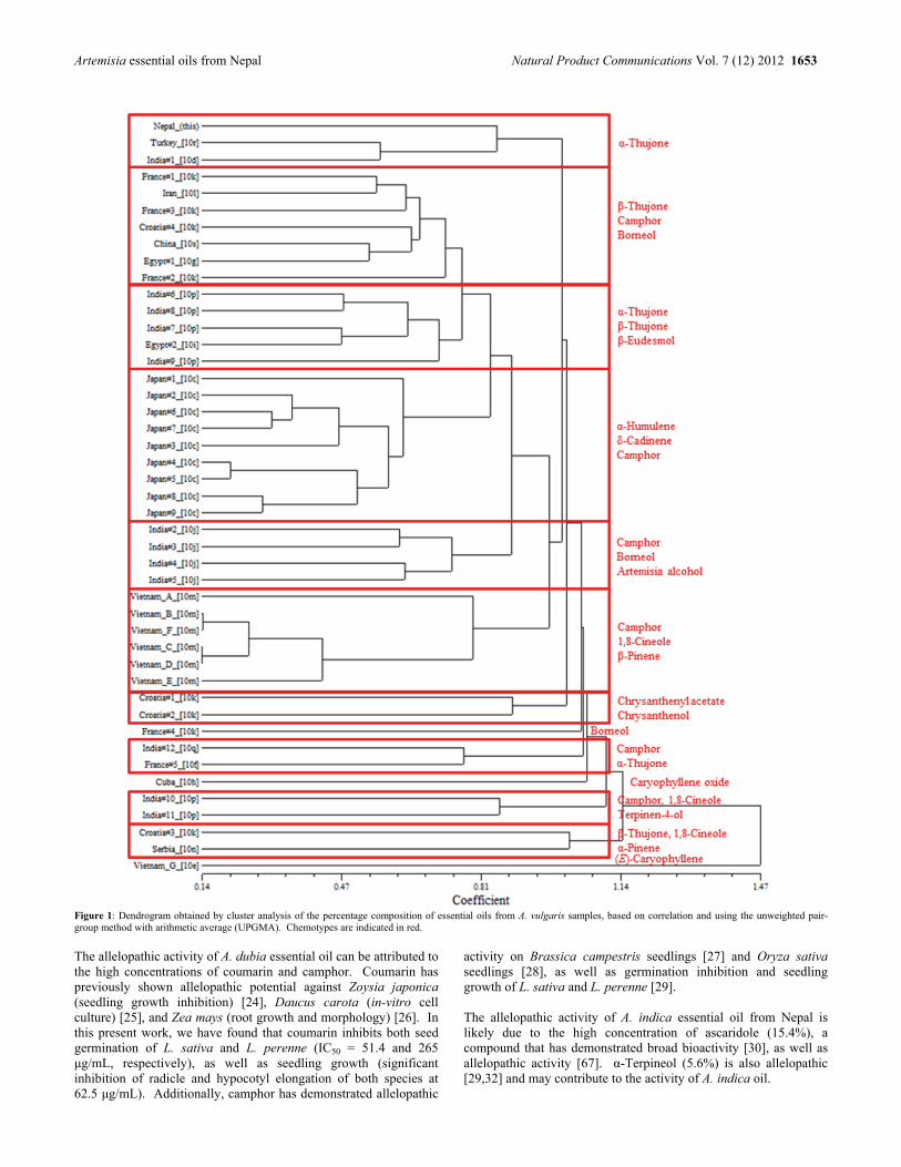
Artemisia essential oils from Nepal Natural Product Communications Vol. 7 (12) 2012 1653
Figure 1: Dendrogram obtained by cluster analysis of the percentage composition of essential oils from A. vulgaris samples, based on correlation and using the unweighted pair-group method with arithmetic average (UPGMA). Chemotypes are indicated in red.
The allelopathic activity of A. dubia essential oil can be attributed to the high concentrations of coumarin and camphor. Coumarin has previously shown allelopathic potential against Zoysia japonica (seedling growth inhibition) [24], Daucus carota (in-vitro cell culture) [25], and Zea mays (root growth and morphology) [26]. In this present work, we have found that coumarin inhibits both seed germination of L. sativa and L. perenne (IC50 = 51.4 and 265 μg/mL, respectively), as well as seedling growth (significant inhibition of radicle and hypocotyl elongation of both species at 62.5 μg/mL). Additionally, camphor has demonstrated allelopathic
activity on Brassica campestris seedlings [27] and Oryza sativa seedlings [28], as well as germination inhibition and seedling growth of L. sativa and L. perenne [29]. The allelopathic activity of A. indica essential oil from Nepal is likely due to the high concentration of ascaridole (15.4%), a compound that has demonstrated broad bioactivity [30], as well as allelopathic activity [67]. α-Terpineol (5.6%) is also allelopathic [29,32] and may contribute to the activity of A. indica oil.
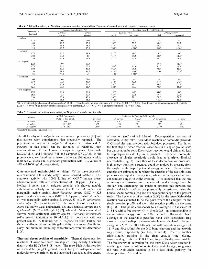
1654 Natural Product Communications Vol. 7 (12) 2012 Satyal et al.
Table 2: Allelopathic activity of Nepalese Artemisia essential oils on lettuce (Lactuca sativa) and perennial ryegrass (Lolium perenne).
Concentration (μg/mL)
Germination Inhibition (%) Seedling Growth (% of Controls) Lactuca sativa
Lolium perenne
Lactuca sativa Lolium perenne radicle hypocotyl radicle hypocotyl
A. dubia 1000 100 100 --- --- --- ---500 76.7 13.3 18.3a 55.2a 30.4a 71.3b
250 65.0 15.0 41.4a 79.2a 56.2a 74.0c
125 46.7 15.0 70.0a > 100e 96.8e > 100e A. indica
1000 96.7 48.3 41.2a 0.0a 16.8a 3.0a 500 53.3 20 73.4b 74.2c 48.3a 38.7a 250 50.0 13.3 81.2c 77.3c 53.2a 29.1a
A. vulgaris 2000 100 60.0 --- --- 47.1a 15.9a 1000 96.7 50.0 74.4b 36.8a 51.1a 18.7a 500 58.3 43.3 96.3e 64.0b 70.2c 56.8c 250 58.3 36.7 > 100e > 100e >100e 67.9d 125 63.3 ntf > 100e > 100e --- ---
Coumarin 500 100 100 --- --- --- --- 250 93.3 43.3 21.8a 42.0a 41.7a 24.7a 125 85.0 30.0 29.1a 48.1a 59.9a 33.7a 62.5 73.3 21.7 52.5a 88.6d 78.5c 58.1a
α/β-Thujones 3000 98.3 78.3 15.3a 0.0a 44.0a 0.0a 1500 93.3 60.0 62.4a 22.2a 67.4a 12.3a 750 90.0 40.0 96.3e 71.1b 76.3c 20.5a 375 35.0 35.0 > 100e 97.1e > 100e 65.1c
a Significantly inhibited compared with controls (P < 0.001). b Significantly inhibited compared with controls (0.001 < P < 0.01). c Significantly inhibited compared with controls (0.01 < P < 0.05). d Significantly inhibited compared with controls (0.5 < P < 0.1). e Not significantly inhibited.f “nt” = not tested.
Table 3: Cytotoxic and antimicrobial activity of Nepalese Artemisia essential oils.
Sample MCF-7 Cytotoxicity Antimicrobial Activity (MIC, μg/mL)
(% kill at 100 μg/mL) B. cereus S. aureus E. coli P. aeruginosa A. niger A. dubia 100 1250 625 1250 2500 313 A. indica 15.0 (2.8)a 625 1250 625 625 625
A. vulgaris 33.6 (2.8) 2500 2500 2500 2500 2500 a Standard deviations in parentheses. The allelopathy of A. vulgaris has been reported previously [11] and this current work complements that previously reported. The phytotoxic activity of A. vulgaris oil against L. sativa and L. perenne in this study can be attributed to relatively high concentrations of the known allelopathic agents 1,8-cineole [27,29,33], α- and β-thujone [34], and camphor [27-29,32]. In the present work, we found that a mixture of α- and β-thujones weakly inhibited L. sativa and L. perenne germination with IC50 values of 448 and 1060 μg/mL, respectively. Cytotoxic and antimicrobial activities: Of the three Artemisia oils examined in this study, only A. dubia showed notable in vitro cytotoxic activity with 100% killing of MCF-7 human breast adenocarcinoma cells at a concentration of 100 μg/mL (Table 3). Neither A. dubia nor A. vulgaris essential oils showed notable antimicrobial activity in our assays (Table 3). A. dubia was marginally active against Staphylococcus aureus (MIC = 625 μg/mL) and Aspergillus niger (MIC = 313 μg/mL), while A. indica oil was marginally active against B. cereus, E. coli, P. aeruginosa, and A. niger (MIC = 625 μg/mL). The crude ethanol extract of A. dubia had shown weak antibacterial activity against S. aureus and S. epidermidis (MIC = 800 μg/mL) [7], while A. dubia essential oil showed weak antifungal activity against Alternaria brassicicola (64% growth inhibition at 10 μL/mL) [8], consistent with our present results. A thujones-rich A. vulgaris oil from Turkey had shown antibacterial and antifungal activity in a zone-of-inhibition assay, but minimum inhibitory concentrations were not determined [10r]. Thermal decomposition of ascaridole: Thermal decomposition reactions of ascaridole were investigated using density functional theory at the B3LYP/6-31G* level. The retro-Diels-Alder reaction of ascaridole (singlet ground state) to give α-terpinene and molecular oxygen (triplet ground state) had a calculated free energy
of reaction (ΔG) of 8.8 kJ/mol. Decomposition reactions of ascaridole, either retro-Diels-Alder reaction or homolytic peroxide O-O bond cleavage, are both spin-forbidden processes. That is, on the first step of either reaction, ascaridole is a singlet ground state but dissociation by retro-Diels-Alder reaction would ultimately lead to triplet-ground-state O2 as a product. Likewise, homolytic cleavage of singlet ascaridole would lead to a triplet diradical intermediate (Fig. 2). In either of these decomposition processes, high-energy transition structures could be avoided by crossing from the singlet to the triplet potential energy surface. The activation energies are estimated to be where the energies of the two spin-state processes are equal in energy (i.e., where the energies cross with concomitant singlet-to-triplet crossing). It is assumed that the rate of intersystem crossing and the rate of bond cleavage must be similar, and calculating the transition probabilities between the singlet and triplet surfaces can presumably be estimated using the Landau-Zener formula [35], but are beyond the scope of this present study. The free energy of activation (ΔG‡) for the retro-Diels-Alder reaction was estimated to be the point where the energies for the singlet reaction profile and the triplet reaction profile are the same (Fig. 3). This point corresponds to an average C-O distance of 2.184 Å with a free energy, G = -540.757851 au, corresponding to an activation energy, ΔG‡ = 170.1 kJ/mol. Homolytic bond cleavage of the ascaridole peroxide bond with subsequent ring closure to give the diepoxide isoascaridole was calculated to be very exergonic (ΔG = -130.1 kJ/mol), but with activation energies of 111.9 and 90.2 kJ/mol for the O-O bond cleavage and the epoxide ring closure, respectively (see Figs. 2 and 4). There is another triplet-singlet crossing in the final epoxide ring closing, corresponding to ΔG‡ = 93.8 kJ/mol, to give finally isoascaridole. The free energy of activation for the retro-Diels-Alder reaction is much higher than that of homolytic O-O bond cleavage, suggesting the retro-Diels-Alder reaction to be a less likely pathway for decomposition of ascaridole.
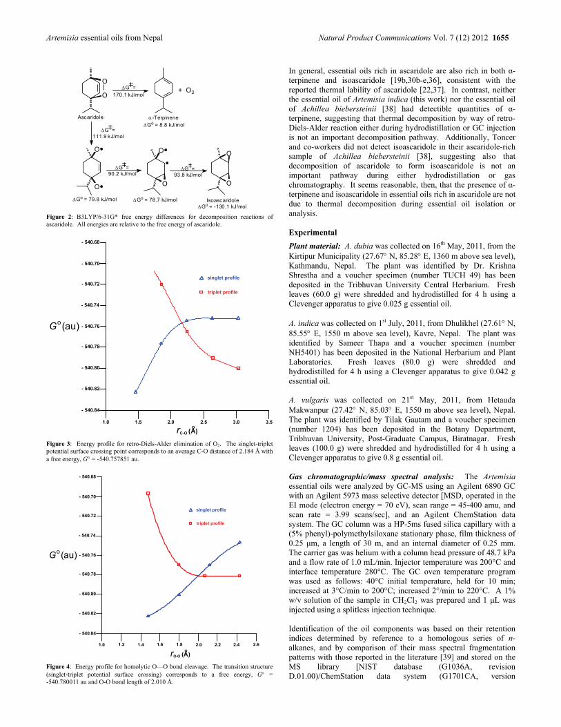
Artemisia essential oils from Nepal Natural Product Communications Vol. 7 (12) 2012 1655
O
O
O
O
O
O
Ascaridole -Terpinene
Isoascaridole
+ O2
Go = 8.8 kJ/mol
G =
170.1 kJ/mol
Go = 79.8 kJ/mol Go = 78.7 kJ/mol
G = 90.2 kJ/mol
Go = -130.1 kJ/mol
G = 111.9 kJ/mol
O
O
G = 93.8 kJ/mol
Figure 2: B3LYP/6-31G* free energy differences for decomposition reactions of ascaridole. All energies are relative to the free energy of ascaridole.
- 540.82
- 540.84
- 540.80
- 540.78
- 540.76
- 540.74
- 540.72
- 540.70
- 540.68
1.0 1.5 2.0 2.5 3.0 3.5
singlet profile
triplet profile
rC-O (A)o
G (au)o
Figure 3: Energy profile for retro-Diels-Alder elimination of O2. The singlet-triplet potential surface crossing point corresponds to an average C-O distance of 2.184 Å with a free energy, G = -540.757851 au.
- 540.82
- 540.84
- 540.80
- 540.78
- 540.76
- 540.74
- 540.72
- 540.70
- 540.68
1.0 1.2 1.4 1.6 1.8 2.0 2.2 2.4 2.6
singlet profile
triplet profile
rO-O (A)o
G (au)o
Figure 4: Energy profile for homolytic O—O bond cleavage. The transition structure (singlet-triplet potential surface crossing) corresponds to a free energy, G = -540.780011 au and O-O bond length of 2.010 Å.
In general, essential oils rich in ascaridole are also rich in both α-terpinene and isoascaridole [19b,30b-e,36], consistent with the reported thermal lability of ascaridole [22,37]. In contrast, neither the essential oil of Artemisia indica (this work) nor the essential oil of Achillea biebersteinii [38] had detectible quantities of α-terpinene, suggesting that thermal decomposition by way of retro-Diels-Alder reaction either during hydrodistillation or GC injection is not an important decomposition pathway. Additionally, Toncer and co-workers did not detect isoascaridole in their ascaridole-rich sample of Achillea biebersteinii [38], suggesting also that decomposition of ascaridole to form isoascaridole is not an important pathway during either hydrodistillation or gas chromatography. It seems reasonable, then, that the presence of α-terpinene and isoascaridole in essential oils rich in ascaridole are not due to thermal decomposition during essential oil isolation or analysis. Experimental
Plant material: A. dubia was collected on 16th May, 2011, from the Kirtipur Municipality (27.67 N, 85.28 E, 1360 m above sea level), Kathmandu, Nepal. The plant was identified by Dr. Krishna Shrestha and a voucher specimen (number TUCH 49) has been deposited in the Tribhuvan University Central Herbarium. Fresh leaves (60.0 g) were shredded and hydrodistilled for 4 h using a Clevenger apparatus to give 0.025 g essential oil. A. indica was collected on 1st July, 2011, from Dhulikhel (27.61 N, 85.55 E, 1550 m above sea level), Kavre, Nepal. The plant was identified by Sameer Thapa and a voucher specimen (number NH5401) has been deposited in the National Herbarium and Plant Laboratories. Fresh leaves (80.0 g) were shredded and hydrodistilled for 4 h using a Clevenger apparatus to give 0.042 g essential oil. A. vulgaris was collected on 21st May, 2011, from Hetauda Makwanpur (27.42 N, 85.03 E, 1550 m above sea level), Nepal. The plant was identified by Tilak Gautam and a voucher specimen (number 1204) has been deposited in the Botany Department, Tribhuvan University, Post-Graduate Campus, Biratnagar. Fresh leaves (100.0 g) were shredded and hydrodistilled for 4 h using a Clevenger apparatus to give 0.8 g essential oil. Gas chromatographic/mass spectral analysis: The Artemisia essential oils were analyzed by GC-MS using an Agilent 6890 GC with an Agilent 5973 mass selective detector [MSD, operated in the EI mode (electron energy = 70 eV), scan range = 45-400 amu, and scan rate = 3.99 scans/sec], and an Agilent ChemStation data system. The GC column was a HP-5ms fused silica capillary with a (5% phenyl)-polymethylsiloxane stationary phase, film thickness of 0.25 μm, a length of 30 m, and an internal diameter of 0.25 mm. The carrier gas was helium with a column head pressure of 48.7 kPa and a flow rate of 1.0 mL/min. Injector temperature was 200°C and interface temperature 280°C. The GC oven temperature program was used as follows: 40°C initial temperature, held for 10 min; increased at 3°C/min to 200°C; increased 2°/min to 220°C. A 1% w/v solution of the sample in CH2Cl2 was prepared and 1 μL was injected using a splitless injection technique. Identification of the oil components was based on their retention indices determined by reference to a homologous series of n-alkanes, and by comparison of their mass spectral fragmentation patterns with those reported in the literature [39] and stored on the MS library [NIST database (G1036A, revision D.01.00)/ChemStation data system (G1701CA, version
1656 Natural Product Communications Vol. 7 (12) 2012 Satyal et al.
C.00.01.080)]. The percentages of each component are reported as raw percentages based on total ion current without standardization. Allelopathic activity assays: An allelopathic bioassay based on lettuce (Lactuca sativa) and perennial rye grass (Lolium perenne) germination and subsequent radicle and hypocotyl growth was used to study the effects of the Artemisia essential oils. Stock solutions of each essential oil (2.0 g/L essential oil and 1.0 g/L Tween-80 in water) were prepared and used for the assays. Two-fold serial dilutions of stock test solutions were prepared to give test concentrations of 4000, 2000, 1000, 500, and 250 µg/mL with the control being 1.0 g/L aqueous Tween-80. Seeds were placed in 6-well test plates (10 seeds per well) each well lined with two layers of Whatman No. 1 filter paper moistened with test solution and the test plates were sealed with Parafilm®. The test plates were incubated at room temperature in the dark for 5 days, after which the number of germinated seeds was determined and the root (radicle) and shoot (hypocotyl) lengths were measured. Student’s t-test [40] was used to compare radicle and hypocotyl test means with controls. Seed germination IC50 values were determined using the Reed-Muench method [41]. Antimicrobial screening: The essential oils were screened for antimicrobial activity against Gram-positive bacteria, Bacillus cereus (ATCC No. 14579), Staphylococcus aureus (ATCC No. 29213); and Gram-negative bacteria, Pseudomonas aeruginosa (ATCC No. 27853) and Escherichia coli (ATCC No. 10798). Minimum inhibitory concentrations (MIC) were determined using the microbroth dilution technique [42]. Dilutions of the essential oil were prepared in cation-adjusted Mueller Hinton broth (CAMHB) beginning with 50 L of 1% w/w solutions of essential oil in DMSO plus 50 L CAMHB. The extract solutions were serially diluted (1:1) in CAMHB in 96-well plates. Organisms at a concentration of approximately 1.5 108 colony forming units (CFU)/mL were added to each well. Plates were incubated at 37°C for 24 h; the final minimum inhibitory concentration (MIC) was determined as the lowest concentration without turbidity. Geneticin was used as a positive antibiotic control; DMSO was used as a negative control. Antifungal activity against Aspergillus niger (ATCC No. 16888) was determined as above using potato dextrose broth inoculated with A. niger hyphal culture diluted to a McFarland turbidity of 1.0. Amphotericin B was the positive control. Cytotoxicity screening: Human MCF-7 breast adenocarcinoma cells (ATCC No. HTB-22) [43] were grown in a 3% CO2 environment at 37°C in RPMI-1640 medium, supplemented with 10% fetal bovine serum, 100,000 units penicillin and 10.0 mg streptomycin per L of medium, 15 mM of Hepes, and buffered with 26.7 mM NaHCO3, pH 7.35. Cells were plated into 96-well cell culture plates at 2.5 104 cells per well. The volume in each well was 100 μL. After 48 h, supernatant fluid was removed by suction and replaced with 100 μL growth medium containing 1.0 μL of
DMSO solution of the essential oil (1% w/w in DMSO), giving a final concentration of 100 μg/mL for each well. Solutions were added to wells in 4 replicates. Medium and DMSO controls (10 μL DMSO/mL) were used. Tingenone [44] was used as a positive control. After the addition of compounds, plates were incubated for 48 h at 37°C in 5% CO2; medium was then removed by suction, and 100 μL of fresh medium was added to each well. In order to establish percent kill rates, the MTT assay for cell viability was carried out [45]. After colorimetric readings were recorded (using a Molecular Devices SpectraMAX Plus microplate reader, 570 nm), average absorbances, standard deviations, and percent kill ratios (%killcmpd/%killDMSO) were calculated. Numerical cluster analysis: A total of 45 Artemisia vulgaris essential oil compositions from this work and the published literature were treated as operational taxonomic units (OTUs). The percentage composition of 36 principle essential oil components (camphor, 1,8-cineole, α-thujone, α-humulene, borneol, β-thujone, δ-cadinene, (E)-caryophyllene, α-pinene, β-pinene, terpinen-4-ol, caryophyllene oxide, α-muurolene, sabinene, trans-chrysanthenyl acetate, α-terpineol, germacrene D, artemisia alcohol, p-cymene, camphene, α-phellandrene, chrysanthenol, spathulenol, γ-cadinene, β-eudesmol, β-elemene, γ-terpinene, trans-sabinene hydrate, chrysanthenone, α-copaene, methyl eugenol, bornyl acetate, linalool, β-selinene, lyratol, and trans-isoelemicin) was used to determine the chemical relationship between the different A. vulgaris essential oil samples by cluster analysis using the STSYSpc software, version 2.2 [46]. Correlation was selected as a measure of similarity, and the unweighted pair-group method with arithmetic average (UPGMA) was used for cluster definition. Density functional theory calculations: All calculations were carried out using Spartan ’10 for Windows [47]. The hybrid B3LYP functional [48] and the 6-31G* basis set [49] were used for the optimization of all stationary points in the gas phase. Frequency calculations were employed to characterize stationary points as minima or first-order saddle points. All reaction and activation enthalpies reported are zero-point (ZPE) corrected and thermally corrected. Entropies were calculated using the linear harmonic oscillator approximation. Two reaction profiles were modeled: A retro-Diels-Alder elimination of dioxygen and a homolytic O-O peroxide cleavage with subsequent diepoxide ring closure. In order to model these decomposition reactions, the reaction profiles were calculated stepwise for both the singlet and triplet spin states at the B3LYP/6-31G* level. Acknowledgments – PS and PP are grateful to Dr. Krishna Shrestha, Tilak Gautam, Kiran Pokharel and Sameer Shrestha for identifying the plant samples. We thank Bernhard Vogler for technical assistance with the GC-MS measurements and James Baird for helpful discussions on singlet-triplet interconversions.
References
[1] Mabberly DJ. (1997) The Plant Book, 2nd Ed. Cambridge University Press, Cambridge, UK, 57. [2] Gewali MB. (2008) Aspects of Traditional Medicine in Nepal. Institute of Natural Medicine, University of Toyama, Japan, 17-20. [3] Tamang G. (2003) An ethnobiological study of the Tamang people. Our Nature, 1, 37-41. [4] Hussain S, Hore DK. (2007) Collection and conservation of major medicinal plants of Darjeeling and Sikkim Himalayas. Indian Journal of
Traditional Knowledge, 6, 352-357. [5] Turin M. (2003) Ethnobotanical notes on Thangmi plant names and their medicinal and ritual uses. Contributions to Nepalese Studies, 30, 19-52. [6] Zheng W, Tan R, Liu Z. (1996) Chemical constituents of essential oil of two varieties of Artemisia dubia Wall. ex Bess. Acta Phytotaxonomica
Sinica, 34, 410-414. [7] Bhatt LR, Lim JA, Baek SH. (2005) Antimicrobial activity of Nepalese medicinal plants on skin microbial inflammation pathogens. Journal of
Cosmetic and Public Health, 1, 6-9. [8] Parajuli RR, Tiwari RD, Chaudhary RP, Gupta VN. (2005) Fungitoxicity of the essential oils of some aromatic plants of Manang against Alternaria
brassicicola. Scientific World, 3, 39-43.

Artemisia essential oils from Nepal Natural Product Communications Vol. 7 (12) 2012 1657
[9] Wu H, Wei Z, Li Y, Liang C, Liang H. (2009) Analysis of the constituents of volatile oil in Artemisia indica from Guangxi. China Pharmacy 2009-09 doi: CNKI:SUN:ZGYA.0.2009.09.024.
[10] (a) Nano GM, Bicchi C, Frattini C, Gallino M. (1976) On the composition of some oils from Artemisia vulgaris. Planta Medica, 30, 211-215; (b) Michaelis K, Vostrowsky O, Paulini H, Zintl R, Knobloch K. (1982) Das ätherische Öl aus Blüten von Artemisia vulgaris L. Zeitschrift für Naturforschung, 37c, 152-158; (c) Aoki T, Shigetaka H. (1985) Seasonal variations and geographical differences of volatile constituents from Artemisia vulgaris L. var. indica Maxim. Bulletin of Suzugamine Women's College of Natural Science, 26, 9-14; (d) Misra LN, Singh SP. (1986) α-Thujone, the major component of the essential oil from Artemisia vulgaris growing wild in Nilgiri hills. Journal of Natural Products, 49, 941-941; (e) Dũng NX, Nam VV, Huóng HT, Leclercq PA. (1992) Chemical composition of the essential oil of Artemisia vulgaris L. var. indica Maxim. from Vietnam. Journal of Essential Oil Research, 4, 433-434; (f) Milhau G, Valentin A, Benoit F, Mallié M, Bastide JM, Pélissier Y, Bessière JM. (1997) In vitro antimalarial activity of eight essential oils. Journal of Essential Oil Research, 9, 329-333; (g) Aboutabl EA, El-Azzouny A, El Dahmy SI. (1998) Composition of the essential oil of Artemisia vulgaris L. grown in Egypt. Journal of Essential Oil-Bearing Plants, 1, 21-27; (h) Pino JA, Rosado A, Fuentes V. (1999) Composition of the essential oil of Artemisia vulgaris L. herb from Cuba. Journal of Essential Oil Research, 11, 477-478; (i) Haggag MY, El Tantawy ME, Ahmed FI, Shams MM. (2000) Study of chemical composition, antimicrobial and anthelmintic activities of the essential oils of Artemisia vulgaris L. and Santolina chamaecyparissus L. cultivated in Egypt. Azerbaijan Journal of Pharmaceutical Sciences, 26, 23-39; (j) Haider F, Dwivedi PD, Naqvi AA, Bagchi GD. (2003) Essential oil composition of Artemisia vulgaris harvested at different growth periods under Indo-Gangetic Plain conditions. Journal of Essential Oil Research, 15, 376-378; (k) Jerkovic I, Mastelic J, Milos M, Juteau F, Masotti V, Vlano J. (2003) Chemical variability of Artemisia vulgaris L. essential oils originated from the Mediterranean are of France and Croatia. Flavour and Fragrance Journal, 28, 436-440; (l) Sefidkon F, Jahili A, Rabie M, Hamzehee B, Asri Y. (2003) Chemical composition of essential oil of five Artemisia species from Iran. Journal of Essential Oil-Bearing Plants, 6, 41-45; (m) Thao NTP, Thuy NT, Hoi TM, Thai TH, Muselli A, Bighelli A, Castola V, Casanova J. (2004) Artemisia vulgaris L. from Vietnam: Chemical variability and composition of the oil along the vegetative life of the plant. Journal of Essential Oil Research, 16, 358-361; (n) Blagojević P, Radulović N, Palić R, Stojanović G. (2006) Chemical composition of the essential oils of Serbian wild-growing Artemisia absinthium and Artemisia vulgaris. Journal of Agricultural and Food Chemistry, 54, 4780-4789; (o) Judžentienė A, Buzelytė J. (2006) Chemical composition of essential oils of Artemisia vulgaris L. (mugwort) from north Lithuania. Chemija, 17, 12-15; (p) Lohani H, Sah S, Singh A, Sati M, Yadav RK. (2007) Chemical composition of essential oil of Artemisia vulgaris L. from three different areas of Uttaranchal. Fafai Journal, 9, 69-72; (q) Govindaraj S, Kumari BDR, Cioni PL, Flamini G. (2008) Mass propagation and essential oil analysis of Artemisia vulgaris. Journal of Bioscience and Bioengineering, 105, 176-183; (r) Baykan Erel S, Reznicek G, Şenol SG, Karabay Yavaşoğul NU, Konyalioğlu S, Zeybek AU. (2011) Antimicrobial and antioxidant properties of Artemisia L. species from western Anatolia. Turkish Journal of Biology, 35, doi:10.3906/biy-0912-27; (s) Zhu L, Li Y, Li B, Lu B, Zhang W. (1995) Artemisia vulgaris Linn. (Compositae). In Aromatic Plants and Essential Constituents (Supplement). Hai Feng Publishing Co., Hong Kong, 135.
[11] Barney JN, Hay AG, Weston LA. (2005) Isolation and characterization of allelopathic volatiles from mugwort (Artemisia vulgaris). Journal of Chemical Ecology, 31, 247-265.
[12] Wang J, Zhu F, Zhou XM, Niu CY Lei CL. (2006) Repellent and fumigant activity of essential oil from Artemisia vulgaris to Tribolium castaneum (Herbst) (Coleoptera: Tenebrionidae). Journal of Stored Products Research, 42, 339-347.
[13] Bhatt LR, Lim JA, Chai KY, Kang JI, Oh HK, Baek SH. (2006) Antioxidative and antimicrobial activities of essential oil from Artemisia vulgaris. Natural Product Sciences, 12, 226-231.
[14] Kováts N, Gölöncsér F, Ács A, Refaey M. (2010) Quantification of the antibacterial properties of Artemisia absinthium, A. vulgaris, Chrysanthemum leucanthemum and Achillea millefolium using the Vibrio fischeri bacterial biassay. Acta Botanica Hungarica, 52, 137-144.
[15] Dob T, Benabdelkader T. (2006) Chemical composition of the essential oil of Artemisia herba-alba grown in Algeria. Journal of Essential Oil Research, 18, 685-690.
[16] Ouachikh O, Bouyanzer A, Bouklah M, Desjobert JM, Costa J, Hammouti B, Majidi L. (2009) Application of essential oil of Artemisia herba alba as green corrosion inhibitor for steel in 0.5 M H2SO4. Surface Review and Letters, 16, 49-54.
[17] Kostyk BC, Borden JH, Gries G. (1993) Photoisomerization of antiaggregation pheromone verbenone: Biological and practical implications with respect to the mountain pine beetle, Dendroctonus poderosae Hopkins (Coleoptera: Scolytidae). Journal of Chemical Ecology, 19, 1749-1759.
[18] Asfaw N, Storesund HJ, Skattebøl L, Aasen AJ. (2001) Coexistence of chrsanthenone, filifolone and (Z)-isogeranic acid in hydrodistillates. Artefacts! Phytochemistry, 58, 489-492.
[19] (a) Carnat AP, Lamaison JL. (1992) Composition of the essential oil of Artemisia molinieri Quézel, Barbero et R. Loisel. Journal of Essential Oil Research, 4, 635-637; (b) Masotti V, Juteau F, Bessière JM, Viano J. (2003) Seasonal and phenological variations of the essential oil from the narrow endemic species Artemisia molinieri and its biological activities. Journal of Agricultural and Food Chemistry, 51, 7115-7121.
[20] Mirjalili BF, Meybody MHH, Arkakani MM, Rustaiyan A, Ameri N, Masoudi S, Bamoniri A. (2006) Chemical composition of the essential oil from aerial parts, leaves, flowers and roots of Artemisia persica Boiss. from Iran. Journal of Essential Oil Research, 18, 544-547.
[21] (a) Monzote Fidalgo L. (2007) Essential oil from Chenopodium ambrosioides as a promising antileishmanial agent. Natural Product Communications, 2, 1257-1262; (b) Monzote L, Nance MR, Garcia M, Scull R, Setzer WN. (2011) Comparative chemical, cytotoxicity and antileishmanial properties of essential oils from Chenopodium ambrosioides. Natural Product Communications, 6, 281-286.
[22] Cavalli JF, Tomi F, Bernardini AF, Casanova J. (2004) Combined analysis of the essential oil of Chenopodium ambrosioides by GC, GC-MS and 13C-NMR spectroscopy: Quantitative determination of ascaridole, a heat-sensitive compound. Phytochemical Analysis, 15, 275-279.
[23] Weston LA, Barney JN, Di Tommaso A. (2005) A review of the biology and ecology of three invasive perennials in New York State: Japanese knotweed (Polygonum cuspidatum), mugwort (Artemisia vulgaris) and pale swallow-wort (Vincetoxicum rossicum). Plant and Soil, 277, 53-69.
[24] Yamamoto Y. (1995) Allelopathic potential of Anthoxanthum odoratum for invading Zoysia-grassland in Japan. Journal of Chemical Ecology, 21, 1365-1373.
[25] Abenavoli MR, Sorgonà A, Sidari M, Badiani M, Fuggi A. (2003) Coumarin inhibits the growth of carrot (Daucus carota L. cv. Saint Valery) cells in suspension culture. Journal of Plant Physiology, 160, 227-237.
[26] Abenavoli MR, Sorgonà A, Albano S, Cacco G. (2004) Coumarin differentially affects the morphology of different root types of maize seedlings. Journal of Chemical Ecology, 30, 1871-1883.
[27] Nishida N, Tamotsu S, Nagata N, Saito C Sakai A. (2005) Allelopathic effects of volatile monoterpenoids produced by Salvia leucophylla: Inhbition of cell proliferation and DNA synthesis in the root apical meristem of Brassica campestris seedlings. Journal of Chemical Ecology, 31, 1187-1203.
[28] Yumi O, Keiko Y, Katsuichiro K. (2011) Allelopathic activity of camphor released from camphor tree (Cinnamomum camphora). Allelopathy Journal, 27, 123-232.
[29] Kennedy JE, Davé PC, Harbin LN, Setzer WN. (2011) Allelopathic potential of Sassafras albidum and Pinus taeda essential oils. Allelopathy Journal, 27, 111-122.
[30] (a) Yen YP, Yeh MJ, Hsiao WF. (2007) Synthesis and nematicidal activity of ascaridole derivatives against Meloidogyne incognita and Aphelenchoides besseyi. Journal of Pesticide Science, 32, 49-52; (b) Torres MCM, Assunção JC, Santiago GMP, Andrade-Neto M, Silveira ER,

1658 Natural Product Communications Vol. 7 (12) 2012 Satyal et al.
Costa-Lotufo LV, Bezerra DP, Filho JDBM, Viana FA, Pessoa ODL. (2008) Larvicidal and nematicidal activities of the leaf essential oil of Croton regelianus. Chemistry and Biodiversity, 5, 2724-2728; (c) Monzote L, Stamberg W, Staniek K, Gille L. (2009) Toxic effects of carvacrol, caryophyllene oxide, and ascaridole from essential oil of Chenopodium ambrosioides on mitochondria. Toxicology and Applied Pharmacology, 240, 337-347; (d) Chu SS, Hu JF, Liu ZL. (2011) Composition of essential oil of Chinese Chenopodium ambrosioides and insecticidal activity against maize weevil, Sitophilus zeamais. Pest Management Science, 67, 714-718; (e) Bai CQ, Liu ZL, Liu QZ. (2011) Nematicidal constituents from the essential oil of Chenopodium ambrosioides aerial parts. E.-Journal of Chemistry, 8, S143-S148.
[31] Jiménez-Osornio FMVZJ, Kumamoto J, Wasser C. (1996) Allelopathic activity of Chenopodium ambrosioides L. Biochemical Systematics and Ecology, 24, 195-205.
[32] De Martino L, Mancini E, Rolim de Almeida LF, De Feo V. (2010) The antigerminative activity of twenty-seven monoterpenes. Molecules, 15, 6630-6637.
[33] (a) Koitabashi R, Suzuki T, Kawazu T, Sakai A, Kuroiwa H, Kuroiwa T. (1997) 1,8-Cineole inhibits root growth and DNA synthesis in the root apical meristem of Brassica campestris L. Journal of Plant Research, 110, 1-6; (b) Romagni JG, Allen SN, Dayan FE. (2000) Allelopathic effects of volatile cineoles on two weedy plant species. Journal of Chemical Ecology, 26, 303-313; (c) Singh HP, Batish DR, Kaur S, Arora K, Kohli RK. (2002) Allelopathic effect of two volatile monoterpenes against bill goat weed (Ageratum conyzoides L.) Crop Protection, 21, 347-350.
[34] (a) Halligan JP. (1975) Toxic terpenes from Artemisia californica. Ecology, 56, 999-1003; (b) Jassbi AR, Zamanizadehnajari S, Baldwin IT. (2010) Phytotoxic volatiles in the roots and shoots of Artemisia tridentata as detected by headspace solid-phase microextraction and gas chromatographic-mass spectrometry analysis. Journal of Chemical Ecology, 36, 1398-1407.
[35] Steinfeld JI, Francisco JS, Hase WL. (1999) Chemical Kinetics and Dynamics, 2nd Ed. Prentice Hall, Upper Saddle River, New Jersey, USA, 247-250.
[36] Koba K, Catherine G, Raynaud C, Chaumont JP, Sanda K, Laurence N. (2009) Chemical composition and cytotoxic activity of Chenopodium ambrosioides L. essential oil from Togo. Bangladesh Journal of Scientific and Industrial Research, 44, 435-440.
[37] (a) Nelson EK. (1920) The composition of oil of Chenopodium from various sources. Journal of the American Chemical Society, 42, 1204-1208; (b) Zand R, Mesrobian RB. (1955) Polymerization of methacrylonitrile initiated by ascaridole and dihydroascaridole. Journal of the American Chemical Society, 77, 6523-6524.
[38] Toncer O, Basbag S, Karaman S, Diraz E, Basbag M. (2010) Chemical composition of the essential oils of some Achillea species growing wild in Turkey. International Journal of Agriculture and Biology, 12, 527-530.
[39] Adams RP. (2007) Identification of Essential Oil Components by Gas Chromatography / Mass Spectrometry, 4th Ed. Allured Publishing, Carol Stream, Illinois.
[40] Zar JH. (1996) Biostatistical Analysis, 3rd Ed. Prentice Hall, Upper Saddle River New Jersey, 123-137. [41] Reed LJ, Muench H. (1938) A simple method of estimating fifty percent end points. American Journal of Hygiene, 27, 493-497. [42] Sahm DH, Washington JA. (1991) Antibacterial susceptibility tests: Dilution methods. In Manual of Clinical Microbiology. Balows A, Hausler
WJ, Herrmann KL, Isenberg HD, Shamody HJ (Eds.). American Society for Microbiology, Washington DC, USA. [43] Soule HD, Vazguez J, Long A, Albert S, Brennan M. (1973) A human cell line from a pleural effusion derived from a breast carcinoma. Journal of
the National Cancer Institute, 51, 1409-1416. [44] Setzer WN, Setzer MC, Hopper AL, Moriarity DM, Lehrman GK, Niekamp KL, Morcomb SM, Bates RB, McClure KJ, Stessman CC, Haber WA.
(1998) The cytotoxic activity of a Salacia liana species from Monteverde, Costa Rica, is due to a high concentration of tingenone. Planta Medica, 64, 583.
[45] Ferrari M, Fornasiero MC, Isetta AM. (1990) MTT colorimetric assay for testing macrophage cytotoxic activity in vitro. Journal of Immunological Methods, 31, 165-172.
[46] Rohlf JF. (2005) NTSYSpc, Numerical Taxonomy and Multivariate Analysis System. Applied Biostatistics Inc., New York. [47] Spartan ’10 for Windows (2010) Wavefunction, Inc., Irvine, California. [48] (a) Becke AD. (1993) Density-functional thermochemistry. III. The role of exact exchange. Journal of Chemical Physics, 98, 5648-5652; (b) Lee C,
Yang W, Parr RG. (1988) Development of the Colle-Salvetti correlation-energy formula into a functional of the electron density. Physical Review B, 37, 785-789.
[49] Hehre WJ, Radom L, Schleyer PvR. (1986) Ab initio Molecular Orbital Theory. Wiley, New York.

Chemical Compositions, Antioxidant and Antimicrobial Activity of the Essential Oils of Piper officinarum (Piperaceae) Wan Mohd Nuzul Hakimi Wan Salleha, Farediah Ahmada,*, Khong Heng Yenb and Hasnah Mohd Sirata aDepartment of Chemistry, Faculty of Science, Universiti Teknologi Malaysia, Skudai, 81310 Johor Bahru, Johor, Malaysia
bSchool of Chemistry and Environmental Studies, Faculty of Applied Sciences, Universiti Teknologi MARA Sarawak, Kota Samarahan, 94300, Sarawak, Malaysia [email protected]
Received: September 29th, 2012; October 25th, 2012
This study was designed to investigate the antioxidant and antimicrobial activities of the essential oils from Piper officinarum C. DC. GC and GC/MS analysis of the leaf and stem oils showed forty one components, representing 85.6% and 93.0% of the oil, respectively. The most abundant components in the leaf oil were β-caryophyllene (11.2%), α-pinene (9.3%), sabinene (7.6%), β-selinene (5.3%) and limonene (4.6%), while β-caryophyllene (10.9%), α-phellandrene (9.3%), linalool (6.9%), limonene (6.7%) and α-pinene (5.0%) were the main components of the stem oil. The antioxidant activities were determined by using complementary tests: namely β-carotene-linoleic acid, DPPH radical scavenging and total phenolic assays. The stems oil showed weak activity (IC50 = 777.4 µg/mL) in the DPPH system, but showed moderate lipid peroxidation inhibition in the β-carotene-linoleic acid system (88.9 ± 0.35%) compared with BHT (95.5 ± 0.30%). Both oils showed weak activity against P. aeruginosa and E. coli with M IC values of 250 µg/mL. Keywords: Chemical composition, Piper officinarum, Essential oils, Antioxidant, Antimicrobial. P. officinarum C. DC is locally known as ‘lada panjang’ or ‘kechundai’ in the Malaysian region. It is a climber that grows in Malaysia, India, China, Indonesia and the Philippines. Pepper obtained from this species has been used as an adulterant for P. longum. In Cambodia, Laos and Vietnam, the plant is used to treat fever, jaundice, rheumatism and neuralgia. In the Philippines, the roots are chewed to promote digestion and externally to heal wounds [1]. Previous phytochemical studies on P. officinarum have resulted in the isolation of a number of amide and lignan compounds [2-5]. In addition, studies on the extracts of this species showed antibacterial, interceptive, antiatherosclerotic and hypolipidaemic activities [6,7]. To the best of our knowledge there is no previous report on the chemical composition of the essential oil of P. officinarum. Here we report the chemical composition, antioxidant and antimicrobial activities of the leaf and stem oils of P. officinarum, collected from Sarawak, Malaysia. Hydrodistillation of the fresh leaves and stems of P. officinarum gave pale yellow oils in yields of 0.26% and 0.22%(w/w), respectively. The chemical compositions of these oils are listed in Table 1. GC and GC-MS analysis successfully detected forty-one components each, which accounted for 85.6% and 93.0%, respectively of the chromatographic components. Sesquiterpene hydrocarbons were the major components in the leaf (41.1%) and stem (43.6%) oils, with β-caryophyllene (11.2% and 10.9%) being the most substantial compound in both oils. Other major compounds were α-pinene (9.3%), sabinene (7.6%), β-selinene (5.3%) and limonene (4.6%) for the leaf oil, and α-phellandrene (9.3%), linalool (6.9%), limonene (6.7%) and α-pinene (5.0%) for the stem oil. Monoterpene hydrocarbons were present in high concentration in both the leaf (31.7%) and stem oils (30.4%). Oxygenated monoterpenes made up a minor fraction (9.2-12.4%), while oxygenated sesquiterpenes formed 3.0-3.6% of the total oil. Nine
components of the stem oil were not detected in the leaf oil. These were α-terpinolene, eugenol, α-ylangene, methyl eugenol, α-cedrene, α-bergamotene, δ-selinene, (E)-nerolidol and α-cadinol. On the other hand, nine components of the leaf oil were not detected in the stem oil (camphene, δ-3-carene, α-terpinene, camphor, β-cubebene, γ-elemene, (E, E)-α-farnesene, germacrene B and globulol). The DPPH free radical scavenging activity and β-carotene/linoleic acid tests were carried out on the essential oils, together with the Folin-Ciocalteu assay, which evaluated the total phenolic content of the essential oils. The results are given in Table 2. The antioxidant activity in the DPPH radical scavenging test is due to the hydrogen donating ability of the test material. The capability of substances to donate hydrogen to convert DPPH into the non-radical form of DPPH can be followed spectrophotometrically [8]. Both oils exhibited weak DPPH radical scavenging activity (IC50 leaf oil, 622.2 µg/mL; stem oil, 777.4 µg/mL) compared with the standard antioxidant, BHT (IC50 = 124.4 µg/mL). The low activity was attributed to the low phenolic content of the essential oils. This was supported by the results of the Folin-Ciocalteu assay on both oils. In the β-carotene/linoleic acid assay, oxidation of linoleic acid produces hydroperoxyl radicals which simultaneously attack the chromophore of β-carotene resulting in bleaching of the reaction emulsion [9]. The effectiveness of the leaf (85.9%) and stem (88.9%) oils was compared with that of BHT (125.5%), but the inhibition values observed were significantly lower than that of BHT. The antimicrobial activity of the essential oils was evaluated against Gram-positive bacteria, Gram-negative bacteria and fungi. Their activity potentials were assessed qualitatively and quantitatively by the presence of inhibition zones, zone diameters and minimum inhibitory concentration (MIC) values. The results are presented in Table 3. The inhibition zones were in the range of 7.0-11.2 mm.
NPC Natural Product Communications 2012 Vol. 7 No. 12
1659 - 1662

1660 Natural Product Communications Vol. 7 (12) 2012 Salleh et al. Table 1: Constituents identified in P. officinarum leaf and stem oils.
Components KI b Percentage a Leaves Stems α-Pinene 930 9.3 ± 0.02 5.0 ± 0.01 Camphene 945 1.5 ± 0.04 - Sabinene 969 7.6 ± 0.01 3.3 ± 0.02 Myrcene 988 1.5 ± 0.11 1.9 ± 0.10 α-Phellandrene 1002 2.2 ± 0.06 9.3 ± 0.02 δ-3-Carene 1008 0.7 ± 0.02 - α-Terpinene 1014 0.4 ± 0.04 - Limonene 1024 4.6 ± 0.1 6.7 ± 0.03 1,8-Cineole 1026 1.0 ± 0.07 1.7 ± 0.06 (Z)-β-Ocimene 1030 0.3 ± 0.05 0.7 ± 0.01 γ-Terpinene 1055 3.6 ± 0.01 2.8 ± 0.05 α-Terpinolene 1086 - 0.7 ± 0.01 Linalool 1092 3.4 ± 0.02 6.9 ± 0.09 Camphor 1142 0.7 ± 0.04 - Terpinen-4-ol 1175 3.6 ± 0.01 3.5 ± 0.03 α-Terpineol 1186 0.5 ± 0.12 0.3 ± 0.06 α-Cubebene 1345 0.3 ± 0.08 1.1 ± 0.02 Eugenol 1356 - 1.1 ± 0.04 α-Ylangene 1372 - 0.8 ± 0.03 α-Copaene 1374 1.7 ± 0.03 3.4 ± 0.05 β-Cubebene 1386 2.7 ± 0.02 - β-Elemene 1389 1.3 ± 0.10 1.0 ± 0.04 Methyl eugenol 1403 - 2.5 ± 0.01 α-Gurjunene 1405 2.2 ± 0.02 0.1 ± 0.10 α-Cedrene 1410 - 0.1 ± 0.12 α-Bergamotene 1412 - 0.2 ± 0.05 β-Caryophyllene 1417 11.2 ± 0.02 10.9 ± 0.03 γ-Elemene 1434 0.2 ± 0.05 - Aromadendrene 1440 0.5 ± 0.02 0.4 ± 0.01 α-Humulene 1452 3.2 ± 0.01 3.1 ± 0.04 Dehydroaromadendrene 1460 0.5 ± 0.04 0.2 ± 0.02 α-Amorphene 1483 3.2 ± 0.03 3.1 ± 0.1 Germacrene D 1485 0.7 ± 0.01 2.1 ± 0.04 β-Selinene 1490 5.3 ± 0.1 2.4 ± 0.1 δ-Selinene 1492 - 0.3 ± 0.05 Cadina-1,4-diene 1495 0.5 ± 0.08 0.3 ± 0.01 α-Selinene 1498 2.5 ± 0.05 2.8 ± 0.01 Bicyclogermacrene 1500 1.2 ± 0.02 5.4 ± 0.02 α-Muurolene 1502 0.2 ± 0.02 0.6 ± 0.08 (E,E)-α-Farnesene 1508 0.7 ± 0.05 - δ-Cadinene 1520 0.2 ± 0.02 2.5 ± 0.02 cis-Calamenene 1528 0.2 ± 0.04 0.2 ± 0.04 α-Calacorene 1545 0.1 ± 0.01 0.1 ± 0.11 Germacrene B 1560 2.5 ± 0.1 - (E)-Nerolidol 1562 - 0.1 ± 0.04 Spathulenol 1578 1.3 ± 0.01 1.3 ± 0.01 Caryophyllene oxide 1582 0.4 ± 0.03 0.4 ± 0.02 Globulol 1592 1.8 ± 0.07 -
Viridiflorol 1595 0.1 ± 0.02 0.3 ± 0.03 α-Cadinol 1652 - 0.9 ± 0.05 Group components Phenylpropanoids - 3.6 ± 0.2 Monoterpene hydrocarbons 31.7 ± 0.2 30.4 ± 0.2 Oxygenated monoterpenes 9.2 ± 0.2 12.4 ± 0.2 Sesquiterpene hydrocarbons 41.1 ± 0.2 43.6 ± 0.2 Oxygenated sesquiterpenes 3.6 ± 0.2 3.0 ± 0.2 Identified components (%) 85.6 ± 0.2 93.0 ± 0.2
a Each value is expressed as means ± SD of three injections. bRetention indices on Ultra-1 capillary column.
Table 2: Antioxidant activity of P. officinarumaleaf and stem oils.
Samples β-Carotene/linoleic acid (%) DPPH IC50 (µg/mL) TPC (mg GA/g) Leaf oil 85.9 ± 0.4 622.2 ± 0.3 30.4 ± 0.4 Stem oil 88.9 ± 0.3 777.4 ± 0.2 44.1 ± 0.4 BHT 125.5 ± 0.3 124.8 ± 0.2 ND
a Data represent mean ±SD of three independent experiments; ND – not determined.
The leaf oil was found to have a weak antimicrobial activity against B. subtlis, P. aeruginosa and E. coli, with MIC values of 250 µg/mL. The stem oil showed similar weak activity against P. aeruginosa and E. coli, with MIC values of 250 µg/mL. Components such as α-pinene, 1,8-cineole, γ-terpinene, linalool and α-terpineol have been found to have relatively strong antimicrobial properties [10]. These components were present in both the leaf and stem oils and,therefore, may have contributed to this antimicrobial activity. Generally, essential oils that contain high proportions of oxygenated monoterpenes have stronger antifungal activities [11]. However, the leaf and stem oils showed only weak antifungal activity against A. niger, with a MIC value of 500 µg/mL. The weak
Table 3: Antimicrobial activity of P. officinarum leaf and stem oilsa
Test microbes Leaf oil Stem oil SS NYS Bacillus subtilis DD 10.5 ± 0.4 11.2 ± 0.3 17.6 ± 0.2 ND MIC 250 500 7.8 ND Staphylococcus aureus DD 8.9 ± 0.4 8.6 ± 0.4 17.8 ± 0.2 ND MIC 500 500 7.8 ND Pseudomonas aeruginosa
DD 8.7 ± 0.3 8.8 ± 0.5 17.2 ± 0.2 ND
MIC 250 250 7.8 ND Pseudomonas putida DD 8.6 ± 0.5 8.5 ± 0.4 17.3 ± 0.2 ND MIC 500 500 7.8 ND Escherichia coli DD 8.2 ± 0.5 8.2 ± 0.4 17.5 ± 0.2 ND MIC 250 250 7.8 ND Candida albicans DD 7.0 ± 0.4 8.0 ± 0.5 ND 15.2 ± 0.2 MIC 1000 1000 ND 7.8 Aspergillus niger DD 7.4 ± 0.4 8.3 ± 0.4 ND 15.3 ± 0.2 MIC 500 500 ND 7.8
a Data represent mean ±SD of three independent experiments. DD – Disc diffusion (zone of inhibition including the diameter of disc: 6 mm); MIC – Mininum inhibitory concentration (µg/mL); SS – streptomycin sulfate; NYS – nystatin; ND – not determined.
antifungal activity of both oils was due to the small amount of oxygenated monoterpenes. Previous studies have reported that the antimicrobial activity of an essential oil results from a complex interaction between different components which may produce additive, synergistic or antagonistic effect [12]. Experimental
Plant materials: A sample of P. officinarum C. DC was collected from Lundu, Sarawak, Malaysia, in January 2010 and identified by Mdm Mohizar Mohamad. The voucher specimen (UiTMKS3004) was deposited at the Natural Products Research & Development Centre (NPRDC), UiTM Sarawak. Solvents and chemicals: β-Carotene, linoleic acid, 1,1-diphenyl-2-picrylhydrazyl (DPPH), gallic acid and butylated hydroxytoluene (BHT) were obtained from Sigma-Aldrich Chemie (Steinheim, Germany). Analytical grade methanol, ethanol and dimethylsulfoxide (DMSO), HPLC grade chloroform, Folin-Ciocalteu’s reagent, anhydrous sodium sulfate, sodium carbonate and polyoxyethylenesorbitan monopalmitate (Tween-40) were purchased from Merck (Darmstadt, Germany). Extraction of essential oils: The fresh leaves and stems were subjected to hydrodistillation in an all glass Dean-Stark apparatus for 8 h. The oils obtained were dried over anhydrous magnesium sulfate and stored at 4-6°C. Gas chromatography (GC): GC analyses were performed on a Hewlett Packard 6890 series II A gas chromatograph equipped with an Ultra-1 column (100% polymethylsiloxanes) (25 m long, 0.33 μm thickness and 0.20 mm inner diameter). Helium was used as carrier gas at a flow rate of 0.7 mL/min. Injector and detector temperatures were set at 250 and 280°C, respectively. Oven temperature was kept at 50°C, then gradually raised to 280°C at 5°C/min and finally held isothermally for 15 min. Diluted samples (1/100 in diethyl ether, v/v) of 1.0 µL were injected manually (split ratio 50:1). The injection was repeated 3 times and the peak area percentages were reported as means ±SD of triplicates. Calculation of peak area percentage was carried out using GC HP Chemstation software (Agilent Technologies). Gas chromatography-mass spectrometry (GC-MS): GC-MS chromatograms were recorded using a Hewlett Packard Model 5890A gas chromatography and a Hewlett Packard Model 5989A mass spectrometer. The GC was equipped with an Ultra-1 column (25 m long, 0.33 μm thickness and 0.20 mm inner diameter). Helium was used as carrier gas at a flow rate of 1 mL/min. Injector temperature was 250°C. Oven temperature was programmed from 50°C (5 min hold) at 10°C/min to 250°C and finally held

Essential oils from Piper officinarum Natural Product Communications Vol. 7 (12) 2012 1661 isothermally for 15 min. For GC-MS detection, an electron ionization system, with ionization energy of 70 eV was used. A scan rate of 0.5 s (cycle time: 0.2 s) was applied, covering a mass range from 50-400 amu. Identification of components: The constituents of the oil were identified by comparison of their MS with reference spectra in the computer library (Wiley), and also by comparing their retention indices with those of either authentic compounds or data in the literature [13]. The quantitative data were obtained electronically from FID area percentage without the use of correction factor. Antioxidant activity - DPPH radical scavenging: The free radical scavenging activity was measured by the DPPH method [14], with minor modifications. Each sample of stock solution (1.0 mg/mL) was diluted to a final concentration of 1000, 500, 250, 125, 62.5, 31.3, 15.6 and 7.8 μg/mL. Then, a total of 3.8 mL of 50 μM DPPH methanolic solution (1 mg/50 mL) was added to 0.2 mL of each sample solution and allowed to react at room temperature for 30 min. The absorbance of the mixtures was measured at 517 nm. A control was prepared without sample or standard and measured immediately at 0 min. The percent inhibitions (I%) of DPPH radical were calculated as follows:
I% = [ (Ablank – Asample) / Ablank ] x 100
where Ablank is the absorbance value of the control reaction (containing all reagents except the test compound) and Asample is the absorbance value of the test compounds. The sample concentration providing 50% inhibition (IC50) was calculated by plotting inhibition percentages against concentrations of the sample. All tests were carried out in triplicate and IC50 values were reported as means ± SD of triplicates. Total phenolic content (TPC): Total phenolic contents of the essential oils were determined as described by Slinkard and Singleton [15]. A sample of stock solution (1.0 mg/mL) was diluted in methanol to final concentrations of 1000, 800, 600, 400, and 200 μg/mL. A 0.1 mL aliquot of sample was pipetted into a test tube containing 0.9 mL of methanol, then 0.05 mL Folin-Ciocalteu’s reagent was added, and the flask thoroughly shaken. After 3 min, 0.5 mL of 5% Na2CO3 solution was added and the mixture allowed to stand for 2 h with intermittent shaking. Then, 2.5 mL of methanol was added and left to stand in the dark for 1 h. The absorbance measurements were recorded at765 nm. The same procedure was repeated for the standard gallic acid solutions and a standard curve obtained with the following equation:
y = 0.0021x – 0.0223, r2 = 0.9928
The concentration of total phenolic compounds in the oils was expressed as mg of gallic acid equivalent per g of sample. Tests were carried out in triplicate and the gallic acid equivalent value was reported as mean ± SD of triplicate. β-Carotene-linoleic acid: The β-carotene-linoleic acid bleaching assay described by Miraliakbari and Shahidi [16] was used, with minor modifications. A mixture of β-carotene and linoleic acid was prepared by mixing together 0.5 mg β-carotene in 1 mL chloroform (HPLC grade), 25 µL linoleic acid and 200 mg Tween 40. The chloroform was then completely evaporated under vacuum and 100 mL of oxygenated distilled water was subsequently added to the residue and mixed gently to form a clear yellowish emulsion. The essential oils and BHT (positive control) were individually dissolved in methanol (2 g/L) and 350 µL volumes of each of them were added to 2.5 mL of the above emulsion in test tubes and mixed thoroughly. The test tubes were incubated in a water bath at 50ºC
for 2 h, together with a negative control (blank) containing the same volume of methanol. The absorbance values were measured at 470 nm on an ultraviolet-visible (UV-vis) spectrophotometer. Antioxidant activities (percentage inhibitions, I%) of the samples were calculated using the following equation:
I% = (Aβ-carotene after 2 h – Ainitial β-carotene) x 100
where Aβ-carotene after 2 h is the absorbance value of β-carotene after 2 h and Ainitial β-carotene is the absorbance value of β-carotene at the beginning of the experiment. All tests were carried out in triplicate and percentage inhibitions were reported as means ± SD of triplicates. Antimicrobial activity: Microbial strains: Staphylococcus aureus (ATCC29737), Bacillus subtilis (ATCC6633), Pseudomonas aeruginosa (ATCC9027), Pseudomonas putida (ATCC49128), Escherichia coli (ATCC10536), Candida albicans (ATCC10231), and Aspergillus niger (ATCC16888) were purchased from Mutiara Scientific, Cheras, Kuala Lumpur, Malaysia. The strains were grown on nutrient agar (Oxoid, Italy) for the bacteria, and potatodextrose agar (PDA) for yeasts and fungi. For the antimicrobial tests, nutrient broth (Oxoid, Italy) for bacteria, and potato dextrose broth (PDB) for yeasts and fungi were used. Disc diffusion: Antimicrobial activity of the essential oils of P. officinarum was determined by the agar disc diffusion method. The essential oils were dissolved in DMSO (4 mg/mL). Antimicrobial tests were carried out by the disc diffusion method [17] using 400 µL of suspension containing 108 CFU/mL of bacteria and 106 CFU/mL of fungi, spread on the nutrient agar (NA) and potato dextrose agar (PDA) mediums, respectively. The disc (6 mm diameter) impregnated with 10 µL of the essential oils and DMSO (negative control) was placed on the inoculated agar, which was incubated for either 24 h at 37°C (bacteria) or 48 h at 30ºC (fungi). Streptomycin sulfate (10 µg/mL) and nystatin (100 IU) were used as the positive controls for bacteria and fungi, respectively. Clear inhibition zones around the discs indicated the presence of antimicrobial activity. All tests and analyses were carried out in triplicate. Minimum inhibitory concentration (MIC):The MIC was determined by the broth micro dilution method using 96-well microplates [18]. The inoculate of the microbial strains was prepared from 24 h broth cultures and suspensions were adjusted to 0.5 McFarland standard turbidity. Essential oil (1 mg) was dissolved in DMSO (1 mL) to obtain 1000 μg/mL stock solution. A number of wells were reserved in each plate for positive and negative controls. Sterile broth (100 μL) was added to the well from row B to H. The stock solutions of samples (100 μL) were added to the wells in rows A and B. Then, the mixture of samples and sterile broth (100 μL) in row B was transferred to each well in order to obtain a twofold serial dilution of the stock samples (concentration of 1000, 500, 250, 125, 62.5, 31.3, 15.6 and 7.8 μg/mL). The inoculum (100 μL) was added to each well. The final volume in each well was 200 µL. Streptomycin sulfate for bacteria and nystatin for fungi were used as positive controls. Plates were incubated at 37°C for 24 h. Microbial growth was indicated by the presence of turbidity and a pellet at the bottom of the well. Statistical analysis: Data obtained from essential oil analysis, antioxidant and antimicrobial activity are expressed as mean values. Statistical analyses were carried out by employing one way ANOVA (p>0.05). A statistical package (SPSS version 11.0) was used for the data analysis.
1662 Natural Product Communications Vol. 7 (12) 2012 Salleh et al. Acknowledgments - The authors thank the Ministry of Science, Technology and Innovation Malaysia for financial support under
vote QJ130000.7126.02H30 and the Faculty of Science, Universiti Teknologi Malaysia for research facilities.
References
[1] Wiart C. (2006) Medicinal Plants of Asia and the Pacific.Taylor & Francis Group, Boca Raton, London, New York.pp 26-27. [2] Gupta OP, Atal CK, Gaind KN. (1972) Occurrence of methyl piperate in Piper officinarum. International Journal of Cancer, 10, 874. [3] (a) Gupta OP, Atal CK, Dhar KL. (1976) Structure of new amide from Piper officinarum. Phytochemistry, 15, 425; (b) Gupta OP, Gupta SC, Atal
CK, Dhar KL.(1976) Structure of a new amide, filfiline isolated from Piper officinarum. Indian Journal of Chemistry, 14B, 912. [4] Gupta OP, Gupta SC, Atal CK, Dhar KL. (1977) A new amide from Piper officinarum. Phytochemistry, 16, 1436-1437. [5] Mrutyunjaya RR, Venkateswarlu Y. (2011) Isolation of three compounds from the roots of Piper officinarum. Research Journal of Chemistry and
Environment, 15, 705-708. [6] Sharma I, Gusain D, Dixit VP, Purohit A. (1998) Piper officinarum: A potent antiatherosclerotic and hypolipidaemic agent. Indian Journal of
Pharmaceutical Sciences, 60, 391-393. [7] Rao RM, Saranapu N, Ramanujan VB, Sastry CPS, Krishnaiah P, Srihari S. (2011) Antibacterial action of crude solvent extracts of Piper
officinarum. Asian Journal of Chemistry, 23, 4024-4026. [8] Soares JR, Dinis TCP, Cunha AP, Almeida LM. (1997) Antioxidant activity of some extracts of Thymus zygis. Free Radical Research, 26, 469-478. [9] Frankel EN. (1998) Lipid Oxidation. The Oily Press, Dundee. pp 23-41. [10] Bakkali F, Averbeck S, Averbeck D, Idaomar M. (2008) Biological effects of essential oils - A review. Food Chemical Toxicology, 46, 446-475. [11] Kordali S, Kotan R, Cakir A. (2007) Screening of antifungal activities of 21 oxygenated monoterpenes in vitro as plant disease control agents.
Allelopathy Journal, 19, 373-391. [12] Xianfei X, Xiaoqiang C, Shunying Z, Guolin Z. (2007) Chemical composition and antimicrobial activity of essential oils of Chaenomeles speciosa
from China. Food Chemistry, 100, 1312-1315. [13] Adams RP. (2001) Identification of essential oil by gas chromatography-quadrupole/mass spectroscopy. Allured Publishing Corporation, Carol,
Stream IL, USA. [14] Loo AY, Jain K, Darah I. (2008) Antioxidant activity of compounds isolated from the pyroligneus acid, Rhizopora apiculata. Food Chemistry, 107,
1151-1160. [15] Slinkard K, Singleton VL. (1977) Total phenol analyses: Automation and comparison with manual methods. American Journal of Enology
Viticulture, 28, 49-55. [16] Miraliakbari H, Shahidi F. (2008) Antioxidant activity of minor components of tree nut oils. Food Chemistry, 111, 421-427. [17] Murray PR, Baron EJ, Pfaller MA, Tenover FC, Yolken RH. (1999) Manual of Clinical Microbiology, 7thed. ASM, Washington, DC, USA. [18] Gulluce M, Sokmen M, Sahin M, Sokmen A, Adiguzel A, Ozer H. (2004) Biological activities of the essential oil and methanolic extract of
Micromeria fruticosa (L.) Druce ssp. serpyllifolia (Bieb) PH Davis plants from the eastern Anatolia region of Turkey. Journal of Science Food Agriculture, 84, 735-741.

Chemical Composition, Antimicrobial and Antioxidant Properties of the Volatile Oil and Methanol Extract of Xenophyllum poposum Ana M. Gonzáleza, María I. Tracannaa, Sara M. Amania, Carola Schuffb, Miguel J. Pocha, Horacio Bachc and César A. N. Catalánb,* aCátedra de Farmacognosia, Facultad de Bioquímica, Química y Farmacia, Universidad Nacional de Tucumán, Ayacucho 491, 4000, S. M. de Tucumán, Argentina
bINQUINOA-CONICET, Instituto de Química Orgánica, Facultad de Bioquímica Química y Farmacia, Universidad Nacional de Tucumán, Ayacucho 491, (T4000INI) S. M. de Tucumán, Argentina
cDepartment of Medicine, Division of Infectious Diseases, University of British Columbia, 410-2660 Oak Street, Vancouver, Canada [email protected]
Received: September 7th, 2012; Accepted: October 14th, 2012
The essential oil and methanol extract of northwestern Argentina medicinal plant Xenophyllum poposum, collected in Catamarca province, were investigated. GC and GC-MS analyses of the essential oil identified 56 compounds accounting for 92.9%. The main components of the oil were -cadinene (16.5%), 6-hydroxytremetone (14.7%), epi--cadinol (12.0%), -cadinol (8.8%), -cadinene (7.5%), 1-epi-cubenol (4.2%) and -muurolene (3.0%). The essential oil exhibited antibacterial activities against five pathogenic strains as well as antifungal activities against two pathogenic fungi. The methanol extract showed antibacterial activity against two strains of Staphylococcus aureus and two pathogenic fungal strains. The main components isolated from the methanol extract were the antifungal 4-hydroxy-3-(isopenten-2-yl)-acetophenone, 6-hydroxytremetone, and tremetone. 6-Hydroxytremetone showed activity against all the fungal strains and one of the S. aureus strains assayed. Antioxidant and radical-scavenging properties of the methanol extract and essential oil were determined using the 2,2’-diphenyl-1-picrylhydrazyl assay and β-carotene bleaching (BCB) test. The methanol extract and the essential oil showed, respectively, moderate and weak antioxidant activity when compared to butylated hydroxytoluene. Keywords: Xenophyllum poposum, Werneria poposa, Asteraceae, Essential oil, New chemotype, Methanol extract, Prenylated p-hydroxyacetophenone, 6-Hydroxytremetone, Antimicrobial activity.
Xenophyllum poposum (Philippi) V.A. Funk (syn. Werneria poposa Philippi) is a perennial rhizomatous plant belonging to the Asteraceae family and one of 21 species of the Andean genus Xenophyllum that has recently been extracted from the genus Werneria s.l. [1]. X. poposum is a fetid sub-shrub known under the common names “poposa”, “pupusa” or “fosfosa” that grows in the high mountains of Northern Argentina, Northern Chile, Bolivia and Southern Peru at 4600-5300 m above sea level [2]. Infusions of its aerial parts are used in folk medicine for the treatment of hypertension, altitude sickness and digestive disorders, such as indigestion, intestinal inflammation, intestinal colics, and diarrhoea [3]. Also, it is used for abdominal pain, rheumatism, and as a food condiment for pneumonia convalescents [3,4]. This herb together with the “chachacoma” (Senecio nutans Sch. Bip.) and the “copa-copa” (Artemisia copa Phil.) are the most common Asteraceae in the Northwestern Argentina folk pharmacopeia [4]. Xenophyllum poposum is frequently confused with X. incisum and, in folk medicine, the uses for both species are essentially the same [5]. Previous investigations on X. poposum (under the old Werneria poposa label) reported the presence of (-)kaur-16-en-19-al from petrol extract, 4-hydroxy-3-(isopenten-2-yl)-acetophenone (1) and 4-hydroxy-3-(3-hydroxyisopentyl)-acetophenone from the methylene chloride extract [6a], and the coumarins aesculetin, fraxetin, isoscopoletin, and dihydroisoscopoletin along with the flavonoid isorhamnetein from the methanol extract (ME) [6b]. Recently, the phytotoxic activities of the benzofuran (2R)-6-hydroxytremetone (2) isolated from this plant have been reported [7].
Following our investigations on herbs used in Andean traditional medicine [8], we report here the chemical composition, antimicrobial and antioxidant activities of both the essential oil and the methanol extract of Xenophyllum poposum. GC and GC/MS analyses of the essential oil led to the identification of 56 compounds accounting for 92.9% of the oil. Table 1 shows the identified constituents, their percentages, retention indices and method of identification. The essential oil was dominated by sesquiterpenoids (72.2%) with almost equal amounts of sesquiterpene hydrocarbons (36.0%) and oxygenated sesquiterpenes (36.2%). The amount of monoterpenoids was very low representing barely 3.6% of the oil. Interestingly, the essential oil contains significant amounts of 6-hydroxytremetone (2) (14.7%) and tremetone (1.7%). Other major components of the volatile oil were: -cadinene (16.5%), 2 (14.7%), epi--cadinol (T-cadinol) (12.0%), -cadinol (8.8%), -cadinene (7.5%), 1-epi-cubenol (4.2%) and -muurolene (3.0%) and an unidentified sesquiterpene alcohol C15H26O (5.8%) (Table 1). A previous investigation on X. poposum collected at an undisclosed location of the Argentine Puna [9] yielded a qualitative very different volatile oil, dominated by monoterpenes (76.4%), with -pinene (21.8%), -pinene (5.5%), terpinen-4-ol (5.3%) and -terpinene (5.2%), as major components. The total amount of sesquiterpenoids was low (6.2%) and neither tremetone analogs nor other benzofuran derivatives were detected [9]. The dissimilar chemical composition exhibited by the essential oil of our collection strongly suggests that we are dealing with a different chemotype, a
NPC Natural Product Communications 2012 Vol. 7 No. 12
1663 - 1666

1664 Natural Product Communications Vol. 7 (12) 2012 González et al.
Table 1: Chemical composition of Xenophyllum poposum essential oil.
a Retention index (RI) on an HP-5 capillary column; b Mass spectrum; c Co-injection with an authentic sample; d 1H NMR; * EIMS m/z (rel. abund. %): 222 [M+] (3); 207 (7); 204 (23); 189 (24); 161 (55); 147 (8); 133 (20); 121 (28); 109 (100); 93 (44); 81 (28); 69 (23); 55 (26); 43 (32); 41 (40).
trait frequently found in aromatic plants. It is worth to note that later collections of X. poposum performed by us in December 2005, March 2009 and March 2011 at the same location (Cerro Pabellón, Catamarca province) yielded volatile oils with chemical compositions essentially identical to the March 2003 collection. The stability of the chemical profile during several years of plants gathered at the same place strongly supports that our collection reperesents a true chemotype. The essential oil composition of the samples collected in 2005, 2009 and 2011 is available on request to the corresponding author.
Besides, GC-MS analysis of another sample of X. poposum purchased at an herbal store in Tucumán city (see Plant Material) showed that it belonged to the same chemotype of our Cerro Pabellón collection as its essential oil was dominated by sesquiterpenoids (72.8%), with -cadinene (18.8%), epi--cadinol (10.0%), -cadinene (9.1%), -cadinol (5.7%), -muurolene (3.3%) and 1-epi-cubenol (1.8%) as main components, which were accompanied by a significant amount of 2 (11.8%). Most of the relevant components of the essential oil from X. poposum are bioactive compounds. Thus, 2 (14.7% in the essential oil) displays potent anticancer activity against HL-60 human leukaemia and HeLa cell lines [10a], plant growth inhibitory [10b], anti HIV-1 [10c] and allergenic [10d] activities. In turn, epi--cadinol (T-cadinol) (12.0%) induces dendritic cells from human monocytes and drive Th1 polarization [11a]. It has also been shown that T-cadinol inhibits induced intestinal hypersecretion in mice and electrically induced contractions of the isolated guinea pig ileum [11b], and possesses calcium antagonist properties [12a]. T-cadinol (12%) and -cadinol (8.8%) were also reported to suppress the nitric oxide production induced by lipopolisaccharides and possess significant anti-inflammatory activity [12b]. -Cadinol also exhibits strong antimite activity against Dermatophagoides pteronyssinus and D. farinae [12c]. The methanol extract (ME) of X. poposum exhibited a moderated scavenging effect in comparison to butylated hydroxytoluene (BHT) (IC50 = 160 ppm for ME; 17 ppm for BHT), while the esential oil and compounds 1 and 2 were inactive (Figure 1). With the -carotene bleaching method, both the ME and the essential oil showed a moderate antioxidant activity (35,9% and 27,1% resp.) in comparison to BHT (Figure 2). The antibacterial screening of essential oil (Table 2) showed antibacterial activities against P. aeruginosa and both E. coli and S. aureus strains assayed. In addition, it showed antibacterial activity against the clinical strains B. cepacia and H. alvei, but at concentrations of 1.1 mg/mL. Furthermore, a strong antifungal activity against filamentous fungi was observed with minimal inhibitory concentrations (MICs) varying between 0.025-0.05 mg/mL. The ME was only active against both S. aureus strains assayed, but also showed similar antifungal activities as the essential oil against filamentous fungi. Interestingly, 2 exhibited weak antibacterial and antifungal activities, but a significant antifungal activity against A. fumigatus. In conclusion, X. poposum contains significant amounts of several powerful bioactive compounds. The known inhibitory effects of T-cadinol on intestinal hypersecretion and ileum contractions [11b] as well as its calcium antagonist properties [12a] are consistent with the use of X. poposum in folk medicine to treat digestive disorders such as indigestion, intestinal inflammation, intestinal colics, abdominal pain and hypertension [3,4]. In addition, because of the co-occurrence of so many bioactive compounds, it should be expected that the consumption of this herb has powerful effects that deserve to be investigated in depth. To the best of our knowledge, this is the first study reporting the antioxidant and antimicrobial activities of X. poposum and the existence of a well-defined essential oil chemotype.
Compound % RIExp RILit [13] Identification
-Pinene < 0.1 933 932 a,b,c -Pinene < 0.1 976 974 a,b,c -Phellandrene < 0.1 1004 1002 a,b -Terpinene < 0.1 1016 1014 a,b,c p-Cymene < 0.1 1023 1020 a,b,c Limonene < 0.1 1027 1024 a,b,c -Phellandrene < 0.1 1028 1025 a,b -Terpinene < 0.1 1056 1054 a,b Undecane < 0.1 1100 1100 a,b,c endo-Fenchol 0.2 1114 1114 a,b cis-para-Menth-2-en-1-ol 0.1 1120 1118 a,b 1-Terpineol 0.1 1131 1130 a,b,c Camphene hydrate 0.1 1145 1145 a,b Isoborneol 0.1 1157 1155 a,b,c Borneol 0.6 1167 1165 a,b,c Terpinen-4-ol 0.5 1174 1174 a,b,c p-Cymen-8-ol 0.1 1180 1179 a,b -Terpineol 1.2 1188 1186 a,b,c Piperitone 0.2 1252 1249 a,b,c -Cubebene 0.1 1349 1345 a,b -Copaene 1.1 1377 1374 a,b -Caryophyllene 0.1 1420 1417 a,b,c -Copaene 0.1 1428 1430 a,b Aromadendrene 0.1 1441 1439 a,b -Humulene 0.3 1454 1452 a,b,c allo-Aromadendrene 1.0 1459 1458 a,b,c cis-Muurola-4(14),5-diene 0.1 1467 1465 a,b -Muurolene 0.9 1477 1478 a,b -Amorphene 1.1 1482 1483 a,b trans-Muurola-4(14),5-diene 0.1 1493 1493 a,bcis-Cadina-1,4-diene 0.5 1496 1495 a,b-Muurolene 3.0 1499 1500 a,b γ-Cadinene 7.5 1511 1513 a,b,c,d δ-Cadinene 16.5 1523 1522 a,b,c,d trans-Cadina-1,4-diene 0.8 1533 1533 a,b α-Cadinene 1.2 1536 1537 a,b,c α-Calacorene 1.1 1545 1544 a,b Spathulenol 0.4 1578 1577 a,b,c Viridiflorol 0.1 1591 1592 a,b β-Oplopenone 0.4 1606 1607 a,b 1,10-di-epi-Cubenol, 1.1 1616 1618 a,b Dehydrotremetone 0.1 1622 - b,c 1-epi-Cubenol 4.2 1626 1627 a,b Sesquiterpene alcohol C15H26O* 5.8 1634 - a,b epi-α-Cadinol (T-cadinol) 12.0 1638 1638 a,b,c,d epi-α-Muurolol 1.7 1641 1640 a,b,c α-Muurolol 1.7 1645 1644 a,b,c α-Cadinol 8.8 1652 1652 a,b,c,d Cadalene 0.4 1672 1675 a,b Tremetone 1.7 1727 - b,c,d 6-Hydroxytremetone 14.7 1854 - b,c,d Nonadecane 0.2 1900 1900 a,bEicosane 0.1 2000 2000 a,bHeneicosane 0.2 2100 2100 a,bDocosane < 0.1 2200 2200 a,bTricosane 0.1 2300 2300 a,bMonoterpene hydrocarbons 0.4 Oxygenated monoterpenes 3.2 Sesquiterpene hydrocarbons 36.0 Oxygenated sesquiterpene 36.2 p-hydroxyacetophenone derivatives
16.5
Others 0.6 TOTAL IDENTIFIED 92.9
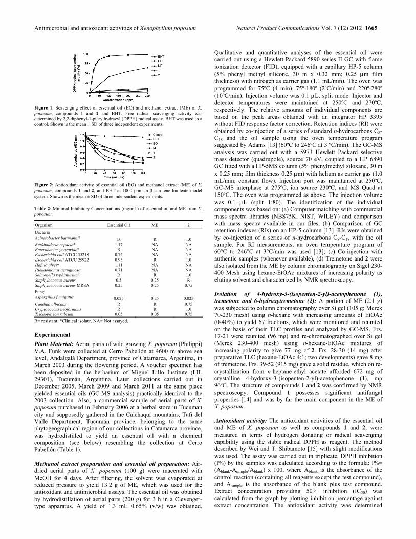
Antimicrobial and antioxidant activities of Xenophyllum poposum Natural Product Communications Vol. 7 (12) 2012 1665
Figure 1: Scavenging effect of essential oil (EO) and methanol extract (ME) of X. poposum, compounds 1 and 2 and BHT. Free radical scavenging activity was determined by 2,2-diphenyl-1-picrylhydrazyl (DPPH) radical assay. BHT was used as a control. Shown is the mean ± SD of three independent experiments.
Figure 2: Antioxidant activity of essential oil (EO) and methanol extract (ME) of X. poposum, compounds 1 and 2, and BHT at 1000 ppm in -carotene-linoleate model system. Shown is the mean ± SD of three independent experiments.
Table 2: Minimal Inhibitory Concentrations (mg/mL) of essential oil and ME from X. poposum.
Organism Essential Oil ME 2
Bacteria Acinetobacter baumannii 1.0 R 1.0 Burkholderia cepacia* 1.17 NA NA Enterobacter gergoviae* R NA NA Escherichia coli ATCC 35218 0.74 NA NA Escherichia coli ATCC 25922 0.95 R 1.0 Hafnia alvei* 1.11 NA NA Pseudomonas aeruginosa 0.71 NA NA Salmonella typhimurium R R 1.0 Staphylococcus aureus 0.5 0.25 R Staphylococcus aureus MRSA 0.25 0.25 0.75
Fungi Aspergillus fumigatus 0.025 0.25 0.025 Candida albicans R R 0.75 Cryptococcus neoformans R R 1.0 Trichophyton rubrum 0.05 0.05 0.75
R= resistant. *Clinical isolate. NA= Not assayed. Experimental
Plant Material: Aerial parts of wild growing X. poposum (Philippi) V.A. Funk were collected at Cerro Pabellón at 4600 m above sea level, Andalgalá Department, province of Catamarca, Argentina, in March 2003 during the flowering period. A voucher specimen has been deposited in the herbarium of Miguel Lillo Institute (LIL 29301), Tucumán, Argentina. Later collections carried out in December 2005, March 2009 and March 2011 at the same place yielded essential oils (GC-MS analysis) practically identical to the 2003 collection. Also, a commercial sample of aerial parts of X. poposum purchased in February 2006 at a herbal store in Tucumán city and supposedly gathered in the Calchaquí mountains, Tafí del Valle Department, Tucumán province, belonging to the same phytogeographical region of our collections in Catamarca province, was hydrodistilled to yield an essential oil with a chemical composition (see below) resembling the collection at Cerro Pabellón (Table 1). Methanol extract preparation and essential oil preparation: Air-dried aerial parts of X. poposum (100 g) were macerated with MeOH for 4 days. After filtering, the solvent was evaporated at reduced pressure to yield 13.2 g of ME, which was used for the antioxidant and antimicrobial assays. The essential oil was obtained by hydrodistillation of aerial parts (200 g) for 3 h in a Clevenger-type apparatus. A yield of 1.3 mL 0.65% (v/w) was obtained.
Qualitative and quantitative analyses of the essential oil were carried out using a Hewlett-Packard 5890 series II GC with flame ionization detector (FID), equipped with a capillary HP-5 column (5% phenyl methyl silicone, 30 m x 0.32 mm; 0.25 m film thickness) with nitrogen as carrier gas (1.1 mL/min). The oven was programmed for 75ºC (4 min), 75º-180º (2ºC/min) and 220º-280º (10ºC/min). Injection volume was 0.1 L, split mode. Injector and detector temperatures were maintained at 250ºC and 270ºC, respectively. The relative amounts of individual components are based on the peak areas obtained with an integrator HP 3395 without FID response factor correction. Retention indices (RI) were obtained by co-injection of a series of standard n-hydrocarbons C8-C18 and the oil sample using the oven temperature program suggested by Adams [13] (60ºC to 246ºC at 3 ºC/min). The GC-MS analysis was carried out with a 5973 Hewlett Packard selective mass detector (quadrupole), source 70 eV, coupled to a HP 6890 GC fitted with a HP-5MS column (5% phenylmethyl siloxane, 30 m x 0.25 mm; film thickness 0.25 m) with helium as carrier gas (1.0 mL/min; constant flow). Injection port was maintained at 250ºC, GC-MS interphase at 275ºC, ion source 230ºC, and MS Quad at 150ºC. The oven was programmed as above. The injection volume was 0.1 L (split 1:80). The identification of the individual components was based on: (a) Computer matching with commercial mass spectra libraries (NBS75K, NIST, WILEY) and comparison with mass spectra available in our files, (b) Comparison of GC retention indexes (RIs) on an HP-5 column [13]. RIs were obtained by co-injection of a series of n-hydrocarbons C8-C18 with the oil sample. For RI measurements, an oven temperature program of 60°C to 246°C at 3°C/min was used [13]; (c) Co-injection with authentic samples (whenever available), (d) Tremetone and 2 were also isolated from the ME by column chromatography on Sigel 230-400 Mesh using hexane-EtOAc mixtures of increasing polarity as eluting solvent and characterized by NMR spectroscopy. Isolation of 4-hydroxy-3-(isopenten-2-yl)-acetophenone (1), tremetone and 6-hydroxytremetone (2): A portion of ME (2.1 g) was subjected to column chromatography over Si gel (105 g; Merck 70-230 mesh) using n-hexane with increasing amounts of EtOAc (0-40%) to yield 67 fractions, which were monitored and reunited on the basis of their TLC profiles and analyzed by GC-MS. Frs. 17-21 were reunited (96 mg) and re-chromatographed over Si gel (Merck 230-400 mesh) using n-hexane-EtOAc mixtures of increasing polarity to give 77 mg of 2. Frs. 28-30 (14 mg) after preparative TLC (hexane-EtOAc 4:1; two developments) gave 8 mg of tremetone. Frs. 39-52 (915 mg) gave a solid residue, which on re-crystallization from n-heptane-ethyl acetate afforded 672 mg of crystalline 4-hydroxy-3-(isopenten-2-yl)-acetophenone (1), mp 96ºC. The structure of compounds 1 and 2 was confirmed by NMR spectroscopy. Compound 1 possesses significant antifungal properties [14] and was by far the main component in the ME of X. poposum. Antioxidant activity: The antioxidant activities of the essential oil and ME of X. poposum as well as compounds 1 and 2, were measured in terms of hydrogen donating or radical scavenging capability using the stable radical DPPH as reagent. The method described by Wei and T. Shibamoto [15] with slight modifications was used. The assay was carried out in triplicate. DPPH inhibition (I%) by the samples was calculated according to the formula: I%= (Ablank-Asample/Ablank) x 100, where Ablank is the absorbance of the control reaction (containing all reagents except the test compound), and Asample is the absorbance of the blank plus test compound. Extract concentration providing 50% inhibition (IC50) was calculated from the graph by plotting inhibition percentage against extract concentration. The antioxidant activity was determined
1666 Natural Product Communications Vol. 7 (12) 2012 González et al.
according to the -carotene bleaching method [16]. Absorbance was
measured at 20 min intervals until the color of -carotene disappeared in the control reaction (t=120 min). Relative antioxidant activities (RAA%) of the extract and oil were calculated from the equation: RAA% = (ASample/ABHT) x 100, where ABHT is the absorbance of the positive control BHT, and ASample is the absorbance of the extract and oil. Experiments were performed in triplicate. Antioxidant capacities of the samples were compared with those of BHT and the control. Antimicrobial activity: Antibacterial assays were performed using the agar diffusion method against Acinetobacter baumannii (ATCC BAA-747), Escherichia coli (ATCC 35218 and 25922), Pseudomonas aeruginosa (ATCC 27853), Salmonella typhimurium (ATCC 13311), Staphylococcus aureus (ATCC 25923) and Methicillin-Resistant Staphylococcus aureus (MRSA) (ATCC 700698). Multi-resistant clinical isolates of Burkholderia cepacia, Enterobacter gergoviae, and Hafnia alvei provided by Dr. Cristina Estrella, Hospital Centro de Salud, San Miguel de Tucumán, Argentina, were also assayed. Antifungal activities were performed using the yeast-like Candida albicans (provided by Vancouver General Hospital, BC, Canada) and Cryptococcus neoformans var. grubii (kindly provided by Dr. Karen Bartlet, University of British Columbia, BC, Canada), while Aspergillus fumigatus (ATCC 1022) and Trichophyton rubrum (ATCC 18758) represented filamentous fungi. Bacteria were grown in Brain Heart Infusion broth fungi. (Laboratories Britania, Argentina) at 37ºC for 18 h, and resuspended in sterile physiological saline with reference to the value 0.5 of the McFarland scale (1.5 x 10⁸ colony forming unit /mL). Briefly,
plates were prepared with a base layer of Müeller–Hinton agar (10 mL) and wells (6 mm of diameter) were made on the surface of the media. Different concentrations (50 μL) of essential oil and ME were placed in the wells. 25 μL of gentamicin and amphotericin B (0.05 mg/mL) were used as positive control for bacteria and fungi, respectively. Fungal strains were grown in Sabouraud broth (B&D), and the antifungal activity against filamentous fungi was assessed from spores obtained as reported Spores were harvested by rubbing the top of sporulated colonies in 2 mL Sabouraud broth containing 10% glycerol. Spores were aliquoted and kept at -20ºC. For yeast-like fungi, the same protocol used for bacterial strains was used, but using Sabouraud broth. The microorganisms were incubated at 37°C aerobically and after 24 h of incubation, the zones of inhibition were measured. Bacterial growth inhibition was determined as the diameter of the inhibition zones around the well. The growth inhibition diameter was an average of four measurements, taken in 90 degrees apart. MIC values were determined by conventional agar plate dilution method [17]. All tests were performed in triplicate. Statistic Analysis: A t-student test was used for statistical analysis. A P value <0.05 was considered significant. Acknowledgements - This work was financially supported by Consejo de Investigaciones de la Universidad Nacional de Tucumán (CIUNT) and Consejo Nacional de Investigaciones Científicas y Técnicas de la República Argentina (CONICET). The authors warmly thank Dr. Viviana Vildoza for providing the standard strains of microorganisms.
References
[1] Funk VA. (1997) Xenophyllum, a new Andean genus extracted from Werneria s.l. (Compositae: Senecioneae). Novon, 7, 235-241. [2] Zuloaga FO, Morrone O. (1999) Catálogo de las plantas vasculares de la República Argentina. Missouri Botanical Garden Press, Missouri, USA. [3] Alonso J, Desmarchelier C. (2006) Plantas Medicinales Autóctonas de la Argentina. Ediciones Fitociencia, Buenos Aires, Argentina. [4] Giberti GC. (1983) Herbal folk medicine in northwestern Argentina: Compositae. Journal of Ethnopharmacology, 7, 321-341. [5] de Marchese MJA, de Heluani CS, Catalán CAN, Griffin CA, Vaughn Jr. JB, Herz W. (2007) Incisol, an alcohol with a novel sesquiterpene
skeleton from Xenophyllum incisum. Biochemical Systematics and Ecology, 35, 169-175. [6] (a) Ponce MA, Gros GE. (1991) Prenylated aromatic ketones from Werneria poposa. The Journal of the Argentine Chemical Society, 79, 197-200;
(b) Ponce MA, Gros GE. (1995) A flavonoid and coumarins from Werneria poposa. Ibid. 83, 93-95. [7] Romano E, Raschi AB, Gonzalez AM, Jaime G, Fortuna MA, Hernandez LR, Bach H, Benavente AM. (2011) Phytotoxic activities of (2R)-6-
hydroxytremetone. Plant Physiology and Biochemistry, 49, 671-675. [8] Lizarraga E, Castro F, Fernández F, de Lampasona MP, Catalán CAN. (2012) Antioxidant, hemolytic and cytotoxic activities of Senecio species
used in traditional medicine of northwestern Argentina. Natural Product Communications, 7, 607-608. [9] Abella L, Cortella AR, Velasco-Negueruela A, Perez-Alonso MJ. (2000) Ethnobotany, volatile oils and secretion tissues of Werneria poposa from
Argentina. Pharmaceutical Biology, 38, 197-203. [10] (a) Liu Q, Shen L, Wang T, Chen J, Qi W, Gao K. (2010) Novel modified furanoeremophilane-type sesquiterpenes and benzofuran derivatives from
Ligularia veitchiana. Food Chemistry, 122, 55-59; (b) Cespedes CL, Uchoa A, Salazar JR, Perich F, Pardo F. (2002) Plant growth inhibitory activity of p-hydroxyacetophenones and tremetones from Chilean endemic Baccharis species and some analogous: A comparative study. Journal of Agricultural and Food Chemistry, 50, 2283–2292; (c) Piacente S, Aquino R, de Tommasi N, Pizza C, de Ugaz OL, Orellana HC, Mahamood N. (1994) Constituents of Werneria ciliolata and their in vitro anti-HIV activity. Phytochemistry, 36, 991–996; (d) Hausen BM, Helmke B. (1995) Butenylbithiophene, -terthienyl and hydroxytremetone as contact allergens in cultivars of marigold (Tagetes sp.). Contact Dermatitis, 33, 33–37.
[11] (a) Takei M, Umeyama A, Arihara S. (2006) T-cadinol and calamenene induce dendritic cells from human monocytes and drive Th1 polarization. European Journal of Pharmacology, 537, 190-199; (b) Claeson P, Andersson R, Samuelsson G. (1991) T-cadinol: a pharmacologically active constituent of scented myrrh: introductory pharmacological characterization and high field 1H- and 13C-NMR data. Planta Medica, 57, 352-356.
[12] (a) Claeson P, Zygmunt P, Hogestatt ED. (1991) Calcium antagonistic properties of the sesquiterpene T-cadinol: a comparison with nimodipine in the isolated rat aorta. Pharmacology and Toxicology, 69, 173-177; (b) Tung YT, Huang CC, Ho ST, Kuo YH, Lin CC, Lin CT, Wu JH. (2011) Bioactive phytochemicals of leaf essential oils of Cinnamomum osmophloeum prevent lipopolysaccharide/D-galactosamine (LPS/D-GalN)-induced acute hepatitis in mice. Journal of Agriculture and Food Chemistry, 59, 8117-8123; (c) Chang ST, Chen PF, Wang SY, Wu HH. (2001) Antimite activity of essential oils and their constituents from Taiwania cryptomerioides. Journal of Medical Entomology, 38, 455-457.
[13] Adams R. (2007) Identification of essential oil components by Gas Chromatography/ Quadrupole Mass Spectroscopy, 4th Edition. Allured Publishing Corporation, Carol Stream, Illinois, USA.
[14] Tomás-Barberán F, Iniesta-Sanmartin E, Tomás-Lorente F, Rumbero A. (1990) Antimicrobial phenolic compounds from three Spanish Helichrysum species. Phytochemistry, 29, 1093-1095.
[15] Wei A, Shibamoto T. (2007) Antioxidant activities and volatile constituents of various essential oils. Journal of Agriculture and Food Chemistry, 55, 1737-1742.
[16] Jayaprakasha GK, Singh RP, Sakariah KK. (2001) Antioxidant activity of grape seed (Vitis vinifera) extracts on peroxidation models in vitro. Food Chemistry, 73, 285-290.
[17] Basualdo J, Coto C, De Torres R. (1996) Microbiología Biomédica. Buenos Aires, Argentina: Atlante.

Phytochemical and Micromorphological Traits of Endemic Micromeria pseudocroatica (Lamiaceae) Dario Kremer1, Ivna Dragojević Müller2, Edith Stabentheiner3, Dubravka Vitali1, Martina Kopričanec2, Mirko Ruščić4, Ivan Kosalec1, Nada Bezić4 and Valerija Dunkić4* 1Faculty of Pharmacy and Biochemistry, University of Zagreb, A. Kovačića 1, 10000 Zagreb, Croatia
2Department of Ecology and Water Protection, Water Supply and Drainage, Patačićkina 1b, 10000 Zagreb, Croatia
3Institute of Plant Sciences, Karl-Franzens University, Schubertstrasse 51, A-8010 Graz, Austria
4Department of Biology, Faculty of Natural Science, University of Split, Teslina 12, 10000 Split, Croatia [email protected]
Received: September 26th, 2012; Accepted: October 31st, 2012
Micromeria pseudocroatica Šilić is an endemic species distributed in southern Croatia. Chemical composition of the essential oil (analysed by GC and GC-MS), and the content of macroelements and trace elements analysed by ICP-AES was conducted. Additionally, a study on the types and distribution of trichomes was done by scanning electron microscopy. The essential oil was characterized by a high concentration of oxygenated monoterpenes, whose major compounds are borneol (22.7% and 24.8%) and camphor (16.1% and 13.9%). The content of Ca was highest (13202.69 mg/kg and 13223.83 mg/kg) among the investigated macroelements, while Fe was the most represented element (100.68 and 326.48 mg/kg) among the micronutrients. The content of potentially toxic elements that is Se, Cu, and Hg was below the limit of quantification. Non-glandular trichomes, peltate trichomes, and two types of capitate trichomes (type 1: one basal epidermal cell, one head cell with subcuticular space; type 2: one basal epidermal cell, two stalk cells, and one head cell with subcuticular space) were observed on leaves, bracteoles, calyx, corolla and stem. Keywords: Micromeria pseudocroatica, Borneol, Macroelements, Trace elements, Trichomes. The family Lamiaceae is one of the largest groups among dicotyledons, comprising more than 240 genera. Many species which belong to this family are highly aromatic due to the presence of external glandular structures that produce essential oil [1]. One of the less investigated genera in this family is Micromeria Benth. The essential oil composition of the Micromeria species is variable. The major constituents of the oil of M. croatica (Pers.) Schott, an endemic Illyric-Balcanic species were caryophyllene oxide and β-caryophyllene [2]. Pulegone and piperitone oxide were the main compounds in the essential oils of M. albanica (Griseb. ex K. Maly) Šilić and M. thymifolia (Scop.) Fritsch.. Spathulenol was the dominant compound in M. parviflora (Vis.) Rchb. [3]. The major constituents of the oil of M. libanotica Boiss., a species endemic to Lebanon, were isomenthone (44.5%), pulegone (13.5%) and isopulegone (6.5%) [4]. Glandular trichomes occurring in the Lamiaceae are the site of essential oil biosynthesis, secretion and accumulation and their structure has been studied by many authors [1, 5-8]. Glandular trichomes of Micromeria fruticosa (L.) Druce and M. croatica were investigated by Werker et al. [5] and Kremer et al. [2], respectively. Although the biological activity of Micromeria species could be attributed to some constituents of the essential oil [9-11], the presence of other biologically active compounds such as microelements could have a synergistic effect with the essential oils. Only few literature data about the content of macroelements and trace elements in Micromeria species are available [2,12]. The genus Micromeria includes 70–90 herbs, sub-shrubs and shrubs distributed throughout the temperate belt [13-15]. Twenty one Micromeria species have been described for Europe [13] and nine (M. croatica, M. dalmatica Benth., M. fruticulosa (Bertol.) Šilić,
M. graeca (L.) Benth., M. juliana (L.) Benth., M. kerneri Murb., M. microphylla (Dum.-Urv.) Benth., M. pseudocroatica Šilić, and M. thymifolia (Scop.) Fritsch.) species for Croatia [16]. M. pseudocroatica is an endemic species distributed in the Mediterranean part of Croatia (Pelješac Peninsula, Korčula Islands) [17,18]. It is a perennial plant with several to numerous stems up to 30 cm long, and single, pink-purple flowers [17]. M. pseudocroatica grows in the crevices of calcareous rocks at altitudes up to about 500 m. The aim of the presented study is to obtain additional knowledge on the genus Micromeria in general, and particularly on M. pseudocroatica, since no data on essential oil, macroelements and trace elements, and micromorphological traits are available for M. pseudocroatica. Aerial parts of wild-growing M. pseudocroatica were analyzed on two different localities, Pijavičino (Pi) and Prapatno (Pr) on Pelješac Peninsula (Croatia). Fifty-two components representing 86.4% of the total oil were identified in the oil from locality Pi and forty-seven components representing 90.4% of the total oil were characterized in oil from Pr. Total yield of the oil was 0.3% and 0.2% for oils from Pi and Pr, respectively. The identified components listed in order of their elution from the VF-5MS column are given in Table 1 together with their percentages of the total mass fraction of the oil. Identified components are classified on the basis of their chemical structures in seven classes. Both samples contained higher concentration of oxygenated monoterpenes (47.3% in Pi and 43.7% in Pr) with borneol (22.7% in Pi and 24.8% in Pr) and camphor (16.1% in Pi and 13.9% in Pr) as the major components of the both oil. Similarly to our results borneol was identified as one of major compounds in the oil
NPC Natural Product Communications 2012 Vol. 7 No. 12
1667 - 1670

1668 Natural Product Communications Vol. 7 (12) 2012 Kremer et al.
Table 1: Phytochemical composition (%) of essential oils of M. pseudocroatica Šilić from the localities Pijavičino (Pi) and Prapatno (Pr).
Component
RI M, pseudocroatica
Identification (Pi) (Pr)
Monoterpene hydrocarbons 1.9 3.2 Camphene 962 0.5 1.9 RI, MS β-Pinene 982 0.2 0.2 RI, MS, S p-Cymene 1021 0.2 0.1 RI, MS Limonene 1032 0.1 - RI, MS, S γ-Terpinene 1057 0.1 0.1 RI, MS Terpinolene 1089 0.8 0.9 RI, MS Oxygenated monoterpenes 47.3 43.7 β-Thujone 1121 - 0.3 RI, MS trans-Pinocarveol 1147 0.1 0.3 RI, MS Camphor 1151 16.1 13.9 RI, MS, S Borneol 1176 22.7 24.8 RI, MS Cymene-8-ol 1179 2.1 0.9 RI, MS Terpinen-4-ol 1184 0.6 0.2 RI, MS Myrtenol 1197 0.6 0.2 RI, MS Verbenone 1204 0.6 0.4 RI, MS Fenchyl acetate 1218 0.1 0.3 RI, MS Thymol methyl ether 1230 0.2 0.1 RI, MS Pulegone 1234 0.5 - RI, MS Carvacrol methyl ether 1241 0.1 0.2 RI, MS Piperitone 1248 0.3 0.4 RI, MS Bornyl acetate 1285 0.4 0.2 RI, MS α-Terpenyl acetate 1349 0.1 0.2 RI, MS Piperitone oxide 1364 2.8 1.3 RI, MS Sesquiterpene hydrocarbons 23.9 32.8 α-Cubebene 1345 1.5 3.5 RI, MS α-Copaene 1377 0.6 0.3 RI, MS β-Bourbonene 1383 0.3 0.3 RI, MS α-Gurjunene 1407 tr 0.5 RI, MS β-Caryophyllene 1424 11.9 17.8 RI, MS, S β -Copaene 1429 0.2 0.3 RI, MS (Z)-β-Farnesene 1454 0.2 0.2 RI, MS α-Humulene 1456 0.6 0.1 RI, MS allo-Aromadendrene 1465 0.2 0.3 RI, MS β-Chamigrene 1477 0.1 0.2 RI, MS Germacrene D 1481 2.1 2.1 RI, MS β-Bisabolene 1494 0.2 0.2 RI, MS Bicyclogermacrene 1500 0.1 0.3 RI, MS δ-Cadinene 1517 5.9 6.7 RI, MS Oxygenated sesquiterpenes 10.7 9.2 Spathulenol 1577 0.1 0.3 RI, MS Caryophyllene oxide 1581 9.3 7.4 RI, MS, S γ-Eudesmol 1632 0.2 0.3 RI, MS α-Cadinol 1655 0.1 - RI, MS α-Bisabolol 1688 0.8 0.3 RI, MS Bisabolol oxide 1748 0.2 0.9 RI, MS
Phenolic compounds 1.0 0.2 Thymol 1290 0.3 - RI, MS, S Carvacrol 1298 0.4 - RI, MS, S Eugenol 1370 0.3 0.2 RI, MS, S Carbonylic compounds 0.5 0.5 3-Octanol acetate 1125 0.3 0.2 RI, MS β-Ionone 1487 0.2 0.3 RI, MS
Hydrocarbons 1.1 0.8 Docosane 2200 0.1 0.2 RI, MS, S Tricosane 2300 0.3 0.2 RI, MS, S Tetracosane 2400 0.2 0.1 RI, MS, S Pentacosane 2500 0.1 0.2 RI, MS, S Hexacosane 2600 0.1 - RI, MS, S Heptacosane 2700 0.3 0.1 RI, MS, S
Total identified (%) 86.4 90.4 Yield (%) 0.3 0.2
RI = VF-5MS, identification by comparison with literature [25] and/or homemade library; MS = identification by NIST02 and Wiley 7 spectral databases; S = identification confirmed with reference compound; tr-traces (mean value below 0.1%); - = not identified.
of M. carminea P.H. Davis, an endemic species of Turkey with percentage of 26.02%, and in the oil of M. cristata (Hampe) Griseb. subsp. phrygia P. H. Davis collected from three different localities (27–39%) [19,20]. Other main components of oil in M. cristata subsp. phrygia were camphor (9–15%) and caryophyllene oxide (4–6%) [20]. The oil from Pr contained higher concentration of sesquiterpene hydrocarbons (32.8%) than the oil from Pi (23.9%), with
β-caryophyllene (11.9% in Pi and 17.8% in Pr) as the dominant component among this class. Caryophyllene oxide was identified as major compound of the both oil in oxygenated sesquiterpenes class with percentages of 9.3% in Pi and 7.4% in Pr. Caryophyllene-oxide was one of major compound in the oil of M. juliana from Croatia [9,21], M. graeca in Greece [22], M. croatica from Serbia [3] and in the oil of M. croatica from Croatia [2]. Groups of monoterpene hydrocarbons, phenolic compound, carbonylic compounds and hydrocarbons represented 4.5% (oil from Pi) and 4.7% (oil from Pr) of the total oil. The contents of macroelements (K, Na, Ca, Mg) and trace elements (Al, Fe, Sr, Zn, B, Mn, Ba, Ni, Pb, V, Cr, Cd, Co, Se, Cu, Hg) in M. pseudocroatica from localities Pijavičino (Pi) and Prapratno (Pr) are shown in Table 2. Among the investigated macroelements the content of Ca was highest (13223.83 mg/kg and 13202.69 mg/kg from locality Pi and Pr, respectively) while the content of Na was lowest (109.54 mg/kg and 243.11 mg/kg from locality Pi and Pr, respectively). Among the micronutrients (B, Fe, Cu, Mn, Zn), the most represented element was Fe (100.68 mg/kg and 326.48 mg/kg from locality Pi and Pr, respectively). Among other trace elements, the most abundant was Al (102.49 mg/kg and 232.29 g/kg from locality Pi and Pr, respectively). The content of several toxic and potentially toxic minerals (Se, Cu, Hg) was below detection limits. According to Kremer et al. [2] M. croatica had the very high potassium content (8275.00–10075.00 mg/kg), which suggests that the accumulation of this macroelement in M. croatica is possible affected by genetic particularity. The content of other macroelements in M. croatica were significantly lower than in samples of M. pseudocroatica. The content of Al (48.13–56.13 mg/kg) and Fe (68.63–78.00 mg/kg) in M. croatica was also significantly lower than in M. pseudocroatica. Contents of other trace elements in M. croatica were not significantly different [2]. Shallari et al. [12] found a significantly higher content of Zn (118 mg/kg), Pb (13 mg/kg), Cr (10 mg/kg), Cd (2 mg/kg), Co (2 mg/kg), and Ni (52 mg/kg) in M. graeca shoots collected at a mining site Rubik (northern Albania). These differences could be explained by high influence of environmental conditions. Table 2: Content of macroelemens and traceelements in herba of M. pseudocroatica.
Element Pijavičino Prapratno
Contents* RSD Contents* RSD (%) Macroelements (mg/kg)
Na 109.54 0.00 0.00 243.11 0.03 0.01 K 1922.21 0.02 0.00 2034.38 0.13 0.01
Ca 13223.83 0.46 0.00 13202.69 0.23 0.00 Mg 2244.54 0.25 0.01 2636.84 0.25 0.01
Trace elements (mg/kg) Al 102.49 9.42 9.19 232.29 42.06 18.11
Fe 100.68 1.27 1.27 326.48 6.17 1.89 Sr 7.88 0.54 6.86 121.99 3.78 3.1 Zn 23.04 0.05 0.23 22.55 0.37 1.65 B 14.41 0.29 2.01 12.52 1.27 10.15
Mn 10.08 0.02 0.02 12.67 0.17 1.31 Ba 4.92 0.31 6.21 5.52 0.81 14.61 Ni 0.82 0.38 45.95 0.85 0.11 13.02 Pb 0.57 0.08 13.98 1.64 0.22 13.12 V 0.29 0.02 6.87 0.96 0.15 15.49 Cr 0.20 0.01 4.34 0.58 0.08 14.32 Cd 0.09 0.03 34.13 0.14 0.04 30.09 Co 0.05 0.01 25.22 0.13 0.02 17.27 Se BLQ – BLQ – Cu BLQ – BLQ – Hg BLQ – BLQ –
*values are means SD (n = 2); RSD, (relative standard deviation; BLQ, below limit of quantitation

Essential oil and macro and trace elements of Micromeria pseudocroatica Natural Product Communications Vol. 7 (12) 2012 1669
Figure 1: SEM micrographs with different types of trichomes on M. pseudocroatica.
Non-glandular trichomes (NG), peltate trichome (P), Type 1 capitate trichomes (C1) and Type 2 capitate trichomes (C2) on outer side of calyx (a); C1 (b) and C2 (c) trichome on the lower leaf side; distribution of the trichomes on the upper (d) and lower (e) leaf surface; C1 trichome on the upper leaf surface (f); outer side of calyx with NG, P, C1, and C2 trichomes (g); inner side of calyx with long NG trichomes (h); NG, and P trichomes on the outer side of corolla (i); NG, C1, and C2 trichomes on the adaxial (j) and abaxial (k) side of bracteole; stem with NG and P trichomes (l). Table 3: Occurrence and frequency of trichomes on aerial parts of M. pseudocroatica from the localities Pijavičino (Pi) and Prapatno (Pr).
Type Leaf Bracteole Calyx Corolla Stem
Ad Ab Ad Ab Ou In Ou In attenuate* + + ± + + ± + ±/+ +
Pi peltate – +/++ – – + – ±/+ – + Cap C1 ± +/++ ± + + ± – – + cap C2 ± +/++ ± ++ ++ – – – +
attenuate + + ± + +/++ + +/++ ±/+ +/++ Pr peltate – ++ – – + – + – + cap C1 ± +/++ ± + + ± – – + cap C2 –/± ++ ± +/++ ++ –/± – – +/++
cap – capitates; Ad – adaxial; Ab – abaxial; Ou – outer; In - inter trichomes: – missing, ± rare, + present, ++ abundant; *attenuate, non-glandular hairs
Both non-glandular and glandular trichomes could be observed on investigated parts (leaves, bracteoles, calyxes, corollas, and stems) of M. pseudocroatica (Figure 1). The occurrence and frequency of trichomes on the adaxial and abaxial side of leaves and bracteoles, outer and inner side of the calyx and corolla, and stem is shown in Table 3. Non-glandular trichomes (Figure 1a, e) were bi-cellular to multicellular, unbranched, uniseriate and folded at different levels. The length of these trichomes varied distinctly from very short hairs on the upper leaf surface (Figure 1f) to very long hairs on the edge of inner calyx side (Figure 1h). Parts of the surface of these trichomes revealed a warty appearance due to the occurrence of cuticular micropapillae (Figure 1a). According to Payne’s [23] plant hair terminology, they could be noted as attenuate hairs. These hairs were identical to non-glandular trichomes found by Kremer et al. [2] in M. croatica. The glandular trichomes could further be divided into peltate and capitate types. Peltate trichomes consisted of a basal cell, a short unicellular stalk, and a multicellular head with a large subcuticular space (Figure 1a and i). They occurred on the abaxial leaf side, on the outer side of the calyx and corolla, and on the stem. Their
presence in Lamiaceae is well documented [1, 5-8, 24]. Two types of capitate trichomes could be observed in M. pseudocroatica. Type one capitate trichome (C1) is composed of one basal epidermal cell and one elliptically formed head cell. C1 trichome was not upright but could be described as clinging to the surface (Figure 1b). This hair type was found on both on the adaxial as well as on the abaxial side of leaves and bracteoles, on the outer and inner side of calyx, and on the stem (Table 3). According to Kremer et al. [2] this type of trichomes was common in Micromeria croatica and it was found on the abaxial leaf surface, on stem, and on calyx. These hairs were also noticed in micrographs of Thymus capitatus (L.) Hoffmanns. (syn. Coridotymus capitatus (L.) Rchb. f.), Majorana syriaca (L.) Rafin., and Satureja thymbra L. presented by Werker et al. [5]. But, the micrographs presented by Werker et al. [5] did not show this hair type in Micromeria fruticosa (L.) Druce. Type two capitate trichome (C2) is composed of one basal epidermal cell, two stalk cells and a single celled head with a subcuticular space (Figure 1c). C2 trichome was upright and quite short. It was found on both adaxial and abaxial side of leaves and bracteoles, on outer and inner side of calyx, and on stem (Table 3). This hair type was reported by Kremer et al. [2] for M. croatica, where it was observed on adaxial and abaxial leaf surface, on calyx, and on stem. Comparison with literature data showed significant difference in chemical traits between in here presented M. pseudocroatica and closely related M. croatica, while micromorphological traits (trichome types) were the same. Presented results give worthy additional knowledge about chemical and micromorphological traits on the genus Micromeria. Experimental
Herbal material and extraction: Randomly selected samples of wild growing plants M. pseudocroatica Šilić were collected during the blooming period in July of 2011 on locus classicus near settlement Pijavičino (GPS coordinates: 42°57'01.5'' N; 17°21'52.2'' E; 443 m a.s.l.'), and on locality Prapratno (GPS coordinates: 42°49'28.1'' N; 17°40'23.7'' E; 159 m a.s.l.'), both on Pelješac Peninsula, Croatia. Dried aerial parts (100 g) were subjected to hydrodistillation for 3 h in Clevenger type apparatus and also used for determination of macroelements and trace elements. Gas chromatography and mass spectrometry (GC, GC/MS): Gas chromatography (GC) analyses were performed on gas chromatograph (model 3900; Varian Inc., Lake Forest, CA, USA) equipped with flame ionization detector, mass spectrometer (model 2100T; Varian Inc.) and capillary column VF-5MS (30 m × 0.25 mm i.d., coating thickness 0.25 μm; Varian Inc.). The chromatographic conditions were as follows: helium was carrier gas at 1 mL·min−1, injector temperature was 250 ºC, and FID detector temperature was 300 ºC. VF-5MS column temperature was programmed at 60 ºC isothermal for 3 min, and then increased to 246 ºC at a rate of 3 ºC·min−1 and held isothermal for 25 min. The injected volume was 1 μL and the split ratio was 1:20. The mass spectrometry (MS) conditions were: ionization voltage 70 eV; ion source temperature 200 ºC; mass scan range: 40–350 mass units. The analyses were carried out in duplicate. The individual peaks were identified by comparison of their retention indices (relative to C8-C25 n-alkanes for VF-5MS) to those of authentic samples and literature [25], as well as by comparing their mass spectra with the Wiley 7 MS library (Wiley, New York, NY, USA) and NIST02 (Gaithersburg, MD, USA) mass spectral database.

1670 Natural Product Communications Vol. 7 (12) 2012 Kremer et al.
Inductively coupled plasma atomic emission spectroscopy (ICP-AES): For determination of macroelements (K, Na, Ca, Mg) and trace elements (Al, Fe, Sr, Zn, B, Mn, Ba, Ni, Pb, V, Cr, Cd, Co, Se, Cu, Hg), duplicates of 2 g ± 0.0005 of homogenized herbal material were wet digested with concentrated HNO3 (5 mL) and 30% H2O2
(2 mL) in the microwave digestion unit (MLS-1200 MEGA Microwave Digestion Systems, Milestone, Bergamo, Italy). The digestion conditions were: 1 min at 250 W (smooth oxidation of organic matter), 1 min at 0 W (proceeding of reaction without addition of energy to avoid run-away temperatures and overpressures), 5 min at 250 W (termination of the soft oxidation of the organic compounds), 5 min at 400 W and 5 min at 600 W (final termination of oxidation processes by applying higher power). After cooling, the digested samples were diluted to 50 mL using deionised water and obtained solutions were used for further investigation. The content of mercury was determined by atomic absorption spectrometry (AAS) on a Perkin-Elmer 4100 Zeeman ZL/FIMS 400 atomic absorption spectrometer (Perkin Elmer, Ueberlingen,
Germany) using flow injection/hydride technique [26], while the content of other investigated elements was determined using inductively coupled plasma atomic emission spectrometry (ICP-AES) on Trace Scan Thermo (Thermo Jarrel Ash Corporation, Franklin, USA) using the standard technique [27]. Micromorphological traits: For SEM-investigation samples (stem, leaf, bracteoles, calyx) were transferred from 70% ethanol to 70% acetone, then dehydrated (70%, 90% and 100% acetone) and subjected to critical point drying using CO2 as the drying medium (CPD030; Baltec). After that, samples were sputter coated with gold (Sputter Coater, AGAR) and examined under the scanning electron microscope XL30 ESEM (FEI) with 20 kV acceleration voltages in high vacuum mode. Common terminology was used in the description of micromorphology [23]. Acknowledgments - This work was supported by the Ministry of Science, Education and Sports of the Republic of Croatia (projects no. 006–0000000–3178, and 177–1191192–0830). We thank Mrs. Gabrijela Čepo for check English style and grammar.
References
[1] Giuliani C, Maleci Bini L. (2008) Insight into the structure and chemistry of glandular trichomes of Labiatae, with emphasis on subfamily Lamioideae. Plant Systematics and Evolution, 276, 199–208.
[2] Kremer D, Stabentheiner E, Dunkić V, Dragojević Müller I, Vujić L, Kosalec I, Ballian D, Bogunić F, Bezić N. (2012) Micromorphological and chemotaxonomical traits of Micromeria croatica (Pers.) Schott.. Chemistry & Biodiversity, 9, 755-768.
[3] Slavkovska V, Couladis M, Bojović S, Tzakou O, Pavlović M, Lakušić B, Jančić R. (2005) Essential oil and its systematic significance in species of Micromeria Bentham from Serbia & Montenegro. Plant Systematics and Evolution, 255, 1–15.
[4] Chalchat JC, Diab Y, Auezova L, Chebib H. (2005) Chemical composition of the essential oil of Micromeria libanotica Boiss., an endemic species of Lebanon. Journal of Essential Oil Research, 17, 449–450.
[5] Werker E, Ravid U, Putievsky E. (1985) Structure of glandular hairs and identification of the main components of their secreted material in some species of the Labiatae. Israel Journal of Botany, 34, 31–45.
[6] Werker E. (2000) Trichome diversity and development. In: Advances in botanical research incorporating advances in plant pathology, Plant trichomes. Vol. 31, Hallahand DL, Gray J, Callow JA (Ed). Academic Press, London UK. 1–35.
[7] Corsi G, Bottega S. (1999) Glandular hairs of Salvia officinalis: New data on morphology, localization and histochemistry in relation to function. Annals of Botany, 84, 657–664.
[8] Fahn A. (2000) Structure and function of secretory cells. In: Advances in botanical research incorporating advances in plant pathology, Plant trichomes. Vol. 31, Hallahand DL, Gray J, Callow JA (Ed). Academic Press, London UK. 37–75.
[9] Stojanović G, Palić I, Jursić-Janković J. (2006) Composition and antmicrobial activity of the essential oil of Micromeria cristata and Micromeria juliana. Flavour and Fragrance Journal, 21, 77–79.
[10] Öztürk M, Kolak U, Topçu G, Öksüz S, Choudhary I. (2011) Antioxidant and anticholinesterase active constituents from Micromeria cilicica by radical-scavenging activity-guided fractionation. Food Chemistry, 126, 31–38.
[11] Öztürk M, Kolak U, Duru ME, Harmandar M. (2009) GC-MS analysis of the antioxidant active fractions of Micromeria juliana with anticholinesterase activity. Natural Product Communications, 4, 1271-1276.
[12] Shallari S, Schwartz C, Hasko A, Morel JL. (1998) Heavy metals in soils and plants of serpentine and industrial sites of Albania. Science of the Total Environment, 209, 133–142.
[13] Chater AO, Guinea E. (1972) Micromeria Benth. In: Flora Europaea. Vol. 3. Tutin TG, Heywood VH, Burges NA, Moore DM, Valentine DH, Walters S, Webb DA (Ed). Cambridge University Press, Cambridge. 167–170.
[14] Wielgorskaya T. (1995) Dictionary of generic names of seed plants. Columbia University, New York. 369. [15] Erhardt W, Götz E, Bödeker N, Seybold S. (2002) Zander – Handwörterbuch der Pflanzennamen. 17. Auflage, Eugen Ulmer GmbH & Co.,
Stuttgart. 576. [16] Lovašen-Eberhardt Ž. (2000) Micromeria Benth. – bresina. In: Index florae Croaticae. Nikolić T. (Ed). Natura Croatica, 3, 19–20. [17] Šilić Č. (1979) Monography of genuses Satureja L., Calamintha Miller, Micromeria Bentham, Acinos Miller and Clinopodium L. in flora of
Yugoslavia [In Bosnian]. Zemaljski muzej BiH, Sarajevo. 237–248. [18] Trinajstić I. (1985) Flora of the Korčula Islands. Acta Botanica Croatica, 44, 107–130. [19] Baser KHC, Kirimer N, Özek T, Tümen G. (1995) Essential Oil of Micromeria carminea Davis PH. Journal of Essential Oil Research, 7, 457–458. [20] Tabanca N, Kirimer N, Demirci B, Demirci F, Başer KH. (2001) Composition and antimicrobial activity of the essential oils of Micromeria
cristata subsp. phrygia and the enantiomeric distribution of borneol. Journal of Agriculture and Food Chemistry, 49, 4300–4303. [21] Mastelić J, Jerković I, Kuštrak D. (2005) Aromatic compounds of Micromeria juliana (L) Bentham ex Reicheneb. from Croatia. Journal of
Essential Oil Research, 17, 516–518. [22] Tzakou O, Couladis M. (2001) The essential oil of Micromeria graeca (L.) Bentham et Reichenb. growing in Greece. Flavour and Fragrance
Journal, 16, 107–109. [23] Payne WW. (1978) A glossary of plant hair terminology. Brittonia, 30, 239–255. [24] Bini Maleci L, Servettaz O. (1991) Morphology and distribution of trichomes in Italian species of Teucrium sect. Chamaedrys (Labiatae) – a
taxonomical evaluation. Plant Systematics and Evolution, 174, 83–91. [25] Adams RP. (2007) Identification of essential oil components by gas chromatography/ mass spectroscopy. 4th. ed., Allured Publishing Corporation.
Carol Stream IL, USA. [26] Perkin Elmer. (1994) Flow Injection Mercury/Hydride Analyses Recommended Analytical Conditions and General Information. Bodenseewerk
Perkin-Elmer GmbH, Überlingen, DE. [27] Jarrel Ash Corporation. (1995) ‘Trace Scan, Atom Scan 16/25 Plasma Spectrometer User's Guide’, Franklin, Massachusetts.

Composition and Variability of the Essential Oil of Salvia nemorosa (Lamiaceae) from the Vienna Area of Austria Remigius Chizzola
Institute of Applied Botany and Pharmacognosy, University of Veterinary Medicine Vienna, A – 1210 Vienna, Veterinaerplatz 1, Austria [email protected]
Received: September 28th, 2012; Accepted: October 17th, 2012
The volatiles present in the aerial parts of Salvia nemorosa L. (Lamiaceae), grave sage, were analysed by GC/MS and GC in plants growing at different sites in the outskirts of Vienna, Austria. The flower oils contained mainly sabinene (37-44%), germacrene D (9-14%), β-caryophyllene (8-12%) and caryophyllene oxide (2.6-4.4%). Leaf samples had β-caryophyllene (14-41%), germacrene D (14-38%) and caryophyllene oxide (5-20%) as main compounds, while stem oils were characterized by an high hexadecanoic acid percentage besides germacrene D and β-caryophyllene. Keywords: Salvia nemorosa, Lamiaceae, Sabinene, β-Caryophyllene, Germacrene D. Salvia nemorosa L., grave sage, (Lamiaceae), a perennial with sessile leaves, purple bracts and lilac flowers in whorls, originally occurred from Eastern and Southeastern Europe eastwards to Central Asia. The natural distribution in Europe is the subcontinental dry parts of the Pannonian, Bohemian and Hercynian floral region [1a]. In the area of Vienna, Austria, the plant reaches the western border of its range [1b]. The plant bears glandular and covering trichomes [1c], and essential oils from the aerial parts rich in sesquiterpenoids [2]. As the mentioned references did not differentiate between the plant organs, the present study reports the composition of the essential oil from stem, leaves and flowers of plants collected at five different sites in the urban area of Vienna (Table 1). The oil content of all plant parts was below 0.05%, but they had distinct oil patterns, as shown in Table 2. The oil from the flowers was dominated by sabinene (37-44%), followed by germacrene D (9-14%), β-caryophyllene (8-12%) and caryophyllene oxide (2.6-4.4%). α-Thujene (5-8%) and γ-terpinene (2-8%) were further prominent monterpenes, while β-pinene, α-terpinene, limonene and p-cymene were minor compounds (0.8-3.4% each). The leaf oils were rich in the sesquiterpenes β-caryophyllene (14-41%), germacrene D (14-38%) and caryophyllene oxide (5-20%). Leaf oils contained also some sabinene (up to 6.6%), other monoterpenes less than 1% each and further sesquiterpenes less than 2% each, including farnesene, α-humulene, γ-muurolene and δ-cadinene. Hexadecanoic acid (56-60%) was the main compound in the stem volatile fractions. The already mentioned sesquiterpenes β-caryophyllene and germacrene D were also major compounds in the stems. The monoterpenes sabinene and γ-terpinene were present in three out of the four stem oil samples. However, based on the composition of the oil samples, one cannot differentiate between plants of the different collecting sites. The plants of all five locations represent the same chemotype and had similarities with plants from Serbia with caryophyllene oxide (13.4%), β-caryophyllene (10.1%) and germacrene D (5.7%) as main components. These plants had also sabinene (4.7%) and terpinen-4-ol (3.7%) as prominent monoterpenes in the oil [2a]. However, other S. nemorosa plants from the same country were characterized by an essential oil dominated by caryophyllene oxide
(23.1%), followed by spathulenol (11.2%), and with appreciable proportions of γ-terpinene (8.6%), E-anethol (7.8%) and humulene epoxide II (5.2%), but devoid of sabinene [2b]. A further type of S. nemorosa essential oil was reported from Iran with β-caryophyllene (41.6%) and germacrene B (21.3%) as main compounds, and further sesquiterpenes such as caryophyllene oxide, spathulenol, cis-β-farnesene and germacrene D (each 5-7%) [2c]. Experimental
Plant material: The plants were collected during July and August 2008 at five locations in the northern and eastern part of Vienna, Austria (Table 1). Most plants were fully flowering. In each case, at least 15 plants were taken together and separated into leaf, stem and flower samples. The plant material was dried in a room in ambient air. The “Exkursionsflora für Österreich, Liechtenstein und Südtirol” [1b] was used to identify the plants. Voucher specimens were deposited in the Herbarium of the University of Vienna (WU-Generale, http:\\herbarium.univie.ac.at). Hydrodistillation: Leaf and stem samples were subjected to hydro-distillation in a Clevenger-type apparatus for 3 h using about 15 g of the plant parts and 200 mL of double distilled water. One mL n-hexane was added to the distillation apparatus to collect the oil. Microdistillation: The distillation of the flower samples was carried out using the automatic microdistillation unit MicroDistiller from Eppendorf (Hamburg, Germany), which allows the simultaneous distillation of 6 samples. About 0.2 to 0.3 g of finely crushed dried plant material and 10 mL distilled water were filled into the sample vial. The collecting vial containing 1 mL water, 0.5 g NaCl and 300 μL n-hexane was connected with a capillary to the sample vial. The heating program applied to the sample vial was 15 min at 108°C and then 45 min at 112°C. The collecting vial was kept at -2°C, where the volatiles were trapped in 0.3 mL n-hexane. All essential oils were stored at -18°C until GC and GC/MS analysis. Gas chromatography: The distilled oils and microdistillates were analyzed with an Agilent Technologies 6890 N GC equipped with a FID. The separation was conducted on a DB-5 narrow bore column 10 m x 0.10 mm i.d. with 0.17 μm film thickness. The
NPC Natural Product Communications 2012 Vol. 7 No. 12
1671 - 1672
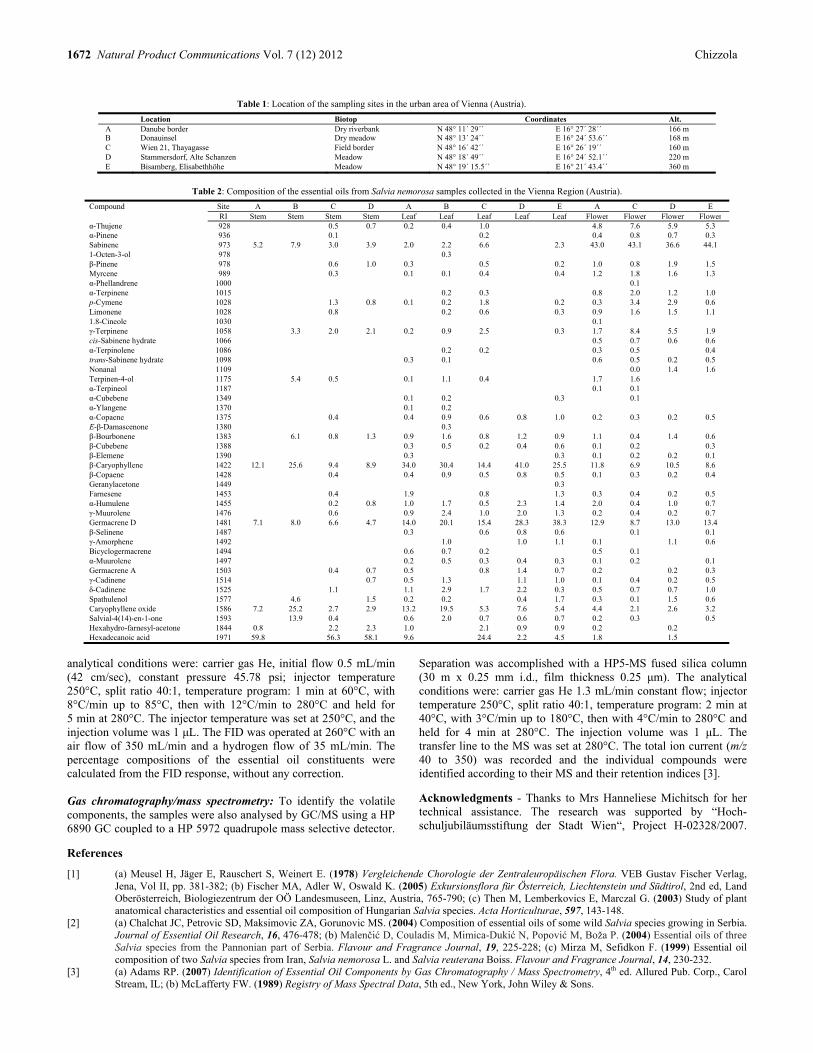
1672 Natural Product Communications Vol. 7 (12) 2012 Chizzola
Table 1: Location of the sampling sites in the urban area of Vienna (Austria).
Location Biotop Coordinates Alt. A Danube border Dry riverbank N 48° 11´ 29´´ E 16° 27´ 28´´ 166 mB Donauinsel Dry meadow N 48° 13´ 24´´ E 16° 24´ 53.6´´ 168 m C Wien 21, Thayagasse Field border N 48° 16´ 42´´ E 16° 26´ 19´´ 160 m D Stammersdorf, Alte Schanzen Meadow N 48° 18´ 49´´ E 16° 24´ 52.1´´ 220 m E Bisamberg, Elisabethhöhe Meadow N 48° 19´ 15.5´´ E 16° 21´ 43.4´´ 360 m
Table 2: Composition of the essential oils from Salvia nemorosa samples collected in the Vienna Region (Austria).
Compound Site A B C D A B C D E A C D E RI Stem Stem Stem Stem Leaf Leaf Leaf Leaf Leaf Flower Flower Flower Flowerα-Thujene 928 0.5 0.7 0.2 0.4 1.0 4.8 7.6 5.9 5.3α-Pinene 936 0.1 0.2 0.4 0.8 0.7 0.3 Sabinene 973 5.2 7.9 3.0 3.9 2.0 2.2 6.6 2.3 43.0 43.1 36.6 44.1 1-Octen-3-ol 978 0.3 β-Pinene 978 0.6 1.0 0.3 0.5 0.2 1.0 0.8 1.9 1.5 Myrcene 989 0.3 0.1 0.1 0.4 0.4 1.2 1.8 1.6 1.3 α-Phellandrene 1000 0.1 α-Terpinene 1015 0.2 0.3 0.8 2.0 1.2 1.0 p-Cymene 1028 1.3 0.8 0.1 0.2 1.8 0.2 0.3 3.4 2.9 0.6 Limonene 1028 0.8 0.2 0.6 0.3 0.9 1.6 1.5 1.1 1.8-Cineole 1030 0.1 γ-Terpinene 1058 3.3 2.0 2.1 0.2 0.9 2.5 0.3 1.7 8.4 5.5 1.9 cis-Sabinene hydrate 1066 0.5 0.7 0.6 0.6 α-Terpinolene 1086 0.2 0.2 0.3 0.5 0.4 trans-Sabinene hydrate 1098 0.3 0.1 0.6 0.5 0.2 0.5 Nonanal 1109 0.0 1.4 1.6 Terpinen-4-ol 1175 5.4 0.5 0.1 1.1 0.4 1.7 1.6 α-Terpineol 1187 0.1 0.1 α-Cubebene 1349 0.1 0.2 0.3 0.1 α-Ylangene 1370 0.1 0.2 α-Copaene 1375 0.4 0.4 0.9 0.6 0.8 1.0 0.2 0.3 0.2 0.5 E-β-Damascenone 1380 0.3 β-Bourbonene 1383 6.1 0.8 1.3 0.9 1.6 0.8 1.2 0.9 1.1 0.4 1.4 0.6 β-Cubebene 1388 0.3 0.5 0.2 0.4 0.6 0.1 0.2 0.3 β-Elemene 1390 0.3 0.3 0.1 0.2 0.2 0.1 β-Caryophyllene 1422 12.1 25.6 9.4 8.9 34.0 30.4 14.4 41.0 25.5 11.8 6.9 10.5 8.6 β-Copaene 1428 0.4 0.4 0.9 0.5 0.8 0.5 0.1 0.3 0.2 0.4 Geranylacetone 1449 0.3 Farnesene 1453 0.4 1.9 0.8 1.3 0.3 0.4 0.2 0.5 α-Humulene 1455 0.2 0.8 1.0 1.7 0.5 2.3 1.4 2.0 0.4 1.0 0.7 γ-Muurolene 1476 0.6 0.9 2.4 1.0 2.0 1.3 0.2 0.4 0.2 0.7 Germacrene D 1481 7.1 8.0 6.6 4.7 14.0 20.1 15.4 28.3 38.3 12.9 8.7 13.0 13.4 β-Selinene 1487 0.3 0.6 0.8 0.6 0.1 0.1 γ-Amorphene 1492 1.0 1.0 1.1 0.1 1.1 0.6 Bicyclogermacrene 1494 0.6 0.7 0.2 0.5 0.1 α-Muurolene 1497 0.2 0.5 0.3 0.4 0.3 0.1 0.2 0.1 Germacrene A 1503 0.4 0.7 0.5 0.8 1.4 0.7 0.2 0.2 0.3 γ-Cadinene 1514 0.7 0.5 1.3 1.1 1.0 0.1 0.4 0.2 0.5 δ-Cadinene 1525 1.1 1.1 2.9 1.7 2.2 0.3 0.5 0.7 0.7 1.0 Spathulenol 1577 4.6 1.5 0.2 0.2 0.4 1.7 0.3 0.1 1.5 0.6 Caryophyllene oxide 1586 7.2 25.2 2.7 2.9 13.2 19.5 5.3 7.6 5.4 4.4 2.1 2.6 3.2 Salvial-4(14)-en-1-one 1593 13.9 0.4 0.6 2.0 0.7 0.6 0.7 0.2 0.3 0.5 Hexahydro-farnesyl-acetone 1844 0.8 2.2 2.3 1.0 2.1 0.9 0.9 0.2 0.2 Hexadecanoic acid 1971 59.8 56.3 58.1 9.6 24.4 2.2 4.5 1.8 1.5
analytical conditions were: carrier gas He, initial flow 0.5 mL/min (42 cm/sec), constant pressure 45.78 psi; injector temperature 250°C, split ratio 40:1, temperature program: 1 min at 60°C, with 8°C/min up to 85°C, then with 12°C/min to 280°C and held for 5 min at 280°C. The injector temperature was set at 250°C, and the injection volume was 1 μL. The FID was operated at 260°C with an air flow of 350 mL/min and a hydrogen flow of 35 mL/min. The percentage compositions of the essential oil constituents were calculated from the FID response, without any correction. Gas chromatography/mass spectrometry: To identify the volatile components, the samples were also analysed by GC/MS using a HP 6890 GC coupled to a HP 5972 quadrupole mass selective detector.
Separation was accomplished with a HP5-MS fused silica column (30 m x 0.25 mm i.d., film thickness 0.25 μm). The analytical conditions were: carrier gas He 1.3 mL/min constant flow; injector temperature 250°C, split ratio 40:1, temperature program: 2 min at 40°C, with 3°C/min up to 180°C, then with 4°C/min to 280°C and held for 4 min at 280°C. The injection volume was 1 μL. The transfer line to the MS was set at 280°C. The total ion current (m/z 40 to 350) was recorded and the individual compounds were identified according to their MS and their retention indices [3].
Acknowledgments - Thanks to Mrs Hanneliese Michitsch for her technical assistance. The research was supported by “Hoch-schuljubiläumsstiftung der Stadt Wien“, Project H-02328/2007.
References
[1] (a) Meusel H, Jäger E, Rauschert S, Weinert E. (1978) Vergleichende Chorologie der Zentraleuropäischen Flora. VEB Gustav Fischer Verlag, Jena, Vol II, pp. 381-382; (b) Fischer MA, Adler W, Oswald K. (2005) Exkursionsflora für Österreich, Liechtenstein und Südtirol, 2nd ed, Land Oberösterreich, Biologiezentrum der OÖ Landesmuseen, Linz, Austria, 765-790; (c) Then M, Lemberkovics E, Marczal G. (2003) Study of plant anatomical characteristics and essential oil composition of Hungarian Salvia species. Acta Horticulturae, 597, 143-148.
[2] (a) Chalchat JC, Petrovic SD, Maksimovic ZA, Gorunovic MS. (2004) Composition of essential oils of some wild Salvia species growing in Serbia. Journal of Essential Oil Research, 16, 476-478; (b) Malenčić D, Couladis M, Mimica-Dukić N, Popović M, Boža P. (2004) Essential oils of three Salvia species from the Pannonian part of Serbia. Flavour and Fragrance Journal, 19, 225-228; (c) Mirza M, Sefidkon F. (1999) Essential oil composition of two Salvia species from Iran, Salvia nemorosa L. and Salvia reuterana Boiss. Flavour and Fragrance Journal, 14, 230-232.
[3] (a) Adams RP. (2007) Identification of Essential Oil Components by Gas Chromatography / Mass Spectrometry, 4th ed. Allured Pub. Corp., Carol Stream, IL; (b) McLafferty FW. (1989) Registry of Mass Spectral Data, 5th ed., New York, John Wiley & Sons.

Essential oil Composition of Ficus benjamina (Moraceae) and Irvingia barteri (Irvingiaceae) Isiaka A. Ogunwandea, *, Razaq Jimoha, Adedoyin A. Ajetunmobia, Nudewhenu O. Avoseha and Guido Flaminib
aNatural Product Unit, Department of Chemistry, Faculty of Science, Lagos State University, P.M.B. 0001, Ojo, Lagos, Nigeria
bDipartimento di Scienze Farmaceutiche, sede Chimica Bioorganica e Biofarmacia, Universita di Pisa, Via Bonanno 33, 56126 Pisa, Italy
Received: November 14th, 2011; Accepted: October 10th, 2012
Essential oils obtained by hydrodistillation of leaves of two Nigerian species were analyzed for their constituents by gas chromatography (GC) and gas chromatography-mass spectrometry (GC-MS). The leaf oil of Ficus benjamina L. (Moraceae), collected during the day, contained high contents of α-pinene (13.9%), abietadiene (9.7%), cis-α-bisabolene (8.2%) and germacrene-D-4-ol (8.4%), while the night sample was dominated by germacrene-D-4-ol (31.5%), 1,10-di-epi-cubenol (8.8%) and hexahydrofarnesylacetone (8.3%). This could be a possible indication of differences in emissions of volatiles by F. benjamina during the day and night. The main compounds of Irvingia barteri Hook. f. (Irvingiaceae) were β-caryophyllene (17.0%), (E)-α-ionone (10.0%), geranial (7.6%), (E)-β-ionone (6.6%) and β-gurjunene (5.1%). Keywords: Ficus benjamina, Irvingia barteri, Essential oil, α-Pinene, Germacrene-D-4-ol, β-Caryophyllene.
In continuation of extensive research on the different volatile emissions from Ficus species [1-3] and other plant from Nigeria, we report herein the volatile compounds of F. benjamina and Irvingia barteri. F. benjamina L. is one of hundreds of species of figs, dozens of which are very common in cultivation. Figs are members of the family Moraceae, a huge family of plants with over 1000 species. Most Moraceae are tropical plants, and F. benjamina is no exception, being native to the tropics of south Asia and northern Australia. However, this is probably the most common of all the Ficus species. The plant is credited with many biologically active compounds and uses [4,5]. α-Copanene, cyclosativene and β-ocimene were the major compounds of its volatile oil [6]. The chemical composition of essential oils of some Ficus species growing in Nigeria have been reported [1-3,7-10]. Phytol and 6,10,14-trimethyl-2-pentadecanone have been described as chemical markers of Nigerian grown Ficus oils [2,3,8-10]. Wild mango, Irvingia barteri O’Rorke Bail (syn Irvingia gabonensis), family Irvingiaceae, is native to some Central and West African countries. The fruit is succulent and available between July and October. Due to the pleasant aroma, the roasted seeds are commonly used in Africa as flavoring in traditional dishes. I. barteri is known in the Nigerian languages by various names such as ‘biri’or ‘goron’(Hausa), ‘oro’ (Yoruba) and ‘obono’ (Igbo). The fruits have been reported to be rich in vitamin C and are widely consumed as a desert fruit or snack throughout Western and Central Africa with various nutritional and dietary aspects [11,12]. Extracts of the plant have been reported to possess antimicrobial activities and to be used as a painkiller for toothache. Some antimicrobial agents such as 3-friedelanone, betulinic acid, oleanolic acid, 3,3′,4′-tri-O-methylellagic acid, 3,4-di-O-methyl-ellagic acid and hardwickiic acid have been isolated from the plant [13]. Literature information is scanty about the volatile constituents except for the key odorant compounds of the seeds and fruits [14-16].
The plant samples yielded low contents of essential oils: 0.1% (v/w; F. benjamina; colorless) and 0.23% (v/w; I. barteri; light yellow), calculated on a dry weight basis. Forty-seven and thirty-eight components respectively were identified from the day and night samples of F. benjamina. The main compounds of the day sample were α-pinene (13.9%), abietadiene (9.7%), germacrene-D-4-ol (8.4%) and cis-α-bisabolene (8.2%). Isobornyl acetate (5.0%) and abietatriene (4.9%) were also present in sizeable quantity. However, germacrene-D-4-ol (31.5%) was the most singly abundant compound of the night sample. Other significant constituents were 1,10-di-epi-cubenol (8.8%), hexahydrofarnesyl-acetone (8.3%), -geranyl acetone (6.2%), cubenol (5.5%) and 1, 8-cineole (4.2%). Significant differences were observed in the major classes of compounds of the day and night samples of F. benjamina (Table 1). Oxygenated sesquiterpenoids were the dominant class of compounds in the night sample. On the other hand, the day sample contained equal proportions of monoterpene hydrocarbons and oxygenated monoterpenes. However, diterpenoids and sulfur compounds, which were present in the day sample, could not be identified in the night sample. The volatiles emitted by plants depend on factors such as climatic conditions, morphology of the plant, and time of collection. The present result is an indication of the possible difference in emissions of volatiles by F. benjamina during the day and night. This is the first report of different volatile emissions of F. benjamina. Previous investigation into the volatiles of this plant [6] identified significant amounts of α-copaene, cyclosativene and β-ocimene. These compounds are conspicuously absent in the present study. Moreover, compounds such as phytol, 6, 10, 14-trimethyl-2-pentadecanone, and acorenone B, which are characteristics of other Ficus species from Nigeria [1-3, 8], were not detected in the present investigation (Table 2). The thirty-six essential oil components of I. barteri were dominated by oxygenated monoterpene (34.0%) and sesquiterpene hydrocarbons (41.5%) (Table 3). The main compounds identified in
NPC Natural Product Communications 2012 Vol. 7 No. 12
1673 - 1675

1674 Natural Product Communications Vol. 7 (12) 2012 Ogunwande et al.
Table 1: Components of F. benjamina essential oil.
Constituents LRI a Percent % (day)
Percent % (night)
(E)-2-Hexenal 856 - 0.9 α-Pinene 939 13.9 1.2 Camphene 953 1.1 0.4 Thuja-2,4(10)-diene 957 0.8 - Benzaldehyde 961 0.1 0.4 Sabinene 976 3.7 - β-Pinene 980 - 0.6 6-Methyl-5-hepten-2-one 985 - 0.5 Myrcene 991 0.8 0.6 α-Phellandrene 1005 0.7 - -3-Carene 1011 tr - α-Terpinene 1018 0.1 - p-Cymene 1027 1.2 tr Limonene 1031 2.0 0.4 1,8-Cineole 1034 2.1 4.2 Phenylacetaldehyde 1045 - 0.5 -terpinene 1062 0.1 - cis-Linalool oxide (furanoid) 1076 0.3 - Dehydro-ρ-cymene 1088 2.0 - Linalool 1099 0.8 1.6 Nonanal 1103 tr 1.3 Dipropyl disulfide 1107 1.2 - α-Campholenal 1127 1.4 - trans-Pinocarveol 1140 2.5 - Camphor 1145 1.2 1.2 Isoborneol 1156 - 1.4 Pinocarvone 1164 1.2 - ρ-Mentha-1,5-dien-8-ol 1166 2.1 - 4-Terpineol 1179 2.5 - ρ-Cymen-8-ol 1185 0.2 - α-Terpineol 1190 - 0.4 Myrtenal 1194 2.4 - Safranal 1200 tr 0.9 Verbenone 1207 2.2 - trans-Carveol 1218 0.2 - β-Cyclocitral 1219 0.5 0.8 Methyl carvacrol 1244 0.2 - Carvone 1245 tr - 3,7-Dimethyl-2,6-octadienal 1268 tr - Isobornyl acetate 1286 5.0 1.0 Carvacrol 1299 - 0.6 Undecanal 1306 - 0.5 Dipropyl trisulfide 1328 1.5 - α-Copaene 1376 - tr (E)-β-Damascone 1409 - 0.5 β-Caryophyllene 1418 0.9 2.3 (E)-α-Ionone 1428 0.2 1.3 (E)-Geranyl acetone 1454 1.2 6.3 (E)-β-Farnesene 1458 0.8 - 9-epi-(E)-Caryophyllene 1467 - 0.2 (E)-β-Ionone 1485 - 1.5 β-Bisabolene 1509 - 1.6 -Cadinene 1524 0.2 tr cis-α-Bisabolene 1555 8.2 - trans-Nerolidol 1565 - 0.5 Germacrene D-4-ol 1575 8.4 31.5 Caryophyllene oxide 1581 - 2.6 n-Hexadecane 1600 - 0.3 1,10-di-epi-Cubenol 1614 0.3 8.8 Cubenol 1638 - 5.5 Pentadecanal 1717 - 1.2 Hexahydrofarnesylacetone 1845 1.2 8.3 Abietatriene 2054 4.9 - Abietadiene 2080 9.7 -
Total 90.0 91.0 Monoterpene hydrocarbons 26.4 3.2 Oxygenated monoterpenes 26.2 21.2
Sesquiterpene hydrocarbons 10.1 4.6 Oxygenated sesquiterpenes 9.9 57.2
Diterpenes 14.6 - Sulfur compounds 2.7 -
Others 0.1 5.6 a Retention indices on HP-5MS capillary column tr, trace amount < 0.1% -, not identified
Table 2: Major compounds of Ficus species from Nigeria.
Species Major compounds Ref F. exasperata 1, 8-cineole (13.8%), ()-phytol (13.7%), ρ-cymene
(11.4%) 7
F. exasperata α-terpineol (33.7%), α-pinene (10.8%), sabinene (5.6%)
9
F. mucosa β-caryophyllene (37.0%), ethyl octanate (13.9%), methyl octanate (8.6%), caryophyllene oxide (6.0%)
1
F. thonningii 6, 10, 14-trimethyl-2-pentadecanone (18.8%), phytol (14.7%), acorenone B (7.6%), β-gurjunene (6.3%)
2
F. lutea acorenone (20.7%), phytol (16.2%), demethoxyageratochromene (6.0%), 6, 10, 14-trimethyl-2-pentadecanone (5.1%), zingiberene (5.2%)
2
F. polita phytol (23.3%), 6, 10, 14-trimethyl-2-pentadecanone (15.0%), ()-6, 10-dimethyl-5,9-undecadien-2-one (7.3%), drimenol (5.8%)
2
F. ovata ()-phytol (24.5%), hexadecanoic acid (10.0%), caryophyllene oxide (7.6%), 6, 10, 14-trimethyl-2-pentadecanone (6.1%)
8
F. elasticoidies ()-phytol (20.9%), 6, 10, 14-trimethyl-2-pentadecanone (8.7%), aβ-caryophyllene (6.8%)
8
F. natalensis subsp. leprieurii
()-phytol (37.6%), 6, 10, 14-trimethyl-2-pentadecanone (24.9%)
8
F. elastica 6, 10, 14-trimethyl-2-pentadecanone (25.9%), geranyl acetone (9.9%), heneicosene (8.4%), 1, 8-cineole (8.2%)
3
F. capensis α-pinene (9.3-36.7%), β-pinene (4.5-14.9%), α-cadinol (10.6%), n-hexanedecanoic acid (15.5-33.3%)
10
Table 3: Essential oil composition of I. barteri.
Constituents LRI a Percent % Sabinene 976 0.7 6-Methyl-5-hepten-2-one 985 tr 2-Pentyl furan 991 0.7 o-Cymene 1022 tr Limonene 1031 2.6 1,8-Cineole 1034 tr Dehydro-p-cymene 1088 tr Nonanal 1103 1.4 2,4-Dimethyl benzaldehyde 1180 tr Naphthalene 1182 tr Safranal 1197 tr β-Cyclocitral 1219 2.1 Neral 1240 2.9 p-Menth-4-en-3-one 1251 1.0 β-Homocyclocitral 1254 1.3 Geranial 1272 7.6 1,2,3,4-Tetrahydro-1,5,7-trimethyl naphthalene 1310 tr 1,2-Dihydro-1,1,6-trimethyl naphthalene 1354 2.1 β-Caryophyllene 1418 17.0 (E)-α-Ionone 1428 10.0 β-Gurjunene 1432 5.1 trans- α-Bergamotene 1439 1.4 (E)-Geranyl acetone 1453 2.5 α-Humulene 1455 1.6 -Muurolene 1477 2.9 (E)-β-Ionone 1488 6.6 Valencene 1492 1.9 α-Muurolene 1499 3.7 Pentadecane 1500 1.5 trans--Cadinene 1513 2.8 -Cadinene 1524 3.6 trans-Nerolidol 1565 1.5 Caryophyllene oxide 1581 2.9 Selin-11-en-4-α-ol 1652 tr Hexahydrofarnesylacetone 1845 1.7 Abietatriene 2054 0.9
Total 90.0% Monoterpene hydrocarbons 3.3 Oxygenated monoterpenes 34.0
Sesquiterpene hydrocarbons 41.5 Oxygenated sesquiterpenes 6.1
Diterpenes 0.9 Others 4.2
a Retention indices on HP-5MS capillary column; tr, trace amount < 0.1%

Essential oil of Ficus benjamina and Irvingia barteri Natural Product Communications Vol. 7 (12) 2012 1675
the oil were β-caryophyllene (17.0%), ()-α-ionone (10.0%), geranial (7.6%), ()-β-ionone (6.6%) and β-gurjunene (5.1%). Previous studies of the volatile contents have been concentrated on fruit samples. There were remarkable differences between this oil composition and previous studies [14-16]. The key aroma compounds in the Nigerian grown species were methional, 2-acetyl-1-pyroline, butan-2,3-dione, pentan-2,3-dione, 2-ethyl-3,5-dimethylpyrazine and 2,3-diethyl-5-methylpyrazine [14]. Samples from Cameroon [15,16] were found to comprise mainly of ethyl-2-methylbutyrate (11.9-14.2%), methyl-2-methylbutyrate (28.6-31.6%), zingiberene (12.4-15.7%), nerolidol (5.1-8.5%), terpinen-4-ol (8.4-7.9%) and α-terpineol (7.9-8.7%). Except for trans-nerolidol, the present oil lacked several of the compounds mentioned above. Experimental
Plant materials: Fresh leaves of Ficus benjamina were collected from Lagos State University, Ojo, Nigeria, in June 2010. The day samples were collected at about noon on sunny days; the night samples were harvested at about 11.30 pm during darkness. The leaves of Irvingia barteri were harvested from Ijede Area, Ikorodu, Lagos, Nigeria, in January 2011. The plants were authenticated by Curators at the Herbarium of the Botany Department, University of Lagos, Nigeria. Voucher specimens LUH 3223 (F. benjamina) and LUH 3323 (I. barteri) were deposited at the Herbarium for future reference. All samples were air-dried under laboratory shade prior to extraction of the oils. Extraction of essential oils: Air-dried leaves (300 g) were subjected to separate hydrodistillation in an all-glass Clevenger type apparatus for 4 h in accordance with British Pharmacopoeia [17].
Chemical analysis: GC analysis was accomplished with a HP-5890 series II instrument equipped with HP-wax and HP-5 capillary columns (both 30 m x 0.25 mm, 0.25 µm film thickness) with the following temperature program; 600C for 10 min, rising from 5oC/min to 220oC. Both injector and detector temperatures were maintained at 250oC; carrier gas, nitrogen (2 mL/min); detector, FID; ratio, 1:30. The volume injected was 0.5 µL. The relative proportions of the oil constituents were percentages obtained (% area) by FID peak-area normalization, without the use of response factor. Gas chromatography-electron ionization mass spectrometry (GC-EIMS) analysis was performed with a Varian CP-3800 gas chromatograph equipped with a HP-5 capillary column (30 m x 0.25 mm; film thickness 0.25 µm) and a Varian Saturn 2000 ion trap mass detector. Analytical conditions; injector and transfer line temperature were 220oC and 240oC, respectively. Oven temperature programmed from 60oC- 244oC at 3oC/min; carrier gas was helium at a flow rate of 1 mL/min; injection of 0.2 µL (10% n-hexane solution); split ratio 1:30. MS were recorded at 70 eV. The acquisition mass range was 30-300 m/z at a scan rate of 1 scan/s. . Compound identification: Identification of the constituents was based on comparison of the retention times with those of authentic samples, and by comparison of their linear indices with a series of n-alkanes. Further identification was also made possible by the use of a MS homemade library built up from pure substances and components of known oils and MS literature data [18, 19].
References
[1] Ogunwande IA, Saroglou V, Skaltsa E, Ogunbinu AO, Kubmawara D. (2009) Constituents of some essential oil-bearing plants from Nigeria. Journal of Essential Oil Research, 21, 61-66.
[2] Ogunwande IA, Sonibare MA, Thang TD, Dung NX, Soladoye MO, Morohunfolu OO. (2008) Comparative analysis of the oils of three Ficus species from Nigeria. Journal of Essential Oil Research, 20, 387-389.
[3] Sonibare MA, Sonibare OO, Akharame OE, Soladoye MO. (2009) Chemical composition of essential oils of Ficus elasticiodes De Wild., Ficus ovata Vahl and Ficus natalensis subsp. leprieurii (Miq.) C.C. Berg from Nigeria. Journal of Essential Oil Bearing Plants, 12, 282-286.
[4] Parveen M, Ghalib RM, Mehdi SH, Ul Hasan Mattu R, Ali M. (2009) A novel antimicrobial triterpenic acid from the leaves of Ficus benjamina var. comosa. Journal of Saudi Chemical Society, 13, 287-290.
[5] Simo CCF, Kouam SF, Poumale HMP, Simo IK, Ngadjui BT, Green IR, Krohn K. (2008) Benjaminamide: A new ceramide and other compounds from the twigs of Ficus benjamina. Biochemical Systematics and Ecology, 36, 238-243.
[6] Grison-Pige L, Edwards AA, Hossaert-McKey H. (1999) Interspecies variation in floral fragrances emitted by tropical Ficus species. Phytochemistry, 52, 1293-1299.
[7] Sonibare MA, Ogunwande IA, Walker TM, Setzer WN, Soladoye MO, Essien E. (2006) Volatile constituents of Ficus exasperata Vahl leaves. Natural Product Communications, 1, 763-765.
[8] Ogunwande IA, Flamini G, Adefuye AE, Lawal NO, Moradeyo S, Avoseh NO. (2011) Chemical compositions of Casuarina equisetifolia L., Eucalyptus toreliana L. and Ficus elastica Roxb. ex Hornem. South Africa Journal of Botany, 77, 645-649.
[9] Oladosu IA, Zubair F, Olawore NO, Shaiq M. (2009) Anticandidal activity of volatile compounds from the root bark of Ficus exasperata Vahl-Holl (Moraceae). Journal of Essential Oil-Bearing Plant, 12, 562-568.
[10] Aboaba SA, Oladosu IA, Ekundayo O. (2010) Chemical composition, acetyl cholinesterase inhibitory properties and brine shrimp toxicity of essential oils from Ficus capensis Thunb from Nigeria. International Journal of Biological and Chemical Sciences, 4, 1491-1500.
[11] Aina JO. (1990) Physico-chemical changes in African mango (Irvingia gabonensis) during normal storage ripening. Food Chemistry, 36, 205-212. [12] Giami SY, Okonkwo VI, Akusu MO. (1994) Chemical composition and functional properties of raw, heat-treated and partially proteolysed wild
mango (Irvingia gabonensis) seed flour. Food Chemistry, 40, 237-243. [13] Fadare DA, Ajaiyeoba EO. (2008) Phytochemical and antimicrobial activities of the wild mango-Irvingia gabonensis extracts and fractions. African
Journal of Medical Science, 37, 119-24. [14] Tairu OO, Hofmann T, Schieberle P. (2000) Studies on the key odorants formed by roasting of wild mango seeds (Irvingia gabonensis). Journal of
Agriculture and Food Chemistry, 48, 2391-2394. [15] Jirovetz L, Buchbeur G, Ngassoum MB. (1999) Analysis of the headspace aroma impact compounds of Spondia cythera (Cajarana) and Irvingia
gabonensis (African bush mango) from Cameroon using GC, GC-MS and olfactometry. American Laboratory, 9, 18-19. [16] Jirovetz L, Buchbeur G, Ngassoum MB. (1999) Aroma compounds of fruit essential oil of Irvingia gabonensis (African bush mango or wild
mango). Nutrition, 23, 9-11. [17] British Pharmacopoeia (1990) HM Stationary Office vol. II, London. [18] Adams RP. (2007) Identification of Essential Oil Components by Gas Chromatography/ Mass Spectrometry, 4th Ed., Carol Stream. IL: Allured
Publishing. [19] Joulain D, Koenig WA. (1998) The Atlas of Spectral Data of Sesquiterpene Hydrocarbons. E. B. Verlag, Hamburg, Germany.


Cytotoxic Agents of the Crinane Series of Amaryllidaceae Alkaloids Jerald J. Naira, Jaume Bastidab, Francesc Viladomatb and Johannes van Stadena* aResearch Centre for Plant Growth and Development, School of Life Sciences, University of KwaZulu-Natal Pietermaritzburg, Private Bag X01, Scottsville 3209, South Africa
bDepartament de Productes Naturals, Facultat de Farmacia, Universitat de Barcelona, 08028, Barcelona, Spain [email protected]
Received: September 14th, 2012; Accepted: October 10th, 2012
In the alkaloid galanthamine, the plant family Amaryllidaceae has endowed the pharmaceutical community with a potent and selective inhibitor of the enzyme acetylcholinestersae (AChE), of prominence in the chemotherapeutic approach towards motor neuron diseases. Following on the commercial success of this prescription drug in the treatment of Alzheimer’s disease, it is anticipated that other drug candidates will in future emerge from the family. In this regard, the phenanthridones, exemplified by narciclasine and pancratistatin, of the lycorine series of Amaryllidaceae alkaloids have shown much promise as remarkably potent and selective anticancer agents, with a drug target of the series destined for the clinical market within the next decade. Given these interesting biological properties and their natural abundance, plants of the Amaryllidaceae have provided a diverse and accessible platform for phytochemical-based drug discovery. The crinane series of Amaryllidaceae alkaloids are also enriched with a significant array of biological properties. As a consequence of their close structural similarity to the anticancer agents of the lycorine series, the cytotoxic potential of crinane alkaloids has been realized through structure-activity relationship (SAR) studies involving targets of both semi-synthetic and natural origin, which has identified several members as leads with promising antiproliferative profiles. As the first of its kind, this review seeks to collate such information from the past few decades in advancing the crinane group as a viable platform for anticancer drug discovery. Keywords: Alkaloids, Amaryllidaceae, Anticancer, Crinane, Cytotoxic.
Amongst the diverse array of biological properties known for the plant family Amaryllidaceae [1], two themes are recurrent: acetylcholinesterase (AChE) inhibition and cytotoxicity. Of significance in the progression of neurodegeneration associated with motor neuron diseases, the former of these has gained much prominence in both the secular and scientific media after commercialization of the Alzheimer’s drug galanthamine 1 [2], derived chiefly from the Amaryllidaceous species Galanthus nivalis (snowdrop) and Narcissus pseudonarcissus (daffodil). More recently, the chemotherapeutic potential of the family has been extended to include members of the lycorine 2 series due to their potent anticancer properties [3]. Galanthamine 1, lycorine 2 and crinine 3 are representative of the three major structural-types for these alkaloids (Scheme 1), while homolycorine 4, tazettine 5 and montanine 6 make up the minor series of compounds discernible within the Amaryllidaceae [1c]. Other less-conspicuous members include degraded, oxidized and truncated variants such as trisphaeridine 11 and ismine 12 [1c]. Biogenetically, all of these compounds are related as a consequence of their common amino acid-derived precursor norbelladine 7 [1c]. As a distinct niche within the lycorine series, the phenanthridones exemplified by narciclasine 8 and pancratistatin 9 (Scheme 1) are known for their potent and cell line-specific anticancer activities, and are presently at various stages of development, with a clinical candidate earmarked for commercialization within the next decade [4]. Mechanistically, these phenanthridones are known to initiate cell death via the apoptotic pathway, as indicated by early activation of caspase-3 followed by flipping of phosphatidyl serine, selectively in mitochondria of cancer cells with minimal effect on normal cells [5]. Given these promising biological properties, accompanied by their synthetically-challenging molecular structures, as well as their limited availability from natural sources, these alkaloids have served to fuel sustained efforts in order to deliver a potent, selective
and readily bioavailable drug target with facile synthetic access [6a-c]. Furthermore, a stronger case could be made for development of these compounds into commercially viable entities by the low interaction of narciclasine and pancratistatin with the cytochrome P450 3A4 isoenzyme, which accounts for the majority of drugs metabolized in humans [6d-f]. In addition, structure-activity relationship (SAR) studies invoking these potential drug targets have collectively facilitated the elucidation of elements of the apoptosis-inducing pharmacophore innate to these molecules [7]. Crinane alkaloids of the Amaryllidaceae, such as crinine 3 (Scheme 1), are a large and expanding group with a host of biological properties [1,8]. Structurally, they comprise the basic phenanthridine nucleus with varying degrees of oxygenation in ring-A, but usually with a methylenedioxy moiety straddling C-8 and C-9 [1c,d]. The presence of the N- to C-10b ethano-bridge is a diagnostic feature of their makeup, and may be either - or -orientated leading to two stereodefined subgroups: - and -crinanes (Schemes 2 and 3) [1c,d]. As a consequence, the C-10b spiro-junction may adopt one of two possible absolute configurations depending on the orientation of the ethano-bridge [1c,d]. In addition, oxygen-related substituents are usually found at various positions in ring-C and at C-11 of the bridge, while a C-1/C-2 double bond is common for many analogues of both subgroups [1c,d]. Given their close structural proximity to the potent anticancer agents of the lycorine group and their biogenesis from a common precursor, crinane compounds have recently emerged as interesting targets for cytotoxicity-based studies [1a,b]. As such, low micromolar activities in several instances were uncovered for representatives of the group, which has subsequently garnered significant interest in these alkaloids as potential leads in the chemotherapeutic approach towards cancer malignancy [1a,b].
NPC Natural Product Communications 2012 Vol. 7 No. 12
1677 - 1688
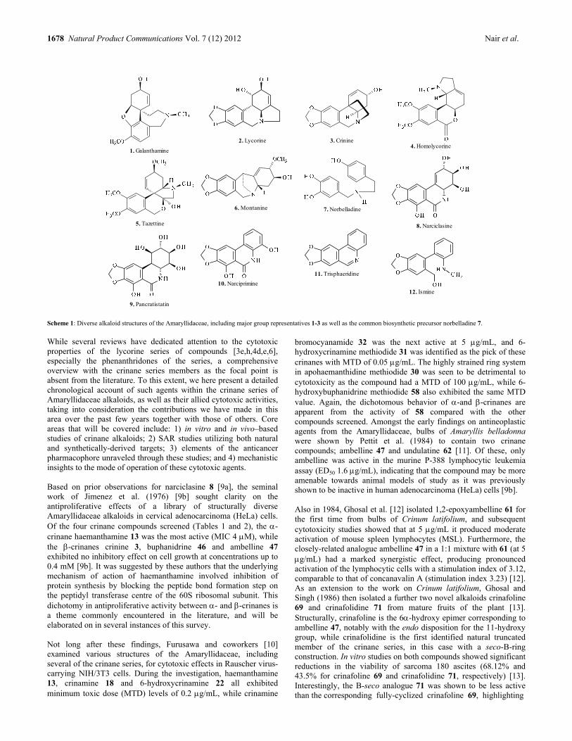
1678 Natural Product Communications Vol. 7 (12) 2012 Nair et al.
8. Narciclasine
9. Pancratistatin
10. Narciprimine
12. Ismine
11. Trisphaeridine
4. Homolycorine
5. Tazettine
6. Montanine
1. Galanthamine
7. Norbelladine
2. Lycorine 3. Crinine
Scheme 1: Diverse alkaloid structures of the Amaryllidaceae, including major group representatives 1-3 as well as the common biosynthetic precursor norbelladine 7.
While several reviews have dedicated attention to the cytotoxic properties of the lycorine series of compounds [3e,h,4d,e,6], especially the phenanthridones of the series, a comprehensive overview with the crinane series members as the focal point is absent from the literature. To this extent, we here present a detailed chronological account of such agents within the crinane series of Amaryllidaceae alkaloids, as well as their allied cytotoxic activities, taking into consideration the contributions we have made in this area over the past few years together with those of others. Core areas that will be covered include: 1) in vitro and in vivo–based studies of crinane alkaloids; 2) SAR studies utilizing both natural and synthetically-derived targets; 3) elements of the anticancer pharmacophore unraveled through these studies; and 4) mechanistic insights to the mode of operation of these cytotoxic agents. Based on prior observations for narciclasine 8 [9a], the seminal work of Jimenez et al. (1976) [9b] sought clarity on the antiproliferative effects of a library of structurally diverse Amaryllidaceae alkaloids in cervical adenocarcinoma (HeLa) cells. Of the four crinane compounds screened (Tables 1 and 2), the -crinane haemanthamine 13 was the most active (MIC 4 M), while the -crinanes crinine 3, buphanidrine 46 and ambelline 47 exhibited no inhibitory effect on cell growth at concentrations up to 0.4 mM [9b]. It was suggested by these authors that the underlying mechanism of action of haemanthamine involved inhibition of protein synthesis by blocking the peptide bond formation step on the peptidyl transferase centre of the 60S ribosomal subunit. This dichotomy in antiproliferative activity between - and -crinanes is a theme commonly encountered in the literature, and will be elaborated on in several instances of this survey. Not long after these findings, Furusawa and coworkers [10] examined various structures of the Amaryllidaceae, including several of the crinane series, for cytotoxic effects in Rauscher virus-carrying NIH/3T3 cells. During the investigation, haemanthamine 13, crinamine 18 and 6-hydroxycrinamine 22 all exhibited minimum toxic dose (MTD) levels of 0.2 g/mL, while crinamine
bromocyanamide 32 was the next active at 5 g/mL, and 6-hydroxycrinamine methiodide 31 was identified as the pick of these crinanes with MTD of 0.05 g/mL. The highly strained ring system in apohaemanthidine methiodide 30 was seen to be detrimental to cytotoxicity as the compound had a MTD of 100 g/mL, while 6-hydroxybuphanidrine methiodide 58 also exhibited the same MTD value. Again, the dichotomous behavior of -and -crinanes are apparent from the activity of 58 compared with the other compounds screened. Amongst the early findings on antineoplastic agents from the Amaryllidaceae, bulbs of Amaryllis belladonna were shown by Pettit et al. (1984) to contain two crinane compounds; ambelline 47 and undulatine 62 [11]. Of these, only ambelline was active in the murine P-388 lymphocytic leukemia assay (ED50 1.6 g/mL), indicating that the compound may be more amenable towards animal models of study as it was previously shown to be inactive in human adenocarcinoma (HeLa) cells [9b]. Also in 1984, Ghosal et al. [12] isolated 1,2-epoxyambelline 61 for the first time from bulbs of Crinum latifolium, and subsequent cytotoxicity studies showed that at 5 g/mL it produced moderate activation of mouse spleen lymphocytes (MSL). Furthermore, the closely-related analogue ambelline 47 in a 1:1 mixture with 61 (at 5 g/mL) had a marked synergistic effect, producing pronounced activation of the lymphocytic cells with a stimulation index of 3.12, comparable to that of concanavalin A (stimulation index 3.23) [12]. As an extension to the work on Crinum latifolium, Ghosal and Singh (1986) then isolated a further two novel alkaloids crinafoline 69 and crinafolidine 71 from mature fruits of the plant [13]. Structurally, crinafoline is the 6-hydroxy epimer corresponding to ambelline 47, notably with the endo disposition for the 11-hydroxy group, while crinafolidine is the first identified natural truncated member of the crinane series, in this case with a seco-B-ring construction. In vitro studies on both compounds showed significant reductions in the viability of sarcoma 180 ascites (68.12% and 43.5% for crinafoline 69 and crinafolidine 71, respectively) [13]. Interestingly, the B-seco analogue 71 was shown to be less active than the corresponding fully-cyclized crinafoline 69, highlighting

Cytotoxic potential of crinane alkaloids Natural Product Communications Vol. 7 (12) 2012 1679
A
B
C1
10a
10b11
12
42. Bulbispermine methiodide
6a
4a
6
3
4
2
7
9
8
10
36. R1=R2=H (Bulbispermine)37. R1=R2=Ac38. R1=R2= (CO)CH2CH3
39. R1=R2= (CO)NH2
40. R1=R2= (CO)NH(CO)CCl341. R1=4-Bromobenzoyl, R2=H
34. R1+R2=OCH2O (Delagoensine)35. R1=R2=OCH3 (Delagoenine)
43. 3-Oxobulbispermine 44. R1=R2=R3=R4=H45. R1=R4=Ac, R2=R3=OAc
-I
I -
30. Apohaemanthidine methiodide
I
31. 6-Hydroxycrinamine methiodide
-
32. Crinamine bromocyanamide 33. Phaedranamine
17. 11-Hydroxyvittatine N-oxide
27. R1=OCH3, R2=H (Dihydrocrinamine)28. R1=OH, R2=H (Dihydrohamayne)
29. Apohaemanthamine
25. R1=R3=H, R2=H, R4=CH3
(8-O-Demethylmaritidine)26. R1=R3=R4=CH3, R2=OH (Papyramine)
18. R1=CH3, R2=R3=H (Crinamine)19. R1=CH3, R2=Ac, R3=H (11-O-Acetylcrinamine)20. R1=R2=CH3, R3=H (11-O-Methylcrinamine)21. R1=CH3, R2=4-Methoxybenzoyl, R3=H22. R1=CH3, R2=H, R3=OH (6-Hydroxycrinamine)23. R1=R2=R3=H (Hamayne)24. R1=Ac, R2=R3=H (3-O-Acetylhamayne)
13. R1=CH3, R2=OH, R3=H (Haemanthamine)14. R1=CH3, R2=OH, R3=OH (Haemanthidine)15. R1=R2=R3=H (Vitattine)16. R1=H, R2=OH, R3=H (11-Hydroxyvitattine)
Scheme 2: Cytotoxic -crinane alkaloids of the Amaryllidaceae. the requirement of an intact phenanthridine nucleus for activity. Furthermore, quaternization of the nitrogen atom was seen to potentiate activity, as the semisynthetically-derived methochloride 70 was twice active as crinafoline 69, with 32.31% viability shown for the sarcoma cells [13]. This work is also significant in that it represents the first demonstration of in vivo effects of crinane alkaloids. In this regard, compounds 69-71 produced marked reductions in growth of sarcoma 180 ascites tumors in mice with cell count values of 10.04 x 10⁶, 5.08 x 10⁶ and 6.14 x 10⁶ shown for the three compounds respectively, compared to untreated control animals (15.88 x 10⁶) [13]. In vivo, quaternization of the nitrogen atom was also seen to be ameliorative towards activity (69 versus 70), and 70 was identified as the choice of this set of compounds. A few years later in 1991, Abd El Hafiz et al. [14] examined the constituents of Crinum augustum and Crinum bulbispermum for activity against human leukemic Molt 4 cells. Of the three crinanes (powelline 52, 6-hydroxycrinine 54 and 6-hydroxybuphanisine 55) tested, only 55 was moderately active, causing a steady decline (up to ~20%) in the viability of leukemia cells over the three-day treatment period at a dosage of 71 g/mL [14] (Table 2). Since 54 and 55 are differentiated by their C-3 substituents, it may be construed that a small hydrogen bond acceptor (such as the methoxyl in 55) as opposed to a donor-acceptor (hydroxyl) in this vicinity is essential for activity against Molt 4 cells. Substituent placement and geometry, as will become increasingly apparent during the course of this survey, have commonly been shown to have a distinctive influence on the cytotoxic abilities of crinane alkaloids.
The work of Likhitwitayawuid et al. (1993) stands out for the multi-cell line approach adopted by the authors in assessing the cytotoxic activities of alkaloid principles of Crinum amabile [15]. In the screen against ten cancers, including both human and animal cells, augustine 60 and crinamine 18 were prominent in most cases, with the best response (ED50 0.6 g/mL) observed in human oral epidermoid carcinoma KB cells and viblastine-resistant KB cells for the two compounds, respectively (Tables 1 and 2) [15]. Augustine 60 was noticeably selective against KB cells as opposed to vinblastine-resistant KB cells (ED50 > 20 g/mL). Buphanisine 53 and amabiline 65 were conspicuous in that they exhibited ED50 values > 20 g/mL in most of the cells tested [15]. The fact that - and -crinane representatives were here indistinguishable based on cytotoxic outcomes suggests that there may be other more subtle features, as will be encountered later in the manuscript, attending the crinane alkaloid anticancer pharmacophore. Soon after this, during the course of the phytochemical investigation of Hymenocallis expansa, Antoun et al. (1993) identified haemanthidine 14 as one of the cytotoxic constituents in the screen against eleven cancer cells [16]. Although the compound exhibited non-selective behavior, of note were the activities against prostate LNCaP (ED50 0.7 g/mL) and P-388 murine lymphocytic leukemia (ED50 0.4 g/mL) cells. Two years later in 1995, Lin et al. examined Hymenocallis littoralis and uncovered inter alia the presence of the crinane alkaloids crinine 3, haemanthamine 13 and 8-O-demethylmaritidine 25 [17]. Of these, only haemanthamine was screened for cytotoxic activity and found to be indiscriminate in a panel comprising eleven cell lines with ED50 values ranging
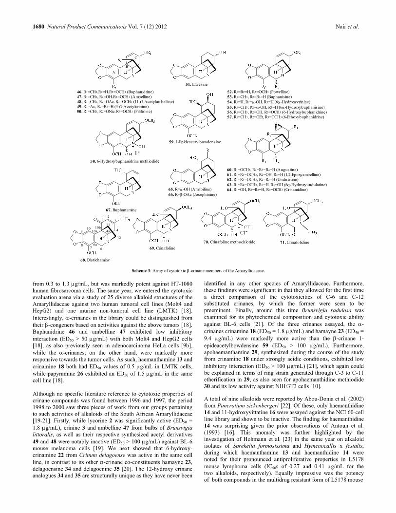
1680 Natural Product Communications Vol. 7 (12) 2012 Nair et al.
70. Crinafoline methochloride
-Cl
1
58. 6-Hydroxybuphanidrine methiodide
2
68. Distichamine
I -
67. Buphanamine
3
60. R1=OCH3, R2=R3=R4=H (Augustine)61. R1=R4=OCH3, R2=OH, R3=H (1,2-Epoxyambelline)62. R1=R4=OCH3, R2=R3=H (Undulatine)63. R1=R4=OCH3, R2=H, R3=OH (6-Hydroxyundulatine)64. R1=OH, R2=R3=H, R4=OCH3 (Crinamidine)
69. Crinafoline
4
71. Crinafolidine
4a
6
51. Elwesine
7
59. 1-Epideacetylbowdensine
8
65. R=-OH (Amabiline)66. R=-OAc (Josephinine)
9
52. R1=R2=H, R3=OCH3 (Powelline)53. R1=CH3, R2=R3=H (Buphanisine)54. R1=H, R2=-OH, R3=H (6-Hydroxycrinine)55. R1=CH3, R2=-OH, R3=H (6-Hydroxybuphanisine)56. R1=CH3, R2=OH, R3=OCH3 (6-Hydroxybuphanidrine)57. R1=CH3, R2=OEt, R3=OCH3 (6-Ethoxybuphanidrine)
10
A
10a10b
11
12
6a
C
B
46. R1=CH3,R2=H, R3=OCH3 (Buphanidrine)47. R1=CH3, R2=OH, R3=OCH3 (Ambelline)48. R1=CH3, R2=OAc, R3=OCH3 (11-O-Acetylambelline)49. R1=Ac, R2=R3=H (3-O-Acetylcrinine)50. R1=CH3, R2=ONic, R3=OCH3 (Filifoline)
Scheme 3: Array of cytotoxic -crinane members of the Amaryllidaceae.
from 0.3 to 1.3 g/mL, but was markedly potent against HT-1080 human fibrosarcoma cells. The same year, we entered the cytotoxic evaluation arena via a study of 25 diverse alkaloid structures of the Amaryllidaceae against two human tumoral cell lines (Molt4 and HepG2) and one murine non-tumoral cell line (LMTK) [18]. Interestingly, -crinanes in the library could be distinguished from their -congeners based on activities against the above tumors [18]. Buphanidrine 46 and ambelline 47 exhibited low inhibitory interaction (ED50 > 50 g/mL) with both Molt4 and HepG2 cells [18], as also previously seen in adenocarcinoma HeLa cells [9b], while the -crinanes, on the other hand, were markedly more responsive towards the tumor cells. As such, haemanthamine 13 and crinamine 18 both had ED50 values of 0.5 g/mL in LMTK cells, while papyramine 26 exhibited an ED50 of 1.5 g/mL in the same cell line [18]. Although no specific literature reference to cytotoxic properties of crinane compounds was found between 1996 and 1997, the period 1998 to 2000 saw three pieces of work from our groups pertaining to such activities of alkaloids of the South African Amaryllidaceae [19-21]. Firstly, while lycorine 2 was significantly active (ED50 = 1.8 g/mL), crinine 3 and ambelline 47 from bulbs of Brunsvigia littoralis, as well as their respective synthesized acetyl derivatives 49 and 48 were notably inactive (ED50 > 100 g/mL) against BL-6 mouse melanoma cells [19]. We next showed that 6-hydroxy-crinamine 22 from Crinum delagoense was active in the same cell line, in contrast to its other -crinane co-constituents hamayne 23, delagoensine 34 and delagoenine 35 [20]. The 12-hydroxy crinane analogues 34 and 35 are structurally unique as they have never been
identified in any other species of Amaryllidaceae. Furthermore, these findings were significant in that they allowed for the first time a direct comparison of the cytotoxicities of C-6 and C-12 substituted crinanes, by which the former were seen to be preeminent. Finally, around this time Brunsvigia radulosa was examined for its phytochemical composition and cytotoxic ability against BL-6 cells [21]. Of the three crinanes assayed, the -crinanes crinamine 18 (ED50 = 1.8 g/mL) and hamayne 23 (ED50 = 9.4 g/mL) were markedly more active than the -crinane 1-epideacetylbowdensine 59 (ED50 > 100 g/mL). Furthermore, apohaemanthamine 29, synthesized during the course of the study from crinamine 18 under strongly acidic conditions, exhibited low inhibitory interaction (ED50 > 100 g/mL) [21], which again could be explained in terms of ring strain generated through C-3 to C-11 etherification in 29, as also seen for apohaemanthidine methiodide 30 and its low activity against NIH/3T3 cells [10]. A total of nine alkaloids were reported by Abou-Donia et al. (2002) from Pancratium sickenbergeri [22]. Of these, only haemanthidine 14 and 11-hydroxyvittatine 16 were assayed against the NCI 60-cell line library and shown to be inactive. The finding for haemanthidine 14 was surprising given the prior observations of Antoun et al. (1993) [16]. This anomaly was further highlighted by the investigation of Hohmann et al. [23] in the same year on alkaloid isolates of Sprekelia formosissima and Hymenocallis x festalis, during which haemanthamine 13 and haemanthidine 14 were noted for their pronounced antiproliferative properties in L5178 mouse lymphoma cells (IC50s of 0.27 and 0.41 g/mL for the two alkaloids, respectively). Equally impressive was the potency of both compounds in the multidrug resistant form of L5178 mouse

Cytotoxic potential of crinane alkaloids Natural Product Communications Vol. 7 (12) 2012 1681
Table 1: Activities of -crinane alkaloid representatives of the Amaryllidaceae in miscellaneous cancer cell line screens.
Compound No. Cell line (Cytotoxicity index) a Reference
L6 BL6 KB A549 OE21 Hs683 U373 SKMEL B16F10 Jurkat HeLa Vero LOVO 6T-CEM HL-60 HT29 H460 RXF393 5123tc 3T3
Haemanthamine 13 7.41 b 0.7 b 4.5 6.8 7.0 3.5 b 8.5 6.8 22 d 4 32 c active 15 0.2 b 3e,f,9b,10,17,26,31,
33,35,36,38
Haemanthidine 14 3.6 b 4.0 3.7 4.3 3.8 4.2 3.1 nd active 16,31,35,36
Vittatine 15 70 d 21.91 15.88 29.57 27,29
11-Hydroxyvittatine 16 4 d 72 c 64 c 3e,f
17 >90 b >50 b 33
Crinamine 18 1.8 b 1.0 b 15.9 b 0.9 b 4.30 b 2.82 b 1.70 b 12.5 0.2 b 10,15,21,26,28
11-Acetylcrinamine 19 na 26
11-Methylcrinamine 20 9.15 b 81.0 b 14.4 b 6.16 b 28
21 na 26
6-Hydroxycrinamine 22 active 0.2 b 10,20
Hamayne 23 9.4 b na 21,29
3-O-Acetylhamayne 24 15.8 b 5.49 b 3.45 b 4.27 b 28
25 >90 b >50 b 4 d 82 c 82 c 3e,f,33
Dihydrocrinamine 27 na 29
Dihydrohamayne 28 active 29
Apohaemanthamine 29 >100 b 21
30 100 b 10
31 0.05 b 10
32 5 b 10
Phaedranamine 33 na 32
Delagoensine 34 na 20
Delagoenine 35 na 20
Bulbispermine 36 11 38 8 36
37 63 nd 90 36
38 >100 nd >100 36
39 50 >100 46 36
40 >100 nd >100 36
41 >100 nd >100 36
42 >100 nd >100 36
3-Oxobulbispermine 43 >100 nd >100 36
44 >100 nd >100 36
45 >100 nd >100 36
BCA-
1
HT-
1080
LUC-
1
MEL-
2
COL-
1
KB-
V1
P-388 A-431 LNCaP ZR-
75-1
L5178 L5178mdr Molt4 LMTK HepG2 COL-2 BC1 HT Lu1 HSC-
2
Haemanthamine 13 0.7 b 0.3 b 3.6 b 1.3 b 5.0 b 1.3 b 0.6 b 0.5 b 0.27 b 0.30 b 1.2 b 0.5 b >50 b 0.6 b active 17,18,23,35
Haemanthidine 14 1.9 b 8.4 b 0.4 b 3.0 b 0.7 b 2.6 b 0.41 b 0.40 b 6.0 b >20 b 1.6 b 2.1 b active 16,23,35
Crinamine 18 1.4 b 1.3 b 1.4 b 5.0 b 1.0 b 0.6 b 0.7 b 6.9 b 1.5 b 0.8 b 0.5 b 0.5 b 10 b 15,18
Papyramine 26 15.8 b 1.5 b 17 b 18
T98G U87 CEM K562 MCF-
7
G-361 SH-SY5Y
Haemanthamine 13 8 6 2.1 3.4 8.1 3.7 36,38
Haemanthidine 14 14 6 36
6-Hydroxycrinamine 22 54.5 37
Bulbispermine 36 9 9 36
37 98 74 36
38 >100 >100 36
39 91 15 36
40 >100 >100 36
41 >100 >100 36
42 >100 >100 36
3-Oxobulbispermine 43 >100 >100 36
44 >100 >100 36
45 >100 >100 36 a Cytoxicity index values expressed as ED50, GI50, IC50, MIC or MTD50 in micromolar (M) units unless otherwise stated. b Values are in g/mL. c Indicates % cell viability. d
Indicates % apoptosis. For compounds indicated as “active”, no cytotoxicity indices were presented in the original work (na=not active, nd=not detected).
lymphoma cells, exhibiting IC50 values of 0.3 and 0.40 g/mL, respectively [23] (Table 1). Around this time, findings by Phan et al. (2003) revealed that while lycorine 2 was strongly active on HepG2,
RD and FL cells, no such activity was detectable for the -crinanes crinine 3 and crinamidine 64 isolated from the Vietnamese species Crinum asiaticum and Crinum latifolium [24]. Crinamidine is the

1682 Natural Product Communications Vol. 7 (12) 2012 Nair et al.
6361 47 13 65 66 3 18 C
20
40
60
80
100
Crinane alkaloids screened for apoptosis induction
Figure 1: Apoptotic indices determined in rat hepatoma (5123tc) cells showing the efficacy of crinamine 18 and haemanthamine 13 at 25 �M after a 48 h treatment (after McNulty et al., 2007).26
Figure 2: Hoechst staining of 5123tc cells 72 h after treatment with various concentrations of crinamine 18. Apoptotic nuclei appear brighter and more fragmented than healthy nuclei (seen in control cells) (after McNulty et al., 2007).26
3-hydroxy analogue corresponding to undulatine 62, previously shown to be inactive in the P-388 cell line [11]. Apart from this, during the phytochemical investigation of Nerine filifolia, a species of the Amaryllidaceae endemic to eastern and southeastern regions of South Africa, we identified several crinanes of the -series, including 6-hydroxybuphanidrine, 6-methoxybuphanidrine, ambelline, 11-O-acetylambelline and filifoline [25]. Of these, only filifoline 50 was screened against rat myoblast (L6) cells and shown to be inactive [25]. The year 2007 is notable in the Amaryllidaceae alkaloid cytotoxicity calendar as it saw the first demonstration of apoptotic effects for crinane-based alkaloid structures [26]. In this regard, we examined a mini-library comprising natural and semi-synthetic derivatives of both series of crinane compounds for apoptosis-inducing effects in rat hepatoma (5123tc) cells [26]. As shown in Figure 1, the -crinanes haemanthamine 13 and crinamine 18 were prominent amongst the other test substrates, with apoptotic indices of 90% and 95%, respectively, after a 48 h treatment at 25 M [26]. Apoptosis induction was indicated by Hoechst staining as shown in Figure 2, by which apoptotic nuclei appeared brighter and more fragmented than healthy nuclei, in this case upon treatment with crinamine 18. Furthermore, respective ED50 values were established at 15 M and 12.5 M for haemanthamine and crinamine (Figure 3) [26]. Of the other compounds screened, only the activity of 6-hydroxyundulatine 63 was noteworthy (25% apoptotic at the same concentration and treatment period) [26]. These results were even more impressive considering the selectivities of haemanthamine 13 and crinamine 18, both of which were seen to exhibit negligible
apoptotic effects in normal human embryonic kidney (293t) cells (Figure 4). Apart from uncovering these novel effects in hepatoma cells, this work was also significant in shedding light on the underlying structural elements of the pharmacophore resident within these crinanes [26]. These were seen to include: 1) -orientation for the 5,10b-ethano bridge, which renders the B,C-ring junction conformationally similar to the phenanthridone drug target pancratistatin 9. As such, the -crinanes crinamine and haemanthamine were clearly far superior to the other -crinanes screened; 2) the stereochemistry at C-3 may not be significant given the relative configurations of the methoxyl group in crinamine and haemanthamine; and 3) requirement of a free hydroxyl at the C-11 position, the function of which may involve hydrogen bond donor-acceptor interactions, as acyl-derivatives (acetate and benzoate) were notably inactive [26]. However, as will be discussed later, the apoptosis-inducing pharmacophore is shown to incorporate other essential structural ingredients. The only work in 2008 on cytotoxicity studies of crinanes was that of Silva et al. [27] in which vittatine 15 from Hippeastrum vittatum was shown to be active against HT29, H460 and RXF393 cells with IC50 values of 21.91, 15.88 and 29.57 g/mL in the three cells, respectively. Interestingly, the dichloromethane and butanol extracts of the plant were markedly more active than neat vittatine 15 in all three cell lines. For example, the best IC50 value for the butanol extract was ascertained in RXF393 cells (2.93 g/mL), while that for the dichloromethane extract was 0.62 g/mL in H460 cells. The year 2009 saw a flurry of activity in the cytotoxicity field pertaining to crinane alkaloids. The first two related to major works involving a diverse Amaryllidaceae alkaloid library screen against HeLa, Vero and Jurkat cells [3e,f]. Of the crinane alkaloids evaluated (Tables 1 and 2), including 11-hydroxyvittatine 16, 8-O-demethylmaritidine 25, ambelline 47, buphanamine 67 and buphanisine 53, only haemanthamine 13 stood out for significantly reducing the viability of HeLa and Vero cells to 21% and 32%, respectively, over a 24 h period at a treatment dose of 25 M [3e,f]. Furthermore, amongst these crinanes, only haemanthamine 13 was capable of apoptosis induction in Jurkat cells (22% over the same period and dosage) [3e,f]. Another important aspect of these investigations was the discovery that buphanamine 67 was the most potent of the twenty-nine compound library in the collagen type 1 invasion assay [3e,f]. At 25 M it produced only ~2% invasion of collagen type 1 by HeLa cells [3e,f], and given its low cytotoxicity, even at relatively high dosages, it may play a significant future chemoprotective role in combination with the phenanthridone anticancer drug targets narciclasine 8 and pancratistatin 9. The work of Sun et al. (2009) on the chemical constituents of Crinum asiaticum from Yunnan province of China revealed inter alia the presence of the -crinanes crinamine 18, 11-O-methylcrinamine 20 and 3-O-acetylhamayne 24 [28]. Subsequent cytotoxicity studies in four cell lines (A549, LOVO, 6T-CEM and HL-60) showed, as expected, crinamine to be superior to the other functionalized analogues [28]. For comparison purposes, in the HL-60 cell line IC50 values determined were 1.70, 6.16 and 4.27 g/mL for the three compounds, respectively [28]. As mentioned above, and again seen here for this crinamine subset, the direct influence of substituents on the cytotoxic ability of crinane alkaloids is quite pronounced, and should thus guide SAR studies aimed at identifying a possible clinical candidate. Finally, the year closed out with further apoptotic studies by our research group on selected
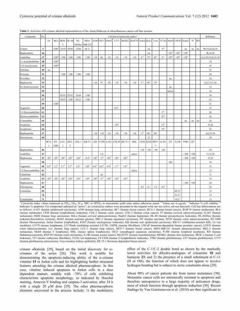
Cytotoxic potential of crinane alkaloids Natural Product Communications Vol. 7 (12) 2012 1683
Table 2: Activities of -crinane alkaloid representatives of the Amaryllidaceae in miscellaneous cancer cell line screens.
Compound No. Cell line (Cytotoxicity index) a Reference
L6 BL6 SKW-
3
HL-60 HL-
60/Dox
MDA-
MB-231
A549 OE21 Hs683 U373 SKMEL B16F10 Jurkat HeLa Vero 5123tc Molt4 LMTK HepG2 FI RD
Crinine 3 >100 b 16.95 20.86 14.04 68.11 na 9 d na na na 9b,19,24,26,34
Buphanidrine 46 na >50 b >50 b >50 b 9b,18,38
Ambelline 47 >100 b >100 >100 >100 >100 >10 86 >10 >10 >10 >10 4 d 79 c 89 c 8 d >50 b >50 b >50 b 3e,f,18,19,26,31,34
11-Acetylambelline 48 >100 b 19
3-O-Acetylcrinine 49 >100 b 19
Filifoline 50 na 25
Elwesine 51 >100 >100 >100 >100 34
Powelline 52 na 14
Buphanisine 53 >10 97 >10 >10 >10 >10 5 d 99 c 79 c 3e,f,15,31,36
6-Hydroxycrinine 54 na 14
55 active 14
56 45.03 95.95 76.69 >100 34
57 59.43 >100 95.22 >100 34
59 >100 b 21
Augustine 60 0.6 b 15
1,2-Epoxyambelline 61 5 d 26
Hydroxyundulatine 63 25 d 26
Crinamidine 64 na na na 24
Amabiline 65 >20 b 17 d 15,26
Josephinine 66 18 d 26
Buphanamine 67 >10 >10 >10 >10 >10 >10 3 d 96 c 90 c 3e,f,31,36
Distichamine 68 2.2 38
BCA-
1
HT-
1080
LUC-
1
MEL-
2
COL-1 KB-V1 KB P-388 A-431 LNCaP ZR-75-1 MSL CEM K562 MCF-
7
G-361 3T3 S-180 T98G U87
Buphanidrine 46 >50 >50 >50 >50 38
Ambelline 47 1.6 active >100 >100 11,12,36
Buphanisine 53 >20 b >20 b >20 b >20 b >20 b 19.8 b >20 b >5 b >20 b >20 b >20 b >100 >100 15,36
58 100 10
Augustine 60 2.8 b 1.2 b 3.7 b 3.2 b 2.4 b >20 b 0.6 b 0.6 b 4.9 b 1.7 b 1.8 b 15
1,2-Epoxyambelline 61 active 12
Undulatine 62 na 11
Amabiline 65 >20 b >20 b >20 b >20 b >20 b >20 b >20 b >5 b >20 b >20 b >20 b 15
Buphanamine 67 >100 >100 36
Distichamine 68 4.5 4.1 2.3 14.7 38
Crinafoline 69 68.12 c 13
70 32.31 c 13
Crinafolidine 71 43.5 c 13 a Cytoxicity index values expressed as ED50, GI50, IC50, MIC or MTD50 in micromolar (M) units unless otherwise stated. b Values are in g/mL. c Indicates % cell viability. d
Indicates % apoptosis. For compounds indicated as “active”, no cytotoxicity indices were presented in the original work (na=not active, nd=not detected). Cell line abbreviations are as follows: A-431 (human epidermoid carcinoma), A549 (human lung carcinoma), BC1 (human breast cancer), BCA-1 (human breast cancer), B16F10 (mouse melanoma), BL6 (mouse melanoma), CEM (human lymphoblastic leukemia), COL-1 (human colon cancer), COL-2 (human colon cancer), FI (human cervical adenocarcinoma), G-361 (human melanoma), H460 (human lung carcinoma), HeLa (human cervical adenocarcinoma), HepG2 (human hepatoma), HL-60 (human promyelocytic leukemia), HL-60/Dox (human leukemia/doxorubicin resistant), Hs683 (human neuronal glioma), HSC-2 (human squamous carcinoma), HT (human sarcoma), HT29 (human colon adenocarcinoma), HT-1080 (human fibrosarcoma), Jurkat (human lymphoblast), K562 (human myelogenous leukemia), KB (human oral epidermoid carcinoma), KB-V1 (vinblastine-resistant KB), L6 (rat myoblast), L5178 (mouse lymphoma), L5178mdr (multidrug resistant L5178), LMTK (murine fibroblast), LNCaP (hormone-dependent human prostatic cancer), LOVO (human colon adenocarcinoma), Lu1 (human lung cancer), LUC-1 (human lung cancer), MCF-7 (human breast cancer), MDA-MB-231 (human adenocarcinoma), MEL-2 (human melanoma), Molt4 (human T lymphoma), MSL (mouse spleen lymphocyte), OE21 (oesophageal squamous carcinoma), P-388 (murine lymphoid neoplasm), RD (human rhabdomyosarcoma), RXF393 (human renal carcinoma), S-180 (mouse ascites tumor), SH-SY5Y (human neurobalstoma), SKMEL (human skin melanoma), SKW-3 (human T-cell leukemia), 3T3 (mouse embryonic fibroblast), 5123tc (rat hepatoma), 6T-CEM (human T-lymphoblastic leukemia), T98G (human glioblastoma), U87 (human glioblastoma), U373 (human glioblastoma astrocytoma), Vero (monkey kidney epithelial), ZR-75-1 (hormone-dependent breast cancer).
crinane alkaloids [29], based on the initial discovery for -crinanes of the series [26]. This work is notable for demonstrating the apoptosis-inducing ability of the -crinane vittatine 15 in Jurkat cells and for highlighting further structural features attending the crinane alkaloid pharmacophore. In this case, vittatine induced apoptosis in Jurkat cells in a dose dependent manner, notably with ~70% of cells exhibiting characteristic apoptotic morphology, as indicated by Hoechst staining, Annexin-V binding and caspase-3 activation, after 24 h with a single 20 M dose [29]. The other pharmacophoric elements uncovered in the process include: 1) the modulatory
effect of the C-1/C-2 double bond as shown by the markedly lower activities for dihydro-analogues of crinamine 18 and hamayne 23; and 2) the presence of a small substituent at C-11 (H or OH), the function of which does not appear to involve hydrogen bonding but is subject to steric constraints alone [29]. About 90% of cancer patients die from tumor metastases [30]. Metastatic cancer cells are intrinsically resistant to apoptosis and therefore unresponsive to a large majority of anticancer drugs, most of which function through apoptosis induction [30]. Recent findings by Van Goietsenoven et al. (2010) are thus significant in

1684 Natural Product Communications Vol. 7 (12) 2012 Nair et al.
focusing on the cytotoxic abilities of diverse Amaryllidaceae alkaloids in apoptosis-resistant cancer cells [31]. Of the five crinane alkaloids evaluated, ambelline 47, buphanamine 67 and buphanisine 53 exhibited low activities against both apoptosis-resistant (A549, OE21, U373 and SKMEL) and apoptosis-sensitive (Hs683 and B16F10) cells [31]. On the other hand, haemanthamine 13 and haemanthidine 14 were markedly active in both cell-forms (IC50s 3.1- 8.5 M) with the best activity seen for the latter in B16F10 cells (IC50 = 3.1 M) [31]. Also in 2010, phaedranamine 33, the enantiomer of buphanamine, was isolated for the first time from the South American Amaryllidaceae species Phaedranassa dubia and shown to be inactive towards L6 cells (IC50 > 300 M) [32]. In 2011, two further Amaryllidaceous species (Galanthus trojanus and Crinum zeylanicum) were examined by our group for cytotoxic effects in various cell lines [33,34]. Firstly, 11-hydroxyvittatine-N-oxide 17 and 8-O-demethylmaritidine 25 from Galanthus trojanus were both weakly active in L6 cells (IC50s > 90 g/mL) and KB cells (IC50s > 50 g/mL) in contrast to the co-constituent haemanthamine which exhibited IC50s of 7.41 and 0.97 g/mL in the two cell lines, respectively [33]. The result for 8-O-demethylmaritidine is in accordance with previous findings by Lin et al. (1995), who showed that the compound was unresponsive in a multi-cell line screen [17], and thus emphasizes the significance of the A-ring attached 1,3-dioxolane moiety to the crinane anticancer pharmacophore. Secondly, bulbs of Crinum zeylanicum were shown to contain inter alia the crinane alkaloids crinine 3, ambelline 47, elwesine 51, 6-hydroxybuphanidrine 56 and 6-ethoxybuphanidrine 57, all of the -subgroup [34] (Table 2). In the four cell lines screened, only crinine was notably active with IC50s of 16.95, 20.86, 14.01 and 68.11 M in SKW-3, HL-60, HI-60/Dox and MDA-MB-231 cells, respectively [34]. The role of the double bond to the integrity of the crinane pharmacophore could be gauged directly by comparison of these responses to those of elwesine 51, the dihydro-analogue of crinine, which exhibited IC50s > 100 M across the four cells [34]. Further mechanistic tests on the active compound crinine via oligonucleosomal DNA fragmentation revealed the apoptotic cell death mode as responsible for its cyto-toxicity [34]. Apart from this, Jitsuno et al. (2011) showed that haemanthamine 13 and haemanthidine 14 from Lycoris albiflora were active against HL-60 and HSC-2 cells (Table 1) [35]. The year 2012 has already seen three detailed studies of the cytotoxic properties of crinane alkaloids [36-38]. Firstly, Luchetti et al. (2012) uncovered the cytotoxic ability of bulbispermine 36, the 11-endo analogue corresponding to hamayne 23, in various cells including apopotosis-resistant and apoptosis-sensitive forms [36]. For example, in the apoptosis-resistant line T98G the compound had a GI50 of 9 M, matched closely by its activity in apoptosis-sensitive HeLa cells (GI50 of 8 M) [36]. Further results showed as expected, the prominence of haemanthamine 13 and haemanthidine 14, as well as the low activity of buphanamine 67, buphanisine 53 and ambelline 47 in these cells [36]. SAR studies revealed that the semi-synthetic transformation products (37-45) of bulbispermine, with the exception of 39 (GI50 15 M in U87 cells), were weakly active (GI50s > 100 M) in most test screens. Secondly, work by Adewusi et al. (2012) on the South African amaryllid Boophone disticha uncovered 6-hydroxycrinamine 22 as the cytotoxic agent in neuroblastoma (SH-SY5Y) cells with IC50s of 54.5 and 61.7 M, as determined by the MTT and neutral red assays, respectively [37]. Finally, we
100
80
60
40
20
C 5uM 10uM 15uM 20uM
Crinamine 18Haemanthamine 13
% A
po
pto
sis
Alkaloid concentration
control 5 M 10 M 15 M 20 M
Figure 3: Apoptosis responses in 5123tc cells treated over 72 h with various concentrations of crinamine and haemanthamine. Effective dose (ED50) was calculated as the concentration inducing apoptosis in 50% of cells (after McNulty et al., 2007).26
100
80
60
40
20
C 5uM 10uM 15uM 20uM
Crinamine 18
Haemanthamine 13
% A
po
pto
sis
control 5 M 10 M 15 M 20 M
Alkaloid concentration Figure 4: Normal human embryonic kidney (293t) cells are unaffected by treatment with various doses of crinamine and haemanthamine after 72 h (after McNulty et al., 2007).26
very recently revealed the antiproliferative properties of the rare Amaryllidaceae crinane constituent distichamine 68 in a mini-panel of cancer cells comprising acute lymphoblastic leukemia (CEM), chronic myelogenous leukemia (K562), breast adenocarcinoma (MCF7), malignant melanoma (G-361) and cervical adenocarcinoma (HeLa), in addition to the normal human fibroblast (BJ) cell line [38]. By means of the Calcein AM assay, distichamine 68 was seen to be active against all cancer cell lines (IC50s 2.2-14.7 M), with the adherent cell line HeLa shown to be the most sensitive (IC50 2.2 M) to this alkaloid [38]. Mechanistic studies then invoked flow cytometry to quantify the distribution of CEM cells across the different phases of the cell cycle, and to determine the sub-G1 fraction as a measure of the proportion of apoptotic cells [38]. The analysis showed that treatment with distichamine 68 increased the proportion of G2/M phase cells in a dose-dependent manner, with concomitant reductions in the proportion of G0/G1 and S cells (Figure 5) [38]. In addition, the proportion of cells with sub-G1 amounts of DNA (apoptotic cells) increased (up to 23.7 %) following a 24 h

Cytotoxic potential of crinane alkaloids Natural Product Communications Vol. 7 (12) 2012 1685
Concentration of alkaloid (M)
Nu
mb
er
of
ce
lls (
%)
Figure 5: Histograms obtained by flow cytometric analysis showing the distributions of CEM cells across the G0/G1, S and G2/M phases of the cell cycle after 24 h treatment with distichamine 68, and the sub-G1 fraction of cells (relative to untreated controls). The bars show the percentage of cells in the corresponding phase (after Nair et al., 2012).38
treatment with 68, relative to that observed in untreated cells, indicating that the compound was clearly capable of cell cycle disturbance and apoptosis induction in CEM cells [38]. Furthermore, distichamine induced a 12.5-fold increase in the activity of caspase-3/7 after 24 h at the highest concentration (20 M) compared to untreated controls (Figure 6). In addition, Western blot analysis was used to detect changes in expression of apoptosis-related proteins in CEM leukemia cells after treatment with distichamine. As shown in Figure 7, treatment with 68 (at 10 and 20 µM) induced cleavage of PARP (89 kDa fragment) after 24 h which also corresponded with decreased levels of procaspase-3. As described above, increased caspase-3 activity was observed over the same treatment period and dosage (Figure 6). Expression of the tumor suppressor protein p53 was discernible in the CEM cell line control, and 68 caused its enhanced expression after 24 h, notably at 10 and 20 µM (Figure 7). No change in the expression of the antiapoptotic protein Bcl-2 was observed, but at 20 µM, a decreased level of the antiapoptotic protein Mcl-1 was detected, indicating the onset of apoptosis. Mcl-1 is necessary for cell viability, and its decreased expression has been suggested as the cause of cell death in CEM cells [39a]. It also plays a crucial role in regulating the apoptosis of T-cells [39b]. Western blot analysis here demonstrated the dose-dependent decrease of procaspase-3 and cleavage of PARP after 24 h treatment with distichamine 68 in CEM leukemia cells (Figure 7), indicating that the compound induces caspase-3 activated apoptosis [38]. Given the unique C-ring structural formulation for distichamine, involving shift of the double bond to C-2/C-3 with concomitant vinylization of the C-3 methoxy group and oxidation at C-1, these findings point at further attendant structural features of the crinane apoptosis-inducing pharmacophore. It now appears that a sp2–hybridised C-3 centre is tolerated at the active site, since most other cytotoxic crinane alkaloids possess a C-1/C-2 double bond. Furthermore, oxygenation at C-1, as is characteristic of the anticancer phenanthridone pancratistatin 9, was here shown to be amenable towards activity. Interestingly, in the case of distichamine, a keto-group is positioned at C-1, as a consequence of which three contiguous sp2–centres are formed in ring-C, similar to that seen for the active phenanthridone narciprimine 10 [38].
Concentration of alkaloid (M)
Re
lati
ve
ca
sp
as
ea
cti
vit
y
Figure 6: Increases in the activities of caspase-3 and 7 in CEM cells treated with distichamine 68 for 24 h, relative to untreated controls. The data shown represent averages from at least three independent experiments performed in triplicate (after Nair et al., 2012).38
Figure 7: Western blot analysis of apoptosis-related proteins (PARP fragment 89 kDa, Rb, p53, Mcl-1, Bcl-2 and procaspase 3) in leukemia (CEM) cells treated with distichamine 68 compared to their expression in untreated control cells. The expression of -actin was monitored as a protein loading control (after Nair et al., 2012).38 In summary, a more comprehensive view has emerged of crinane alkaloids of the Amaryllidaceae as potent and selective cytotoxic agents with significant potential for preclinical development. This promising status was drawn out of multi-cell line approaches invoking targets of synthetic as well as natural origin. As such, both in vitro and in vivo models of study support the ameliorative effects of crinane compounds as anticancer agents. This survey has accounted for 54 cancer cell lines (Tables 1 and 2), including human and animal forms, against which the cytotoxic abilities of a total of 71 crinane alkaloids have been measured. As also shown in Tables 1 and 2, of the -series, not surprisingly, haemanthamine 13, haemanthidine 14 and crinamine 18 are the most commonly targeted agents in such studies. Within the -series, ambelline 47 and buphanisine 53 appear routinely across most of the cell lines screened. In terms of potency, synthetic 6-hydroxycrinamine methiodide 31 is the most active crinane identified to date with a MTD of 0.05 g/mL in NIH/3T3 cells. In animal models of study, the natural -crinanes, haemanthamine 13, crinamine 18 and 6-hydroxycrinamine 22 were the most active each with MTD of 0.2 g/mL in NIH/3T3 cells. In human cancers, haemanthamine was again the most

1686 Natural Product Communications Vol. 7 (12) 2012 Nair et al.
potent compound with an ED50 of 0.3 g/mL in HT-1080 fibrosarcoma cells. Furthermore, selective cytotoxic abilities have been demonstrated for crinamine and haemanthamine, both of which, for example, targeted rat hepatoma (5123tc) cells as opposed to normal human embryonic kidney (293t) cells. Of the -crinanes evaluated, augustine 60 was the most active with an ED50 of 0.6 g/mL in both KB (human oral epidermoid carcinoma) and U373 (human glioblastoma astrocytoma) cells. Given the close structural proximity of these alkaloids to the potent phenanthridone anticancer agents of the family, such as narciclasine and pancratistatin, as well as their common biogenetic origin, it is possible that a common, though still elusive, biological target may be operable for these agents. Stucture-activity relationship studies, again involving natural as well as synthetically-derived targets, have provided useful insights to the structural details of the anticancer pharmacophore attending these molecules. Features that have come to light from such studies include: 1) an intact phenanthridine nucleus is essential since truncated analogues were markedly less active, as seen for crinafoline 69 against crinafolidine 71; 2) presence of the A-ring methylenedioxyphenyl moiety since structures incorporating other substituents (as in 8-O-demethylmaritidine 25) were seen to be less active; 3) B-ring modification effects such as quaternization of the nitrogen atom were shown to be
deleterious; 4) small substituents with hydrogen bond donor/acceptor capability at C-11 ( to the nitrogen atom) with either exo- or endo-disposition were ameliorative (as in crinamine 18 and bulbispermine 36, respectively), but less so for substitutions to the nitrogen atom at C-6 (6-hydroxycrinamine 22) or C-12 (delagoensine 34); 5) stereochemistry at C-3 may not be significant given the relative configurations of the methoxyl group in crinamine 18 and haemanthamine 13, as well as its planar geometry in distichamine 68; 6) additional oxygenation of ring-A, reminiscent of the potent anticancer phenanthridones narcicalsine 8 and pancratistatin 9, is viewed as beneficial to the pharmacophore, as shown for distichamine 68; and 7) although active compounds were found across both - and -members of the series, the geometry of the 5,10b-ethano bridge appears to play a significant role given the consistent potency of the former series across most of the cell lines screened. In addition, mechanistic studies have recently uncovered the apoptotic mode of death as responsible for the cytotoxicity of several crinane compounds. These distinctive features should prove useful in guiding the development of a clinical drug target. As such, efforts to further refine the active pharmacophore associated with these molecules are an ongoing concern in our laboratories. Acknowledgment - Financial support of this work by the University of KwaZulu-Natal is gratefully acknowledged.
References
[1] (a) Bastida J, Lavilla R, Viladomat F. (2006) Chemical and biological aspects of Narcissus alkaloids. In The Alkaloids. Cordell GA (Ed). Elsevier, Amsterdam, Netherlands, 87-179; (b) Bastida J, Berkov S, Torras L, Pigni NB, De Andrade JP, Martinez V, Codina C, Viladomat F. (2011) Chemical and biological aspects of Amaryllidaceae alkaloids. In Recent Advances in Pharmaceutical Sciences. Muňoz-Torrero D (Ed). Transworld Research Network, Trivandrum, India, 65-100; (c) Jin Z. (2011) Amaryllidaceae and Sceletium alkaloids. Natural Product Reports, 28, 1126-1142; (d) Viladomat F, Bastida J, Codina C, Nair JJ, Campbell WE. (1997) Alkaloids of the South African Amaryllidaceae. In Recent Research Developments in Phytochemistry. Vol. 1, Pandali SG (Ed). Research Signpost Publishers, Trivandrum, India. 131- 171.
[2] (a) Harvey AL. (1995) The pharmacology of galanthamine and its analogues. Pharmacology & Therapeutics, 68, 113-128; (b) Houghton PJ, Ren Y, Howes M-J. (2006) Acetylcholinesterase inhibitors from plants and fungi. Natural Product Reports, 23, 181-199; (c) Williams P, Sorribas A, Howes M-J. (2011) Natural drugs as a source of Alzheimer’s drug leads. Natural Product Reports, 28, 48-77; (d) Lopez S, Bastida J, Viladomat F, Codina C. (2002) Acetylcholinesterase inhibitory activity of some Amaryllidaceae alkaloids and Narcissus extracts. Life Sciences, 71, 2521-2529; (e) Elgorashi EE, Stafford GI, Van Staden J. (2004) Acetylcholinesterase enzyme inhibitory effects of Amaryllidaceae alkaloids. Planta Medica, 70, 260-262; (f) McNulty J, Nair JJ, Little JRL, Brennan JD, Bastida J. (2010) Structure-activity relationship studies on acetylcholinesterase inhibition in the lycorine series of Amaryllidaceae alkaloids. Bioorganic and Medicinal Chemistry Letters, 20, 5290-5294; (g) Nair JJ, Van Staden J. (2012) Acetylcholinesterase inhibition within the lycorine series of Amaryllidaceae alkaloids. Natural Product Communications, 7, 959-962.
[3] (a) De Leo P, Dalessandro G, De Santis A, Arrigoni O. (1973) Inhibitory effect of lycorine on cell division and cell elongation. Plant and Cell Physiology, 14, 481-486; (b) Liu J, Hu W, He L, Ye M, Li Y. (2004) Effects of lycorine on HL-60 cells via arresting cell cycle and inducing apoptosis. FEBS letters, 578, 245-250; (c) Liu J, Li Y, Tang LJ, Zhang GP, Hu WX. (2007) Treatment of lycorine on SCID mice model with human APL cells. Biomedicine & Pharmacotherapy, 61, 229-234; (d) Li S, Chen C, Zhang H, Guo H, Wang H, Wang L, Zhang X, Hua S, Yu J, Xiao P, Li Y, Liu J, Tang L, Shi Y, Ren W, Hu W. (2007) Apoptosis induced by lycorine in KM3 cells is associated with the G0/G1 cell cycle arrest. Oncology Reports, 17, 377-384; (e) Evidente A, Kornienko A. (2009) Anticancer evaluation of structurally diverse Amaryllidaceae alkaloids and their synthetic derivatives. Phytochemistry Reviews, 8, 449-459; (f) Evidente A, Kireev AS, Jenkins AR, Romero AE, Steelant WFA, Van Slambrouck S, Kornienko A. (2009) Biological evaluation of structurally diverse Amaryllidaceae alkaloids and their synthetic derivatives: discovery of novel leads for anticancer drug design. Planta Medica, 75, 501-507; (g) McNulty J, Nair JJ, Bastida J, Pandey S, Griffin C. (2009) Structure-activity studies on the lycorine pharmacophore: a potent inducer of apoptosis in human leukemia cells. Phytochemistry, 70, 913-919; (h) Lamoral-Theys D, Decaestecker C, Mathieu V, Dubois J, Kornienko A, Kiss R, Evidente A, Pottier L. (2010) Lycorine and its derivatives for anticancer drug design. Mini-Reviews in Medicinal Chemistry, 10, 41-50; (i) Evdokimov NM, Lamoral-Theys D, Mathieu V, Andolfi A, Frolova LV, Pelly SC, Van Otterlo WAL, Magedov IV, Kiss R, Evidente A, Kornienko A. (2011) In search of a cytostatic agent derived from the alkaloid lycorine: synthesis and growth inhibitory properties of lycorine derivatives. Bioorganic & Medicinal Chemistry, 19, 7252-7261.
[4] (a) Pettit GR, Gaddamidi V, Cragg GM, Herald DL, Sagawa Y. (1984) Isolation and structure of pancratistatin. Journal of the Chemical Society Chemical Communications, 24, 1693-1694; (b) Pettit GR, Gaddamidi V, Herald DL, Singh SB, Cragg GM, Schmidt JM, Boettner FE, Williams M, Sagawa Y. (1986) Antineoplastic agents. 120. Pancratium littorale. Journal of Natural Products, 49, 995-1002; (c) Pettit GR, Pettit GR III, Backhaus RA, Boyd MR, Meerow AW. (1993) Cell growth inhibitory isocarbostyrils from Hymenocallis. Journal of Natural Products, 56, 1682-1687; (d) Kornienko A, Evidente A. (2008) Chemistry, biology and medicinal potential of narciclasine and its congeners. Chemical Reviews, 108, 1982-2014; (e) Ingrassia L, Lefranc F, Mathieu V, Darro F, Kiss R. (2008) Amaryllidaceae isocarbostyril alkaloids and their derivatives as promising antitumor agents. Translational Oncology, 1, 1-13; (f) Ingrassia L, Lefranc F, Dewelle J, Pottier L, Mathieu V, Spiegl-Kreinecker S, Sauvage S, El Yazidi M, Dehoux M, Berger W, Van Quaquebeke E, Kiss R. (2009) Structure-activity relationship analysis of novel derivatives of narciclasine (an Amaryllidaceae isocarbostyril derivative) as potential anticancer agents. Journal of Medicinal Chemistry, 52, 1100-1114; (g) Van Goietsenoven G, Hutton J, Becker J-P, Lallemand B, Robert F, Lefranc F, Pirker C, Vandenbussche G, Van Antwerpen P, Evidente A, Berger W, Prévost M, Pelletier J, Kiss R, Kinzy TG, Kornienko A, Mathieu V. (2010) Targeting of eEF1A with Amaryllidaceae isocarbostyrils as a strategy to combat melanomas. The FASEB Journal, 24, 4575-4584.
[5] (a) Kekre N, Griffin C, McNulty J, Pandey S. (2005) Pancratistatin causes early activation of caspase-3 and the flipping of phosphatidyl serine followed by rapid apoptosis specifically in human lymphoma cells. Cancer Chemotherapy & Pharmacology, 56, 29-38; (b) McLachlan A, Kekre N,

Cytotoxic potential of crinane alkaloids Natural Product Communications Vol. 7 (12) 2012 1687
McNulty J, Pandey S. (2005) Pancratistatin: A natural anticancer compound that targets mitochondria specifically in cancer cells to induce apoptosis. Apoptosis, 10, 619-630.
[6] (a) Hudlicky T. (1996) Design constraints in practical syntheses of complex molecules: current status, case studies with carbohydrates and alkaloids, and future perspectives. Chemical Reviews, 96, 3-30; (b) Rinner U, Hudlicky T. (2005) Synthesis of Amaryllidaceae constituents-an update. Synlett, 3, 365-387; (c) Manpadi M, Kornienko A. (2008) Total synthesis of pancratistatin. A review. Organic Preparations and Procedures International, 40, 107-161; (d) McNulty J, Nair JJ, Singh M, Crankshaw DJ, Holloway AC, Bastida J. (2009) Selective cytochrome P450 3A4 inhibitory activity of Amaryllidaceae alkaloids. Bioorganic and Medicinal Chemistry Letters, 19, 3233-3237; (e) McNulty J, Nair JJ, Singh M, Crankshaw DJ, Holloway AC, Bastida J. (2010) Cytochrome P450 3A4 inhibitory activity studies within the lycorine series of alkaloids. Natural Product Communications, 5, 1195-1200; (f) McNulty J, Thorat A, Vurgun N, Nair JJ, Makaji E, Crankshaw DJ, Holloway AC, Pandey S. (2011) Human cytochrome P450 liability studies of trans-dihydronarciclasine: a readily available, potent and selective cancer cell growth inhibitor. Journal of Natural Products, 74, 106-108.
[7] (a) McNulty J, Mo R. (1998) Diastereoselective intramolecular nitroaldol entry to lycoricidine alkaloids. Journal of the Chemical Society Chemical Communications, 933-934; (b) McNulty J, Mao J, Gibe R, Mo R, Wolf S, Pettit GR, Herald DL, Boyd MR. (2001) Studies directed towards the refinement of the pancratistatin cytotoxic pharmacophore. Bioorganic & Medicinal Chemistry Letters, 11, 169-172; (c) Pettit GR, Melody N, Herald DL. (2001) Antineoplastic agents. 450. Synthesis of (+)-pancratistatin from (+)-narciclasine as relay. Journal of Organic Chemistry, 66, 2583-2587; (d) Hudlicky T, Rinner U, Gonzales D, Akgun H, Schilling S, Siengalewicz P, Martinot TA, Pettit GR. (2002) Total synthesis and biological evaluation of Amaryllidaceae alkaloids: narciclasine, ent-7-deoxypancratistatin, regioisomer of 7-deoxypancratistatin, 10b-epi-deoxypancratistatin, and truncated derivatives. Journal of Organic Chemistry, 67, 8726-8743; (e) Rinner U, Hillebrenner HL, Adams DR, Hudlicky T, Pettit GR. (2004) Synthesis and biological activity of some structural modifications of pancratistatin. Bioorganic & Medicinal Chemistry Letters, 14, 2911-2915; (f) Rinner U, Hudlicky T, Gordon H, Pettit GR. (2004) A -carboline-1-one mimic of the anticancer Amaryllidaceae constituent pancratistatin: synthesis and biological evaluation. Angewandte Chemie International Edition in English., 43, 5342-5346; (g) McNulty J, Larichev V, Pandey S. (2005) A synthesis of 3-deoxydihydrolycoricidine: refinement of a structurally minimum pancratistatin pharmacophore. Bioorganic & Medicinal Chemistry Letters, 15, 5315-5318; (h) Pettit GR, Eastham SA, Melody N, Orr B, Herald DL, McGregor J, Knight JC, Doubek DL, Pettit GR III, Garner LC, Bell JA. (2006) Isolation and structural modification of 7-deoxynarciclasine and 7-deoxy-trans-dihydronarciclasine. Journal of Natural Products, 69, 7-13; (i) McNulty J, Nair JJ, Griffin C, Pandey S. (2008) Synthesis and biological evaluation of fully functionalized seco-pancratistatin analogues. Journal of Natural Products, 71, 357-363.
[8] (a) Ghosal S, Saini KS, Razdan S. (1985) Crinum alkaloids: their chemistry and biology. Phytochemistry, 24, 2141-2156; (b) Fennell CW, Van Staden J. (2001) Crinum species in traditional and modern medicine. Journal of Ethnopharmacology, 78, 15-26; (c) Tram NTM, Titorenkova TV, Bankova VS, Handjieva NV, Popov SS. (2002) Crinum L. (Amaryllidaceae). Fitoterapia, 73, 183-208.
[9] (a) Ceriotti G. (1967) Narciclasine: an antimitotic substance from Narcissus bulbs. Nature, 213, 595-596; (b) Jimenez A, Santos A, Alonso G, Vazquez D. (1976) Inhibitors of protein synthesis in eukaryotic cells. Comparative effects of some Amaryllidaceae alkaloids. Biochimica et Biophysica Acta, 425, 342-348.
[10] Furusawa E, Irie H, Combs D, Wildman WC. (1980) Therapeutic activity of pretazettine on Rauscher leukemia: comparison with the related Amaryllidaceae alkaloids. Experimental Chemotherapy, 26, 36-45.
[11] Pettit GR, Gaddamidi V, Goswami A, Cragg GM. (1984) Antineoplastic agents. 99. Amaryllis belladonna. Journal of Natural Products, 47, 796-801.
[12] Ghosal S, Saini KS, Arora VK. (1984) 1,2-Epoxyambelline, an immunostimulant alkaloid from Crinum latifolium. Journal of Chemical Research (S), 232-233.
[13] Ghosal S. (1986) Chemical constituents of Amaryllidaceae. Part 24. Crinafoline and crinafolidine, two antitumor alkaloids from Crinum latifolium. Journal of Chemical Research (S), 312-313.
[14] Abd El Hafiz MA, Ramadan MA, Jung ML, Beck JP, Anton R. (1991) Cytotoxic activity of Amaryllidaceae alkaloids from Crinum augustum and Crinum bulbispermum. Planta Medica, 57, 437-439.
[15] Likhitwitayawuid K, Angerhofer CK, Chai H, Pezzuto JM, Cordell GA, Ruangrungsi N. (1993) Cytotoxic and antimalarial alkaloids from the bulbs of Crinum amabile. Journal of Natural Products, 56, 1331-1338.
[16] Antoun MD, Mendoza NT, Rios YR, Proctor, GR, Wickramaratne DBM, Pezzuto JM, Kinghorn AD. (1993) Cytotoxicity of Hymenocallis expansa alkaloids. Journal of Natural Products, 56, 1423-1425.
[17] Lin L-Z, Hu S-F, Chai H-B, Pengsuparp T, Pezzuto JM, Cordell GA, Ruangrungsi N. (1995) Lycorine alkaloids from Hymenocallis littoralis. Phytochemistry, 40, 1295-1298.
[18] Weniger B, Italiano L, Beck JP, Bastida J, Bergoñon S, Codina C, Lobstein A, Anton R. (1995) Cytotoxic activity of Amaryllidaceae alkaloids. Planta Medica, 61, 77-79.
[19] Campbell WE, Nair JJ, Gammon DW, Bastida J, Codina C, Viladomat F, Smith PJ, Albrecht CF. (1998) Cytotoxic and antimalarial alkaloids from Brunsvigia littoralis. Planta Medica, 64, 91-93.
[20] Nair JJ, Campbell WE, Gammon DW, Albrecht CF, Viladomat F, Codina C, Bastida J. (1998) Alkaloids from Crinum delagoense. Phytochemistry, 49, 2539-2543.
[21] Campbell WE, Nair JJ, Gammon DW, Codina C, Bastida J, Viladomat F, Smith PJ, Albrecht CF. (2000) Bioactive alkaloids from Brunsvigia radulosa. Phytochemistry, 53, 587-591.
[22] Abou-Donia AH, Amer ME, Darwish FA, Kassem FF, Mammoda HM, Abdel-Kader MS, Zhou B-N, Kingston DGI. (2002) Two new alkaloids of the crinane series from Pancratium sickenbergeri. Planta Medica, 68, 379-381.
[23] Hohmann J, Forgo P, Molnár J, Wolfard K, Molnár A, Thalhammer T, Mathé I, Sharples D. (2002) Antiproliferative Amaryllidaceae alkaloids isolated from the bulbs of Sprekelia formosissima and Hymenocallis x festalis. Planta Medica, 68, 454-457.
[24] Phan TS, Tran BD, Bach D, Phan MG, Nguyen TM, Hoang TH, Le MH. (2003) Investigation of antimicrobial and cytotoxic properties of alkaloids from some species of Crinum in Vietnam. Tap Chi Duoc Hoc, 4, 18-21.
[25] Nair JJ, Campbell WE, Brun R, Viladomat F, Codina C, Bastida J. (2005) Alkaloids from Nerine filifolia. Phytochemistry, 66, 373-382. [26] (a) McNulty J, Nair JJ, Codina C, Bastida J, Pandey S, Gerasimoff J, Griffin C. (2007) Selective apoptosis-inducing activity of crinum-type
Amaryllidaceae alkaloids. Phytochemistry, 68, 1068-1074; (b) Griffin C, Sharda N, Sood, D, Nair JJ, McNulty J, Pandey S. (2007) Selective cytotoxicity of pancratistatin-related natural Amaryllidaceae alkaloids: evaluation of the activity of two new compounds. Cancer Cell International, 7, 10-16.
[27] Silva AFS, De Andrade JP, Machado KRB, Rocha AB, Apel MA, Sobral MEG, Henriques AT, Zuanazzi JAS. (2008) Screening for cytotoxic activity of extracts and isolated alkaloids from bulbs of Hippeastrum vittatum. Phytomedicine, 15, 882-885.
[28] Sun Q, Shen Y-H, Tian J-M, Tang J, Su J, Liu R-H, Li H-L, Xu X-K, Zhang W-D. (2009) Chemical constituents of Crinum asiaticum L. var. sinicum Baker and their cytotoxic activities. Chemistry & Biodiversity, 6, 1751-1757.
[29] McNulty J, Nair JJ, Bastida J, Pandey S, Griffin C. (2009) Structure activity studies on the crinane alkaloid apoptosis-inducing pharmacophore. Natural Product Communications, 4, 483-488.

1688 Natural Product Communications Vol. 7 (12) 2012 Nair et al.
[30] Simpson CD, Anyiwe K, Schimmer AD. (2008) Anoikis resistance and tumor metastasis. Cancer Letters, 272, 177-185. [31] Van Goietsenoven G, Andolfi A, Lallemand B, Cimmino A, Lamoral-Theys D, Gras T, Abou-Donia A, Dubois J, Lefranc F, Mathieu V, Kornienko
A, Kiss R, Evidente A. (2010) Amaryllidaceae alkaloids belonging to different structural subgroups display activity against apoptosis-resistant cancer cells. Journal of Natural Products, 73, 1223-1227.
[32] Osorio EJ, Berkov S, Brun R, Codina C, Viladomat F, Cabezas F, Bastida J. (2010) In vitro antiprotozoal activity of alkaloids from Phaedranassa dubia (Amaryllidaceae). Phytochemistry Letters, 3, 161-163.
[33] Kaya GI, Sarıkaya B, Onur MA, Somer NU, Viladomat F, Codina C, Bastida B, Lauinger IL, Kaiser M, Tasdemir D. (2011) Antiprotozoal alkaloids from Galanthus trojanus. Phytochemistry Letters, 4, 301-305.
[34] Berkov S, Romani S, Herrera M, Viladomat F, Codina C, Momekov G, Ionkova I, Bastida J. (2011) Antiproliferative alkaloids from Crinum zeylanicum. Phytotherapy Research, 25, 1686-1692.
[35] Jitsuno M, Yokosuka A, Hashimoto K, Amano O, Sakagami H, Mimaki Y. (2011) Chemical constituents of Lycoris albiflora and their cytotoxic activities. Natural Product Communications, 6, 187-192.
[36] Luchetti G, Johnston R, Mathieu V, Lefranc F, Hayden K, Andolfi A, Lamoral-Theys D, Reisenauer MR, Champion C, Pelly SC, van Otterlo WAL, Magedov IV, Kiss R, Evidente A, Rogelj S, Kornienko A. (2012) Bulbispermine: a crinine-type Amaryllidaceae alkaloid exhibiting cytostatic activity toward apoptosis-resistant glioma cells. ChemMedChem, 7, 815-822.
[37] Adewusi EA, Fouche G, Steenkamp V. (2012) Cytotoxicity and acetylcholinesterase inhibitory activity of an isolated crinine alkaloid from Boophone disticha (Amaryllidaceae). Journal of Ethnopharmacology, 143, 572-578.
[38] Nair JJ, Rárová L, Strnad M, Bastida J, Van Staden J. (2012) Apoptosis-inducing effects of distichamine and narciprimine, rare alkaloids of the plant family Amaryllidaceae. Bioorganic and Medicinal Chemistry Letters, 22, 6195-6199.
[39] (a) Opferman JT, Letai A, Beard C, Sorcinelli MD, Ong CC, Korsmeyer SJ. (2003) Development and maintenance of B and T lymphocytes require antiapoptotic MCL-1. Nature, 426, 671-676; (b) Inuzuka H, Shaik S, Onoyama I, Gao D, Tseng A, Maser RS, Zhai B, Wan L, Gutierrez A, Lau AW, Xiao Y, Christie AL, Aster J, Settleman J, Gygi SP, Kung AL, Look T, Nakayama KI, DePinho RA, Wei W. (2011) SCFFBW7 regulates cellular apoptosis by targeting MCL1 for ubiquitylation and destruction. Nature, 471, 104-109.

2012 Volume 7
Natural Product Communications 7 (1-12) 1-1690 (2012)
ISSN 1934-578X (print) ISSN 1555-9475 (online)
Natural Product Communications

INFORMATION FOR AUTHORS Full details of how to submit a manuscript for publication in Natural Product Communications are given in Information for Authors on our Web site http://www.naturalproduct.us. Authors may reproduce/republish portions of their published contribution without seeking permission from NPC, provided that any such republication is accompanied by an acknowledgment (original citation)-Reproduced by permission of Natural Product Communications. Any unauthorized reproduction, transmission or storage may result in either civil or criminal liability. The publication of each of the articles contained herein is protected by copyright. Except as allowed under national “fair use” laws, copying is not permitted by any means or for any purpose, such as for distribution to any third party (whether by sale, loan, gift, or otherwise); as agent (express or implied) of any third party; for purposes of advertising or promotion; or to create collective or derivative works. Such permission requests, or other inquiries, should be addressed to the Natural Product Inc. (NPI). A photocopy license is available from the NPI for institutional subscribers that need to make multiple copies of single articles for internal study or research purposes. To Subscribe: Natural Product Communications is a journal published monthly. 2012 subscription price: US$1,995 (Print, ISSN# 1934-578X); US$1,995 (Web edition, ISSN# 1555-9475); US$2,495 (Print + single site online); US$595 (Personal online). Orders should be addressed to Subscription Department, Natural Product Communications, Natural Product Inc., 7963 Anderson Park Lane, Westerville, Ohio 43081, USA. Subscriptions are renewed on an annual basis. Claims for nonreceipt of issues will be honored if made within three months of publication of the issue. All issues are dispatched by airmail throughout the world, excluding the USA and Canada.
NPC Natural Product Communications
EDITOR-IN-CHIEF
DR. PAWAN K AGRAWAL Natural Product Inc. 7963, Anderson Park Lane, Westerville, Ohio 43081, USA [email protected]
EDITORS
PROFESSOR ALEJANDRO F. BARRERO Department of Organic Chemistry, University of Granada, Campus de Fuente Nueva, s/n, 18071, Granada, Spain [email protected]
PROFESSOR ALESSANDRA BRACA Dipartimento di Chimica Bioorganicae Biofarmacia, Universita di Pisa, via Bonanno 33, 56126 Pisa, Italy [email protected]
PROFESSOR DEAN GUO State Key Laboratory of Natural and Biomimetic Drugs, School of Pharmaceutical Sciences, Peking University, Beijing 100083, China [email protected]
PROFESSOR YOSHIHIRO MIMAKI School of Pharmacy, Tokyo University of Pharmacy and Life Sciences, Horinouchi 1432-1, Hachioji, Tokyo 192-0392, Japan [email protected]
PROFESSOR STEPHEN G. PYNE Department of Chemistry University of Wollongong Wollongong, New South Wales, 2522, Australia [email protected]
PROFESSOR MANFRED G. REINECKE Department of Chemistry, Texas Christian University, Forts Worth, TX 76129, USA [email protected]
PROFESSOR WILLIAM N. SETZER Department of Chemistry The University of Alabama in Huntsville Huntsville, AL 35809, USA [email protected]
PROFESSOR YASUHIRO TEZUKA Institute of Natural Medicine Institute of Natural Medicine, University of Toyama, 2630-Sugitani, Toyama 930-0194, Japan [email protected]
PROFESSOR DAVID E. THURSTON Department of Pharmaceutical and Biological Chemistry, The School of Pharmacy, University of London, 29-39 Brunswick Square, London WC1N 1AX, UK [email protected]
ADVISORY BOARD Prof. Berhanu M. Abegaz Gaborone, Botswana
Prof. Viqar Uddin Ahmad Karachi, Pakistan
Prof. Øyvind M. Andersen Bergen, Norway
Prof. Giovanni Appendino Novara, Italy
Prof. Yoshinori Asakawa Tokushima, Japan
Prof. Lee Banting Portsmouth, U.K.
Prof. Julie Banerji Kolkata, India
Prof. Anna R. Bilia Florence, Italy
Prof. Maurizio Bruno Palermo, Italy
Prof. César A. N. Catalán Tucumán, Argentina
Prof. Josep Coll Barcelona, Spain
Prof. Geoffrey Cordell Chicago, IL, USA
Prof. Ana Cristina Figueiredo Lisbon, Portugal
Prof. Cristina Gracia-Viguera Murcia, Spain
Prof. Duvvuru Gunasekar Tirupati, India
Prof. Kurt Hostettmann Lausanne, Switzerland
Prof. Martin A. Iglesias Arteaga Mexico, D. F, Mexico
Prof. Leopold Jirovetz Vienna, Austria
Prof. Karsten Krohn Paderborn, Germany
Prof. Hartmut Laatsch Gottingen, Germany
Prof. Marie Lacaille-Dubois Dijon, France
Prof. Shoei-Sheng Lee Taipei, Taiwan
Prof. Francisco Macias Cadiz, Spain
Prof. Imre Mathe Szeged, Hungary
Prof. Joseph Michael Johannesburg, South Africa
Prof. Ermino Murano Trieste, Italy
Prof. M. Soledade C. Pedras Saskatoon, Canada
Prof. Luc Pieters Antwerp, Belgium
Prof. Peter Proksch Düsseldorf, Germany
Prof. Phila Raharivelomanana Tahiti, French Polynesia
Prof. Luca Rastrelli Fisciano, Italy
Prof. Monique Simmonds Richmond, UK
Prof. John L. Sorensen Manitoba, Canada
Prof. Valentin Stonik Vladivostok, Russia
Prof. Winston F. Tinto Barbados, West Indies
Prof. Sylvia Urban Melbourne, Australia
Prof. Karen Valant-Vetschera Vienna, Austria
HONORARY EDITOR
PROFESSOR GERALD BLUNDEN The School of Pharmacy & Biomedical Sciences,
University of Portsmouth, Portsmouth, PO1 2DT U.K.

Contents of Volume 7 2012
Number 1
New Iridoid from Aerial Parts of Mussaenda roxburghii Utpal Chandra De, Ranjit Ghosh, Sanjib Chowdhury and Biswanath Dinda 1
A New Megastigmane Glycoside, Phoenixoside A, from Phoenix dactylifera Sumbul Azmat, Aqib Zahoor, Rehana Ifzal, Viqar Uddin Ahmad and Faryal Vali Mohammed 3
New 3,4-Seco-ent-kaurene Dimers from Croton micans Elsa Mateu, Katiuska Chavez, Ricarda Riina, Reinaldo S. Compagnone, Franco Delle Monache and Alírica I. Suárez 5
Diacarperoxide S, New Norterpene Cyclic Peroxide from the Sponge Diacarnus megaspinorhabdosa Sabrin R. M. Ibrahim 9
Constituents of Kenyan Gardenia volkensii Esther W. Kinuthia, Moses K. Langat, Elizabeth M. Mwangi and Peter K Cheplogoi 13
Oral Administration of Cimicifuga racemosa Extract Attenuates Immobilization Stress-Induced Reactions Isao Nadaoka, Kazuki Watanabe, Masaaki Yasue, Manabu Sami, Yasushi Kitagawa and Yoshihiro Mimaki 15
Complete NMR Assignments of Tubulosine Venkata Siva Satyanarayana Kantamreddi and Colin W. Wright 19
Application of Mixture Analysis to Crude Materials from Natural Resources (III)[1]: NMR Spectral Studies to Analyze Chalcones from Angelica keiskei Eriko Fukuda, Masaki Baba, Yoshihiro Uesawa, Osamu Kamo, Kazunori Arifuku, Koji Tsubono and Yoshihito Okada 21
A Validated Chromatographic Method for the Determination of Flavonoids in Copaifera langsdorffii by HPLC João Paulo B. de Sousa, Ana Paula S. Brancalion, Milton G. Júnior and Jairo K. Bastos 25
Luteolin Induces Mitochondria-dependent Apoptosis in Human Lung Adenocarcinoma Cell Qing Chen, Shengming Liu, Jinghong Chen, Qianqian Zhang, Shijie Lin, Zhiming Chen and Jianwei Jiang 29
Diuretic Activity of Lophophytum leandri Antonio Bracci, Anibal G. Amat, Francesco Maione, Carla Cicala, Nicola Mascolo and Vincenzo De Feo 33
Evaluation of the Hypocholesterolemic Effect and Phytochemical Screening of the Hydroethanolic Extract of Crataegus aronia from Jordan Entisar K. Al-Hallaq, Fatma U. Afifi and Shtaywy S. Abdalla 35
Secondary Metabolites, Cytotoxic Response by Neutral Red Retention and Protective Effect Against H2O2 Induced Cytotoxicity of Sedum caespitosum Didem Şöhretoğlu and Suna Sabuncuoğlu 39
Effect of Hibiscus sabdariffa and its Anthocyanins on some Reproductive Aspects in Rats Badreldin H. Ali, Intisar Al-Lawati, Sumyia Beegam, Amal Ziada, Suhail Al salam, Abderrahim Nemmar and Gerald Blunden 41
New Galloyl Derivative from Winged Sumac (Rhus copallinum) Fruit Hang Ma, Tao Yuan, Antonio González-Sarrías, Liya Li, Maxwell E. Edmonds and Navindra P. Seeram 45
Phytochemical Analysis and Antioxidant Capacity of BM-21, a Bioactive Extract Rich in Polyphenolic Metabolites from the Sea Grass Thalassia testudinum Erik L. Regalado, Roberto Menendez, Olga Valdés, Ruth A. Morales, Abilio Laguna, Olivier P. Thomas, Yasnay Hernandez, Clara Nogueiras and Anake Kijjoa 47
Herbicidal Activity of Curvulinic Acid Isolated from Nimbya alternantherae Jun Li, Yonghao Ye, Xiaoyang Wang and Liyao Dong 51
Xanthones with Antiproliferative Effects on Prostate Cancer Cells from the Stem Bark of Garcinia xanthochymus Feng Ji, Zhanlin Li, Gaofeng Liu, Shengli Niu, Nan Zhao, Xiaoqiu Liu and Huiming Hua 53
Aromatic Hydroxyl Group Plays a Critical Role in Antibacterial Activity of the Curcumin Analogues Mi Kyoung Kim, Jun Cheol Park and Youhoon Chong 57
Analysis of Danshen and Twelve Related Salvia Species Luyang Lu, Yuan Liu, Zhifeng Zhang and Hao Zhang 59
Meliloester, a New Melilotic Ester from Melilotus alba Rasheeda Khatoon, Nikhat Saba, Aqib Zahoor, Shazia Summer and Viqar Uddin Ahmad 61
A New Long-Chain Unsaturated Ester and Other Constituents of Hypericum tomentosum Ouassila Touafek, Zahia Kabouche, Joël Boustie and Christian Bruneau 63
Microbial Conversion of Tomato by a Plant Pathogenic Bacterium Pectobacterium atrosepticum: a Plant-Microbial Approach to Control Pathogenic Candida Species Vivek K. Bajpai, Sun Chul Kang, Soon-Gu Lee and Kwang-Hyun Baek 65
Antibacterial and Antiparasitic Effects of Bothropoides lutzi venom Ramon R.P.P.B. de Menezes, Alba F. C. Torres, Thiala S. J. da Silva, Daniel F. de Sousa, Danya B. Lima, Diva B. Norjosa, Nádia A. P. Nogueira, Maria F. Oliveira, Márcia R. de Oliveira, Helena S. A. Monteiro and Alice M. C. Martins 71
Natural Product Communications

ii Contents of Volume 7 (1-12) 2012 Natural Product Communications
Ternary Liquid-Liquid Equilibria Measurement for Epoxidized Soybean Oil + Acetic Acid + Water Shuang-Fei Cai, Li-Sheng Wang, Guo-Qing Yan, Yi Li, Yun-Xia Feng and Rong-Gang Linghu 75
Chemical Constituents of the Essential Oil from Aerial Parts and Fruit of Anisosciadium orientale Vahid Rowshan, Ahmad Hatami, Atefeh Bahmanzadegan and Mahnaz Yazdani 79
GC-MS Analysis of Ziziphora clinopodioides Essential Oil from North Xinjiang, China Xiaoying Zhou, Qian Yu, Haiyan Gong and Shuge Tian 81
Analysis of the Essential Oil of Teucrium polium ssp. capitatum from the Balkan Peninsula Violeta Mitić, Olga Jovanović, Vesna Stankov-Jovanović, Bojan Zlatkovic and Gordana Stojanovic 83
Composition of the Essential Oil of Pogostemon travancoricus var. travancoricus Ramar Murugan and Gopal Rao Mallavarapu 87
Liquid CO2 Extraction of Jasminum grandiflorum and Comparison with Conventional Processes Om Prakash, Deeptanjali Sahoo and Prasant Kumar Rout 89
Chemical Composition of Volatile Oils from the Pericarps of Indian Sandalwood (Santalum album) by Different Extraction Methods Xin Hua Zhang, Jaime A. Teixeira da Silva, Yong Xia Jia, Jie Tang Zhao and Guo Hua Ma 93
Fast Quality Assessment of German Chamomile (Matricaria chamomilla L.) by Headspace Solid-Phase Microextraction: Influence of Flower Development Stage Mohammad Rafieiolhossaini, An Adams, Hamid Sodaeizadeh, Patrick Van Damme and Norbert De Kimpe 97
Floral Scent Composition of Plumeria tuberculata Analyzed by HS-SPME Disnelys Báez, Jorge A. Pino and Diego Morales 101
Identification and Quantification of the Antimicrobial Components of a Citrus Essential Oil Vapor Carol A. Phillips, Konstantinos Gkatzionis, Katie Laird, Jodie Score, Avinash Kant and Mark D. Fielder 103
Composition, Antioxidant and Antimicrobial Activities of the Leaf Essential Oil of Machilus japonica from Taiwan Chen-Lung Ho and Yu-Chang Su 109
Chemical Composition, Antimicrobial, Antiradical and Anticholinesterase activity of the Essential Oil of Pulicaria stephanocarpa from Soqotra Nasser A. Awadh Ali, Rebecca A. Crouch, Mohamed A. Al-Fatimi, Norbert Arnold, Axel Teichert, William N. Setzer and Ludger Wessjohann 113
Composition of the Essential Oils and Antibacterial Activities of Hymenocrater yazdianus, Stachys obtusicrena and Nepeta asterotricha Three Labiatae Herbs Growing Wild in Iran Shiva Masoudi, Abdolhossein Rustaiyan, Razieh Mohebat and Mohammad Hossein Mosslemin 117
Antibacterial Activities of Essential Oils Extracted from Leaves of Murraya koenigii by Solvent-Free Microwave Extraction and Hydro-Distillation Naciye Erkan, Zhou Tao, H. P. Vasantha Rupasinghe, Burcu Uysal and Birsen S. Oksal 121
Chemical Composition, Antifungal and Herbicidal Effects of Essential oil Isolated from Chersodoma argentina (Asteraceae) Rosana Alarcón, Soledad Ocampos, Adriana Pacciaroni and Virginia Sosa 125
Essential Oil Composition and Acaricidal Activity of Schinus terebinthifolius from Atlantic Forest of Pernambuco, Brazil against Tetranychus urticae Aline Fonseca do Nascimento, Claudio Augusto Gomes da Camara, Marcílio Martins de Moraes and Clécio Souza Ramos 129
Evaluation of the Anti-Leishmania major Activity of Satureja bakhtiarica Essential Oil in vitro Ghasem Mohammadpour, Eisa Tahmasbpour Marzony and Mahin Farahmand 133
Spartium junceum Aromatic Water: Chemical Composition and Antitumor activity Teresa Cerchiara, Serafina V. Straface, Giuseppe Chidichimo, Emilia L. Belsito, Angelo Liguori, Barbara Luppi, Federica Bigucci and Vittorio Zecchi 137 Additions/Coreections The Chemical Composition of the Essential Oils of Euphorbia caracasana and E. cotinifolia (Euphorbiaceae) from Venezuela Janne Rojas, Shirley Baldovino, Marietta Vizcaya, Luis B. Rojas and Antonio Morales Natural Product Communications, 4 (4), 571 (2009) 141
Number 2
Antiviral Furanosesquiterpenes from Commiphora erythraea Elio Cenci, Federica Messina, Elisabetta Rossi, Francesco Epifano and Maria Carla Marcotullio 143
Sesquiterpenes from the Inner Bark of the Silver Birch and the Paper Birch Roderick J. Weston and Gerald J. Smith 145
Two New Ballonigrin-type Diterpenoids from the Roots of Ballota limbata Umar Farooq, Afsar Khan, Ather Farooq Khan, Saleha Suleman Khan, Rizwana Sarwar, Viqar Uddin Ahmad and Amir Waseem 149
A Novel Anticancer Diterpenoid from Jatropha gossypifolia Abiodun Falodun, Udo Kragl, Serge-Mitherand Tengho Touem, Alexander Villinger, Thomas Fahrenwaldt and Peter Langer 151
Antihepatitis B Virus Constituents of Solanum erianthum Shen-Chieh Chou, Tsurng-Juhn Huang, En-Hung Lin, Chun-Huei Huang and Chang-Hung Chou 153
Physical Properties and Molecular Conformations of Indole Alkaloids and Model Protein Interactions – Theoretical vs. Experimental Study Bojidarka Ivanova and Michael Spiteller 157
Natural Product Communications Contents of Volume 7 (1-12) 2012 iii
Antiparasitic and Anticancer Constituents of the Endophytic Fungus Aspergillus sp. strain F1544 Sergio Martínez-Luis, Lilia Cherigo, Elizabeth Arnold, Carmenza Spadafora, William H. Gerwick and Luis Cubilla-Rios 165
Chemical and Bioactivity Evaluation of the Bark of Neonauclea purpurea Netiya Karaket, Kanyaratt Supaibulwatana, Supatsara Ounsuk, Valérie Bultel-Poncé, Van Cuong Pham and Bernard Bodo 169
Antifeedants from Chinese Medicinal Herb, Erythrina variegata var. orientalis, Against Maize Weevil Sitophilus zeamais Zhi Long Liu, Sha Sha Chu, Guo Hua Jiang and Shao Liang Liu 171
Kaempferol Tri- and Tetraglycosides from the Flowers of Clematis Cultivar Keisuke Sakaguchi, Junichi Kitajima and Tsukasa Iwashina 173
Flowers from Kalanchoe pinnata are a Rich Source of T Cell-Suppressive Flavonoids Marcela A. S. Coutinho, Michelle F. Muzitano, Elaine A. Cruz, Maria C. Bergonzi, Carlos R. Kaiser, Luzineide W. Tinoco, Anna R. Bilia, Franco F. Vincieri, Bartira Rossi-Bergmann and Sônia S. Costa 175
Novel Flavanes from Livistona halongensis Tran Van Loc, Pham Duc Thang, Nguyen The Anh, Pham Thi Ninh, Trinh Thi Thuy and Tran Van Sung 179
Chemical Profiling of Constituents of Smilacis glabrae Using Ultra-high Pressure Liquid Chromatography Coupled with LTQ Orbitrap Mass Spectrometry Xiong Li, Yu Feng Zhang, Liu Yang, Yi Feng, Yuan Hui Deng, Yi Ming Liu and Xing Zeng 181
Phenolic Constituents from the Heartwood of Artocapus altilis and their Tyrosinase Inhibitory Activity Mai Ha Khoa Nguyen, Hai Xuan Nguyen, Mai Thanh Thi Nguyen and Nhan Trung Nguyen 185
Chemical Constituents of two Sages with Free Radical Scavenging Activity Omur L. Demirezer, Perihan Gurbuz, Ayşe Kuruuzum-Uz, Zuhal Guvenalp Cavit Kazaz and Ali A. Donmez 187
Synthesis of Uliginosins A and B George A. Kraus and Feng Liu 191
Eucleanal: A New Napthalene Derivative from Euclea divinorum Margaret Mwihaki Ng’ang’a, Hidayat Hussain, Sumesh Chhabra, Caroline Langat-Thoruwa, Karsten Krohn, Javid Hussain, Ahmed Al-Harrasi and Ivan Robert Green 193
A Double-blind, Randomized, Clinical Trial on the Antileishmanial Activity of a Morinda citrifolia (Noni) Stem Extract and its Major Constituents Fouzia A. Sattar, Fayaz Ahmed, Nadeem Ahmed, Samina A. Sattar, Muhammad A. K. Malghani and Muhammad I. Choudhary 195
Biotransformation of Gambogenic Acid by Chaetomium globosum CICC 2445 Si-Jia Tao, Yang Wang, Xing Zhang, Shu-Hong Guan and De-An Guo 197
Between Species Diversity of Hypericum perforatum and H. maculatum by the Content of Bioactive Compounds Edita Bagdonaite, Valdimaras Janulis, Liudas Ivanauskas and Juozas Labokas 199
Total Phenolic Content and Antioxidant Activity of Salvia spp. Exudates Laura Giamperi, Anahi Bucchini, Angela Bisio, Emanuela Giacomelli, Giovanni Romussi and Donata Ricci 201
An Efficient Microwave Assisted Extraction of Phenolic Compounds and Antioxidant Potential of Ginkgo biloba Pushpinder Kaur, Abha Chaudhary, Bikram Singh and Gopichand 203
Synthesis and Anti-tumor Activity Evaluation of Rhein-Aloe Emodin Hybrid Molecule Ye-fei Yuan, Xiang-yu Hu, Ying He and Jia-gang Deng 207
A New Peptide Isolated from a Marine Derived Streptomyces bacillaris Youcai Hu and John B. MacMillan 211
Biological Activity and Tissue Specific Accumulation of Fluorescently Labeled Methyl Jasmonate Naoki Kitaoka, Yuzou Sano, Seizo Fujikawa, Kensuke Nabeta and Hideyuki Matsuura 215
A New Resin Glycoside from Ipomoea maxima Kazune Koryudzu, Midori A. Arai, Firoj Ahmed, Samir K. Sadhu and Masami Ishibashi 219
Hypoglycemic Constituents of Gynura divaricata subsp. formosana Shen-Chieh Chou, Lee-Ming Chuang and Shoei-Sheng Lee 221
Constituents of South Indian Vetiver Oils Gopal Rao Mallavarapu, Kodakandla V. Syamasundar, S. Ramesh and Bhaskaruni R. Rajeswara Rao 223
Composition of Seed Essential Oils of Rhododendron tomentosum Asta Judzentiene, Rita Butkiene, Jurga Budiene, Félix Tomi and Joseph Casanova 227
The Leaf Essential Oils of Five Vietnamese Desmos Species (Annonaceae) Do Ngoc Dai, Tran Minh Hoi, Tran Dinh Thang and Isiaka A. Ogunwande 231
Essential Oil Composition of Cachrys cristata – a rare and Endangered Species in the Flora of Serbia Jelena S. Matejić, Ana M. Džamić, Mihailo S. Ristić, Vladimir N. Ranđelović and Petar D. Marin 235
Volatiles from Magnolia grandiflora Flowers: Comparative Analysis by Simultaneous Distillation-Extraction and Solid Phase Microextraction Disnelys Báez, Jorge A. Pino and Diego Morales 237
Essential Oil Composition, Cytotoxic and Antibacterial Activities of Five Etlingera Species from Borneo Charles Santhanaraju Vairappan, Thilahgavani Nagappan and Kishneth Palaniveloo 239
Essential Oil Composition and Antibacterial Activity of Monticalia greenmaniana (Asteraceae) José Cárdenas, Janne Rojas, Luís Rojas-Fermin, María Lucena and Alexis Buitrago 243
Bioactivity of the Essential Oil of an Argentine Collection of Acanthospermum hispidum (Asteraceae) Mariana Alva, Susana Popich, Susana Borkosky, Elena Cartagena and Alicia Bardón 245
Antibacterial Activity and Comparison of the Volatile Constituents Obtained by Several Extraction Methods from the Flowers, Stems and Leaves of Astrodaucus orientalis Marzieh Torabbeigi, Parviz Abroomand Azar, Anousheh Sharifan and Zahra Aghaei Meibodi 249

iv Contents of Volume 7 (1-12) 2012 Natural Product Communications
In vitro Inhibitory Activity of Essential Oil Vapors against Ascosphaera apis Pavel Kloucek, Jakub Smid, Jaroslav Flesar, Jaroslav Havlik, Dalibor Titera, Vojtech Rada, Ondrej Drabek and Ladislav Kokoska 253
Chemical Composition and Biological Activity of Essential Oil from Pulicaria undulata from Yemen Nasser A. Awadh Ali, Farukh S. Sharopov, Mehdi Alhaj, Gabrielle M. Hill, Andrea Porzel, Norbert Arnold, William N. Setzer, Jürgen Schmidt and Ludger Wessjohann 257
Composition, Antioxidant, Antimicrobial and Anti-wood-decay Fungal Activities of the Twig Essential Oil of Taiwania cryptomerioides from Taiwan Chen-Lung Ho, Su- Sing Yang, Tsong-Min Chang and Yu-Chang Su 261
Essential Oil from the Leaves of Annona vepretorum: Chemical Composition and Bioactivity Emmanoel Vilaça Costa, Lívia Macedo Dutra, Paulo Cesar de Lima Nogueira, Valéria Regina de Souza Moraes, Marcos José Salvador, Luis Henrique Gonzaga Ribeiro and Fernanda Ramos Gadelha 265
Chemical Composition and Insecticidal Activity of the Essential Oil of Artemisia eriopoda Against Maize Weevil, Sitophilus zeamais Guo Hua Jiang, Quan Ru Liu, Sha Sha Chu and Zhi Long Liu 267
The Chemical Composition of the Essential Oils of Cirsium palustre and C. rivulare and their Antiproliferative Effect Jolanta Nazaruk, Ewa Karna and Danuta Kalemba 269 Review/Account Phytotherapy of Alcoholism Michał Tomczyk, Marijana Zovko-Končić and Lech Chrostek 273
Number 3
Efficient Preparation of Incensole and Incensole Acetate, and Quantification of These Bioactive Diterpenes in Boswellia papyrifera by a RP-DAD-HPLC Method Michael Paul and Johann Jauch 283
A Comparison Investigation on the Solubilization of Betulin and Betulinic Acid in Cyclodextrin Derivatives Hai Ming Wang, Codruta M. Şoica and Gerhard Wenz 289
Two New Antimicrobial Metabolites from the Endophytic Fungus, Seimatosporium sp. Hidayat Hussain, Karsten Krohn, Barbara Schulz, Siegfried Draeger, Mamona Nazir and Muhammad Saleem 293
Simulation of Intramolecular Hydrogen Bond Dynamics in Manzamine A as a Sensitive Test for Charge Distribution Quality Dmitry A. Shulga, Dmitry I. Osolodkin, Vladimir A. Palyulin and Nikolay S. Zefirov 295
A Quinoline Based bis-Urea Receptor for Anions: A Selective Receptor for Hydrogen Sulfate Tiffany H. Russ, Avijit Pramanik, Maryam E. Khansari, Bryan M. Wong and Md. Alamgir Hossain 301
DNA Binding Studies of Vinca Alkaloids: Experimental and Computational Evidence Prateek Pandya, Surendra P. Gupta, Kumud Pandav, Ritu Barthwal, B. Jayaram and Surat Kumar 305
Natural Flavonoids Interact with Dinitrobenzene System in Aprotic Media: An Electrochemical Probing Nasima Arshad, Naveed K. Janjua, Athar Y. Khan, Azra Yaqub, Torsten Burkholz and Claus Jacob 311
Live Cell Imaging of a Fluorescent Gentamicin Conjugate Jorge O. Escobedo, Yu-Hsuan Chu, Qi Wang, Peter S. Steyger and Robert M. Strongin 317
Molecular Recognition of Carbohydrates: Evaluation of the Binding Properties of Pyrazole-based receptors and their Comparison with Imidazole- and Indole-based Systems Claudia Sonnenberg, André Hartmann and Monika Mazik 321
Transformations of Griseofulvin in Strong Acidic Conditions – Crystal Structures of 2'-Demethylgriseofulvin and Dimerized Griseofulvin Barbara Leśniewska, Said Jebors, Anthony W. Coleman and Kinga Suwińska 327
Rapid Acyl Migration Between Pyrogallyl 1,2- and 1,3-Dipivaloates Yaming Shan, Jimmy (Jingyue) Liu, Nigam P. Rath and George W. Gokel 333
Toward Engineering Intra-Receptor Interactions Into Bis(crown ethers) Martin R. Krause and Stefan Kubik 337
Monitoring Stepwise Proteolytic Degradation of Peptides by Supramolecular Domino Tandem Assays and Mass Spectrometry for Trypsin and Leucine Aminopeptidase Garima Ghale, Nikolai Kuhnert and Werner M. Nau 343
Intracellular Localization of PNA in Human Cells upon its Introduction by Electroporation Eri Noguchi, Narumi Shigi and Makoto Komiyama 349
Fliposomes: pH-controlled Release from Liposomes Containing New trans-2-Morpholinocyclohexanol-based Amphiphiles that Perform a Conformational Flip and Trigger an Instant Cargo Release upon Acidification Yu Zheng, Xin Liu, Nataliya M. Samoshina, Vyacheslav A. Chertkov, Andreas H. Franz, Xin Guo and Vyacheslav V. Samoshin 353
Competition of Ester, Amide, Ether, Carbonate, Alcohol and Epoxide Ligands in the Dirhodium Experiment (Chiral Discrimination by NMR Spectroscopy) Jens T. Mattiza, Vera Meyer, Gülsüm Özüduru, Tanja Heine, Joerg Fohrer and Helmut Duddeck 359
Synthesis of Acetylene Bridged Germanium Phthalocyanines Felicitas Plenzig, Alexey Lyubimtsev and Michael Hanack 363
Host-Guest Chemistry of Alkaloids Anatoly K. Yatsimirsky 369

Natural Product Communications Contents of Volume 7 (1-12) 2012 v
Proanthocyanidins: Oligomeric Structures with Unique Bio-chemical Properties and Great Therapeutic Promise Zhanjie Xu, Peng Du, Peter Meiser and Claus Jacob 381
Aurones: Interesting Natural and Synthetic Compounds with Emerging Biological Potential Clemens Zwergel, François Gaascht, Sergio Valente, Marc Diederich, Denyse Bagrel and Gilbert Kirsch 389
Sulfur and Sulfur Compounds in Plant Defence Ifeanyi D. Nwachukwu, Alan J. Slusarenko and Martin C. H. Gruhlke 395
The Effect of Diallyl Polysulfanes on Cellular Signaling Cascades Mathias Montenarh and Nathaniel. E. B. Saidu 401
Interactions of Calix[n]arenes with Nucleic Acids Miao Li, Max Sena Peters and Thomas Schrader 409
Number 4
Determination of the Absolute Stereochemistry of Limonene and -Santalol by Raman Optical Activity Spectroscopy Akira Sakamoto, Nao Ohya, Toshio Hasegawa, Hiroaki Izumi, Nakako Tokita and Yoshiaki Hamada 419
Four New Eremophilane-Type Alcohols from Cremanthodium helianthus Collected in China Yoshinori Saito, Mayu Ichihara, Yasuko Okamoto, Xun Gong, Chiaki Kuroda and Motoo Tori 423
Culcitiolides A-D, Four New Eremophilane-Type Sesquiterpene Derivatives from Senecio culcitioides Hiroshi Nozaki, Ken-ichiro Hayashi, Mikiko Kawai, Taichi Mitsui, Masahiro Kido, Hiroyuki Tani, Daisuke Takaoka, Hidemitsu Uno, Susumu Ohira, Atsuto Kuboki and Nobuyasu Matsuura 427
Complex Diversity in Ligularia kanaitzensis Anna Shimizu, Yurika Suzuki, Atsushi Torihata, Ryo Hanai, Yoshinori Saito, Motoo Tori, Xun Gong, and Chiaki Kuroda 431
Chemical Constituents from Farfugium japonicum var. formosanum Sung-Fei Hsieh, Tain-Jye Hsieh, Mohamed El-Shazly, Ying-Chi Du, Chin-Chung Wu, Tsong-Long Hwang, Yang-Chang Wu and Fang-Rong Chang 435
Transannular Cyclization of (4S,5S)-Germacrone-4,5-epoxide into Guaiane and Secoguaiane-type Sesquiterpenes Masanori Kuroyanagi, Osamu Shirota, Setsuko Sekita and Takahisa Nakane 441
Four New Guaianolides and Acetylenic Alcohol from Saussurea katochaete Collected in China Yoshinori Saito, Yuko Iwamoto, Yasuko Okamoto, Xun Gong, Chiaki Kuroda and Motoo Tori 447
Four New Bisabolane-type Sesquiterpenes from Ligularia lankongensis Hiroshi Hirota, Yurie Horiguchi, Satoru Kawaii, Chiaki Kuroda, Ryo Hanai and Xun Gong 451
Enantioselective Synthesis of the Bisabolane Sesquiterpene (+)-1-Hydroxy-1,3,5-bisabolatrien-10-one and Revision of its Absolute Configuration Stefano Serra 455
Stereoselective Synthesis of Bicyclo[3.1.1]heptane Derivatives via Intramolecular Photocycloaddition Reaction Kiyoshi Honda, Mari Konishi, Manabu Kawai, Akihisa Yamada, Yasuhiko Takahashi, Yujiro Hoshino and Seiichi Inoue 459
Acaricidal and Repellent Activity of Terpenoids from Seaweeds Collected in Pernambuco, Brazil Flávia Souza Born, Éverson Miguel Bianco and Claudio Augusto Gomes da Camara 463
Glucosylated Sesquiterpene Alcohols from Fraxinus griffithii Rene Angelo Macahig, Liva Harinantenaina, Sachiko Sugimoto, Katsuyoshi Matsunami, Hideaki Otsuka, Yoshio Takeda and Takakazu Shinzato 467
Chemical Constituents of the Aerial Parts of Scutellaria lateriflora and their -Glucosidase Inhibitory Activities Minpei Kuroda, Katsura Iwabuchi and Yoshihiro Mimaki 471
Phorbol Esters with Wnt Signal-Augmenting Effects Isolated from Excoecaria indica Tatsuhiro Yamaguchi, Kazufumi Toume, Midori A. Arai, Firoj Ahmed, Samir K. Sadhu and Masami Ishibashi 475
Triangulene C, a New Cubitane-based Diterpenoid from the Soft Coral Sinularia triangula Huey-Jen Su, Nai-Lun Lee, Mei-Chin Lu and Jui-Hsin Su 479
Cladieunicellin H, a New Hemiketal Eunicellin-based Diterpenoid from the Octocoral Cladiella sp. Yung-Husan Chen, Chia-Ying Tai, Tsong-Long Hwang and Ping-Jyun Sung 481
A New Diterpene from the Leaves of Andrographis paniculata Nees Parvataneni Radhika, Yejella Rajendra Prasad and Kattupalli Sowjanya 485
New Meroterpenoids from the Marine Sponge Aka coralliphaga Larisa K. Shubina, Anatoly I. Kalinovsky, Tatyana N. Makarieva, Sergey N. Fedorov, Sergey A. Dyshlovoy, Pavel S. Dmitrenok, Irina I. Kapustina, Ernesto Mollo, Natalia K. Utkina, Vladimir B. Krasokhin, Vladimir A. Denisenko and Valentin A. Stonik 487
New Triterpenoid Saponins from Leaves of Panax japonicus (3). Saponins of the Specimens Collected in Miyazaki Prefecture Kouichi Yoshizaki and Shoji Yahara 491
Oropheayunnol, an Unusual 22,23-epoxy Apotirucallane Triterpenoid from Orophea yunnanensis Wen-Jian Lan, Jun Wang, Yan-Qiong Guo and Sheng Yin 495
New Metabolites from the Marine-derived Fungus Aspergillus fumigatus Shamil Sh. Afiyatullov, Olesya I. Zhuravleva, Alexandr S. Antonov, Anatoly I. Kalinovsky, Michail V. Pivkin, Ekaterina S. Menchinskaya and Dmitry L. Aminin 497
A New Unusual ∆11(12)-Oleane Triterpene and Anti-complementary Triterpenes from Prunella vulgaris Spikes Dongsheng Du, Zhihong Cheng and Daofeng Chen 501
Anilides and Toluidides of 3β-Acetyloleanolic Acid Barbara Bednarczyk – Cwynar 507

vi Contents of Volume 7 (1-12) 2012 Natural Product Communications
Putralone, a Novel 10α-Hydroxy-25-nor D:A friedo-oleanane Triterpenoid from Putranjiva roxburghii Tulika Mukherjee, Tapas Sarkar, Piyali Paul, Ajit K. Chakraborty, Parasuraman Jaisankar and Siba Brata Mukhopadhyay 511
Ardisikivuoside, a New Triterpenoid Saponin from Ardisia kivuensis (Myrsinaceae) Blanche L. Ndontsa, Alembert Tchinda, Rémy B. Teponno, James S. Mpetga, Michel Frédérich and Pierre Tane 515
Triterpene Glycosides from the Sea Cucumber Eupentacta fraudatrix. Structure and Biological Action of Cucumariosides A1, A3, A4, A5, A6, A12 and A15, Seven New Minor Non-sulfated Tetraosides and Unprecedented 25-Keto, 27-Norholostane Aglycone Alexandra S. Silchenko, Anatoly I. Kalinovsky, Sergey A. Avilov, Pelageya V. Andryjaschenko, Pavel S. Dmitrenok, Ekaterina A. Martyyas and Vladimir I. Kalinin 517
A New Geranylated Aromatic Compound from the Mushroom Hericium erinaceum Yasunori Yaoita, Shiori Yonezawa, Masao Kikuchi and Koichi Machida 527
Formation of Tetrahydrocurcumin by Reduction of Curcumin with Cultured Plant Cells of Marchantia polymorpha Kei Shimoda, Naoji Kubota, Hirotaka Hirano, Masahiro Matsumoto, Hatsuyuki Hamada and Hiroki Hamada 529
Regioselective Formation of Silybin-23--D-glucoside by Glucosylation of Silybin with Cultured Plant Cells of Eucalyptus perriniana Kei Shimoda, Hiroya Imai, Tadakatsu Mandai and Hiroki Hamada 531
Review/Account Terpenoids and Related Compounds from Plants of the Family Compositae (Asteraceae) Yasunori Yaoita, Masao Kikuchi and Koichi Machida 533
Diversity of Furanoeremophilanes in Major Ligularia Species in the Hengduan Mountains Chiaki Kuroda, Ryo Hanai, Hajime Nagano, Motoo Tori and Xun Gong 539
Number 5
Gerald Blunden Award (2011) On-line (HPLC-NMR) and Off-line Phytochemical Profiling of the Australian Plant, Lasiopetalum macrophyllum Michael Timmers and Sylvia Urban 551
Original Paper
Iridoid and Phenolic Glycosides from the Roots of Prismatomeris connata Shixiu Feng, Jijiang Bai, Shengxiang Qiu (Samuel), Yong Li and Tao Chen 561
Effect of some ent-Kaurenes on the Viability of Human Peripheral Blood Mononuclear Cells Yndra Cordero, Grecia M. Corao, José A. Cova and Alfredo Usubillaga 563
Steroidal Glycosides from Veronica chamaedrys L. Part I. The Structures of Chamaedrosides C, C1, C2, E, E1 and E2 Alexandra Marchenko, Pavel Kintya, Bozena Wyrzykiewicz and Elena Gorincioi 565
Identification of the Plant Origin of the Botanical Biomarkers of Mediterranean type Propolis Milena Popova, Boryana Trusheva, Simone Cutajar, Daniela Antonova, David Mifsud, Claude Farrugia and Vassya Bankova 569
Alkaloids from Some Amaryllidaceae Species and Their Cholinesterase Activity Lucie Cahlíková, Nina Benešová, Kateřina Macáková, Radim Kučera, Václav Hrstka, Jiří Klimeš, Luděk Jahodář and Lubomír Opletal 571
Phytochemical and Biological Activity Studies of the Bhutanese Medicinal Plant Corydalis crispa Phurpa Wangchuk, Paul A. Keller, Stephen G. Pyne, Thanapat Sastraruji, Malai Taweechotipatr, Roonglawan Rattanajak, Aunchalee Tonsomboon and Sumalee Kamchonwongpaisan 575
On the Biosynthetic Pathway of Papaverine via (S)-Reticuline – Theoretical vs. Experimental Study Bojidarka Ivanova and Michael Spiteller 581
Diversification of Exudate Flavonoid Profiles in Further Primula spp. Tshering Doma Bhutia and Karin M. Valant-Vetschera 587
Three New Biflavonoids from Chinese Dragon’s Blood, Dracaena cochinchinensis Jing Guan and Shun-Xing Guo 591
Secondary Metabolites from Polar Fractions of Piper umbellatum Turibio Kuiate Tabopda, Anne-Claire Mitaine-Offer, Tomofumi Miyamoto, Chiaki Tanaka, Bonaventure Tchaleu Ngadjui and Marie-Aleth Lacaille-Dubois 595
A New Antimycobacterial Furanolignan from Leucophyllum frutescens Blanca Alanís-Garza, Ricardo Salazar-Aranda, Rosalba Ramírez-Durón, Elvira Garza-González and Noemí Waksman de Torres 597
Water-soluble Constituents of the Heartwood of Streblus asper Jun Li, Mao-Tong Tang, Qiang Wu, Hong Chen, Xiao-Tao Niu, Xin-Lan Guan, Jian Li, Sheng-Ping Deng, Xiao-Jian Su and Rui-Yun Yang 599
Lichen Depsides and Depsidones Reduce Symptoms of Diseases Caused by Tobacco Mosaic Virus (TMV) in Tobacco Leaves Ingrid Ramírez, Soledad Araya, Marisa Piovano, Marcela Carvajal, Alvaro Cuadros-Inostroza, Luis Espinoza, Juan Antonio Garbarino and Hugo Peña-Cortés 603
Antioxidant, Hemolytic and Cytotoxic Activities of Senecio Species used in Traditional Medicine of Northwestern Argentina Emilio Lizarraga, Felipe Castro, Francisco Fernández, Marina P. de Lampasona and César A. N. Catalán 607

Natural Product Communications Contents of Volume 7 (1-12) 2012 vii
Isolation of Antitrypanosomal Compounds from Vitis repens, a Medicinal Plant of Myanmar Khine Swe Nyunt, Ahmed Elkhateeb, Yusuke Tosa, Kensuke Nabata, Ken Katakura and Hideyuki Matsuura 609
Evaluation of Analgesic Activities of Tremetone Derivatives Isolated from the Chilean Altiplano Medicine Parastrephia lepidophylla Julio Benites, Eunices Gutierrez, José López, Mauricio Rojas, Leonel Rojo, Maria do Céu Costa, Maria Pilar Vinardell and Pedro Buc Calderon 611
Ajuganane: A New Phenolic Compound from Ajuga bracteosa Javid Hussain, Naeema Begum, Hidayat Hussain, Farman Ullah Khan, Najeeb Ur Rehman, Ahmed Al-Harrasi and Liaqat Ali 615
Base-mediated Transformations of 3,5-Dibromoverongiaquinol from the Sponge Aplysina sp. to Cavernicolins-1, -2 and a Subereatensin Analogue Elena A. Santalova 617
The Origin of Virgin Argan Oil’s High Oxidative Stability Unraveled Saïd Gharby, Hicham Harhar, Dominique Guillaume, Aziza Haddad and Zoubida Charrouf 621
Chemical Composition of Essential Oils from a Multiple Shoot Culture of Telekia speciosa and Different Plant Organs Anna Wajs-Bonikowska, Anna Stojakowska and Danuta Kalemba 625
Evaluation of Volatile Constituents of Cochlospermum angolense Michele Leonardi, Silvia Giovanelli, Pier Luigi Cioni, Guido Flamini and Luisa Pistelli 629
GC-MS Analysis of Aroma of Medemia argun (Mama-n-Khanen or Mama-n-Xanin), an Ancient Egyptian Fruit Palm Arafa I. Hamed, Michele Leonardi, Anna Stochmal, Wieslaw Oleszek and Luisa Pistelli 633
Composition of the Essential Oil of Wild Growing Artemisia vulgaris from Erie, Pennsylvania Jack D. Williams, Ayman M. Saleh and Dom N. Acharya 637
Chemical Composition of the Essential Oils from the Flower, Leaf and Stem of Lonicera japonica Nenad Vukovic, Miroslava Kacaniova, Lukas Hleba and Slobodan Sukdolak 641
Jasminum sambac Flower Absolutes from India and China – Geographic Variations Norbert A. Braun and Sherina Sim 645
In vitro Bioactivity of Essential Oils and Methanol Extracts of Salvia reuterana from Iran Javad Safaei Ghomi, Reihaneh Masoomi, Fereshteh Jookar Kashi and Hossein Batooli 651
Investigation of the Volatile Constituents of Different Gynura Species from Two Chinese Origins by SPME/GC-MS Jian Chen, An Adams, Sven Mangelinckx, Bing-ru Ren, Wei-lin Li, Zheng-tao Wang and Norbert De Kimpe 655
Volatiles from Michelia champaca Flower: Comparative analysis by Simultaneous Distillation-Extraction and Solid Phase Microextraction Disnelys Báez, Diego Morales and Jorge A. Pino 659
Chemical Composition and Antibacterial Activity of the Essential Oil of Espeletia nana Alexis Peña, Luis Rojas, Rosa Aparicio, Libia Alarcón, José Gregorio Baptista, Judith Velasco, Juan Carmona and Alfredo Usubillaga 661
The Composition and Antimicrobial Activities of Cyperus conglomeratus, Desmos chinensis var. lawii and Cyathocalyx zeylanicus Essential Oils Abdulkhader Hisham, Koranappallil B. Rameshkumar, Neelam Sherwani, Salim Al-Saidi and Salma Al-Kindy 663
Composition, Antimicrobial and Free-radical Scavenging Activities of the Essential Oil of Plectranthus marrubatus Kaleab Asres, Solomon Tadesse, Avijit Mazumder and Franz Bucar 667
Constituents and Antimicrobial Activity of the Essential Oils from Flower, Leaf and Stem of Helichrysum armenium Khodam-Ali Oji and Ali Shafaghat 671
Review/Account Plant Essential Oils and Mastitis Disease: Their Potential InhibitoryEffects on Pro-inflammatory Cytokine Production in Response toBacteria Related Inflammation Ibrahim Taga, Christopher Q. Lan and Illimar Altosaar 675
Number 6
Distribution of Drimane Sesquiterpenoids and Tocopherols in Liverworts, Ferns and Higher Plants: Polygonaceae, Canellaceae and Winteraceae Species Yoshinori Asakawa, Agnieszka Ludwiczuk, Liva Harinantenaina, Masao Toyota, Mayumi Nishiki, Alicia Bardon and Kaeko Nii 685
A Novel Isopimarane Diterpenoid with Acetylcholinesterase Inhibitory Activity from Nepeta sorgerae, an Endemic Species to the Nemrut Mountain Anıl Yılmaz, Pınar Çağlar, Tuncay Dirmenci, Nezhun Gören and Gülaçtı Topçu 693
Synthesis of Taxane ABC Tricyclic Skeleton from Lycoctonine Xiao-Xia Liang, Pei Tang, Qiao-Hong Chen and Feng-Peng Wang 697
Two Antiproliferative Saponins of Tarenna grevei from the Madagascar Dry Forest Liva Harinantenaina, Peggy J. Brodie, Martin W. Callmander, L. Jérémie Razafitsalama, Vincent E. Rasamison, Etienne Rakotobe and David G. I. Kingston 705
Revisit to 25R/25S Stereochemical Analysis of Spirostane-type Steroidal Sapogenins and Steroidal Saponins via 1H NMR Chemical Shift Data Pawan K. Agrawal, Torsten Burkholz and Claus Jacob 709
Structure-Cardiac Activity Relationship of C19-Diterpenoid Alkaloids Xi-Xian Jian, Pei Tang, Xiu-Xiu Liu, Ruo-Bing Chao, Qiao-Hong Chen, Xue-Ke She, Dong-Lin Chen and Feng-Peng Wang 713

viii Contents of Volume 7 (1-12) 2012 Natural Product Communications
Unusual Reactions of a 7,17-seco-type C19-Diterpenoid Alkaloid Derived from Deltaline Ling Wang, Qiao-Hong Chen, and Feng-Peng Wang 721
Cytotoxic and Anti-HIV Phenanthroindolizidine Alkaloids from Cryptocarya chinensis Tian-Shung Wu, Chung-Ren Su and Kuo-Hsiung Lee 725
New Indole Alkaloid from Peschiera affinis (Apocynaceae) Allana Kellen L. Santos, Luciana L. Machado, Ayla Marcia C. Bizerra, Francisco José Q. Monte, Gilvandete M. P. Santiago, Raimundo Braz-Filho and Telma L. G. Lemos 729
Indole Alkaloids from Vinca major and V. minor Growing in Turkey Fatemeh Bahadori, Gülaçtı Topçu, Mehmet Boğa, Ayla Türkekul, Ufuk Kolak and Murat Kartal 731
Determination of the Absolute Configuration of 19-OH-(-)-eburnamonine Using a Combination of Residual Dipolar Couplings, DFT Chemical Shift Predictions, and Chiroptics Pablo Trigo-Mouriño, Roxana Sifuentes, Armando Navarro-Vázquez, Chakicherla Gayathri, Helena Maruenda and Roberto R. Gil 735
Unusual Nitrogenous Derivatives from Alstonia Shin-Jowl Tan, G. Subramaniam, Noel F. Thomas and Toh-Seok Kam 739
Photoactivated [3+2] Addition of 6,7-seco-angustilobine B to Fullerene [C60] Allan Patrick G. Macabeo, Dietmar Gehle, Karsten Krohn, Scott G. Franzblau and Alicia M. Aguinaldo 743
Flavone C-glycosides from Anthurium andraeanum Benjamin R. Clark, Jon Y. Suzuki, Barbara J. Bliss and Robert P. Borris 747
LC-PDA-ESI/MS Identification of the Phenolic Components of Three Compositae Spices: Chamomile, Tarragon, and Mexican Arnica Long-Ze Lin and James M. Harnly 749
Cinnamoylphenethyl Amides from Polygonum hyrcanicum Possess Anti-Trypanosomal Activity Fahimeh Moradi-Afrapoli, Nargues Yassa, Stefanie Zimmermann, Soodabeh Saeidnia, Abbas Hadjiakhoondi, Samad N. Ebrahimi and Matthias Hamburger 753
Comparative Study of the in vitro Bioactivities of Bupleurum rigidum and B. fruticescens Jose M. Prieto, Makanjuola O. Ogunsina, Andrea Novak, Amit Joshi, Judit Kokai, Ines Da Costa Rocha and Manuel Pardo de Santayana 757
Influence of Nutrient Medium Composition on in vitro Growth, Polyphenolic Content and Antioxidant Activity of Alchemilla mollis Marina Stanilova, Rossen Gorgorov, Antoaneta Trendafilova, Milena Nikolova and Antonina Vitkova 761
Saracoside: A New Lignan Glycoside from Saraca indica, a Potential Inhibitor of DNA Topoisomerase IB
Tulika Mukherjee, Sayan Chowdhury, Ashish Kumar, Hemanta K Majumder, Parasuraman Jaisanka and Sibabrata Mukhopadhyay 767
Secondary Metabolite Production by the Basidiomycete, Lentinus strigellus, under Different Culture Conditions Bartholomeu A. Barros-Filho, Maria C. F. de Oliveira, Jair Mafezoli, Francisco G. Barbosa and Edson Rodrigues-Filho 771
Phenylethanoid Glycosides isolated from Paraboea paniculata Tan Hor Yue, Anthony Ho Siong Hock, Lim Chan Kiang and Lim Yang Mooi 775
Villarinol, a new Alkenoyloxyalkenol Derivative from the Endemic Philippine Rubiaceae species Villaria odorata Allan Patrick G. Macabeo, Jalil A. Avila, Grecebio Jonathan D. Alejandro, Scott G. Franzblau, Simeon F. Kouam, Hidayat Hussain and Karsten Krohn 779
Isolation and Characterization of New Ceramides from Aerial Parts of Lepidaploa cotoneaster Edlene O. dos Santos, Marilena Meira, Ademir E. do Vale, Jorge M. David, Luciano P. de Queiróz and Juceni P. David 781
cis-Aconitic Anhydride Ethyl Ester and Phenolic Compounds from the Seeds of Alisma orientale Ming Zhao, Jing-ying Chen, Li-jia Xu, Tanja Goedecke, Xiao-qi Zhang, Jin-ao Duan and Chun-tao Che 785
Suppression of Nitric Oxide Synthase by Thienodolin in Lipopolysaccharide-stimulated RAW 264.7 Murine Macrophage Cells Eun-Jung Park, John M. Pezzuto, Kyoung Hwa Jang, Sang-Jip Nam, Sergio A. Bucarey and William Fenical 789
Review/Account Structural Characterization and Biological Effects of Constituents of the Seeds of Alpinia katsumadai (Alpina Katsumadai Seed) Joo-Won Nam and Eun-Kyoung Seo 795
Inventory, Constituents and Conservation of Biologically Important Sumatran Plants Dayar Arbain 799
Herbal Medicine in Healthcare-An Overview Mohammed Mosihuzzaman 807
Number 7
Chemical Constituents of Blumea balsamifera of Indonesia and Their Protein Tyrosine Phosphatase 1B Inhibitory Activity Azis Saifudin, Ken Tanaka, Shigetoshi Kadota and Yasuhiro Tezuka 815
A New Sesquiterpene from an Endophytic Aspergillus versicolor Strain Xiang-Hong Liu, Feng-Ping Miao, Xiao-Dong Li, Xiu-Li Yin and Nai-Yun Ji 819
Skin Permeation of Cacalol, Cacalone and 6-epi-Cacalone Sesquiterpenes from a Nanoemulsion María Luisa Garduño-Ramírez, Beatriz Clares, Valeri Domínguez-Villegas, Concepción Peraire, María Adolfina Ruiz, María Luisa García and Ana C. Calpena 821
Compounds with Antiproliferative Activity on Five Human Cancer Cell Lines from South Korean Carpesium triste Hyung-In Moon 825
Natural Product Communications Contents of Volume 7 (1-12) 2012 ix
Biogenetic-type Synthesis of 2-Hydroxy-4,4,7-trimethyl-1(4H)-naphthalenone, a Modified Apocarotenoid from Ipomoea pes-caprae Kamalesh P. Pai Fondekar, Shashikumar K. Paknikar, Savia Torres and Shrivallabh P. Kamat 827
Ixoroid: A New Triterpenoid from the Flowers of Ixora coccinea Muhammad Ali Versiani, Ambreen Ikram, Salman Khalid, Shaheen Faizi, and Iftikhar Ahmed Tahiri 831
Distinguishing Between R- and S-Antcin C and Their Cytotoxicity Ting-Yu Lin, Shih-Chang Chien, Yueh-Hsiung Kuo and Sheng-Yang Wang 835
Chemical Investigation of Saponins from Twelve Annual Medicago Species and their Bioassay with the Brine Shrimp Artemia salina Aldo Tava and Luciano Pecetti 837
Inhibition of cPLA2 and sPLA2 Activities in Primary Cultures of Rat Cortical Neurons by Centella asiatica Water Extract Patrícia P. Defillipo, André H. Raposo, Alessandra G. Fedoce, Aline S. Ferreira, Hudson C. Polonini, Wagner F. Gattaz and Nádia R. B. Raposo 841
Triterpene Glycosides from the Sea Cucumber Eupentacta fraudatrix. Structure and Cytotoxic Action of Cucumariosides A2, A7, A9, A10, A11, A13 and A14, Seven New Minor Non-Sulfated Tetraosides and an Aglycone with an Uncommon 18-Hydroxy Group Alexandra S. Silchenko, Anatoly I. Kalinovsky, Sergey A. Avilov, Pelageya V. Andryjaschenko, Pavel S. Dmitrenok, Ekaterina A. Martyyas and Vladimir I. Kalinin 845
Two New Asterosaponins from the Far Eastern Starfish Lethasterias fusca Natalia V. Ivanchina, Anatoly I. Kalinovsky, Alla A. Kicha, Timofey V. Malyarenko, Pavel S. Dmitrenok, Svetlana P. Ermakova and Valentin A. Stonik 853
Corylucinine, a new Alkaloid from Corydalis cava (Fumariaceae), and its Cholinesterase Activity Zdeněk Novák, Jakub Chlebek, Lubomír Opletal, Pavel Jiroš, Kateřina Macáková, Jiří Kuneš and Lucie Cahlíková 859
Improved Method for Isolation of Lycopsamine from Roots of Comfrey (Symphytum officinale) Damjan Janeš, Boštjan Kalamar and Samo Kreft 861
Trigonelline and other Betaines in Species of Laminariales Gerald Blunden, Michael D. Guiry, Louis D. Druehl, Kazuhiro Kogame and Hiroshi Kawai 863
Anticomplement and Antimicrobial Activities of Flavonoids from Entada phaseoloides Ke Li, Shihua Xing, Mengyue Wang, Ying Peng, Yuqiong Dong and Xiaobo Li 867
Antioxidant Compounds from Algerian Convolvulus tricolor (Convolvulaceae) Seed Husks Nassira Kacem, Anne-Emmanuelle Hay, Andrew Marston, Amar Zellagui, Salah Rhouati and Kurt Hostettmann 873
Quality Control and Analytical Test Method for Taxus baccata Tincture Preparation Pamela Vignolini, Beatrice Gehrmann, Matthias Friedrich Melzig, Leonardo Borsacchi, Arianna Scardigli and Annalisa Romani 875
Chalcones in Bioactive Argentine Propolis Collected in Arid Environments Eliana Solórzano, Nancy Vera, Soledad Cuello, Roxana Ordoñez, Catiana Zampini, Luis Maldonado, Enrique Bedascarrasbure and María I. Isla 879
Inhibitory Effect of Hexahydrocurcumin on Human Platelet Aggregation Huei-Ping Dong, Rei-Cheng Yang, I-Chun Chunag, Li-Ju Huang, Hsing-Tan Li, Hsin-Liang Chen and Chung-Yi Chen 883
Biotransformation of Salvianolic acid B by Fusarium oxysporum f. sp. Cucumerinum and Its Two Degradation Routes Shidong Kan, Huimin Lin, Ji’an Li, Lei Shao and Daijie Chen 885
Phytopathogenic Fungal Inhibitors from Celery Seeds Tao Liu, Fu-Guang Liu, Hui-Qin Xie and Qing Mu 889
Synthesis and Antimicrobial Activities of Some Sulphur Containing Chromene Derivatives Tuba Şerbetçi, Seher Birteksöz, Soizic Prado, Sylvie Michel and François Tillequin 891
Effect of Polyamines on Shoot Multiplication and Furanocoumarin Production in Ruta graveolens Cultures Renuka Diwan and Nutan Malpathak 895
Antihyperglycemic agents from Ammannia multiflora Harish C. Upadhyay, Natasha Jaiswal, Akhilesh K. Tamrakar, Arvind K. Srivastava, Namita Gupta and Santosh K. Srivastava 899
Free Radical Scavenging Activities of Naturally Occurring and Synthetic Analogues of Sea Urchin Naphthazarin Pigments Natalia K. Utkina and Natalia D. Pokhilo 901
Drynariae Rhizoma Increases Immune Response in Mice Hyo-Jin An, Gil-Goo Lee and Kyung-Tae Lee 905
Antioxidant, Antimicrobial and Wound Healing Activities of Boesenbergia rotunda Rungrat Jitvaropas, Suphaket Saenthaweesuk, Nuntiya Somparn, Amornnat Thuppia, Seewaboon Sireeratawong and Waranyoo Phoolcharoen 909
Revisit to (Z)-Civetone Synthesis Hisahiro Hagiwara, Teppei Adachi, Tomomi Nakamura, Takashi Hoshi and Toshio Suzuki 913
Search for Bioactive Compounds from Cantharellus cibarius Włodzimierz Maria Daniewski, Witold Danikiewicz, W. Marek Gołębiewski, Mirosław Gucma, Agnieszka Łysik, Jacek Grodner and Elżbieta Przybysz 917
Fatty Acid Composition of Juniperus Species (Juniperus Section) Native to Turkey Ayşegül Güvenç, Nurgün Küçükboyacı and Ahmet Ceyhan Gören 919
c-AMP Dependent Protein Kinase A Inhibitory Activity of Six Algal Extracts from South Eastern Australia and Their Fatty Acid Composition Ana Zivanovic and Danielle Skropeta 923

x Contents of Volume 7 (1-12) 2012 Natural Product Communications
Quantitative and Physical Evaluation of Patchouli Essential Oils Obtained from Different Sources of Pogostemon cablin Norma Hussin, Luigi Mondello, Rosaria Costa, Paola Dugo, Nik Idris Nik Yusoff, Mohd Ambar Yarmo, Ahmad Ab.Wahab and Mamot Said 927
Essential Oil Composition of Prasium majus from Croatia Igor Jerković, Marko Šuste, Željan Maleš and Kroata Hazler Pilepić 931
Composition and Antipathogenic Activities of the Twig Essential Oil of Chamaecyparis formosensis from Taiwan Chen-Lung Ho, Kuo-Feng Hua, Kuan-Ping Hsu, Eugene I-Chen Wang and Yu-Chang Su 933
In vitro Antimicrobial Properties and Chemical Composition of Santolina chamaecyparissus Essential Oil from Algeria Samah Djeddi, Khadidja Djebile, Ghania Hadjbourega, Zoubida Achour, Catherine Argyropoulou and Helen Skaltsa 937
Chemical Composition and in vitro Antimicrobial Activity of the Essential Oil of the Flowers of Tridax procumbens Rajesh K. Joshi and Vijaylaxmi Badakar 941
Chemical Composition and Antimicrobial Activity of Essential Oil of Heracleum rigens Nataraj Jagannath, Hanumanthaiah Ramakrishnaiah, Venkatarangaiah Krishna and Prameela Javarayi Gowda 943
Chemical Composition and in vitro Evaluation of Antimicrobial and Anti-acetylcholinesterase Properties of the Flower Oil of Ferula lutea Mansour Znati, Aymen Jabrane, Hafedh Hajlaoui, Fethia Harzallah-Skhiri Jalloul Bouajila, Joseph Casanova and Hichem Ben Jannet 947
Determination of Antioxidant Properties of 26 Chilean Honeys and a Mathematical Association Study with their Volatile Profile Elizabeth Sánchez, Marisa Piovano, Erika Valdés, Manuel E. Young, Cristian A. Acevedo and Mauricio Osorio 951
Chemical Constituents and Antioxidant and Biological Activities of the Essential Oil from Leaves of Solanum spirale Sukanya Keawsa-ard, Boonsom Liawruangrath, Saisunee Liawruangrath, Aphiwat Teerawutgulrag and Stephen G. Pyne 955 Review/Account Acetylcholinesterase Inhibition within the Lycorine Series of Amaryllidaceae Alkaloids Jerald J. Nair and Johannes van Staden 959
Alkaloids Produced by Endophytic Fungi: A Review Yanyan Zhang, Ting Han, Qianliang Ming, Lingshang Wu, Khalid Rahman and Luping Qin 963
Number 8
Terpenoids from Cichorium intybus Chang-Xin Zhou, Li Zou, Zong-Zheng Zhao, Hong Zhu, Qiao-Jun He, Bo Yang and Li-She Gan 971
(6R,9S)-6′′-(4′′-Hydroxybenzoyl)-Roseoside, a New Megastigmane Derivative from Ouratea polyantha and its Effect on Hepatic Glucose-6-phosphatase Jairo Bermúdez, María Rodríguez, Masahisa Hasegawa, Freddy González-Mujica, Sandra Duque and Yoichiro Ito 973
The Cytotoxic Activity of Diterpenoids from Isodon species Ayumi Ohsaki, Masaaki Ozawa, Kanki Komiyama, Akio Kishida and Takahiko Isobe 977
Anti-proliferative Activity and Apoptotic Potential of Britannin, a Sesquiterpene Lactone from Inula aucheriana Maryam Hamzeloo Moghadam, Homa Hajimehdipoor, Soodabeh Saeidnia, Azadeh Atoofi, Roxana Shahrestani, Roger W. Read and Mahmoud Mosaddegh 979
Betulin as an Antitumor Agent Tested in vitro on A431, HeLa and MCF7, and as an Angiogenic Inhibitor in vivo in the CAM Assay Cristina Adriana Dehelean, Stefana Feflea, Judit Molnár, Istvan Zupko and Codruta Soica 981
Constituents of Twig Bark of Pear Cultivars (Pyrus species) Hideyuki Tomosaka, Hideaki Tamimoto, Yuki Tsukagoshi, Yasutsugu Suzuki, Hisako Ooka and Michiya Ota 987
Bio-assay Guided Isolation of α-Glucosidase Inhibitory Constituents from Eclipta alba Deepak Kumar, Raghuvir H. Gaonkar, Rina Ghosh and Bikas C. Pal 989
Chromolithic Method Development, Validation and System Suitability Analysis of Ultra-Sound Assisted Extraction of Glycyrrhizic Acid and Glycyrrhetinic Acid from Glycyrrhiza glabra Suphla Gupta, Rajni Sharma, Pankaj Pandotra, Sundeep Jaglan and Ajai Prakash Gupta 991
Furostanol Saponin and Diphenylpentendiol from the Roots of Asparagus racemosus Upendra Sharma, Neeraj Kumar and Bikram Singh 995
Quinolizidine Alkaloids from Sophora velutina subsp. zimbabweensis (Fabaceae: Sophoreae) Erick Korir, Joyce J. Kiplimo, Neil R. Crouch, Nivan Moodley and Neil. A. Koorbanally 999
Flavonoids from the Japanese Monotypic Genus, Nipponanthemum Ayumi Uehara and Tsukasa Iwashina 1005
The Inhibitory Effects of Representative Chalcones Contained in Angelica keiskei on Melanin Biosynthesis in B16 Melanoma Cells Enos Tangke Arung, Shoko Furuta, Kazuhiro Sugamoto, Kuniyoshi Shimizu, Hiroya Ishikawa, Yoh-ichi Matsushita and Ryuichiro Kondo 1007
Flavonoid Constituents of Pistacia integerrima Zia Ullah, Rashad Mehmood, Muhammad Imran, Abdul Malik and Rehana A. Afzal 1011
Antioxidant and Anti-cholinesterase Activity of Globularia meridionalis Extracts and Isolated Constituents Rosa Tundis, Marco Bonesi, Federica Menichini, Monica R. Loizzo, Filomena Conforti, Giancarlo Statti, Filippo M. Pirisi and Francesco Menichini 1015
Antioxidant Activities and Total Phenolic and Flavonoid Contents in Three Indigenous Medicinal Vegetables of North-east India Jyotirekha G. Handique, Manas Pratim Boruah and Dipika Kalita 1021

Natural Product Communications Contents of Volume 7 (1-12) 2012 xi
In Vitro Antiviral Activity of Heterophyllaea pustulata Extracts Brenda S. Konigheim, Mauricio Beranek, Laura R. Comini, Javier J. Aguilar, Juliana Marioni, José L. Cabrera, Marta S. Contigiani and Susana C. Núñez Montoya 1025
Re-investigation of the Anthraquinone Pool of Rhamnus spp.: Madagascin from the Fruits of Rhamnus cathartica and R. intermedia Francesco Epifano, Salvatore Genovese, Dario Kremer, Marco Randic, Giuseppe Carlucci and Marcello Locatelli 1029
Two New Phloroglucinol Derivatives and Five Photosensitizing Pheophorbides from Syzygium polyanthum Leaves (Salam) Lee Wei Har, Khozirah Shaari, Lee Hong Boon, Fadzly A. Kamarulzaman and Intan S. Ismail 1033
Production of Curcuminoids in Different in vitro Organs of Curcuma longa Laura Pistelli, Alessandra Bertoli, Federica Gelli, Laura Bedini, Barbara Ruffoni and Luisa Pistelli 1037
Two New Alkylresorcinols from Homalomena wendlandii and Their Cytotoxic Activity Luis A. Sánchez, Dionisio Olmedo, José Luis López-Pérez, Todd D. Williams and Mahabir P. Gupta 1043
Amides and an Alkaloid from Portulaca oleracea Tetsuo Kokubun, Geoffrey C. Kite, Nigel C. Veitch and Monique S. J. Simmonds 1047
Synthesis and Biological Evaluation of Combretastatin A-4 and Three Combretastatin-Based Hybrids Miguel A. González, David Pérez-Guaita, Lee S. Agudelo-Goméz, Verónica Tangarife-Castaño, Bibiana Zapata and Liliana Betancur-Galvis 1051
Two New p-Terphenyl Derivatives from the Marine Fungal Strain Aspergillus sp. AF119 Shao-Song Liu, Bao-Bing Zhao, Chun-Hua Lu, Jing-Jing Huang and Yue-Mao Shen 1057
Quantitative Analysis of Fructo-oligosaccharides in Gynura divaricata subsp. formosana by High Performance Anion Exchange Chromatography−Pulsed Amperometric Detection Shen-Chieh Chou and Shoei-Sheng Lee 1063
Polyketide Metabolites from the Endophytic Fungus Microdiplodia sp. KS 75-1 Yoshihito Shiono, Taiji Hatakeyama, Tetsuya Murayama and Takuya Koseki 1065
Muscone Exerts Neuroprotection in an Experimental Model of Stroke via Inhibition of the Fas Pathway Gang Wei, Dong-Feng Chen, Xiao-Ping Lai, Dong-Hui Liu, Ru-Dong Deng, Jian-Hong Zhou, Sai-Xia Zhang, Yi-Wei Li, Hui Li and Qiong-Dan Zhang 1069
Cytokinin and Auxin Effect on the Terpenoid Profile of the Essential Oil and Morphological Characteristics of Shoot Cultures of Artemisia alba Kalina Danova, Milka Todorova, Antoaneta Trendafilova and Ljuba Evstatieva 1075
Variation in the Volatile Constituents of Different Plant Parts of Ligusticopsis wallichiana from Western Himalaya, India Rajendra C. Padalia, Ram S. Verma, Amit Chauhan, Chandan S. Chanotiya and Anju Yadav 1077
Determination of Chemical Constituents of Leaf and Stem Essential Oils of Artemisia monosperma from Central Saudi Arabia Merajuddin Khan, Ahmad A. Mousa, Kodakandla V. Syamasundar and Hamad Z. Alkhathlan 1079
Chemical Composition of the Essential Oils of Centaurea formanekii and C. orphanidea ssp. thessala, Growing Wild in Greece Mariem Ben Jemia, Carmen Formisano, Svetlana Bancheva, Maurizio Bruno and Felice Senatore 1083
Phytochemical Profiles of Volatile Constituents from Centaurea ragusina Leaves and Flowers and their Antimicrobial Effects Olivera Politeo, Mirjana Skocibusic, Ivana Carev, Franko Burcul, Igor Jerkovic, Mladenka Sarolic and Mladen Milos 1087
Chemical Composition and Antimicrobial Activity of Essential Oil of Different Parts of Seseli rigidum Mirjana Marčetić, Dragana Božić, Marina Milenković, Branislava Lakušić and Nada Kovačević 1091
Chemical Composition, Olfactory Analysis and Antibacterial Activity of Thymus vulgaris Chemotypes Geraniol, 4-Thujanol/Terpinen-4-ol, Thymol and Linalool Cultivated in Southern France Erich Schmidt, Jürgen Wanner, Martina Höferl, Leopold Jirovetz, Gerhard Buchbauer, Velizar Gochev, Tania Girova, Albena Stoyanova and Margit Geissler 1095
Chemical Composition, Antimicrobial, Antioxidant and Cytotoxic Activity of Essential Oils of Plectranthus cylindraceus and Meriandra benghalensis from Yemen Nasser A. Awadh Ali, Martina Wurster, Annika Denkert, Norbert Arnold, Iman Fadail, Gamal Al-Didamony, Ulrike Lindequist, LudgerWessjohann and William N. Setzer 1099
Electrophysiological Responses of the Naupactus bipes Beetle to Essential Oils from Piperaceae Species Clécio S. Ramos, Marisi G. Soares, Adalberto M. da Silva, Luciane G. Batista-Pereira, Arlene G. Corrêa and Massuo J. Kato 1103
Inhibition of Essential Bacterial Peptidyl-tRNA Hydrolase Activity by Tropical Plant Extracts Hana McFeeters, Morgan J. Gilbert, Rachel M. Thompson, William N. Setzer, Luis R. Cruz-Vera, and Robert L. McFeeters 1107
Review/Account Analytical Profiling of Bioactive Constituents from Herbal Products, using Metabolomics - A Review Nanjappan Satheeshkumar, Narayanan Nisha, Nirmal Sonali, Jayabalan Nirmal, Gaurav K. Jain, and Vudataneni Spandana 1111
Number 9
Catalytic and Molecular Properties of Rabbit Liver Carboxylesterase Acting on 1,8-Cineole Derivatives María del H. Loandos, Ana C. Muro, Margarita B. Villecco, Marcelo F. Masman, Paul G.M. Luiten, Sebastian A. Andujar, Fernando D. Suvire and Ricardo D. Enriz 1117
Antiparasitic and Anticancer Carvotacetone Derivatives of Sphaeranthus bullatus Francis Machumi, Abiy Yenesew, Jacob O. Midiwo, Matthias Heydenreich, Erich Kleinpeter, Babu L. Tekwani, Shabana I. Khan, Larry A. Walker and Ilias Muhammad 1123

xii Contents of Volume 7 (1-12) 2012 Natural Product Communications
Naturally Occurring Limonene to Cinnamyl-type γ-Butyrolactone Substituted Aldol Condensation Derivatives as Antioxidant Compounds Pushpinder Kaur, Pralay Das, Abha Chaudhary and Bikram Singh 1127
Sesquiterpenes from Onopordum illyricum and their Antifeedant Activity Sergio Rosselli, Antonella Maria Maggio, Marisa Canzoneri, Monique S. J. Simmonds and Maurizio Bruno 1131
Three New Lactarane Sesquiterpenoids from the Mushroom Russula sanguinea Yasunori Yaoita, Moe Hirao, Masao Kikuchi and Koichi Machida 1133
Royleanumin, a New Phytotoxic neo-Clarodane Diterpenoid from Teucrium royleanum Shabir Ahmad, Muhammad Arfan, Naheed Riaz, Riaz Ullah, Ziarat Shah and Azhar Ul-Haq Ali Shah 1137
Bioactive-guided Identification of Labdane Diterpenoids from Aerial Parts of Aristeguietia glutinosa as anti-Trypanosoma cruzi agents Javier Varela, María L. Lavaggi, Mauricio Cabrera, Alejandra Rodríguez, Patricio Miño, Ximena Chiriboga, Hugo Cerecetto and Mercedes González 1139
Characterization of Yew Tree (Taxus) Varieties by Fingerprint and Principal Component Analyses Kalle Truus, Merike Vaher, Maria Borissova, Marju Robal, Tuuli Levandi, Rando Tuvikene, Peeter Toomik and Mihkel Kaljurand 1143
Identification of Triterpenes from Milkweed (Asclepias syriaca) Erzsébet Háznagy-Radnai, Edit Wéber, Szilvia Czigle and Imre Máthé 1147
Evaluation of the Anti-angiogenic Activity of Saponins from Maesa lanceolata by Different Assays Kenn Foubert, Annelies Breynaert, Mart Theunis, Rita Van Den Bossche, Guido R. Y De Meyer, André Van Daele, Ahmad Faizal, Alain Goossens, Danny Geelen, Edward M. Conway, Arnold Vlietinck, Luc Pieters and Sandra Apers 1149
Conversion of Protopanaxadiol Type Saponins to Ginsenoside Rg3 by Lemon Cheng-Peng Sun, Wei-Ping Gao, Bao-Zhong Zhao and Le-Qin Cheng 1155
Triterpene Glycosides from the Sea Cucumber Eupentacta fraudatrix. Structure and Biological Activity of Cucumariosides B1 and B2,Two New Minor Non-Sulfated Unprecedented Triosides Alexandra S. Silchenko, Anatoly I. Kalinovsky, Sergey A. Avilov, Pelageya V. Andryjaschenko, Pavel S. Dmitrenok, Ekaterina A. Martyyas and Vladimir I. Kalinin 1157
Osteoclastogenesis Inhibitory Effect of Ergosterol Peroxide Isolated from Pleurotus eryngii Satoru Yokoyama, Tran Hai Bang, Kuniyoshi Shimizu and Ryuichiro Kondo 1163
A Novel Sterol Sulfate and New Oligosaccharide Polyester from the Aerial Parts of Polygala sibirica Yue Lin Song, Si Xiang Zhou, He Lin Wei, Yong Jiang and Peng Fei Tu 1165
Angustinine – A New Benzopyridoquinolizine Alkaloid from Alangium lamarckii Mumu Chakraborty and Sibabrata Mukhopadhyay 1169
Antifungal Activity of Hydrochloride Salts of Tylophorinidine and Tylophorinine Mini Dhiman, Rajashri R. Parab, Sreedharannair L. Manju, Dattatraya C. Desai and Girish B. Mahajan 1171
Evaluation of Antibacterial and Anti-biofilm Activities of Cinchona Alkaloid Derivatives against Staphylococcus aureus Malena E. Skogman, Janni Kujala, Igor Busygin, Reko Leino, Pia M. Vuorela and Adyary Fallarero 1173
Simultaneous Determination of Alkaloids and Flavonoids from Aerial Parts of Passiflora Species and Dietary Supplements using UPLC-UV-MS and HPTLC Bharathi Avula, Yan-Hong Wang, Chidananda Swamy Rumalla, Troy J. Smillie and Ikhlas A. Khan 1177
Chemical Fingerprinting by RP-RRLC-DAD and Principal Component Analysis of Ziziphora clinopodioides from Different Locations Shuge Tian, Qian Yu, Lude Xin, Zhaohui Sunny Zhou and Halmuart·Upur 1181
Simultaneous Determination of Four Major Constituents of Semen Vaccariae Using HPLC-DAD Haijiang Zhang, Wei Yao, Yunyun Chen, Peipei He, Yao Chen, Peipei Chen, Jia Han and Xiaoyu Li 1185
Biological Activity of Isoflavonoids from Azorella madreporica Luisa Quesada, Carlos Areche, Luis Astudillo, Margarita Gutiérrez, Beatriz Sepúlveda and Aurelio San-Martín 1187
Analysis of Homoisoflavonoids in Caesalpinia digyna by HPLC-ESI-MS, HPLC and Method Validation Somendu K. Roy, Amit Srivastava and Sanjay M. Jachak 1189
Oligonol-induced Degradation of Perilipin 1 is Regulated through Lysosomal Degradation Machinery Junetsu Ogasawara, Kentaro Kitadate, Hiroshi Nishioka, Hajime Fujii, Takuya Sakurai, Takako Kizaki, Tetsuya Izawa, Hitoshi Ishida and Hideki Ohno 1193
Plant Growth Inhibitor from the Malaysian Medicinal Plant Goniothalamus andersonii and Related Species Tomoko Takemura, Tsunashi Kamo, Raihan Ismil, Baki Bakar, Naoya Wasano, Syuntaro Hiradate and Yoshiharu Fujii 1197
Furanone Derivatives from Terrestrial Streptomyces spp. Muhammad Arfan, Khaled A. Shaaban, Anja Schüffler and Hartmut Laatsch 1199
Cytotoxicity Analysis of Active Components in Bitter Melon (Momordica charantia) Seed Extracts Using Human Embryonic Kidney and Colon Tumor Cells Elizabeth S. Chipps, Renuka Jayini, Shoko Ando, April D. Protzman, M. Zubayed Muhi, M. Abdul Mottaleb, Ahmed Malkawi and M. Rafiq Islam 1203
Phytochemical and Biological Studies on Some Egyptian Seaweeds Khaled N. M. Elsayed, Mohamed M. Radwan, Sherif H. M. Hassan, Mohamed S. Abdelhameed, Ibraheem B. Ibraheem, and Samir A. Ross 1209
Electrochemical Characterization of the Marine Antioxidant Gadusol Ernesto Maximiliano Arbeloa, Cristina Luján Ramírez, Raúl Ariel Procaccini and María Sandra Churio 1211
Insecticidal Properties of Annonaceous Acetogenins and Their Analogues. Interaction with Lipid Membranes Lilian Di Toto Blessing, Juan Ramos, Sonia Diaz, Aída Ben Altabef, Alicia Bardón, Margarita Brovetto, Gustavo Seoane and Adriana Neske 1215

Natural Product Communications Contents of Volume 7 (1-12) 2012 xiii
Activity of Alkanediol Alkanoates against Pathogenic Plant Fungi Rhizoctonia solani and Sclerotium rolfsii Paraj Shukla, Suresh Walia, Vivek Ahluwalia, Balraj S. Parmar and Muraleedharan G. Nair 1219
A Somaclonal Variant of Rose-Scented Geranium (Pelargonium spp.) with Moderately High Content of Isomenthone in its Essential Oil Swaroop S Kulkarni, Nagawara S Ravindra, Kalavagunta V N S Srinivas and Raghavendra N Kulkarni 1223
Comparative Study of the Chemical Composition of Essential Oils of Five Tagetes Species Collected in Venezuela Kaylin Armas, Janne Roja, Luis Rojas and Antonio Morales 1225
Chemical Composition and Antiphytoviral Activity of Essential Oil of Micromeria graeca Elma Vuko, Valerija Dunkić, Nada Bezić, Mirko Ruščić and Dario Kremer 1227
Chemical Composition and Biological Activity of the Essential Oil from Jamaican Cleome serrata Megil J. McNeil, Roy B. R. Porter and Lawrence A. D. Williams 1231
Chemical Compositions and Biological Activities of Amomum subulatum Essential Oils from Nepal Prabodh Satyal, Noura S. Dosoky, Brittany L. Kincer and William N. Setzer 1233
Chemical Composition and Antimicrobial Activity of Salvia x jamensis Essential Oil Daniele Fraternale, Guido Flamini, Angela Bisio, Maria Cristina Albertini and Donata Ricci 1237
Antimicrobial and Antioxidant Activities of Stachys lavandulifolia subsp. lavandulifolia Essential Oil and its Infusion Gökalp İşcan, Betül Demirci, Fatih Demirci, Fatih Göger, Neşe Kırımer, Yavuz B. Köse and Kemal Hüsnü Can Başer 1241
Composition, Anticancer, and Antimicrobial Activities in vitro of the Heartwood Essential Oil of Cunninghamia lanceolata var. konishii from Taiwan Yu-Chang Su, Kuan-Ping Hsu, Eugene I-Chen Wang and Chen-Lung Ho 1245
Review/Account Secondary Metabolites of Eichhornia crassipes (Waterhyacinth): A Review (1949 to 2011) Pottail Lalitha, Shubashini.K.Sripathi and Ponnusamy Jayanthi 1249
Number 10
HPLC Analysis and Cytotoxic Activity of Vernonia cinerea Mom Khay, Phirom Toeng, Valérie Mahiou-Leddet, Fathi Mabrouki, Kim Sothea, Evelyne Ollivier, Riad Elias and Sok-Siya Bun 1259
Antiplasmodial Activity of the Ethnobotanical Plant Cassia fistula Mary H. Grace, Carmen Lategan, Rocky Graziose, Peter J. Smith, Ilya Raskin and Mary Ann Lila 1263
Trichilone, a New C21 Steroid from Trichilia connaroides Qiang Zhang, Yu Zhang, Hong-Ping He, Ying-Tong Di and Xiao-Jiang Hao 1267
Three Novel Immunosuppressive Steroidal Glycosides from the Stems of Stephanotis mucronata Xiao-yu Li, Shu-ling Zong, Feng-yang Chen, Shi-fang Xu and Yi-ping Ye 1269
Optimization of Carbon Source for Hairy Root Growth and Withaferin A and Withanone Production in Withania somnifera Ganeshan Sivanandhan, Manoharan Rajesh, Muthukrishnan Arun, Murugaraj Jeyaraj, Gnanajothi Kapil Dev, Markandan Manickavasagam, Natesan Selvaraj and Andy Ganapathi 1271
Quantitative Analysis of Substituted N,N-Dimethyl-tryptamines in the Presence of Natural Type XII Alkaloids Bojidarka Ivanova and Michael Spiteller 1273
HPLC Analysis of Stemokerrine and Oxystemokerrine in the Thai Medicinal Plant Stemona kerrii Sumet Kongkiatpaiboon, Vichien Keeratinijakal and Wandee Gritsanapan 1277
Identification of Pavinane Alkaloids in the Genera Argemone and Eschscholzia by GC-MS Lucie Cahlíková, Radim Kučera, Anna Hošťálková, Jiří Klimeš and Lubomír Opletal 1279
Effect of Piperine on the Pharmacokinetics and Pharmacodynamics of Glimepiride in Normal and Streptozotocin - induced Diabetic Rats Ciddi Veeresham, Samala Sujatha and Thatipamula Sandya Rani 1283
In Silico Prediction of the Cosmetic Whitening Effects of Naturally Occurring Lead Compounds Pedro Fong and Henry H. Y. Tong 1287
Profiling Flavonoid Cytotoxicity in Human Breast Cancer Cell Lines: Determination of Structure-Function Relationships Sina Yadegarynia, Anh Pham, Alex Ng, Duong Nguyen, Tetiana Lialiutska, Anthony Bortolazzo, Valentin Sivryuk, Martina Bremer and J. Brandon White 1295
Two New Flavonoids from the Seeds of Derris scandens Rachakunta Munikishore, Aluru Rammohan, Adivireddy Padmaja, Duvvuru Gunasekar, Alexandre Deville and Bernard Bodo 1305
Dihydroflavonol and Flavonol Derivatives from Macaranga recurvata Mulyadi Tanjung, Euis H. Hakim, Elfahmi, Jalifah Latip and Yana M. Syah 1309
Flavonoids Bearing an O-arabinofuranosyl-(1→3)-rhamnoside Moiety from Cladocolea micrantha: Inhibitory Effect on Human Melanoma Cells Anderson C. Guimarães, Alvicler Magalhães, Marcos J. Nakamura, Antonio C. Siani, Christina Barja-Fidalgo and André L. F. Sampaio 1311
Effect of Seasonality on Chemical Composition and Antibacterial and Anticandida Activities of Argentine Propolis. Design of a Topical Formulation María Inés Isla, Yanina Dantur, Ana Salas, Carolina Danert, Catiana Zampini, Myriam Arias, Roxana Ordóñez, Luis Maldonado, Enrique Bedascarrasbure and María Inés Nieva Moreno 1315

xiv Contents of Volume 7 (1-12) 2012 Natural Product Communications
Larvicidal Activity of Isoflavonoids from Muellera frutescens Extracts against Aedes aegypti Charlotte Nirma, Alice M S. Rodrigues, Charlie Basset, Lionel Chevolot, Romain Girod, Christian Moretti, Didier Stien, Isabelle Dusfour and Véronique Eparvier 1319
Phenolic Compounds in Five Epilobium Species Collected from Estonia Indrek Remmel, Lauri Vares, Lauri Toom, Vallo Matto and Ain Raal 1323
A New Prenylated Acetophenone from the Root Bark of Derris indica Naushad Edayadulla and Penugonda Ramesh 1325
Phytotoxic Furanocoumarins from the Shoots of Semenovia transiliensis Shobha Sondhia, Stephen O. Duke, Solomon Green III, Nadezhda G. Gemejiyeva, Leonid K. Mamonov and Charles L Cantrell 1327
Chemoenzymatic Synthesis of Two New Halogenated Coumarin Glycosides as Potential Antifungal Agents Liangbin Zhou, Ling Liu, Tian Tian, Bailin Xue and Rongmin Yu 1331
Two New Naphthoquinone Derivatives from the Stem Bark of Callicarpa maingayi Sumayah Mohammed Asiri, Khozirah Shaari, Faridah Abas, Nabil Ali Al-Mekhlafi and Nordin H. Lajis 1333
Novel Sorbicillin Analogues from the Marine Fungus Trichoderma sp. Associated with the Seastar Acanthaster planci Wen-Jian Lan, Yang Zhao, Zhong-Liang Xie, Li-Zhen Liang, Wei-Yan Shao, Long-Ping Zhu, De-Po Yang, Xiao-Feng Zhu and Hou-Jin Li 1337
Cytotoxic Bioactivity of some Phenylpropanoic Acid Derivatives Guillermo F. Reta, Carlos E. Tonn, Carla Ríos-Luci, Leticia G. León, Eduardo Pérez-Roth, José M. Padrón, and Osvaldo J. Donadel 1341
Anti-Helicobacter pylori Activities of Natural Isopentenyloxycinnamyl Derivatives from Boronia pinnata Cécile Ribeiro da Silva, Valérie Michel, Salvatore Genovese, Marie-Christine Prévost, Francesco Epifano and Eliette Touati 1347
Thymofolinoates A and B, New Cinnamic Acid Derivatives from Euphorbia thymifolia Riaz Hussain, Bakhat Ali, Muhammad Imran and Abdul Malik 1351
Efficient Counter-current Chromatographic Isolation and Structural Identification of Two New Cinnamic Acids from Echinacea purpurea Ying Lu, JiaYin Li, MiLu Li, Xia Hu, Jun Tan and Zhong Hua Liu 1353
Norlignans from Asparagus cochinchinensis Xing-Nuo Li, Chu Chu, Dong-Ping Cheng, Sheng-Qiang Tong and Ji-Zhong Yan 1357
Novel Application of Mahua (Madhuca sp.) Flowers for Augmented Protease Production from Aeromonas sp. S1 Amrik Bhattacharya, Vandana Saini and Anshu Gupta 1359
Chemical Composition of the Fatty Oils of the Seeds of Cleome viscosa Accessions Rashmi Kumari, Gopal Rao Mallavarapu, Vinod Kumar Jain and Sushil Kumar 1363
Essential Oil and other Constituents from Magnolia ovata Fruit Letícia F. L. Barros, Carlos Augusto Ehrenfried, Dilamara Riva, Andersson Barison, Renato de Mello-Silva and Maria Elida A. Stefanello 1365
Chemical Composition of the Essential Oil of Zingiber zerumbet var. darcyi Virendra S. Rana, Mercedes Verdeguer and Maria A. Blazquez 1369
Compositions of the Volatile Oils of Citrus macroptera and C. maxima Virendra S. Rana and Maria A. Blazquez 1371
Chemical Compositions of Essential Oils from Xyloselinum vietnamense and X. selinum leonidii Tran Huy Thai, Nguyen Sinh Khang, Nguyen Thi Hien, Tran Minh Hoi and Nguyen Tien Dat 1373
Chemotaxonomical Markers in Essential Oil of Murraya koenigii Thilahgavani Nagappan, Perumal Ramasamy and Charles Santhanaraju Vairappan 1375
Essential Oil Composition and Antibacterial Activity of Anthemis mixta and A. tomentosa (Asteraceae) Carmen Formisano, Daniela Rigano, Felice Senatore, Francesco Maria Raimondo, Antonella Maggio and Maurizio Bruno 1379
Antifungal and Antibacterial Activity of the Essential oil of Chamaecyparis lawsoniana from Spain Jesús Palá-Paúl, Jaime Usano-Alemany, Elena Granda and Ana-Cristina Soria 1383
Chemical Composition and Biological Activities of Essential Oil from Hyptis crenata Growing in the Brazilian Cerrado Ivana Maria Póvoa Violante, Walmir Silva Garcez, Carolina da Silva Barbosa and Fernanda Rodrigues Garcez 1387
Larvicidal Activity against Aedes aegypti of Essential Oils from Northeast Brazil Patrícia L. Lavor, Gilvandete M. P. Santiago, Roberto W. da Silva Gois, Leôncio M. de Sousa, Gabrieli da P. Bezerra, Nirla R. Romero, Ângela M. C. Arriaga, Telma L. G. Lemos, Péricles B. Alves and Paulo C. S. Gomes 1391
Composition and Antipseudomonal Effect of Essential Oils Isolated from Different Lavender Species Anna Végh, Tímea Bencsik, Péter Molnár, Andrea Böszörményi, Éva Lemberkovics, Krisztina Kovács, Béla Kocsis and Györgyi Horváth 1393
Essential Oil of Croton argyrophylloides: Toxicological Aspects and Vasorelaxant Activity in Rats Aldair de França-Neto, Ana Carolina Cardoso-Teixeira, Thiago Coutinho Medeiros, Maria do Socorro Quinto-Farias, Celia Maria de Souza Sampaio, Andrelina Noronha Coelho-de-Souza, Saad Lahlou and José Henrique Leal-Cardoso 1397
Antimicrobial Activity of Blended Essential Oil Preparation Sarin Tadtong, Supatcha Suppawat, Anchalee Tintawee, Phanida Saramas, Suchada Jareonvong and Tapanee Hongratanaworakit 1401
Number 11
Conformational Analysis of Antistaphylococcal Sesquiterpene Lactones from Inula helenium Essential Oil Polina D. Blagojević and Niko S. Radulović 1407
Ophiobolin O and 6-Epi-ophiobolin O, Two New Cytotoxic Sesterterpenes from the Marine Derived Fungus Aspergillus sp. Dahai Zhang, Seketsu Fukuzawa, Masayuki Satake, Xianguo Li, Takafumi Kuranaga, Ai Niitsu, Kentaro Yoshizawa and Kazuo Tachibana 1411

Natural Product Communications Contents of Volume 7 (1-12) 2012 xv
Anti-Human Coronavirus (anti-HCoV) Triterpenoids from the Leaves of Euphorbia neriifolia Fang-Rong Chang, Chiao-Ting Yen, Mohamed EI-Shazly, Wen-Hsun Lin, Ming-Hong Yen, Kuei-Hsiang Lin and Yang-Chang Wu 1415
30-Normedicagenic Acid Glycosides from Chenopodium foliosum Paraskev T. Nedialkov, Zlatina Kokanova-Nedialkova, Daniel Bücherl, Georgi Momekov, Jörg Heilmann, and Stefan Nikolov 1419
A New Monodesmosidic Triterpenoid Saponin from the Leaves of Pometia pinnata Faryal Vali Mohammad, Mushtaq Noorwala, Viqar Uddin Ahmad, Aqib Zahoor and Nordin HJ. Lajis 1423
Antiproliferative Oleanane Saponins from Dizygotheca elegantissima Antonio Vassallo, Mariasabina Pesca, Letizia Ambrosio, Nicola Malafronte, Nadia Dekdouk Melle, Fabrizio Dal Piaz and Lorella Severino 1427
New Holostan-type Triterpene Glycosides from the Sea Cucumber Apostichopus japonicus Zenglei Wang, Wei Gong, Guoquan Sun, Hua Tang, Baoshu Liu, Ling Li, Yanghua Yi and Wen Zhang 1431
Freshwater Clam Extract Inhibits Inflammatory Responses in LPS-activated Macrophages by Reducing the Activation of Mitogen-activated Protein Kinases and NF-κB Kuo-Feng Hua, Guan-Ming Chen, Chen-Lung Ho, Ming-Chung Chen, Yi-Lin Sophia Chen, Wei-Jung Chen, Jeng-Fang Huang, Yuan-Shing Perng and Chai-Ching Lin 1435
A New Monoterpenoid Oxindole Alkaloid from Hamelia patens Micropropagated Plantlets David Paniagua-Vega, Carlos M. Cerda-García-Rojas, Teresa Ponce-Noyola and Ana C. Ramos-Valdivia 1441
Preparation of O-Methyl Substituted 2-Oxofuro- and 2-Oxopyrrolidinoindolines by Reductive Lactonization of Oxindol-3-ylacetic Acids Martha S. Morales-Ríos, Ernesto Rivera-Becerril, Perla Y. López-Camacho, Nadia A. Pérez-Rojas and Oscar R. Suárez-Castillo 1445
Classification Using NMR-based Metabolomics of Sophora flavescens Grown in Japan and China Ryuichiro Suzuki, Yuriko Ikeda, Akari Yamamoto, Toyoe Saima, Tatsuya Fujita, Tatsuo Fukuda, Eriko Fukuda, Masaki Baba, Yoshihito Okada and Yoshiaki Shirataki 1453
Treatment of Licorice Seeds with Colchicine: Changes in Seedling DNA Levels and Anthocyanin and Glycyrrhizic Acid Contents of Derived Callus Cultures Françoise Bernard, Nahid Moghbel and Sahar Hassannejad 1457
Effects of Anhydroicaritin and 2″-Hydroxy-3″-en-anhydroicaritin on the Proliferation and Differentiation of MC3T3–E1 Osteoblasts Yanping Li, Xuehui Zhang, Hui Peng, Rongtao Li and Xuliang Deng 1461
Chemical and Biological Investigation of Some Secondary Metabolites in Atriplex halimus Growing in Egypt Amal Kabbash and Nagwa Shoeib 1465
Flavonoids in Selected Primula spp.: Bridging Micromorphologywith Chemodiversity Tshering Doma Bhutia, Karin M. Valant-Vetschera, Wolfram Adlassnig and Lothar Brecker 1469
Neohesperidin Induces Cellular Apoptosis in Human Breast Adenocarcinoma MDA-MB-231 Cells via Activating the Bcl-2/Bax-mediated Signaling Pathway Fei Xu, Jia Zang, Daozhen Chen, Ting Zhang, Huiying Zhan, Mudan Lu and Hongxiang Zhuge 1475
Rotenoid Derivatives from Derris trifoliata with Nitric Oxide Production Inhibitory Activity Chihiro Ito, Tomiyasu Murata, Hugh T.-W. Tan, Norio Kaneda, Hiroshi Furukawa and Masataka Itoigawa 1479
Bioactive Compounds and Antioxidative, Antileukemic and Anti-MMPs Activity of Eleutherococcus Species Cultivated in Poland Daniel Załuski, Helena D. Smolarz and Urszula Gawlik-Dziki 1483
Chemical Profile, Radical Scavenging and Cytotoxic Activity of Yellow Gentian Leaves (Genitaneae luteae folium) Grown in Northern Regions of Montenegro Jasmina Balijagić, Teodora Janković, Gordana Zdunić, Jelena Bošković, Katarina Šavikin, Dejan Gođevac, Tatjana Stanojković, Miodrag Jovančević and Nebojša Menković 1487
Thymus mastichina: Chemical Constituents and their Anti-cancer Activity Joana Gordo, Patrícia Máximo, Eurico Cabrita, Ana Lourenço, Abel Oliva, Joana Almeida, Mariana Filipe, Pedro Cruz, Rita Barcia, Miguel Santos and Helder Cruz 1491
Bioactive Lipids Metabolites in Amanita virosa Francesca Cateni, Marina Zacchigna, Bojan Doljak, Marko Anderluh, Giuseppe Procida and Andrej Piltaver 1495
Glycoglycerolipids from Stellera chamaejasme Liping Liu, Junqi Zhang, Xiaoyang Wang and Hongbing Wang 1499
Effect of 1,5-Anhydro-D-Fructose on the Inhibition of Adipogenesis in 3T3-L1 Adipocytes Akiko Kojima-Yuasa, Yohei Deguchi, Yotaro Konishi and Isao Matsui-Yuasa 1501
Preparation of a Novel Glucuronomannan from Auricularia auricala and its Immunological Activity Zonghong Li, Dan Jiang, Hongtao Bi, Dazheng Liu, Sungju Jang and Yifa Zhou 1507
Microwave-Assisted Isolation of Essential Oils from Nepeta crispa and N. racemosa and Comparisons with the Conventional Method Ameneh Porgham Daryasari, Mojtaba Soleimani, Azam Ghorbani, Homayon Kheiri and Mohammad Porgham Daryasari 1511
Composition of the Essential Oils from Peucedanum cervaria and P. alsaticum Growing Wild in the Urban Area of Vienna (Austria) Remigius Chizzola 1515
Volatile Oil Constituents of Different Parts of Artemisia chamaemelifolia and the Composition and Antibacterial Activity of the Aerial Parts of A. turcomanica from Iran Shiva Masoudi, Abdolhossein Rustaiyan and Maryam Vahedi 1519

xvi Contents of Volume 7 (1-12) 2012 Natural Product Communications
Volatile Composition and Biological Activity of Key Lime Citrus aurantifolia Essential Oil Federica Spadaro, Rosaria Costa, Clara Circosta and Francesco Occhiuto 1523
Chemical Composition, Antioxidant Properties and Antimicrobial Activity of the Essential Oil of Murraya paniculata Leaves from the Mountains of Central Cuba Elisa Jorge Rodríguez, Guillermo Ramis-Ramos, Yvan Vander Heyden, Ernesto F. Simó-Alfonso, María Jesús Lerma-García, Yanelis Saucedo-Hernández, Urbano Monteagudo, Yeni Morales, Beatriz Holgado and José Manuel Herrero-Martínez 1527
Chemical Composition and Anti-proliferative Properties of Bursera graveolens Essential Oil Lianet Monzote, Gabrielle M. Hill, Armando Cuellar, Ramón Scull and William N. Setzer 1531
Caryophyllene-Rich Essential Oil of Didymocarpus tomentosa: Chemical Composition and Cytotoxic Activity Prameela Javarayi Gowda, Hanumanthaiah Ramakrishnaiah, Venkatarangaiah Krishna, Sivakrishna Narra and Nataraj Jagannath 1535
Effects of Inhaled Lavender Essential Oil on Stress-Loaded Animals: Changes in Anxiety-Related Behavior and Expression Levels of Selected mRNAs and Proteins Mizuho Takahashi, Akiharu Yoshino, Ayako Yamanaka, Chihiro Asanuma, Tadaaki Satou, Shinichiro Hayashi, Yoshinori Masuo, Kiyomi Sadamoto and Kazuo Koike 1539
Review/Account Trichoderma Secondary Metabolites that Affect Plant Metabolism Francesco Vinale, Krishnapillai Sivasithamparam, Emilio L. Ghisalberti, Michelina Ruocco, Sheridan Woo and Matteo Lorito 1545
An Inside to the Better Understanding of the Ethnobotanical Route to Drug Discovery- the Need of the Hour Vivekananda Mandal, Velmani Gopal and Subhash C. Mandal 1551
Number 12
A Comparative Study of the Antioxidant/Prooxidant Effects of Carvacrol and Thymol at Various Concentrations on Membrane and DNA of Parental and Drug Resistant H1299 Cells Aysun Ozkan and Ayse Erdogan 1557
New Secoiridoid Glycosides from the Buds of Lonicera macranthoides Jiang Liu, Jing Zhang, Feng Wang and Xingfu Chen 1561
Chemical Constituents from the Aerial Parts of Gynura bicolor Jian Chen, Sven Mangelinckx, An Adams, Wei-lin Li, Zheng-tao Wang and Norbert De Kimpe 1563
Chemical Constituents of Ligularia nelumbifolia and L. subspicata Hybrid Collected in Shangrila County, Yunnan Province of China Ryo Hanai, Hiroka Yamada, Yurika Suzuki, Hajime Nagano, Takayuki Kawahara, Jiao-Jun Yu, Xun Gong and Chiaki Kuroda 1565
A Practical, Enantiospecific Synthesis of (S)-trans-γ-Monocyclofarnesol Stefano Serra 1569
ACAT Inhibitory Activity of Exudates from Calocedrus macrolepis var. formosana Yu-Hsin Hsieh, Kuan-Jung Chen, Shih-Chang Chien, Wen-Ling Cheng, Jun-Hong Xiao and Sheng-Yang Wang 1573
Cucurbitane-Type Triterpenoids from the Fruit Pulp of Momordica charantia Yun-Wen Liao, Chiy-Rong Chen, Yueh-Hsiung Kuo, Jue-Liang Hsu, Wen-Ling Shih, Hsueh-Ling Cheng, Tzou-Chi Huang and Chi-I Chang 1575
Two Novel Phenethylamine Alkaloids from Streptomyces sp. YIM10049 Xueqiong Yang, Guangwei He, Lixing Zhao, Yabin Yang, Yun Liu, Lihua Xu and Zhongtao Ding 1579
Alkaloids from Cinnamomum philippinense Hsing-Tan Li, Wei-Jen Li, Hui-Ming Wu and Chung-Yi Chen 1581
Conversional Synthesis of Heteratisine Ling Wang, Qi-Feng Chen and Feng-Peng Wang 1583
Flavonoids and Triterpenes from the leaves of Artocarpus fulvicortex Shajarahtunnur Jamil, Muhammad Taher, Hasnah M. Sirat and Nur Azlin Othman 1587
Dihydrospinochalcone-A and Epi-flemistrictin-B, Natural Isocordoin Derivatives from the Root Extract of Lonchocarpus xuul Fabiola Escalante-Erosa, Brenda González-Morales, Ramiro F. Quijano-Quiñones, Gumersindo Miron-López and Luis M. Peña-Rodríguez 1589
Phenolic Compounds, Including Novel C-glycosylflavone, from the Flowers of the Tall Bearded Iris Cultivar ‘Victoria Falls’ Takayuki Mizuno, Tsutomu Yabuya, Nobuhiro Sasaki and Tsukasa Iwashina 1591
Two New Biisoflavonoids from the Roots of Daphne oleoides Shazia Yasmeen, Muhammad Aijaz Anwar, Sadia Ferheen, Nighat Afza, Abdul Malik and Lubna Iqbal 1595
Biflavonoids from the Unripe Fruits of Clusia paralicola and their Antioxidant Activity Rafaela Ferreira Oliveira, Celso Amorim Camara, Maria de Fátima Agra and Tania Maria Sarmento Silva 1597
Ochnaflavone and Ochnaflavone 7-O-methyl Ether two Antibacterial Biflavonoids from Ochna pretoriensis (Ochnaceae) Tshepiso J. Makhafola, Babatunde B. Samuel, Esameldin E. Elgorashi and Jacobus N. Eloff 1601
Factors Affecting the Separation and Bioactivity of Red Clover (Trifolium pratense) Extracts Assayed against Clostridium sticklandii, a Ruminal Hyper Ammonia-producing Bacterium Isabelle A. Kagan and Michael D. Flythe 1605
Exploitation of the Antioxidant Potential of Geranium macrorrhizum (Geraniaceae): Hepatoprotective and Antimicrobial Activities Niko S. Radulović, Milan B. Stojković, Snežana S. Mitić, Pavle J. Randjelović, Ivan R. Ilić, Nikola M. Stojanović and Zorica Z. Stojanović-Radić 1609

Natural Product Communications Contents of Volume 7 (1-12) 2012 xvii
Differential Expression of Benzophenone Synthase and Chalcone Synthase in Hypericum sampsonii Lili Huang, Hong Wang, Hechun Ye, Zhigao Du, Yansheng Zhang, Ludger Beerhues and Benye Liu 1615
Efficient Preparation of Hangekobokuto (Banxia-Houpo-Tang) Decoction by Adding Perilla Herb before Decoction is Finished Megumi Sumino, Yuko Saito, Fumio Ikegami, Yoshiro Hirasaki and Takao Namiki 1619
Anti-neutrophilic Inflammatory Secondary Metabolites from the Traditional Chinese Medicine, Tiankuizi Chia-Lin Lee, Tsong-Long Hwang, Chieh-Yu Peng, Chao-Jung Chen, Yuan-Shiun Chang and Yang-Chang Wu 1623
DART MS Based Chemical Profiling for Therapeutic Potential of Piper betle Landraces Vikas Bajpai, Renu Pandey, Mahendra Pal Singh Negi, Nikhil Kumar and Brijesh Kumar 1627
Total Phenolic Content and Antioxidative Properties of Commercial Tinctures Obtained from Some Lamiaceae Plants Adam Kowalczyk, Izabela Biskupa and Izabela Fecka 1631
Synthesis, Antifungal Activity and Structure-Activity Relationships of Vanillin oxime-N-O-alkanoates Vivek Ahluwalia, Nandini Garg, Birendra Kumar, Suresh Walia and Om P. Sati 1635
Cinnamic Acid Derivatives Acting Against Aspergillus Fungi. Taq Polymerase I a Potential Molecular Target Lucas J. Gutierrez, Maria L. Mascotti, Marcela Kurina-Sanz, Carlos R. Pungitore, Ricardo D. Enriz and Fernando A. Giannini 1639
Insecticidal Effects of Acetogenins from Rollinia occidentalis Seed Extract Diego Tolosa, Olga Álvarez Colom, Alicia Bardón and Adriana Neske 1645
Conservation and Multiplication of Encapsulated Micro Shoots of Rauvolfia vomitoria - an Endangered Medicinal Tree: ISSR and RAPD based Evaluation of Genetic Fidelity of Converted Plantlets Shakti Mehrotra, Liaq Ur Rahman, Jahnvi Mishra and Arun K. Kukreja 1647
Bioactivities of Volatile Components from Nepalese Artemisia Species Prabodh Satyal, Prajwal Paudel, Ananad Kafle, Suraj K. Pokharel, Bimala Lamichhane, Noura S. Dosoky, Debra M. Moriarity and William N. Setzer 1651
Chemical Compositions, Antioxidant and Antimicrobial Activity of the Essential Oils of Piper officinarum (Piperaceae) Wan Mohd Nuzul Hakimi Wan Salleh, Farediah Ahmad, Khong Heng Yen and Hasnah Mohd Sirat 1659
Chemical Composition, Antimicrobial and Antioxidant Properties of the Volatile Oil and Methanol Extract of Xenophyllum poposum Ana M. González, María I. Tracanna, Sara M. Amani, Carola Schuff, Miguel J. Poch, Horacio Bach and César A. N. Catalán 1663
Phytochemical and Micromorphological Traits of Endemic Micromeria pseudocroatica (Lamiaceae) Dario Kremer, Ivna Dragojević Müller, Edith Stabentheiner, Dubravka Vitali, Martina Kopričanec, Mirko Ruščić, Ivan Kosalec, Nada Bezić and Valerija Dunkić 1667
Composition and Variability of the Essential Oil of Salvia nemorosa (Lamiaceae) from the Vienna Area of Austria Remigius Chizzola 1671
Essential oil Composition of Ficus benjamina (Moraceae) and Irvingia barteri (Irvingiaceae) Isiaka A. Ogunwande, Razaq Jimoh, Adedoyin A. Ajetunmobi, Nudewhenu O. Avoseh and Guido Flamini 1673
Review/Account Cytotoxic Agents of the Crinane Series of Amaryllidaceae Alkaloids Jerald J. Nair, Jaume Bastida, Francesc Viladomat and Johannes van Staden 1677

Volume 7 (1-12) 2012
Author Index Abas, F ..................................... 1333 Abdalla, SS .................................. 35 Abdelhameed, MS ................... 1209 Acevedo, CA ............................. 951 Acharya, DN .............................. 637 Achour, Z ................................... 937 Adachi, T ................................... 913 Adams, A .....................97,655,1563 Adlassnig, W ........................... 1469 Afifi, FU ...................................... 35 Afiyatullov, SS .......................... 497 Afza, N ..................................... 1595 Afzal, RA ................................. 1011 Agra,MF................................... 1597 Agrawal, PK .............................. 709 Agudelo-Goméz, LS ................ 1051 Aguilar, JJ ................................ 1025 Aguinaldo, AM .......................... 743 Ahluwalia, V ................... 1219,1635 Ahmad, F ................................. 1659 Ahmad, S ................................. 1137 Ahmad, VU .............. 3,61,149,1423 Ahmed, F .....................195,219,475 Ahmed, N ................................... 195 Ajetunmobi, AA ...................... 1673 Al salam, S ................................... 41 Alanís-Garza, B ......................... 597 Alarcón, L .................................. 661 Alarcón, R .................................. 125 Albertini, MC ........................... 1237 Al-Didamony, G ...................... 1099 Alejandro, GJD .......................... 779 Al-Fatimi, MA ........................... 113 Alhaj, M ..................................... 257 Al-Hallaq, EK .............................. 35 Al-Harrasi, A ...................... 193,615 Ali, B ........................................ 1351 Ali, BH ......................................... 41 Ali, L .......................................... 615 Ali, NAA.................................... 113 Alkhathlan, HZ ........................ 1079 Al-Kindy, S ................................ 663 Al-Lawati, I ................................. 41 Almeida, J ................................ 1491 Al-Mekhlafi, NA ..................... 1333 Al-Saidi, S ................................. 663 Altabef, AB .............................. 1215 Altosaar, I .................................. 675 Alva, M ...................................... 245 Alves, PB ................................. 1391 Amani, SM ............................... 1663 Amat, AG ..................................... 33 Ambrosio, L ............................. 1427 Aminin, DL ................................ 497 An, HJ ........................................ 905 Anderluh, M ............................ 1495 Ando, S .................................... 1203 Andryjaschenko, PV ..517,845,1157 Andujar, SA ............................. 1117 Anh, NT ..................................... 179 Antonov, AS .............................. 497 Antonova, D ............................... 569 Anwar, MA .............................. 1595 Aparicio, R ................................. 661 Apers, S.................................... 1149
Arai, MA ............................ 219,475 Araya, S ..................................... 603 Arbain, D ................................... 799 Arbeloa, EM ............................ 1211 Areche, C ................................ 1187 Arfan, M ......................... 1137,1199 Argyropoulou, C ....................... 937 Arias, M .................................. 1315 Arifuku, K ................................... 21 Armas, K ................................. 1225 Arnold, E ................................... 165 Arnold, N .................. 113,257,1099 Arriaga, AMC ......................... 1391 Arshad, N .................................. 311 Arun, M ................................... 1271 Arung, ET ................................ 1007 Asakawa, Y ............................... 685 Asanuma, C ............................. 1539 Asiri, SM ................................. 1333 Asres, K ..................................... 667 Astudillo, L ............................. 1187 Atoofi, A ................................... 979 Avila, JA ................................... 779 Avilov, SA ................ 517,845,1157 Avoseh, NO ............................. 1673 Avula, B .................................. 1177 Awadh Ali, NA ................ 257,1099 Azar, PA .................................... 249 Azmat, S ........................................ 3 Baba, M .............................. 21,1453 Bach, H .................................... 1663 Badakar, V ................................ 941 Baek, KH ..................................... 65 Báez, D ........................ 101,237,659 Bagdonaite, E ............................ 199 Bagrel, D ................................... 389 Bahadori, F ................................ 731 Bahmanzadegan, A .......................... 79 Bai, J .......................................... 561 Bajpai, V ................................. 1627 Bajpai, VK .................................. 65 Bakar, B .................................. 1197 Balijagić, J ............................... 1487 Bancheva, S ............................. 1083 Bang, TH ................................. 1163 Bankova, V ................................ 569 Baptista, JG ............................... 661
Barbosa, CS ............................. 1387 Barbosa, FG .............................. 771 Barcia, R .................................. 1491 Bardón, A ......... 245,685,1215,1645 Barison, A ............................... 1365 Barja-Fidalgo, C ...................... 1311 Barros, LFL ............................. 1365 Barros-Filho, BA ....................... 771 Barthwal, R ............................... 305 Başer, KHC ............................. 1241 Basset, C .................................. 1319 Bastida, J ................................. 1677 Bastos, JK .................................... 25 Batista-Pereira, LG ................. 1103 Batooli, H .................................. 651 Bedascarrasbure, E ........... 879,1315 Bedini, L .................................. 1037
Bednarczyk – Cwynar, B .......... 507 Beegam, S .................................... 41 Beerhues, L .............................. 1615 Begum, N ................................... 615 Belsito, EL ................................. 137 Bencsik, T ................................ 1393 Benešová, N ............................... 571 Benites, J.................................... 611 Beranek, M .............................. 1025 Bergonzi, MC ............................ 175 Bermúdez J, .............................. 973 Bernard, F ................................ 1457 Bertoli, A ................................. 1037 Betancur-Galvis, L .................. 1051 Bezerra, GP ............................. 1391 Bezić, N .......................... 1227,1667 Bhattacharya, A ....................... 1359 Bhutia, TD ........................ 587,1469 Bi, H ........................................ 1507 Bianco, EM ................................ 463 Bigucci, F .................................. 137 Bilia, AR .................................... 175 Birteksöz, S ............................... 891 Bisio, A ............................. 201,1237 Biskup, I .................................. 1631 Bizerra, AM ............................... 729 Blagojević, PD ......................... 1407 Blazquez, MA ................. 1369,1371 Blessing, LDT ......................... 1215 Bliss, BJ ..................................... 747 Blunden, G ............................ 41,863 Bodo, B ............................. 169,1305 Boğa, M ..................................... 731 Bonesi, M ................................ 1015 Boon, LH ................................. 1033 Borissova, M .......................... 1143 Borkosky, S ............................... 245 Born, FS ..................................... 463 Borris, RP .................................. 747 Borsacchi, L ............................... 875 Bortolazzo, A ........................... 1295 Boruah, MP ............................. 1021 Bošković, J .............................. 1487 Bossche, RVD ......................... 1149 Böszörményi, A ....................... 1393 Bouajila, J .................................. 947 Boustie, J ..................................... 63 Božić, D ................................... 1091 Bracci, A ...................................... 33 Brancalion, APS .......................... 25 Braun, NA ................................. 645 Braz-Filho, R ............................. 729 Brecker, L ................................ 1469 Bremer, M................................ 1295 Breynaert, A ............................ 1149 Brodie, PJ .................................. 705 Brovetto, M ............................. 1215 Bruneau, C ................................... 63 Bruno, M................ 1083,1131,1379 Bucar, F ..................................... 667 Bucarey, SA ............................... 789 Bucchini, A ................................ 201 Buchbauer, G .......................... 1095 Bücherl, D................................ 1419 Budiene, J .................................. 227
Buitrago, A ................................ 243 Bultel-Poncé, V ......................... 169 Bun, SS ................................... 1259 Burcul, F ................................. 1087 Burkholz, T ........................ 311,709 Busygin, I ................................ 1173 Butkiene, R ............................... 227 Cabrera, JL .............................. 1025 Cabrera, M .............................. 1139 Cabrita, E ................................ 1491 Çağlar, P .................................... 693 Cahlíková, L............................ 1279 Cahlíková, L.............................. 571 Cahlíková, L.............................. 859 Cai, SF ........................................ 75 Calderon, PB ............................. 611 Callmander, MW ...................... 705 Calpena, AC .............................. 821 Camara, CA ............................ 1597 Camara, CAG............................ 463 Cantrell, CL ............................ 1327 Canzoneri, M .......................... 1131 Cárdenas, J ................................ 243 Cardoso-Teixeira, AC ............. 1397 Carev, I .................................... 1087 Carlucci, G .............................. 1029 Carmona, J ................................ 661 Cartagena, E .............................. 245 Carvajal, M ............................... 603 Casanova, J ........................ 227,947 Castro, F .................................... 607 Catalán, CAN ................... 607,1663 Cateni, F .................................. 1495 Cenci, E ..................................... 143 Cerchiara, T .............................. 137 Cerda-García-Rojas, CM ........ 1441 Cerecetto, H ............................ 1139 Chakraborty, AK ...................... 511 Chakraborty, M ....................... 1169 Chang, CI ................................ 1575 Chang, FR ........................ 435,1415 Chang, TM ................................ 261 Chang, YS ............................... 1623 Chanotiya, CS ......................... 1077 Chao, RB ................................... 713 Charrouf, Z ................................ 621 Chaudhary, A ................... 203,1127 Chauhan, A ............................. 1077 Chavez, K .......................................5 Che, CT ..................................... 785 Chen, CJ .................................. 1623 Chen, CR ................................. 1575 Chen, CY ......................... 883,1581 Chen, D .............................. 501,885 Chen, DF ................................. 1069 Chen, DL ................................... 713 Chen, FY ................................. 1269 Chen, GM ................................ 1435 Chen, H ..................................... 599 Chen, HL ................................... 883 Chen, J ........................ 29,655,1563 Chen, JY .................................... 785 Chen, KJ .................................. 1573 Chen, MC ................................ 1435
Natural Product Communications

ii Natural Product Communications Volume 7 (1-12) 2012 Author Index
Chen, P ..................................... 1185 Chen, Q ........................................ 29 Chen, QF .................................. 1583 Chen, QH .....................697,713,721 Chen, T ...................................... 561 Chen, WJ.................................. 1435 Chen, X .................................... 1561 Chen, Y .................................... 1185 Chen, YH ................................... 481 Chen, YLS ............................... 1435 Chen, Z ........................................ 29 Chen, D .................................... 1475 Cheng, DP ................................ 1357 Cheng, HL................................ 1575 Cheng, LQ................................ 1155 Cheng, WL ............................... 1573 Cheng, Z .................................... 501 Cheplogoi, PK ............................. 13 Cherigo, L .................................. 165 Chertkov, VA ............................. 353 Chevolot, L .............................. 1319 Chhabra, S.................................. 193 Chidichimo, G ........................... 137 Chien, SC .......................... 835,1573 Chihiro Ito, C ........................... 1479 Chipps, ES ............................... 1203 Chiriboga, X ............................ 1139 Chizzola, R ..................... 1515,1671 Chlebek, J .................................. 859 Chong, Y ...................................... 57 Chou, CH ................................... 153 Chou, SC ....................153,221,1063 Choudhary, MI ........................... 195 Chowdhury, S ......................... 1,767 Chrostek, L ................................ 273 Chu, C ...................................... 1357 Chu, SS ............................... 171,267 Chu, YH ..................................... 317 Chuang, LM ............................... 221 Chunag, IC ................................. 883 Churio, MS .............................. 1211 Cicala, C ...................................... 33 Cioni, PL .................................... 629 Circosta, C ............................... 1523 Clares, B .................................... 821 Clark, BR ................................... 747 Coelho-de-Souza, AN .............. 1397 Coleman, AW ............................ 327 Colom, OÁ ............................... 1645 Comini, LR .............................. 1025 Compagnone, RS ........................... 5 Conforti, F ............................... 1015 Contigiani, MS ......................... 1025 Conway, EM ............................ 1149 Corao, GM ................................. 563 Cordero, Y ................................. 563 Corrêa, AG ............................... 1103 Costa, EV ................................... 265 Costa, MDC ............................... 611 Costa, R............................. 927,1523 Costa, SS .................................... 175 Coutinho, MAS .......................... 175 Cova, JA .................................... 563 Crouch, NR ................................ 999 Crouch, RA ................................ 113 Cruz, EA .................................... 175 Cruz, H ..................................... 1491 Cruz, P ..................................... 1491 Cruz-Vera, LR ......................... 1107 Cuadros-Inostroza, A ................. 603 Cubilla-Rios, L .......................... 165 Cuellar, A ................................. 1531 Cuello, S .................................... 879 Cutajar, S ................................... 569 Czigle, S ................................... 1147 da Camara, CAG ........................ 129 da Silva, AM ............................ 1103 da Silva, CR ............................. 1347
da Silva, JAT ............................... 93 da Silva, TSJ ............................... 71 Daele, AV ................................ 1149 Dai, DN ..................................... 231 Damme, PV ................................. 97 Danert, C ................................. 1315 Daniewski, WM ........................ 917 Danikiewicz, W ......................... 917 Danova, K ............................... 1075 Dantur, Y ................................. 1315 Daryasari, AP .......................... 1511 Daryasari, MP ......................... 1511 Das, P ...................................... 1127 Dat, NT .................................... 1373 David, JM .................................. 781 David, JP ................................... 781 de França-Neto, A ................... 1397 De Kimpe, N ........................... 1563 De Kimpe, N ............................. 655 de Lampasona, MP .................... 607 de Mello-Silva, R .................... 1365
de Menezes, RRPB ..................... 71 de Moraes, MM ......................... 129 de Oliveira, MCF ...................... 771 de Oliveira, MR .......................... 71 de Sousa, DF ............................... 71 de Sousa, JPB .............................. 25 de Torres NW ............................ 597 De, UC .......................................... 1 Defillipo, PP .............................. 841 Deguchi, Y .............................. 1501 Dehelean, CA ............................ 981 Demirci, B ............................... 1241 Demirci, F ............................... 1241 Demirezer, OL .......................... 187 Deng, JUG ................................. 207 Deng, RD ................................ 1069 Deng, SP .................................... 599 Deng, X ................................... 1461 Deng, YH .................................. 181 Denisenko, VA .......................... 487 Denkert, A ............................... 1099 Desai, DC ................................ 1171 Dev, GK .................................. 1271 Deville, A ................................ 1305 Dhiman, M .............................. 1171 Di, YT ..................................... 1267 Diaz, S ..................................... 1215 Diederich, M ............................. 389 Dinda, B ........................................ 1 Ding, Z .................................... 1579 Dirmenci, T ............................... 693 Diwan, R ................................... 895 Djebile, K .................................. 937 Djeddi, S .................................... 937 Dmitrenok, PS .................... 487,517 Dmitrenok, PS ........... 845,853,1157 do Nascimento, AF ................... 129 Doljak, B ................................. 1495 Domínguez-Villegas, V ............ 821 Donadel, OJ ............................. 1341 Dong, HP ................................... 883 Dong, L ....................................... 51 Dong, Y ..................................... 867 Donmez, AA ............................. 187 dos Santos, EO .......................... 781 Dosoky, NS ............................. 1233 Drabek, O .................................. 253 Draeger, S .................................. 293 Druehl, LD ................................ 863 Du, D ......................................... 501 Du, P .......................................... 381 Du, YC ...................................... 435 Du, Z ....................................... 1615 Duan, JA .................................... 785 Duddeck, H ............................... 359 Dugo, P ...................................... 927 Duke, SO ................................. 1327 Dunkić, V ....................... 1227,1667
Duque, S .................................... 973 Dusfour, I ................................. 1319 Dutra, LM .................................. 265 Dyshlovoy, SA .......................... 487 Džamić, AM .............................. 235 Ebrahimi, SN ............................. 753 Edayadulla, N .......................... 1325 Edmonds, ME .............................. 45 Ehrenfried, CA ........................ 1365 EI-Shazly, M ........................... 1415 Elfahmi .................................... 1309 Elgorashi, EE ........................... 1601 Elias, R .................................... 1259 Elkhateeb, A .............................. 609 Eloff, JN .................................. 1601 Elsayed, KNM ......................... 1209 El-Shazly, M .............................. 435 Enriz, RD ........................ 1117,1639 Eparvier, V .............................. 1319 Epifano, F ................ 143,1029,1347 Erdogan, A ............................... 1557 Erkan, N ..................................... 121 Ermakova, SP ............................ 853 Escalante-Erosa, F ................... 1589 Escobedo, JO ............................. 317 Espinoza, L ................................ 603 Evstatieva, L ............................ 1075 Fadail, I .................................... 1099 Fahrenwaldt, T ........................... 151 Faizal, A .................................. 1149 Faizi, S ....................................... 831 Fallarero, A .............................. 1173 Falodun, A ................................. 151 Farahmand, M ........................... 133 Farooq, U ................................... 149 Farrugia, C ................................. 569 Fecka, I .................................... 1631 Fedoce, AG ................................ 841 Fedorov, SN ............................... 487 Feflea, S ..................................... 981 Feng, S ....................................... 561 Feng, Y ...................................... 181 Feng, YX ..................................... 75 Fenical, W.................................. 789 Feo, VD ....................................... 33 Ferheen, S ................................ 1595 Fernández, F .............................. 607 Ferreira, AS ............................... 841 Fielder, MD ............................... 103 Filipe, M .................................. 1491 Flamini, G ................ 629,1237,1673 Flesar, J ...................................... 253 Flythe, MD .............................. 1605 Fohrer, J ..................................... 359 Fondekar, KPP ........................... 827 Fong, P ..................................... 1287 Formisano, C ........................... 1083 Formisano, C ........................... 1379 Foubert, K ................................ 1149 Franz, AH .................................. 353 Franzblau, SG ..................... 743,779 Fraternale, D ............................ 1237 Frédérich, M .............................. 515 Fujii, H ..................................... 1193 Fujii, Y ..................................... 1197 Fujikawa, S ................................ 215 Fujita, T ................................... 1453 Fukuda, E ............................ 21,1453 Fukuda, T ................................. 1453 Fukuzawa, S ............................ 1411 Furukawa, H ............................ 1479 Furuta, S .................................. 1007 Gaascht, F .................................. 389 Gadelha, FR ............................... 265 Gan, LS ...................................... 971 Ganapathi, A ............................ 1271
Gao, WP .................................. 1155 Gaonkar, RH ............................. 989 Garbarino, JA ............................ 603 Garcez, FR .............................. 1387 Garcez, WS ............................. 1387 García, ML ................................ 821 Garduño-Ramírez, ML ............. 821 Garg, N .................................... 1635 Garza-González, E .................... 597 Gattaz, WF ................................ 841 Gawlik-Dziki, U ..................... 1483 Gayathri, C ................................ 735 Geelen, D ................................ 1149 Gehle, D .................................... 743 Gehrmann, B ............................. 875 Geissler, M .............................. 1095 Gelli, F .................................... 1037 Gemejiyeva, NG ..................... 1327 Genovese, S ................... 1029,1347 Gerwick, WH ............................ 165 Ghale, G .................................... 343 Gharby, S .................................. 621 Ghisalberti, EL ........................ 1545 Ghomi, JS .................................. 651 Ghorbani, A ............................ 1511 Ghosh, R ................................ 1,989 Giacomelli, E ............................ 201 Giamperi, L ............................... 201 Giannini, FA ........................... 1639 Gil, RG ...................................... 735 Gilbert, MJ .............................. 1107 Giovanelli, S ............................. 629 Girod, R .................................. 1319 Girova, T ................................. 1095 Gkatzionis, K ............................ 103 Gochev, V ............................... 1095 Gođevac, D ............................. 1487 Goedecke, T .............................. 785 Göger, F .................................. 1241 Gois, RWS .............................. 1391 Gokel, GW ................................ 333 Gołębiewski, WM ..................... 917 Gomes, PCS ............................ 1391 Gong, H ....................................... 81 Gong, W .................................. 1431 Gong, X ........ 423,431,447,451,539, Gong, X ................................... 1565 González, AM ......................... 1663 González, M ............................ 1139 González, MA ......................... 1051 González-Morales, B .............. 1589 González-Mujica, F .................. 973 González-Sarrías, A .................... 45 Goossens, A ............................ 1149 Gopal, V .................................. 1551,
Gopichand ................................. 203 Gordo, J ................................... 1491 Gören, AC ................................. 919 Gören, N .................................... 693 Gorgorov, R .............................. 761 Gorincioi, E ............................... 565 Gowda, PJ ............................... 1535 Gowda, PJ ................................. 943 Grace, MH .............................. 1263 Granda, E ................................ 1383 Graziose, R .............................. 1263 Green III, S ............................. 1327 Green, IR ................................... 193 Gritsanapan, W ....................... 1277 Grodner, J .................................. 917 Gruhlke, MCH .......................... 395 Guan, J ...................................... 591 Guan, SH ................................... 197 Guan, XL .................................. 599 Gucma, M ................................. 917 Guillaume, D ............................. 621 Guimarães, AC ....................... 1311 Guiry, MD ................................. 863 Gunasekar, D .......................... 1305

Author Index Natural Product Communications Volume 7 (1-12) 2012 iii
Guo, DA ..................................... 197 Guo, SX ..................................... 591 Guo, X ........................................ 353 Guo, YQ ..................................... 495 Gupta, A ................................... 1359 Gupta, AP .................................. 991 Gupta, MP ................................ 1043 Gupta, N ..................................... 899 Gupta, S ..................................... 991 Gupta, SP ................................... 305 Gurbuz, P ................................... 187 Gutierrez, E ................................ 611 Gutierrez, LJ ............................ 1639 Gutiérrez, M ............................. 1187 Güvenç, A .................................. 919 Haddad, A .................................. 621 Hadjbourega, G .......................... 937 Hadjiakhoondi, A....................... 753 Hagiwara, H ............................... 913 Hajimehdipoor, H ...................... 979 Hajlaoui, H ................................. 947 Hakim, EH ............................... 1309 Hamada, H .......................... 529,531 Hamada, Y ................................. 419 Hamburger, M ........................... 753 Hamed, AI.................................. 633 Han, J ....................................... 1185
Han, T ........................................ 963 Hanack, M.................................. 363 Hanai, R ............. 431,451,539,1565 Handique, JG ........................... 1021 Hao, XJ .................................... 1267 Har, LW ................................... 1033 Harhar, H ................................... 621 Harinantenaina, L ........467,685,705 Harnly, JM ................................. 749 Hartmann, A .............................. 321 Harzallah-Skhiri, F .................... 947 Hasegawa, M ............................. 973 Hasegawa, T .............................. 419 Hassan, SHM ........................... 1209 Hassannejad, S ......................... 1457 Hatakeyama, T ......................... 1065 Hatami, A ..........................................79 Havlik, J ..................................... 253 Hay, AE ..................................... 873 Hayashi, K ................................. 427 Hayashi, S ................................ 1539 Háznagy-Radnai, E .................. 1147 He, G ........................................ 1575 He, HP ...................................... 1267 He, P ........................................ 1185 He, QJ ........................................ 971 He, Y .......................................... 207 Heilmann, J .............................. 1419 Heine, T ..................................... 359 Hernandez, Y ............................... 47 Herrero-Martínez, JM .............. 1527 Heyden, YV ............................. 1527 Heydenreich, M ...................... 1123 Hien, NT .................................. 1373 Hill, GM ............................ 257,1531 Hiradate, S ............................... 1197 Hirano, H ................................... 529 Hirao, M ................................... 1133 Hirasaki, Y ............................... 1619 Hirota, H .................................... 451 Hisham, A .................................. 663 Hleba, L ..................................... 641 Ho, CL ....... 109,261,933, 1245,1435 Hock, AHS ................................. 775 Höferl, M ................................. 1095 Hoi, TM ............................ 231,1373 Holgado, B ............................... 1527 Honda, K .................................... 459 Hongratanaworakit, T .............. 1401 Horiguchi, Y .............................. 451 Horváth, G ............................... 1393
Hoshi, T ..................................... 913 Hoshino, Y ................................ 459 Hossain, MA ............................. 301 Hošťálková, A ......................... 1279 Hostettmann, K ......................... 873 Hrstka, V ................................... 571 Hsieh, SF ................................... 435 Hsieh, TJ ................................... 435 Hsieh, YH ................................ 1573 Hsu, JL .................................... 1575 Hsu, KP ............................ 933,1245 Hu, X ....................................... 1353 Hu, XY ...................................... 207 Hu, Y ......................................... 211 Hua, H ......................................... 53 Hua, KF ............................ 933,1435 Huang, CH ................................ 153 Huang, JF ................................ 1435 Huang, JJ ................................. 1057 Huang, L .................................. 1615 Huang, LJ .................................. 883 Huang, TC ............................... 1575 Huang, TJ .................................. 153 Hussain, H ............ 193,293,615,779 Hussain, J ........................... 193,615 Hussain, R ............................... 1351 Hussin, N ................................... 927 Hwang, TL ................ 435,481,1623 Ibraheem, IB ............................ 1209 Ibrahim, SRM................................ 9 Ichihara, M ................................ 423 Ifzal, R ........................................... 3 Ikeda, Y ................................... 1453 Ikegami, F ............................... 1619 Ikram, A .................................... 831 Ilić, IR ..................................... 1609 Imai, H ...................................... 531 Imran, M ......................... 1011,1351 Inoue, S ..................................... 459 Iqbal, L .................................... 1595 İşcan, G ................................... 1241 Ishibashi, M ........................ 219,475 Ishida, H .................................. 1193 Ishikawa, H ............................. 1007 Isla, MI ............................. 879,1315 Islam, MR ................................ 1203 Ismail, IS ................................. 1033 Ismil, R .................................... 1197 Isobe, T ...................................... 977 Ito, Y ......................................... 973 Itoigawa, M ............................. 1479 Ivanauskas, L ............................ 199 Ivanchina, NV ........................... 853 Ivanova, B ................. 157,581,1273 Iwabuchi, K ............................... 471 Iwamoto, Y ................................ 447 Iwashina, T .............. 173,1005,1591 Izawa, T ................................... 1193 Izumi, H .................................... 419 Jabrane, A .................................. 947 Jachak, SM .............................. 1189 Jacob, C ....................... 311,381,709 Jagannath, N ..................... 943,1535 Jaglan, S .................................... 991 Jahodář, L .................................. 571 Jain, GK .................................. 1111 Jain, VK .................................. 1363 Jaisanka, P ................................. 767 Jaisankar, P ................................ 511 Jaiswal, N .................................. 899 Jamil, S .................................... 1587 Janeš, D ..................................... 861 Jang, KH .................................... 789 Jang, S ..................................... 1507 Janjua, NK ................................. 311 Janković, T .............................. 1487 Jannet, HB ................................. 947
Janulis, V ................................... 199 Jareonvong, S .......................... 1401 Jauch, J ...................................... 283 Jayanthi, P ............................... 1249 Jayaram, B ................................. 305 Jayini, R ................................... 1203 Jebors, S ..................................... 327 Jemia, MB ............................... 1083 Jerković, I ......................... 931,1087 Jeyaraj, M ................................ 1271 Ji, F .............................................. 53 Ji, NY ......................................... 819 Jia, YX ......................................... 93 Jian, XX ..................................... 713 Jiang, D .................................... 1507 Jiang, GH ............................ 171,267 Jiang, J ......................................... 29 Jiang, Y .................................... 1165 Jimoh, R ................................... 1673 Jiroš, P ....................................... 859 Jirovetz, L ................................ 1095 Jitvaropas, R .............................. 909 Joshi, A ...................................... 757 Joshi, RK ................................... 941 Jovančević, M .......................... 1487 Jovanović, O ................................ 83 Judzentiene, A ........................... 227 Júnior, MG ................................... 25 Kabbash, A .............................. 1465 Kabouche, Z ................................ 63 Kacaniova, M ............................ 641 Kacem, N ................................... 873 Kadota, S ................................... 815 Kagan, IA ................................ 1605 Kaiser, CR ................................. 175 Kalamar, B ................................. 861 Kalemba, D ......................... 269,625 Kalinin, VI ................. 517,845,1157 Kalinovsky, AI ............ 487,497,517 Kalinovsky, AI .......... 845,853,1157 Kalita, D .................................. 1021 Kaljurand, M ........................... 1143 Kam, TS ..................................... 739 Kamarulzaman, FA ................ 1033 Kamat, SP .................................. 827 Kamchonwongpaisan, S ............ 575 Kamo, O ...................................... 21 Kamo, T ................................... 1197 Kan, S ........................................ 885 Kaneda, N ................................ 1479 Kang, SC...................................... 65 Kant, A ...................................... 103 Kantamreddi, KSS ....................... 19 Kapustina, II .............................. 487 Karaket, N.................................. 169 Karna, E ..................................... 269 Kartal, M.................................... 731 Kashi, FJ .................................... 651 Katakura, K ............................... 609 Kato, MJ .................................. 1103 Kaur, P .............................. 203,1127 Kawahara, T ........................... 1565 Kawai, H .................................... 863 Kawai, M ............................ 427,459 Kawaii, S ................................... 451 Kazaz, ZGC ............................... 187 Keawsa-ard, S ............................ 955 Keeratinijakal, V ..................... 1277 Keller, PA .................................. 575 Khalid, S .................................... 831 Khan, A...................................... 149 Khan, AF ................................... 149 Khan, AY ................................... 311 Khan, FU ................................... 615 Khan, IA .................................. 1177 Khan, M ................................... 1079 Khan, SI ................................... 1123 Khan, SS .................................... 149
Khang, NS ............................... 1373 Khansari, ME ............................ 301 Khatoon, R .................................. 61 Khay, M .................................. 1259 Kheiri, H ................................. 1511 Kiang, LC .................................. 775 Kicha, AA ................................. 853 Kido, M ..................................... 427 Kijjoa, A ...................................... 47 Kikuchi, M ................ 527,533,1133 Kim, MK ..................................... 57 Kimpe, ND .................................. 97 Kincer, BL .............................. 1233 Kingston, DGI ........................... 705 Kintya, P ................................... 565 Kinuthia, EW .............................. 13 Kiplimo, JJ ................................ 999 Kırımer, N ............................... 1241 Kirsch, G ................................... 389 Kishida, A ................................. 977 Kitadate, K .............................. 1193 Kitagawa, Y ................................ 15 Kitajima, J ................................. 173 Kitaoka, N ................................. 215 Kite, GC .................................. 1047 Kizaki, T ................................. 1193 Kleinpeter, E ........................... 1123 Klimeš, J .......................... 571,1279 Kloucek, P ................................. 253 Kocsis, B ................................. 1393 Kogame, K ................................ 863 Koike, K .................................. 1539 Kojima-Yuasa, A .................... 1501 Kokai, J ..................................... 757 Kokanova-Nedialkova, Z ........ 1419 Kokoska, L ................................ 253 Kokubun, T ............................. 1047 Kolak, U .................................... 731 Komiyama, K ............................ 977 Komiyama, M ........................... 349 Kondo, R ........................ 1007,1163 Kongkiatpaiboon, S ................ 1277 Konigheim, BS ....................... 1025 Konishi, M ................................ 459 Konishi, Y ............................... 1501 Koorbanally, NA ....................... 999 Kopričanec, M ........................ 1667 Korir, E ..................................... 999 Koryudzu, K.............................. 219 Kosalec, I ................................ 1667 Köse, YB ................................. 1241 Koseki, T ................................. 1065 Kouam, SF ................................ 779 Kovačević, N .......................... 1091 Kovács, K ................................ 1393 Kowalczyk, A ......................... 1631 Kragl, U .................................... 151 Krasokhin, VB .......................... 487 Kraus, GA ................................. 191 Krause, MR ............................... 337 Kreft, S ...................................... 861 Kremer, D ............. 1029,1227,1667 Krishna, V ................................. 943 Krohn, K .............. 193,293,743,779 Kubik, S .................................... 337 Kuboki, A .................................. 427 Kubota, N .................................. 529 Kučera, R ......................... 571,1279 Küçükboyacı, N ........................ 919 Kuhnert, N ................................ 343 Kujala, J .................................. 1173 Kukreja, AK ............................ 1647 Kulkarni, RN ........................... 1223 Kulkarni, SS ............................ 1223 Kumar, A .................................. 767 Kumar, B ........................ 1627,1635 Kumar, D .................................. 989 Kumar, N ......................... 995,1627 Kumar, S .......................... 305,1363

iv Natural Product Communications Volume 7 (1-12) 2012 Author Index
Kumari, R ................................ 1363 Kuneš, J...................................... 859 Kuo, YH ............................ 835,1575 Kuranaga, T ............................. 1411 Kurina-Sanz, M ....................... 1639 Kuroda, C ...... 423,431,447,451,539 Kuroda, C ................................. 1565 Kuroda, M .................................. 471 Kuroyanagi, M ........................... 441 Kuruuzum-Uz, A ....................... 187 Laatsch, H ................................ 1199 Labokas, J .................................. 199 Lacaille-Dubois, MA ................. 595 Laguna, A .................................... 47 Lahlou, S .................................. 1397 Lai, XP ..................................... 1069 Laird, K ...................................... 103 Lajis, NH.................................. 1333 Lajis, NHJ ................................ 1423 Lakušić, B ................................ 1091 Lalitha, P .................................. 1249 Lan, CQ ...................................... 675 Lan, WJ ............................. 495,1337 Langat, MK .................................. 13 Langat-Thoruwa, C .................... 193 Langer, P .................................... 151 Lategan, C ................................ 1263 Latip, J ..................................... 1309 Lavaggi, ML ............................ 1139 Lavor, PL ................................. 1391 Leal-Cardoso, JH ..................... 1397 Lee, CL .................................... 1623 Lee, GG ...................................... 905 Lee, KH ...................................... 725 Lee, KT ...................................... 905 Lee, NL ...................................... 479 Lee, SG ........................................ 65 Lee, SS .............................. 221,1063 Leino, R ................................... 1173 Lemberkovics, E ...................... 1393 Lemos, TLG ............................. 1391 Lemos, TLG ............................... 729 León, LG .................................. 1341 Leonardi, M ............................... 629 Leonardi, M ............................... 633 Lerma-García, MJ .................... 1527 Leśniewska, B ............................ 327 Levandi, T ................................ 1143 Li, H ......................................... 1069 Li, HJ ....................................... 1337 Li, HT ............................... 883,1581 Li, J ................................ 51,599,885 Li, JY ....................................... 1353 Li, K ........................................... 867 Li, L .................................... 45,1431 Li, ML ...................................... 1353 Li, R ......................................... 1461 Li, W ................................. 655,1563 Li, WJ ...................................... 1581 Li, X ................. 181,867,1185,1411 Li, XD ........................................ 819 Li, XN ...................................... 1357 Li, XY ...................................... 1269 Li, Y .............................75,561,1461 Li, YW ..................................... 1069 Li, Z .................................... 53,1507 Lialiutska, T ............................. 1295 Liang, LZ ................................. 1337 Liang, XX .................................. 697 Liao, YW ................................. 1575 Liawruangrath, B ....................... 955 Liawruangrath, S ....................... 955 Liguori, A .................................. 137 Lila, MA .................................. 1263 Lima, DB ..................................... 71 Lin, CC .................................... 1435 Lin, EH ...................................... 153 Lin, H ......................................... 885
Lin, KH ................................... 1415 Lin, LZ ...................................... 749 Lin, S ........................................... 29 Lin, TY ...................................... 835 Lin, WH .................................. 1415 Lindequist, U ........................... 1099 Linghu, RG .................................. 75 Liu, B.............................. 1431,1615 Liu, D ...................................... 1507 Liu, DH ................................... 1069 Liu, F ......................................... 191 Liu, FG ...................................... 889 Liu, G .......................................... 53 Liu, J ............... 333,1331,1499,1561 Liu, QR ...................................... 267 Liu, S ........................................... 29 Liu, SL ...................................... 171 Liu, SS ..................................... 1057 Liu, T ......................................... 889 Liu, X ................................... 53,353 Liu, XH ..................................... 819 Liu, XX ..................................... 713 Liu, Y ................................. 59,1579 Liu, YM ..................................... 181 Liu, ZH .................................... 1353 Liu, ZL ............................... 171,267 Lizarraga, E ............................... 607 Loandos, MH .......................... 1117 Loc, TV ..................................... 179 Locatelli, M ............................. 1029 Loizzo, MR ............................. 1015 López, J ..................................... 611 López-Camacho, PY ............... 1445 López-Pérez, JL ...................... 1043 Lorito, M ................................. 1545 Lourenço, A ............................ 1491 Lu, CH ..................................... 1057 Lu, L ............................................ 59 Lu, M ....................................... 1475 Lu, MC ...................................... 479 Lu, Y ....................................... 1353 Lucena, M ................................. 243 Ludwiczuk, A ............................ 685 Luiten, PGM............................ 1117 Luppi, B .................................... 137 Łysik, A ..................................... 917 Lyubimtsev, A ........................... 363 Ma, GH ........................................ 93 Ma, H........................................... 45 Mabrouki, F ............................. 1259 Macabeo, APG ................... 743,779 Macahig, RA ............................. 467 Macáková, K ...................... 571,859 Machado, LL ............................. 729 Machida, K .............. 527,533,11333 Machumi, F ............................. 1123 MacMillan, JB ........................... 211 Mafezoli, J ................................. 771 Magalhães, A .......................... 1311 Maggio, A ............................... 1379 Maggio, AM ............................ 1131 Mahajan, GB ........................... 1171 Mahiou-Leddet, V ................... 1259 Maione, F .................................... 33 Majumder, HK .......................... 767 Makarieva, TN .......................... 487 Makhafola, TJ ......................... 1601 Malafronte, N .......................... 1427 Maldonado, L .......................... 1315 Maldonado, L ............................ 879 Maleš, Z .................................... 931 Malghani, MAK ........................ 195 Malik, A ................ 1011,1351,1595 Malkawi, A .............................. 1203 Mallavarapu, GR ......... 87,223,1363 Malpathak, N ............................. 895 Malyarenko, TV ........................ 853 Mamonov, LK ......................... 1327
Mandai, T .................................. 531 Mandal, SC .............................. 1551 Mandal, V ................................ 1551 Mangelinckx, S ................. 655,1563 Manickavasagam, M ............... 1271 Manju, SL ................................ 1171 Marčetić, M ............................. 1091 Marchenko, A ............................ 565 Marcotullio, MC ........................ 143 Marin, PD .................................. 235 Marioni, J ................................. 1025 Marston, A ................................. 873 Martínez-Luis, S ........................ 165 Martins, AMC ............................. 71 Martyyas, EA ............. 517,845,1157 Maruenda, H .............................. 735 Marzony, ET .............................. 133 Mascolo, N .................................. 33 Mascotti, ML ........................... 1639 Masman, MF ........................... 1117 Masoomi, R ............................... 651 Masoudi, S ........................ 117,1519 Masuo, Y ................................. 1539 Matejić, JS ................................. 235 Mateu, E ........................................ 5 Máthé, I .................................... 1147 Matsui-Yuasa, I ....................... 1501 Matsumoto, M ........................... 529 Matsunami, K ............................ 467 Matsushita, Y ........................... 1007 Matsuura, H ........................ 215,609 Matsuura, N ............................... 427 Mattiza, JT ................................. 359 Matto, V ................................... 1323 Máximo, P ............................... 1491 Mazik, M ................................... 321 Mazumder,.A ............................. 667 McFeeters, H ........................... 1107 McFeeters, RL ......................... 1107 McNeil, MJ .............................. 1231 Medeiros, TC ........................... 1397 Mehmood, R ............................ 1011 Mehrotra, S .............................. 1647 Meibodi, ZA .............................. 249 Meira, M .................................... 781 Meiser, P .................................... 381 Melle, ND ................................ 1427 Melzig, MF ................................ 875 Menchinskaya, ES ..................... 497 Menendez, R ................................ 47 Menichini, F ............................ 1015 Menković, N ............................ 1487 Messina, F ................................. 143 Meyer, GRY ............................ 1149 Meyer, V .................................... 359 Miao Li, M ................................ 409 Miao, FP .................................... 819 Michel, S.................................... 891 Michel, V ................................. 1347 Midiwo, JO .............................. 1123 Mifsud, D ................................... 569 Milenković, M ......................... 1091 Milos, M .................................. 1087 Mimaki, Y ............................ 15,471 Ming, Q...................................... 963 Miño, P .................................... 1139 Miron-López, G ....................... 1589 Mishra, J .................................. 1647 Mitaine-Offer, AC ..................... 595 Mitić, SS .................................. 1609 Mitić, V........................................ 83 Mitsui, T .................................... 427 Miyamoto, T .............................. 595 Mizuno, T ................................ 1591 Moghadam, MH ........................ 979 Moghbel, N .............................. 1457 Mohammad, FV ....................... 1423 Mohammadpour, G ................... 133 Mohammed, FV ............................. 3
Mohebat, R ................................ 117 Mollo, E .................................... 487 Molnár, J ................................... 981 Molnár, P ................................ 1393 Momekov, G ........................... 1419 Monache, FD .................................5 Mondello, L .............................. 927 Monte, FJQ ............................... 729 Monteagudo, U ....................... 1527 Monteiro, HSA ........................... 71 Montenarh, M ........................... 401 Monzote, L .............................. 1531 Moodley, N ............................... 999 Mooi, LY .................................. 775 Moon, HI ................................... 825 Moradi-Afrapoli, F ................... 753 Moraes, VRS ............................. 265 Morales, A .............................. 1225 Morales, D ................. 101,237,659, Morales, RA ................................ 47 Morales, Y .............................. 1527 Morales-Ríos, MS ................... 1445 Moreno, MIN .......................... 1315 Moretti, C ................................ 1319 Mosaddegh, M .......................... 979 Mosihuzzaman, M .................... 807 Mosslemin, MH ........................ 117 Mottaleb, MA.......................... 1203 Mousa, AA .............................. 1079 Mpetga, JS ................................ 515 Mu, Q ........................................ 889 Muhammad, I .......................... 1123 Muhi, MZ ................................ 1203 Mukherjee, T ............................. 511 Mukherjee, T ............................. 767 Mukhopadhyay, S ............ 767,1169 Mukhopadhyay, SB .................. 511 Müller, ID ............................... 1667 Munikishore, R ....................... 1305 Murata, T ................................ 1479 Murayama, T ........................... 1065 Muro, AC ................................ 1117 Murugan, R ................................. 87 Muzitano, MF ........................... 175 Mwangi, EM ............................... 13 Nabata, K .................................. 609 Nabeta, K .................................. 215 Nadaoka, I ................................... 15 Nagano, H ........................ 539,1565 Nagappan, T ..................... 239,1375 Nair, JJ ............................. 959,1677 Nair, MG ................................. 1219 Nakamura, MJ ......................... 1311 Nakamura, T ............................. 913 Nakane, T .................................. 441 Nam, JW ................................... 795 Nam, SJ ..................................... 789 Namiki, T ................................ 1619 Narra, VKS ............................. 1535 Nau, WM .................................. 343 Navarro-Vázquez, A ................. 735 Nazaruk, J ................................. 269 Nazir, M .................................... 293 Ndontsa, BL .............................. 515 Nedialkov, PT ......................... 1419 Negi, MPS ............................... 1627 Nemmar, A .................................. 41 Neske, A ......................... 1215,1645 Ng, A ....................................... 1295 Ng’ang’a, MM .......................... 193 Ngadjui, BT .............................. 595 Nguyen, D ............................... 1295 Nguyen, HX .............................. 185 Nguyen, MHK .......................... 185 Nguyen, MTT ........................... 185 Nguyen, NT .............................. 185 Nii, K......................................... 685 Niitsu, A .................................. 1411
Author Index Natural Product Communications Volume 7 (1-12) 2012 v
Nikolov, S ................................ 1419 Nikolova, M ............................... 761 Ninh, PT ..................................... 179 Nirma, C .................................. 1319 Nirmal, J .................................. 1111 Nisha, N ................................... 1111 Nishiki, M .................................. 685 Nishioka, H .............................. 1193 Niu, S ........................................... 53 Niu, XT ...................................... 599 Noguchi, E ................................. 349 Nogueira, NAP ............................ 71 Nogueira, PCL ........................... 265 Nogueiras, C ................................ 47 Noorwala, M ............................ 1423
Norjosa, DB ................................. 71 Novak, A .................................... 757 Novák, Z .................................... 859 Nozaki, H ................................... 427 Núñez Montoya, SC ................ 1025 Nwachukwu, ID ......................... 395 Nyunt, KS .................................. 609 Ocampos, S ................................ 125 Occhiuto, F .............................. 1523 Ogasawara, J ............................ 1193 Ogunsina, MO ........................... 757 Ogunwande, IA ................. 231,1673 Ohira, S ...................................... 427 Ohno, H.................................... 1193 Ohsaki, A ................................... 977 Ohya, N ...................................... 419 Oji, KA ...................................... 671 Okada, Y .................................. 1453 Okada, Y ...................................... 21 Okamoto, Y ........................ 423,447 Oksal, BS ................................... 121 Oleszek, W ................................. 633 Oliva, A .................................... 1491 Oliveira, MF ................................ 71 Oliveira, RF ............................. 1597 Ollivier, E ................................ 1259 Olmedo, D................................ 1043 Ooka, H ...................................... 987 Opletal, L ...................571,859,1279 Ordoñez, R ........................ 879,1315 Osolodkin, DI ............................ 295 Osorio, M ................................... 951 Ota, M ........................................ 987 Othman, NA ............................. 1587 Otsuka, H ................................... 467 Ounsuk, S ................................... 169 Ozawa, M ................................... 977 Ozkan, A .................................. 1557 Özüduru, G ................................ 359 Pacciaroni, A ............................. 125 Padalia, RC .............................. 1077 Padmaja, A ............................... 1305 Padrón, LM .............................. 1341 Paknikar, SK .............................. 827 Pal, BC ....................................... 989 Palaniveloo, K ........................... 239 Palá-Paúl, J .............................. 1383 Palyulin, VA .............................. 295 Pandav K .................................... 305 Pandey, R ................................. 1627 Pandotra, P ................................. 991 Pandya, P ................................... 305 Paniagua-Vega, D .................... 1441 Parab, RR ................................. 1171 Park, EJ ...................................... 789 Park, JC ........................................ 57 Parmar, BS ............................... 1219 Paul, M ....................................... 283 Paul, P ........................................ 511 Pecetti, L .................................... 837 Peña, A ....................................... 661 Peña-Cortés, H ........................... 603
Peña-Rodríguez, LM ............... 1589 Peng, CY ................................. 1623 Peng, H .................................... 1461 Peng, Y ...................................... 867 Peraire, C ................................... 821 Pérez-Guaita, D ....................... 1051 Pérez-Rojas, NA ..................... 1445 Pérez-Roth, E .......................... 1341 Perng, YS ................................ 1435 Pesca, M .................................. 1427 Peters, MS ................................. 409 Pezzuto, JM ............................... 789 Pham, A ................................... 1295 Pham, VC .................................. 169 Phillips, CA ............................... 103 Phoolcharoen, W ....................... 909 Piaz, FD ................................... 1427 Pieters, L ................................. 1149 Pilepić, KH ................................ 931 Piltaver, A ............................... 1495 Pino, JA ....................... 101,237,659 Piovano, M ......................... 603,951 Pirisi, FM ................................ 1015 Pistelli, L ................... 629,633,1037 Pivkin, MV ................................ 497 Plenzig, F .................................. 363 Poch, MJ .................................. 1663 Pokhilo, ND .............................. 901 Politeo, O ................................ 1087 Polonini, HC.............................. 841 Ponce-Noyola, T ..................... 1441 Popich, S ................................... 245 Popova, M ................................. 569 Porter, RBR ............................. 1231
Porzel, A .................................... 257 Prado, S ..................................... 891 Prakash, O ................................... 89 Pramanik, A .............................. 301 Prasad, YR ................................ 485 Prévost, MC ............................ 1347 Prieto, JM .................................. 757 Procaccini, RA ........................ 1211 Procida, G ................................ 1495 Protzman, AD.......................... 1203 Przybysz, E ................................ 917 Pungitore, CR .......................... 1639 Pyne, SG ............................. 575,955 Qin, L ........................................ 963 Qiu (Samuel), S ......................... 561 Queiróz, LP ............................... 781 Quesada, L .............................. 1187 Quijano-Quiñones, RF ............ 1589 Quinto-Farias, MS ................... 1397 Raal, A .................................... 1323 Rada, V ...................................... 253 Radhika, P ................................. 485 Radulović, NS ......................... 1407 Radulović, NS ......................... 1609 Radwan, MM .......................... 1209 Rafieiolhossaini, M ..................... 97 Rahman, K ................................ 963 Rahman, LU ............................ 1647 Raimondo, FM ........................ 1379 Rajesh, M ................................. 1271 Rajeswara Rao, BR ................... 223 Rakotobe, E ............................... 705 Ramakrishnaiah, H ........... 943,1535 Ramasamy, P ........................... 1375 Ramesh, P ................................ 1325 Ramesh, S .................................. 223 Rameshkumar, KB .................... 663 Ramírez, CL ............................ 1211 Ramírez, I .................................. 603 Ramírez-Durón, R ..................... 597 Ramis-Ramos, G ..................... 1527 Rammohan, A ......................... 1305 Ramos, CS ........................ 129,1103
Ramos, J .................................. 1215 Ramos-Valdivia, AC ............... 1441 Rana, VS ......................... 1369,1371 Ranđelović, VN ........................ 235 Randic, M ................................ 1029 Randjelović, PJ ....................... 1609 Rani, TS ................................... 1283 Raposo, AH ............................... 841 Raposo, NRB ............................. 841 Rasamison, VE .......................... 705 Raskin, I ................................... 1263 Rath, NP .................................... 333 Rattanajak, R ............................. 575 Ravindra, NS ........................... 1223 Razafitsalama, LJ ...................... 705 Read, RW .................................. 979 Regalado, EL ............................... 47 Remmel, I ................................ 1323 Ren, B ........................................ 655 Reta, GF ................................... 1341 Rhouati, S .................................. 873 Riaz, N ..................................... 1137 Ribeiro, LHG ............................. 265 Ricci, D ............................. 201,1237 Rigano, D ................................. 1379 Riina, R .......................................... 5 Ríos-Luci, C ............................ 1341 Ristić, MS .................................. 235 Riva, D ..................................... 1365 Rivera-Becerril, E .................... 1445 Robal, M ................................. 1143 Rocha, IDC ................................ 757 Rodrigues, AMS ...................... 1319 Rodrigues-Filho, E .................... 771 Rodríguez, A ........................... 1139 Rodríguez, EJ .......................... 1527 Rodríguez, M ............................. 973 Rojas, J ............................. 243,1225 Rojas, L............................. 661,1225 Rojas, M .................................... 611 Rojas-Fermin, L ......................... 243 Rojo, L ....................................... 611 Romani, A ................................. 875 Romero, NR ............................. 1391 Romussi, G ................................ 201 Ross, SA .................................. 1209 Rosselli, S ................................ 1131 Rossi, E ...................................... 143 Rossi-Bergmann, B ................... 175 Rout, PK ...................................... 89 Rowshan, V ...................................... 79 Roy, SK ................................... 1189 Ruffoni, B ................................ 1037 Ruiz, MA ................................... 821 Rumalla, CS ............................. 1177 Ruocco, M ............................... 1545 Rupasinghe, HPV ...................... 121 Ruščić, M ........................ 1227,1667 Russ, TH .................................... 301 Rustaiyan, A ..................... 117,1519 Saba, N ........................................ 61 Sabuncuoğlu, S ............................ 39 Sadamoto, K ............................ 1539 Sadhu, SK .................................. 219 Sadhu, SK .................................. 475 Saeidnia, S ................................. 753 Saeidnia, S ................................. 979 Saenthaweesuk, S ...................... 909 Sahoo, D ...................................... 89 Said, M ...................................... 927 Saidu, NEB ................................ 401 Saifudin, A ................................. 815 Saima, T ................................... 1453 Saini, V .................................... 1359 Saito, Y ............... 423,431,447,1619 Sakaguchi, K ............................. 173 Sakamoto, A .............................. 419 Sakurai, T ............................... 1193
Salas, A ................................... 1315 Salazar-Aranda, R ..................... 597 Saleh, AM ................................. 637 Salleh, WMNHW ................... 1659 Salvador, MJ ............................. 265 Sami, M ....................................... 15 Samoshin, VV ........................... 353 Samoshina, NM ........................ 353 Sampaio, ALF ......................... 1311 Sampaio, CMS ........................ 1397 Samuel, BB ............................. 1601 Sánchez, E ................................. 951 Sánchez, LA ............................ 1043 San-Martín, A ......................... 1187 Sano, Y ...................................... 215 Santalova, EA ........................... 617 Santayana, MP .......................... 757 Santiago, GMP ........................ 1387 Santiago,GMP ........................... 729 Santos, AKL.............................. 729 Santos, M ................................ 1491 Saramas, P ............................... 1401 Sarkar, T .................................... 511 Sarolic, M ................................ 1087 Sarwar, R .................................. 149 Sasaki, N ................................. 1591 Sastraruji, T ............................... 575 Satake, M ................................ 1411 Satheeshkumar, N ................... 1111 Sati, OP ................................... 1635 Satou, T ................................... 1539 Sattar, FA .................................. 195 Sattar, SA .................................. 195 Satyal, P .................................. 1233 Saucedo-Hernández, Y ........... 1527 Šavikin, K ............................... 1487 Scardigli, A ............................... 875 Schmidt, E ............................... 1095 Schmidt, J .................................. 257 Schrader, T ................................ 409 Schuff, C ................................. 1663 Schüffler, A ............................. 1199 Schulz, B ................................... 293 Score, J ...................................... 103 Scull, R .................................... 1531 Seeram, NP ................................. 45 Sekita, S .................................... 441 Selvaraj, N .............................. 1271 Senatore, F ..................... 1083,1379 Seo, EK ..................................... 795 Seoane, G ................................ 1215 Sepúlveda, B ........................... 1187 Şerbetçi, T ................................. 891 Serra, S ............................. 455,1569 Setzer, WN ........ 113,257, 1099,1107 Setzer, WN ............ ,1233, 1531,1651 Severino, L .............................. 1427 Shaaban, KA ........................... 1199 Shaari, K ........................ 1033,1333 Shafaghat, A.............................. 671 Shah, AUA .............................. 1137 Shah, Z .................................... 1137 Shahrestani, R ........................... 979 Shan, Y ...................................... 333 Shao, L ...................................... 885 Shao, WY ................................ 1337 Sharifan, A ................................ 249 Sharma, R .................................. 991 Sharma, U ................................. 995 Sharopov, FS ............................. 257 She, XK ..................................... 713 Shen, YM ................................ 1057 Sherwani, N .............................. 663 Shigi, N ..................................... 349 Shih, WL ................................. 1575 Shimizu, A ................................ 431 Shimizu, K ..................... 1007,1163 Shimoda, K ........................ 529,531 Shinzato, T ................................ 467

vi Natural Product Communications Volume 7 (1-12) 2012 Author Index
Shiono, Y ................................. 1065 Shirataki, Y .............................. 1453 Shirota, O ................................... 441 Shoeib, N ................................. 1465 Shubina, LK ............................... 487 Shukla, P .................................. 1219 Shulga, DA ................................ 295 Siani, AC.................................. 1311 Sifuentes, R ................................ 735 Silchenko, AS ............517,845,1157 Silva, TMS ............................... 1597 Sim, S ......................................... 645 Simmonds, MSJ .............. 1047,1131 Simó-Alfonso, EF .................... 1527 Singh, B .....................203,995,1127 Sirat, HM ........................ 1587,1659 Sireeratawong, S ........................ 909 Sivanandhan, G ........................ 1271 Sivasithamparam, K ................ 1545 Sivryuk, V ................................ 1295 Skaltsa, H ................................... 937 Skocibusic, M .......................... 1087 Skogman, ME .......................... 1173 Skropeta, D ................................ 923 Slusarenko, AJ ........................... 395 Smid, J ....................................... 253 Smillie, TJ ................................ 1177 Smith, GJ ................................... 145 Smith, PJ .................................. 1263 Smolarz, HD ............................ 1483 Soares, MG .............................. 1103 Sodaeizadeh, H ............................ 97 Şöhretoğlu, D ............................... 39 Soica, C ...................................... 981 Şoica, CM .................................. 289 Soleimani, M ........................... 1511 Solórzano, E ............................... 879 Somparn, N ................................ 909 Sonali, N .................................. 1111 Sondhia, S ................................ 1327 Song, YL .................................. 1165 Sonnenberg, C ........................... 321 Soria, AC ................................. 1383 Sosa, V ....................................... 125 Sothea, K.................................. 1259 Sousa, LM ................................ 1391 Sowjanya, K ............................... 485 Spadafora, C .............................. 165 Spadaro, F ................................ 1523 Spandana, V ............................. 1111 Spiteller, M ................157,581,1273 Srinivas, KVNS ....................... 1223 Sripathi, SK ............................. 1249 Srivastava, A ............................ 1189 Srivastava, AK ........................... 899 Srivastava, SK ........................... 899 Stabentheiner, E ....................... 1667 Stanilova, M ............................... 761 Stankov-Jovanović, V .................. 83 Stanojković, T .......................... 1487 Statti, G .................................... 1015 Stefanello, MEA ...................... 1365 Steyger, PS ................................. 317 Stien, D .................................... 1319 Stochmal, A ............................... 633 Stojakowska, A .......................... 625 Stojanovic, G ............................... 83 Stojanović, NM ........................ 1609 Stojanović-Radić, ZZ .............. 1609 Stojković, NB .......................... 1609 Stonik, VA .......................... 487,853 Stoyanova, A ........................... 1095 Straface, SV ............................... 137 Strongin, RM ............................. 317 Su, CR ........................................ 725 Su, HJ ......................................... 479 Su, JH ......................................... 479 Su, XJ ......................................... 599 Su, YC ................ 109,261,933,1245
Suárez, AI ...................................... 5 Suárez-Castillo, OR ................ 1445 Subramaniam, G ....................... 739 Sugamoto, K............................ 1007 Sugimoto, S ............................... 467 Sujatha, S ................................ 1283 Sukdolak, S ............................... 641 Sumino, M ............................... 1619 Summer, S ................................... 61 Sun, CP .................................... 1155 Sun, G ...................................... 1431 Sung, PJ ..................................... 481 Sung, TV ................................... 179 Supaibulwatana, K .................... 169 Suppawat, S ............................. 1401 Šuste, M ................................... 931 Suvire, FD ............................... 1117 Suwińska, K .............................. 327 Suzuki, Y ................................ 1565 Suzuki, JY ................................. 747 Suzuki, R ................................. 1453 Suzuki, T ................................... 913 Suzuki, Y ............................ 431,987 Syah, YM ................................ 1309 Syamasundar, KV .......... 1079,1309 Tabopda, TK ............................. 595 Tachibana, K ........................... 1411 Tadesse, S .................................. 667 Tadtong, S ............................... 1401 Taga, I ....................................... 675 Taher, M .................................. 1587 Tahiri, IA ................................... 831 Tai, CY ...................................... 481 Takahashi, M ........................... 1539 Takahashi, Y ............................. 459 Takaoka, D ................................ 427 Takeda, Y .................................. 467 Takemura, T ............................ 1197 Tamimoto, H ............................. 987 Tamrakar, AK ........................... 899 Tan, SJ ...................................... 739 Tan, HTW ............................... 1479 Tan, J ....................................... 1353 Tanaka, C .................................. 595 Tanaka, K .................................. 815 Tane, P ...................................... 515 Tang, H .................................... 1431 Tang, MT .................................. 599 Tang, P ............................... 697,713 Tangarife-Castaño, V .............. 1051 Tani, H ...................................... 427 Tanjung, M .............................. 1309 Tao, SJ ....................................... 197 Tao, Z ........................................ 121 Tava, A ...................................... 837 Taweechotipatr, M .................... 575 Tchinda, A ................................. 515 Teerawutgulrag, A .................... 955 Teichert, A ................................ 113 Tekwani, BL............................ 1123 Teponno, RB ............................. 515 Tezuka, Y .................................. 815 Thai, TH .................................. 1373 Thang, PD ................................. 179 Thang, TD ................................. 231 Theunis, M .............................. 1149 Thomas, NF ............................... 739 Thomas, OP ................................. 47 Thompson, RM ....................... 1107 Thuppia, A ................................ 909 Thuy, TT ................................... 179 Tian, S ....................... 81,1181,1331 Tillequin, F ................................ 891 Timmers, M ............................... 551 Tinoco, LW ............................... 175 Tintawee, A ............................. 1401 Titera, D .................................... 253 Todorova, M............................ 1075
Toeng, P ................................... 1259 Tokita, N .................................... 419 Tolosa, D ................................. 1645 Tomczyk, M .............................. 273 Tomi, F ...................................... 227 Tomosaka, H ............................. 987 Tong, HHY .............................. 1287 Tong, SQ.................................. 1357 Tonn, CE.................................. 1341 Tonsomboon, A ......................... 575 Toom, L ................................... 1323 Toomik, P ................................ 1143 Topçu, G ............................. 693,731 Torabbeigi, M ............................ 249 Tori, M .................. 423,431,447,539 Torihata, A ................................. 431 Torres, AFC ................................. 71 Torres, S .................................... 827 Tosa, Y ...................................... 609 Touafek, O ................................... 63 Touati, E .................................. 1347 Touem, ST ................................. 151 Toume, K ................................... 475 Toyota, M .................................. 685 Tracanna, MI ........................... 1663 Trendafilova, A ................ 761,1075 Trigo-Mouriño, P....................... 735 Trusheva, B ............................... 569 Truus, K ................................... 1143 Tsubono, K .................................. 21 Tsukagoshi, Y ............................ 987 Tu, PF ...................................... 1165 Tundis, R ................................. 1015 Türkekul, A ............................... 731 Tuvikene, R ............................. 1143 Uehara, A ................................. 1005 Uesawa, Y.................................... 21 Ullah, R.................................... 1137 Ullah, Z .................................... 1011 Uno, H ....................................... 427 Upadhyay, HC ........................... 899 Upur, H .................................... 1181 Ur Rehman, N ............................ 615 Urban, S ..................................... 551 Usano-Alemany, J ................... 1383 Usubillaga, A ...................... 563,661 Utkina, NK ................................ 487 Utkina, NKI ............................... 901 Uysal, B ..................................... 121 Vahedi, M ................................ 1519 Vaher, M .................................. 1143 Vairappan, CS .................. 239,1375 Valant-Vetschera, KM ..... 587,1469 Valdés, E.................................... 951 Valdés, O ..................................... 47 Vale, AE .................................... 781 Valente, S .................................. 389 van Staden, J ..................... 959,1677 Varela, J ................................... 1139 Vares, L ................................... 1323 Vassallo, A .............................. 1427 Veeresham, C .......................... 1283 Végh, A.................................... 1393 Veitch, NC ............................... 1047 Velasco, J ................................... 661 Vera, N ...................................... 879 Verdegue, M ............................ 1369 Verma, RS ............................... 1077 Versiani, MA ............................. 831 Vignolini, P ............................... 875 Viladomat, F ............................ 1677 Villecco, MB ........................... 1117 Villinger, A ................................ 151 Vinale, F .................................. 1545 Vinardell, MP ............................ 611 Vincieri, FF ............................... 175 Violante, IMP .......................... 1387
Vitali, D .................................. 1667 Vitkova, A ................................. 761 Vlietinck, A ............................. 1149 Vuko, E ................................... 1227 Vukovic, N ................................ 641 Vuorela, PM ............................ 1173 Wahab, AA ............................... 927 Wajs-Bonikowska, A ................ 625 Walia, S ................................... 1219 Walia, S ................................... 1635 Walker, LA ............................. 1123 Wang, EIC .............................. 1245 Wang, F ................................... 1561 Wang, FIC ................................. 933 Wang, FP ........... 697,713,721,1583 Wang, H ......................... 1499,1615 Wang, HM ................................ 289 Wang, J ..................................... 495 Wang, L ........................... 721,1583 Wang, LS .................................... 75 Wang, M ................................... 867 Wang, Q .................................... 317 Wang, SY ......................... 835,1573 Wang, X ............................. 51,1499 Wang, Y .................................... 197 Wang, YH ............................... 1179 Wang, Z .................. 655,1431,1563 Wangchuk, P ............................. 575 Wanner, J ................................ 1095 Wasano, N ............................... 1197 Waseem, A ................................ 149 Watanabe, K ................................ 15 Wéber, E ................................. 1147 Wei, G ..................................... 1069 Wei, HL .................................. 1165 Wenz, G .................................... 289 Wessjohann, L .......... 113,257,1099 Weston, RJ ................................ 145 White, JB ................................ 1295 Williams, JD ............................. 637 Williams, LAD ....................... 1231 Williams, TD .......................... 1043 Wong, BM ................................ 301 Woo, S .................................... 1545 Wright, CW ................................. 19 Wu, CC ..................................... 435 Wu, HM .................................. 1581 Wu, L ........................................ 963 Wu, Q ........................................ 599 Wu, TS ...................................... 725 Wu, YC ................... 435,1415,1623 Wurster, M .............................. 1099 Wyrzykiewicz, B ...................... 565 Xiao, JH .................................. 1573 Xie, HQ ..................................... 889 Xie, ZL .................................... 1337 Xin, L ...................................... 1181 Xing, S ...................................... 867 Xu, F ....................................... 1475 Xu, L ....................................... 1579 Xu, LJ ........................................ 785 Xu, SF ..................................... 1269 Xu, Z ......................................... 381 Xue, B ..................................... 1331 Yabuya, T ................................ 1591 Yadav, A ................................. 1077 Yadegarynia, S ........................ 1295 Yahara, S ................................... 491 Yamada, A ................................ 459 Yamada, H .............................. 1565 Yamaguchi, T............................ 475 Yamamoto, A .......................... 1453 Yamanaka, A .......................... 1539 Yan, GQ ...................................... 75 Yan, JZ .................................... 1357 Yang, B ..................................... 971

Author Index Natural Product Communications Volume 7 (1-12) 2012 vii
Yang, DP.................................. 1337 Yang, L ...................................... 181 Yang, RC ................................... 883 Yang RY .................................... 599 Yang, SS .................................... 261 Yang, X .................................... 1575 Yang, Y .................................... 1575 Yao, W ..................................... 1185 Yaoita, Y ....................527,533,1133 Yaqub, A .................................... 311 Yarmo, MA ................................ 927 Yasmeen, S .............................. 1595 Yassa, N ..................................... 753 Yasue, M ...................................... 15 Yatsimirsky, AK ........................ 369 Yazdani, M .......................................79 Ye, H ........................................ 1615 Ye, Y ............................................ 51 Ye, YP ...................................... 1269 Yen, CT .................................... 1415 Yen, KH ................................... 1659 Yen, MH .................................. 1415 Yenesew, A .............................. 1123 Yi, Y ........................................ 1431
Yılmaz, A .................................. 693 Yin, S ........................................ 495 Yin, XL ..................................... 819 Yokoyama, S .......................... 1163 Yonezawa, S .............................. 527 Yoshino, A .............................. 1539 Yoshizaki, K.............................. 491 Yoshizawa, K .......................... 1411 Young, ME ................................ 951 Yu, J ........................................ 1565 Yu, Q .................................. 81,1181 Yu, R ....................................... 1331 Yuan, T ........................................ 45 Yuan, YF ................................... 207 Yue, TH ..................................... 775 Yusoff, NIK .............................. 927 Zacchigna, M .......................... 1495 Zahoor, A ........................ 3,61,1423 Załuski, D ................................ 1483 Zampini, C ....................... 879,1315 Zang, J ..................................... 1475 Zapata, B ................................. 1051 Zdunić, G ................................ 1487
Zecchi, V ................................... 137 Zefirov, NS ................................ 295 Zellagui, A ................................. 873 Zeng, X ...................................... 181 Zhan, H .................................... 1475 Zhang, D .................................. 1411 Zhang, H ...................................... 59 Zhang, J .......................... 1499,1561 Zhang, Q ............................. 29,1267 Zhang, QD ............................... 1069 Zhang, SX ................................ 1069 Zhang, T .................................. 1475 Zhang, W ................................. 1431 Zhang, X ........................... 197,1461 Zhang, XH ................................... 93 Zhang, XQ ................................. 785 Zhang, Y .................. 963,1267,1615 Zhang, YF .................................. 181 Zhang, Z ...................................... 59 Zhao, BB.................................. 1057 Zhao, JT ....................................... 93 Zhao, L .................................... 1575 Zhao, M ..................................... 785 Zhao, N ........................................ 53
Zhao, Y ................................... 1337 Zhao, ZZ ................................... 971 Zheng, Y ................................... 353 Zhou, CX .................................. 971 Zhou, JH .................................. 1069 Zhou, L .................................... 1331 Zhou, X ....................................... 81 Zhou, Y ................................... 1507 Zhu, H ....................................... 971 Zhu, LP ................................... 1337 Zhu, XF ................................... 1337 Zhuge, H ................................. 1475 Zhuravleva, OI .......................... 497 Ziada, A ...................................... 41 Zimmermann, S ........................ 753 Zivanovic, A ............................. 923 Zlatkovic, B ................................ 83 Znati, M .................................... 947 Zong, SL ................................. 1269 Zou, L ........................................ 971 Zovko-Končić, M ..................... 273 Zupko, I ..................................... 981 Zwergel, C ................................ 389

Volume 7 (1-12) 2012
Keywords Index A2780 705 A549 29 Abiodone 151 Absolute 89 Absolute configuration 419,835 ABTS 901,1211,1631 Acanthaceae 485 Acanthospermum hispidum 245 Acaricidal activity 129,463 Acetic acid 75 Acetophenone 1325 Acetylcholinesterase 571,859,959 Acetylene bridged oligomers 363 Acetylenic 1077 Acetylenic alcohol 447 Acid-triggered leakage 353 Aconitine-type alkaloid 713 Actein 15 Acteoside 775 Acute toxicity 1397 Acyl migration 333 Acyl phloroglucinol 191 Acyl-CoA 1573 Acylglycerols 917 Adulteration 807 Aedes aegypti 1319,1391 Aerial parts 1165 Aeromonas sp. S1 1359 Agathisflavone 973 Ajuga bracteosa 615 Ajuganane 615 Aka coralliphaga 487 Alangiaceae 1169 Alangium alkaloids 19 Alangium lamarckii 19,1169 Alantolactone 1407 Albican-11 819 Alcoholism 273 Alcohols, Ethers 359 Aldol condensation 1127 Algae 923 Alisma orientale 785 Alkaloid 959,1169,1579 Alkaloids 369,739,799,963, 1177,1249,1365,1677 Alkanediol dialkanoates 1219 Alkanes 1363 Alkenoyloxy alkenol 779 Alkylresorcinol 1043 Allelochemicals 1327 Allelopathy 1137,1233,1651 Allicin 395 Aloe-emodin 207 Alpinia katsumadai 795 Alstonia 739 Alzheimer´s disease 571,859,959 Amanita virosa 1495 Amaryllidaceae 959,1677 Amazonian mistletoe 1311 Amide alkaloids 595 Amides 359 Aminobutyric acid betaine 863 Aminoglycosides 317 Ammaniol 899 Ammannia multiflora 899
AMP-activated protein kinase 1501 Ampelopsis japonica 1287 Anacardiaceae 1011 Analgesic activity 611 Analysis of hallucinogens 1273 Andrographis paniculata 485 Angelica keiskei 21,1007 Angiogenesis 1149 Angustinine 1169 Anhydro-D-fructose 1501 Anhydroicaritin 1461 Anilides 507 Anion coordination 301 Anion selectivity 301 Anisosciadium orientale 79 Annona vepretorum 265 Annonaceous acetogenins 1215 Annual Medicago spp 837 Anthemis mixta 1379 Anthemis tomentosa 1379 Anthocyanins 41,1457 Anthraquinone extracts 1025 Anthraquinones 1029 Anthurium 747 Antiangiogenic effect 981 Antibacterial 597,891,1107 Antibacterial activity 57,121,239, 243,245,249,661,955,1095,1187, 1315,1383, 1387,1393,1519,1601 Anti-bacterial compounds 1347 Antibacterial effect 71 Antibiotic 1107 Anticancer 1123,1557,1677 Anticancer activity 1245 Anticholinesterase activity 575, 693,731,947,1015 Antcin C 835 Anticomplement 867 Anti-complementary activity 501 Antifeedant 171 Antifeedant activity 245,1131 Antifungal 891,1171 Antifungal activity 125,1051,1219 Antifungal activity 1315,1383,1387,1431,1635 Anti-HBV599 Anti-hepatitis B virus 153 Antiherpetic activity 1025 Antihypercholesterolemic agents 35 Antihyperglycemic 899 Anti-inflammatory 435,575,675,757, Antileishmanial 1123,1465 Antimalarial 1209 Anti-melanoma activity 1311 Antimicrobial 103,113, 867,909,947, 1099,1171,1173,1209,1231, Antimicrobial 1233,1241,1401,1651, 1659 Antimicrobial activity 109,261,293, 381,485,663,667,937,941,943, 1065, 1087,1091,1237,1245,1379,1527, 1609,1663 Antimicrobial effect 671 Antimicrobial 9 Anti-multidrug resistance 1465
Antimycotic effect 65 Anti-neutrophilic inflammatory activity 1623 Antinutritional effects 381 Antioxidant 47,203,607,757,909, 1021,1099, 1127,1241,1373,1483, 1557,1659 Antioxidant activity 109,201,261, 611,693,731,875,951,955, 1015, 1527, 1597,1631 Antioxidants 381 Antiparasite effect 71 Antipathogenic activity 933 Antiplasmodial 575,1123,1263, Antiproliferative 705 Anti-proliferative activity 269 Antiradical activity 875 Anti-Trypanosoma cruzi activity 1139 Antitrypanosomal activities 609 Antitumor 1341 Antitumor activity 137 Antiviral 1025 Antiviral activity 143,603,1051 Anti-wood-decay fungal activity 261 Antrodia cinnamomea 835 Anxiolytic-like effect 1539 Apiaceae 79,235,1077,1373,1515 Apium graveolens 889 Aplysina 617 Apocarotenoid 827 Apocynaceae 101,729 Apoptosis 29,401,1203,1295 Apostichopus japonicas 1431 Apotirucallane 495 Aqueous extract 41 Arabidopsis thaliana root inhibition assay 215 Arabinopyranosyl dimethyl- secologanol 1561 Aracaceae 3 Araceae 1043 Araliaceae 1427 Ardisia kivuensis 515 Ardisikivuoside 515 Argentine propolis 879 Argentinean propolis 1315 Arid environments 879 Aristeguietia glutinosa 1139 Aroma compounds 633 Aromatic water 137 Artemia salina 837 Artemisia alba 1075 Artemisia chamaemelifolia 1519 Artemisia dubia 1651 Artemisia eriopoda 267 Artemisia indica 1651 Artemisia monosperma 1079 Artemisia turcomanica 1519 Artemisia vulgaris 637,1391,1651 Arteriosclerosis 1573 Artocapus altilis 185 Artocarpus fulvicortex 1587 Ascaridol 227 Ascaridole 1651
Asclepiadaceae 1269 Asclepias syriaca 1147 Asparagus cochinchinensis 1357 Asparagus racemosus 995 Aspergillus 1639 Aspergillus fumigatus 497 Aspergillus sp 1057,1411 Aspergillus sp. strain F1544 165 Aspergillus versicolor 819 Asteraceae 243,423,431,435,447, 451,533,611,625,671,941,1005, 1079, 1083,1123,1131,1225,1379, 1407,1663 Asterosaponins 853 Astrodaucus orientalis 249 Atomic charges 295 Atorvastatin 35 atpB-rbcL 1565 atpB-rbcL 451 Atriplex halimus L 1465 Auricularia auricala 1507 Aurones 389 Autofluorescence 1469 Azorella madreporica 1187 B lymphocyte proliferations 1507 B16 melanoma cells 1007 Ballonigrin lactone A and B149 Ballota limbata149 Basidiomycete 771 Basidiomycetes 1495 Bax-mediated signaling pathway 1475 Bayogenin 837 Bearded iris 1591 Benzenoids 89 Benzoic acid hexyl ester 651 Benzophenone synthase 1615 Benzylidenebenzofuran-3(2H)-ones 389 Bergapten 1327 Betaines 863 Betula papyrifera 145 Betula pendula 145 Betulin 289,981 Betulinic Acid 289 Bhutanese traditional medicine 575 Bicyclogermacrene 651 Bicyclogermacrene 87 Bicyclo[3.1.1]heptane ring system 459 Biflavonoid glucosides 1597 Bi-hydroxyorobol 1595 Biisoflavonoids 1595 Binding sites 359 Bioactivity 879 Bioassay-guided isolation 1327 Biocontrol 1545 Biofilms 1173 Bioflavonoids 591 Biological activities 963 Biological activity 265,389,795 Biomarkers 1539 Biossay 837 Biosynthetic pathway 581
Natural Product Communications

ii Natural Product Communications Volume 7 (1-12) 2012 Keywords Index
Biotherapy 675 Biotransformation 197,885 Bis(crown ethers) 337 Bisabolane 451 Bisabolane sesquiterpenes 455 Bisabolol 117 Bitter Melon 1203 Black cardamom 1233 Blended essential oil 1401 Blumea balsamifera 815 BM-21 47 Boesenbergia rotunda 909 Bone regenerative medicine 1461 Borneo 239 Borneol 671,1667 Bornyl acetate 943 Boswellia papyrifera 283 Bothropoides lutzi 71 Bridged cyclobutanes 459 Brine shrimp lethality 1233 Britannin 979 Bud absolute 645 Bupleurum 757 Bursera graveolens 1531 Butyrolactone 1127 Butyrylcholinesterase 571,859 C19 –Diterpenoid Alkaloids 697,721, 713,1583 Cacalol 821 Cacalone 821 Cachrys cristata 235 Cadambine 169 Cadinene 671 Cadinol 261,629,671,933 Caesalpinia digyna 1189 Caffeoylquinic acids 607,749,873 Calceoralarioside E 775 Calixarenes 409 Callicarpa maingayi 1333 Callicarpaquinone A 1333 Callicarpaquinone B 1333 Calocedrus macrolepis Kurz var. formosana 1573 Camalexin 395 Cambodian biodiversity 1259 Camphor 937,1651 Cancer 401 Cancer prevention 789 Candida species 65 Canisctrocarpus cervicornis 463 Cantharellaceae 917 Cantharellus cibarius 917 Capparidaceae 1363 Caprifoliaceae 1561 Caprifoliaceae 641 Capsella bursa-pastoris 51 Carbohydrate receptors 321 Carbon source 1271 Carbonates 359 Cardioactive evaluation 713 Carotene 827 Carpesium triste 825 Carvacrol 109,1557 Carvotacetones 1123 Carvotanacetone 257 Caryophyllene 117,629,1375,1535, 1671,1673 Caryophyllene oxide 117,1083 Caryophyllene 8R,9R-oxide 815 Caspases 401 Cassia fistula 1263 Catalytic reaction 1411 Catechin derivatives 181 Cavernicolin 617 CCl4-induced liver damage 1609 CD 735 Cedrol 1245 Celery seed 889
Cell apoptosis 1475 cell cycle arrest 1295 Cell division 349 Cell growth inhibition 53 Cell morphology 137 Cell proliferation 401 Centaurea formanekii 1083 Centaurea orphanidea ssp. Thessala 1083 Centaurea ragusina L 1087 Centella asiatica 841 Ceramides 781 Cerebral ischemia 1069 C-glycosylflavones 1591 Chaetomium globosum CICC 2445 197 Chalcone 1589 Chalcone synthase 1615 Chalcones 879,1007 Chalkbrood disease 253 Chamaecyparis formosensis 933 Chamaecyparis lawsoniana 1383 Chamomile 97,749 Chanterelle 917 ChemGPS-NP 1173 Chemical composition 103,947, 1531,1659 Chemical constituents 795,955 Chemistry 799 Chemodiversity 587 Chemoenzymatic synthesis 1331 Chemotaxonomy 1375 Chemotype 1095 Chenopodium foliosum 1419 Chersodoma argentina 125 Chikusetsusaponin 491 Chiral differentiation 359 Chiral dirhodium complexes 359 Chiral resolution 369 Chlorocoumarin-glucopyranoside 1331 Cholesterol acyltransferase 1573 Chorioallantoic membrane 981 Chromenes 891 Chromolith-100, Cichorium intybus 971 Cimicifuga racemosa 15 Cimiracemoside F 15 Cinchona alkaloid 1173 Cineole 117,1117,1233,1651 Cinnamamides1047 Cinnamic acid derivatives 1351 Cinnamic acids 1353 Cinnamomum philippinense 1581 Cinnamosma 685 Cinnamoyl phenylethylamides 1047 Cinnamoylphenethyl amide 753 circular dichroism 591 Cirsium palustre 269 Cirsium rivulare 269 cis-Aconitic anhydride ethyl ester 785 Citronellol 1223 Citronelol 101 Citrus aurantifolia 1523 Citrus macroptera 1371 Citrus maxima 1371 Civetone 913 Cladiella 481 Cladieunicellin H 481 Cladocolea micrantha 1311 Clary sage oil 1401 Clematis 173 Cleome serrate 1231 Cleome viscosa 1363 Cleomeaceae 1231 Clonogenic assay 853 Clusiaceae 63
Clusiaceae, Clusia paralicola 1597
Cochlospermaceae 629 Cochlospermum angolense 629 Codium fragile 819 Colon cancer 1491 Combretastatin A-4 1051 Comfrey 861 Commiphora erythraea (Burseraceae) 143 Compositae 221,533,781,1063,1519 Computer-aided drug discovery 1287 Concrete 89 Conformational Analysis 735,1407 Conformational switch 353 Conservation 799 Constituent 533 Contact toxicity 267 Controlled release 353 Conversion 1155 Convolvulaceae 219 Convolvulus tricolor 873 Copaifera langsdorffii 25 Corbicula fluminea 1435 Corydalis cava 859 Corydalis crispa 575 Corylucinine, Countercurrent chromatography 973 Crassulaceae 175 Crataegus aronia 35 Cremanthodium helianthus 423 Crinane 1677 Croton argyrophylloides 1397 Croton micans 5 Cryptocarya chinensis 725 Crystal structure 327 Cubenol 937 Cubitane 479 Cucumariosides 517,845,1157 Cucumber mosaic virus 1227 Cucurbitaceae 1575 Cucurbitane 1575 Cucurbituril 343 Culcitiolide A-D 427 Cultured plant cells 529 Cultured plant cells 531 Cunninghamia lanceolata var. konishii 1245 Cupressaceae 261,919,933 Cupressus sempervirens 569 Curcumin 529,1037 Curcumin analogue 57 Curry leaf 121 Curvulinic acid 51 Cyathocalyx zeylanicus Champ 663 Cyclic voltammetry 311,1211 [3+2] Cycloaddition 743 Cyclodextrin Derivatives 289 Cymbopogon flexuosus 1391 CYP2C9 1283 Cyperus conglomeratus Rott 663 Cytokines 675 Cytotoxic 9,239,891,1099,1677 Cytotoxic activity 1051 Cytotoxic activity 607,825,955, 1057,1333,1499,1535 Cytotoxicities 1337 Cytotoxicity 39,45,495,835,853,971, 977,979, 1025,1043,1203,1233, 1259,1263,1411,1419,1487,1531, 1651 Dammarane type saponin 491 Danshen 59 Daphne oleoides 1595 DART-MS 1627 Date palm 3 Degradation 1193 Deltaline 1583
Demethoxycurcumin, bis-Demethoxycurcumin 1037 Demethylgriseofulvin 327 Density functional theory 1651 Deoxyactein 15 Depsides 603 Depsidones 603 Dereplication 1033 dereplication 551 Derris indica 1325 Derris scandens 1305 Derris trifoliate 1479 Desmos chienensis231 Desmos chinensis var. lawii 663 Desmos cochinchinensis231 Desmos dumosus231 Desmos penducolosus var. tonkinensis231 Desmos penduculosus231 Deuteration experiment 721 DFT 735 Diabetes 815 Diabetes mellitus 989,1563 Diacarnus megaspinorhabdosa 9 Diacarperoxide S 9 Diallyl polysulfanes 401 Dibenzothiophenes 891 Dibromoverongiaquinol 617 Dichlorophthalocyanine 363 Diclofenac acid 821 Didymocarpus tomentosa 1535 Dietary Supplements 1177 Diethylene glycol diesters 1219 Differentiation 1461 Dihydrocadambine 169 Dihydrocurcumin 529 Dihydroenmein 977 Dihydroflavonol 1309 Dihydroisocordoin 1589 Dihydrospinochalcone A 1589 Dihydroxygambogenic acid 197 Dihydroxylupanine 999 Dihydroxyolic-11-ene-28-oic acid 501 Dihydroxyphenethyl-apiofuranosyl-glucopyranoside 775 Diiminoisoindoline 363 Dimers 5 Dimethoxybenzoquinone 169 Dimethoxycinnamoyl-embinin 747 Dimethoxysesamin 597 Dinitrobenzene 311 Diosphenol 827 Diphenylpentendiol 995 Diplophyllin 1407 Direct micelles 1211 Disaccharides 595 Disc Diffusion assay 1087 Diterpene glucoside 471 Diterpenoid 475 Diterpenoid 479 Diterpenoids149 Diuretic activity 33 Diversity 451 Dizygotheca elegantissima 1427 DNA 409,1557 DNA binding constants 305 DNA double-strand break 1057 DNA interaction 305 DNA topoisomerase IB 767 Docking 1117 Documentation 807 DOSY 21 DPPH 113,901,1021,1127,1631 DPPH free radical scavenging assay 775 DPPH radical scavenging 1465 DPPH radical-scavenging activity 487

Keyword Index Natural Product Communications Volume 7 (1-12) 2012 iii
Dracaena cochinchinensis 591 Drimanes 685 Drug discovery 1111,1551 Drug-DNA binding 305 Drupamine 1639 Drynariae Rhizoma 905 DSC 1215 D-Valine 211 Ebenaceae 193 Echinacea purpurea 1353 Eclalbasaponins 989 Eclipta alba 989 Efficacy 807 Eichhornia crassipes 1249 Elastase 481 Electroantennography 1103 Electroporation 349 Elemanes 1131 Elemene 237,659,1083 Elemental sulfur 395 Eleutherococcus 1483 Eleutherosides 1483 Enantioselective synthesis 455 Encapsulation 1647 Endophytic fungi 293,963 Enlargement of B ring 697 Entada phaseoloides 867 Enthalpy-entropy compensation 337 ent-Labdane diterpenes 485 Enzyme assay-guided fractionation 471 Enzyme assays, Enzyme inhibition 381 Enzyme inhibitors 923 Enzyme-mediated resolution 1569 Enzymes 343 Epi-flemistrictin B 1589 Epi-ophiobolin O 1411 Epoxides 359 Epoxidized soybean oil 75 Eremophilane 423 Eremophilane 427 Ergosterol peroxide 1163 Erysopine 171 Erysovine 171 Erythrina variegate 171 Eschscholzia pulchella 1279 Espeletia nana 661 Essential enzyme inhibition 1107 Essential oil 1083,1091,1095,1103, Essential oil 79,81,87,89,109,113, 117,125,133, 239,245,257,261,269, 625,629,637, 661,667, 931,933,937, 943,955,1225,1227,1231,1237,1241,1245,1365,1369,1371,1373,1375, 1379,1383,1387,1397,1511,1515, 1527,1531,1535,1663,1673 Essential oil composition 83, 129 223,227,235,243,249,265,651,663, 671,941,1519 Essential oil profile 1075 Essential oil, Bark 145 Essential oils 103,253,641,675,1077, 1099,1391,1393,1659 Esters 359 Ethnobotanical 1551 Ethnomedicine 1551 Ethyl secologanoside 1561 Ethylene glycol diesters 1219 Etlingera 239 Eucalyptus perriniana 531 Euclea divinorum 193 Eucleanal 193 Eudesmanes 1131 Eudesmol 261 Eunicellin 481 Eupentacta fraudatrix 517,845,1157 Euphorbia neriifolia 1415
Euphorbia thymifolia 1351 Euphorbiaceae 5,475,1309,1351, 1415 Expression analysis 1539 Extract 151 Extraction 203 Exudate 1469 Exudate Flavonoids 587 Exudates 201 Fabaceae 61,999,1305 Falcarinol 941,1091,1077 Farnesene 97 Farfugium japonicum 435 Farina 587 Fas death pathway 1069 Fatty acids 919,923 Fatty oil composition 1363 Ferns 685 Ferruginol 261,1573 Ferruloyl-embinin 747 Ferula lutea 947 Ficus benjamina 1673 Fingerprint 1181 Fingerprint analysis 1143 Fingerprinting 807 Flavanol 1309 Flavanones 1491 Flavone 153,747 Flavone glycosides 595,1491 Glycoside 747 Flavones 1005 Flavonoid 1469 Flavonoid glycosides 551,1011,1185 Flavonoids 33,179,187,199,311,749, 761,867, 1015,1177,1249,1305, 1311,1323,1487,1587 Flavonol glycosides173,1465 Flavonols 25,1005 Fliposomes 353 Floral scent 101,237 Flower 659 Flower absolute 645 Flower development stage 97 Flower essential oil 947 Flowers 175,1087 Fluorescence 305,343 Fluorescence microscopy 1173 Fluorescence staining 981 Fluorescent-labeled compound 215 Folk medicine 675 Forced swimming test 905 Fractional crystallization 1569 Fragmentation pathway 1279 Fragrance ingredient 913 Frankincense resins 283 Fraxinus griffithii 467 Free radical scavenging activity 187,667 French Guiana 1319 Freshwater clam 1435 Friedelane-type 987 Fructo-oligosaccharides 221,1063 Fruit 1575 Fruit oil 1515 FTIR 1215 Fullerene[C60], Photoaddition 743 Fumigant 267 Fumiquinazolines 497 Fumoquinone 165 Furanocoumarin production 895 Furanocoumarins 1327 Furanodienone 143 Furanoeremophilanes 431,539,1565 Furanones 1199 Furospirostane 565 Furostane 565 Spirostane 565
Fusarium oxysporum f. sp. Cucumerinum 885 Gadusolate 1211 Galenical preparations 1631 Gallic acid 1051 Gambogenic acid 197 Garcinia xanthochymus 53 Gardenia volkensii 13 Gastroprotective activity 1187 GC (FID, RI, MS) 227 GC 81,641,931 GC/FID 1535 GC/GC-MS analysis 93 GC/MS 89,101,237,571,641,659, 931,941,947,1237,1241,1527,1535 GC/MS analysis 79 GC-EIMS analysis 987 GC-FID 89,1523 GC-FID response factor 927 GC-MS 81,113,257,629,633,637, 655, 889,919,1087,1099,1279,1523 GC-MS analyses 937 GC-MS, Patchouli 927 Gene expression 1615 Genetic fidelity 1647 Gentamicin 317 Gentiana lutea leaves 1487 Geographic variations 645 Geranial 101,237,1223 Geranium macrorrhizum 1609 Geranylated aromatic compound 527 Germacrane 441,1131 Germacrene B 87 Germacrene D 87,117,235,237,243, 629,651,1671 Germacrene-D-4-ol 1673 Germacrone-4,5-epoxide 441 Germination 51 Germplasm conservation 1647 Ginkgo biloba 1287 Ginkgo biloba 203,1287 Ginsenoside Rg3 1155 Glandular Trichome 1469 Glimepiride 1283 Globularia meridionalis 1015 Glucose uptake 899 Glucose-6-phosphatase 973 Glucosidase 471,989 Glucosinolates 395 Glucosylation 531 Glucuronomannan 1507 Glycerol 2-phosphate dehydrogenase (GPDH) 1501 Glycinebetaine 863 Glycoglycerolipid 1499 Glycolipid 1263 Glycosides 1427,1563 Glycyrrhetinic acid 991 Glycyrrhiza glabra 991,1457 Glycyrrhizic acid 99,1457 Goniothalamin 1197 Goniothalamus 1197 Griseofulvin dimerization 327 Growth inhibition 41 Growth phase dependent addition 895 Guaiane 441 Guaianolides 447 Guest 369 Guttiferae 53 Gynura bicolor 655,1563 Gynura divaricata subsp. Formosana 221,1063 Gynura divaricate 655 Gynura medica 655 HAB 1605 Hairy roots 1271
Hamelia patens 1441 Hangekobokuto 1619 HCA 1181 Head space solid-phase microextraction 249 Hederagenin 837,1423 HeLa cell 349 Helichrysum armenium 671 Helicobacter pylori infection 1347 Hengduan Mountains 539 HEp-2 cells 143 HER2 1295 Heracleum rigens 943 Herbal medicine 807 Herbicidal activity 51,125 Hericiaceae 527 Hericium erinaceum 527 Heteratisine 1583 Heterophyllaea pustulata 1025 Hexadecanoic acid 1083 Hexahydrocurcumin 883 Hexose transport assay 221 Hibiscus sabdariffa 41 High-speed counter-current chromatography 1353 Holistic approach 807 Homalomena wendlandii 1043 Homoisoflavonoids 1189 Honey 951 Horner-Wadsworth-Emmons reaction 1569 Host 369 Host-guest complex 301 Hot-taste 685 HPAEC-PAD 1063 HPLC 25,59,199,283,1021,1185, 1189 HPLC analysis 1259,1277 HPLC/DAD/MS 875 HPLC-ESI/MS 873 HPLC-ESI-MS 1189 HPLC-NMR 551 HS-SPME 101,629,633 Human coronaviruses (HCoVs) 1415 Human mononuclear cells 563 Human platelets 883 Human tumor cell lines 825 Humulene 1375 HUVEC 1149 Hybrid 1565 Hydrocharitaceae 47 Hydro-distillation 121,249,637,1523 Hydrogel 1315 Hydroxycurcumin 57 Hydroxy-3″-en-anhydroicaritin 1461 Hydroxy-trifluoromethylcoumarin-glucoside 1331 Hydroxybenzaldehyde 779 Hydroxybenzoyl-roseoside 973 Hydroxyderricin 21 Hydroxyisovaleric acid 211 Hydroxy-olean-12-en-28-oic acid 13 Hydroxy-olivacine 729 Hydroxypolyneuridine 731 hydroxytremetone 1663 Hymenocrater yazdianus 117 Hypericum maculatum 199 Hypericum perforatum 199 Hypericum sampsonii 1615 Hypericum tomentosum L 63 Hyptis crenata 1387 Ichthyotoxic 1033 Immobilization stress 15 Immunohistochemical staining 1539 Immunosuppressive activities 1269 Immunosuppressive effect 175 In vitro cultures 1441

iv Natural Product Communications Volume 7 (1-12) 2012 Keywords Index
Incensole 283 Incensole acetate 283 Indian sandalwood 93 Indole 659 Indole alkaloids 157,735 Inducible nitric oxide synthase 789 Induction of plant resistance 1545 Inflammation 675,789,1435 Infusion 1241 Inhibitors 1495 Insecticidal 1233 Insecticides 1319 Intellectual property rights 807 Interleukin-2 1419 Intracellular localization 349 Intramolecular Hydrogen bond 295 Intra-receptor interactions 337 Inula aucheriana 979 Inula helenium L 1407 Ionones 827 Ipomoea maxima 219 Ipomoea pes-caprae 827 Iridaceae 1591 Iridoid 1,13 Iridoid glycosides 561 Irregular monoterpenoids 1075 Irvingia barteri 1673 Isoalantolactone 1407 Isoalantolactone 625 iso-Ascaridol 227 Isocordoin 1589 Isodon 977 Isoflavones 1187 Isoflavonoids 1319,1605 Isolation 861,917 Isolithospermic acid 885 Isomenthone 1223 Isopimarane diterpene 693 Isoquinoline alkaloids 575,581 Isorhamnetin-3-O-glycosides 873 Isothermal titration calorimetry 337 isovalencenol 223 Isovallesiachotamine 731 ISSR 1647 ITS 1565 ITS 431,451,539,1565 Ixora coccinea 831 Ixoroid 831 Jasmine oil 1401 Jasminum grandiflorum L. 89 Jasminum sambac 645 Jatropha gossypifolia 151 Juniperus 919 Juniperus chinensis 455 Juniperus communis var. Communis 919 Juniperus communis var. Saxatilis 919 Juniperus drupacea 919 Juniperus formosana 455 Juniperus oxycedrus subsp. Macrocarpa 919 Juniperus oxycedrus subsp. Oxycedrus 919 Jurkat 45 1483 Kaempferol glycoside 173 Kalanchoe pinnata 175 Kamebanin 977 Kampo medicine 1619 Kaurene derivatives 563 Khusimol 223 Labdane diterpenoids 1139 Labiatae 615 Lactarane sesquiterpenoid 1133 Lady’s mantle 761
Lamiaceae 83,87,117,149,187,471,667, 1137,1387,1671 Laminariales 863 Landraces 1627 Larvicidal activity 1391 Lathyrane diterpenes 151 Lauraceae 109,1581 Laurencia dendroidea 463 Lavandula officinalis 1539 Lavender oil 1401 Lavender species 1393 LC-DAD-ESI-MS 1037 LC-MS/MS 181 LC-PDA-ESI/MS 749 Lead compound discovery 1107 Leaf 491 Leaves 1079,1087 Leguminosae 1325,1479,1589 Leishmania amazonensis 1531 Leishmania donovani 165 Leishmania major 133 Leishmanicidal activity 195 Lemon 1155 Lentinus strigellus 771 Lepidaploa cotoneaster 781 Lethasterias fusca 853 Leucophyllum frutescens 597 Lichen compounds, Light petroleum extract 889 Lignan glycosides 767 Lignans 597,1043,1357 Lignoids 1365 Ligularia 539 Ligularia kanaitzensis 431 Ligularia lankongensis 451 Ligularia nelumbifolia 1565 Ligularia subspicata 1565 Ligusticopsis wallichiana 1077 Liliaceae 1357 Lime Mexicana 1523 Limonene 79,419,671,943,1079,1127,1371,1383 -Santalol 419 Lindera aggregate 1287 Linear retention index (LRI) 927 Linoleic acid 1363 Lipids 1495 Liquid CO2 extract 89 Liquid-liquid equilibria 75 Listeria innocua 121 Liverworts 685 Livistona halongensis 179 L-Lactic acid 211 Lonchocarpus xuul 1589 Long-chain unsaturated esters 63 Lonicera japonica 641 Lonicera macranthoides 1561 Lophophytum leandri, Loranthaceae 1311 Low-density lipoproteins 35 LPS 1435 Lupane-type 987 Lupenyl acetate 1147 Lutein 1263 Luteolin 29 L-Valine 211 Lycoctonine 697 Lycoctonine type 721 Lycoctonine-type alkaloid 713 Lycopsamine 861 Lycorine 959 Lymphocyte proliferation 175 Lysosome 1193 Macaranga recurvate 1309 Macarecurvatins A and B 1309 Machilus japonica 109
Macroalgae 1209 Macrocycles 343 Macroelements 1667 Macrophages 789,1507 Macrophomina phaseolina 1635 Madagascin 1029 Maesa lanceolata 1149 Magnolia grandiflora 237 Magnolia ovate 1365 Magnoliaceae 237,1365 Mahua flower 1359 Mangiferin 1591 Manzamine A 295 MAPK 1435 Marchantia polymorpha 529 Marfey’s method 211 Marine fungus 1337 Marine products 1211 Marine sponge 487 Marine-derived fungus 497 Mass spectrometry 157,343,581,637,1295 Mastitis 675 MCF-7 165 MDA-MB-231 cells 1475 Medemia argun 633 Media optimization 1359 Medicagenic acid 837 Medicinal plants 799,1323 Megastigmane glycoside 3 Megastigmane sesquiterpene 785 Megastigmanes 973 Melanin inhibition 1007 Meliloester 61 Melilotic ester 61 Melilotus alba 61 Melissa officinalis 1631 Membrane 1557 Mentha piperita 1631 Meriandra 1099 Metabolomics 1111,1453 Metal chelation 381 Metalloproteinases 1483 Methanol extract 1609,1663 Method validation 1189 Methoxytremetone 611 Methyl benzoate 659 Methyl jasmonate 215 Methyl octanoate 633 Methylene linker 305 Mexican arnica 749 Michelia champaca 659 Microbial conversion 65 Microdilution assay 1087 Microdiplodia sp 1065 Micromeria graeca 1227 Micromeria pseudocroatica 1667 Microrhizomes 1037 Microwave 203 Microwave assisted HS-SPME 249 Microwave extraction, 121 Microwave hydrodistillation 1511 Minor fatty acids 1363 Mitochondria 29 Mitochondrial Toxicity 1295 Mixture analysis 21 Molecular dynamics 295,1639 Molecular Modeling 1117 Molecular recognition 321,337,409 Molluscicidal activity 245 Momordica charantia 1203,1575 Monensin 1199 Monoterpenes231 Monotypic genus 1005 Monticalia greenmaniana 243 Moraceae 599,1587 Morinda citrifolia 195 Morindicone 195 Morinthone 195
Morphological characteristics 1075 Mosher’s reagents 211 Mouse model 1347 Mouse peritoneal macrophage 905 MTT assay 775,979,981 Muellera frutescens 1319 Multiple shoots 625 Murraya koenigii (L.) Spreng 1375 Murraya koenigii L 121 Murraya Paniculata 1527 Muscone 1069 Mushrooms 527,1133,1495 Musk xylene 1083 Mussaenda roxburghii 1 Muurolol 933 Mycobacterium tuberculosis 597 Myrcene 661 Myristicin 79 Myrrhone 143 Myrsinaceae 515 Nanoemulsion 821 Naphthazarins 901 NAPRALERT 1551 Narcissus poeticus var recurvus 571 Natural antibacterial 675 Natural antioxidants 311 Natural products 191,293,551,735,1111 Naupactus bipes 1103 Nebularine 9 Nematocidal 1233 Neohesperidin 1475 Neonauclea purpurea 169 Nepeta asterotricha 117 Nepeta crispa 1511 Nepeta racemosa 1511 Nepeta sorgerae 693 Nerine filamentosa 571 Nerine filifolia 571 Neuronal apoptosis 1069 Neurons 841 Neutral red assay 39 New chemotype 1663 NF-B 1435 Nicotiana tabacum 603 Nimbya alternantherae 51 Nipponanthemum 1005 Nitric oxide 789,905,1479,1507 Nitrogenous compounds 739 Nivefuranone 1065 N-methylenehydroxycytisine 999 NMR 3,19,173,193,227,289,359, 487,511,591,615,709,729,767,831, 873,1169,1423,1427,1453 NMR analysis 565 NMR data 1147 NMR spectroscopy 1441 Nonadyne 1077 Nootkatone 1371 Nordammarane 497 Nor-friedo-oleanane 511 Norisoprenoids 1563 Norlignan glycoside 1357 Normedicagenic acid 1419 Norsesterpene 9 Novel target 1107 NTFP 1359 Nuclear factor B 427 Nutraceuticals 47 Nutritional alterations 1645 Nystose 221 Obesity 815 Ochna pretoriensis 1601 Ochnaflavone 1601 Ochnaflavone 7-O- methyl ether 1601

Keyword Index Natural Product Communications Volume 7 (1-12) 2012 v
Octaalkoxylphthalocyaninatogermanium dichlorides 363 Octaalkylphthalocyaninatogermanium dichlorides 363 Octanoic acid 633 Ocimene 237 Odor evaluation 645 Oleaceae 467,645 Oleanolic acid 507 Olefin metathesis 913 Oleic acid 913,1363 Oligonol 1193 Oligosaccharide polyester 1165 Olivacine 729 Olivacine-2N-oxide 729 O-Methylneocaryachine 1279 Onopordum Illyricum 1131 Ophiobolin O 1411 Opioid receptors 1209 Optical spectroscopy 157,581 Optimized tube dilution method 1393 Organic semisynthesis 283 Orophea yunnanensis 495 Osteoclastogenesis inhibitory effect 1163 Ototoxic drugs 317 Ouratea polyantha 973 Oxidative stability 621 Oxime 1203 Oxime esters 1635 Oxindole alkaloids 1441 Oxofuroindolines 1445 Oxopyrrolidinoindolines 1445 Oxygenated oleanane saponin 705 Oxyprenylated derivatives 1347 Oxyprenylated secondary metabolites 1029 Oxystemokerrine 1277 P-388 cells 1309 p53 1295 Palmitic acid 1363 Panax japonicas 491 Papaveraceae 1279 Paraboea paniculata (Gesneriaceae) 775 Parainfluenza type 3 virus143 Parastrephia lepidophylla 611 Passiflora incarnate 1177 Passiflora species 1177 Pavinane alkaloids 1279 Pavine alkaloids 725 PC-3 cell line 53 PCA 1181,1453 p-Coumaric acid 185 p-Cymene 667 Pear 987 Pectobacterium atrosepticum 65 Pelargonium 1223 Peptide 211 Peptidyl-tRNA Hydrolase 1107 Pericarp 93 Perilipin 1 1193 Perilla Herb 1619 Perillaldehyde 1619 Peripheral blood 563 Permeation 821 Peroxisome proliferator-activated receptor (PPARγ 1501 Peschiera affinis 729 Petigrain oil 1401 Peucedanum alsaticum 1515 Peucedanum cervaria 1515 Pharmacodynamics 1283 Pharmacokinetics 1283 Phellandrene 1515 Phenalenones 1249 Phenanthroindolizidine 1171
Phenanthroindolizidine alkaloid 725 Phenethylamine 1579 Phenolic 185,1605 Phenolic acids 187 Phenolic compounds 47,785,1143 Phenolic glycosides 561 Phenolics 45,471 Phenylethanoid glycosides 775 Phenylethanoids 1015 Phenylglycine methyl ester 835 Phenylpropanoic acids 1341 Phenylpropanoid glycosides 181 Pheophorbides 1033 Phloroglucinol derivatives 1033 Phoenix dactylifera 3 Phoenixoside A 3 Phospholipase A2 841 Phospholipid 621 Photocycloaddition 459 Photocytotoxic 1033 pH-Sensitive liposomes 353 Physical properties 157 Phytochemical analysis 47,1111 Phytochemical screening 1107 Phytochemicals 1249 Phytochemistry 757,1469 Phytol 1231,1263 Phytopathogenic fungus 889 Phytotherapy 273 Phytotoxin 1327 Pinacol rearrangement 697,721 Pinene 117,237,243,661,671,943, 1079,1515,1673 Piper betle 1627 Piper officinarum 1659 Piper tuberculatum 1391 Piper umbellatum 595 Piperaceae 595,1103 Piperine 1283 Pistacia integerrima 1011 Pistacide A 1011 Pistacide B 1011 Pivalic acid 333 Plant 1551 Plant defence 395 Plant extracts 273 Plant growth inhibitory 1197 Plant growth promotion 1545 Plant growth regulators 761,1075 Plant hormone 215 Plant metabolites 1645 Plant origin 569 Plant secondary metabolites 1347 Plasmid relaxation 767 Plasmodium falciparum 165 Platelet aggregation 883 Plectranthus 1099 Plectranthus marrubatus 667 Pleurotus eryngii 1163 Plumeria tuberculata 101 p-Menthanone 81 PNA 349 Pogostemon 87 Pogostemon cablin 927 Pogostone 87 Polyamines 895 Polygala sibirica 1165 Polygalasibiricose I 1165 Polygalasterol A 1165 Polygonaceae 753 Polygonum 685 Polygonum hyrcanicum 753 Polygonum multiflorum 1331 Polyketide 1065 Polyphagous lepidopteran 1645 Polyphenol mixture 1193 Polyphenols 875,1323,1483 Polyploidy 1457 Poly-unsaturated fatty acid 153
Pometia pinnata 1423 Pongamia glabra 1325 POPC 1215 Portulaca oleracea 1047 Portulacaceae 1047 Portulacaldehyde 1047 Prasium majus L 931 Pregnane 1267 Prenylated p-hydroxyacetophenone 1663 Preparative chromatography 1147 Preservation 621 Primary culture 841 Primula 1469 Primula 587 Dionysia 587 Principal component analysis 1143 Prismaconnatoside 561 Prismatomeris connate 561 Proanthocyanidins 381 Proaporphine alkaloids 725 Processing 1185 Profiling 1627 Projection to latent structures 951 Proliferation 1461 Propolis 569 Propylene glycol diesters 1219 Protease 1359 Proteases 343 Protecting group 333 Protein kinase A 923 Protein tyrosine phosphatase 1B (PTP1B) 815 Protopanaxadiol-type saponins 1155 Prunella vulgaris 501 Pseudomonas aeruginosa 1393 Pseudowintera 685 Pseurotins 165 Pulegone 81 Pulicaria 113,257 Purine-pyrimidine specificity 305 Putranjiva roxburghii 511 Pyridine 1581 Pyrogallol 333,1051 Pyrogallol[4]arene 333 Pyrrolizidine alkaloids 861 Pyrus communis 987 Pyrus serotina 987 Quantitative analysis 927 Quantitative PCR 1615 Quantum chemistry 157,581 Quercetin glycosides 175 Quercetin-glucopyranoside 33 Rabbit liver carboxylesterase 1117 Radical scavenging 311,609,1609 Radical scavenging activity 901 Raman optical activity (ROA) 419 Ranunculaceae 1623 RAPD analysis 1647 Rat aorta ring assay 1149 Rat reproductive system 41 Rauvolfia vomitoria 1647 RAW 264.7 1479 RDC 735 3T3-L1 cells 1501 Reactive oxygen species 401,1501 Rearrangement 1445 Recognition 369 Reduction 529 Relationship 963 Resin glycoside 219 Rhamnus 1029 Rhein 207 Rhein-aloe emodin hybrid molecule 207 Rhipicephalus (Boophilus) microplus 1387
Rhizoctonia solanii 1219,1635 Rhododendron tomentosum 227 Rhus copallinum 45 Roasting 621 Root 991 ROS 1057 Rosmarinic acid 1619 ROSY 21 Rotamers 1591 Rotenoid derivatives 1479 Royleanumin 1137 RP-HPLC-UV/VIS 1029 RT-PCR 1539 Rubiaceae 1,561,705,779,831 Russula sanguinea 1133 Russulaceae 1133 Ruta graveolens 895 Rutaceae 1371,1523 Rutaceae 1523 sabinene 1515 Sabinene 1515,1671 Safety 807 Salvia 187 Salvia nemorosa 1671 Salvia officinalis 1631 Salvia przewalskii 59 Salvia reuterana 651 Salvia species 59,201 Salvia x jamensis 1237 Salvianolic acid B 885 Sampsoniones 1615 Santalaceae 93 Santalum album 93 Santolina chamaecyparissus L 937 Sapindaceae 1423 Sapogenins 837 Saponins 837,1149 SAR 899 Saracoside 767 Satureja bakhtiarica 133 Saussurea katochaete 447 Schinus terebinthifolius 129 Sclerotium rolfsii 1219 Sclerotium rolfsii 1635 Screening 253 Scrophulariaceae 565 Scutellaria lateriflora 471 SDE 237 SDSE 97 Sea cucumbers 517,845,1157,1431 Sea urchins 901 Seasonality 1315 Seaweeds 1209 Seco-angustilobine B 743 seco-ent-Kaurene 5 Secoguaiane 441 Secoiridoid glycosides 1561 Secoiridoids 1487 Secondary metabolites 771,1545 Sedum caespitosum 39 Seed oil 227,621,919 Seeds 13,785,1305 Seimatosporium sp 293 Selectivity index 1025 Semen Vaccariae 1185 Semenovia transiliensis 1327 Semiaquilegia adoxoides 1623 Senecio culcitioides 427 Senecio nutans 607 Senecio spegazzinii 607 Senecio viridis Separation 1353 Seseli rigidum 1091 Sesquiterpene 441,451,819 Sesquiterpene alcohol 467 Sesquiterpene hydroquinones 487 Sesquiterpene lactone 1259 Sesquiterpenes 231,423,435,447,

vi Natural Product Communications Volume 7 (1-12) 2012 Keywords Index
815,1365,1535,1569 Sesquiterpenoids 145,431,539,971, 1565 Sesterterpene 1411 Shanzhiol 1 Shatavarol 995 Shatavaroside C 995 Shoot cultures 1075 Shoot multiplication 895 Shoot oil 227 Siderophores 1545 Silybin 531 Silybin glucoside 531 Sinularia triangula 479 Sitophilus zeamais 171,267 Sitosterol 779 Small molecules 1069 Smilacis glabrae 181 Solanaceae 65,153 Solanum erianthum 153 Solanum spirale 955 Solubilization 289 Solvent effect 333 Somaclonal variant 1223 Sophora flavescens 1453 Sophora velutina subsp. Zimbabweensis 999 Soqotra 113 Sorbicillin analogues 1337 Spartium junceum L 137 Spasmolytic activity 1523 Sphaeranthus bullatus 1123 Spiro-indole diketopiperazines 497 Spirostanes 709 SPME 97,237,655 SPME-GCMS 145 Sponge 617 Square-wave voltammetry 1211 SRB 825 Stachys lavandulifolia 1241 Stachys obtusicrena 117 Standardization 807 Staphylococcus aureus 1173 Starfish 853 Stearic acid 1363 Stellera chamaejasme 1499 Stem 1079 Stem extract 195 Stemokerrine 1277 Stemona kerrii 1277 Stemonaceae 1277 Stephanotis mucronata 1269 Stereochemistry 709,1441 Stereoselective synthesis 1569 Sternbergia lutea 571 Steroidal alkaloid glycoside 153 Steroidal glycosides 565,1269 Steroidal sapogenins 709 Steroidal saponin 995 Steroidal saponins 709 Sterol sulfate 1165 Sterols 1209 Storage 621
Strebluslignanol-glucopyranoside 599 Streblus asper Lour. 599 Streptomyces 211 Streptomyces sp 1199,1579 Stress 1539 Structural Elucidation 735 Structure 533 Structure activity relationships 1635 Structure elucidation 193,591 Structure identification 1353 Structure-activity relationships 57,901,1341,1415 Subereatensin 617 Suberosin 235 Submerged fermentation 1359 Substituted tryptamines 1273 Sulfates 487 Sumatra 799 Superoxide anion 481,1021 Supramolecular chemistry 321,343 Symphytum officinale L 861 Synergistic interactions 245 Synthesis 191,389,1445 Syzygium polyanthum 1033 Tagetes 1225 Taiwania cryptomerioides 261 Tannins 761,1323 Tanshinones 59 Taq polymerase I 1639 Tarenna 705 Tarragon 749 tau-cadinol 633 Taxoids 697,1143 Taxus varieties 1143 Tea tree oil 675 Telekia speciosa 625 Terpenes 973,1563 Terpenoid 533,831 Terphyl acid 1057 Terphyl diacid 1057 Terpinene 117,667 Terpinen-4-ol 117 Terpinolene 79,1079 Terrefuranone 1065 Trichloromethyl-dihydropalmatine 859 Tetrahydrocurcumin 529 Tetrahydroxy-methoxyflavane 179 Tetranor-labd-7-en-l7 1139 Tetranychus urticae 463 Teucrium polium L. ssp. Capitatum 83 Teucrium royleanum 1137 Thalassia 47 Thienodolin 789 Thin-layer chromatography 1605 Thrombin 1495 Thujone 1651 Thyme 253 Thymofolinoates A and B 1351 Thymol 109,667,1557 Thymol derivatives 625
Thymus mastichina L 1491 Thymus vulgaris 1095 Tiankuizi 1623 Tissue culture 1457 TLC 875,1147 Tobacco mosaic virus 603 Tocopherols 685 Toluidides 507 Tomato (Solanum esculentum) 65 Topical ointment 195 Total cholesterol 35 Total flavonoid 1021 Total phenolic 1021 Total phenolic content 201,203,951,1631 Total phenolics 607 Toxicity 837,1215,1645 Trace elements 1667 Traditional plant uses 33 trans-Anethole 1225 Transannular cyclization 441 Transfection 409 Transgenic hairy roots 1331 trans-Ocimenone 1225 Tremetone 611 Trichilia connaroides 1267 Trichilone 1267 Trichoderma 1545 Trichoderma sp 1337 Trichomes 1667 Trichorabdal B 977 Tridax procumbens L 941 Trifolium pretense 1605 Trigonelline 863 Trimethyl-1(4H)-naphthalenone 827 Triterpene glycosides 517,845,1157,1431 Triterpene saponin 1419 Triterpenes 187, 501,507,987,1147,1427,1491,1587 Triterpenic amides 507 Triterpenoid 13,495,497,511,1575 Triterpenoid saponin 491,515,1423 Triterpenoids 693,1415 Tropical plant extracts 1107 brucei rhodesiense 753 Trypanosoma cruzi 165 Trypanosoma evansi 609 Tubulosine 19 Tumor cell 1531 Tumor necrosis factor alpha 905 Turmeric, In vitro shoots 1037 Twig bark 987 Two spotted spider mite 129 Tylophora indica 1171 Tylophorinidine 1171 Tylophorinine 1171 Type XII indole alkaloids 1273 Tyramine 889 Tyrosinase 1287 Tyrosinase inhibitory activity 185 Uliginosin 191 Umbelliferae 841
Unsaturated compounds 1127 UPLC-UV 1177 Urea receptor 301 UV-Vis titrations 301 Vaccaria hispanica 1185 Validated chromatographic method 25 Vallesiachotamine 731 Vanillin 1635 Vascular smooth muscles 1397 Vasorelaxant effect 1397 Velutinine 999 Vernonia cinerea 1259 Vernonia cotoneaster 781 Verongida 617 Veronica chamaedrys L. 565 vetiselinenol 223 Vetiveria zizanioides223 Vetivones 223 Villaria odorata 779 Vinca minor 731 Vincristine-resistant P388 977 Vitis repens 609 VLC 1147 Volatile 655 volatile constituents 629 Volatile oil 93 Volatile organic compounds 951 Volatile screening 97 Volatiles 659 Vulgarole 637 Water 75 Waterhyacinth 1249 Werneria poposa 1663 Winged Sumac 45 Withaferin A 1271 Withania somnifera 1271 Withanone 1271 Wnt signal 475 Wound healing 909 X-ray crystallography 511 Xanthine/xanthine oxidase 1021 Xanthoangelol 21 Xanthone 53 Xanthotoxin 1327 Xenophyllum poposum 1663 Xyloselinum leonidii 1373 Xyloselinum vietnamense 1373 Ylang ylang oil 1401 Zebrafish assay 1149 Zerumbone 1369 Zingiberene 637 Zingiber officinale 883 Zingiber zerumbet var. darcyi 1369 Zingiberaceae 795,1369 Ziziphora clinopodioide 81,1181

Natural Product Communications Vol. 7 (12) 2012 1689
Manuscripts in Press Volume 7, Number 12 (2012)
Effect of Argan and Olive Oil Consumption on the Hormonal Profile of Androgens Among Healthy Adult Moroccan Men Abdelfettah Derouiche, Ali Jafri, Issam Driouch, Mohammed El Khasmi, Ahmed Adlouni, Nada Benajiba, Youssef Bamou, Rachid Saile and Mohammed Benouhoud
The Social and Environmental Context of Argan Oil Production Yann le Polain de Waroux
Triterpenoids from Argania spinosa: 20 Years of Research Imane Chafchaouni-Moussaoui, Zoubida Charrouf and Dom Guillaume
Modeling of the Distribution of Heavy Metals and Trace Elements in Argan Forest Soil and Parts of Argan Tree Faez A. E. Mohammed, Rahma Bchitou, Mohamed Boulmane, Ahmed Bouhaouss and Dominique Guillaume
Argan Oil-contained Antioxidants for Human Mitochondria Luis C. López, Carmen Cabrera-Vique, Carmen Venegas, Laura García-Corzo, Marta Luna-Sánchez, Darío Acuña-Castroviejo and Germaine Escames
Volatile Compound Formation During Argan Kernel Roasting Hanae El Monfalouti, Zoubida Charrouf, Manuela Giordano, Dominique Guillaume, Badreddine Kartah, Hicham Harhar, Saïd Gharby, Clément Denhez, and Giuseppe Zeppa
Climatic Conditions and Herbivory Effects on Morphological Plasticity of Argania spinosa Fatima Ain-Lhout, María Zunzunegui, Mari Cruz Díaz Barradas, Juan Jáuregui, Tarik Tagma and Said Boutaleb
Chemical Changes in Extra Virgin Argan Oil after Thermal Treatment Saïd Gharby, Hicham Harhar, Badr Eddine Kartah, Dom Guillaume and Zoubida Charrouf
Some Secrets of Argania spinosa Water Economy in a Semiarid Climate Mari Cruz Díaz Barradas, María Zunzunegui, Mari Paz Esquivias, Said Boutaleb, Javier Valera-Burgos, Tarek Tagma and Fátima Ain-Lhout
Can Fruit-form be a Marker for Argan Oil Production? Saïd Gharby, Hicham Harhar, Badr Eddine Kartah, Hanae El Monfalouti, Clément Denhez, Miloudi Hilali, Dom Guillaume and Zoubida Charroufa
Argan Oil and Postmenopausal Moroccan Women: Impact on the Vitamin E Profile Hanae El Monfalouti, Zoubida Charrouf, Asma El Hamdouchi, Hanane Labraimi, Imane Chafchaouni-Moussaoui, Badreddine Kartah, Khalid El Kari, Yahya Bensouda, Abdelfettah Derouich, Sylvie Dodin, Clément Denhez, Dom Guillaume and Hassan Agnaou
Germination Success and Seedling Development of Argania spinosa under Different Climatic Conditions and Browsing Intensity María Zunzunegui, Juan Jáuregui, Fatima Ain-Lhout, Said Boutaled, Leonor Álvarez-Cansino andMariPaz Esquivias
Quality Parameters for Cold Pressed Edible Argan Oils Bertrand Matthäus
Headspace, Solid-phase Micro-extraction, Gas Chromatographic-Mass Spectrometric Analysis of Terpenoids in the Latex of Euphorbia Species Asmita V. Patel, Stephen Sumner, H. Leslie Thompson, Gerald Blunden, David Wright, Liu Jun-feng3 and Zan Jun-feng
Fusaodavinvin, a Novel Metabolite Containing Sulfur from the Endophytic Fungus Fusarium sp. (CTGU-ZL-34) Liang Zhang, Yanhui Liu, Zhangshuang Deng, Zhiyong Guo, Jianfeng Chen, Xuan Tu and Kun Zou
Anti-metastatic Activities of Bibenzyls from Dendrobium pulchellum Pithi Chanvorachote, Akkarawut Kowitdamrong, Thidarat Ruanghirun, Boonchoo Sritularak, Chutichot Mungmee and Kittisak Likhitwitayawuid
Antifeedant Activity of Spin-Labeled Derivatives of Deoxypodophyllotoxin against Brontispa longissima Larvae Gang Feng, Jing Zhang, Liu Yang, Ying-Qian Liu, Zhi-Wei Zhang, Xuan Tian, Qi-An Jin and Zheng-Qiang Peng
Trichodermaerin, a New Diterpenoid Lactone from the Marine Fungus Trichoderma erinaceum Associated with the Sea Star Acanthaster planci Zhong-Liang Xie, Hou-Jin Li, Lai-You Wang, Wan-Ling Liang, Wei Liu and Wen-Jian Lan
Two New Amides from Streptomyces michiganensis Jinghua Xue, Liangxiong Xu, Zi-Hua Jiang and Xiaoyi Wei
Chemical Composition and Antibacterial Activity of Essential Oils from Myrcia alagoensis (Myrtaceae) Aline do N. Silva, Ana Paula T. Uetanabaro and Angélica M. Lucchese
Microbial Transformations of Isophorone by Alternaria alternata and Neurospora crassa Ismail Kiran, Özge Özşen, Turgay Çelik, Semra İlhan, Bükay Yenice Gürsu and Fatih Demirci
Chemical Composition and Biological Activities of the Essential Oil from Calamintha nepeta Plants from the Wild in Southern Italy Emilia Mancini, Laura De Martino, Hanna Malova and Vincenzo De Feo
Chemical Compositions and Antimicrobial and Antioxidant Activities of the Essential Oils from Magnolia grandiflora, Chrysactinia mexicana, and Schinus molle Found in Northeast Mexico Laura Guerra-Boone, Rocío Álvarez-Román, Ricardo Salazar-Aranda, Anabel Torres-Cirio, Verónica Mayela Rivas-Galindo, Noemí Waksman de Torres, Gloria María González González, and Luis Alejandro Pérez-López
Challenges of Curcumin Bioavailability: Novel Aerosol Remedies Parasuraman Aiya Subramani and Venkata R. Narala
Pancreatic Lipase Inhibitory Activity of Cassiamin A, a Bianthraquinone from Cassia siamea Dilip Kumar, Aniket Karmase, Sneha Jagtap, Ruchi Shekhar and Kamlesh K. Bhutani
Summation Solute Hydrogen Bonding Acidity Values for Hydroxyl Substituted Flavones Determined by NMR Spectroscopy William L. Whaley, Ekua M. Okoso-amaa, Cody L. Womack, Anna Vladi-mirova, Laura B. Rodgers, Margaret J. Risher and Michael H. Abraham
Inhibition of In Vitro Leukotriene B4 Biosynthesis in Human Neutrophil Granulocytes and Docking Studies of Natural Quinones Premysl Landa, Zsofia Kutil, Veronika Temml, Jan Malik, Ladislav Kokoska, Ute Widowitz, Marie Pribylova, Marcela Dvorakova, Petr Marsik, Daniela Schuster, Rudolf Bauer and Tomas Vanek
Norcucurbitane Triterpenoids from the Fruits of Momordica charantia var. abbreviata Yun-Wen Liao, Chiy-Rong Chen, Jue-Liang Hsu, Yun-Sheng Lin, Hsueh-Ling Cheng, Wen-Ling Shih, Yueh-Hsiung Kuo and Chi-I Chang
Phenolic Content and DPPH Radical Scavenging Activity of the Flowers and Leaves of Trifolium repens Agnieszka Kicel and Maria Wolbiś
Effect of Nine Plant Volatiles in the Field on the Sex Pheromones of Leguminivora glycinivorella Daihua Hu, Juntao Feng, Zhihui Wang, Hua Wu and Xing Zhang
Molecular Analysis of Vitex Species Using Candidate DNA Barcoding and PCR-RFLP of the matK Gene for Authentication of Vitex glabrata Waranyoo Phoolcharoen and Suchada Sukrong
Evaluation of the Quality of Chinese and Vietnamese Cassia Using LC-MS and Multivariate Analysis Ken Tanaka, Feng Li, Yasuhiro Tezuka, Shiro Watanabe, Nobuo Kawahara and Hiroaki Kida
Natural Product Communications


Natural Product Communications Vol. 7 (12) 2012 Published online (www.naturalproduct.us)
Conservation and Multiplication of Encapsulated Micro Shoots of Rauvolfia vomitoria - an Endangered Medicinal Tree: ISSR and RAPD based Evaluation of Genetic Fidelity of Converted Plantlets Shakti Mehrotra, Liaq Ur Rahman, Jahnvi Mishra and Arun K. Kukreja 1647
Bioactivities of Volatile Components from Nepalese Artemisia Species Prabodh Satyal, Prajwal Paudel, Ananad Kafle, Suraj K. Pokharel, Bimala Lamichhane, Noura S. Dosoky, Debra M. Moriarity and William N. Setzer 1651
Chemical Compositions, Antioxidant and Antimicrobial Activity of the Essential Oils of Piper officinarum (Piperaceae) Wan Mohd Nuzul Hakimi Wan Salleh, Farediah Ahmad, Khong Heng Yen and Hasnah Mohd Sirat 1659
Chemical Composition, Antimicrobial and Antioxidant Properties of the Volatile Oil and Methanol Extract of Xenophyllum poposum Ana M. González, María I. Tracanna, Sara M. Amani, Carola Schuff, Miguel J. Poch, Horacio Bach and César A. N. Catalán 1663
Phytochemical and Micromorphological Traits of Endemic Micromeria pseudocroatica (Lamiaceae) Dario Kremer, Ivna Dragojević Müller, Edith Stabentheiner, Dubravka Vitali, Martina Kopričanec, Mirko Ruščić, Ivan Kosalec, Nada Bezić and Valerija Dunkić 1667
Composition and Variability of the Essential Oil of Salvia nemorosa (Lamiaceae) from the Vienna Area of Austria Remigius Chizzola 1671
Essential oil Composition of Ficus benjamina (Moraceae) and Irvingia barteri (Irvingiaceae) Isiaka A. Ogunwande, Razaq Jimoh, Adedoyin A. Ajetunmobi, Nudewhenu O. Avoseh and Guido Flamini 1673
Review/Account Cytotoxic Agents of the Crinane Series of Amaryllidaceae Alkaloids Jerald J. Nair, Jaume Bastida, Francesc Viladomat and Johannes van Staden 1677

Natural Product Communications 2012
Volume 7, Number 12
Contents
Original Paper Page
A Comparative Study of the Antioxidant/Prooxidant Effects of Carvacrol and Thymol at Various Concentrations on Membrane and DNA of Parental and Drug Resistant H1299 Cells Aysun Ozkan and Ayse Erdogan 1557
New Secoiridoid Glycosides from the Buds of Lonicera macranthoides Jiang Liu, Jing Zhang, Feng Wang and Xingfu Chen 1561
Chemical Constituents from the Aerial Parts of Gynura bicolor Jian Chen, Sven Mangelinckx, An Adams, Wei-lin Li, Zheng-tao Wang and Norbert De Kimpe 1563
Chemical Constituents of Ligularia nelumbifolia and L. subspicata Hybrid Collected in Shangrila County, Yunnan Province of China Ryo Hanai, Hiroka Yamada, Yurika Suzuki, Hajime Nagano, Takayuki Kawahara, Jiao-Jun Yu, Xun Gong and Chiaki Kuroda 1565
A Practical, Enantiospecific Synthesis of (S)-trans-γ-Monocyclofarnesol Stefano Serra 1569
ACAT Inhibitory Activity of Exudates from Calocedrus macrolepis var. formosana Yu-Hsin Hsieh, Kuan-Jung Chen, Shih-Chang Chien, Wen-Ling Cheng, Jun-Hong Xiao and Sheng-Yang Wang 1573
Cucurbitane-Type Triterpenoids from the Fruit Pulp of Momordica charantia Yun-Wen Liao, Chiy-Rong Chen, Yueh-Hsiung Kuo, Jue-Liang Hsu, Wen-Ling Shih, Hsueh-Ling Cheng, Tzou-Chi Huang and Chi-I Chang 1575
Two Novel Phenethylamine Alkaloids from Streptomyces sp. YIM10049 Xueqiong Yang, Guangwei He, Lixing Zhao, Yabin Yang, Yun Liu, Lihua Xu and Zhongtao Ding 1579
Alkaloids from Cinnamomum philippinense Hsing-Tan Li, Wei-Jen Li, Hui-Ming Wu and Chung-Yi Chen 1581
Conversional Synthesis of Heteratisine Ling Wang, Qi-Feng Chen and Feng-Peng Wang 1583
Flavonoids and Triterpenes from the leaves of Artocarpus fulvicortex Shajarahtunnur Jamil, Muhammad Taher, Hasnah M. Sirat and Nur Azlin Othman 1587
Dihydrospinochalcone-A and Epi-flemistrictin-B, Natural Isocordoin Derivatives from the Root Extract of Lonchocarpus xuul Fabiola Escalante-Erosa, Brenda González-Morales, Ramiro F. Quijano-Quiñones, Gumersindo Miron-López and Luis M. Peña-Rodríguez 1589
Phenolic Compounds, Including Novel C-glycosylflavone, from the Flowers of the Tall Bearded Iris Cultivar ‘Victoria Falls’ Takayuki Mizuno, Tsutomu Yabuya, Nobuhiro Sasaki and Tsukasa Iwashina 1591
Two New Biisoflavonoids from the Roots of Daphne oleoides Shazia Yasmeen, Muhammad Aijaz Anwar, Sadia Ferheen, Nighat Afza, Abdul Malik and Lubna Iqbal 1595
Biflavonoids from the Unripe Fruits of Clusia paralicola and their Antioxidant Activity Rafaela Ferreira Oliveira, Celso Amorim Camara, Maria de Fátima Agra and Tania Maria Sarmento Silva 1597
Ochnaflavone and Ochnaflavone 7-O-methyl Ether two Antibacterial Biflavonoids from Ochna pretoriensis (Ochnaceae) Tshepiso J. Makhafola, Babatunde B. Samuel, Esameldin E. Elgorashi and Jacobus N. Eloff 1601
Factors Affecting the Separation and Bioactivity of Red Clover (Trifolium pratense) Extracts Assayed against Clostridium sticklandii, a Ruminal Hyper Ammonia-producing Bacterium Isabelle A. Kagan and Michael D. Flythe 1605
Exploitation of the Antioxidant Potential of Geranium macrorrhizum (Geraniaceae): Hepatoprotective and Antimicrobial Activities Niko S. Radulović, Milan B. Stojković, Snežana S. Mitić, Pavle J. Randjelović, Ivan R. Ilić, Nikola M. Stojanović and Zorica Z. Stojanović-Radić 1609
Differential Expression of Benzophenone Synthase and Chalcone Synthase in Hypericum sampsonii Lili Huang, Hong Wang, Hechun Ye, Zhigao Du, Yansheng Zhang, Ludger Beerhues and Benye Liu 1615
Efficient Preparation of Hangekobokuto (Banxia-Houpo-Tang) Decoction by Adding Perilla Herb before Decoction is Finished Megumi Sumino, Yuko Saito, Fumio Ikegami, Yoshiro Hirasaki and Takao Namiki 1619
Anti-neutrophilic Inflammatory Secondary Metabolites from the Traditional Chinese Medicine, Tiankuizi Chia-Lin Lee, Tsong-Long Hwang, Chieh-Yu Peng, Chao-Jung Chen, Yuan-Shiun Chang and Yang-Chang Wu 1623
DART MS Based Chemical Profiling for Therapeutic Potential of Piper betle Landraces Vikas Bajpai, Renu Pandey, Mahendra Pal Singh Negi, Nikhil Kumar and Brijesh Kumar 1627
Total Phenolic Content and Antioxidative Properties of Commercial Tinctures Obtained from Some Lamiaceae Plants Adam Kowalczyk, Izabela Biskup and Izabela Fecka 1631
Synthesis, Antifungal Activity and Structure-Activity Relationships of Vanillin oxime-N-O-alkanoates Vivek Ahluwalia, Nandini Garg, Birendra Kumar, Suresh Walia and Om P. Sati 1635
Cinnamic Acid Derivatives Acting Against Aspergillus Fungi. Taq Polymerase I a Potential Molecular Target Lucas J. Gutierrez, Maria L. Mascotti, Marcela Kurina-Sanz, Carlos R. Pungitore, Ricardo D. Enriz and Fernando A. Giannini 1639
Insecticidal Effects of Acetogenins from Rollinia occidentalis Seed Extract Diego Tolosa, Olga Álvarez Colom, Alicia Bardón and Adriana Neske 1645
Continued inside backcover