novel supramolecular assemblies based on sulfur-nitrogen ...shura.shu.ac.uk/19755/1/10697057.pdf ·...
TRANSCRIPT
Novel supramolecular assemblies based on sulfur-nitrogen radicals.
HARGREAVES, Stephen.
Available from Sheffield Hallam University Research Archive (SHURA) at:
http://shura.shu.ac.uk/19755/
This document is the author deposited version. You are advised to consult the publisher's version if you wish to cite from it.
Published version
HARGREAVES, Stephen. (2000). Novel supramolecular assemblies based on sulfur-nitrogen radicals. Doctoral, Sheffield Hallam University (United Kingdom)..
Copyright and re-use policy
See http://shura.shu.ac.uk/information.html
Sheffield Hallam University Research Archivehttp://shura.shu.ac.uk
Fines are charged at 50p per hour“ 5 FEB 2002
^ 6" i ✓>
2 0 MAR 2002
ProQuest Number: 10697057
All rights reserved
INFORMATION TO ALL USERS The quality of this reproduction is dependent upon the quality of the copy submitted.
In the unlikely event that the author did not send a com ple te manuscript and there are missing pages, these will be noted. Also, if material had to be removed,
a note will indicate the deletion.
uestProQuest 10697057
Published by ProQuest LLC(2017). Copyright of the Dissertation is held by the Author.
All rights reserved.This work is protected against unauthorized copying under Title 17, United States C ode
Microform Edition © ProQuest LLC.
ProQuest LLC.789 East Eisenhower Parkway
P.O. Box 1346 Ann Arbor, Ml 48106- 1346
Novel Supramolecular Assemblies Based on Sulfur-Nitrogen Radicals
Stephen Hargreaves (BSc Hons)
A thesis submitted in partial fulfilment of the requirements ofSheffield Hallam University
for the degree of Doctor of Philosophy
November 2000
AbstractThis thesis describes the synthesis of a range of novel dithiadiazolyl radicals. The structures of these compounds are discussed. The physical properties of several compounds have been investigated using EPR spectroscopy and magnetic susceptibility studies.
Chapter one begins with an overview of the chemistry of 1,2,3,5-dithiadiazolyl radicals. A general discussion of the history of organic conductors and magnets, and the terms involved in some of the techniques used is given in order to provide a background to the work presented.
The second chapter outlines the synthesis and general characterisation of all the dithiadiazolyl radicals discussed in this thesis. A proposed mechanism for the conversion of parent nitriles into dithiadiazolyl radicals has been included.
The third chapter describes the solid state structures of three dichlorophenyl dithiadiazolyl derivatives (2,4-, 2,5- and 3,5-dichlorophenyl-l,2,3,5-dithiadiazolyl). A further polymorph of 3,5-dichlorophenyl-l,2,3,5-dithiadiazolyl has also been included. The magnetic susceptibility of 2,4- and 3,5-dichlorophenyl-1,2,3,5-dithiadiazolyl has been investigated and the EPR analysis of all three compounds has been performed. These compounds are the first examples of neutral dithiadiazolyl radicals that form evenly spaced, segregated stacks in the solid state.
Chapter four describes the dimer stacking structures of two further dichlorophenyl dithiadiazolyl derivatives (2,3- and 3,4-dichlorophenyl-1,2,3,5-dithiadiazolyl).
The fifth chapter discusses the association of 3,5-dibromphenyl-1,2,3,5-dithiadiazolyl in the solid state. An investigation of this compound by EPR spectroscopy is also presented.
Chapter six describes the trans cofacial association ofp-iodophenyl- 1,2,3,5-dithiadiazolyl in Ihe solid state, only the second published example of this mode of dimerisation. The synthesis of o- and p-iodobenzonitrile are also described. An investigation of the EPR signal of this compound has also been included.
Chapter seven describes the specialised techniques used in the synthesis of all the compounds. A list of the instruments used for analysis is also included.
AcknowledgementsFirstly I would like to thank my supervisor Neil Bricklebank for his help during my PhD,
Norman Bell for his time taken to read my work and the Materials Research institute for
financial support.
I am deeply indebted to Harry Adams and Sharon Spey who have work above and beyond
the call of duty to complete my crystal structures. Frank Mabbs has shown enthusiasm and
has spent a considerable amount of time trying to gain a clear picture of EPR
measurements of my compounds. I would also like to thank Fernando Palacio and his
students for completing my magnetic susceptibility measurements.
Thanks to all of my colleagues namely Donna, Jo, Matt, Jackie, Chris, John, Sarah, Bob
(Rory), Anna, Cristina and Tahir. They have all played a part in keeping me sane but not
always sober. The technical staff of the Division of Chemistry have also played an
important role in the support of my research, especially Kev (I will never say no)
Osbourne.
My girlfriend, Monica has tirelessly supported me throughout my work and has been a
constant source of tea during my writing up.
Finally I would like to thank my parents for supporting me even though I still haven’t got a
proper job and often find it difficult to balance my books.
ContentsCHAPTER 1
INTRODUCTION
1.0 INTRODUCTION.................................................................................................. 2
1.1 BRIEF HISTORY OF SULFUR NITROGEN COMPOUNDS.............................. 2
1.2 DITHIADIAZOLYLS............................................................................................ 4
1.3 SYNTHETIC METHODS FOR DITHIADIAZOLYLIUM SALTS........................5
1.4 SYNTHETIC METHODS FOR DITHIADIAZOLYLS.......................................... 9
1.5 STRUCTURAL ASPECTS OF DITHIADIAZOLYL RADICALS......................10
1.5.1 Cis-cofacial Type Configurations.................................................................. 11
1.5.2 Twisted Type Configurations..........................................................................13
1.5.3 Trans-antarafacial Configurations................................................................ 13
1.5.4 Trans-cofacial Configurations...................................................................... 14
1.5.5 Other Conformations..................................................................................... 14
1.6 MAGNETISM AND NON-METALLIC MAGNETS..........................................18
1.7 CONDUCTION IN ORGANIC MATERIALS.................................................... 25
1.8 ELECTRON PARAMAGNETIC RESONANCE..................................................28
1.8.1 Experimental Considerations....................................................................... 29
1.8.2 g-value...........................................................................................................30
1.8.3 Fine, Hyperfine andSupetfine Structure....................................................... 30
1.8.4 EPR Spectroscopic Studies o f Dithiadiazolyl Radicals.................................. 31
1.9 AIMS OF THE PRESENT WORK........................................................................34
1.10 REFERENCES.................................................................................................... 34
CHAPTER 2SYNTHESIS
2.0 INTRODUCTION................................................................................................ 42
2.1 SYNTHESIS AND MECHANISMS.....................................................................44
2.2 EXPERIMENTAL................................................................................................ 48
2.2.1......Preparation o f 3,5-dichlorophenyl-l, 2,3,5-dithiadiazolyl (1)...................... 48
2.2.2..... Preparation o f 2,5-dichlorophenyl-l, 2,3,5-dithiadiazolyl (2)...................... 49
2.2.3 Preparation o f 2,4-dichlorophenyl-1,2,3,5-dithiadiazolyl (3)...................... 50
2.2.4 Preparation o f 3,4-dichlorophenyl-1,2,3,5-dithiadiazolyl (4)...................... 51
2.2.5 Preparation o f 2,3-dichlorophenyl-l,2,3,5-dithiadiazolyl (5)...................... 52
2.2.6 Preparation o f 3,5-dibromophenyl-l, 2,3,5-dithiadiazolyl (6)...................... 53
2.2.7 Preparation o f 4-iodophenyl-l, 2,3,5-dithiadiazolyl (7).................................. 54
2.2.8 Preparation o f 2-iodophenyl-l,2,3,5-dithiadiazolyl (8).................................. 55
2.3 REFERENCES.....................................................................................................56
ii
CHAPTER 3STACKING MOTIFS FOR DICHLOROPHENYL
DITHIADIAZOLYL RADICALS
3.0 INTRODUCTION................................................................................................ 58
3.1 X-RAY CRYSTAL STRUCTURES.....................................................................61
3.1.1 X-ray Crystal Structure o f 2,4-dichlorophenyl-l, 2,3,5-dithiadiazolyl........... 66
3.1.2 X-ray Crystal Structure o f 2,5-dichlorophenyl-l, 2,3,5-dithiadiazolyl........... 72
3.1.3 X-ray Crystal Structure o f 3,5-dichlorophenyl-l, 2,3,5-dithiadiazolyl........... 77
3.1.3.1 Structure o f jF3_..................................................................................78
3.1.3.2 Structure o f p t l .................................................................................84
3.2 MAGNETIC MEASUREMENTS.........................................................................90
3.2.1 The Diamagnetic Contribution......................................................................90
3.2.2 Features at Low Temperature...................................................................... 94
3.2.3 Features at High Temperature..................................................................... 95
3.2.4 Conclusion................................................................................................... 98
3.3 ELECTRON PARAMAGNETIC RESONANCE..................................................98
3.3.1 Frozen Glass Studies....................................................................................98
3.3.2 Powder Studies......................................................................................... 103
3.3.3 Single Crystal EPR Measurements on Compound 3...................................107
3.4 CONCLUSIONS.................................................................................................. I l l
3.5 REFERENCES.................................................................................................... 112
iii
CHAPTER 4DIMER STACKING MOTIFS FOR DICHLOROPHENYL
DITHIADIAZOLYL RADICALS
4.0 INTRODUCTION............................................................................................... 116
4.1 X-RAY CRYSTAL STRUCTURES................................................................... 118
4.1.1 X-ray Crystal Structure o f2,3-dichlorophenyl-l,2,3,5-dithiadiazolyl 122
4.1.2 X-ray Crystal Structure o f 3,4-dichlorophenyl-l,2,3,5-dithiadiazolyl........... 126
4.2 CONCLUSIONS.................................................................................................. 130
4.3 REFERENCES.................................................................................................... 131
CHAPTER 5THE STRUCTURE AND PHYSICAL PROPERTIES OF
3,5-DEBROMOPHENYL-l,2,3,5-DITHIADIAZOLYL
5.0 INTRODUCTION............................................................................................... 133
5.1 X-RAY CRYSTAL STRUCTURE.......................................................................134
5.2 ELECTRON PARAMAGNETIC RESONANCE STUDY.................................. 141
5.2.1 Frozen Glass Studies................................................................................... 141
5.3 CONCLUSIONS.................................................................................................... 143
5.4 REFERENCES....................................................................................................... 144
CHAPTER 6THE SYNTHESIS, STRUCTURE AND PHYSICAL PROPERTIES OF
IODOPHENYL DITHIADIAZOLYL RADICALS
6.0 INTRODUCTION............................................................................................... 147
6 .1 SYNTHESIS OF PARA AND OR77/0-IODOBENZONITRILE........................148
6.2 X-RAY CRYSTAL STRUCTURE OF
P-IODOPHENYL-1,2,3,5-DITHIADIAZOLYL.................................................149
6.3 EPR STUDY OF P-IODOPHENYL-1,2,3,5-DITHIADIAZOLYL.....................156
6.3.1 Frozen Glass Studies...................................................................................156
6.4 EXPERIMENTAL...............................................................................................158
6.4.1 Preparation ofp-Iodobenzonitrile............................................................... 158
6.4.2 Preparation o f o-Iodobenzonitrile............................................................... 159
6.5 CONCLUSIONS..................................................................................................160
6 .6 REFERENCES................................................................................................... 161
v
CHAPTER 7GENERAL EXPERIMENTAL
7.0 INTRODUCTION.......................................................................................... 164
7.1 PHYSICAL METHODS.................................................................................164
7.1.1 Infra-Red Spectroscopy........................................................................... 164
7.1.2 Mass Spectrometry................................................................................... 164
7.1.3 }H and13 C NMR Spectroscopy................................................................. 165
7.1.4 Differential Scanning Calorimetry........................................................... 165
7.1.5 Electron Paramagnetic Resonance.......................................................... 165
7.1.6 Single Crystal X-Ray Diffraction.............................................................. 165
7.1.7 Elemental Analysis................................................................................... 166
7.1.8 Magnetic Measurements.......................................................................... 166
7.2 SOLVENTS......................................................................................................166
7.2.1 Diethyl ether (Fisher).............................................................................. 166
7.2.2 Tetrahydrofuran (Sigma-Aldrich)............................................................. 166
7.2.3 Dichloromethane (Fisher)....................................................................... 167
7.2.4 Acetonitrile (Fisher).................................................................................167
7.2.5 Dimethylformamide.................................................................................167
7.2.6 Toluene (Fisher).......................................................................................167
7.3 REAGENTS................................................................................ 167
7.4 REFERENCES................................................................................................ 168
OVERALL CONCLUSIONS AND SUGGESTIONS................................................ 169
vi
APPENDICES
APPENDIX 1................................................................................................................171Crystal data and structure refinement:2.4-dichlorophenyl-l, 2,3,5-dithiadiazolyl...............................................................171
APPENDIX 2 ................................................................................................................173Crystal data and structure refinement:2.5-dichlorophenyl-l, 2,3,5, -dithiadiazolyl.............................................................. 173
APPENDIX 3 ................................................................................................................175Crystal data and structure refinement:/3-(3,5-dichlorophenyl-l, 2,3,5,-dithiadiazolyl)........................................................175
APPENDIX 4 ................................................................................................................178Crystal data and structure refinement:a-(3,5-dichlorophenyl-l,2,3,5, -dithiadiazolyl)........................................................ 178
APPENDIX 5 ................................................................................................................184Supporting data for magnetic measurement plots................................................... 184Figure 3.25 M(T) at 10,000 Oe (no Correction) Curie-Weissfitfor 3 .....................184Figure 3.26 M (l) at 10,000 Oe (no correction) Curie-Weiss fitfo r 1 ..................... 185Figure 3.27 M(H/T) (low temperature) diamagnetism removed Brillouin fit for 3... 186 Figure 3.28 M(H/T) (low temperature) diamagnetism removed Brillouin fitfo r 1... 187Figure 3.29 xT (7) (diamagnetism removed) “dimer” fit for 3 ...............................188Figure 3.30 xT (I) (diamagnetism removed) “dimer” fit for 1 ...............................189
APPENDIX 6 ................................................................................................................190Crystal data and structure refinement:2.3-dichlorophenyl-l, 2,3,5-dithiadiazolyl............................................................... 190
APPENDIX 7 ................................................................................................................ 192Crystal data and structure refinement:3.4-dichlorophenyl-l, 2,3,5-dithiadiazolyl...............................................................192
APPENDIX 8 ................................................................................................................194Crystal data and structure refinement:3.5-dibromophenyl-l, 2,3,5-dithiadiazolyl...............................................................194
APPENDIX 9 ................................................................................................................196Crystal data and structure refinement:p-iodophenyl-1,2,3,5-dithiadiazolyl........................................................................ 196
APPENDIX 10.............................................................................................................198Published paper..................................................................................................... 198
1.0 Introduction
The search for non-metallic conductors and magnets has become a significant field of
research over the past few decades. The first purely organic conductor was a peiylene-
bromine salt, discovered in 1954.1 Experimental observations of co-operative magnetic
effects in a purely organic material were first reported in 1985.2 Since the discovery of
these two unusual organic compounds, the number of organic materials exhibiting
conductive or magnetic properties has increased dramatically.
1.1 Brief History of Sulfur Nitrogen Compounds
The study of sulfur-nitrogen compounds has attracted the attention of chemists for decades.
Unusual structures that pose considerable problems in terms of simple bonding theory have
been observed in many novel cyclic and acyclic compounds. Tremendous additional
interest was stimulated by the discovery of the polymer (SN)X that has a metal-like
conductivity at room temperature and which becomes superconducting below 0.3 K.3
Although many recent discoveries have heightened the interest in this group of compounds
the field is not new. In 1835, W. Gregory4 added sulfur dichloride to an aqueous solution
of ammonia to give a yellow precipitate of sulfur contaminated with S4N4. It was not until
1851 and 1896 that the stoichiometry and tetrametric nature, respectively, of pure S4N4
was elucidated. Its cyclic, pseudo-cluster structure was not revealed until 1944.
Nitrogen and sulfur are diagonally related in the periodic table and therefore might be
expected to have similar electronic charge densities for similar co-ordination numbers.
Likewise, they have similar electronegativities (N = 3.0, S = 2.5) 5 that can become even
closer when additional electron withdrawing groups are bonded to the S atom. Extensive
2
covalent bonding into acyclic, cyclic and polycyclic molecular structures is therefore
expected.
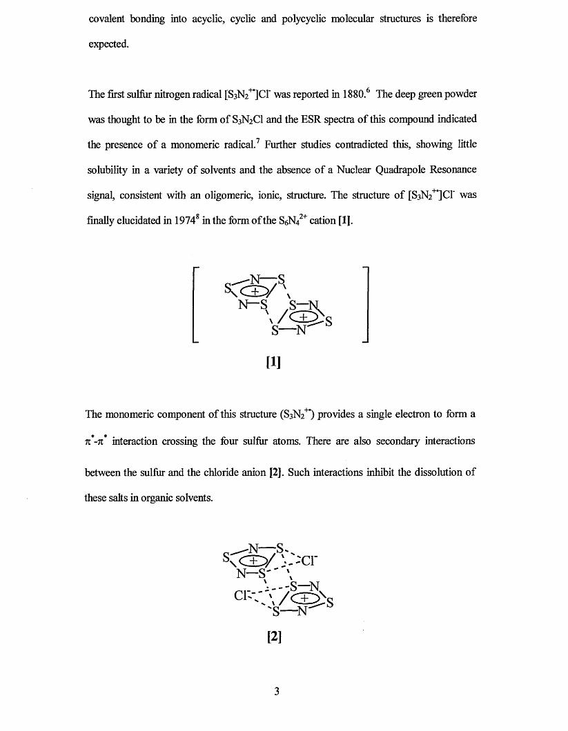
The first sulfiir nitrogen radical [S3N2+,]C1‘ was reported in 1880.6 The deep green powder
was thought to be in the form of S3N2CI and the ESR spectra of this compound indicated
the presence of a monomeric radical.7 Further studies contradicted this, showing little
solubility in a variety of solvents and the absence of a Nuclear Quadrapole Resonance
signal, consistent with an oligomeric, ionic, structure. The structure of [S3N2+#]Cf was
finally elucidated in 19748 in the form of the S6N42+ cation [1].
—;SK c ± y v
N—S S—N\ / < ^ s
S— N
[1]
The monomeric component of this structure (S3N2+*) provides a single electron to form a
n-n* interaction crossing the four sulfiir atoms. There are also secondary interactions
between the sulfiir and the chloride anion [2]. Such interactions inhibit the dissolution of
these salts in organic solvents.
Since then many such structures have been reported,9’10’11’12’13’14’15 all showing the overlap
of two singularly occupied molecular orbitals (SOMO) at sulfur to form dimers. The heat
of dimerisation is small (-47 ± 3 kJ mol'1 for [Sa^^JCf) ,7 which is enough to show some
paramagnetic behaviour even in the solid state.7’16
1.2 Dithiadiazolyls
As a development of the [SsN2+*] radical, it was thought that similar iso-electronic radicals
could be obtained by substituting one of the sulfiirs by an RC unit (R = alkyl, aryl or
halogen). The physical and chemical properties could then be investigated for a large
variety of structures in which the properties could be modified by the variation of the R
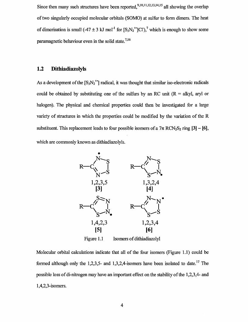
substituent. This replacement leads to four possible isomers of a In RCN2S2 ring [3] - [6],
which are commonly known as dithiadiazolyls.
A - s / ^ SR— C. | R— C |
VS VN-1,2,3,5 1,3,2,4
[3] [4]S c - . . •
/ / N / / NR— C I R— C |
VN- Vs1,4,2,3 1,2,3,4
[5] [6]Figure 1.1 Isomers of dithiadiazolyl
Molecular orbital calculations indicate that all of the four isomers (Figure 1.1) could be
formed although only the 1,2,3,5- and 1,3,2,4-isomers have been isolated to date.17 The
possible loss of di-nitrogen may have an important effect on the stability of the 1,2 ,3,4- and
1,4,2,3-isomers.
4
The first dithiadiazolyl was prepared as a dithiadiazolylium salt [RCN2S2]+X‘ in 1977 by
G.G. Alange et al.n The reduction of this to form the dithiadiazolyl radical (RCN2S2*)
(R = Ph, X = NCS' and I') by heating, or by dissolving the salts in 1,2 -dimethoxyethane.
Chemical reduction of dithiadiazolylium salts then followed in 1982 by L.N. Markovski et
1.3 Synthetic Methods for Dithiadiazolylium Salts
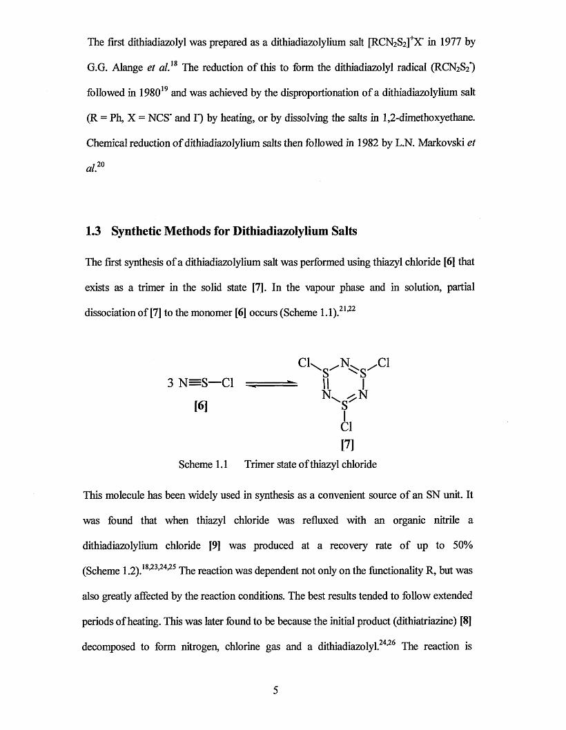
The first synthesis of a dithiadiazolylium salt was performed using thiazyl chloride [6] that
exists as a trimer in the solid state m . In the vapour phase and in solution, partial
dissociation of [7] to the monomer [6] occurs (Scheme 1.1) .21,22
was found that when thiazyl chloride was refluxed with an organic nitrile a
dithiadiazolylium chloride [9] was produced at a recovery rate of up to 50%
(Scheme 1.2) .18,23,24,25 The reaction was dependent not only on the functionality R, but was
also greatly affected by the reaction conditions. The best results tended to follow extended
periods of heating. This was later found to be because the initial product (dithiatriazine) [8]
decomposed to form nitrogen, chlorine gas and a dithiadiazolyl24,26 The reaction is
followed in 198019 and was achieved by the disproportionation of a dithiadiazolylium salt
3 N = S — Cl
[6]
■Cl
Cl[7]
Scheme 1.1 Trimer state of thiazyl chloride
This molecule has been widely used in synthesis as a convenient source of an SN unit. It
5
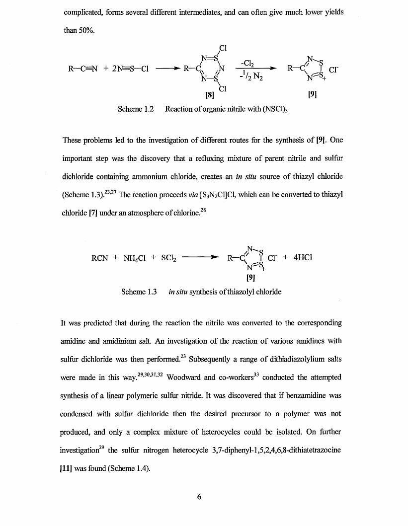
complicated, forms several different intermediates, and can often give much lower yields
than 50%.
/C1C1
R—C=N + 2N=S—Cl ------ ► R—C. ,N —j - 2-----► R—C I c i 'V // 1/ X T \N—S _ /2 N 2 N ^ +
\
[8 ] 01 [9]
Scheme 1.2 Reaction of organic nitrile with (NSC1)3
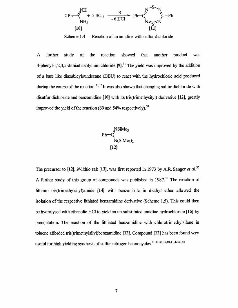
These problems led to the investigation of different routes for the synthesis of [9]. One
important step was the discovery that a refluxing mixture of parent nitrile and sulfur
dichloride containing ammonium chloride, creates an in situ source of thiazyl chloride
(Scheme 1.3) 23,27 The reaction proceeds via [S3N2C1]C1, which can be converted to thiazyl
chloride [7] under an atmosphere of chlorine.
/N" qRCN + NH4CI + SC12 ---------- ► R— c( I Cl' + 4HC1
isr^-h[9]
Scheme 1.3 in situ synthesis of thiazolyl chloride
It was predicted that during the reaction the nitrile was converted to the corresponding
amidine and amidinium salt. An investigation of the reaction of various amidines with
sulfur dichloride was then performed.23 Subsequently a range of dithiadiazolylium salts
were made in this way.29,30,31,32 Woodward and co-workers33 conducted the attempted
synthesis of a linear polymeric sulfur nitride. It was discovered that if benzamidine was
condensed with sulfur dichloride then the desired precursor to a polymer was not
produced, and only a complex mixture of heterocycles could be isolated. On further
investigation29 the sulfur nitrogen heterocycle 3,7-diphenyl-l,5,2,4,6,8-dithiatetrazocine
[11] was found (Scheme 1.4).
6
N il „ N ' 'S''N
2 M . * 3 8 0 ,1 f ~ ?"NH2 N^g-^N[10] [11]
Scheme 1.4 Reaction of an amidine with sulfur dichloride
A further study of the reaction showed that another product was
4-phenyl-l,2,3,5-dithiadiazolylium chloride [9] .31 The yield was improved by the addition
of a base like diazabicyloundecane (DBU) to react with the hydrochloric acid produced
during the course of the reaction.33,32 It was also shown that changing sulfur dichloride with
disulfur dichloride and benzamidine [10] with its tris(trimethysilyl) derivative [12], greatly
improved the yield of the reaction (60 and 54% respectively) .34
NSiMe3// jPh-q
N(SiMe3) 2
[12]
The precursor to [12], TV-lithio salt [13], was first reported in 1973 by A.R. Sanger et al.3S
A further study of this group of compounds was published in 1987.36 The reaction of
lithium bis(trimethylsilyl)amide [14] with benzonitrile in diethyl ether allowed the
isolation of the respective lithiated benzamidine derivative (Scheme 1.5). This could then
be hydrolysed with ethanolic HC1 to yield an un-substituted amidine hydrochloride [15] by
precipitation. The reaction of the lithiated benzamidine with chlorotrimethylsilane in
toluene afforded tris(trimethylsilyl)benzamidine [12]. Compound [12] has been found very
useful for high yielding synthesis of sulfur-nitrogen heterocycles.31,37,38,39,40,41,42,43,44
7

SiMe3
Li[N(SiMe3)2] /fJSiMe3
-*- R—C<r Li+Et20 \ -NSiMe,
NH R— q -HCI
NH2
[15]
N(SiMe3)2
[12]
Scheme 1.5 Amidine synthesis
The conversion of benzamidines such as [12] into the 1,2,3,5-dithiadiazolylium chlorides
whether the amidine itself or its 7V-lithio salt [13] is used. Further purification by Soxhlet
extraction of the dithiadiazolylium salt with sulfiir dioxide is necessary when proceeding
via the A-lithio salt [13]. The reaction, although giving high yields, has limitations
especially for compounds containing protons in the a-position, due to complex side
reactions. One method of solving this problem has been to use .yy/w-triazines [16] as the
starting material (Scheme 1.6 ).45
Scheme 1.6 Amidine synthesis via sym-triazine
In addition to the routes outlined earlier, other methods have been reported in the literature
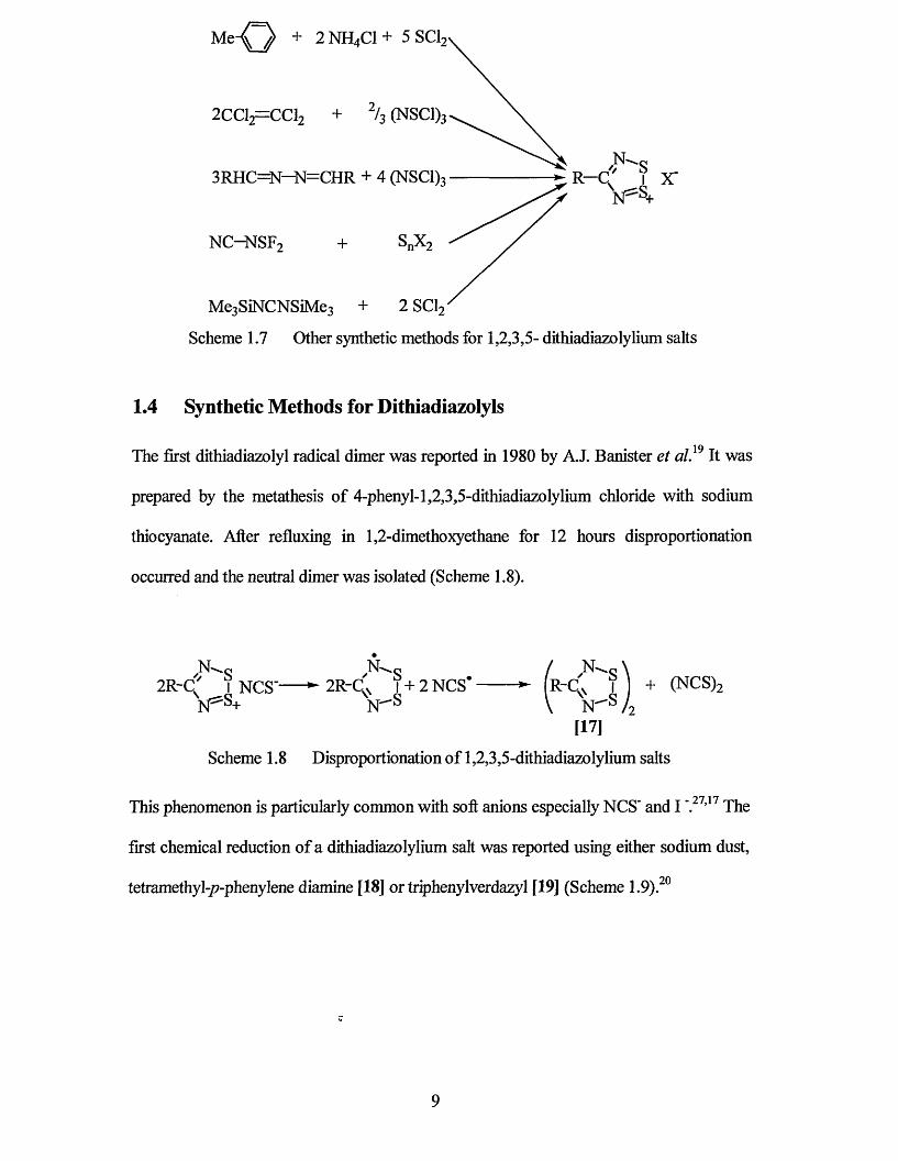
for making dithiadiazolylium salts. Selections of these are outlined in
[9] can be achieved by the addition of excess sulfiir dichloride. The reaction will proceed
H
3 Li[N(SiMe3)2] 3 Me3SiCl 3 H -C n
N(SiMe3)2
NSiMe3
[16]
(Scheme 1.7).23,25,27,46,47,48
8
M,K D + 2 NH4C1 + 5 SCI,
2CC12=CC12 + 2h (NSC1)3
3RHC=N-N=CHR + 4 (NSC1)3
NC-NSF2 + SnX2
Me3SiNCNSiMe3 + 2 SC12
Scheme 1.7 Other synthetic methods for 1,2,3,5- dithiadiazolylium salts
1.4 Synthetic Methods for Dithiadiazolyls
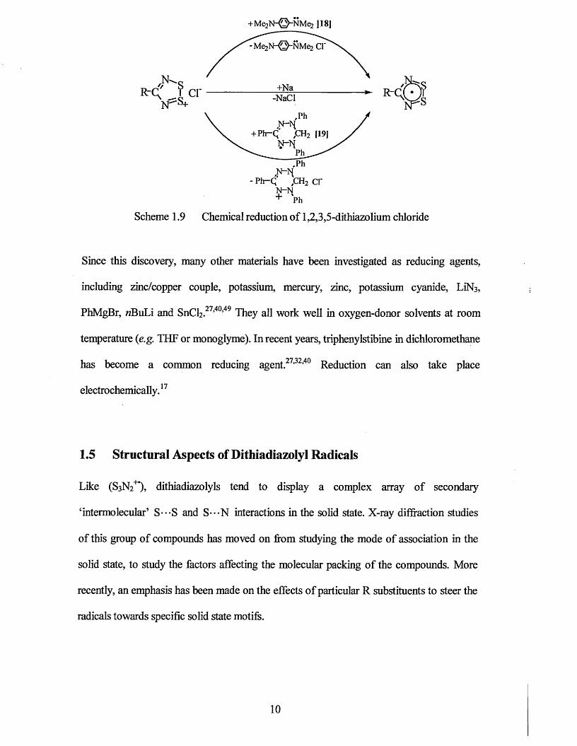
The first dithiadiazolyl radical dimer was reported in 1980 by A.J. Banister et al, 19 It was
prepared by the metathesis of 4-phenyl-1,2,3,5-dithiadiazolylium chloride with sodium
thiocyanate. After refluxing in 1,2-dimethoxyethane for 12 hours disproportionation
occurred and the neutral dimer was isolated (Scheme 1.8).
N - s / N - s \2R-C' I NCS' ^ 2R~C ̂ 1+2 NCS* ► R~C ̂ I + (NCS)2
N=5S+ N " s \ N" s / 2[17]
Scheme 1.8 Disproportionation of 1,2,3,5-dithiadiazolylium salts
This phenomenon is particularly common with soft anions especially NCS' and IV27,17 The
first chemical reduction of a dithiadiazolylium salt was reported using either sodium dust,
tetramethyl-/?-phenylene diamine [18] or triphenylverdazyl [19] (Scheme 1.9).
9
N^„R-q" ? c r
+Me2N-©-NMe>2 |18]
-Me2N -© -N M e2 Cl
+Na-NaCl
,PhN~h{+ P h -q ,c h 2 [19]
N-NPh
.PhN~h{
-Ph-Q yCH2 cr N -N+
R-Q(*
Ph
Scheme 1.9 Chemical reduction of 1,2,3,5-dithiazolium chloride
Since this discovery, many other materials have been investigated as reducing agents,
including zinc/copper couple, potassium, mercury, zinc, potassium cyanide, LiN3,
PhMgBr, «BuLi and SnCh.27,40’49 They all work well in oxygen-donor solvents at room
temperature (e.g. THF or monoglyme). In recent years, triphenylstibine in dichloromethane
has become a common reducing agent.27,32,40 Reduction can also take place
electrochemically. 17
1.5 Structural Aspects of Dithiadiazolyl Radicals
Like (S3N2+*), dithiadiazolyls tend to display a complex array of secondary
‘intermolecular’ S---S and S---N interactions in the solid state. X-ray diffraction studies
of this group of compounds has moved on from studying the mode of association in the
solid state, to study the factors affecting the molecular packing of the compounds. More
recently, an emphasis has been made on the effects of particular R substituents to steer the
radicals towards specific solid state motifs.
10
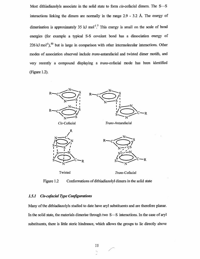
Most dithiadiazolyls associate in the solid state to form c/s-cofacial dimers. The S---S
interactions linking the dimers are normally in the range 2.9 - 3.2 A. The energy of
dimerisation is approximately 35 kJ mol'1.7 This energy is small on the scale of bond
energies (for example a typical S-S covalent bond has a dissociation energy of
226 kJ mol'1) ,50 but is large in comparison with other intermolecular interactions. Other
modes of association observed include /ram,-antarafacial and twisted dimer motifs, and
very recently a compound displaying a frvms-cofacial mode has been identified
(Figure 1.2).
C/s-Cofacial rraws-Antarafacial
Twisted 7>vm?-Cofacial
Figure 1.2 Conformations of dithiadiazolyl dimers in the solid state
1.5.1 Cis-cofacial Type Configurations
Many of the dithiadiazolyls studied to date have aryl substituents and are therefore planar.
In the solid state, the materials dimerise through two S - • - S interactions. In the case of aiyl
substituents, there is little steric hindrance, which allows the groups to he directly above
11
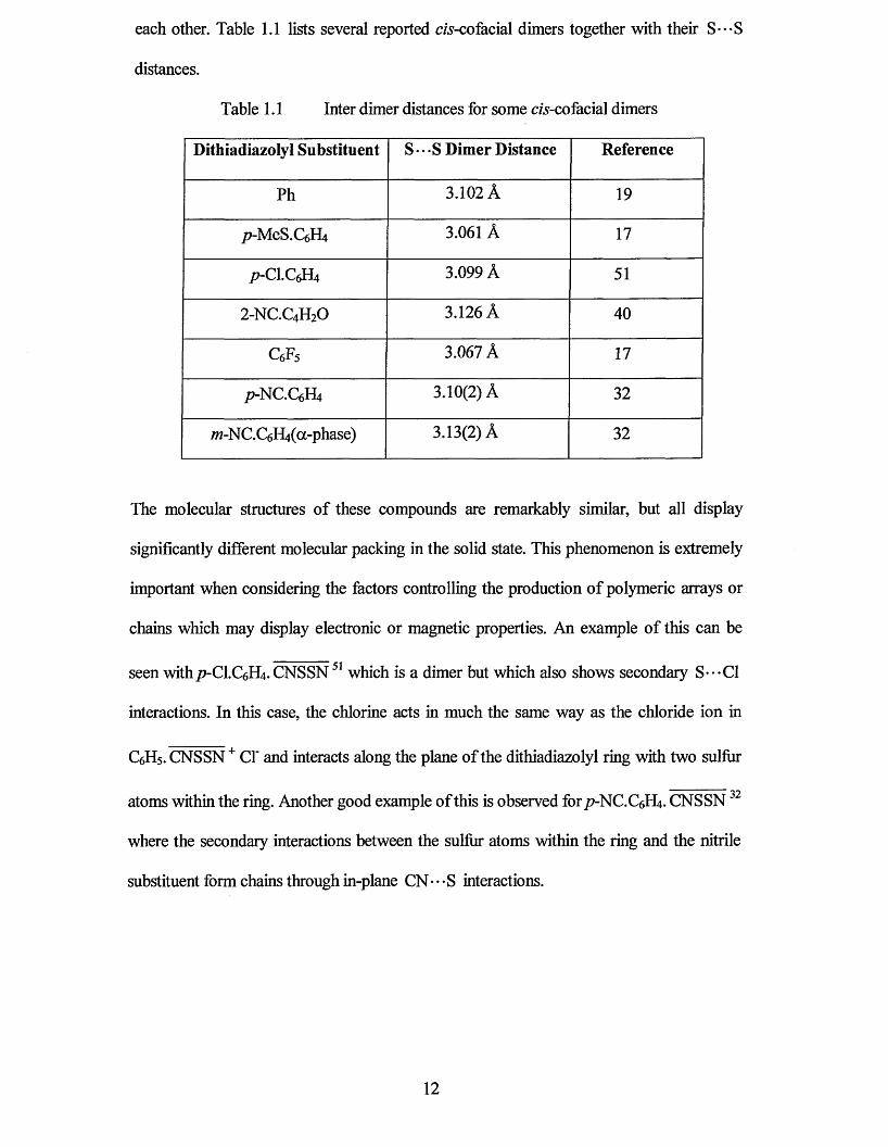
each other. Table 1.1 lists several reported m-cofacial dimers together with their S---S
distances.
Table 1.1 Inter dimer distances for some c/s-cofacial dimers
Dithiadiazolyl Substituent S • • -S Dimer Distance Reference
Ph 3.102 A 19
p-MeS.CfJff 3.061 A 17
p-C\.Q£U 3.099 A 51
2 -NC.C4H2O 3.126 A 40
c 6f 5 3.067 A 17
p-NC.C6H4 3.10(2) A 32
7w-NC.C6H4(a-phase) 3.13(2) A 32
The molecular structures of these compounds are remarkably similar, but all display
significantly different molecular packing in the solid state. This phenomenon is extremely
important when considering the factors controlling the production of polymeric arrays or
chains which may display electronic or magnetic properties. An example of this can be
seen withp-C\.C$U. CNSSN51 which is a dimer but which also shows secondary S---C1
interactions. In this case, the chlorine acts in much the same way as the chloride ion in
C6H5. CNSSN + Cl' and interacts along the plane of the dithiadiazolyl ring with two sulfur
1 00atoms within the ring. Another good example of this is observed for / 7-NC.C6H4. CNSSN
where the secondary interactions between the sulfur atoms within the ring and the nitrile
substituent form chains through in-plane CN • • - S interactions.
12
1.5.2 Twisted Type Configurations
This configuration usually occurs when the substituent is non-planar e.g. CF3,25 Me,49
/Bu.17,52 In order to minimise steric repulsion the molecules adopt a twisted conformation
in which there is one strong S* • - S contact and some weaker S - • - N interactions. The inter
dimer distance (S---S distance) is comparable to that of the c/s-cofacial conformation. It
has also been shown that because of the large steric crowding involved in these
compounds, the melting points are often low. Indeed the /Bu derivative is a paramagnetic
liquid at room temperature but upon cooling, the compound associates as a dimer. A
further example was also observed in adamantyl- 1,2,3,5-dithiadiazolyl.53 The structure was
surprisingly similar to that of smaller alkyl substituents, despite its rigidity and significant
steric influence. A twisted conformation was also observed for 2 ,3 -F2.C6H3. CNSSN 54,55
which had an S---S interdimer distance of 3.020(4) A. This structure was the first
observed when the R substituent was planar and illustrates how electrostatic repulsion can
be used to alter the solid state structure.
1.5.3 Trans-antarafacial Configurations
To date, only one compound has been reported with this /raws-antarafacial conformation.
This is despite the fact that the energy difference between the cis and trans isomers is very
small. In the case of W-NC.C6H4 CNSSN32 there are two morphologies. The first,
(a-phase) shows the c/s-cofacial dimer with ordered crystal packing through secondary
S---CN interactions. The second (p-phase) is the /raws-antarafacial dimer which shows
similar S---S distances and also shows strong S---CN secondary interactions which
results in a chain type structure.
13
1.5.4 Trans-cofadal Configurations
Until very recently, no /nms’-cofacial configurations had been published. The
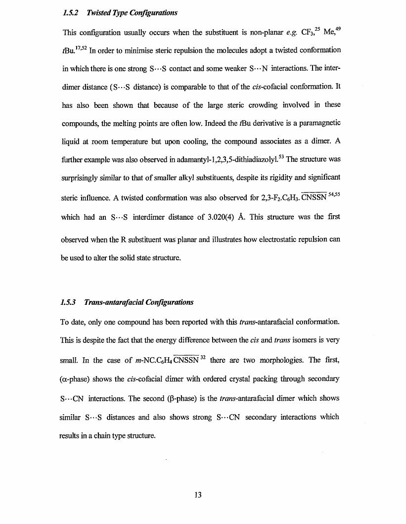
2 ,2 ’-dimethylbiphenylene bridged derivative [2 0] ,56 contains one dithiadiazolyl associated
in a dimerised /ram-cofacial manner across an inversion centre to an equivalent radical on
a neighbouring molecule. The contact between these rings is small (3.24 A), which is well
within the distance effected by spin pairing. It is unclear why this cofacial association was
favoured in this compound. The other side of the di-radical has no close spin-paired
interactions with any neighbouring ring. This renders the compound paramagnetic in the
solid state. However, no long range magnetic ordering is observed.
[20]
1.5.5 Other Conformations
During the past few years, the emphasis in much of the work into these systems has been
to control the molecular packing, several notable advances have been made leading to
derivatives with novel structures and properties. Oakley and co-workers39 reported the first
example of a dithiadiazolyl, 1,3-phenylene-bis-dithiadiazolyl, which crystallised as one
dimensional stacks. However, it was discovered that Peierls distortion caused a subtle
rocking at opposite ends of each molecule. This in turn produced long and short centroid-
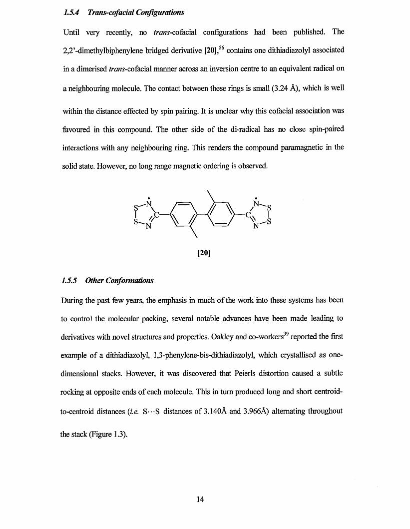
to-centroid distances (i.e. S---S distances of 3.140A and 3.966A) alternating throughout
the stack (Figure 1.3).
14
N(4)
N(3) 5(4)5(3)
Figure 1.3 The Solid State Structure of 1,3[NSSNC .C6H4. CNSSN ]
Subsequently, the same workers co-sublimed simple dithiadiazolyls such as Ph. CNSSN
with iodine.57 The result was a p-doped charge transfer complex, where stacks of
dithiadiazolyls associated with iodide or triiodide anions. The complex of
1,3-[NSSNC.C6H4.CNSSN] with iodine was observed to be a semi-conductor at room
temperature with a conductivity of 100 Scm'1.
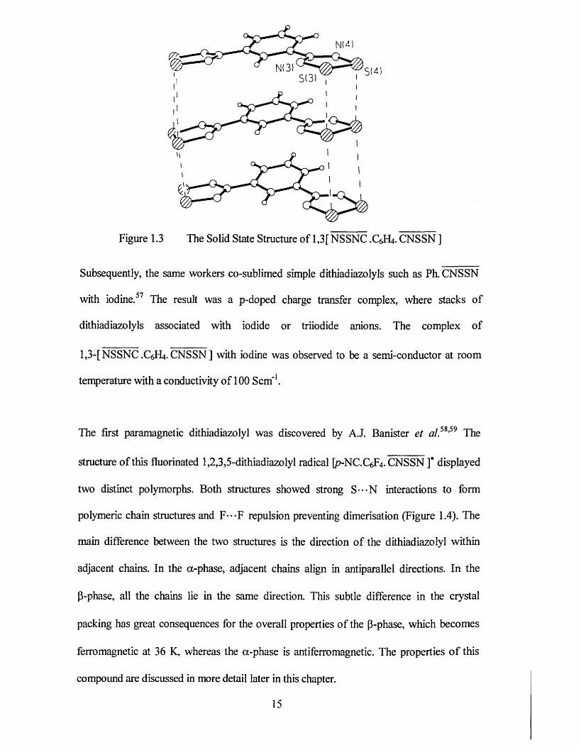
The first paramagnetic dithiadiazolyl was discovered by A.J. Banister et al.5*’59 The
structure of this fluorinated 1,2,3,5-dithiadiazolyl radical [p-NC.C6F4. CNSSN ]* displayed
two distinct polymorphs. Both structures showed strong S---N interactions to form
polymeric chain structures and F---F repulsion preventing dimerisation (Figure 1.4). The
main difference between the two structures is the direction of the dithiadiazolyl within
adjacent chains. In the a-phase, adjacent chains align in antiparallel directions. In the
p-phase, all the chains lie in the same direction. This subtle difference in the crystal
packing has great consequences for the overall properties of the P-phase, which becomes
ferromagnetic at 36 K, whereas the a-phase is antiferromagnetic. The properties of this
compound are discussed in more detail later in this chapter.
15
__s \
\o -
rv _ / ’— p -
</ \>
■ uS(2C)
.—oN(3A)
S(10)
S(2) N(2| ' o H 3 1lA-T=(6)
SHI N d ) F (1 ) 0 ' * Q F I 2 I
X-ao—
S ~ \
r \Figure 1.4 The a (above) and (3 (bottom) structures of/7-CN.C6F4. CNSSN
Further work by Banister and co-workers54,55 into the repulsion of fluorinated substituents
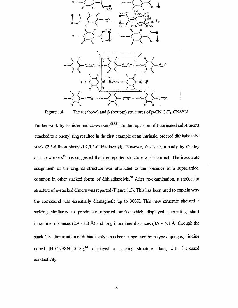
attached to a phenyl ring resulted in the first example of an intrinsic, ordered dithiadiazolyl
stack (2,5-difluorophenyl-l,2,3,5-dithiadiazolyl). However, this year, a study by Oakley
and co-workers60 has suggested that the reported structure was incorrect. The inaccurate
assignment of the original structure was attributed to the presence of a superlattice,
common in other stacked forms of dithiadiazolyls.60 After re-examination, a molecular
structure of 7i-stacked dimers was reported (Figure 1.5). This has been used to explain why
the compound was essentially diamagnetic up to 300K. This new structure showed a
striking similarity to previously reported stacks which displayed alternating short
intradimer distances (2.9 - 3.0 A) and long interdimer distances (3.9 - 4.1 A) through the
stack. The dimerisation of dithiadiazolyls has been suppressed by p-type doping e.g. iodine
doped [H. CNSSN ].0.18I2 61 displayed a stacking structure along with increased
conductivity.
16

c -B £ r" .
Figure 1.5 The solid state structure o f 2,5-F2.C6H3. CNSSN60
ft 9More recently, a monomeric dithiadiazolyl [21] has been reported.
SIs
F F
[21]
Compound [21] has a molecular structure showing columns o f radicals perpendicular to
the crystallographic <3-axis. The association out of plane takes the form of a twisted motif
with each dithiadiazolyl ring lying directly above the next. The interaction mimics that of
the twisted dithiadiazolyls described earlier (Section 1.5.2) but shows a larger angle
between dithiadiazolyl units, approaching the frww-antarafacial configuration. In this case,
the out of plane contacts fall in the range 3.675 - 3.999 A which is well above the normal
dimer separation (2.9 - 3.1 A). Compound [21] has an effective magnetic moment at room
temperature o f 1.45 pe- Above 60 K, the magnetic susceptibility follows the Curie-Weiss
law (9 = -27 K); however, there is no evidence for long-range magnetic order down to
1.8 K.
17 I
1.6 Magnetism and non-Metallic Magnets
If two objects attract each other and also repel each other (depending on their relative
orientations), then these objects might be called magnets. In addition, when certain objects
are attracted to, but not repelled by each other then these objects may be said to consist of
magnetic materials. Magnetic phenomena has been known and exploited for centuries.
Since the first known magnetic material, magnetite, many other magnetic materials have
been used and investigated.63,64 One of the main challenges in the field of molecular
materials concerns the design and synthesis of compounds exhibiting spontaneous
magnetisation.65,66 Compounds of this type have only been described in the past decade,
and despite much effort, this class of compound is still small in size.
It is important first to understand some of the definitions as a background to magnetic
behaviour.67,68 Although magnetism has been known and used for hundreds of years it was
not until the nineteenth century that a link was found between electricity and magnetism.
Michael Faraday stated that the force driving a current around a circuit, i.e. the
electromotive force (Volts) is equal to the rate of change of flux through a circuit. The
effect of a magnetic field on a material can be defined in several equations. It can be shown
that in a vacuum the magnetic induction of a material, B, is directly proportional to the
field, H (Am'1) (Equation 1.1).
B =p 0H Equation 1.1
where po is a universal constant, the permeability o f a vacuum. In a vacuum, B and H are
always parallel to each other. When a material is present, this equation is not followed as
the material acquires a dipole moment. In this case the material has a magnetisation, M,
which is the dipole moment per unit volume. The relationship can then be shown to be:
B = p 0( H + M ) Equation 1.2
The magnetisation of a material in general depends on the magnetic field acting upon it.
For most materials M is proportional to H and is related by:
M = %H Equation 1.3
where % is the magnetic susceptibility and is a property of the material. The cgs units of
molecular susceptibility (used in this thesis) are emu.Oe'1. mol'1. The emu is an
electromotive unit with dimensions of volume so that 1 emu is equivalent to 1 cm .
Another way of characterising the material magnetically is to use the magnetic
permeability, p. This is related to the magnetic susceptibility by:
[i = 1 +y Equation 1.4
It then follows that the magnetic permeability is related to the magnetic induction by the
following equation:
B =\l ofiH Equation 1.5
Both magnetic susceptibility and magnetic permeability can be used to characterise the
nature of a magnetic material. For small values of % then p becomes very close to 1 and is
of very little use. However, for larger values, and therefore materials of more practical
interest, p can be used.
19
The classification of magnetic materials can be aided by studying the magnetic
susceptibility of materials as a function of temperature. Compounds which contain only
pairs of electrons are repelled by magnetic field and are said to be diamagnetic. Their
susceptibility is negative and independent of temperature. Compounds which contain
unpaired electrons are attracted into the magnetic field and their susceptibility is positive.
These compounds are described as paramagnetic and have a susceptibility which is
temperature dependent (discussed in more detail later). In reality, all paramagnetic
compounds contain some electron pairs (core electrons and electrons involved in bond
formation) and so the observed susceptibility is the sum of the diamagnetic and
paramagnetic components (Equation 1.6).
X . b s = Z p + Z d Equation 1.6
If we are interested in the paramagnetism arising from the unpaired electrons (%p) then a
careful correction for the sample diamagnetism must be made. This is often estimated
using Pascal’s constants, although more elaborate methods can be applied. For now, we
will concentrate on the sample paramagnetism.
Pierre Curie found an empirical relationship between the value of %v and the absolute
temperature, T. For many paramagnetic compounds there is an inverse-relationship
between x and T (Equation 1.7). Thus as a sample is cooled its susceptibility rises rapidly.
At low temperatures some deviation from the Curie-Weiss behaviour (Equation 1.7) is
often observed.
Cv = --------------------- Equation 1.7
T ± 0
20
If 0 = 0 the compound behaves as a perfect paramagnet, i.e. each molecule acts
independently from its neighbours. However in many compounds, there may be some
degree of communication between neighbours (often referred to as a magnetic exchange
interaction). This communication may try to align the unpaired electrons on neighbouring
molecules anti-parallel to one another. These molecules are said to be
antiferromagnetically coupled and in this case 9 < 0. In contrast, local co-parallel
alignment of electrons gives rise to ferromagnetic coupling, signified by 0 > 0. The sign
and magnitude of 0 give and indication of the type and strength of the coupling between
neighbouring molecules.
In the mean field approach, for ferromagnetically coupled compounds when T = 0, then
%-> oo, i.e. all the unpaired electrons move co-operatively and this known as a
magnetically ordered state. This temperature is known as the critical temperature. In reality
many compounds begin to deviate from Curie-Weiss behaviour will usually order
somewhat below 10 1. Since the co-operativity is essentially required to propagate
throughout the three-dimensional solid, it requires communication in three dimensions. For
compounds exhibiting strong communication between spins in only on or two dimensions,
then magnetic order is frequently not observed at any finite temperature. Some common
forms of long range magnetic order are briefly outline:-
In a ferromagnet, all the electrons align co-parallel at absolute zero (T = 0 K) giving rise to
a spontaneous magnetisation, M, even in the absence of an applied field, H. Under these
circumstances, Equation 1.3 is no longer applicable and the sample susceptibility is
meaningless. Instead, we discuss the spontaneous magnetisation. At 0 K all the electrons
align perfectly and we observe saturation magnetisation, Ms. On warming above OK some
thermal energy leads to electron motion away from perfect alignment and the saturation
21
magnetisation steadily decreases, reaching zero at the magnetic ordering temperature or
Curie temperature, Tc. Above the ordering temperature the material is paramagnetic and %
becomes meaningful once more. At temperatures well above the ordering temperature the
compound will obey Equation 1.7 with 0 > 0.
In an antiferromagnet, the spins align antiparallel throughout the solid below the Neel
temperature, Tn. An antiferromagnet is often characterised by a sharp maximum in the
susceptibility. On cooling a paramagnet, % is expected to increase but, as the
antiferromagnetic interactions become stronger (in relation to the thermal energy kT) then
the electrons begin to align antiparallel to each other rather than all parallel with the
applied field and the susceptibility begins to decrease. For a three-dimensional order the
transition is usually sharp whereas low-dimensional magnetic interactions give rise to
broad maxima in the susceptibility. Well above the ordering temperature an
antiferromagnet is expected to follow Equation 1.7 with 0 < 0.
In a ferrimagnet, electrons on neighbouring molecules interact antiferromagnetically
(i.e. 0 < 0). However, the interacting molecules may bear spins of differing size (e.g. two
unpaired electrons on one molecule, S = 1, interacting a neighbouring molecule bearing
one unpaired electron, S = lA). In this case, antiparallel alignment will lead to a greater
number of spins pointing in one direction. Below the critical temperature, Tc, the
compound behaves much like a ferromagnet, although the magnitude of the saturation
magnetisation is lower [cf 2S for a ferromagnet vs 2(Sa - Sb) for a ferrimagnet].
More esoteric types of magnetic order and partially ordered states are also known. These
include asperomagnetism, canted antiferromagnetism and spin-glass behaviour and are
22
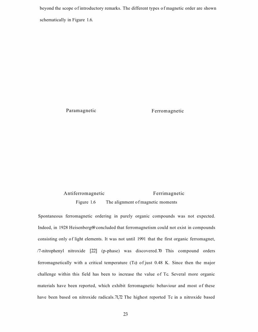
beyond the scope o f introductory remarks. The different types o f magnetic order are shown
schematically in Figure 1.6.
Paramagnetic Ferromagnetic
Antiferromagnetic Ferrimagnetic
Figure 1.6 The alignment o f magnetic moments
Spontaneous ferromagnetic ordering in purely organic compounds was not expected.
Indeed, in 1928 Heisenberg69 concluded that ferromagnetism could not exist in compounds
consisting only o f light elements. It was not until 1991 that the first organic ferromagnet,
/7-nitrophenyl nitroxide [22] (p-phase) was discovered.70 This compound orders
ferromagnetically with a critical temperature (Tc) o f just 0.48 K. Since then the major
challenge within this field has been to increase the value of Tc. Several more organic
materials have been reported, which exhibit ferromagnetic behaviour and most o f these
have been based on nitroxide radicals.71,72 The highest reported Tc in a nitroxide based
23
material is 1.48 K for A^TV-dioxy-1,3,5,7-tetramethyl-2,6-diazaadamantane [23],71 Another
compound found to exhibit weak ferromagnetism is the verdazyl radical TPV. 73 In this
compound, the ferromagnetism results from canted, or non-collinear, antiferromagnetic
ordering. A significant advance was then made with ferromagnetic ordering in the
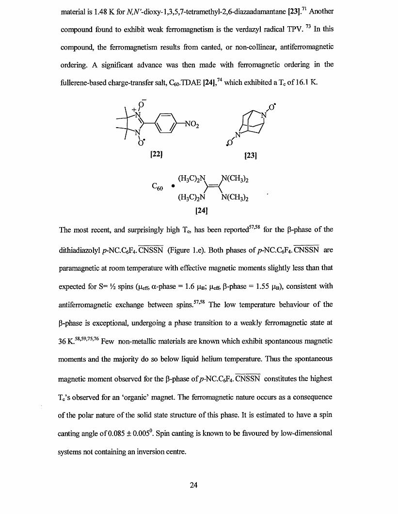
fiillerene-based charge-transfer salt, C6o-TDAE [24] ,74 which exhibited a Tc of 16.1 K.
^ 0 ^ - N O , { Q
oJ .o '[22] [23]
^60 *(H3C)2N N(CH3)2
[24]
The most recent, and surprisingly high Tc, has been reported57,58 for the p-phase of the
dithiadiazolyl p-NC.C^fy. CNSSN (Figure l.e). Both phases of /7-NC.C6F4. CNSSN are
paramagnetic at room temperature with effective magnetic moments slightly less than that
expected for S= V2 spins (pefr, a-phase = 1.6 pB; peff, P-phase = 1.55 pB), consistent with
antiferromagnetic exchange between spins.57,58 The low temperature behaviour of the
P-phase is exceptional, undergoing a phase transition to a weakly ferromagnetic state at
36 k .58,59,75,76 Few non-metallic materials are known which exhibit spontaneous magnetic
moments and the majority do so below liquid helium temperature. Thus the spontaneous
magnetic moment observed for the P-phase of/7-NC.C6F4. CNSSN constitutes the highest
Tc’s observed for an ‘organic’ magnet. The ferromagnetic nature occurs as a consequence
of the polar nature of the solid state structure of this phase. It is estimated to have a spin
canting angle o f0.085 ± 0.005°. Spin canting is known to be favoured by low-dimensional
systems not containing an inversion centre.
(H3C)2N , N ( C H 3)2
/ \
24
1.7 Conduction in Organic Materials
The classic electron conduction model for a metal considers that a valence electron of an
atom can move freely between all the atoms within the solid. The energy band model for
partially filled bands (metals) shows that the energy required to excite valence electrons so
that they are mobile is veiy small. For filled bands, the energy required to excite the
valence electrons to make them mobile is much greater, which leads to materials that are
either semiconductors or insulators. These models are based on interaction between atoms
in a solid lattice. Thus the extension of electrical conductivity models from atom based
systems to those made up of molecules is dependent on the interactions in the solid state.
Molecular based materials which have electrons delocalised over the entire solid may
exhibit high metal-like conductivities. It was postulated, as far back as 1911,77 that
molecule-based compounds could exhibit conductivities similar to those of metals. These
predictions were realised in 19541 and since then there has been extraordinarily large
interests in this area.78
The primary motivation for the extensive interest in the synthesis of this group of
molecular conductors is the potential technological applications of these materials. The
simple fact that many of the materials have a much lower density (about 1.5 g/cm3) than
metals (9 g/cm3 for copper) makes their use very appealing. Moreover, a combination of
different physical properties and their ease of fabrication also makes them veiy inviting.
Over the past 20 years research into the design of molecular conducting materials has
focused primarily on organic donor molecules like TTF [25], TMTSF [26], BEDT-TTF
[27] and acceptors such as TCNQ [28]. For these systems, conductivity is only possible
after oxidation (p-doping) of the neutral molecule, to form a charge transfer (CT) salt. This
conductivity arises from the generation of a partially filled energy band, which is
25
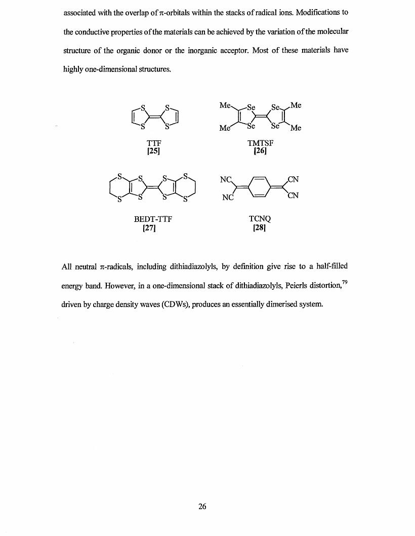
associated with the overlap of 7t-orbitals within the stacks of radical ions. Modifications to
the conductive properties of the materials can be achieved by the variation of the molecular
structure of the organic donor or the inorganic acceptor. Most of these materials have
highly one-dimensional structures.
TTF[25]
Me
Me'
Se S e -^ Me
Se Se
TMTSF[26]
BEDT-TTF[271
NG>
NC
= \ „CN<CN
TCNQ[28]
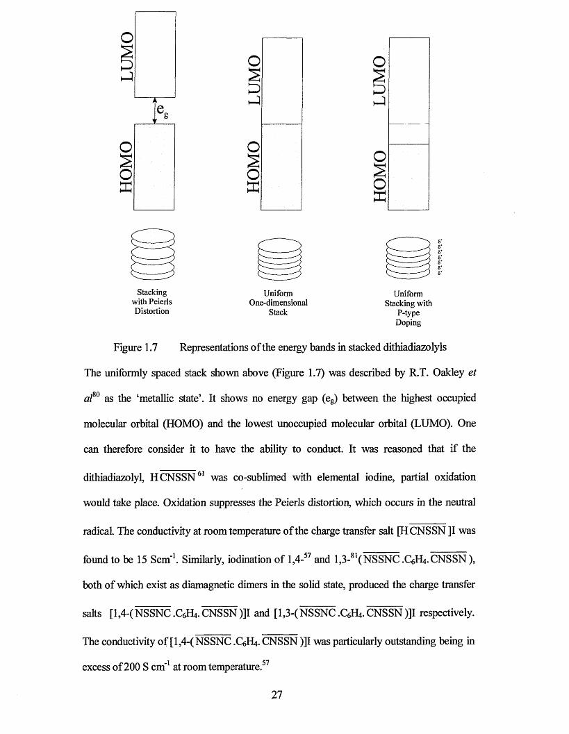
All neutral 7r-radicals, including dithiadiazolyls, by definition give rise to a half-filled
• • 79energy band. However, in a one-dimensional stack of dithiadiazolyls, Peierls distortion,
driven by charge density waves (CDWs), produces an essentially dimerised system.
26
Stacking with Peierls Distortion
UniformOne-dimensional
Stack
Uniform Stacking with
P-type Doping
Figure 1.7 Representations of the energy bands in stacked dithiadiazolyls
The uniformly spaced stack shown above (Figure 1.7) was described by R.T. Oakley et
af° as the ‘metallic state’. It shows no energy gap (eg) between the highest occupied
molecular orbital (HOMO) and the lowest unoccupied molecular orbital (LUMO). One
can therefore consider it to have the ability to conduct. It was reasoned that if the
dithiadiazolyl, H CNSSN61 was co-sublimed with elemental iodine, partial oxidation
would take place. Oxidation suppresses the Peierls distortion, which occurs in the neutral
radical. The conductivity at room temperature of the charge transfer salt [H CNSSN ]I was
found to be 15 Scm'1. Similarly, iodination of 1,4-57 and 1,3-81(NSSNC .C6H4.CNSSN),
both of which exist as diamagnetic dimers in the solid state, produced the charge transfer
salts [l,4-(NSSNC.C6H4.CNSSN)]I and [1,3-(NSSNC.C6H4.CNSSN)]I respectively.
The conductivity of [1,4-(NSSNC .C6H4. CNSSN )]I was particularly outstanding being in
excess of200 S cm’1 at room temperature.57
27
1.8 Electron Paramagnetic Resonance
Electron Paramagnetic Resonance (EPR) spectroscopy is a technique used to study
materials that contain unpaired (paramagnetic) electrons82 and is invaluable for
investigating free radicals such as dithiadiazolyls.
The technique is often considered more difficult to obtain detailed information from than
experiments such as NMR and Mossbauer spectroscopy. More knowledge about the
specific ion in question is often needed for detailed interpretation. In addition, some
systems may give weak or broad signals that require cooling to low temperatures to gain
high quality spectra. Despite these difficulties, the technique is considered complementary
to NMR, which cannot be used with paramagnetic samples under normal conditions.
Considerable parallels exist between NMR and EPR because both depend on the magnetic
moment of a spinning particle, either the nucleus or the electron. For electrons with a spin
V2, two energy states are produced by the interaction with a magnetic field, B. They differ
in energy by:
AE = g/zB Equation 1.8
The value of g depends on the identity of the particle and p is its magnetic moment. For a
free electron, g is 2.0023 and for a proton, the value is 5.5856.82 The magnetic moment
varies considerably because of its dependence on mass.
ehfi = ----------------------- Equation 1.9
4 jun
28

In the case of EPR pB (the Bohr magneton) is 9.274 x 10'24 JT 1. The spectra obtained by
EPR depend on determining the g-values for the impaired electrons in the sample, which
are different to those of the free electrons and are dependent on the chemical environment
of the paramagnetic atom. This is usually achieved by using a fixed frequency, usually
9 GHz, and varying the applied field. The method used is the opposite to that of NMR
because the chamber where the sample is kept has to be tuned to the particular wavelength
used. It is therefore not possible to adjust the wavelength without an adjustable chamber.
The frequency of 9GHz was chosen because similar systems using this frequency were
already being used for marine radar (X-band), and were well understood. Some
spectrometers operate at 36GHz (Q-band) because the technology of airport radar can be
utilised.
1.8.1 Experimental Considerations
EPR studies can be performed on liquids, solids and solutions. The technique is very
sensitive so only a small sample is required. Because of this high sensitivity, every effort
must be made to omit all traces of additional paramagnetic impurities. Solid samples are
measured in silica tubes rather than glass to avoid the contamination from iron (El) in the
glass. Air is often removed from the tubes prior to analysis to avoid line broadening from
the presence of molecular oxygen. Line broadening due to spin-spin relaxation effects can
be minimised by dissolving the sample in a solvent at a concentration of about
5 mmol dm"3. A small amount of another solvent such as dimethylsulfoxide or glycerol is
useful to encourage the formation of a glass when cooling. This in turn helps to prevent the
crystallisation of the solid sample from the solvent during cooling.
In order to reduce noise, most spectrometers use a modular technique, which superimposes
a cyclic variation on the magnetic field. The detection of the signal involves examining the
29
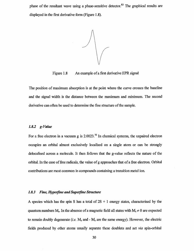
phase of the resultant wave using a phase-sensitive detector.83 The graphical results are
displayed in the first derivative form (Figure 1.8).
Figure 1.8 An example of a first derivative EPR signal
The position of maximum absorption is at the point where the curve crosses the baseline
and the signal width is the distance between the maximum and minimum. The second
derivative can often be used to determine the fine structure of the sample.
1.8.2 g-Value
For a free electron in a vacuum g is 2.0023.16 In chemical systems, the unpaired electron
occupies an orbital almost exclusively localised on a single atom or can be strongly
delocalised across a molecule. It then follows that the g-value reflects the nature of the
orbital. In the case of free radicals, the value of g approaches that of a free electron. Orbital
contributions are most common in compounds containing a transition metal ion.
1.8.3 Fine, Hyperfine and Superfine Structure
A species which has the spin S has a total of 2S + 1 energy states, characterised by the
quantum numbers Ms. In the absence of a magnetic field all states with Ms ̂ 0 are expected
to remain doubly degenerate (i.e. Ms and - Ms are the same energy). However, the electric
fields produced by other atoms usually separate these doublets and act via spin-orbital
30
coupling. This is known as zero field splitting and depend on the structure of the sample
under investigation. The presence of a magnetic field removes the remaining degeneracy
and allows transitions between adjacent states to be observed. The appearance of more
than one line (when S = Vi) is known as fine structure.
The spectra produced by EPR may have some additional fine structure when the atom on
which the impaired spin is centred also has a spin (exactly analogous to NMR). A nucleus
of spin I gives rise to the splitting of the EPR line into 21 + 1 components, all with equal
intensity and separated with a coupling constant, A. This phenomenon is known as
hyperfine structure. In an analogous way, nuclei on adjacent atoms may give splitting of
the spectrum. This is known as superfine structure and is dependent on the extent of
delocalisation of the unpaired electron on to these atoms, decreasing rapidly with the
number of bonds involved. It is usually only detectable for nuclei of atoms directly bound
to that containing the unpaired electrons.
1.8.4 EPR Spectroscopic Studies o f Dithiadiazolyl Radicals
Free radicals, whether organic or inorganic, usually give sharp spectra with g close to 2. It
may be possible to see hyperfine and superfine structure, both of which are usually well
resolved.
The structural study of inorganic ring systems by EPR spectroscopy has received
significant interest in recent years16 and has proved to be a vital tool in the study of
dithiadiazolyls. Reviews of the technique and the characterisation of spectra in relation to
sulfur nitrogen radicals have been published.16,83,84 The technique has been described83 as
being unrivalled as a probe of the structure of paramagnetic molecules, leading to
unequivocal assignment of ground state geometry and electronic wavefimction.
31
Isotropic and powder EPR spectra have been obtained for a range of 1,2 ,3 ,5 -dithiadiazolyl
derivatives. Some of the data is summarised in Table 1.2.
Table 1.2 EPR data for different substituents attached to 1,2 ,3 ,5-dithiadiazolyl
Substituent Temperature Solvent g aN
CeHs20 298 K QdVTHF 2.0104 0.49
c 6h 5' 205 K dg-toluene 2.01019 0.517
c 6f 585 219 K ^8-toluene 2 .0 1 0 1 2 0.505
/Bu86 203 K dg-toluene 2.01059 0.51
2 .0121 0.52
Me7 203 K ^8-toluene 2.0104 0.538
CF3k 193 K (/s-toluene 2.00939 0.49
c f 323 298 K liquid SO2 0.51
CC1320 298 K CeHe/THF 2.0104 0.49
1,4-Q,H4-J8 273 K CHCI3 2.011 0.51 mT
U - q i V * 213 K CHCI3 2.011 0.51 mT
NSSNC-CNSSN87 273 K CHCI3 2.011 0.5 mT... —JJ3 ....
298 K liquid SO2 0.506
Cl25 298 K liquid SO2 0.53
Br25 298 K liquid SO2 0.5■ j88 298 K SO2/CFCI3 2.0106 0.5
Simple solution state measurements give well resolved isotropic spectra with hyperfine
splitting to two equivalent 14N nuclei (aN ~ 0.5 mT), to yield 1:2:3:2:1 quintet. An example
of the spectrum for Ph CNSSN* is shown below (Figure 1.9). Although the coupling to
sulfur is also large (as ~ 0.62 mT), the low abundance of 33S means that this frequency is
not normally observed. Further splitting has also been observed as a result of the R
32
substituent e.g. in CF3. CNSSN , 85 coupling to fluorine produces a superfine splitting with
a 1:3:3:1 pattern.
All the dithiadiazolyls analysed to date have shown large isotopic g-values of
approximately 2.01. There is little variation in giso and aN for dithiadiazolyls. EPR has also
been used to investigate the thermodynamic nature of the monomer-dimer equilibrium in
solution.7,52 Anisotropic spectra of frozen glass, powder and single crystal samples have
been obtained in order to separate the hyperfine coupling into x, y and z directions (similar
to solid state NMR). It has been used to calculate the percentage occupancy (spin density)
of the radical on the ring atoms. Single crystal measurements on Ph. CNSSN89 and
1,3-(NSSNC .C6H4. CNSSN)39 have revealed the orientations of the SOMO and further
splitting due to dipolar coupling.
Figure 1.9 EPR solution spectrum of Ph. CNSSN
33
1.9 Aims of the Present Work
The project aimed to synthesise several novel dithiadiazolyl radicals possessing various
halogenated phenyl substituents. The effect of the halogen substituents and isomers on the
molecular packing was to be investigated. Information on the intermolecular interactions
of particular halogen atoms in the solid state, and the significance of their position on the
phenyl ring will be discussed.
Products with interesting structures were then examined for their physical properties
{i.e. magnetic or electrical). A comparison could then be drawn between the physical
properties and the packing structure to learn more about electron mobility between
adjacent dithiadiazolyl rings in the solid state.
1.10 References
1 H. Akamatsu, H. Inokuchi and Y. Matsunaga, Nature, 1954,173,168.
2 (a) J.S. Miller, A.J. Epstein and W.M. Reiff, Mol. Liq. Cry St., 1985, 120, 27;
(b) J.S. Miller, AJ. Epstein and W.M. Reiff, Mol. Liq. Cryst., 1986, 120, 234;
(c) J.S. Miller, J.C. Calabrese, A.J. Epstein, R.W. Bigelow, J.H. Zhang and W.M.
Reiff, J. Chem. Soc., Chem. Commun., 1986, 1026; (d) J.S. Miller, J.C. Calabrese,
H. Rommelmann, S. Chittapeddi, J.H. Zhang, W.M. Reiff and A.J. Epstein, Mol
Liq. Cryst., 1987,109, 769.
3 M Schluter, J. R. Chelikowsky and M. L. Cohen, Phys. Rev. Lett., 1975,35, 869.
4 W. Gregory, J. Pharm. Chim., 1835,21,315.
5 F.A. Cotton, G. Wilkinson and P.L. Gaus, Basic Inorganic Chemistry, second
edition, John Wiley and Sons, 1987,62.

6 E. Demarcay, Compt. Rend. 1880,91, 854.
7 S.A Fairhurst, K.M. Johnson, L.H. Sutcliffe, K.F. Preston, AJ. Banister,
Z.V. Hauptman and J. Passmore, J. Chem. Soc., Dalton Trans., 1986,1465.
8 A.J. Banister, H.G. Clarke, I. Rayment and H.M.M. Shearer, Inorg. Nucl. Chem.
Lett., 1974,10,647.
9 R.W.H. Small, A.J. Banister and Z.V. Hauptman, J. Chem. Soc., Dalton Trans.,
1984,1377.
10 R.J. Gillespie, P.R. Ireland and J.E. Vekris, Can. J. Chem., 1975,53, 3147.
11 R. Gleiter, R. Bartetzko and P. Hoffmann, Z Naturforsch. B, 1980,35B, 1166.
12 B. Krebs and G. Henkel, Chem. Ber., 1980,113,226.
13 R.J. Gillespie, J.P. Kent and J.F. Sawyer, Inorg. Chem., 1981,20, 3784
14 U. Thewalt and M. Burger, Z. Naturforch. B., 1981,36B, 293.
15 B. Ayres, A.J. Banister, P.D. Coates, M.I. Hansford, J.M. Rawson, C.E.F. Rickard,
M.B. Hursthouse, K.M.A. Malik and M. Motevalli, J. Chem. Soc., Dalton Trans.,
1992,3097.
16 K.F. Preston and L.H. Sutcliffe, Magn. Reson. Chem., 1990,28,189.
17 J.M. Rawson, A.J. Banister and I. Lavender, Adv. Heterocycl. Chem., 1995, 62,137.
18 G.G. Alange, A.J. Banister, B. Bell and P.W. Millen, Inorg. Nucl. Chem. Lett., 1977,
13,143.
19 A. Vegas, A. Perez-Salazar, A.J. Banister and R.G. Hey, J. Chem. Soc., Dalton
Trans., 1980,1812.
20 L.N. Markovski, O.M. Polumbrik, V.S. Talanov and Yu.G. Shermolovich,
Tetrahedron Lett., 1982,23, 761.
21 R.L. Patton and W.L. Jolly, Inorg. Chem., 1970,9,1079.
22 J. Passmore and M. Schriver, Inorg. Chem., 1988,27, 2749.
35
23 G.G. Alange, A.J. Banister, B. Bell and P.W. Millen, J. Chem. Soc., Perkin Trans. 1,
1979,1192.
24 H.-U. Hofs, R. Mews and G.M. Sheldrick, Angew. Chem., Int. Ed. Engl., 1984, 23,
988.
25 H.-U. Hofs, J.W. Bats, R. Gleiter, G. Hartmann, R. Mews, M. Eckert-Maksic,
H. Oberhammer and G.M. Sheldrick, Chem. Ber., 1985,118,3781.
26 T. Chivers, J.F. Richardson and N.R.M. Smith, Inorg. Chem., 1986,25,47.
27 A.J. Banister, N.R.M. Smith and R.G. Hey, J. Chem. Soc., Perkin Trans. 1, 1983,
1181.
28 W.L. Jolly and K.D. Maguire, Inorg. Synth, 1967,9,102.
29 M. Amin and C.W. Rees, J. Chem. Soc., Chem. Commun., 1989,1137.
30 A.W Cordes, J.D Goddard, R.T. Oakley and N.P.C. Westwood, J. Am. Chem. Soc.,
1989, 111, 6147.
31 M. Amin, C.W. Rees, J. Chem. Soc., Perkin Trans. 1 ,1989,2495.
32 A.W. Cordes, R.C. Haddon, R.G. Hicks, R.T. Oakley and T.T.M. Palstra, Inorg.
Chem., 1992,31,1802.
33 I. Ernest, W. Holick, G. Rihs, D. Schomburg, G. Shoham, D. Wenkert and
R.B. Woodward, J. Am. Chem. Soc., 1981,103,1540.
34 U. Scholz, H.W Roesky, J. Schimkowiak and M. Noltemeyer, Chem. Ber., 1989,
122,1067.
35 A.R. Sanger, Inorg. Nucl. Chem. Lett., 1973,9, 351.
36 R.T. Boere, R.T. Oakley and R.W. Reed, J. Organomet. Chem., 1987,331,161.
37 P.D.B. Belluz, A.W. Cordes, E.M. Kristov, S.W. Liblong and R.T Oakley, J. Am.
Chem. Soc., 1989, 111, 9276.
38 A.W. Cordes, R.C. Haddon, R.T. Oakley, L.F. Schneemeyer, J.V. Waszczak,
K.M. Young and N.M. Zimmerman, J. Am. Chem. Soc., 1991,113, 582.
36
39 M.P. Andrews, A.W. Cordes, D.C. Douglas, R.M. Fleming, S.H. Glarum,
R.C. Haddon, P. Marsh, RT. Oakley, T.T.M. Palstra, L.F. Schneemeyer,
G.W. Trucks, R. Tycko, J.V. Waszczak, K.M. Young and N.M. Zimmerman, J. Am.
Chem. Soc., 1991,113,3559.
40 AW. Cordes, C.M. Chamchoumis, R.G. Hicks, R.T. Oakley, K.M. Young and
R.C. Haddon, Can. J. Chem., 1992,70,919.
41 AW. Cordes, R.C. Haddon, R.G. Hicks, R.T. Oakley, T.T.M. Palstra, L.F.
Schneemeyer and J.V. Waszczak, J. Am. Chem. Soc., 1992,114, 5000.
42 AJ. Banister, I. Lavender, J.M Rawson, and R.J. Whitehead, J. Chem. Soc., Dalton
Trans., 1992,1449.
43 C.M. Aheme, A.J. Banister, I.B.Gorrell, M.I. Hansford, Z.V. Hauptman, A.W. Luke
and J.M. Rawson, J. Chem. Soc., Dalton Trans., 1993, 967.
44 A.J. Banister, I. Lavender, J.M. Rawson, W. Clegg, B.K. Tanner and
RJ. Whitehead,./ Chem. Soc., Dalton Trans., 1993,1421.
45 AW. Cordes, S.H. Glarum, R.C. Haddon, R. Hallford, R.G. Hicks, D.K. Kennepohl,
R.T. Oakley, T.T.M. Palstra and S.R. Scott, J. Chem. Soc., Chem. Commun., 1992,
1265.
46 H.W. Roesky and T. Muller, Chem. Ber., 1978, 111, 2960.
47 P. Klinzing, A. El-Kohli, U. Patt-Siebel, U. Muller and K. Dehnicke, Z. Anorg. Allg.
Chem., 19SS, 562,31.
48 H.-U Hofs, R. Mews, W. Clegg, M. Noltemeyer, M. Schmidt and G.M. Sheldrick,
Chem. Ber., 1983,116,416.
49 A.J. Banister, M.I. Hansford, Z.V. Hauptman, S.T. Wait and W. Clegg, J. Chem.
Soc., Dalton Trans., 1989,1705.
37
50 J.E. Huheey, E.A Keiter and R.L. Keiter, Inorganic chemistry: principles of
structure and reactivity, 4th ed., Harper Collins, 1993.
51 R.T. Boere and K.H. Moock, Z Anorg. Allg. Chem., 1994,620,1589.
52 W.V.F. Brookes, N. Burford, J. Passmore, M.J. Schriver and L.H. Sutcliffe, J. Chem.
Soc., Chem. Commun., 1987, 69.
53 J.N. Bridson, S.B. Copp, M.J. Schriver, S. Zhu and M.J. Zaworotko, Can. J. Chem.,
1994,72,1143.
54 A.J. Banister, A.S. Batsanov, O.G. Dawe, P.L. Herbertson, J.A.K. Howard, S. Lynn,
I. May, J.N.B. Smith, J.M. Rawson, T.E. Rogers, B.K. Tanner, G. Antorrena and
F. Palacio, J. Chem. Soc., Dalton Trans., 1997,2539.
55 AJ. Banister, AS. Batsanov, O.G. Dawe, J.AK. Howard, J.E. Davies, J.M. Rawson
and J.N.B. Smith, Phosphorus, Sulfur, and Silicon, 1997,124,553.
56 T.M. Barclay, A.W. Cordes, N.A. George, R.C. Haddon, M.E. Itkis and
R.T. Oakley, Chem. Commun., 1999,2269.
57 C.D. Bryan, A.W. Cordes, R.M. Fleming, N.A. George, S.H. Glarum, R.C. Haddon,
R.T. Oakley, T.T.M. Palstra, A.S. Perel, L.F. Schneemeyer and J.V. Waszczak,
Nature, 1993,365, 821.
58 AJ. Banister, N. Bricklebank, W. Clegg, M.R.J. Elsegood, C.I. Gregory,
I. Lavender, J.M. Rawson and B.K. Tanner, J. Chem. Soc., Chem. Commun., 1995,
679.
59 AJ. Banister, N. Bricklebank, I. Lavender, J.M. Rawson, C.I. Gregory, B.K. Tanner,
W. Clegg, M.R.J. Elsegood and F. Palacio, Angew. Chem. Int. Ed. Engl., 1996, 35,
2533.
60 L. Beer, A.W. Cordes, D.J.T. Myles, R.T. Oakley and N.J. Taylor, Cryst. Eng.
Comm., 2000,20.
38
61 C.D. Biyan, A.W. Cordes, R.C. Haddon, R.G. Hicks, D.K. Kennephohl,
C.D. MacKinnon, R.T. Oakley, T.T.M. Palstra, A.S. Perel, S.R. Scott,
L.F. Schneemeyer and J.V. Waszczak, J. Am. Chem. Soc., 1994,116,1205.
62 G. Antorrena, J.E. Davies, M. Hartley, F. Palacio, J.M. Rawson, J.N.B. Smith and
A. Steiner, Chem. Commun., 1999,1393.
63 O. Kahn, J.S. Miller and F. Palacio, Magnetic Molecular Materials, Kluwer Acad.
Publ., 1991, E198,1.
64 J.S. Miller and A.J. Epstein, Angew. Chem. Int. Ed. Engl., 1994,33,385.
65 R.C. Carlin, Magnetochemistry,Springer-verlag, Berlin Heidelberg, 1986.
66 J. Crange, The Magnetic Properties o f Solids, Edward Arnold, London, 1977.
67 L. Smart and E. Moore, Solid State Chemistry: An introduction, Chapman & Hall.
68 L.V. Interrante and M.J. Hampden-Smith, Chemistry o f Advanced Materials, an
Overview, Wiley-VCH, 1998
69 W. Heisenberg, Z.Phys., 1928,49,619.
70 M. Tamura, Y. Nakazawa, D. Shiomi, K. Nozawa, Y. Hosokoshi, M. Ishikawa,
M. Takahashi, M. Kinoshita, Chem. Phys. Lett., 1991,186,401.
71 R. Chiarelli, M.A. Novak, A. Rassat and J.L. Thorlence, Nature, 1993,363,147.
72 J. Veciana, Adv. Mater., 1995,7,221.
73 T. Tomoyoshi, Phys. Rev., 1994, B49,16301.
74 P.-M. Allemand, K.C. Khemani, A.Koch, F. Wudl, K. Holczer, S. Donovan,
G. Gruner and J.D. Thompson, Science, 1991,253,301.
75 F. Palacio, G. Antorrena, M. Castro, R. Burriel, J.M. Rawson, J.N.B. Smith,
N. Bricklebank, J. Novoa and C. Ritter, Physical Review Letters, 1997, 79,2336.
76 PJ. Langley, J.M. Rawson, J.N.B. Smith, M. Schuler, R. Bachmann, A. Schweiger,
F. Palacio, G. Antorrena, G. Gescheidt, A. Quintel, P. Rechsteiner and J. Hulliger, J.
Mater. Chem., 1999,9,1431.
39
77 H.N. McCoy and W.C. Moore, J Am. Chem. Soc., 1911,33,273.
78 M.R. Biyce, Chem. Soc. Rev., 1991,20,355 and references therin.
79 R.E. Peirels, QuantumTheory o f Solids, Oxford University Press, London, 1955,
108.
80 A.W. Cordes, R.C. Haddon, and R.T. Oakley, Adv. Mater., 1994,6, 798.
81 C.D. Biyan, A.W. Cordes, R.M. Fleming, N.A. George, S.H. Glarum, R.C. Haddon,
C.D. MacKinnon, R.T. Oakley, T.T.M. Palstra and A.S. Perel, J. Am. Chem. Soc.,
1995,117,6880.
82 J.E. Wertz and J.R. Bolton, Electron Spin Resonance, Elementary Theory and
Practical Applications, McGraw-Hill, 1972.
83 S.A. Fairhurst, K.F. Preston and L.H. Sutcliffe, Phosphorus, Sulfur and Silicon,
1994,93-94,105.
84 J. Grassman and J. Fabian, Magn. Reson. Chem., 1996,34,913.
85 S.A. Fairhurst, L.H. Sutcliffe, K.F. Preston, AJ. Banister, A.S. Partington,
J.M. Rawson, J. Passmore and M. Schriver, Magn. Reson. Chem., 1993,31,1027.
86 Y,-L, Chung, S.A. Fairhurst, D.G. Gillies, K.F. Preston and L.H. Sutcliffe, Magn.
Reson. Chem., 1992,30,666.
87 C.D. Bryan, A.W. Cordes, J.D. Goddard, R.C. Haddon, R.G. Hicks,
C.D. MacKinnon, R.C. Mawhinney, R.T. Oakley, T.T.M. Palstra and A.S. Perel,
J. Am. Chem. Soc., 1996,118,330.
88 N. Burford, J. Passmore and MJ. Schriver, J. Chem. Soc., Chem. Commun., 1986,
141.
89 F.L. Lee, K.F. Preston, A.J. Williams, L.H. Sutcliffe, A.J. Banister and S.T. Wait,
Magn. Reson. Chem., 1989,27,1161.
40
2.0 Introduction
This chapter reports the synthesis of neutral radical dithiadiazolyls with varying R-groups.
On each occasion, an investigation of the crystal structure was performed in collaboration
with H. Adams and S. Spey at the Department of Chemistry, University of Sheffield.
Derivatives with interesting structures were examined further by electron paramagnetic
resonance (EPR) in collaboration with Dr F. Mabbs and co-workers at the EPSRC cwEPR
service, Department of Chemistry, University of Manchester. Magnetic susceptibility
experiments were performed by Professor F. Palacio at the University of Zaragoza. This
chapter will discuss the synthesis of all the dithiadiazolyls described in this thesis.
The main section of work undertaken was the synthesis of a range of dichlorophenyl
dithiadiazolyls 1-5. Preparation of all the possible isomers of CI2.C6H3. CNSSN were
attempted (except 2 ,6 -Cl2.C6H3. CNSSN )* and all have an assigned crystal structure.
Cl C l
Cl Cl1 2
3 4
Cl Cl
5
42
The synthesis and structure of 3.5-dibromophenyl-l,2.3,5-dithiadiazolyl 6 is reported and a
comparison of the structure has been made with relevant examples from the literature and
that of the chloro-derivatives.
Br.
Br
/ - sCv\Vs
The syntheses of two iodophenyl dithiadiazolyls 7 and 8 are described. The crystal
structure of 7 is reported.

2.1 Synthesis and Mechanisms
As outlined in the previous chapter (Section 1,3) there are several published methods2 for
the synthesis of dithiadiazolylium salts and for their subsequent reduction to the
corresponding dithiadiazolyl radical. The chosen method for these compounds was from
the appropriate halogenated benzonitrile derivative via an amidinium salt,3 as shown in
Scheme 2.1.
.N—R— C = N
Na[N(SiMe3)2]Et20
y N~SiMe3 'VRCr ^ Na\ ©
N-SiMe3
2SC12 ,0 °C
//N- sR— C J Cl' + NaCl + SiMe3Cl
N " S+
Filter
* N- S R— C | Cl' + NaCl
Vs+SbPhi
R— C//\ oN
Sublimation
CH2Ch
//N sR—C | + By-products
VsScheme 2.1 Synthetic strategy for the preparation of 1,2,3,5-dithiadiazolyls
44
The reaction of an organic nitrile with lithium bis(trimethylsilyl)amide was first reported in
1973 by A.R Sanger et al? The reaction between benzonitrile and lithium
bis(trimethylsilyl)amide allowed the isolation o fa .lithiated benzamidine derivative,4 I t was
also found that the reaction seemed to be solvent dependant as the same synthesis in
petroleum gave only sym-2,4,6-triphenyltnazme.4 The synthetic route is easily completed
at room temperature for any nitrile that lacks an a-hydrogen The presence of an
a-Jiydrogen leads to the formation ofa carbanion, as shown below (9),5
Li ,NH 1 / /R—CH—
N(SiMe3)29
For similar reasons the reaction of lithium bis(trimethylsilyl)amide with nitriles containing
an a-chlorine (e.g, trichbroacetonitrile) also fails, with the elimination of lithium chloride.
This also had implications for some of the dichlorobenzonitrile derivatives that I planned
to use in this study (c.g. Z4-dichlorobenzonitrile and 2r5-dichlorobenzonitrile) which
contain chlorine in the ortho position. I have found that sodium bis(trimethylsilyl)amide
does not react with ortho chlorine substituents in dichlorobenzonitriles to produce a
carbanion. Instead, it yields the desired metalated amidine shown in Scheme 2.2.
Moreover, JSlafN(SiMe3)2] is Jess moisture sensitive than the lithiated derivative and
produces comparable yields. For this reason, the sodium derivative has been used for all
the isomers made (regardless o f the position of the chlorine substituents).
Scheme 2.2 shows a proposed mechanism6 for the addition of sodium bis(trimethylsilyl)
amide to an organic nitrile giving a metalated amidinium salt. The salt gains stability
through resonance between the two carbon nitrogen bonds.
45
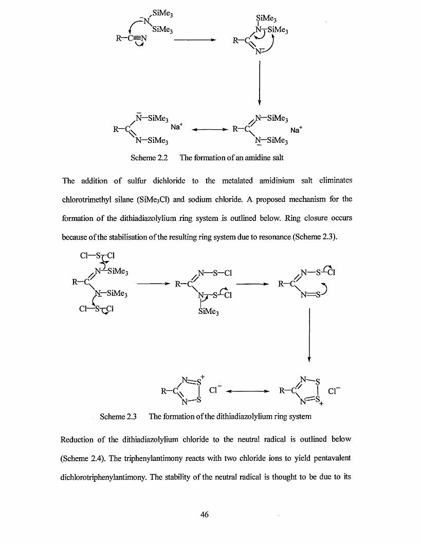
’ S i M e 3 c -a / t/ - I V ? M e 3
( SiMe3 NySiMe3R— C—N ► R—Cv
KP
R -C/
N~SiMeqNa+
/ N-SiM e3 ^ R -C ^ Na+
N-SiM e3N-SiMe3
Scheme 2.2 The formation of an amidine salt
The addition of sulfur dichloride to the metalated amidinium salt eliminates
chlorotrimethyl silane (SiMesCl) and sodium chloride. A proposed mechanism for the
formation of the dithiad iazolyl ium ring system is outlined below. Ring closure occurs
because of the stabilisation of the resulting ring system due to resonance (Scheme 2.3).
Cl—St-C1
/ N —SiMe3 R -C ^
N—SiMe3rci—s-̂ ci
R-C.&\
N—S—ClR—C
*N— S-*-Cl
Nt-S-'-CIrSiMe3
\ }N = S -y
R -C/
Nr
*N'
cr R-C.N-
\ crN'
Scheme 2.3 The formation of the dithiadiazolylium ring system
Reduction of the dithiadiazolylium chloride to the neutral radical is outlined below
(Scheme 2.4). The triphenylantimony reacts with two chloride ions to yield pentavalent
dichlorotriphenylantimony. The stability of the neutral radical is thought to be due to its
46
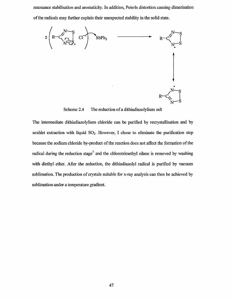
resonance stabilisation and aromaticity. In addition, Peierls distortion causing dimeiisation
of the radicals may further explain their unexpected stability in the solid state.
Scheme 2.4 The reduction of a dithiadiazolylium salt
The intermediate dithiadiazolylium chloride can be purified by recrystallisation and by
soxhlet extraction with liquid SO2. However, I chose to eliminate the purification step
because the sodium chloride by-product of the reaction does not affect the formation of the
radical during the reduction stage7 and the chlorotrimethyl silane is removed by washing
with diethyl ether. After the reduction, the dithiadiazolyl radical is purified by vacuum
sublimation. The production of crystals suitable for x-ray analysis can then be achieved by
sublimation under a temperature gradient.
47
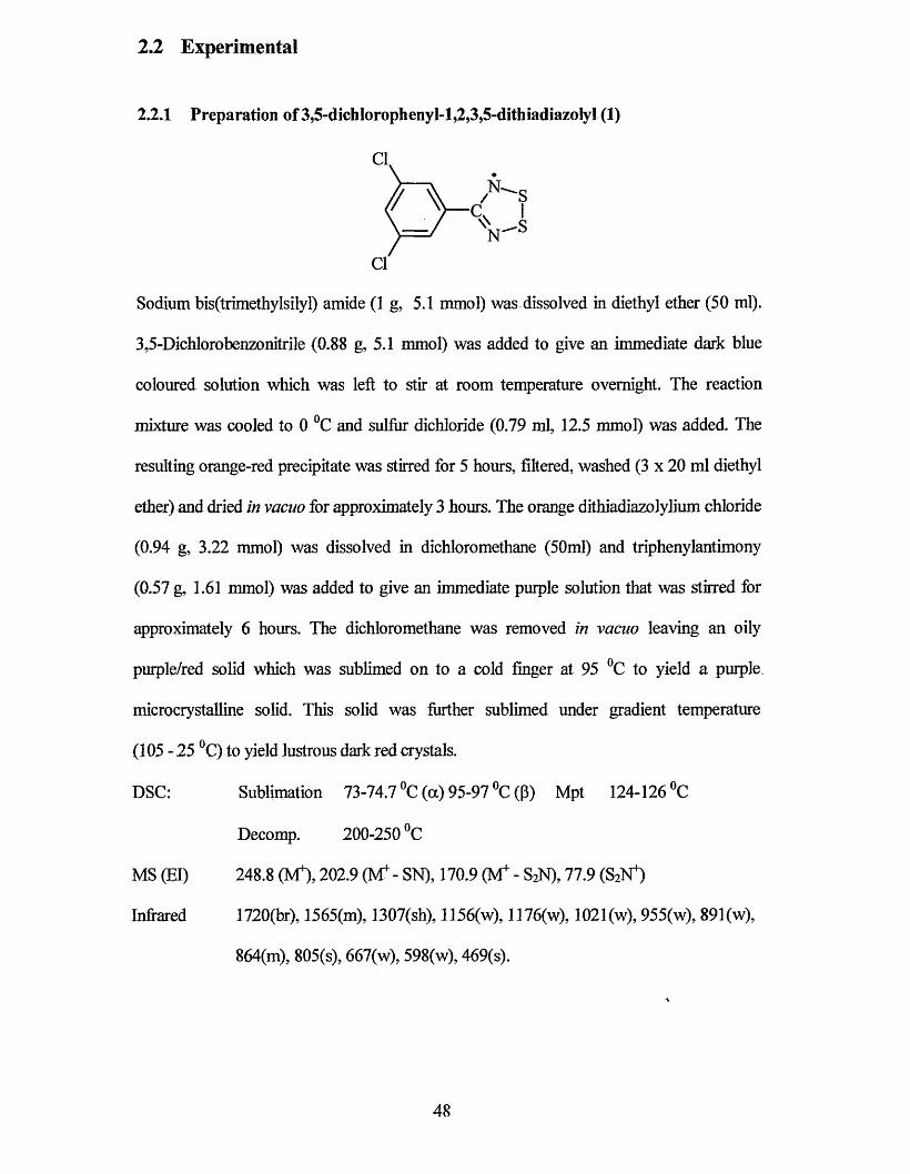
2.2 Experimental
2.2.1 Preparation of3,5-dich!orophenyI-l,2,3,5-dithiadiazoIyl (1)
% AN
Sodium bis(trimethylsilyl) amide (1 g, 5.1 mmol) was dissolved in diethyl ether (50 ml).
3,5-Dichlorobenzonitrile (0.88 g, 5.1 mmol) was added to give an immediate dark blue
coloured solution which was left to stir at room temperature overnight. The reaction
mixture was cooled to 0 °C and sulfur dichloride (0.79 ml, 12.5 mmol) was added. The
resulting orange-red precipitate was stirred for 5 hours, filtered, washed (3 x 20 ml diethyl
ether) and dried in vacuo for approximately 3 hours. The orange dithiadiazolylium chloride
(0.94 g, 3.22 mmol) was dissolved in dichloromethane (50ml) and triphenylantimony
(0.57 g, 1.61 mmol) was added to give an immediate purple solution that was stirred for
approximately 6 hours. The dichloromethane was removed in vacuo leaving an oily
purple/red solid which was sublimed on to a cold finger at 95 °C to yield a purple
microcrystalline solid. This solid was further sublimed under gradient temperature
(105 - 25 °C) to yield lustrous dark red crystals.
DSC: Sublimation 73-74.7 °C (a) 95-97 °C (p) Mpt 124-126 °C
Decomp. 200-250 °C
MS (El) 248.8 (M4), 202.9 (M4- SN), 170.9 (M4 - S2N), 77.9 (S2N4)
Infrared 1720(br), 1565(m), 1307(sh), 1156(w), 1176(w), 1021(w), 955(w), 891(w),
864(m), 805(s), 667(w), 598(w), 469(s).
48
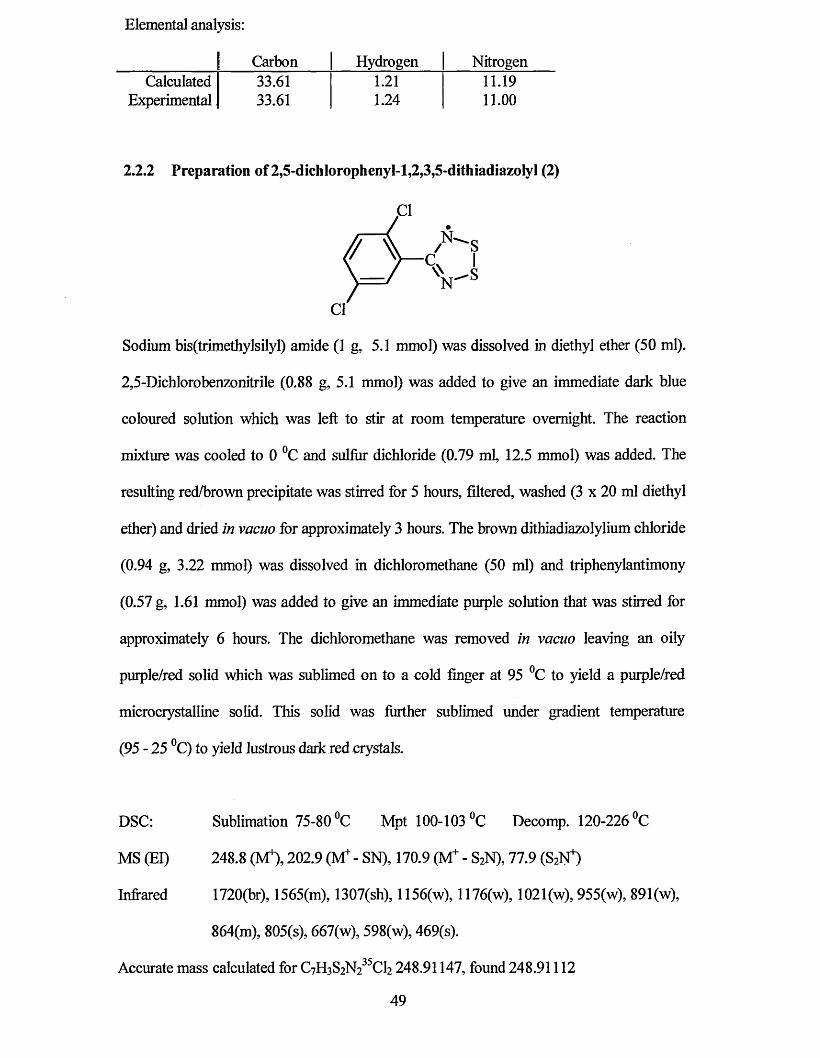
Elemental analysis:
| Carbon Hydrogen NitrogenCalculated I 33.61 1.21 11.19
Experimental J 33.61 1.24 11.00
2.2.2 Preparation of 2,5-dichlorophenyl-l,2,3,5-dithiadiazoIyl (2)
Sodium bis(trimethylsilyl) amide (1 g, 5.1 mmol) was dissolved in diethyl ether (50 ml).
2,5-Dichlorobenzonitrile (0.88 g, 5.1 mmol) was added to give an immediate dark blue
coloured solution which was left to stir at room temperature overnight. The reaction
mixture was cooled to 0 °C and sulfur dichloride (0.79 mb 12.5 mmol) was added. The
resulting red/brown precipitate was stirred for 5 hours, filtered, washed (3 x 20 ml diethyl
ether) and dried in vacuo for approximately 3 hours. The brown dithiadiazolylium chloride
(0.94 g, 3.22 mmol) was dissolved in dichloromethane (50 ml) and triphenylantimony
(0.57 g, 1.61 mmol) was added to give an immediate purple solution that was stirred for
approximately 6 hours. The dichloromethane was removed in vacuo leaving an oily
purple/red solid which was sublimed on to a cold finger at 95 °C to yield a purple/red
microcrystalline solid. This solid was further sublimed under gradient temperature
(95 - 25 °C) to yield lustrous dark red crystals.
DSC: Sublimation 75-80 °C Mpt 100-103 °C Decomp. 120-226 °C
MS (El) 248.8 (M4), 202.9 (Mf - SN), 170.9 (M+ - S2N), 77.9 (S2N4)
Infrared 1720(br), 1565(m), 1307(sh), 1156(w), 1176(w), 1021(w), 955(w), 891(w),
864(m), 805(s), 667(w), 598(w), 469(s).
Accurate mass calculated for C7H3S2N235Cl2 248.91147, found 248.91112
49
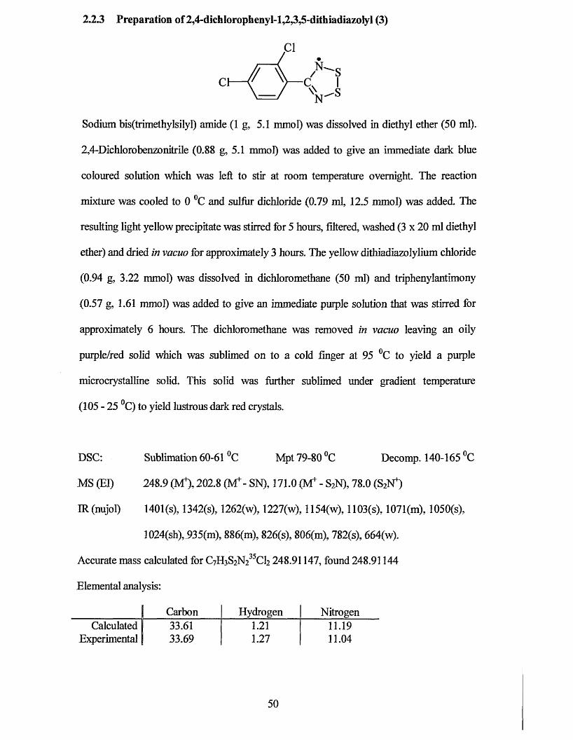
2.23 Preparation of2,4-dich!orophenyl-l,2,3,5-dithiadiazoIyl (3)
N
Sodium bis(trimethylsilyl) amide (1 g, 5.1 mmol) was dissolved in diethyl ether (50 ml).
2,4-Dichlorobenzonitrile (0.88 g, 5.1 mmol) was added to give an immediate dark blue
coloured solution which was left to stir at room temperature overnight. The reaction
mixture was cooled to 0 °C and sulfur dichloride (0.79 ml, 12.5 mmol) was added. The
resulting light yellow precipitate was stirred for 5 hours, filtered, washed (3 x 20 ml diethyl
ether) and dried in vacuo for approximately 3 hours. The yellow dithiadiazolylium chloride
(0.94 g, 3.22 mmol) was dissolved in dichloromethane (50 ml) and triphenylantimony
(0.57 g, 1.61 mmol) was added to give an immediate purple solution that was stiixed for
approximately 6 hours. The dichloromethane was removed in vacuo leaving an oily
purple/red solid which was sublimed on to a cold finger at 95 °C to yield a purple
microcrystalline solid. This solid was further sublimed under gradient temperature
(105 - 25 °C) to yield lustrous dark red crystals.
DSC: Sublimation 60-61 °C Mpt 79-80 °C Decomp. 140-165 °C
MS (El) 248.9 (M4), 202.8 (Mf- SN), 171.0 (M+ - S2N), 78.0 (S2N4)
IR (nujol) 1401 (s), 1342(s), 1262(w), 1227(w), 1154(w), 1103(s), 1071(m), 1050(s),
1024(sh), 935(m), 886(m), 826(s), 806(m), 782(s), 664(w).
Accurate mass calculated for C7H3S2N235Cl2 248.91147, found 248.91144
Elemental analysis:
1 Carbon Hydrogen NitrogenCalculated 1 33.61 1.21 11.19
Experimental j 33.69 1.27 11.04
50
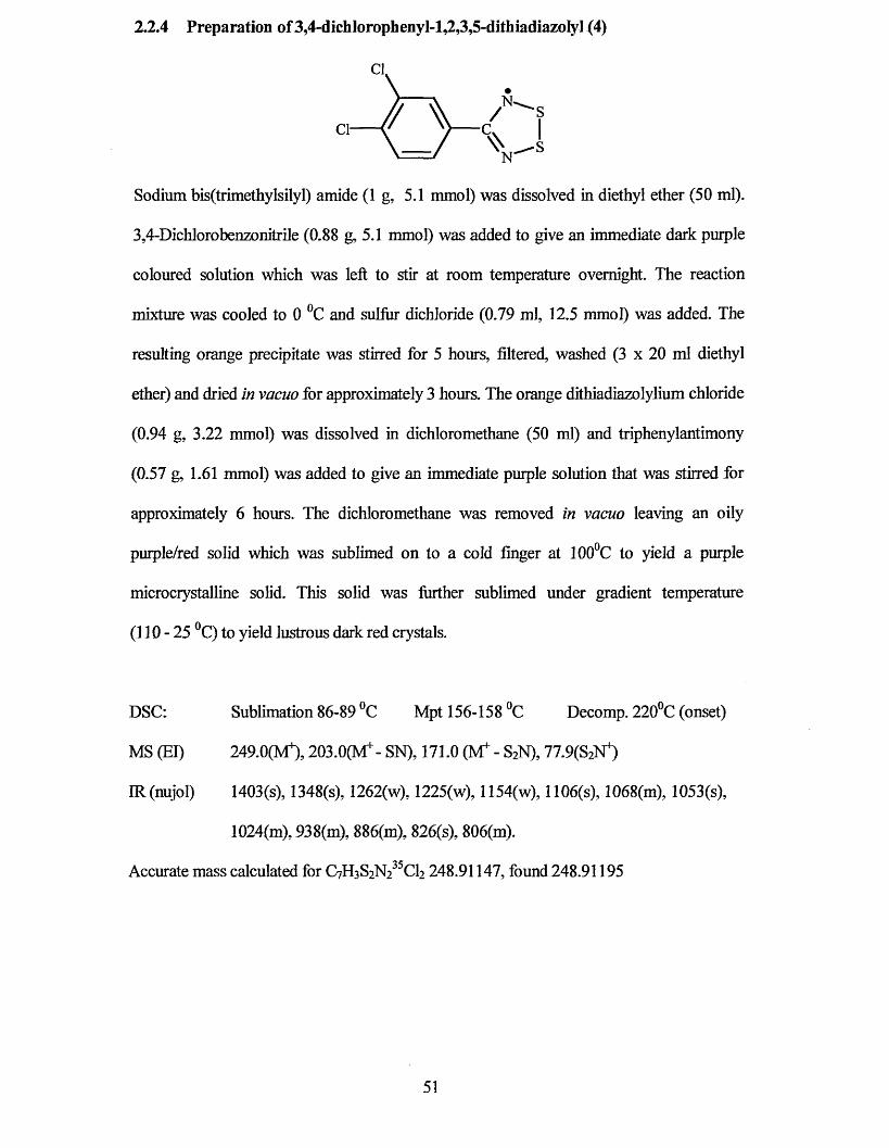
2.2.4 Preparation of 3,4-dichlorophenyl-l,2,3,5-dithiadiazoIyI (4)
Cl
f/ V_ASV *
Sodium bis(trimethylsilyl) amide (1 g, 5.1 mmol) was dissolved in diethyl ether (50 ml).
3,4-Dichlorobenzonitrile (0.88 g, 5.1 mmol) was added to give an immediate dark purple
coloured solution which was left to stir at room temperature overnight. The reaction
mixture was cooled to 0 °C and sulfur dichloride (0.79 ml, 12.5 mmol) was added. The
resulting orange precipitate was stirred for 5 hours, filtered, washed (3 x 20 ml diethyl
ether) and dried in vacuo for approximately 3 hours. The orange dithiadiazolylium chloride
(0.94 g, 3.22 mmol) was dissolved in dichloromethane (50 ml) and triphenylantimony
(0.57 g, 1.61 mmol) was added to give an immediate purple solution that was stirred for
approximately 6 hours. The dichloromethane was removed in vacuo leaving an oily
purple/red solid which was sublimed on to a cold finger at 100°C to yield a purple
microcrystalline solid. This solid was further sublimed under gradient temperature
(110 - 25 °C) to yield lustrous dark red crystals.
DSC: Sublimation 86-89 °C Mpt 156-158 °C Decomp. 220°C (onset)
MS (El) 249.0(M+), 2 0 3 .0 ^ - SN), 171.0 (Mf - S2N), 77.9(82^
IR (nujol) 1403(s), 1348(s), 1262(w), 1225(w), 1154(w), 1106(s), 1068(m), 1053(s),
1024(m), 938(m), 886(m), 826(s), 806(m).
Accurate mass calculated for C7H3S2N235C12 248.91147, found 248.91195
51
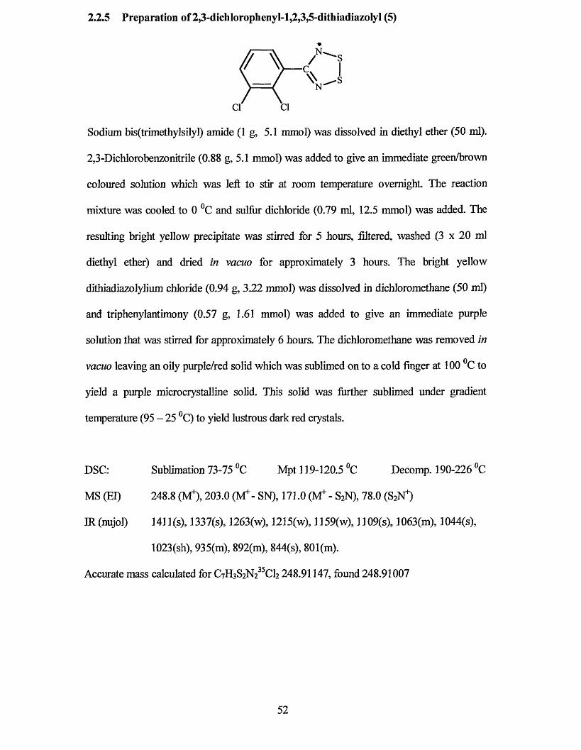
2.2.5 Preparation of 2,3-dichIorophenyl-l,2,3,5-dithiadiazolyl (5)
/ scw I
Sodium bis(trimethylsilyl) amide (1 g, 5.1 mmol) was dissolved in diethyl ether (50 ml).
2,3-Dichlorobenzonitrile (0.88 g, 5.1 mmol) was added to give an immediate green/brown
coloured solution which was left to stir at room temperature overnight The reaction
mixture was cooled to 0 °C and sulfur dichloride (0.79 ml, 12.5 mmol) was added. The
resulting bright yellow precipitate was stirred for 5 hours, filtered, washed (3 x 20 ml
diethyl ether) and dried in vacuo for approximately 3 hours. The bright yellow
dithiadiazolylium chloride (0.94 g, 3.22 mmol) was dissolved in dichloromethane (50 ml)
and triphenylantimony (0.57 g, 1.61 mmol) was added to give an immediate purple
solution that was stirred for approximately 6 hours. The dichloromethane was removed in
vacuo leaving an oily purple/red solid which was sublimed on to a cold finger at 100 °C to
yield a purple microcrystalline solid. This solid was further sublimed under gradient
temperature (95 - 25 °C) to yield lustrous dark red crystals.
DSC: Sublimation 73-75 °C Mpt 119-120.5 °C Decomp. 190-226 °C
MS (El) 248.8 (M4), 203.0 (M4- SN), 171.0 (M4 - S2N), 78.0 (S2N4)
IR(nujoJ) 141 l(s), 1337(s), 1263(w), 1215(w), 1159(w), 1109(s), 1063(m), 1044(s),
1023(sh), 935(m), 892(m), 844(s), 801(m).
Accurate mass calculated for 248.91147, found 248.91007
52
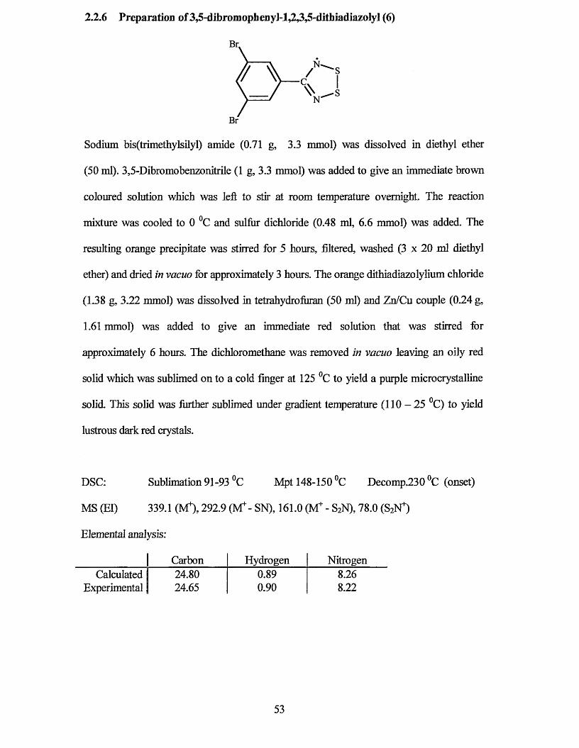
2.2.6 Preparation of3,5-dibromophenyl-l,2,3,5-dithiadiazoly] (6)
Sodium bis(trimethylsilyl) amide (0.71 g, 3.3 mmol) was dissolved in diethyl ether
(50 ml). 3,5-Dibromobenzonitrile (1 g, 3.3 mmol) was added to give an immediate brown
coloured solution which was left to stir at room temperature overnight. The reaction
mixture was cooled to 0 °C and sulfur dichloride (0.48 ml, 6.6 mmol) was added. The
resulting orange precipitate was stirred for 5 hours, filtered, washed (3 x 20 ml diethyl
ether) and dried in vacuo for approximately 3 hours. The orange dithiadiazolylium chloride
(1.38 g, 3.22 mmol) was dissolved in tetrahydrofiiran (50 ml) and Zn/Cu couple (0.24 g,
1.61 mmol) was added to give an immediate red solution that was stirred for
approximately 6 hours. The dichloromethane was removed in vacuo leaving an oily red
solid which was sublimed on to a cold finger at 125 °C to yield a purple microcrystalline
solid. This solid was further sublimed under gradient temperature (110-25 °C) to yield
lustrous dark red crystals.
DSC: Sublimation 91-93 °C Mpt 148-150 °C Decomp.230 °C (onset)
MS (El) 339.1 (M4), 292.9 (M4"- SN), 161.0 (VT - S2N), 78.0 (SiN*)
Elemental analysis:
| Carbon Hydrogen NitrogenCalculated 1 24.80 0.89 8.26
Experimental | 24.65 0.90 8.22
Br.
N'/
\ \N'
53
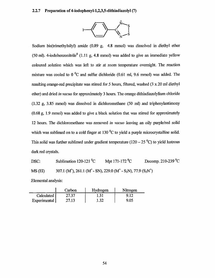
2.2.7 Preparation of 4-iodophenyl-l,2,3,5-dithiadiazolyl (7)
Sodium bis(trimethylsilyl) amide (0.89 g, 4.8 mmol) was dissolved in diethyl ether
(50 ml). 4-iodobenzonitrile8 (1.11 g, 4.8 mmol) was added to give an immediate yellow
coloured solution which was left to stir at room temperature overnight The reaction
mixture was cooled to 0 °C and sulfur dichloride (0.61 ml, 9.6 mmol) was added. The
resulting orange-red precipitate was stirred for 5 hours, filtered, washed (3 x 20 ml diethyl
ether) and dried in vacuo for approximately 3 hours. The orange dithiadiazolylium chloride
(L32 g, 3.85 mmol) was dissolved in dichloromethane (50 ml) and triphenylantimony
(0.68 g, 1.9 mmol) was added to give a black solution that was stirred for approximately
12 hours. The dichloromethane was removed in vacuo leaving an oily purple/red solid
which was sublimed on to a cold finger at 130 °C to yield a purple microcrystalline solid.
This solid was further sublimed under gradient temperature (120 - 25 °C) to yield lustrous
dark red crystals.
DSC: Sublimation 120-121 °C Mpt 171-172 °C Decomp. 210-239 °C
MS (El) 307.1 (M4), 261.1 (IVT- SN), 229.0 (M4 - S2N), 77.9 (S2N4)
Elemental analysis:
Carbon Hydrogen NitrogenCalculated 27.37
Experimental 27.131.311.32
9.129.05
54
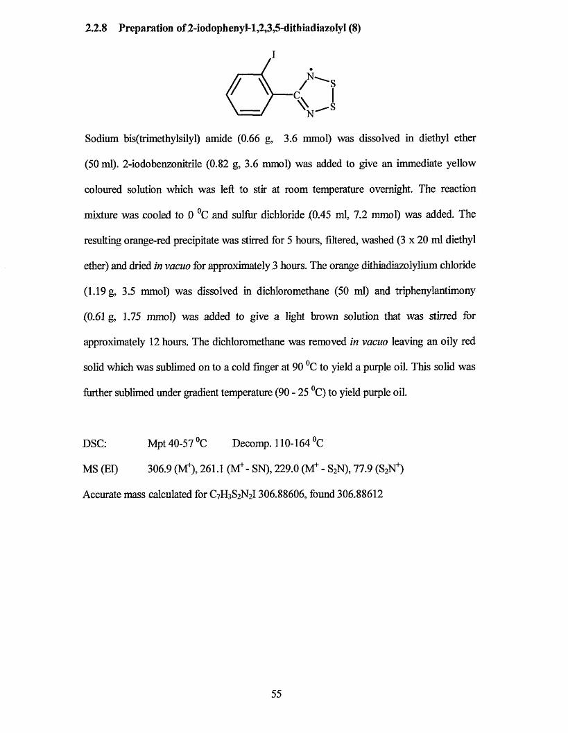
2.2,8 Preparation of 2-iodophenyI-l,2,3,5-dithiadiazoIyI (8)
/ i c\ \XVS
Sodium bis(trimethylsilyl) amide (0.66 g, 3.6 mmol) was dissolved in diethyl ether
(50 ml). 2-iodobenzonitrile (0.82 g, 3.6 mmol) was added to give an immediate yellow
coloured solution which was left to stir at room temperature overnight. The reaction
mixture was cooled to 0 °C and sulfur dichloride (0.45 ml, 7 .2 mmol) was added. The
resulting orange-red precipitate was stirred for 5 hours, filtered, washed (3 x 20 ml diethyl
ether) and dried in vacuo for approximately 3 hours. The orange dithiadiazolylium chloride
(1.19 g, 3.5 mmol) was dissolved in dichloromethane (50 ml) and triphenylantimony
(0,61 g, 1,75 mmol) was added to give a light brown solution that was stirred for
approximately 12 hours. The dichloromethane was removed in vacuo leaving an oily red
solid which was sublimed on to a cold finger at 90 °C to yield a purple oil. This solid was
further sublimed under gradient temperature (90 - 25 °C) to yield purple oil.
DSC: Mpt 40-57 °C Decomp. 110-164 °C
MS (El) 306.9 (M*), 261.1 (M+- SN), 229.0 (M+ - S2N), 77.9 (S2N")
Accurate mass calculated for C7H3S2N21306,88606, found 306.88612
55
2.3 References
1 Synthesis was unsuccessful
2 J.M. Rawson, AJ. Banister and L Lavender, Adv. Heterocycl Chem., 1995, 62, 137
and references therein.
3 AR. Sanger, Inorg. Nucl. Chem. Letters, 1973,9,351.
4 R.T. Boere, R.T Oakley and R.W. Reed, J. Organomet. Chem., 1987,331,161.
5 R.T. Boere, RT Oakley and R.W. Reed,./ Organomet Chem., 1987,331, 161.
6 M. Amin and C.W. Rees, J. Chem. Soc., Perkin Trans. 1 ,1989,2495.
7 I. May, PhD Thesis, Univeristy of Durham, 1995.
8 (a) N. Bricklebank, S. Hargreaves and S.E. Spey, Polyhedron, 2000, 19, 1163.
(b) E.O. Schlemperand D.Britton, Acta. Cryst, 1965,18,419.
56
Chapter 3
Stacking Motifs for Dichlorophenyl
Dithiadiazolyl Radicals
57
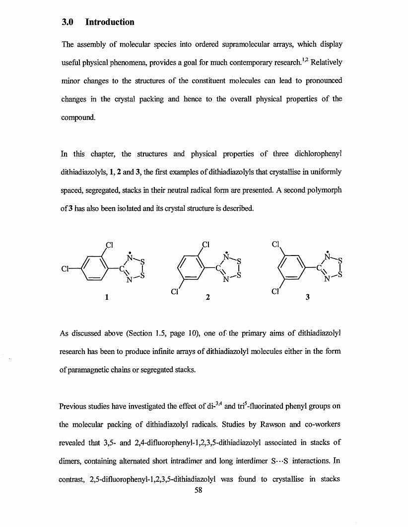
3.0 Introduction
The assembly of molecular species into ordered supramolecular arrays, which display
useful physical phenomena, provides a goal for much contemporary research.1,2 Relatively
minor changes to the structures of the constituent molecules can lead to pronounced
changes in the crystal packing and hence to the overall physical properties of the
compound.
In this chapter, the structures and physical properties of three dichlorophenyl
dithiadiazolyls, 1,2 and 3, the first examples of dithiadiazolyls that crystallise in uniformly
spaced, segregated, stacks in their neutral radical form are presented. A second polymorph
of 3 has also been isolated and its crystal structure is described.
As discussed above (Section 1.5, page 10), one of the primary aims of dithiadiazolyl
research has been to produce infinite arrays of dithiadiazolyl molecules either in the form
of paramagnetic chains or segregated stacks.
Previous studies have investigated the effect of di-3,4 and tri5-fluorinated phenyl groups on
the molecular packing of dithiadiazolyl radicals. Studies by Rawson and co-workers
revealed that 3,5- and 2,4-difluorophenyl-l,2,3,5-dithiadiazolyl associated in stacks of
dimers, containing alternated short intradimer and long interdimer S---S interactions. In
contrast, 2,5-difluorophenyl-l,2,3,5-dithiadiazolyl was found to crystallise in stacks58
containing uniformly spaced radical units with an unexpectedly long intermolecular S---S
distance of 3.554(3) A. However, a re-examination of the structure of 2,5-difluorophenyl-
1,2,3,5-dithiadiazolyl by Oakley and co-workers6 discovered that the original structure was
a superlattice and the true structure was actually segregated stacks of dimer pairs, like
those of the analogous 2,4- and 3,5-difluorophenyl isomers. This conclusion is supported
by the absence of paramagnetic or conductive behaviour that might be expected for an
evenly spaced (undimerised) radical.6 Stacking motifs within dithiadiazolyls have also
been observed in the form of partially oxidised radicals,7,8,9 which exhibit conductivity at
room temperature.
Fluorine •••Fluorine interactions are generally considered to be of a repulsive nature.10 It
has been suggested that homonuclear interactions between heavier halogens are attractive10
and that Cl - • - Cl interactions can have a marked effect on the solid state structure.11
It has long been accepted that many intermolecular X---X (X= Cl, Br and I) distances in
• • 12molecular crystals are significantly less than the sum of the accepted van der Waals radii.
These X - • - X interactions have special significance in crystal engineering or the deliberate
• 13design of organic crystal structures because they may be used to create layered motifs,
which are associated with specific solid state reactivity patterns. These have previously
been described as “donor-acceptor”, “secondary” and “charge transfer” interactions. More
recently, the interactions have been described in terms of highest occupied molecular
orbital and lowest unoccupied molecular orbital (HOMO-LUMO) and nucleophilic
attack.14 In all such cases, it has been noticed that there is a directional preference in which
contacting groups position themselves. Indeed, it has been shown11 that in organic
molecular solids, association is only via certain geometrical motifs.10 This is thought to be
due to either specific attractive forces of chlorine in certain directions (i.e. increased
59
1 *7attraction) or non-spherical shapes with polar flattening in a close packed structure
{i.e. decreased repulsion). A statistical approach employed by Desiraju and Parthasarathy
concluded that there were indeed attractive (and anisotropic) forces within Cl*-Cl
interactions.10 Irrespective of the origin of the effect, it would appear that chlorine atoms
attached directly to an organic group tend to “steer” the crystal structure to a so-called
“p-mode”.11 This is characterised by a short crystallographic axis of c.a. 4 A with the
molecules packing in layered sheets and stacks. This phenomenon has been observed for at
least one hundred published polychlorinated-aromatics.10 The p-structure may therefore
have great significance in the design of low dimensional organic metals and
superconductors.
To date only one chlorine-substituted phenyl derivative of a dithiadiazolyl radical has been
reported, /?-chlorophenyl-1,2,3,5-dithiadiazolyl.15 In the solid state it is a dimer
(Figure 3.1) but exhibits packing of an unusual type for dithiadiazolyls. The discrete
dimers form into two interpenetrating ribbons, which are orthogonal to one another. The
ribbons are connected by a triangle of S* • - Cl contacts along the plane of the dithiadiazolyl
ring of 3.3 8 A and 3.52 A. These distances are less than the sum of the van der Waals radii
(3.5 - 3.7 A). The publication noted the similarity of interaction between this compound
and cyano substituents attached to aryl dithiadiazolyls.16,17
60
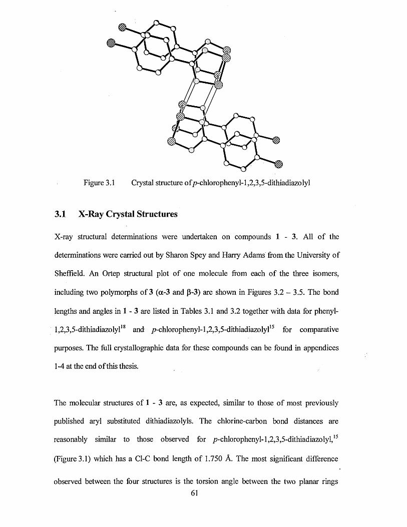
Figure 3.1 Crystal structure of/?-chlorophenyl-l,2,3,5-dithiadiazolyl
3.1 X-Ray Crystal Structures
X-ray structural determinations were undertaken on compounds 1 - 3 . All of the
determinations were carried out by Sharon Spey and Harry Adams from the University of
Sheffield. An Ortep structural plot of one molecule from each of the three isomers,
including two polymorphs of 3 ( a - 3 and p - 3 ) are shown in Figures 3.2 - 3.5. The bond
lengths and angles in 1 - 3 are listed in Tables 3.1 and 3.2 together with data for phenyl-
1,2,3,5-dithiadiazolyl18 and /7-chlorophenyl-1,2,3,5-dithiadiazolyl15 for comparative
purposes. The full crystallographic data for these compounds can be found in appendices
1-4 at the end of this thesis.
The molecular structures of 1 - 3 are, as expected, similar to those of most previously
published aryl substituted dithiadiazolyls. The chlorine-carbon bond distances are
reasonably similar to those observed for /?-chlorophenyl-l,2,3,5-dithiadiazolyl,15
(Figure 3.1) which has a Cl-C bond length of 1.750 A. The most significant difference
observed between the four structures is the torsion angle between the two planar rings61
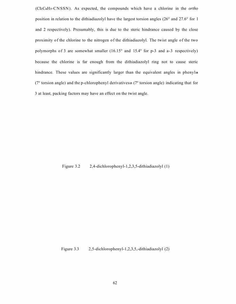
(CI2C6H3-CNSSN). As expected, the compounds which have a chlorine in the ortho
position in relation to the dithiadiazolyl have the largest torsion angles (26° and 27.6° for 1
and 2 respectively). Presumably, this is due to the steric hindrance caused by the close
proximity o f the chlorine to the nitrogen o f the dithiadiazolyl. The twist angle o f the two
polymorphs o f 3 are somewhat smaller (16.15° and 15.4° for p-3 and a-3 respectively)
because the chlorine is far enough from the dithiadiazolyl ring not to cause steric
hindrance. These values are significantly larger than the equivalent angles in phenyl18
(7° torsion angle) and the p-chlorophenyl derivatives15 (7° torsion angle) indicating that for
3 at least, packing factors may have an effect on the twist angle.
Figure 3.2 2,4-dichlorophenyl-1,2,3,5-dithiadiazolyl (1)
Figure 3.3 2,5-dichlorophenyl-1,2,3,5,-dithiadiazolyl (2)
62
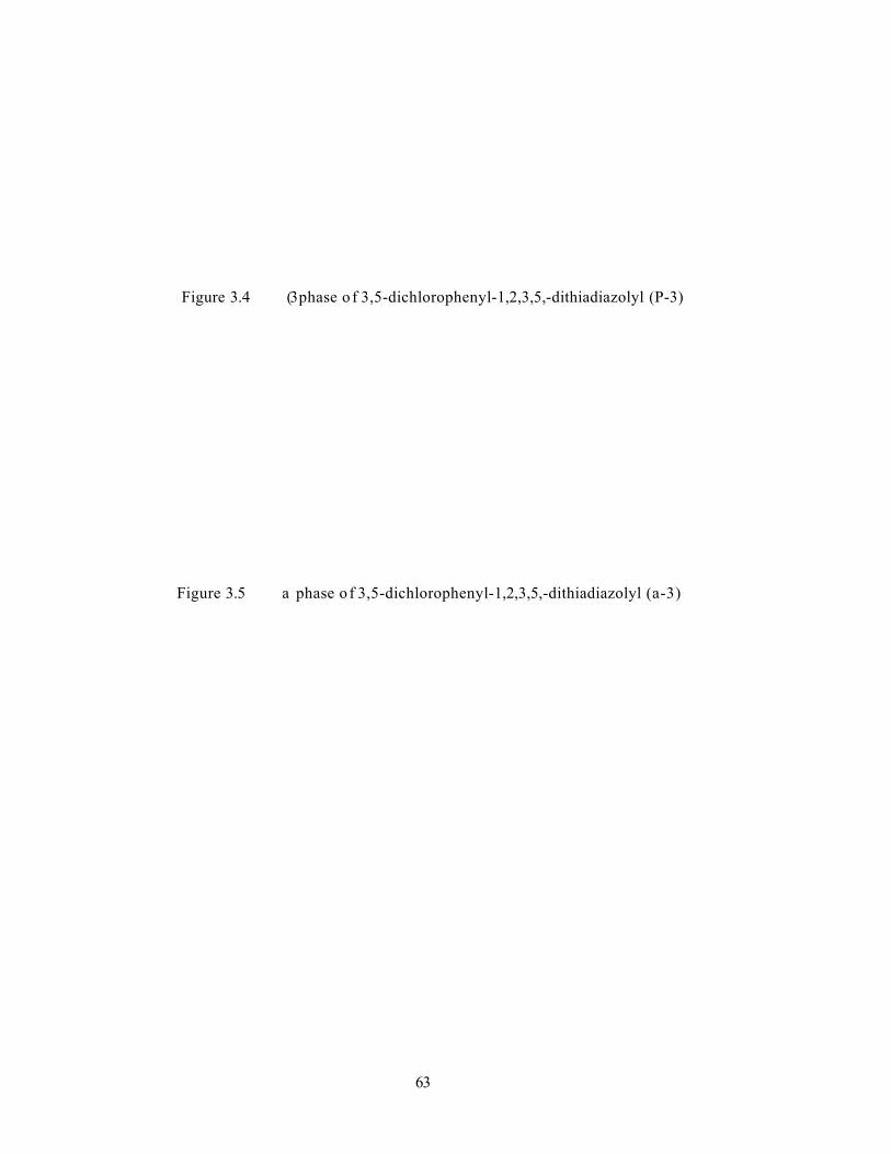
Figure 3.4 (3 phase o f 3,5-dichlorophenyl-1,2,3,5,-dithiadiazolyl (P-3)
Figure 3.5 a phase o f 3,5-dichlorophenyl-1,2,3,5,-dithiadiazolyl (a-3)
63

Table 3.1 Bond lengths of 1-3 (A).
B o n d D is ta n c e (A )
Atom No. 1 2 P-3* / > *a-3 P h p -c ic 6h 4
Cl(l)-C(l) 1.727(4) 1.735(3) - - - -
Cl(l)-C(2) - - 1.743(5) 1.727(4) - -
Cl(l)-C(4) - - - - - 1.750(4)
Cl(2)-C(3) 1.727(5) - - - - -
Cl(2)-C(4) - 1.738(3) 1.732(5) 1.743(4) - -
S(l)-N(l) 1.620(5) 1.631(3) 1.633(5) 1.626(4) 1.63 1.643(10)
S(l)-S(2) 2.090(2) 2.0973(18) 2.0865(19) 2.0920(16) 2.096 2.103(5)
S(2)-N(2) 1.625(4) 1.623(3) 1.627(5) 1.634(4) 1.63 1.651(10)
N(l)-C(7) 1.332(5) 1.335(5) 1.347(6) 1.336(5) 1.32 1.320(1)
N(2)-C(7) 1.332(6) 1.338(5) 1.339(6) 1.343(5) 1.31 1.340(1)
C(l)-C(2) 1.377(6) 1.387(5) 1.376(7) 1.391(5) 1.37 1.365(7)
C(l)-C(6) 1.406(6) 1.398(5) 1.390(7) 1.398(5) 1.37 1.410(7)
C(2)-C(3) 1.388(6) 1.378(5) 1.386(7) 1.390(5) 1.39 1.385(8)
C(3)-C(4) 1.373(6) 1.380(5) 1.382(7) 1.377(6) 1.42 1.371(7)
C(4)-C(5) 1.384(6) 1.384(5) 1.385(7) 1.376(6) 1.36 1.376(8)
C(5)-C(6) 1.396(5) 1.404(4) 1.397(6) 1.400(5) 1.38 1.401(6)
C(6)-C(7)*
1.471(6) 1.484(5) 1.476(7) 1.476(6) 1.50 1.472(7)jj* ----- - - _
Denotes the bond lengths of a representative molecule within an asymmetric unit
containing more than one molecule.
64
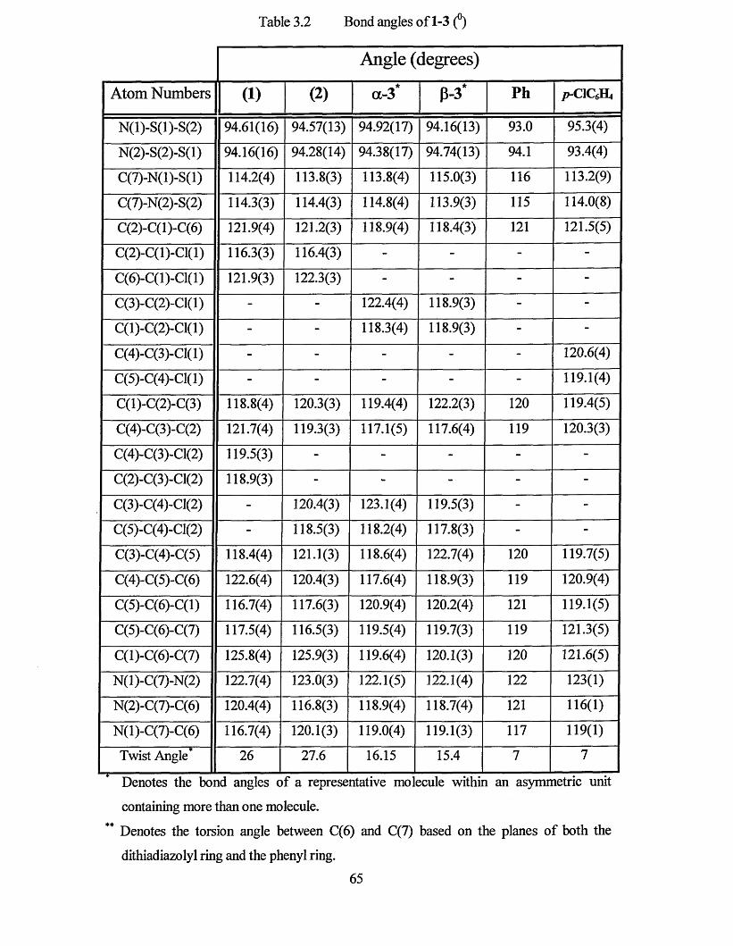
Table 3.2 Bond angles of 1-3 (°)
Angle (degrees)
Atom Numbers (1) (2) a-3* "kP-3 P h p - CIQH4
N(l)-S(l)-S(2) 94.61(16) 94.57(13) 94.92(17) 94.16(13) 93.0 95.3(4)
N(2)-S(2)-S(l) 94.16(16) 94.28(14) 94.38(17) 94.74(13) 94.1 93.4(4)
C(7)-N(l)-S(l) 114.2(4) 113.8(3) 113.8(4) 115.0(3) 116 113.2(9)
C(7)-N(2)-S(2) 114.3(3) 114.4(3) 114.8(4) 113.9(3) 115 114.0(8)
C(2)-C(l)-C(6) 121.9(4) 121.2(3) 118.9(4) 118.4(3) 121 121.5(5)
C(2)-C(1)-C1(1) 116.3(3) 116.4(3) - - - -
C(6)-C( 1 )-Cl( 1) 121.9(3) 122.3(3) - - - -
C(3)-C(2)-C1(1) - - 122.4(4) 118.9(3) - -
C(1)-C(2)-C1(1) - - 118.3(4) 118.9(3) - -
C(4)-C(3)-C1(1) - - - - - 120.6(4)
C(5)-C(4)-C1(1) - - - - - 119.1(4)
C(l)-C(2)-C(3) 118.8(4) 120.3(3) 119.4(4) 122.2(3) 120 119.4(5)
C(4)-C(3)-C(2) 121.7(4) 119.3(3) 117.1(5) 117.6(4) 119 120.3(3)
C(4)-C(3)-C1(2) 119.5(3) - - - - -
C(2)-C(3)-C1(2) 118.9(3) - - - - -
C(3)-C(4)-C1(2) - 120.4(3) 123.1(4) 119.5(3) - -
C(5)-C(4)-C1(2) - 118.5(3) 118.2(4) 117.8(3) - -
C(3)-C(4)-C(5) 118.4(4) 121.1(3) 118.6(4) 122.7(4) 120 119.7(5)
C(4)-C(5)-C(6) 122.6(4) 120.4(3) 117.6(4) 118.9(3) 119 120.9(4)
C(5)-C(6)-C(l) 116.7(4) 117.6(3) 120.9(4) 120.2(4) 121 119.1(5)
C(5)-C(6)-C(7) 117.5(4) 116.5(3) 119.5(4) 119.7(3) 119 121.3(5)
C(l)-C(6)-C(7) 125.8(4) 125.9(3) 119.6(4) 120.1(3) 120 121.6(5)
N(l)-C(7)-N(2) 122.7(4) 123.0(3) 122.1(5) 122.1(4) 122 123(1)
N(2)-C(7)-C(6) 120.4(4) 116.8(3) 118.9(4) 118.7(4) 121 116(1)
N(l)-C(7)-C(6) 116.7(4) 120.1(3) 119.0(4) 119.1(3) 117 119(1)
Twist Angle* 26 27.6 16.15 15.4 7 7
Denotes the bond angles of a representative molecule within an asymmetric unit
containing more than one molecule.
** Denotes the torsion angle between C(6) and C(7) based on the planes of both the
dithiadiazolyl ring and the phenyl ring.
65
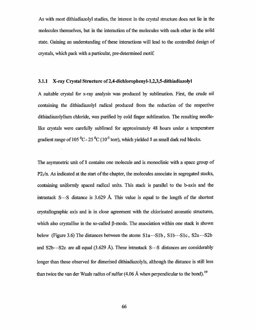
As with most dithiadiazolyl studies, the interest in the crystal structure does not lie in the
molecules themselves, but in the interaction of the molecules with each other in the solid
state. Gaining an understanding of these interactions will lead to the controlled design of
crystals, which pack with a particular, pre-determined motif.
3.1.1 X-ray Crystal Structure of 2,4-dichlorophenyl-l,2,3,5-dithiadiazolyl
A suitable crystal for x-ray analysis was produced by sublimation. First, the crude oil
containing the dithiadiazolyl radical produced from the reduction of the respective
dithiadiazolylium chloride, was purified by cold finger sublimation. The resulting needle
like crystals were carefully sublimed for approximately 48 hours under a temperature
gradient range of 105 °C- 25 °C (10'3 torr), which yielded 1 as small dark red blocks.
The asymmetric unit of 1 contains one molecule and is monoclinic with a space group of
P2i/n. As indicated at the start of the chapter, the molecules associate in segregated stacks,
containing uniformly spaced radical units. This stack is parallel to the b-axis and the
intrastack S---S distance is 3.629 A. This value is equal to the length of the shortest
crystallographic axis and is in close agreement with the chlorinated aromatic structures,
which also crystallise in the so-called p-mode. The association within one stack is shown
below (Figure 3.6) The distances between the atoms Sla---Slb, S ib-•-Sic, S2a--*S2b
and S2b"*S2c are all equal (3.629 A). These intrastack S---S distances are considerably
longer than those observed for dimerised dithiadiazolyls, although the distance is still less
than twice the van der Waals radius of sulfur (4.06 A when perpendicular to the bond).19
66
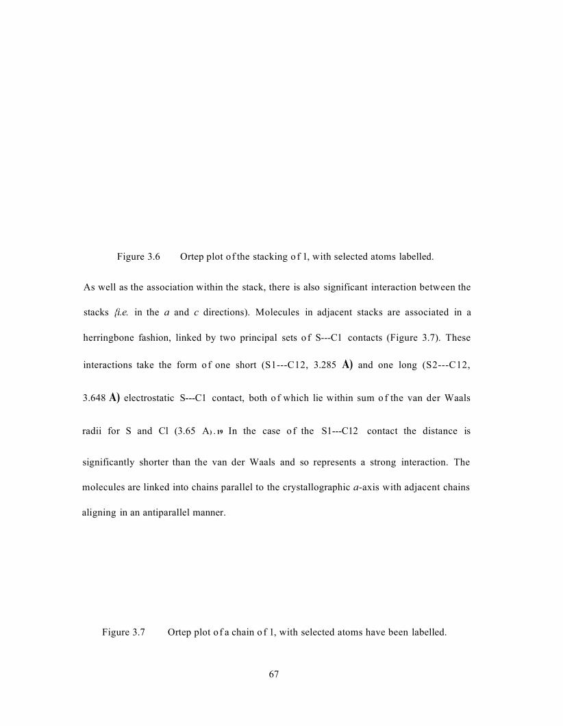
Figure 3.6 Ortep plot of the stacking o f 1, with selected atoms labelled.
As well as the association within the stack, there is also significant interaction between the
stacks {i.e. in the a and c directions). Molecules in adjacent stacks are associated in a
herringbone fashion, linked by two principal sets o f S---C1 contacts (Figure 3.7). These
interactions take the form of one short (S1---C12, 3.285 A) and one long (S2---C12,
3.648 A) electrostatic S---C1 contact, both o f which lie within sum o f the van der Waals
radii for S and Cl (3.65 A) . 19 In the case o f the S1---C12 contact the distance is
significantly shorter than the van der Waals and so represents a strong interaction. The
molecules are linked into chains parallel to the crystallographic a-axis with adjacent chains
aligning in an antiparallel manner.
Figure 3.7 Ortep plot of a chain o f 1, with selected atoms have been labelled.
67
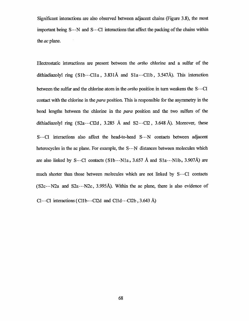
Significant interactions are also observed between adjacent chains (Figure 3.8), the most
important being S - • - N and S* • - Cl interactions that affect the packing of the chains within
the ac plane.
Electrostatic interactions are present between the ortho chlorine and a sulfur of the
dithiadiazolyl ring (S ib-•-Clla, 3.831A and S la—C llb , 3.547A). This interaction
between the sulfur and the chlorine atom in the ortho position in turn weakens the S---C1
contact with the chlorine in the para position. This is responsible for the asymmetry in the
bond lengths between the chlorine in the para position and the two sulfurs of the
dithiadiazolyl ring (S2a---C12d, 3.285 A and S2---C12, 3.648 A). Moreover, these
S---C1 interactions also affect the head-to-head S---N contacts between adjacent
heterocycles in the ac plane. For example, the S---N distances between molecules which
are also linked by S---C1 contacts (Slb---N la, 3.657 A and Sla---N lb, 3.907A) are
much shorter than those between molecules which are not linked by S---C1 contacts
(S2c---N2a and S2a---N2c, 3.995A). Within the ac plane, there is also evidence of
Cl • • • Cl interactions ( Cll b • • • C12d and Cll d • • • C12b, 3.643 A)
68
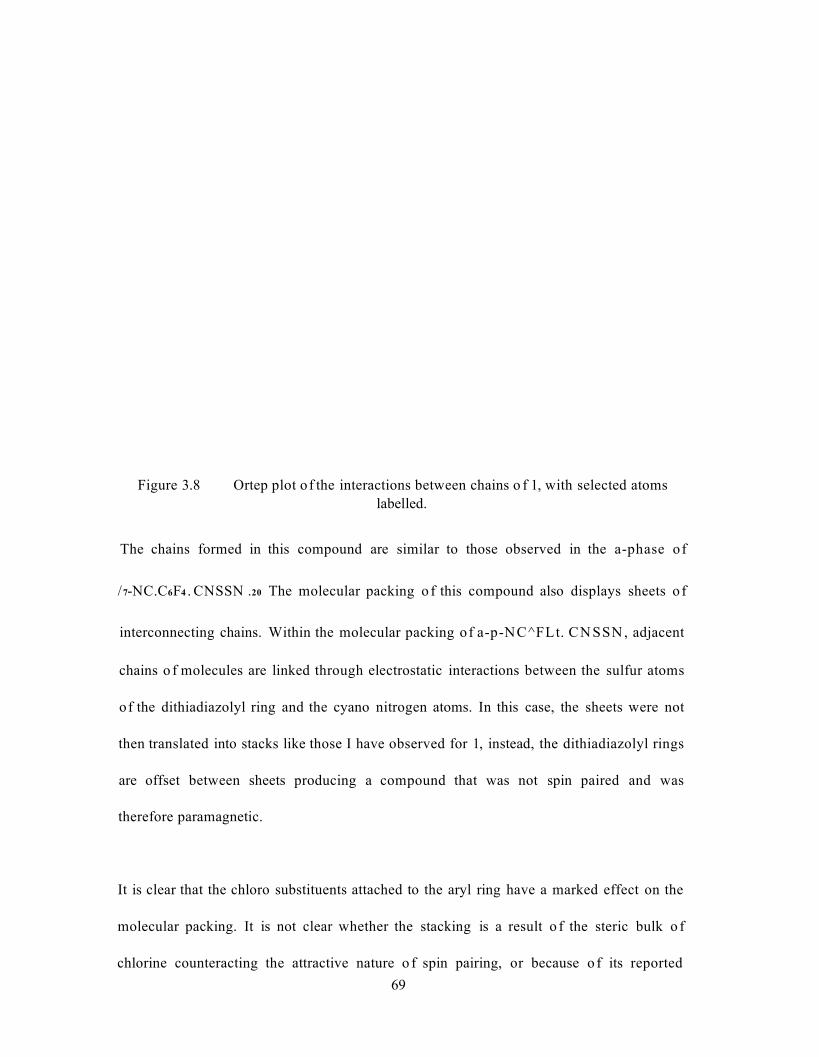
Figure 3.8 Ortep plot of the interactions between chains o f 1, with selected atomslabelled.
The chains formed in this compound are similar to those observed in the a-phase of
/7-NC.C6F4 . CNSSN .20 The molecular packing o f this compound also displays sheets o f
interconnecting chains. Within the molecular packing of a-p-NC^FLt. CN SSN , adjacent
chains o f molecules are linked through electrostatic interactions between the sulfur atoms
of the dithiadiazolyl ring and the cyano nitrogen atoms. In this case, the sheets were not
then translated into stacks like those I have observed for 1, instead, the dithiadiazolyl rings
are offset between sheets producing a compound that was not spin paired and was
therefore paramagnetic.
It is clear that the chloro substituents attached to the aryl ring have a marked effect on the
molecular packing. It is not clear whether the stacking is a result o f the steric bulk o f
chlorine counteracting the attractive nature o f spin pairing, or because o f its reported69
directional effects.11 Studies of the literature would suggest that this type of structure is
typical of chlorinated aryl groups, many of which exhibit stacked and layered motifs.10 The
structure does show some similarities to that ofp-C\.C$U. CNSSN 15 (Figure 3.1) because
of the close proximity of the S - • - Cl interactions within a chain of molecules. Despite this,
the molecular packing of 1 and p-C\.C<£U. CNSSN are considerably different. This can
only be attributed to the interactions of the second chlorine both within the plane and
through the stack. Unfortunately, although all the other isomers of the difluorophenyl-
1,2,3,5-dithiadiazolyl have been reported,4 the 2,4-isomer was not studied so no
comparison can be drawn.
Figure 3.9 shows the overall crystal packing of 1 looking down the crystallographic &-axis.
In the light of new literature6 describing the ability of this type of compound to form a
superlattice, great effort was taken to search for this possibility. No superlattice structure
could be refined from the data produced.
70

71
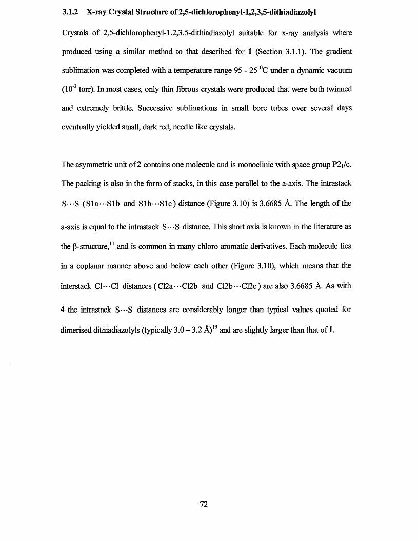
3.1.2 X-ray Crystal Structure of 2,5-dichlorophenyl-l,2,3,5-dithiadiazolyl
Crystals of 2,5-dichlorophenyl-l,2,3,5-dithiadiazolyl suitable for x-ray analysis where
produced using a similar method to that described for 1 (Section 3.1.1). The gradient
sublimation was completed with a temperature range 95 - 25 °C under a dynamic vacuum
(10'3 torr). In most cases, only thin fibrous crystals were produced that were both twinned
and extremely brittle. Successive sublimations in small bore tubes over several days
eventually yielded small, dark red, needle like crystals.
The asymmetric unit of 2 contains one molecule and is monoclinic with space group P2j/c.
The packing is also in the form of stacks, in this case parallel to the a-axis. The intrastack
S---S (Sla-*-Slb and Slb--*Slc) distance (Figure 3.10) is 3.6685 A. The length of the
a-axis is equal to the intrastack S---S distance. This short axis is known in the literature as
the p-structure,11 and is common in many chloro aromatic derivatives. Each molecule lies
in a coplanar manner above and below each other (Figure 3.10), which means that the
interstack C1---C1 distances (C12a---C12b and C12b-*-C12c) are also 3.6685 A. As with
4 the intrastack S---S distances are considerably longer than typical values quoted for
dimerised dithiadiazolyls (typically 3.0 - 3.2 A)19 and are slightly larger than that of 1.
72
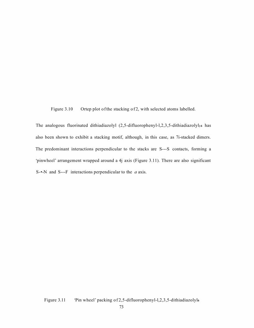
Figure 3.10 Ortep plot o f the stacking o f 2, with selected atoms labelled.
The analogous fluorinated dithiadiazolyl (2,5-difluorophenyl-l,2,3,5-dithiadiazolyl) 6 has
also been shown to exhibit a stacking motif, although, in this case, as 7i-stacked dimers.
The predominant interactions perpendicular to the stacks are S---S contacts, forming a
‘pinwheel’ arrangement wrapped around a 4j axis (Figure 3.11). There are also significant
S - • - N and S---F interactions perpendicular to the a axis.
Figure 3.11 ‘Pin wheel’ packing o f 2,5-difluorophenyl-l,2,3,5-dithiadiazolyl6
73

The structure of 2 within the be plane consists of a complex array of S---N, S---C1 and
C1---C1 interactions. Chlorines in the meta position of the phenyl ring are associated to
both of the sulfur atoms within an adjacent dithiadiazolyl ring. The interactions form a
triangle of short (Slc---C12a, 3.432 A) and long (S2c---C12a, 3.653 A) electrostatic
S---C1 contacts. This compares to the sum of the van der Waals radii for sulfur and
chlorine of 3.65 A. Furthermore, the same chlorine is also involved in a strong tail-to-tail
type interaction with a meta substituted chlorine on an adjacent molecule (C12a--*C12b ,
3.321 A). The chlorine atoms in the ortho position of the phenyl ring are weakly associated
to one sulfur atom of an adjacent molecule (Sic*••Clld, 3.656 A). Finally, the
dithiadiazolyls are linked together in a head-to-head fashion through weak S---N
interactions (S2c***N2b, 3.336 A and S2b-*-N2c, 3.337 A). This compares to the sum of
the van der Waals radii for sulfur and nitrogen of 3.4 A. The pin wheel arrangement
observed for 2,5-difluorophenyl-l,2,3,5-dithiadiazolyl is not present in the packing of 2
because of the absence of any S - • *S interactions.
74
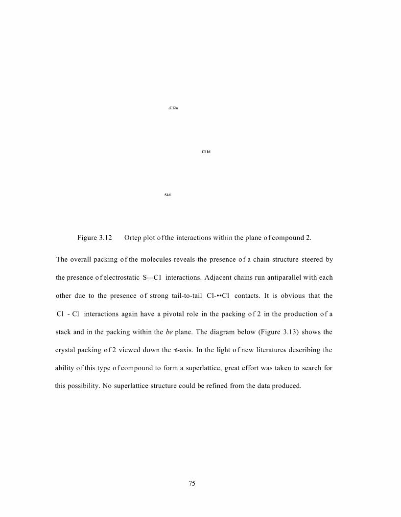
, CI2a
Cl Id
S id
Figure 3.12 Ortep plot of the interactions within the plane o f compound 2.
The overall packing o f the molecules reveals the presence o f a chain structure steered by
the presence o f electrostatic S---C1 interactions. Adjacent chains run antiparallel with each
other due to the presence o f strong tail-to-tail Cl-••Cl contacts. It is obvious that the
Cl - Cl interactions again have a pivotal role in the packing o f 2 in the production o f a
stack and in the packing within the be plane. The diagram below (Figure 3.13) shows the
crystal packing o f 2 viewed down the <2-axis. In the light o f new literature6 describing the
ability o f this type o f compound to form a superlattice, great effort was taken to search for
this possibility. No superlattice structure could be refined from the data produced.
75

76
Figure 3:13 Ortep plot of the packing structure of 2
3.1.3 X-ray Crystal Structure of 3,5-dichIorophenyl-l,2,3,5-dithiadiazoIyI
The temperature controlled sublimation of 3,5-dichlorophenyl-l,2,3,5-dithiadiazolyl 3 ,
yielded one of two polymorphs, not distinctive in appearance but possessing veiy different
molecular packing. Gradient sublimation of the cold finger sublimed product at relatively
high temperature (145 °C) compared with the melting point of the compound yielded the
P-structure. If the isomer was sublimed at low temperature (95 °C) and therefore very
slowly over several days the a-structure was formed. The only chemical difference
observed between the two polymorphs was in the DSC thermogram (Figure 3.14).
Although both forms have very similar melting points, their sublimation points are very
different. A third curve has been included showing the thermogram of the product
retrieved after sublimation at 120 °C. The sublimation points of the two polymorphs can be
observed on the same curve.
The possibility that the polymorphs were actually a product of a superlattice was
investigated by determining the structure of several different crystals, from different
batches of material, and by the re-analysis of collected data. It was discovered that the two
structures would not converge or refine to create any other structures. It was therefore
concluded that the structures are correct and true polymorphs. It should also be noted that
P~3 cannot be a superlattice of a - 3 because of its higher order of symmetry in relation to
the other polymorph (C2/c and P-l for p - 3 and a - 3 respectively)
77
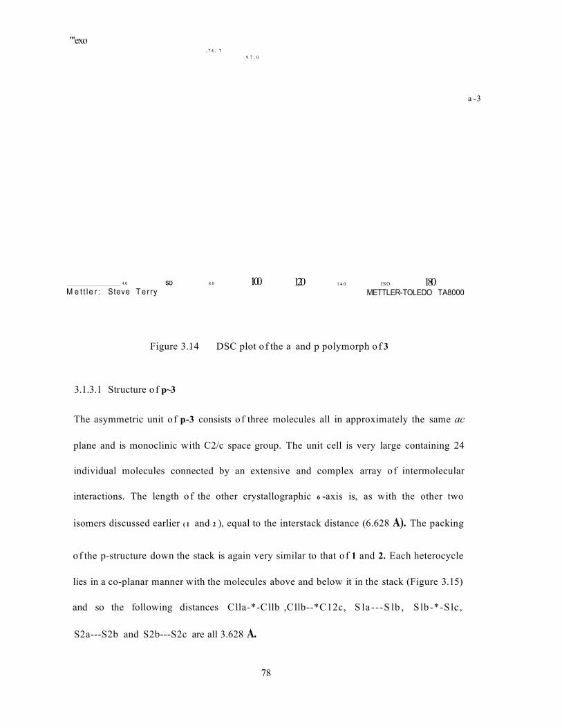
'"exo, 7 4 . 7
9 7 . 0
a -3
_______________________ 4 0
M e t t le r : Steve T e rry8 0 100so 120 1 4 0 IS O 180
METTLER-TOLEDO TA8000
Figure 3.14 DSC plot of the a and p polymorph of 3
3.1.3.1 Structure o f p~3
The asymmetric unit o f p-3 consists of three molecules all in approximately the same ac
plane and is monoclinic with C2/c space group. The unit cell is very large containing 24
individual molecules connected by an extensive and complex array o f intermolecular
interactions. The length o f the other crystallographic 6 -axis is, as with the other two
isomers discussed earlier ( 1 and 2 ), equal to the interstack distance (6.628 A). The packing
of the p-structure down the stack is again very similar to that o f 1 and 2. Each heterocycle
lies in a co-planar manner with the molecules above and below it in the stack (Figure 3.15)
and so the following distances Clla-*-Cllb ,Cllb--*C12c, S la ---S lb , S lb-*-Slc,
S2a---S2b and S2b---S2c are all 3.628 A.
78
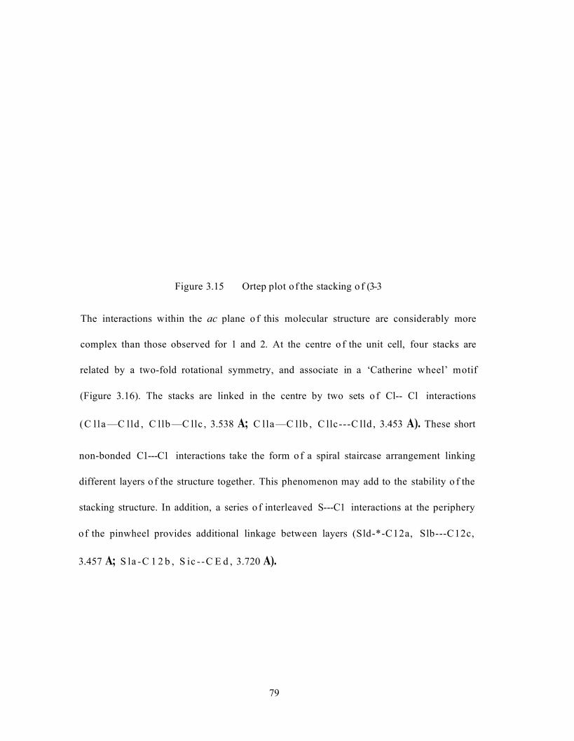
Figure 3.15 Ortep plot of the stacking o f (3-3
The interactions within the ac plane o f this molecular structure are considerably more
complex than those observed for 1 and 2. At the centre o f the unit cell, four stacks are
related by a two-fold rotational symmetry, and associate in a ‘Catherine wheel’ motif
(Figure 3.16). The stacks are linked in the centre by two sets o f Cl-- Cl interactions
(C lla —C lld , C llb —C llc , 3.538 A; C lla —C llb , C llc---C lld , 3.453 A). These short
non-bonded C1---C1 interactions take the form o f a spiral staircase arrangement linking
different layers o f the structure together. This phenomenon may add to the stability o f the
stacking structure. In addition, a series o f interleaved S---C1 interactions at the periphery
of the pinwheel provides additional linkage between layers (Sld-*-C12a, Slb---C12c,
3.457 A; S la -C 1 2 b , S ic --C E d , 3.720 A).
79
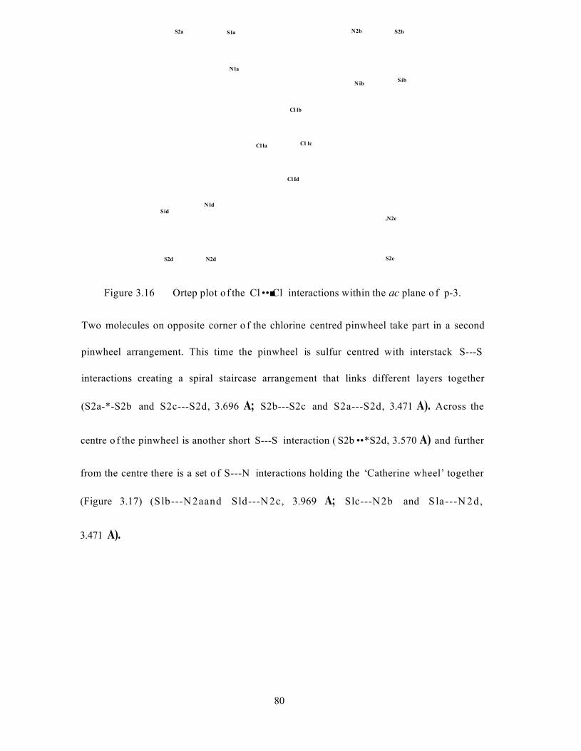
N2b S2bS2a S la
N la
SibN ib
Cl lb
Cl 1cCl la
Cl Id
N ldSid
,N2c
S2cS2d N2d
Figure 3.16 Ortep plot of the Cl • • ■ Cl interactions within the ac plane o f p-3.
Two molecules on opposite corner o f the chlorine centred pinwheel take part in a second
pinwheel arrangement. This time the pinwheel is sulfur centred with interstack S---S
interactions creating a spiral staircase arrangement that links different layers together
(S2a-*-S2b and S2c---S2d, 3.696 A; S2b---S2c and S2a---S2d, 3.471 A). Across the
centre o f the pinwheel is another short S---S interaction ( S2b • • *S2d, 3.570 A) and further
from the centre there is a set o f S---N interactions holding the ‘Catherine wheel’ together
(Figure 3.17) (S lb---N 2aand Sld---N 2c, 3.969 A; Slc---N2b and S la---N 2d,
3.471 A).
80
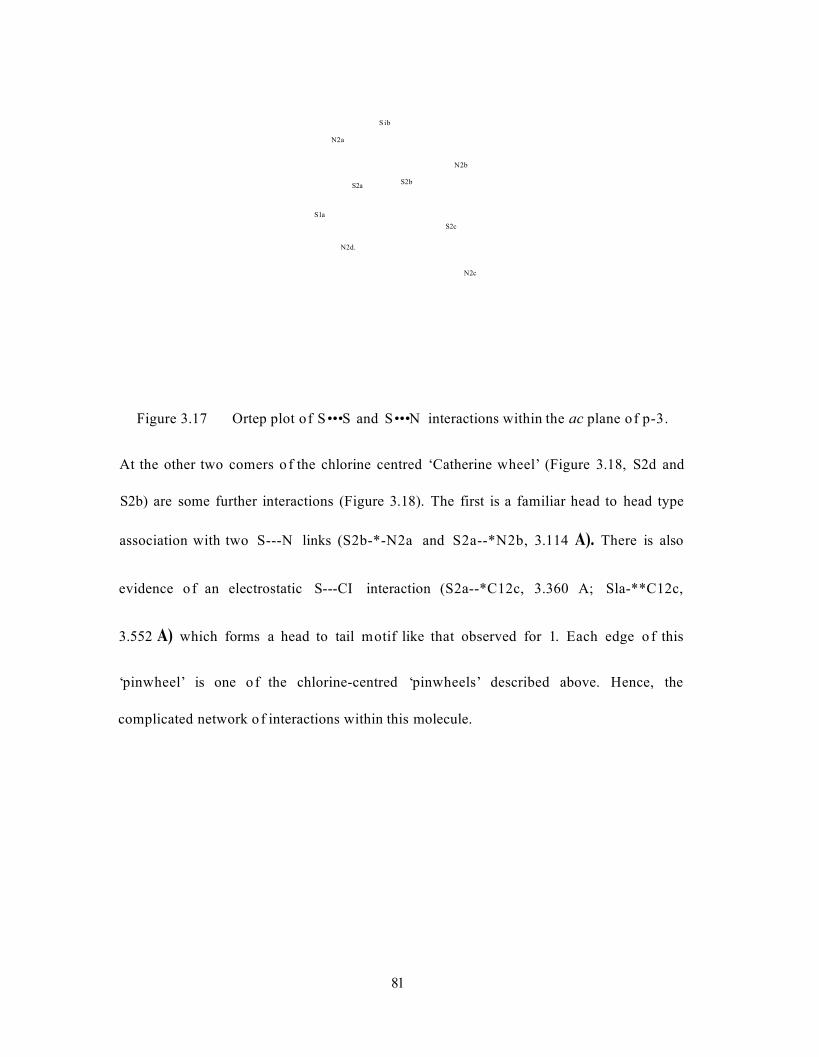
N2a
S2a
Sla
S ib
N2b
S2b
S2c
N2c
N2d.
Figure 3.17 Ortep plot of S • • • S and S • • • N interactions within the ac plane of p-3.
At the other two comers o f the chlorine centred ‘Catherine wheel’ (Figure 3.18, S2d and
S2b) are some further interactions (Figure 3.18). The first is a familiar head to head type
association with two S---N links (S2b-*-N2a and S2a--*N2b, 3.114 A). There is also
evidence o f an electrostatic S---CI interaction (S2a--*C12c, 3.360 A; Sla-**C12c,
3.552 A) which forms a head to tail motif like that observed for 1. Each edge o f this
‘pinwheel’ is one o f the chlorine-centred ‘pinwheels’ described above. Hence, the
complicated network o f interactions within this molecule.
81

Cl I a
.Cl Id
Nla. S2b,N2a
N2b'S2a N ibCI2d
,CI2c
CI2b Cl lb
Figure 3.18 Ortep plot o f the further interactions o f p-3 within the ac plane
The interactions involved in the crystal structure o f p-3 are very different to those observed
for 1 and 2. The previous structures exhibited a small number o f intermolecular
interactions holding the units together in an ordered manner. In this case, within one plane,
the dithiadiazolyl units seem to lie in almost random directions and several different
interactions hold the units together as a network. It is interesting to note that the reported
crystal structure o f 3,5-difluorophenyl-l,2,3,5-dithiadiazolyl4 also displays a stacked
structure but one that has suffered from Peierls distortion producing alternating long and
short interactions along the stack. The structure o f 3,5-difIuorophenyl-l,2,3,5-
dithiadiazolyl4 is more analogous to the structure o f a-3, which will be discussed in the
next section. The pinwheel arrangement o f p-3 shows initial similarities to that reported for
2,5-difluorophenyl-l,2,3,5-dithiadiazolyl6 (Figure 3.11) but possesses a lower order o f
symmetry (space group o f monoclinic C2/c compared to tetragonal I4|/a) and has not
suffered Peierls distortion. Figure 3.19 shows the overall packing o f p-3 in the solid state.
82
83
Figure 3.19 Ortep plot of the packing of bcta-3
3.1.3.2 Structure of a-3
The asymmetric unit of a-3 consists of twelve molecules linked as 7t-stacked dimers by
short (« 3.25 A) and long («3.98 A) S---S interactions. Much like the asymmetric unit for
P-3, the groups are also joined within the be plane. The unit cell is triclinic with a space
group P-l, which contains an inversion centre that generates a unit cell containing 24
individual molecules connected (like p-3) by an extensive and complex array of
intermolecular interactions. In a-3, the length of the shortest crystallographic axis is
7.2484(8) A, which is much larger than that described for other chloro-aromatic
compounds that exhibit a p-type structure. This is seen in the crystal packing which shows
stacking interactions, although the individual groups do not eclipse each other throughout
the stack and so cannot be described in terms of a 7r-stack.
Figure 3.20 below shows the intrastack interactions of a-3. The molecular packing
contains dimers linked by long S---S contacts. However, adjacent dimers within the stack
are not coplanar. The result is a structure that is similar to that of 1,3-phenylene-
bis(l,2,3,5-dithiadiazolyl)8 which can be described in terms of a zigzag stack. Within the
stack, the S - • - S contacts vary by approximately 0.8 A between short and long distances. In
contrast, the related C1---C1 contacts only vary by less than 0.1 A. This again shows that
chlorine is having an effect on the packing of the molecules and that the Peierls distortion
of the unpaired electrons seems almost localised at the dithiadiazolyl rings. Some selected
bond distances to illustrate this point are as follows; Sla---Slb and Sic •••Sid, 3.232 A;
S2a***S2b and S2c---S2d, 3.310 A; Sib—Sic, 4.019 A; S2b---S2c, 3.941 A;
84

C lla —Cllb and C llc —C lld , 3.615 A; C12a---C12b and C12c—C12d, 3.630 A;
C llb —C llc , 3.637 A; C12b• • -C12c, 3.619 A.
C12a
CllS2a
S l a
Cl lb
Cll
C
Figure 3.20 Ortep plot of the stacking o f a-3
The structure o f a-3 within the be plane is similar to that o f p-3. Although having a
different space group (P-l compared to C2/c for a-3 and P-3 respectively) the overall
crystal packing within the plane is very similar. At the centre of the unit cell is a ‘Catherine
wheel’ type association (Figure 3.21). The chlorines are linked in the centre by four
principal short C1-- C1 contacts, two of which are 3.439 A in length, and the others
3.412 A. These values are slightly shorter than those observed for p-3 (3.538 A and
3.453 A). In a similar way to p-3, the C1-- C1 contacts join different layers o f the stack
through short interstack interactions, which again may in turn help to stabilise the stack.
85

Figure 3.21 Ortep plot of the CF • - Cl ‘Catherine wheel’ type interactions in a-3
Two comers o f the chlorine centred ‘Catherine wheel’ connect in a further head-to-head
(i.e. S---S) interaction creating a second ‘Catherine wheel’ arrangement (Figure 3.22). At
the centre o f this ‘Catherine wheel’, there are two principal short S---S contacts (two at
3.383 A and two at 3.376 A) and one short contact across the centre o f the wheel
(3.516 A). This set o f contacts are (like the chlorine interactions) smaller than those
observed for p-3 (3.696 A and 3.471 A) and shorter than the S---S interaction across the
centre (3.570 A). This set o f interactions also exhibit a ‘spiral staircase’ arrangement
linking different layers of the structure together.
86
Figure 3.22 Ortep plot of the S---S ‘Catherine wheel’ type interactions in a-3
At the other two comers o f the chlorine centred ‘Catherine wheel’ are some further
interactions (Figure 3.23). The first is a familiar head-to-head type association with two
S---N links (shortest contacts 3.126 and 3.128 A). There is also evidence o f an
electrostatic S---C1 interaction (S2a---C12c, 3.360 A; Sla---C12c, 3.552 A) which forms
a head-to-tail motif similar to that observed for 1.
Figure 3.23 Ortep plot of other interactions within a-3
87
Figure 3.24 shows an Ortep plot of the overall packing in a-3. The crystal packing bears a
striking similarity to the reported structure of 2,5-difluorophenyl-l,2,3,5-dithiadiazolyl4
which also exhibits a Peierls distortion to form stacks of dimerised radical units (interdimer
and intradimer S---S distances are 4.04 A and 3.16 A respectively). When compared to
p-3 it is noted that the structure within the ac plane is very similar. The overall packing
only differs because of the short and long S* • - S contacts in a-3. It would seem reasonable
to assume that the two polymorphs are very close in energy to each other, which is also
supported by the small differences in sublimation temperature required to change the
crystal structure. As described at the beginning of this section the only appreciable physical
difference between p-3 and a-3 is the sublimation temperature. The physical data supplied
in the remainder of this chapter concerns the p polymorph, as this is of most interest due to
its infinite equidistant stacking structure.
88
89
Figure 3.24 Ortep plot of the packing of alfa-
3.2 Magnetic Measurements
Two types of magnetisation measurements have been made:
1) Temperature dependent magnetisation at constant applied field of 10 kOe in the
temperature range from 1.8 to 300 K.
2) Isothermal magnetisation, from 0 to 50 kOe, at constant temperatures 1.8 and 3 K.
Experimental and numerical data for these experiments are listed appendix 5.
At present two of the samples have been investigated, 2,4-dichlorophenyl-l,2,3,5-
dithiadiazolyl (1) and 3,5-dichlorophenyl-l,2,3,5-dithiadiazolyl (3).
The magnetisation of both samples is very weak as shown in the experimental points of
Figures 3.25 and 3.26. Between room temperature and about 10K the magnetisation is
negative although slowly increasing. Below about 10K it becomes positive and increases
more rapidly. A small shoulder observed at about 45K indicates the presence of a very
small amount of oxygen in the sample holder. These results are indicative of the presence
of a few paramagnetic centres in a mainly diamagnetic sample.
3.2.1 The Diamagnetic Contribution
The diamagnetic contribution from the sample to the magnetisation can be estimated in
various ways:
Evaluating the diamagnetic contributions from the capsule and cotton wool,21 and
estimating that of the sample using Pascal's tables. We then have the gelatine capsule at
-3.92(2)xl0"7 emu/Oe.g and the cotton at -4.56(2)xl0~7 emu/O e.g. From Pascal's tables
90
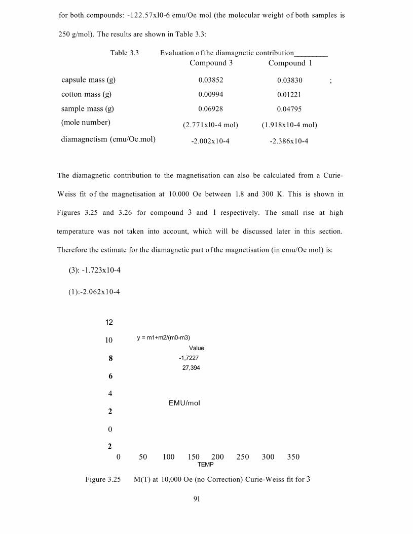
for both compounds: -122.57xl0-6 emu/Oe mol (the molecular weight o f both samples is
250 g/mol). The results are shown in Table 3.3:
Table 3.3 Evaluation o f the diamagnetic contribution_________Compound 3 Compound 1
capsule mass (g) 0.03852 0.03830 ;
cotton mass (g) 0.00994 0.01221
sample mass (g)
(mole number)
0.06928
(2.771xl0-4 mol)
0.04795
(1.918x10⁻⁴ mol)
diamagnetism (emu/Oe.mol) -2.002x10⁻⁴ -2.386x10⁻⁴
The diamagnetic contribution to the magnetisation can also be calculated from a Curie-
Weiss fit o f the magnetisation at 10.000 Oe between 1.8 and 300 K. This is shown in
Figures 3.25 and 3.26 for compound 3 and 1 respectively. The small rise at high
temperature was not taken into account, which will be discussed later in this section.
Therefore the estimate for the diamagnetic part o f the magnetisation (in emu/Oe mol) is:
(3): -1.723x10⁻⁴
(1):-2.062x10⁻⁴
12
y = m1+m2/(m0-m3)10Value
8 -1,722727,394
6
4EMU/mol
2
0
20 50 100 150 200 250 300 350
TEMP
Figure 3.25 M(T) at 10,000 Oe (no Correction) Curie-Weiss fit for 3
91
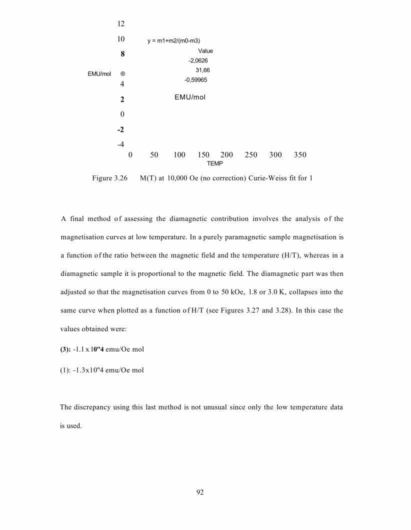
12
10
8
EMU/mol ®
4
2
0
-2
-40 50 100 150 200 250 300 350
TEMP
Figure 3.26 M(T) at 10,000 Oe (no correction) Curie-Weiss fit for 1
y = m1+m2/(m0-m3)
Value-2,0626
31,66-0,59965
EMU/mol
A final method o f assessing the diamagnetic contribution involves the analysis o f the
magnetisation curves at low temperature. In a purely paramagnetic sample magnetisation is
a function of the ratio between the magnetic field and the temperature (H/T), whereas in a
diamagnetic sample it is proportional to the magnetic field. The diamagnetic part was then
adjusted so that the magnetisation curves from 0 to 50 kOe, 1.8 or 3.0 K, collapses into the
same curve when plotted as a function o f H/T (see Figures 3.27 and 3.28). In this case the
values obtained were:
(3): -1.1 x 10"4 emu/Oe mol
(1): -1.3x10"4 emu/Oe mol
The discrepancy using this last method is not unusual since only the low temperature data
is used.
92
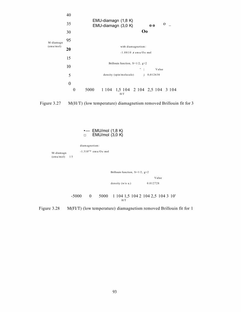
40
35
30
95M -diamagn (emu/mol)
20
15
10
5
00 5000 1 104 1,5 104 2 104 2,5 104 3 104
H/T
Figure 3.27 M(H/T) (low temperature) diamagnetism removed Brillouin fit for 3
EMU-diamagn (1,8 K)EMU-diamagn (3,0 K) o o o _
O o
with diam agnetism :
-1 .0 8 1 0 A em u/O e mol
Brillouin function, S=1/2, g=2
” | Value
density (spin/m olecule) j 0 ,012638
M -diamagn (emu/mol) 1 5
•— EMU/mol (1,8 K)□ EMU/mol (3,0 K)
diam agnetism :
-1.310"* em u/O e mol
Brillouin function, S=1/2, g=2
Value
density (w/o u.) 0 .012728
-5000 0 5000 1 104 1,5 104 2 104 2,5 104 3 10'H/T
Figure 3.28 M(FI/T) (low temperature) diamagnetism removed Brillouin fit for 1
93
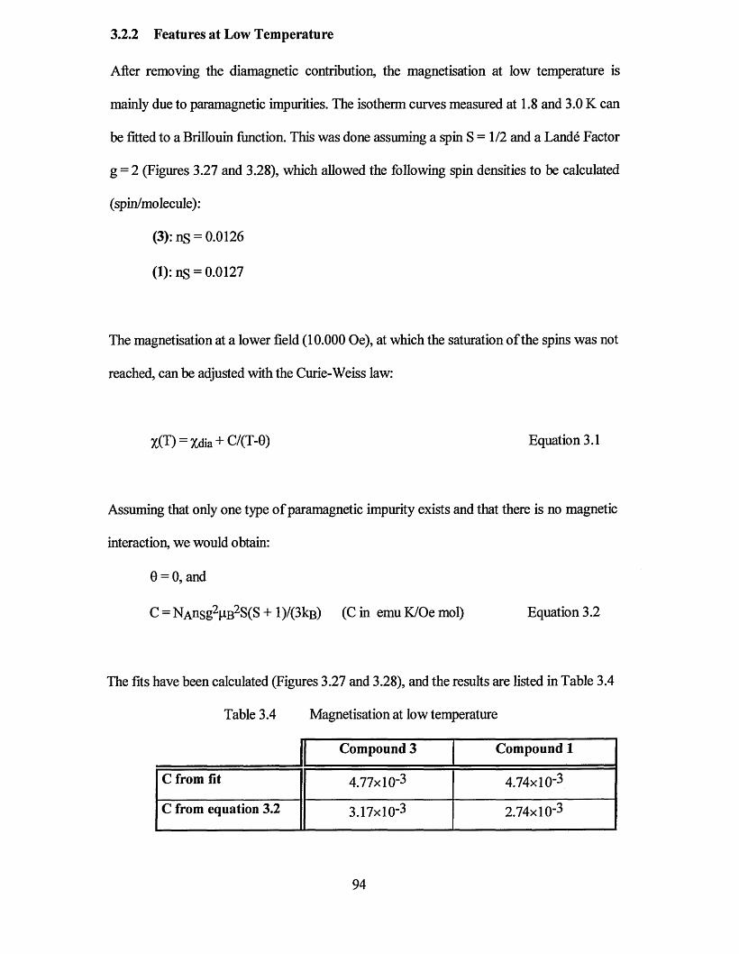
3.2.2 Features at Low Temperature
After removing the diamagnetic contribution, the magnetisation at low temperature is
mainly due to paramagnetic impurities. The isotherm curves measured at 1.8 and 3.0 K can
be fitted to a Brillouin function. This was done assuming a spin S = 1/2 and a Lande Factor
g = 2 (Figures 3.27 and 3.28), which allowed the following spin densities to be calculated
(spin/molecule):
(3): ns = 0.0126
(l):ns = 0.0127
The magnetisation at a lower field (10.000 Oe), at which the saturation of the spins was not
reached, can be adjusted with the Curie-Weiss law:
X(T) = Xdia + C/(T-0) Equation 3.1
Assuming that only one type of paramagnetic impurity exists and that there is no magnetic
interaction, we would obtain:
0 = 0, and
C = NAnsg2PB2S(S + 1)/(31cb ) (C in emu K/Oe mol) Equation 3.2
The fits have been calculated (Figures 3.27 and 3.28), and the results are listed in Table 3.4
Table 3.4 Magnetisation at low temperature
Compound 3 Compound 1
C from fit 4.77xl0-3 4.74x10⁻³
C from equation 3.2 3.17x10⁻³ 2.74x10⁻³
94
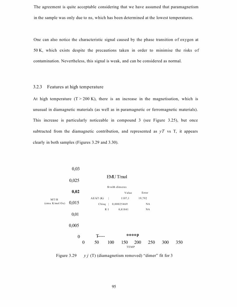
The agreement is quite acceptable considering that we have assumed that paramagnetism
in the sample was only due to ns, which has been determined at the lowest temperatures.
One can also notice the characteristic signal caused by the phase transition o f oxygen at
50 K, which exists despite the precautions taken in order to minimise the risks o f
contamination. Nevertheless, this signal is weak, and can be considered as normal.
3.2.3 Features at high temperature
At high temperature (T > 200 K), there is an increase in the magnetisation, which is
unusual in diamagnetic materials (as well as in paramagnetic or ferromagnetic materials).
This increase is particularly noticeable in compound 3 (see Figure 3.25), but once
subtracted from the diamagnetic contribution, and represented as yT vs T, it appears
clearly in both samples (Figures 3.29 and 3.30).
0,03
0,025
0,02M T/H
(em u K/mol O e) 0,015
0,01
0,005
00 50 100 150 200 250 300 350
TEM P
Figure 3.29 y j (T) (diamagnetism removed) “dimer” fit for 3
EMU T/mol
fit with d im eres
V alue Error
AE/kT (K) |
Chisq | 0 ,00025469
R I 0,81841
1107,1 19,792
NA
NA
oooopT-----
95
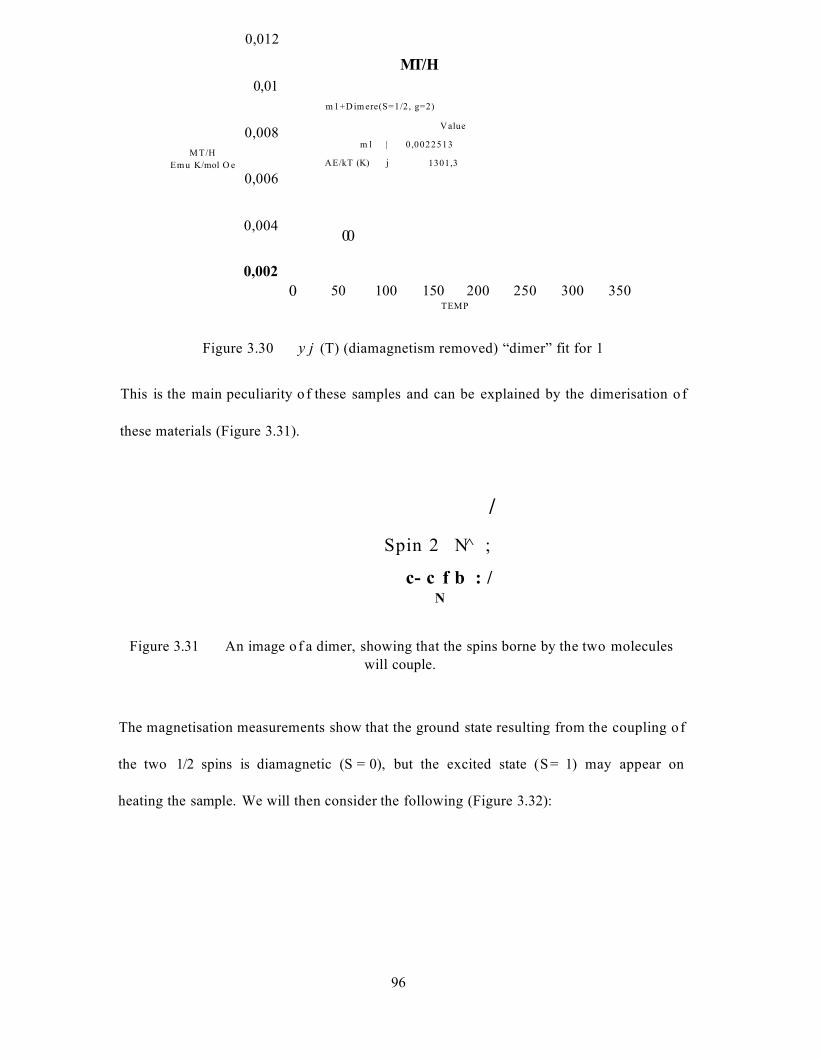
M T/H Emu K/mol O e
0,012
MT/H0,01
m 1+D im ere(S=1/2, g=2)
Value0,008m l | 0 ,0022513
1301,3AE/kT (K) j
0,006
0,00400
0,00250 100 150 200 250 300 3500
TEMP
Figure 3.30 y j (T) (diamagnetism removed) “dimer” fit for 1
This is the main peculiarity o f these samples and can be explained by the dimerisation o f
these materials (Figure 3.31).
/Spin 2 N̂ ;
c- c f b : /N
Figure 3.31 An image o f a dimer, showing that the spins borne by the two moleculeswill couple.
The magnetisation measurements show that the ground state resulting from the coupling o f
the two 1/2 spins is diamagnetic (S = 0), but the excited state (S = 1) may appear on
heating the sample. We will then consider the following (Figure 3.32):
96
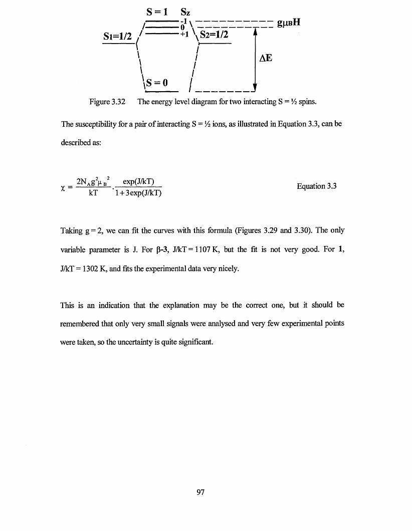
S = 1 Sz -1\ gJXBH
\S = 0
AE
Figure 3.32 The energy level diagram for two interacting S = XA spins.
The susceptibility for a pair of interacting S = 1A ions, as illustrated in Equation 3.3, can be
described as:
X kT 1 + 3exp(J/kT)
Taking g = 2, we can fit the curves with this formula (Figures 3.29 and 3.30). The only
variable parameter is J. For p-3, J/kT=1107K, but the fit is not very good. For 1,
J/kT = 1302 K, and fits the experimental data very nicely.
This is an indication that the explanation may be the correct one, but it should be
remembered that only very small signals were analysed and very few experimental points
were taken, so the uncertainty is quite significant.
2NAgy B2 exp(J/kT) Equation 3.3
97
3.2.4 Conclusion
These materials mainly show diamagnetic behaviour. At low temperature, a contribution
from paramagnetic impurities is observed; this can be estimated at a concentration of about
1 spin per 100 molecules. Finally, a rise of the magnetisation is visible at T>200K,
particularly for p-3. This increase may be assigned to the thermal excitation of the
magnetic state of dimers. The explanation is in a good agreement with the experimental
data, particularly for 1, and gives an energy splitting corresponding to 1100 to 1300 K
between the S = 0 and S = 1 states.
3.3 Electron Paramagnetic Resonance
Each of the compounds listed at the start of the chapter (1, 2 and 3) have been studied by
electron paramagnetic resonance (EPR) spectroscopy. The properties of the monomeric
radicals were investigated by analysis of a frozen glass sample in dimethyl formamide.
Preliminary studies of the solid state properties were carried out on powder samples. A
single crystal study of compound 3 has also been undertaken.
3.3.1 Frozen Glass Studies
All sample preparation was carried out in a dry nitrogen glove box and sealed prior to
analysis. Solution samples were prepared by the dissolution of a few milligrams of 1, 2 or
3 in approximately 0.5 ml of DMF. The resulting solution was treated with a drop of
toluene to aid the formation of a satisfactory ‘glass’ when analysed. The solution was
added to a quartz tube, which was sealed prior to analysis.
98
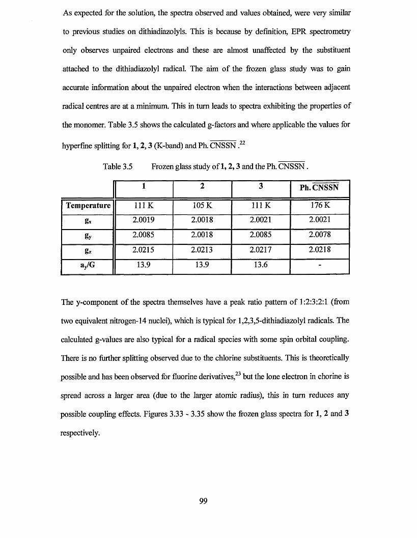
As expected for the solution, the spectra observed and values obtained, were very similar
to previous studies on dithiadiazolyls. This is because by definition, EPR spectrometry
only observes unpaired electrons and these are almost unaffected by the substituent
attached to the dithiadiazolyl radical. The aim of the frozen glass study was to gain
accurate information about the unpaired electron when the interactions between adjacent
radical centres are at a minimum. This in turn leads to spectra exhibiting the properties of
the monomer. Table 3.5 shows the calculated g-factors and where applicable the values for
hyperfine splitting for 1,2,3 (K-band) and Ph. CNSSN .22
Table 3.5 Frozen glass study of 1,2,3 and the Ph. CNSSN.
1 2 3 Ph. CNSSN
Temperature 111 K 105 K 111 K 176 K
gx 2.0019 2.0018 2.0021 2.0021
gy 2.0085 2.0018 2.0085 2.0078
gz 2.0215 2.0213 2.0217 2.0218
ay/G 13.9 13.9 13.6 -
The y-component of the spectra themselves have a peak ratio pattern of 1:2:3:2:1 (from
two equivalent nitrogen-14 nuclei), which is typical for 1,2,3,5-dithiadiazolyl radicals. The
calculated g-values are also typical for a radical species with some spin orbital coupling.
There is no further splitting observed due to the chlorine substituents. This is theoretically
possible and has been observed for fluorine derivatives,23 but the lone electron in chorine is
spread across a larger area (due to the larger atomic radius), this in turn reduces any
possible coupling effects. Figures 3.33 - 3.35 show the frozen glass spectra for 1, 2 and 3
respectively.
99
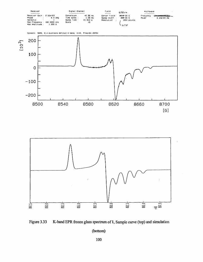
R e ce iv e r
R e c e i v e r G a m P n a s e Harm on ic moo F re q u e n c y Moo A m p l i tu d e
2 50e *030 . 0
100 000 0 kHz 1 . 9 9 5 G
S igna l Cnannel
C o n v e r s io n 4 0 . 9 6 msl im e Cons t : 1 . 2 0 m3Sweep Time : 4 1 . 9 4 3 sS c a l e 19
C e n t e r F i e l o Sweep WiOtn P e s o l u t i o n
Y f i ' l -v e e « - oo g
22©-. 00 G1024 p o i n t s
L ,
Microwave
F re q u e n c y • '3*w3 . 44 e*0 0 mW
Comment: S620. 2 . 4 d i c n l o r o d m f / t o l K band. 111K. F r e q - 2 4 .15724
200
100
100
200
8 5 0 0 8 5 4 0 8 5 8 0 8 6 2 0 8 6 6 0 8 7 0 0
[G]
Figure 3.33 K-band EPR frozen glass spectrum of 1, Sample curve (top) and simulation
( b o t t o m )
100
uo
n
R ece iver
R e c e i v e r Ga in P h ase Harmonic Mao F re q u e n c y Moo A m p l i tu d e
S ig n a l Channel
Oeg0 . OOe *03
0 . 0 110 0 .0 0 0 0 kHz
1 .0 0 0 G
C o n v e r s io n Time Cons t Sweep Time S c a l e
163 04 ms I 20 ms
167 772 S 17
C ente r F ie ldSweep W idthP e s o lu t io n
585+&-00 G -22 0 .0 0 G ^ 1024 p o i n t s
-x-n *
Freouency 23 HBUOOOff'-GHZPower 3 62e*00 mw
Comment: s 6 0 5 2 . 5 O i c h l o r o / t o l u e n e 110K 23.0660GHz
4 0 0
200
-2 0 0
- 4 0 0
8 4 0 0 8 4 4 0 8 4 8 0 8 5 2 0 8 5 6 0 8 6 0 0
Figure 3.34 K-band EPR frozen glass spectrum of 2, Sample curve (top) and simulation
(bottom)101
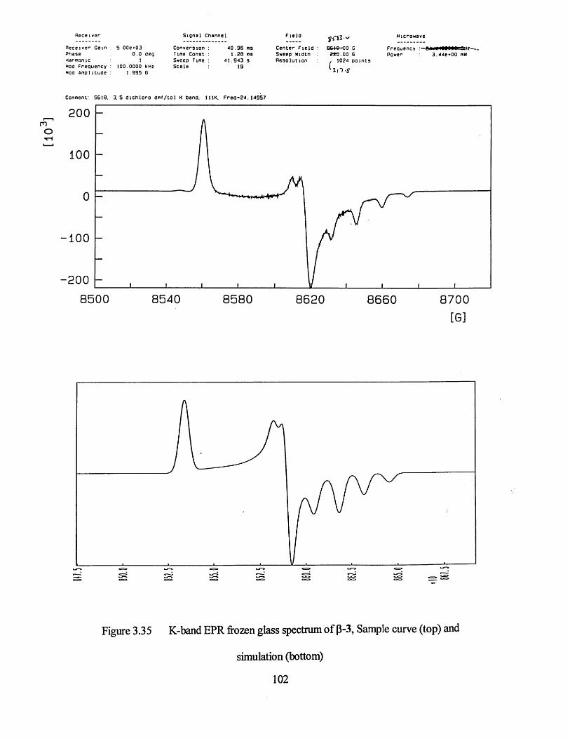
S ig n a l Channel
d e c e i v e r Gain 5 OOe *03 C o n v e r s io n : 4 0 . 9 6 ms C e n t e r F i e l d :P h ase 0 . 0 deg Time Co n s t : 1 . 2 6 ms Sweep MidtnHarmon ic 1 Sweep Time : 4 1 . 9 4 3 s R e s o l u t i o nMod F r e q u e n c y 100 .0000 kHz S c a l e 19Moo A m p l i tu d e 1 .9 9 5 G
B 6+ 6-00 G2 « j . o o G
102a p a i n t s
Microwave
F r e q u e n c y 83 .4 4 e + 0 0 mW
Comment: S618. 3 . 5 d i c h l o r o O m f / to l K dand. 111K, F r e q - 2 4 . 1 4 9 5 7
200
100
100
-2008 5 0 0 8 5 4 0 8 5 8 0 8 6 2 0 8 6 6 0 8 7 0 0
[ G ]
Figure 3.35 K-band EPR frozen glass spectrum of p-3, Sample curve (top) and
s i m u l a t i o n ( b o t t o m )
102
3.3.2 Powder Studies
Powder samples were prepared by grinding approximately 0.5 g of pure crystalline sample
into a fine powder. The powder was then transferred into a quartz tube in a glove box to
prevent degradation and was sealed prior to analysis. The analysis of the samples was then
performed at different temperatures and frequencies.
Figures 3.36 - 3.38 show the powder spectra of 1, 2 and 3 respectively. At room
temperature, there is little fine detail in the spectra of 2 and 3 but on cooling to 105 K there
is significant resolution enhancement. This suggests there is some isolated monomeric
centres contributing to the spectrum. Compound 1 shows significant fine structure at room
temperature which may indicate the presence of monomeric centres at high temperature.
All the results obtained for powder measurement appear to contradict the results obtained
for the magnetic data that indicated that the samples became more diamagnetic as
temperature was decreased. Studies on further powders of the same materials revealed that
the measurements were not reproducible. It is thought that the act of grinding the
crystalline material to produce a powder actually destroys or modifies the solid state
packing of the compounds leading to the inconsistent EPR signals.
103
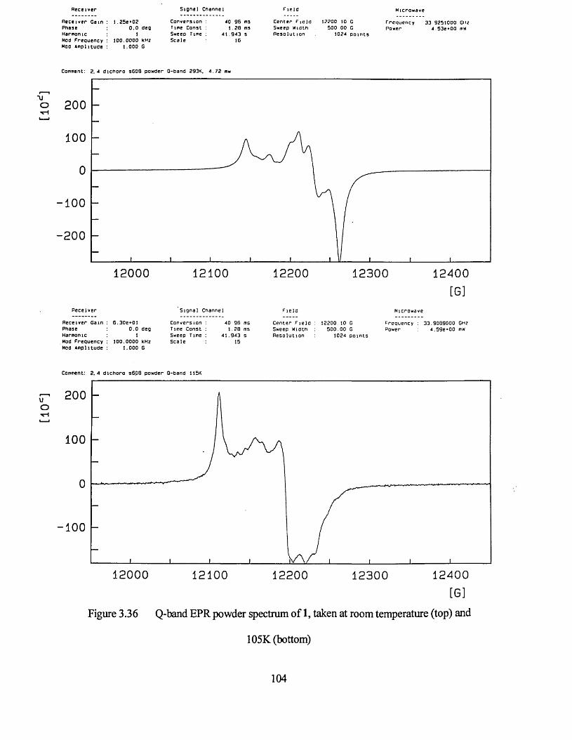
[10c
] [1
0*]
R ece ive r
R e c e i v e r Gain P h ase Harmonic Moo F r e q u e n c y Moo A m p l it u d e
S ig n a l Channel
I 25e*020 . 0 deg 1
10 0 .0 0 0 0 kHz 1 .0 0 0 G
C o n v e r s ion l im e Cons t Sweep Time S c a l e
40 96 ms 1 . 2 8 ms
4 1 . 9 4 3 s 16
F ie ld
C ente r F ie ld 12200 10 GSweep W idth 500 00 GR e s o lu tio n 1024 p o in ts
rrcq u e n cy 33 9251000 GHzPower 4.53e*00 mw
Comment: 2 . 4 d i c h o r o s 6 0 8 powder Q-Dand 293K. 4 . 7 2 mw
200
100
0
-1 0 0
-2 0 0
1 2 0 0 0 1 2 1 0 0 1 2 2 0 0 1 2 3 0 0 1 2 4 0 0
[ G ]
R e c e i v e r S i g n a l Chann el F i e l d Microwa ve
R e c e iv e r Ga in : Phase Harmonic Mod F r e q u e n c y : Mod A m p l i tu d e :
6 .3 0 e+ 0 10 . 0 deg
11 0 0 .0 0 0 0 kHz
1 .0 0 0 G
C o n v e r s io n : 4 0 . 9 6 ms Time C o n s t : 1 . 2 8 ms Sweep Time : 4 1 . 9 4 3 s S c a l e : 15
C e n t e r F i e l a : Sweep Width R e s o l u t i o n
1 2 2 0 0 .1 0 G F re q u e n c y 5 0 0 . 0 0 G Power
1024 p o i n t s
3 3 . 9 0 8 9 0 0 0 GHz 4 . 5 9 e * 0 0 mW
Comment: 2 . 4 d i c h o r o s 6 p 8 pow der Q-Dand 115K
200
100
0
100
1 2 0 0 0 1 2 1 0 0 1 2 2 0 0 1 2 3 0 0 1 2 4 0 0
[ G ]
Figure 3.36 Q-band EPR powder spectrum of 1, taken at room temperature (top) and
105K (bottom)
104

In ten s ity
800_
600_
400_
2 0 0 _
- 2 0 0 _
-400_
-600_
3340 3345 3350 3355 3360 3365 3370 3375 3380 3385 3390 3395 3400[G]
[*10A 3]1 2 .5 .
1 0 . 0_
5.0_
2 .5_
0 .0_
-5.0_- 7 . 5 _
- 1 0 . 0 _
- 1 2 . 5 _
3320 3330 3340 3350 3360 3370 3380 34003390 3410 3420[G]
Figure 3.37 X-band EPR powder spectrum of 2, taken at room temperature (top) and
105K (bottom)
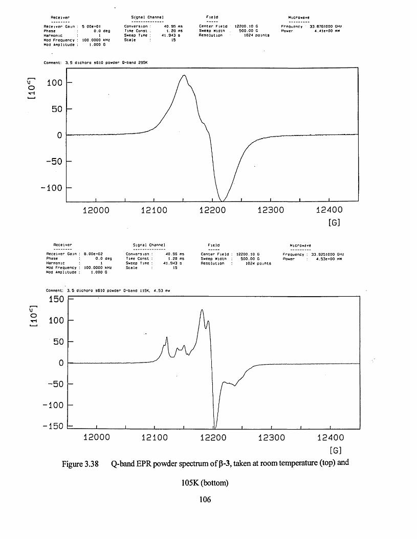
R e ce ive r
R e c e i v e r G a in P h a s e Harmon ic Mod F r e q u e n c y Moo A m p l i t u d e
5 OOe*OI0 . 0 deg 1
100 0000 kHz 1 .0 0 0 G
S ig n a l Channel
C o n v e r s io n : 4 0 . 9 6 msrime C o n s t ; 1 . 2 8 msSweep Time : 4 1 . 9 4 3 sS c a l e 15
F ie ld
C ente r F ie ld 12200.10 GSweep W idth 500 .00 GR e s o lu t io n 1024 p o in ts
Freouency • 33.8761000 GHzPower 4 .4 ie *0 0 mw
Comment: 3. 5 d i c h o r o s6 1 0 powder Q-Dand 295K
100
5 0
- 5 0
100
1 2 0 0 0 1 2 1 0 0 1 2 2 0 0 1 2 3 0 0 1 2 4 0 0
[ G ]
R e c e i v e r S i g n a l Channe l F i e l d Microwa ve
R e c e i v e r Ga in P h ase Harmonic Mod F r e q u e n c y Mod A m p l i tu d e
B .0 0 e * 0 20 . 0 deg
11 0 0 .0 0 0 0 kHz
1 .0 0 0 G
C o n v e r s i o n : 4 0 . 9 6 ms Time C o n s t : 1 . 2 8 ms Sweep Time : 4 1 . 9 4 3 s S c a l e 15
C e n t e r F i e l d : Sweep Width R e s o l u t i o n
1 2 2 0 0 .1 0 G F r e q u e n c y 5 0 0 . 0 0 G Power
1024 p o i n t s
3 3 .9 2 5 1 0 0 0 GHz 4 .5 3 e + 0 0 mW
Comment: 3 . 5 d i c h o r o 561 0 pow der 0 - 0 a n d 115K. 4 . 5 3 mw
1 5 0
o 100
5 0
5 0
-100
- 1 5 0
1 2 0 0 0 1 2 1 0 0 1 2 2 0 0 1 2 3 0 0 1 2 4 0 0
[ G ]
Figure 3.38 Q-band EPR powder spectrum of p-3, taken at room temperature (top) and
105K (bottom)
106
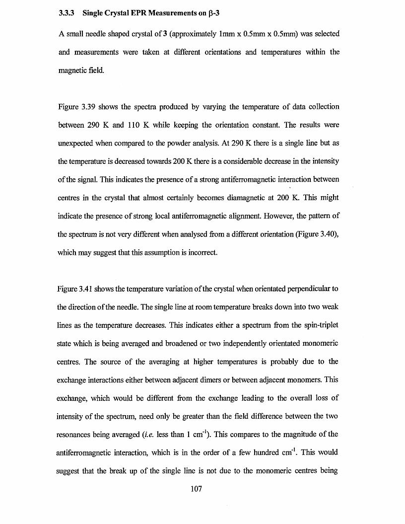
3.3.3 Single Crystal EPR Measurements on p-3
A small needle shaped crystal of 3 (approximately 1mm x 0.5mm x 0.5mm) was selected
and measurements were taken at different orientations and temperatures within the
magnetic field.
Figure 3.39 shows the spectra produced by varying the temperature of data collection
between 290 K and 110 K while keeping the orientation constant. The results were
unexpected when compared to the powder analysis. At 290 K there is a single line but as
the temperature is decreased towards 200 K there is a considerable decrease in the intensity
of the signal. This indicates the presence of a strong antiferromagnetic interaction between
centres in the crystal that almost certainly becomes diamagnetic at 200 K. This might
indicate the presence of strong local antiferromagnetic alignment. However, the pattern of
the spectrum is not very different when analysed from a different orientation (Figure 3.40),
which may suggest that this assumption is incorrect.
Figure 3.41 shows the temperature variation of the crystal when orientated perpendicular to
the direction of the needle. The single line at room temperature breaks down into two weak
lines as the temperature decreases. This indicates either a spectrum from the spin-triplet
state which is being averaged and broadened or two independently orientated monomeric
centres. The source of the averaging at higher temperatures is probably due to the
exchange interactions either between adjacent dimers or between adjacent monomers. This
exchange, which would be different from the exchange leading to the overall loss of
intensity of the spectrum, need only be greater than the field difference between the two
resonances being averaged (i.e. less than 1 cm'1). This compares to the magnitude of the
antiferromagnetic interaction, which is in the order of a few hundred cm'1. This would
suggest that the break up of the single line is not due to the monomeric centres being
107
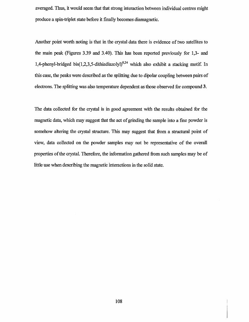
averaged. Thus, it would seem that that strong interaction between individual centres might
produce a spin-triplet state before it finally becomes diamagnetic.
Another point worth noting is that in the crystal data there is evidence of two satellites to
the main peak (Figures 3.39 and 3.40). This has been reported previously for 1,3- and
1,4-phenyl-bridged bis(l,2,3,5-dithiadiazolyl)8,24 which also exhibit a stacking motif. In
this case, the peaks were described as the splitting due to dipolar coupling between pairs of
electrons. The splitting was also temperature dependent as those observed for compound 3.
The data collected for the crystal is in good agreement with the results obtained for the
magnetic data, which may suggest that the act of grinding the sample into a fine powder is
somehow altering the crystal structure. This may suggest that from a structural point of
view, data collected on the powder samples may not be representative of the overall
properties of the crystal. Therefore, the information gathered from such samples may be of
little use when describing the magnetic interactions in the solid state.
108
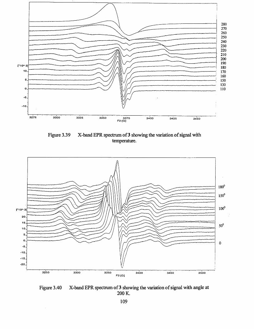
I
I I I '----------------1--------'--------!--------1--------1 ] , ' 1 r— ;------------------------------------------;-------- ,--------, _ _
3300 3325 3350 3375 3400 3425F2:[G]
Figure 3.39 X-band EPR spectrum of 3 showing the variation of signal withtemperature.
180
1 5 0
m20.
15
10
-5
-10
-1 5 .
- 2 0 .
3250 3300 3350 3400 3450 3500F2:[G]
Figure 3.40 X-band EPR spectrum of 3 showing the variation of signal with angle at200 K.
109
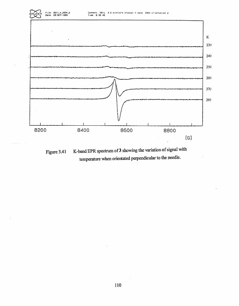
K
2 3 0
2 4 0
2 5 0
2 6 0
2 7 0
2 9 5
8 2 0 0 8 400 8 6 0 0 8 8 0 0
[G]
Figure 3.41 K-band EPR spectrum of 3 showing the variation of signal with
temperature when orientated perpendicular to the needle.
o r ?BflUKERC X J
F i l e :□ a te :
S6U_K_295K_2 0-S E P-1999
Comment: S6 1I . 3 . 5 a i c h l o r o c r y s t a l K oana. 295K o r i e n t a t i o n o Time: 9 : 3 0 : 4 6
110
3.4 Conclusions
In summary, I have successfully characterised the first three examples of neutral
dithiadiazolyl radicals displaying a stacked structure containing evenly spaced radical
units. I have also shown that chlorine substituents can direct the packing of dithiadiazolyl
radicals towards layered/stacked motifs. An investigation of the physical properties of
these systems showed the presence of some paramagnetic behaviour within the samples.
EPR measurements on crystal samples exhibited dipolar coupling. It is clear that the
exchange interactions within these structures are more complex than expected and further
solid state EPR measurements are desirable.
I l l
3.5 References
1 H. Genin and R Hoffinan, Macromolecules, 1998,31,444.
2 J.S. Miller and AJ. Epstein, Angew. Chem. Int. Ed. Engl.,1994,33,385.
3 A.J. Banister, A.S. Batsanov, O.G. Dawe, P.L. Herbertson, J.A.K. Howard, S. Lynn,
I. May, J.N.B. Smith, J.M. Rawson, T.E. Rogers, B.K. Tanner, G. Antorrena and
F. Palacio, J. Chem. Soc., Dalton Trans., 1997,2539.
4 A.J. Banister, A.S. Batsanov, O.G. Dawe, J.A.K. Howard, J.E. Davies, J.M. Rawson
and J.N.B. Smith, Phosphorus, Sulfur, and Silicon, 1997,124, 553.
5 J.E. Davies, RJ. Less, I. May and J.M. Rawson, New J. Chem., 1998,763.
6 L. Beer, A.W. Cordes, D.J.T. Myles, RT. Oakley and N.J. Taylor, Cryst. Eng.
Comm., 2000,20.
7 (a) C.D. Bryan, A.W. Cordes, R.M. Fleming, N.A. George, S.H. Glarum,
RC. Haddon, RT. Oakley, T.T.M. Palstra, A.S. Perel, L.F. Schneemeyer and
J.V. Waszezak, Nature, 1993,365, 821; (b) C.D. Bryan, A.W. Cordes, R.C. Haddon,
R.G. Hicks, D.K. Kennephohl, C.D. MacKinnon, RT. Oakley, T.T.M. Palstra,
A.S. Perel, S.R Scott, L.F. Schneemeyer and J.V. Waszezak, J. Am. Chem. Soc.,
1994, 116, 1205; (c) A.W. Cordes, RC. Haddon, and RT. Oakley, Adv. Mater.,
1994, 6, 798; (d) C.D. Bryan, A.W. Cordes, RC. Haddon, R.G. Hicks, R.T. Oakley,
T.T.M. Palstra, A.S. Perel and S.R Scott, Chem. Mater., 1994, 6, 508;
(e) C.D. Bryan, A.W. Cordes, R.M. Fleming, N.A. George, S.H. Glarum,
RC. Haddon, C.D. MacKinnon, RT. Oakley, T.T.M. Palstra and A.S. Perel, J. Am.
Chem. Soc., 1995, 117, 6880; (f) C.D. Bryan, A.W. Cordes, J.D. Goddard,
RC. Haddon, R.G. Hicks, C.D. MacKinnon, RC. Mawhinney, RT. Oakley,
T.T.M. Palstra and A.S. Perel, J. Am. Chem. Soc., 1996,118, 330; (g) A.W. Cordes,
112
N.A. George, R.C. Haddon, D.K Kennepohl, R.T. Oakley, T.T.M. Palstra and
R.W. Reed, Chem. Mater.., 1996,8,2774.
8 M.P. Andrews, A.W. Cordes, D.C. Douglas, R.M. Fleming, S.H. Glarum,
R.C. Haddon, P. Marsh, R.T. Oakley, T.T.M. Palstra, L.F. Schneemeyer,
G.W. Trucks, R. Tycko, J.V. Waszezak, KM. Young and N.M. Zimmerman, J. Am.
Chem. Soc., 1991,113,3559.
9 (a) A.W. Cordes, R.C. Haddon, R.G. Hicks, R.T. Oakley, T.T.M. Palstra,
L.F. Schneemeyer and J.V. Waszezak, J. Am. Chem. Soc., 1992, 114, 5000;
(b) A.W. Cordes, R.C. Haddon, R.G. Hicks, D.K Kennepohl, R.T. Oakley,
T.T.M. Palstra, L.F. Schneemeyer, S.R. Scott and J.V. Waszezak, Chem. Mater.,
1993, 5, 820; (c) C.D. Bryan, A.W. Cordes, R.C. Haddon, R.G. Hicks, R.T. Oakley,
T.T.M. Palstra and A.S. Perel, J. Chem. Soc., Chem. Commun., 1994,1447.
10 G.R. Desiraju and R. Parthasarathy, J. Am. Chem. Soc., 1989, 111, 8725.
11 J.A.R.P. Sarma and G.R. Desiraju, Acc. Chem. Res., 1986,19,222.
12 (a)T. Sakurai, M. Sundaralingham and G.A. Jeffrey, Acta Cryst., 1963, 16, 354;
(b)K. Yamasaki, J. Phys. Soc. Jprt., 1962, 17, 1262; (c) S.C. Nyburg, J. Chem.
Phys., 1964,40,2493.
13 D.E. Williams and L.-Y. Hsu, Acta Cryst., 1985, A41,296.
14 (a) N. Ramasubba, R. Parthasarathy and P. Murray-Rust, J. Am. Chem. Soc., 1986,
108, 4308. (b) J.D. Dunitz, Philos. Trans. R. Soc. London, 1975, B272, 99.
(c) K Fukui, T. Yonezawa and H. Shinga, J. Chem. Phys., 1952,20,722.
15 R.T. Boere and K.H. Moock, Z. anorg. allg. Chem., 1994,620,1589.
16 A.W. Cordes, C.M. Chamchoumis, R.G. Hicks, R.T. Oakley, KM. Young and
R.C. Haddon, Can. J. Chem., 1992,70, 919.
113
17 A.W. Cordes, R.C. Haddon, R.G. Hicks, R.T. Oakley and T.T.M. Palstra, Inorg.
Chem., 1992,31,1802.
18 A. Vegas, A. Perez-Salazar, A.J. Banister and R.G. Hey, J. Chem. Soc., Dalton
Trans., 1980,1812.
19 S.C. Nyburg and C.H. Faerman, Acta. Cryst., 1985, B41,274.
20 A.J. Banister, N. Bricklebank, W. Clegg, M.R.J. Elsegood, C.I. Gregory,
I. Lavender, J.M. Rawson and B.K. Tanner, J. Chem. Soc., Chem. Commun., 1995,
679.
21 G. Antorrena, Ph.D. thesis, Universidad de Zaragoza.
22 S.A Fairhurst, K.M. Johnson, H. Sutcliffe, K.F. Preston, A.J. Banister,
Z.V. Hauptman and J. Passmore, J. Chem. Soc., Dalton Trans., 1986,1465.
23 S.A. Fairhurst, L.H. Sutcliffe, K.F. Preston, A.J. Banister, A.S. Partington,
J.M. Rawson, J. Passmore and M. Schriver, Magn. Reson. Chem., 1993,31,1027.
24 A.W. Cordes, R.C. Haddon, R.T. Oakley, L.F. Schneemeyer, J.V. Waszezak,
K.M. Young and N.M. Zimmerman, J. Am. Chem. Soc., 1991,113, 582.
114
Chapter 4
Dimer Stacking Motifs for
Dichlorophenyl Dithiadiazolyl
Radicals
115
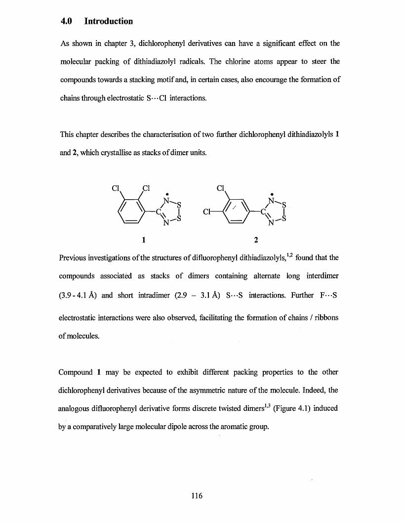
4.0 Introduction
As shown in chapter 3, dichlorophenyl derivatives can have a significant effect on the
molecular packing of dithiadiazolyl radicals. The chlorine atoms appear to steer the
compounds towards a stacking motif and, in certain cases, also encourage the formation of
chains through electrostatic S* • • Cl interactions.
This chapter describes the characterisation of two further dichlorophenyl dithiadiazolyls 1
and 2, which crystallise as stacks of dimer units.
1 2
^ /SCA I Cl— ( ' ')— C. IVs \=/ V®
Previous investigations of the structures of difluorophenyl dithiadiazolyls,1,2 found that the
compounds associated as stacks of dimers containing alternate long interdimer
(3.9-4.1 A) and short intradimer (2.9 - 3.1 A) S---S interactions. Further F - S
electrostatic interactions were also observed, facilitating the formation of chains / ribbons
of molecules.
Compound 1 may be expected to exhibit different packing properties to the other
dichlorophenyl derivatives because of the asymmetric nature of the molecule. Indeed, the
analogous difluorophenyl derivative forms discrete twisted dimers1,3 (Figure 4.1) induced
by a comparatively large molecular dipole across the aromatic group.
116
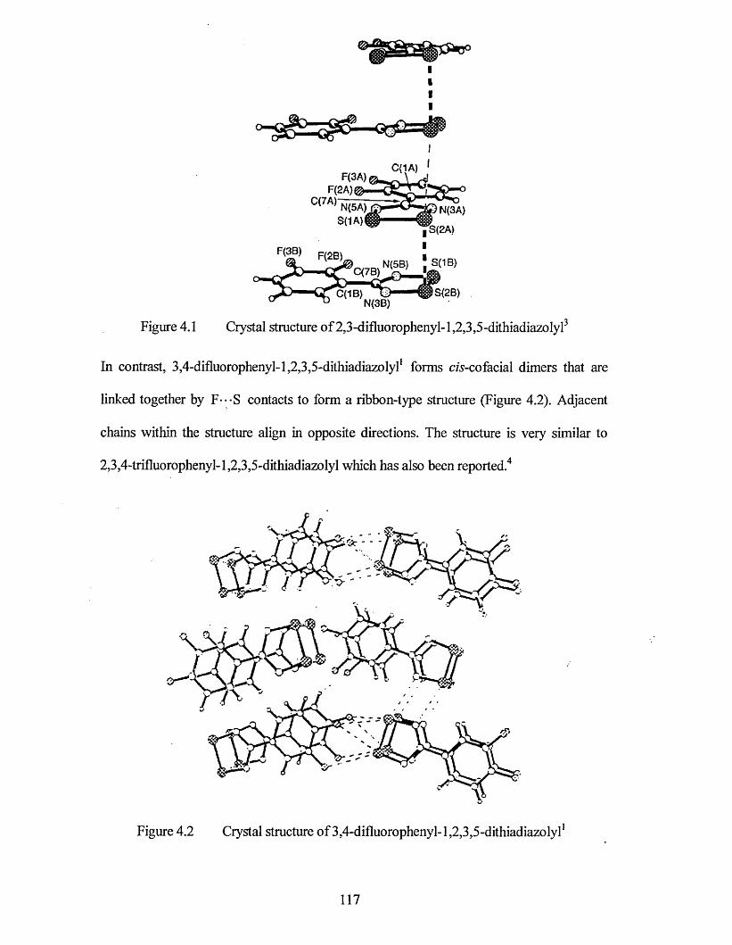
Figure 4.1 Crystal structure of 2,3-difluorophenyl-1,2,3,5-dithiadiazolyl3
In contrast, 3,4-difluorophenyl-l,2,3,5-dithiadiazolyl1 forms cA-co facial dimers that are
linked together by F---S contacts to form a ribbon-type structure (Figure 4.2). Adjacent
chains within the structure align in opposite directions. The structure is very similar to
2,3,4-trifluorophenyl-l,2,3,5-dithiadiazolyl which has also been reported.4
Figure 4.2 Crystal structure of 3,4-difluorophenyl-1,2,3,5-dithiadiazolyl1
117
The only chlorine derivative reported to date (p-chlorophenyl-1,2,3,5-dithiadiazolyl) 5 has a
crystal structure where S---C1 interactions were observed at a 90° angle with respect to the
planes of the two rings (Figure 3.1, page 61). The formation of ribbons was still observed
within the structure indicating that chlorine in the para position can facilitate this sort of
ribbon/chain structure. This was also true for 2,4-dichlorophenyl-l,2,3,5-dithiadiazolyl
(Figure 3.9, page 71) that also has significant S---C1 interactions resulting in the formation
of ribbons of CI2.C6H3. CNSSN molecules throughout the structure.
4.1 X-Ray Structures
X-ray structural determinations were undertaken on 1 and 2. An Ortep structural plot of the
asymmetric units from both isomers 1 and 2 are shown in Figure 4.3 and 4.4 respectively.
The bond lengths and angles of the two molecules (denoted by a and b) within the
asymmetric units of 1 and 2 are listed in Table 4.1 and 4.2 together with data for phenyl-
1.2.3.5-dithiadiazolyl6 and p-chlorophenyl-1,2,3,5-dithiadiazolyl5 for comparative
purposes. The full crystallographic data for compounds 1 and 2 can be found in appendix 6
and 7 respectively at the end of the thesis.
The molecular structures of 1 and 2 are similar to those of the majority of aryl
1.2.3.5-dithiadiazolyls and to the other isomers of dichlorophenyl-1,2,3,5-dithiadiazolyl
described earlier (Chapter 3). The chlorine-carbon bond distances (1.722 - 1.753 A) are
reasonably similar to those observed for77-chlorophenyl-1,2,3,5-dithiadiazolyl5 [Figure 3.1
(1.750 A)]. The most significant difference observed between the two structures is the
torsion angle between the two planar rings (CI2.C6H3. CNSSN ). As might be expected
compound 1, which has chlorine in the ortho position, has a relatively large torsion angle
(27.5°), whereas the torsion angle observed for compound 2 is significantly smaller (13.4°).
Presumably, this is due to the reduced steric hindrance.
118
Figure 4.3 2,3-dichlorophenyl-1,2,3,5-dithiadiazolyl (1)
3,4-dichlorophenyl-1,2,3,5-dithiadiazolyl (2)Figure 4.4
119
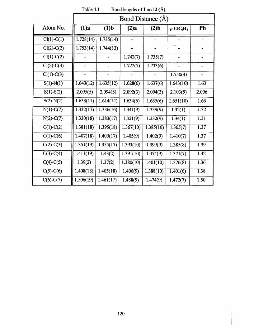
Table 4.1 Bond lengths of 1 and 2 (A).
Bond Distance (A )
Atom No. ( l)a ( l)b (2)a (2)b /j-ClQft, P h
Cl(l)-C(l) 1.728(14) 1.735(14) - - - -
Cl(2)-C(2) 1.753(14) 1.744(13) - - - -
Cl(l)-C(2) - - 1.742(7) 1.735(7) - -
Cl(2)-C(3) - - 1.722(7) 1.733(6) - -
Cl(l)-C(3) - - - - 1.750(4) -
S(l)-N(l) 1.643(12) 1.635(12) 1.628(6) 1.637(6) 1.643(10) 1.63
S(l)-S(2) 2.095(5) 2.094(5) 2.092(3) 2.094(3) 2.103(5) 2.096
S(2)-N(2) 1.633(11) 1.614(14) 1.634(6) 1.635(6) 1.651(10) 1.63
N(l)-C(7) 1.332(17) 1.336(16) 1.341(9) 1.339(9) 1.32(1) 1.32
N(2)-C(7) 1.330(18) 1.383(17) 1.321(9) 1.332(9) 1.34(1) 1.31
C(l)-C(2) 1.381(18) 1.395(18) 1.367(10) 1.385(10) 1.365(7) 1.37
C(l)-C(6) 1.407(18) 1.409(17) 1.405(9) 1.402(9) 1.410(7) 1.37
C(2)-C(3) 1.351(19) 1.355(17) 1.393(10) 1.399(9) 1.385(8) 1.39
C(3)-C(4) 1.411(19) 1.43(2) 1.391(10) 1.374(9) 1.371(7) 1.42
C(4)-C(5) 1.39(2) 1.37(2) 1.380(10) 1.401(10) 1.376(8) 1.36
C(5)-C(6) 1.408(18) 1.405(18) 1.406(9) 1.388(10) 1.401(6) 1.38
C(6)-C(7) 1.506(19) 1.461(17) 1.488(9) 1.474(9) 1.472(7) 1.50
120
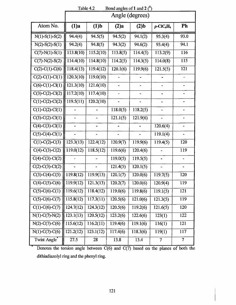
Table 4.2 Bond angles of 1 and 2 (°)Angle (degrees)
Atom No. (l)a (l)b (2)a (2)b p-CICA Ph
N(l)-S(l)-S(2) 94.4(4) 94.5(5) 94.5(2) 94.1(2) 95.3(4) 93.0
N(2)-S(2)-S(l) 94.2(4) 94.8(5) 94.3(2) 94.6(2) 93.4(4) 94.1
C(7)-N(l)-S(l) 113.8(10) 115.2(10) 113.8(5) 114.4(5) 113.2(9) 116
C(7)-N(2)-S(2) 114.4(10) 114.8(10) 114.2(5) 114.3(5) 114.0(8) 115
C(2)-C(l)-C(6) 118.4(13) 119.4(12) 120.1(6) 119.9(6) 121.5(5) 121
C(2)-C(1)-C1(1) 120.3(10) 119.0(10) - - - -
C(6)-C(1)-C1(1) 121.3(10) 121.6(10) - - - -
C(3)-C(2)-C1(2) 117.2(10) 117.4(10) - - - -
C(1)-C(2)-C1(2) 119.5(11) 120.2(10) - - - -
C(1)-C(2)-C1(1) - - 118.0(5) 118.2(5) - -
C(3)-C(2)-C1(1) - - 121.1(5) 121.9(6) - -
C(4)-C(3)-C1(1) - - - - 120.6(4) -
C(5)-C(4)-C1(1) - - - - 119.1(4) -
C(l)-C(2)-C(3) 123.3(13) 122.4(12) 120.9(7) 119.9(6) 119.4(5) 120
C(4)-C(3)-C(2) 119.0(12) 118.5(12) 119.6(6) 120.4(6) - 119
C(4)-C(3)-C1(2) - - 119.0(5) 119.5(5) - -
C(2)-C(3)-C1(2) - - 121.4(5) 120.1(5) - -
C(3)-C(4)-C(5) 119.8(12) 119.9(13) 120.1(7) 120.0(6) 119.7(5) 120
C(4)-C(5)-C(6) 119.9(12) 121.3(13) 120.2(7) 120.0(6) 120.9(4) 119
C(5)-C(6)-C(l) 119.6(12) 118.4(12) 119.0(6) 119.8(6) 119.1(5) 121
C(5)-C(6)-C(7) 115.8(12) 117.3(11) 120.5(6) 121.0(6) 121.3(5) 119
C(l)-C(6)-C(7) 124.7(12) 124.3(12) 120.5(6) 119.2(6) 121.6(5) 120
N(l)-C(7)-N(2) 123.1(13) 120.5(12) 123.2(6) 122.6(6) 123(1) 122
N(2)-C(7)-C(6) 115.6(12) 116.2(11) 119.4(6) 119.1(6) 116(1) 121
N(l)-C(7)-C(6) 121.2(12) 123.1(12) 117.4(6) 118.3(6) 119(1) 117
Twist Angle" 27.5 28 13.8 13.4 7 7ifc 1 1 1 mm i l ! ■ „ , II ■■ m i
Denotes the torsion angle between C(6) and C(7) based on the planes of both the
dithiadiazolyl ring and the phenyl ring.
121
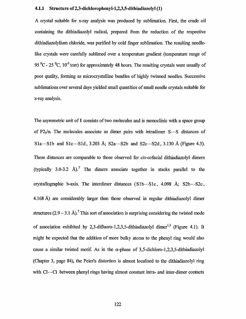
4.1.1 Structure of 2,3-dichIorophenyl-l,2,3,5-dithiadiazolyI (1)
A crystal suitable for x-ray analysis was produced by sublimation. First, the crude oil
containing the dithiadiazolyl radical, prepared from the reduction of the respective
dithiadiazolylium chloride, was purified by cold finger sublimation. The resulting needle
like crystals were carefully sublimed over a temperature gradient (temperature range of
95 °C - 25 °C, 10'3 torr) for approximately 48 hours. The resulting crystals were usually of
poor quality, forming as microcrystalline bundles of highly twinned needles. Successive
sublimations over several days yielded small quantities of small needle crystals suitable for
x-ray analysis.
The asymmetric unit of 1 consists of two molecules and is monoclinic with a space group
of P2i/n. The molecules associate as dimer pairs with intradimer S***S distances of
Sla***Slb and Slc***Sld, 3.203 A; S2a***S2b and S2c***S2d, 3.130 A (Figure 4.5).
These distances are comparable to those observed for cw-cofacial dithiadiazolyl dimers
(typically 3.0-3.2 A).7 The dimers associate together in stacks parallel to the
ciystallographic b-axis. The interdimer distances (Sib*•-Sic, 4.098 A; S2b***S2c,
4.168 A) are considerably larger than those observed in regular dithiadiazolyl dimer
structures (2.9 - 3.1 A).7 This sort of association is surprising considering the twisted mode
of association exhibited by 2,3-difluoro-l,2,3,5-dithiadiazolyl dimer1,3 (Figure 4.1). It
might be expected that the addition of more bulky atoms to the phenyl ring would also
cause a similar twisted motif. As in the a-phase of 3,5-dichloro-1,2,3,5-dithiadiazolyl
(Chapter 3, page 84), the Peierls distortion is almost localised to the dithiadiazolyl ring
with C1---C1 between phenyl rings having almost constant intra- and inter-dimer contacts
122
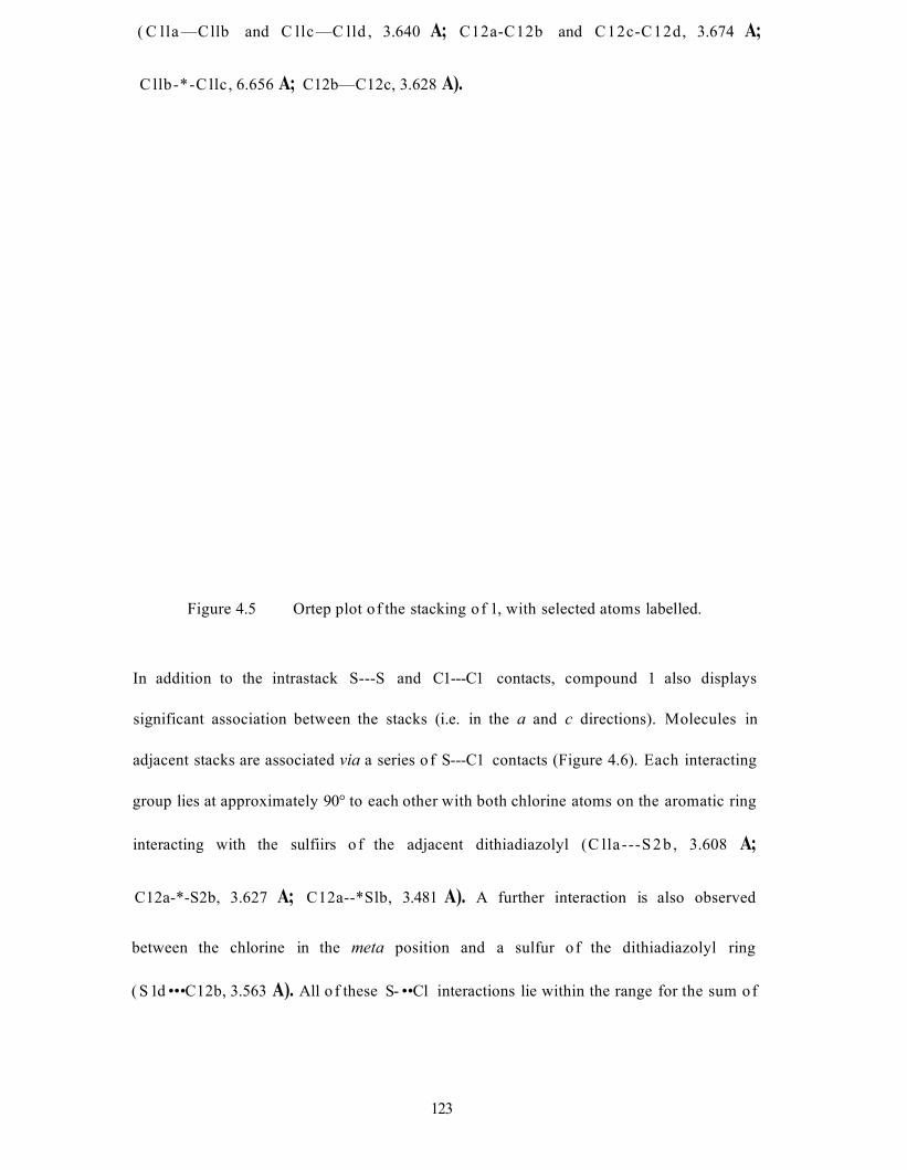
( C lla —Cllb and C llc —C lld , 3.640 A; C12a-C12b and C12c-C12d, 3.674 A;
C llb-*-C llc, 6.656 A; C12b—C12c, 3.628 A).
Figure 4.5 Ortep plot of the stacking o f 1, with selected atoms labelled.
In addition to the intrastack S---S and C1---C1 contacts, compound 1 also displays
significant association between the stacks (i.e. in the a and c directions). Molecules in
adjacent stacks are associated via a series o f S---C1 contacts (Figure 4.6). Each interacting
group lies at approximately 90° to each other with both chlorine atoms on the aromatic ring
interacting with the sulfiirs o f the adjacent dithiadiazolyl (C lla ---S 2b , 3.608 A;
C12a-*-S2b, 3.627 A; C12a--*Slb, 3.481 A). A further interaction is also observed
between the chlorine in the meta position and a sulfur o f the dithiadiazolyl ring
( S 1 d • • • C12b, 3.563 A). All o f these S- • • Cl interactions lie within the range for the sum of
123
the van der Waals radii o f sulfur and chlorine (3.5 A -3.7 A)8 which suggests that the
S - Cl contacts within the molecule are relatively strong.
Cl lc,C12cS2a
CI2d
S2c► -C l la
V* Cl IdS2b
C12a
C12b S2dSidCl lb
Figure 4.6 Ortep plot of the interactions o f 1 within the ac plane, with selected atomslabelled.
Unlike the other dichlorophenyl dithiadiazolyl isomers shown in chapter 3 there would
seem to be no C1---C1 interactions in the a or c direction. Figure 4.7 shows the overall
packing o f 1. Although not obvious initially, the whole structure can be viewed as a series
of chains arranged in a herringbone fashion with each group interacting at an angle o f
approximately 90° to the adjacent group. Each adjacent chain is linked by one S---C1
interaction per molecule [C12b---Sld, 3.563 A(Figure 4.6)] and is aligned anti-parallel to
one another. The packing (Figure 4.7) again illustrates the ability o f chlorine substituents
to create a stacked structure and its ability, through electrostatic interactions, to form
chains. Unfortunately, in this case the spin pairing within the molecule is the predominant
feature and is almost certain to make the compound diamagnetic.
124
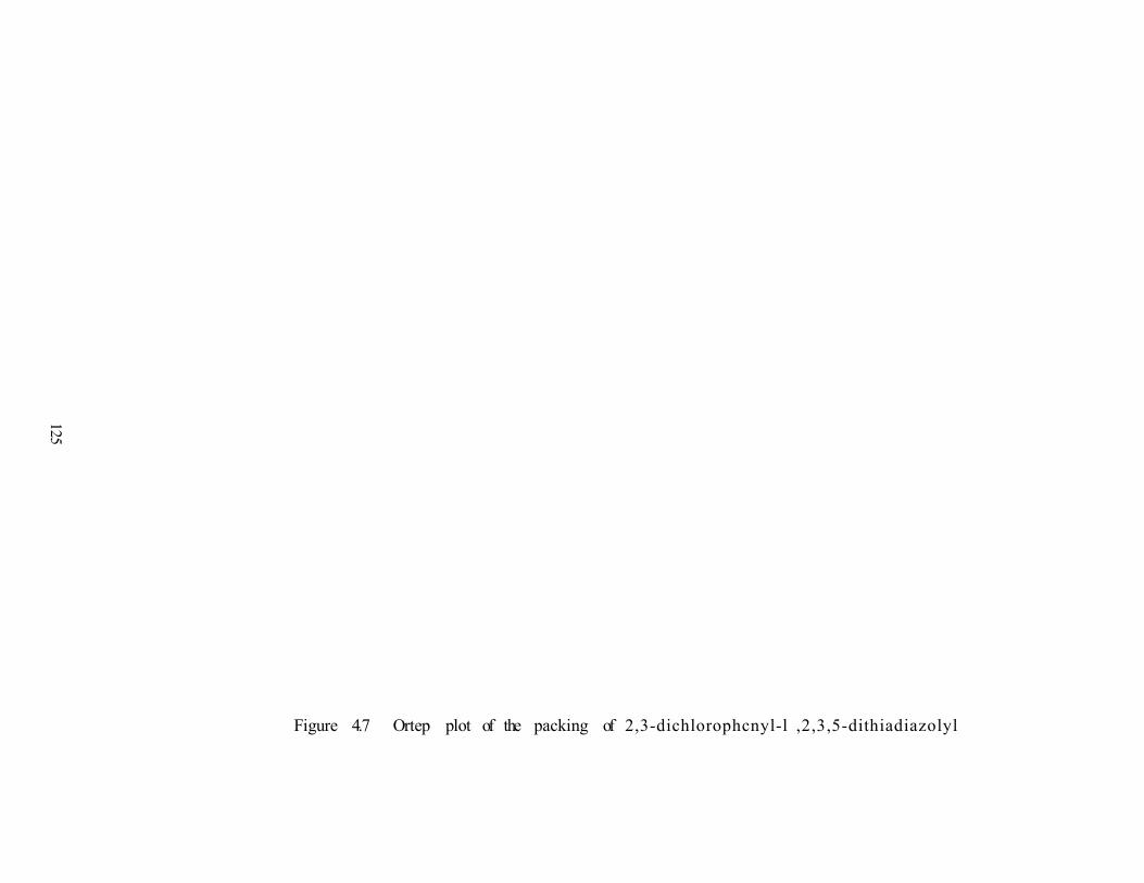
125
Figure 4.7 Ortep plot of the packing of 2,3-dichlorophcnyl-l ,2,3,5-dithiadiazolyl
4.1.2 X-ray Crystal Structure of 3,4-dichIorophenyI-l,2,3,5-dithiadiazolyl (2)
Crystals suitable for x-ray analysis were produced using a similar method to that described
for 1 (Section 3.1.1, page 66). The gradient sublimation was completed using a
temperature range 110-25 °C under a dynamic vacuum (10'3 torr). The resulting crystals
were usually twinned and of poor quality but repeated sublimation over several days
yielded small dark red crystals, suitable for crystallographic studies.
The asymmetric unit of 2 contains two molecules and is monoclinic with space group
P2i/c. The molecules also associate as dimer pairs with intradimer S***S distances of
Sla***Slb and S lc - S ld , 3.125 A; S2a***S2b and S2c***S2d, 3.207 A (Figure 4.8)
which are similar to those observed for c/s-cofacial dithiadiazolyl dimers (typically
2.9 - 3.1 A).7 The dimers associate in stacks parallel to the crystallographic a-axis. The
interdimer distances are also considerably larger than those observed in regular
dithiadiazolyl dimer structures (Sib*•-Sic, 4.103 A; S2b***S2c, 4.033 A). In a similar
way to 1, the Peierls distortion is almost localised to dithiadiazolyl rings with the intra and
interdimer C1---C1 contacts of the phenyl rings being almost constant (Clla***Cllb and
Cllc***Clld, 3.628 A; C12a—C12b and C12c***C12d, 3.569 A; Cllb***Cllc, 6.589 A;
C12b***C12c, 3.672 A).
126

Figure 4.8 Ortep plot of the stacking o f 2, with selected atoms labelled.
Within the plane parallel to the b and c axis the molecules are linked into chains through
electrostatic S---C1 interactions (Figure 4.9). There is significant interaction between both
of the chlorine atoms with both o f the sulfur atoms of the dithiadiazolyl ring ( C12b • • - S 1 a ,
3.351 A; C llc -S 2 a ,3 .3 2 8 A).
Figure 4.9 Ortep plot of the chain structure o f 1.
127
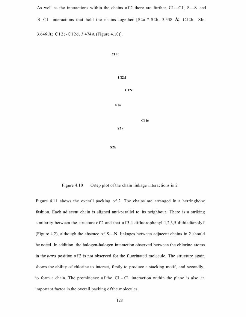
As well as the interactions within the chains of 2 there are further C1---C1, S---S and
S - C l interactions that hold the chains together [S2a-*-S2b, 3.338 A; C12b---Slc,
3.646 A; C12c-C12d, 3.474A (Figure 4.10)].
Cl Id
CI2d
C12c
Cl 1 c
S la
S2a
S2b
Figure 4.10 Ortep plot o f the chain linkage interactions in 2.
Figure 4.11 shows the overall packing o f 2. The chains are arranged in a herringbone
fashion. Each adjacent chain is aligned anti-parallel to its neighbour. There is a striking
similarity between the structure o f 2 and that o f 3,4-difluorophenyl-1,2,3,5-dithiadiazolyl1
(Figure 4.2), although the absence o f S---N linkages between adjacent chains in 2 should
be noted. In addition, the halogen-halogen interaction observed between the chlorine atoms
in the para position o f 2 is not observed for the fluorinated molecule. The structure again
shows the ability o f chlorine to interact, firstly to produce a stacking motif, and secondly,
to form a chain. The prominence o f the Cl - Cl interaction within the plane is also an
important factor in the overall packing o f the molecules.
128
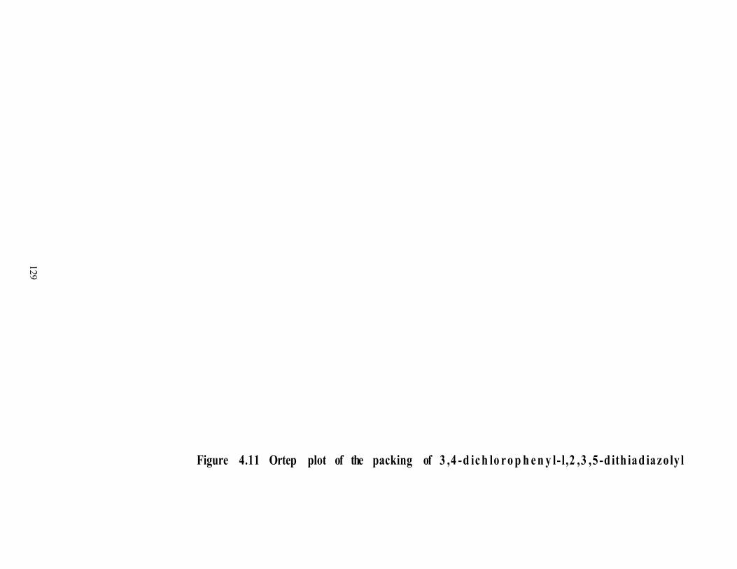
129
Figure 4.11 Ortep plot of the packing of 3 ,4 -d ic h lo r o p h e n y l- l,2 ,3 ,5 -d ith iad iazo ly l
4.2 Conclusions
Although the two compounds presented in this chapter are of little interest either
magnetically or electronically, they illustrate the effects of chlorine substituents on the
molecular packing of dithiadiazolyls. The use of chlorine in place of previously reported
fluorinated derivatives has made a marked difference on the overall structure of the
compounds under investigation. In addition, observations on the extent of electrostatic
S---C1 interactions to form chains of radical units may also have future use in the
development of molecular magnets.
130
4.3 References
1 A.J. Banister, A.S. Batsanov, O.G. Dawe, J.A.K. Howard, J.E. Davies, J.M. Rawson
and J.N.B. Smith, Phosphorus, Sulfur, and Silicon, 1997,124,553.
2 L. Beer, A.W. Cordes, D.J.T. Myles, R.T. Oakley and N.J. Taylor, Cryst. Eng
Comm., 2000,20.
3 A.J. Banister, A.S. Batsanov, O.G. Dawe, P.L. Herbertson, J.A.K. Howard, S. Lynn,
I. May, J.N.B. Smith, J.M. Rawson, T.E. Rogers, B.K. Tanner, G. Antorrena and
F. Palacio, J. Chem. Soc., Dalton Trans., 1997,2539.
4 A.M.T. Bell, J.N.B. Smith, J.P. Attfield, J.M. Rawson, K. Shankland and
W.I.F. David, NewJ. Chem., 1999,23,565.
5 R.T. Boere and K.H. Moock, Z. Anorg. Allg. Chem., 1994,620,1589.
6 A. Vegas, A. Perez-Salazar, A.J. Banister and R.G. Hey, J. Chem. Soc., Dalton
Trans., 1980,1812.
7 J.M. Rawson, A.J. Banister and I. Lavender, Adv. Heterocycl. Chem., 1995,62,137.
8 S.C. Nyburg and C.H. Faerman, Acta. Cryst., 1985, B41,274.
131
Chapter 5
The Structure and Physical
Properties of 3,5-dibromophenyl-
1,2,3,5-dithiadiazolyl
132
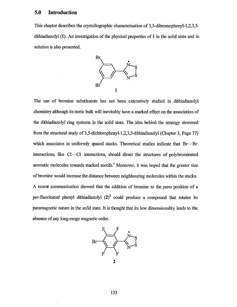
5.0 Introduction
This chapter describes the crystallographic characterisation of 3,5-dibromophenyl-l,2,3,5-
dithiadiazolyl (1). An investigation of the physical properties of 1 in the solid state and in
solution is also presented.
The use of bromine substituents has not been extensively studied in dithiadiazolyl
chemistry although its steric bulk will inevitably have a marked effect on the association of
the dithiadiazolyl ring systems in the solid state. The idea behind the strategy stemmed
from the structural study of 3,5-dichlorophenyl-l,2,3,5-dithiadiazolyl (Chapter 3, Page 77)
which associates in uniformly spaced stacks. Theoretical studies indicate that Br -*Br
interactions, like C1---C1 interactions, should direct the structures of polybrominated
aromatic molecules towards stacked motifs. 1 Moreover, it was hoped that the greater size
of bromine would increase the distance between neighbouring molecules within the stacks.
A recent communication showed that the addition of bromine to the para position of a
per-fluorinated phenyl dithiadiazolyl (2 )2 could produce a compound that retains its
paramagnetic nature in the solid state. It is thought that its low dimensionality leads to the
absence of any long-range magnetic order.
Br
Br1
F F
2
133
5.1 X-Ray Crystal Structure
An Ortep structural plot of the asymmetric unit o f 1 shown in Figure 5.1. Bond lengths and
angles for 1 are listed in Tables 5.1 and 5.2 together with data for (PhCNSSN and 22 for
comparative purposes. The full crystallographic data for 1 can be found in appendix 8 .
The molecular structure o f 1 is, as expected, similar to the molecular structure o f most
previously published aryl substituted dithiadiazolyls. Within each molecule, the aryl and
dithiadiazolyl rings are essentially coplanar with a slight torsion angle between the
dithiadiazolyl ring and the aryl ring o f 7.4° and 5.5° (two molecules in the asymmetric
unit). This value is similar to that observed for aryl substituents not possessing a large
substituent in the ortho position (eg Ph. CNSSN torsion angle = 7°).
Figure 5.1 Ortep plot of 3,5-dibromophenyl-1,2,3,5-dithiadiazolyl (1)
134
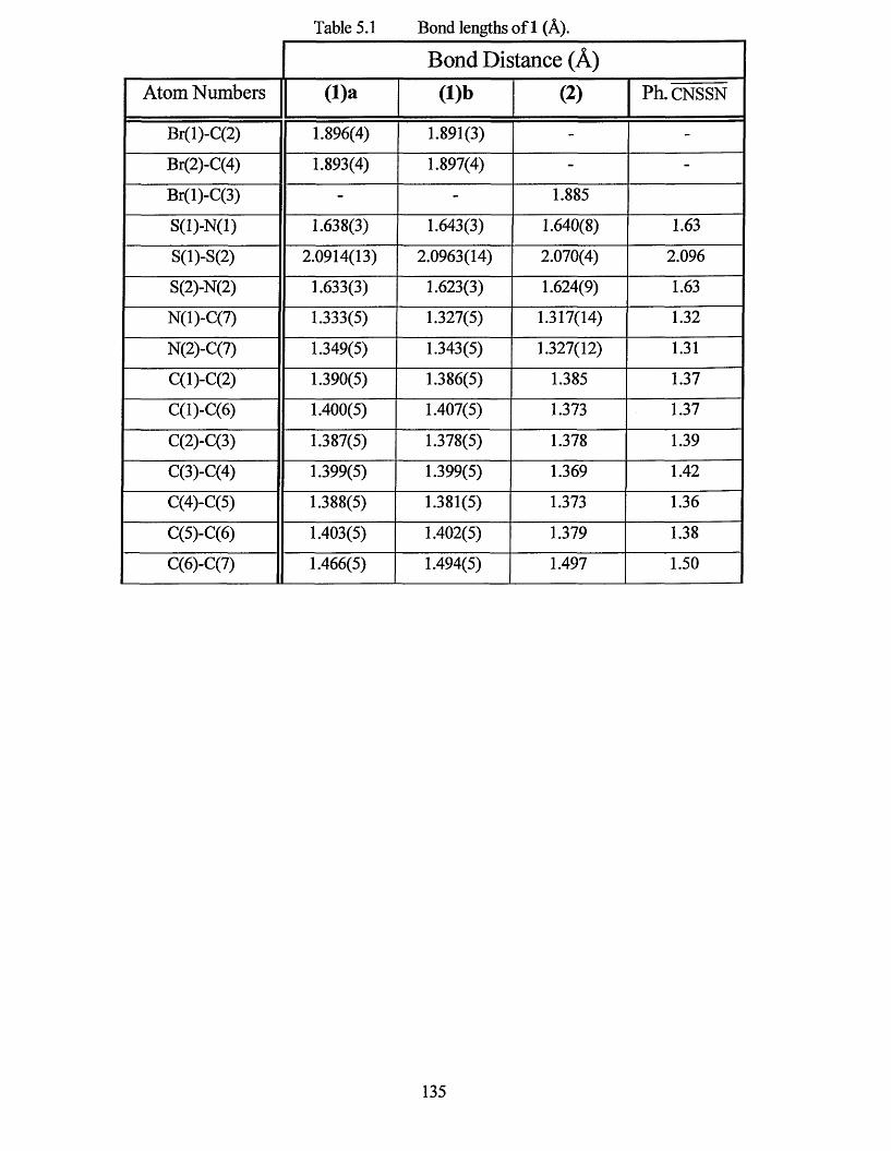
Table 5.1 Bond lengths of 1 (A).
Bond Distance (A)Atom Numbers (l)a (l)b (2) Ph. CNSSN
Br(l)-C(2) 1.896(4) 1.891(3) - -
Br(2)-C(4) 1.893(4) 1.897(4) - -
Br(l)-C(3) - - 1.885
S(l)-N(l) 1.638(3) 1.643(3) 1.640(8) 1.63
S(l)-S(2) 2.0914(13) 2.0963(14) 2.070(4) 2.096
S(2)-N(2) 1.633(3) 1.623(3) 1.624(9) 1.63
N(l)-C(7) 1.333(5) 1.327(5) 1.317(14) 1.32
N(2)-C(7) 1.349(5) 1.343(5) 1.327(12) 1.31
C(l)-C(2) 1.390(5) 1.386(5) 1.385 1.37
C(l)-C(6 ) 1.400(5) 1.407(5) 1.373 1.37
C(2)-C(3) 1.387(5) 1.378(5) 1.378 1.39
C(3)-C(4) 1.399(5) 1.399(5) 1.369 1.42
C(4)-C(5) 1.388(5) 1.381(5) 1.373 1.36
C(5)-C(6) 1.403(5) 1.402(5) 1.379 1.38
C(6)-C(7) 1.466(5) 1.494(5) 1.497 1.50
135
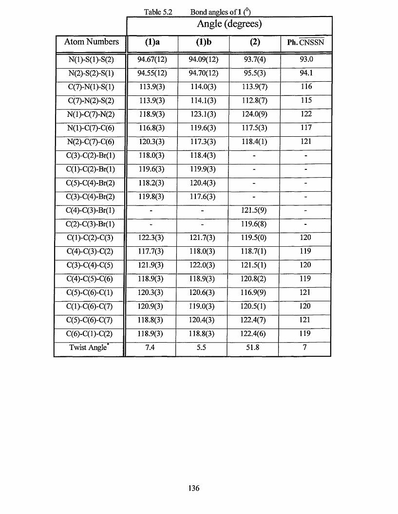
Table 5.2 Bond angles of 1 (°)Angle (degrees)
Atom Numbers ( l)a (l)b (2) Ph. CNSSN
N(l)-S(l)-S(2) 94.67(12) 94.09(12) 93.7(4) 93.0
N(2)-S(2)-S(l) 94.55(12) 94.70(12) 95.5(3) 94.1
C(7)-N(l)-S(l) 113.9(3) 114.0(3) 113.9(7) 116
C(7)-N(2)-S(2) 113.9(3) 114.1(3) 112.8(7) 115
N(l)-C(7)-N(2) 118.9(3) 123.1(3) 124.0(9) 122
N(l)-C(7)-C(6) 116.8(3) 119.6(3) 117.5(3) 117
N(2)-C(7)-C(6) 120.3(3) 117.3(3) 118.4(1) 121
C(3)-C(2)-Br(l) 118.0(3) 118.4(3) - -
C(l)-C(2)-Br(l) 119.6(3) 119.9(3) - -
C(5)-C(4)-Br(2) 118.2(3) 120.4(3) - -
C(3)-C(4)-Br(2) 119.8(3) 117.6(3) - -
C(4)-C(3)-Br(l) - - 121.5(9) -
C(2)-C(3)-Br(l) - - 119.6(8) -
C(l)-C(2)-C(3) 122.3(3) 121.7(3) 119.5(0) 120
C(4)-C(3)-C(2) 117.7(3) 118.0(3) 118.7(1) 119
C(3)-C(4)-C(5) 121.9(3) 122.0(3) 121.5(1) 120
C(4)-C(5)-C(6) 118.9(3) 118.9(3) 120 .8(2 ) 119
C(5)-C(6)-C(l) 120.3(3) 120.6(3) 116.9(9) 121
C(l)-C(6)-C(7) 120.9(3) 119.0(3) 120.5(1) 120
C(5)-C(6)-C(7) 118.8(3) 120.4(3) 122.4(7) 121
C(6)-C(l)-C(2) 118.9(3) 118.8(3) 122.4(6) 119
Twist Angle* 7.4 5.5 51.8 7
136

The unit cell o f 1 contains eight molecules o f (Br2 .C6H3. CNSSN )2, two halves o f which
are in the asymmetric unit. Figure 5.2 shows the association o f two dimers within the unit
cell. The mode o f association within each dimer is cz's-co facial, which is the most common
motif for dithiadiazolyl radicals. The intradimer distances are short (S la - - -S lb , 3.180 A;
S2 a---S2 b , 2.999 A) lying within the normal range for dithiadiazolyl dimers
(3.0 - 3.2 A).4 Unlike many other dithiadiazolyls, the individual molecules within a dimer
do not lie co-planar to each other. Instead, the steric repulsion causes the molecules to twist
with respect to each other. The bromine contacts within the dimer are considerably longer
than those o f sulfur (Brla-- -Brlb, 3.966 A; Br2a---Br2b, 4.270 A), which might be
expected because o f the larger size o f bromine compared to sulfur. In fact the Br-- Br
contacts within the dimer fall outside that o f the sum o f the van der Waals radii (3.72 A) 3
so can be classed as having no interaction with each other. To a lesser extent, this type o f
twisted dimer has also been observed for Ph. CNSSNJ but the effect in 1 is much larger.
The dihedral angle S-S to S-S are 6.56° and 17.6° for Ph. CNSSN and 1 respectively.
Figure 5.2 Ortep plot o f the dimerisation o f 1
137
Each dimer pair shown in Figure 5.2 is associated as a stack of two dimers aligned in
antiparallel directions (Figure 5.3). They are related via a 2-fold rotation and a glide plane
in the c-direction. Although forming part o f a stack o f four individual dithiadiazolyl
molecules, they are not joined by any close interactions. The reason for this packing
arrangement is likely to be due to the steric repulsion o f the large bromine atoms between
adjacent dimers. This type o f association was also observed for / 7-NC.C6H4 . CNSSN6
(Figure 5.4). In this case, the electrostatic attraction o f the more negative nitrile group to
the sulfur o f the layer above may be the controlling factor of the packing o f the molecules.
Perpendicular to the stack o f two dimers is a second pair o f dimers, which associate in an
identical stack (Figure 5.3). The separate stacks are attached together by weak B r - S
interactions (3.310 A in comparison to the sum of the van der Waals radii o f 3.75 A).3 The
association o f the dimers perpendicular to each other shows no close B r - B r contacts.
The apparent attractive nature o f chlorine observed in earlier chapters (chapters 3 and 4)
does not occur for bromine atoms, which appear to repel each other in the solid state.
Figure 5.3 Ortep plot of the interactions o f 1
138
I I
I I I I
I
I I I \
Figure 5.4 The crystal structure of/7-CN.C6H4. CNSSN
The interactions observed in Figures 5.2 and 5.3 are repeated for every stack of two dimers
within the structure. This creates an overall packing structure (Figure 5.5) with
dithiadiazolyl units lying in two distinct planes. It is unclear why the molecules pack in this
way, but it could be due to the directional nature of halogen interactions.7
139
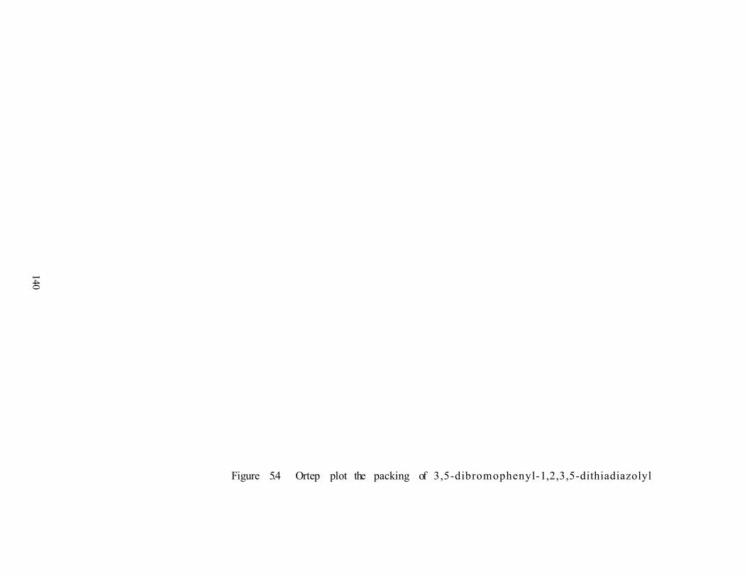
140
Figure 5.4 Ortep plot the packing of 3,5-dibromophenyl-1,2,3,5-dithiadiazolyl
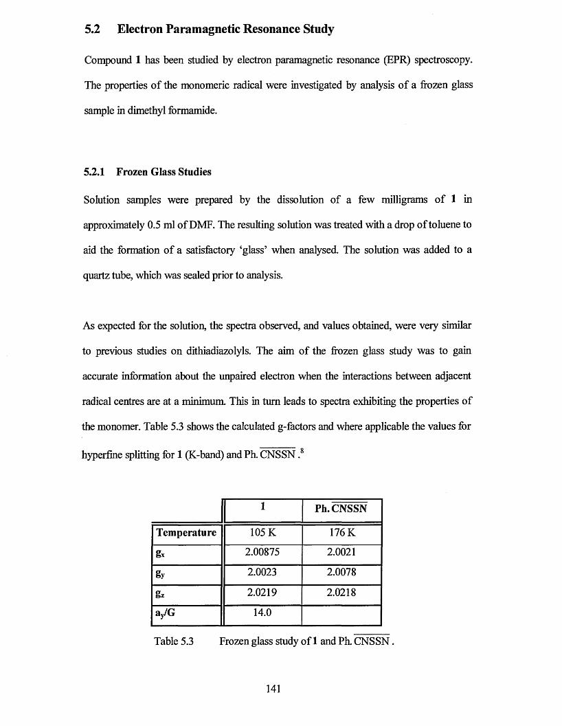
5.2 Electron Paramagnetic Resonance Study
Compound 1 has been studied by electron paramagnetic resonance (EPR) spectroscopy.
The properties of the monomeric radical were investigated by analysis of a frozen glass
sample in dimethyl formamide.
5.2.1 Frozen Glass Studies
Solution samples were prepared by the dissolution of a few milligrams of 1 in
approximately 0.5 ml of DMF. The resulting solution was treated with a drop of toluene to
aid the formation of a satisfactory ‘glass’ when analysed. The solution was added to a
quartz tube, which was sealed prior to analysis.
As expected for the solution, the spectra observed, and values obtained, were very similar
to previous studies on dithiadiazolyls. The aim of the frozen glass study was to gain
accurate information about the unpaired electron when the interactions between adjacent
radical centres are at a minimum. This in turn leads to spectra exhibiting the properties of
the monomer. Table 5.3 shows the calculated g-factors and where applicable the values for
hyperfine splitting for 1 (K-band) and Ph. CNSSN .8
1 Ph. CNSSN
Temperature 105 K 176 K
gx 2.00875 2.0021
gy 2.0023 2.0078
gz 2.0219 2.0218
ay/G 14.0
Table 5.3 Frozen glass study of 1 and Ph. CNSSN.
141
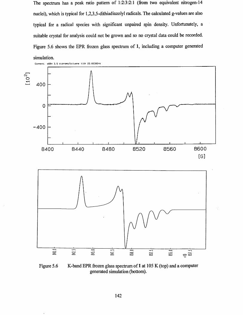
The spectrum has a peak ratio pattern of 1:2:3:2:1 (from two equivalent nitrogen-14
nuclei), which is typical for 1,2,3,5-dithiadiazolyl radicals. The calculated g-values are also
typical for a radical species with significant unpaired spin density. Unfortunately, a
suitable crystal for analysis could not be grown and so no crystal data could be recorded.
Figure 5.6 shows the EPR frozen glass spectrum of 1, including a computer generated
simulation.Comment: s684 3 . 5 t ) lO r o m o / to lu e n e 110K 23.6530GHz
4 0 0
0
- 4 0 0
8 6 0 08 5 6 08 5 2 08 4 8 08 4 0 0 8 4 4 0
[G]
Figure 5.6 K-band EPR frozen glass spectrum of 1 at 105 K (top) and a computergenerated simulation (bottom).
5.3 Conclusions
From the results reported in this chapter, it is clear that bromine atoms can have a
significant effect on the dimerisation of dithiadiazolyls. The slightly twisted mode of
association within the dimer is more pronounced than previously observed in the phenyl
derivative, but is much less than the twisted conformations reported previously.9 The
observed structural features clearly result from the large steric bulk of the bromine atoms.
143
5.4 References
1 (a) G.R. Desiraju and R. Parthasarathy, J. Am. Chem. Soc., 1989, 111, 8725; (b)
V.R. Pedireddi, D.S. Reddy, B.S. Goud, D.C. Craig, A.D.Rae and G.R. Desiraju,
J. Chem. Soc., Perkin Trans. 2,1994,2353.
2 G. Antorrena, J.E. Davies, M. Hartley, F. Palacio, J.M. Rawson, J.N.B. Smith and
A. Steiner, Chem. Commun., 1999,1393.
3 A. Vegas, A. Perez-Salazar, A.J. Banister and R.G. Hey, J. Chem. Soc., Dalton
Trans., 1980,1812.
4 J.M. Rawson, A.J. Banister and I. Lavender, Adv. Heterocycl. Chem., 1995, 62,137.
5 S.C. Nyburg and C.H. Faerman, Acta. Cry St., 1985, B41,274.
6 A.W. Cordes, R.C. Haddon, R.G. Hicks, R.T. Oakley and T.T.M. Palstra, Inorg.
Chem., 1992,31,1802.
7 G.R. Desiraju and R. Parthasarathy, J. Am. Chem. Soc., 1989, 111, 8725.
8 S.A Fairhurst, K.M. Johnson, H. Sutcliffe, K.F. Preston, A.J. Banister,
Z.V. Hauptman and J. Passmore, J. Chem. Soc., J. Chem. Soc., Dalton Trans.,
Trans., 1986,1465.
9 (a) H.-U. Hofs, J.W. Bats, R. Gleiter, G. Hartmann, R. Mews, M. Eckert-Maksic,
H. Oberhammer and G.M. Sheldrick, Chem. Ber., 1985,118,3781; (b) A.J. Banister,
M.I. Hansford, Z.V. Hauptman, S.T. Wait and W. Clegg, J. Chem. Soc., Dalton
Trans., 1989,1705; (c) W.V.F. Brookes, N. Burford, J. Passmore, M.J. Schriver and
L.H. Sutcliffe, J. Chem. Soc., Chem. Commun., 1987, 69; (d) A.J. Banister,
A.S. Batsanov, O.G. Dawe, P.L. Herbertson, J.A.K. Howard, S. Lynn, I. May,
J.N.B. Smith, J.M. Rawson, T.E. Rogers, B.K. Tanner, G. Antorrena and F. Palacio,
J. Chem. Soc., Dalton Trans., 1997, 2539; (e) A.J. Banister, A.S. Batsanov,
144

O.G. Dawe, J.A.K. Howard, J.E. Davies, J.M. Rawson and J.N.B. Smith,
Phosphorus, Sulfur, and Silicon, 1997,124, 553.
145
Chapter 6
The Synthesis, Structure and Physical
Properties of Iodophenyl-
1,2,3,5- Dithiadiazolyl Radicals
146
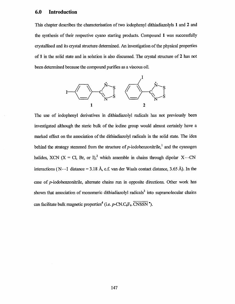
6.0 Introduction
This chapter describes the characterisation of two iodophenyl dithiadiazolyls 1 and 2 and
the synthesis of their respective cyano starting products. Compound 1 was successfully
crystallised and its crystal structure determined. An investigation of the physical properties
of 1 in the solid state and in solution is also discussed. The crystal structure of 2 has not
been determined because the compound purifies as a viscous oil.
1 2
The use of iodophenyl derivatives in dithiadiazolyl radicals has not previously been
investigated although the steric bulk of the iodine group would almost certainly have a
marked effect on the association of the dithiadiazolyl radicals in the solid state. The idea
behind the strategy stemmed from the structure of p-iodobenzonitrile,1 and the cyanogen
halides, XCN (X = Cl, Br, or I),2 which assemble in chains through dipolar X---CN
interactions (N---I distance = 3.18 A, c.f. van der Waals contact distance, 3.65 A). In the
case of /7-iodobenzonitrile, alternate chains run in opposite directions. Other work has
shown that association of monomeric dithiadiazolyl radicals3 into supramolecular chains
can facilitate bulk magnetic properties4 (i.e. p-CN.C^. CNSSN *).
147
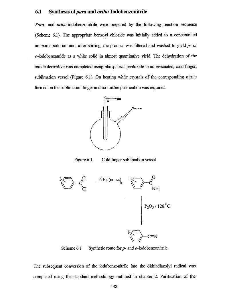
6.1 Synthesis of para and orfTfo-Iodobenzonitrile
Para- and o/Y/ro-iodobenzonitrile were prepared by the following reaction sequence
(Scheme 6.1). The appropriate benzoyl chloride was initially added to a concentrated
ammonia solution and, after stirring, the product was filtered and washed to yield p- or
0 -iodobenzamide as a white solid in almost quantitative yield. The dehydration of the
amide derivative was completed using phosphorus pentoxide in an evacuated, cold finger,
sublimation vessel (Figure 6.1). On heating white crystals of the corresponding nitrile
formed on the sublimation finger and no further purification was required.
Water
Vacuum
Figure 6.1 Cold finger sublimation vessel
P2 0 5 /120 °C
V
Scheme 6.1 Synthetic route forp- and o-iodobenzonitrile
The subsequent conversion of the iodobenzonitrile into the dithiadiazolyl radical was
completed using the standard methodology outlined in chapter 2. Purification of the
148
radicals was completed by vacuum sublimation. Compound 1, after sublimation onto a
cold finger, crystallised in a gradient sublimer as dark red/black blocks, suitable for x-ray
and EPR crystal analysis. In contrast, compound 2 forms as a viscous purple/ red oil. The
reason for this would seem to lie in the steric hindrance caused by the iodine group in the
ortho positioa A similar effect was observed for a tertiary butyl derivative, which also
hinders dimerisation of the dithiadiazolyl ring.5
6.2 X-ray Crystal Structure of/7-iodophenyI-l,2,3,5-dithiadiazolyl
An Ortep plot of the asymmetric unit is shown in Figure 6.2. The bond lengths and angles
in 1 are listed in Tables 6.1 and 6.2 together with data for (Ph CNSSN )£ and
/Modobenzonitrile1 for comparative purposes. The full crystallographic data for compound
1 can be found in appendix 9.
The molecular structure of 1 is, as expected, similar to the molecular structures of most
other aiyl substituted dithiadiazolyls. Within each molecule the aryl and dithiadiazolyl
rings are essentially coplanar with a slight torsion angle between the dithiadiazolyl ring
[root mean square (r.m.s.) A = 0.0077 A] and the aiyl ring (r.m.s. A = 0.0089 A) of 8°. This
value is similar to that observed for aryl substituents not possessing a bulky substituent in
the ortho position (eg Ph. CNSSN torsion angle = 7°).
149
C5C6
WN2C3C2
Figure 6.2 Ortep plot of 1 (p-iodophenyl-1,2,3,5-dithiadiazolyl)
Table 6.1 Bond lengths of 1 (A).
Bond Distance (A) ,Atom Numbers (1) p-ICefttCN Ph. CNSSN
I(l)-C (l) 2.094(6) 2.064 -
S(l)-N(l) 1.635(5) - 1.63
S(l)-S(2) 2.098(2) - 2.096
S(2)-N(2) 1.629(5) - 1.63
N(l)-C(7) 1.337(7) - 1.32
N(2)-C(7) 1.347(6) - 1.31 |
C(l)-C(2) 1.371(7) 1.407 1.39
C(l)-C(6) 1.388(8) 1.407 1.42
C(2)-C(3) 1.376(8) 1.385 1.37
C(3)-C(4) 1.398(8) 1.401 1.37
C(4)-C(5) 1.410(7) 1.401 1.38
C(5)-C(6) 1.365(7) 1.385 1.36
C(4)-C(7) 1.464(7) 1.370 1.50
150
Table 6.2 Bond angles of 1 (°)Angle (degrees)
Atom Numbers (1) /7-IC6H4CN Ph. CNSSN
N(l)-S(l)-S(2) 94.45(18) - 93.0
N(2)-S(2)-S(l) 94.26(17) - 94.1
C(7)-N(l)-S(l) 114.6(4) - 116
C(7)-N(2)-S(2) 114.8(4) - 115
N(l)-C(7)-N(2) 121.9(5) - 122
N(l)-C(7)-C(4) 119.4(5) - 117
N(2)-C(7)-C(4) 118.7(5) - 121
C(2)-C(l)-I(l) 120.6(4) 121 -
C(6)-C(l)-I(l) 118.4(4) 121 -
C(l)-C(2)-C(3) 119.7(5) 121.2 120
C(2)-C(l)-C(6) 120.9(5) 118.0 119
C(4)-C(3)-C(2) 121.2(5) 120.1 121
C(3)-C(4)-C(5) 117.3(5) 119.5 121
C(4)-C(5)-C(6) 121.5(5) 120.1 119
C(5)-C(6)-C(l) 119.2(5) 121.2 120
C(3)-C(4)-C(7) 121.3(5) 120.25 120
C(5)-C(4)-C(7) 121.4(5) 120.25 119
Twist Angle* 8 - 7
Like the majority of dithiadiazolyl compounds 1 associates as dimers in the solid-state.
However, the mode of association is unusual. Two molecules of 1 come together in a
/nmy-co facial manner across an inversion centre (Figure 6.3). This is only the second
example of a dithiadiazolyl adopting this mode of association, the first
[2,2’dimethyldiphenylene radical (3)] was reported only recently.7 One of the heterocycles
in 3 associates in a /nms-cofacial manner, the other heterocycle is un-associated resulting
in a paramagnetic material.
151
ch3
3
The interannular separation can be calculated to be the gap between two mid points o f the
heterocyclic rings. For 1 this value is 3.121 A which is significantly shorter than that of 3
(3.24 A). These compared favourably to the normal separation observed for dithiadiazolyl
dimers, showing m-cofacial and /ram-antarafacial modes of association (3.0 - 3.2 A).
However, the interdimer S---S contacts for 1 (Sla---Slb and S2a-*-S2b, 3.696 A) are
significantly longer than the normal S---S distance in dithiadiazolyl dimers (S---S
contacts typically 3.0 - 3.2 A). In 3 similar interdimer S---S distances are observed
[6.68(2) A]. Each dimer pair also has two sets o f S---N interactions (Nla-*-Slb and
N2a---S2a, 3.152 A; N2a-*-S2b and Nlb---Sla 3.116 A). The asymmetry o f these
interactions is due to further interactions between one sulfur atom and an iodine atom o f an
adjacent molecule (vide infra).
' h
Figure 6.3 Ortep plot o f the trans-co facial association o f 1.
152

As noted by Oakley and co-workers,7 the /ram-cofacial mode of dimerisation adopted by 1
and 3 lies close to the transition state for the proposed photochemical isomerisation of
1,3,2,4- to 1,2,3,5-dithiadiazolyl (Scheme 6.2).5,8,9 Indeed the molecular structures of 1 and
3 show many similarities with that of 5,5’(1,4-phenylene)bis( 1,3,2,4-dithiadiazolyl) 4,8
which contains short S---S (3.214 A) and S---N (3.346 A) interactions between parallel
dithiadiazolyl rings. The /ra/w-cofacial mode of dimerisation observed in 1 is unusual and
I speculate that the molecules dimerise in this way to alleviate steric hindrance between the
two iodine atoms that would occur if the molecules associated in a c/s-cofacial manner.
However, it is less obvious why /ra/w-antarafacial or twisted geometries are disfavoured in
this instance. In this study, the correlation between 1 and 4 is limited to a purely structural
comparison. Further work should include theoretical studies to determine what factors
prevent the molecules of 1 from sliding to give a /ram-antarafacial geometry.
Scheme 6.2 Photochemical isomerisation of 1,2,3,4- to 1,2,3,5- dithiadiazolyl.5,8
molecule and a sulfur atom of an adjacent molecule (Figure 6.4). The I---S contact
of the contact vector relative to the bond).11 The second I • • • S contact (11 • • • S1, 4.068 A) is
N—S,R— C
‘S—N
4
Each molecule of 1 is linked through a weak interaction between an iodine atom of one
(I1---S2, 3.82 A) is significantly longer than a conventional S-I bond10 but is within the
sum of the van der Waals radii of iodine and sulfur (3.4 - 4.1 A depending on orientation
153
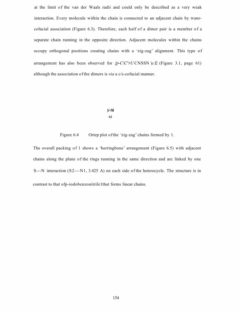
at the limit o f the van der Waals radii and could only be described as a very weak
interaction. Every molecule within the chain is connected to an adjacent chain by trans-
cofacial association (Figure 6.3). Therefore, each half o f a dimer pair is a member o f a
separate chain running in the opposite direction. Adjacent molecules within the chains
occupy orthogonal positions creating chains with a ‘zig-zag’ alignment. This type o f
arrangement has also been observed for {p-C\C^rU CNSSN )2 12 (Figure 3.1, page 61)
although the association o f the dimers is via a c/s-cofacial manner.
)/-SlS2
Figure 6.4 Ortep plot of the ‘zig-zag’ chains formed by 1.
The overall packing o f 1 shows a ‘herringbone’ arrangement (Figure 6.5) with adjacent
chains along the plane o f the rings running in the same direction and are linked by one
S---N interaction (S2---N1, 3.425 A) on each side o f the heterocycle. The structure is in
contrast to that ofp-iodobenzonitrile1 that forms linear chains.
154
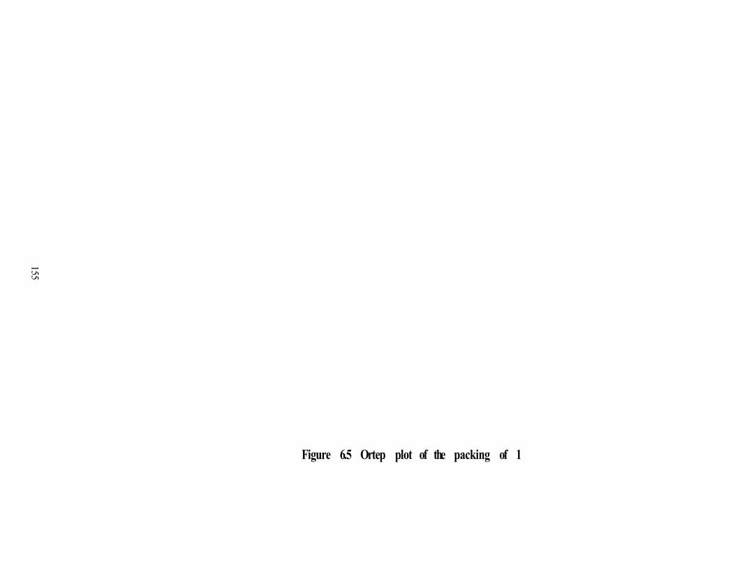
155
Figure 6.5 Ortep plot of the packing of 1
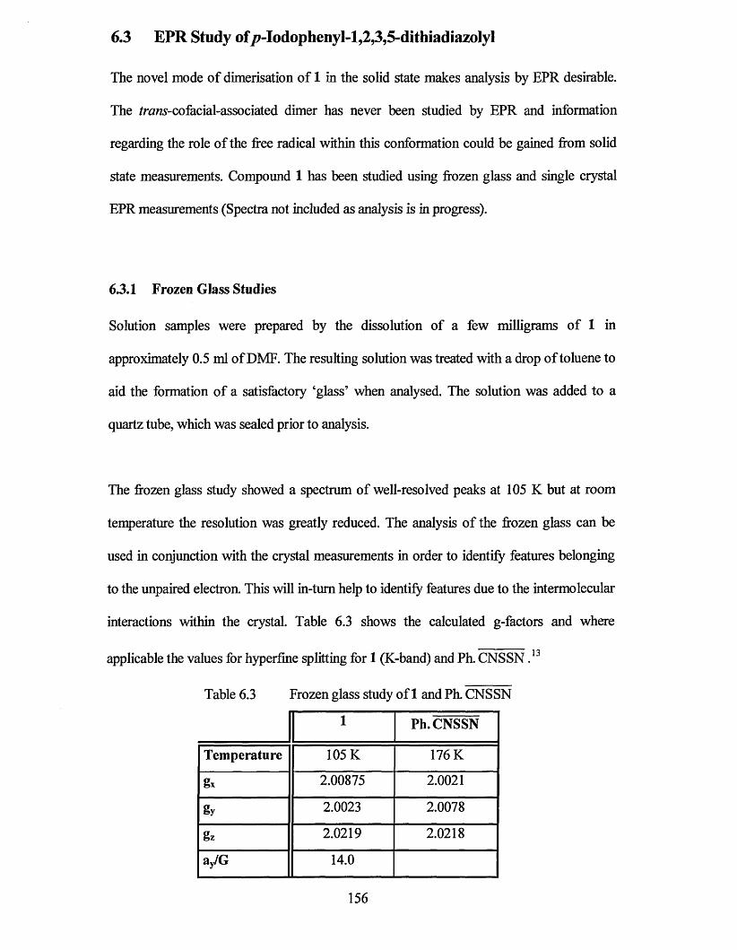
6.3 EPR Study of /?-Iodophenyl-l,2,3,5-dithiadiazoIyl
The novel mode of dimerisation of 1 in the solid state makes analysis by EPR desirable.
The /raws-co facial-associated dimer has never been studied by EPR and information
regarding the role of the free radical within this conformation could be gained from solid
state measurements. Compound 1 has been studied using frozen glass and single crystal
EPR measurements (Spectra not included as analysis is in progress).
6.3.1 Frozen Glass Studies
Solution samples were prepared by the dissolution of a few milligrams of 1 in
approximately 0.5 ml of DMF. The resulting solution was treated with a drop of toluene to
aid the formation of a satisfactory ‘glass’ when analysed. The solution was added to a
quartz tube, which was sealed prior to analysis.
The frozen glass study showed a spectrum of well-resolved peaks at 105 K but at room
temperature the resolution was greatly reduced. The analysis of the frozen glass can be
used in conjunction with the crystal measurements in order to identify features belonging
to the unpaired electron. This will in-tum help to identify features due to the intermolecular
interactions within the crystal. Table 6.3 shows the calculated g-factors and where
applicable the values for hyperfine splitting for 1 (K-band) and Ph. CNSSN ,13
Table 6.3 Frozen glass study of 1 and Ph. CNSSN
1 Ph. CNSSN
Temperature 105 K 176 K
gx 2.00875 2.0021
gy 2.0023 2.0078
gz 2.0219 2.0218
ay/G 14.0
156

UOT]
The spectrum has a peak ratio pattern of 1:2:3:2:1 (from two equivalent nitrogen-14
nuclei), which is typical for 1,2,3,5-dithiadiazolyl radicals. The calculated g-values are also
typical for a radical species with significant unpaired spin density. Figure 6.6 shows the
EPR frozen glass spectrum of 1, including a computer generated simulation.
H e c e i v e r S i g n a l Cna nn el F i e l d
R e c e i v e r Ga in : 2 . 0 0 e * 0 4 C on v er s ion 1 6 3 .8 4 ms C e n t e r F i e l d :P n a s e 0 . 0 deg Time Cons t 1 .2 8 ms Sweep WidtnHarmonic 1 Sweep Time 1 6 7 .7 7 2 s R e s o l u t i o nMoo F re q u e n c y : 100 0000 kHz S c a l e 17Moo A m p l it u d e : 1 .0 0 0 G
85+fl.OO G 2 2 6 . 0 0 G
/ 1024 p a i n t s
l P
F r e q u e n c y : -20-r€643OOO'-13Hz- Power 3 .6 2 e + 0 0 mW
Comment: sB84 3 . 5 t J iD r o m o / to lu e n e 110K 23.0638GHz
4 0 0
0
- 4 0 0
8 6 0 08 4 0 0 8 4 4 0 8 4 8 0 8 5 2 0 8 5 6 0
[G]
Figure 6.6 K-band EPR frozen glass spectrum of 1 (top), and a computer simulatedspectrum (bottom)
157
6.4 Experimental
6.4.1 Preparation of/?-IodobenzonitriIe
Finely powdered /?-iodobenzoyl chloride (5.32 g, 0.02 mol) was slowly added to ice cold
0.880 ammonia solution (20 ml). The mixture was stirred for two hours and allowed to
warm to room temperature. The resulting precipitate, p-\odobenzamide, was filtered,
washed with water and dried. The /?-iodobenzamide was then transferred to a 250 cm
round bottomed flask fitted with a side arm and stopcock, together with a cold-finger. An
excess of phosphorus pentoxide was added to the flask and the reactants thoroughly mixed.
The flask was heated in vacuo (130 °C). The final product, p-iodobenzonitrile was isolated
directly onto the cold finger from the reaction mixture as white needles. Purification of the
final product was achieved by further vacuum sublimation (2.68 g, 59% yield)
Mpt 124-126 °C
MS (El) 228.9 (M4), 102.0 (M4 - 1), 75.0
IR (KBr), v j c m 1 2225(C=N), 1578,1475,1390,1054,1010, 894, 541
5h (250MHz, CDC13; Me4Si) 7.85 (2H, d, J8.3 Hz), 7.37 (2H, d, J8.3 Hz)
6c (250MHz, CDC13; Me4Si) 138.86,133.53,118.57,112.09,100.68
158

6.4.2 Preparation of 0 -Iodobenzonitrile
Finely powdered o-iodobenzoyl chloride (2.66 g, 0.01 mol) was slowly added to ice cold
0.880 ammonia solution (20 ml). The mixture was stirred for thirty minutes and allowed to
warm to room temperature. The resulting white precipitate, o-iodobenzamide, was filtered,
washed with water and dried. The o-iodobenzamide was then transferred to a 250 cm
round bottomed flask fitted with a side arm and stopcock, together with a cold-finger. An
excess of phosphorus pentoxide was added to the flask and the reactants thoroughly mixed.
The flask was heated in vacuo (90 °C). The final product, o-iodobenzonitrile was isolated
directly onto the cold finger from the reaction mixture as white needles. Purification of the
final product was achieved by further vacuum sublimation (1.68 g, 73% yield)
Mpt 42-50 °C
MS (El) 229.0 (M4), 102.] (M+-I), 75.0
IR (KBr), v j c m ' 2229(ON), 1582,1465,1395,1053,1009,894,541
5H (250MHz, CDCl3;Me4Si) 7.93 (1H, dd, .77.9 Hz + 0.8 Hz), 7.61 (1H, dd,./7.7H z +
1.4 Hz), 7.47 (1H, td, J l . l Hz + 1 Hz), 7.30 (1H, td, J 6.0
Hz+ 3.3 Hz)
5c (250MHz, CDClj; Me4Si) 139.91,134.62,134.05,128.64,120.1,119.67,98.74
159
6.5 Conclusions
From the results reported in this chapter, it is clear that the iodine atom has a marked effect
on the supramolecular structure of the compounds. As anticipated, the iodine substituent
facilitated the production of chains by interacting with a sulfur atom on an adjacent
molecule. The mode of association of 1 is only the second example of a trans-cofacial
association and as such little is known about the reasons why this structure is favoured
over /nms’-antarafacial or twisted geometries. Further EPR investigation of the crystals of 1
are being investigated presently to gain more information on this little known
conformation.
160

6.6 References
1 E.O. Schlemper and D.Britton, Acta. Cryst.,. 1965,18,419.
2 R.B. Heiart and G.B. Carpenter, Acta. Cryst., 1956,9, 60.
3 (a) A.J. Banister, N. Bricklebank, W. Clegg, M.R.J. Elsegood, C.I. Gregory,
I. Lavender, J.M. Rawson and B.K. Tanner, J. Chem. Soc., Chem. Commun., 1995,
679; (b) A.J. Banister, N. Bricklebank, I. Lavender, J.M. Rawson, C.I. Gregory,
B.K. Tanner, W. Clegg, M.R.J. Elsegood and F. Palacio, Angew. Chem. Int. Ed.
Engl., 1996,35,2533.
4 (a) F. Palacio, G. Antorrena, M. Castro, R. Buniel, J.M. Rawson, J. Nicholas,
B. Smith, N. Bricklebank, J. Novoa and C. Ritter, Physical Review Letters, 1997,79,
2336; (b) P.J. Langley, J.M. Rawson, J.N.B. Smith, M. Schuler, R. Bachmann,
A. Schweiger, F. Palacio, G. Antorrena, G. Gescheidt, A. Quintel, P. Rechsteiner
and J. Hulliger, J. Mater. Chem., 1999, 9,1431.
5 W.V.F. Brookes, N. Burford, J. Passmore, M.J. Schriver and L.H. Sutcliffe, J. Chem.
Soc., Chem. Commun., 1987, 69.
6 A. Vegas, A. Perez-Salazar, A.J. Banister and R.G. Hey, J. Chem. Soc., Dalton
Trans., 1980,1812.
7 T.M. Barclay, A.W. Cordes, N.A. George, R.C. Haddon, M.E. Itkis and
R.T. Oakley, Chem. Commun., 1999,2269.
8 C. Aheme, A.J. Banister, A.W. Luke, J.M. Rawson, and R.J. Whitehead, J. Chem.
Soc., Dalton Trans., 1992,1277.
9 (a) N. Burford, J. Passmore and X. Sun, Phosphorus, Sulfur and Silicon, 1994,
93-94, 487; (b) N. Burford, J. Passmore and M.J. Schriver, chem J. Chem. Soc.,
Chem. Commun., 1986, 141. (c) J. Passmore and X.Sun, Inorg. Chem., 1996, 35,
1313.
161
10 N. Bricklebank, P.J. Skabara, D.E. Hibbs, M. Hursthouse and K.M.A. Malik,
J. Chem. Soc., Dalton Trans., 1999,3007.
11 S.C. Nyburg and C.H. Faerman, Acta. Cry St., 1985, B41,274.
12 R.T. Boere and K.H. Moock, Z. anorg. allg. Chem., 1994,620,1589.
13 S.A Fairhurst, K.M. Johnson, H. Sutcliffe, K.F. Preston, A.J. Banister,
Z.V. Hauptman and J. Passmore, J. Chem. Soc., Dalton Trans., 1986,1465.
162
Chapter 7
General Experimental
163
7.0 Introduction
Many of the reactions undertaken, and the compounds prepared, are sensitive to air and
moisture. It was therefore necessary to complete most reactions, and store most products,
under an inert atmosphere.
All air and moisture sensitive materials were handled under dry nitrogen in a glove box
fitted with a phosphorus pentoxide drying column. Reactions were completed using
standard vacuum line techniques. All the nitrogen used was oxygen free (BOC) and was
dried further by passing over phosphorus pentoxide. All glassware was pre-dried
(c.a. 130 °C) for at least six hours prior to use. Equipment utilised in sublimations was
flame dried under vacuum, prior to use.
7.1 Physical Methods
7.1.1 Infra-red Spectroscopy
Infra red spectra were recorded as nujol mulls if they were thought to be unstable to air or
moisture, or by KBr disc for normal analysis. The instrument used was an ATI Mattson
Genesis series FTTR.
7.1.2 Mass Spectrometry
Mass spectra were recorded using a VG Micromass 7070F mass spectrometer, using El or
FAB ionisation. A direct insertion probe was used for samples. Air and moisture sensitive
compounds were taken from a sealed tube and analysed a quickly as possible to minimise
any decomposition.
164
7.1.3 *H and 13C NMR Spectroscopy
]H and I3C nmr spectra were obtained on a Bruker AC250 nmr spectrometer. Samples
were dissolved in a deuterated solvent in a nitrogen glove box and were analysed
immediately.
7.1.4 Differential Scanning Calorimetry
Differential scanning calorimetry (d.s.c) measurements were undertaken using a Mettler-
Toledo TA8000.
7.1.5 Electron Paramagnetic Resonance
Electron paramagnetic resonance (EPR or ESR) spectra were obtained in conjunction with
Dr F. Mabbs at the University of Manchester (EPSRC cwEPR service centre). Solution
and powder samples were analysed on a Bruker EMX and single crystal analysis was
performed on a Bruker ESP300E.
7.1.6 Single Crystal X-Ray Diffraction
Single crystal x-ray crystallography was performed by Harry Adams and Sharon Spey at
the University of Sheffield. Data collected were measured on a Bruker Smart CCD area
detector with Oxford Cryosystems low temperature system. Measurements were corrected
for Lorentz and polarisation effects and for absorption by semi-empirical methods based
on symmetry-equivalent and repeated reflections. Structures were solved by direct
methods and refined by full matrix least squares methods on F^. Scattering factors were
inlaid from the program package SHELXTL1 as implemented on the Viglen Pentium
computer.
165
7.1.7 Elemental Analysis
Carbon hydrogen and nitrogen analysis was performed by Medac Ltd, Brunei Science
Centre, Coopers Hill Lane, Englefield Green, Egham
7.1.8 Magnetic Measurements
Magnetic experiments were performed by Fernando Palacio, G Antorenna and Michel
Julier (Instituto de Ciencia de la Materia de Aragon, Universidad de Zaragoza).
Magnetisation of the samples was measured using a SQUID magnetometer from Quantum
Devices. The polycrystalline samples were transfered into gelatine capsules inside a glove
box. The capsules were then topped with cotton wool. The contact between the capsules
and air was reduced to less than 5 minutes.
7.2 Solvents
All solvents used were pre-dried and distilled before use. Solvents not freshly distilled
were stored under nitrogen in flask equipped with a Teflon vacuum tap. Before storage, the
solvent was de-gassed via a freeze-thaw cycle.
7.2.1 Diethyl ether (Fisher)
Refluxed with sodium metal and benzophenone (indicator) under a constant stream of
nitrogen.
7.2.2 Tetrahydrofuran (Sigma-Aldrich)
Refluxed with potassium metal and benzophenone (indicator) under a constant stream of
nitrogen.
166
7.2.3 Dichloromethane (Fisher)
Refluxed with phosphourus pentoxide (P2O5) for at least 4 hours under a constant stream
of nitrogea
7.2.4 Acetonitrile (Fisher)
Refluxed with Caffe under a constant stream of nitrogen.
7.2.5 Dimethylformamide
Purchased pre-dried from Sigma-Aldrich and degassed by passing nitrogen through the
solvent just prior to use.
7.2.6 Toluene (Fisher)
Refluxed with sodium metal and benzophenone (indicator) under a constant stream of
nitrogea
7.3 Reagents
All the starting compounds were obtained from various chemical suppliers and used as
received.
167

7.4 References
1 SHELXTL version, An integrated system for solving and refining crystal structures from
diffraction data (Revision 5.1), Bruker AXS LTD
Overall Conclusions and SuggestionsThe work in this thesis has shown that the use of mono- and poly- halogenated phenyl
substituents can have a significant effect on the molecular packing of 1,2,3,5-dithiadiazolyl
radicals. Dichlorophenyl groups clearly enhance the ability of 1,2,3,5-dithiadiazolyls to
associate in stacks. Two types of stacking motif have been identified; (i) dimer stacks and
(ii) 7r-stacks containing uniformly spaced radical units. The latter is unprecedented for
neutral mono-functional dithiadiazolyl radicals. In contrast, bromine was shown to exhibit
a large steric hindrance, distorting the cis cofacial dimer association to form a slightly
twisted motif. Unlike chlorine there seemed to be little or no homonuclear interactions.
The molecular structure of iodophenyl derivatives exhibited significant steric hindrance
producing one viscous oil (or^o-iodophenyl-1,2,3,5-dithiadiazolyl), common in highly
steric hindered systems and a trans cofacial association (pwra-iodophenyl-1,2,3,5-
dithiadiazolyl). Halogen-•-sulfur interactions, which link adjacent molecules into chains,
have also been observed for the chlorine and iodine derivatives.
Further studies into the magnetic susceptibility and EPR signals of the compounds may
uncover additional information about the interaction of the molecules. An investigation of
further chlorophenyl derivatives containing a cyano substituent may promote the formation
of chains while still retaining significant stacking properties. Studies of poly-halogenated
derivatives containing different halogen atoms would also be of interest.
169
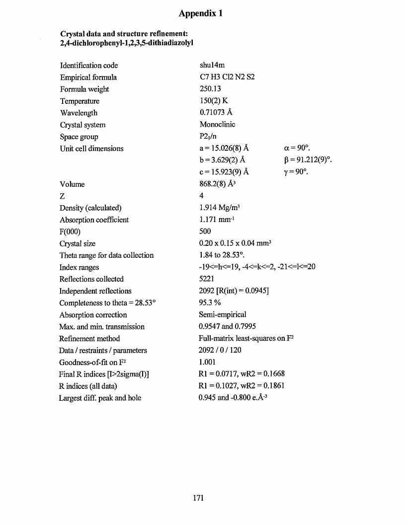
Appendix 1
Ciystal data and structure refinement:2,4-dichIorophenyl-l,2,3,5-dithiadiazoIyl
Identification code Empirical formula Formula weight Temperature Wavelength Crystal system Space group Unit cell dimensions
VolumeZDensity (calculated)Absorption coefficient F(000)Crystal sizeTheta range for data collection Index ranges Reflections collected Independent reflections Completeness to theta = 28.53°Absorption correction Max. and min. transmission Refinement method Data / restraints / parameters Goodness-of-fit on F2 Final R indices p>2sigma(I)]R indices (all data)Largest diff. peak and hole
shul4mC7H3 C12N2S2250.13 150(2) K 0.71073 A Monoclinic P2j/na= 15.026(8) A a = 90°.b = 3.629(2) A p = 91.212(9)°.c= 15.923(9) A y = 90°.868.2(8) A3 41.914 Mg/m3 1.171 mm-1 5000.20 x 0.15 x 0.04 mm31.84 to 28.53°.-19<=h<=19, -4<=k<=2, -21<=1<=20 52212092 [R(int) = 0.0945]95.3 %Semi-empirical 0.9547 and 0.7995 Full-matrix least-squares on F2 2092/0/1201.001R l= 0.0717, wR2 = 0.1668 R l= 0.1027, wR2 = 0.1861 0.945 and-0.800 e.A'3
171
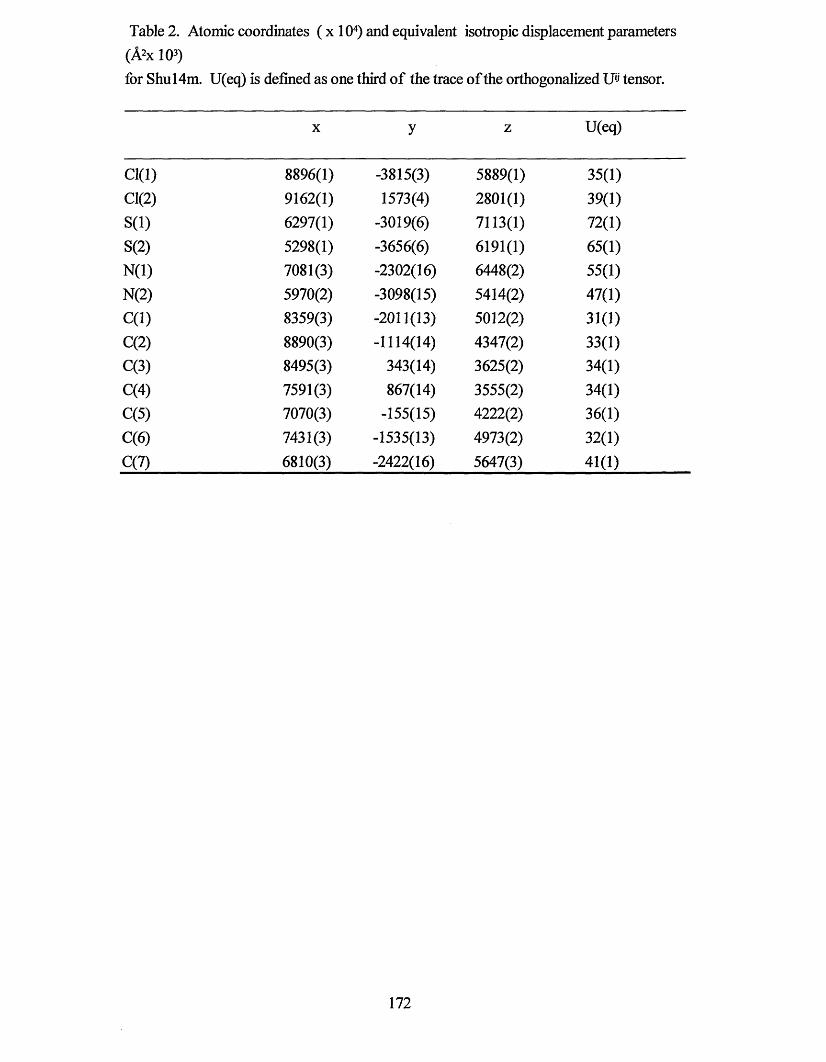
Table 2. Atomic coordinates ( x 104) and equivalent isotropic displacement parameters (A2x 103)for Shu 14m. U(eq) is defined as one third of the trace of the orthogonalized Uij tensor.
X y z U(eq)
0(1) 8896(1) -3815(3) 5889(1) 35(1)0(2) 9162(1) 1573(4) 2801(1) 39(1)S(l) 6297(1) -3019(6) 7113(1) 72(1)S(2) 5298(1) -3656(6) 6191(1) 65(1)N(l) 7081(3) -2302(16) 6448(2) 55(1)N(2) 5970(2) -3098(15) 5414(2) 47(1)C(l) 8359(3) -2011(13) 5012(2) 31(1)C(2) 8890(3) -1114(14) 4347(2) 33(1)C(3) 8495(3) 343(14) 3625(2) 34(1)C(4) 7591(3) 867(14) 3555(2) 34(1)C(5) 7070(3) -155(15) 4222(2) 36(1)C(6) 7431(3) -1535(13) 4973(2) 32(1)C(7) 6810(3) -2422(16) 5647(3) 41(1)
172
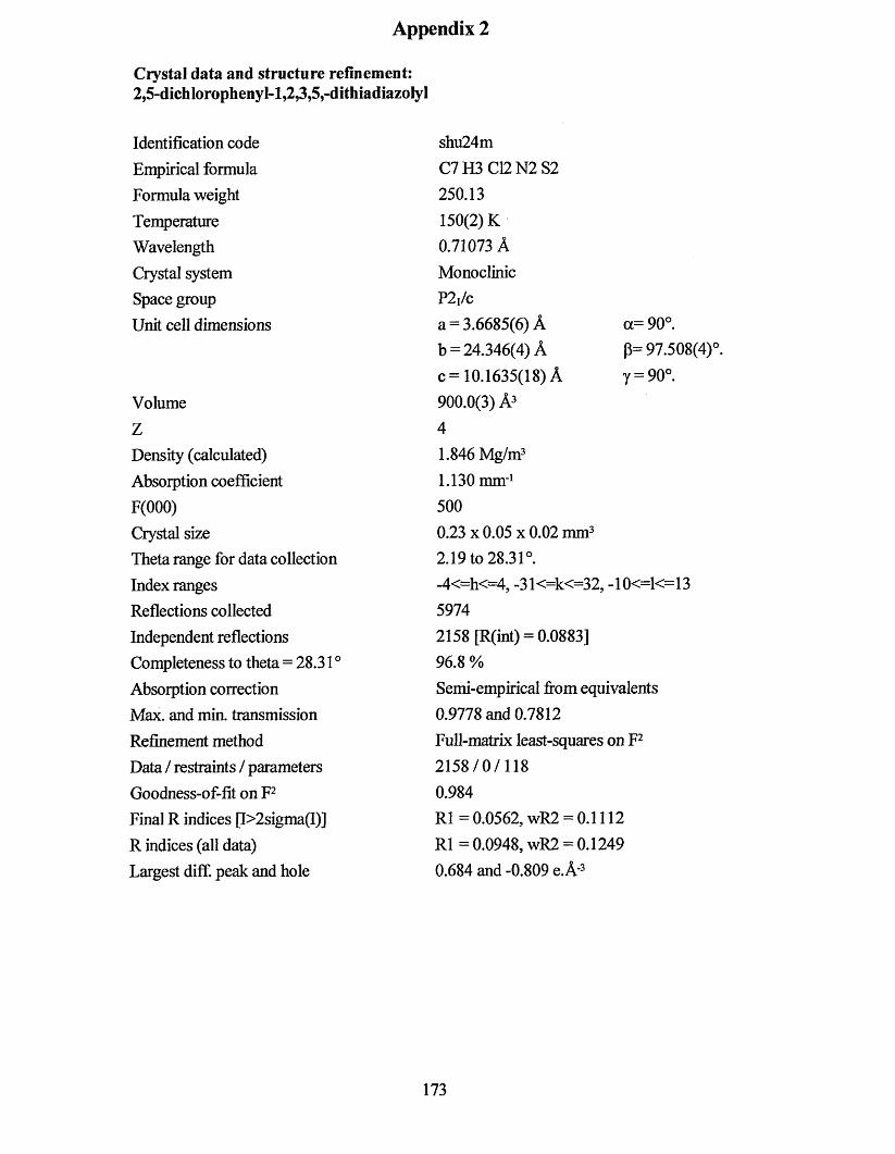
Appendix 2
Crystal data and structure refinement:2,5-dichlorophenyl-l,23»5,-dithiadiazoIyl
Identification code Empirical formula Formula weight Temperature Wavelength Crystal system Space group Unit cell dimensions
VolumeZDensity (calculated)Absorption coefficient F(000)Crystal sizeTheta range for data collection Index ranges Reflections collected Independent reflections Completeness to theta = 28.31 °Absorption correction Max. and min. transmission Refinement method Data / restraints / parameters Goodness-of-fit on F2 Final R indices p>2sigma(I)]R indices (all data)Largest diff. peak and hole
shu24mC7H3 C12N2 S2250.13 150(2) K 0.71073 A Monoclinic P2,/ca = 3.6685(6) A a=90°.b = 24.346(4) A P= 97.508(4)°.c= 10.1635(18) A y = 90°.900.0(3) A3 41.846 Mg/m31.130 mm-1 5000.23 x 0.05 x 0.02 mm3 2.19 to 28.31°.-4<=h<=4, -31<=k<=32, -10<=1<=13 59742158 [R(int) = 0.0883]96.8 %Semi-empirical from equivalents 0.9778 and 0.7812 Full-matrix least-squares on P 2158/0/118 0.984R1 = 0.0562, wR2 = 0.1112 R l= 0.0948, wR2 = 0.1249 0.684 and-0.809 e.A'3
173
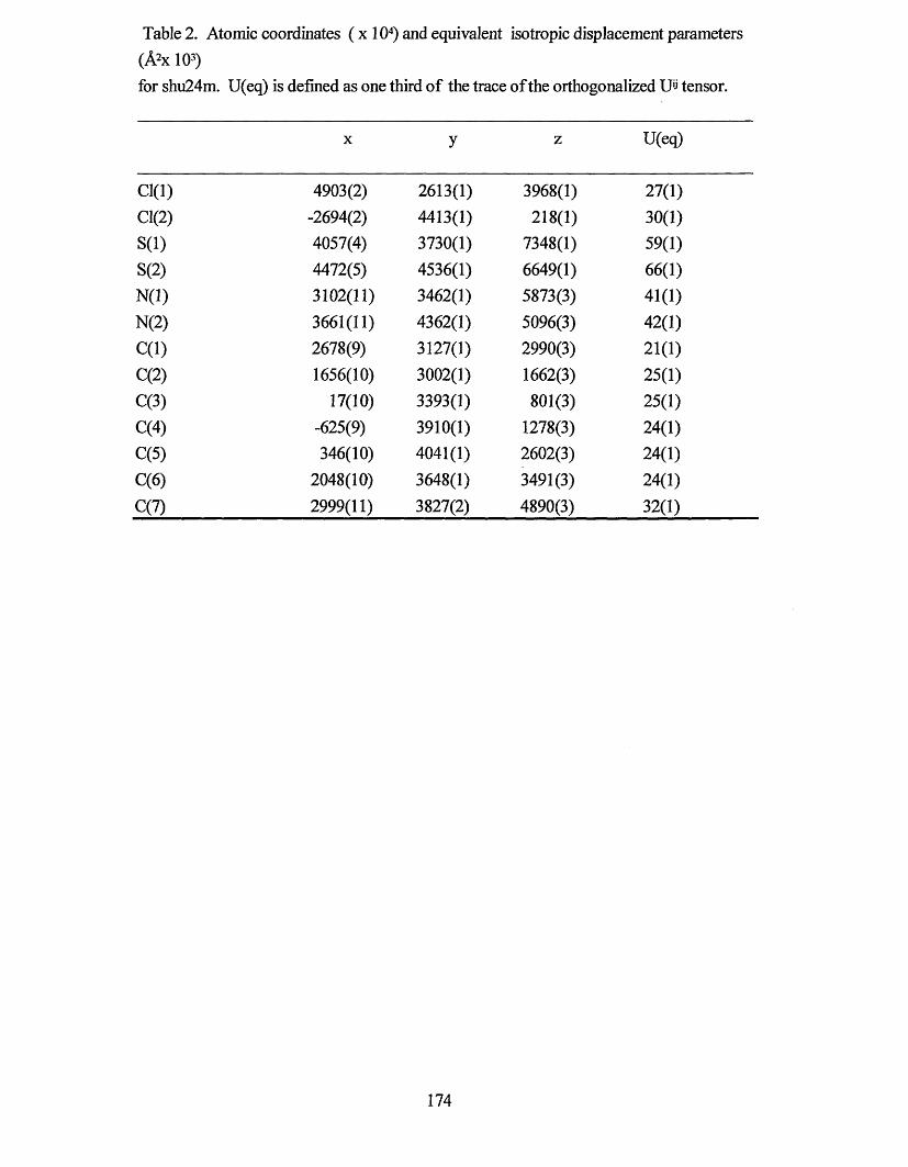
Table 2. Atomic coordinates ( x 104) and equivalent isotropic displacement parameters (A2x 103)for shu24m. U(eq) is defined as one third of the trace of the orthogonalized U'j tensor.
X y z U(eq)
Cl(l) 4903(2) 2613(1) 3968(1) 27(1)Cl(2) -2694(2) 4413(1) 218(1) 30(1)S(l) 4057(4) 3730(1) 7348(1) 59(1)S(2) 4472(5) 4536(1) 6649(1) 66(1)N(l) 3102(11) 3462(1) 5873(3) 41(1)N(2) 3661(11) 4362(1) 5096(3) 42(1)C(l) 2678(9) 3127(1) 2990(3) 21(1)C(2) 1656(10) 3002(1) 1662(3) 25(1)C(3) 17(10) 3393(1) 801(3) 25(1)C(4) -625(9) 3910(1) 1278(3) 24(1)C(5) 346(10) 4041(1) 2602(3) 24(1)C(6) 2048(10) 3648(1) 3491(3) 24(1)C(7) 2999(11) 3827(2) 4890(3) 32(1)
174

Appendix 3
Crystal data and structure refinement:p-(3,5-dichIorophenyl-l,2,3,5,-dithiadiazolyl)
Identification code Empirical formula Formula weight Temperature Wavelength Crystal system Space group Unit cell dimensions
VolumeZDensity (calculated) Absorption coefficient F(000)Crystal sizeTheta range for data collection Index ranges Reflections collected Independent reflections Completeness to theta = 28.32° Absorption correction Max. and min. transmission Refinement method Data / restraints / parameters Goodness-of-fit on F2 Final R indices [I>2sigma(I)]R indices (all data)Largest diff. peak and hole
shunearm C7H3 C12N2S2250.13 150(2) K 0.71073 A Monoclinic C2/ca = 41.546(7) A oc=90°.b = 3.6280(6) A p= 99.013(3)cc = 36.326(6) A y = 90°.5407.7(15) A3 241.843 Mg/m3 1.128 mm-1 30000.38 x 0.17 x 0.07 mm3 0.99 to 28.32°.-54<=h<=55, -4<=k<=4, -48<=1<=35 162276391 [R(int) = 0.1203]94.9 %Semi-empirical from equivalents 0.9252 and 0.6738 Full-matrix least-squares on F2 6391/0/352 1.067R1 = 0.0556, wR2 = 0.1314 R l= 0.0765, wR2 = 0.1540 0.669 and-1.301 e.A-3
175
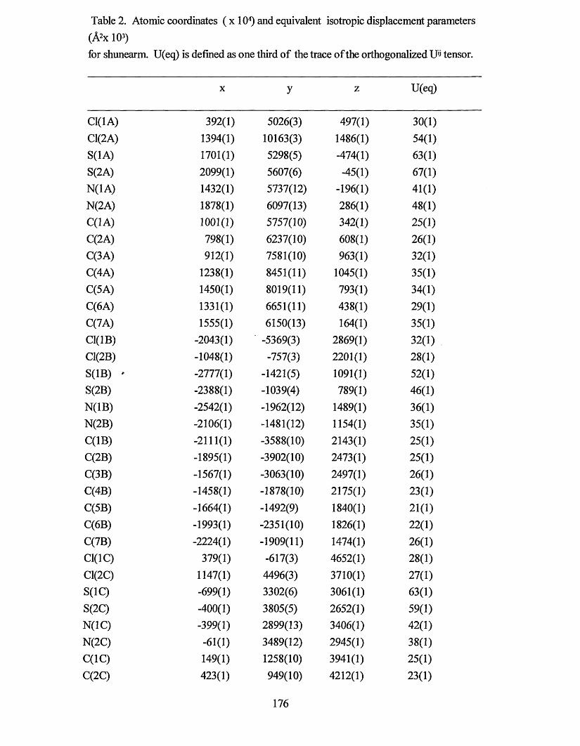
Table 2. Atomic coordinates ( x 104) and equivalent isotropic displacement parameters (A2x 103)for shunearm. U(eq) is defined as one third of the trace of the orthogonalized U'j tensor.
X y z U(eq)
C1(1A) 392(1) 5026(3) 497(1) 30(1)C1(2A) 1394(1) 10163(3) 1486(1) 54(1)S(1A) 1701(1) 5298(5) -474(1) 63(1)S(2A) 2099(1) 5607(6) -45(1) 67(1)N(1A) 1432(1) 5737(12) -196(1) 41(1)N(2A) 1878(1) 6097(13) 286(1) 48(1)C(1A) 1001(1) 5757(10) 342(1) 25(1)C(2A) 798(1) 6237(10) 608(1) 26(1)C(3A) 912(1) 7581(10) 963(1) 32(1)C(4A) 1238(1) 8451(11) 1045(1) 35(1)C(5A) 1450(1) 8019(11) 793(1) 34(1)C(6A) 1331(1) 6651(11) 438(1) 29(1)C(7A) 1555(1) 6150(13) 164(1) 35(1)C1(1B) -2043(1) -5369(3) 2869(1) 32(1)C1(2B) -1048(1) -757(3) 2201(1) 28(1)S(1B) - -2777(1) -1421(5) 1091(1) 52(1)S(2B) -2388(1) -1039(4) 789(1) 46(1)N(1B) -2542(1) -1962(12) 1489(1) 36(1)N(2B) -2106(1) -1481(12) 1154(1) 35(1)C(1B) -2111(1) -3588(10) 2143(1) 25(1)C(2B) -1895(1) -3902(10) 2473(1) 25(1)C(3B) -1567(1) -3063(10) 2497(1) 26(1)C(4B) -1458(1) -1878(10) 2175(1) 23(1)C(5B) -1664(1) -1492(9) 1840(1) 21(1)C(6B) -1993(1) -2351(10) 1826(1) 22(1)C(7B) -2224(1) -1909(11) 1474(1) 26(1)C1(1C) 379(1) -617(3) 4652(1) 28(1)C1(2C) 1147(1) 4496(3) 3710(1) 27(1)S(1C) -699(1) 3302(6) 3061(1) 63(1)S(2C) -400(1) 3805(5) 2652(1) 59(1)N(1C) -399(1) 2899(13) 3406(1) 42(1)N(2C) -61(1) 3489(12) 2945(1) 38(1)C(1C) 149(1) 1258(10) 3941(1) 25(1)C(2C) 423(1) 949(10)
176
4212(1) 23(1)
C(3C) 732(1) 1896(10) 4145(1) 24(1)C(4C) 762(1) 3186(10) 3792(1) 23(1)C(5C) 497(1) 3549(10) 3511(1) 24(1)C(6C) 189(1) 2603(10) 3590(1) 24(1)C(7C) -104(1) 3008(12) 3299(1) 32(1)
177
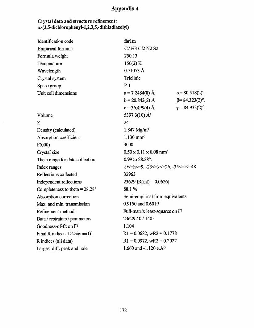
Appendix 4
Crystal data and structure refinement:a-(3,5-dichIorophenyI-l,2,3,5,-dithiadiazolyl)
Identification code Empirical formula Formula weight Temperature Wavelength Crystal system Space group Unit cell dimensions
VolumeZDensity (calculated) Absorption coefficient F(000)Crystal sizeTheta range for data collection Index ranges Reflections collected Independent reflections Completeness to theta = 28.28° Absorption correction Max. and min. transmission Refinement method Data / restraints / parameters Goodness-of-fit on F2 Final R indices (f>2sigma(I)]R indices (all data)Largest diff. peak and hole
<x= 80.518(2)°. P= 84.323(2)°. y = 84.933(2)°.
farlmC7H3C12N2S2250.13 150(2) K 0.71073 A Triclinic P-la = 7.2484(8) A b = 20.842(2) A c = 36.499(4) A 5397.3(10) A3 241.847 Mg/m31.130 mm-1 30000.50 x 0.11 x 0.08 mm3 0.99 to 28.28°.-9<=h<=9, -23<=k<=26, -35<=1<=48 3296323629 [R(int) = 0.0626]88.1 %
Semi-empirical from equivalents0.9150 and 0.6019Ful 1-matrix least-squares on F223629/0/14051.104R1 = 0.0682, wR2 = 0.1778 R l= 0.0972, wR2 = 0.2022 1.660 and-1.120 e.A'3
178
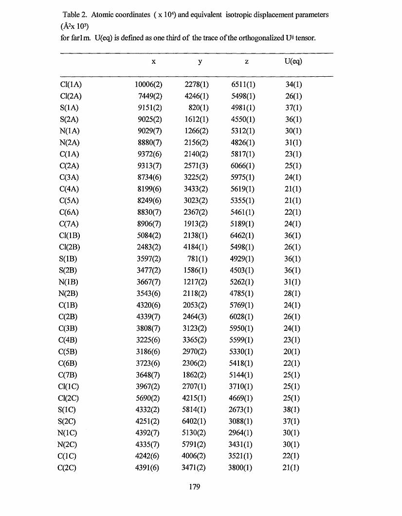
Table 2. Atomic coordinates ( x 104) and equivalent isotropic displacement parameters (A2x 103)for farl m. U(eq) is defined as one third of the trace of the orthogonalized Uij tensor.
X y z U(eq)
C1(1A) 10006(2) 2278(1) 6511(1) 34(1)C1(2A) 7449(2) 4246(1) 5498(1) 26(1)S(1A) 9151(2) 820(1) 4981(1) 37(1)S(2A) 9025(2) 1612(1) 4550(1) 36(1)N(1A) 9029(7) 1266(2) 5312(1) 30(1)N(2A) 8880(7) 2156(2) 4826(1) 31(1)C(1A) 9372(6) 2140(2) 5817(1) 23(1)C(2A) 9313(7) 2571(3) 6066(1) 25(1)C(3A) 8734(6) 3225(2) 5975(1) 24(1)C(4A) 8199(6) 3433(2) 5619(1) 21(1)C(5A) 8249(6) 3023(2) 5355(1) 21(1)C(6A) 8830(7) 2367(2) 5461(1) 22(1)C(7A) 8906(7) 1913(2) 5189(1) 24(1)C1(1B) 5084(2) 2138(1) 6462(1) 36(1)C1(2B) 2483(2) 4184(1) 5498(1) 26(1)S(1B) 3597(2) 781(1) 4929(1) 36(1)S(2B) 3477(2) 1586(1) 4503(1) 36(1)N(1B) 3667(7) 1217(2) 5262(1) 31(1)N(2B) 3543(6) 2118(2) 4785(1) 28(1)C(1B) 4320(6) 2053(2) 5769(1) 24(1)C(2B) 4339(7) 2464(3) 6028(1) 26(1)C(3B) 3808(7) 3123(2) 5950(1) 24(1)C(4B) 3225(6) 3365(2) 5599(1) 23(1)C(5B) 3186(6) 2970(2) 5330(1) 20(1)C(6B) 3723(6) 2306(2) 5418(1) 22(1)C(7B) 3648(7) 1862(2) 5144(1) 25(1)C1(1C) 3967(2) 2707(1) 3710(1) 25(1)C1(2C) 5690(2) 4215(1) 4669(1) 25(1)S(1C) 4332(2) 5814(1) 2673(1) 38(1)S(2C) 4251(2) 6402(1) 3088(1) 37(1)N(1C) 4392(7) 5130(2) 2964(1) 30(1)N(2C) 4335(7) 5791(2) 3431(1) 30(1)C(1C) 4242(6) 4006(2) 3521(1) 22(1)C(2C) 4391(6) 3471(2)
179
3800(1) 21(1)
C(3C) 4845(6) 3521(2) 4157(1) 21(1)
C(4C) 5152(6) 4132(2) 4229(1) 20(1)
C(5C) 5018(6) 4680(2) 3961(1) 21(1)
C(6C) 4564(6) 4615(2) 3606(1) 20(1)
C(7C) 4421(7) 5205(2) 3320(1) 25(1)
C1(1D) 8981(2) 2706(1) 3711(1) 24(1)
C1(2D) 10656(2) 4269(1) 4638(1) 23(1)
S(1D) 8819(2) 5788(1) 2632(1) 36(1)
S(2D) 8700(2) 6395(1) 3037(1) 36(1)
N(1D) 9115(6) 5116(2) 2931(1) 26(1)
N(2D) 8986(7) 5798(2) 3385(1) 29(1)
C(1D) 9201(6) 4005(2) 3502(1) 20(1)
C(2D) 9380(6) 3479(2) 3786(1) 20(1)
C(3D) 9849(6) 3549(2) 4138(1) 20(1)
C(4D) 10118(6) 4171(2) 4197(1) 20(1)
C(5D) 9922(6) 4715(2) 3927(1) 21(1)
C(6D) 9443(6) 4631(2) 3575(1) 19(1)
C(7D) 9171(7) 5211(2) 3282(1) 24(1)
C1(1E) 1252(2) 7100(1) 2196(1) 25(1)
C1(2E) 2721(2) 9101(1) 2855(1) 28(1)
S(1E) 1219(2) 9776(1) 777(1) 31(1)
S(2E) 1024(2) 10559(1) 1078(1) 30(1)
N(1E) 1462(7) 9214(2) 1143(1) 28(1)
N(2E) 1285(6) 10090(2) 1475(1) 25(1)
C(1E) 1451(6) 8330(2) 1829(1) 19(1)
C(2E) 1608(6) 7919(2) 2164(1) 21(1)
C(3E) 2016(6) 8145(2) 2484(1) 22(1)
C(4E) 2265(6) 8805(2) 2457(1) 22(1)
C(5E) 2123(6) 9232(2) 2129(1) 21(1)
C(6E) 1703(6) 8995(2) 1812(1) 20(1)
C(7E) 1482(6) 9449(2) 1463(1) 20(1)
C1(1F) 6240(2) 7095(1) 2207(1) 25(1)
C1(2F) 7727(2) 9077(1) 2885(1) 28(1)
S(1F) 6640(2) 9781(1) 801(1) 33(1)
S(2F) 6556(2) 10553(1) 1106(1) 34(1)
N(1F) 6730(6) 9214(2) 1165(1) 27(1)
N(2F) 6623(6) 10080(2) 1504(1) 27(1)
C(1F) 6506(6) 8326(2) 1850(1) 19(1)
C(2F) 6609(6) 7916(2) 2187(1) 21(1)
C(3F) 6977(6) 8129(2)
180
2509(1) 23(1)
C(4F) 7266(6) 8781(2) 2489(1) 21(1
C(5F) 7204(6) 9211(2) 2159(1) 21(1
C(6F) 6808(6) 8981(2) 1838(1) 18(1
C(7F) 6708(7) 9444(2) 1487(1) 21(1
Cl(l G) 7415(2) 5742(1) 366(1) 24(1
C1(2G) 4038(2) 7293(1) 1302(1) 24(1
S(1G) 4926(2) 3603(1) 1966(1) 36(1
S(2G) 4347(2) 4209(1) 2370(1) 35(1
N(1G) 5243(7) 4198(2) 1618(1) 30(1
N(2G) 4585(6) 4881(2) 2074(1) 27(1
C(1G) 6193(6) 5289(2) 1078(1) 21(1
C(2G) 6381(6) 5835(2) 804(1) 21(1
C(3G) 5743(6) 6451(2) 872(1) 22(1
C(4G) 4902(6) 6517(2) 1222(1) 19(1
C(5G) 4689(6) 5989(2) 1505(1) 21(1
C(6G) 5334(6) 5369(2) 1429(1) 21(1
C(7G) 5065(7) 4790(2) 1719(1) 25(1
C1(1H) 2473(2) 5773(1) 332(1) 24(1
C1(2H) -978(2) 7294(1) 1280(1) 25(1
S(1H) 521(2) 3598(1) 1916(1) 37(1
S(2H) -87(2) 4187(1) 2331(1) 37(1
N(1H) 629(7) 4207(2) 1575(1) 29(1
N(2H) -63(7) 4868(2) 2039(1) 29(1
C(1H) 1322(6) 5309(2) 1043(1) 20(1
C(2H) 1441(6) 5860(2) 771(1) 19(1
C(3H) 779(6) 6472(2) 841(1) 20(1
C(4H) -70(7) 6525(2) 1194(1) 21(1
C(5H) -228(7) 5991(2) 1476(1) 21(1
C(6H) 466(7) 5383(2) 1395(1) 22(1
C(7H) 329(7) 4793(2) 1683(1) 26(1
C1(1I) 2413(2) 2132(1) 1464(1) 35(1
C1(2I) 4912(2) 4184(1) 502(1) 25(1
S(1I) 6068(2) 789(1) -76(1) 36(1
S(2I) 6234(2) 1596(1) -501(1) 36(1
N(1I) 5440(7) 1224(2) 256(1) 31(1
N(2I) 5623(7) 2128(2) -221(1) 29(1
C(1I) 3891(6) 2050(2) 770(1) 24(1
C(2I) 3406(7) 2465(3) 1027(1) 26(1
C(3I) 3686(6) 3121(2) 955(1) 24(1
C(4I) 4484(6) 3368(2) 601(1) 19(1
181
C(5I) 4982(6) 2975(2) 330(1) 19(1)
C(6I) 4696(6) 2309(2) 416(1) 21(1)
C(7I) 5263(7) 1866(2) 137(1) 25(1)
C1(1J) 7345(2) 2296(1) 1506(1) 35(1)
C1(2J) 9962(2) 4248(1) 489(1) 27(1)
S(1J) 10448(2) 814(1) -15(1) 37(1)
S(2J) 10642(2) 1602(1) -449(1) 36(1)
N(1J) 10011(7) 1270(2) 313(1) 31(1)
N(2J) 10257(7) 2153(2) -179(1) 29(1)
C(1J) 8752(7) 2145(2) 816(1) 24(1)
C(2J) 8352(7) 2581(3) 1064(1) 25(1)
C(3J) 8700(6) 3236(2) 971(1) 23(1)
C(4J) 9499(7) 3442(2) 612(1) 22(1)
C(5J) 9926(7) 3024(2) 352(1) 21(1)
C(6J) 9543(6) 2366(2) 456(1) 20(1)
C(7J) 9957(7) 1907(2) 185(1) 24(1)
C1(1K) 2007(2) 2890(1) 2803(1) 25(1)
C1(2K) 5154(2) 883(1) 2149(1) 27(1)
S(1K) 1886(2) 218(1) 4225(1) 31(1)
S(2K) 2366(2) -564(1) 3923(1) 30(1)
N(1K) 2221(6) 779(2) 3858(1) 27(1)
N(2K) 2796(6) -97(2) 3529(1) 26(1)
C(1K) 2471(6) 1658(2) 3171(1) 19(1)
C(2K) 2755(6) 2067(2) 2833(1) 20(1)
C(3K) 3574(6) 1841(2) 2516(1) 22(1)
C(4K) 4142(6) 1179(2) 2541(1) 21(1)
C(5K) 3905(6) 757(2) 2874(1) 20(1)
C(6K) 3036(6) 1000(2) 3189(1) 19(1)
C(7K) 2684(7) 541(2) 3542(1) 21(1)
C1(1L) 7000(2) 2923(1) 2799(1) 26(1)
C1(2L) 10132(2) 949(1) 2113(1) 27(1)
S(1L) 7332(2) 231(1) 4200(1) 33(1)
S(2L) 7915(2) -542(1) 3895(1) 33(1)
N(1L) 7510(7) 798(2) 3835(1) 29(1)
N(2L) 8165(6) -69(2) 3498(1) 27(1)
C(1L) 7524(6) 1686(2) 3154(1) 19(1)
C(2L) 7756(6) 2107(2) 2819(1) 21(1)
C(3L) 8580(6) 1893(2) 2495(1) 23(1)
C(4L) 9149(6) 1237(2) 2512(1) 21(1)
C(5L) 8957(6) 802(2) 2841(1) 20(1)
182
C(6L) 8138(6) 1033(2) 3162(1) 19(1)C(7L)________________7919(7) 566(2) 3515(1)_______22(1)
183
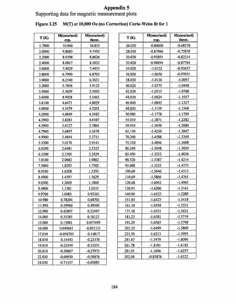
Appendix 5Supporting data for magnetic measurement plots
Figure 3.25 M(T) at 10,000 Oe (no Correction) Curie-Weiss fit for 3
T (K)M(emu/mol)
exp.M(emu/mol)
theor.26.030 -0.80600 -0.6857828.030 -0.87966 -0.7587630.030 -0.93891 -0.8221432.020 -0.98694 -0.8774434.020 -1.0122 -0.9265736.020 -1.0050 -0.9703138.030 -1.0126 -1.009740.020 -1.0375 -1.044842.020 -1.0515 -1.076844.010 -1.0624 -1.105746.040 -1.0845 -1.132748.020 -1.1159 -1.156850.080 -1.1778 -1.179955.010 -1.2871 -1.228259.910 -1.3650 -1.268465.150 -1.4230 -1.304770.240 -1.4588 -1.334975.310 -1.4846 -1.360880.240 -1.5048 -1.383085.450 -1.5233 -1.403690.520 -1.5387 -1.421495.600 -1.5525 -1.4373100.60 -1.5646 -1.4515110.69 -1.5866 -1.4761120.68 -1.6042 -1.4965130.91 -1.6206 -1.5141140.96 -1.6322 -1.5289151.03 -1.6427 -1.5418161.10 -1.6504 -1.5531171.18 -1.6551 -1.5631181.23 -1.6581 -1.5719191.29 -1.6565 -1.5798201.35 -1.6499 -1.5869221.50 -1.6213 -1.5993241.67 -1.5479 -1.6096261.78 -1.4181 -1.6182281.93 -1.1696 -1.6257302.08 -0.85876 -1.6322
T (K) M(enni/moI)exp.
M(emu/mol)theor.
1.7900 10.966 10.8552.0000 9.8683 9.74922.2000 8.9398 8.86262.4000 8.0817 8.10322.6000 7.4029 7.44552.8000 6.7909 6.87033.0000 6.2540 6.36313.2000 5.7856 5.91233.4000 5.3659 5.50923.6000 4.9928 5.14653.8100 4.6471 4.80294.0000 4.3479 4.52034.2000 4.0849 4.24824.3900 3.8283 4.01074.5900 3.9127 3.78044.7900 3.6897 3.56784.9900 3.4844 3.37115.5200 3.0170 2.91416.0200 2.6481 2.55236.5200 2.3305 2.24297.0100 2.0682 1.98027.5000 1.8295 1.75028.0100 1.6208 1.53938.4900 1.4397 1.36299.0300 1.2669 1.18609.4800 1.1381 1.05339.9700 1.0083 0.9220110.980 0.78204 0.6870311.990 0.59960 0.4904012.990 0.45897 0.3249714.000 0.35385 0.1812315.000 0.15081 0.05749916.000 0.040661 -0.05113117.010 -0.056705 -0.1481718.010 -0.14443 -0.2337619.010 -0.22459 -0.3105120.010 -0.30067 -0.3797522.030 -0.60930 -0.5007624.030 -0.71557 -0.60085
184
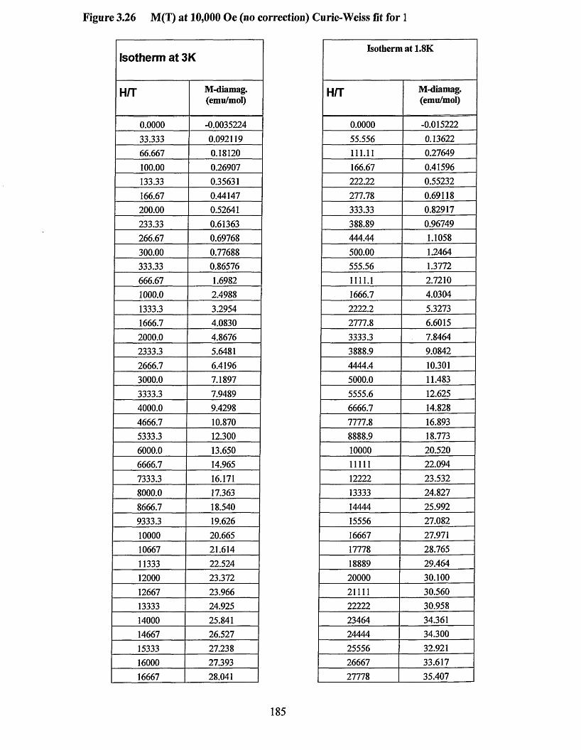
Figure 3.26 M(T) at 10,000 Oe (no correction) Curie-Weiss fit for 1
Isotherm at 3K
H/T M-diamag.(emu/mol)
0.0000 -0.003522433.333 0.09211966.667 0.18120100.00 0.26907133.33 0.35631166.67 0.44147200.00 0.52641233.33 0.61363266.67 0.69768300.00 0.77688333.33 0.86576666.67 1.69821000.0 2.49881333.3 3.29541666.7 4.08302000.0 4.86762333.3 5.64812666.7 6.41963000.0 7.18973333.3 7.94894000.0 9.42984666.7 10.8705333.3 12.3006000.0 13.6506666.7 14.9657333.3 16.1718000.0 17.3638666.7 18.5409333.3 19.62610000 20.66510667 21.61411333 22.52412000 23.37212667 23.96613333 24.92514000 25.84114667 26.52715333 27.23816000 27.39316667 28.041
Isotherm at 1.8K
H/T M-diamag.(emu/mol)
0.0000 -0.01522255.556 0.13622111.11 0.27649166.67 0.41596222.22 0.55232277.78 0.69118333.33 0.82917388.89 0.96749444.44 1.1058500.00 1.2464555.56 1.37721111.1 2.72101666.7 4.03042222.2 5.32732777.8 6.60153333.3 7.84643888.9 9.08424444.4 10.3015000.0 11.4835555.6 12.6256666.7 14.8287777.8 16.8938888.9 18.77310000 20.52011111 22.09412222 23.53213333 24.82714444 25.99215556 27.08216667 27.97117778 28.76518889 29.46420000 30.10021111 30.56022222 30.95823464 34.36124444 34.30025556 32.92126667 33.61727778 35.407
185
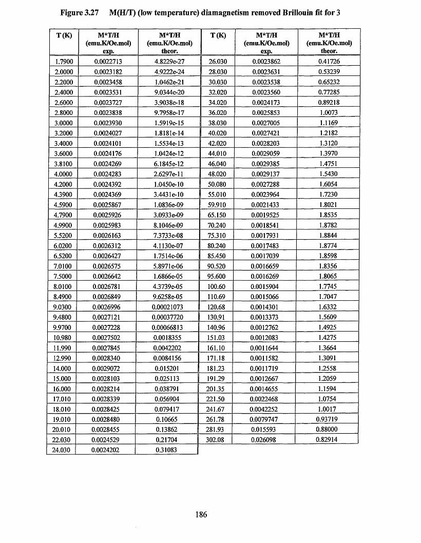
Figure 3.27 M(H/T) (low temperature) diamagnetism removed Brillouin fit for 3
T(K) M*T/H(emu.K/Oe.mol)
exp.
M*T/H(emu.K/Oe.mol)
theor.
T(K) M*T/H(emu.K/Oe.mol)
exp.
M*T/H(emu.K/Oe.mol)
theor.1.7900 0.0022713 4.8229e-27 26.030 0.0023862 0.417262.0000 0.0023182 4.9222e-24 28.030 0.0023631 0.532392.2000 0.0023458 1.0462e-21 30.030 0.0023538 0.652322.4000 0.0023531 9.0344e-20 32.020 0.0023560 0.772852.6000 0.0023727 3.9038e-18 34.020 0.0024173 0.892182.8000 0.0023838 9.7958e-17 36.020 0.0025853 1.00733.0000 0.0023930 1.5919e-15 38.030 0.0027005 1.11693.2000 0.0024027 1.8181e-14 40.020 0.0027421 1.21823.4000 0.0024101 1.5534e-13 42.020 0.0028203 1.31203.6000 0.0024176 1.0424e-12 44.010 0.0029059 1.39703.8100 0.0024269 6.1845e-12 46.040 0.0029385 1.47514.0000 0.0024283 2.6297e-l 1 48.020 0.0029137 1.54304.2000 0.0024392 1.0450e-10 50.080 0.0027288 1.60544.3900 0.0024369 3.443 le-10 55.010 0.0023964 1.72304.5900 0.0025867 1.0836e-09 59.910 0.0021433 1.80214.7900 0.0025926 3.0933e-09 65.150 0.0019525 1.85354.9900 0.0025983 8.1046e-09 70.240 0.0018541 1.87825.5200 0.0026163 7.3733e-08 75.310 0.0017931 1.88446.0200 0.0026312 4.1130e-07 80.240 0.0017483 1.87746.5200 0.0026427 1.7514&-06 85.450 0.0017039 1.85987.0100 0.0026575 5.897le-06 90.520 0.0016659 1.83567.5000 0.0026642 1.6866e-05 95.600 0.0016269 1.80658.0100 0.0026781 4.3739e-05 100.60 0.0015904 1.77458.4900 0.0026849 9.6258e-05 110.69 0.0015066 1.70479.0300 0.0026996 0.00021073 120.68 0.0014301 1.63329.4800 0.0027121 0.00037720 130.91 0.0013373 1.56099.9700 0.0027228 0.00066813 140.96 0.0012762 1.492510.980 0.0027502 0.0018355 151.03 0.0012083 1.427511.990 0.0027845 0.0042202 161.10 0.0011644 1.366412.990 0.0028340 0.0084156 171.18 0.0011582 1.309114.000 0.0029072 0.015201 181.23 0.0011719 1.255815.000 0.0028103 0.025113 191.29 0.0012667 1.205916.000 0.0028214 0.038791 201.35 0.0014655 1.159417.010 0.0028339 0.056904 221.50 0.0022468 1.075418.010 0.0028425 0.079417 241.67 0.0042252 1.001719.010 0.0028480 0.10665 261.78 0.0079747 0.9371920.010 0.0028455 0.13862 281.93 0.015593 0.8800022.030 0.0024529 0.21704 302.08 0.026098 0.8291424.030 0.0024202 1 0.31083 I
186
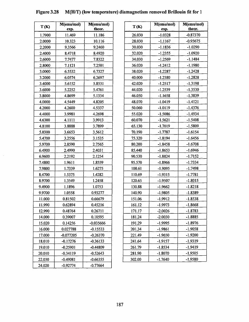
Figure 3.28 M(H/T) (low temperature) diamagnetism removed Brillouin fit for 1
T(K) M(emu/mol)exp.
M(emu/mol)theor.
1.7900 11.469 11.1862.0000 10.323 10.1162.2000 9.3566 9.24602.4000 8.4718 8.49202.6000 7.7477 7.83222.8000 7.1121 7.25013.0000 6.5532 6.73273.2000 6.0574 6.26973.4000 5.6152 5.85313.6000 5.2252 5.47613.8000 4.8699 5.13344.0000 4.5449 4.82054.2000 4.2600 4.53374.4000 3.9981 4.26984.6300 4.1111 3.99134.8100 3.8888 3.78995.0300 3.6653 3.56125.4700 3.2556 3.15355.9700 2.8590 2.75656.4900 2.4990 2.40316.9600 2.2192 2.12547.4800 1.9611 1.85597.9800 1.7339 1.62758.4700 1.5375 1.42828.9700 1.3549 1.24589.4900 1.1896 1.07539.9700 1.0558 0.9327711.000 0.81502 0.6667911.990 0.62894 0.4521612.990 0.48764 0.2671114.000 0.39007 0.1059515.020 0.14256 -0.03566616.000 0.027788 -0.1553317.000 -0.077205 -0.2637018.010 -0.17276 -0.3613319.010 -0.25901 -0.4480920.010 -0.34119 -0.5264322.030 -0.49081 -0.6635524.020 -0.92774 -0.77664
T(K) M(emu/mol)exp.
M(emu/mol)theor.
26.030 -1.0328 -0.8737028.030 -1.1167 -0.9567530.030 -1.1836 -1.029032.020 -1.2355 -1.092034.030 -1.2569 -1.148436.020 -1.2412 -1.198038.020 -1.2287 -1.242840.000 -1.2380 -1.282842.020 -1.2517 -1.319844.020 -1.2539 -1.353046.050 -1.1658 -1.383948.070 -1.0419 -1.412150.060 -1.0119 -1.437655.020 -1.5086 -1.493460.070 -1.5621 -1.540865.130 -1.7019 -1.580970.190 -1.7787 -1.615475.320 -1.8194 -1.645680.200 -1.8458 -1.670885.440 -1.8653 -1.694690.530 -1.8824 -1.715295.570 -1.8966 -1.7334100.61 -1.9095 -1.7498110.69 -1.9315 -1.7781120.65 -1.9507 -1.8015130.88 -1.9662 -1.8218140.90 -1.9805 -1.8389151.06 -1.9912 -1.8538161.12 -1.9973 -1.8668171.17 -2.0026 -1.8783181.24 -2.0030 -1.8885191.29 -1.9995 -1.8976201.34 -1.9861 -1.9058221.49 -1.9650 -1.9200241.64 -1.9157 -1.9319261.79 -1.8534 -1.9419281.90 -1.8070 -1.9505302.00 -1.7640 -1.9580
187
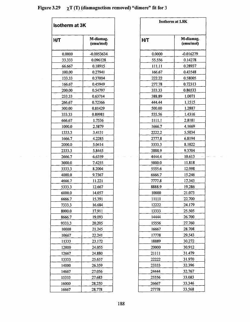
Figure 3.29 %T (T) (diamagnetism removed) “dimere” fit for 3
Isotherm at 3K
H/T M-diamag.(emu/mol)
0.0000 -0.005363433.333 0.09612866.667 0.18915100.00 0.27941133.33 0.37004166.67 0.45949200.00 0.54797233.33 0.63714266.67 0.72366300.00 0.81429333.33 0.89981666.67 1.75361000.0 2.58791333.3 3.41311666.7 4.22852000.0 5.04142333.3 5.84452666.7 6.63593000.0 7.42553333.3 8.20044000.0 9.73674666.7 11.2215333.3 12.6676000.0 14.0576666.7 15.3917333.3 16.6848000.0 17.9118666.7 19.0939333.3 20.20510000 21.24510667 22.24111333 23.17212000 24.05512667 24.88013333 25.65714000 26.35914667 27.05615333 27.68316000 28.25016667 28.778
Isotherm at 1.8K
H/T M-diamag.(emu/mol)
0.0000 -0.01627955.556 0.14278111.11 0.28937166.67 0.43548222.22 0.58005277.78 0.72313333.33 0.86533388.89 1.0071444.44 1.1515500.00 1.2887555.56 1.43161111.1 2.81811666.7 4.16692222.2 5.50342777.8 6.81943333.3 8.10223888.9 9.37044444.4 10.6135000.0 11.8185555.6 12.9986666.7 15.2467777.8 17.3438888.9 19.28610000 21.07311111 22.70012222 24.17913333 25.50514444 26.70015556 27.76016667 28.70817778 29.54318889 30.27220000 30.91221111 31.47922222 31.97023333 32.39624444 32.76725556 33.08326667 33.34627778 33.568
188
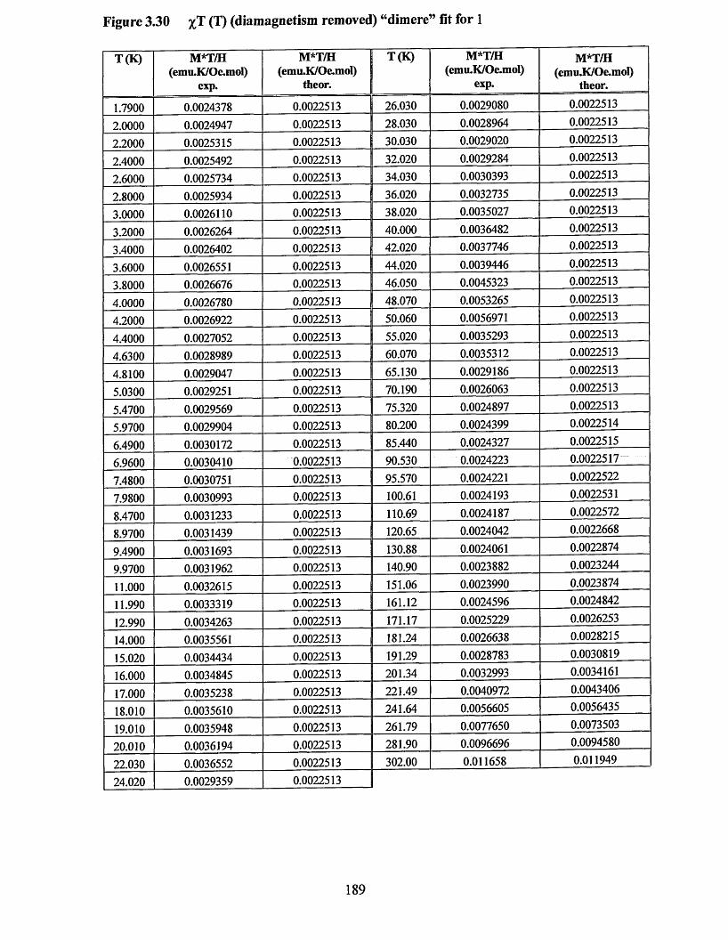
Figure 3.30 %T (T) (diamagnetism removed) “dimere” fit for 1
T(K) M*T/H(emu.K/Oe.mol)
exp.
M*T/H(emu.K/Oe.mol)
theor.
T(K) M*T/H(emu.K/Oe.mol)
exp.
M*T/H(emu.K/Oe.mol)
theor.
1.7900 0.0024378 0.0022513 26.030 0.0029080 0.0022513
2.0000 0.0024947 0.0022513 28.030 0.0028964 0.0022513
2.2000 0.0025315 0.0022513 30.030 0.0029020 0.0022513
2.4000 0.0025492 0.0022513 32.020 0.0029284 0.0022513
2.6000 0.0025734 0.0022513 34.030 0.0030393 0.0022513
2.8000 0.0025934 0.0022513 I 36.020 0.0032735 0.0022513
3.0000 0.0026110 0.0022513 I 38.020 0.0035027 0.0022513
3.2000 0.0026264 0.0022513 40.000 0.0036482 0.0022513
3.4000 0.0026402 0.0022513 42.020 0.0037746 0.0022513
3.6000 0.0026551 0.0022513 44.020 0.0039446 0.0022513
3.8000 0.0026676 0.0022513 46.050 0.0045323 0.0022513
4.0000 0.0026780 0.0022513 48.070 0.0053265 0.0022513
4.2000 0.0026922 0.0022513 50.060 0.0056971 0.0022513
4.4000 0.0027052 0.0022513 55.020 0.0035293 0.0022513
4.6300 0.0028989 0.0022513 60.070 0.0035312 0.0022513
4.8100 0.0029047 0.0022513 65.130 0.0029186 0.0022513
5.0300 0.0029251 0.0022513 70.190 0.0026063 0.0022513
5.4700 0.0029569 0.0022513 75.320 0.0024897 0.0022513
5.9700 0.0029904 0.0022513 80.200 0.0024399 0.0022514
6.4900 0.0030172 0.0022513 85.440 0.0024327 0.0022515
6.9600 0.0030410 0.0022513 90.530 0.0024223 0.0022517
7.4800 0.0030751 0.0022513 95.570 0.0024221 0.0022522
7.9800 0.0030993 0.0022513 100.61 0.0024193 0.0022531
8.4700 0.0031233 0.0022513 110.69 0.0024187 0.0022572
8.9700 0.0031439 0.0022513 120.65 0.0024042 0.0022668
9.4900 0.0031693 0.0022513 130.88 0.0024061 0.0022874
9.9700 0.0031962 0.0022513 140.90 0.0023882 0.0023244
11.000 0.0032615 0.0022513 151.06 0.0023990 0.0023874
11.990 0.0033319 0.0022513 161.12 0.0024596 0.0024842
12.990 0.0034263 0.0022513 171.17 0.0025229 0.0026253
14.000 0.0035561 0.0022513 181.24 0.0026638 0.0028215
15.020 0.0034434 0.0022513 191.29 0.0028783 0.0030819
16.000 0.0034845 0.0022513 201.34 0.0032993 0.0034161
17.000 0.0035238 0.0022513 221.49 0.0040972 0.0043406
18.010 0.0035610 0.0022513 241.64 0.0056605 0.0056435
19.010 0.0035948 0.0022513 261.79 0.0077650 0.0073503
20.010 0.0036194 0.0022513 281.90 0.0096696 0.0094580
22.030 0.0036552 0.0022513 302.00 0.011658 0.011949
24.020 0.0029359 0.0022513
189

Appendix 6
Crystal data and structure refinement:2,3-dichlorophenyI-l, 2,3,5-dithiadiazolyl
Identification code Empirical formula Formula weight Temperature Wavelength Crystal system Space group Unit cell dimensions
VolumeZDensity (calculated)Absorption coefficient F(000)Crystal sizeTheta range for data collection Index ranges Reflections collected Independent reflections Completeness to theta = 28.36°Absorption correction Max. and min. transmission Refinement method Data / restraints / parameters Goodness-of-fit on F2 Final R indices [I>2sigma(I)]R indices (all data)Extinction coefficient Largest diff. peak and hole
shul3hC7H3C12N2S2250.13 150(2) K 0.71073 A Monoclinic P2i/na = 7.2936(14) A a=90°.b = 13.572(3) A P= 101.468(3)°.c= 17.910(4) A y = 90°.1737.5(6) A3 81.912 Mg/m3 1.170 mm'1 10000.44x0.28x0.28 mm3 1.90 to 28.36°.-9<=h<=9, -17<=k<=18, -18<=1<=23 111554145 [R(int) = 0.1670]95.5 %Semi-empirical from equivalents 0.7353 and 0.6270 Full-matrix least-squares on F2 4145/0/236 1.219R1 =0.1625, wR2 = 0.3651 R l= 0.1919, wR2 = 0.3733 0.0021(8)1.907 and-1.413 e.A-3
190
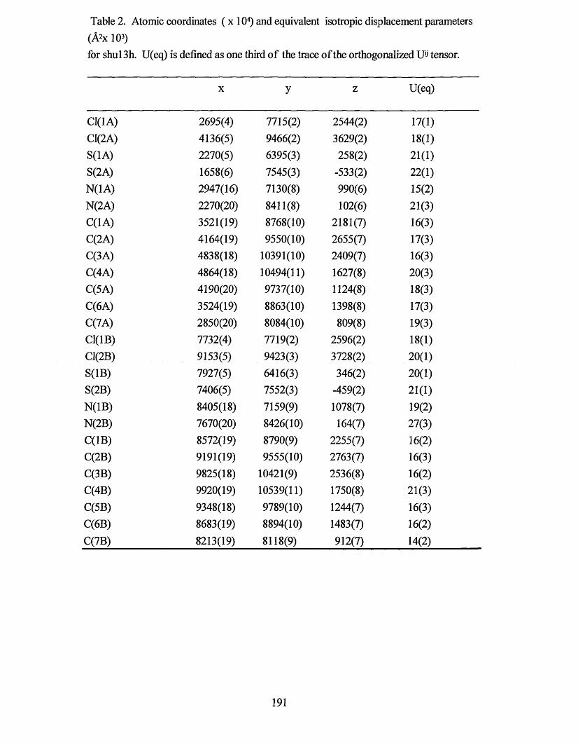
Table 2. Atomic coordinates (x 104) and equivalent isotropic displacement parameters (A2x 103)for shul 3h. U(eq) is defined as one third of the trace of the orthogonalized Uij tensor.
X y z U(eq)
C1(1A) 2695(4) 7715(2) 2544(2) 17(1)CI(2A) 4136(5) 9466(2) 3629(2) 18(1)S(1A) 2270(5) 6395(3) 258(2) 21(1)S(2A) 1658(6) 7545(3) -533(2) 22(1)N(1A) 2947(16) 7130(8) 990(6) 15(2)N(2A) 2270(20) 8411(8) 102(6) 21(3)C(1A) 3521(19) 8768(10) 2181(7) 16(3)C(2A) 4164(19) 9550(10) 2655(7) 17(3)C(3A) 4838(18) 10391(10) 2409(7) 16(3)C(4A) 4864(18) 10494(11) 1627(8) 20(3)C(5A) 4190(20) 9737(10) 1124(8) 18(3)C(6A) 3524(19) 8863(10) 1398(8) 17(3)C(7A) 2850(20) 8084(10) 809(8) 19(3)C1(1B) 7732(4) 7719(2) 2596(2) 18(1)C1(2B) 9153(5) 9423(3) 3728(2) 20(1)S(1B) 7927(5) 6416(3) 346(2) 20(1)S(2B) 7406(5) 7552(3) -459(2) 21(1)N(1B) 8405(18) 7159(9) 1078(7) 19(2)N(2B) 7670(20) 8426(10) 164(7) 27(3)C(1B) 8572(19) 8790(9) 2255(7) 16(2)C(2B) 9191(19) 9555(10) 2763(7) 16(3)C(3B) 9825(18) 10421(9) 2536(8) 16(2)C(4B) 9920(19) 10539(11) 1750(8) 21(3)C(5B) 9348(18) 9789(10) 1244(7) 16(3)C(6B) 8683(19) 8894(10) 1483(7) 16(2)C(7B) 8213(19) 8118(9) 912(7) 14(2)
191
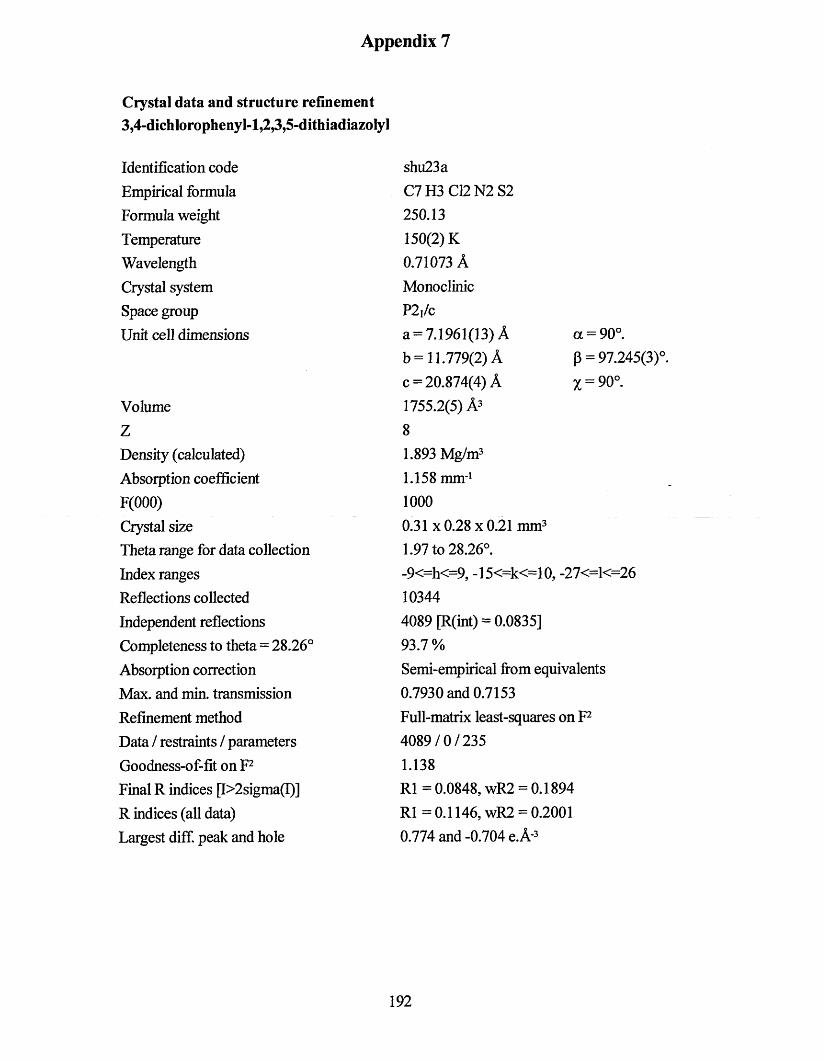
Appendix 7
Crystal data and structure refinement3,4-dichlorophenyl-l,2,3,5-dithiadiazolyl
Identification code Empirical formula Formula weight Temperature Wavelength Crystal system Space group Unit cell dimensions
VolumeZDensity (calculated)Absorption coefficient F(000)Crystal sizeTheta range for data collection Index ranges Reflections collected Independent reflections Completeness to theta = 28.26°Absorption correction Max. and min. transmission Refinement method Data / restraints / parameters Goodness-of-fit on F2 Final R indices [I>2sigma(I)]R indices (all data)Largest diff. peak and hole
shu23aC7H3C12N2S2250.13 150(2) K 0.71073 A Monoclinic P2j/ca = 7.1961(13) A a = 90°.b = 11.779(2) A p = 97.245(3)°.c = 20.874(4) A x = 90°.1755.2(5) A3 81.893 Mg/m3 1.158 mm*1 10000.31 x 0.28 x 0.21 mm3 1.97 to 28.26°.-9<=h<=9, -15<=k<=10, -27<=1<=26 103444089 [R(int) = 0.0835]93.7 %Semi-empirical from equivalents 0.7930 and 0.7153 Full-matrix least-squares on F2 4089/0/235 1.138R1 =0.0848, wR2 = 0.1894 R1 =0.1146, wR2 = 0.2001 0.774 and-0.704 e.A-3
192
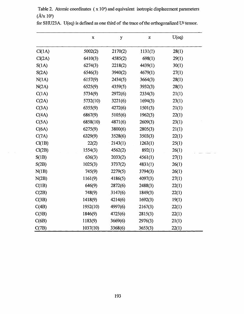
Table 2. Atomic coordinates ( x 104) and equivalent isotropic displacement parameters (A2x 103)for SHU23 A. U(eq) is defined as one third of the trace of the orthogonalized U« tensor.
X y z U(eq)
C1(1A) 5002(2) 2170(2) 1131(1) 28(1)C1(2A) 6410(3) 4585(2) 698(1) 29(1)S(1A) 6274(3) 2218(2) 4439(1) 30(1)S(2A) 6546(3) 3940(2) 4679(1) 27(1)N(1A) 6157(9) 2434(5) 3664(3) 28(1)N(2A) 6525(9) 4359(5) 3932(3) 28(1)C(1A) 5734(9) 2972(6) 2334(3) 21(1)C(2A) 5732(10) 3221(6) 1694(3) 23(1)C(3A) 6355(9) 4272(6) 1501(3) 21(1)C(4A) 6867(9) 5105(6) 1962(3) 22(1)C(5A) 6858(10) 4871(6) 2609(3) 23(1)C(6A) 6275(9) 3800(6) 2805(3) 21(1)C(7A) 6329(9) 3528(6) 3503(3) 22(1)C1(1B) 22(2) 2143(1) 1263(1) 25(1)C1(2B) 1554(3) 4562(2) 892(1) 26(1)S(1B) 636(3) 2033(2) 4561(1) 27(1)S(2B) 1025(3) 3737(2) 4831(1) 26(1)N(1B) 745(9) 2279(5) 3794(3) 26(1)N(2B) 1161(9) 4186(5) 4097(3) 27(1)C(1B) 646(9) 2872(6) 2488(3) 22(1)C(2B) 748(9) 3147(6) 1849(3) 22(1)C(3B) 1418(9) 4214(6) 1692(3) 19(1)C(4B) 1952(10) 4997(6) 2167(3) 22(1)C(5B) 1846(9) 4725(6) 2815(3) 22(1)C(6B) 1183(9) 3669(6) 2976(3) 21(1)C(7B) 1037(10) 3368(6) 3653(3) 22(1)
193

Appendix 8
Crystal data and structure refinement3,5-dibromophenyl-l,2,3,5-dithiadiazoIyl
Identification code Empirical formula Formula weight Temperature Wavelength Crystal system Space group Unit cell dimensions
VolumeZDensity (calculated) Absorption coefficient F(000)Crystal sizeTheta range for data collection Index ranges Reflections collected Independent reflections Completeness to theta = 28.36° Absorption correction Max. and min. transmission Refinement method Data / restraints / parameters Goodness-of-fit on F2 Final R indices p>2sigma(I)]R indices (all data)Largest diff. peak and hole
shu8aC7H3Br2N2S2339.05 150(2) K 0.71073 A Monoclinic C2/ca= 15.0126(12) A a=90°.b= 16.3582(13) A p= 113.9450(10)cc= 16.9218(14) A y = 90°.3798.0(5) A3 162.372 Mg/m3 8.925 mm*1 25760.45 x 0.45 x 0.30 mm3 1.94 to 28.36°.-20<=h<=20, -21<=k<=21, -11<=1<=21 123434539 [R(int) = 0.0493]95.3 %Semi-empirical0.1749 and 0.1078Full-matrix least-squares on F24539/0/2351.109R l= 0.0354, wR2 = 0.0939 R1 = 0.0452, wR2 = 0.1101 1.146 and-1.941 e.A'3
194
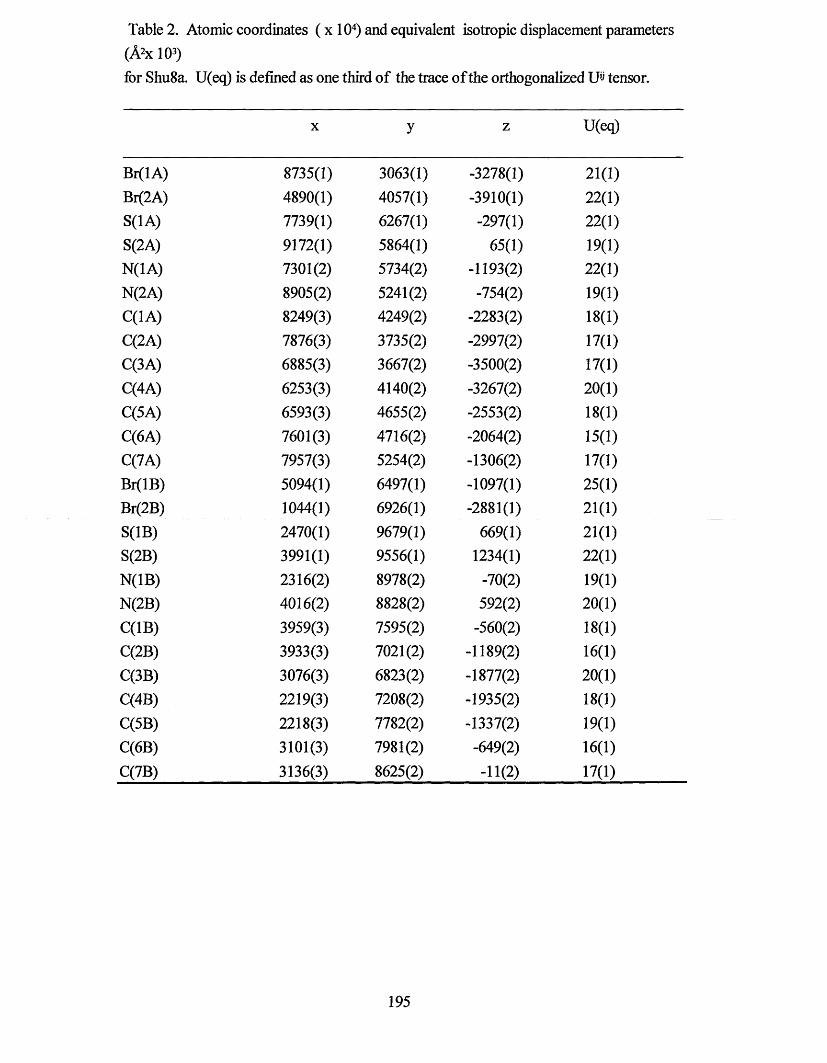
Table 2. Atomic coordinates (x 104) and equivalent isotropic displacement parameters (A2x 103)for Shu8a. U(eq) is defined as one third of the trace of the orthogonalized Uij tensor.
X y z U(eq)
Br(lA) 8735(1) 3063(1) -3278(1) 21(1)Br(2A) 4890(1) 4057(1) -3910(1) 22(1)S(1A) 7739(1) 6267(1) -297(1) 22(1)S(2A) 9172(1) 5864(1) 65(1) 19(1)N(1A) 7301(2) 5734(2) -1193(2) 22(1)N(2A) 8905(2) 5241(2) -754(2) 19(1)C(1A) 8249(3) 4249(2) -2283(2) 18(1)C(2A) 7876(3) 3735(2) -2997(2) 17(1)C(3A) 6885(3) 3667(2) -3500(2) 17(1)C(4A) 6253(3) 4140(2) -3267(2) 20(1)C(5A) 6593(3) 4655(2) -2553(2) 18(1)C(6A) 7601(3) 4716(2) -2064(2) 15(1)C(7A) 7957(3) 5254(2) -1306(2) 17(1)Br(lB) 5094(1) 6497(1) -1097(1) 25(1)Br(2B) 1044(1) 6926(1) -2881(1) 21(1)S(1B) 2470(1) 9679(1) 669(1) 21(1)S(2B) 3991(1) 9556(1) 1234(1) 22(1)N(1B) 2316(2) 8978(2) -70(2) 19(1)N(2B) 4016(2) 8828(2) 592(2) 20(1)C(1B) 3959(3) 7595(2) -560(2) 18(1)C(2B) 3933(3) 7021(2) -1189(2) 16(1)C(3B) 3076(3) 6823(2) -1877(2) 20(1)C(4B) 2219(3) 7208(2) -1935(2) 18(1)C(5B) 2218(3) 7782(2) -1337(2) 19(1)C(6B) 3101(3) 7981(2) -649(2) 16(1)C(7B) 3136(3) 8625(2) -11(2) 17(1)
195
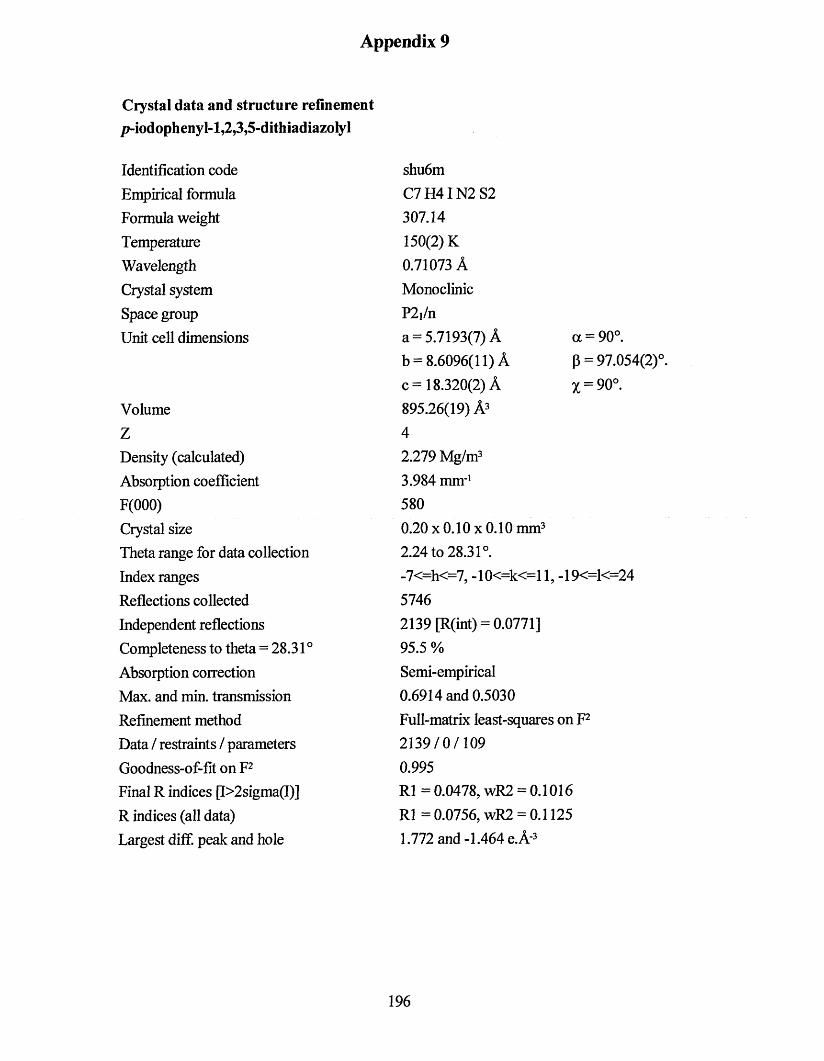
Appendix 9
Crystal data and structure refinement/ModophenyI-l,2,3»5-dithiadiazolyl
Identification code Empirical formula Formula weight Temperature Wavelength Crystal system Space group Unit cell dimensions
VolumeZDensity (calculated) Absorption coefficient F(000)Crystal sizeTheta range for data collection Index ranges Reflections collected Independent reflections Completeness to theta = 28.31 ° Absorption correction Max. and min. transmission Refinement method Data / restraints / parameters Goodness-of-fit on F2 Final R indices [I>2sigma(I)]R indices (all data)Largest diff. peak and hole
shu6mC7H4IN2S2307.14 150(2) K 0.71073 A Monoclinic P2,/na = 5.7193(7) A a =b = 8.6096(11) A P =c= 18.320(2) A x =895.26(19) A3 42.279 Mg/m3 3.984 mm-1 5800.20 x 0.10 x 0.10 mm3 2.24 to 28.31°.-7<=h<=7, -10<=k<=ll, -19<= 57462139 [R(int) = 0.0771]95.5 %Semi-empirical0.6914 and 0.5030Full-matrix least-squares on F22139/0/1090.995R l= 0.0478, wR2 = 0.1016 R1 = 0.0756, wR2 = 0.1125 1.772 and-1.464 e.A'3
90°.97.054(2)°.90°.
T<=24
196
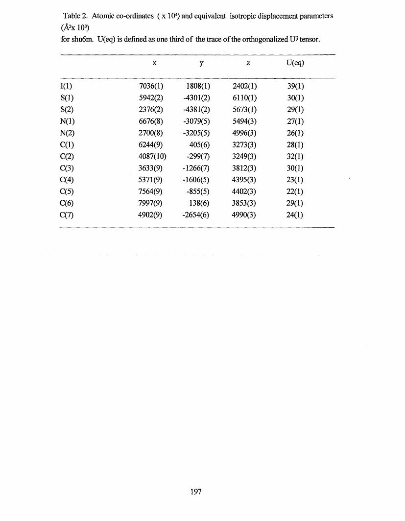
Table 2. Atomic co-ordinates ( x 104) and equivalent isotropic displacement parameters (A2x 103)for shu6m. U(eq) is defined as one third of the trace of the orthogonalized U'J tensor.
X y z U(eq)
1(1) 7036(1) 1808(1) 2402(1) 39(1)S(l) 5942(2) -4301(2) 6110(1) 30(1)S(2) 2376(2) -4381(2) 5673(1) 29(1)N(l) 6676(8) -3079(5) 5494(3) 27(1)N(2) 2700(8) -3205(5) 4996(3) 26(1)C(l) 6244(9) 405(6) 3273(3) 28(1)C(2) 4087(10) -299(7) 3249(3) 32(1)C(3) 3633(9) -1266(7) 3812(3) 30(1)C(4) 5371(9) -1606(5) 4395(3) 23(1)C(5) 7564(9) -855(5) 4402(3) 22(1)C(6) 7997(9) 138(6) 3853(3) 29(1)C(7) 4902(9) -2654(6) 4990(3) 24(1)
197
Appendix 10
PE R G A M O N
www.olscvicr ill/locate/pol)
Polyhedron l ‘> 1 2<XX)> 11f>3—1166POLYHEDRON
Modification of the solid state structures of dithiadiazolyl radicals: crystal structure o f /?-iodophenyl-l,2,3,5-dithiadiazolyl
Neil Bricklebanka *, Stephen Hargreavesa, Sharon E. Spey b•* Division o f Chemistry and M aterials Research Institute. Sheffield Hallam University. C ity Cam/ms. Sheffield SI IWB. UK
h Department o f Chemistry. U niversity o f Sheffield. Western Bank. Sheffield S3 7HF. U K
Received 4 January 2000: accepted 27 March 2000
Abstract
The solid state structure of the 1,2,3.5-dithiadiazolyl />-ICftH4CNSSN (3) is described. The molecules associate in dimer pairs, across an inversion centre, in an unusual rra/w-cofacial manner. The separation between the heterocyclic rings is 3.121 A. The intradimer sulfur contacts (3.696 A) are larger than those observed in dithiadiazolyls that associate in the more usual ct.v-cofacial manner. Individualp-IC„H4CNSSN molecules are linked into zigzag chains through weak I---S interactions (3.82 A t: each half of a [p-ICftH4CNSSN]: dimer is a member of separate chains that run in opposite directions. ©2000 Elsevier Science Ltd All rights reserved.
Keywords: Dithiadiazolyl radicals
1. Introduction
In recent years. 1.2.3.5-dithiadiazolyl (D T D A ) radicals have been studied as building blocks for the design and synthesis o f m olecular m agnets [1 -5 ] and conductors [6 - 9 ] . The steric and electronic effects o f the substituent R can be used to modify the m olecular structure and hence the physical behaviour o f the radical. In the solid state the m ajority o f DTDA radicals associate as spin-paired dim ers through the form ation o f out-of-plane interm olecular S - - S interactions (F ig . 1) with a typical interm olecular separation in the range 3 .0 -3 .2 A. Crystal engineering o f DTDAs has focused on obtaining m aterials in which the radicals are aligned into infinite chains or stacked in colum ns: one o f the principal tools used to achieve this has been to use nitrile groups as structure-influencing supram olecular synthons [1 .2 .8 ,9 ]. The idea behind this strategy stemm ed from the structure o f p-iodobenzonitrile [10] and the cyanogen halides [ I I ] . which associate as ribbon-like chains through weak X - C N interactions. Sim ilarly, cyano-functionalised DTDAs (e.g . com pounds 1 and 21 also associate as chains through CN- • -S interactions [ 1 .2 .81.
Halogen atoms can also have a marked effect on the m olecular packing, although only fluorinated system s have received extensive study [1 —1.12.13]. Partial fluorination o f the aryl
co-cofacia l twisted
ii-e-N
/roTU-antarafacial
"Corresponding auihor. Tel.: -1-44-114-223 41>3I: fax: 1-44-114-225 3()Ph: e-mail; n.hricklebankt-nMui.ac.uk
Fig. 1. Modes o f dimerisation for DTDA radicals.
group can lead to the d isruption o f the dim erisation process: for exam ple. [ 2,3-F2C 6H:iC N SSN ] ; associates as an unusual twisted dim er [ 12]. w hereas 2 .5-F2C 6H3C N SSN assem bles in uniform stacks with an unusually long interannular spacing [3 .5 4 4 (3 ) A] [1 2 ] . Total fluorination results in more dramatic changes: both /?-N C C 6F4C N SSN (2 ) [1 -3 ] a n d p- BrC„F4CNSSN [4 ] do not associate as dim ers and consequently are param agnetic at room tem perature. The latter com pound associates in colum ns, w hereas the com bination o f fluorine atom s and a nitrile group in com pound 2 leads to a com pound that assem bles with a chain-like structure [ 1.2 ]. Upon cooling to below 36 K. one polym orph o f com pound 2 undergoes a transition to a weakly ferrom agnetic state [ 3 ]. This is the highest ferrom agnetic ordering tem perature reported to date for a non-m etallic m olecular magnet.
We are currently investigating the effect o f the heavier halogen atoms on the supram olecular structures o f dithia- diazolyl radicals. As part o f this work, we decided to make />-iodophenyl-DTDA (3 ) . In particular, we were keen to determ ine the structure-influencing properties o f the iodine
(1277-55S~ IXI S - m.v front matter «.'2(XH) F.lxevier Science Ltd All lights reserved. / ’/ / S 0 2 ” -5.'>S'i n o 1 0 0 5 S 4 • 5
198
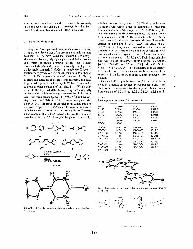
■V llilt khhi ink .7 ill. / I'nlilii ilinii 1'I I yilili, I // 11 hh
atom and to see w hether it would also promote the assem bly o f the m olecules into chains, as is observed for //-iodoben- /.onitrilo and cyano-funetionalised DTDAs ( 1 and 2).
2. R esu lts an d d iscussion
Com pound 3 was prepared from /?-iodobenzonitrile using a slightly modified version of the proven metal am idine route (Schem e 1). W e have found that sodium b isftrim ethy l- s ily ljam ide gives slightly higher yields with iodo-. brom o- and chloro-substituted arom atic nitriles than lithium bisftrim ethylsilyl )am ide, which is usually em ployed in dithiadiazolyl synthesis [ 14]. Crystals suitable for X-ray d iffraction were grow n by vacuum sublim ation as described in Section 4. The asym m etric unit o f com pound 3 (F ig . 2) contains one m olecule o f unexceptional geom etry. The bond lengths and angles o f the heterocycle (T able 1) are sim ilar to those o f other m em bers o f this class [1 4 ]. W ithin each m olecule the arvl and dithiadiazolyl rings are essentially coplanar with a slight tw ist angle between the dithiadiazole ring [root mean square (r.m .s.) A = 0.0077 A] and the aryl ring [r.m .s. A = 0.0089 A] of 8°. However, com pared with other DTDAs. the m ode o f association in com pound 3 is unusual. T w op-IC 6H4CNSSN m olecules associate in a tntns- cofacial m anner across an inversion centre ( Fig. 3 ) . The only other exam ple o f a DTDA radical adopting this m ode of association is the 2.2 'dim ethylbiphenylene radical (4 ) .
“O O “O O O O1 2 F 3
0-00O —^ ----- -OO£ )F te r
KOCHzCfe, PbjSb, 12h
Scheme 1.
w Inch was reported very recently 15 1. The distance between the heterocycles within dim ers o f com pound 3 ( m easured from the mid-point o f the rings) is 3.121 A. This is significantly shorter than that in com pound 4 (3 .2 4 A ) and is sim ilar to those observed in DTDAs that associate in the r/.v-cofacial or m/n.v-antarafacial modes. How ever, the intradim er sulfur contacts in com pound 3 |</( S I- -S2A ) and / / ( S 2 - S I A ) = 3.696 A | are long when com pared with the equivalent distance in DTDAs that associate in a c/.v-cofacial or trans- antarafacial m anner (typically 3 .0 -3 .2 A ) and are sim ilar to those in com pound 4 [3.68( 2) A ]. Each dim er pair also has two sets o f intradim er su lfur-n itrogen interactions [</(S I--N 2 A ),//(S IA ---N 2 ) = 3 .116 A] and [//(S2- -N1 A ), d{ S2A- - N1 ) = 3 .1 5 2 A ]. The asym m etry in these interactions results from a further interaction betw een one o f the sulfurs with the iodine atom o f an adjacent m olecule (see below ).
As noted by Oakley and co-w orkers [5 ], the rra//5-cofacial mode o f dim erisation adopted by com pounds 3 and 4 lies close to the transition state for the proposed photochem ical isom erisation o f 1.3.2.4- to 1.2.3.5-DTDAs (Schem e 2)
Table IBond length- i A i and angles ('» in compound 3
ll-C I 2 .094(6) C 1-C 2 1.371(7)S I-S 2 2 .098(2) C I-C 6 1.388(8)S I-N I 1.635(5) C 2-C 3 1.376(8)S 2-N 2 1.629(5) C 3-C 4 1.398(8)N I-C 7 1.337(7) C 4-C 5 1.410(7)N 2-C 7 1.347(6) C 5-C 6 1.365(7)C 7-C 4 1.464(7)
N I-S I -S 2 94.45 ( 18) C 3-C 4-C 5 117.3(5)N 2-S 2 -S I 94.26( 17) C 3-C 4-C 7 121.3(5)C 7 -N I-S I 114.6(4) C 5-C 4-C 7 121.4(5)C 7-N 2-S 2 114.8(4) C 6-C 5-C 4 121.5(5)C 2-C I-C 6 120.9(5) C 5-C 6-C I 119.2 (5 )C 2-C I-II 120.6(4) N I-C 7 -N 2 121.9(5)C 6-C 1-II 118.4(4) N 1-C 7-C 4 119.4(5)C 1-C 2-C 3 119.7(5) N 2-C 7-C 4 118.7(5)C 2-C 3-C 4 • 121.2(5)
C U JU O T A )
Fig. 3. Dimeri'jtion in compound 3 illustrating the //v/H.v-cofaeial mode of a'M'oiation.
Fig. 2. ORTFP plot o f as> mmotric unit o f compound 3 showing atom lahel- lins: scheme.
N—S
,1 / " S • •2! ' - • i 4 1: ii*
4L>-"Scheme 2.
199
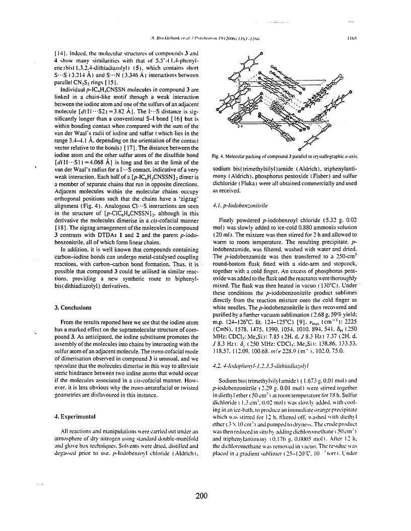
.V. H m k l r h m ik r t ill. / l ‘n l\ l n ' i tn i i i l 'Jl2HIX)i I //>.< -I lf>/, I IfO
114 1. Indeed, the m olecular structures o f com pounds 3 and 4 show many sim ilarities with that o f 5 .5 '-( 1,4-phenyI- en e)b is( 1 .3 ,2 .4-dithiadia/olyl) (5) . which contains short S - S (3 .214 A ) a n d S - N (3 .346 A) interactions betw een parallel C N : S ; rings [ 15 1.
Individual p -lC hH4CNSSN molecules in com pound 3 are linked in a chain-like m otif through a weak interaction between the iodine atom and one o f the sulfurs o f an adjacent m olecule [< /(H ---S2) = 3 .8 2 A |. The I - S distance is sig nificantly longer than a conventional S -I bond [1 6 ] but is within bonding contact when com pared with the sum o f the van der W aal’s radii o f iodine and sulfur ( which lies in the range 3.4-4.1 A, depending on the orientation o f the contact vector relative to the bonds) [ 17 ]. The distance betw een the iodine atom and the other sulfur atom o f the disulfide bond [ r / ( U - " S l ) = 4 .0 6 8 A] is long and lies at the lim it o f the van der W aal’s radius for a l - S contact, indicative o f a very weak interaction. Each half o f a [p-IC 6H4C N S S N ]2 d im er is a m em ber o f separate chains that run in opposite directions. Adjacent m olecules within the m olecular chains occupy orthogonal positions such that the chains have a ‘z igzag’ alignm ent (F ig . 4 ) . Analogous C l - S interactions are seen in the structure o f [p-C lC 6H4C N S SN ]: , although in this derivative the m olecules dim erise in a cis-cofacial m anner [1 8 ]. The zigzag arrangem ent o f the molecules in com pound 3 contrasts with DTDAs 1 and 2 and the parent p-iodo- benzonitrile, all o f which form linear chains.
In addition, it is well known that com pounds containing carbon-iodine bonds can undergo m etal-catalysed coupling reactions, w ith carbon-carbon bond form ation. Thus, it is possible that com pound 3 could be utilised in sim ilar reactions. providing a new synthetic route to biphenyl- b is( d ith iad iazo ly l) derivatives.
3. C onclusions
From the results reported here we see that the iodine atom has a m arked effect on the supram olecular structure o f com pound 3. As anticipated, the iodine substituent prom otes the assem bly o f the m olecules into chains by interacting w ith the sulfur atom o f an adjacent m olecule. The rra/is-cofacial m ode o f dim erisation observed in com pound 3 is unusual, and we speculate that the m olecules dim erise in this way to alleviate steric hindrance betw een two iodine atoms that would occur if the m olecules associated in a m -co facia l manner. H ow ever. it is less obvious why the rra/w-antarafacial or tw isted geom etries are disfavoured in this instance.
4. E x p erim en ta l
All reactions and m anipulations were carried out under an atm osphere o f dry nitrogen using standard double-m anifold and glove box techniques. Solvents were dried, distilled and degassed prior to use. p-Iodoben/.oyl chloride (A ld rich ).
Fig. 4. Molecular packing o f compound 3 parallel to crystallographic u-axis.
sodium b is(trim ethv lsily l)am ide (A ld rich ), triphenylanti- mony (A ld rich ), phosphorus pentoxide ( Fisher) and sulfur dichloride ( Fluka) w ere all obtained com m ercially and used as received.
4.1. p-Iodobenzonitrile
Finely powdered p-iodobenzoyl chloride (5 .32 g. 0.02 m ol) was slowly added to ice-cold 0.880 am m onia solution (20 m l). The m ixture was then stirred for 2 h and allow ed to warm to room tem perature. The resulting precipitate, p- iodobenzam ide. was filtered, w ashed with w ater and dried. The p-iodobenzam ide was then transferred to a 250-cnv* round-bottom flask fitted w ith a side-arm and stopcock, together with a cold finger. An excess o f phosphorus pentoxide was added to the flask and the reactants were thoroughly mixed. The flask was then heated in vacuo ( 130CC ). U nder these conditions the p-iodobenzonitrile product sublim es directly from the reaction m ixture onto the cold finger as white needles. The p-iodobenzonitrile is then recovered and purified by a further vacuum sublim ation (2 .68 g. 59% yield: m.p. 124-426°C. lit. 124-125°C ) [9 ] . ( c m ' 1): 2225(O N ). 1578. 1475. 1390. 1054. 1010. 894. 541. 6h ( -5 0 M Hz: CDCU: M e4S i): 7.85 (2H . d. J 8.3 H z) 7.37 (2H . d. J 8.3 H z). 5, (250 M Hz: CDC1,: M e4S i): 138.86. 133.53. 118.57. 112.09. 100.68. m le 228.9 (m * ). 102.0. 75.0.
4.2. 4-lodophenyl-1.2.3.5-ilirhiailiazolyl
Sodium bis(trim ethy lsily lam ide) (1 .6 7 3 g .0.01 m ol) and p-iodobenzonitrile (2 .29 g. 0.01 m ol) were stirred together in diethyl ether (50 cm ’ ) at room tem perature for 18 h. Sulfur dichloride ( 1.3 cnv \ 0.02 m o l) was slowly added, with coo ling in an ice-bath. to produce an im m ediate orange precipitate which was stirred for 12 h. filtered off. washed with diethyl ether (3 \ 10 c m s) and pum ped to dryness. The crude product was then reduced in situ b\ adding dichlorom ethane i 5 0 cm ’ ) and triphenylantim ony i 0.176 g. 0.0005 mol ) . A fter 12 h. the dichlorom ethane was removed in vacuo. The residue w as placed in a gradient sublim er ( 2 5 -1 2 0 ‘C. 10 ’ to rn . Under
200
I I Mi .V. t im k li’ln ink a <//. / 1‘n h h v tlr n n I 'h l iH H h I //>.< / lfif>
those conditions, the product forms as deep red-b lack blocks (0 .5 6 g . I S7r yield: m.p. I72 .5°C ). Found: C. 27.13: H. 1.32: N. 9.05. C 7H4IN2S 5 requires: C. 27.37: H. 1.31: N. 9.12% . i/lliai ( c m* ' ) : m / e 307.1 ( m * ). 261.1, 229.1, 180.0. 134.1. 102.1.77.9.
4.3. Crystal data
C7H4IN2S 2 (M = 307.14) crystallises as deep red blocks: crystal dim ensions 0 .2 0 X 0 .1 0 X 0 .1 0 mm. M onoclinic, a = 5 .7 1 9 3 (7 ) , 6 = 8 .6 0 9 6 ( 1 1 ) , c = 18.320(2) A, /3 = 97 .054(2)°, t / = 895.26( 19) A3, Z = 4 ,D c = 2.279 M g m ~ \ space group P2, In ( a non-standard setting o f F2, / c, C21,. No. 14). M o K a radiation ( F = 0.71073 A ), /z(M o K a ) = 3 .9 8 4 m m -1 , F (0 0 0 ) = 5 8 0 . Data collected were m easured on a Bruker Sm art C C D area detector with Oxford Cryosystem s low-tem perature system . Cell param eters were refined from the setting angles o f 111 reflections (6 range 2 .24-28 .31°). Reflections were m easured from a hemisphere o f data collected o f frames each covering 0.3 degrees in om ega. O f the 5746 reflections m easured, all o f which were corrected for Lorentz and polarisation effects and for absorption by semi- empirical m ethods based on sym m etry-equivalent and repeated reflections ( m inim um and maxim um transm ission coefficients o f 0 .5030 and 0 .6914), 1474 independent reflections exceeded the significance level I F | /a ( I F I ) > 4.0. The structure was solved by direct m ethods and refined by full- matrix least-squares m ethods on F 2. Hydrogen atom s were placed geom etrically and refined with a riding m odel and with Uiso constrained to be 1.2 tim es U^ o f the carrier atom. Refinement converged at a final R - 0.0478 (vv/?: = 0 .1 125, for all 2139 data. 109 param eters, m ean and m axim um 5 / t r 0.000, 0 .000) w ith allowance for the thermal anisotropy o f all non-hydrogen atom s. M inim um and maxim um final electron density was — 1.464 and 1.772 e A " \ A weighting scheme u = l / [ r r ( F 02) + (0 .0 5 8 7 F )2 + 0 .0 0 F ], where P = ( F 02 + 2 F 2) / 3 was used in the latter stages o f refinement. Com plex scattering factors were taken from the program package SH ELX TL [19] as im plem ented on a Viglen Pentium com puter.
Supplementary data
Supplementary data are available from the CCDC. 12 Union Road. Cam bridge CB2 1EZ, UK (fax: + 44-1223- 336033: e-mail: deposit@ ccdc.cam .ac.uk) on request, quoting the deposition num ber 137/938.
A cknow ledgem ents
W e would like to thank Sheffield Hallam University fo ra studentship ( S .H .), the Royal Society for an equipm ent grant (N .B .) and the Royal Society o f Chem istry Research Fund and the Nuffield Foundation for financial support (N .B .) .
R eferences
[ 11 A.J. Banister. N. Bricklebank. W. C legg. M .R J. Elsegood. C.l. Gregory. I. Lavender. J.M. Rawson. B.K. Tanner. J. Chem. Soc.. Chem. Commun. (1 9 9 5 ) 679.
[2J A.J. Banister. N. Bricklebank. I. Lavender. J.M. Rawson. C.L Gregory. B.K. Tanner. W. Clegg. M .R J. Elsegood.F. Palacio. A ngew. Chem. j n t . Ed. Engl. 35 <1996) 2533.
[31 F. Palacio. G. Antorrena. M. Castro. R. Burriel. J.M. Rawson. J.N.B. Smith. N. Bricklebank. J. Novoa. C. Ritter. Phys. Rev. Lett. 79 (1 9 9 7 ) 2336.
[4 ] G.Antorrena.J.E. D avies.M . Hartley.F.Palacio.J.M .R awson.J.N.B. Smith. A. Steiner. Chem. Commun. (1 9 9 9 ) 1393.
[5 ] T.M. B arc lay . A.W. Cordes. N.A. George. R.C. Haddon. M.E. Itkis. R.T. Oakley. Chem. Commun. (1 9 9 9 ) 2269.
[ 6 1 A.W. Cordes. R.C. Haddon. R .T.O akley. Adv. Mater. 6 (19 9 4 ) 798. [ 7 1 C.D. Bryan. A.W. Cordes. R.M. Fleming. N.A. George. S.H. Glarum.
R.C. Haddon. R.T. Oakley. T.T.M." Palstra. A .S. Perel. L.F. Schneemeyer. J.V. Waszczak. Nature 365 (1993) 821.
[ 81 A.W. Cordes. R.C. Haddon. R.G. Hicks. R.T. Oakley. T.T.M. Palstra.Inorg. Chem. 31 (19 9 2 ) 1802.
[91 A.W. Cordes. C.M. Chamchoumis. R.G. Hicks. R.T. Oakley. K.M.Young. R.C. Haddon. Can. J. Chem. 70 (1992 ) 919.
[ 10] E.O. Schlemper. D. Britton. Acta Crystallogr. 18 (1965 ) 419.[1 1 ] R.B. Heiart. G.B. Carpenter. Acta Crystallogr. 9 (19 5 6 ) 60.[1 2 ] A.J. Banister. A.S. Batsanov. O.G. Dawe. P.L. Herbertson. J.A.K.
Howard. S. Lynn. I. May. J.N.B. Smith. J..V1. Rawson. T.E. Rogers. B.K. Tanner. G. Antorrena. F. Palacio. J. Chem. Soc.. Dalton Trans. (1997 ) 2539.
[1 3 ] A.M.T. Bell. J.N.B. Smith. J.P. Attfield. J.M. Rawson. K. Shankland. W.I.F. David. New J. Chem. (1999) 565.
[ 14] A.J. Banister. I. Lavender. J.M. Rawson. Adv. Heterocyl. Chem. 62 (1995) 137.
[ 15] A.J. Banister. J.M. Rawson. W. C legg. S.L. Birkby. J. Chem. Soc.. Dalton Trans. (1991) 1099.
[1 6 ] N. Bricklebank. P.J. Skabara. D.E. Hibbs. M. Hursthouse. K.M.A. Malik. J. Chem. Soc.. Dalton Trans. (1 9 9 9 ) 3007.
[1 7 ] S.C. Nyburg. C.H. Faerman. Acta Crytsallogr.. Sect. B 41 (1 9 8 5 ) 274.
[ 1 8 1 R.T. Boere. K.H. M oock. M. Parvez. Z. Anorg. A llg. Chem. 620 (1994 ) 1589.
[1 9 ] SHELXTL. An integrated system for solving and refining crystal structures from diffraction data (revision 5 .1 ). Brucker AXS Ltd.
201