novel polyoxyethylene containing glycolipids are synthesized in
TRANSCRIPT
Hindawi Publishing CorporationJournal of LipidsVolume 2011, Article ID 676535, 12 pagesdoi:10.1155/2011/676535
Research Article
Novel Polyoxyethylene-Containing Glycolipids Are Synthesizedin Corynebacterium matruchotii and Mycobacterium smegmatisCultured in the Presence of Tween 80
Cindy Wang,1 Engy A. Mahrous,2 Richard E. Lee,2 Martha M. Vestling,3 and Kuni Takayama1, 4
1 Mycobacteriology Research Laboratory, William S. Middleton Memorial Veterans Hospital, Madison, WI 53705, USA2 Chemical Biology & Therapeutics, St. Jude Children’s Research Hospital, Memphis, TN 38105, USA3 Department of Chemistry, University of Wisconsin, Madison, WI 53706, USA4 Department of Bacteriology, University of Wisconsin, Madison, WI 53706, USA
Correspondence should be addressed to Kuni Takayama, [email protected]
Received 16 March 2010; Accepted 21 April 2010
Academic Editor: Sampath Parthasarathy
Copyright © 2011 Cindy Wang et al. This is an open access article distributed under the Creative Commons Attribution License,which permits unrestricted use, distribution, and reproduction in any medium, provided the original work is properly cited.
The addition of polyoxyethylene sorbitan monooleate (Tween 80) to a culture of mycobacteria greatly influences cell permeabilityand sensitivity to antibiotics but very little is known regarding the underlying mechanism. Here we show that Corynebacteriummatruchotii (surrogate of mycobacteria) converts Tween 80 to a structural series of polyoxyethylenic acids which are then usedto form novel series-2A and series-2B glycolipids. Minor series-3 glycolipids were also synthesized. The polyoxyethylenic acidsreplaced corynomycolic acids in the cell wall. Correspondingly the trehalose dicorynomycolate content was reduced. MALDImass spectrometry, MS-MS, 1H-NMR, and 13C-NMR were used to characterize the series-2 glycolipids. Series-2A glycolipidis trehalose 6-C36:2-corynomycolate-6′-polyoxyethylenate and series-2B glycolipid is trehalose 6-C36:2-corynomycolate-6′-furanring-containing polyoxyethylenate. Mycobacterium smegmatis grown in the presence of Tween 80 also synthesizes series-2 typeglycolipids. The synthesis of these novel glycolipids in corynebacteria and mycobacteria should result in gross changes in the cellwall permeability and drug sensitivity.
1. Introduction
The cell wall of Mycobacterium tuberculosis is important toits virulence and intrinsic antimicrobial resistance. Severalof the firstline antimycobacterial drugs target the enzymesinvolved in cell wall synthesis. Mycolic acids, long-chainα-alkyl, and β-hydroxy fatty acids are major characteristicconstituents of a distinct group of Gram-positive bacte-ria of the Corynebacterineae which includes mycobacteria,corynebacteria, norcardia, and rhodococci. The mycobac-terial and corynebacterial cell wall core is composed ofmycolyl arabinogalactan-peptidoglycan complex which issurrounded by free lipids [1–3]. In addition, mycolicacids are also esterified to trehalose, glucose, and glycerol.Both cell wall-bound mycolates and mycolate-containingglycolipids are crucial part of the outer permeability barrierthat confers low permeability and the intrinsic resistance tomany antibiotics [1, 2].
The composition and amount of mycolic acids havebeen shown to affect the virulence, growth rate, colonymorphology and permeability of M. tuberculosis [1, 2,4, 5]. In Corynebacterineae trehalose dimycolate is themajor structural constituents of cell wall glycolipids andtrehalose monomycolate (TMM) is the carrier of mycolicacids during biosynthesis of the cell wall. Corynebac-terium glutamicum defective in trehalose biosynthesis isunable to synthesize corynomycolate-containing glycol-ipids of trehalose monocorynomycolate (TMCM), trehalosedicorynomycolate (TDCM), and cell wall arabinogalactan-corynomycolates when sucrose is used as carbon source [6].In a recent study, the content of cell wall corynomycolicacid was manipulated in a C. glutamicum mutant straindeficient in intrinsic trehalose synthesis by using differentcarbon sources [7]. Eradication or impaired cell wall coryno-mycolic acid synthesis resulted in increased susceptibility to
2 Journal of Lipids
antibiotics and augmented permeability to glycerol, indi-cating a crucial role of cell wall corynomycolic/mycolicacid content in maintaining the permeability barrier ofCorynebacterineae [7].
Tween 80 is a nonionic, surface-active detergent oftenadded to liquid media to reduce cell clumping and obtainhomogeneous cell suspensions of mycobacteria. It has beenshown that Tween 80 increases the sensitivity of Mycobac-terium avium complex to antituberculosis drugs [8, 9].The level of glycopeptidolipids was reduced with increasein Tween 80 cell concentration in the culture mediumof the M. avium-Mycobacterium intracellulare complex [8].Furthermore, Tween 80 caused elongation and inhibitedthe formation of fibrillar network material on the L1 layer[10]. It has been proposed that Tween 80 acts directlyon the mycobacterial cell wall and subsequently alters itspermeability [8]. These findings are consistent with theidea that Tween 80 increases cell-envelope permeabilitythereby enhancing drug penetrability. These results led tothe recommendation that Tween 80 should not be used indrug susceptibility test media and results of cells that havebeen cultured in the medium containing Tween 80 shouldbe interpreted with caution [9, 11]. Supplementation ofculture medium with Tween 80 has been shown to triggeran increase in L-glutamate production by at least 10-foldin C. glutamicum ATCC 13869 [12]. The molecular basisof Tween-induced antimycobacterial susceptibility and L-glutamate overproduction remains unclear.
In this study, we investigated the possibility that Tween80 is used to synthesize novel glycolipids which could alterthe cell wall permeability in Corynebacterium matruchotii, amodel organism for mycobacteria. We compared the lipidprofile of C. matruchotii grown in the presence and absenceof Tween 80 and discovered that this nonionic detergentinduced the synthesis of significant amounts of novel andstructurally related glycolipids containing polyoxyethyleneunits (polyethylene glycol) derived from Tween 80. Theseglycolipids called series-2A and -2B are synthesized by theesterification of polyoxyethylenic acid with TMCM to formTDCM-like glycolipids. We also show that Mycobacteriumsmegmatis is able to synthesize polyoxyethylenate-containingglycolipid. The identification of these novel glycolipids hasled us to suggest the mechanism of change in permeabilityand drug sensitivity of corynebacteria/mycobacteria causedby the presence of Tween 80 in the culture medium.
2. Materials and Methods
2.1. Bacterial Strains and Growth Conditions. C. matruchotiiATCC14266 and C. glutamicum ATCC13032 were obtainedfrom American Type Culture Collection (Manassas, VA,USA). C. matruchotii was grown to late-log phase in brainheart infusion supplemented with 2% yeast extract in thepresence or absence of 0.05% Tween 80. C. glutamicumwas grown to late-log phase in Luria-Bertani medium inthe presence or absence of 0.05% Tween 20. M. smegmatismc2155 was grown to late-log phase in the glycerol-alanine-salts medium [13] plus and minus Tween 80. Corynebacteria
were grown in the incubator/shaker at 30◦C and 150 rpmwhile M. smegmatis was grown at 37◦C and 150 rpm.
2.2. Preparation of Total Cellular Mycolic/Corynomycolic AcidMethyl Esters (MAME) and Fatty Acid Methyl Esters (FAME).C. matruchotii and M. smegmatis (each at 200 mg wet weight)were saponified in 1.0 ml of 2 M KOH at 65◦C for 3 hourswith intermittent mixing. The mixtures were then acidifiedwith HCl, extracted with 2.5 ml of chloroform/methanol(2 : 1), methylated with diazomethane and analyzed by silicagel thin layer chromatography (TLC).
2.3. Extraction of Total Lipids from Cells of Corynebacteriumand Mycobacterium. C. matruchotii, C. glutamicum, and M.smegmatis (each at 100 g wet weight) were extracted with500 ml of chloroform/methanol (2 : 1) at room temperaturefor 16 hours with stirring and the extracted residues were re-extracted twice. The pooled extracts were analyzed by TLCand used as the source of TMCM, TDCM, series-2 glycolipid,and series-3 glycolipid.
2.4. Preparation of the Cell Wall Skeleton. The delipidatedcells of C. matruchotii and C. glutamicum (1.6 g dry wt.) wereboiled in 15 ml of 1% SDS (sodium dodecyl sulfate), washedin ethanol and dried to yield the cell wall skeleton con-taining the peptidoglycan-arabinogalactan-corynomycolatecomplex.
2.5. Isolation and Purification of TMCM, TDCM, Series-2 Gly-colipid, and Series-3 Glycolipid. The chloroform/methanolextracts (1.5 g each) of C. matruchotii and C. glutamicumgrown in the presence of Tween 80 or Tween 20 (for thelatter) were dissolved in diethyl ether at 25 mg/ml, cooledto 4◦C, two volumes of cold methanol were added andmixed. The supernatant containing the series-2 glycolipid,series-3 glycolipid and TMCM was separated from theprecipitate containing the TDCM and a trace amount ofTMCM by centrifugation. Each of the two fractions was thenfractionated on a silica gel column (grade 62, Merck, 3.2 ×23 cm) using a stepwise gradient of one bed volume each ofchloroform/methanol/concentrated ammonium hydroxide(CMN) (a) 90 : 10 : 1.5, (b) 85 : 15 : 1.5, (c) 80 : 20 : 1.5, (d)70 : 30 : 1.5, and (e) 55 : 45 : 1.5 (v/v). About 6 ml fractionswere collected and analyzed by TLC. From the supernatant,series-2 glycolipid was partially separated from series-3glycolipid by using CMN (85 : 15 : 1.5). Elution of series-2 glycolipid was followed by that of series-3 glycolipid.Series-3 glycolipid is a minor component that is difficultto purify. From the precipitate, TMCM eluted from thesilica gel column at CMN (80 : 20 : 1.5) whereas TDCMeluted at CMN (85 : 15 : 1.5). Each of these fractions wasfurther fractionated on a Sephadex LH20 gel filtrationcolumn (2.1 × 150 cm) in chloroform/methanol (4 : 1) andanalyzed by TLC and MALDI mass spectrometry. Silicagel column chromatography of the chloroform/methanolextracts without solvent precipitation yielded a mixtureof series-2, series-3 glycolipids and TDCM in the CMN(85 : 15 : 1.5) effluent.

Journal of Lipids 3
For the preparation of series-2-type glycolipid from M.smegmatis, the chloroform/methanol extract was precipi-tated in cold diethyl ether/methanol and carried throughthe two column fractionations (see above). Final purificationwas accomplished on a Sephadex LH20/LH60 mixed bed gelfiltration column (2.1 × 150 cm) in chloroform/methanol(4 : 1).
2.6. Isolation and Purification of Corynomycolic Acids from theCell Wall Skeleton. The cell wall skeleton of C. matruchotiiand C. glutamicum (0.8 g) was saponified in 2 M KOH andthe free fatty acids were recovered as previously described.The fatty acids were methylated with diazomethane andthe MAME was separated from FAME by silica gel columnchromatography (1 × 22 cm) using a stepwise gradient ofdiethyl ether in hexane (25 ml). The purified MAME wasanalyzed by MALDI mass spectrometry.
2.7. Mild Base Hydrolysis of Series-2 Glycolipid and CellWall Skeleton. Purified polyoxyethylene-containing series-2 glycolipid (2 mg) was mixed with aqueous 15% tetra-butyl ammonium hydroxide (TBAH) (1 ml) and incubatedat 100◦C for 60 minutes [14]. To this mixture 1 ml ofdichloromethane and 0.1 ml of iodomethane were added andincubated at room temperature with vigorous mixing for30 minutes. The methyl esters in the reaction mixture wereanalyzed by MALDI mass spectrometry for the presence ofmethyl polyoxyethylenates.
The cell wall skeletons (0.8 g dry wt) from C. matruchotiigrown in the presence and absence of Tween 80 were treatedwith TBAH as described above. Then 0.3 ml of 1-iododecanewas added, incubated further and the n-decanoyl esters wereextracted with chloroform/methanol (2 : 1) and fractionatedon a silica gel column using a stepwise gradient in diethylether in hexane. Pooled fractions were then analyzed byMALDI mass spectrometry for the presence of n-decanoylpolyoxyethylenates.
2.8. Analysis of Corynomycolic Acid and Sugar Carrier Derivedfrom Purified Series-2 Glycolipid. Purified series-2 glycolipid(5 mg) was saponified in 1.0 ml of 2 M KOH as previouslydescribed [15]. This reaction mixture was acidified, 2.5 mlof chloroform/methanol (2 : 1) was added and mixed toyield a two-phase system. The lower organic layer containedthe corynomycolic acid which was methylated with dia-zomethane, purified on a silica gel column and analyzed byTLC and MALDI mass spectrometry.
The upper aqueous layer was assayed for the presenceof reducing sugar [16]. The presence of glucose was assayedbefore and after hydrolysis of the aqueous layer in 2 Mtrifluoracetic acid at 100◦C for 2 hours using the glucoseoxidase assay kit (Sigma, St. Louis, MO). The sugar inthe aqueous layer was derivatized to the TMS-sugar withTMSI (N-trimethylsilyl imidazole, Pierce, Rockford, IL) andanalyzed by TLC and MALDI mass spectrometry.
2.9. Silica Gel TLC. Silica gel GHL (0.25 mm, Analtech,Newark, DE, USA) was used for all TLC analyses. For
the analysis of MAME and FAME the solvent systemwas petroleum ether/diethyl ether (7 : 1), for the analysisof TMCM, TDCM, and series-2 glycolipid the solventsystem was chloroform/methanol/concentrated ammoniumhydroxide (35 : 15 : 1.5) and for the analysis of TMS-derivatives of the sugar the solvent system was chloro-form/methanol (98 : 2). The lipids were detected by sprayingthe TLC plate with 0.6% dichromate in 55% sulfuric acidfollowed by charring. The glycolipids were revealed byspraying the plate with Bial’s reagent (Sigma-Aldrich, St.Louis, MO, USA) followed by heating at 110◦C for 10minutes.
2.10. Nuclear Magnetic Resonance Spectroscopy (NMR). Ninemg of purified series-2 glycolipid were dissolved in 600 μlof CDCl3 (deuterated chloroform, Cambridge Isotope Lab-oratories, Cambridge, MA). One dimensional 1H-NMR andtwo dimensional 1H-1H-COSY, 1H-1H-TOCSY and 1H-13C-HSQC spectra of the sample were acquired using a500 MHz Varian-INOVA NMR spectrometer equipped witha 5 mm triple resonance trpfg probe (Varian Inc., Palo Alto,CA). Signals in the NMR spectra were referenced to atetramethylsilane (TMS) internal standard.
2.11. MALDI Mass Spectrometry. MALDI experiments wereperformed on a Bruker Ultraflex III mass spectrometer(Billerica, MA) equipped with a SMART BEAM laser, a LIFTcell and Compass v. 1.2 software. The samples dissolved inchloroform/methanol (2 : 1 or 4 : 1) were placed on top ofa thin layer of 2.5-dihydroxybenzoic acid on the stainlesssteel target. Calibration spots were prepared by placing PEG1500 or PPG 1000 in methanol on top of a thin layerof 2,5-dihydroxybenzoic acid close to the sample location.Fragmentation (MS-MS) was generated by raising the laserpower and the potential of the LIFT cell.
3. Results
3.1. Novel Glycolipids Are Present in the TDCM-Like Fractionof the Chloroform/Methanol Extract of C. matruchotii Grownin the Presence of Tween 80. When we fractionated thechloroform/methanol extract of C. matruchotii grown inthe presence of Tween 80 by a procedure that should yieldTDCM (silica gel and Sephadex LH20 gel filtration columnchromatography), the MALDI spectrum showed very littleTDCM but instead revealed several series of novel TDCM-like glycolipids (Figure 1). We have named these glycolipidsseries-2A, -2B, -3A, and -3B. The MALDI spectrum showsoverlapping series-2A (m/z 997 to 1349) and -2B (m/z 1023to 1595) and overlapping series-3A (m/z 613 to 921) and -3B(m/z 653 to 917) molecular ion peaks. These peaks appearedat intervals of 44 atomic mass units (amu). The TDCM-likefraction from C. matruchotii grown in the absence of Tween80 did not show these molecular ions.
3.2. A Comparison of the Corynomycolate Content in theCell Wall Skeleton of C. matruchotii Grown in the Presenceand Absence of Tween 80. The synthesis of these novel

4 Journal of Lipids
613
657
697
741
701
745
785
789 873829
1067
1111
1155
1199 1287
1243
1331
1375
1419
1425(TDCM)1463
15071551
1595
13491305
126112171173
11291085
1041
1023997917
921877
833
653
0
100
200
300
400
500
600
700
800
900
700 900 1100 1300 1500
m/z
Rel
ativ
eab
un
dan
ce
Figure 1: Partial MALDI mass spectrum of TDCM-like glycolipidfrom C. matruchotii grown in the presence of Tween 80. Theglycolipids in the chloroform/methanol extract were purified bysilica gel and Sephadex LH20 column chromatography. Molecularion peaks (MNa+) at m/z 613 to 921 represent series-3 glycolipidsand those at m/z 997 to 1595 represent series-2 glycolipids.TDCM (m/z 1425) represents a glycolipid containing two C36:2-corynomycolates.
glycolipids might affect the cellular corynomycolate content.The corynomycolate content in the cell wall skeleton of C.matruchotii grown in the presence and absence of Tween 80was determined as MAME by MALDI mass spectrometry(Table 1). In the absence of Tween 80 in the culture medium,the MALDI spectrum showed the presence of a wide rangeof molecular ions for MAME from C26:0 to C36:1 with themost abundant molecular ions appearing at m/z 533 and559 representing methyl C32:0-, and C34:1-corynomycolates,respectively. The cell wall skeleton of C. matruchotii grown inthe presence of Tween 80 showed a single prominent methylC36:2-corynomycolate presumably derived from the Claisen-type condensation of two molecules of oleic acid from Tween80 [17]. Other molecular ions for methyl C30:1-, C33:1-, C34:1-and C36:1-corynomycolates were intermediate in intensity.The relative abundance of methyl C32:0-corynomycolate waslow (<10).
3.3. Effect of Growing C. matruchotii and C. glutamicum inthe Presence of Tween 80 and Tween 20 on the CorynomycolateComposition of Purified TMCM and TDCM. The coryno-mycolate content of purified TMCM and TDCM from C.matruchotii grown in the presence of Tween 80 and C. glu-tamicum grown in the presence of Tween 20 was determinedby MALDI mass spectrometry of the intact lipids as well asafter saponification and methylation of the corynomycolicacids. The results showed that the major corynomycolateis C36:2 in both TMCM and TDCM of C. matruchotii andC28:0 in both TMCM and TDCM of C. glutamicum. This issimilar to the corynomycolate composition of the cell wallskeleton of C. matruchotii as reported above. As in the cell
wall skeleton, the C36:2-corynomycolate was not the majorcomponent in TMCM or TDCM of C. matruchotii grown inthe absence of Tween 80. Thus the oleic acid derived fromTween 80 and myristic acid derived from Tween 20 are theprobable sources of the C36:2- and C28:0-corynomycolates,respectively [18].
3.4. Purification and MALDI Analysis of Series-2 Glycolipidfrom C. matruchotii. We have purified series-2 glycolipidfrom the chloroform/methanol extract of C. matruchotiigrown in the presence of Tween 80. MALDI spectrum of thissample revealed the clear presence to two structural series ofglycolipids (Table 2). The size range of the molecular ionsof series-2A glycolipid was from m/z 953 to 1129 with themost abundant peak appearing at m/z 953. The molecularions of series-2B glycolipid were from m/z 1023 to 1551 withthe most abundant peak appearing at m/z 1155. The minorseries-2A lipid differed from the major series-2B lipid by26/18 amu. In each set the molecular ion peaks appearedat 44 amu intervals suggesting that the difference is theoxyethylene unit (O–CH2–CH2) derived from Tween 80.
3.5. Relative Distribution of TMCM, TDCM, Series-2 Gly-colipid, and Cell Wall Skeleton Corynomycolic Acid in C.matruchotii Grown in the Presence and Absence of Tween 80.The cellular content of TMCM, TDCM, series-2 glycolipid,and cell wall skeleton corynomycolic acid were quantifiedfor C. matruchotii grown in the presence and absence ofTween 80. Cells grown in the presence of Tween 80 contained0.18% TMCM, 0.08% TDCM, 0.41% series-2 glycolipid,and 0.08% cell wall skeleton corynomycolate (dry weightof cells). The series-3 glycolipid was not quantified due totheir relatively low abundance and difficulty of purification.Cells grown in the absence of Tween 80 contained 0.18%TMCM, 0.40% TDCM, 0.0% series-2 glycolipid, and 0.93%cell wall skeleton corynomycolate. Thus in the presence ofTween 80, the synthesis of TDCM and incorporation ofcorynomycolic acid into the cell wall were depressed withthe appearance of the novel series-2 and series-3 glycolipids.Similar differences were found in the TDCM and cell wallskeleton corynomycolate contents of C. glutamicum grownin the presence and absence of Tween 20.
3.6. Chemical Composition of Series-2 Glycolipid. The puri-fied series-2 glycolipid was saponified with 2 N KOH, acid-ified, the organic layer of chloroform/methanol/water two-phase system was methylated and analyzed by MALDI massspectrometry. The spectrum showed a single prominentmolecular ion peak (MNa+) at m/z 585 which representedmethyl C36:2-corynomycolate (MAME). There was no evi-dence for the presence of FAME. Thus the fatty acid presentin series-2 glycolipid is C36:2-corynomycolic acid.
Purified sample of series-2 glycolipid was saponified andthe water soluble fraction was examined to determine thenature of the carrier of the lipids (corynomycolate andpolyoxyethylenate) which was provisionally assumed to be asugar. It was negative in the test for reducing sugar and afterbase hydrolysis, it was positive for glucose in the enzymatic

Journal of Lipids 5
Table 1: Molecular ions in MALDI mass spectra of MAME from the cell wall skeleton of C. matruchotii grown in the presence and absenceof Tween 80.
Molecular ion: Relative abundancea
MNa+ (m/z) M Corynomycolate no/Tween 80 w/Tween 80
449 426 C26:0 14 —
475 452 C28:1 12 —
477 454 C28:0 11 —
503 480 C30:1 — 34
505 482 C30:0 35 —
519 496 C31:0 37 —
531 508 C32:1 32 —
533 510 C32:0 100 —
545 522 C33:1 — 15
557 534 C34:2 17 —
559 536 C34:1 99 24
561 538 C34:0 10 —
585 562 C36:2 37 100
587 564 C36:1 21 26aThe most abundant molecular ions are given in bold face lettering. Molecular ion peaks with relative abundance of <10 were omitted.
Table 2: Molecular ions in MALDI mass spectrum of two structural series of purified series-2 glycolipid isolated from C. matruchotii grownin the presence of Tween 80.
Series-2A glycolipids: Relative Series-2B glycolipids: Relative
MNa+(m/z) M abundancea MNa+(m/z) M abundancea
953 930 100 1023 1000 32
997 974 50 1067 1044 58
1041 1018 31 1111 1088 74
1085 1062 27 1155 1132 100
1129 1106 29 1199 1176 93
— — — 1243 1220 73
— — — 1287 1264 93
— — — 1331 1308 65
— — — 1375 1352 51
— — — 1419 1396 32
— — — 1463 1440 25
— — — 1507 1484 23
— — — 1551 1528 24aThe most abundant molecular ions are given in bold face lettering.
glucose oxidase assay. We converted the sugar derived fromseries-2 glycolipid to the TMS-derivative and analyzed theproduct by TLC and MALDI mass spectrometry. Thisproduct comigrated with authentic TMS-trehalose on TLC.MALDI spectrum of the TMS-derivative showed molecularion peaks at m/z 726, 798, 870 and 942 (Table 3). Thesepeaks represented molecular ions with varying degree ofderivatization of trehalose with hexa-TMS trehalose beingthe most prominent. These results are consistent with thecarrier sugar being trehalose.
We concluded that the series-2 glycolipid contains tre-halose, C36:2-corynomycolic acid, and two structural seriesof polyoxyethylenic acids (which are the variables). We then
attempted to isolate the polyoxyethylenic acids from series-2 glycolipid by mild base hydrolysis with TBAH. We wereunsuccessful when we used the much stronger KOH reagent.Purified series-2 glycolipid was hydrolyzed in 15% TBAHat 100◦C, the fatty acids were methylated and analyzed byMALDI mass spectrometry (Figure 2). The spectrum showedtwo series of molecular ion peaks occurring at intervals of44 amu. They were MNa+ at m/z (A) 317, 361, 449, and 537;(B) 611, 655 and 699 as remnants of degradation. We couldnot associate the structures of these molecular ions with thepolyoxyethylenic acids as shown in Figure 3. The molecularion at m/z 585 represents methyl C36:2-corynomycolate.This is additional evidence that series-2 glycolipid contains

6 Journal of Lipids
Table 3: Molecular ions from MALDI mass spectrum of TMSderivative of trehalose derived from purified series-2 glycolipid ofC. matruchotii grown in the presence of Tween 80.
MNa+(m/z) M Tentative identification Relative abundance
726 703 Trehalose(TMS)5 18
798 775 Trehalose(TMS)6 100
870 847 Trehalose(TMS)7 23
942 919 Trehalose(TMS)8 13
Series-2 glycolipid from C. matruchotii was hydrolyzed under basiccondition and the aqueous layer in the chloroform/methanol/water two-phase system was desalted. The sugar was converted to their TMS derivativeand analyzed by MALDI mass spectrometry.
317
361
449
537585
655
699
B611
A500
1000
1500
2000
300 400 500 600 700
m/z
Rel
ativ
eab
un
dan
ce
Figure 2: MALDI mass spectrum of methyl polyoxyethylenatesderived from purified series-2 glycolipid from C. matruchotii grownin the presence of Tween 80. The free acids from TBAH-hydrolyzedseries-2 glycolipid were esterified with methyl iodide and analyzedby MALDI mass spectrometry.
polyoxyethylenate, replacing one corynomycolate in thestructure of TDCM.
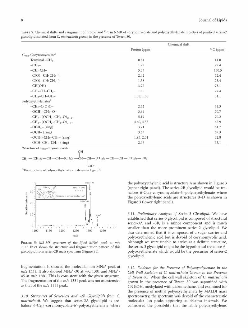
3.7. Proton and 13C-NMR Analysis of Series-2 Glycolipid. Thenovel series-2 glycolipid is composed of trehalose, C36:2-corynomycolate, and polyoxyethylenate. We used 2D-NMRexperiments to assign the proton and carbon chemical shiftsfor residues within the sample that corresponded to eachof the three parts in the structure. The results are given inTables 4 and 5. Signals from proton and carbon NMR wereused to identify diagnostic biomarker peaks for trehalose,corynomycolate and polyoxyethylenate of series-2 glycolipid[19]. NMR analysis of the trehalose moiety gave the usualvalues for the chemical shift for H-1 through H-5 and C-1 through C-5 (Table 4). Of interest to us were the valuesof the chemical shifts of the methylene protons at position6 where acylation might be expected to occur. The normalvalues for these methylene protons would be below 4.0 ppm.The observed downfield shift of these protons to 3.96 and4.76 ppm is an indication that the C-6 position is acylated.This downfield shifting of resonances due to acylation of
Table 4: Chemical shifts and assignment of proton and 13C in NMRof the trehalose moiety of purified series-2 glycolipid isolated fromC. matruchotii grown in the presence of Tween 80.
Proton Chemical shift (ppm) 13C Chemical shift (ppm)
H-1 5.05 C-1 94.3
H-2 3.64 C-2 71.9
H-3 3.88 C-3 92.9
H-4 3.31 C-4 71.7
H-5 4.21 C-5 70.0
H-6, H-6′ 3.96, 4.76 C-6 64.6
the sugar at the various positions is well established byobservations on numerous sugar derivatives [20, 21]. Theseresults are consistent with the structure of trehalose 6-C36:2-corynomycolate-6′-polyoxyethylenate where acylationoccurs at both of C-6 and C-6′ positions.
We have identified all of the protons and carbonresonances for all of the components of the corynomycolateand polyoxyethylenate in the series-2 glycolipid structure(Table 5). We identified the resonances of 1H and 13C associ-ated with the two double bonds in the corynomycolate at 5.33and 130.5 ppm, respectively. The 1H and 13C associated withthe α-alkyl carbon gave resonances of 1.58 and 25.4 ppm,respectively. The 1H and 13C associated with the β-hydroxycarbon gave the expected resonances of 3.72 and 73.1 ppm,respectively. These NMR results further confirm the presenceof corynomycolate in the series-2 glycolipid.
The proton and carbon resonances of key componentsof the proposed polyoxyethylenic acid (Figure 3) have beenidentified (Table 5). The 1H and 13C resonances associatedwith the fatty acyl α-position gave resonances of 2.34 and34.3 ppm, respectively. The 1H and 13C signals associatedwith the oxyethylenic unit gave resonances of 3.64 and70.7 ppm, respectively. The 1H and 13C signals associatedwith the furan ring structure of polyoxyethylenic acidhave been identified within the sample. These results areconsistent with the presence of polyoxyethylenic acyl groupin series-2 glycolipid.
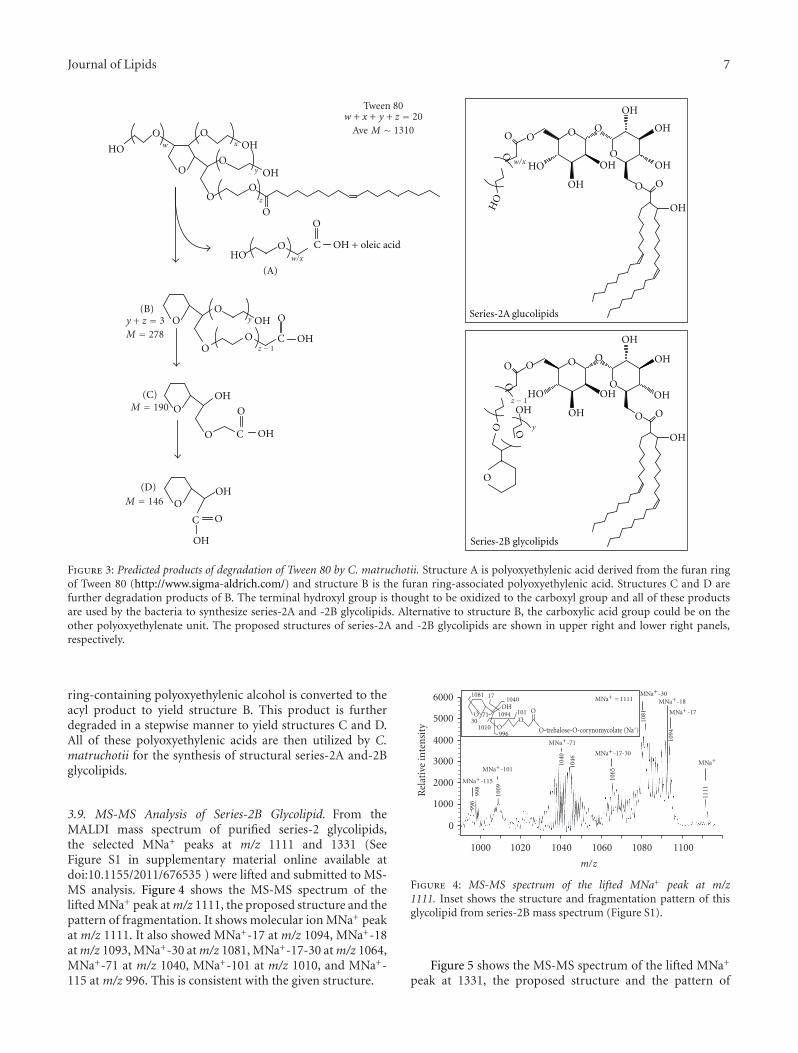
3.8. Biological Degradation of Tween 80 and the Generationof Polyoxyethylenic Acids. We suggest that C. matruchotiimetabolizes Tween 80 to form fragments containing carboxylgroup that are used for the synthesis of series-2 glycolipid. Wehave deduced the probable structure of the polyoxyethylenatein series-2 glycolipid based on (a) the known structureof Tween 80, (b) molecular weights of series-2A and -2Bglycolipids, (c) the requirement that the polyoxyethylenicacid contain a single hydroxyl group, and (d) our knowledgeof the chemical composition of the series-2 glycolipid. Theproposed structures of the polyoxyethylenic acid moiety ofseries-2 glycolipid are given in Figure 3. We suggest thattwo polyoxyethylene units are first removed from the furanring of Tween 80 and oxidized to the polyoxyethylenic acid(structure A). The oleoyl acyl group is cleaved and convertedto the C36:2-corynomycolic acid by the ortholog of Pks13condensase from M. tuberculosis [17]. The remaining furan
Journal of Lipids 7
HO OHOO
O
O
O
O
OH
O
OH + oleic acidOHO
C
O
O
O
OH
O
O
OHC
O
O OHC
OOOH
OOH
C O
OH
O
O O
OHO
OO
OH
OH
OH
OH
OH O O
OH
O
O
O O
OHO
Series-2A glucolipids
OO
OH
OH
OH
OH
OH O O
OH
Series-2B glycolipids
O
O
OH
HO
(A)
(B)
(C)
(D)
z − 1
w/x
w x
y
z
w/x
z − 1
y
y
y + z = 3M = 278
M = 190
M = 146
Tween 80w + x + y + z = 20
Ave M ∼ 1310
Figure 3: Predicted products of degradation of Tween 80 by C. matruchotii. Structure A is polyoxyethylenic acid derived from the furan ringof Tween 80 (http://www.sigma-aldrich.com/) and structure B is the furan ring-associated polyoxyethylenic acid. Structures C and D arefurther degradation products of B. The terminal hydroxyl group is thought to be oxidized to the carboxyl group and all of these productsare used by the bacteria to synthesize series-2A and -2B glycolipids. Alternative to structure B, the carboxylic acid group could be on theother polyoxyethylenate unit. The proposed structures of series-2A and -2B glycolipids are shown in upper right and lower right panels,respectively.
ring-containing polyoxyethylenic alcohol is converted to theacyl product to yield structure B. This product is furtherdegraded in a stepwise manner to yield structures C and D.All of these polyoxyethylenic acids are then utilized by C.matruchotii for the synthesis of structural series-2A and-2Bglycolipids.
3.9. MS-MS Analysis of Series-2B Glycolipid. From theMALDI mass spectrum of purified series-2 glycolipids,the selected MNa+ peaks at m/z 1111 and 1331 (SeeFigure S1 in supplementary material online available atdoi:10.1155/2011/676535 ) were lifted and submitted to MS-MS analysis. Figure 4 shows the MS-MS spectrum of thelifted MNa+ peak at m/z 1111, the proposed structure and thepattern of fragmentation. It shows molecular ion MNa+ peakat m/z 1111. It also showed MNa+-17 at m/z 1094, MNa+-18at m/z 1093, MNa+-30 at m/z 1081, MNa+-17-30 at m/z 1064,MNa+-71 at m/z 1040, MNa+-101 at m/z 1010, and MNa+-115 at m/z 996. This is consistent with the given structure.
1111
1046
1040
1009
998
996
1081
1094
1065
OOH
O
OO
1094
17
101
101030
1081
71
1040
996 O-trehalose-O-corynomycolate (Na+)
0
1000
2000
3000
4000
5000
6000
Rel
ativ
ein
ten
sity
MNa+ = 1111MNa+-30
MNa+-17-30
MNa+
MNa+-71
MNa+-101
MNa+-115
MNa+-17
MNa+-18
1000 1020 1040 1060 1080 1100
m/z
Figure 4: MS-MS spectrum of the lifted MNa+ peak at m/z1111. Inset shows the structure and fragmentation pattern of thisglycolipid from series-2B mass spectrum (Figure S1).
Figure 5 shows the MS-MS spectrum of the lifted MNa+
peak at 1331, the proposed structure and the pattern of
8 Journal of Lipids
Table 5: Chemical shifts and assignment of proton and 13C in NMR of corynomycolate and polyoxyethylenate moieties of purified series-2glycolipid isolated from C. matruchotii grown in the presence of Tween 80.
Chemical shift
Proton (ppm) 13C (ppm)
C36:2-Corynomycolatea
Terminal –CH3 0.84 14.0
–CH2– 1.28 29.4
–CH=CH– 5.33 130.5
–C(O) –CH(CH2–)– 2.42 52.4
–C(O) –CH(CH2–)– 1.58 25.4
–CH(OH) – 3.72 73.1
–CH=CH–CH2– 1.96 27.4
–CH2–CH–OH– 1.38, 1.56 34.1
Polyoxyethylenatesb
–CH2–C(O)O– 2.32 34.3
–OCH2–CH2–O– 3.64 70.7
–CH2– (OCH2–CH2–O)m−1− 5.19 70.2
–CH2– (OCH2–CH2–O)n−1− 4.60, 4.38 62.9
–OCH2– (ring) 3.71 61.7
–OCH– (ring) 3.63 69.3
–OCH2–CH2–CH2– (ring) 1.93, 2.01 32.8
–OCH–CH2–CH2– (ring) 2.06 33.1aStructure of C36:2-corynomycolate:
CH3 CH3
OH
(CH2)7 (CH2)6(CH2)7 (CH2)7CH CH CH CHCH CH
COO−bThe structures of polyoxyethylenates are shown in Figure 3.
1301
1304
1331
1286
Trehalose-O-corynomycolate (Na+)
45
1286
30
1301
O O
OO
O
O O
OH
0
10
20
30
MNa+ = 1331y + z = 9
MNa+-30
MNa+-45
MNa+
y − 1
z − 1
1100 1150 1200 1250 1300 1350
m/z
Rel
ativ
ein
ten
sity
Figure 5: MS-MS spectrum of the lifted MNa+ peak at m/z1331. Inset shows the structure and fragmentation pattern of thisglycolipid from series-2B mass spectrum (Figure S1).
fragmentation. It showed the molecular ion MNa+ peak atm/z 1331. It also showed MNa+-30 at m/z 1301 and MNa+-45 at m/z 1286. This is consistent with the given structure.The fragmentation of the m/z 1331 peak was not as extensiveas that of the m/z 1111 peak.
3.10. Structures of Series-2A and -2B Glycolipids from C.matruchotii. We suggest that series-2A glycolipid is tre-halose 6-C36:2-corynomycolate-6′-polyoxyethylenate where
the polyoxyethylenic acid is structure A as shown in Figure 3(upper right panel). The series-2B glycolipid would be tre-halose 6-C36:2-corynomycolate-6′-polyoxyethylenate wherethe polyoxyethylenic acids are structures B–D as shown inFigure 3 (lower right panel).
3.11. Preliminary Analysis of Series-3 Glycolipid. We haveestablished that series-3 glycolipid is composed of structuralseries-3A and -3B, is a minor component and is muchsmaller than the more prominent series-2 glycolipid. Wealso determined that it is composed of a sugar carrier andpolyoxyethylenic acid but is devoid of corynomycolic acid.Although we were unable to arrive at a definite structure,the series-3 glycolipid might be the hypothetical trehalose-6-polyoxyethylenate which would be the precursor of series-2glycolipid.
3.12. Evidence for the Presence of Polyoxyethylenate in theCell Wall Skeleton of C. matruchotii Grown in the Presenceof Tween 80. When the cell wall skeleton of C. matruchotiigrown in the presence of Tween 80 was saponified with2 N KOH, methylated with diazomethane, and examined forthe presence of methyl polyoxyethylenate by MALDI massspectrometry, the spectrum was devoid of the characteristicmolecular ion peaks appearing at 44 amu intervals. Weconsidered the possibility that the labile polyoxyethylenic
Journal of Lipids 9
acid did not survive this harsh basic treatment or that thisester linkage is stable to base hydrolysis. Assuming that theformer is the case, we tried a milder form of base hydrolysis.
The cell wall skeleton of C. matruchotii grown in thepresence and absence of Tween 80 were hydrolyzed in15% TBAH and the free fatty acids were esterified with 1-iododecane to yield a more hydrophobic n-decanoyl ratherthan a methyl ester. These esters were purified on a silica gelcolumn to obtain three pooled fractions: (a) nonpolar, (b)intermediate polarity, and (c) high polarity fractions. Thesesix samples were analyzed by MALDI mass spectrometry.
MALDI spectrum of the nonpolar decanoyl esters fromthe cell wall skeleton of C. matruchotii grown in thepresence of Tween 80 showed MNa+ molecular ion peaksfor decanoyl corynomycolates. However, the molecular ionpeaks at intervals of 44 amu which is characteristic of poly-oxyethylenates were absent in all nonpolar and intermediatepolarity decanoyl ester fractions.
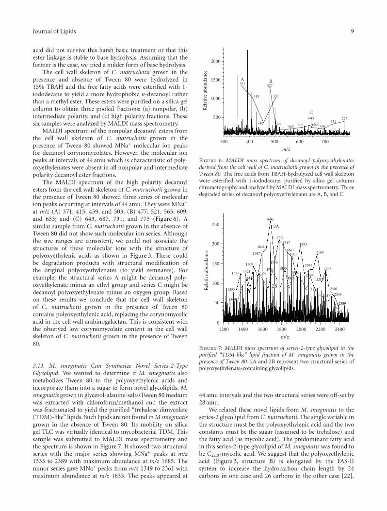
The MALDI spectrum of the high polarity decanoylesters from the cell wall skeleton of C. matruchotii grown inthe presence of Tween 80 showed three series of molecularion peaks occurring at intervals of 44 amu. They were MNa+
at m/z (A) 371, 415, 459, and 503; (B) 477, 521, 565, 609,and 653; and (C) 643, 687, 731, and 775 (Figure 6). Asimilar sample from C. matruchotii grown in the absence ofTween 80 did not show such molecular ion series. Althoughthe size ranges are consistent, we could not associate thestructures of these molecular ions with the structure ofpolyoxyethylenic acids as shown in Figure 3. These couldbe degradation products with structural modification ofthe original polyoxyethylenates (to yield remnants). Forexample, the structural series A might be decanoyl poly-oxyethylenate minus an ethyl group and series C might bedecanoyl polyoxyethylenate minus an oxygen group. Basedon these results we conclude that the cell wall skeletonof C. matruchotii grown in the presence of Tween 80contains polyoxyethylenic acid, replacing the corynomycolicacid in the cell wall arabinogalactan. This is consistent withthe observed low corynomycolate content in the cell wallskeleton of C. matruchotii grown in the presence of Tween80.
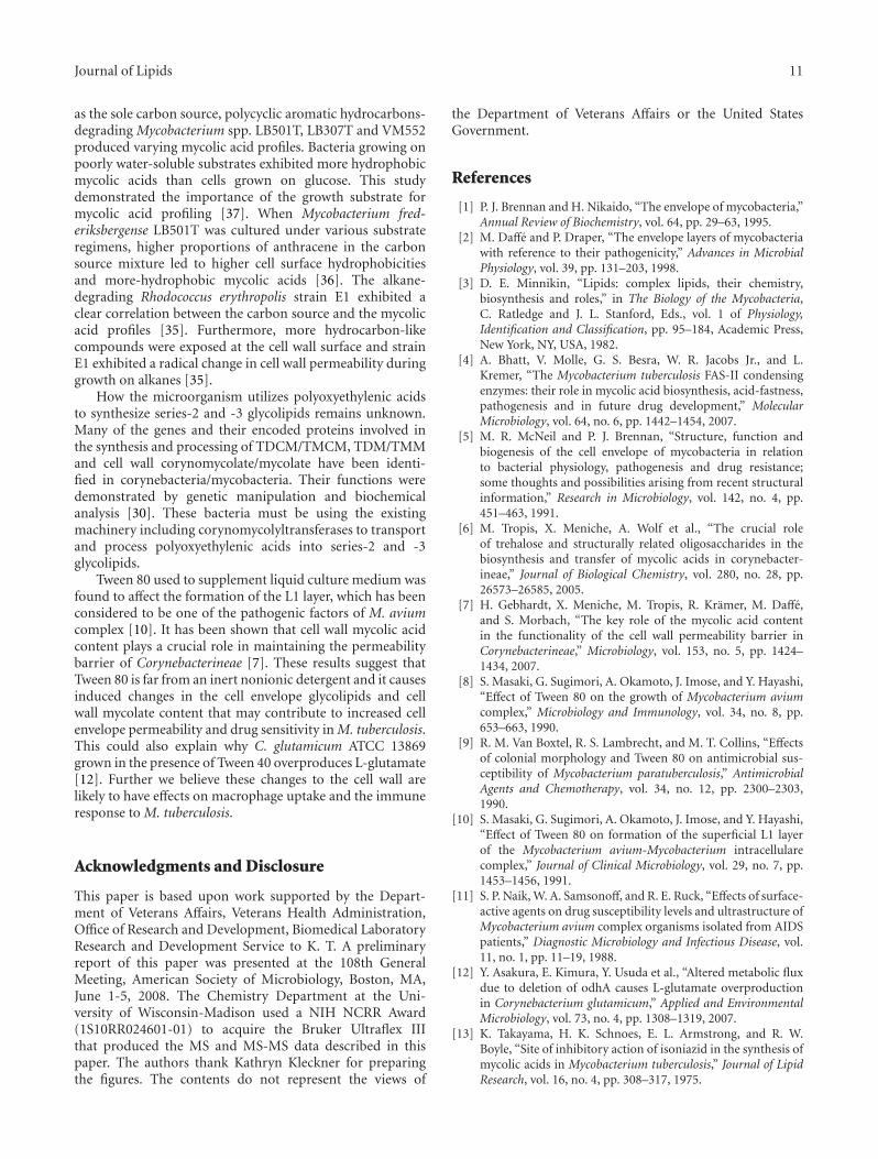
3.13. M. smegmatis Can Synthesize Novel Series-2-TypeGlycolipid. We wanted to determine if M. smegmatis alsometabolizes Tween 80 to the polyoxyethylenic acids andincorporate them into a sugar to form novel glycolipids. M.smegmatis grown in glycerol-alanine-salts/Tween 80 mediumwas extracted with chloroform/methanol and the extractwas fractionated to yield the purified “trehalose dimycolate(TDM)-like” lipids. Such lipids are not found in M smegmatisgrown in the absence of Tween 80. Its mobility on silicagel TLC was virtually identical to mycobacterial TDM. Thissample was submitted to MALDI mass spectrometry andthe spectrum is shown in Figure 7. It showed two structuralseries with the major series showing MNa+ peaks at m/z1333 to 2389 with maximum abundance at m/z 1685. Theminor series gave MNa+ peaks from m/z 1349 to 2361 withmaximum abundance at m/z 1833. The peaks appeared at
371
415
459
503
477
521
643
A B
565 609 653 687731
775
C500
1000
1500
2000
300 400 500 600 700
m/z
Rel
ativ
eab
un
dan
ce
Figure 6: MALDI mass spectrum of decanoyl polyoxyethylenatesderived from the cell wall of C. matruchotii grown in the presence ofTween 80. The free acids from TBAH-hydrolyzed cell wall skeletonwere esterified with 1-iododecane, purified by silica gel columnchromatography and analyzed by MALDI mass spectrometry. Threedegraded series of decanoyl polyoxyethylenates are A, B, and C.
1349
1333
1377 1421
1437
1509
1569
1597
1641
1701
1729
1833
1921
1949
1773
1817
18611905
1993
2081
2169
2257
20972213
2141
2317
2301
2345
23612389
2A
1685
2B
0
50
100
150
200
250
1200 1400 1600 1800 2000 2200 2400
m/z
Rel
ativ
eab
un
dan
ce
Figure 7: MALDI mass spectrum of series-2-type glycolipid in thepurified “TDM-like” lipid fraction of M. smegmatis grown in thepresence of Tween 80. 2A and 2B represent two structural series ofpolyoxyethylenate-containing glycolipids.
44 amu intervals and the two structural series were off-set by28 amu.
We related these novel lipids from M. smegmatis to theseries-2 glycolipid from C. matruchotii. The single variable inthe structure must be the polyoxyethylenic acid and the twoconstants must be the sugar (assumed to be trehalose) andthe fatty acid (as mycolic acid). The predominant fatty acidin this series-2-type glycolipid of M. smegmatis was found tobe C22:0-mycolic acid. We suggest that the polyoxyethylenicacid (Figure 3, structure B) is elongated by the FAS-IIsystem to increase the hydrocarbon chain length by 24carbons in one case and 26 carbons in the other case [22].

10 Journal of Lipids
The two modified structure B’s are the variable compo-nents of series-2-type glycolipid with y+z ranging from 3to 27. From this information the entire structural seriescan be tentatively defined as trehalose 6-C22:0-mycolate-6′-modified structure B. Although further work is neededto determine the precise structure, these results clearlydemonstrates that M. smegmatis can also metabolize Tween80 and use the degradation products for the synthesisof novel series-2-type glycolipids. Identical series-2-typeglycolipids were found in the “TDM-like” fraction fromM. tuberculosis H37Rv grown as submerged culture inMiddlebrook 7H9 medium supplemented with 0.2% glyceroland 0.25% Tween 80 (K. Takayama, C. Wang and L. Armitige,unpublished results).
4. Discussion
These novel series-2A and series-2B glycolipids from C.matruchotii grown in the presence of Tween 80 were purifiedby solvent precipitation, silica gel column chromatography,and Sephadex LH20 column chromatography. They werethen characterized by chemical analyses, MALDI massspectrometry, MS-MS, 1H-NMR, and 13C-NMR. Theseglycolipids exist in structural series differing by 44 amu.
This study showed that C. matruchotii and M. smegmatiscan absorb Tween 80, degrade it and use the degrada-tion products (polyoxyethylenic acids and oleic acid) asbuilding blocks to synthesize these novel glycolipids thatbecomes part of the cell envelope. The polyoxyethylenic acidsreplaces corynomycolic/mycolic acids in the synthesis ofthese predominant glycolipids and cell wall arabinogalactan-corynomycolate/mycolate. This is accompanied by markedreduction in TDCM and cell wall corynomycolic acidcontents in C. matruchotii. Bacteria grown in the presenceof Tween 80 are known to show gross changes in the cell’sphysical and biological properties.
The ability of these microorganisms to process Tween80 did not evolve but rather is an adaptive response usingexisting biochemical machinery. So how do these microor-ganisms process Tween 80? We suggest that in C. matruchotii,Tween 80 is first transported into a cellular compartmentcalled the periplasmic space where it is degraded to thepolyoxyethylenic acids (structures A to D, Figure 3) and oleicacid. Oleic acid is transported into the cytoplasm whereit is converted to C36:2-corynomycolic acid by Claisen-typecondensation [17, 22]. It is then transported out of the cellsand becomes trehalose 6-C36:2 corynomycolate (TMCM).The polyoxyethylenic acids in the periplasm are probablytransferred to trehalose to form analogs of TMCM, trehalose6-polyoxyethylenates. This hypothetical compound would bethe substrate for the synthesis of series-2 glycolipid. In thecase of M. smegmatis, the polyoxyethylenic acid (structure B,Figure 3) might be transported into the cytoplasm and enterthe FAS-II system for the fatty acyl elongation reactions [22].This elongated product is then transported outside the cellslike mycolic acids, forms analogs of TMM and are used tosynthesize the series-2-type glycolipid. M. tuberculosis H37Rvalso seems to have this capability.
The four probable reactions involved in the biologicaldegradation of Tween 80 are (a) cleavage of oleate esterlinkage by an esterase, (b) cleavage of the ether linkages andthe formation of primary alcohol, (c) conversion of primaryalcohol to aldehyde, and finally (d) oxidation of the aldehydeto carboxylic acid. Examples of all of these reactions in othermicroorganisms are found in the literature and a few aregiven below.
The overall Tween 80-hydrolyzing activity of M. smeg-matis was found to be strongly positive whereas that ofM. tuberculosis was variable [23]. Mycobacteria are knownto utilize oleic acid from Tween 80 as a carbon source[24]. A heat-stable Tween-hydrolyzing esterase with narrowsubstrate specificity was isolated and purified from M.smegmatis [25]. Others found that many of the rapidlygrowing mycobacteria produce esterase with Tween 80-hydrolyzing activity [26]. A Tween-hydrolyzing esterase waslocated on the surface of cell envelope of M. smegmatis andit was actively secreted into the culture medium [27]. Theseenzymes would generate oleic acid from Tween 80.
In the Gram-positive Pseudonocardia sp. strain K1, theether bond in polyethylene glycol was cleaved by a Fe-dependent superoxide dismutase exhibiting diglycolic aciddehydrogenase/diglycolic acid oxidase activity [28, 29]. Wehave searched the corynebacteria and mycobacteria genomesand identified an enzyme annotated as Mn-dependentsuperoxide dismutase/sod A, which could cleave the etherbonds in Tween 80 to generate precursors to structures A andB (Figure 3).
The oxidation of the primary hydroxyl group inpolyethylene glycol (PEG) to an aldehyde was demonstratedin Sphingopyxis terrae by a novel FAD-containing alcoholdehydrogenase encoded by pegA [30] and subsequently to thecarboxylic acid by a novel NADP-containing nicotinoproteinaldehyde dehydrogaenase encoded by pegC [31]. The acyl-CoA ligase (pegE) catalyzes the ligation of PEG-carboxylate(polyoxyethylenate) and CoA on the cytoplasmic membrane[32]. We have identified homologs of PEG dehydrogenase(pegA), PEG-aldehyde dehydrogenase (pegC), acyl-CoA lig-ase (pegE), and AraC-type transcription regulator (pegR)in corynebacteria and mycobacteria genomes by sequencehomology searches (in silico analysis).
There are examples of microorganisms utilizing varioussubstrates to synthesize novel glycolipids with somewhatsimilar structure as series-2 and -3 glycolipids. Rhodococcussp. strain MS11 grew well on a great number of n-alkanes[33]. When cultured on medium chain length n-alkanes (C10
to C17), strain MS11 produced biosurfactants lowering thesurface tension of the cultures. The two major componentsof glycolipids consisted of trehalose esterified at the C-2 orC-4 position with a succinic acid and at the C-2′ positionwith a decanoic acid. Rhodococcus opcus 1CP was foundto utilize C10 to C16n-alkanes as sole carbon sources [34].Presumably the n-alkanes were converted to mycolic acidsand incorporated into TDM.
A number of studies have shown that the structure ofthe mycolic alkyl chains is affected by the nature of thecarbon source [35–37]. When glucose, alkanes, polycyclicaromatic hydrocarbons or Luria-Bertani medium was used
Journal of Lipids 11
as the sole carbon source, polycyclic aromatic hydrocarbons-degrading Mycobacterium spp. LB501T, LB307T and VM552produced varying mycolic acid profiles. Bacteria growing onpoorly water-soluble substrates exhibited more hydrophobicmycolic acids than cells grown on glucose. This studydemonstrated the importance of the growth substrate formycolic acid profiling [37]. When Mycobacterium fred-eriksbergense LB501T was cultured under various substrateregimens, higher proportions of anthracene in the carbonsource mixture led to higher cell surface hydrophobicitiesand more-hydrophobic mycolic acids [36]. The alkane-degrading Rhodococcus erythropolis strain E1 exhibited aclear correlation between the carbon source and the mycolicacid profiles [35]. Furthermore, more hydrocarbon-likecompounds were exposed at the cell wall surface and strainE1 exhibited a radical change in cell wall permeability duringgrowth on alkanes [35].
How the microorganism utilizes polyoxyethylenic acidsto synthesize series-2 and -3 glycolipids remains unknown.Many of the genes and their encoded proteins involved inthe synthesis and processing of TDCM/TMCM, TDM/TMMand cell wall corynomycolate/mycolate have been identi-fied in corynebacteria/mycobacteria. Their functions weredemonstrated by genetic manipulation and biochemicalanalysis [30]. These bacteria must be using the existingmachinery including corynomycolyltransferases to transportand process polyoxyethylenic acids into series-2 and -3glycolipids.
Tween 80 used to supplement liquid culture medium wasfound to affect the formation of the L1 layer, which has beenconsidered to be one of the pathogenic factors of M. aviumcomplex [10]. It has been shown that cell wall mycolic acidcontent plays a crucial role in maintaining the permeabilitybarrier of Corynebacterineae [7]. These results suggest thatTween 80 is far from an inert nonionic detergent and it causesinduced changes in the cell envelope glycolipids and cellwall mycolate content that may contribute to increased cellenvelope permeability and drug sensitivity in M. tuberculosis.This could also explain why C. glutamicum ATCC 13869grown in the presence of Tween 40 overproduces L-glutamate[12]. Further we believe these changes to the cell wall arelikely to have effects on macrophage uptake and the immuneresponse to M. tuberculosis.
Acknowledgments and Disclosure
This paper is based upon work supported by the Depart-ment of Veterans Affairs, Veterans Health Administration,Office of Research and Development, Biomedical LaboratoryResearch and Development Service to K. T. A preliminaryreport of this paper was presented at the 108th GeneralMeeting, American Society of Microbiology, Boston, MA,June 1-5, 2008. The Chemistry Department at the Uni-versity of Wisconsin-Madison used a NIH NCRR Award(1S10RR024601-01) to acquire the Bruker Ultraflex IIIthat produced the MS and MS-MS data described in thispaper. The authors thank Kathryn Kleckner for preparingthe figures. The contents do not represent the views of
the Department of Veterans Affairs or the United StatesGovernment.
References
[1] P. J. Brennan and H. Nikaido, “The envelope of mycobacteria,”Annual Review of Biochemistry, vol. 64, pp. 29–63, 1995.
[2] M. Daffe and P. Draper, “The envelope layers of mycobacteriawith reference to their pathogenicity,” Advances in MicrobialPhysiology, vol. 39, pp. 131–203, 1998.
[3] D. E. Minnikin, “Lipids: complex lipids, their chemistry,biosynthesis and roles,” in The Biology of the Mycobacteria,C. Ratledge and J. L. Stanford, Eds., vol. 1 of Physiology,Identification and Classification, pp. 95–184, Academic Press,New York, NY, USA, 1982.
[4] A. Bhatt, V. Molle, G. S. Besra, W. R. Jacobs Jr., and L.Kremer, “The Mycobacterium tuberculosis FAS-II condensingenzymes: their role in mycolic acid biosynthesis, acid-fastness,pathogenesis and in future drug development,” MolecularMicrobiology, vol. 64, no. 6, pp. 1442–1454, 2007.
[5] M. R. McNeil and P. J. Brennan, “Structure, function andbiogenesis of the cell envelope of mycobacteria in relationto bacterial physiology, pathogenesis and drug resistance;some thoughts and possibilities arising from recent structuralinformation,” Research in Microbiology, vol. 142, no. 4, pp.451–463, 1991.
[6] M. Tropis, X. Meniche, A. Wolf et al., “The crucial roleof trehalose and structurally related oligosaccharides in thebiosynthesis and transfer of mycolic acids in corynebacter-ineae,” Journal of Biological Chemistry, vol. 280, no. 28, pp.26573–26585, 2005.
[7] H. Gebhardt, X. Meniche, M. Tropis, R. Kramer, M. Daffe,and S. Morbach, “The key role of the mycolic acid contentin the functionality of the cell wall permeability barrier inCorynebacterineae,” Microbiology, vol. 153, no. 5, pp. 1424–1434, 2007.
[8] S. Masaki, G. Sugimori, A. Okamoto, J. Imose, and Y. Hayashi,“Effect of Tween 80 on the growth of Mycobacterium aviumcomplex,” Microbiology and Immunology, vol. 34, no. 8, pp.653–663, 1990.
[9] R. M. Van Boxtel, R. S. Lambrecht, and M. T. Collins, “Effectsof colonial morphology and Tween 80 on antimicrobial sus-ceptibility of Mycobacterium paratuberculosis,” AntimicrobialAgents and Chemotherapy, vol. 34, no. 12, pp. 2300–2303,1990.
[10] S. Masaki, G. Sugimori, A. Okamoto, J. Imose, and Y. Hayashi,“Effect of Tween 80 on formation of the superficial L1 layerof the Mycobacterium avium-Mycobacterium intracellularecomplex,” Journal of Clinical Microbiology, vol. 29, no. 7, pp.1453–1456, 1991.
[11] S. P. Naik, W. A. Samsonoff, and R. E. Ruck, “Effects of surface-active agents on drug susceptibility levels and ultrastructure ofMycobacterium avium complex organisms isolated from AIDSpatients,” Diagnostic Microbiology and Infectious Disease, vol.11, no. 1, pp. 11–19, 1988.
[12] Y. Asakura, E. Kimura, Y. Usuda et al., “Altered metabolic fluxdue to deletion of odhA causes L-glutamate overproductionin Corynebacterium glutamicum,” Applied and EnvironmentalMicrobiology, vol. 73, no. 4, pp. 1308–1319, 2007.
[13] K. Takayama, H. K. Schnoes, E. L. Armstrong, and R. W.Boyle, “Site of inhibitory action of isoniazid in the synthesis ofmycolic acids in Mycobacterium tuberculosis,” Journal of LipidResearch, vol. 16, no. 4, pp. 308–317, 1975.

12 Journal of Lipids
[14] M. E. Hamid, D. E. Minnikin, M. Goodfellow, and M.Ridell, “Thin-layer chromatographic analysis of glycolipidsand mycolic acids from Mycobacterium farcinogenes, Mycobac-terium senegalense and related taxa,” Zentralblatt fur Bakteri-ologie, vol. 279, no. 3, pp. 354–367, 1993.
[15] K. Takayama, B. Hayes, M. M. Vestling, and R. J. Massey,“Transposon-5 mutagenesis transforms Corynebacteriummatruchotii to synthesize novel hybrid fatty acids thatfunctionally replace corynomycolic acid,” Biochemical Journal,vol. 373, no. 2, pp. 465–474, 2003.
[16] W. R. Kenealy and T. W. Jeffries, “Rapid 2,2′-bicinchoninic-based xylanase assay compatible with high throughput screen-ing,” Biotechnology Letters, vol. 25, no. 19, pp. 1619–1623,2003.
[17] D. Portevin, C. De Sousa-D’Auria, C. Houssin et al., “A polyke-tide synthase catalyzes the last condensation step of mycolicacid biosynthesis in mycobacteria and related organisms,”Proceedings of the National Academy of Sciences of the UnitedStates of America, vol. 101, no. 1, pp. 314–319, 2004.
[18] J. P. Wynn and C. Ratledge, “Evidence that the rate-limitingstep for the biosynthesis of arachidonic acid in Mortierellaalpina is at the level of the 18:3 to 20:3 elongase,” Microbiology,vol. 146, no. 9, pp. 2325–2331, 2000.
[19] E. A. Mahrous, R. B. Lee, and R. E. Lee, “A rapid approach tolipid profiling of mycobacteria using 2D HSQC NMR maps,”Journal of Lipid Research, vol. 49, no. 2, pp. 455–463, 2008.
[20] L. D. Hall, “Physical methods for structural analysis,” in TheCarbohydrates, W. Pigman and D. Horton, Eds., vol. IB, pp.1303–1306, Academic Press, New York, NY, USA, 2nd edition,1980.
[21] D. Horton, J. B. Hughes, J. S. Jewell, K. D. Philips, andW. N. Turner, “Anomeric equilibria in derivatives of aminosugars. Nuclear magnetic resonance studies on acetylatedamino sugars and specifically deuterated analogs,” Journal ofOrganic Chemistry, vol. 32, no. 4, pp. 1073–1080, 1967.
[22] K. Takayama, C. Wang, and G. S. Besra, “Pathway to synthesisand processing of mycolic acids in Mycobacterium tuberculo-sis,” Clinical Microbiology Reviews, vol. 18, no. 1, pp. 81–101,2005.
[23] P. A. Jenkins, S. R. Pattyn, and F. Portaels, “Diagnosticbacteriology,” in The Biology of the Mycobacteria, C. Ratledgeand J. Standford, Eds., vol. 1 of Physiology, Identification andClassification, pp. 441–470, Academic Press, New York, NY,USA, 1982.
[24] R. J. Dubois and G. Middlebrook, “The effect of wetting agentson the growth of tubercule bacilli,” The Journal of ExperimentalMedicine, vol. 99, pp. 81–88, 1948.
[25] H. Tomioka, “Purification and characterization of the Tween-hydrolyzing esterase of Mycobacterium smegmatis,” Journal ofBacteriology, vol. 155, no. 3, pp. 1249–1259, 1983.
[26] H. Saito, H. Tomioka, T. Watanabe, and T. Yoneyama,“Mycobacteriocins produced by rapidly growing mycobacteriaare tween-hydrolyzing esterases,” Journal of Bacteriology, vol.153, no. 3, pp. 1294–1300, 1983.
[27] C. Raynaud, G. Etienne, P. Peyron, M.-A. Laneelle, and M.Daffe, “Extracellular enzyme activities potentially involved inthe pathogenicity of Mycobacterium tuberculosis,” Microbiol-ogy, vol. 144, no. 2, pp. 577–587, 1998.
[28] M. Yamashita, A. Tani, and F. Kawai, “Cloning and expressionof an ether-bond-cleaving enzyme involved in the metabolismof polyethylene glycol,” Journal of Bioscience and Bioengineer-ing, vol. 98, no. 4, pp. 313–315, 2004.
[29] M. Yamashita, A. Tani, and F. Kawai, “A new ether bond-splitting enzyme found in Gram-positive polyethylene gly-col 6000-utilizing bacterium, Pseudonocardia sp. strain K1,”Applied Microbiology and Biotechnology, vol. 66, no. 2, pp. 174–179, 2004.
[30] M. Sugimoto, M. Tanabe, M. Hataya, S. Enokibara, J. A.Duine, and F. Kawai, “The first step in polyethylene glycoldegradation by sphingomonads proceeds via a flavopro-tein alcohol dehydrogenase containing flavin adenine dinu-cleotide,” Journal of Bacteriology, vol. 183, no. 22, pp. 6694–6698, 2001.
[31] T. Ohta, A. Tani, K. Kimbara, and F. Kawai, “A novel nicotino-protein aldehyde dehydrogenase involved in polyethyleneglycol degradation,” Applied Microbiology and Biotechnology,vol. 68, no. 5, pp. 639–646, 2005.
[32] A. Tani, P. Somyoonsap, T. Minami, K. Kimbara, and F. Kawai,“Polyethylene glycol (PEG)-carboxylate-CoA synthetase isinvolved in PEG metabolism in Sphingopyxis macrogoltabidastrain 103,” Archives of Microbiology, vol. 189, no. 4, pp. 407–410, 2008.
[33] P. Rapp and L. H. E. Gabriel-Jurgens, “Degradation ofalkanes and highly chlorinated benzenes, and productionof biosurfactants, by a psychrophilic Rhodococcus sp. andgenetic characterization of its chlorobenzene dioxygenase,”Microbiology, vol. 149, no. 10, pp. 2879–2890, 2003.
[34] S. Niescher, V. Wray, S. Lang, S. R. Kaschabek, and M.Schlomann, “Identification and structural characterisationof novel trehalose dinocardiomycolates from n-alkane-grownRhodococcus opacus 1CP,” Applied Microbiology and Biotechnol-ogy, vol. 70, no. 5, pp. 605–611, 2006.
[35] I. Sokolovska, R. Rozenberg, C. Riez, P. G. Rouxhet, S. N.Agathos, and P. Wattiau, “Carbon source-induced modifica-tions in the mycolic acid content and cell wall permeabilityof Rhodococcus erythropolis E1,” Applied and EnvironmentalMicrobiology, vol. 69, no. 12, pp. 7019–7027, 2003.
[36] L. Y. Wick, N. Pasche, S. M. Bernasconi, O. Pelz, and H.Harms, “Characterization of Multiple-Substrate Utilizationby Anthracene-Degrading Mycobacterium frederiksbergenseLB501T,” Applied and Environmental Microbiology, vol. 69, no.10, pp. 6133–6142, 2003.
[37] L. Y. Wick, P. Wattiau, and H. Harms, “Influence of thegrowth substrate on the mycolic acid profiles of mycobacteria,”Environmental Microbiology, vol. 4, no. 10, pp. 612–616, 2002.

Submit your manuscripts athttp://www.hindawi.com
Hindawi Publishing Corporationhttp://www.hindawi.com Volume 2014
Anatomy Research International
PeptidesInternational Journal of
Hindawi Publishing Corporationhttp://www.hindawi.com Volume 2014
Hindawi Publishing Corporation http://www.hindawi.com
International Journal of
Volume 2014
Zoology
Hindawi Publishing Corporationhttp://www.hindawi.com Volume 2014
Molecular Biology International
GenomicsInternational Journal of
Hindawi Publishing Corporationhttp://www.hindawi.com Volume 2014
The Scientific World JournalHindawi Publishing Corporation http://www.hindawi.com Volume 2014
Hindawi Publishing Corporationhttp://www.hindawi.com Volume 2014
BioinformaticsAdvances in
Marine BiologyJournal of
Hindawi Publishing Corporationhttp://www.hindawi.com Volume 2014
Hindawi Publishing Corporationhttp://www.hindawi.com Volume 2014
Signal TransductionJournal of
Hindawi Publishing Corporationhttp://www.hindawi.com Volume 2014
BioMed Research International
Evolutionary BiologyInternational Journal of
Hindawi Publishing Corporationhttp://www.hindawi.com Volume 2014
Hindawi Publishing Corporationhttp://www.hindawi.com Volume 2014
Biochemistry Research International
ArchaeaHindawi Publishing Corporationhttp://www.hindawi.com Volume 2014
Hindawi Publishing Corporationhttp://www.hindawi.com Volume 2014
Genetics Research International
Hindawi Publishing Corporationhttp://www.hindawi.com Volume 2014
Advances in
Virolog y
Hindawi Publishing Corporationhttp://www.hindawi.com
Nucleic AcidsJournal of
Volume 2014
Stem CellsInternational
Hindawi Publishing Corporationhttp://www.hindawi.com Volume 2014
Hindawi Publishing Corporationhttp://www.hindawi.com Volume 2014
Enzyme Research
Hindawi Publishing Corporationhttp://www.hindawi.com Volume 2014
International Journal of
Microbiology