novel genetic variants in bag3 and tnnt2 in a swedish ... · bag3 c.785c[t(p.ala262val). ... novel...
TRANSCRIPT

ORIGINAL ARTICLE
Novel Genetic Variants in BAG3 and TNNT2 in a Swedish Familywith a History of Dilated Cardiomyopathy and Sudden CardiacDeath
Eva Fernlund1,5 • A. Walinder Osterberg1 • E. Kuchinskaya2 • M. Gustafsson3 •
K. Jansson3,4 • C. Gunnarsson2,6
Received: 18 February 2017 / Accepted: 8 June 2017 / Published online: 1 July 2017
� The Author(s) 2017. This article is an open access publication
Abstract Familial dilated cardiomyopathy is a rare cause
of dilated cardiomyopathy (DCM), especially in childhood.
Our aim was to describe the clinical course and the genetic
variants in a family where the proband was a four-month-
old infant presenting with respiratory problems due to
DCM. In the family, there was a strong family history of
DCM and sudden cardiac death in four generations. DNA
was analyzed initially from the deceased girl using next-
generation sequencing including 50 genes involved in
cardiomyopathy. A cascade family screening was per-
formed in the family after identification of the TNNT2 and
the BAG3 variants in the proband. The first-degree relatives
underwent clinical examination including biochemistry
panel, cardiac ultrasound, Holter ECG, exercise stress test,
and targeted genetic testing. The index patient presented
with advanced DCM. After a severe clinical course, the
baby had external left ventricular assist as a bridge to heart
transplantation. 1.5 months after transplantation, the baby
suffered sudden cardiac death (SCD) despite maximal
treatment in the pediatric intensive care unit. The patient
was shown to carry two heterozygous genetic variants in
the TNNT2 gene [TNNT2 c.518G[A(p.Arg173Gln)] and
BAG3 [BAG3 c.785C[T(p.Ala262Val)]. Two of the
screened individuals (two females) appeared to carry both
the familial variants. All the individuals carrying the
TNNT2 variant presented with DCM, the two adult patients
had mild or moderate symptoms of heart failure and
reported palpitations but no syncope or presyncopal attacks
prior to the genetic diagnosis. The female carriers of
TNNT2 and BAG3 variants had more advanced DCM. In
the family history, there were three additional cases of
SCD due to DCM, diagnosed by autopsy, but no genetic
analysis was possible in these cases. Our findings suggest
that the variants in TNNT2 and BAG3 are associated with a
high propensity to life-threatening cardiomyopathy pre-
senting from childhood and young adulthood.
Keywords Familial DCM � DCM � SCD � BAG3 � TNNT2
Introduction
Cardiomyopathies are defined as myocardial disorders in
which the heart is structurally and functionally abnormal;
in the absence of coronary artery disease, valvular heart
disease, hypertension, or congenital heart disease sufficient
to cause the observed myocardial abnormality [1].
In pediatric cardiomyopathy registries, the incidence of
DCM have been reported to be 1/140 000–1/170 000 [2, 3],
the clinical course is often severe [2]. Dilated cardiomy-
opathy is also the most frequent underlying diagnosis
leading to pediatric heart transplantation [3].
& Eva Fernlund
1 Department of Pediatrics, Department of Clinical
Experimental Medicine, Linkoping University, Linkoping,
Sweden
2 Department of Clinical Genetics, Department of Clinical
Experimental Medicine, Linkoping University, Linkoping,
Sweden
3 Department of Cardiology, Linkoping University, Linkoping,
Sweden
4 Department of Clinical Physiology, Linkoping University,
Linkoping, Sweden
5 Pediatric Heart Center, Lund University, S-22185 Lund,
Sweden
6 Centre for Rare Diseases in South East Region of Sweden,
Linkoping University, Linkoping, Sweden
123
Pediatr Cardiol (2017) 38:1262–1268
DOI 10.1007/s00246-017-1655-0

At young ages, DCM may be caused by congenital heart
disease, coronary anomalies, arrhythmias, myocarditis, myo-
pathies, or metabolic cardiomyopathy, although some cases
remain idiopathic [2, 4, 5]. Familial dilated cardiomyopathy
(FDC) is a rare cause of DCM, especially in childhood [2].
Dilated Cardiomyopathy is characterized primarily by
left ventricular dilatation and impaired systolic function
and is one of the leading causes of heart failure with high
morbidity and mortality. Pediatric DCM is defined by the
presence of left ventricular end diastolic diameter ([2SD,
in relation to body surface area), fractional shortening less
than 25% ([2SD), and ejection fraction (EF) less than 45%
([2SD), excluding any known cause of myocardial disease
[4, 6]. The disease occurs even in pediatric cases and the
incidence among children have been shown to be higher in
infants (\1 year old) compared to patients ages 1–18 years
[7, 8] and is higher among boys than girls [9]. Familial
dilated cardiomyopathy is identified in 20–48% of cases
with DCM [4], less common in pediatric DCM [2]. If the
pedigree can reveal more than one individual with DCM
are denoted as FDC [4, 10].
The genetic spectra have involvedvariants in over 50genes
of diverse ontology, most of which encoding sarcomeric or
sarcomeric-associated proteins [11]. Most variants lead to an
autosomal dominant pattern of inheritance; however, a
minority is associated with recessive, X-linked or maternal
mitochondrial forms. Penetrance may be incomplete (the
proportion of mutation-positive individuals who show the
phenotype) and disease expression (the degree of severity
among known affected, mutation-positive individuals) is
variable. The wide spectra of the expression of the disease in
the same family can make the clinical follow-up difficult.
In the published guidelines from 2009 and 2011, genetic
evaluation is recommended in families with DCM as car-
diovascular screening of at-risk family members and con-
sideration of genetic testing in individuals with DCM [11].
Guidelines from 2011 recommended LMNA and SCN5A
genetic testing for individuals with DCM and significant
conduction system disease or premature, unexpected sud-
den cardiac death in a family [12]. However, these guide-
lines were published before some important reports that
pointed out new genes of interest for developing DCM, for
example TTN [13].
Our aim is to present a family with a history of SCD and
dilated cardiomyopathy, the presentation of heart failure in
infants and to discuss the clinical relevance of genetic testing.
Clinical Description
The index patient is a girl, born after a normal gestation.
The first months in life were happy. At three months of age,
there was onset of recurrent crying attacks especially at
night, failure to thrive, and a mild transient respiratory
infection occurring at the same time. Feeding difficulties
started at the age of 3.5 months, after some days increasing
breathing problems.
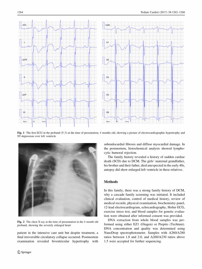
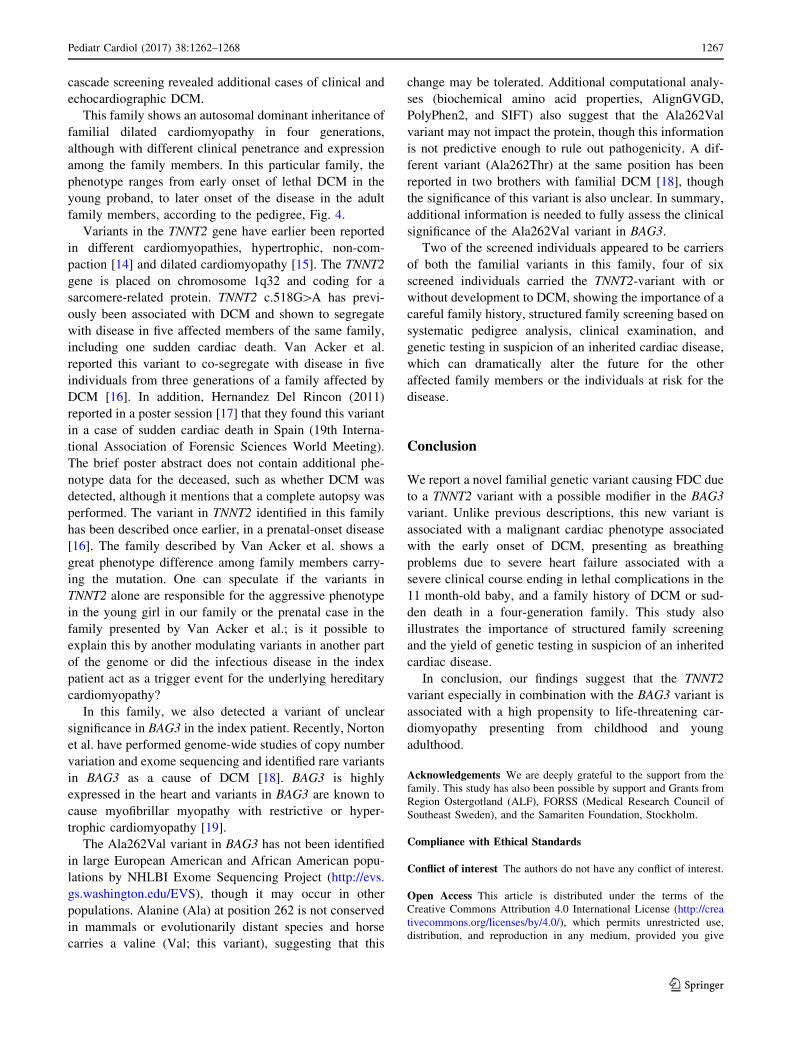
The girl was admitted to the hospital at 4 months of age
with severe breathing problems. She was pale, in prechock,
arterial oxygen saturation 70%, pH 7.14, base excess-12.
An initial treatment with furosemide and CPAP (Continu-
ous Positive Airway pressure) was successful. Standard
12-lead ECG was severely pathologic showing sinus
tachycardia, enlarged P-waves, generally enlarged ampli-
tudes, and QTc prolongation, Fig. 1. Chest X-ray revealed
a magnificent enlargement of the heart, Fig. 2. Echocar-
diography revealed a dilated cardiomyopathy and poor left
ventricular contractility, Fig. 3. The girl was transferred to
the pediatric cardiology center where additional cardiac
examinations were performed.
Echocardiography at presentation showed a severely
enlarged left ventricle, LVID 47 mm (upper limit 26 mm
relative to body surface area), fractional shortening 12%,
Fig. 3. The left atrium was enlarged, moderate mitral regur-
gitation was present, but no structural mitral valve abnor-
malities were observed. The aortic valve was found to be
bicuspid, the annulus 8 mm (z-score -1.6 SD) but no mea-
sureable stenosis in the aortic valve or aortic archwas present.
The girl underwent a cardiac catheterization that excluded
ALCAPA (anomalous left coronary artery from pulmonary
artery) as well as significant aortic stenosis. During the pro-
cedure, there was need for cardiac resuscitation due to a cir-
culatory collapse. After Levosimendan infusion and some
days of stabilization, a cardiac biopsy could be performed that
showed a picture of lymphocytic myocarditis.
The pharmacologic treatment consisted of captopril,
carvedilol, warfarin, furosemide, and spironolactone.
Because of the severe left ventricular impairment,
immunoglobulin and interferon A were also given to the
patient. Despite intensive pharmacological treatment due to
heart failure and repeated Levosimendan infusions, the
ventricular function declined over time.
There was a second circulatory collapse by the age of
6.5 months that ended in implantation of a left ventricular
assist device, that later was changed to the Berlin Heart
EXCOR� Pediatric Ventricular Assist Device (VAD). A
cardiac transplantation was performed at 9.5 months of
age. The follow-up biopsies were initially normal as well
as the left ventricular contractility assessed by echocar-
diography. One month post-transplant, the echo showed a
good systolic function but mild progressive septal hyper-
trophy, decreasing tissue Doppler velocities (TDI), but
unremarkable cardiac biopsy. 1.5 months post-transplant,
an impressive septal hypertrophy was present, further drop
in TDI and diastolic function but good systolic function
were noted. Optimal medication was administrated to the
Pediatr Cardiol (2017) 38:1262–1268 1263
123
patient in the intensive care unit but despite treatment, a
final irreversible circulatory collapse occurred. Postmortem
examination revealed biventricular hypertrophy with
subendocardial fibrosis and diffuse myocardial damage. In
the postmortem, histochemical analysis showed lympho-
cytic humoral rejection.
The family history revealed a history of sudden cardiac
death (SCD) due to DCM. The girls’ maternal grandfather,
his brother and their father, died unexpected in the early 40s,
autopsy did show enlarged left ventricle in these relatives.
Methods
In this family, there was a strong family history of DCM,
why a cascade family screening was initiated. It included
clinical evaluation, control of medical history, review of
medical records, physical examination, biochemistry panel,
12-lead electrocardiogram, echocardiography, Holter ECG,
exercise stress test, and blood samples for genetic evalua-
tion were obtained after informed consent was provided.
DNA extraction from whole blood samples was per-
formed using either EZ1 (Oiagen) or Prepito (Techtum).
DNA concentration and quality was determined using
NanoDrop spectrophotometer. Samples with A260/A280
ratios between 1.8 and 2.0, and A260/A230 ratios above
1.5 were accepted for further sequencing.
Fig. 1 The first ECG in the proband (V:3) at the time of presentation, 4 months old, showing a picture of electrocardiographic hypertrophy and
ST-depression over left ventricle
Fig. 2 The chest X-ray at the time of presentation in the 4 month old
proband, showing the severely enlarged heart
1264 Pediatr Cardiol (2017) 38:1262–1268
123

DNA was analyzed and the following genes were
included: ABCC9, ACTC1, ACTN2, ANKRD1, BAG3,
CASQ2, CAV3, CRYAB, CSRP3, CTF1, DES, DSG2, DSP,
DTNA, EMD, FHL2, GATAD1, GLA, JUP, LAMA4,
LAMP2, LDB3, LMNA, MYBPC3, MYH6, MYH7, MYL2,
MYL3, MYLK2, MYOZ2, NEBL, NEXN, PKP2, PLN,
PRKAG2, RBM20, RYR2, SCN5A, SGCD, TAZ, TCAP,
TMEM43, TMPO, TNNC1, TNNI3, TNNT2, TPM1, TTN,
TTR, and VCL. Variants were reported according to HGVS
nomenclature (www.hgvs.org/mutnomen). This test was
performed by oligonucleotide-based target capture (Sures-
elect, Agilent) followed by next-generation sequencing
(Illumina HiSeq 2000). All clinically significant and novel
variants were confirmed by independent Sanger sequencing.
Upon completion of basic clinical and genetic evalua-
tion, the affected and at-risk family members underwent a
clinical follow-up program.
Results
At early stage, a pedigree was performed in the present
family, Fig. 4, where DCM could be found in four gener-
ations in the present family. The index patient (V:3) pre-
sented with advanced DCM and severe heart failure. After a
severe clinical course with decline of cardiac function, there
was a need of external left ventricular assist as a bridge to
heart transplantation. A successful heart transplant was
performed, but 1.5 months after transplantation the baby
suffered sudden cardiac death (SCD) despite maximal
treatment in the pediatric intensive care unit. The genetic
evaluation of the index patient showed two heterozygous
genetic variants in TNNT2 c.518G[A(p.Arg173Gln) and
BAG3 c.785C[T(p.Ala262Val). These genetic variants
were shown to be inherited from the maternal family.
At the same time, as the diagnosis of the index patient,
the girls’ maternal uncle (IV:1), 35 years old at that time,
was diagnosed with DCM. Due to non-specific chest dis-
comfort, left bundle branch block on ECG, and the history
of early cardiac death in the family, the uncle underwent
echocardiography that revealed a moderately dilated left
ventricle (LVID 66 mm), mild mitral regurgitation, and
reduced ejection fraction (EF 35%). Usual workup
including radionuclide myocardial perfusion imaging
excluded ischemic heart disease as a cause of his cardiac
dysfunction. After initiation of pharmacological treatment
with beta-blockers and ACE inhibitors, the patient became
asymptomatic and NT-proBNP and Troponin T levels
returned to normal range. However, serial echocardio-
graphic exams demonstrated a slow decline in cardiac
function, ejection fraction declined to 25%, and he was
provided with a primary prophylactic CRT-D device. After
CRT-D implantation, there have been improvement of
systolic function to EF 50% and reduction of left ventric-
ular dilatation (LVID 62 mm), reduced mitral regurgitation
and left atria is normal in size at the last follow-up. There
are indirect signs of diastolic dysfunction though normal
E/evalues. In the ICD-arrhythmia recordings (CRT-D
device), there have been several short non-sustained VT
but no anti-tachycardia therapies or ICD-discharges have
been delivered to the patient.
The son of the uncle (V:1) was found to be carrier of the
TNNT2 variant at 14 years, and recently, at the age of
18 years, he has been found to have DCM-diagnosis with
moderately reduced EF why ACE-I therapy was initiated.
The mother (IV:2) of the index patient was at the time
34 years of age, complained of shortness of breath and
palpitations especially during physical activity, but she had
performed two successful pregnancies without symptoms.
She went through a thoughtful investigation and was found
to have a dilated left ventricle 60 mm and a reduced
ejection fraction 25%. There were no signs of clinical heart
failure, arrhythmia, or coronary artery disease. After initi-
ation of pharmacological treatment with beta-blockers and
Fig. 3 The echocardiogram at the time of presentation in the proband. a Apical four-chamber view showing the enlarged left ventricle. b Short-
axis view showing the enlarged left chamber
Pediatr Cardiol (2017) 38:1262–1268 1265
123

ACE inhibitors, she responded well, is asymptomatic and
her levels of NT-proBNP are normal. Her left ventricle
inner diastolic diameter (LVID) was 56 mm and EF was
45% at the last follow-up. She received a primary pro-
phylactic ICD. No anti-tachycardia therapies or ICD-dis-
charges have been delivered to this patient.
Subsequently, two of the screened individuals were found
to be carriers of the TNNT2 and BAG3 variants. The family
members with the variant in TNNT2 all showed echocar-
diographic DCM, while the family members with TNNT2
andBAG3 variants were found to havemore advancedDCM.
The index patient suffered fatal DCM and the adult case had
moderate reduction of left ventricular systolic function.
During follow-up, the adult case has shown decline of left
ventricular function measured by ejection fraction, and has
received a primary prophylactic ICD. The two cases with
sole TNNT2 variant have got the diagnosis of DCM at young
age and one of them has received a CRT-D device. In the
family history, there were three additional cases of SCD due
to DCM (III:2, III:3 and II:1), unfortunately, no genetic test
could be performed in these historic cases.
Discussion
Recurrent breathing problems in an infant along with failure
to thrive should always lead to suspicion of an underlying
heart disease. As clinical-physical examination does not
always give correct clues to distinguishing breathing
problems due to heart disease from obstructive bronchitis,
chest X-ray is of great importance in these cases, directing
the patient to echocardiography in the first line diagnostics.
The diagnosis of DCM in an infant is crucial, as it is usually
accompanied with high morbidity and risk of mortality;
therefore, these pediatric cases of DCM most often require
admittance to pediatric cardiology center for further diag-
nostic interventions and advanced treatment.
In this actual case, the index patient suffered a severe
DCM with a complicated and fatal clinical course despite
optimal treatment. The proband was shown to carry two
heterozygous genetic variants in TNNT2 c.518G[A
(p.Arg173Gln) and BAG3 c.785C[T(p.Ala262Val), a
combination that has not been described earlier. In this
family, there was a striking family history of DCM and the
Fig. 4 The pedigree of the family. The proband V:3 developed DCM at 4 months of age, V:1 developed clinical DCM at the age of 18 years,
and IV:1 and IV:2 at the early thirties. In the older generations, SCD due to DCM in their early forties (II:1, III:2, III:3)
1266 Pediatr Cardiol (2017) 38:1262–1268
123
cascade screening revealed additional cases of clinical and
echocardiographic DCM.
This family shows an autosomal dominant inheritance of
familial dilated cardiomyopathy in four generations,
although with different clinical penetrance and expression
among the family members. In this particular family, the
phenotype ranges from early onset of lethal DCM in the
young proband, to later onset of the disease in the adult
family members, according to the pedigree, Fig. 4.
Variants in the TNNT2 gene have earlier been reported
in different cardiomyopathies, hypertrophic, non-com-
paction [14] and dilated cardiomyopathy [15]. The TNNT2
gene is placed on chromosome 1q32 and coding for a
sarcomere-related protein. TNNT2 c.518G[A has previ-
ously been associated with DCM and shown to segregate
with disease in five affected members of the same family,
including one sudden cardiac death. Van Acker et al.
reported this variant to co-segregate with disease in five
individuals from three generations of a family affected by
DCM [16]. In addition, Hernandez Del Rincon (2011)
reported in a poster session [17] that they found this variant
in a case of sudden cardiac death in Spain (19th Interna-
tional Association of Forensic Sciences World Meeting).
The brief poster abstract does not contain additional phe-
notype data for the deceased, such as whether DCM was
detected, although it mentions that a complete autopsy was
performed. The variant in TNNT2 identified in this family
has been described once earlier, in a prenatal-onset disease
[16]. The family described by Van Acker et al. shows a
great phenotype difference among family members carry-
ing the mutation. One can speculate if the variants in
TNNT2 alone are responsible for the aggressive phenotype
in the young girl in our family or the prenatal case in the
family presented by Van Acker et al.; is it possible to
explain this by another modulating variants in another part
of the genome or did the infectious disease in the index
patient act as a trigger event for the underlying hereditary
cardiomyopathy?
In this family, we also detected a variant of unclear
significance in BAG3 in the index patient. Recently, Norton
et al. have performed genome-wide studies of copy number
variation and exome sequencing and identified rare variants
in BAG3 as a cause of DCM [18]. BAG3 is highly
expressed in the heart and variants in BAG3 are known to
cause myofibrillar myopathy with restrictive or hyper-
trophic cardiomyopathy [19].
The Ala262Val variant in BAG3 has not been identified
in large European American and African American popu-
lations by NHLBI Exome Sequencing Project (http://evs.
gs.washington.edu/EVS), though it may occur in other
populations. Alanine (Ala) at position 262 is not conserved
in mammals or evolutionarily distant species and horse
carries a valine (Val; this variant), suggesting that this
change may be tolerated. Additional computational analy-
ses (biochemical amino acid properties, AlignGVGD,
PolyPhen2, and SIFT) also suggest that the Ala262Val
variant may not impact the protein, though this information
is not predictive enough to rule out pathogenicity. A dif-
ferent variant (Ala262Thr) at the same position has been
reported in two brothers with familial DCM [18], though
the significance of this variant is also unclear. In summary,
additional information is needed to fully assess the clinical
significance of the Ala262Val variant in BAG3.
Two of the screened individuals appeared to be carriers
of both the familial variants in this family, four of six
screened individuals carried the TNNT2-variant with or
without development to DCM, showing the importance of a
careful family history, structured family screening based on
systematic pedigree analysis, clinical examination, and
genetic testing in suspicion of an inherited cardiac disease,
which can dramatically alter the future for the other
affected family members or the individuals at risk for the
disease.
Conclusion
We report a novel familial genetic variant causing FDC due
to a TNNT2 variant with a possible modifier in the BAG3
variant. Unlike previous descriptions, this new variant is
associated with a malignant cardiac phenotype associated
with the early onset of DCM, presenting as breathing
problems due to severe heart failure associated with a
severe clinical course ending in lethal complications in the
11 month-old baby, and a family history of DCM or sud-
den death in a four-generation family. This study also
illustrates the importance of structured family screening
and the yield of genetic testing in suspicion of an inherited
cardiac disease.
In conclusion, our findings suggest that the TNNT2
variant especially in combination with the BAG3 variant is
associated with a high propensity to life-threatening car-
diomyopathy presenting from childhood and young
adulthood.
Acknowledgements We are deeply grateful to the support from the
family. This study has also been possible by support and Grants from
Region Ostergotland (ALF), FORSS (Medical Research Council of
Southeast Sweden), and the Samariten Foundation, Stockholm.
Compliance with Ethical Standards
Conflict of interest The authors do not have any conflict of interest.
Open Access This article is distributed under the terms of the
Creative Commons Attribution 4.0 International License (http://crea
tivecommons.org/licenses/by/4.0/), which permits unrestricted use,
distribution, and reproduction in any medium, provided you give
Pediatr Cardiol (2017) 38:1262–1268 1267
123

appropriate credit to the original author(s) and the source, provide a
link to the Creative Commons license, and indicate if changes were
made.
References
1. Elliott PM, Anastasakis A, Borger MA et al (2014) 2014 ESC
Guidelines on diagnosis and management of hypertrophic car-
diomyopathy: the Task Force for the Diagnosis and Management
of Hypertrophic Cardiomyopathy of the European Society of
Cardiology (ESC). Eur Heart J 35(39):2733–2779
2. Wilkinson JD, Landy DC, Colan SD et al (2010) The pediatric
cardiomyopathy registry and heart failure: key results from the
first 15 years. Heart Fail Clin 6(4):401–413
3. Kirk R, Dipchand AI, Rosenthal DN et al (2014) The Interna-
tional Society for Heart and Lung Transplantation Guidelines for
the management of pediatric heart failure: executive summary.
[Corrected]. J Heart Lung Transplant 33(9):888–909
4. Taylor MR, Carniel E, Mestroni L (2006) Cardiomyopathy,
familial dilated. Orphanet J Rare Dis 1:27
5. Wren C (2006) Screening children with a family history of sud-
den cardiac death. Heart 92(7):1001–1006
6. Pettersen MD, Du W, Skeens ME, Humes RA (2008) Regression
equations for calculation of z scores of cardiac structures in a
large cohort of healthy infants, children, and adolescents: an
echocardiographic study. J Am Soc Echocardiogr 21(8):922–934
7. Nugent AW, Daubeney PE, Chondros P et al (2003) The epi-
demiology of childhood cardiomyopathy in Australia. N Engl J
Med 348(17):1639–1646
8. Lipshultz SE, Sleeper LA, Towbin JA et al (2003) The incidence
of pediatric cardiomyopathy in two regions of the United States.
N Engl J Med 348:1647–1655
9. Jefferies JL, Towbin JA (2010) Dilated cardiomyopathy. Lancet
375(9716):752–762
10. Mestroni L, Rocco C, Gregori D et al (1999) Familial dilated car-
diomyopathy: evidence for genetic and phenotypic heterogeneity.
Heart Muscle Disease Study Group. J Am Coll Cardiol
34(1):181–190
11. Hershberger RE, Cowan J, Morales A, Siegfried JD (2009) Pro-
gress with genetic cardiomyopathies: screening, counseling, and
testing in dilated, hypertrophic, and arrhythmogenic right ven-
tricular dysplasia/cardiomyopathy. Circ Heart Fail 2(3):253–261
12. Ackerman MJ, Priori SG, Willems S et al (2011) HRS/EHRA
expert consensus statement on the state of genetic testing for the
channelopathies and cardiomyopathies: this document was
developed as a partnership between the Heart Rhythm Society
(HRS) and the European Heart Rhythm Association (EHRA).
Europace 13(8):1077–1109
13. Herman DS, Lam L, Taylor MR et al (2012) Truncations of titin
causing dilated cardiomyopathy. N Engl J Med 366(7):619–628
14. Luedde M, Ehlermann P, Weichenhan D et al (2010) Severe
familial left ventricular non-compaction cardiomyopathy due to a
novel troponin T (TNNT2) mutation. Cardiovasc Res
86(3):452–460
15. Jachymova M, Muravska A, Palecek T et al (2012) Genetic
variation screening of TNNT2 gene in a cohort of patients with
hypertrophic and dilated cardiomyopathy. Physiol Res
61(2):169–175
16. Van Acker H, De Sutter J, Vandekerckhove K, de Ravel TJ,
Verhaaren H, De Backer J (2010) Dilated cardiomyopathy caused
by a novel TNNT2 mutation-added value of genetic testing in the
correct identification of affected subjects. Int J Cardiol
144(2):307–309
17. Hernandez Del Rincon et al (2011) Unpublished data, Poster
Presentation, 19th International Association of Forensic Sciences
World Meeting, Spain
18. Norton N, Li D, Rieder MJ et al (2011) Genome-wide studies of
copy number variation and exome sequencing identify rare
variants in BAG3 as a cause of dilated cardiomyopathy. Am J
Hum Genet 88(3):273–282
19. Selcen D, Muntoni F, Burton BK et al (2009) Mutation in BAG3
causes severe dominant childhood muscular dystrophy. Ann
Neurol 65(1):83–89
1268 Pediatr Cardiol (2017) 38:1262–1268
123