novel anticancerous compounds from sargassum wightii: in ... · marine seaweeds are rich in novel...
TRANSCRIPT

272 © 2017 Journal of Advanced Pharmacy Education & Research | Published by SPER Publications
Introduction
Lung cancer is the major cause of cancer mortality worldwide accounting for 1.5 million deaths in 2012.[1] It constitutes 13% of the newly diagnosed cancer cases in 2015.[2] Among the major two types of lung cancer, the non-small cell lung cancer (NSCLC) contributes to 80% of lung cancer deaths, which urges the need for novel therapies in the effective treatment and management of NSCLC.[3]
ABSTRACT
Non-small cell lung cancer (NSCLC) contributes to 80% of lung cancer death. The poor survival rate is contributed by the uncontrolled proliferation, evasion of apoptosis, ubiquitous expression of cell survival genes, and resistance to anticancer therapies. This prompts the search for novel and potent drugs for the effective treatment and management of NSCLC. Marine seaweeds are rich in novel bioactives widely employed in pharma, medical, cosmetic, and food industries. For the current study, the ethyl acetate extract of Sargassum wightii is utilized to test antiproliferative efficacy against the NSCLC cell line A549. From ethyl acetate extract, two compounds, namely, n-hexadecanoic acid and l-(+)-ascorbic acid 2,6 dihexadecanoate were identified by mass spectrometry analysis. These compounds interacted with the cell survival protein PI3K which is upregulated in most of human cancers. The in silico results demonstrated that the algal compounds interacted with the target PI3K with a C score of 5. The in vitro antiproliferative activity of the ethyl acetate extract was analyzed by MTT assay. The apoptotic hallmarks including fragmentation of nuclei and DNA were observed in treated cells. The real-time polymerase chain reaction analysis of gene encoding PI3K showed the downregulation of the gene. The current results suggest that the compounds of S. wightii have antiproliferative activity and can control lung cancer through induction of apoptosis.
Keywords: Non-small cell lung cancer, Sargassum wightii, antiproliferative property, n-hexadecanoic acid, l–(+)-ascorbic acid 2, 6 dihexadecanoate
Novel anticancerous compounds from Sargassum wightii: In silico and in vitro approaches to test the antiproliferative efficacy
S. M. Fazeela Mahaboob Begum1, S. Priya1,2, Raji Sundararajan3, S. Hemalatha1
1School of Life Sciences, B.S. Abdur Rahman University, Chennai, Tamil Nadu, India, 2Xinovem, Golden Jubilee Biotech Park, Chennai, India, 3School of Engineering Technology, Purdue University, West Lafayette, IN 47907, USA
Correspondence: S. Hemalatha, School of Life Sciences, B.S. Abdur Rahman University, Chennai, Tamil Nadu, India. E-mail: [email protected]
Apoptosis or programed cell death maintains the balance between cell proliferation and cell death. Uncontrolled proliferation results in oncogenesis. In NSCLC cell lines, the deletion or inactivation of the tumor suppressor genes directly contributes to the uncontrolled proliferation and prolonged survival of cancer cells.[4] Hence, drugs that can inhibit uncontrolled proliferation and induce apoptosis may be effective in the management and treatment of cancer.
A number of FDA-approved anticancer drugs are derived from the sea, including cytarabine, eribulin mesylate, and trabectedin. This has triggered the pharmaceutical industries to focus on marine natural products, and many marine bioactives have entered into the pre-clinical and clinical trials.[5] Marine seaweeds are a rich source in novel bioactives, which are widely employed in pharma, medical, cosmetic, and food industries. The marine brown algae of genus Sargassum is reported to possess antithrombotic, antiplatelet, antiviral, and anticancer properties.[6-8] In the current study, the marine brown algae Sargassum wightii was extracted, and the phytoconstituents were analyzed for the antiproliferative efficacy against the NSCLC cell line
How to cite this article: Begum SMFM, Priya S, Sundararajan R, Hemalatha S. Novel anticancerous compounds from Sargassum wightii: In silico and in vitro approaches to test the antiproliferative efficacy. J Adv Pharm Edu Res 2017;7(3):272-277.
Source of Support: Nil, Conflict of Interest: None declared.
Access this article online
Website: www.japer.in E-ISSN: 2249-3379
This is an open access journal, and articles are distributed under the terms of the Creative Commons Attribution-NonCommercial-ShareAlike 4.0 License, which allows others to remix, tweak, and build upon the work non-commercially, as long as appropriate credit is given and the new creations are licensed under the identical terms.
Original Article

Begum, et al.: Antiproliferative activity of Sargassum wightii
273Journal of Advanced Pharmacy Education & Research | Jul-Sep 2017 | Vol 7 | Issue 3
A549. The outcomes of the study showed that the algal bioactives inhibited cancer cell proliferation through the induction of apoptosis in A549 cells. The results thus suggested that the algal bioactives can be utilized for the development of novel anticancer agents in the treatment of NSCLC.
Materials and Methods
Collection and identification of marine algae
The marine brown algae S. wightii was collected from the Mandapam coast of Tamil Nadu, India. The algae were identified and authenticated by Dr. Saravanan, Scientist, CMFRI, Mandapam, Tamil Nadu, India. The algae were washed in water to remove debris, shade dried, powdered, and were used for further studies.
Extraction and characterization of the algal extract
The dried algal powder was subjected to sequential extraction.[9] The phytoconstituents analysis was carried out in the extracts for detection of alkaloids, flavonoids, saponins, tannins, glycosides, carbohydrates, proteins, fats, and oils.[10] The total polyphenol content was estimated.[11] Gallic acid was used as a standard. The experiment was done in triplicate and the data were recorded as mean ±standard deviation (SD). Based on the phytochemical analysis, the ethyl acetate extract (SEA) was subjected to Fourier-transform infrared (FTIR) analysis in JASCO spectrometer, and the spectra in the range 400–4000 cm−1 were recorded.[12] The SEA (5 mg) was filtered in 0.45 µm filter and subjected to high-performance liquid chromatography (HPLC) analysis in Shimadzu HPLC 9A fitted with LC 20AD binary gradient pump, SPD-M20A Diode array detector, and RF-fluorescence detector. The separation was carried out in Enable C18 G column of 250 × 4.6 mm fitted with a C-18 guard column using acetonitrile and water as the mobile phase in the ratio of 70:30. The flow rate was maintained as 1 ml/min. The injection volume was 20 µl. The total running time was 25 min. The retention time (RT) of the compounds in all extract was recorded.[13] The advanced mass spectroscopy (Shimadzu GC-MS QP2010 ultra) with direct injection port was used for the mass spectrometry analysis of SEA. The sample was directly sent to mass spectra using the direct injection port (200°C), and the emerging fragment ions were collected. The ion source was maintained at 200°C and interface at 250°C. The detector voltage was maintained at 0.7kV throughout the analysis. The probable structure based on the ion fragmentation pattern was derived from the National Institute of Standards and Technologies 14 library search.
Molecular docking analysis
The structures of the compound n-hexadecanoic acid (PubchemCID:985) and l-(+)- ascorbic acid 2,6 dihexadecanoate (PubchemCID:54686917) identified in SEA by MS analysis were obtained from PubChem database (www.ncbi.nlm.nih.gov/pubchem), and the X-ray crystal structures of the target receptor (PI3K) of humans were retrieved from protein data bank (http//www.rscb.org/pdb). The compounds for docking were prepared as per the ligand preparation program, and Dock suite program of the
SYBYL- × 1.3 was utilized. The prepared ligands were docked with the target protein PI3k based on the induced fit docking protocol. The interaction of the algal compounds with the target was visualized in Pymol, a python-based visualization tool (www.Pymol.org).
In vitro cell viability assay
The human adenocarcinoma cell line (A549) procured from NCCS, Pune, India, was used to test the antiproliferative efficacy of SEA. The cells were cultured in Dulbecco’s modified Eagle’s Medium (HiMedia) supplemented with 10% fetal bovine serum (HiMedia) and 1% antibiotic cocktail (Himedia). The cells were seeded in 96-well plate (1.5 × 10⁵ cells) and treated with different concentrations of SEA (1, 500, 1000, 1500, and 2000 µg/ml) for 24 h to analyze the cell viability and determine the inhibitory concentration (IC50) by MTT assay.[14] A549 cells treated with 1500 µg/ml of SEA for 24 h were analyzed for the hallmarks of apoptosis by staining with DAPI and acridine orange and ethidium bromide (1:1) mixture under fluorescent microscope (Zeiss Axio). DNA was isolated from the SEA-treated and control cells by Trizol method (Takaraa) and separated by electrophoresis (1.5% Agarose) at 75V for 40 min (BioRad) and observed in ChemiDoc (Eppendorf). The control was represented by ethyl acetate-treated cells, and the experiment was performed in triplicate.
Analysis of PI3K by real-time polymerase chain reaction (PCR)
Total RNA was isolated from control and SEA-treated cells by trizol method, followed by cDNA synthesis using the high capacity cDNA reverse transcription kit (Applied Biosystem) as per manufacturer’s instruction. The cDNA synthesized was used for real-time PCR analysis of cell survival gene (PI3K) using SYBR Green PCR master mix (Bio-Rad). The analysis was done in triplicate, the fold change in gene expression was calculated based on 2−DDct, and the expression was normalized with the housekeeping gene β actin. The primers used are PIK3CA (F) TGCAAAGAATCAGAACAATGCC: PI3KCA (R) CACGGAGGCATTCTAAAGTCA. βActin ( F ) TAG A AG C C T C T T C AT G G AC A AC : β A c t i n ( R ) GTATCAGGCATGCAACACAAG.
Results and Discussion
The phytochemical analysis of S. wightii revealed the presence of flavonoids, tannins, glycosides, terpenoids, carbohydrates, fats, resins, and phytosterols in the ethyl acetate, ethanol and methanol extracts; however, terpenoids, saponins, carbohydrates, proteins, and resins were not detected in hexane and DCM extracts [Table 1]. The abundance of phytoconstituents in S. wightii ethyl acetate extract (SEA) may be attributed to the mid-polar nature of the solvent. In previous studies, the non-polar solvents such as benzene, chloroform, and petroleum ether were used for extracting S. wightii,[15] but extraction with ethyl acetate increased the concentration of the phytoconstituents than the non-polar solvents.
Table 1 shows the results of preliminary phytochemical analysis of S. wightii by sequential extraction method. The phytoconstituents were found in different concentration in each extract.
Begum, et al.: Antiproliferative activity of Sargassum wightii
274 Journal of Advanced Pharmacy Education & Research | Jul-Sep 2017 | Vol 7 | Issue 3
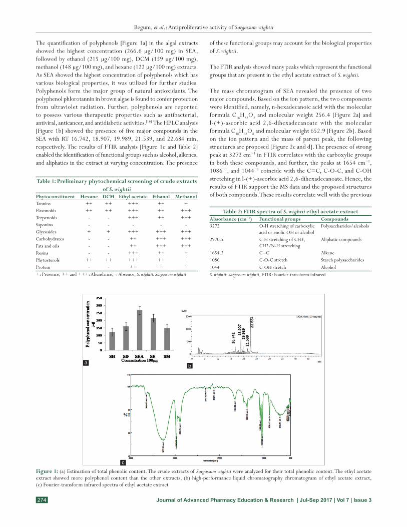
The quantification of polyphenols [Figure 1a] in the algal extracts showed the highest concentration (266.6 µg/100 mg) in SEA, followed by ethanol (215 µg/100 mg), DCM (159 µg/100 mg), methanol (148 µg/100 mg), and hexane (122 µg/100 mg) extracts. As SEA showed the highest concentration of polyphenols which has various biological properties, it was utilized for further studies. Polyphenols form the major group of natural antioxidants. The polyphenol phlorotannin in brown algae is found to confer protection from ultraviolet radiation. Further, polyphenols are reported to possess various therapeutic properties such as antibacterial, antiviral, anticancer, and antidiabetic activities.[16] The HPLC analysis [Figure 1b] showed the presence of five major compounds in the SEA with RT 16.742, 18.907, 19.989, 21.539, and 22.684 min, respectively. The results of FTIR analysis [Figure 1c and Table 2] enabled the identification of functional groups such as alcohol, alkenes, and aliphatics in the extract at varying concentration. The presence
of these functional groups may account for the biological properties of S. wightii.
The FTIR analysis showed many peaks which represent the functional groups that are present in the ethyl acetate extract of S. wightii.
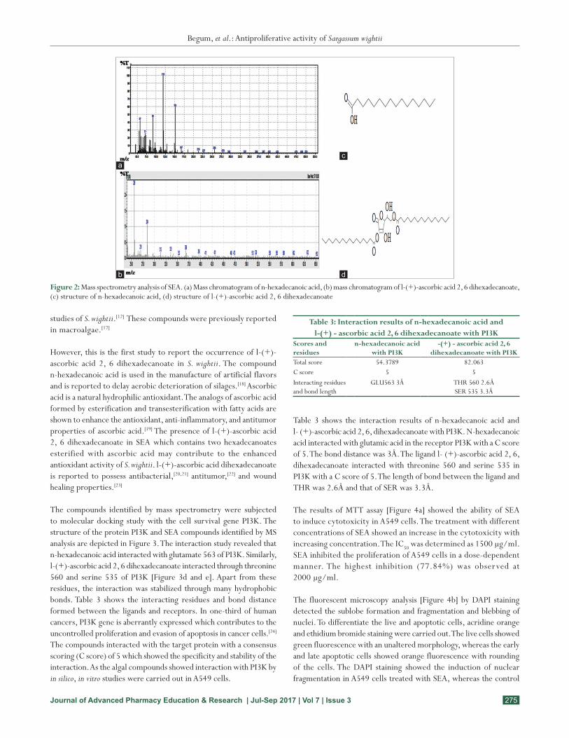
The mass chromatogram of SEA revealed the presence of two major compounds. Based on the ion pattern, the two components were identified, namely, n-hexadecanoic acid with the molecular formula C16H32O2 and molecular weight 256.4 [Figure 2a] and l-(+)-ascorbic acid 2,6-dihexadecanoate with the molecular formula C38H68O8 and molecular weight 652.9 [Figure 2b]. Based on the ion pattern and the mass of parent peak, the following structures are proposed [Figure 2c and d]. The presence of strong peak at 3272 cm−1 in FTIR correlates with the carboxylic groups in both these compounds, and further, the peaks at 1654 cm−1, 1086−1, and 1044−1 coincide with the C=C, C-O-C, and C-OH stretching in l-(+)-ascorbic acid 2,6-dihexadecanoate. Hence, the results of FTIR support the MS data and the proposed structures of both compounds. These results correlate well with the previous
Table 1: Preliminary phytochemical screening of crude extracts of S. wightii
Phytoconstituent Hexane DCM Ethyl acetate Ethanol MethanolTannins ++ ++ +++ ++ +Flavonoids ++ ++ +++ ++ +++Terpenoids - - +++ ++ +++Saponins - - - - -Glycosides + + +++ +++ +++Carbohydrates - - ++ +++ +++Fats and oils - - ++ +++ +++Resins - - +++ ++ +Phytosterols ++ ++ +++ ++ +Protein - - ++ + ++: Presence, ++ and +++: Abundance, -: Absence, S. wightii: Sargassum wightii
Table 2: FTIR spectra of S. wightii ethyl acetate extractAbsorbance (cm−1) Functional groups Compounds3272 O-H stretching of carboxylic
acid or enolic OH or alcoholPolysaccharides/alcohols
2970.5 C-H stretching of CH3, CH2/N-H stretching
Aliphatic compounds
1654.2 C=C Alkene1086 C-O-C stretch Starch polysaccharides1044 C-OH stretch AlcoholS. wightii: Sargassum wightii, FTIR: Fourier-transform infrared
Figure 1: (a) Estimation of total phenolic content. The crude extracts of Sargassum wightii were analyzed for their total phenolic content. The ethyl acetate extract showed more polyphenol content than the other extracts, (b) high-performance liquid chromatography chromatogram of ethyl acetate extract, (c) Fourier-transform infrared spectra of ethyl acetate extract
c
ba
Begum, et al.: Antiproliferative activity of Sargassum wightii
275Journal of Advanced Pharmacy Education & Research | Jul-Sep 2017 | Vol 7 | Issue 3
studies of S. wightii.[12] These compounds were previously reported in macroalgae.[17]
However, this is the first study to report the occurrence of l-(+)-ascorbic acid 2, 6 dihexadecanoate in S. wightii. The compound n-hexadecanoic acid is used in the manufacture of artificial flavors and is reported to delay aerobic deterioration of silages.[18] Ascorbic acid is a natural hydrophilic antioxidant. The analogs of ascorbic acid formed by esterification and transesterification with fatty acids are shown to enhance the antioxidant, anti-inflammatory, and antitumor properties of ascorbic acid.[19] The presence of l-(+)-ascorbic acid 2, 6 dihexadecanoate in SEA which contains two hexadecanoates esterified with ascorbic acid may contribute to the enhanced antioxidant activity of S. wightii. l-(+)-ascorbic acid dihexadecanoate is reported to possess antibacterial,[20,21] antitumor,[22] and wound healing properties.[23]
The compounds identified by mass spectrometry were subjected to molecular docking study with the cell survival gene PI3K. The structure of the protein PI3K and SEA compounds identified by MS analysis are depicted in Figure 3. The interaction study revealed that n-hexadecanoic acid interacted with glutamate 563 of PI3K. Similarly, l-(+)-ascorbic acid 2, 6 dihexadecanoate interacted through threonine 560 and serine 535 of PI3K [Figure 3d and e]. Apart from these residues, the interaction was stabilized through many hydrophobic bonds. Table 3 shows the interacting residues and bond distance formed between the ligands and receptors. In one-third of human cancers, PI3K gene is aberrantly expressed which contributes to the uncontrolled proliferation and evasion of apoptosis in cancer cells.[24] The compounds interacted with the target protein with a consensus scoring (C score) of 5 which showed the specificity and stability of the interaction. As the algal compounds showed interaction with PI3K by in silico, in vitro studies were carried out in A549 cells.
Table 3 shows the interaction results of n-hexadecanoic acid and l- (+)-ascorbic acid 2, 6, dihexadecanoate with PI3K. N-hexadecanoic acid interacted with glutamic acid in the receptor PI3K with a C score of 5. The bond distance was 3Å. The ligand l- (+)-ascorbic acid 2, 6, dihexadecanoate interacted with threonine 560 and serine 535 in PI3K with a C score of 5. The length of bond between the ligand and THR was 2.6Å and that of SER was 3.3Å.
The results of MTT assay [Figure 4a] showed the ability of SEA to induce cytotoxicity in A549 cells. The treatment with different concentrations of SEA showed an increase in the cytotoxicity with increasing concentration. The IC50 was determined as 1500 µg/ml. SEA inhibited the proliferation of A549 cells in a dose-dependent manner. The highest inhibition (77.84%) was observed at 2000 µg/ml.
The fluorescent microscopy analysis [Figure 4b] by DAPI staining detected the sublobe formation and fragmentation and blebbing of nuclei. To differentiate the live and apoptotic cells, acridine orange and ethidium bromide staining were carried out. The live cells showed green fluorescence with an unaltered morphology, whereas the early and late apoptotic cells showed orange fluorescence with rounding of the cells. The DAPI staining showed the induction of nuclear fragmentation in A549 cells treated with SEA, whereas the control
Figure 2: Mass spectrometry analysis of SEA. (a) Mass chromatogram of n-hexadecanoic acid, (b) mass chromatogram of l-(+)-ascorbic acid 2, 6 dihexadecanoate, (c) structure of n-hexadecanoic acid, (d) structure of l-(+)-ascorbic acid 2, 6 dihexadecanoate
d
c
b
a
Table 3: Interaction results of n‑hexadecanoic acid and l‑(+) ‑ ascorbic acid 2, 6 dihexadecanoate with PI3K
Scores and residues
n‑hexadecanoic acid with PI3K
‑(+) ‑ ascorbic acid 2, 6 dihexadecanoate with PI3K
Total score 54.3789 82.063C score 5 5Interacting residues and bond length
GLU563 3Å THR 560 2.6ÅSER 535 3.3Å

Begum, et al.: Antiproliferative activity of Sargassum wightii
276 Journal of Advanced Pharmacy Education & Research | Jul-Sep 2017 | Vol 7 | Issue 3
cells showed intact nuclei. The results of agarose electrophoresis [Figure 4c] showed the fragmentation of DNA isolated from SEA-treated cells, whereas the control cells showed intact DNA. The real-time expression of gene encoding PI3K [Figure 4d] showed the
downregulation in SEA-treated cells. The electrophoretic separation of the real-time product showed the absence of DNA band in the treated group [Figure 4e] compared to the control. The antiproliferative activity of SEA may be due to the presence of the newly identified
Figure 3: Molecular Docking analysis. (a) Structure of PI3K, (b) structure of n-hexadecanoic acid, (c) structure of l-(+)-ascorbic acid 2, 6 dihexadecanoate, (d) interaction of PI3K with n-hexanoic acid, (e) interaction of PI3K with l-(+)-ascorbic acid 2, 6 dihexadecanoate. The interaction studies were done using SYBL X 1.3
d
cba
e
Figure 4: (a) Cytotoxic activity of SEA against A549 cells. The A549 cells were incubated with SEA at varying concentrations (1, 500, 1000, 1500, and 2000 µg/ml) for 24 h and the cytotoxicity was assessed by MTT assay. The IC50 was found as 1500 µg/ml. Control cells were treated with ethyl acetate. The data presented are mean±SD, (b) analysis of apoptosis by fluorescent microscopy. DAPI staining of control (i) and treated (ii) showing fragmented and lobbed nuclei. Acridine orange and ethidium bromide staining of control (iii) and treated (iv) showing live and apoptotic cells. The experiment was done in triplicate, (c) agarose gel electrophoresis showing intact DNA (lane 1) and fragmented DNA (lane 2), (d) fold change in PI3K gene expression in treated A549 cells, (e) agarose gel electrophoresis showing real-time polymerase chain reaction products
dc
ba
e
Begum, et al.: Antiproliferative activity of Sargassum wightii
277Journal of Advanced Pharmacy Education & Research | Jul-Sep 2017 | Vol 7 | Issue 3
algal compounds l-(+)-ascorbic acid 2, 6 dihexadecanoate and n-hexadecanoate.
Conclusion
The inhibition of proliferation in cancer cells is an essential criterion for an anticancer drug. In the current study, SEA inhibited cell proliferation through the induction of apoptosis. This suggested that the algal bioactives possess anticancer activity which can be further studied for the development of effective anticancer drug in the management of NSCLC. The presence of polyphenols, flavonoids, and phytosterols and l-(+)-ascorbic acid dihexadecanoate in SEA may contribute to its antiproliferative nature. The identification of nuclear fragmentation, blebbing, and late apoptotic cells in SEA-treated cells by fluorescent microscopy confirmed the ability of SEA compounds to induce apoptosis. The real-time analysis of expression of gene PI3K showed that the inhibition of cell proliferation is mediated by the induction of apoptosis, through suppression of PI3K gene expression in the treated cells. The results of the in vitro study coincide with the in silico interaction study where the algal compounds showed good interaction with PI3K protein. As the outcomes of the current study highlight the downregulation of cell survival gene, the compounds in SEA may contribute to this effect. As marine natural products are gaining importance in the field of anticancer research, this study opens up a new avenue about marine algae which can be utilized effectively in the field of biomedical research.
References
1. Kumar R, Lu SK, Minchom A, Sharp A, Davidson M, Gunapala R, et al. A phase 1b trial of the combination of an all-oral regimen of capecitabine and erlotinib in advanced non-small cell lung cancer in Caucasian patients. Cancer Chemother Pharmacol 2016;77:375-83.
2. World Health Organization Cancer Mortality Database; 2015. Available from: http://www.Who.int/mediacenter/factsheets/fs/297/len. [last accessed on 2016 Oct 20].
3. American Cancer Society. Available from : http://www.cancer.org. [last accessed on 2016 Oct 22].
4. Perez-Ramirez C, Cañadas-Garre M, Jiménez-Varo E, Faus-Dáder MA, Calleja-Hernández MA. PTEN and PI3K/AKT in non-small-cell lung cancer. Pharmacogenomics J 2015;16:631-47.
5. Marine Pharmacology. Available from: http://www.marinepharmacology.midwestern.edu/clinPipeline.htm. [last accessed on 2016 Oct 22].
6. Jin W, Zhang W, Wang J, Yao J, Xie E, Liu D, et al. A study of neuroprotective and antioxidant activities of heteropolysaccharides from six Sargassum species. Int J Biol Macromol 2014;67:336-42.
7. Pathmanathan MK, Uthayarasa K, Jeyadevan JP, Jeyaseelan EC. In vitro antibacterial activity and phytochemical analysis of some selected medicinal plants. Int J Pharm Biol Arch 2010;1:291-9.
8. Harborne JB. Phytochemical methods. A Guide to Modern Techniques of Plant Analysis. 3rd ed. New York: Chapman and Hall; 1998. p. 1-150.
9. Singleton VL, Rossi JA. Colorimetry of total phenolics with phosphomolybdic-phosphotungstic acid reagents. Am J Enol Viticult 1965;16:144-58.
10. Kannan S. FT-IR and EDS analysis of the seaweeds Sargassum wightii (brown algae) and Gracilaria corticata (red algae). Int J Curr Microbiol App Sci 2014;3:341-51.
11. Gurav N, Kardani K, Solanki B, Patel B. Quantification of phenolic compound gallic acid in polyherbal ranger capsule by high performance chromatographic method. Int J Pharm Sci Res 2013;25:183-7.
12. Mosmann T. Rapid colorimetric assay for cellular growth and survival: Application to proliferation and cytotoxicity assays. J Immunol Methods 1983;65:55-63.
13. Marimuthu J, Essakimuthu P, Narayanan J, Anantham B, Tharmaraj RJ, Arumugam S. Phytochemical characterization of brown seaweed Sargassum wightii. Asian Pac J Trop Dis 2012;2:S109-13.
14. Machu L, Misurcova L, Ambrozova JV, Orsavova J, Mlcek J, Sochor J, et al. Phenolic content and antioxidant capacity in algal food products. Molecules 2015;20:1118-33.
15. Kumar P, Senthamilselvi S, Govindaraju M. GC-MS profiling and antibacterial activity of Sargassum tenerrimum. J Pharm Res 2012;6:88-92.
16. Al- saif S,Abdel Raouf N, El-Wazanani H and Aref I. Antibacterial substances from marine algae isolated from Jeddah coast of Red sea, Saudi Arabia.Saudi J BioSci 2014;21(1):57-64.
17. Kumar P, Selvi SS, Prabha AL, Kumar KP, Kumar RSG, Govindaraju M. Synthesis of silvernanoparticles from Sargassum tenerrimum and screening the phytochemicals for its antibacterial activity. Nano Biomed Eng 2012;4(1):12- 16.
18. Ohyama Y, Hara SI, Masaki S. The use of caproic acid to prevent aerobic deterioration of silages after opening, with special reference to the amounts and time of application. J Sci Food Agric 1977;28:369-74.
19. Mohamed R, Dharmappa KK, Tarannum S, Jameel NM, Kannum SA, Ashrafulla HS, et al. Chemical modification of ascorbic acid and evaluation of its lipophilic derivatives as inhibitors of secretory phospholipase A(2) with anti-inflammatory activity. Mol Cell Biochem 2010;345:69-76.
20. Arun K, Shrabani P, Pingalkumari S, Thirugnanasambandan S, Kathiresan K. Antibacterial and phytochemical assessment on various extracts of Ipomoea pes-caprae (l.) r. br through FTIR and GC-MS spectroscopic analysis. Asian J Pharm Clin Res 2104;7:134-8.
21. Soad M, El-Din M, El-Ahwany AM. Bioactivity and phytochemical constituents of marine red seaweeds (Jania rubens, Corallina mediterranea and Pterocladia capillacea). J Taibah Univ Sci 2016;10:471-84.
22. Botzki A, Rigden DJ, Braun S, Nukui M, Salmen S, Hoechstetter J, et al. L-Ascorbic acid 6-hexadecanoate, a potent hyaluronidase inhibitor. X-ray structure and molecular modeling of enzyme-inhibitor complexes. J Biol Chem 2004;279:45990-7.
23. Akinmoladun AC, Ibukun EO, Afor E, Akinsinlola BL, Onibon TR, Akinboboye AO, et al. Chemical constituents and antioxidant activity of Boonei Alstonia. Afr J Biotechnol 2007;6:1197.
24. Sun ZJ, Chen G, Hu X, Zhang W, Liu Y, Zhu LX, et al. Activation of PI3K/Akt/IKK-alpha/NF-kappaB signaling pathway is required for the apoptosis-evasion in human salivary adenoid cystic carcinoma: Its inhibition by quercetin. Apoptosis 2010;15:850-63.