note to users - core.ac.uk · lipoproteine de tres basse densite 1 "xenon x coefficient de...
TRANSCRIPT

NOTE TO USERS
This reproduction is the best copy available.
UMI


Universite de Sherbrooke
MESURE DU FLOT SANGUIN DANS LE TISSU ADIPEUX : IMPLANTATION ET MISE AU
POINT DE LA METHODE A SHERBROOKE
par Elizabeth Martin
Departement de physiologie et biophysique, Service d'Endocrinologie
Memoire presente a la Faculte de medecine et des sciences de la sante En vue de l'obtention du grade de
Maitre es sciences (M.Sc.)
Juin 2009
Evaluateurs Jean-Luc Ardilouze, MD, PhD. Programme de physiologie
Emanuel Escher, PhD. Programme de pharmacologic Eric Rousseau, PhD. Programme de physiologie

1*1 Library and Archives Canada
Published Heritage Branch
395 Wellington Street Ottawa ON K1A 0N4 Canada
Bibliotheque et Archives Canada
Direction du Patrimoine de I'edition
395, rue Wellington Ottawa ON K1A 0N4 Canada
Your file Votre reference ISBN: 978-0-494-61424-2 Our file Notre reference ISBN: 978-0-494-61424-2
NOTICE: AVIS:
The author has granted a nonexclusive license allowing Library and Archives Canada to reproduce, publish, archive, preserve, conserve, communicate to the public by telecommunication or on the Internet, loan, distribute and sell theses worldwide, for commercial or noncommercial purposes, in microform, paper, electronic and/or any other formats.
L'auteur a accorde une licence non exclusive permettant a la Bibliotheque et Archives Canada de reproduire, publier, archiver, sauvegarder, conserver, transmettre au public par telecommunication ou par I'lnternet, preter, distribuer et vendre des theses partout dans le monde, a des fins commerciales ou autres, sur support microforme, papier, electronique et/ou autres formats.
The author retains copyright ownership and moral rights in this thesis. Neither the thesis nor substantial extracts from it may be printed or otherwise reproduced without the author's permission.
L'auteur conserve la propriete du droit d'auteur et des droits moraux qui protege cette these. Ni la these ni des extraits substantiels de celle-ci ne doivent etre imprimes ou autrement reproduits sans son autorisation.
In compliance with the Canadian Privacy Act some supporting forms may have been removed from this thesis.
Conformement a la loi canadienne sur la protection de la vie privee, quelques formulaires secondaires ont ete enleves de cette these.
While these forms may be included in the document page count, their removal does not represent any loss of content from the thesis.
Bien que ces formulaires aient inclus dans la pagination, tl n'y aura aucun contenu manquant.
1+1
Canada

RESUME
Le flot sanguin est un facteur determinant de l'activite metabolique du tissu adipeux. En
effet, tout echange metabolique ou hormonal depend obligatoirement du produit du flot
sanguin et de la difference arterioveineuse du metabolite etudie. Le stockage et la liberation
energetique de meme que la secretion d'hormones et d'adipocytokines necessitent une
regulation precise du flot sanguin dans le tissu adipeux (FSTA).
Ce memoire comportera essentiellement trois parties.
Dans la premiere, nous ferons la recension des ecrits qui concernent la regulation du FSTA.
Nous avons ecrit une revue sur le sujet. Nous decrirons le role de Pinsuline, des hormones
gastro-intestinales, du systeme nerveux sympathique et parasympathique, de 1'endotheline et
de diverses cytokines. Nous montrerons aussi que l'absence d'elevation du FSTA en
condition postprandiale est une caracteristique de l'insulino-resistance et de l'obesite. Cette
caracteristique justifie 1'exploration de la physiologie vasculaire du tissu adipeux pour une
meilleure comprehension des mecanismes patho-physiologiques qui sous-tendent ces deux
pathologies.
Dans la deuxieme partie, nous discuterons des diverses techniques de mesure du FSTA chez
Phumain, la mesure de clairance du 133Xe, la microdialyse ou d'autres methodes plus
dispendieuses ou non validees. Nous decrirons ensuite une nouvelle methode, la
microinfusion, qui permet de mesurer le FSTA avec la technique de clairance du 133Xe et de
le faire varier simultanement en infusant dans le site radioactif, de facon continue, des agents
vasoactifs. Un detecteur place au-dessus de la source permet l'enregistrement de la clairance

du 133Xe du tissu adipeux vers la circulation sanguine en relation avec les changements
environnementaux induits. Le tissu adipeux sous-cutane abdominal a ete choisi comme site
anatomique d'investigation. Cette methode permet la comparaison directe entre des
composes vasoactifs, agents pharmacologiques ou hormones, et un controle contralateral
situe a la meme hauteur sur l'abdomen. La description precise de cette methode fait Pobjet
d'un deuxieme article. Nous y montrons aussi que la microinfusion ameliore la precision et
la faisabilite des etudes physiologiques et pharmacologiques in vivo chez l'humain et qu'elle
a deja permis 1'evaluation de plusieurs acteurs potentiels impliques dans la regulation du
FSTA chez des sujets sains.
Enfin, dans la troisieme partie nous ferons le point de notre experience de maitrise, des
difficultes et des deboires que nous avons eus dans la mise au point de la technique a
Sherbrooke.
Nous conclurons en montrant que la microinfusion va permettre d'ameliorer notre
comprehension des mecanismes sous-jacents au syndrome d'insulino-resistance, au
developpement du diabete de type 2 et des maladies cardiovasculaires. Son implantation et sa
mise au point a Sherbrooke sont done amplement justifiees.
Nombre de mots : 416
Mots cles : activite metabolique, flot sanguin, microinfusion, tissu adipeux, traceur
radioactif.

TABLE DES MATIERES
LISTE DES ILLUSTRATIONS
LISTES DES SIGLES, ABREVIATIONS ET SYMBOLES
RESUME
CHAPITRE 1
INTRODUCTION
Anatomie du tissu adipeux Histologic Vascularisation Innervation
Implication du systeme nerveux sympathique Implication du systeme nerveux parasympathique
Le TA comme site de stockage energetique Source plasmatique de TAG La lipogenese de novo
Le TA comme tissu fournisseur d'energie
Le TA et la steroidogenese
Le TA comme organe endocrine
Activite metabolique du tissue adipeux: importance du flot sang
CHAPITRE 2
ARTICLES SCIENTIFIQUES INTEGRES
Article 1) Update on adipose tissue blood flow regulation Avant-propos de 1'article Resume Article
Article 2) A new technique for investigating the metabolism and subcutaneous adipose tissue
Avant-propos de Particle Resume Article

CHAPITRE 3
ENONCE DES OBJECTIFS DU PROJET DE MAITRISE
CHAPITRE 4
RESULTATS ET DISCUSSION
Resultats initiaux
Objectif revise
Parametres du Mediscint System
Calibration du Mediscint System
Experiences acquises
Preuves a 1'appui
CHAPITRE 5
CONCLUSION ET PERSPECTIVES
REMERCIEMENTS
LISTE DES REFERENCES
ANNEXE

LISTE DES ILLUSTRATIONS
in
FIGURES
CHAPITRE 1
1.1 Tissu adipeux uniloculaire 2
1.2 Adipocytes 3
1.3 Metabolisme du tissu adipeux 13
CHAPITRE 4
4.1 Comparaison des Mediscint System Dr Carey vs Dr Ardilouze. Mesure de
variations depseudo flotpar intervalles de 10 min 107
4.2 Stimulation du FSTA par microinfusion d'isoproterenol 1 x 10"4 M chez 6 sujets.
Mesures effectuees avec notre Mediscint System 109
4.3 Deroulement des tests de validite 114
4.4 Mesure du FSTSA - test de validite, sujet 1 116
4.5 Mesure du FSTA - test de validite, sujet 2 117

LISTES DES SIGLES, ABREVIATIONS ET SYMBOLES
ACTH
AGL
AMPc
Angll
apoB-48
apoB-100
apoC-II
ASP
ATP
CsI(Tl)
CV
Dil
ET
FIAF
FSTA
GLP-1
HSL
IL-6
ISO
LPL
NO
PAI-1
Hormone corticotrope (Adreno cortico tropic hormone)
Acide gras libre
Adenosine monophosphate cyclique
Angiotensine II
Apolipoproteine B-48
Apolipoproteine B-100
Apolipoproteine C-II
Acylation stimulating protein
Adenosine triphosphate
Cesium iodine thallium
Coefficient de variation
l,r-dioctadecyl-3,3,3',3'- tetramethylindocarbocyanine perchlorate
Endotheline
Fasting induced adipose factor
Flot sanguine dans le tissu adipeux
Glucagon-like peptide 1
Lipase hormono-sensible
Interleukine-6
Isoproterenol
Lipoproteine lipase
Oxyde nitrique
Plasminogen activator inhibitor-1

ppp
SNPS
SNS
S-RaMS
TA
TABc
TABn
TAG
TNF-a
TSH
VCAM-1
VLDL
133Xe
Voie des pentoses phosphate
Systeme nerveux parasympathique
Systeme nerveux sympathique
Spatial Radiation Monitoring System
Tissu adipeux
Tissu adipeux blanc ou tissu adipeux uniloculaire
Tissu adipeux brun ou tissu adipeux multiloculaire
Triacylglycerol
Tumor necrosis factor-a
Hormone thyreostimuline (thyroid stimulating hormone)
Vascular cell adhesion molecule-1
Lipoproteine de tres basse densite
1 "Xenon
X Coefficient de partition tissu/sang

Introduction
CHAPITRE 1
1
Anatomie du tissu adipeux
Histologic
Le tissu adipeux (TA) est un tissu conjonctif areolaire modifie en vue du stockage des
nutriments. Les adipocytes, communement appeles cellules adipeuses ou graisseuses, y
predominent. lis derivent de precurseurs mesenchymateux, les lipoblastes. Les adipocytes
sont soit isoles, soit surtout regroupes en amas dans les tissus de soutien laches, et constituent
le type cellulaire predominant du TA (Stevens et al, 2006). Deux types de tissu servent au
stockage des graisses : les tissus adipeux uniloculaire (TA blanc) et multiloculaire (TA
brun).
Le TA uniloculaire se developpe a partir du mesenchyme embryonnaire avec la formation de
cellules fusiformes (lipoblastes) contenant de petites vacuoles lipidiques. Ces cellules se
differencient en adipocytes, source ulterieure d'energie pour d'autres tissus de l'organisme
(Stevens et al, 2006). Les adipocytes composent a 90% le TA blanc (TABc) et sont entasses
de manieres tres serres entre eux dans le tissu. lis sont separes en lobules par de minces
feuillets de tissu conjonctif lache permettant le passage de vaisseaux sanguins et de nerfs
jusqu'aux cellules (Fig 1.1). Le tissu conjonctif comprend des fibroblastes, des leucocytes,
des macrophages, des pre-adipocytes, des fibres de collagene et des mastocytes (Gurr et al,
1978). On y retrouve egalement des canaux lymphatiques (Pond et al, 1995). Bien que tres
peu d'etudes aient investigue sur l'association entre le TA et les noeuds lymphatiques, il

2
setnble que les cellules adipeuses nourrissetit et regulent le metabolisme des cellules
lymphatiques (Pond et al, 1995)..
Ref: www.lumen.luc.edu/lumen/MedEd/Histol
Les adipocytes ont une forme arrondie ou polyedrique due a leur organisation. lis stockent
les triglycerides dans une gouttelette lipidique unique qui occupe la plus grande partie de leur
espace cellulaire. Le noyau de l'adipocyte est comprime par l'inclusion lipidique et repousse
contre la membrane plasmatique ce qui donne cette forme d'anneau a la cellule. Le
cytoplasme est reduit a une mince couronne peripherique (Fig 1.2). On retrouve les
mitochondries dans la partie plus epaisse de la couronne (1-2 microns) pres du noyau. Les
adipocytes sont parmi les plus grandes cellules du corps, ils peuvent varier de 25 a 200
microns. Ils sont incapables de se diviser. Ils gonflent ou retrecissent a mesure qu'ils
absorbent ou liberent des graisses.

: 20 H t_J
Capillaire V Espace interstitiel
• .xM:
|
Noyau
Fig 1.2 - Adipocytes. Le noyau de l'adipocyte est comprime par 1'inclusion lipidique et repousse contre la membrane plasmatique.
Ref: www.lumen.luc.edu/lumen/MedEcl/Histol
Approximativement, 60 a 85% de la masse du TABc est composee de lipides, dont 90 a 99%
sont des triglycerides. Des petites quantites d'acides gras libres, de diglycerides, de
cholesterol, de phospholipides et de monoglycerides sont egalement presentes. Les variations
dans la diete peuvent faire varier le profil lipidique des acides gras libres dans le tissu (Field
et al, 1989; Raclot et al, 1995). Le restant de la masse du TABc provient de sa composition
en eau (5-30%) et proteines (2-3%).
Le TABc est la forme principale de stockage des graisses chez l'adulte. II est sert egalement
de tissu de soutien.
Le TA multiloculaire, ou TA brun (TABn) a comme difference visuelle majeure avec le
TABc sa couleur, resultant des nombreuses mitochondries presentes dans le cytoplasme et
des multiples gouttelettes permettant le stockage des lipides (Gesta et al, 2007). Les cellules
du TABn possedent une capacite d'oxydation beaucoup plus elevee que les adipocytes du
TABc. Les nombreuses mitochondries permettent le decouplage de Toxydation et de la
dephosphorylation. An lieu de produire de l'ATP, les cellules du TABn degagent de la
chaleur (Gartner et al, 2004). Get effet thermogenique est du a la presence de la proteine

! i 4
decouplante 1 (UCP1) et poSsiblement de ces homologues, UCP2 et UCP3 (Boss'e/ al,
1998) dans les mitochondries. On retrouve dans le TABn un tres grand nombre de capillaires
sanguins. L'elevation du flot sanguin dans ces capillaires permet le transport de la chaleur
produite par le metabolisme des graisses vers le reste de l'organisme. Le TABn est present
chez le mammifere hibernant, le foetus et le nouveau-ne. Chez l'adulte, on n'en retrouve que
de petites quantites, mais il est de plus en plus evident qu'il pourrait jouer un role, au moins
chez certains individus, dans l'accroissement de la depense energetique, prevenant ainsi
l'obesite (Wheater et al, 2001). Nous ne decrirons pas plus ce tissu : ce memoire porte sur le
TABc.
Sans les reserves de graisses accumulees dans le TA nous ne pourrions survivre a plus de
quelques semaines de jeune. Le TA est certes abondant: il constitue de 10 a 30% de la masse
d'un individu moyen (20% chez Phomme et 30% chez la femme). La proportion de la masse
representee par la graisse peut meme atteindre 3-5% chez l'athlete et les personnes tres
minces et aller jusqu'a 70%> etplus chez les personnes obeses (Jensen, 2002).
Le TA peut apparaitre dans presque toutes les regions ou le tissu conjonctif areolaire est
abondant, mais il s'accumule generalement dans le tissu sous-cutane, ou il joue aussi le role
d'amortisseur et d'isolant. Puisque le TABc conduit mal la chaleur, elle contribue a prevenir
la perte de chaleur corporelle. le TABc s'accumule en outre dans la moelle osseuse jaune,
autour des reins, derriere les bulbes de l'oeil ainsi qu'a des endroits genetiquement
determines, comme l'abdomen et les hanches. Nous n'irons pas plus loin dans la description
du TA intra-abdominal, malgre son importance aujourd'hui, puisque le TABc visceral
compte pour beaucoup au chapitre de la resistance a l'insuline et du syndrome metabolique :
ce memoire porte sur le TABc.

5
Vascularisation
Le TA est un tissu richement vascularise, signe de sa grande activite metabolique. Le nombre
de capillaires par unite de volume cytoplasmique est superieur au niveau du TA que dans le
muscle squelettique (Gersh et al, 1945). Meme au stade preliminaire du developpement de
l'organisme, une relation spatiale tres serree est observee entre les vaisseaux sanguins et les
adipocytes (Crandall et al, 1997).
Les capillaires composant le systeme de vascularisation sont tres minces. lis sont supportes
par une fine couche de tissu conjonctif. On retrouve frequemment des capillaires ayant des
diametres inferieurs a 4-5 micrometres. En ligne droite, ou en tracant des courbes, ils se
fraient un chemin au travers des adipocytes (Ryan et al, 1989).
L'importance de l'espace interstitiel du TA, chez les personnes de poids normal, est
inferieure a plusieurs autres tissues : elle represente environ 10% du poids du tissu, compare
a plus de 20% dans les autres tissus (Bjorntorp et al, 1966). Le coefficient de filtration
capillaire, ou la capacite de filtration par le systeme vasculaire, est 2-3 fois superieur dans le
TA que dans le muscle squelettique au repos (Fredholm et al, 1969). Le TA pouvant
composer jusqu'a plus de 50% du poids corporel total, le volume de fluide compris dans le
TA peut done devenir tres important (Waki et al, 1991).
II existe une relation inverse entre la grosseur de Padipocyte et Papport sanguin. II a ete
observe chez le chien que le flot sanguin est inversement proportionnel a la grosseur des
adipocytes, soit un flot sanguin plus lent en presence de larges cellules graisseuses et vice
versa (Di Girolamo et al, 1971).
6
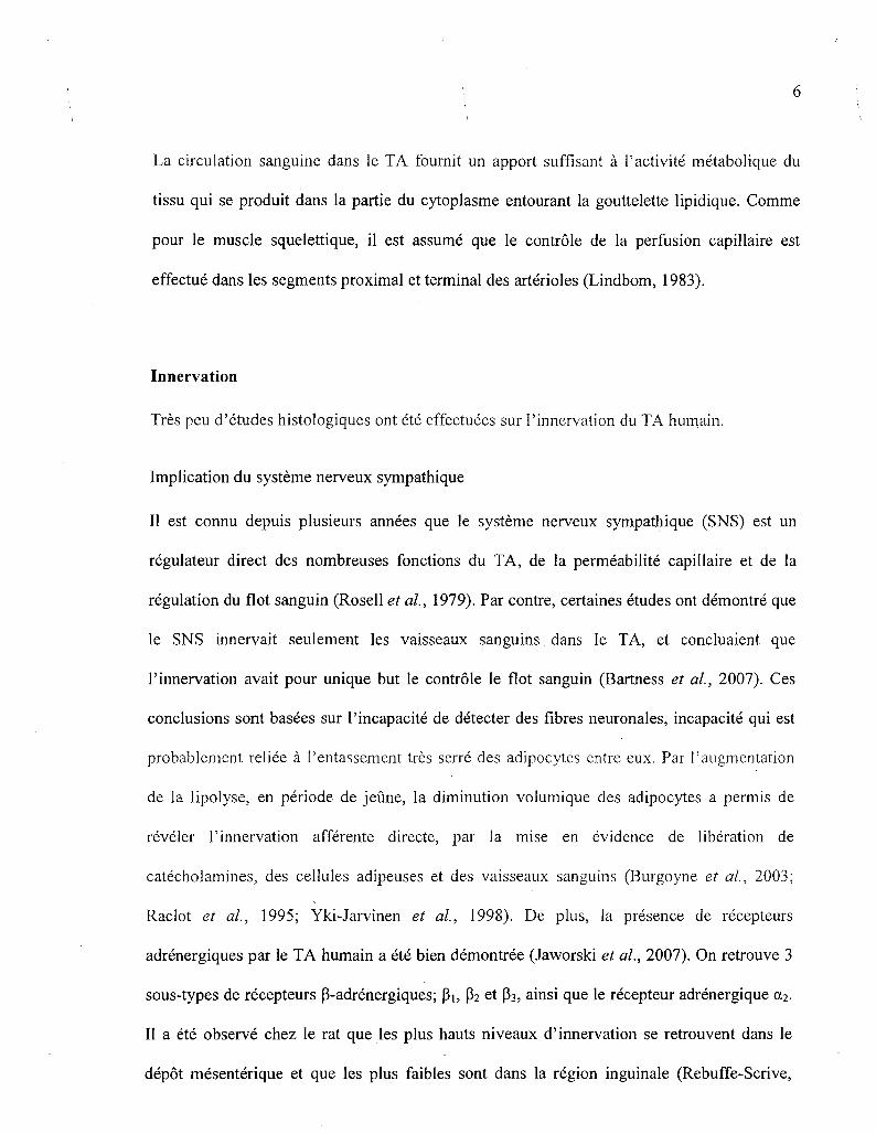
La circulation sanguine dans le TA fournit un apport suffisant a Pactivite metabolique du
tissu qui se produit dans la partie du cytoplasme entourant la gouttelette lipidique. Comme
pour le muscle squelettique, il est assume que le controle de la perfusion capillaire est
effectue dans les segments proximal et terminal des arterioles (Lindbom, 1983).
Innervation
Tres peu d'etudes histologiques ont ete effectuees sur Pinnervation du TA humain.
Implication du systeme nerveux sympathique
II est connu depuis plusieurs annees que le systeme nerveux sympathique (SNS) est un
regulateur direct des nombreuses fonctions du TA, de la permeabilite capillaire et de la
regulation du flot sanguin (Rosell et al, 1979). Par contre, certaines etudes ont demontre que
le SNS innervait seulement les vaisseaux sanguins, dans le TA, et concluaient que
Pinnervation avait pour unique but le controle le flot sanguin (Bartness et al, 2007). Ces
conclusions sont basees sur Pincapacite de detecter des fibres neuronales, incapacite qui est
probablement reliee a Pentassement tres serre des adipocytes,entre eux. Par Paugmentation
de la lipolyse, en periode de jeune, la diminution volumique des adipocytes a permis de
reveler Pinnervation afferente directe, par la mise en evidence de liberation de
catecholamines, des cellules adipeuses et des vaisseaux sanguins (Burgoyne et al, 2003;
Raclot et al, 1995; Yki-Jarvinen et al, 1998). De plus, la presence de recepteurs
adrenergiques par le TA humain a ete bien demontree (Jaworski et al., 2007). On retrouve 3
sous-types de recepteurs P-adrenergiques; Pi, P2 et P3, ainsi que le recepteur adrenergique a2.
II a ete observe chez le rat que les plus hauts niveaux d'innervation se retrouvent dans le
depot mesenterique et que les plus faibles sont dans la region inguinale (Rebuffe-Scrive,

• 7
1991). Une autre evidence de 1'innervation afferente directe des adipocytes par les neurones
post-ganglionnaires du SNS a ete fournie par 1'injection de traceurs fluorescent anterograde,
l,l'-dioctadecyl-3,3,3',3'- tetramethylindocarbocyanine perchlorate (Dil) et fluorescent
retrograde, FluoroGold, chez des hamsters Siberien avec l'observation d'anneaux
fluorescents entourant les cellules adipeuses des regions sous-cutanee et inguinale (Bartness
et al, 2007; Youngstrom et al, 1995). II a ete demontre que certaines des innervations de ces
deux regions ne provenaient pas des memes parties du SNS, soit une innervation via les
ganglions Ti3; L1-L2 pour la region sous-cutane, soit une innervation via seulement le
ganglion T13, pour la region inguinale (Youngstrom et al, 1995), revelant une structure
d'activation selective du TA.
Comme 1'innervation du TA peut avoir differents effets sur ses fonctions, elle semble
egalement avoir differents effets sur les actions efferentes du tissu en envoyant des signaux
tant a la moelle epiniere qu'au cerveau. L'injection de petites quantites de leptine dans le TA
peri-renal chez le rat a ete associee avec l'augmentation de l'activite sympathique renale
efferente sans alterer les concentrations de leptine plasmatique (Tanida et al, 2000). Des
etudes chez les hamsters et les rats ont demontre une connexion neuronale directe entre le
TA et differentes regions du cerveau impliquees, via le SNS, dans la regulation de plusieurs
systemes tel que le systeme cardiovasculaire (Bamshad et al, 1998).
Les effets similaires induits par les catecholamines et les agonistes et les effets opposes
obtenus avec les antagonistes des recepteurs adrenergiques sur le TA humain et animal, tant
au niveau in vitro que in vivo, indiquent que le mode d'innervation observe dans les
conditions experimentales animales se retrouve egalement chez 1'humain.

8
S'il1 a ete demontre que le TA humain est innerve, il reste a determiner si cette activite
neuronale peut avoir un effet sur la regulation metabolique du tissu. Des etudes ont tente de
determiner le role du SNS sur la lipolyse, par 1'interruption des signaux sympathiques
descendant resultant d'une lesion a la moelle epiniere ou des ganglions sympathiques. Ces
etudes ont demontre que 1'innervation sympathique n'avait pas d'effets clairs sur les niveaux
basaux de lipolyse du TA (Karlsson et al, 1995), mais que 1'innervation sympathique en
condition d'excitation sympathique induisait la lipolyse (Karlsson et al, 1997). Des etudes
chez des sujets sains ont egalement demontre, par une stimulation in situ d'un nerf cutane et
de la mesure de la liberation de glycerol dans la zone innervee, que la stimulation neuronale
induisait la lipolyse (Dodt et al, 2003). Meme si de nombreuses etudes etablissent que
Pinnervation du TA par le SNS est le principal regulateur de la lipolyse sous-cutanee, la
neuroanatomie du SNS dans le TA reste a etre decrite en details chez 1'humain.
Implication du systeme nerveux parasympathique
La plupart des tissus sont doublement innerves par les deux composantes du systeme nerveux
autonomes, soit le SNS et le systeme nerveux parasympathique (SNPS), deux systemes dont
les actions sont generalement opposees. Bien que plusieurs etudes suggerent la presence
d'une innervation du SNPS dans le TA en plus de l'innervation SNS, le sujet reste encore
chaudement debattu. Une etude utilisant la microdialyse a demontre qu'une stimulation des
recepteurs nicotiniques augmentait la lipolyse, alors qu'une stimulation des recepteurs
muscariniques diminuait 1'activite de lipolyse du TA in vivo chez 1'humain (Andersson et al,
1995). A l'inverse, certaines etudes vont contre une composante SNPS dans le TA (Kreier et
al, 2002). Par exemple, il n'y a pas a) d'identification de marqueurs du SNPS au niveau du
TA et b) d'identification de ganglions du SNPS a Pinterieur ou environnant le TA

: 9
(Youngstrom et al.,. 1995). Une autre etude a egalement examine trois marqueurs du SNPS,
dans trois modeles animaux et dans trois tissus adipeux differents. Leurs resultats amenent a
conclure a l'absence neuroanatomique de l'innervation du SNPS au niveau du TA (Bartness
et al, 2007). Mais ces resultats, parfois anciens, sont combattus par des travaux recents :
l'innervation parasympathique du TA a ete demontree par l'utilisation d'un « retrograde
transneuronal tracer pseudo-rabies virus » (Kreier et al, 2002) sur du TA chirurgicalement
denerve de fibres sympathiques. De plus, suite a une vagotomie, la captation des acides gras
libres (AGL) et du glucose mediee par Taction de l'insuline fut diminuee, et l'activite de la
lipase hormono-sensible (HSL) augmentee. Ces donnees indiquent un role potentiel
anabolique du nerf vague sur le TA. Les masses graisseuses sous-cutanee et intra-abdominale
semblent egalement etre innerves par des neurones parasympathiques et sympathiques
(Kreier et al, 2002).
Devant ces resultats opposes, 1'implication du SNPS dans l'activite du TA doit etre
investiguee. Une implication hypothetique des roles opposes du SNS et SNPS suggererait un
certain controle neuronal dans les processus catabolique et anabolique du TA. Malgre des
opinions dichotomiques sur Pimplication du SNPS, il est clair que l'innervation du systeme
nerveux autonome dans le TA est un facteur d'importance majeur dans la regulation de
l'activite du tissu.
Le TA comme site de stockage energetique
Longtemps, le TA fut considere comme un lieu de stockage inerte. L'interet pour ce tissu
c'est accentue lors de la decouverte de son role dans la transformation des hormones
stero'idiennes et quand le lien entre bbesite, diabete et maladies cardiovasculaires a ete fait.

! • 10
La recherche sur 16 TA peut etre divisee en trois periodes. La premiere oil1, les deux aspects
metaboliques distincts du TA ont ete consideres : le stockage des triglycerides (TAG) apres
les repas et la liberation des AGL dans l'organisme lorsque requis (periode d'exercice et
jeune). La deuxieme ou le TA a ete compris comme un lieu d'interconversion pour les
hormones steroidiennes (Siiteri, 1987) et, plus recemment, la troisieme qui considere le TA
comme un organe endocrine multifonctionnel produisant et secretant une variete de facteurs,
hormones, cytokines et autres, ayant des effets locaux et systemiques (Hauner et ah, 2002).
Les TAG contenus dans les adipocytes constituent une source d'energie importante de notre
organisme. L'importance du depot et de l'utilisation des graisses dans le TA est en grande
partie determinee par 1'apport alimentaire et la depense energetique. Deux routes majeures
permettent 1'apport des TAG au TA.
Source plasmatique de TAG
Dans le plasma, les TAG sont presents sous la forme de particules de lipoproteines, de
chylomicrons et de VLDL (lipoproteines de tres basse densite). Les chylomicrons
transportent les lipides absorbes provenant du tube digestif. lis ont la densite la plus faible de
toutes les lipoproteines, ce sont des gouttelettes hydrosolubles de lipoproteines. lis
contiennent en leur centre des TAG et du cholesterol entoures de phospholipides et d'une
« pellicule » de proteines (proteines de structure, apoB-48). Le foie est la principale source
de VLDL. Les VLDL contiennent des apolipoproteines (apo) B-100 et sont tres riches en
TAG. Les VLDL transportent les TAG, qui ont ete fabriques ou transformes dans le foie, de
la circulation sanguine vers les tissus peripheriques avec une preference pour le TA. Lorsque

11
tous ces TAG ont ete conduits a leur point de destination, les residus des VLDL sont
convertis en LDL (lipoproteines de basse densite), riches en cholesterol (Karpe et al, 1993).
Ces larges particules contenant les TAG sont trop grosses pour pouvoir traverser les
membranes des capillaires vers les fluides interstitielles. Les chylomicrons adherent a des
sites de liaison sur la surface interne, 1'endothelium, des capillaires du TA ou des muscles
squelettiques. Les TAG des chylomicrons sont ensuite hydrolyses en AGL et en glycerol
grace a une lipoproteine lipase (LPL) (Fielding et al., 1998; Olivecrona et al, 1995), une
enzyme extracellulaire, associee a l'endothelium capillaire, produite par les adipocytes et
activee par l'apolipoproteine C-II (apoC-II). Les AGL et le glycerol liberes peuvent alors
traverser les parois des capillaires et servir de source d'energie cellulaire, ou etre
emmagasine sous forme de lipides dans le TA (Fig 1.3). Une fois a Pinterieur de l'adipocyte,
les AGL sont esterifies a l'aide du glycerol-3-phosphate pour reformer un TAG. Ainsi ils
peuvent rejoindre la gouttelette lipidique de la cellule pour entreposage. L'activite de la LPL,
l'esterification et la production de glycerol-3-phosphate via la glycolyse sont stimules par
l'insuline (Frayn et al, 1994). Cette influence de l'insuline indique qu'apres un repas
typique, avec teneur en lipides et en glucose, le captage et 1'entreposage des graisses dans le
TA sont efficacement stimules. Une autre proteine, « Acylation stimulating protein » (ASP)
entre egalement dans la regulation de la lipogenese (Baldo et al, 1993). ASP est produite
dans les adipocytes. La production d'ASP in vivo est augmentee en periode post-prandiale
(Saleh et al, 1998), donnant a l'ASP un role potentiel dans la coordination de l'entreposage
des graisses apres l'ingestion d'un repas.
La voie de la LPL est une voie importante pour le stockage des graisses dans les adipocytes.
Cependant, il est connu que les personnes deficientes en LPL ont des adipocytes relativement

,12
normaux (Peeva et al, 1992). Chez des souris dont la LPL est absente, on retrouve une
masse de TA normale. D'autres voies pour le stockage des graisses doivent done exister. II
est arguments que la synthese de TA en cas d'absence de la LPL est effectuee via la
lipogenese de novo (Weinstock et al, 1997).
La lipogenese de novo
La lipogenese de novo est une autre voie de la syntheese du TA. Cette voie est egalement
stimulee a de multiples points par Pinsuline. La synthese des TAG se produit lorsque les
concentrations d'ATP dans les cellules et de glucose dans le sang sont elevees. L'exces
d'ATP entraine une accumulation des intermediates du metabolisme du glucose, dont
l'acetyl CoA qui en cas d'exces est achemine vers les voies de syntheses des TAG (Fig 1.3).
La premiere etape, regulee par Pinsuline, est la transformation par l'acetyl CoA carboxylase
de l'acetyl CoA en malonyl CoA. Par la suite, la chaine d'acides gras s'allonge de deux
atomes de carbones a la fois. Comme l'acetyl CoA, un intermediate du catabolisme du
glucose, est egalement un point de depart pour la synthese des acides gras, le glucose peut
facilement etre converti en lipides. Meme avec un regime pauvre en lipides, un apport
excessif de glucides fournit routes les matieres premieres necessaires a la formation des
TAG. Lorsque la glycemie est elevee, la lipogenese devient l'activite principale du TA.
13
Glucose-6-phosphate
/ Giycofyse
PPP • »
I NADPH + H+
CO,
Lipoger&se
fi-Qxjifation Acyl-CoA
ATP CoA
2 CO, ACETYL-CoA'. SYNTHETASE.
AGL
Esteriftcattan
TAG'
Glycerol-3-phosphate
AGL
...
Lipofyst
LIPASE HORMONO-SENSIBLE
Insuline
AGL
UPQPROTEWE LIPASE
Glycerol
Glucose
Glycerol
TAG
(chylomicrons, VLDL)
AGL Glycerol
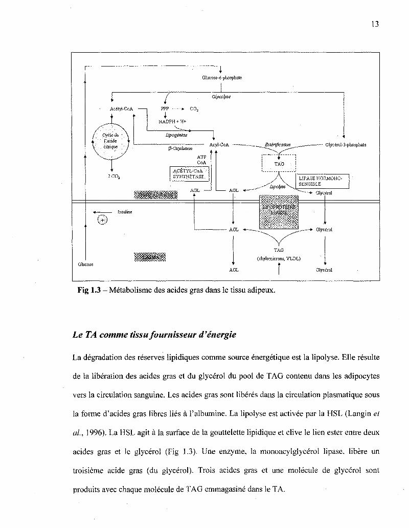
Fig 1.3 - Metabolisme des acides gras dans le tissu adipeux.
Le TA comme tissu fournisseur d'energie
La degradation des reserves lipidiques comme source energetique est la lipolyse. Elle resulte
de la liberation des acides gras et du glycerol du pool de TAG contenu dans les adipocytes
vers la circulation sanguine. Les acides gras sont liberes dans la circulation plasmatique sous
la forme d'acides gras libres lies a l'albumine. La lipolyse est activee par la HSL (Langin et
al, 1996). La HSL agit a la surface de la gouttelette lipidique et clive le lien ester entre deux
acides gras et le glycerol (Fig 1.3). tine enzyme, la monoacylglycerol lipase, libere un
troisieme acide gras (du glycerol). Trois acides gras et line molecule de glycerol sont
produits avec chaque molecule de TAG emmagasine dans le TA.

• ; H
La regulation de HSL est tres precise. La HSL est inactive lorsque les niveaux d'insuline
sont eleves (Coppack et al, 1989). La HSL est activee par phosphorylation via une proteine
kinase A, elle-meme activee par l'elevation cellulaire des niveaux d'AMPc. La
phosphorylation de HSL est parallelement accompagnee de sa translocation du cytosol vers
la surface de la gouttelette lipidique de l'adipocyte et de la phosphorylation de la perilipine,
une proteine masquant le depot lipidique qui, apres stimulation, permet Faeces a la HSL
(Clifford et al, 2000). Une augmentation de la lipolyse, par la phosphorylation de la
peripiline et de la HSL, est stimulee par l'action des catecholamines sur les recepteurs P-
adrenergiques. En condition de jeune prolonge, une stimulation via les recepteurs P-
adrenergiques et la desinhibition progressive de l'insuline, agissant comme co-regulateur sur
la lipolyse, entrainent une augmentation reguliere de l'activite lipolytique (Arner et al,
1990). Dans ces conditions de jeune, une secretion d'hormones de croissance (Samra et al,
1999) et une elevation matinale du Cortisol (Samra et al, 1996a) semblent egalement jouer
des roles modulateurs.
La dephosphorylation de HSL, mediee par l'insuline, la rend inactive. Cette action de
l'insuline implique une degradation des niveaux d'AMPc en 5'-AMP par la 3'5' nucleotide
phosphodiesterase cyclique. L'insuline stimule egalement la phosphodiesterase et la lipase
phosphatase qui inactive la HSL. Cette inhibition a un effet puissant et rapide, elle a lieu a
des bas niveaux de concentrations d'insuline et des le debut de leur elevation. La
concentration maximale d'insuline necessaire pour la suppression de la liberation des
graisses dans la circulation plasmatique chez l'humain a jeun est d'environ 90 pmol/L alors
qu'elle est de 200 pmol/L pour la stimulation de la lipogenese en condition post-prandiale
(Campbell et al, 1992). L'insuline inhibe la liberation des AGL du TA, entrainant une chute
15
de la quantite des AGL plasmatiques circulant. Elle stimule la lipogenese et la synthese des
acylglyceroles et augmente l'oxydation du glucose en CO2 par la voie des pentoses
phosphates (PPP). L'insuline favorise la lipogenese et restreint la lipolyse. La HSL est
egalement activee par l'hormone corticotrope (ACTH), l'hormone thyreostimuline (TSH), le
glucagon, l'adrenaline, la noradrenaline et la vasopressine. En plus de l'insuline, elle est
inhibee par la prostaglandine E et l'acide nicotinique (Watt et al, 2008).
Le TA et la stero'idogenese
Le TA est egalement un site d'inter-conversion des hormones steroi'diennes. II produit des
steroi'des sexuels et des glucocorticoi'des, tels que la production d'cestrogenes via les
androgenes et du Cortisol apartir de la cortisone (Boulton et al, 1992; Katz et al, 1999). La
participation du TA pour la conversion des steroi'des sexuels est quantitativement importante,
elle represente de 10-20% de la production totale de l'organisme (Katz et al, 1999). Cette
conversion est finement regulee par l'insuline et augmente dans les cas d'obesite (Bjorntorp,
1996; Frayn era/., 2003).
Le TA comme organe endocrine
Comme mentionne precedemment, le TA a ete longtemps considere comme un organe passif
servant uniquement au stockage energetique, puis un site d'inter-conversion. Ces dix
dernieres annees, la recherche a demontre la nature complexe de ce tissu et renverse ce point
de vue traditionnel. Les adipocytes sont maintenant reconnus pour secreter une grande
variete d'hormones et de facteurs, appeles adipokines. Elles peuvent jouer un role tant au
niveau local (autocrine, paracrine), qu'au niveau systemique (endocrine) (Goossens, 2008).
\ \ 1 6
Cytokines, facteurs de crdissance, adiponectine, resistine, adipsine, leptine, «acylation
stimulating protein » (ASP), «plasminogen activator inhibitor-1 » (PAI-1), lipoproteine
lipase (LPL), «tumor necrosis factor-a » (TNF-a), interleukine-6 (IL-6), « fasting induced
adipose factor » (FIAF), « vascular cell adhesion molecule-1 » (VCAM-1), composantes du
systeme renine-angiotensine font entre autres partie des adipokines secretees par le TA
(Frayn et at, 2003; Fruhbeck et at, 2001; Kershaw et at, 2004). Le TA envoie et recoit de
nombreux signaux endocrines, metaboliques et inflammatoires. II semble moduler en partie
l'homeostasie de l'organisme par de multiples systemes incluant, les reponses immunitaires
et inflammatoires, la coagulation sanguine, la reproduction, les fonctions endothelials et la
differentiation des adipocytes. II est de plus en plus evident qu'il existe une interaction
croisee entre les cellules adipeuses et plusieurs autres tissus comme l'endothelium, les
muscles, le foie, le pancreas, les glandes surrenales et les structures du systeme nerveux
centrale. Ainsi, en plus de pouvoir moduler ses propres activites metaboliques, le TA peut
transmettre des signaux vers d'autres tissus et moduler leur metabolisme (Goossens, 2008).
Les adipocytes expriment egalement de nombreux recepteurs qui permettent la reponse a des
signaux afferents, tels que des hormones et des signaux emis par le systeme nerveux central
(Goossens, 2008). Plusieurs facteurs derives des adipocytes (surtout la cytokine TNFa) ont
ete identifies pour avoir des effets sur la transduction du signal de l'insuline et ainsi
contribuer au developpement de l'insulino-resistance et de l'atherosclerose chez les patients
obeses avec ou sans diabete de type 2 (Frayn et at, 2003; Yudkin, 2007).
Activite metabolique du tissue adipeux: importance duflot sanguin
Le stockage et la liberation d'energie, la secretion d'adipokines demandent une regulation
precise du flot sanguin dans le tissu adipeux (FSTA), c'est un processus hautement

• 17
coordohne avec un controle constant et changement rapide sel'on les besoins metaboliques.
Quatre points soulignent Pimportance d'un FSTA coordonne et regule.
Premierement, les echanges hormonaux et metaboliques (Ex) dans un tissu dependent du
produit du flot sanguin et de la difference de concentration arterio-veineuse du metabolite ou
de 1'hormone (A-V) :
Ex = BF x (A-V)
Deuxiemement, le FSTA semble augmenter pendant la mobilisation d'energie du TA
par l'organisme ou vice versa : la liberation d'AGL (periode de jeune, exercice prolonge), la
clairance des TAG de la circulation sanguine vers le TA (periode postprandiale). Les
variations de FSTA semblent etre reliees a l'activite metabolique du tissu. La clairance des
TAG du sang, refletant a un premier niveau l'activite des lipoproteines lipases, augmente en
parallele avec 1'augmentation du flot sanguin lorsque se dernier est stimule par une infusion
d'epinephrine (Samra et al, 1996b). Cependant, lorsque stimule par un repas, le flot sanguin
augmente rapidement et n'est pas synchronise avec 1'augmentation de la concentration
plasmatique de TAG, 1'action des LPL ou le depot de graisse dans le TA qui surviennent plus
tardivement. L'elevation du flot sanguin apres l'ingestion d'un repas semble davantage liee
avec 1'augmentation de la concentration plasmatique d'insuline et la suppression de la
liberation des AGL par le TA (Coppack et ah, 1992). L'augmentation du FSTA en periode
postprandiale pourrait servir a la livraison d'un signal, l'insuline par exemple, pour initier
l'augmentation postprandiale de l'activite des LPL. L'insuline a egalement comme action de
supprimer la liberation des AGL, ce qui altere les reponses vasodilatatrices (Steinberg et al,
1997). Les AGL contenus dans le TA sont sujets a de plus grandes variations que celles que
Ton peut trouver dans le muscle squelettique. S'il existe une relation physiologique entre les
18
variations de concentrations d'AGL et le flot sanguin, le TA est l'organe primaire pour la
regulation de ces variations. Peut-importe le lien physiologique de la regulation du FSTA, il
est regule selon l'apport nutritionnel a l'organisme.
Troisiemement, une alteration dans la regulation du FSTA est reliee a l'obesite et a
l'insulino-resistance (Jansson et al, 1998; Karpe et al, 2002b; Summers et al, 1999). Chez
les personnes obeses et/ou souffrant d'une resistance a 1'insuline, le FSTA a jeun est
inferieur que chez les personnes en sante de poids normal, tandis que 1'augmentation
postprandiale du FSTA est abolie (Blaak et al, 1995; Coppack et al, 1992; Jansson et al,
1998; Karpe et al, 2002b; Summers et al, 1996; Virtanen et al, 2001). II a egalement ete
demontre que l'elevation de FSTA postprandiale est egalement abolie chez tous les stades de
diabete de type 2 : parents relies au premier degre avec des sujets diabetiques de type 2,
sujets avec une intolerance au glucose, sujets diabetiques de type 2 avec hyperglycemic
postprandiale et glycemie normale a jeun, et sujets diabetiques de type 2 avec hyperglycemic
postprandiale et en periode de jeune (Dimitriadis et al, 2007). II peut etre specule qu'une
mauvaise reponse du FSTA apres un repas diminue la clairance, par le TA, des lipides
provenant des lipoproteines. Les lipides sont alors rediriges vers d'autres organes moins bien
adaptes pour leur stockage (foie, muscles squelettiques). II pourrait en resulter une
augmentation de la production de VLDL par le foie et une hypertriglyceridemie, conditions
qui augmentent le risque de developpement des maladies cardiovasculaires.
Quatriemement, l'importante fonction endocrine du TA pourrait etre facilitee par les
variations de FSTA, regulant ainsi les entrees et sorties des hormones. Un
dysfonctionnement dans la regulation du FSTA pourrait avoir pour consequence d'attenuer
les fonctions endocrines du tissu.

: : .19
En resume, une diminution du FSTA peut avoir un effet negatif sur le metabolisme en
entrainant une diminution du captage par le TA des acides gras, du glucose et d'hormones,
une alteration dans la liberation de produits de la lipolyse et de peptides du TA vers la
circulation (Samra et al, 1996b) et en diminuant la qualite de la signalisation entre le TA et
les autres tissus metaboliquement actifs. Une meilleure comprehension des bases
physiologiques de la regulation du FSTA pourrait permettre une meilleure comprehension du
lien entre un dysfonctionnement du FSTA et les problemes metaboliques observes en
condition d'insulino-resistance.
. 20
CHAPITRE 2
Articles scientifiques integres
Article 1) Update on adipose tissue blood flow regulation
Avant-propos de l'article
L'article intitule «Update on adipose tissue blood flow regulation)) est une continuite de
1'introduction de ce memoire. II resume la regulation physiologique connue du FSTA et
amene egalement de nouvelles hypotheses. Les auteurs de cet article sont Pascal Brassard,
Elizabeth Martin, Philippe Yale, Andre C. Carpentier et Jean-Luc Ardilouze. Cet article est
une recension des ecrits concernant la regulation du FSTA. Ce manuscrit etait d'abord
destine a une publication dans journal Circulation qui voulait inclure un article de ce type
dans un numero special. L'echeance etant depasse, l'article a ete mis a jour depuis. II est
dans l'attente d'une lecture des co-auteurs afin de pouvoir apporter les corrections
necessaires, puis determiner dans quel journal ce travail est susceptible d'etre publie. Les
modifications mineures quant au format seront alors effectuees.
Resume
Le TA est maintenant reconnu comme un organe metaboliquement tres actif et endocrine,
delivrant un melange complexe de substrats et d'hormones exercant de multiples effets
pleiotropes. Le debit du flot sanguin est un regulateur important des fonctions du TA. Les
fonctions du TA ayant un impact important sur l'organisme, la regulation de ses fonctions
necessite une extreme precision. Le FSTA varie selon differentes conditions physiologiques
; 21;
de l'organisme et sa regulation se trouve modifiee dans les cas d'obesite et d'insulino-
resi stance.
Dans cette article, nous decrirons la physiologie du FSTA en conditions pre et
postprandiales, en situation d'exercice physique et selon le poids des individus. De plus, le
role du SNS, du SNPS, de l'insuline, de l'oxyde nitrique et du systeme renine angiotensine
dans la regulation du FSTA seront decrits. II sera egalement discute de 1'effet des diverses
adipocytokines et facteurs inflammatoires liberes par les adipocytes sur le FSTA, le TA et
l'organisme. Certains facteurs physiologiques du FSTA jouenl un role dans la resistance a
l'insuline, suggerant qu'une deregulation du FSTA pourrait etre une composante du
syndrome d'insulino-resistance. Une meilleure comprehension de la regulation du FSTA
conduira a de nouvelles hypotheses physio-pathologiques et perspectives therapeutiques.
Contribution personnelle a cet article :
• Rescension des ecrits de la litterature.
Redaction de Particle en collaboration avec Pascal Brassard, PhD.

Article
|j
22
UPDATE ON ADIPOSE TISSUE BLOOD FLOW REGULATION
Authors:
Pascal Brassard1'2, Elizabeth Martin1'2, Philippe Yale2, Andre C. Carpentier2 and Jean-Luc
Ardilouze2*.
]Both authors contributed equally to this work.
Institutions:
2Diabetes and metabolism research group, Division of Endocrinology, Department of
Medicine, Centre Hospitalier Universitaire de Sherbrooke, Universite de Sherbrooke,
Quebec, Canada.
*Corresponding author:
Jean-Luc Ardilouze, MD, PhD
Endocrine Division
Centre Hospitalier Universitaire de Sherbrooke
3001 12th Ave N.:, Sherbrooke, Qc, J1H 5N4
Tel: 819-564-5241
Sources of support:
This work was supported by the Canadian Institutes of Health Research (CIHR), Fonds de la
recherche en sante du Quebec (FRSQ), and Diabete Quebec.

23
Jean-Luc Ardilouze is a CIHR scholar (2006 CIHR New Investigator Award) and is member
of the FRSQ-funded Centre de recherche clinique Etienne-LeBel.
Number of words: 6536
Abbreviations: ATBF, adipose tissue blood flow; CVD, cardiovascular diseases; GLP-1,
glucagon-like-peptide-1; HSL, hormone-sensitive lipase; LPL, lipoprotein lipase, NEFA,
non-esterified fatty acids; NO, Nitric Oxide; TG, Triglycerides; V02max, maximal oxygen
volume uptake.

'; • 2 4
ABSTRACT
Any metabolic or hormonal exchange through a tissue depends on the product of the blood
flow and the arteriovenous difference. Therefore, given the storage and endocrine roles of
adipose tissue, the regulation of adipose tissue blood flow (ATBF) is of pivotal importance.
This paper is an update of the literature, presenting our knowledge and hypothesis on ATBF
regulation. It was shown that insulin per se has no vasodilatory effect, in the resting state as
well as during the postprandial period. Consequently, it has been hypothesised that insulin
may act indirectly on ATBF via sympathetic activation, and that nitric oxide (NO) may be an
overall major regulator of ATBF. Indeed, fasting ATBF is primarily controlled by NO,
circulating angiotensine II and to some extent under a-adrenergic. Although the postprandial
enhancement of ATBF is independent of NO and is controlled principally, for 60%, by the p-
adrenergic system. This suggests that the postprandial regulation of ATBF is complex and
may involve other regulatory factors. It is also well known that dependently of the
physiological condition of the organism, ATBF varies. In vivo studies have shown that
adipose tissue is an efficient buffer against the postprandial flux of non-esterified fatty acids
in the circulation, protecting other tissues. In excess of fat tissue, this buffering effect may be
impaired. This condition includes also a reduced postprandial ATBF response, potentially
causing metabolic alterations. Consequently, an alteration in the regulation of ATBF seems
to play a role in the development of insulin resistance syndrome. For the future, a better
understanding of ATBF regulation could lead to new pathophysiological hypothesis and
therapeutic perspectives for obesity, diabetes and cardiometabolic diseases.
Words: 266
Key words: adipose tissue, blood flow.

25
INTRODUCTION: ADIPOSE TISSUE
Despite its functional importance, for a long time, adipose tissue was considered as an
inactive energy store, its sole purpose being the storage of fat for insulation purposes and
provision of a protective cushion. Its low rate of oxygen consumption(Frayn, Humphreys, S.
M, and Coppack, S. W. 1995; Coppack et al. 1990b) would confirm this thought. Scientists
were not interested in adipose tissue until it became clear that obesity was related to
cardiovascular diseases (CVD) and diabetes. Then, two distinct aspects of the metabolism of
adipose tissue have been considered: TG storage after a meal, and liberation of fatty acids
when other tissues in the body require it, i.e. during exercise and overnight fasting. Over the
last two decades it has became clear that adipose tissue is not only involved in metabolism
but that it plays an extremely important role as a regulator of the flow of energy-providing
substances. Thereafter, the interconversion of steroid hormones was discovered.(Siiteri 1987)
More lately, it has been recognised that adipose tissue is also a multifunctional organ that
produces a variety of secretory factors that act either at the local level (autocrine / paracrine /
intracrine) or at the systemic level (endocrine).(Hauner and Hochberg, Z. 2002)
Moreover, adipose tissue makes up 10-30% of body weight in normal weight individuals,
normally up to 20% in male and up to 30% in female, but its mass can vary from as little as
3-5% of body weight in lean, athletic subjects to 70% in very obese subjects.(Jensen 2002)
The adipose tissue is the major energy storage organ of the body. It does indeed store
triglyceride (TG). In fact, approximately 80%> of adipose tissue weight is lipid, and over 90%
of lipids are stored TG.(Shen et al. 2003) In healthy individual, it is an efficient manager of
non-esterified fatty acids (NEFA), the major secretory product from adipose tissue. NEFA
are derived from the lipolysis of stored triglycerides, and regulated by adipocyte and

'• 26
nonadipocyte factors. If intracellular adipocyte hydrolysis of triglycerides (lipolysis) exceeds
intracellular adipocyte esterification of NEFA (lipogenesis), then NEFA undergo a net
release into the circulation as an energy supply for other tissues, such as heart and skeletal
muscle. A sustained, excessive net increase in circulating NEFA contributes to metabolic
disease. Chronic increases in circulating NEFA worsen glucose metabolism due to lipotoxic
effects upon muscle and liver, which contribute to insulin resistance,(Carpentier 2008) as
well as lipotoxic effects on pancreas, which contribute to insulinopenia.(DeFronzo 2004;
Lewis et al. 2002) These new findings explain why the hypothesis recently shift from an
exclusive glucocentric point of view to a lipocentric viewpoint.(Savage, Petersen, K. F., and
Shulman, G. I. 2005)
ADIPOSE TISSUE BLOOD FLOW IN RELATION TO THE MAIN FUNCTIONS OF
THE TISSUE
Given the important metabolic and endocrine roles of fat tissue, an adequate perfusion is
required. Three points underline the importance of a co-ordinated blood flow in adipose
tissue and justify more research about it.
Firstly, in healthy people, during fasting or during exercise, adipose tissue releases NEFA,
and requires a supply of plasma albumin for the transport of these NEFA into the circulation.
After feeding, there is a need to increase substrate delivery for TG clearance. Adipose tissue
blood flow (ATBF) is increased in states of fat mobilization (fasting and prolonged exercise)
and fat deposition (postprandial period). In all these states, the increase in ATBF appears to
be related to the metabolic activity of the tissue.

• • 2 7
The physiological significance of the nutrient-related increase in ATBF is not completely
understood. It is interesting that TG extraction from blood, considered primarily to reflect
LPL activity increases in parallel with raised blood flow when increased by adrenaline
infusion.(Samra et al. 1996b) This may imply that TG extraction in adipose tissue is limited
by substrate delivery. During that study, a rise in net NEFA efflux was also found,
representing NEFA generated by the increase rate of LPL action and by activation of HSL.
However, the peak in ATBF after a normal meal is early, and does not seem to be temporally
co-ordinated with the later peak in plasma TG concentration, LPL action or adipose tissue fat
deposition but more with increase in insulin concentration and suppression of NEFAs after a
mixed meal.(Carpentier et al. 2007; Coppack et al. 1992) The increased postprandial blood
flow might serve to deliver a signal, such as insulin, to initiate the postprandial increase in
LPL activity. Insulin also suppresses NEFAs, which have been shown to impair the
vasodilatory response.(Steinberg et al. 1997) Obviously, compared with skeletal muscle, the
environment within adipose tissue is subject to much larger fluctuations in NEFA
concentration. Thus, if the physiological relationship between fluctuation in NEFA
concentrations and blood flow exists, adipose tissue would be the primary organ for such
regulation.
Secondly, impaired regulation of ATBF has been linked to obesity and insulin
resistance.(Jansson, Larsson, A., and Lonnroth, P. N. 1998; Karpe et al. 2002c; Summers,
Samra, J. S., and Frayn, K. N. 1999) Reduced basal ATBF and blunted postprandial rise of
ATBF in insulin resistant obese subjects may decrease chylomicron-TG delivery to adipose
tissue LPL in the postprandial period. It results in higher chylomicron-TG concentrations for
a longer period after meals in these subjects. This will lead in turn to increased production of
atherogenic particles. It could be proposed that impaired postprandial vasodilation, a
• 2 8
potential feature of glucose intolerance,(Karpe et al. 2002c) is also the cause of the impaired
lipid metabolism in insulin resistant subjects and predisposes to CVD. Dimitriadis et al.
demonstrated that after meal ingestion, insulin-stimulated ATBF was decreased in insulin
impaired glucose tolerance subjects as well as in diabetics who had postprandial
hyperglycemia but normal fasting plasma glucose levels, and subjects with type 2 diabetes
with both postprandial and fasting hyperglycemia.(Dimitriadis et al. 2007) Which is
completely the opposite of an efficient postprandial response to food intake. Moreover, it has
been shown that those defects can be observed early in the natural history of type 2
diabetic.(Brassard et al. 2008) Nevertheless, this very interesting experiment pointed out the
fact that the decrease in ATBF seems to come proportionally with the insulin sensitivity
level. This defect could be considered as an early marker of insulin resistance that precedes
the development of type 2 diabetes.
Thirdly, the important endocrine functions of adipose tissue (discussed above) may be
facilitated by variations in the inflows and the outflows from the tissue, which demonstrate
the central role of ATBF in the adipose tissue physiology.
ATBF PHYSIOLOGY
ATBF variations during fasting and meals
After an overnight fast, abdominal ATBF is typically around 3-5 ml. 100 g tissue"1.min"1,
whereas in resting skeletal muscle the value is about 1.5 ml. 100 g tissue"1.min"1.(Elia and
Kurpad, A. 1993) Skeletal muscle blood flow can increase many-fold (perhaps 20-fold)
during exercise. ATBF is also very adaptable. In some lean, healthy subjects subcutaneous
abdominal blood flow increases several-fold (up to 4-fold) in response to a meal.(Biilow et
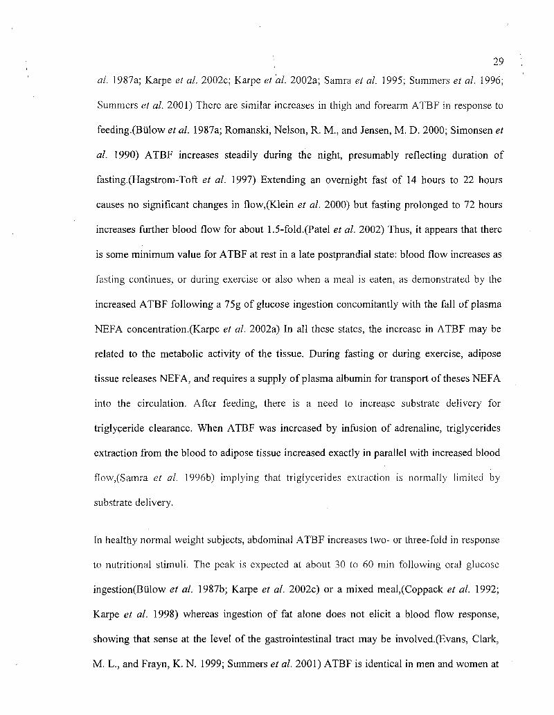
29
al. 1987a; Karpe et al. 2002c; Karpe et al. 2002a; Samra et al. 1995; Summers et al. 1996;
Summers et al. 2001) There are similar increases in thigh and forearm ATBF in response to
feeding.(Biilow et al. 1987a; Romanski, Nelson, R. M., and Jensen, M. D. 2000; Simonsen et
al. 1990) ATBF increases steadily during the night, presumably reflecting duration of
fasting.(Hagstrom-Toft et al. 1997) Extending an overnight fast of 14 hours to 22 hours
causes no significant changes in flow,(Klein et al. 2000) but fasting prolonged to 72 hours
increases further blood flow for about 1.5-fold.(Patel et al. 2002) Thus, it appears that there
is some minimum value for ATBF at rest in a late postprandial state: blood flow increases as
fasting continues, or during exercise or also when a meal is eaten, as demonstrated by the
increased ATBF following a 75g of glucose ingestion concomitantly with the fall of plasma
NEFA concentration.(Karpe et al. 2002a) In all these states, the increase in ATBF may be
related to the metabolic activity of the tissue. During fasting or during exercise, adipose
tissue releases NEFA, and requires a supply of plasma albumin for transport of theses NEFA
into the circulation. After feeding, there is a need to increase substrate delivery for
triglyceride clearance. When ATBF was increased by infusion of adrenaline, triglycerides
extraction from the blood to adipose tissue increased exactly in parallel with increased blood
flow,(Samra et al. 1996b) implying that triglycerides extraction is normally limited by
substrate delivery.
In healthy normal weight subjects, abdominal ATBF increases two- or three-fold in response
to nutritional stimuli. The peak is expected at about 30 to 60 min following oral glucose
ingestion(Bulow et al. 1987b; Karpe et al. 2002c) or a mixed meal,(Coppack et al. 1992;
Karpe et al. 1998) whereas ingestion of fat alone does not elicit a blood flow response,
showing that sense at the level of the gastrointestinal tract may be involved.(Evans, Clark,
M. L., and Frayn, K. N. 1999; Summers et al. 2001) ATBF is identical in men and women at

30
the abdomen levei but may be higher in the postprandial period in the female
thigh.(Romanski, Nelson, R. M., and Jensen, M. D. 2000)
ATBFin relation with the weight
Fasting ATBF and its increase after oral glucose are impaired in insulin-resistant subjects,
such as those who are obese or have type 2 diabetes.(Coppack et al. 1990a; Jansson et al.
1992; Romanski, Nelson, R. M., and Jensen, M. D. 2000) ATBF may be influenced by
obesity and glucose intolerance by different modes of action.
Fasting ATBF is negatively correlated with BMI, as is the postprandial rise in
ATBF.(Summers et al. 1996) However, obesity only explains a part of the rather high
variability in postprandial regulation of ATBF seen between subjects. Other factors
determine ATBF responsiveness to nutrients. It has been shown, with a cross sectional
study,(Karpe et al. 2002c) that the primary determinant of responsiveness is not obesity per
se but the associated insulin resistance, in particular the adipose tissue insulin sensitivity,
reflected through a novel insulin sensitivity index based on NEFA suppression and
postprandial glucose and insulin concentrations.(Belfiore, Iannello, S., and Volpicelli, G.
1998) No studies have been conducted on the effects of long-term weight loss and weight
maintenance on ATBF. Only short-term studies, in a context of drastic weight loss, can be
found in the literature. For example, it has been reported that water content, a potential
reflection of nutritive blood flow,(Laine et al. 1998; Raitakari et al. 1997) was increased
after a 15.6-kg-in-9-week weight loss, suggesting that improved insulin sensitivity may
contribute to improve ATBF.(Laaksonen et al. 2003)

ATBFin relation with exercise
In humans the efficient role of exercise on weight and insulin sensitivity of both whole body
and muscle is well established.(Henriksen 2002) However, the role of fitness or physical
activity on ATBF has not been thoroughly assessed and the results are inconsistent. ATBF
rises during moderate-intensity exercise (4 to 6 hours).(Biilow 1981; Stallknecht et al. 2001)
Surprisingly, ATBF did not increase in non-trained young men (22.3±1.5 yr) who performed
two rounds of 60 min on a bicycle,(Stich et al. 2000) nor in older women (75 yr).(Lange et
al. 2002)
The effect of long-term exercise on ATBF is also controversial. Only three studies have been
published on that topic. One study shows that 16 weeks of cycle exercise training did not
affect ATBF despite improved V02max, in five healthy untrained men.(Horowitz et al.
1999) Another study comparing eight endurance-trained and eight sedentary young men
showed that ATBF in trainees was not significantly higher than sedentary
subjects.(Stallknecht et al. 2000) Finally, eleven obese non-diabetic males taking part in a
12-week aerobic training program improved their V02inax but failed to improve
ATBF.(Stich el al. 1999) However, long-term exercise is clearly associated with
improvement in insulin sensitivity(Henriksen 2002; Houmard et al. 2004) and impaired
regulation of ATBF seems to be another facet of the insulin resistance syndrome.(Karpe et
al. 2002c) It can be concluded that further studies in this area are needed. Moreover, because
plasma fatty acid concentrations increase dramatically immediately after intense exercise, it
has been hypothesized that the reduction in fatty acid release into the circulation might result
from a restriction in ATBF,(Hodgetts et al. 1991) mediated by catecholamine-stimulated
vasoconstriction in adipose tissue blood vessels. In addition, endurance-trained athletes have

: 3,2
a greater ATBF in response to epinephrine infusion compared with sedentary control
subjects.(Stallknecht et al. 1995) Therefore, catecholamine delivery to adipose tissue might
be greater during exercise.
ATBF changes in other physiological situations
The change from upright to supine position elicits an instantaneous ATBF increment, in
accordance with a decrease in central and local postural sympathetic vasoconstrictor
activity.(Sindrup et al. 1991) The sitting position also decreases ATBF, in the anterior tibial
muscle.(Balldin 1976) During sleep, approximately 90 minutes after start of sleeping, a
blood flow increment of considerable magnitude is observed for 100 minutes.(Sindrup et al.
1991) It has also be reported that mental stress(Fernqvist and Linde, B. 1988) or bladder
filling(Fagius and Karhuvaara, S. 1989) increase ATBF. All these physiological modulations
have in common that they may be regulated by sympathetic tone.
VASOACTIVE REGULATORS IN THE FASTING AND POSTPRANDIAL STATE
Over the last decade, adipose tissue has been increasingly recognized as a complex organ
with a host of endocrine and paracrine effects that affect an array of metabolic and other
functions in the organism.(Mohamed-Ali, Pinkney, J. H., and Coppack, S. W. 1998) ATBF is
a dynamic aspect of adipose tissue function. This parameter is disturbed in obesity and this
disturbance of ATBF regulation has been shown to be closely associated with insulin
resistance.(Jansson, Larsson, A., and Lonnroth, P. N. 1998; Karpe et al. 2002c; Summers et
al. 1996; Virtanen et al. 2002) ATBF regulation in the postprandial period might well be an
essential component of normal adipose tissue metabolic function; for instance, a raised in
ATBF leads to increased clearance of TG by the tissue.(Samra et al. 1996b) It might also be

, 3 3
a further aspect of disturbed endothelial function in obesity. Regulation of ATBF has been
studied extensively and reviewed by Frayn and Macdonald.(Frayn et al. 2003)
Sympathetic nervous system
Sympathetic stimulation increases vascular permeability within adipose tissue.(Rosell,
Axelrod, J., and Kopin, I. J. 1964) This does not occur in other tissues such as skin or
skeletal muscle. This increase in permeability is inhibited by a-receptor blocking agents. It
has been suggested that the endothelial cells of the vessels within adipose tissue may carry
a-receptors that when stimulated, cause contraction of cells and increase the pore size
between endothelial cells. Even during sympathetic induced vasoconstriction, the product of
permeability and capillary surface area for solutes is increased (figure l).(Fredholm and
Sollevi, A. 1981) This may have important implications for the metabolic processes within
the tissue. Moreover, enhancement of ATBF may have importance in metabolic physiology
in that the extraction of plasma triglycerides increases with increasing blood flow.(Samra et
al. 1996b) The increase in postprandial ATBF is much more important with oral glucose
administration than with intravenous glucose administration.(Karpe et al. 2002c) In humans,
adrenergic influences are predominant, with P-mediated vasodilation(Hjemdahl et al. 1983)
and a2-mediated vasoconstriction (figure l).(Galitzky et al. 1993) These influences may
explain the increased blood flow during fasting or exercise that circulating cathecholamines
are more important in the exercise-induced increase in ATBF than sympathetic nerve activity
(figure l).(Stallknecht et al. 2001)

34
Figure 1: Hypothetic mechanism schema of the regulation of adipopse tissue blood flow in human.
The increase in blood flow following feeding has not been fully explained. Time course of
insulin concentration in plasma does not seem to be the responsible signal.(Karpe et al.
2002a; Summers et al. 1996) Also, adrenaline and noradrenaline levels were observed to
increase in response to ingestion of oral glucose and might be related to the increased ATBF
stimulated by glucose.(Bulow et al, 1987a) Links between splanchnic bed glucoreceptors and
the sympathoadrenal system have been hypothesized in mammals and may therefore account
for a difference in blood flow response between the experimental procedures.(Donovan,
Cane, P., and Bergman, R. N. 1991) Sympathetic receptor blockers have been shown to
reduce the postprandial response by 55%, making it a strong candidate as a regulator. Gut
hormones may also be responsible, but their role in vasodilation remains unclear and needs
to be clarified. The incretin's system could be part of the explanation. An obvious candidate
is glucagon-like-peptide-l (GLP-1) (figure 1). GLP-1 is the most important incretin in

35
humans.(Ahren 2003; Meier et al. 2002) GLP-1 receptors have been detected in adipose
tissue. Moreover, GLP-1 is secreted throughout the day and in increased amounts after
meals. From baseline, GLP-1 increases by 120% after glucose load.(Rask et al. 2004) In
addition, is has been shown that GLP-1 has a vasodilatory effect in hepatic and pulmonary
arteries and it has been demonstrated that GLP-1 mediate a concentration dependent
vasorelaxation of rats aorta.(Joyner and Norton, C. C. 1976; Golpon et al. 2001; Green et al.
2008) As P-adrenoceptor subtypes, his receptor belongs to the superfamily of seven
transmembrane domain G protein-coupled receptors that positively regulate intracellular
cAMP levels via adenylate cyclase.(Bertin et al. 2001) One study was performed in humans
to test directly whether GLP-1 has actions on lipolysis in adipose tissue and was
inconclusive, probably because the ethanol escape technique under microdialysis condition
was used.(Bertin et al. 2001; Lafontan and Arner, P. 1996) This technique only gives an
estimation of ATBF, and is considered as an indirect and a non-quantitative method.(Karpe
et al. 2002b) The role of GLP-1 on ATBF regulation remains to be established. C-peptide, a
hormone which is derived from the catabolism of pro-insulin has been shown to have some
vasodilatory properties in cutaneous tissue by stimulating eNOS activity,(Wahren et al.
2000) is not likely to be a candidate for the postprandial increase in ATBF since its plasma
concentration decreased during a intravenous euglycaemic insulin-glucose regimen.(Karpe et
al. 2002a) There is also abundant evidence that P-adrenergic stimulation increases ATBF.
This may be induced, for instance, by adrenaline infusion(Samra et al. 1996a) or by local
delivery of P-adrenergic stimuli by microdialysis of the tissue using isoprenaline,
dobutamine,(Barbe et al. 1996) or isoproterenol.(Millet et al. 1998) In contrast, experiments
using a-adrenergic stimuli such as clonidine(Galitzky et al. 1993) and
norfenefrine(Flechtner-Mors et al. 2002) show a predominantly inhibitory effect of a-

36
adrenoceptors on ATBF. It has been previously shown that the degree of insulin sensitivity
seems to be closely related to ATBF responsiveness,(Karpe et al. 2002c) but insulin per se
does not seem to stimulate blood flow in adipose tissue(Karpe et al. 2002a) as it may do in
muscle.(Baron et al. 2000) We have shown that the postprandial increase in insulin
concentrations may lead to activation of the sympathetic nervous system, with a subsequent
enhancement of ATBF.(Karpe et al. 2002a) We also observed, using microinfusion
technique, that propranolol, a P-adrenoceptor antagonist, had no effect on ATBF during the
preprandial period.(Ardilouze et al. 2004) The blood flow response to feeding is completely
blocked in some depots and partially blocked in other by propranolol infusion, a (3-receptor
antagonist,(Ardilouze et al. 2004; Simonsen et al. 1990) suggesting that it is dependent upon
sympathetic activation induced by postprandial hyperinsulinemia.
Moreover, it has been shown that phentolamine, a non-specific ai-adrenoceptor antagonist,
promoted an increase of the fasting blood flow during the preprandial period,(Stich et al.
2003) a finding that is in agreement with studies using microdialysis in which clonidine (an
©^-adrenoceptor agonist) induced vasoconstriction and inhibited lipolysis,(Galitzky et al.
1993) suggesting the vasoconstricting role of a-adrenoceptors (figure 1). Furthermore, the
effects of yohimbine, a specific a,2-adrenoceptor antagonist, are greater and sustained
compared to those of phentolamine in inhibiting ATBF, suggesting that ©^-adrenoceptor
activation leads to vasoconstriction and that a,] -receptors have little effect in the adipose
tissue microcirculation.(Ardilouze et al. 2004) We can therefore conclude that a.2-
adrenoceptor is the main vasoconstrictor in the preprandial period, whereas the p-
adrenoceptor has no preprandial effect, but has a strong regulation role according to its
postprandial vasodilator effect.
• ' ; 3 7
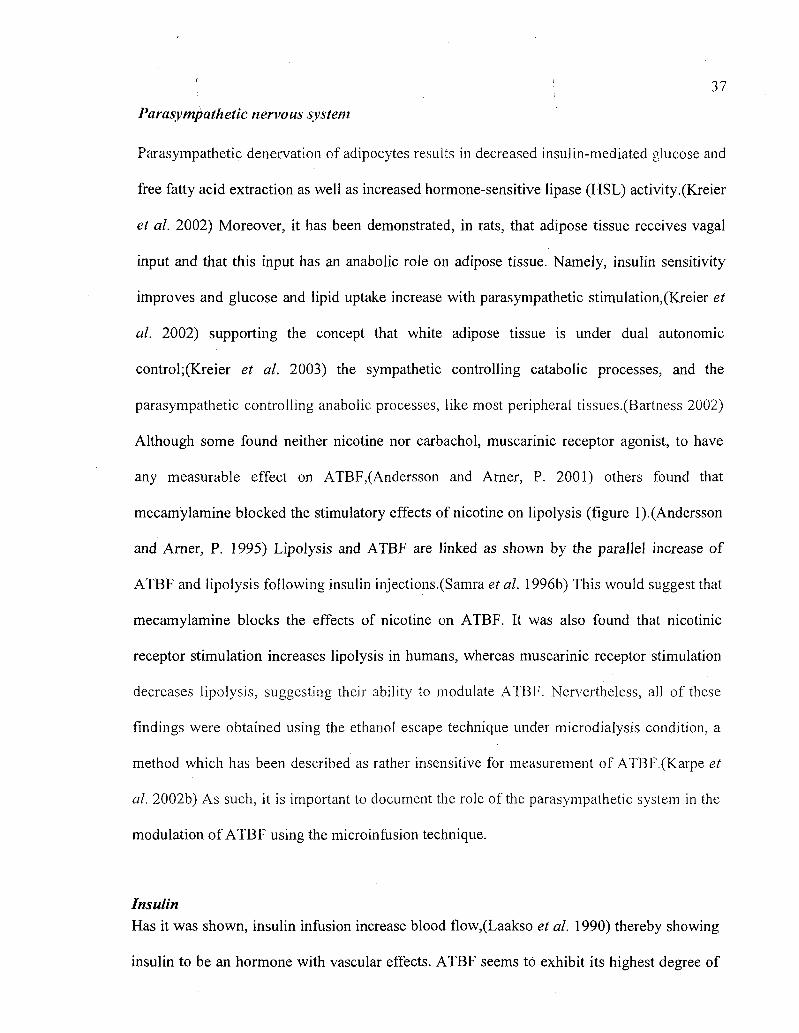
Parasympathetic nervous system
Parasympathetic denervation of adipocytes results in decreased insulin-mediated glucose and
free fatty acid extraction as well as increased hormone-sensitive lipase (HSL) activity.(Kreier
et al. 2002) Moreover, it has been demonstrated, in rats, that adipose tissue receives vagal
input and that this input has an anabolic role on adipose tissue. Namely, insulin sensitivity
improves and glucose and lipid uptake increase with parasympathetic stimulation,(Kreier et
al. 2002) supporting the concept that white adipose tissue is under dual autonomic
control;(Kreier et al. 2003) the sympathetic controlling catabolic processes, and the
parasympathetic controlling anabolic processes, like most peripheral tissues.(Bartness 2002)
Although some found neither nicotine nor carbachol, muscarinic receptor agonist, to have
any measurable effect on ATBF,(Andersson and Arner, P. 2001) others found that
mecamylamine blocked the stimulatory effects of nicotine on lipolysis (figure l).(Andersson
and Arner, P. 1995) Lipolysis and ATBF are linked as shown by the parallel increase of
ATBF and lipolysis following insulin injections.(Samra et al. 1996b) This would suggest that
mecamylamine blocks the effects of nicotine on ATBF. It was also found that nicotinic
receptor stimulation increases lipolysis in humans, whereas muscarinic receptor stimulation
decreases lipolysis, suggesting their ability to modulate ATBF. Nervertheless, all of these
findings were obtained using the ethanol escape technique under microdialysis condition, a
method which has been described as rather insensitive for measurement of ATBF.(Karpe et
al. 2002b) As such, it is important to document the role of the parasympathetic system in the
modulation of ATBF using the microinfusion technique.
Insulin Has it was shown, insulin infusion increase blood flow,(Laakso et al. 1990) thereby showing
insulin to be an hormone with vascular effects. ATBF seems to exhibit its highest degree of
38
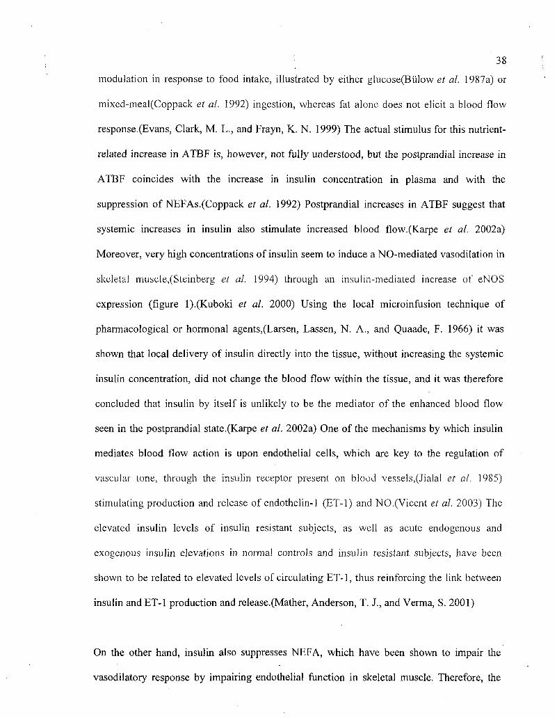
modulation in response to food intake, illustrated by either glucose(Biilow et al. 1987a) or
mixed-meal(Coppack et al. 1992) ingestion, whereas fat alone does not elicit a blood flow
response.(Evans, Clark, M. L., and Frayn, K. N. 1999) The actual stimulus for this nutrient-
related increase in ATBF is, however, not fully understood, but the postprandial increase in
ATBF coincides with the increase in insulin concentration in plasma and with the
suppression of NEFAs.(Coppack et al. 1992) Postprandial increases in ATBF suggest that
systemic increases in insulin also stimulate increased blood flow.(Karpe et al. 2002a)
Moreover, very high concentrations of insulin seem to induce a NO-mediated vasodilation in
skeletal muscle,(Steinberg et al. 1994) through an insulin-mediated increase of eNOS
expression (figure l).(Kuboki et al. 2000) Using the local microinfusion technique of
pharmacological or hormonal agents,(Larsen, Lassen, N. A., and Quaade, F. 1966) it was
shown that local delivery of insulin directly into the tissue, without increasing the systemic
insulin concentration, did not change the blood flow within the tissue, and it was therefore
concluded that insulin by itself is unlikely to be the mediator of the enhanced blood flow
seen in the postprandial state.(Karpe et al. 2002a) One of the mechanisms by which insulin
mediates blood flow action is upon endothelial cells, which are key to the regulation of
vascular tone, through the insulin receptor present on blood vessels,(Jialal et al. 1985)
stimulating production and release of endothelin-1 (ET-1) and NO.(Vicent et al. 2003) The
elevated insulin levels of insulin resistant subjects, as well as acute endogenous and
exogenous insulin elevations in normal controls and insulin resistant subjects, have been
shown to be related to elevated levels of circulating ET-1, thus reinforcing the link between
insulin and ET-1 production and release.(Mather, Anderson, T. J., and Verma, S. 2001)
On the other hand, insulin also suppresses NEFA, which have been shown to impair the
vasodilatory response by impairing endothelial function in skeletal muscle. Therefore, the

: ; 39
elevated levels of NEFA observed in insulin resistance syndrome could be the cause to the
endothelial dysfunction.(Steinberg et al. 1997) Obviously, compared with skeletal muscle,
the environment within adipose tissue is subject to much larger fluctuations in NEFA
concentration. Thus, if the physiological relationship between fluctuation in NEFA
concentrations and blood flow exists, adipose tissue would be the primary organ for such
regulation. However, direct infusion of insulin in adipose tissue does not affect ATBF. May
be because such a concentration of insulin certainly would reduce the NEFA levels, and
therefore it can be deduced that this reduction had no effect on ATBF.
Nitric oxide
The formation of nitric oxide (NO) has been demonstrated in human adipocytes and
preadipocytes.(Andersson et al. 1999; Ribiere et al. 1996) NO is released in white adipose
tissue by preadipocytes, adipocytes, vascular endothelial cells, and vascular smooth muscle
cells.(Gaudiot et al. 2000; Linz et al. 1999; Ribiere et al. 1996) The enzymes responsible are
endothelial NO synthase (eNOS) in vascular endothelial cells and adipocytes as well as the
inducible NO synthase (iNOS) in all cell types when appropriately stimulated. There is a
constitutive expression of iNOS, although at low levels, in white adipose tissue. In vitro, NO
formation in adipose tissue is upregulated by lipopolysaccharides, cytokines such as TNF-a
and interferon-y, noradrenalin, insulin and angiotensine (Ang) II.(Hennington et al. 1998)
NO can activate soluble guanylate cyclase, resulting in cGMP formation(Gaudiot et al. 2000)
and the subsequent cGMP-dependant protein kinases signal explain the biological effects of
NO.(Ignarro 2002) Moreover, mRNA expression of several subunits of the soluble
guanylylcyclase and the two isoforms of the cGMP-dependant protein kinase was reported in
isolated human adipocytes and preadipocytes.(Engeli and Sharma, A. M. 2000)

• 4 0
Moreover, it has been shown that high concentrations of NO inhibit lipolysis stimulated by
different pathways, depending on the NO species (NO+, NO", NO"). NO+ increases basal
lipolysis, whereas NO' and NO" show no effect on basal lipolysis. Low levels of intracellular
NO, on the other hand, are necessary for both basal and stimulated lipolysis, because they
keep protein kinase A in an active state due to NO's antioxidant-related properties.(Gaudiot
et al. 2000) NO is also responsible for blunting the TNF-ct-stimulated lipoprotein lipase
(LPL)-activity and uncoupling protein-2 (UCP2) expression. Thus NO appears to function to
keep adipocytes in their present status, inhibiting lipolysis and energy dissipation as well as
lipogenesis. In addition to its effects on adipocytes, NO is a strong vasodilator in vascular
smooth muscle cells(Linz et al. 1999) and takes part in signaling processes leading to
angiogenesis in vascular endothelial cells.(Pedram, Razandi, M., and Levin, E. R. 2001)
In concert with the blood flow regulation in many other tissues, NO could be a major
vasodilator in adipose tissue, or possibly a direct or indirect mediator of insulin
vasodilation.(Steinberg et al. 1994) After a NO-mediated vasodilation was described during
an euglycaemic hyperinsulinemic clamp giving high concentrations of insulin,(Steinberg et
al. 1994) it has been shown in vitro that e-NOS is regulated at the level of expression and
activity by insulin.(Kuboki et al. 2000) However, it has been pointed out that the
vasodilatory effect of insulin occurs only at pharmacologic insulin doses.(Yki-Jarvinen and
Utriainen, T. 1998) However, JVG-monomethyl-Larginine (L-NMMA), an NO synthase
inhibitor, did not seem to alter local ATBF(Andersson et al. 1999) and attenuated the
isoproterenol-induced vasodilation(Jordan et al. 2001) only during adipose tissue
microdialysis, suggesting that NO was not a mediator of ATBF. These measurements were
made by use of the ethanol outflow/inflow ratio, which has an inherently low
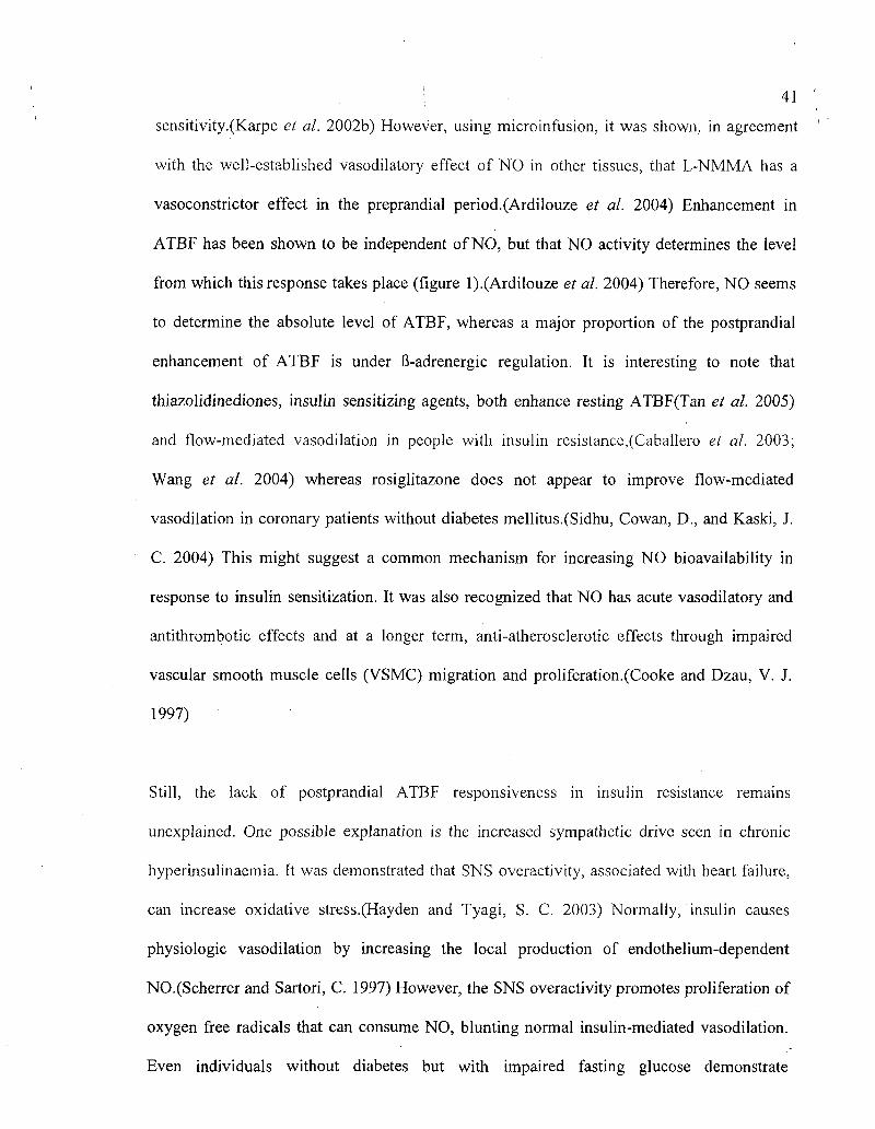
41
sensitivity.(Karpe et al. 2002b) However, using microinfusion, it was shown, in agreement
with the well-established vasodilatory effect of NO in other tissues, that L-NMMA has a
vasoconstrictor effect in the preprandial period.(Ardilouze et al. 2004) Enhancement in
ATBF has been shown to be independent of NO, but that NO activity determines the level
from which this response takes place (figure 1).(Ardilouze et al. 2004) Therefore, NO seems
to determine the absolute level of ATBF, whereas a major proportion of the postprandial
enhancement of ATBF is under B-adrenergic regulation. It is interesting to note that
thiazolidinediones, insulin sensitizing agents, both enhance resting ATBF(Tan et al. 2005)
and flow-mediated vasodilation in people with insulin resistance,(Caballero et al. 2003;
Wang et al. 2004) whereas rosiglitazone does not appear to improve flow-mediated
vasodilation in coronary patients without diabetes mellitus.(Sidhu, Cowan, D., and Kaski, J.
C. 2004) This might suggest a common mechanism for increasing NO bioavailability in
response to insulin sensitization. It was also recognized that NO has acute vasodilatory and
antithrombotic effects and at a longer term, anti-atherosclerotic effects through impaired
vascular smooth muscle cells (VSMC) migration and proliferation.(Cooke and Dzau, V. J.
1997)
Still, the lack of postprandial ATBF responsiveness in insulin resistance remains
unexplained. One possible explanation is the increased sympathetic drive seen in chronic
hyperinsulinaemia. It was demonstrated that SNS overactivity, associated with heart failure,
can increase oxidative stress.(Hayden and Tyagi, S. C. 2003) Normally, insulin causes
physiologic vasodilation by increasing the local production of endothelium-dependent
NO.(Scherrer and Sartori, C. 1997) However, the SNS overactivity promotes proliferation of
oxygen free radicals that can consume NO, blunting normal insulin-mediated vasodilation.
Even individuals without diabetes but with impaired fasting glucose demonstrate

! '. 42
significantly lowered endothelium-dependent vasodilation compared with control
subjects.(Vehkavaara et ah 1999)
Renin angiotensin system
Renin angiotensin system (RAS) is a major player in the vascular tone and it is not
impossible that RAS can also have some implications in ATBF regulation. Angiotensin
peptides are cleaved from circulating angiotensinogen produced in the liver. Several proteins
of the classic RAS are also produced in adipose tissue. These include renin, angiotensinogen,
Ang I, Ang II, Ang type 1 (ATi) and type 2 (AT2) receptors, Angiotensin converting enzyme
(ACE), and other proteases capable of producing Ang II like chymase, cathepsins D and G
and tonin.(Engeli et ah 2003; Trayhurn and Beattie, J. H. 2001) The first peptide to emerge is
Ang I, which is devoid of biological activity. It can be cleaved to Ang II by ACE, which is
present on preadipocytes, adipocytes, vascular endothelial cells, and vascular smooth muscle
cells, or to Ang 1-7 by neutral endopeptidase, which is expressed on vascular endothelial
cells.(Engeli, Negrel, R., and Sharma, A. M. 2000) Ang 1-7 stimulates vascular endothelial
cells to secrete NO and prostacyclin, leading to vasodilation, possibly by potentiating the
action of kinins.(Linz et ah 1999) In addition to hepatic angiotensinogen production,
adipocytes have also shown to be an important source of Ang II precursor, one of the major
vasoconstrictive and proatherogenic peptide.(Fruhbeck et ah 2001; Lavoie and Sigmund, C.
D. 2003; Safonova et ah 1997; Trayhurn and Beattie, J. H. 2001) The expression of
angiotensinogen, ACE and ATi receptors is higher in visceral adipose tissue compared to
subcutaneous adipose tissue.(Engeli et ah 2003; Goossens, Blaak, E. E., and van Baak, M. A.
2003) Consequently, a role in local ATBF and fatty acid re-esterification rates have been
suggested for. angiotensinogen. Furthermore, components of the RAS such as

•: 43
i angiotensinogen and Ang II are induced during adipogenesis. Moreover, Ang II promotes
adipocyte growth and differentiation, both directly by promoting lipogenesis and indirectly
by stimulating prostaglandin synthesis. (Engeli et al. 2003) Ang II from mature adipocytes,
however, may inhibit further recruitment of preadipocytes.(Engeli et al. 2003) Some
lipophilic angiotensin receptor blockers (ARBs) appear to induce PPARy activity in adipose
tissue. Therefore, the type 2 diabetes protection observed after RAS inhibition may be
partially linked to a thiazolidinedione-like effect.(Towfighi and Ovbiagele, B. 2008; Tuck
and Corry, Dalila B 2005) TNF-cc, an inducer of angiotensinogen in hepatocytes, is elevated
in hyperinsulinemic, obese individuals and may provide a link in mediating insulin
upregulation of the RAS in adipose tissue. Furthermore, thiazolidinediones lower blood
pressure in vivo, and downregulation of the RAS in adipose tissue may contribute to this
effect.(Harte et al. 2005) Although Ang II is best known for its role in the regulation of
vascular tone, aldosterone secretion from adrenal gland and sodium and water reabsorption
from the kidney, all of which contribute to blood pressure regulation, it can also affect cell
growth and differentiation.(Engeli et al. 2003) Thus, the adipose tissue RAS is a potential
link between obesity and hypertension. Ang II stimulates the prostacyclin production and
release, which in turn is a factor that participate in the induction of preadipocytes
differentiation to mature adipocytes.(Zorad et al. 1995) In contrast, Ang II has been shown to
inhibits adipose conversion of preadipocytes.(Schling and Loffler, G. 2001) There is some
evidence showing that the expression of angiotensinogen is regulated by nutritional and
hormonal factors. Thus, it was previously demonstrated that angiotensinogen expression
increased in obesity and appear to be regulated by the nutritional status with a decrease
mRNA levels during fasting and an increase upon refeeding and these changes are
accompanied by parallel changes in blood pressure. Consequently, a role in local ATBF and
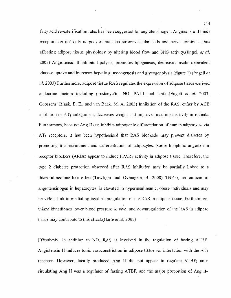
! 44
fatty acid re-esterification rates has been suggested for angiotensinogen. Angiotensin II binds
receptors on not only adipocytes but also stromavascular cells and nerve terminals, thus
affecting adipose tissue physiology by altering blood flow and SNS activity.(Engeli et al.
2003) Angiotensin II inhibits lipolysis, promotes lipogenesis, decreases insulin-dependent
glucose uptake and increases hepatic glucoeogenesis and glycogenolysis (figure 1).(Engeli et
al. 2003) Furthermore, adipose tissue RAS regulates the expression of adipose tissue-derived
endocrine factors including prostacyclin, NO, PAI-1 and leptin.(Engeli et al. 2003;
Goossens, Blaak, E. E., and van Baak, M. A. 2003) Inhibition of the RAS, either by ACE
inhibition or ATi antagonism, decreases weight and improves insulin sensitivity in rodents.
Furthermore, because Ang II can inhibits adipogenic differentiation of human adipocytes via
ATi receptors, it has been hypothesised that RAS blockade may prevent diabetes by
promoting the recruitment and differentiation of adipocytes. Some lipophilic angiotensin
receptor blockers (ARBs) appear to induce PPARy activity in adipose tissue. Therefore, the
type 2 diabetes protection observed after RAS inhibition may be partially linked to a
thiazolidinedione-like effect.(Towfighi and Ovbiagele, B. 2008) TNF-a, an inducer of
angiotensinogen in hepatocytes, is elevated in hyperinsulinemic, obese individuals and may
provide a link in mediating insulin upregulation of the RAS in adipose tissue. Furthermore,
thiazolidinediones lower blood pressure in vivo, and downregulation of the RAS in adipose
tissue may contribute to this effect.(Harte et al. 2005)
Effectively, in addition to NO, RAS is involved in the regulation of fasting ATBF.
Angiotensin II induces tonic vasoconstriction in adipose tissue via interaction with the ATi
receptor. However, locally produced Ang II did not appear to regulate ATBF; only
circulating Ang II was a regulator of fasting ATBF, and the major proportion of Ang II-

: < 45
induced vasoconstriction was NO independent.(Masegosa et al. 1992) Furthermore, as NO,
Ang II has no major effect on the postprandial enhancement of ATBF.(Masegosa et al. 1992)
Moreover, Ang II is known to potentiate the inhibitory effects of NEFA on insulin-mediated
glucose uptake and systemic blockade of the RAS by inhibition of the ACE, has been shown
to improve insulin sensitivity and possibly prevent the development of type 2
diabetes.(Hansson et al. 1999) In addition to its well-known vasoconstricting effect,
Boschmann et al. showed that Ang II inhibits lipolysis and glucose uptake in adipose
tissue.(Boschmann et al. 2001) They also demonstrated that abdominal fat depots seem to be
more sensitive to these effects of Ang II than femoral adipose tissue during stimulation with
isoproterenol, whereas under baseline conditions, femoral fat depots seem to be more
sensitive to Ang II.(Boschmann et al. 2001) Moreover, Ang II induces many vascular effects
besides vasoconstriction such as vascular remodeling and plaque rupture.(Schiffrin 2002)
Therefore, a role for local Ang II beyound its classical systemic effect on blood pressure
regulation link adipose tissue mass enlargement with hypertension via angiotensinogen
production. Furthermore, we should keep in mind that a large part of the growth and
inflammatory responses of Ang II are mediated by oxidative stress stimulation.
Monocyte/macrophage adhesion and accumulation are prevented by NO, which is
counterbalanced by Ang II, probably by the generation of reactive oxygen species
(ROS).(Lyon, Law, R. E., and Hsueh, W. A. 2003) Therefore, an imbalance between Ang II
and NO leads endothelial dysfunction with lost of vasodilator capacity, an increased
monocyte/macrophage infiltration and the increase of pro-coagulant activity in the vessel.

! 46
Other vasoactive regulators
Kinins are derived from kininogen by the action of kallikrein. It is still unresolved whether
kininogen and kallikrein are acquired from the circulation or if they are locally produced.
The circulating kallikrein kinin system can be activated on vascular endothelial cells, but
studies with rat adipocytes also indicate a local production. Kinins elicit NO and prostacyclin
secretion from vascular endothelial cells and are therefore potent vasodilators.(Linz et al.
1999) They also increase the permeability of the endothelium and lead to marked
enhancement of the insulin sensitivity of adipocytes.(Caldiz and de Cingolani, G. E. 1999)
The presence of circulating RAS components makes kinins as potential vasoactive regulators
in ATBF.
Endothelin is produced in vascular endothelial cells through cleavage of proendothelin to big
endothelin and subsequent conversion to endothelin by endothelin-converting
enzyme.(Kentsch and Otter, W. 1999) Endothelin-1 (ET-1) is a peptide of 21 amino acid
residues, the most potent vasoconstrictor substance known.(Camarda et al. 2002) ET-1
release is stimulated by angiotensin II, antidiuretic hormone, thrombin, cytokines, ROS,
calcium ions, adrenergic receptors, various cytokines and shearing forces acting on the
vascular endothelium. Insulin has been shown to directly stimulate ET-1 gene transcription
in endothelial cells.(Oliver et al. 1991) ET-1 release is inhibited by NO, as well as by
prostacyclin and atrial natriuretic peptide (ANP). The vascular endothelium is the most
abundant source of ET-1 in vz'vo(Masaki 2000) and endothelin is secreted mainly abluminally
and acts in a paracrine and autocrine fashion on cells in its immediate vicinity.(Kentsch and
Otter, W. 1999) VSMC respond with long-lasting constriction, whereas vascular endothelial
cells react with short-term release of NO and prostacyclin, although vasoconstriction
predominates in adipose tissue vasculature. Endothelin is a strong inhibitor of preadipocyte
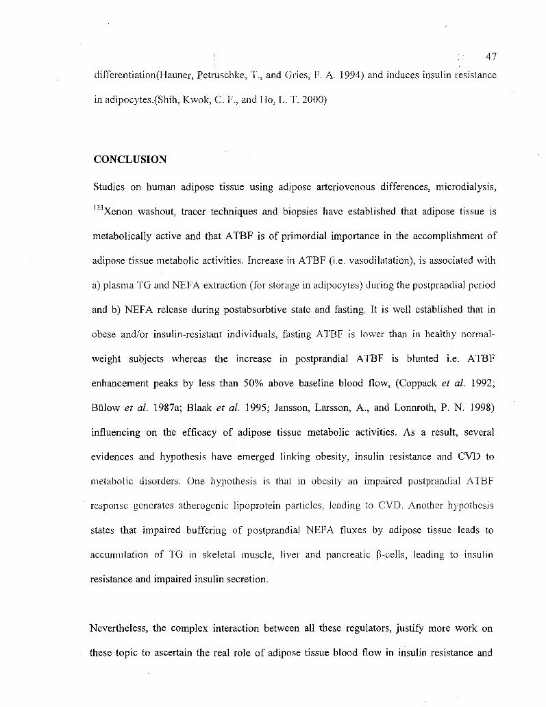
! : 47
differentiation(Hauner, Petruschke, T., and Gries, F. A. 1994) and induces insulin resistance
in adipocytes.(Shih, Kwok, C. F., and Ho, L. T. 2000)
CONCLUSION
Studies on human adipose tissue using adipose arteriovenous differences, microdialysis,
Xenon washout, tracer techniques and biopsies have established that adipose tissue is
metabolically active and that ATBF is of primordial importance in the accomplishment of
adipose tissue metabolic activities. Increase in ATBF (i.e. vasodilatation), is associated with
a) plasma TG and NEFA extraction (for storage in adipocytes) during the postprandial period
and b) NEFA release during postabsorbtive state and fasting. It is well established that in
obese and/or insulin-resistant individuals, fasting ATBF is lower than in healthy normal-
weight subjects whereas the increase in postprandial ATBF is blunted i.e. ATBF
enhancement peaks by less than 50% above baseline blood flow, (Coppack et al. 1992;
Biilow et al. 1987a; Blaak et al. 1995; Jansson, Larsson, A., and Lonnroth, P. N. 1998)
influencing on the efficacy of adipose tissue metabolic activities. As a result, several
evidences and hypothesis have emerged linking obesity, insulin resistance and CVD to
metabolic disorders. One hypothesis is that in obesity an impaired postprandial ATBF
response generates atherogenic lipoprotein particles, leading to CVD. Another hypothesis
states that impaired buffering of postprandial NEFA fluxes by adipose tissue leads to
accumulation of TG in skeletal muscle, liver and pancreatic P-cells, leading to insulin
resistance and impaired insulin secretion.
Nevertheless, the complex interaction between all these regulators, justify more work on
these topic to ascertain the real role of adipose tissue blood flow in insulin resistance and

'. '. 48
other associated pathologies. Thus, the regulation of ATBF is complex and still not fully
understood. In the actual context of world obesity epidemic, it is essential to pursue the
research on ATBF regulation for a better understanding of the mechanisms of insulin
resistance syndrome development to diabetes and cardiovascular diseases. A systems biology
approach incorporating genomics, proteomics, metabolomics and integrative physiology into
in vivo adipose tissue studies will clarify the links between adipose tissue and vascular
disease and raised the physiological importance of ATBF regulation in the general metabolic
homeostasis.
•: 49
REFERENCES
Ahren, B. 2003. Gut peptides and type 2 diabetes mellitus treatment. Curr. Diab. Rep. 3(5):
365-372.
Andersson, K. and Arner, P. 1995. Cholinoceptor-mediated effects on glycerol output from
human adipose tissue using in situ microdialysis. Br. J. Pharmacol. 115(7): 1155-1162.
Andersson, K. and Arner, P. 2001. Systemic nicotine stimulates human adipose tissue
lipolysis through local cholinergic and catecholaminergic receptors. Int. J. Obes. Relat Metab
Disord. 25(8): 1225-1232.
Andersson, K., Gaudiot, N., Ribiere, C, Elizalde, M., Giudicelli, Y., and Arner, P. 1999. A
nitric oxide-mediated mechanism regulates lipolysis in human adipose tissue in vivo. Br. J.
Pharmacol. 126(7): 1639-1645.
Ardilouze, J.L., Fielding, B. A., Currie, J. M., Frayn, K. N., and Karpe, F. 2004. Nitric oxide
and beta-adrenergic stimulation are major regulators of preprandial and postprandial
subcutaneous adipose tissue blood flow in humans. Circulation 109(1): 47-52.
Balldin, U.I. 1976. The effects of body position and a vasodilator on xenonl33 elimination
from human subcutaneous fat. Undersea Biomed. Res. 3(4): 379-385.
Barbe, P., Millet, L., Galitzky, J., Lafontan, M., and Berlan, M. 1996. In situ assessment of
the role of the beta 1-, beta 2- and beta 3-adrenoceptors in the control of lipolysis and
nutritive blood flow in human subcutaneous adipose tissue. Br. J. Pharmacol. 117(5): 907-
913.

50
Baron, A.D., Tarshoby, M., Hook, G., Lazaridis, E. N., Cronin, J., Johnson, A., and
Steinberg, H. O. 2000. Interaction between insulin sensitivity and muscle perfusion on
glucose uptake in human skeletal muscle: evidence for capillary recruitment. Diabetes 49(5):
768-774.
Bartness, T.J. 2002. Dual innervation of white adipose tissue: some evidence for
parasympathetic nervous system involvement. J. Clin. Invest 110(9): 1235-1237.
Belfiore, F., Iannello, S., and Volpicelli, G. 1998. Insulin sensitivity indices calculated from
basal and OGTT-induced insulin, glucose, and FFA levels. Mol. Genet. Metab 63(2): 134-
141.
Bertin, E., Arner, P., Bolinder, J., and Hagstrom-Toft, E. 2001. Action of glucagon and
glucagon-like peptide-l-(7-36) amide on lipolysis in human subcutaneous adipose tissue and
skeletal muscle in vivo. J. Clin. Endocrinol. Metab 86(3): 1229-1234.
Blaak, E.E., van Baak, M. A., Kemerink, G. J., Pakbiers, M. T., Heidendal, G. A., and Saris,
W. H. 1995. Beta-adrenergic stimulation and abdominal subcutaneous fat blood flow in lean,
obese, and reduced-obese subjects. Metabolism 44(2): 183-187.
Boschmann, M., Ringel, J., Klaus, S., and Sharma, A. M. 2001. Metabolic and hemodynamic
response of adipose tissue to angiotensin II. Obes. Res. 9(8): 486-491.
Brassard, P., Frisch, F., Lavoie, F., Cyr, D., Bourbonnais, A., Cunnane, S. C, Patterson, B.
W., Drouin, R., Baillargeon, J. P., and Carpentier, A. C. 2008. Impaired plasma nonesterified
fatty acid tolerance is an early defect in the natural history of type 2 diabetes. J. Clin.
Endocrinol. Metab 93(3): 837-844.

5 1
Biilow, J.' 1981. Human adipose tissue blood flow during prolonged exercise, III. Effect of
beta-adrenergic blockade, nicotinic acid and glucose infusion. Scand. J. Clin. Lab Invest
41(4): 415-424.
Biilow, J., Astrup, A., Christensen, N. J., and Kastrup, J. 1987a. Blood flow in skin,
subcutaneous adipose tissue and skeletal muscle in the forearm of normal man during an oral
glucose load. Acta Physiol Scand. 130(4): 657-661.
Biilow, J., Jelnes, R., Astrup, A., Madsen, J., and Vilmann, P. 1987b. Tissue/blood partition
coefficients for xenon in various adipose tissue depots in man. Scand. J. Clin. Lab Invest
47(1): 1-3.
Caballero, A.E., Saouaf, R., Lim, S. C, Hamdy, O., Abou-Elenin, K., O'Connor, C, Logerfo,
F. W., Horton, E. S., and Veves, A. 2003. The effects of troglitazone, an insulin-sensitizing
agent, on the endothelial function in early and late type 2 diabetes: a placebo-controlled
randomized clinical trial. Metabolism 52(2): 173-180.
Caldiz, C.I. and de Cingolani, G. E. 1999. Insulin resistance in adipocytes from
spontaneously hypertensive rats: effect of long-term treatment with enalapril and losartan.
Metabolism 48(8): 1041-1046.
Camarda, V., Rizzi, A., Calo, G., Gendron, G., Perron, S. I., Kostenis, E., Zamboni, P.,
Mascoli, F., and Regoli, D. 2002. Effects of human urotensin II in isolated vessels of various
species; comparison with other vasoactive agents. Naunyn Schmiedebergs Arch. Pharmacol.
365(2): 141-149.
Carpentier, A.C. 2008. Postprandial fatty acid metabolism in the development of lipotoxicity
and type 2 diabetes. Diabetes Metab 34(2): 97-107.
52
Carpentier, A.C., Frisch, F., Brassard, P., Lavoie, F., Bourbonnais, A., Cyr, D., Giguere, R.,
and Baillargeon, J. P. 2007. Mechanism of insulin-stimulated clearance of plasma
nonesterified fatty acids in humans. Am. J. Physiol Endocrinol. Metab 292(3): E693-E701.
Cooke, J.P. and Dzau, V. J. 1997. Nitric oxide synthase: role in the genesis of vascular
disease. Annu. Rev. Med 48489-509.
Coppack, S.W., Evans, R. D., Fisher, R. M., Frayn, K. N., Gibbons, G. F., Humphreys, S.
M., Kirk, M. L., Potts, J. L., and Hockaday, T. D. 1992. Adipose tissue metabolism in
obesity: lipase action in vivo before and after a mixed meal. Metabolism 41(3): 264-272.
Coppack, S.W., Fisher, R. M., Gibbons, G. F., Humphreys, S. M., McDonough, M. J., Potts,
J. L., and Frayn, K. N. 1990a. Postprandial substrate deposition in human forearm and
adipose tissues in vivo. Clin. Sci. (Lond) 79(4): 339-348.
Coppack, S.W., Frayn, K. N., Humphreys, S. M., Whyte, P. L., and Hockaday, T. D. 1990b.
Arteriovenous differences across human adipose and forearm tissues after overnight fast.
Metabolism 39(4): 384-390.
DeFronzo, R.A. 2004. Dysfunctional fat cells, lipotoxicity and type 2 diabetes. Int. J. Clin.
Pract. Suppl(143):9-21.
Dimitriadis, G., Lambadiari, V., Mitrou, P., Maratou, E., Boutati, E., Panagiotakos, D. B.,
Economopoulos, T., and Raptis, S. A. 2007. Impaired postprandial blood flow in adipose
tissue may be an early marker of insulin resistance in type 2 diabetes. Diabetes Care 30(12):
3128-3130.
Donovan, CM., Cane, P., and Bergman, R. N. 1991. Search for the hypoglycemia receptor
using the local irrigation approach. Adv. Exp. Med Biol 291185-196.
: ': 53
Elia, M. and Kurpad, A. 1993. What is the blood flow to resting human muscle? Clin. Sci.
(Lond) 84(5): 559-563.
Engeli, S., Negrel, R., and Sharma, A. M. 2000. Physiology and pathophysiology of the
adipose tissue renin-angiotensin system. Hypertension 35(6): 1270-1277.
Engeli, S., Schling, P., Gorzelniak, K., Boschmann, M, Janke, J., Ailhaud, G., Teboul, M.,
Massiera, F., and Sharma, A. M. 2003. The adipose-tissue renin-angiotensin-aldosterone
system: role in the metabolic syndrome? Int. J. Biochem. Cell Biol 35(6): 807-825.
Engeli, S. and Sharma, A. M. 2000. Role of adipose tissue for cardiovascular-renal
regulation in health and disease. Horm. Metab Res. 32(11-12): 485-499.
Evans, K., Clark, M. L., and Frayn, K. N. 1999. Effects of an oral and intravenous fat load on
adipose tissue and forearm lipid metabolism. Am. J. Physiol 276(2 Pt 1): E241-E248.
Fagius, J. and Karhuvaara, S. 1989. Sympathetic activity and blood pressure increases with
bladder distension in humans. Hypertension 14(5): 511-517.
Fernqvist, E. and Linde, B. 1988. Potent mental stress and insulin absorption in normal
subjects. Diabetes Care 11(8): 650-655.
Flechtner-Mors, M., Jenkinson, C. P., Alt, A., Adler, G., and Ditschuneit, H. H. 2002. In
vivo alpha(l)-adrenergic lipolytic activity in subcutaneous adipose tissue of obese subjects.
J. Pharmacol. Exp. Ther. 301(1): 229-233.
Frayn, K.N., Humphreys, S. M., and Coppack, S. W. 1995. Fuel selection in white adipose
tissue. Proc. Nutr. Soc. 54(1): 177-189.
Frayn, K.N., Karpe, F., Fielding, B. A., Macdonald, I. A., and Coppack, S. W. 2003.
Integrative physiology of human adipose tissue. Int. J. Obes. Relat Metab Disord. 27(8): 875-
888.
Fredholm, B.B. and Sollevi, A. 1981. The release of adenosine and inosine from canine
subcutaneous adipose tissue by nerve stimulation and noradrenaline. J. Physiol 313351-367.
Fruhbeck, G., Gomez-Ambrosi, J., Muruzabal, F. J., and Burrell, M. A. 2001. The adipocyte:
a model for integration of endocrine and metabolic signaling in energy metabolism
regulation. Am. J. Physiol Endocrinol. Metab 280(6): E827-E847.
Galitzky, J., Lafontan, M, Nordenstrom, J., and Arner, P. 1993. Role of vascular alpha-2
adrenoceptors in regulating lipid mobilization from human adipose tissue. J. Clin. Invest
91(5): 1997-2003.
Gaudiot, N., Ribiere, C., Jaubert, A. M., and Giudicelli, Y. 2000. Endogenous nitric oxide is
implicated in the regulation of lipolysis through antioxidant-related effect. Am. J. Physiol
Cell Physiol 279(5): C1603-C1610.
Golpon, H.A., Puechner, A., Welte, T., Wichert, P. V., and Feddersen, C. O. 2001.
Vasorelaxant effect of glucagon-like peptide-(7-36)amide and amylin on the pulmonary
circulation of the rat. Regul. Pept. 102(2-3): 81-86.
Goossens, G.H., Blaak, E. E., and van Baak, M. A. 2003. Possible involvement of the
adipose tissue renin-angiotensin system in the pathophysiology of obesity and obesity-related
disorders. Obes. Rev. 4(1): 43-55.

55
Green, B.D., Hand, K. V., Dougan, J. El, McDonnell, B. M, Cassidy, R. S., and Grieve, D. J.
2008. GLP-1 and related peptides cause concentration-dependent relaxation of rat aorta
through a pathway involving KATP and cAMP. Arch. Biochem. Biophys. 478(2): 136-142.
Hagstrom-Toft, E., Bolinder, J., Ungerstedt, U., and Arner, P. 1997. A circadian rhythm in
lipid mobilization which is altered in IDDM. Diabetologia 40(9): 1070-1078.
Hansson, L., Lindholm, L. H., Niskanen, L., Lanke, J., Hedner, T., Niklason, A.,
Luomanmaki, K., Dahlof, B., de Faire, U., Morlin, C, Karlberg, B. E., Wester, P. O., and
Bjorck, J. E. 1999. Effect of angiotensin-converting-enzyme inhibition compared with
conventional therapy on cardiovascular morbidity and mortality in hypertension: the
Captopril Prevention Project (CAPPP) randomised trial. Lancet 353(9153): 611-616.
Harte, A., McTernan, P., Chetty, R., Coppack, S., Katz, J., Smith, S., and Kumar, S. 2005.
Insulin-mediated upregulation of the renin angiotensin system in human subcutaneous
adipocytes is reduced by rosiglitazone. Circulation 111(15): 1954-1961.
Hauner, H. and Hochberg, Z. 2002. Endocrinology of adipose tissue. Horm. Metab Res.
34(11-12): 605-606.
Hauner, H., Petruschke, T., and Gries, F. A. 1994. Endothelin-1 inhibits the adipose
differentiation of cultured human adipocyte precursor cells. Metabolism 43(2): 227-232.
Hayden, M.R. and Tyagi, S. C. 2003. Myocardial redox stress and remodeling in metabolic
syndrome, type 2 diabetes mellitus, and congestive heart failure. Med Sci. Monit. 9(7):
SR35-SR52.

Hennington, B.S., Zhang, H., Miller, M. T., Granger, J. P., and Reckelhoff, J. F. 1998.
Angiotensin II stimulates synthesis of endothelial nitric oxide synthase. Hypertension 31(1 Pt
2): 283-288.
Henriksen, E.J. 2002. Invited review: Effects of acute exercise and exercise training on
insulin resistance. J. Appl. Physiol 93(2): 788-796.
Hjemdahl, P., Linde, B., Daleskog, M., and Belfrage, E. 1983. Sympathoadrenal regulation
of adipose tissue blood flow in dog and man. Gen. Pharmacol. 14(1): 175-177.
Hodgetts, V., Coppack, S. W., Frayn, K. N., and Hockaday, T. D. 1991. Factors controlling
fat mobilization from human subcutaneous adipose tissue during exercise. J. Appl. Physiol
71(2): 445-451.
Horowitz, J.F., Braudy, R. J., Martin, W. H., Ill, and Klein, S. 1999. Endurance exercise
training does not alter lipolytic or adipose tissue blood flow sensitivity to epinephrine. Am. J.
Physiol 277(2 Pt 1): E325-E331.
Houmard, J.A., Tanner, C. J., Slentz, C. A., Duscha, B. D., McCartney, J. S., and Kraus, W.
E. 2004. Effect of the volume and intensity of exercise training on insulin sensitivity. J.
Appl. Physiol 96(1): 101-106.
Ignarro, L.J. 2002. Wei Lun Visiting Professorial Lecture: Nitric oxide in the regulation of
vascular function: an historical overview. J. Card Surg. 17(4): 301-306.
Jansson, P.A., Larsson, A., and Lonnroth, P. N. 1998. Relationship between blood pressure,
metabolic variables and blood flow in obese subjects with or without non-insulin-dependent
diabetes mellitus. Eur. J. Clin. Invest 28(10): 813-818.

i 5 7
Jansson, P.A., Larsson, A., Smith, U., and Lonnroth, P. 1992. Glycerol production in
subcutaneous adipose tissue in lean and obese humans. J. Clin. Invest 89(5): 1610-1617.
Jensen, M.D. 2002. Adipose tissue and fatty acid metabolism in humans. J. R. Soc. Med 95
SuppI 423-7.
Jialal, I., Crettaz, M., Hachiya, H. L., Kahn, C. R., Moses, A. C, Buzney, S. M., and King,
G. L. 1985. Characterization of the receptors for insulin and the insulin-like growth factors
on micro- and macrovascular tissues. Endocrinology 117(3): 1222-1229.
Jordan, J., Tank, J., Stoffels, M., Franke, G., Christensen, N. J., Luft, F. C, and Boschmann,
M. 2001. Interaction between beta-adrenergic receptor stimulation and nitric oxide release on
tissue perfusion and metabolism. J. Clin. Endocrinol. Metab 86(6): 2803-2810.
Joyner, L.P. and Norton, C. C. 1976. The immunity arising from continuous low-level
infection with Eimeria maxima and Eimeria acervulina. Parasitology 72(1): 115-125.
Karpe, F., Fielding, B. A., Ardilouze, J. L., Ilic, V., Macdonald, I. A., and Frayn, K. N.
2002a. Effects of insulin on adipose tissue blood flow in man. J. Physiol 540(Pt 3): 1087-
1093.
Karpe, F., Fielding, B. A., Ilic, V., Humphreys, S. M., and Frayn, K. N. 2002b. Monitoring
adipose tissue blood flow in man: a comparison between the (133)xenon washout method
and microdialysis. Int. J. Obes. Relat Metab Disord. 26(1): 1-5.
Karpe, F., Fielding, B. A., Ilic, V., Macdonald, I. A., Summers, L. K., and Frayn, K. N.
2002c. Impaired postprandial adipose tissue blood flow response is related to aspects of
insulin sensitivity. Diabetes 51(8): 2467-2473.

• '58
Karpe, F., Olivecrona, T., Olivecrbna, G., Samra, J. S., Summers, L. K., Humphreys, S. M.',
and Frayn, K. N. 1998. Lipoprotein lipase transport in plasma: role of muscle and adipose
tissues in regulation of plasma lipoprotein lipase concentrations. J. Lipid Res. 39(12): 2387-
2393.
Kentsch, M. and Otter, W. 1999. Novel neurohormonal modulators in cardiovascular
disorders. The therapeutic potential of endopeptidase inhibitors. Drugs R. D. 1(4): 331-338.
Klein, S., Horowitz, J. F., Landt, M., Goodrick, S. J., Mohamed-Ali, V., and Coppack, S. W.
2000. Leptin production during early starvation in lean and obese women. Am. J. Physiol
Endocrinol. Metab 278(2): E280-E284.
Kreier, F., Fliers, E., Voshol, P. J., Van Eden, C. G., Havekes, L. M., Kalsbeek, A., Van
Heijningen, C. L., Sluiter, A. A., Mettenleiter, T. C, Romijn, J. A., Sauerwein, H. P., and
Buijs, R. M. 2002. Selective parasympathetic innervation of subcutaneous and intra
abdominal fat—functional implications. J. Clin. Invest 110(9): 1243-1250.
Kreier, F., Yilmaz, A., Kalsbeek, A., Romijn, J. A., Sauerwein, H. P., Fliers, E., and Buijs,
R. M. 2003. Hypothesis: shifting the equilibrium from activity to food leads to autonomic
unbalance and the metabolic syndrome. Diabetes 52(11): 2652-2656.
Kuboki, K., Jiang, Z. Y., Takahara, N., Ha, S. W., Igarashi, M., Yamauchi, T., Feener, E. P.,
Herbert, T. P., Rhodes, C. J., and King, G. L. 2000. Regulation of endothelial constitutive
nitric oxide synthase gene expression in endothelial cells and in vivo : a specific vascular
action of insulin. Circulation 101(6): 676-681.
59
Laakso, M., Edelman, S. V., Olefsky, J. M., Brechtel, G., Wallace, P.,and Baron, A. D.
1990. Kinetics of in vivo muscle insulin-mediated glucose uptake in human obesity. Diabetes
39(8): 965-974.
Laaksonen, D.E., Nuutinen, J., Lahtinen, T., Rissanen, A., and Niskanen, L. K. 2003.
Changes in abdominal subcutaneous fat water content with rapid weight loss and long-term
weight maintenance in abdominally obese men and women. Int. J. Obes. Relat Metab Disord.
27(6): 677-683.
Lafontan, M. and Arner, P., 1996. Application of in situ microdialysis to measure metabolic
and vascular responses in adipose tissue. Trends Pharmacol. Sci. 17(9): 309-313.
Laine, H., Knuuti, M. J., Ruotsalainen, U., Utriainen, T., Oikonen, V., Raitakari, M.,
Luotolahti, M., Kirvela, O., Vicini, P., Cobelli, C, Nuutila, P., and Yki-Jarvinen, H. 1998.
Preserved relative dispersion but blunted stimulation of mean flow, absolute dispersion, and
blood volume by insulin in skeletal muscle of patients with essential hypertension.
Circulation 97(21): 2146-2153.
Lange, K.H., Lorentsen, J., Isaksson, F., Simonsen, L., Juul, A., Christensen, N. J., Kjaer,
M., and Bulow, J. 2002. Subcutaneous abdominal adipose tissue lipolysis during exercise
determined by arteriovenous measurements in older women. J. Am. Geriatr. Soc. 50(2): 275-
281.
Larsen, O.A., Lassen, N. A., and Quaade, F. 1966. Blood flow through human adipose tissue
determined with radioactive xenon. Acta Physiol Scand. 66(3): 337-345.
Lavoie, J.L. and Sigmund, C. D. 2003. Minireview: overview of the renin-angiotensin
system—an endocrine and paracrine system. Endocrinology 144(6): 2179-2183.

60
Lewis, G.F., Carpentier, A., Adeli, K., and Giacca, A. 2002. Disordered fat storage and
mobilization in the pathogenesis of insulin resistance and type 2 diabetes. Endocr. Rev.
23(2): 201-229.
Linz, W., Wohlfart, P., Scholkens, B. A., Malinski, T., and Wiemer, G. 1999. Interactions
among ACE, kinins and NO. Cardiovasc. Res. 43(3): 549-561.
Lyon, C.J., Law, R. E., and Hsueh, W. A. 2003. Minireview: adiposity, inflammation, and
atherogenesis. Endocrinology 144(6): 2195-2200.
Masaki, T. 2000. The endothelin family: an overview. J. Cardiovasc. Pharmacol. 35(4 Suppl
2): S3-S5.
Masegosa, M.A., Diaz, Torrens J., Gomez Ruiz, F. T., Sanchez, Guzman A., Gomez,
Caravaca J., and Donate, Perez F. 1992. [A spontaneous pseudoaneurysm of the superficial
femoral artery. Apropos a case]. Angiologia 44(3): 110-112.
Mather, K., Anderson, T. J., and Verma, S. 2001. Insulin action in the vasculature:
physiology and pathophysiology. J. Vase Res. 38(5): 415-422.
Meier, J.J., Gallwitz, B., Schmidt, W. E., andNauck, M. A. 2002. Glucagon-like peptide 1 as
a regulator of food intake and body weight: therapeutic perspectives. Eur. J. Pharmacol.
440(2-3): 269-279.
Millet, L., Barbe, P., Lafontan, M., Berlan, M., and Galitzky, J. 1998. Catecholamine effects
on lipolysis and blood flow in human abdominal and femoral adipose tissue. J. Appl. Physiol
85(1): 181-188.

61
Mohamed-Ali, V., Pinkney, J. H.; and Coppack, S. W. 1998. Adipose tissue as an endocrine
and paracrine organ. Int. J. Obes. Relat Metab Disord. 22(12): 1145-1158.
Oliver, F.J., de la, Rubia G., Feener, E. P., Lee, M. E., Loeken, M. R., Shiba, T.,
Quertermous, T., and King, G. L. 1991. Stimulation of endothelin-1 gene expression by
insulin in endothelial cells. J. Biol Chem. 266(34): 23251-23256.
Patel, J.N., Coppack, S. W., Goldstein, D. S., Miles, J. M., and Eisenhofer, G. 2002.
Norepinephrine spillover from human adipose tissue before and after a 72-hour fast. J. Clin.
Endocrinol. Metab 87(7): 3373-3377.
Pedram, A., Razandi, M., and Levin, E. R. 2001. Natriuretic peptides suppress vascular
endothelial cell growth factor signaling to angiogenesis. Endocrinology 142(4): 1578-1586.
Raitakari, M., Nuutila, P., Knuuti, J., Raitakari, O. T., Laine, H., Ruotsalainen, U., Kirvela,
O., Takala, T. O., Iida, H., and Yki-Jarvinen, H. 1997. Effects of insulin on blood flow and
volume in skeletal muscle of patients with IDDM: studies using [150]H20, [150]CO, and
positron emission tomography. Diabetes 46(12): 2017-2021.
Rask, E., Olsson, TV, Soderberg, S.; Hoist, Jj J., Tura, A., Pacini, G., and Ahren, B. 2004.
Insulin secretion and incretin hormones after oral glucose in non-obese subjects with
impaired glucose tolerance. Metabolism 53(5): 624-631.
Ribiere, C, Jaubert, A. M., Gaudiot, N., Sabourault, D., Marcus, M. L., Boucher, J. L.,
Denis-Henriot, D., and Giudicelli, Y. 1996. White adipose tissue nitric oxide synthase: a
potential source for NO production. Biochem. Biophys. Res. Commun. 222(3): 706-712.

• • 62
Romanski, S.A., Nelson, R. M., and Jensen, M. D. 2000. Meal fatty acid uptake in adipose
tissue: gender effects in nonobese humans. Am. J. Physiol Endocrinol. Metab 279(2): E455-
E462.
Rosell, S., Axelrod, J., and Kopin, I. J. 1964. Release Of Tritiated Epinephrine Following
Sympathetic Nerve Stimulation. Nature 201301
Safonova, I., Aubert, J., Negrel, R., and Ailhaud, G. 1997. Regulation by fatty acids of
angiotensinogen gene expression in preadipose cells. Biochem. J. 322 ( Pt 1)235-239.
Samra, IS., Frayn, K. N., Giddings, J. A, Clark, M. L., and Macdonald, I. A. 1995.
Modification and validation of a commercially available portable detector for measurement
of adipose tissue blood flow. Clin. Physiol 15(3): 241-248.
Samra, J.S., Simpson, E. J., Clark, M. L., Forster, C. D., Humphreys, S. M., Macdonald, I.
A., and Frayn, K. N. 1996a. Effects of adrenaline infusion on the interstitial environment of
subcutaneous adipose tissue as studied by microdialysis. Clin. Sci. (Lond) 91(4): 425-430.
Samra, J.S., Simpson, E. J., Clark, M. L., Forster, C. D., Humphreys, S. M., Macdonald, I.
A., and Frayn, K. N. 1996b. Effects of epinephrine infusion on adipose tissue: interactions
between blood flow and lipid metabolism. Am. J. Physiol 271(5 Pt 1): E834-E839.
Savage, D.B., Petersen, K. F., and Shulman, G. I. 2005. Mechanisms of insulin resistance in
humans and possible links with inflammation. Hypertension 45(5): 828-833.
Scherrer, U. and Sartori, C. 1997. Insulin as a vascular and sympathoexcitatory hormone:
implications for blood pressure regulation, insulin sensitivity, and cardiovascular morbidity.
Circulation 96(11): 4104-4113.

63
Schiffrin, E.L. 2002. Vascular and cardiac benefits of angiotensin receptor blockers. Am. J.
Med 113(5): 409-418.
Schling, P. and Loffler, G. 2001. Effects of angiotensin II on adipose conversion and
expression of genes of the renin-angiotensin system in human preadipocytes. Horm. Metab
Res. 33(4): 189-195.
Shen, W., Wang, Z., Punyanita, M, Lei, J., Sinav, A., Krai, J. G., Imielinska, C, Ross, R.,
and Heymsfield, S. B. 2003. Adipose tissue quantification by imaging methods: a proposed
classification. Obes. Res. 11(1): 5-16.
Shih, K.C., Kwok, C. F., and Ho, L. T. 2000. Combined use of insulin and endothelin-1
causes decrease of protein expression of beta-subunit of insulin receptor, insulin receptor
substrate-1, and insulin-stimulated glucose uptake in rat adipocytes. J. Cell Biochem. 78(2):
231-240.
Sidhu, J.S., Cowan, D., and Kaski, J. C. 2004. Effects of rosiglitazone on endothelial
function in men with coronary artery disease without diabetes mellitus. Am. J. Cardiol.
94(2): 151-156.
Siiteri, P.K. 1987. Adipose tissue as a source of hormones. Am. J. Clin. Nutr. 45(1 Suppl):
277-282.
Simonsen, L., Bulow, J., Astrup, A., Madsen, J., and Christensen, N. J. 1990. Diet-induced
changes in subcutaneous adipose tissue blood flow in man: effect of beta-adrenoceptor
inhibition. Acta Physiol Scand. 139(2): 341-346.
•: 64
Sindrup, J.H., Kastrup-, J., Jorgensen, B., Bulow, J., and Lassen, N. A. 1991. Nocturnal
variations in subcutaneous blood flow rate in lower leg of normal human subjects. Am. J.
Physiol 260(2 Pt 2): H480-H485.
Stallknecht, B., Larsen, J. J., Mikines, K. J., Simonsen, L., Bulow, J., and Galbo, H. 2000.
Effect of training on insulin sensitivity of glucose uptake and lipolysis in human adipose
tissue. Am. J. Physiol Endocrinol. Metab 279(2): E376-E385.
Stallknecht, B., Lorentsen, J., Enevoldsen, L. H., Bulow, J., Biering-Sorensen, F., Galbo, H.,
and Kjaer, M. 2001. Role of the sympathoadrenergic system in adipose tissue metabolism
during exercise in humans. J. Physiol 536(Pt 1): 283-294.
Stallknecht, B., Simonsen, L., Bulow, J., Vinten, J., and Galbo, H. 1995. Effect of training on
epinephrine-stimulated lipolysis determined by microdialysis in human adipose tissue. Am.
J. Physiol 269(6 Pt 1): E1059-E1066.
Steinberg, H.O., Brechtel, G., Johnson, A., Fineberg, N., and Baron, A. D. 1994. Insulin-
mediated skeletal muscle vasodilation is nitric oxide dependent. A novel action of insulin to
increase nitric oxide release. J. Clin. Invest 94(3): 1172-1179.
Steinberg, H.O., Tarshoby, M., Monestel, R., Hook, G., Cronin, J., Johnson, A., Bayazeed,
B., and Baron, A. D. 1997. Elevated circulating free fatty acid levels impair endothelium-
dependent vasodilation. J. Clin. Invest 100(5): 1230-1239.
Stich, V., de, Glisezinski, I, Berlan, M., Bulow, J., Galitzky, J., Harant, I., Suljkovicova, H.,
Lafontan, M., Riviere, D., and Crampes, F. 2000. Adipose tissue lipolysis is increased during
a repeated bout of aerobic exercise. J. Appl. Physiol 88(4): 1277-1283.

\ \ 6 5
Stich, V., de, Glisezinski, I, Galitzky, J., Hejnova, J., Crampes, F., Riviere, D., and Berlan,
M. 1999. Endurance training increases the beta-adrenergic lipolytic response in subcutaneous
adipose tissue in obese subjects. Int. J. Obes. Relat Metab Disord. 23(4): 374-381.
Stich, V., Pelikanova, T., Wohl, P., Sengenes, C, Zakaroff-Girard, A., Lafontan, M., and
Berlan, M. 2003. Activation of alpha2-adrenergic receptors blunts epinephrine-induced
lipolysis in subcutaneous adipose tissue during a hyperinsulinemic euglycemic clamp in
men. Am. J. Physiol Endocrinol. Metab 285(3): E599-E607.
Summers, L.K., Callow, J., Samra, J. S., Macdonald, I. A., Matthews, D. R., and Frayn, K.
N. 2001. The effect on adipose tissue blood flow of isoenergetic meals containing different
amounts and types of fat. Int. J. Obes. Relat Metab Disord. 25(9): 1294-1299.
Summers, L.K., Samra, J. S., and Frayn, K. N. 1999. Impaired postprandial tissue regulation
of blood flow in insulin resistance: a determinant of cardiovascular risk? Atherosclerosis
147(1): 11-15.
Summers, L.K., Samra, J. S., Humphreys, S. M., Morris, R. J., and Frayn, K. N. 1996.
Subcutaneous abdominal adipose tissue blood flow: variation within and between subjects
and relationship to obesity. Clin. Sci. (Lond) 91(6): 679-683.
Tan, G.D., Fielding, B. A., Currie, J. M., Humphreys, S. M., Desage, M., Frayn, K. N.,
Laville, M., Vidal, H., and Karpe, F. 2005. The effects of rosiglitazone on fatty acid and
triglyceride metabolism in type 2 diabetes. Diabetologia 48(1): 83-95.
Towfighi, A. and Ovbiagele, B. 2008. Partial peroxisome proliferator-activated receptor
agonist angiotensin receptor blockers. Potential multipronged strategy in stroke prevention.
Cerebrovasc. Dis. 26(2): 106-112.

66
Trayhurn, P. and Beattie, J. H. 2001. Physiological role of adipose tissue: white adipose
tissue as an endocrine and secretory organ. Proc. Nutr. Soc. 60(3): 329-339.
Tuck, M.L. and Corry, Dalila B. 2005. The metabolic effects of angiotensin-receptor
blockers. Current Hypertension Reports 7(2): 85-87.
Vehkavaara, S., Seppala-Lindroos, A., Westerbacka, J., Groop, P. H., and Yki-Jarvinen, H.
1999. In vivo endothelial dysfunction characterizes patients with impaired fasting glucose.
Diabetes Care 22(12): 2055-2060.
Vicent, D., Ilany, J., Kondo, T., Naruse, K., Fisher, S. J., Kisanuki, Y. Y., Bursell, S.,
Yanagisawa, M., King, G. L., and Kahn, C. R. 2003. The role of endothelial insulin signaling
in the regulation of vascular tone and insulin resistance. J. Clin. Invest 111(9): 1373-1380.
Virtanen, K.A., Lonnroth, P., Parkkola, R., Peltoniemi, P., Asola, M., Viljanen, T., Tolvanen,
T., Knuuti, J., Ronnemaa, T., Huupponen, R., and Nuutila, P. 2002. Glucose uptake and
perfusion in subcutaneous and visceral adipose tissue during insulin stimulation in nonobese
and obese humans. J. Clin. Endocrinol. Metab 87(8): 3902-3910.
Wahren, J., Ekberg, K., Johansson, J., Henriksson, M., Pramanik, A., Johansson, B. L.,
Rigler, R., and Jornvall, H. 2000. Role of C-peptide in human physiology. Am. J. Physiol
Endocrinol. Metab 278(5): E759-E768.
Wang, T.D., Chen, W. J., Lin, J. W., Chen, M. F., and Lee, Y. T. 2004. Effects of
rosiglitazone on endothelial function, C-reactive protein, and components of the metabolic
syndrome in nondiabetic patients with the metabolic syndrome. Am. J. Cardiol. 93(3): 362-
365.

67
Yki-Jarvinen, H. and Utriainen, T. 1998. Insulin-induced vasodilatation: physiology or
pharmacology? Diabetologia 41(4): 369-379.
Zorad, S., Fickova, M., Zelezna, B., Macho, L., and Krai, J. G. 1995. The role of angiotensin
II and its receptors in regulation of adipose tissue metabolism and cellularity. Gen. Physiol
Biophys. 14(5): 383-391.

68
Article 2) A new technique for investigating the metabolism and
pharmacology of subcutaneous adipose tissue
Avant-propos de 1'article
L'article «A new technique for investigating the metabolism and pharmacology of
subcutaneous adipose tissue» est ecrit sous la forme d'un article scientifique de revue et vient
d'etre soumis au Canadian Journal of Physiology and Pharmacology. Les co-auteurs sont
Elizabeth Martin, Pascal Brassard, Andre C. Carpentier et Jean-Luc Ardilouze.
Resume
Plusieurs techniques ont ete developpees pour la mesure du FSTA chez l'humain : la
methode de la mesure de la clairance du Xe, considered comme l'etalon or dans ce
domaine, la microdialyse et d'autres methodes plus dispendieuses et/ou non-validees. Ce
manuscrit decrit la microinfusion du TA, une nouvelle methode unique puisque qu'elle
permet de mesurer de maniere quantitative le FSTA localement au site d'investigation et de
faire varier simultanement le flot sanguin en infusant de facon continue, des agents
vasoactifs. Cette methode permet la comparaison directe des effets d'agents
pharmacologiques ou d'hormones sur le FSTA avec une mesure controle controlateral. Elle
est basee sur la technique de la clairance du Xe. Le TA sous-cutane abdominal a ete choisi
comme site anatomique d'investigation. La microinfusion dans le TA ameliore la precision et
la faisabilite des etudes physiologiques et pharmacologiques du FSTA in vivo chez l'humain.
D'ailleurs, elle a deja permis 1'identification de plusieurs acteurs impliques dans la regulation
du FSTA chez des sujets sains. Cette nouvelle methode d'investigation permettra de
determiner les principes regissant la regulation du FSTA et par le fait meme de mieux

: ; 69
comprendre le developpement de la resistance a Pinsuline, du diabete de type 2 et" des
maladies cardiovasculaires.
Contribution personnelle a cet article :
Mise au point de la technique de microinfusion du TA a Sherbrooke.
. Rescension des ecrits de la litterature.
Redaction de Particle.

70
Article
A NEW TECHNIQUE FOR INVESTIGATING THE METABOLISM AND
PHARMACOLOGY OF SUBCUTANEOUS ADIPOSE TISSUE
Authors:
Elizabeth Martin1, Pascal Brassard1, Andre C. Carpentier1 and Jean-Luc Ardilouze1 .
Institutions:
'Diabetes and Metabolism Research Group, Division of Endocrinology, Department of
Medicine, Centre Hospitalier Universitaire de Sherbrooke, Universite de Sherbrooke,
Quebec, Canada
*Corresponding author:
Jean-Luc Ardilouze, MD, PhD
Endocrine Division
Centre Hospitalier Universitaire de Sherbrooke
3001 12th Ave N., Sherbrooke, Qc, J1H 5N4
Tel: 819-564-5241
Sources of support:
This work was supported by the Canadian Institutes of Health Research (CIHR), the Fonds
de la recherche en sante du Quebec (FRSQ) and Diabete Quebec.

71
Jean-Luc Ardilouze is a CIHR scholar (2006 CIHR New Investigator Award) and is a
member of the FRSQ-funded Centre de recherche clinique Etienne-LeBel.
Number of words: 4301

• 72
ABSTRACT
According to the Flick's principle, any metabolic or hormonal exchange through a tissue
depends on the product of blood flow and arteriovenous difference. Therefore, given the
storage and endocrine roles of adipose tissue, the regulation of adipose tissue blood flow
(ATBF) is of pivotal importance. Monitoring ATBF in humans can be achieved through
different methodologies, such as the 133Xe wash-out technique, considered the gold standard,
microdialysis as well as other methods that are not well validated. This report describes a
new method, called adipose tissue microinfusion (ATM), which combines simultaneous
quantification of ATBF by the 13jXe wash-out technique and induced variations of blood
flow by local infusion of vasoactive agents. The most appropriate site for these investigations
is the subcutaneous adipose tissue of the anterior abdominal wall. This innovative method
allows direct comparison of the effects of any vasoactive compound, drug or hormone on
ATBF, with a contralateral saline control side. The method improves the accuracy and
feasibility of physiological and pharmacological studies on the regulation of ATBF in vivo in
humans.
Words: 172
Key words: adipose tissue, blood flow, 133xenon wash-out.
Abbreviations: ATBF, adipose tissue blood flow; ATM, adipose tissue microinfusion;
SCAT, subcutaneous adipose tissue; TAG, triacylglycerol; FFA, free fat acid; LPL,
lipoprotein lipase; T2DM, Type 2 Diabetes Mellitus.

: : 73
INTRODUCTION: THE IMPORTANCE OF BLOOD FLOW IN ADIPOSE TISSUE
Adipose tissue is more than a passive repository for excess energy. Adipose tissue also
expresses and secretes a considerable number of proteins and other molecules that are
involved in multiple physiological or pathological processes (Frayn et al. 2003). These
functions require the precise regulation of blood flow in adipose tissue (ATBF), with minute-
by-minute control and rapid shifts in metabolic flux. The importance of ATBF regulation is
highlighted by the following classic equation (Flick's principle): any metabolic or hormonal
exchange (Ex) depends on the product of blood flow (BF) and arteriovenous difference (A-
V):
Ex = BF x (A-V) (Equation 1)
In addition, variation in ATBF appears to be related to the tissue's metabolic activity. ATBF
increases during fat mobilization, e.g. TAG clearance for fat deposition (postprandial period)
(Samra et al. 1996) and release of FFA (fasting, post absorptive state, prolonged exercise)
(Coppack et al. 1992). The physiological postprandial doubling of ATBF begins some 30 to
60 min following oral glucose load and lasts 2 to 2.5 hours (Bulow et al. 1987).
Impaired regulation of ATBF is linked to obesity and insulin resistance (Jansson et al. 1998;
Summers et al. 1999; Karpe et al. 2002c). In obese and/or insulin-resistant individuals,
fasting ATBF is lower than in healthy normal-weight subjects and the increase in
postprandial ATBF is blunted e.g. ATBF enhancement peaks under 50% above baseline
ATBF (Coppack et al. 1992; Blaak et al. 1995; Jansson et al. 1998; Karpe et al. 2002c).
Fasting and the postprandial rise of ATBF negatively correlate with BMI (Summers et al.
1996) with the primary determinant of responsiveness not being obesity jeer se but rather the
associated insulin resistance (Karpe et al. 2002c; Laaksonen et al. 2003). ATBF is blunted or

•: 74 '.
suppressed after meal ingestion at all stages leading up to type 2 diabetes (Dimitriadis et al.
2007). These lines of evidence suggest that impaired regulation of ATBF is another
important facet of insulin resistance syndrome, therefore rendering ATBF physiology a
highly relevant research topic.
MONITORING ATBF: METHODS
1. Xenon wash-out
Blood flow measurement in tissue based on the clearance rate of a radioactive isotope
injected directly into that tissue (wash-out technique) was introduced in the late 1940s (Kety
1948): the faster the disappearance of the isotope, the higher the blood flow through the
tissue. This theory relies on the assumption that a diffusion coefficient equilibrium is
maintained between fat-dissolved and blood isotope, i.e. that at all times during the study: C
= X x Cbiood, where C is the amount of isotope per g of fat tissue and Cbi00d is the amount of
isotope per ml of blood (Larsen et al. 1966). The perfusion coefficient in the tissue region
under study can be then calculated as the product of the exponential rate constant (k) per min
(calculated from the semilog plot of disappearance of counts vs. time) and the tissue/blood X
of the tracer and applied as F = k x X x 100, where F is the perfusion coefficient in ml x 100
g of tissue"1 x min"1 (Bulow 2001).
The inert and lipophilic nature of ' Xe makes it ideal for studies in adipose tissue (Lassen et
al. 1963; Larsen et al. 1966). I33Xe has the advantage of considerable fat solubility and thus
long retention in the tissue; it can be assumed to freely cross lipid-containing cellular
membranes (Lassen 1964). Another lipophilic isotope is 99mTc (Linde and Hjemdahl 1982),

75
but because of its weak lipophilicity, it clears too rapidly from adipose tissue to conduct
lengthy ATBF studies such as pre- and postprandial assessment of ATBF.
133Xe is a y-emitting isotope (half life = 5.2 days) produced by the fission of 235Uranium.
After separation and purification, the 133Xe radionuclide is supplied in gaseous form in vials
with diluents such as CO2, air or carrier Xe gas (Saha 1998). 133Xe is quantitatively exhaled
during passage through the lungs, and therefore recirculation is not an issue. It is routinely
used for lung scans (Campbell et al. 1992). For ATBF experiments, 133Xe is usually injected
into the tissue in a small volume of gas, although dilution into isotonic saline is also possible.
However, due to the limited use of the saline preparation, its production has been
discontinued, and 133Xe is currently only available in gaseous form. Both two tissue labeling
modes for ATBF measurement yield identical wash-out rates of 3Xe (Simonsen et al.
2003). Equilibrium diffusion space after injection can be calculated using the equation:
r = 2y/DXe x t (Jelnes et al. 1985), where r is the radius of the sphere in which equilibrium
takes place and Dxe denotes the constant of diffusion for Xe (DXe : 2 xlO"5cm2s'1). After a
45 to 60 min equilibration period, the deposit of Xe is extended over a volume of about 1
cm (Larsen et al. 1966).
2. Microdialysis and Ethanol Outflow/Inflow Ratio
This technique is based on the principle that when a substance which is not metabolized
locally in the tissue and does not affect local BF, is added to the microdialysis perfusate, the
rate of diffusion out of the probe will depend on the concentration gradient between the
perfusate and the interstitial space. It is assumed that the turnover of interstitial fluid is
directly proportional to tissue blood flow. This allows for repeated sampling of interstitial

: . 76
fluid of adipose tissue and infusion of effector molecules into the tissue without influencing
whole body function (Lafontan and Arner 1996). Microdialysis has been widely used to
investigate lipolysis (Arner et al. 1990; Bolinder et al. 2000) as well as lactate (Hagstrom et
al. 1990) and adenosine metabolism (Lonnroth et al. 1989) in human adipose tissue.
Indirect estimation of local ATBF can be performed using microdialysis with the ethanol
escape method, originally established for muscle (Hickner et al. 1992) and adapted for
adipose tissue (Galitzky et al. 1993). The technique only provides an estimation of the
difference in ATBF, and is therefore an indirect and non-quantitative method. In studies
comparing the ethanol escape method with the Xe wash-out technique (Karpe et al.
2002b), the decrease in ethanol ratio was delayed (approximately 40 min) in comparison
with the increase in ATBF elicited by the ingestion of glucose. In addition, while ATBF
recorded by 133Xe showed a sharp decline following an initial sharp postprandial increase,
the ethanol ratio showed no tendency toward a return to baseline. This latter method is
therefore either not sufficiently sensitive, or sufficiently rapid, to respond to the subsequent
decrease in ATBF.
3. Laser Doppler
Laser-doppler fluximetry utilizes monochromatic laser light to measure red cell flux within
blood vessels into adipose tissue (Kastrup et al. 1987). Few publications have reported
considerable spatial and temporal heterogeneity from subject to subject in both animals
(Clark et al. 2001) and humans (Klassen et al. 1996). Initially described as a promising and
alternatively non-invasive technique for studying subcutaneous adipose tissue (SCAT) using
a thin optical fibre (Crandall et al. 1997), the feasibility of this technique in humans remains
to be demonstrated.

': 77
4. Positron Emission Tomography (PET)
This method implies intravenous injection of freely diffusible [150]-labelled water and
scanning of a defined adipose tissue volume (Virtanen et al. 2001). The PET technique
allows direct regional assessment of perfusion without invasive catheterization or
1 W
interference from other tissues. Reported ATBF values appear to be comparable to the Xe
wash-out technique.
A major advantage of blood flow determination with PET is the ability to simultaneously
assess blood flow in several tissues. Both subcutaneous and visceral abdominal blood flow
were shown to be lower in obese versus non-obese subjects during a euglycemic
hyperinsulinemic clamp (Virtanen et al. 2001) although visceral blood flow was not different
from the subcutaneous depot (Virtanen et al. 2002). Myocardial and muscle blood flow can
also be assessed using compartmental modeling of tissue [nC]-acetate or [13N]-ammonia
kinetics with PET (Kotzerke et al. 2001; van den Hoff et al. 2001). Using sequential PET
tracer techniques, it is also possible to simultaneously monitor adipose tissue glucose uptake
(Jelnes et al. 1985). In addition, time resolution of this technique is very high, allowing its
use in non-steady states (e.g. postprandial state). The disadvantages of this method include
its high costs and the requirement of on-site facilities and expertise for producing [150]H20.
It also requires sequential or simultaneous MRI or CT for precise definition of regions of
interest. Moreover, radiation exposure limits the use of this technique for repeated
measurements within the same individual.
78
5. Need for a new method to explore ATBF regulation
The 133Xenon (133Xe) wash-out technique is advantageous over other methods because it is
reliable, safe, less costly and is considered the gold standard for monitoring ATBF
(Enoksson et al. 1995). It has been used under pre- and post-prandial as well as in exercise
conditions to assess the effect of systemic delivery (orally or parentally) of vasoactive
compounds (hormones or pharmacological agents) on ATBF (Bulow 1981; Simonsen et al.
1990; Stallknecht et al. 2001).
However, this implies whole-body exposure to vasoactive agents. Pharmacological
interventions may induce unwarranted well-known side effects such as hypoglycemia (with
insulin), bradycardia and low blood pressure (BP) (with a- and P-blockers), high BP (with
A^-monomethyl-L-arginine, L-NMMA) and bronchospasm (with P-blockers). In addition,
such side effects may interfere with the interpretation of results, as hypoglycemia normally
inhibits endogenous insulin secretion and any change in BP will induce sympathetic- and
parasympathetic-mediated inverse reactions. Moreover, more than one experimental day is
needed for each subject, e.g. one day for assessment of the vasoactive agent, and a second
day for placebo control.
As outlined above, because other techniques for monitoring changes in ATBF are either less
discriminative, as of yet not fully validated, or expensive to set-up, a new method or a
combination of techniques was needed to meet ATBF research criteria. The specifications of
such a method should ideally avoid systemic delivery of drugs, allow for pharmacological as
well as physiological studies without interference from systemic circulation, allow same

79
subject control sites, be well tolerated and safe, be reliable and reproducible, be user friendly
in patients, and finally not be overly expensive.
THE NEW ATM TECHNIQUE (Figure 1)
A novel technique, termed "adipose tissue microinfusion (ATM)", was recently developed
by one (JLA) of us (Karpe F et al. 2002a). This method offers the unique possibility of
quantitatively monitoring local ATBF in vivo in humans using the 133Xe wash-out technique
and simultaneously altering local ATBF by the local administration of vasoactive agents
1 ^1
infused directly into the exact location where Xe has been deposited.
1. Syringes and catheters. Syringes (BD-
Canada, Oakville, ON, Canada, 1 ml,
25G5/8) filled with saline are installed
into a precision pump (CMA100 pump,
Chromatography Sciences Company, St-
••XeipBRinaarfflEB&Ging
Safte jpffllraij:
Laurent, Canada). A catheter (Quick-set Figure 1. ATM. The method is based on 13 Xe wash-out and allows for simultaneous
infusion set, Minimed, Medtronic of monitoring and manipulation of local adipose tissue blood flow with vasoactive agents and
Canada Ltd, Mississauga, Canada) is homolateral control. A CT-scanner view of the anterior abdominal wall is reprensented.
connected to each syringe. This infusion Detectors of 133Xe gamma-recording are placed over the abdomen, above the
set is comprised of a straight 100-cm long connector part of the catheters and the cannulas insert in the subcutaneous adipose
teflon catheter with detachable tubing tissue where 133Xe has been injected (through the cannula).
from the head (or connector) at the
infusion site. Disconnection of the tubing line from the connector is simple and easily
performed. The head is incorporated into an adhesive pad and contains a cannula (25G x 9 or
6 mm long) with an internal diameter of 0.38 mm and outer diameter of 1.5 mm, and a 60 u.1

80
chamber. The total priming volume of the system is 150 ul. After their respective connection
to each syringe, the catheters are flushed until the first drop of the infusion is seen at the end
of the cannula. Thereafter, the pump is set at 2 ul/min. and catheters are inserted into the
abdominal SCAT after careful evaluation so as to avoid puncture or traumatism to any tiny
subcutaneous vein of the underlying vasculature in the chosen anterior abdominal area. It
may be advisable to visualize the vascularization of the underlying tissue by using either a
red cold light source or ultrasound. Insertion of the cannula of the two (or four) catheters
containing saline into the abdominal SCAT is made 8 to 10 cm on either side of the midline
of the abdomen (Ardilouze et al. 2004a), with two sets installed 8 cm above the umbilicus
and two sets 6 to 8 cm below. The insertion needle is removed and the adhesive hub of the
catheter is carefully kept in place until removal, at the end of the experiment.
2. 133Xe (Bristol-Myers Squibb Canada Co., St-Laurent, Canada) is retrieved from the
storage vial using a fine needle syringe (BD ultra-fine 29G 12,7 mm) and injected through
the hub of the connector described above. The volume of 133Xe injected is dependent of the
amount of radioactivity required, usually 0.5 to 1 MBq, and is calculated according to the
specific activity on the day of manufacturing. Xe is injected at least 20 minutes after
insertion of the catheters in SCAT; 20 min is the average time needed for the tissue to
recover. Duration of injection of 133Xe is 1 minute with the pump injection speed set at 60
ul/min.
3. Detectors and tape. After injection of 133Xe, probes (y-counter probe CsI(Tl) scintillator
detector, John Caunt Scientific Ltd, Oxford, UK) are taped firmly over the head of the
catheter, e.g. over the 133Xe-labelled adipose tissue. The base of each probe must be cut out
in order to fit exactly over the head of the catheters. Insufficient taping will cause movement
! '. 81
artifacts that hamper accurate data collection. The respective detectors are placed 30 mm
over each site of infusion in order to reduce statistical variation according to detector
geometry with respect to the inverse square law (1/r ). The patient is placed in supine
position and must remain as motionless as possible throughout the experiment, so that only
smooth respiratory movements of the abdominal wall can affect the geometry and only
physiological and/or pharmacological stimulation have influence on ATBF variation
monitoring.
Following 133Xe injection, the cannulas are perfused for an additional 60 min at 2 jJ/min to
allow equilibration e.g. disappearance of hyperemia and diffusion/equilibration of 133Xe into
the tissue.
4. ATBF measurement and calculation. Radioactivity disappearance is monitored by
collecting continuous 20 secondes (s). readings from the y-counter probe. The curve of the
decline of radioactivity is followed onscreen and the decision to initiate baseline experiment
recording is taken 15 min after stable decay, upon observation of an approximately mono-
exponential curve.
ATBF is usually calculated from the semilog plot of disappearance of radioactive counts
versus time in 20 s intervals. For the calculation of quantitative values of ATBF (per lOOg of
tissue), the partition coefficient of 133Xe between tissue and blood (X) is assumed to be 10
ml/g (Yeh and Peterson 1965). ATBF can be calculated as follows: ATBF (ml x lOOg tissue"1
x min"1) = slope of semilog plot (in counts/sec) x A, (ml/g) x 100 (g) x 60 (s) (Larsen et al.
1966).

82
5. ATM experimental design. Experiments usually begin at 7.00-8.00 AM after an
overnight fast. Dissolution of vasoactive agents, as necessary, and dilution at optimal
concentrations are prepared beforehand under sterile conditions. Usual concentrations vary
between 10"8 and 10"4 mM, delivered at an infusion rate of 2 u,l/min, resulting in very
nominal amounts of infused agents. Ideally, syringes containing saline or vasoactive agents
are installed in two microinfusion pump(s). Syringes containing vasoactive agents are
connected to catheters at the outset to ensure that each complete infusion set contains the
vasoactive agent prior to connection to the hub of the catheter and to prevent any delay in
action.
During the first portion of the experiment, saline-containing infusion sets are connected to
the hub. Following the above-mentioned equilibration period, while saline is being infused,
baseline fasting ATBF is recorded for a period of 30 min. Thereafter, at the randomly chosen
sites, saline catheter(s) are switched to those containing vasoactive compounds by
disconnection-reconnection at each hub of the infusion set, while maintaining the infusion
rate at 2 ul/min.
Thereafter, ATBF is recorded over various time intervals, depending on the objective of the
experiment: e.g. to show the effect of different concentrations vs. the effect of various
vasoactive agents (see below). In any event, the recording of blood flow will assess the effect
of treatment per se while the saline (control) infusion is continued on the contralateral site to
examine whether there is any change in ATBF during the experiment that is not attributable
to infusion of pharmacological agent. ATBF regulation can be tested before and during
specific conditions such as before, during and after physical exercise or pre- and

': 83
postprandialy. In the latter instance, 75g oral glucose in 200 mL of water is usually the most
tested physiological condition (Ardilouze et al. 2004a).
DISCUSSION
ATM is able to quantify local ATBF (at two or four sites, depending on the number of
detectors available), with at least one site perfused with saline as an intrinsic control for
direct comparison, and for example, before and after a meal (Ardilouze et al. 2004b).
Furthermore, with four detectors, it is then possible to assess two vasoactive compounds or
two concentrations of the same drug, in the same subject at the same time. Conversely, the
effect on ATBF of two or three consecutive concentrations of any compound administered
for 30-40 min. can be compared to saline. Such procedures are relatively easy to follow
although every detail counts.
ATM has a purely local effect, allowing dissection of central vs. local peripheral effects, as
demonstrated by 1) the absence of clinicalor systemic biochemical changes induced during
the experiments, 2) by the minute amount of drug delivered during each experiment and 3)
the absence of effect of the drug at the contralateral control site (Ardilouze et al. 2004a).
However, in contrast to the microdialysis technique, direct sampling from adipose tissue,
such as for metabolites, is not possible using ATM. The two techniques could therefore be
seen as complementary. ATM could improve the interpretation of metabolite variations
recorded by microdialysis by providing accurate blood flow in the area.
Despite the many advantages of ATM, some concerns need to be addressed.

84
1. Inter-individual variability. Inter-individual difference is a typical feature in human
experiments. In a group of 38 subjects (median BMI=24 kg/m2, with a range of 19-53), a
basal ATBF inter-individual variation ranging from 0.56 to 10.41 ml x 100 g tissue"1 x min"1
was reported (Summers et al. 1996). Given the rather large BMI range, this variation is not
surprising (Karpe et al. 2002c) although there was no statistical difference between two
measurements taken the same day in the same subject. Despite the inter-individual variability
in measurements, the microinfusion technique has the advantage of monitoring both sides of
the abdomen, each subject acting as its own control.
2. Qualitative data. Under ATM conditions, it is not possible to delineate the exact 'sphere
of influence' of the target molecule in comparison with 133Xe. The infused substance has to
be distributed in a volume of the same size as the radioisotope, which requires a very high
diffusion coefficient. On the one hand, large molecules diffuse more slowly through the
extracellular space, since molecular radius is inversely proportional to diffusivity (Stokes-
Einstein's diffusivity equation). On the other hand, lipophilic molecules, such as Xe, are
absorbed by adipocytes more quickly than hydrophilic molecules. Clearly, the technique can
only yield qualitative estimates of function.
3. The coefficient of partition of * 3Xe, X. The partition coefficient X can be seen as an
issue. The X of 133Xe has been estimated using skinfold thickness (Jansson and Lonnroth
1995), the double isotope technique (Jelnes et al. 1985) and other indirect calculations
(Bulow et al. 1987; Blaak et al. 1995). Although demanding and not of routine use in clinical
research, perhaps the best method is based on biochemical analysis of adipose tissue biopsies
using the Ostwald solubility coefficient of 133Xe in protein, water, lipid, red blood cells and
plasma. In a cohort of lean subjects, the average X calculated for 133Xe was 8.2±1.2 ml.g"1 in

85
the SCAT of the abdomen and the thigh. The value of X varies with BMI. The coefficient
was higher in obese subjects compared to lean subjects, 9.9 ml.g"1 vs. 8.6 ml.g"1, when
estimated from abdominal fat cell size (needle biopsies from SCAT) (Jansson and Lonnroth
1995). The composition of the environment also has an incidence on X. When water content
is increased by 20%, X is reduced by 24% (Jelnes et al. 1985). This is a potential factor for
the preferential use of gas instead of 33Xe dissolved in isotonic saline.
In clinical research practice, a X average value of 10 ml/g is used for calculation of ATBF,
even when comparing adipose tissue depots (Enevoldsen et al. 2001) due to the difficulty in
determining X in vivo in humans as well as some uncertainty over whether X for !33Xe affects
relative changes in blood flow during an experiment. On a particular day, it can be
reasonably assumed that X will remain constant. However, this rule may affect comparison
between different groups of subjects (e.g. normal weight vs. obese subjects).
4. Radioactivity concern. In order to estimate the dose received, a calibrator (Capintec Inc,
Ramsey, NJ, USA) is used for measuring syringe radioactivity immediately before and after
injection. Generally, a dose of 0.5 to 1 MBq of gaseous I33Xe is injected at each site. With
ATM, four-site experiments can be performed simultaneously with a total amount of
radioactivity under 4 MBq. A local depot of 5.7 MBq activity of 133Xe corresponds to an
effective absorbed dose of 1 (j,Sv (Sievert unit) (Bulow 2001). As a measure of comparison, a
single chest X-ray is approximately 0.02 mSv, e.g. 20 u.Sv. The effective absorbed dose
accepted per year for a Canadian resident not working in the nuclear domain is lmSv in
accordance with the Canadian Nuclear Safety Commission. For a subject participating in an
ATM study, the effective absorbed dose hence represents less than 0.7 uSv, or 0.07% of the
total yearly recommended dose.

86
5. Volume concern. Since some subjects may feel pain or discomfort at the injection site
after administration of important volumes, it is recommended that the volume of injected
133Xe gas not exceed 100 ul. Moreover, in addition to being painful, larger volumes may
give rise to initial problems with either very low or no wash-out caused by the compression
of microcirculation, resulting in the depot not being washed-out for an undefined period of
time (Bulow 2001).
6. Site of exploration. The SCAT of the anterior abdominal wall is the preferred anatomical
site of investigation because it is easily accessible in contrast to visceral adipose tissue, and
also because it is metabolically active, in contrast to gluteal and thigh adipose tissue (Tan et
al. 2004). However, a fascia divides the SCAT into two layers. One concern may reside in
the exact site where ATM is performed, e.g. in the superficial or deep layer. The superficial
layer in each individual at the antero-lateral part of the abdominal wall is relatively constant
in normal-weight subjects and accounts for 60% of thickness, e.g. 12±6 mm (Johnson et al.
1996) to 14.8± 4.2 mm (Tafeit et al. 2000), depending upon BMI and the method of
measurement used. The length of the cannula used for ATM is 6 mm; consequently it can be
assumed that the end of the cannula, where 133Xe is injected, is 6 mm beneath the surface of
the skin, with 15 to 80 ul of 133Xe distributed in a tissue volume of approximately 1 cm3.
Both right and left sides of the abdomen, at the same level, are studied simultaneously in
each subject to allow direct comparison of the effects of the vasoactive compounds on one
site with that of the contralateral control site. We have shown no observable difference in
ATBF, in healthy subjects, between left and right sides at either level, although ATBF is

87
higher in the upper than in the lower areas of the abdominal anterior wall (Ardilouze et al.
2004b).
CONTRIBUTION OF ATM: PHARMACOLOGICAL EXPLORATION OF ATBF
REGULATION
Since the ATM method was set up, our understanding of ATBF regulation has improved. In
contrast to muscle, it has been shown that insulin has no direct vasodilatatory effect on
ATBF, either pre- or postprandialy, or at physiological or elevated pharmacological
concentrations (Karpe et al. 2002a). It has also been demonstrated that the postprandial
increase in ATBF is related to activation of the sympathetic nervous system (SNS) and that
NO determines the absolute level of fasting ATBF whereas a major proportion of
postprandial enhancement of ATBF is under p-adrenergic regulation. By contrast, a-
adrenoceptor stimulation induces mild vasoconstriction during fasting and does not appear to
have an effect on postprandial ATBF enhancement. More importantly, SNS has been shown
to have a direct local effect on adipose tissue, following a non-humoral pathway (Ardilouze
et al. 2004a). It is estimated that only 50-60% of postprandial ATBF control is dependent
upon SNS activation (Ardilouze et al. 2004a). Therefore, other regulatory factors (potentially
incretins) are involved in this process. The role of the renin-angiotensin system on ATBF has
also been investigated. Ang II induces tonic vasoconstriction in AT via interaction with the
Ang II type 1 receptor although it has no major effect on the postprandial enhancement of
ATBF. Circulating, but not locally produced Ang II regulates fasting ATBF, with the major
proportion of Ang II-induced vasoconstriction being NO independent (Goossens et al. 2006).
• ' ; 88
CONCLUSION
The ATM technique is unique in the sense that it allows for direct quantification of ATBF
and the simultaneous investigation of potential regulatory agents. ATM has a defined local
effect, enabling the dissection of central vs. local peripheral regulation of ATBF. The use of
very finite amounts of vasoactive agents together with the absence of drug effect at the
contralateral control site are important and advantageous features of the technique. Since
failure to regulate ATBF in the postprandial period seems to be a distinct feature of insulin
resistance and obesity, this new labelling technique may help to demystify the principles
governing the regulation of ATBF and by the same token, improve our understanding of the
development of insulin resistance, T2DM and cardiometabolic diseases.

REFERENCES
89
Ardilouze, J.L. 2004. Regulation of adipose tissue blood flow in humans. PhD thesis.
University of Oxford: 1-251.
Ardilouze, J.L., Fielding, B.A., Currie, J.M., Frayn, K.N., and Karpe, F. 2004a. Nitric oxide
and beta-adrenergic stimulation are major regulators of preprandial and postprandial
subcutaneous adipose tissue blood flow in humans.[see comment]. Circulation 109(1): 47-52.
Ardilouze, J.L., Karpe, F., Currie, J.M., Frayn, K.N., and Fielding, B.A. 2004b.
Subcutaneous adipose tissue blood flow varies between superior and inferior levels of the
anterior abdominal wall. Int. J. Obes. Relat. Metab. Disord. 28(2): 228-233.
Arner, P., Kriegholm, E., Engfeldt, P., and Bolinder, J. 1990. Adrenergic regulation of
lipolysis in situ at rest and during exercise. J. Clin. Invest. 85(3): 893-898.
Blaak, E.E., van Baak, M.A., Kemerink, G.J., Pakbiers, M.T., Heidendal, G.A., and Saris,
W.H. 1995. Beta-adrenergic stimulation and abdominal subcutaneous fat blood flow in lean,
obese, and reduced-obese subjects. Metab. Clin. Exp. 44(2): 183-187.
Bolinder, J., Kerckhoffs, D.A., Moberg, E., Hagstrom-Toft, E., and Arner, P. 2000. Rates of
skeletal muscle and adipose tissue glycerol release in nonobese and obese subjects. Diabetes
49(5): 797-802.
Bulow, J. 2001. Measurement of adipose tissue blood flow. Meth. Mol. Biol. 155: 281-293.
Bulow, J. 1981. Human adipose tissue blood flow during prolonged exercise, III. Effect of
beta-adrenergic blockade, nicotinic acid and glucose infusion. Scand. J. Clin. Lab. Invest.
41(4): 415-424.

: 90
Bulow, J., Jelnes, R., Astrup, A., Madsen, J., and Vilmann, P. 1987. Tissue/blood partition
coefficients for xenon in various adipose tissue depots in man. Scand. J. Clin. Lab. Invest.
47(1): 1-3.
Campbell, P.J., Carlson, M.G., Hill, J.O., and Nurjhan, N. 1992. Regulation of free fatty acid
metabolism by insulin in humans: role of lipolysis and reesterification. Am. J. Physiol. 263(6
Pt 1): E1063-E1069.
Canadian Nuclear Safety Commission, http://www.nuclearsafety.gc.ca
Chen, R.Y., Fan, F.C., Kim, S., Jan, K.M., Usami, S., and Chien, S. 1980. Tissue-blood
partition coefficient for xenon: temperature and hematocrit dependence. J. Appl. Physiol.
49(2): 178-183.
Clark, A.D., Youd, J.M., Rattigan, S., Barrett, E.J., and Clark, M.G. 2001. Heterogeneity of
laser Doppler flowmetry in perfused muscle indicative of nutritive and nonnutritive flow.
Am. J. Physiol. Heart. Circ. Physiol. 280(3): H1324-H1333.
Coppack, S.W., Evans, R.D., Fisher, R.M., Frayn, K.N., Gibbons, G.F., Humphreys, S.M.,
Kirk, M.L., Potts, J.L., and Hockaday, T.D. 1992. Adipose tissue metabolism in obesity:
lipase action in vivo before and after a mixed meal. Metabolism: Metab. Clin. Exp. 41(3):
264-272.
Crandall, D.L., Hausman, G.J., and Krai, J.G. 1997. A review of the microcirculation of
adipose tissue: anatomic, metabolic, and angiogenic perspectives. Microcirculation 4(2):
211-232.
Dimitriadis, G., Lambadiari, V., Mitrou, P., Maratou, E., Boutati, E., Panagiotakos, D.B.,
Economopoulos, T., and Raptis, S.A. 2007. Impaired postprandial blood flow in the adipose

• ; 91
tissue may be an early marker of insulin resistance in type 2 diabetes. Diabetes Care 30:
3128-3130.
Enevoldsen, L.H., Simonsen, L., Stallknecht, B., Galbo, H., and Bulow, J. 2001. In vivo
human lipolytic activity in preperitoneal and subdivisions of subcutaneous abdominal
adipose tissue. Am. J. Physiol. Endocrinol. Metab. 281(5): El 110-E1114.
Enoksson, S., Nordenstrom, J., Bolinder, J., and Arner, P. 1995. Influence of local blood
flow on glycerol levels in human adipose tissue. Int. J. Obes. 19(5): 350-354.
Frayn, K.N., Ardilouze, J.L., Fielding, B.A., and Karpe, F. 2004. Coupling of metabolism
and cardiovascular responses. Clin. Sci. (Colch) 106(5): 547-548.
Frayn, K.N., Karpe, F., Fielding, B.A., Macdonald, I.A., and Coppack, S.W. 2003.
Integrative physiology of human adipose tissue. Int. J. Obes. 27(8): 875-888.
Galitzky, J., Lafontan, M., Nordenstrom, J., and Arner, P. 1993. Role of vascular alpha-2
adrenoceptors in regulating lipid mobilization from human adipose tissue. J. Clin. Invest.
91(5): 1997-2003.
Goossens, G.H., McQuaid, S.E., Dennis, A.L., van Baak, M.A., Blaak, E.E., Frayn, K.N.,
Saris, W.H., and Karpe, F. 2006. Angiotensin II: a major regulator of subcutaneous adipose
tissue blood flow in humans. J. Physiol. (Lond.) 571 (Pt 2): 451-460.
Hagstrom, E., Arner, P., Ungerstedt, U., and Bolinder, J. 1990. Subcutaneous adipose tissue:
a source of lactate production after glucose ingestion in humans. Am. J. Physiol. 258(5 Pt 1):
E888-E893.

•: 92
Hickner, R.C., Rosdahl, H., Borg, I., Ungerstedt, U., Jorfeldt, L.1, and Henriksson, J. 1992.
The ethanol technique of monitoring local blood flow changes in rat skeletal muscle:
implications for microdialysis. Acta Physiol. Scand. 146(1): 87-97.
Jansson, P.A., Larsson, A., and Lonnroth, P.N. 1998. Relationship between blood pressure,
metabolic variables and blood flow in obese subjects with or without non-insulin-dependent
diabetes mellitus. Eur. J. Clin. Invest. 28(10): 813-818.
Jansson, P.A. and Lonnroth, P. 1995. Comparison of two methods to assess the tissue/blood
partition coefficient for xenon in subcutaneous adipose tissue in man. Clin. Physiol. 15(1):
47-55.
Jelnes, R., Astrup, A., and Bulow, J. 1985. The double isotope technique for in vivo
determination of the tissue-to-blood partition coefficient for xenon in human subcutaneous
adipose tissue—an evaluation. Scand. J. Clin. Lab. Invest. 45(6): 565-568.
Johnson, D., Cormack, G.C., Abrahams, P.H., and Dixon, A.K. 1996. Computed
tomographic observations on subcutaneous fat: implications for liposuction. Plast. Reconstr.
Surg. 97(2): 387-396.
Kaplan, N.M. 1992. Effects of antihypertensive therapy on insulin resistance. Hypertension
19(1 Suppl): 116-118.
Karpe, F., Fielding, B.A., Ardilouze, J.L., Ilic, V., Macdonald, I.A., and Frayn, K.N. 2002a.
Effects of insulin on adipose tissue blood flow in man. J. Physiol. (Lond.) 540(Pt 3): 1087-
1093.

• 93
Karpe, F., Fielding, B.A., Ilic, V., Humphreys, S.M., and Frayn, K.N. 2002b. Monitoring
adipose tissue blood flow in man: a comparison between the (133)xenon washout method
and microdialysis. Int. J. Obes. 26(1): 1-5.
Karpe, F., Fielding, B.A., Ilic, V., Macdonald, I.A., Summers, L.K., and Frayn, K.N. 2002c.
Impaired postprandial adipose tissue blood flow response is related to aspects of insulin
sensitivity. Diabetes 51(8): 2467-2473.
Kastrup, J., Bulow, J., and Lassen, N.A. 1987. A comparison between 133Xenon washout
technique and Laser Doppler flowmetry in the measurement of local vasoconstrictor effects
on the microcirculation in subcutaneous tissue and skin. Clin. Physiol. 7(5): 403-409.
Kety, S.S. 1948. Quantitative measurement of regional circulation by the clearance of
radioactive sodium. American Journal of medical Science : 215(3):252.
Klassen, G.A., Janigan, D., Paton, B., Mathews, J., Perey, B., Macdonald, A.S., and Barclay,
K.D. 1996. Body position, age and mass effect of adiposity on adipose tissue red cell flux in
morbid obesity. Obes. Res. 4(2): 145-151.
Kotzerke, J., Glatting, G., van den Hoff, J., Hoher, M., Neumaier, B., Wohrle, J., and Reske,
S.N. 2001. Validation of myocardial blood flow estimation with nitrogen-13 ammonia PET
by the argon inert gas technique in humans. Eur. J. Nucl. Med. 28(3): 340-345.
Laaksonen, D.E., Nuutinen, J., Lahtinen, T., Rissanen, A., and Niskanen, L.K. 2003.
Changes in abdominal subcutaneous fat water content with rapid weight loss and long-term
weight maintenance in abdominally obese men and women. Int. J. Obes. 27(6): 677-683.
Lafontan, M. and Arner, P. 1996. Application of in situ microdialysis to measure metabolic
and vascular responses in adipose tissue. Trends Pharmacol. Sci. 17(9): 309-313.
•: ' 94
Larsen, O.A., Lassen, N.A., and Quaade, F. 1966. Blood flow through human adipose tissue
determined with radioactive xenon. Acta Physiol. Scand. 66(3): 337-345.
Lassen, N.A. 1964. Muscle blood flow in normal man and in patients with intermittent
claudication evaluated by simultaneous Xel33 and Na24 clearances. J. Clin. Invest. 43:
1805-1812.
Lassen, N.A., Hoedt-Rasmussen, K., Lindbjerg, I., Pedersen, F., and Munck, O. 1963.
Muscle blood flow determined by use of xenon 133. Scand J Clin Lab Invest 15(Suppl 76):61.
Linde, B. and Hjemdahl, P. 1982. Effect of tilting on adipose tissue vascular resistance and
sympathetic activity in humans. Am. J. Physiol. 242(2): H161-167.
Lonnroth, P., Jansson, P.A., Fredholm, B.B., and Smith, U. 1989. Microdialysis of
intercellular adenosine concentration in subcutaneous tissue in humans. Am. J. Physiol.
256(2 Pt 1): E250-E255.
Peltoniemi, P., Lonnroth, P., Laine, H., Oikonen, V., Tolvanen, T., Gronroos, T., Strindberg,
L., Knuuti, J., and Nuutila, P. 2000. Lumped constant for [(18)F]fluorodeoxyglucose in
skeletal muscles of obese and nonobese humans. Am. J. Physiol. Endocrinol. Metab. 279(5):
E1122-E1130.
Saha, G.B. Fundamentals of Nuclear Pharmacy, Fourth Edition., Springer, New York. 1998.
358 p.
Samra, J.S., Simpson, E.J., Clark, M.L., Forster, CD., Humphreys, S.M., Macdonald, I.A.,
and Frayn, K.N. 1996. Effects of epinephrine infusion on adipose tissue: interactions
between blood flow and lipid metabolism. Am. J. Physiol. 271(5 Pt 1): E834-E839.

• ; 95
Simonsen, L., Bulow, J., Astrup, A., Madsen, J., and Christensen, N.J. 1990. Diet-induced
changes in subcutaneous adipose tissue blood flow in man: effect of beta-adrenoceptor
inhibition. Acta Physiol. Scand. 139(2): 341-346.
Simonsen, L., Enevoldsen, L.H., and Bulow, J. 2003. Determination of adipose tissue blood
flow with local 133Xe clearance. Evaluation of a new labelling technique. Clin. Physiol.
Funct. Imaging 23(6): 320-323.
Stallknecht, B., Lorentsen, J., Enevoldsen, L.H., Bulow, J., Biering-Sorensen, F., Galbo, H.,
and Kjaer, M. 2001. Roleof the sympathoadrenergic system in adipose tissue metabolism
during exercise in humans. J. Physiol. (Lond.) 536(Pt 1): 283-294.
Summers, L.K., Samra, J.S., and Frayn, K.N. 1999. Impaired postprandial tissue regulation
of blood flow in insulin resistance: a determinant of cardiovascular risk? Atherosclerosis
147(1): 11-15.
Summers, L.K., Samra, J.S., Humphreys, S.M., Morris, R.J., and Frayn, K.N. 1996.
Subcutaneous abdominal adipose tissue blood flow: variation within and between subjects
and relationship to obesity. Clin. Sci. 91(6): 679-683.
Tafeit, E., Moller, R., Pieber, T.R., Sudi, K., and Reibnegger, G. 2000. Differences of
subcutaneous adipose tissue topography in type-2 diabetic (NIDDM) women and healthy
controls. Am. J. Phys. Anthropol. 113(3): 381-388.
Tan, G.D., Goossens, G.H., Humphreys, S.M., Vidal, H., and Karpe, F. 2004. Upper and
lower body adipose tissue function: a direct comparison of fat mobilization in humans. Obes.
Res. 12(1): 114-118.

96
van den Hoff, J., Burchert, W., Borner, A.R.!, Fricke, H., Kuhnel, G., Meyer, G.J., Otto, D.,
Weckesser, E., Wolpers, H.G., and Knapp, W.H. 2001. [1-(1 l)C]Acetate as a quantitative
perfusion tracer in myocardial PET. J. Nucl. Med. 42(8): 1174-1182.
Virtanen, K.A., Lonnroth, P., Parkkola, R., Peltoniemi, P., Asola, M., Viljanen, T., Tolvanen,
T., Knuuti, J., Ronnemaa, T., Huupponen, R., and Nuutila, P. 2002. Glucose uptake and
perfusion in subcutaneous and visceral adipose tissue during insulin stimulation in nonobese
and obese humans. J. Clin. Endocrinol. Metab. 87(8): 3902-3910.
Virtanen, K.A., Peltoniemi, P., Marjamaki, P., Asola, M., Strindberg, L., Parkkola, R.,
Huupponen, R., Knuuti, J., Lonnroth, P., and Nuutila, P. 2001. Human adipose tissue glucose
uptake determined using (18)F-fluoro-deoxy-glucose ((18)FFDG) and PET in combination
with microdialysis. Diabetologia 44(12): 2171-2179.
Yeh, S.Y. and Peterson, R.E. 1965. Solubility of krypton and xenon in blood, protein
solutions, and tissue homogenates. J. Appl. Physiol. 20(5): 1041-1047.

! ' : 97
CHAPITRE 3
Enonce des objectifs du pro jet de maitrise
Comme mentionne precedemment, le stockage et la liberation d'energie, de meme que la
secretion d'hormones et d'adipocytokines demandent une regulation precise du FSTA (Frayn
et al, 2003). II a ete demontre (Ardilouze, 2004; Blaak et al, 1995; Bulow et al, 1987;
Coppack et al, 1992; Jansson et al, 1998) que chez les sujets obeses et/ou insulinoresistants,
a jeun, le flot sanguin du TA (FSTA) est 50% inferieur a celui des sujets sains de poids
normal. De meme, l'augmentation du flot sanguin apres les repas est abolie. Ces observations
suggerent que la deregulation du FSTA pourrait etre une composante du syndrome
d'insulino-resistance (Karpe et al, 2002a).
A l'aide de la methode de microinfusion du TA, il a ete demontre que dans le TA l'insuline
per se n'avait pas d'effet vasodilatateur comme c'est le cas dans le muscle squelettique et ce,
tant en periode pre que postprandiale (Karpe et al, 2002a). Consequemment, il a ete emis
comme hypothese que l'insuline agissait indirectement sur le FSTA via 1'activation du SNS
et que le NO etait un regulateur majeur du FSTA basal. Effectivement, le FSTA a jeun est
surtout sous le controle du NO (Ardilouze et al, 2004a) et de l'Ang II circulante (Goossens
et al, 2006), mais egalement, a un moindre degre, sous 1'influence des recepteurs a-
adrenergiques (Ardilouze et al, 2004a). L'elevation postprandiale du FSTA est independante
du NO et est principalement regulee par 1'activation des recepteurs P-adrenergiques a 60%
(Ardilouze et al, 2004a). La regulation postprandiale du FSTA est complexe et implique
probablement d'autres facteurs. Le tonus vasculaire depend de l'equilibre entre les facteurs
vasodilatateurs et vasoconstricteurs et il est etabli que l'endotheline (ET) est le pendant du

NO (Steinberg et al, 1994) pour ce qui est du tonus vasculaire sur les arteres de petit et de
moyen calibre. Le SNPS a egalement ete montre comme implique dans le metabolisme du
TA (Kreier et al, 2002). De plus, il a aussi ete montre que l'augmentation du FSTA est
nettement plus elevee apres ingestion orale de 75 g de glucose qu'apres infusion
intraveineuse de quantites de glucose et d'insuline suffisantes pour donner les memes
niveaux plasmatiques de glucose et d'insuline (Karpe et al, 2002a). Les roles de VET, du
SNPS et du systeme gastro-intestinal dans la regulation du FSTA reste a etre explores.
Le but initial de mon projet de maitrise etait done de : Determiner le role du systeme
nerveux parasympathique, de l'endotheline et des hormones gastro-intestinales dans la
regulation du flot sanguin du tissu adipeux in vivo chez l'humain.
Hypotheses : Le SNPS, l'ET et les hormones gastrointestinales jouent un role dans la
regulation du FSTA chez l'humain.
Objectifs : Investiguer les roles 1) de l'ET en utilisant des antagonistes, selectif et non-
selectif des recepteurs ETA et ETB, 2) du SNPS a l'aide d'antagonistes, muscarinique et
nicotinique, et 3) de 1'hormone gastro-intestinale GLP-1, avec le GLP-1 et 1'antagoniste de
ses recepteurs, l'exendin 9-39. La methode choisie pour ces etudes est la methode de
microinfusion du TA. Comme dans un premier temps ces etudes sont d'ordre physiologique,
elles doivent etre realisees chez des sujets sains.
Resultats et Discussion
CHAPITRE 4
99 !
Resultats initiaux
Les etudes sur la regulation du FSTA par la methode de microinfusion du TA avec le
Oakfield Mediscint System ont commence a l'automne 2006 a Sherbrooke. Rapidement, il
est apparu que les resultats obtenus ne correspondaient pas avec ceux retrouves dans la
litterature et que des modifications au protocole devaient etre effectuees. Sous la supervision
du Dr Ardilouze, dont la these de doctorat porte sur la technique de microinfusion du TA, et
qui done a mis la technique au point, toute la methodologie clinique a ete revue. Au mois
d'avril 2007, chaque detail experimental avait ete revu, corrige si necessaire, et valide. Les
modifications apportees ont ete, entre autre :
1) La seringue pour l'injection du 133Xe via le catheter dans le TA sous-cutane : la
longueur de Paiguille doit etre telle que le bout de Paiguille corresponde avec la fin
du catheter de maniere a ce que la quantite de 133Xe injectee soit localisee a l'endroit
mem ou il y aura microinfusion d'agents pharmacologiques.
I T T ] 'X'X
2) Le temps d'injection du Xe : le temps d'injection du Xe a ete modifie a 60
secondes pour empecher une perte de radio-isotope via le catheter et pour eviter de
compresser la microcirculation du TA.

'. ' 100
3) La microinfusion de solution saline pendant l'injection de 133Xe : 1''augmentation du
debit de microinfusion de solution saline a 60 ul/min pendant la minute d'injection
creee un courant qui entraine le I3 Xe vers le TA et evite le refoulement.
4) Les sondes de 133Xe sont placees a 3 cm de l'abdomen des sujets afin de diminuer la
variation dans les mesures selon la loi de 1'inverse de la distance au carre, 1/r2, pour
la geometrie des detecteurs.
5) Le temps adequat est alloue pour l'atteinte de l'equilibre de diffusion du 133Xe entre
le TA et la microcirculation : suite a l'injection du 133Xe dans le TA, une perfusion de
solution saline a 2 ul/min via le catheter pendant 60 min permet la distribution
adequate du 133Xe entre les compartiments selon son coefficient de partition (A,, voir
notre article No2. Dans la litterature concernant le 133Xe et la mesure du flot sanguin
dans le tissu adipeux, le coefficient de partition est nomme A, (Yeh. et al, 1965.)) et la
disparition de l'hyperemie induite par l'injection du traceur.
Malgre une mise en place d'une methodologie clinique experimentale optimale, les
difficultes dans l'obtention de resultats valides ont persiste. Ci-dessous, un abrege des
resultats obtenus :
a) 13 experimentations cbez des sujets sains ont ete effectuees. Dans aucun cas nous
n'avons pu mesurer une elevation de FSTA suivant l'ingestion de 75 g de glucose ce
qui, on l'a vu, represente le « repas test» le plus utilise. II est vrai qu'il existe une
classe de sujets « non-repondeurs », mais avec le nombre de sujets investigues, 13, il
; 101
est quasi invraisemblable de ne jamais avoir pii observer une elevation postprandiale
deFSTA.
b) Des stimulations du FSTA avec de 1'isoproterenol a des concentrations
pharmacologiques de 1 x 10"5 M et 1 x 10"4 M chez des sujets en bonne sante n'ont
pas permis d'obtenir des resultats correspondant avec ceux de la litterature (Karpe et
al, 2002a).
c) En un an, la seule elevation de FSTA mesuree chez un sujet en sante, a ete obtenue
avec une double stimulation physiologique et pharmacologique, soit un 75 g de
glucose ajoute a une microinfusion d'isoproterenol 1 x 10"4 M. Et encore le FSTA
maximal atteint etait-il inferieur a ce qui est rapporte dans la litterature.
d) Chez nos sujets, aucune analyse convaincante des valeurs de base du FSTA n'a pu
etre effectuee etant donne la grande variation dans les donnees et ce, meme si les
sujets etaient calmes et au repos. Certains sujets ont meme participe 2 fois a
l'experimentation, eliminant la variable «stress» comme cause potentielle de
variabilite.
e) II est connu que le FSTA de base a jeun est plus eleve a la partie superieure de
l'abdomen qu'a sa partie inferieure et qu'il n'y a aucune difference dans les valeurs
de FSTA entre les 2 cotes de l'abdomen a chacun des deux niveaux (Ardilouze et al,
2004b; Simonsen et al, 2003) Nos enregistrements n'ont montre aucune difference
entre les valeurs de FSTA prises au niveau superieur et inferieur de l'abdomen. De
'.' 102
plus, aucune correspondance entre le cote gauche et droit pour un meme niveau de
l'abdomen n'a pu etre etablie.
Tous les enregistrements de FSTA pour cette periode ont ete etudies plusieurs fois afin de
decouvrir un probleme potentiel dans le traitement de nos donnees. A l'ete 2007, il a ete
conclu que le probleme de la mesure du FSTA ne se trouvait pas dans la gestion clinique de
Pexperimentation, ni dans 1'interpretation des donnees, mais bien au niveau de l'appareil de
mesure servant a la collecte des donnees : le Oakfield Mediscint System.
Objectif revise
LE TITRE DE MON PROJET DE MAITRISE A DONC ETE REVISE POUR DEVENIR :
MESURE DU FLOT SANGUIN DANS LE TISSU ADIPEUX: IMPLANTATION ET MISE
A U POINT DE LA METHODE A SHERBROOKE.
But: Determiner le defaut de fonctionnement de l'appareil de mesure, le Mediscint System.
L'hypothese de travail a egalement du etre redefinie : le probleme de la mesure du FSTA
n'est pas relie au protocole clinique, mais a 1'instrument de mesure.
Les objectif ont aussi ete changes : 1) Investiguer chaque composante de l'appareil de
mesure, le Mediscint System, en contexte experimental afin de determiner la source du
probleme dans la prise des mesures de radioactivite. 2) Utiliser des experts externes et 3)
faire des tests comparatifs.
• ! 103
L'appareil pour la mesure de la clairance du Xe est compose de 3 parties distinctes :
1) Y-counter probes (CsI(Tl) scintillator detector, Oakfield Instruments, Eynsham, UK).
Ce sont 4 sondes de detection d'emission de rayonnements radioactifs y. Le 133Xe,
produit de l'235uranium, se desintegre par emission beta en 133cesium ; son temps de
demi-vie physique est de 5,245 jours. Sa disintegration emet un rayonnement y d'une
energie de 81 KeV. Le rayonnement y excite le scintillateur dans la sonde qui emet
une longueur d'onde de 540 nm. Le nombre de photons lumineux reemis par le cristal
est proportionnel a 1'energie cedee par le photon y incident. Par la suite, une
photodiode transforme le signal lumineux en charge electrique (Coulombs).
2) Mediscint instrument (Oakfield Instruments). C'est une interface entre les sondes de
detections et l'ordinateur contenant le programme d'acquisition des donnees. Elle
convertit la charge electrique en une variation de tension (Volts). La valeur de tension
mesuree est done proportionnelle a la disintegration d'un atome de 133Xe,
correspondant a un « count». Le nombre de fois ou cette tension est mesuree pendant
20 secondes, correspond au nombre de « counts ».
3) Spatial Radiation Monitoring System, (S-RaMS, John Caunt Scientific Ltd, Oxford,
UK). S-RaMS est un programme qui fournit une methode de configuration, de
controle, d'enregistrement et d'analyse des donnees collectees.
Afin d'investiguer adequatement les differents points de l'appareil, de l'expertise exterieure
a Pequipe de recherche fut sollicitee.

' 104
1) Jean-Philippe Gauthier ingenieur, etudiant au doctorat, du Centre des technologies
avancees BRP de l'universite de Sherbrooke a participe a la verification des
parametres du S-RaMS et a evalue le fonctionnement du Mediscint instrument.
2) L'equipe de recherche du Dr George Dimitriadis de l'universite d'Athenes, qui
possede un Mediscint System pour des etudes de FSTA, a permis la verification des
parametres du S-RaMS et des caracteristiques des sondes pour la detection du 133Xe.
3) Le Dr G.B. Carey du «Departement of Animal and Nutritional Science» de
l'universite du New Hampshire nous a prete son Mediscint System pour que nous
puissions faire des etudes comparatives en conditions ex et in vivo.
Parametres du Mediscint System
Les differents parametres du programme S-RaMS ; sensibilite, calibration, temps mort et
valeur seuil, ont ete verifies et compares avec ceux de l'equipe de recherche du Dr
Dimitriadis. La compagnie fabriquant le Mediscint System (John Caunt Scientific Ltd), que
nous avons alors consultee, nous a certifie que les parametres programmes etaient adequats
pour la mesure du 133Xe et qu'il n'y avait pas lieu de les modifier.
Les caracteristiques de fabrication des sondes de detection ont egalement ete verifiees. Nous
savions que la compagnie Oakfield Instruments avait eu de la difficulte pour la fabrication de
nos sondes. Effectivement, les caracteristiques de nos sondes different de celles de l'equipe
du Dr Dimitriadis: il n'y a pas de collimateur dans nos sondes et la valeur du seuil
energetique de detection est differente. Les sondes utilisees en Grece possedent un seuil

! ; 105
energetique superieur a 80 ReV, c'est-a-dire qu'elles detectent des rayonnements radioactifs
y possedant une energie superieure a 80 KeV. Le xenon emettant un rayonnement y de 81
KeV lors d'une disintegration, ces sondes sont tres specifiques pour la mesure de Pactivite
du xenon radioactif. Les sondes de notre Mediscint System a Sherbrooke possedent un seuil
energetique approximatif de 60 KeV. Ces sondes sont capables de mesurer toute energie y
emise superieure a 60 KeV. Nos mesures comportent done un enorme bruit de fond, ce qui
pourrait expliquer en partie les variations dans nos mesures. Nos sondes sont done peu
specifiques a la mesure du 133Xe. Pour ce qui est des autres parametres tels que le materiel
de fabrication et la grandeur du crystal constituant les sondes, les deux appareils
correspondent.
Le Mediscint System a egalement ete teste en presence de source radioactive de 33Xe. Ces
tests ont permis de determiner que le systeme avait une dynamique rapide, la variation dans
ces mesures est immediate selon qu'on ajoute ou retire une source de Xe. La mise a la
terre de l'appareil est adequate. Une mise a la terre est necessaire pour un appareil tel que le
Mediscint System qui possede une carcasse metallique, jouant office de cage de Faraday,
afin d'eviter les interferences externes et les dangers d'electrocution en cas de probleme (ex.
un fil electrique denude qui toucherait a la carcasse metallique).
De plus, nous avons montre que la capacite de mesurer les signaux radioactifs est
independante de la temperature des sondes de detections (en comparant des mesures prises
alors nous avions place les sondes a l'exterieur en plein hiver avec les mesures prises dans la
salle d'experimentation). La temperature de la piece ou se deroule 1'experimentation
clinique, de meme que la temperature sur l'abdomen du participant ou sont deposees les
sondes, n'interferent done pas avec les valeurs mesurees.
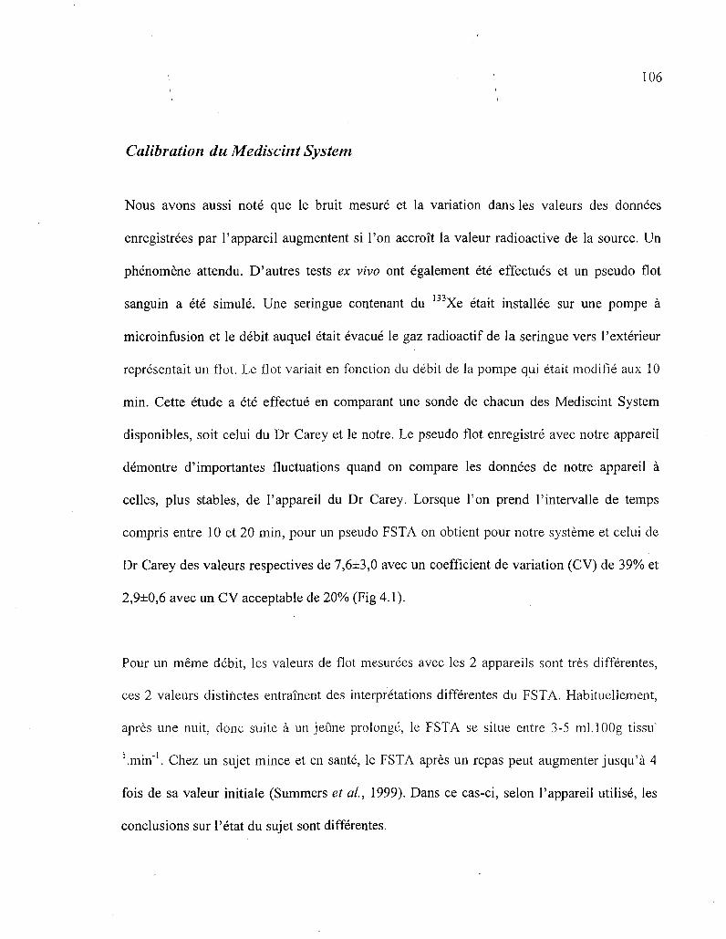
106
Calibration du Mediscint System
Nous avons aussi note que le bruit mesure et la variation dans les valeurs des donnees
enregistrees par l'appareil augmentent si Ton accroit la valeur radioactive de la source. Un
phenomene attendu. D'autres tests ex vivo ont egalement ete effectues et un pseudo flot
sanguin a ete simule. Une seringue contenant du 133Xe etait installee sur une pompe a
microinfusion et le debit auquel etait evacue le gaz radioactif de la seringue vers l'exterieur
representait un flot. Le Hot variait en fonction du debit de la pompe qui etait modifie aux 10
min. Cette etude a ete effectue en comparant une sonde de chacun des Mediscint System
disponibles, soit celui du Dr Carey et le notre. Le pseudo flot enregistre avec notre appareil
demontre d'importantes fluctuations quand on compare les donnees de notre appareil a
celles, plus stables, de l'appareil du Dr Carey. Lorsque Ton prend l'intervalle de temps
compris entre 10 et 20 min, pour un pseudo FSTA on obtient pour notre systeme et celui de
Dr Carey des valeurs respectives de 7,6±3,0 avec un coefficient de variation (CV) de 39% et
2,9±0,6 avec un CV acceptable de 20% (Fig 4.1).
Pour un meme debit, les valeurs de flot mesurees avec les 2 appareils sont tres differentes,
ces 2 valeurs distinctes entrainent des interpretations differentes du FSTA. Habituellement,
apres une nuit, done suite a un jeune prolonge, le FSTA se situe entre 3-5 ml.lOOg tissu'
'.min'1. Chez un sujet mince et en sante, le FSTA apres un repas peut augmenter jusqu'a 4
fois de sa valeur initiale (Summers et al, 1999). Dans ce cas-ci, selon l'appareil utilise, les
conclusions sur l'etat du sujet sont differentes.

107
0 5 10 15 20 25 30 35 40 45 50 55 60 65 Temps —•— Mediscint Ardilouze |
- « — Mediscint Carey
Fig 4.1 - Comparaison des Mediscint System Dr Carey vs Dr Ardilouze. Mesure de variations de pseudo flot par intervalles de 10 min.
Des etudes comparatives entre les 2 Mediscint System disponibles ont egalement ete
effectuees en condition in vivo chez 6 sujets jeunes, en sante et en bonne condition physique
(i.e. theoriquement de bons repondeurs en condition postprandiale). Le protocole etait base
sur des etudes precedentes (Ardilouze et al., 2004a; Karpe et al, 2002a). Le FSTA etait
enregistre avec une sonde du Dr Carey d'un cote de l'abdomen et une de nos sondes de
l'autre cote. Une 2e sonde de notre appareil etait positionnee a la partie inferieure de
1'abdomen du meme cote que son homologue a titre de controle. Le FSTA basal etait
enregistre sur une p^riode de 30 min pendant une microinfusion, via les 3 catheters, de
solution saline a un debit de 2 ul/min. Par la suite, un bolus de 75 g de glucose oral etait
administre au sujet. Une periode de 2h30 min etait allouee pour la mesure du FSTA
postprandial. Cet intervalle de temps etait suivi d'une microinfusion d'isoproterenol (ISO, 1

108
x 10" M, 2 ul/min) pour 2 sites, ou de salin'(site controle) pour 40 min. L'activite radioactive
pour les 2 sites microperfuses avec de 1'ISO etait enregistree avec une sonde du Dr Carey et
une de nos sondes. L'activite radioactive au site controle recevant du salin etait mesuree par
une de nos sondes. Les resultats enregistres par le Mediscint System de Sherbrooke ont
donne des reponses contrastantes avec celles retrouvees dans la litterature. Les reponses
postprandiales affichees par l'appareil etaient retardees, 1'augmentation maximale enregistree
survennaient 130 a 140 min suivant le repas comparee a 40 min habituellement. Les mesures
de FSTA n'affichaient pas de tendance de retour vers leurs valeurs initiales, et ce meme
apres 2h30 min suivant 1'ingestion du « repas » de 75 g de glucose. Suite a la stimulation
avec 1'ISO 1 x 10"4M, l'elevation de FSTA enregistree etait de 40% par rapport a la valeur
basale, une faible elevation considerant ce que la litterature rapporte dans ces memes
conditions experimentales, soit des chiffres de 1'ordre de 200% et plus (Karpe et at, 2002a).
Etonnamment, le Mediscint System fournit par le Dr Carey n'a pas non plus montre des
resultats attendus d'apres la litterature. Suivant la stimulation avec 1'ISO 1 x 10⁻⁴ M,
l'elevation de FSTA enregistree n'etait que de 40 a 50%. II est difficile pour nous d'associer
ces resultats a un mauvais protocole experimental puisque nous l'avons compare avec ceux
des autres equipes qui utilisent la meme technique et toutes les etapes sont respectees.
L'appareil du Dr Carey fait partie de la premiere generation des Mediscint System fabriques.
II date de 2001. Depuis 2004, le Dr Carey n'effectue plus de mesure du FSTA et entrepose
son appareil dans une ferme porcine. Les conditions d'entreposage ont probablement
endommage ses qualites de sensibilite de detection. Malheureusement, c'est le seul appareil
avec lequel nous avons pu comparer le notre.
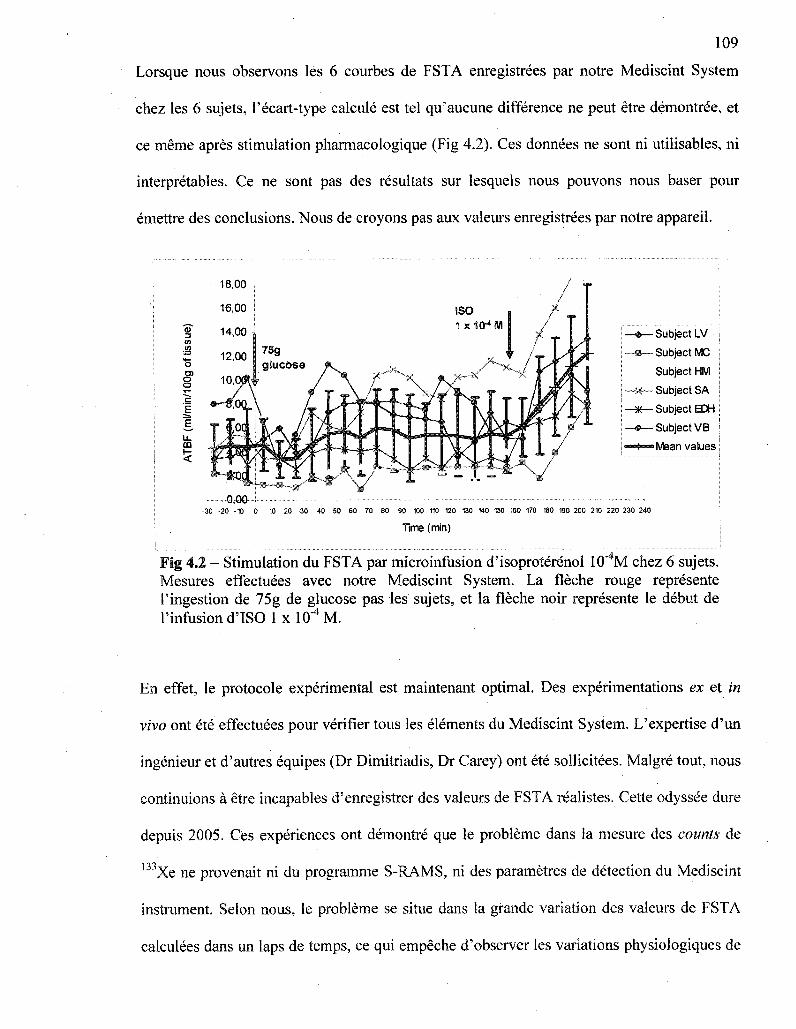
109
Lorsque nous observons les 6 courbes de FSTA enregistrees par notre Mediscint System
chez les 6 sujets. l'ecart-type calcule est tel qu'aucune difference ne peut etre demontree, et
ce meme apres stimulation pharmacologique (Fig 4.2). Ces donnees ne sont ni utilisables, ni
interpretables. Ce ne sont pas des resultats sur lesquels nous pouvons nous baser pour
emettre des conclusions. Nous de croyons pas aux valeurs enregistrees par notre appareil.
(I)
.<a
o o
E
u.
CD
5
—*— Subject LV
•—•— Subject MC
Subject HM
—x~- Subject SA
— « — Subject EDH
—®— Subject VB
I Mean values
30 -20 -10 0 10 20 30 40 50 60 70 60 90 100 110 120 130 140 150 160 170 160 190 200 210 220 230 240
Time (min)
Fig 4.2 - Stimulation du FSTA par microinfusion d'isoproterenol 10 M chez 6 sujets. Mesures effectuees avec notre Mediscint System. La fleche rouge represente Tingestion de 75g de glucose pas les sujets, et la fleche noir represente le debut de l'infusiond'IS0 1xlO"4M.
En effet, le protocole experimental est maintenant optimal. Des experimentations ex et in
vivo ont ete effectuees pour verifier tous les elements du Mediscint System. L'expertise d'un
ingenieur et d'autres equipes (Dr Dimitriadis, Dr Carey) ont ete sollicitees. Malgre tout, nous
continuions a etre incapables d'enregistrer des valeurs de FSTA realistes. Cette odyssee dure
depuis 2005. Ces experiences ont demontre que le probleme dans la mesure des counts de
l33Xe ne provenait ni du programme S-RAMS, ni des parametres de detection du Mediscint
instrument. Selon nous, le probleme se situe dans la grande variation des valeurs de FSTA
calculees dans un laps de temps, ce qui empeche d'observer les variations physiologiques de

• • i i o
flot. La detection de la' radioactivite par les sondes du Mediscint System semble egalement
etre problematique, est-ce un probleme de sensibilite?
Fin avril 2008, la compagnie John Caunt Scientific Ltd comprend finalement que notre
Mediscint System contient des defauts et decide de le reprendre pour effectuer des tests et les
corrections necessaires pur le rendre fonctionnel. Elle evaluera la sensibilite des sondes, la
valeur seuil de detection des sondes, la sensibilite des sondes aux radiofrequences et la
synchronisation de l'appareil.
Notre Mediscint System est done retourne a la compagnie en Angleterre. Plusieurs actions y
ont ete posees dans le but d'ameliorer le fonctionnement de l'appareil. Au moment d'ecrire
ces lignes, le Mediscint System ne nous a toujours pas ete retourne.
1) La quatrieme et derniere version du programme S-RaMS a ete installee. Bien que
selon nous le probleme ne se situe pas au niveau du programme, e'est une mise a jour
interessante puisque nous possesions la toute premiere version.
2) La sensibilite des sondes a ete ajustee a la meme valeur.
3) II y a eu desassemblage complet des sondes afin d'en diminuer le bruit interne, detail
probablement responsable de la grande variation dans les donnees enregistrees par le
programme.
4) Des tests plus techniques ont ete effectues en conditions ex vivo sur l'appareil.
; 111
1 5) Finalement il y a egalement eu validation du bon fonctionnement de l'appareil repare
en conditions cliniques et le Dr Manolopoulos, etudiant au PhD, a mesure le FSTA
chez certains de ses volontaires a Oxford. A ce jour, les tests semblent indiquer que
notre Mediscint System fonctionne adequatement pour la mesure du FSTA par la
methode de microinfusion du TA. II devrait revenir a Sherbrooke sous peu, pret a
entrer en fonction pour 1'investigation de nos hypotheses sur la regulation du FSTA.
Experiences acquises
Toutes ces experimentations sur la mesure du FSTA en conditions ex et in vivo, afin de
demontrer la non fonctionnalite de l'appareil de mesure, ont demande beaucoup de mon
temps durant ces 2 annees de maitrise. Du cote « resultats », je n'en ai aucun. Par contre, du
cote «apprentissage », « acquisition de connaissance», objectifs de tout etudiant a la
maitrise, j 'ai recolte beaucoup.
a) Tout d'abord, j 'ai connu le « mauvais » cote de la science. Tout ne va pas
toujours bien, ce n'est pas aussi simple que l'on croit. Je dois dire que j'ai
decouvert que j'etais une personne assez optimiste !
b) Sur le plan scientifique, j'ai appris la methode scientifique, la demarche de
resolution d'un probleme, j'ai acquis des connaissances en gestion clinique,
en medecine nucleaire, quelques principes de genie electrique.
c) Je me suis egalement impliquee dans d'autres projets du laboratoire. J'ai
effectue des ELISA pour determiner la concentration plasmatique de

«monocyte chemotactic protein» (MCP-1), une proteine qui induit le
recrutement des monocytes dans le processus inflammatoire, d'echantillons
plasmatique de patients diabetiques. J'ai egalement participe a une methode
d'isolation differentielle des leucocytes sur gradient discontinu a l'aide de
l'Optiprep.
d) J'ai appris a rediger une demande de bourse et j 'en ai obtenu 3 : une de
Diabete Quebec, une de la faculte de medecine et des sciences de la sante de
Puniversite de Sherbrooke et une des Fonds de la recherche en sante du
Quebec (FRSQ) comme etudiante d'ete.
e) J'ai aussi implante la mesure du FSTA dans le projet AGL4 de l'equipe du
Dr Andre C. Carpentier a Puniversite de Sherbrooke qui s'intitule: "Role de la
lipolyse intravasculaire et de Papport exogene de gras dans le metabolisme
postprandial des AGL plasmatiques, la fonction du tissu adipeux ainsi que la
modulation du systeme immunitaire inne et acquis dans le syndrome
metabolique". Notre but est de mesurer le FSTA en condition de resistance a
Pinsuline et apres un repas d'acides gras satures seulement et apres un repas
d'acides gras polyinsatures seulement. Bien que les mesures de FSTA
effectuees jusqu'a maintenant ne sont pas valides pour emettre des
conclusions, la mise en place de la mesure dans Petude est accomplie. Quand
notre appareil va revenir d'Angleterre, nous serons prets a faire des
enregistrements valables, l'equipe est rodee.
• - 1 1 3
f) Demierement, j 'a i aussi redige une proposition de protbcole de recherche
soumise a une compagnie pharmaceutique. Cette demande s'intitule: « A new
facet of the incretin effect: post prandial regulation of blood flow in adipose
tissue » (voir annexe). Je l'ajoute en annexe de mon memoire pour demontrer
des accomplissements supplementaires a mon projet initial.
g) De plus, a Fete 2007 notre equipe de recherche a cree la Clinique de
prelevements et de recherche pendant la grossesse du CHUS. En plus de tous
les prelevements relies a la grossesse, la clinique participe entre autres choses
au depistage precoce du diabete gestationnel. Les femmes obtenant une valeur
de glycemie superieure ou egale a 7,2 mmol/L 1 heure suivant l'ingestion
d'un jus contenant 50g de glucose doivent effectuer la mesure de leurs
glycemies capillaires 4 fois par jour durant 7 jours. A la clinique, les femmes
recoivent done un rapide enseignement sur le fonctionnement d'un
glucometre et la maniere adequate de 1'utiliser. Un suivi telephonique est
effectue une semaine plus tard. Durant une periode de Pete 2008, j ' a i donne
ces enseignements. En plus d'une expertise scientifique. je.suis allee me
chercher de 1'experience de vulgarisation avec une population cible.
Mon projet de maitrise n'a peut-etre pas donne les resultats escomptes au depart, par contre,
cela m'a permis de toucher a plusieurs domaines. En plus des connaissances techniques
apprises, j ' a i egalement su developper des aptitudes de vulgarisation, d'ecriture, de
perseverance et d'autonomie. Je suis capable de travailler en equipe et aller chercher
l'information dont j ' a i besoin pour mettre mes projets a terme. Ces acquis me seront tres
utiles pour mes cheminements futurs.

114
Preuves a Vappui
Mi-mars 2009, le Mediscint System est de retour dans nos laboratoires et pret a entrer en
fonction. Les sondes servant a la mesure du rayonnement y du b3Xe ont ete modifiees et
testees par la compagnie John Caunt Scientific en Angleterre. Le 16 et le 28 avril 2009 des
tests de validite de l'appareil en contexte experimental clinique sur des sujets humains sains
ont ete effectues (Fig 4.3). Afin de pouvoir mesurer le FSTA apres simulation physiologique
Mesure de la clairance du I33Xe
Mise en place des catheters Injection 133Xe
Ingestion de 75g de glucose
Changement d'infusion au site experimental
Temps (min) -80 -60
Site controle
Site experimental
1
-30
L 0
Infusion de solution saline
Infusion de solution saline
I
150
1.
195
_ _ .
Fig 4.3 - Deroulement des tests de validite : Mesure du FSTA a 2 sites au niveau de la partie superieure de l'abdomen. Les sites sont situes sur une meme ligne parallele a 5 cm au-dessus du nombril, a une distance de 6-8 cm. Mesure de la clairance du 133Xe du temps -30 a 195 min. Ingestion de 75 g de glucose au temps 0. A 150 min, changement d'infusion au site experimental pour de 1'ISO 1 x 10"4 M.
et pharmacologique, un 75 g de glucose a ete donne aux sujets, suivi d'une infusion d'ISO 1
x 10"4 M 150 min plus tard. Excepte cette double simulation, la methodologie pour la mesure
du FSTA fut la meme que celle decrite precedemment. L'objectif des ces tests de validite
etait de verifier si la mesure de la clairance du ,33Xe avec le Mediscint System ajuste
permettait d'obtenir des resultats de FSTA comparables a ceux retrouves dans la litterature

lors d'une stimulation physiologique suite a 1'ingestion de 75g de glucose et/ou lors d'une
infusion d'ISO l x 10"4M.
Suivant un jeune d'une nuit, le FSTA abdominal se situe habituellement en moyenne entre 3
et 5 ml/min/100 g de tissu (Elia et al, 1993). Chez les 2 sujets etudies, le FSTA a jeun, au
temps 0, variait selon les sites et les individus entre 1,7 et 5 ml/min/100 g de tissu (Fig 4.4 et
4.5). Evidemment, seulement 2 sujets ont ete etudies, ce qui ne permet sans doute pas
d'etablir de conclusion definitive. En revanche, apres comparaison avec d'autres resultats
obtenus par mon superviseur ou dans la litterature, nous pensons que ces resultats sont tout a
fait valides et representatifs de ce que l'on s'attend a trouver chez des sujets sains.
Chez des sujets en sante et de poids normal, comme les 2 participants aux tests de validite, le
FSTA abdominal s'eleve de 2 a 3 fois au-dessus de sa valeur initiale suite a un stimulus
nutritionnel oral. Cette elevation de FSTA est observee 30 a 60 min suivant l'ingestion du
repas (Karpe et al., 2002; Biilow et al., 1987). Lors des 2 etudes effectuees, le FSTA suite a
l'ingestion de 75 g de glucose au temps 0 s'eleve 2 fois au-dessus de sa valeur initiale. Le pic
maximal est atteint dans les 50 min suivant la prise de glucose oral. Suite a l'infusion d'ISO
1 x 10"4 M, on observe une elevation de 3 fois du FSTA aux sites experimentaux chez les 2
sujets par rapport aux valeurs de FSTA precedent le debut de l'infusion. Cette elevation
concorde encore une fois avec les resultats retrouves dans la litterature (Ardilouze, 2004).

116
Mesure du FSTA - test de validite, sujet 1
>•*>. s g •*3
£ O) o o
in/1
E 2 U
l)
2 (0 u.
16.00
14,00
12,00
10,00
8,00
6,00
4,00
*2l£ » • '
,0,00
-30 -10
- sujet 1 - site controle
sujet 1 - site experimental
10 30 50 70 90 110 130 150 170 190 210 230 250
Temps (min)
Fig 4.4 - Mesure du FSTA - test de validite, sujet 1. Mesure du FSTA chez un sujet sain et de poids normal afin de tester la fonctionnalite de 1'appareil de mesure de la clairance du 133Xe, le Mediscint System. La fleche rouge indique le moment de 1'ingestion du 75 g de glucose et la fleche rose, le debut de l'infusion de 1TSO 1 x 10"4 M au site experimental. Le site controle est infuse avec de la solution saline tout au long de 1' experimentation.
Ces tests de validite ont permis de confirmer que les changements effectues sur le Mediscint
Systeme ont rendu 1'appareil utilisable pour les mesures de FSTA chez l'humain. Les
resultats correspondent a ceux retrouves dans la litterature. Ces tests permettent egalement de
conclure une fois de plus que notre hypothese quant a la non fonctionnalite de 1'appareil de
mesure, le Mediscint System, pour les mesures de FSTA, etait valable.
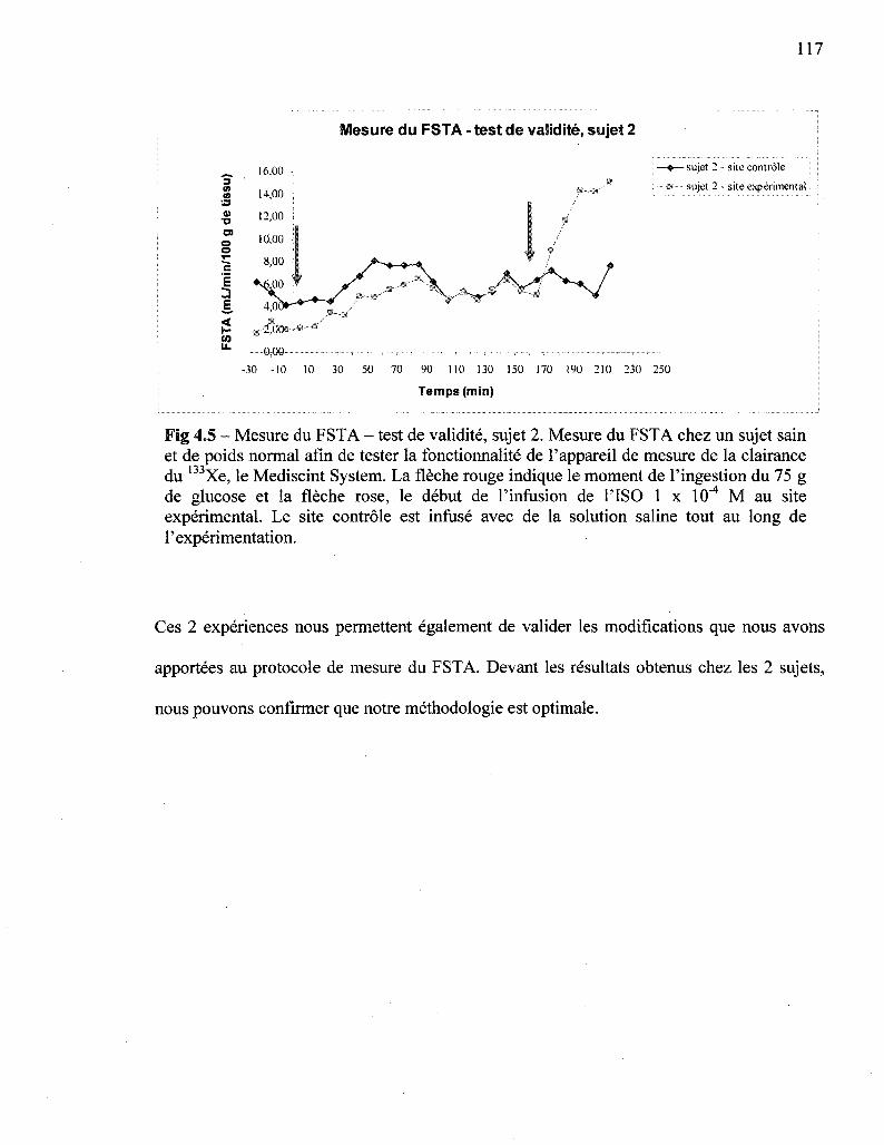
117
Mesure du FSTA - test de validite, sujet 2
10 (0
» •o
E
E
w u.
- sujet 2 - site controle
sujet 2 - site experimental i
-30 -10 10 30 50 70 90 110 130 150 170 190 210 230 250
Temps (min)
Fig 4.5 - Mesure du FSTA - test de validite, sujet 2. Mesure du FSTA chez un sujet sain et de poids normal afin de tester la fonctionnalite de l'appareil de mesure de la clairance du 133Xe, le Mediscint System. La fleche rouge indique le moment de 1'ingestion du 75 g de glucose et la fleche rose, le debut de 1'infusion de 1TSO 1 x 10"4 M au site experimental. Le site controle est infuse avec de la solution saline tout au long de 1'experimentation.
Ces 2 experiences nous permettent egalement de valider les modifications que nous avons
apportees au protocole de mesure du FSTA. Devant les resultats obtenus chez les 2 sujets,
nous pouvons confirmer que notre methodologie est optimale.
• \ 1 1 8
CHAPITRE 5
Conclusion et perspectives
Le FSTA est un sujet d'etude fascinant lorsque 1'on realise l'importance de son role dans
l'activite metabolique complexe du tissue adipeux et que l'on met cette physiologie dans la
perspective de Pepidemie mondiale d'obesite.
Certes, mes premiers objectifs de maitrise n'ont pu etre realises et cet echec relatif est du a
une defectuosite de l'appareil de mesure. Par contre, nous avons plusieurs succes a notre
actif. Premiere etape a ces etudes, j 'a i pu mettre au point la methode de mesure du FSTA a
Sherbrooke, la microinfusion du TA, par 1'application de la demarche scientifique. Nous
avons identifie le probleme et pose les actions pour le resoudre. Entre temps, j ' a i ecris deux
articles de revue ; Update on adipose tissue blood flow regulation et A new technique to
investigate the metabolism and pharmacology of adipose tissue, en plus de m'impliquer sur
d'autres projets de notre equipe de recherche et du service d'endocrinologie.
Maintenant que le Mediscint System est revenu a Sherbrooke en etat de fonctionner et que
des tests de validite de l'appareil sur la mesure du FSTA ont ete effectueS, nous sommes en
mesure de :
1. Realiser mes premiers objectifs de maitrise :
Nous allons etre les lers a explorer le role du SNPS dans la regulation du FSTA, a
determiner le role du GLP-1 sur le FSTA et nous allons completer avec l'etude de

•; i i 9
l'ET : puisqu'il a ete demontre que le NO est vasodilatateur, l'ET devrait etre
vasoconstrictrice.
2. Tenter de decouvrir quels sont les mecanismes qui modifient le FSTA chez les sujets
insulino-resistants
3. Tenter de decouvrir quels sont les anomalies dans la regulation du FSTA chez les
non-repondeurs de poids normal, une nouvelle entite a risque.
4. Valider (en collaboration avec Dr Andre Carpentier) l'utilisation de [nC]-acetate
pour la mesure du FSTA avec la methode d'imagerie TEP et la methode de
microinfusion dans le but d'effectuer des etudes de FSTA simultanement avec des
etudes metaboliques.
5. Investiguer (en collaboration avec Dr Remi Rabassa-Lhoret, Universite de Montreal)
le FSTA dans une population tres particuliere, les obeses metaboliquement normaux
et les normaux metaboliquement obeses.
On voit que beaucoup de travail reste a faire dans le domaine de la recherche sur la
regulation du FSTA, un domaine important pour notre comprehension des mecanismes de
developpement du syndrome d'insulino-resistance au diabete de type 2 et des maladies
cardiovasculaires. Nous sommes sur la bonne voie!

REMERCIEMENTS
120
Ce memoire de maitrise n'aurait pu etre realise sans la precieuse collaboration et le support
de plusieurs personnes.
Tout d'abord, il est essentiel d'exprimer toute ma gratitude envers le Dr Jean-Luc Ardilouze
pour m'avoir accueillie dans son equipe de recherche et cm en moi. Ses qualites
d'enseignant, sa motivation, son audace, son experience et ses nombreux encouragements
ont ete indispensables a ma reussite.
Je tiens egalement a remercier toute 1'equipe de laboratoire du Dr Ardilouze,
particulierement les Drs Julie Menard et Pascal Brassard pour leurs precieux conseils, leurs
mots d'encouragements et leur amitie. L'ambiance qui regne au sein de Pequipe m'a motivee
a me depasser, mais surtout a apprecier mes 2 annees passees a la maitrise.
Je rem ere ie Dr Guy Lamoureux pour son expertise en imagerie medicale, Dr Catherine Pepin
pour ses connaissances et ses nombreuses explications sur le fonctionnement de la detection
de la radioactivite et Jean-Philippe Gauthier, ingenieur, pour sa collaboration dans les tests ex
vivo de l'appareil de mesure, le Mediscint System. Je rem ere ie egalement le Dr Gale Carey
qui nous a permis d'emprunter son Mediscint System et qui a repondu a nos nombreuses
questions, tant sur la methodologie de la mesure du flot sanguin dans le tissu adipeux, que
sur l'enjeu des elections americaines. Ce fut une rencontre tres enrichissante!
; • i 2 i
Je m'en voudrais de ne pas souligner la precieuse collaboration du groupe de recherche en
diabetologie et des infirmieres Marie-Josee Gosselin, Diane Lessard et Carol-Lynn
Thibodeau.
Un merci tout particulier a ma famille et mes amis pour leur support constant, leur interet
face a l'avancement de mon projet et leur comprehension. A mes parents, qui m'ont transmis
le gout de me depasser et d'aller au fond des choses, merci, je ne serais surement pas rendue
la ouje suis sans eux.
Finalement, je voudrais remercier toutes les personnes qui ont accepte de participer a mon
projet en tant que sujets d'etudes. Sans vous, mon projet n'aurait pas ete possible.
Un dernier merci aux Drs Emanuel Escher et Eric Rousseau pour avoir accepte d'etre
membres de mon jury.
LISTE DES REFERENCES
Andersson, K. et Arner, P. (1995). Cholinoceptor-mediated effects on glycerol output from
human adipose tissue using in situ microdialysis. British journal of pharmacology, vol.
115,n°7 ,p . 1155-1162.
Ardilouze, J.L. (2004). Regulation of adipose tissue blood flow in humans, PhD thesis.
University of Oxford, p. 1-251.
Ardilouze, J.L., Fielding, B.A., Currie, J.M., Frayn, K.N. et Karpe, F. (2004a). Nitric oxide
and beta-adrenergic stimulation are major regulators of preprandial and postprandial
subcutaneous adipose tissue blood flow in humans.[see comment]. Circulation, vol. 109,
n° 1, p. 47-52.
Ardilouze, J.L., Karpe, F., Currie, J.M., Frayn, K.N. et Fielding, B A . (2004b). Subcutaneous
adipose tissue blood flow varies between superior and inferior levels of the anterior
abdominal wall. International Journal of Obesity & Related Metabolic Disorders:
Journal of the International Association for the Study of Obesity, vol. 28, n° 2, p. 228-
233.
Arner, P., Kriegholm, E., Engfeldt, P. et Bolinder, J. (1990). Adrenergic regulation of
lipolysis in situ at rest and during exercise. Journal of Clinical Investigation, vol. 85, n°
3, p. 893-898.
Baldo, A., Sniderman, A.D., St-Luce, S., Avramoglu, R.K., Maslowska, M , Hoang, B.,
Monge, J.C., Bell, A., Mulay, S. et Cianflone, K. (1993). The adipsin-acylation

'• • 1 2 3
stimulating protein system and regulation of intracellular triglyceride synthesis. Journal
of Clinical Investigation, vol. 92, n° 3, p. 1543-1547.
Bamshad, M , Aoki, V.T., Adkison, M.G., Warren, W.S. et Bartness, T.J. (1998). Central
nervous system origins of the sympathetic nervous system outflow to white adipose
tissue. American Journal of Physiology, vol. 275, n° 1 Pt 2, p. R291-9.
Bartness, T.J. et Song, C.K. (2007). Thematic review series: adipocyte biology. Sympathetic
and sensory innervation of white adipose tissue. Journal of lipid research, vol. 48, n° 8,
p. 1655-1672.
Bjorntorp, P. (1996). The regulation of adipose tissue distribution in humans. International
Journal of Obesity & Related Metabolic Disorders: Journal of the International
Association for the Study of Obesity, vol. 20, n° 4, p. 291-302.
Bjorntorp, P., Hood, B., Martinsson, A. et Persson, B. (1966). The composition of human
subcutaneous adipose tissue in obesity. Acta Medica Scandinavica, vol. 180, n° 1, p.
117-121.
Blaak, E.E., van Baak, M.A., Kemerink, G.J., Pakbiers, M.T., Heidendal, G.A. et Saris, W.H.
(1995). Beta-adrenergic stimulation and abdominal subcutaneous fat blood flow in lean,
obese, and reduced-obese subjects. Metabolism: Clinical & Experimental, vol. 44, n° 2,
p. 183-187.
Boss, O., Muzzin, P. et Giacobino, J.P. (1998). The uncoupling proteins, a review. European
Journal of Endocrinology, vol. 139, n° l ,p . 1-9.
Boulton, K.L., Hudson, D.U., Coppack, S.W. etFrayn, K.N. (1992). Steroid hormone
interconversions in human adipose tissue in vivo. Metabolism: Clinical & Experimental,
vol. 41, n° 5, p. 556-559.
Bulow, J., Astrup, A., Christensen, N.J. et Kastrup, J. (1987). Blood flow in skin,
subcutaneous adipose tissue and skeletal muscle in the forearm of normal man during an
oral glucose load. Acta Physiologica Scandinavica, vol. 130, n° 4, p. 657-661.
Burgoyne, R.D. et Clague, M.J. (2003). Calcium and calmodulin in membrane fusion.
Biochimica et biophysica acta, vol. 1641, n° 2-3, p. 137-143.
Campbell, P.J., Carlson, M.G., Hill, J.O. etNurjhan, N. (1992). Regulation of free fatty acid
metabolism by insulin in humans: role of lipolysis and reesterification. American
Journal of Physiology, vol. 263, n° 6 Pt 1, p. E1063-9.
Clifford, G.M., Londos, C, Kraemer, F.B., Vernon, R.G. et Yeaman, S.J. (2000).
Translocation of hormone-sensitive lipase and perilipin upon lipolytic stimulation of rat
adipocytes. Journal of Biological Chemistry, vol. 275, n° 7, p. 5011-5015.
Coppack, S.W., Evans, R.D., Fisher, R.M., Frayn, K.N., Gibbons, G.F., Humphreys, S.M,
Kirk, M.L., Potts, J.L. et Hockaday, T.D. (1992). Adipose tissue metabolism in obesity:
lipase action in vivo before and after a mixed meal. Metabolism: Clinical &
Experimental, vol. 41, n° 3, p. 264-272.
Coppack, S.W., Frayn, K.N, Humphreys, S.M, Dhar, H. et Hockaday, T.D. (1989). Effects
of insulin on human adipose tissue metabolism in vivo. Clinical science, vol. 77, n° 6, p.
663-670.
Crandall, D.L., Hausman, G.J. et Krai, J.G. (1997). A review of the microcirculation of
adipose tissue: anatomic, metabolic, and angiogenic perspectives. Microcirculation, vol.
4, n° 2, p. 211-232.
Di Girolamo, M., Skinner, N.S.,Jr, Hanley, H.G. et Sachs, R.G. (1971). Relationship of
adipose tissue blood flow to fat cell size and number. American Journal of Physiology,
vol. 220, n° 4, p. 932-937.
Dimitriadis, G., Lambadiari, V., Mitrou, P., Maratou, E., Boutati, E., Panagiotakos, D.B.,
Economopoulos, T. et Raptis, S.A. (2007). Impaired postprandial blood flow in the
adipose tissue may be an early marker of insulin resistance in type 2 diabetes. Diabetes
care,
Dodt, C., Lonnroth, P., Wellhoner, J.P., Fehm, H.L. et Elam, M. (2003). Sympathetic control
of white adipose tissue in lean and obese humans. Acta Physiologica Scandinavica, vol.
177, n° 3, p. 351-357.
Field, C.J., Goruk, S.D., Wierzbicki, A.A. et Clandinin, M.T. (1989). The effect of dietary fat
content and composition on adipocyte lipids in normal and diabetic states. International
journal of obesity, vol. 13, n° 6, p. 747-756.
Fielding, B.A. et Frayn, K.N. (1998). Lipoprotein lipase and the disposition of dietary fatty
acids. British Journal of Nutrition, vol. 80, n° 6, p. 495-502.
Frayn, K.N., Karpe, F., Fielding, B.A., Macdonald, LA. et Coppack, S.W. (2003). Integrative
physiology of human adipose tissue. International Journal of Obesity & Related
Metabolic Disorders: Journal of the International Association for the Study of Obesity,
vol. 27, n° 8, p. 875-888.

.. ' 126
Frayn, K.N., Shadid, S., Hamlani, R., Humphreys, 8.M., Clark, M.L., Fielding, B.A., Boland,
O. et Coppack, S.W. (1994). Regulation of fatty acid movement in human adipose tissue
in the postabsorptive-to-postprandial transition. American Journal of Physiology, vol.
266 ,n°3Pt l ,p . E308-17.
Fredholm, B.B., Linde, B., Oberg, B. et Rosell, S. (1969). Studies on the capillary exchange
function in canine subcutaneous adipose tissue. Bibliotheca anatomica, vol. 10, p. 241-
243.
Fruhbeck, G., Gomez-Ambrosi, J., Muruzabal, F.J. etBurrell, M.A. (2001). The adipocyte: a
model for integration of endocrine and metabolic signaling in energy metabolism
regulation. American Journal of Physiology - Endocrinology & Metabolism, vol. 280, n°
6, p. E827-47.
Gartner, L.P. et Hiatt, J.L. (2004). Atlas en couleur d'histologie. 2e edition edition, Belin,
Editions Pradel, 397 p.
Gersh, I. et Still, M. (1945). Blood vessels in fat tissue: relation to problem of gaz exchange.
J Exp Med, vol. 81, p. 219-232.
Gesta, S., Tseng, Y.H. et Kahn, C.R. (2007). Developmental origin of fat: tracking obesity to
its source. Cell, vol. 131, n° 2, p. 242-256.
Goossens, G.H. (2008). The role of adipose tissue dysfunction in the pathogenesis of obesity-
related insulin resistance. Physiology & behavior, vol. 94, n° 2, p. 206-218.
Goossens, G.H., McQuaid, S.E., Dennis, A.L., van Baak, M.A., Blaak, E.E., Frayn, K.N.,
Saris, W.H. et Karpe, F. (2006). Angiotensin II: a major regulator of subcutaneous

• 127
adipose tissue blood flow in humans. Journal of Physiology, vol. 571, n° Pt 2, p. 451-
460.
Gurr, M.I. et Kirtland, J. (1978). Adipose tissue cellularity: a review. I. Techniques for
studying cellularity. International journal of obesity, vol. 2, n° 4, p. 401-427.
Hauner, H. et Hochberg, Z. (2002). Endocrinology of adipose tissue. Hormone & Metabolic
Research, vol. 34, n° 11-12, p. 605-606.
Jansson, P.A., Larsson, A. et Lonnroth, P.N. (1998). Relationship between blood pressure,
metabolic variables and blood flow in obese subjects with or without non-insulin-
dependent diabetes mellitus. European journal of clinical investigation, vol. 28, n° 10,
p. 813-818.
Jaworski, K., Sarkadi-Nagy, E., Duncan, R.E., Ahmadian, M. et Sook, H.S. (2007).
Regulation of triglyceride metabolism. IV. Hormonal regulation of lipolysis in adipose
tissue. American journal of physiology - gastrointestinal and liver physiology, vol. 293,
p. G1-G4.
Jensen, M.D. (2002). Adipose tissue and fatty acid metabolism in humans. Journal of the
Royal Society of Medicine, vol. 95, n° Suppl 42, p. 3-7.
Karlsson, A.K., Attvall, S., Jansson, P.A., Sullivan, L. et Lonnroth, P. (1995). Influence of
the sympathetic nervous system on insulin sensitivity and adipose tissue metabolism: a
study in spinal cord-injured subjects. Metabolism: Clinical & Experimental, vol. 44, n°
1, p. 52-58.
Karlsson, A.K., Elam, M., Friberg, P., Biering-Sorensen, F., Sullivan, L. et Lonnroth, P.
(1997). Regulation of lipolysis by the sympathetic nervous system: a microdialysis study

in normal and spinal cord-injured subjects. Metabolism: Clinical & Experimental, Vol.
46,n°4, p. 388-394.
Karpe, F., Fielding, B.A., Ardilouze, J.L., Ilic, V., Macdonald, LA. et Frayn, K.N. (2002a).
Effects of insulin on adipose tissue blood flow in man. Journal of Physiology, vol. 540,
n°Pt3,p. 1087-1093.
Karpe, F., Fielding, B.A., Ilic, V., Macdonald, I.A., Summers, L.K. et Frayn, K.N. (2002b).
Impaired postprandial adipose tissue blood flow response is related to aspects of insulin
sensitivity. Diabetes, vol. 51, n° 8, p. 2467-2473.
Karpe, F., Steiner, G., Olivecrona, T., Carlson, L.A. et Hamsten, A. (1993). Metabolism of
triglyceride-rich lipoproteins during alimentary lipemia. Journal of Clinical
Investigation, vol. 91, n° 3, p. 748-758.
Katz, J.R., Mohamed-Ali, V., Wood, P.J., Yudkin, J.S. et Coppack, S.W. (1999). An in vivo
study of the cortisol-cortisone shuttle in subcutaneous abdominal adipose tissue. Clinical
endocrinology, vol. 50, n° 1, p. 63-68.
Kershaw, E.E. et Flier, J.S. (2004). Adipose tissue as an endocrine organ. Journal of Clinical-
Endocrinology & Metabolism, vol. 89, n° 6, p. 2548-2556.
Kreier, F., Fliers, E., Voshol, P.J., Van Eden, C.G., Havekes, L.M., Kalsbeek, A., Van
Heijningen, C.L., Sluiter, A.A., Mettenleiter, T.C., Romijn, J.A., Sauerwein, H.P. et
Buijs, R.M. (2002). Selective parasympathetic innervation of subcutaneous and intra
abdominal fat—functional implications. Journal of Clinical Investigation, vol. 110, n° 9,
p. 1243-1250.
[ 129
Langin, D1., Holm, C. et Lafontan, M. (1996). Adipocyte hormone-Sensitive lipase: a major
regulator of lipid metabolism. Proceedings of the Nutrition Society, vol. 55, n° IB, p.
93-109.
Lindbom, L. (1983). Microvascular blood flow distribution in skeletal muscle. An intravital
microscopic study in the rabbit. Acta Physiologica Scandinavica, vol. 525, n°
Supplementum. 525, p. 1-40.
Olivecrona, G. et Olivecrona, T. (1995). Triglyceride lipases and atherosclerosis. Current
opinion in lipidology, vol. 6, n° 5, p. 291-305.
Peeva, E., Brun, L.D., Ven Murthy, M.R., Despres, J.P., Normand, T., Gagne, C , Lupien,
P.J. et Julien, P. (1992). Adipose cell size and distribution in familial lipoprotein lipase
deficiency. International Journal of Obesity & Related Metabolic Disorders: Journal of
the International Association for the Study of Obesity, vol. 16, n° 10, p. 737-744.
Pond, C M . et Mattacks, C.A. (1995). Interactions between adipose tissue around lymph
nodes and lymphoid cells in vitro. Journal of lipid research, vol. 36, n° 10, p. 2219-
2231.
Raclot, T. et Groscolas, R. (1995). Selective mobilization of adipose tissue fatty acids during
energy depletion in the rat. Journal of lipid research, vol. 36, n° 10, p. 2164-2173.
Rebuffe-Scrive, M. (1991). Neuroregulation of adipose tissue: molecular and hormonal
mechanisms. International journal of obesity, vol. 15, n° Suppl 2, p. 83-86.
Rosell, S. et Belfrage, E. (1979). Blood circulation in adipose tissue. Physiological reviews,
vol. 59, n° 4, p. 1078-1104.
' 130
Ryan, T.J. et Curri, S.B. (1989). The development of adipose tissue and its relationship to the
vascular system. Clinics in dermatology, vol. 7, n° 4, p. 1-8.
Saleh, J., Summers, L.K., Cianflone, K., Fielding, B.A., Sniderman, A.D. et Frayn, K.N.
(1998). Coordinated release of acylation stimulating protein (ASP) and triacylglycerol
clearance by human adipose tissue in vivo in the postprandial period. Journal of lipid
research, vol. 39, n° 4, p. 884-891.
Samra, J.S., Clark, M.L., Humphreys, S.M., MacDonald, I.A., Bannister, P.A., Matthews,
D.R. et Frayn, K.N. (1999). Suppression of the nocturnal rise in growth hormone
reduces subsequent lipolysis in subcutaneous adipose tissue. European journal of
clinical investigation, vol. 29, n° 12, p. 1045-1052.
Samra, J.S., Clark, MX., Humphreys, S.M., Macdonald, I.A., Matthews, D.R. et Frayn, K.N.
(1996a). Effects of morning rise in Cortisol concentration on regulation of lipolysis in
subcutaneous adipose tissue. American Journal of Physiology, vol. 271, n° 6 Pt 1, p.
E996-1002.
Samra, J.S., Simpson, E.J., Clark, M.L., Forster, CD. , Humphreys, S.M., Macdonald, I.A. et
Frayn, K.N. (1996b). Effects of epinephrine infusion on adipose tissue: interactions
between blood flow and lipid metabolism. American Journal of Physiology, vol. 271, n°
5Pt l ,p . E834-9.
Siiteri, P.K. (1987). Adipose tissue as a source of hormones. American Journal of Clinical
Nutrition, vol. 45, n° 1 Suppl, p. 277-282.
' 131
Simonsen, L., Enevoldsen, L.H. et Bulow, J. (2003). Determination of adipose tissue blood
flow with local 133Xe clearance. Evaluation of a new labelling technique. Clinical
Physiology & Functional Imaging, vol. 23, n° 6, p. 320-323.
Steinberg, H.O., Brechtel, G., Johnson, A., Fineberg, N. et Baron, A.D. (1994). Insulin-
mediated skeletal muscle vasodilation is nitric oxide dependent. A novel action of
insulin to increase nitric oxide release. Journal of Clinical Investigation, vol. 94, n° 3, p.
1172-1179.
Steinberg, H.O., Tarshoby, M., Monestel, R., Hook, G., Cronin, J., Johnson, A., Bayazeed,
B. et Baron, A.D. (1997). Elevated circulating free fatty acid levels impair endothelium-
dependent vasodilation. Journal of Clinical Investigation, vol. 100, n° 5, p. 1230-1239.
Stevens, A. et Lowe, J. (2006). Histologic humain. 3e edition edition, Editions Elsevier, 459
P-
Summers, L.K., Samra, J.S. et Frayn, K.N. (1999). Impaired postprandial tissue regulation of
blood flow in insulin resistance: a determinant of cardiovascular risk? Atherosclerosis,
vol. 147, n° 1, p. 11-15.
Summers, L.K., Samra, J.S., Humphreys, S.M., Morris, R.J. et Frayn, K.N. (1996).
Subcutaneous abdominal adipose tissue blood flow: variation within and between
subjects and relationship to obesity. Clinical science, vol. 91, n° 6, p. 679-683.
Tanida, M., Iwashita, S., Ootsuka, Y., Terui, N. et Suzuki, M. (2000). Leptin injection into
white adipose tissue elevates renal sympathetic nerve activity dose-dependently through
the afferent nerves pathway in rats. Neuroscience letters, vol. 293, n° 2, p. 107-110.

! ! 132
Virtanen, K.A., Peltoniemi, P., Marjamaki, P., Asola, M., Strindberg, L., Parkkola, R.,
Huupponen, R., Knuuti, J., Lonnroth, P. etNuutila, P. (2001). Human adipose tissue
glucose uptake determined using (18)F-fluoro-deoxy-glucose ((18)FFDG) and PET in
combination with microdialysis. Diabetologia, vol. 44, n° 12, p. 2171-2179.
Waki, M., Krai, J.G., Mazariegos, M., Wang, J., Pierson, R.N.Jr et Heymsfield, S.B. (1991).
Relative expansion of extracellular fluid in obese vs. nonobese women. American
Journal of Physiology, vol. 261, n° 2 Pt 1, p. E199-203.
Watt, M.J. et Steinberg, G.R. (2008). Regulation and function of triacylglycerol lipases in
cellular metabolism. BiochemicalJournal, vol. 414, p. 313-325.
Weinstock, P.H., Levak-Frank, S., Hudgins, L.C., Radner, H., Friedman, J.M., Zechner, R. et
Breslow, J.L. (1997). Lipoprotein lipase controls fatty acid entry into adipose tissue, but
fat mass is preserved by endogenous synthesis in mice deficient in adipose tissue
lipoprotein lipase. Proceedings of the National Academy of Sciences of the United States
of America, vol. 94, n° 19, p. 10261-10266.
Wheater, P.R., Young, B. et Heat, J.W. (2001). Histologic fonctionnelle. 4e edition edition,
Paris, De Boeck Universite, 413 p.
Yeh, S.Y. et Peterson, R.E. (1965). Solubility of krypton and xenon in blood, protein
solutions, tissue homogenates. Journal of Applied Physiology, vol. 20, p. 1041-1047.
Yki-Jarvinen, H. et Utriainen, T. (1998). Insulin-induced vasodilatation: physiology or
pharmacology?[see comment]. Diabetologia, vol. 41, n° 4, p. 369-379.

r 133
Youngstrom, T.G. et Bartness, T.J. (1995). Catecholaminergic innervation of white adipose
tissue in Siberian hamsters. American Journal of Physiology, vol. 268, n° 3 Pt 2, p.
R744-51.
Yudkin, J.S. (2007). Inflammation, obesity, and the metabolic syndrome. Hormone &
Metabolic Research, vol. 39, n° 10, p. 707-709.

134
ANNEXE PROPOSITION DE PROTOCOLE DE RECHERCHE

Research proposal
135
A NEW FACET OF THE INCRETIN EFFECT: POST PRANDIAL REGULATION OF BLOOD FLOW IN ADIPOSE TISSUE
by
Elizabeth Martin, BSc and Jean-Luc Ardilouze, MD, PhD
O I *
E™"" 4
IL m
• oral 75-g glucose o iv glucose and insulin
n=15
-20 20 40 80 SO tOO 120 140 160 180
Tim© (mill) J. Physiol, 2002
Figure 1. The iricretin effect on postprandiai regulation of adipose tissue biood flow (ATBF) in 15 healthy subjects. ATBF increased by 113% (p< 0.05) after oral absorption of 75-g of glucose at time 0, although similar plasma concentrations of insulin and glucose (achieved by dynamic intravenous infusions of glucose and insulin) resulted in lower effect (+50%, p< 0.05).
• BACKGROUND
136
1) Importance of blood flow in adipose tissue - Relation with the insulin resistance syndrome
Energy storage and disposal as well as secretion of hormones and adipocytokines require a precise regulation of blood flow in adipose tissue (ATBF).1 It was well established2"7 that in obese and/or insulin-resistant individuals, fasting ATBF is lower than in healthy normal-weight subjects whereas the increase in postprandial ATBF is blunted i.e. ATBF enhancement peaks by less than 50% above baseline blood flow. Accordingly, we hypothesized that ATBF dysregulation is a component of insulin-resistance syndrome.8
2) A novel technique of investigation
The regulation of ATBF is not fully understood. In order to explore that regulation, we setup8 and established recently in Sherbrooke a new technique of investigation, the "adipose tissue microinfusion (ATM)" technique, which allows accurate and quantitative measurements of the effects of vasoactive hormonal or pharmacological agents on ATBF. Briefly, ATM is based on the I33xenon washout technique which is the gold standard for ATBF recording; an infusion of vasoactive substance (or saline for control in the same subject) is made directly into the exact location into which '"xenon has been deposited, allowing the recording of 133xenon disappearance e.g. ATBF, in relation to changes in local tissue infusion. The subcutaneous abdominal adipose tissue of the anterior abdominal wall is the anatomical site of investigation because of its relevancy and easy accessibility. We have four detectors available, and then four sites can be microinfused. Right and left sides, at the upper or lower level of the abdomen, are studied simultaneously to allow direct comparison of the effect of the vasoactive substance on one side with the contralateral saline control side on the other. Each subject acts as his own control. This design has been chosen because we have previously demonstrated that there is no fasting or postprandial ATBF difference between the right and the left side of the abdomen.
3) Assessment of ATBF regulating factors using the ATM technique
Using the ATM technique, we provided evidence that, in adipose tissue, insulin per se has no vasodilatory effect, in the resting state as well as during the postprandial period. Consequently, we hypothesised that insulin may act indirectly on ATBF via sympathetic activation, and that nitric oxide (NO) may be an overall major regulator of ATBF. Indeed, fasting ATBF is primarily under NO tone11 and circulating Ang II12 and to some extent under a-adrenergic control, although the postprandial enhancement of ATBF is independent of NO and is controlled principally by the P-adrenergic system but for only 60%. n This suggests that the postprandial regulation of ATBF is complex and may involve the gastro intestinal tract.
• OUR LAB GENERAL WORKING HYPOTHESES
1) Increase in ATBF (i.e. vasodilatation), is associated with a) plasma TG and FFA extraction (for storage in adipocytes) during the postprandial period and b) NEFA release during postabsorbtive state and fasting. In other words, a reduced ATBF, as seen in obese subjects, in patients with type 2 diabetes and, more recently in normal-weight subjects with

137
familial history of diabetes and cai;diometabolic diseases but no insulin resistance, may lead to decreased plasma TG and FFA extraction, resulting in postprandial hyperlipidemia; 2) This defect in fat storage in adipose tissue, in turn, increases fat storage in other deposit sites such as liver or muscle, which is predisposing condition to insulin resistance, diabetes and cardiovascular diseases.
• RATIONAL OF THE PRESENT STUDY
Little is known regarding gut hormones and ATBF. The physiological increase in ATBF after a meal could be compared to the increase in intestinal blood flow. Splanchnic blood flow also increases sharply in response to oral but not intravenous glucose.13 We showed (figure 1) that an increase in ATBF is more significant after a 75-g glucose oral ingestion than after intravenous glucose and insulin infusions needed to obtain the same plasma glucose and insulin levels.8 These data add strong evidence to the potential role of the gastrointestinal tract on ATBF.
Incretins could be important factors. An obvious candidate is glucagon-like-peptide-1 (GLP-1). GLP-1 is the most important incretin in humans.14>'5 GLP-1 receptors have been detected in adipose tissue. GLP-1 is secreted throughout the day and in increased amounts after meals. From baseline, GLP-1 increases by 120% after glucose load.16 Also, GLP-1 is a vasodilatator in hepatic and pulmonary arteries.17'1 As P-adrenoceptor subtypes, his receptor belongs to the superfamily of seven transmembrane domain G protein-coupled receptors that positively regulate intracellular cAMP levels via adenylate cyclase.19
One study was performed in humans to test directly whether GLP-1 has actions on lipolysis in adipose tissue and was inconclusive, probably because the ethanol escape technique under microdialysis condition was used.19 This technique only gives an estimation of ATBF, it is an indirect and a non-quantitative method. When compared to 133Xe washout technique,20 the decrease in ethanol ratio was delayed (approximately 40 min.) in comparison with the increase in ATBF elicited by the ingestion of glucose. The method is either not sufficiently sensitive, or sufficiently rapid, to respond to the subsequent postprandial decrease in ATBF: when ATBF recorded by ' Xe had shown, following a postprandial sharp increase, a sharp decline, the ethanol ratio showed no tendency to return to baseline.
In summary, several lines of evidence suggest that the gastrointestinal tract may be involved in the regulation of ATBF. We propose to first evaluate the role of GLP-1, although the role of other intestinal hormones in regulating ATBF after glucose or mixed meals remains to be established.
• HYPOTHESIS OF THIS STUDY: GLP-1 has a vasodilator impact on postprandial ATBF regulation.
• OBJECTIVES
To assess the effect on ATBF of GLP-1 [7-36] (the major form of circulating GLP-1), exendin [9-39] (an antagonist of GLP-1R) and sitagliptin (a DPP-IV inhibitor).

138
• RESEARCH PLAN
• Experiments with ATBF hypothetic stimulating agents: assessment of ATBF responses to GLP-1 [7-36] and DPP-IV inhibitor sitagliptin.
Experimental protocols. The ATM technique will be used for quantification of ATBF responses to 3 consecutive concentrations (10"5 M, 10"4 M and 10"3 M) which are those used in our previous experiments.3'8'12 After baseline ATBF recording, at one upper and one lower site chosen by blocked randomisation (during each experiment, the 2 agents will be tested in the same subject), the lowest concentration (105) of agents will be micro-perfused for 40 min. at an infusion rate of 2 ul min"1. Thereafter, at the same active sites, concentrations will be successively increased for two 40 min. periods. At the contralateral control sites, the saline infusion will be continued until the end of the experiment.
Subjects - Samples size. Given that no accurate data are available in literature and according to our previous experience, a minimum of 12 healthy young non smoker subjects will be necessary to find a physiological (30 to 50 %) difference.
Experimental days = 12 (one experimental day per subject).
Variables of interest. As previously, we will compare means in ATBF iAUCs and ATBF peak-baseline ratios, between tested and controlled sides.
Expected findings. We will show that GLP-1 and sitagliptin have a vasodilatator effect in subcutaneous adipose tissue.
• Experiments with blocker (exendin r9-391).
The blocking principle. The so-called 'blocking principle' used in the present study is based on a straightforward idea. Assuming that the optimal ATBF increase is obtained during a physiological stimulus, blockade of the putative mediator of the response, when compared to a control, will assess the effect of the particular pathway. Accordingly, GLP-1 antagonist exendin [9-391 will be microinfused to evaluate the endogenous ATBF regulation before and after oral glucose intake.
Dose-response experiments will be performed beforehand.
a Six concentrations of exendin [9-39] need to be tested since exendin has not been used under microinfusion or microdialysis conditions and therefore no "blocking" dosage indication is available. Chosen concentrations (10"5 M, 10"4 M and 10"3 M) and design of the
i o n
present studies are those used in our previous experiments, ' ' although exendin will be additionally tested at low concentrations (10~6 M, 10"7 M, and 10"8 M) because highest concentrations could be in the partial agonist range.
a Our previous dose-response studies have shown that 5 subjects are sufficient to determine optimal concentrations. Subjects will be challenged with 75-g glucose for endogenous stimulation of ATBF.
D Three out of the four microinfusion sites will be randomly infused with one of three concentrations, with the fourth being a saline control site. Concentrations will be also randomly allocated.
139
• Experimental days = 10 (each subject will participate in 2 studies).
Final experimental protocol. After the optimal concentration is determined, 7 additional experiments will be performed for a total of 12 assessments of the optimal dose.
Variables of interest. As previously, we will compare means in ATBF iAUCs and ATBF peak-baseline ratios, between tested and controlled sides.
Expected findings. We will show that exendin [9-39] reduces by 30 to 40% the postprandial increase of ATBF.
• EXPECTED OUTCOMES OF THIS RESEARCH PROGRAM
1) Additional knowledge If our present hypothesis is right, these studies will demonstrate that the incretin effect is complementary to the ATBF postprandial stimulatory adrenergic activity.
2) Promising upshot for sitagliptin From a therapeutic prospective, the present studies will also support the utilization of inhibitors of DPP-4 such as Januvia. Indeed, these studies will demonstrate that DPP-4 inhibitors restore a lost function e.g. improve postprandial ATBF.
This in turn will suggest, in line with the general working hypotheses of our lab, that this class of drug may improve fat storage. Therefore, similar studies will be required to explore the ATBF-related mechanisms of hyperlipemia in subjects with low fasting ATBF and abolished ATBF response to meal, these are subjects with obesity, prediabetes or type 2 diabetes. To our knowledge, the lipids issue has never been carefully and specifically addressed in published clinical trials assessing DPP-4 inhibitors. Some papers reported that treatment with sitagliptin led to statistically significant lipids profile improvements, although no convincing mechanism was proposed to explain these data.
Accordingly, the aim of forthcoming studies could be to show that DPP-4 inhibitors restore the normal postprandial doubling or tripling of ATBF in patients who have lost this characteristic and, in turn, improve the lipids profile. This could reveal a new mechanism of action of Januvia and lead to new clinical applications.
• CONCLUSION
The demonstration of the role of GLP-1 as a complement to the other mechanisms which interact in the regulation of ATBF could pave the way to new physiopathological hypotheses and new therapeutic indications of sitagliptin in obese patients with insulino-resistance syndrome or type 2 diabetes.
140
REFERENCES
1. Frayn KN, Karpe F, Fielding BA, et al. Integrative physiology of human adipose tissue. Int J Obes Relat Metab Disord. Aug 2003;27(8):875-888.
2. Coppack SW, Evans RD, Fisher RM, et al. Adipose tissue metabolism in obesity: lipase action in vivo before and after a mixed meal. Metabolism. Mar 1992;41(3):264-272.
3. Ardilouze J-L, Fielding B, Frayn K, et al. Failure to increase postprandial adipose tissue blood flow is associated with tissue resistance to adrenergic activation. Obes Res. 2005;Submitted.
4. Ardilouze JL. Regulation of adipose tissue blood flow in humans. PhD thesis. University of Oxford. 2004:1 -251.
5. Bulow J, Astrup A, Christensen NJ, et al. Blood flow in skin, subcutaneous adipose tissue and skeletal muscle in the forearm of normal man during an oral glucose load. Acta Physiol Scand. 1987;130(4):657-661.
6. Jansson PA, Larsson A, Lonnroth PN. Relationship between blood pressure, metabolic variables and blood flow in obese subjects with or without non-insulin-dependent diabetes mellitus. Eur J Clin Invest. 1998;28(10):813-818.
7. Blaak EE, van Baak MA, Kemerink GJ, et al. Beta-adrenergic stimulation and abdominal subcutaneous fat blood flow in lean, obese, and reduced-obese subjects. Metabolism. 1995;44(2):183-187.
8. Karpe F, Fielding BA, Ardilouze JL, et al. Effects of insulin on adipose tissue blood flow in man. J Physiol. May 1 2002;540(Pt 3): 1087-1093.
9. Martin E, Brassard P, Ardilouze JL. A new technique to investigate adipose tissue metabolism. Submitted to Canadian Journal of Physiology and Pharmacology.
10. Ardilouze JL, Karpe F, Currie JM, Frayn KN, Fielding BA. Subcutaneous adipose tissue blood flow varies between superior and inferior levels of the anterior abdominal wall. Int J Obes Relat Metab Disord. 2004;28:228-233.
11. Ardilouze JL, Fielding BA, Currie JM, et al. Nitric oxide and beta-adrenergic stimulation are major regulators of preprandial and postprandial subcutaneous adipose tissue blood flow in humans. Circulation. Jan 6 2004;109(l):47-52.
12. Goossens GH, McQuaid SE, et al. Angiotensin II: a major regulator of subcutaneous adipose tissue blood flow in humans. J Physiol. 2006, 571, pt2, 451-460.
13. Brundin T, Branstrom R, Wahren J. Effects of oral vs. i.v. glucose administration on splanchnic and extrasplanchnic 02 uptake and blood flow. Am J Physiol. 1996;271:E496-504.
14. Ahren B. Gut peptides and type 2 diabetes mellitus treatment. Curr Diab Rep. 2003; 3:365-372.
15. Meier JJ, Gallwitz B, Schmidt WE, Nauck MA. Glucagon-like peptide 1 as a regulator of food intake and body weight: therapeutic perspectives. Eur J Pharmacol. 2002; 440: 269-279.
16. Rask E, Olsson T, Soderberg S, Hoist Jj J, Tura A, Pacini G, Ahren B. Insulin secretion and incretin hormones after oral glucose in non-obese subjects with impaired glucose tolerance. Metabolism. 2004:53:624-631.
17. Nishizawa M, Moore MC, Shiota M, Gustavson SM, Snead WL, Neal DW, Cherrington AD. Effect of intraportal glucagon-like peptide-1 on glucose metabolism in conscious dogs. Am J Physiol Endocrinol Metab. 2003;284:E1027-1036.

141
18. Golpon HA, Puechner A, Welte T, Wichert PV, Feddersen CO. Vasorelaxant effect of glucagon-like peptide-(7-36)amide and amylin on the pulmonary circulation of the rat. RegulPept. 2001:102:81-86.
19. Bertin E, Arner P, Bolinder J, Hagstrom-Toft E. Action of glucagon and glucagon-like peptide-1-(7-36) amide on lipolysis in human subcutaneous adipose tissue and skeletal muscle in vivo. J Clin Endocrinol Metab. 2001; 86: 1229-1234.
20. Karpe F, Fielding BA, Hie V, Humphreys SM, Frayn KN. Monitoring adipose tissue blood flow in man: a comparison between the 133xenon washout method and microdialysis. IntJObes Relat Metab Disord. 2002;26:1-5.