non specific immune reactions -...
TRANSCRIPT

Non Specific Immune
Reactions
INTRODUCTION
FIRST BARRIER
• Mechanical defense
SECOND BARRIER
•Phagocytosis
•Inflammation
•Complement activation

Learning Objectives:-
1. Explain the concept of non-specific immune response
2. Describes the first defense and examples
3. Describes the scope of the second line and the examples
a) Explain the stages in phagocytosis and cells
involved
b) Explain the concept and components of
inflammation and the process involved
c) Describes how the activation of complement
system and its importance
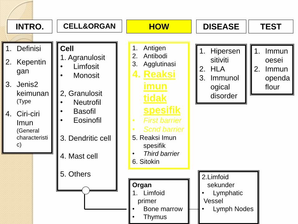
INTRO. CELL&ORGAN HOW DISEASE TEST
1. Definisi
2. Kepentin
gan
3. Jenis2
keimunan(Type
4. Ciri-ciri
Imun (General
characteristi
c)
Cell
1. Agranulosit
• Limfosit
• Monosit
2, Granulosit
• Neutrofil
• Basofil
• Eosinofil
3. Dendritic cell
4. Mast cell
5. Others
Organ
1. Limfoid
primer
• Bone marrow
• Thymus
2.Limfoid
sekunder
• Lymphatic
Vessel
• Lymph Nodes
1. Antigen
2. Antibodi
3. Agglutinasi
4. Reaksi
imun
tidak
spesifik • First barrier
• Scnd barrier 5. Reaksi Imun
spesifik
• Third barrier
6. Sitokin
1. Hipersen
sitiviti
2. HLA
3. Immunol
ogical
disorder
1. Immun
oesei
2. Immun
openda
flour

Introduction
Mechanisms to fight / protect the body
banked dangerous pathogens through
existing mechanisms naturally from birth

Factors influencing the immune system:
The influence of age
Dietary factors
Hormone
Genetic

IMMUNITY
TAK SPESIFIK SPESIFIK
First barrier:
•Skin
•Membrane
mucosa
Second barrier:
•Phagocytosis
•Inflammation
•Antimicrobial protein
(complement system)
Third barrier:
Lymphocyte
Antibodies

Non-specific immunity Vs Specific
immunity

Body Defense against pathogen
1. First barrier
◦ Non-specific defense that stops all pathogens from invading the body.
Examples: Skin and mucous membranes.
2. Second barrier
◦ Non-specific defense that provides rapid response to entry of pathogens into the body.
◦ Respond after the pathogen enters the body
◦ Examples: Fever, phagocytes (macrophages and neutrophils), inflammation.

3. Third barrier
◦ Immune response against a specific antigen
◦ Attack specific target antigens after antigen
beyond the second line
◦ Examples: Antibodies and lymphocytes.

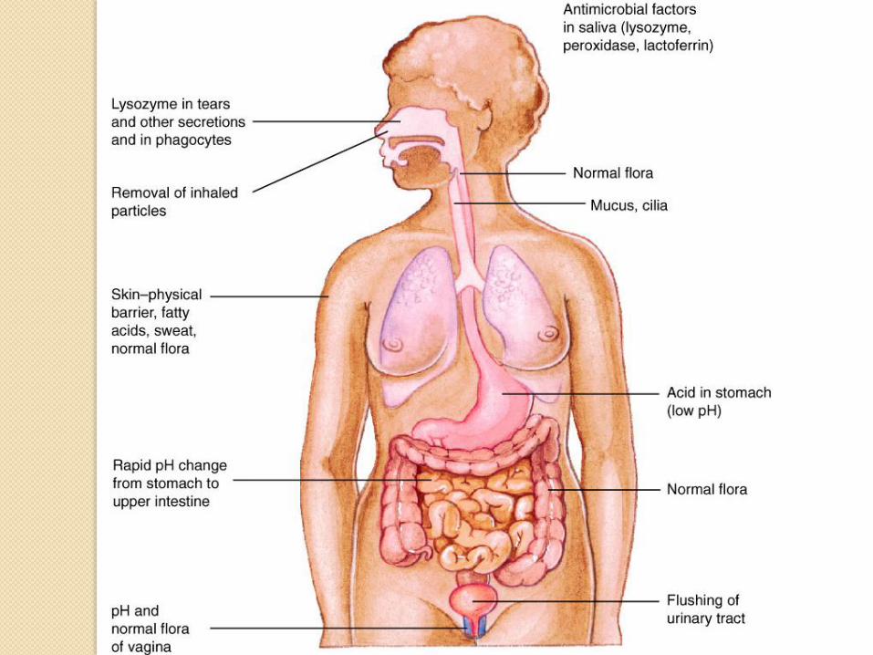
FIRST BARRIER
1. Skins
The largest organ in the human
Normal skin flora inhibit the presence of
other pathogens by producing antibacterial
fatty acids
Epidermis - protect the body from injury and
infection
Sweat - sebum secretion, lactic acid & fatty
acids which lower the pH of the skin
Lysozyme sweat - kill bacteria

2. Mucosal Membrane
Goblet cells from epithelial secrete mucus
Protect epidermal cells and prevent
membrane dehydration
Trap pathogens before the infection occur

Example:
◦ Lysozyme in saliva kill bacteria
◦ Lysozyme in tears break the bacterial cell wall
◦ Cilium in airways and coughing sweep foreign
particles out of the body

1. Respiration System
◦ This entire system is lined with moist
epithelium.
◦ In the upper respiratory tract the
epithelium contains mucus-secreting cells
and is covered with cilia that sweep mucus
up to be coughed up.
MECHANICAL DEFENSE
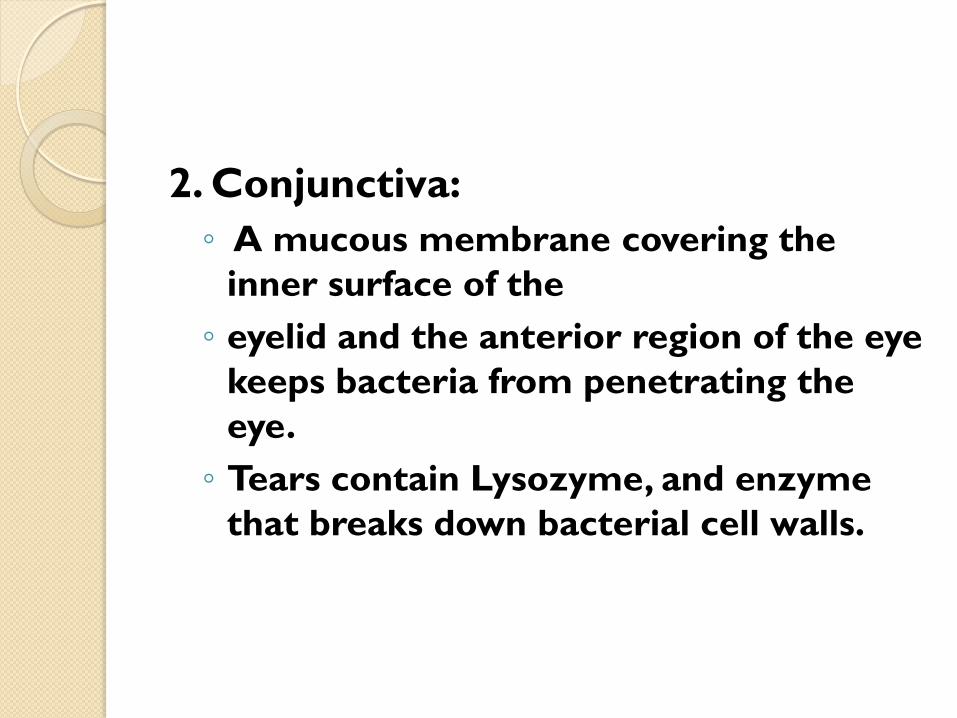
2. Conjunctiva:
◦ A mucous membrane covering the
inner surface of the
◦ eyelid and the anterior region of the eye
keeps bacteria from penetrating the
eye.
◦ Tears contain Lysozyme, and enzyme
that breaks down bacterial cell walls.

3. Lacrimal apparatus:
Continual washing and blinking prevents
microbes from settling on the eye surface.
4. Saliva:
Washes microbes from teeth and mouth
mucous membranes.
5. Mucus:
Thick secretion that traps many microbes.
6. Nose Hair:
Coated with mucus filter dust, pollen, and
microbes.

7. Coughing and sneezing:
Expel foreign objects.
9. Urination:
Cleanses urethra.
10. Vaginal Secretions:
Remove microbes from genital
tract.

SECOND BARRIER
•PHAGOCYTOSIS
•INFLAMATION
•COMPLEMENT SYSTEM

Definition
Ingestion by individual cell of invading
foreign particles
Characteristic of phagocytes cells
◦ Actively phagocytic
◦ Contains digestive enzymes to digest particles
◦ Important link between innate immunity and
acquired immunity
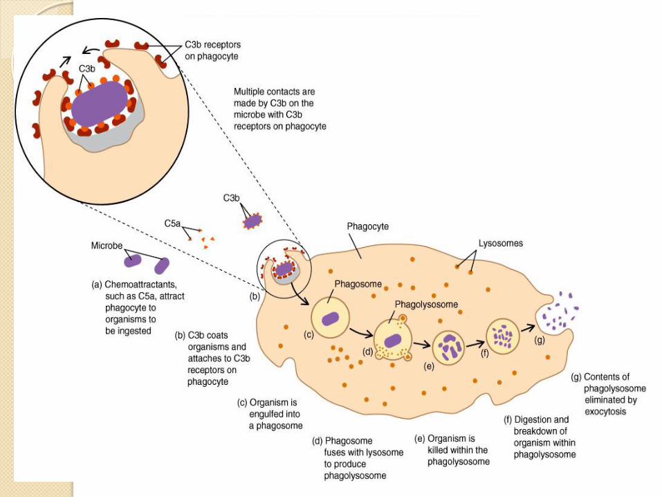
PHAGOCYTOSIS

Phagocytosis Process
1) Chemotaxis
Cells phagocytes move toward chemical stimulation
release by infectious agents and damaged tissue.
Phagocytes will release cytokine resulting in
activation of the complement
Cytokine stimulates other cell responses to
infectious agents

Phagocytes are Attracted to Site of
Infection by Chemotaxis
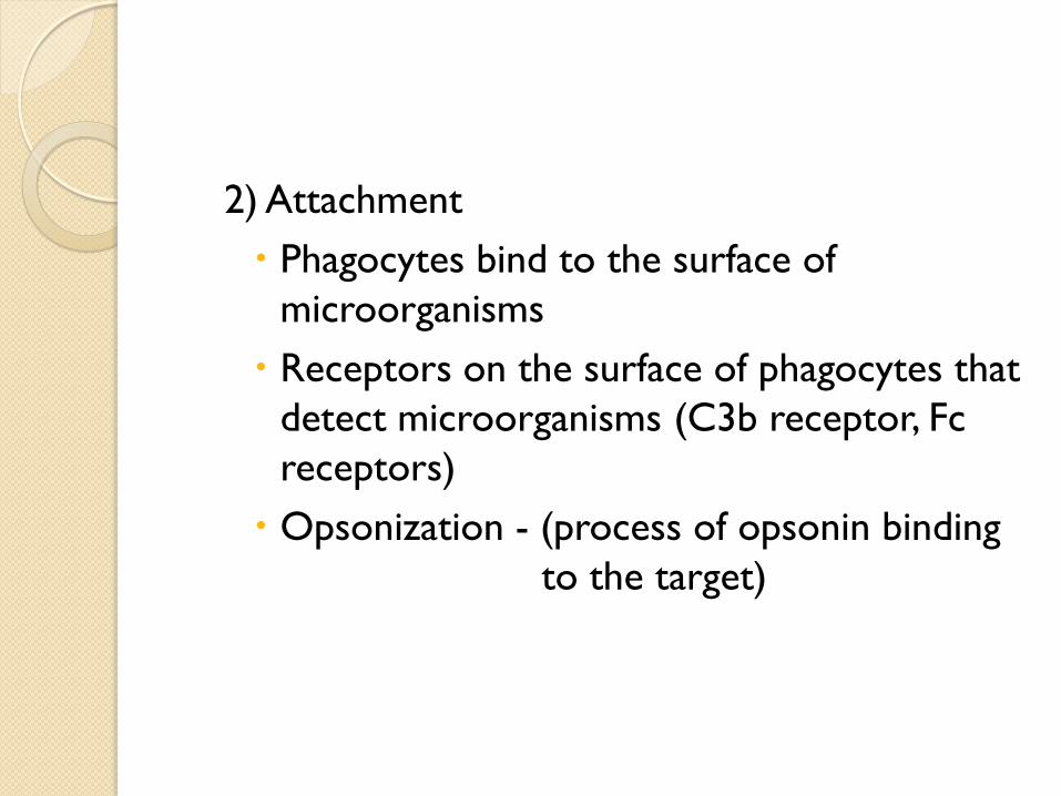
2) Attachment
Phagocytes bind to the surface of
microorganisms
Receptors on the surface of phagocytes that
detect microorganisms (C3b receptor, Fc
receptors)
Opsonization - (process of opsonin binding
to the target)

3) Ingestion
◦ Swallow and form a layer of wall (capsule)
around pathogen.
◦ The capsule is called phagosome

4) Digestion
Enzim Lysozyme in the phagocyte will merge with
phagosome and formed phagolysosome
To destroy and breakdown pathogen into smaller
molecules.
5) Exocytosis
- Undigested and unused particles will be remove
from cells.
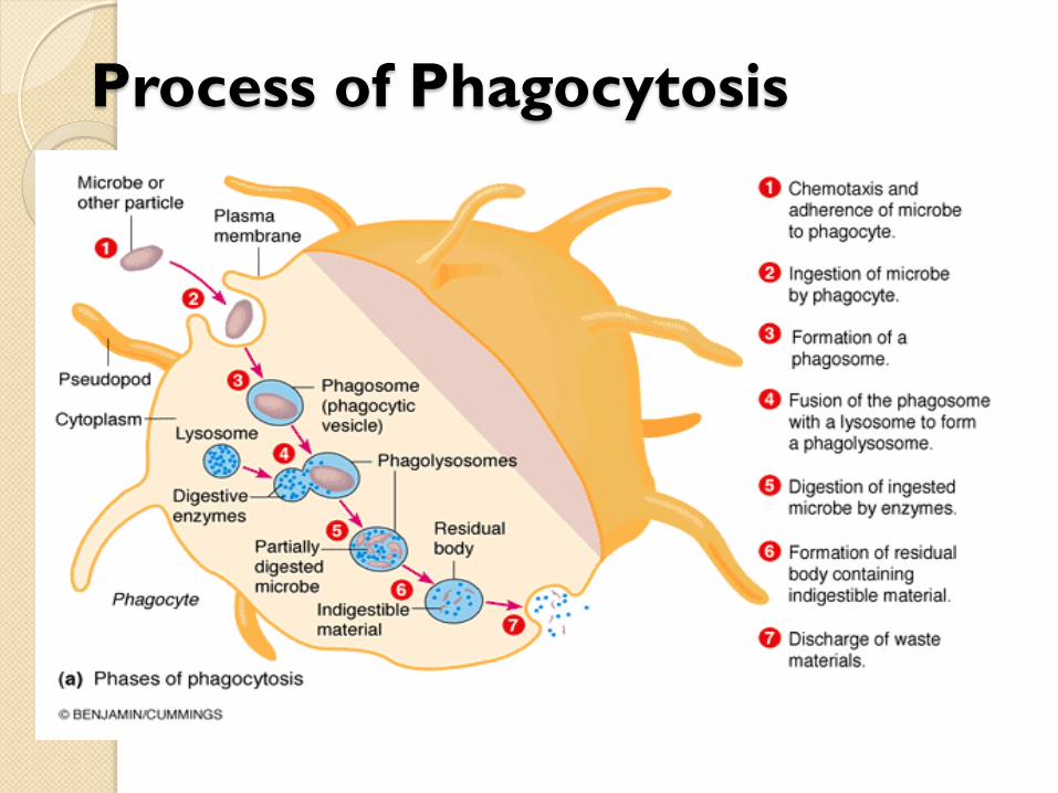
Process of Phagocytosis

Phagocyte Cell
1. Neutrophil
2. Macrophage
3. Eosinophil
4. Basophil
5. Dendritic cell
6. Mast cell
Professional

Macrophage Attacking E.coli (SEM x8,800)
28

Alveolar (Lung) Macrophage Attacking E. coli (SEM x10,000)
29

SECOND BARRIER
•PHAGOCYTOSIS
•INFLAMATION
•COMPLEMENT SYSTEM

WHAT YOU SHOULD KNOW?
Definition
Causes
Characteristics
Proceses
Chemical
Mediator
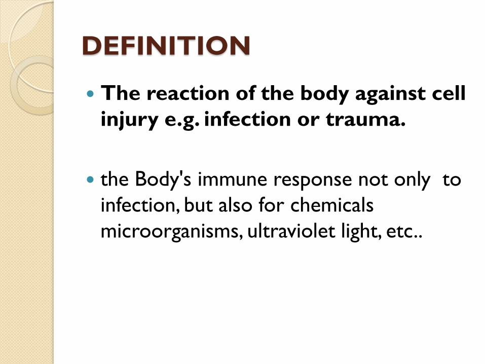
DEFINITION
The reaction of the body against cell
injury e.g. infection or trauma.
the Body's immune response not only to
infection, but also for chemicals
microorganisms, ultraviolet light, etc..


CAUSES
Burns
Chemical irritants
Frostbite
Toxins
Infection by pathogens
Physical injury, blunt or penetrating
Immune reactions due to hypersensitivity
Radiation

CHARACTERISTICS
Redness (RUBOR):
◦ When the cells damaged, histamine is released from basophils and mast cells.
◦ Histamine causes dilation of blood vessels and increased it permeability.
◦ Dilation of blood vessels cause an increase in blood flow to the injured area and the area becomes red.
Heat (CALOR):
◦ Increased blood flow to the injury area will cause the skin to feel warm when the touch.

• Swelling (TUMOUR):
- Increase permeability of the blood vessel causes the fluid to gather in the injury causes edema or swelling.
• Pain/(DALOR):
- Pain associated with tissue damage & also due to the secretion of bradykinin a peptides at the site of injury.
- Substances called prostaglandin seem to intensify bradykinin’s effect.
• Loss of Function (FUNCTIO LAESA)

INFLAMATION PROCESES
1. TISSUE DAMAGE
- Injury to the tisue and blood capilaries :
cuts
- Injured cells will secrete chemical
mediator e.g. bradykinin
- Blood clotting mechanisms activated.

2) VASODILATION AND INCREASE
PERMEABILITY OF BLOOD VESSEL.
- This will cause extra volume of blood flow to
injury area.
- Increase permeability will make more
immunity cells (macrophage, neutrophil)
transferred to injury area.
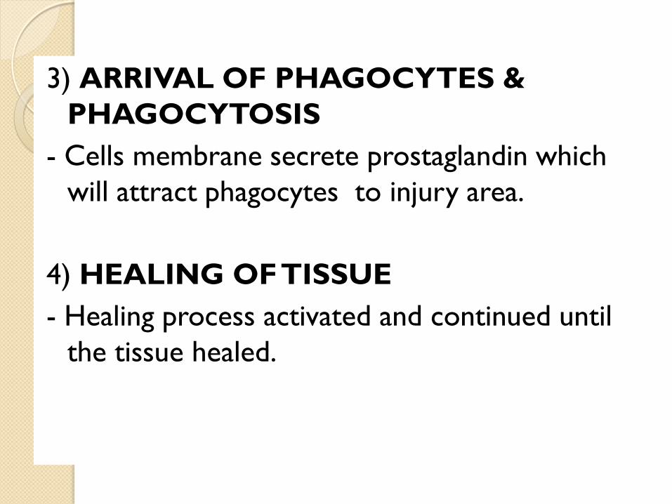
3) ARRIVAL OF PHAGOCYTES &
PHAGOCYTOSIS
- Cells membrane secrete prostaglandin which
will attract phagocytes to injury area.
4) HEALING OF TISSUE
- Healing process activated and continued until
the tissue healed.


(CHEMICAL MEDIATOR)

1) Histamine
Mast cells and basophile released histamine
Histamine
◦ a vasoactive amine which cause blood vessel dilation, increase permeability and increase muscle contractions.
◦ Fluid from blood vessel move into suraunding tissue - hyperemia.
◦ Constriction of bronchia and mucus secretion during allergy.

2) Bradykinin
Bradykinin ia a plasma protein produce during clotting factors production.
Bradykinin also a vasoactive mediator which increase vascular permeability, vasodilation, and muscle contraction.
It also stimulate pain sensation.
3) Prostaglandins
More than one types
Released from cells membrane
Prostaglandins
◦ Causes redness by increasing vasodilatation and permeability
◦ Pains (increase sensitivity of nerve endings)
◦ Increase platelets count for blood clotting.
◦ Act as chemotactic factor to attract neutrophils to injury area.

BLOOD VESSEL CHANGES
DURING INFALAMTION

1) Dilation of blood vessel
Chemical mediators release will cause the muscle of vessel to relax which allow a lot blood to flow in.
Vasodilation occurs first at the arteriole level, progressing to the capillary level.
Brings about a net increase in the amount of blood
This result more protein plasma, glucose and oxygen and cells to be transferred to injury area.
2) Blood Vessel Permeability
Increase blood clotting efficiency.
Because increase permeability a lot of fluid gather to tissue surrounding the injury resulting to edema, swelling and pains.

CELULAR CHANGES

1) Neutrophils
Neutrophils are pulled from the blood stream to
the site of inflammation.
more neutrophils are released from the bone
marrow.
2) Monocytes
Arrive latter
Monocytes are large immature forms of a
macrophage, which normally circulate in the blood
stream for about 3 days before maturing and
becoming a fixed macrophage

3) Neutrophils and the monocytes
They destroy the antigens that they have
ingested.
These cells have granulated enzymes
(lysosomes - contain lytic enzymes),
which help digest what they has engulfed.
MOLECULAR CHANGES

INTERFERON:
Interferons are a group of protein / chemical
mediators releases pd naturally by cells of
injured tissues during inflammatory process
because of viral infection or cancer.
Also play a role as immune stimulants, immune
regulators and in intercommunication of cells.
Interferons are part of non-specific defense
mechanisms
The Most common are alpha, beta and gamma
inteferons.
1. TYPE I: Fights viruses and cancer.
Alpha Interferon:
About 20 types of known. Dihasilkan oleh T sel (T-lymphocytes) and macrophages.
Stimulate Natural Killer Cells to kill virus infected cells.
Beta Interferon:
One type. Released by fibroblasts and epithelial cells infected by virus.
Stimulate production of antiviral proteins (AVP) which will disrupt virus replication process.

2. TYPE II:
Gamma Interferon:
◦ Produce by lymphocytes and NK cells
which is sensitive to foreign antigens
such as spt tumor cells, viruses and
bacteria.
◦ Increase activity - lymphocytes, NK cells
and macrophages.


FEVER
Increasing of basal body temperature because of Pyrogens (substance/agent producing fever),
Indirectly increase the setting of the temperature-regulating center in the brain, the hypothalamus.
Exogenous pyrogens cause by pathogens toxin.
Pathogens Toxin Cause Fever by
◦ Stimulate production of endogenous pyrogen from
macrophages.
◦ This endogenous pyrogen called interleukin-1 (IL-
1) is spread into blood flow & stimulate
prostaglandin production.
◦ prostaglandins reset the hypothalamus thermostat
at a higher temperature.

Importance :
◦ (1) Increase body temperature to optimum
temperature which will inhibit microorganism
Growth.
◦ (2) increasing the rate of chemical reactions
in the body
◦ (3) fever makes a patient feel ill, condition which
makes the patient more likely to rest.

Cells injured
PATHOGENS INVADE TISSUES
Capillaries become more permeable
Release histamine and other substances
Blood vessels dilate
Antibodies pass from blood into inflamed area
Phagocytes migrate to region
Brings needed phagocytes,
nutrients, and antibodies
Increased blood flow to area
Systemic response
Phagocytosis
Fever
Redness Increased temperature
Release endogenous pyrogens
Pain Edema
CONCLUSIONS

http://en.wikipedia.org/wiki/Inflammation#Proc
ess_of_acute_inflammation
SECOND BARRIER
•PHAGOCYTOSIS
•INFLAMATION
•COMPLEMENT SYSTEM

Refers to a group of 30 yg protein plays a role in the
body's defense.
Non-specific protein and is always present in the
body.
The complement system is activated by antigen-
antibody complex reaction.
Complement activation causes:
◦ 1. Cause a chain of reactions that can lyse the cell.
◦ 2. Increase phagocytosis.
◦ 3. Cause release of histamine by mast cells, which results in inflammation.
INTRO

Figure 21.13

COMPLEMENT
ACTIVATION

Complement activation can occur with 3
pathway
The pathway can occur simultaneously
Name of pathway
◦ Classical pathway
◦ Alternative pathway
◦ Lectin Pathway
All 3 pathway is difference in how the
process of forming C5 convertase.

Classical Pathway
Classical pathway required 9 complement
protein.
C1 to C9 respectively

C1
C1 exists in blood serum as a molecular complex containing:
◦ 6 molecules of C1q
◦ 2 molecules of C1r
◦ 2 molecules of C1s
C1q activated when it bind with Fc site on antibody.
C1q need at least 2 IgG or one IgM to bind.
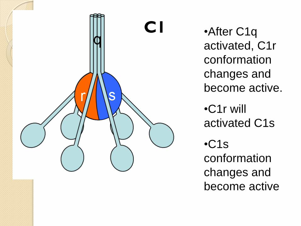
C1 •After C1q
activated, C1r
conformation
changes and
become active.
•C1r will
activated C1s
•C1s
conformation
changes and
become active

Activation of C1s will cleave C2 & C4 complement
protein:
◦ C4 is cleaved into to C4a & C4b
C4a, released into blood stream (smaller,
inactive).
C4b bind to pathogen membrane
◦ C2 is cleaved into
C2a, which binds noncovalently to a site on
C4b.
Smaller, inactive, fragment of C2b which diffuses
away.
◦ The complex of C4b•2a is called "C3
convertase”

C3
◦ C3 is the most abundant protein of the complement
system. Because of its abundance and its ability to
activate itself
C4b•2a cuts C3 into two major fragments:
◦ C3b, which binds covalently to C4b•2a.
◦ Form C4b•2a•3b (C5 convertase)
◦ Macrophages and neutrophils have receptors for
C3b and can bind the C3b-coated cell or particle
preparatory to phagocytosis.
◦ C3b also an opsonin.

C3a is a small fragment that released into the
surrounding fluids. It can bind to receptors on
basophils and mast cells triggering them to
release their vasoactive contents (e.g.,
histamine).
Involve during severe allergic (anaphylactic
shock)

C5 (C4b•2a•3b)
◦ C5 convertase will cleave C5 complement
protein.
◦ C5 cleaved into C5a and C5b
C5a released into blood stream
C5b bind to the surface of phatogen
◦ This form the membrane attack complex
(MAC)

• Binding of C5b on pathogens surface will
be followed by successive binding of
complement protein C6, C7, C8 and C9 to
C5b.
• This event cause
– Holes drilled into pathogens cells membrane
poly C9.
– Holes permit movement of liquid into the cells
– This cause the pathogens cells to burst (lysis)

Complement Protein Source
◦ Intestinal epithelium
◦ Macrophage
◦ Liver
◦ Spleen


77

Alternative Pathway
In natural condition there always C3
complement protein floating in blood
flow.
This free C3 is slowly cleave into C3a
and C3b.
To form C5 convertase alternative
pathway need
C3 protein, factor B factor D and
properdin

Cleave C3b bound directly to foreign cells
surface.
Then factor B bind to C3b
Then factor D bind to C3bB complex
Binding of factor D causes factor B to
cleave and form C3bBb complex
C3bBb have same function as C3
convertase.
Properdin then bind to C3bBb to stabilize
it.

C3bBb complex can cleave free C3
protein to form C3a and C3b.
C3b then bind to C3bBb to form
C3bBb3b which have the same function as
C5 convertase.
From this onwards the formation of
membrane attack complex (MAC) is same
as classical pathways.

Lectin pathway
Another protein that can initiate
formation of C5 convertase is called
mannose-binding lectin (MBL)
MBL can bind to mannose residue on the
surface of microorganism cells.
MBL is a protein produce during
inflammatory reaction.
MBL function is similar to C1q

When MBL attach to cells surface two
protein that is MBL-associated serine
proteases,MASP-1 and MASP-2 bind to MBL.
The structure is similar to C1qrs complex.
This complex then cleave C4 and C2 protein
to form C3 convertase.
From this stage formation of MAC complex
on bacterial/ antigen is similar to classical
pathway.


END