non-specific and specific interactions on functionalized polymer surface
TRANSCRIPT
Ns
Ja
5b
a
ARRAA
KPFSPB
1
aeatbiatIbragme
0d
Colloids and Surfaces B: Biointerfaces 83 (2011) 220–228
Contents lists available at ScienceDirect
Colloids and Surfaces B: Biointerfaces
journa l homepage: www.e lsev ier .com/ locate /co lsur fb
on-specific and specific interactions on functionalized polymer surfacetudied by FT-SPR
izheng Weia,b, Lesan Yana,b, Xiuli Hua, Xuesi Chena, Yubin Huanga, Xiabin Jinga,∗
State Key Laboratory of Polymer Physics and Chemistry, Changchun Institute of Applied Chemistry, Chinese Academy of Sciences,625 Renmin Street, Changchun 130022, PR ChinaGraduate School of Chinese Academy of Sciences, Beijing 100049, PR China
r t i c l e i n f o
rticle history:eceived 20 April 2010eceived in revised form 8 November 2010ccepted 10 November 2010vailable online 18 November 2010
eywords:olymer filmourier transformurface plasmon resonancerotein adsorption
a b s t r a c t
Fourier transform surface plasmon resonance (FT-SPR) was utilized to study specific and non-specificinteractions between proteins and a biotinylated polymer film by monitoring adsorptions of strep-tavidin (SAv) and bovine serum albumin (BSA) on the polymer films. The biotinylated polymer,poly(lactide-co-2,2-dihydroxymethyl-propylene carbonate-graft-biotin) [P(LA-co-DHC/biotin)], was pre-pared by ring-opening copolymerization of lactide and a OH-bearing cyclic carbonate monomer, followedby biotinylation of the OH groups. The copolymer was coated onto the FT-SPR chip and vacuum-dried,hydrated at 70 ◦C, and treated with a blocking agent respectively to achieve different surface status. TheFT-SPR results showed that the vacuum-dried film had the most BSA adsorption; hydration treatment ledto migration of the biotin moieties from inner film to surface and thus resulted in less BSA adsorption;blocking layer on the polymer surface saturated the active sites for physical and chemical adsorptions
iotin–avidin interaction on the surface and thus weakened the BSA adsorption. Adsorption of SAv displayed similar polymer-surface-status dependence, i.e., more adsorption on vacuum-dried surface, less adsorption on hydratedsurface and the least adsorption on blocked surface. Compared with BSA, SAv showed more enhancedadsorptions on P(LA-co-DHC/biotin) surface because of the specific interaction of biotin moieties in thepolymer with SAv molecules, especially on the blocked surface. The above semi-quantified results further
syste
indicate that the FT-SPRbio-molecules.. Introduction
Biomaterials have found wide applications in disease diagnosisnd therapy, such as biosensors, surgical implants, tissue-ngineering scaffolds, drug-delivery devices (DDDs), etc. In thesepplications, biomaterials are in contact with biomolecules, cells,issues or organs, and interactions of polymer surfaces with theseiomolecules, cells, tissues or organs play an important role
n determining biomedical functions of these materials. Proteindsorption is known as the first event following the implanta-ion of biomaterials and it determines many subsequent events.n some cases, protein adhesion can be a necessary process for aiomaterial to perform its function, for example, when the boneepair materials are implanted into human body, desired protein
dsorption on the materials will improve the cell adhesion androwth. For this purpose, incorporation of RGD peptides on theaterials was reported to promote adherence, spread and prolif-ration of the desired cells on the implant surface [1]. However,
∗ Corresponding author. Tel.: +86 431 85262775; fax: +86 431 85262775.E-mail address: [email protected] (X. Jing).
927-7765/$ – see front matter © 2010 Elsevier B.V. All rights reserved.oi:10.1016/j.colsurfb.2010.11.020
m is suitable for investigating interactions between polymer surface and
© 2010 Elsevier B.V. All rights reserved.
in most cases, non-specific protein adsorption leads to undesir-able events, including thrombus formation, foreign body reaction,bacterial infection, and other responses [2]. For instance, when amacromolecule was used as a drug carrier and was sent into humanbody, too much protein adsorption may enlarge the particle size,thus prevent the cell uptake, or even cause thrombus. In the inter-vention diagnosis and therapy, if the device surface is stained byundesired proteins, the sensitivity of the detector will degrade, oreven leads to adverse effects [3]. Most biosensors are based uponspecific interactions between antibody and antigen or between lig-and and receptor [4–6], and if the sensor is stained by proteinsadsorbed non-specifically, it will surely do damage to the accuracyand sensitivity of the detection [7]. Similarly, undesired proteinadsorption also does great harm to in vitro biosensors, blood trans-fusion vessels [8], marine coatings [9], and so on, in addition to thein vivo applications above.
Therefore, lots of effort have been made to enhance or to reduce
protein adsorption on biomaterial surfaces, e.g., by adjusting thehydrophilicity [10,11], charge [12,13] and microphase separation[14] of the surface. PEGylation is one of the most popular strate-gies often used to modify the proteins [15–18], materials [14,19,20]as well as the surface of the biosensor platforms [7]. But most of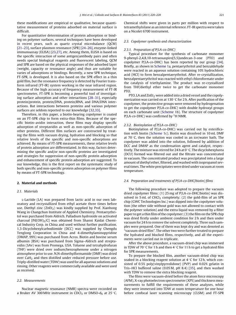
es B: B
ttd
ii[itna(vFgfBsipas
ocaoiraomaaobb
2
2
ouWtcc1T(av(aoTra
2
a
J. Wei et al. / Colloids and Surfac
hese modifications are empirical or qualitative, because quanti-ative measurement of proteins adsorbed on material surface isifficult.
For quantitative determination of protein adsorption or bind-ng on polymer surfaces, several techniques have been developedn recent years, such as quartz crystal microbalance (QCM)21–23], surface plasmon resonance (SPR) [24–26], enzyme-linkedmmunoassay (ELISA) [23,27], etc. Among them, ELISA is based onhe specific interaction of some antigen/antibody pairs and ofteneeds special biological reagents and fluorescent labeling, QCMnd SPR are based on the physical responses of the adsorbed layerweight, capacity or resonance angle) and thus are suitable foraries of adsorptions or bindings. Recently, a new SPR technique,T-SPR, is developed. It is also based on the SPR effect in a thinold film, but the resonance frequency is detected by Fourier trans-orm infrared (FT-IR) system working in the near infrared region.ecause of the high accuracy of frequency measurement of FT-IRpectrometer, FT-SPR is becoming a powerful tool of investigat-ng surface adsorption and other interactions [28–31], especiallyrotein/protein, protein/DNA, protein/RNA, and DNA/DNA inter-ctions. But interactions between proteins and various polymerurfaces are seldom reported to our knowledge [32,33].
Therefore, in this paper, a biotin-bearing copolymer is coatedn an FT-SPR chip to form extra-thin films. Because of the spe-ific biotin–avidin interaction, these films may display specificdsorption of streptavidin as well as non-specific adsorption ofther proteins. Different film surfaces are constructed by treat-ng the films with vacuum drying, hydration and blocking so thatelative levels of the specific and non-specific adsorptions arechieved. By means of FT-SPR measurements, these relative levelsf protein adsorption are differentiated. In this way, factors deter-ining the specific and/or non-specific proteins are investigated
nd strategies for suppression of non-specific protein adsorptionnd enhancement of specific protein adsorption are suggested. Tour knowledge, this is the first report on the systematic study ofoth specific and non-specific protein adsorption on polymer filmsy means of FT-SPR technology.
. Material and methods
.1. Materials
l-Lactide (LA) was prepared from lactic acid in our own lab-ratory and recrystallized from ethyl acetate three times beforese. Diethyl zinc (ZnEt2) was kindly supplied by Prof. Xianhongang in Changchun Institute of Applied Chemistry. Pentaerythri-
ol was purchased from Aldrich. Palladium hydroxide on activatedharcoal [Pd(OH)2/C] was obtained from Shanxi Kaidai Chemi-al Industry Corp. in China and used without further purification.,3-Dicyclohexylcarbodiimide (DCC) was supplied by Chengduenglong Corporation in China and 4-dimethylaminopyridineDMAP, 99%) was purchased from Acros. Biotin and bovine serumlbumin (BSA) was purchased from Sigma–Aldrich and strepta-idin (SAv) was from Promega, USA. Toluene and tetrahydrofuranTHF) were dried over sodium/benzophenone under a nitrogentmosphere prior to use. N,N-dimethylformamide (DMF) was driedver CaH2 and then distilled under reduced pressure before use.riply-distilled water (TDW) was used for all aqueous solutions andinsing. Other reagents were commercially available and were useds received.
.2. Measurements
Nuclear magnetic resonance (NMR) spectra were recorded onBruker AV 300 MHz instrument in CDCl3 or DMSO-d6 at 25 ◦C.
iointerfaces 83 (2011) 220–228 221
Chemical shifts were given in parts per million with respect totetramethylsilane as an internal reference. FT-IR spectra were takenon a Nicolet 6700 instrument.
2.3. Copolymer synthesis and characterization
2.3.1. Preparation of P(LA-co-DHC)Typical procedure for the synthesis of carbonate monomer
9-phenyl-2,4,8,10-tetraoxaspiro[5,5]undecan-3-one (PTO) andcopolymer P(LA-co-DHC) has been reported by our group [34].Briefly, as shown in Scheme 1a, pentaerythritol and benzaldehydewere reacted in an aqueous solution containing 10% hydrochloricacid (HCl) to form benzalpentaerythritol. After re-crystallization,benzalpentaerythritol was reacted with ethyl chloroformate underthe catalysis of triethylamine. The product was re-crystallizedfrom THF/diethyl ether twice to get the carbonate monomerPTO.
PTO, LA and ZnEt2 were added into a dried vessel and the copoly-merization was carried on at 130 ◦C for 2 h. After purification of thecopolymer, the protective groups were removed by hydrogenationto get the copolymer P(LA-co-DHC) with double hydroxyl groupson each carbonate unit (Scheme 1b). The structure of copolymerP(LA-co-DHC) was confirmed by 1H NMR.
2.3.2. Biotinylation of P(LA-co-DHC)Biotinylation of P(LA-co-DHC) was carried out by esterifica-
tion with biotin (Scheme 1c). Biotin was dissolved in 10 mL DMFat 70 ◦C, then the solution was cooled in an ice/water bath. Thecopolymer was added into the solution, followed by addition ofDCC and DMAP as the condensation agent and catalyst, respec-tively. The mixture was stirred for 24 h at 0 ◦C. The dicyclohexylurea(DCU) formed was filtered out and the filtrate was concentratedin vacuum. The concentrated product was precipitated into a largeamount of diethyl ether, filtered, and washed with isopropanol sev-eral times. The white precipitates were dried under vacuum at roomtemperature.
2.4. Preparation and treatments of P(LA-co-DHC/biotin) films
The following procedure was adopted to prepare the vacuumdried copolymer films: (1) 25 mg of P(LA-co-DHC/biotin) was dis-solved in 5 mL of CHCl3 completely; (2) the gold-film of an SPRchip (GWC Technologies Inc.) was dipped into the copolymer solu-tion (the other side without gold was not allowed to contact withthe polymer solution) and the extra liquid was removed by a filterpaper to get a thin film of the copolymer; (3) the film on the SPR chipwas dried firstly under ambient condition for 2 h and then undervacuum for 24 h to remove the solvent completely. Three such sam-ples were prepared. One of them was kept dry and was denoted as“vacuum-dried film”. The other two were further treated to preparethe hydrated and blocked films, respectively, and all the experi-ments were carried out in triplicate.
After the above procedure, a vacuum-dried chip was immersedin TDW of 70 ◦C for 1 h and then 4 ◦C for 11 h to get a hydrated filmfor SPR measurements.
To prepare the blocked film, another vacuum-dried chip wassoaked in a blocking reagent solution at 4 ◦C for 12 h, which con-sisted of 0.5% poly(vinylpyrrolidone) (PVP) and 0.02% gelatin inTris–HCl buffered saline (0.05 M, pH 8.4) [35], and then washedwith TDW to remove the extra blocking reagent.
The films were vacuum-dried before the atom force microscopy(AFM), X-ray photoelectron spectrometer (XPS) and thickness mea-surements to fulfill the requirements of these analyses, whilethey were immersed into TDW at room temperature for one hourbefore confocal laser scanning microscopy (CLSM) and FT-SPR
222 J. Wei et al. / Colloids and Surfaces B: Biointerfaces 83 (2011) 220–228
+
O
OH3C
CH3
O
O
Et2Zn O
O
O O
O O
Ph
O
m n
H2 , Pd (OH)2/CO
O
O O
OH OH
O
mn
2 h130ºC(b) PTO
P(LA-co-DHP) P(LA-co-DHC)
50ºC 48 h
O
HOH OH
OH OH
+
OH OH
O O
Ph
Cl OC2H5
O
HCl, H2O
20ºC 5 h
O O
O O
Ph
O
TEA/THF, 0ºC 2 h
(a)PTO
O
O
O O
O
m n
N
N
H
H
OS
HO
O
+ DCC/DMAP
THF, 0ºC 24 h(c)
N
N
H
H
OS
O
O
N
N
H
H
O S
O
OP(LA-co-DHC)
P(LA-co-DHC/biotin)
S er, 9d HC).
mfi
2
f3t
ftv
lSsiet
s(ttt
with a Leica TCS SP2 CLSM (Leica Microsystems Heidelberg GmbH,
cheme 1. Preparation procedure of P(LA-co-DHC/biotin). (a) Synthesis of monomeprotection of copolymer P(LA-co-DHC); (c) biotinylation of copolymer P(LA-co-D
easurements to achieve equilibrated water absorption of thelms.
.5. Characterization of the copolymer films
Atom force microscopy (AFM) measurements were performedor the smoothness of the copolymer film on the SPR chip with SPI800/SPA 300HV (Seiko Instrument Inc.) in tapping mode at roomemperature in air. The tip was of OMCL-ACTS-W type.
The film thickness was determined using a Dektak 6 M sur-ace profiler (Veeco Instruments Inc., USA) by purposely scratchinghe polymer-coated area and by subsequent profile analysis in theicinity of the scratches.
The surface elemental composition of the polymer film was ana-yzed on an Escalab-MKII X-ray photoelectron spectrometer (VGcientific Ltd., UK) using Mg K� radiation (1253.6 eV) as the X-rayource for excitation. The typical operating pressure in the analyt-cal chamber was in the range of 10−9 to 10−10 Torr. The bindingnergy of the experimental spectra was calibrated on the basis ofhe most intense peak of C1s at 284.5 eV.
The contact angle of the films were measured by a DSA-10 drop
hape analyzer (Germany) equipped with a charge-coupled deviceCCD) camera. The water droplets had a fixed volume of 2 �L. Whenhe water droplet attached the film surface, the picture was cap-ured by the CCD camera and the contact angle was calculated byhe DSA software.-phenyl-2,4,8,10-tetraoxaspiro[5,5]undecan-3-one (PTO); (b) polymerization and
2.6. Confocal laser scanning microscopy (CLSM) measurements
2.6.1. FITC labeling of BSA200 mg of BSA (0.003 mmol) was dissolved in 1.5 mL of TDW
and FITC (fluorescein isothiocyanate, 5 mg, 0.013 mmol) solutionin 10 mL carbonate buffer (1000 mL buffer consisted of NaHCO37.56 g, Na2CO3 1.06 g and NaCl 7.36 g, pH 9.0) was added into theBSA solution dropwise under stirring. The reaction lasted for 2 h indarkness. After the reaction, the solution was dialyzed against PBS(phosphate buffered saline, pH 7.4) for 3 days, followed by freeze-drying.
2.6.2. CLSM measurementFor CLSM measurement, the copolymer was spin-coated onto
glass chips and the coatings were treated in the same manner asfor those coated on the SPR chips. The treated glass chips wereimmersed in BSA-FITC solution (1 mg/mL in PBS, pH 7.4) for 5 min,then washed with TDW for 5 times and dried at room temper-ature. Finally, each chip was covered with a cover glass with athin layer of glycerin in between. CLSM images were collected
Germany) equipped with an 20× dry objective (NA = 0.7) usingdigital zooms of 1–32× attached to a Leica DM IRE2 invertedmicroscope. Confocal optical sections were collected in the image-scan x–y–z mode. The samples were excited by a 543 nm He/Nelaser.

J. Wei et al. / Colloids and Surfaces B: Biointerfaces 83 (2011) 220–228 223
urve w
2d
IIittssgbIaatoboriFta
qglvfrt
Fig. 1. (A) Schematic illustration of the SPR sensor; (B) a typical reflection c
.7. Fourier-transform surface plasmon resonance (FT-SPR)etection
FT-SPR measurements were carried out using a SPR-100 (GWCnstruments, WI, USA) module coupled with a Nicolet 6700 FT-R spectrometer (Thermo-Electron, Madison, WI, USA) workingn the near infrared (NIR) region which was equipped with aungsten halogen near-infrared light source, a CaF2 beam split-er and an InGaAs detector. The SPR-100 module consists of aample cell and a set of light path accessory. Structure of theample cell is shown in Fig. 1A. The NIR light beam enters thelass prism at a fixed incidence angle, reaches and is reflectedy the gold film on the SPR chip, and finally detected by the FT-
R spectrometer. When a surface plasmon resonance takes place,minimum light intensity of the reflected beam is obtained atcertain NIR frequency (Fig. 1B) due to the resonance absorp-
ion, and this frequency is called resonance frequency. Unlike thether SPR devices which measure the intensity of the reflectedeam at a fixed frequency as a function of incidence angle tobtain the “resonance angle”, FT-SPR measures the intensity of theeflected beam as a function of incident beam frequency at a givenncidence angle. Because of the high accuracy and sensitivity ofourier-transform infrared spectroscopy, FT-SPR is more sensitivehan other techniques based on direct measurement of resonancengle.
According to the principle of FT-SPR, the intrinsic resonance fre-uency is determined by the thickness and refractive index of theold film of the SPR chip. When the gold film is in contact with a
ayer of solid or liquid, the resonance frequency will shift to a higheralue (Fig. 1C) because of the involvement of this layer in the sur-ace plasmon resonance, and the frequency shift (termed as “SPResponse”) is dependent on the refractive index and thickness ofhe layer. Therefore, the SPR chip serves as a sensor to the chemicalith a resonance peak; (C) a typical adsorption curve determined by FT-SPR.
composition, density and/or thickness of the layer in contact withthe gold surface.
In the FT-SPR module, the gold film on the SF-10 glass chipis 45 nm in thickness and is a square of 18 mm × 18 mm. In thecenter of the square, a circular sample cell of 10 mm in diam-eter is formed by a rubber O-ring. The capacity of the flowcell was 60 �L. The other side of the chip is brought into opti-cal contact with the SF-10 equilateral prism using a refractiveindex matching fluid (Cargille Laboratories, USA). Circulation wascarried out by a Masterflex L/S (Cole-Parmer, USA) peristalticpump at a circulating rate of 0.18 mL/min. FT-SPR data werecollected using the Omnic software version 7.0 (Thermo Elec-tron), at the resolution of 32 cm−1 over the range from 6000 to12,000 cm−1.
In the present study, the P(LA-co-DHC/biotin) film on the SPRchip was the first layer. A protein solution was pumped throughthe outer surface of polymer coating. When protein moleculeswere adsorbed onto the P(LA-co-DHC/biotin) coating, the reso-nance frequency of the whole system would change. The frequencyshift would correspond to the proteins adsorbed on the polymersurface.
Experimentally, PBS was first circulated through the flow celland the resonance frequency was adjusted to ca. 9000 cm−1. Thisfrequency was considered as the baseline of adsorption. Then, BSAor SAv solution in PBS buffer (pH 7.4) of 0.2 mg/mL was allowedto flow through the cell, and its resonance frequency was plot-ted against time to demonstrate the protein adsorption process.After the resonance frequency leveled off, PBS was circulated again
through the cell to rinse the adsorbate layer on the SPR chip. Theresonance frequency decreased correspondingly. The difference ofthe final resonance frequency from the initial baseline was consid-ered to reflect the amount of the protein adsorbed on the polymerfilm.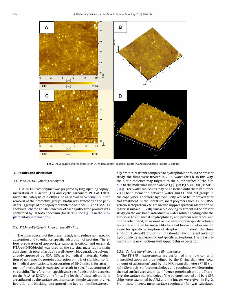
224 J. Wei et al. / Colloids and Surfaces B: Biointerfaces 83 (2011) 220–228
tin) c
3
3
murdscp
3
afPcatimsoah
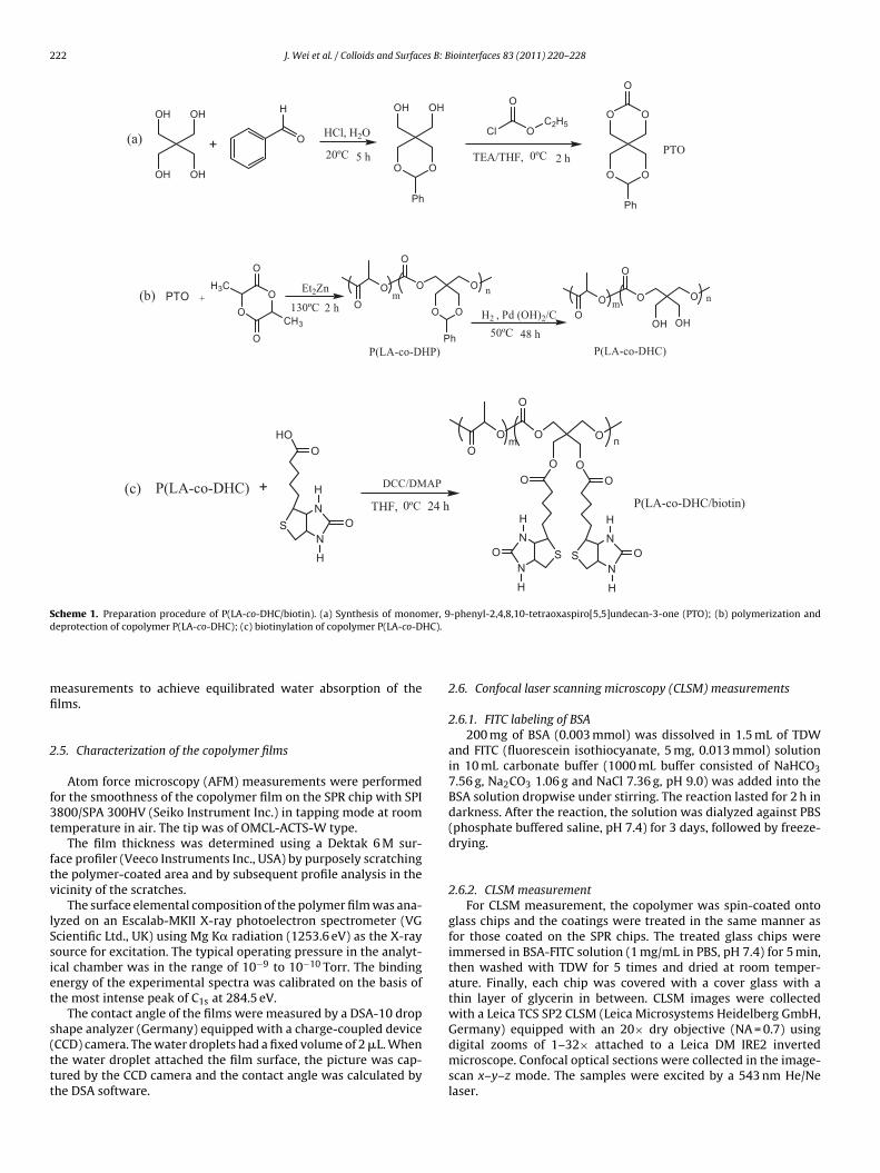
Fig. 2. AFM images and roughness of P(LA-co-DHC/bio
. Results and discussion
.1. P(LA-co-DHC/biotin) copolymer
P(LA-co-DHP) copolymer was prepared by ring-opening copoly-erization of l-lactide (LA) and cyclic carbonate PTO at 130 ◦C
nder the catalysis of diethyl zinc as shown in Scheme 1b. Afteremoval of the protective groups, biotin was attached to the pen-ant OH groups of the copolymer with the help of DCC and DMAP ashown in Scheme 1c. The structure of each synthesized product wasonfirmed by 1H NMR spectrum (for details, see Fig. S1 in the sup-lementary information).
.2. P(LA-co-DHC/biotin) film on the SPR chips
The main concern of the present study is to reduce non-specificdsorption and to enhance specific adsorption of proteins. There-ore, preparation of appropriate samples is critical and essential.(LA-co-DHC/biotin) was used as the starting material. Its mainonstituent is poly(l-lactide), a well-known biodegradable polymerlready approved by FDA, USA as biomedical materials. Reduc-ion of non-specific protein adsorption on it is of significance forts medical applications. Incorporation of DHC units is for attach-
ent of biotin, that is expected to result in specific adsorption oftretavidin. Therefore, non-specific and specific adsorptions coexistn the P(LA-co-DHC/biotin) films. The levels of these adsorptionsre adjusted by the surface treatments, i.e., simple vacuum-drying,ydration and blocking. It is reported that hydrophilic films are usu-
oated SPR chip (A and B) and bare SPR chip (C and D).
ally protein-resistant compared to hydrophobic ones. In the presentstudy, the films were treated in 70 ◦C water for 1 h. In this way,the biotin moieties may migrate to the outer surface of the filmdue to the molecular motion above Tg (Tg of P(LA-co-DHC) is 50 ◦C[34]). Free water molecules may be adsorbed onto the film surfacevia H-bond formation between water and CO and NH groups inthe copolymer. Therefore hydrophilicity would be improved afterthis treatment. In the literature, inert polymers such as PVP, BSA,gelatin, lactoprotein, etc. are used to suppress protein adsorption onmaterial surface [35–38]. Surface-blocking treatment in the presentstudy, on the one hand, introduces a water soluble coating onto thefilm so as to enhance its hydrophilicity and protein resistance, andon the other hand, all or most active sites for non-specific adsorp-tions are saturated by surface blockers but biotin moieties are leftalone for specific adsorption of streptavidin. In short, the threekinds of P(LA-co-DHC/biotin) films should have different levels ofhydrophilicity, non-specific and specific adsorptions. The measure-ments in the next sections will support this expectation.
3.2.1. Surface morphology and film thicknessThe FT-SPR measurements are performed in a flow cell with
a specified apparent area defined by the O-ring diameter (totalamount of adsorption) and by the NIR beam diameter (FT-IR sig-
nal). Obviously, surface morphology and roughness will determinethe real surface area and thus influence protein adsorption. There-fore, the surface morphologies of the polymer-coated and bare SPRchips were examined by AFM and the images were given in Fig. 2.From these images, mean surface roughness (Ra) was calculated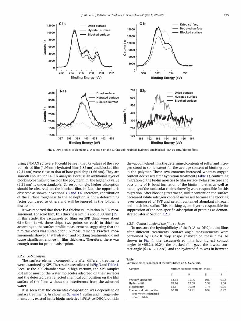
J. Wei et al. / Colloids and Surfaces B: Biointerfaces 83 (2011) 220–228 225
4034024014003993983971000
1500
2000
2500
3000
3500
4000
Co
un
ts /
s
Dried surface
Hytrated surface
Blocked surface
N1s
167166165164163162161100
200
300
400
500
600
700
Co
un
ts /
s
Dried surface Hytrated surface Blocked surface
S2p
5365345325300
3000
6000
9000
12000
15000
18000
Co
un
ts /
s
Binding Energy (eV)
Dried surface Hytrated surface Blocked surface
O1s
292290288286284282
0
2000
4000
6000
8000
10000
12000
Co
un
ts /
s
Binding Energy (eV)
Dried surface
Hytrated surface
Blocked surface
C1s
s of t
uu(sb(soofd
sI6afisce
3
wBlasw
sm
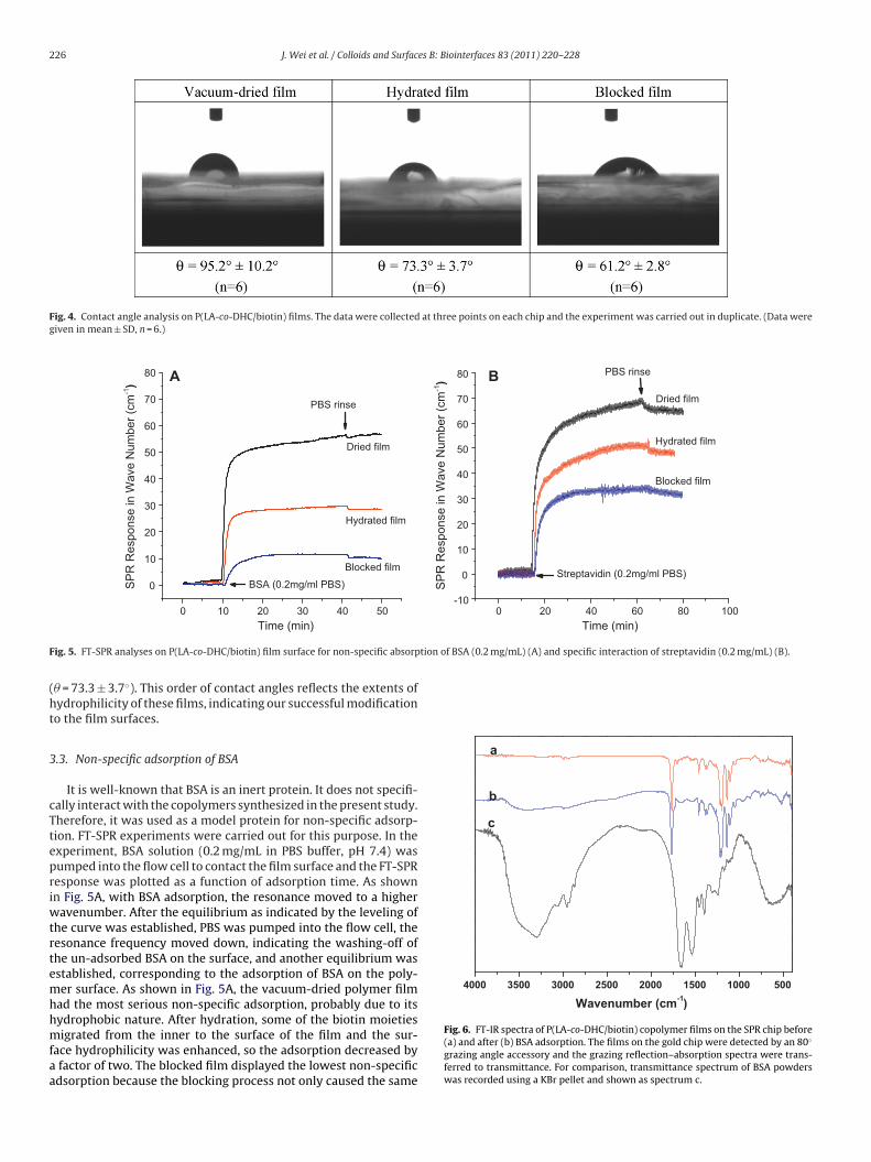
after different treatments, contact angle measurements wereperformed by DSA-10 drop shape analyzer on these films. Asshown in Fig. 4, the vacuum-dried film had highest contactangles (� = 95.2 ± 10.2◦), the blocked film gave the lowest con-tact angle (� = 61.2 ± 2.8◦), and the hydrated film was in between
Table 1Surface element contents of the films based on XPS analysis.
Samples Surface element contents (mol%)
C O N S
Vacuum-dried film 63.33 35.65 0.80 0.22
Binding Energy (eV)
Fig. 3. XPS profiles of elements C, O, N and S on the surface
sing SPIMAN software. It could be seen that Ra values of the vac-um dried film (1.95 nm), hydrated film (1.85 nm) and blocked film2.31 nm) were close to that of bare gold chip (1.66 nm). They aremooth enough for FT-SPR analysis. Because an additional layer oflocking coating is formed on the polymer film, the higher Ra value2.31 nm) is understandable. Correspondingly, higher adsorptionhould be observed on the blocked film. In fact, the opposite isbserved as shown in Sections 3.3 and 3.4. Therefore, contributionf the surface roughness to the adsorption is not a determiningactor compared to others and will be ignored in the followingiscussion.
It was reported that there is a thickness limitation in SPR mea-urement. For solid film, this thickness limit is about 300 nm [39].n this study, the vacuum-dried films on SPR chips were about5 ± 8 nm (n = 6, three chips, two points on each) in thicknessccording to the surface profile measurement, suggesting that thelm thickness was suitable for SPR measurements. Practical mea-urements showed that hydration and blocking treatments did notause significant change in film thickness. Therefore, there wasnough room for protein adsorption.
.2.2. XPS analysisThe surface element compositions after different treatments
ere examined by XPS. The results are collected in Fig. 3 and Table 1.ecause the XPS chamber was in high vacuum, the XPS samples
ost all or most of the water molecules adsorbed on their surfacesnd the detected data reflected chemical composition on the film
urface of the films without the interference from the adsorbedater.It is seen that the elemental composition was dependent onurface treatments. As shown in Scheme 1, sulfur and nitrogen ele-ents only existed in the biotin moieties in P(LA-co-DHC/biotin). In
Binding Energy (eV)
he dried, hydrated and blocked P(LA-co-DHC/biotin) films.
the vacuum-dried film, the determined contents of sulfur and nitro-gen stood to some extent for the average content of biotin groupin the polymer. These two contents increased whereas oxygencontent decreased after hydration treatment (Table 1), confirmingmigration of the biotin moieties to film surface. Polar structure andpossibility of H-bond formation of the biotin moieties as well asmobility of the molecular chains above Tg were responsible for thismigration. After blocking treatment, sulfur content on the surfacedecreased while nitrogen content increased because the blockinglayer composed of PVP and gelatin contained abundant nitrogenand much less sulfur. This blocking agent layer is responsible forsuppression of the non-specific adsorption of proteins as demon-strated later in Section 3.2.3.
3.2.3. Contact angle of the film surfacesTo measure the hydrophilicity of the P(LA-co-DHC/biotin) films
Hydrated film 67.74 27.68 3.52 1.06Blocked film 65.31 30.69 3.75 0.25Theoretical ratios of the
copolymer (calculatedfrom 1H NMR)
60.18 38.41 0.94 0.47
226 J. Wei et al. / Colloids and Surfaces B: Biointerfaces 83 (2011) 220–228
Fig. 4. Contact angle analysis on P(LA-co-DHC/biotin) films. The data were collected at three points on each chip and the experiment was carried out in duplicate. (Data weregiven in mean ± SD, n = 6.)
100806040200-10
0
10
20
30
40
50
60
70
80 PBS rinse
Blocked film
Hydrated film
Dried film
Streptavidin (0.2mg/ml PBS)
SP
R R
espo
nse
in W
ave
Num
ber
(cm
-1) B
0
10
20
30
40
50
60
70
80
SP
R R
espo
nse
in W
ave
Num
ber
(cm
-1)
A
BSA (0.2mg/ml PBS)
Time (min)
PBS rinse
Dried film
Hydrated film
Blocked film
F tion of BSA (0.2 mg/mL) (A) and specific interaction of streptavidin (0.2 mg/mL) (B).
(ht
3
cTtepriwtrtemhhmfaa
5001000150020002500300035004000
b
c
Wavenumber (cm-1)
a
50403020100
Time (min)
ig. 5. FT-SPR analyses on P(LA-co-DHC/biotin) film surface for non-specific absorp
� = 73.3 ± 3.7◦). This order of contact angles reflects the extents ofydrophilicity of these films, indicating our successful modificationo the film surfaces.
.3. Non-specific adsorption of BSA
It is well-known that BSA is an inert protein. It does not specifi-ally interact with the copolymers synthesized in the present study.herefore, it was used as a model protein for non-specific adsorp-ion. FT-SPR experiments were carried out for this purpose. In thexperiment, BSA solution (0.2 mg/mL in PBS buffer, pH 7.4) wasumped into the flow cell to contact the film surface and the FT-SPResponse was plotted as a function of adsorption time. As shownn Fig. 5A, with BSA adsorption, the resonance moved to a higher
avenumber. After the equilibrium as indicated by the leveling ofhe curve was established, PBS was pumped into the flow cell, theesonance frequency moved down, indicating the washing-off ofhe un-adsorbed BSA on the surface, and another equilibrium wasstablished, corresponding to the adsorption of BSA on the poly-er surface. As shown in Fig. 5A, the vacuum-dried polymer film
ad the most serious non-specific adsorption, probably due to its
ydrophobic nature. After hydration, some of the biotin moietiesigrated from the inner to the surface of the film and the sur-ace hydrophilicity was enhanced, so the adsorption decreased byfactor of two. The blocked film displayed the lowest non-specificdsorption because the blocking process not only caused the same
Fig. 6. FT-IR spectra of P(LA-co-DHC/biotin) copolymer films on the SPR chip before(a) and after (b) BSA adsorption. The films on the gold chip were detected by an 80◦
grazing angle accessory and the grazing reflection–absorption spectra were trans-ferred to transmittance. For comparison, transmittance spectrum of BSA powderswas recorded using a KBr pellet and shown as spectrum c.
J. Wei et al. / Colloids and Surfaces B: Biointerfaces 83 (2011) 220–228 227
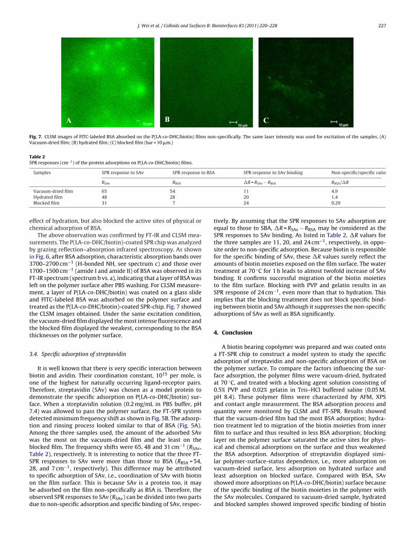
Fig. 7. CLSM images of FITC-labeled BSA absorbed on the P(LA-co-DHC/biotin) films non-specifically. The same laser intensity was used for excitation of the samples. (A)Vacuum-dried film; (B) hydrated film; (C) blocked film (bar = 10 �m.)
Table 2SPR responses (cm−1) of the protein adsorptions on P(LA-co-DHC/biotin) films.
Samples SPR response to SAv SPR response to BSA SPR response to SAv binding Non-specific/specific ratio
RSAv RBSA �R = RSAv − RBSA RBSA/�R
ec
sbi31Flmattttt
3
boTdf7dtAwbTS2tobod
Vacuum-dried film 65 54Hydrated film 48 28Blocked film 31 7
ffect of hydration, but also blocked the active sites of physical orhemical adsorption of BSA.
The above observation was confirmed by FT-IR and CLSM mea-urements. The P(LA-co-DHC/biotin)-coated SPR chip was analyzedy grazing reflection–absorption infrared spectroscopy. As shown
n Fig. 6, after BSA adsorption, characteristic absorption bands over700–2700 cm−1 (H-bonded NH, see spectrum c) and those over700–1500 cm−1 (amide I and amide II) of BSA was observed in itsT-IR spectrum (spectrum b vs. a), indicating that a layer of BSA waseft on the polymer surface after PBS washing. For CLSM measure-
ent, a layer of P(LA-co-DHC/biotin) was coated on a glass slidend FITC-labeled BSA was adsorbed on the polymer surface andreated as the P(LA-co-DHC/biotin)-coated SPR-chip. Fig. 7 showedhe CLSM images obtained. Under the same excitation condition,he vacuum-dried film displayed the most intense fluorescence andhe blocked film displayed the weakest, corresponding to the BSAhicknesses on the polymer surface.
.4. Specific adsorption of streptavidin
It is well known that there is very specific interaction betweeniotin and avidin. Their coordination constant, 1015 per mole, isne of the highest for naturally occurring ligand-receptor pairs.herefore, streptavidin (SAv) was chosen as a model protein toemonstrate the specific adsorption on P(LA-co-DHC/biotin) sur-ace. When a streptavidin solution (0.2 mg/mL in PBS buffer, pH.4) was allowed to pass the polymer surface, the FT-SPR systemetected minimum frequency shift as shown in Fig. 5B. The adsorp-ion and rinsing process looked similar to that of BSA (Fig. 5A).mong the three samples used, the amount of the adsorbed SAvas the most on the vacuum-dried film and the least on the
locked film. The frequency shifts were 65, 48 and 31 cm−1 (RSAv,able 2), respectively. It is interesting to notice that the three FT-PR responses to SAv were more than those to BSA (RBSA = 54,8, and 7 cm−1, respectively). This difference may be attributed
o specific adsorption of SAv, i.e., coordination of SAv with biotinn the film surface. This is because SAv is a protein too, it maye adsorbed on the film non-specifically as BSA is. Therefore, thebserved SPR responses to SAv (RSAv) can be divided into two partsue to non-specific adsorption and specific binding of SAv, respec-11 4.920 1.424 0.29
tively. By assuming that the SPR responses to SAv adsorption areequal to those to SBA, �R = RSAv − RBSA may be considered as theSPR responses to SAv binding. As listed in Table 2, �R values forthe three samples are 11, 20, and 24 cm−1, respectively, in oppo-site order to non-specific adsorption. Because biotin is responsiblefor the specific binding of SAv, these �R values surely reflect theamounts of biotin moieties exposed on the film surface. The watertreatment at 70 ◦C for 1 h leads to almost twofold increase of SAvbinding. It confirms successful migration of the biotin moietiesto the film surface. Blocking with PVP and gelatin results in anSPR response of 24 cm−1, even more than that to hydration. Thisimplies that the blocking treatment does not block specific bind-ing between biotin and SAv although it suppresses the non-specificadsorptions of SAv as well as BSA significantly.
4. Conclusion
A biotin bearing copolymer was prepared and was coated ontoa FT-SPR chip to construct a model system to study the specificadsorption of streptavidin and non-specific adsorption of BSA onthe polymer surface. To compare the factors influencing the sur-face adsorption, the polymer films were vacuum-dried, hydratedat 70 ◦C, and treated with a blocking agent solution consisting of0.5% PVP and 0.02% gelatin in Tris–HCl buffered saline (0.05 M,pH 8.4). These polymer films were characterized by AFM, XPSand contact angle measurement. The BSA adsorption process andquantity were monitored by CLSM and FT-SPR. Results showedthat the vacuum-dried film had the most BSA adsorption; hydra-tion treatment led to migration of the biotin moieties from innerfilm to surface and thus resulted in less BSA adsorption; blockinglayer on the polymer surface saturated the active sites for phys-ical and chemical adsorptions on the surface and thus weakenedthe BSA adsorption. Adsorption of streptavidin displayed simi-lar polymer-surface-status dependence, i.e., more adsorption onvacuum-dried surface, less adsorption on hydrated surface and
least adsorption on blocked surface. Compared with BSA, SAvshowed more adsorptions on P(LA-co-DHC/biotin) surface becauseof the specific binding of the biotin moieties in the polymer withthe SAv molecules. Compared to vacuum-dried sample, hydratedand blocked samples showed improved specific binding of biotin
2 es B: B
wiapapiapn
A
FsCM2
A
t
R
[
[[[
[
[[[[[
[[
[[[[
[
[[[[[
[[[
[
28 J. Wei et al. / Colloids and Surfac
ith SAv because the biotin moieties migrated to the surface dur-ng the sample treatments. In short, surface hydration and blockingre simple and effective means of post-treatment of biomedicalolymer materials for both suppression of non-specific proteindsorption and enhancement of specific interaction of bioactiveroteins like streptavidin. The above semi-quantified results also
ndicate that the FT-SPR system is suitable for investigating inter-ctions between polymer surface and bio-molecules. The samplereparation is simple. Fluorescent or immunological labeling is noteeded. The detection is sensitive and semi-quantitative.
cknowledgements
Financial support was provided by the National Natural Scienceoundation of China (Project No. 20674084, 50733003, A3 Fore-ight Program No. 20621140369), by “100 Talents Program” of thehinese Academy of Sciences (No. KGCX2-YW-802), and by theinistry of Science and Technology of China (“973 Project”, No.
009CB930102; “863 Project”, No. 2007AA03Z535).
ppendix A. Supplementary data
Supplementary data associated with this article can be found, inhe online version, at doi:10.1016/j.colsurfb.2010.11.020.
eferences
[1] M.D. Pierschbacher, E. Ruoslahti, Nature 309 (1984) 30.[2] W. Feng, S. Zhu, K. Ishihara, J.L. Brash, Langmuir 21 (2005) 5980.
[3] S.F. Chen, S.Y. Jiang, Adv. Mater. 20 (2008) 335.[4] E. Katz, I. Willner, Electroanalysis 15 (2003) 913.[5] D.G. Myszka, Curr. Opin. Biotechnol. 8 (1997) 50.[6] T. Vo-Dinh, B. Cullum, Fresenius J. Anal. Chem. 366 (2000) 540.[7] C. Boozer, Q. Yu, S. Chen, C.-Y. Lee, J. Homola, S.S. Yee, S. Jiang, Sens. ActuatorB: Chem. 90 (2003) 22.
[
[
[[
iointerfaces 83 (2011) 220–228
[8] J.L. Brash, J. Biomater. Sci. Polym. Ed. 11 (2000) 1135.[9] J.F. Briand, Biofouling 25 (2009) 297.10] P. Harder, M. Grunze, R. Dahint, G.M. Whitesides, P.E. Laibinis, J. Phys. Chem. B
102 (1998) 426.11] L. Li, S. Chen, J. Zheng, B.D. Ratner, S. Jiang, J. Phys. Chem. B 109 (2005) 2934.12] S. Chen, J. Zheng, L. Li, S. Jiang, J. Am. Chem. Soc. 127 (2005) 14473.13] R.E. Holmlin, X. Chen, R.G. Chapman, S. Takayama, G.M. Whitesides, Langmuir
17 (2001) 2841.14] G.O. Brown, C. Bergquist, P. Ferm, K.L. Wooley, Unusual, J. Am. Chem. Soc. 127
(2005) 11238.15] M. Ogris, S. Brunner, S. Schuller, R. Kircheis, E. Wagner, Gene Ther. 6 (1999) 595.16] J.M. Harris, N.E. Martin, M. Modi, Clin. Pharmacokinet. 40 (2001) 539.17] R. Langer, Science 293 (2001) 58.18] R. Langer, D.A. Tirrell, Nature 428 (2004) 487.19] E. Ostuni, R.G. Chapman, R.E. Holmlin, S. Takayama, G.M. Whitesides, Langmuir
17 (2001) 5605.20] K.L. Prime, G.M. Whitesides, Science 252 (1991) 1164.21] C.B. Hansen, G.Y. Kao, E.H. Moase, S. Zalipsky, T.M. Allen, Biochim. Biophys. Acta
1239 (1995) 133.22] M.C. Woodle, D.D. Lasic, Biochim. Biophys. Acta 1113 (1992) 171.23] B.D. Ratner, S.J. Bryant, Annu. Rev. Biomed. Eng. 6 (2004) 41.24] C.J. Wilson, R.E. Clegg, D.I. Leavesley, M.J. Pearcy, Tissue Eng. 11 (2005) 1.25] A. Dolatshahi-Pirouz, K. Rechendorff, M.B. Hovgaard, M. Foss, J. Chevallier, F.
Besenbacher, Colloid Surf. B: Biointerfaces 66 (2008) 53.26] A.G. Hemmersam, K. Rechendorff, M. Foss, D.S. Sutherland, F. Besenbacher, J.
Colloid Interface Sci. 320 (2008) 110.27] L. Guicai, S. Xiaoli, Y. Ping, Z. Ansha, H. Nan, Solid State Ionics 179 (2008) 932.28] K.D. Pavey, C.J. Olliff, Biomaterials 20 (1999) 885.29] M. Malmsten, Colloids Surf. A: Physicochem. Eng. Aspects 159 (1999) 77.30] V. Silin, H. Weetall, D.J. Vanderah, J. Colloid Interface Sci. 185 (1997) 94.31] A. Higuchi, K. Sugiyama, B.O. Yoon, M. Sakurai, M. Hara, M. Sumita, S.-i. Sug-
awara, T. Shirai, Biomaterials 24 (2003) 3235.32] A.G. Frutos, S.C. Weibel, R.M. Corn, Anal. Chem. 71 (1999) 3935.33] C. Zhao, Y. Song, J. Ren, X. Qu, Biomaterials 30 (2009) 1739.34] Z.G. Xie, C.H. Lu, X.S. Chen, L. Chen, Y. Wang, X.L. Hu, Q. Shi, X.B. Jing, J. Polym.
Sci. Polym. Chem. 45 (2007) 1737.35] Q. Shi, X.S. Chen, T.C. Lu, X.B. Jing, Biomaterials 29 (2008) 1118.
36] T.C. Lu, C. Deng, J. Sun, X.S. Chen, P.B. Zhang, X.B. Jing, Chem. J. Chin. Univ. 29(2008) 837.37] A.J. Baeumner, C. Jones, C.Y. Wong, A. Price, Anal. Bioanal. Chem. 378 (2004)
1587.38] A.J. Baeumner, J. Pretz, S. Fang, Anal. Chem. 76 (2004) 888.39] C.T. Campbell, G. Kim, Biomaterials 28 (2007) 2380.