non-local means variants for denoising of diffusion-weighted and … · 2016-12-27 · weighted...
TRANSCRIPT

Non-local means variants for denoising of
diffusion-weighted and diffusion tensor MRI.
Nicolas Wiest-Daessle, Sylvain Prima, Pierrick Coupe, Sean Patrick Morrissey,
Christian Barillot
To cite this version:
Nicolas Wiest-Daessle, Sylvain Prima, Pierrick Coupe, Sean Patrick Morrissey, Christian Bar-illot. Non-local means variants for denoising of diffusion-weighted and diffusion tensor MRI..10th International Conference on Medical Image Computing and Computer-Assisted Interven-tion, Oct 2007, Brisbane, Australia. Springer, 4792 (Pt 2), pp.344-51, 2007, <10.1007/978-3-540-75759-7 42>. <inserm-00193788>
HAL Id: inserm-00193788
http://www.hal.inserm.fr/inserm-00193788
Submitted on 4 Dec 2007
HAL is a multi-disciplinary open accessarchive for the deposit and dissemination of sci-entific research documents, whether they are pub-lished or not. The documents may come fromteaching and research institutions in France orabroad, or from public or private research centers.
L’archive ouverte pluridisciplinaire HAL, estdestinee au depot et a la diffusion de documentsscientifiques de niveau recherche, publies ou non,emanant des etablissements d’enseignement et derecherche francais ou etrangers, des laboratoirespublics ou prives.

Non-local means variants for denoising ofdiffusion-weighted and diffusion tensor MRI
Nicolas Wiest-Daessle, Sylvain Prima, Pierrick Coupe, Sean Patrick Morrissey,Christian Barillot
Unit/Project VisAGeS U746, INSERM - INRIA - CNRS - Univ-Rennes 1,IRISA campus Beaulieu 35042 Rennes Cedex, France
nwiestda,sprima,pcoupe,spmorris,[email protected],http://www.irisa.fr/visages
Abstract. Diffusion tensor imaging (DT-MRI) is very sensitive to cor-rupting noise due to the non linear relationship between the diffusion-weighted image intensities (DW-MRI) and the resulting diffusion tensor.Denoising is a crucial step to increase the quality of the estimated tensorfield. This enhanced quality allows for a better quantification and a bet-ter image interpretation. The methods proposed in this paper are basedon the Non-Local (NL) means algorithm. This approach uses the naturalredundancy of information in images to remove the noise. We introducethree variations of the NL-means algorithms adapted to DW-MRI and toDT-MRI. Experiments were carried out on a set of 12 diffusion-weightedimages (DW-MRI) of the same subject. The results show that the in-tensity based NL-means approaches give better results in the contextof DT-MRI than other classical denoising methods, such as GaussianSmoothing, Anisotropic Diffusion and Total Variation.
1 Introduction
Image processing procedures needed for fully automated and quantitative analy-sis (registration, segmentation, visualisation) require images with the best signal-to-noise ratio and the least artifacts in order to improve their performances. Mostof the time, the hardware introduces artifacts during the acquisition (noise, in-tensity non-uniformities, geometrical deformations). Therefore, one critical issueis to remove the noise while keeping relevant image information. This is par-ticularly true for diffusion-weighted MRI (DW-MRI) especially when they areacquired with high diffusion (b-value) coefficient. This paper focuses on denois-ing using variants of the non-local means (NL-means) method modified to dealwith DT-MRI (NLMt) and DW-MRI, either gradient-by-gradient (NLM) or asa multi-spectral (NLMv) image. The NL-means variants are compared with thesimple Gaussian Filter (GF), the Anisotropic Diffusion (AD) [13] and the TotalVariation (TV) [15]. In particular the AD filter is frequently used for diffusionimage denoising [4, 9] or tensor field regularisation [16].
The original publication is available at www.springerlink.com
HA
L author manuscript inserm
-00193788, version 1
HAL author manuscriptMed Image Comput Comput Assist Interv Int Conf Med Image Comput Comput Assist Interv 2007;4792:344-51

2 Methods
2.1 The non-local means algorithm
First introduced by Buades et al. in [3], the NL-means algorithm is based on thenatural redundancy of information in images to remove noise. In the theoreticalformulation of the NL-means algorithm, the restored intensity of the voxel xi,NL(v)(xi), is a weighted average of all voxels intensities in the image I. Let usdenote:
NL(v)(xi) =∑xj∈I
w(xi, xj)v(xj), (1)
where v is the intensity function and thus v(xj) is the intensity of voxel xj andw(xi, xj) the weight assigned to v(xj) in the restoration of v(xi). More precisely,the weight quantifies the similarity of voxels xi and xj under the assumptionsthat w(xi, xj) ∈ [0, 1] and
∑xj∈I w(xi, xj) = 1.
Fig. 1. Two-dimensional illustra-tion of the NL-meansprinciple. The restoredvalue of voxel xi is aweighted average of allintensities of voxels xj
in the search volume Vi.The weight w(xi, xj) isbased on the similarityof the intensities in cubicneighborhoods Ni and Nj
around xi and xj .
In practice, voxels similar to i are only searched over a neighborhood Vi,so Eq. 1 is: NL(v)(xi) =
∑xj∈Vi
w(xi, xj)v(xj). For each voxel xj in Vi, theweight w(xi, xj) is related to the distance d(v(Ni), v(Nj)), Ni and Nj beingneighborhoods around xi and xj , following:
w(xi, xj) =1
Z(i)e−
d(v(Ni),v(Nj))(hσ)2 (2)
where Z(i) is a normalization constant with Z(i) =∑
j w(xi, xj), σ is theestimation of the standard deviation of the noise using the pseudo-residuals
2
HA
L author manuscript inserm
-00193788, version 1

method [8] and h acts as a filtering parameter (for more details see [6] andFig. 1). The distance d is expressed in general terms as: d(v(Ni), v(Nj)) =√
1N
∑Nk ∆(v(yk), v(zk)) where N = cardNi = cardNj and yk and zk are
the k-th voxels in the neighborhoods Ni and Nj . For a grey-level image, ∆is ∆(v(yk), v(zk)) = ‖v(yk)− v(zk)‖2.
2.2 DW- and DT-MRI adaptations
This section introduces the NL-means as a method to remove noise from eitherthe whole DW-MR dataset (with n directions, n ≥ 6, plus the B0 image) or theresulting DT-MR image. Three variants are proposed here, two acting on theDW-MRI and one on the DT-MRI:
1. NLM: each DW-MRI is denoised individually as described in Section 2.1and the DT-MRI is estimated from these denoised DW-MRI,
2. NLMv: the whole set of DW-MRI is considered as a multi-spectral image,each voxel being a (n + 1)-dimensional vector. The ∆ is defined as:
∆(v(yk), v(zk)) =n+1∑i=1
‖vi(yk)− vi(zk)‖2, (3)
vi(.) being the i-th component of the vector v(.).3. NLMt: the DT-MRI is computed from the raw DW-MRI and then denoised.
The weighted average of the MRI intensities (grey levels) Eq. 1 is replacedby a Log-Euclidean weighted average [1, 12]: of the image diffusion tensors.∆ is defined as:
∆(v(yk), v(zk)) = ‖ log(v(yk)−12 v(zk)v(yk)−
12 )‖2, (4)
v(yk) and v(zk) being the tensors at voxels yk and zk.
The Log-Euclidean framework could have been replaced by other methods [7,12,17].
2.3 Implementation details
The NLM method uses a cubic neighborhood (cardNi = 27). For NLMv andNLMt, considering that both vectors and tensors convey enough information fordenoising, card Ni is set to 1. In these cases, having larger neighborhoods Ni
makes it difficult to find similar blocks in the search area Vi and thus limitsthe denoising capacities of the algorithms. The search area Vi is chosen to beidentical for all the NL-means variants (cardVi = 113 voxels).
3
HA
L author manuscript inserm
-00193788, version 1

2.4 Comparison measure
A comparison measure is needed to validate the denoising methods with respectto a ground truth. We define the distance between two DT-MRI as:
RMS =
√1
cardΩ
∑card Ω
d(I , Id
)2
, (5)
with Ω the masked diffusion tensor image grid, I the reference DT-MRI, Id thedenoised DT-MRI, and d a distance over tensors. The Log-Euclidean distanceis selected as it is specifically designed for tensors as described in Sec. 2.2. Thecomparison is restricted to cerebral tissues, where the estimation of a diffusiontensor is relevant.
3 Validation and results
3.1 Dataset
In order to evaluate the performances of the different algorithms on DT-MRImultiple tests are performed on a reference data set. The reference data set isconstructed by averaging multiple acquisitions of the same subject. The acquisi-tion protocol is a single-shot spin echo EPI sequence on a Siemens 1.5T scanner,with diffusion encoding (10 directions, b = 1000 s/mm2, voxels=1.875×1.875×5mm3, 24 slices, 24 cm FOV). The acquisition is repeated 12 times with identicalslice locations and each acquisition has a run time of 8 minutes. Each diffusion-weighted acquisition is corrected for distortions [18].
Numerous methods exist for the estimation of the tensor [10,16]. We simplychoose to estimate the tensor by classical linear regression.
3.2 Leave-one-out comparison
To assess the validity of the proposed denoising methods, a leave-one-out ap-proach is devised. For each DW acquisition Ii
noisy the 11 other DW-MRI areaveraged gradient-by-gradient, giving Ii
average (cf Fig. 2, left). A DT-MRI isestimated from Ii
average and serves as a comparison basis. The selected image,Iinoisy (or its corresponding DT-MRI), is then denoised with the 6 denoising
techniques, and the resulting denoised DT-MRI is estimated. The error betweenthis denoised image and the ground truth data built from Ii
average is computedusing the measure defined in Section 2.4. The process is then iterated, yielding12 RMS errors, which are finally averaged to give a global RMS error. Theseerror measures are displayed on Fig. 2 (right) for the 6 denoising methods. Thisleave-one-out method helps avoiding the introduction of bias.
The denoising using the NLMt techniques yields very poor results, probablydue to the poor redundancy of tensor information in the image. Computing theweights for each voxel shows that on average only 8 significantly similar tensorsare found, whereas for grey level or vector images the number of significantlysimilar blocks is generally higher than 100.
4
HA
L author manuscript inserm
-00193788, version 1

0.2
0.25
0.3
0.35
0.4
0.45
0.5
Noisy AD GF NLM NLMv TV
RM
S
Fig. 2. Left: Scheme of the first step of the leave-one-out validation. An acqui-sition is selected (I1
noisy), and the others are averaged, giving I1average. The DW-MRI
(or DT-MRI) corresponding to I1noisy are denoised with each of the 6 algorithm; the
associated DT-MRI is computed and quantitatively compared with the DT-MRI com-puted from I1
average, giving an error measure ε1. The process is then repeated withi = 2, ..., 12 and the global error measure is computed as 1
12
Pi εi. Right: Error plot
of the RMS for each method. The bar length indicates the min and max error overthe 12 experiments; the middle mark indicates the mean value. The acronyms are asfollows: GF: Gaussian Filter, AD: Anisotropic Diffusion, TV: Total Variation, NLMv:NL-means vector, NLM: NL-means gradient-by-gradient, NLMt: NL-means tensor. TheNLMt method is not plotted due to poor results: average RMS is 1.2.
3.3 Comparison with different noise levels
In this section, the average of the 12 images Iavg is used as a reference. A newimage In is built by adding Rician noise with different levels. In Collins et al. [5],the noise percentage p is related to the standard deviation of the Gaussian noiseσ and the mean value ν of the brightest tissue following p = 100σ/ν. The sameidea is used here with Rician noise. The mean intensity of the CSF in the non-diffusion-weighted image (S0) is used for the ν value. The RMS error is computedbetween each denoised DT-MRI and the ground truth DT-MRI computed fromIaverage.
Results are shown in Fig. 3. At low levels of noise (below 4%), TV and ADperform better than the NL-means filters. That could be partially explained bythe fact that the estimation of the noise by pseudo-residuals used in the NL-means variants is known to be overestimated for these low levels of noise. Athigher levels (in the range 5-10%), usually met in real DW-MRI, the NL-meansfilters outperform all the other filters, NLM being constantly better than NLMv.
3.4 Choice of the filters parameters
Each proposed method needs specific parameters for denoising. For a fair com-parisons of all the methods, those parameters are selected with an optimisationprocedure so that each method gives its best result for a given experiment. In
5
HA
L author manuscript inserm
-00193788, version 1

0
0.1
0.2
0.3
0.4
0.5
0.6
0.7
0.8
0 2 4 6 8 10 12 14 16
RM
S
Noise Level
noiseNLM
NLMvGauss
ADTV
Fig. 3. Plot of different noise levels and RMS. Noise is added to the reference image.The image is then denoised and compared to the original.
practice, according to the 12 different RMS errors computed in the leave-one-outexperiment, the noise level is between 6 and 7 percents. All the parameters areoptimised for this level of noise added to the Iaverage DW-MRI.
This optimisation is performed with the Nelder-Mead’s downhill simplex al-gorithm [14]. The cost function for this optimiser is the measure described inSec. 2.4. Initial guesses for the parameters are empirically chosen after a fewmanual tests, and are used to initialise the downhill simplex. The unknown pa-rameters are: number of iterations and regularisation strength (TV and AD),kernel size (GF), and filtering parameter h (NL-means variants).
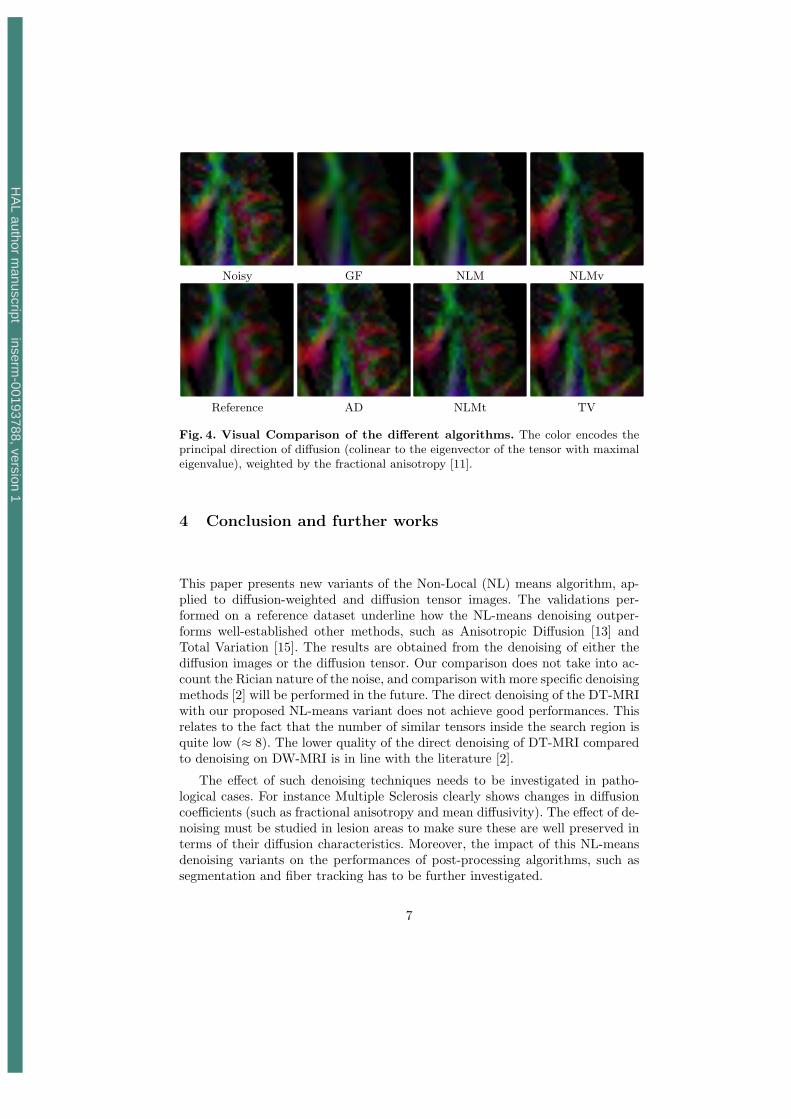
3.5 Visual assessment
In Figure 4, we display axial slices at the level of the ventricles for the groundtruth data (DT-MRI computed from Iaverage), the raw DT-MRI computed fromone of the acquisitions Ii
noisy, and the 6 denoised images. The color encodes theprincipal direction of diffusion (colinear to the eigenvector of the tensor withmaximal eigenvalue), weighted by the fractional anisotropy [11]. The referenceimage has smooth color transitions but also sharp edges. The GF filtered imageefficiently removes the noise but suppresses the edges and lowers the anisotropyof tensors. The standard NL-means seems to be the best filter, followed by TV,NLMv, AD and NLMt, which confirms the quantitative values in Fig. 3.
6
HA
L author manuscript inserm
-00193788, version 1
Noisy GF NLM NLMv
Reference AD NLMt TV
Fig. 4. Visual Comparison of the different algorithms. The color encodes theprincipal direction of diffusion (colinear to the eigenvector of the tensor with maximaleigenvalue), weighted by the fractional anisotropy [11].
4 Conclusion and further works
This paper presents new variants of the Non-Local (NL) means algorithm, ap-plied to diffusion-weighted and diffusion tensor images. The validations per-formed on a reference dataset underline how the NL-means denoising outper-forms well-established other methods, such as Anisotropic Diffusion [13] andTotal Variation [15]. The results are obtained from the denoising of either thediffusion images or the diffusion tensor. Our comparison does not take into ac-count the Rician nature of the noise, and comparison with more specific denoisingmethods [2] will be performed in the future. The direct denoising of the DT-MRIwith our proposed NL-means variant does not achieve good performances. Thisrelates to the fact that the number of similar tensors inside the search region isquite low (≈ 8). The lower quality of the direct denoising of DT-MRI comparedto denoising on DW-MRI is in line with the literature [2].
The effect of such denoising techniques needs to be investigated in patho-logical cases. For instance Multiple Sclerosis clearly shows changes in diffusioncoefficients (such as fractional anisotropy and mean diffusivity). The effect of de-noising must be studied in lesion areas to make sure these are well preserved interms of their diffusion characteristics. Moreover, the impact of this NL-meansdenoising variants on the performances of post-processing algorithms, such assegmentation and fiber tracking has to be further investigated.
7
HA
L author manuscript inserm
-00193788, version 1
References
1. V. Arsigny, P. Fillard, X. Pennec, and N. Ayache. Fast and simple calculus ontensors in the Log-Euclidean framework. In MICCAI’05, volume 3749, pages 115–122, October 2005.
2. S. Basu, P.x T. Fletcher, and Ross T. Whitaker. Rician noise removal in diffusiontensor mri. In MICCAI’2006, pages 117–125, 2006.
3. A. Buades, B. Coll, and J. M. Morel. A review of image denoising algorithms, witha new one. Multiscale Modeling & Simulation, 4(2):490–530, 2005.
4. B. Chen and E. Hsu. Pde denoising of MR diffusion tensor imaging data. InISBI’04, pages 1040–1042, 2004.
5. D. L. Collins, A. P. Zijdenbos, V. Kollokian, J. G. Sled, N. J. Kabani, C. J. Holmes,and A. C. Evans. Design and construction of a realistic digital brain phantom.IEEE Trans. Med. Imaging, 17(3):463–468, 1998.
6. P. Coupe, P. Yger, and C. Barillot. Fast Non Local Means Denoising for 3D MRImages. In MICCAI’2006, volume 4191, pages 33–40, October 2006.
7. P. T. Fletcher and S. C. Joshi. Principal geodesic analysis on symmetric spaces:statistics of diffusion tensors. In CVAMIA and MMBIA 2004, volume 3117, pages87–98. Springer, May 2004.
8. T. Gasser, L. Sroka, and C. Steinmetz. Residual variance and residual pattern innon linear regression. Biometrika, 73(3):625–633, 1986.
9. J. E. Lee, M. K. Chung, and A. L. Alexander. Evaluation of anisotropic filters fordiffusion tensor imaging. In Biomedical Imaging: Macro to Nano, 2006. 3rd IEEEInternational Symposium on, pages 77–78, 2006.
10. J.-F. Mangin, C. Poupon, C. Clark, D. Le Bihan, and I. Bloch. Distortion correctionand robust tensor estimation for MR diffusion imaging. Med Image Anal, 6(3):191–198, September 2002.
11. S. Pajevic and C. Pierpaoli. Color schemes to represent the orientation ofanisotropic tissues from diffusion tensor data: application to white matter fibertract mapping in the human brain. Magn Reson Med, 42(3):526–540, September1999.
12. X. Pennec, P. Fillard, and N. Ayache. A Riemannian framework for tensor com-puting. International Journal of Computer Vision, 66(1):41–66, January 2006.
13. P. Perona and J. Malik. Scale-space and edge detection using anisotropic diffusion.IEEE Trans. Pattern Anal. Mach. Intell., 12(7):629–639, 1990.
14. W. H. Press, B. P. Flannery, S. A. Teukolsky, and W. T. Vetterling. NumericalRecipes: The Art of Scientific Computing. Cambridge University Press, Cambridge(UK) and New York, 2nd edition, 1992.
15. L. I. Rudin, S. Osher, and E. Fatemi. Nonlinear total variation based noise removalalgorithms. pages 259–268, 1992.
16. D. Tschumperle and R. Deriche. Variational frameworks for DT-MRI estimation,regularization and visualization. In ICCV’03, pages 116–121, October 2003.
17. Z. Wang and B. C. Vemuri. DTI segmentation using an information theoretic tensordissimilarity measure. IEEE Trans Med Imaging, 24(10):1267–1277, October 2005.
18. N. Wiest-Daessle, S. Prima, S. P. Morrissey, and C. Barillot. Validation of a newoptimisation algorithm for registration tasks in medical imaging. In ISBI’07, April2007.
8
HA
L author manuscript inserm
-00193788, version 1