nocturnal and diurnal digestive responses in byblis
TRANSCRIPT

64 Carnivorous Plant Newsletter
Technical Refereed Contribution
Nocturnal and diurnal digestive responses in Byblis gigantea, Drosophyllum lusitanicum, and Roridula gorgonias
Dr Gregory Allan • Birmingham • United Kingdom • [email protected]
Introduction
Byblis Salisb. is a highly understudied carnivorous genus currently consisting of eight recognized species – now with motile adhesive traps (Allan 2019 [p 51 this issue])). In particular, the carnivorous syndrome exhibited by the genus is imperfectly understood when compared to other carnivorous plant genera. The leaves, stems, pedicels, and se-pals bear stalked glands and digestive glands. The stalked glands vary in length, even within indi-vidual plants, the longest stalks reaching 2.6 mm in length (McPherson 2010). Each stalked gland bears a droplet of mucilage, assumed to be sugar-based and water-soluble (Bauer et al. 2018), that is responsible for prey-capture. The far more nu-merous digestive glands sit in rows in longitudi-nal grooves on the epidermis (Lloyd 1942) (see Fig. 1) and are usually assumed to secrete diges-tive fluids in response to prey-capture, as well as to absorb the products of digestion (Cross et al. 2018). The stalked glands are capable of collaps-ing in response to detection of animal proteins, apparently due to a rapid loss of cell turgidity. In consequence, prey can be brought within the range of the secretions of the digestive glands.
Of the eight Byblis species, B. gigantea Lindl. and B. lamellata Conran & Lowrie, are perennials with very limited ranges in coastal Western Aus-tralia. B. gigantea inhabits sandy, nutrient-poor, winter-wet substrate in the Swan River drainage area, whilst B. lamellata is found in well-drained sandy heathland between approximately 100 km and 300 km north of Perth (Lowrie 2013). Both perennial species experience a Mediterranean climate with hot and sunny weather for large parts of the year, and both are generally in active growth for at least some of the warmer months (Lowrie 2013 and pers. comm). B. gigantea is the representative of the genus that was investi-gated for the purposes of this experiment (Fig. 2). The other six species are annuals from north-
Figure 1: Closeup of Byblis gigantea leaf showing rows of digestive glands (photo by Alexander Fisch).
Figure 2: A cultivated Byblis gigantea.

65Volume 48 June 2019
ern regions of Australia (and in the case of at least one species, the island of New Guinea), where they experience tropical and semi-arid climates (Lowrie 2013).
Published studies into the reaction of perennial Byblis to the capture of animal proteins are few and contradictory. Over a century ago, Bruce (1905) demonstrated that B. gigantea appears to be able to digest egg albumen, but that digestion is only evident when the albumen is placed in direct contact with the digestive glands. At around the same time, Fenner (1904) reported that the digestive glands produce copious secretions upon contact with prey, and that these glands appear dry after a period of four to six hours. But Lloyd (1942) applied carmine fibrin to the glands of B. gigantea and, over a period of two weeks, reported no evidence of digestion. More recently, Skates (2016) dem-onstrated, through stable isotope techniques, that wild B. gigantea obtained 31% of their nitrogen from prey (a smaller proportion than that found in the annual species tested). She did not, however, publish any findings as to whether the nitrogen had been sequestered directly though the actions of the plants’ digestive secretions, or indirectly via excretions of Setocoris bugs that commonly inhabit Byblis species (although her research into the carnivorous syndrome is ongoing, and likely to yield very significant results). Thus, Lloyd’s 1942 observations remain the most recently published re-search into the reaction of perennial Byblis digestive glands to contact with animal proteins.
More detailed research has in recent years been conducted into the production of enzymes by the annual species, also with inconsistent results. Hartmeyer (1997) concluded that the annual B. liniflora Salisb. does not produce proteases when it did not digest the gelatine layer of strips of pho-tography film which were attached to the leaves 12 hours after the application of a 10% solution of yeast in water. When tested in the same manner, eight species, and one hybrid, of Drosera yielded positive results. Hartmeyer’s findings, perhaps coupled with Lloyd’s aforementioned failure to de-tect digestion in B. gigantea and the frequent presence on wild Byblis of Setocoris bugs which might act as commensals, precipitated doubts as to the ability of the entire Byblis genus (Hartmeyer 1998) or just the perennial species (Panaw et al. 2017) to produce digestive enzymes. Later experiments with film strips, however, have shown evidence of protease production by the digestive glands of an-other annual, B. filifolia Planch. (Hartmeyer 2005). A similar film-strip test with B. liniflora which was conducted in 2019 by this author also seemed to yield positive results (see below discussion). It has also been shown that phosphatase is produced by the digestive glands of B. liniflora (Płachno et al. 2005). Thus, the entire genus is now usually treated as able to produce its own digestive enzymes (Cross et al. 2018). Probably the absence of perennial species from these interesting investigations is attributable to their scarcity in collections worldwide; seed of perennial species can be difficult to obtain and hard to germinate, whilst living plants are only extremely rarely offered for sale.
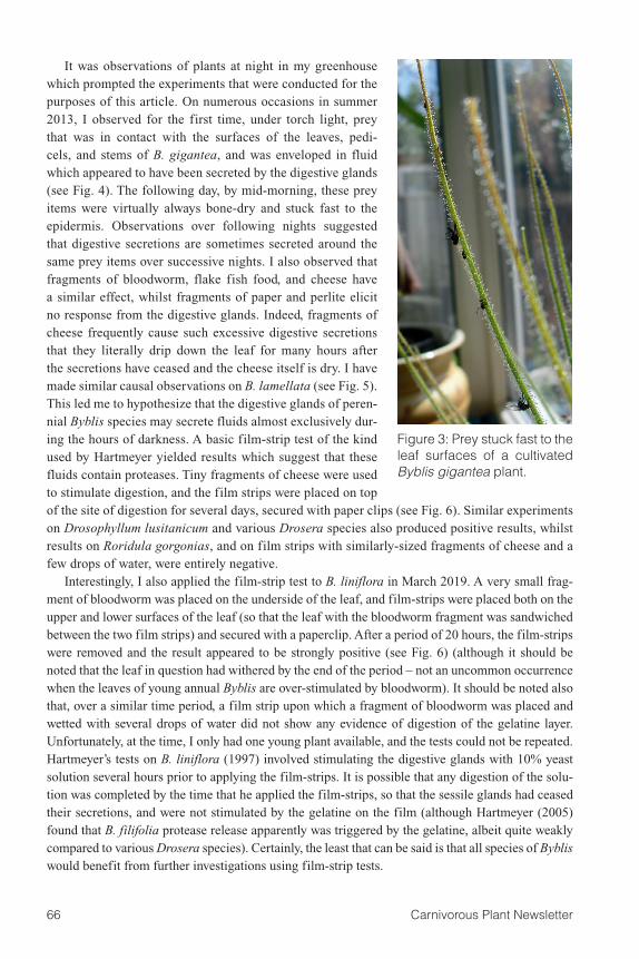
My own personal observations over a period of several years were also initially mystifying. I have grown B. gigantea in a greenhouse in Birmingham, United Kingdom, since 2011, and B. lamel-lata in the same location since 2015. The greenhouse is heated in the winter, remaining frost-free. Plants are unshaded, and are exposed to full sun year-round, although supplemental lighting is used between November and March. For some time, however, I was perplexed as to whether observable digestive fluids are secreted by the plants. I routinely observed freshly captured prey adhered to the stalked glands, as well as older prey items which were bone-dry and stuck fast (to the extent they were difficult to dislodge) to the surfaces of the leaves (and sometimes the pedicels and stems) (Fig. 3). Despite close and repeated scrutiny, however, I was unable to observe any evidence of prey being in contact with digestive fluids. Prey appeared to be bone-dry at all times. Part of this puzzle has been solved by the recent discovery that the stalked glands are able to collapse upon contact with prey so as to convey the latter towards the digestive glands. But this motility does not explain how prey becomes stuck to the epidermis as a bone-dry husk.
66 Carnivorous Plant Newsletter
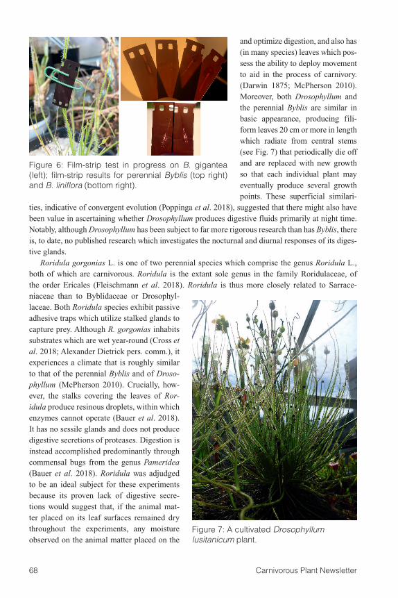
It was observations of plants at night in my greenhouse which prompted the experiments that were conducted for the purposes of this article. On numerous occasions in summer 2013, I observed for the first time, under torch light, prey that was in contact with the surfaces of the leaves, pedi-cels, and stems of B. gigantea, and was enveloped in fluid which appeared to have been secreted by the digestive glands (see Fig. 4). The following day, by mid-morning, these prey items were virtually always bone-dry and stuck fast to the epidermis. Observations over following nights suggested that digestive secretions are sometimes secreted around the same prey items over successive nights. I also observed that fragments of bloodworm, flake fish food, and cheese have a similar effect, whilst fragments of paper and perlite elicit no response from the digestive glands. Indeed, fragments of cheese frequently cause such excessive digestive secretions that they literally drip down the leaf for many hours after the secretions have ceased and the cheese itself is dry. I have made similar causal observations on B. lamellata (see Fig. 5). This led me to hypothesize that the digestive glands of peren-nial Byblis species may secrete fluids almost exclusively dur-ing the hours of darkness. A basic film-strip test of the kind used by Hartmeyer yielded results which suggest that these fluids contain proteases. Tiny fragments of cheese were used to stimulate digestion, and the film strips were placed on top of the site of digestion for several days, secured with paper clips (see Fig. 6). Similar experiments on Drosophyllum lusitanicum and various Drosera species also produced positive results, whilst results on Roridula gorgonias, and on film strips with similarly-sized fragments of cheese and a few drops of water, were entirely negative.
Interestingly, I also applied the film-strip test to B. liniflora in March 2019. A very small frag-ment of bloodworm was placed on the underside of the leaf, and film-strips were placed both on the upper and lower surfaces of the leaf (so that the leaf with the bloodworm fragment was sandwiched between the two film strips) and secured with a paperclip. After a period of 20 hours, the film-strips were removed and the result appeared to be strongly positive (see Fig. 6) (although it should be noted that the leaf in question had withered by the end of the period – not an uncommon occurrence when the leaves of young annual Byblis are over-stimulated by bloodworm). It should be noted also that, over a similar time period, a film strip upon which a fragment of bloodworm was placed and wetted with several drops of water did not show any evidence of digestion of the gelatine layer. Unfortunately, at the time, I only had one young plant available, and the tests could not be repeated. Hartmeyer’s tests on B. liniflora (1997) involved stimulating the digestive glands with 10% yeast solution several hours prior to applying the film-strips. It is possible that any digestion of the solu-tion was completed by the time that he applied the film-strips, so that the sessile glands had ceased their secretions, and were not stimulated by the gelatine on the film (although Hartmeyer (2005) found that B. filifolia protease release apparently was triggered by the gelatine, albeit quite weakly compared to various Drosera species). Certainly, the least that can be said is that all species of Byblis would benefit from further investigations using film-strip tests.
Figure 3: Prey stuck fast to the leaf surfaces of a cultivated Byblis gigantea plant.

67Volume 48 June 2019
The purpose of the experiments which are summarized in this article was to investigate the hy-pothesis regarding nocturnal secretion of digestive fluids. The other subjects of this experiment were Drosophyllum lusitanicum L. and Roridula gorgonias Planch. The carnivorous D. lusitanicum is the only extant representative of the genus Drosophyllum L. It inhabits dry coastal areas in southwestern Spain, Portugal, and northern Morocco, where it experiences a Mediterranean climate (McPherson 2010) that is similar to that in which perennial Byblis (especially B. lamellata) are found (Brewer et al. 2018; Paniw et al. 2017). Drosophyllum is the sole extant genus within the family Drosophyl-laceae and belongs to the order Nepenthales. It is thus unrelated to Byblis, which is the sole extant genus within the family Byblidaceae and belongs to the order Lamiales (Fleischmann et al. 2018). Perhaps unsurprisingly, Drosophyllum’s carnivorous syndrome is fundamentally different from that of Byblis because, unlike those of the former, its stalked and sessile glands are vascularized (Lloyd 1942). Accordingly, its stalked glands produce constant secretions of mucilage (Darwin 1875), al-though the digestive glands release secretions only in response to prey capture (Darwin 1875; Lloyd 1942). Drosophyllum nevertheless exhibits several superficial similarities to Byblis. Although it is a passive adhesive carnivore, it shares with Byblis the character of producing stalked glands which are primarily responsible for prey-capture, and sessile glands which perform the functions of diges-tion and absorption. In neither Drosophyllum nor Byblis are the leaves able to move in response to the capture of prey. This can be contrasted with Drosera, a genus which has sophisticated stalked glands (true tentacles (Bartosz et al. 2018)) which are able to work in co-ordination to secure prey
Figure 5: Byblis lamellata photographed soon after sunrise showing clear evidence of digestive secretions which were released nocturnally around the cheese. By the time that this photograph was taken, the digestive secretions had ceased, and the excess digestive secretions (often stimulated by cheese fragments) have started to drip down the leaf. Such secretions are gradually re-absorbed.
Figure 4: A night time shot of a fly’s head enveloped in digestive secretions whilst an adult Setocoris bibliphilus lurks on a leaf in the background.
68 Carnivorous Plant Newsletter
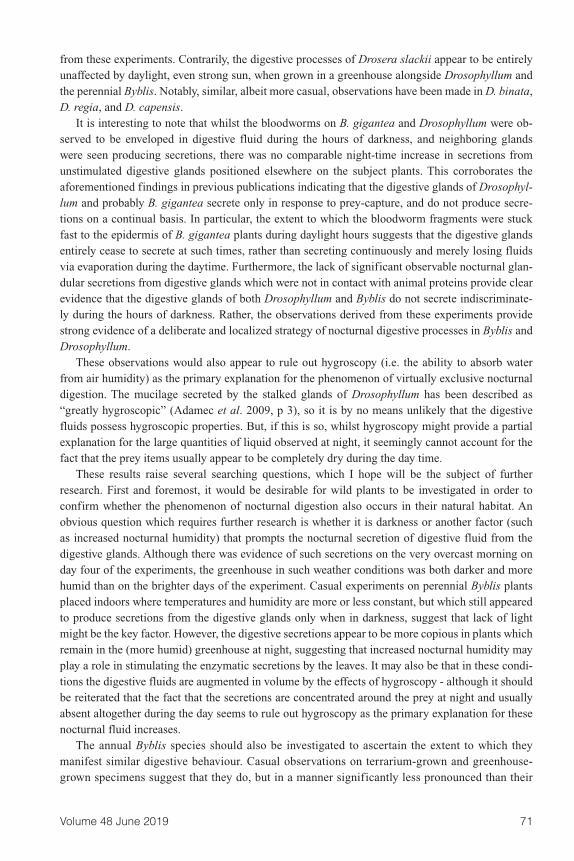
and optimize digestion, and also has (in many species) leaves which pos-sess the ability to deploy movement to aid in the process of carnivory. (Darwin 1875; McPherson 2010). Moreover, both Drosophyllum and the perennial Byblis are similar in basic appearance, producing fili-form leaves 20 cm or more in length which radiate from central stems (see Fig. 7) that periodically die off and are replaced with new growth so that each individual plant may eventually produce several growth points. These superficial similari-
ties, indicative of convergent evolution (Poppinga et al. 2018), suggested that there might also have been value in ascertaining whether Drosophyllum produces digestive fluids primarily at night time. Notably, although Drosophyllum has been subject to far more rigorous research than has Byblis, there is, to date, no published research which investigates the nocturnal and diurnal responses of its diges-tive glands.
Roridula gorgonias L. is one of two perennial species which comprise the genus Roridula L., both of which are carnivorous. Roridula is the extant sole genus in the family Roridulaceae, of the order Ericales (Fleischmann et al. 2018). Roridula is thus more closely related to Sarrace-niaceae than to Byblidaceae or Drosophyl-laceae. Both Roridula species exhibit passive adhesive traps which utilize stalked glands to capture prey. Although R. gorgonias inhabits substrates which are wet year-round (Cross et al. 2018; Alexander Dietrick pers. comm.), it experiences a climate that is roughly similar to that of the perennial Byblis and of Droso-phyllum (McPherson 2010). Crucially, how-ever, the stalks covering the leaves of Ror-idula produce resinous droplets, within which enzymes cannot operate (Bauer et al. 2018). It has no sessile glands and does not produce digestive secretions of proteases. Digestion is instead accomplished predominantly through commensal bugs from the genus Pameridea (Bauer et al. 2018). Roridula was adjudged to be an ideal subject for these experiments because its proven lack of digestive secre-tions would suggest that, if the animal mat-ter placed on its leaf surfaces remained dry throughout the experiments, any moisture observed on the animal matter placed on the
Figure 6: Film-strip test in progress on B. gigantea (left); film-strip results for perennial Byblis (top right) and B. liniflora (bottom right).
Figure 7: A cultivated Drosophyllum lusitanicum plant.

69Volume 48 June 2019
glands of Drosophyllum or Byblis could reasonably be attributed to the digestive secretions rather than to the products of condensation, or to fluid secreted by the animal matter.
Methods and Materials
The experiments here described consisted of placing fragments of dried bloodworm in such a po-sition that they were in direct contact with the leaf surfaces of cultivated specimens of Byblis gigan-tea, Drosophyllum lusitanicum, and Roridula gorgonias. The fragments were observed at various intervals over several consecutive 24-hour periods in order to ascertain at what time of day (if any) digestive activities took place. The subject plants were all growing in a greenhouse in Birmingham, UK, in full exposure to sun. The greenhouse door was open at all times. Five plants were used: one B. gigantea that was in flower; one sub-adult B. gigantea that was due to flower within the next two or so months; one seedling of B gigantea that was approximately five months old; one Drosophyl-lum that was several years old and had flowered a few months previously; and one R. gorgonias that was in flower. Two fragments were placed on separate leaves of each plant.
Results
The results are set out in the table below:
Table 1.
Day 1 1 2 2 2 3 3 3 4 4 4 5 5 5 6
Observation Time
D E N D E N D E N D** E N D E N
Drosophyllum fragment 1
A + ++++ - - +++ - - - - - - - - -
Drosophyllum fragment 2
A + ++++ - - - - - - - - - - - -
R. gorgonias fragment 1
A - - - - - - - - - - - - - -
R. gorgonias fragment 2
A - - - - - - - - - - - - - -
B. gigantea seedling fragment 1
A - ++ - ++ +++ - - +++ ++++ - ++ - - -
B. gigantea seedling fragment 2
A - ++ - ++ -* - - - + - - - - -
B. gigantea sub-adult fragment 1
A - +++ - ++ +++ - - - + - - - - -
B. gigantea sub-adult fragment 2
A - +++ - ++ ++ - - ++ + - - - - -
B. gigantea flowering plant fragment 1
A - +++ - ++ +++ - - +++ + - - - - -
B. gigantea flowering plant fragment 2
A - +++ - ++ +++ - - - - - - - - -

70 Carnivorous Plant Newsletter
The fragments were observed for glandular activity between 29 July 2016, when they were add-ed, and 3 August 2016, when the observations ceased. Observations were made during the day time (between 10:00 and 14:00 BST), evening (between 18:00 and 20:30), and night (between 00:00 and 02:00). Sunrise at this time of year in Birmingham, where the experiments were conducted, is ap-proximately 05:30, and sunset is approximately 21:00. The surprising results of these experiments prompted similar observations of the reaction of a specimen of Drosera slackii to bloodworm frag-ments over a period of several days.
A similar experiment was conducted on a specimen of Drosera slackii grown in identical condi-tions as the plants in the table above showed a very different response. A fragment of bloodworm was added at 11:45, and already by 13:45, copious secretions were being produced while the ten-tacles and leaf had bent over, smothering the bloodworm. This process continued, apparently unaf-fected by daylight and darkness, for a 24-hour period, after which the remnants of the bloodworm began to dry out, and no further evidence of digestive secretions could be perceived.
Discussion
These results suggest a clear propensity in B. gigantea, and also in Drosophyllum, to release di-gestive secretions, and to absorb the products of digestion, outside of daylight hours. R. gorgionias, as expected, demonstrated no reaction to the bloodworm fragments. In particular, it should be noted that the bloodworm fragment appeared bone-dry and stuck fast to the epidermis of B. gigantea dur-ing the periods in which no glandular activity could be perceived. The results also suggest that B. gigantea and Drosophyllum (but particularly the former) may repeat the process of nocturnal diges-tion and absorption for each prey item over several successive nights. The aforementioned causal observations of B. lamellata suggest that this perennial species displays a similar propensity. Casual observations on trapped prey over a period of several years are consistent with the results obtained
Key
Days: 1= 29 July; 2= 30 July; 3= 31 July; 4= 1 August; 5= 2 August; 6= 3 August
Observation time:D = day (observation made between 10:00 and 14:00)E = evening (observation made between 18:00 and 20:30)N = night time (observation made between 00:00 and 02:00)Note that sunrise at this time of year is approximately 05:30, and sunset is approximately 21:00.
Observation of glandular activityA = fragment of bloodworm added- = no glandular activity apparent+ = slight glandular activity present (bloodworm wet where in contact with epidermis)++ = significant glandular activity present (bloodworm saturated and/or secretions from surrounding sessile glands beginning to pool on epidermis+++ = copious glandular activity present (bloodworm saturated and significant pooling of secretions from sessile glands on epidermis)++++ = extremely copious glandular activity present (bloodworm saturated and extreme pooling of secretions from sessile glands on epidermis)*The bloodworm was not visible from this point onwards. It may have been entirely digested.**This was a very wet and overcast day.
71Volume 48 June 2019
from these experiments. Contrarily, the digestive processes of Drosera slackii appear to be entirely unaffected by daylight, even strong sun, when grown in a greenhouse alongside Drosophyllum and the perennial Byblis. Notably, similar, albeit more casual, observations have been made in D. binata, D. regia, and D. capensis.
It is interesting to note that whilst the bloodworms on B. gigantea and Drosophyllum were ob-served to be enveloped in digestive fluid during the hours of darkness, and neighboring glands were seen producing secretions, there was no comparable night-time increase in secretions from unstimulated digestive glands positioned elsewhere on the subject plants. This corroborates the aforementioned findings in previous publications indicating that the digestive glands of Drosophyl-lum and probably B. gigantea secrete only in response to prey-capture, and do not produce secre-tions on a continual basis. In particular, the extent to which the bloodworm fragments were stuck fast to the epidermis of B. gigantea plants during daylight hours suggests that the digestive glands entirely cease to secrete at such times, rather than secreting continuously and merely losing fluids via evaporation during the daytime. Furthermore, the lack of significant observable nocturnal glan-dular secretions from digestive glands which were not in contact with animal proteins provide clear evidence that the digestive glands of both Drosophyllum and Byblis do not secrete indiscriminate-ly during the hours of darkness. Rather, the observations derived from these experiments provide strong evidence of a deliberate and localized strategy of nocturnal digestive processes in Byblis and Drosophyllum.
These observations would also appear to rule out hygroscopy (i.e. the ability to absorb water from air humidity) as the primary explanation for the phenomenon of virtually exclusive nocturnal digestion. The mucilage secreted by the stalked glands of Drosophyllum has been described as “greatly hygroscopic” (Adamec et al. 2009, p 3), so it is by no means unlikely that the digestive fluids possess hygroscopic properties. But, if this is so, whilst hygroscopy might provide a partial explanation for the large quantities of liquid observed at night, it seemingly cannot account for the fact that the prey items usually appear to be completely dry during the day time.
These results raise several searching questions, which I hope will be the subject of further research. First and foremost, it would be desirable for wild plants to be investigated in order to confirm whether the phenomenon of nocturnal digestion also occurs in their natural habitat. An obvious question which requires further research is whether it is darkness or another factor (such as increased nocturnal humidity) that prompts the nocturnal secretion of digestive fluid from the digestive glands. Although there was evidence of such secretions on the very overcast morning on day four of the experiments, the greenhouse in such weather conditions was both darker and more humid than on the brighter days of the experiment. Casual experiments on perennial Byblis plants placed indoors where temperatures and humidity are more or less constant, but which still appeared to produce secretions from the digestive glands only when in darkness, suggest that lack of light might be the key factor. However, the digestive secretions appear to be more copious in plants which remain in the (more humid) greenhouse at night, suggesting that increased nocturnal humidity may play a role in stimulating the enzymatic secretions by the leaves. It may also be that in these condi-tions the digestive fluids are augmented in volume by the effects of hygroscopy - although it should be reiterated that the fact that the secretions are concentrated around the prey at night and usually absent altogether during the day seems to rule out hygroscopy as the primary explanation for these nocturnal fluid increases.
The annual Byblis species should also be investigated to ascertain the extent to which they manifest similar digestive behaviour. Casual observations on terrarium-grown and greenhouse-grown specimens suggest that they do, but in a manner significantly less pronounced than their
72 Carnivorous Plant Newsletter
perennial counterparts. In particular, it can often be observed that in bright light, annual Byblis species usually secrete some digestive fluids soon after animal proteins become adhered to the stalked glands, as is evident from some of the time-lapse videos described in Allan (2019 [p 51 this issue]) (in particular those of B. liniflora and B. rorida). By way of comparison, the time-lapse video of motility in B. gigantea, recorded in good light, shows no evidence whatsoever of digestive secretions. But it can also frequently be observed in all annual species that the volume of fluid enveloping preys greatly increases during hours of darkness, and that this cycle can be re-peated over several 24-hour periods. Whether increased nocturnal secretions in the annual species are caused by low light levels or higher nocturnal humidity, and the extent to which hygroscopy plays a role, is currently unclear.
Another question is why the carnivorous cycles of perennial Byblis and Drosophyllum have adapted in this manner. It might tentatively be suggested that the tendency towards nocturnal diges-tive and absorptive activity may be an adaption to the harsh natural climate that Drosophyllum and the perennial Byblis inhabit. Both may be found in active growth in very hot and dry conditions with relatively low humidity. Indeed, the surface of the sandy substrate which is inhabited by both species of perennial Byblis when in active growth may become sufficiently hot to fry an egg (Allen Lowrie, pers. comm.). But the precise reason for this apparent adaption remains elusive. A possible answer is that secretion of digestive fluids in the hot and sunny summer conditions experienced by Drosophyllum and the perennial Byblis would be likely to lead to much costly evaporative loss, an affliction that would be almost entirely avoided by secretion during the hours of darkness. This is certainly an aspect which warrants further research.
Conclusion
The research conducted for the purposes of this article suggest that B. gigantea and Drosophyl-lum (and probably also B. lamellata) secrete digestive fluids, and absorb the products of digestion, primarily during the hours of darkness. This phenomenon, which is suggestive of a hitherto unrecog-nized level of sophistication in the carnivorous syndrome, has not previously been reported in either genus, or indeed in any other carnivorous plants. Further research, however, is needed in order to verify this phenomenon, and to discover its causes and the precise purpose which it serves.
Acknowledgments: A debt of gratitude is owed by the author to Allen Lowrie, Andreas Fleischmann, and Fernando Rivadavia for their encouragement and instructive advice regarding the various stages of this research. I am also extremely grateful to Alexander Fisch for providing the photograph in Figure 1 and granting permission for it to be used in this article. Particular thanks should be extend-ed to Fernando Rivadavia for his advice and comments on an earlier draft of this article. Fernando devoted a lot of his time and expertise to assisting me in this endeavor. In respect of any errors or omissions, the usual caveats apply.
ReferencesAdamec, L. 2009. Ecophysiological investigation on Drosophyllum lusitanicum: Why doesn’t the
plant dry out? Carnivorous Plant Newsletter 38: 71-74.Allan, G. 2019. Evidence of motile traps in Byblis. Carnivorous Plant Newsletter 48: 51-63.Bartosz, J., Płachno, B.J., and Muravnik, L.E. 2018 Function anatomy of carnivorous traps. In:
Ellison, A.M., & Adamec, L. (eds.): Carnivorous Plants: Physiology, Ecology, and Evolution. Oxford University Press: 167-179.
73Volume 48 June 2019
Bauer, U., Jetter, R., and Poppinga, S. 2018. Non-motile traps. In: Ellison, A.M., & Adamec, L. (eds.): Carnivorous Plants: Physiology, Ecology, and Evolution. Oxford University Press: 194-206.
Brewer, J.S., and Schlauer, J. 2018. Biogeography and habitats of carnivorous plants. In: Ellison, A.M., & Adamec, L. (eds.): Carnivorous Plants: Physiology, Ecology, and Evolution. Oxford University Press: 7-21.
Bruce, A.N. 1905. On the activity of the glands of Byblis gigantea. Notes of the Royal Botanic Garden of Edinburgh 16: 89-98.
Cross, A.T., Paniw, M., Scatigna, A.V., Kalfas, N., Anderson, B., Givnish, T.J., and Fleischmann, A. 2018. Systematics and evolution of small genera of carnivorous plants. In: Ellison, A.M., & Adamec, L. (eds.): Carnivorous Plants: Physiology, Ecology, and Evolution. Oxford University Press: 120-134.
Darwin, C. 1875. Insectivorous Plants. D. Appleton and Company. New York: 1-462.Fenner, C.A. 1904. Beiträge zur Kenntnis der Anatomie, Entwickelungsgeschichte und Biologie des
Laubblätter und Drüsen eniiger Insektivoren. Flora 93: 335-434.Fleischman, A., Schlauer, J., Smith, S.A., and Givinish, T.G. 2018 Evolution of carnivory in angio-
sperms. In: Ellison, A.M., & Adamec, L. (eds.): Carnivorous Plants: Physiology, Ecology, and Evolution. Oxford University Press: 22-41.
Hartmeyer, I., and Hartmeyer, S. 2005. Byblis filifolia als echte Karnivore rehabilitiert. Das Taublatt 53: 4-5.
Hartmeyer, S. 1997. Carnivory of Byblis revisited: a simple method for enzyme testing on carnivo-rous plants. Carnivorous Plant Newsletter 26: 39-45.
Hartmeyer, S. 1998. Carnivory in Byblis revisited II: the phenomenon of symbiosis on insect trap-ping plants. Carnivorous Plant Newsletter 27: 110-113.
Lloyd, F. 1942. The Carnivorous Plants. Waltham. Massachusetts: 1-352.Lowrie, A. 2013. Carnivorous Plants of Australia Magnum Opus, Volume 1. Redfern Natural His-
tory Productions. Poole, Dorset, England: 1-458.Paniw, M., Gil-Cabeza, E., and Ojeda, F. 2017. Plant carnivory beyond bogs: reliance on prey feed-
ing in Drosophyllum lusitanicum (Drosophyllaceae) in dry Mediterranean heathland habitats. Annals of Botany 119: 1035-41.
Płachno, B.J., Adamec, L., Lichtscheidl, I.K., Peroutka, M., Adlassnig, W., and Vrba, J. 2006. Fluo-rescence labelling of phosphatase activity in digestive glands of carnivorous plants. Plant Biol-ogy 8: 813-820.
Płachno, B.J., and Muravnik, L.E. 2018. Functional anatomy of carnivorous traps. In: Ellison, A.M., & Adamec, L. (eds.): Carnivorous Plants: Physiology, Ecology, and Evolution. Oxford Univer-sity Press: 167-179.
Poppinga, S., Alamsyah, F., Bauer, U., Fleischmann, A., Horstmann, M., Klink, S., Kruppert, S., Lin, Q., Müller, U., Northrop, A., Płachno, B.J., Prins, A., Scharmann, M., Sirová, D., Skates, L., Westermeier, A., and Ellison, A.M. 2018 What’s new in the world of carnivorous plants - sum-mary of two symposia held in July 2017. Carnivorous Plant Newsletter 47: 18-27.
Skates, L., Dixon, K., Gebauer, G., Stevens, and Cross, A. 2016. Carnivorous Plants. Poster at the XI ICPS Conference Kew, 5-7 August 2016.