nmr structure and dynamics of the agonist dynorphin ... · pdf filenmr structure and dynamics...
TRANSCRIPT

NMR structure and dynamics of the agonist dynorphinpeptide bound to the human kappa opioid receptorCasey O’Connora,b, Kate L. Whitea,b,c, Nathalie Doncescud, Tatiana Didenkoa, Bryan L. Rothc, Georges Czaplickid,Raymond C. Stevensa,b,1, Kurt Wüthricha,e,f,1, and Alain Milona,d,1
aDepartment of Integrative Structural and Computational Biology, The Scripps Research Institute, La Jolla, CA 92037; bDepartments of Biological Sciencesand Chemistry, Bridge Institute, University of Southern California, Los Angeles, CA 90089; cDepartment of Pharmacology, University of North CarolinaChapel Hill Medical School, Chapel Hill, NC 27514; dInstitute of Pharmacology and Structural Biology, CNRS and Université de Toulouse-Paul Sabatier, 31077Toulouse, France; eDepartment of Molecular Biology and Biophysics, ETH Zurich, CH-8093 Zurich, Switzerland; and fThe Skaggs Institute for ChemicalBiology, The Scripps Research Institute, La Jolla, CA 92037
Edited by Michael F. Summers, Howard Hughes Medical Institute, University of Maryland, Baltimore County, Baltimore, MD, and accepted by the EditorialBoard August 13, 2015 (received for review June 2, 2015)
The structure of the dynorphin (1–13) peptide (dynorphin) bound tothe human kappa opioid receptor (KOR) has been determined byliquid-state NMR spectroscopy. 1H and 15N chemical shift variationsindicated that free and bound peptide is in fast exchange in solu-tions containing 1 mM dynorphin and 0.01 mM KOR. Radioligandbinding indicated an intermediate-affinity interaction, with a Kd of∼200 nM. Transferred nuclear Overhauser enhancement spectros-copy was used to determine the structure of bound dynorphin. TheN-terminal opioid signature, YGGF, was observed to be flexibly dis-ordered, the central part of the peptide from L5 to R9 to form a he-lical turn, and the C-terminal segment from P10 to K13 to be flexiblydisordered in this intermediate-affinity bound state. Combining mo-lecular modeling with NMR provided an initial framework for under-standing multistep activation of a G protein-coupled receptor by itscognate peptide ligand.
GPCR activation | transferred NOE | 15N relaxation |molecular dynamics simulations | ligand binding affinity
Gprotein-coupled receptors (GPCRs) are the largest super-family of membrane proteins in the human genome and play
a critical role in human physiology by initiating signal transduc-tion in response to extracellular stimuli (1, 2). Since 2007, 89GPCR crystal structures have been reported, including receptorsin inactive and active states, as well as the beta-2 adrenergicreceptor (β2-AR) bound to heterotrimeric G proteins (3). NMRspectroscopy has revealed that the intrinsic conformationalheterogeneity of GPCRs is influenced by ligand pharmacology,membrane composition, and effector interactions (4–6). Thesestructural biology studies have provided atomic-resolution insightsof systems defined by dynamic structural rearrangements that arecorrelated with diverse cellular and physiological outcomes.The classic opioid receptors (δ/κ/μ) are GPCRs activated in
response to binding enkephalin-like peptide agonists and are theprimary targets of widely prescribed pain medications (7). Thekappa opioid receptor (KOR) and its cognate peptide dynorphinare implicated in neuronal pathways associated with addiction,pain, reward, mood, cognition, and perception (8, 9). NonselectiveKOR antagonists such as naltrexone have been prescribed for al-cohol dependence with limited efficacy in humans, and next-gen-eration KOR antagonists continue to be developed to treat drugaddiction and other disorders. Although much is known regardingthe antagonist-bound, inactive state of GPCRs, including the crys-tal structure of JDTic-bound KOR, the interaction of these re-ceptors with neuropeptide agonists remains largely unknown (10).Peptide agonist-bound structures have thus far been limited to aconformationally stabilized neurotensin receptor, likely corre-sponding to a low-energy peptide-receptor state (11–13).Dynorphin was discovered by Goldstein and Chavkin as the
endogenous activating neuropeptide for KOR, with a “low-reso-lution” structural model of interaction proposed to PNAS in 1981(14, 15). Dynorphins are derived from the precursor prodynorphin,
with dynorphin A(1–17), dynorphin B(1–13), and alpha neoen-dorphin sharing a highly conserved N-terminal sequence andcharge distribution (16). Dynorphin A(1–13) was shown to act asan agonist on opioid kappa receptors in vivo (17). Physiologicalactivation of KOR is mainly associated with unwanted effectssuch as dysphoria, anhedonia, and hallucinations, and a currenthypothesis in the field is that KOR functionally selective ligandsmay produce analgesia without dysphoria (18, 19). Functionalselectivity has emerged as the leading model to understand theability of a ligand to activate a subset of signaling cascades,providing a framework for developing next-generation drugs withrationally designed pharmacological profiles (20).The seminal work of Schwyzer in the 1970s and 1980s led to a
model of KOR activation by dynorphin that proceeds via a multi-step binding mechanism (14, 21, 22). Thereby, low- to intermediate-affinity binding states of dynorphin correspond to binding to cell-surface membranes or to extracellular loops of the GPCR. A“message–address” paradigm has been formulated based onstructure–activity relations observed with dynorphin analogs (21–26). Accordingly, the N-terminal YGGF “message” sequence,which is common to all opioid peptides, was found to be re-sponsible for receptor activation. A C-terminal “address” sequencewas further found to contribute via electrostatically driven in-teractions to KOR subtype specificity. In the context of this para-digm, the present study yields intriguing data on the N-terminal
Significance
The human kappa opioid receptor (KOR) is implicated in ad-diction, pain, reward, mood, cognition, and perception. Acti-vation of KOR by the neuropeptide dynorphin is critical inmediating analgesia and tolerance. Our solution NMR study ofdynorphin (1–13) provided quantitative data on a KOR-boundconformation. Analysis of the peptide structure and dynamicsrevealed a central helical turn bounded on both sides by flex-ibly disordered peptide segments. Future drug developmentwill benefit from knowledge of the dynorphin structure boundto its human receptor.
Author contributions: R.C.S., K.W., and A.M. designed research; C.O., K.L.W., T.D., G.C.,and A.M. performed research; C.O., K.L.W., N.D., T.D., B.L.R., and G.C. contributed newreagents/analytic tools; C.O., G.C., R.C.S., K.W., and A.M. analyzed data; and C.O., T.D.,B.L.R., G.C., R.C.S., K.W., and A.M. wrote the paper.
The authors declare no conflict of interest.
This article is a PNAS Direct Submission. M.F.S. is a guest editor invited by the EditorialBoard.
Data deposition: NMR, atomic coordinates, chemical shifts, and restraints have been depos-ited in the Protein Data Bank, www.pdb.org (PDB ID code 2N2F) and in the BioMagResBank,www.bmrb.wisc.edu (accession no. 25597).1To whom correspondence may be addressed. Email: [email protected], [email protected], or [email protected].
This article contains supporting information online at www.pnas.org/lookup/suppl/doi:10.1073/pnas.1510117112/-/DCSupplemental.
11852–11857 | PNAS | September 22, 2015 | vol. 112 | no. 38 www.pnas.org/cgi/doi/10.1073/pnas.1510117112

segment of the KOR-bound opioid peptide dynorphin. The meth-ods used, NMR in solution and molecular dynamics simulations,enabled us to define structural ensembles of KOR-bound dynorphinand to characterize internal peptide motions in the presently pre-pared low-affinity receptor-bound state.
ResultsWe prepared KOR with the approach used to solve the crystalstructure in complex with JDTic by X-ray crystallography (10). Themembrane fraction of Spodoptera frugiperda cells transiently ex-pressing KOR was isolated and radioligand binding assays per-formed to assess receptor function. Dynorphin binding affinity wasdetermined as ∼200 nM at pH 7.4 and 6.1, consistent with an in-termediate-affinity interaction (Fig. S1A). Following reconstitutionof the receptor in detergent micelles and purification to >95%homogeneity, similar binding was measured and the preparationwas stable at 7 °C for over a week (Fig. S1B). The following ob-servations, made in the absence of G protein, thus characterize anintermediate state of KOR along its activation pathway, similar towhat was recently described by 13C-NMR for β2-AR (27).The dynorphin (1–13) peptide, YGGFLRRIRPKLK, was
15N-labeled at residues G2, G3, F4, L5, R6, R7, I8, R9, and L12.[15N, 1H]-heteronuclear single quantum correlation (HSQC) NMRspectra of 15N-(GFLIR)-dynorphin at pH 6.1 and pH 7.4 indicatedthe peptide adopts a random coil conformation in aqueous solution.Owing to little dependence of binding on pH, measurements weremade at pH 6.1 to reduce proton exchange with solvent. Resonanceassignments were obtained with 2D [1H, 1H]-total correlation spec-troscopy (TOCSY) and nuclear Overhauser effect spectros-copy (NOESY), 2D [15N, 1H]-HSQC, [13C, 1H]-HSQC, and 3DCBCANH experiments, using standard pulse sequences and aclassical 1H assignment strategy for small peptides (28). 1H, 13C, and15N resonance assignments were deposited to the Biological Mag-netic Resonance Data Bank (BMRB; accession no. 25597) (29).Detergent-reconstituted KOR was added to an aqueous solu-
tion of dynorphin at a ratio of 1:100, with a 1 mM final concen-tration of peptide. KOR-specific binding of dynorphin wasreversed by the addition of the high-affinity ligand JDTic at amolar ratio of 1:1 with respect to dynorphin. We thus isolated bydifference analysis the nonspecific binding of dynorphin to themixed detergent-sterol micelle of dodecyl maltoside/cholesterylhemisuccinate (DDM/CHS). NMR-derived values such as chemical
shift, NOE intensity, and relaxation rate constants (R1, R2, and1H-15N NOEs) were obtained with and without JDTic to charac-terize a KOR-bound conformation of dynorphin.
Chemical Shift Perturbations. KOR-binding produced distinct pat-terns of dynorphin 1H, 15N, and 13C chemical shift changes detectedin 1D proton and 2D heteronuclear correlation NMR experiments,as shown in Fig. 1. The observation of chemical shift changes showsthat the exchange rate between KOR-bound and KOR-unboundstates is fast on the millisecond time scale, as the frequency dif-ference between bound and unbound state is on the order of 103
Hz (Fig. 1C; see Supporting Information for further considerationson exchange rates). The sequence-specific changes were largestfrom L5 to R9, with smaller changes observed from G2 to F4and K11 to K13. 15N-1H correlation experiments acquired withoutproton decoupling in the in-phase/anti-phase method (HSQC-IPAP) were obtained to determine 1JNH (30). No significant vari-ations on the 15N-1H couplings was observed in HSQC-IPAP ex-periments, consistent with negligible residual dipolar couplings,and therefore anisotropic interactions do not contribute to theobserved phenomena. The shifts observed on 1H and 15N are toolarge to be accounted for by secondary structure alone and likelyreflect a ring current effect due to the receptor proximity. Wemeasured the 13C chemical shifts of C′, Cα, and Cβ of 13C-labeledarginine residues (R6, R7, and R9), because these values report onsecondary structure (31). Based on carbon chemical shifts, a helicalconformation was expected for R6 and R7, in contrast to R9. Inaddition to JDTic competition, the inhibition of dynorphin-specificbinding was also observed using the highly potent KOR agonistICI-199,441 (32). The observations and analysis performed heretherefore describe a property of KOR–dynorphin-specific in-teraction reversible by agonists and antagonists.
Nuclear Overhauser Effects and Structure Determination. Where li-gand exchange between a bound and free state is considered fast(koff > R1), transferred NOEs (trNOEs) originating from thereceptor-bound state may be observed on the free peptidespectrum. The measured 1H R1 values for amide protons in thefree state depended on the residue and ranged from 1.2 to 1.8 s−1
(Fig. S2). As expected from simulations (Fig. S3), our mea-surements confirm the dominant contribution of unbounddynorphin on R1. When koff > σij, the cross-relaxation rate σij is
8.29
125
.0
124
.0
ω1(
15N
) [pp
m]
8.38 8.34
124
.0
123
.0
ω2(1H) [ppm]
8.6 8.4 8.2 8.0
+KOR
+JDTic
8.8
A B I8 L5
ω1(1H) [ppm]
7.8
R7G2
R9K11
L12I8 K13
G3L5R6
F4
C
∆δ(1 H
) [PP
M]
G2 G3 F4 L5 R6 R7 I8 R9 K11 L12 K13-0.08
-0.04
0
0.04
0.08
-0.9
-0.6
-0.3
0
0.3
0.6
0.9
∆δ(15
N) [
PPM
]
Fig. 1. Influence of KOR and JDTic on NMR spectra of dynorphin. (A) (top, black) 1D 1H-NMR expansion of the NH region of dynorphin in aqueous solution at pH 6.1acquired at 280 K. (middle, red) Addition of DDM/CHS reconstituted KOR induces significant line broadening for all resonances. The least broadened resonances were K11,L12, and K13. (bottom, blue) The addition of JDTic largely reversed the observed broadening and chemical shift changes, particularly for residues F4 to R9. (B) Overlay oftwo [15N, 1H]-HSQC-IPAP spectra: (red) KOR + dynorphin and (blue) KOR + dynorphin + JDTic. L5 chemical shift perturbations are mainly in the 15N dimension whereas I8changes were observed in both 1H and 15N dimensions. The complete spectrum is available in Fig. S4. (C) Chemical shift perturbations of dynorphin as a result of KORbinding, calculated as the difference of chemical shifts of dynorphin with KOR and KOR + JDTic. Gray bars (left axis): 1H chemical shifts; white bars (right axis): 15N chemicalshifts. The [KOR]/[dynorphin] ratio was 1/100. Almost no chemical shift variations were observed for the C-terminal tripeptide segment.
O’Connor et al. PNAS | September 22, 2015 | vol. 112 | no. 38 | 11853
BIOPH
YSICSAND
COMPU
TATIONALBIOLO
GY
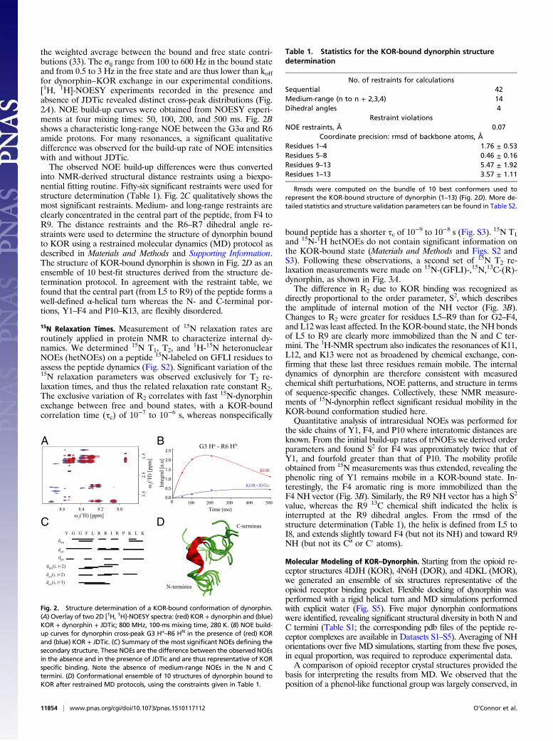
the weighted average between the bound and free state contri-butions (33). The σij range from 100 to 600 Hz in the bound stateand from 0.5 to 3 Hz in the free state and are thus lower than kofffor dynorphin–KOR exchange in our experimental conditions.[1H, 1H]-NOESY experiments recorded in the presence andabsence of JDTic revealed distinct cross-peak distributions (Fig.2A). NOE build-up curves were obtained from NOESY experi-ments at four mixing times: 50, 100, 200, and 500 ms. Fig. 2Bshows a characteristic long-range NOE between the G3α and R6amide protons. For many resonances, a significant qualitativedifference was observed for the build-up rate of NOE intensitieswith and without JDTic.The observed NOE build-up differences were thus converted
into NMR-derived structural distance restraints using a biexpo-nential fitting routine. Fifty-six significant restraints were used forstructure determination (Table 1). Fig. 2C qualitatively shows themost significant restraints. Medium- and long-range restraints areclearly concentrated in the central part of the peptide, from F4 toR9. The distance restraints and the R6–R7 dihedral angle re-straints were used to determine the structure of dynorphin boundto KOR using a restrained molecular dynamics (MD) protocol asdescribed in Materials and Methods and Supporting Information.The structure of KOR-bound dynorphin is shown in Fig. 2D as anensemble of 10 best-fit structures derived from the structure de-termination protocol. In agreement with the restraint table, wefound that the central part (from L5 to R9) of the peptide forms awell-defined α-helical turn whereas the N- and C-terminal por-tions, Y1–F4 and P10–K13, are flexibly disordered.
15N Relaxation Times. Measurement of 15N relaxation rates areroutinely applied in protein NMR to characterize internal dy-namics. We determined 15N T1, T2, and
1H-15N heteronuclearNOEs (hetNOEs) on a peptide 15N-labeled on GFLI residues toassess the peptide dynamics (Fig. S2). Significant variation of the15N relaxation parameters was observed exclusively for T2 re-laxation times, and thus the related relaxation rate constant R2.The exclusive variation of R2 correlates with fast 15N-dynorphinexchange between free and bound states, with a KOR-boundcorrelation time (τc) of 10−7 to 10−6 s, whereas nonspecifically
bound peptide has a shorter τc of 10−9 to 10−8 s (Fig. S3). 15N T1and 15N-1H hetNOEs do not contain significant information onthe KOR-bound state (Materials and Methods and Figs. S2 andS3). Following these observations, a second set of 15N T2 re-laxation measurements were made on 15N-(GFLI)-,15N,13C-(R)-dynorphin, as shown in Fig. 3A.The difference in R2 due to KOR binding was recognized as
directly proportional to the order parameter, S2, which describesthe amplitude of internal motion of the NH vector (Fig. 3B).Changes to R2 were greater for residues L5–R9 than for G2–F4,and L12 was least affected. In the KOR-bound state, the NH bondsof L5 to R9 are clearly more immobilized than the N and C ter-mini. The 1H-NMR spectrum also indicates the resonances of K11,L12, and K13 were not as broadened by chemical exchange, con-firming that these last three residues remain mobile. The internaldynamics of dynorphin are therefore consistent with measuredchemical shift perturbations, NOE patterns, and structure in termsof sequence-specific changes. Collectively, these NMR measure-ments of 15N-dynorphin reflect significant residual mobility in theKOR-bound conformation studied here.Quantitative analysis of intraresidual NOEs was performed for
the side chains of Y1, F4, and P10 where interatomic distances areknown. From the initial build-up rates of trNOEs we derived orderparameters and found S2 for F4 was approximately twice that ofY1, and fourfold greater than that of P10. The mobility profileobtained from 15N measurements was thus extended, revealing thephenolic ring of Y1 remains mobile in a KOR-bound state. In-terestingly, the F4 aromatic ring is more immobilized than theF4 NH vector (Fig. 3B). Similarly, the R9 NH vector has a high S2
value, whereas the R9 13C chemical shift indicated the helix isinterrupted at the R9 dihedral angles. From the rmsd of thestructure determination (Table 1), the helix is defined from L5 toI8, and extends slightly toward F4 (but not its NH) and toward R9NH (but not its Cα or C′ atoms).
Molecular Modeling of KOR–Dynorphin. Starting from the opioid re-ceptor structures 4DJH (KOR), 4N6H (DOR), and 4DKL (MOR),we generated an ensemble of six structures representative of theopioid receptor binding pocket. Flexible docking of dynorphin wasperformed with a rigid helical turn and MD simulations performedwith explicit water (Fig. S5). Five major dynorphin conformationswere identified, revealing significant structural diversity in both N andC termini (Table S1; the corresponding pdb files of the peptide re-ceptor complexes are available in Datasets S1–S5). Averaging of NHorientations over five MD simulations, starting from these five poses,in equal proportion, was required to reproduce experimental data.A comparison of opioid receptor crystal structures provided the
basis for interpreting the results from MD. We observed that theposition of a phenol-like functional group was largely conserved, in
A B
C DdNN
dαN
dβN
dNN(i, i+2)
dαN(i, i+2)
dαN(i, i+3)
Y G G F L R R I R P K L K
2.5
2.0
1.5
1.0
0.5
0.0
Inte
gral
[a.u
]
100 200 300 400 5000
Time (ms)
KOR
KOR+JDTic
N-terminus
C-terminus
G3 Hα - R6 HN
ω2(1H) [ppm]
ω1(1 H
) [pp
m]
8.6 8.4 8.2 8.0
3.5
2
.5
1.5
Fig. 2. Structure determination of a KOR-bound conformation of dynorphin.(A) Overlay of two 2D [1H, 1H]-NOESY spectra: (red) KOR + dynorphin and (blue)KOR + dynorphin + JDTic; 800 MHz, 100-ms mixing time, 280 K. (B) NOE build-up curves for dynorphin cross-peak G3 Hα
–R6 HN in the presence of (red) KORand (blue) KOR + JDTic. (C) Summary of the most significant NOEs defining thesecondary structure. These NOEs are the difference between the observed NOEsin the absence and in the presence of JDTic and are thus representative of KORspecific binding. Note the absence of medium-range NOEs in the N and Ctermini. (D) Conformational ensemble of 10 structures of dynorphin bound toKOR after restrained MD protocols, using the constraints given in Table 1.
Table 1. Statistics for the KOR-bound dynorphin structuredetermination
No. of restraints for calculationsSequential 42Medium-range (n to n + 2,3,4) 14Dihedral angles 4
Restraint violationsNOE restraints, Å 0.07
Coordinate precision: rmsd of backbone atoms, ÅResidues 1–4 1.76 ± 0.53Residues 5–8 0.46 ± 0.16Residues 9–13 5.47 ± 1.92Residues 1–13 3.57 ± 1.11
Rmsds were computed on the bundle of 10 best conformers used torepresent the KOR-bound structure of dynorphin (1–13) (Fig. 2D). More de-tailed statistics and structure validation parameters can be found in Table S2.
11854 | www.pnas.org/cgi/doi/10.1073/pnas.1510117112 O’Connor et al.

terms of position in the orthosteric site, for antagonist-boundstructures of KOR-JDTic, DOR-DIPP, and MOR-funaltrexamine(Fig. 4A). Following MD simulations the “KOR-1” structuralmodel revealed Y1 in a position near the phenol-piperidine fused-ring system of JDTic, resembling a previously proposed pose (Fig.4B) (9). The “KOR-2” pose of dynorphin placed the Y1 side chainin the sodium allosteric site (Fig. 4C). D3.32 made polar contactswith Y1, G2, and G3 in both KOR-1 and KOR-2 models, whereasR7 made a polar contact in KOR-2 but not KOR-1. W6.48 andN7.45 made contacts with Y1 in the KOR-2 model exclusively.Table 2 and Table S1 summarize the findings from modeling andMD simulations, with further details given in Materials and Meth-ods and Supporting Information.
DiscussionSince the 1970s, pharmacologists and biochemists have attemptedto determine opioid peptide conformations capable of explainingtheir activity and to develop drugs mimicking these conformations(34). In aqueous solutions, peptides often exist as a dynamic en-semble of random coil conformers, with specific folds stabilized byorganic solvents or micelles (35–37). Studies of liposome-boundpeptides likewise indicate that nonpolar or membrane-like envi-ronments stabilize peptide structure (23, 24, 38–41). Schwyzer in-troduced the “membrane compartment concept” that postulatedthe membrane-bound state as part of the binding mechanism,thereby reducing the available peptide conformations toward anactivating conformation in complex with receptor (21, 22, 25). Formany years, the direct analysis of the peptide–receptor complexwas not possible due to the lack of suitable receptor preparations.Recently, several neurotensin–receptor complexes have been stud-ied both by X-ray crystallography and solid-state NMR (11–13),and the structure of the Leukotriene B4 (a proinflammatory lipidmediator), in complex with the human BLT2 receptor, was de-termined by liquid-state NMR (41).Recent advances in producing the opioid receptors (and other
GPCRs) in milligram quantities via transient insect cell expressionand stabilization in a bicelle-like architecture of mixed detergent-sterol micelles has opened new avenues for opioid receptorstructural biology (42). This progress culminated in 2012, with re-ports of inactive state structures determined by X-ray crystallogra-phy of the four opioid receptors in complex with antagonists orinverse agonists (10, 43–46). Currently, the only reported 3Dstructure of the human KOR is in complex with JDTic, a highlypotent KOR antagonist.NMR using (15N-13C)–labeled ligand represents a powerful com-
plementary alternative to crystallographic approaches in obtainingstructural information of receptor activation by peptide agonists.Owing to the moderate affinity of dynorphin with KOR recon-stituted in detergent micelles and fast association rate, the ligand
dissociation rate is also fast on the NMR chemical shift timescale (47). This context made possible the observation of trNOEsand a straightforward interpretation of 15N relaxation rates (33).The receptor–peptide interaction was therefore ideal to determinea KOR-bound conformation of dynorphin via the trNOE method,as well as to characterize internal peptide dynamics in a boundstate. Such an approach would be prohibited in a comparablestudy of the high-affinity state, which is characterized by lownanomolar Kd and longer off-rate. In this case, changes todynorphin structure and dynamics are expected as a result of Gprotein binding to KOR. Nevertheless, NMR observation of thehigh-affinity state of dynorphin can be pursued by deuteration ofthe receptor, peptide, and cognate inhibitory G proteins andpreparation of a 1:1:1 complex at millimolar concentrations.The computational approaches were insufficient to determine
reasonable models of dynorphin–KOR binding, because multipleconformers of similar energy were observed. The NMR observa-tions of structure and dynamics were required to limit the startingposes of dynorphin for MD. We identified the pose KOR-1 astypical of an inactive state based on the proximity of Y1 to thephenol-like functional group of JDTic (Fig. 4B and Fig. S6B). Incontrast, we speculate the conformational change of the peptidefound in KOR-2 correlates with an activated state (Fig. 4 B and Cand Fig. S6D). The position of Y1 in the KOR-2 model correspondswith an established role in activation, with the dynorphin (2–13)peptide previously reported to bind weakly and not activate KOR(48). In this “active” conformation, the N terminus of dynorphinforms polar interactions with N3.35 and D3.32 side chains, whereasthe Y1 phenol ring is involved in a π-stacking interaction with W6.48
and an H-bond with N7.45. As part of the allosteric sodium site theseresidues stabilize an inactive state of the receptor, with changes todynorphin structure providing agonist–receptor contacts amonghighly conserved residues in transmembrane helices 6 and 7 (43).Interestingly, this conformational change may be associated with an
A B
C D
Fig. 4. Ligand poses from modeling the dynorphin–KOR complex. (A) Antag-onist binding poses from structures of KOR-JDTic (4DJH), DOR-DIPP (4RWA), andMOR-FNA (4DKL). JDTic is shown in green, DIPP in purple, and β-funaltrexaminein salmon. (B) Structure of dynorphin in complex with “KOR-1” from MD simu-lations with Y1 in a position near the fused phenol-piperidine ring system ofJDTic. (C) Zoom on dynorphin Y1 in the “KOR-2” complex. Y1 is positioned to-ward the sodium allosteric binding site. Dynorphin–KOR contacts are given inTable 2 and Table S1. (D) Visual representation of order parameters derived fromNMR relaxation measurements. The width of the cone indicates the flexibility ofG2 (orange), R6 (green), and L12 (red) dynorphin residues in a KOR-bound state.
G2 G3 F4 L5 R6 R7 I8 R9 L12 G2 G3 F4 L5 R6 R7 I8 R9 L12
A B
60
40
20
0
R2 (s-1) S2
1.0
0.5
0.0
Fig. 3. Characterization of internal dynorphin dynamics. (A) 15N R2 re-laxation rate constants (s−1) measured at 600 MHz at 280 K on 15N-(GFLI)-,15N,13C-(R)-dynorphin in the presence of (red) KOR and (blue) KOR + JDTic.(B) (gray) Order parameters profile S2/S2max of NH bond vectors derived fromR2 as described in Supporting Information. The order parameters describethe amplitude of the NH bond fluctuations in the KOR bound state, nor-malized to L5. (white) Best-fit S2 profiles calculated with the ensemble ofconformers identified by docking and MD simulations.
O’Connor et al. PNAS | September 22, 2015 | vol. 112 | no. 38 | 11855
BIOPH
YSICSAND
COMPU
TATIONALBIOLO
GY

increased penetration of water into the receptor cavity, which hasbeen linked to the activation mechanism upon agonist binding (49).Mutations of the sodium site in DOR shift nalfurafine, an antago-nist, to an arrestin-biased ligand, establishing this sodium site as akey mediator of receptor function (43). The KOR-2 conformationmay hint at the mechanism of dynorphin functional selectivity,which has been reported as a G protein-biased agonist in phar-macological in vitro studies (50, 51). The proposed model may betested in future studies by mutagenesis of KOR residues that sur-round dynorphin in the KOR-2 conformation.The direct quantification of neuropeptide dynamics in an in-
termediate-affinity receptor-bound state yields useful insights forthe design of KOR-targeting antagonists as well as the biology ofpeptide-activated receptors. More surprising than the well-definedα-helical turn between L5 and R9 was the significant motion ob-served for the N and C termini. Such motion was expected for theC-terminal part of dynorphin: Indeed, within the message–addressparadigm the highly positively charged C-terminal “address” isexpected to produce favorable but nonspecific electrostatic in-teractions with the negatively charged extracellular loop 2. Incontrast, this mobility was entirely unexpected for the first fourresidues YGGF known to be crucial for the activation of opioidreceptors and form the so-called “message” part of the peptide.The biology of peptide agonists may also be reflected in flexibly
disordered N and C termini in a bound, but not activating, state. Itis feasible that a combination of attractive and repulsive forceshave been selected for through evolution so that peptide agonistsdo not remain bound for excessively long periods, allowing enke-phalinases to degrade potent bioactive neuropeptides (52). WhereasGPCRs exist in an array of states with variable ligand affinity, theobservations of dynorphin in complex with KOR indicate thatthere are multiple bound states of the peptide that correspond withvarious ensembles of activated receptor. We postulate that theKOR-bound conformation reported here, retaining a significantdegree of freedom, reflects the mechanism of receptor binding andactivation. It would involve, in an initial stage, the association ofhelix L5 to R9 into the binding pocket and, in a second stage, theconversion of the receptor into the active conformation concomi-tant with structural immobilization of the N-terminal “message”part of the peptide.
Materials and MethodsPeptide Synthesis. Peptide synthesis was performed using standard solid-phase synthesis as described in Supporting Information.
KOR Expression, Purification, and Reconstitution in Detergent Micelles. KORsamples were prepared as previously published and described in SupportingInformation (10).
NMR Experiments. The NMR data were measured at 280 K on a Bruker Avance III800 MHz for the 15N-(GFLI)–labeled dynorphin and on a Bruker Avance III600 MHz for the 15N(GFLI)- and 15N-13C(R)–labeled dynorphin. The peptide wasdissolved to 1 mM in a buffer containing 40 mM deuterated MES (Mesd), pH 6.1,150 mM KCl, 100 μM 2,2 dimethyl-2-silapentane-5-sulfonic acid (DSS), and 10%D2O for frequency lock. To a 1 mM peptide solution was added KOR recon-stituted in detergent micelles to a final concentration of 10 μM, a 1:100 ratio ofreceptor to ligand. The concentration of DDM was 8 mM and CHS 1.6 mM, re-spectively, as measured by 1H-NMR. Standard 1D 1H, 1D 13C, 2D [1H, 1H]-TOCSY,and NOESY experiments were acquired using excitation sculpting for watersuppression (53–55). [15N, 1H]-HSQC, [13C, 1H]-HSQC, [15N, 1H]-IPAP-HSQC, andCBCANH pulse programs were used to perform the 1H, 15N and 13C-Arg as-signments of dynorphin in aqueous solvent, with KOR, and with KOR and JDTic(30, 56–58). The assignments were obtained using the standard reported strategyfor 1H, with 15N and 13C assignments transferred to heteronuclear correlationexperiments based on 1H assignments (28). The 1H, 15N, and 13C assignments ofthe free peptide have been deposited in the BMRB (accession no. 25597).
NOESY experiments were acquired at four mixing times: 50, 100, 200, and500ms to generate build-up curves. 15N relaxation rates, R1 (inversion recovery),R2 (CPMG), and 1H-15N hetNOEs were measured with established experiments(59). After data acquisition in the presence of KOR, JDTic was added to a finalconcentration of 1 mM and the complete set of NMR experiments was per-formed again to report on the nonspecific binding of dynorphin.
Structure Determination. Direct comparison of NOE spectra were normalizedin the following manner. NOE volumes in spectra with and without JDTicwere integrated for all mixing times. The integrals were rescaled to take intoaccount the interactions between groups of equivalent spins. The build-upcurves were fitted to a biexponential analytic function, which permittedestimation of the cross-relaxation rates. The NOEs in the bound state werecalculated assuming a weighted average of the free and bound states, withweights equal to populations in both states. Finally, the NOEs were calibratedwith respect to the HN-Hα peaks from the backbone of the peptide. Theinitial set of NOEs contained 105 peaks, from which 22 indirect NOEs wereeliminated, characterized by sigmoidal build-up curves, as well as 20 NOEswith uncertain integrals due to peak overlap. The intraresidual HN-Hα NOEswere used only for calibration. Hence, in total, 56 NOEs and 4 dihedral anglerestraints (for R6 and R7 dihedral angles) were used for the structure de-termination (Table 1). Further details of the structure determination pro-tocol are given in Table S2. The ensemble of 10 best structures have beendeposited in the Protein Data Bank (ID code 2N2F).
15N Relaxation Rates. Relaxation rateswereanalyzedusing the standardequationsdescribed by Farrow (59). In conditions of fast exchange between three states,namely receptor-bound, nonspecific binding, and the free peptide in solution,relaxation parameters were measured as a weighted average of their respectivevalues in each state (60). Owing to the R1 and hetNOE dependence on rotationaldiffusion correlation times (Fig. S3), it is academic to establish R1 and hetNOEshould not be significantly affected by the small fraction of bound receptor,whereas the R2 contribution arising from the bound fraction is proportional to theNH order parameters in the bound state (Supporting Information).
Molecular Modeling of KOR–Dynorphin Complexes. Details of the molecularmodeling protocols are given in Supporting Information and briefly summarizedhere. We started from the 3D structure of KOR-JDTic published in 2012 (9).Because JDTic induces certain conformational changes by disrupting the saltbridge involving Gln115, Asp138, and Tyr320, we mutated the known structureof MOR into KOR (45). Missing side chains were added and optimized using theSCWRL4 software (61). Out of thousands of possible poses, 10 poses per struc-ture were retained based on a combination of (i) docking score, (ii) interactionof the positively charged C terminus with the negatively charged extracellularloops, and (iii) the competitive binding with JDTic. Each of these poses was thensubmitted to a 50-ns MD simulation in explicit water. The equilibrated parts ofthe trajectories (the last 20 ns) were used for subsequent analyses.
The energies of intermolecular interaction between dynorphin and KORweredetermined using the MMPBSA method (62). The flexibility of the C terminus onthe nanosecond scale was clearly demonstrated in MD simulations (Fig. S4). Incontrast, the N terminus is fairly rigid in each simulation, with the existence ofdistinct starting conformations consistent with reorientations on a slower timescale. The interconversion between these conformations was still not observedafter a 1-μs MD simulation performed on KOR-2, with NMR restraints requiredfixing the central helix. TheMD runs were performed over 50 ns in a periodic boxwith explicit solvent, including four water molecules present in crystal structuresof KOR, DOR, and MOR, and with ions neutralizing the charges of the system.The equilibrated parts of the trajectories have been subject to detailed analysis.
Table 2. Most important contacts between dynorphin and KOR
Dynorphin KOR-1 KOR-2
Interaction energy −22.68 kcal·mol−1 −21.11 kcal·mol−1
Tyr1 Asp138 (3.32) Asp138 (3.32)Met142 (3.36) Asn141 (3.35)Val230 (5.42) Asn322 (7.45)
Trp287 (6.48)Gly2 Asp138 (3.32) Asp138 (3.32)
Tyr139 (3.33)Gly3 Asp138 (3.32) Asp138 (3.32)Arg6 Glu209Arg7 Asp223 (5.35) Asp138 (3.32)
Met226 (5.38)
The receptor contacts are shown with Ballesteros and Weinstein number-ings for helical residues in parentheses (63). A more detailed list of contacts forthe five major conformations of dynorphin is provided in Table S1. The in-teraction energies were computed using contacts of dynorphin 1–8 residues.
11856 | www.pnas.org/cgi/doi/10.1073/pnas.1510117112 O’Connor et al.

ACKNOWLEDGMENTS. We thank G.J. Kroon and O. Saurel for help in acquir-ing 15N relaxation data, H. Mazarguil for help with peptide synthesis and puri-fication, and A.Walker for final edits. University Paul Sabatier Toulouse allowedA. Milon to spend a sabbatical semester at The Scripps Research Institute. Thiswork was supported by National Institutes of Health/National Institute of
General Medical Sciences Roadmap Initiative for Structural Biology Grant P50GM073197, Protein Structure Initiative (PSI-Biology) Grant U54 GM094618, andNational Institute of Drug Abuse Project Grant P01 DA035764 for Structure-Function of Opioid Receptors. K.W. is the Cecil H. and Ida M. Green Professor ofStructural Biology at The Scripps Research Institute.
1. Katritch V, Cherezov V, Stevens RC (2012) Diversity and modularity of G protein-coupled receptor structures. Trends Pharmacol Sci 33(1):17–27.
2. Katritch V, et al. (2014) Allosteric sodium in class A GPCR signaling. Trends Biochem Sci39(5):233–244.
3. Katritch V, Cherezov V, Stevens RC (2013) Structure-function of the G protein-coupledreceptor superfamily. Annu Rev Pharmacol Toxicol 53:531–556.
4. Liu JJ, Horst R, Katritch V, Stevens RC, Wüthrich K (2012) Biased signaling pathways inβ2-adrenergic receptor characterized by 19F-NMR. Science 335(6072):1106–1110.
5. Kofuku Y, et al. (2014) Functional dynamics of deuterated β2 -adrenergic receptor in lipidbilayers revealed by NMR spectroscopy. Angew Chem Int Ed Engl 53(49):13376–13379.
6. Manglik A, et al. (2015) Structural insights into the dynamic process of β2-adrenergicreceptor signaling. Cell 161(5):1101–1111.
7. Pasternak GW (2014) Opioids and their receptors: Are we there yet? Neuropharma-cology 76(Pt B):198–203.
8. Carroll FI, Carlezon WA, Jr (2013) Development of κ opioid receptor antagonists.J Med Chem 56(6):2178–2195.
9. Vardy E, et al. (2013) Chemotype-selective modes of action of κ-opioid receptor ag-onists. J Biol Chem 288(48):34470–34483.
10. Wu H, et al. (2012) Structure of the human κ-opioid receptor in complex with JDTic.Nature 485(7398):327–332.
11. Luca S, et al. (2003) The conformation of neurotensin bound to its G protein-coupledreceptor. Proc Natl Acad Sci USA 100(19):10706–10711.
12. White JF, et al. (2012) Structure of the agonist-bound neurotensin receptor. Nature490(7421):508–513.
13. Egloff P, et al. (2014) Structure of signaling-competent neurotensin receptor 1 ob-tained by directed evolution in Escherichia coli. Proc Natl Acad Sci USA 111(6):E655–E662.
14. Chavkin C, Goldstein A (1981) Specific receptor for the opioid peptide dynorphin:Structure–activity relationships. Proc Natl Acad Sci USA 78(10):6543–6547.
15. Chavkin C, Goldstein A (1981) Demonstration of a specific dynorphin receptor inguinea pig ileum myenteric plexus. Nature 291(5816):591–593.
16. Chavkin C, Bakhit C, Weber E, Bloom FE (1983) Relative contents and concomitantrelease of prodynorphin/neoendorphin-derived peptides in rat hippocampus. ProcNatl Acad Sci USA 80(24):7669–7673.
17. Oka T, et al. (1982) Evidence that dynorphin-(1-13) acts as an agonist on opioid kappa-receptors. Eur J Pharmacol 77(2-3):137–141.
18. Roth BL, et al. (2002) Salvinorin A: A potent naturally occurring nonnitrogenouskappa opioid selective agonist. Proc Natl Acad Sci USA 99(18):11934–11939.
19. Tao YM, et al. (2008) LPK-26, a novel kappa-opioid receptor agonist with potent anti-nociceptive effects and low dependence potential. Eur J Pharmacol 584(2-3):306–311.
20. White KL, et al. (2015) The G protein-biased κ-opioid receptor agonist RB-64 is analgesicwith a unique spectrum of activities in vivo. J Pharmacol Exp Ther 352(1):98–109.
21. Schwyzer R (1986) Estimated conformation, orientation, and accumulation of dynorphinA-(1-13)-tridecapeptide on the surface of neutral lipid membranes. Biochemistry 25(15):4281–4286.
22. Sargent DF, Schwyzer R (1986) Membrane lipid phase as catalyst for peptide-receptorinteractions. Proc Natl Acad Sci USA 83(16):5774–5778.
23. Björnerås J, et al. (2014) Direct detection of neuropeptide dynorphin A binding to thesecond extracellular loop of the κ opioid receptor using a soluble protein scaffold.FEBS J 281(3):814–824.
24. Zhang L, DeHaven RN, Goodman M (2002) NMR and modeling studies of a synthetic ex-tracellular loop II of the kappa opioid receptor in a DPC micelle. Biochemistry 41(1):61–68.
25. Erne D, Sargent DF, Schwyzer R (1985) Preferred conformation, orientation, and accumu-lation of dynorphin A-(1-13)-tridecapeptide on the surface of neutral lipid membranes.Biochemistry 24(16):4261–4263.
26. Portoghese PS (1989) Bivalent ligands and the message-address concept in the designof selective opioid receptor antagonists. Trends Pharmacol Sci 10(6):230–235.
27. Nygaard R, et al. (2013) The dynamic process of β(2)-adrenergic receptor activation.Cell 152(3):532–542.
28. Wüthrich K (1986) NMR of Proteins and Nucleic Acids (Wiley, New York).29. Ulrich EL, et al. (2008) BioMagResBank. Nucleic Acids Res 36(Database issue):D402–D408.30. Ottiger M, Delaglio F, Bax A (1998) Measurement of J and dipolar couplings from
simplified two-dimensional NMR spectra. J Magn Reson 131(2):373–378.31. Wishart DS (2011) Interpreting protein chemical shift data. Prog Nucl Magn Reson
Spectrosc 58(1-2):62–87.32. Costello GF, Main BG, Barlow JJ, Carroll JA, Shaw JS (1988) A novel series of potent
and selective agonists at the opioid kappa-receptor. Eur J Pharmacol 151(3):475–478.33. Clore GM, Gronenborn AM (1983) Theory of the time-dependent transferred nuclear
Overhauser effect: Applications to structural analysis of ligand–protein complexes insolution. J Magn Reson 53(3):423–442.
34. Roques BP, Garbay-Jaureguiberry C, Oberlin R, Anteunis M, Lala AK (1976) Confor-mation of Met5-enkephalin determined by high field PMR spectroscopy. Nature262(5571):778–779.
35. Lancaster CR, et al. (1991) Mimicking the membrane-mediated conformation of dy-norphin A-(1-13)-peptide: Circular dichroism and nuclear magnetic resonance studiesin methanolic solution. Biochemistry 30(19):4715–4726.
36. Spadaccini R, Crescenzi O, Picone D, Tancredi T, Temussi PA (1999) Solution structure
of dynorphin A (1-17): A NMR study in a cryoprotective solvent mixture at 278 K.
J Pept Sci 5(7):306–312.37. Naito A, Nishimura K (2004) Conformational analysis of opioid peptides in the solid
states and the membrane environments by NMR spectroscopy. Curr Top Med Chem
4(1):135–145.38. Milon A,Miyazawa T, Higashijima T (1990) Transferred nuclear Overhauser effect analyses
of membrane-bound enkephalin analogues by 1H nuclear magnetic resonance: correla-tion between activities and membrane-bound conformations. Biochemistry 29(1):65–75.
39. Augé S, Bersch B, Tropis M, Milon A (2000) Characterization of substance P-membrane
interaction by transferred nuclear Overhauser effect. Biopolymers 54(5):297–306.40. Lind J, Gräslund A, Mäler L (2006) Membrane interactions of dynorphins. Biochemistry
45(51):15931–15940.41. Catoire LJ, et al. (2010) Structure of a GPCR ligand in its receptor-bound state: leu-
kotriene B4 adopts a highly constrained conformation when associated to human
BLT2. J Am Chem Soc 132(26):9049–9057.42. Thompson AA, et al. (2011) GPCR stabilization using the bicelle-like architecture of
mixed sterol-detergent micelles. Methods 55(4):310–317.43. Fenalti G, et al. (2014) Molecular control of δ-opioid receptor signalling. Nature
506(7487):191–196.44. Granier S, et al. (2012) Structure of the δ-opioid receptor bound to naltrindole. Nature
485(7398):400–404.45. Manglik A, et al. (2012) Crystal structure of the μ-opioid receptor bound to a mor-
phinan antagonist. Nature 485(7398):321–326.46. Thompson AA, et al. (2012) Structure of the nociceptin/orphanin FQ receptor in
complex with a peptide mimetic. Nature 485(7398):395–399.47. Catoire LJ, Damian M, Baaden M, Guittet E, Banères JL (2011) Electrostatically-driven
fast association and perdeuteration allow detection of transferred cross-relaxation
for G protein-coupled receptor ligands with equilibrium dissociation constants in the
high-to-low nanomolar range. J Biomol NMR 50(3):191–195.48. Lai J, et al. (2006) Dynorphin A activates bradykinin receptors to maintain neuro-
pathic pain. Nat Neurosci 9(12):1534–1540.49. Yuan S, et al. (2015) The mechanism of ligand-induced activation or inhibition of
μ- and κ-opioid receptors. Angew Chem Int Ed Engl 54(26):7560–7563.50. Bruchas MR, et al. (2007) Stress-induced p38 mitogen-activated protein kinase acti-
vation mediates kappa-opioid-dependent dysphoria. J Neurosci 27(43):11614–11623.51. White KL, et al. (2014) Identification of novel functionally selective κ-opioid receptor
scaffolds. Mol Pharmacol 85(1):83–90.52. Gorenstein C, Snyder SH (1980) Enkephalinases. Proc R Soc Lond B Biol Sci 210(1178):
123–132.53. Piotto M, Saudek V, Sklenár V (1992) Gradient-tailored excitation for single-quantum
NMR spectroscopy of aqueous solutions. J Biomol NMR 2(6):661–665.54. Shaka AJ, Lee CJ, Pines A (1988) Iterative schemes for bilinear operators; Application
to spin decoupling. J Magn Reson 77(2):274–293.55. Hwang TL, Shaka AJ (1995) Water suppression that works. Excitation sculpting using
arbitrary wave-forms and pulsed-field gradients. J Magn Reson A 112(2):275–279.56. Sklenar V, Piotto M, Leppik R, Saudek V (1993) Gradient-tailored water suppression
for H-1-N-15 Hsqc experiments optimized to retain full sensitivity. J Magn Reson A
102(2):241–245.57. Vuister GW, Bax A (1992) Resolution enhancement and spectral editing of uniformly C-13-
enriched proteins by homonuclear broad-band C-13 decoupling. J Magn Reson 98(2):428–435.
58. Grzesiek S, Bax A (1992) An efficient experiment for sequential backbone assignment
of medium-sized isotopically enriched proteins. J Magn Reson 99(1):201–207.59. Farrow NA, et al. (1994) Backbone dynamics of a free and phosphopeptide-complexed Src
homology 2 domain studied by 15N NMR relaxation. Biochemistry 33(19):5984–6003.60. Woessner DE (1996) Relaxation effects of chemical exchange. Encyclopedia of NMR,
eds Grant DM, Harris RK (Wiley, New York), Vol 6.61. Krivov GG, Shapovalov MV, Dunbrack RL, Jr (2009) Improved prediction of protein
side-chain conformations with SCWRL4. Proteins 77(4):778–795.62. Miller BR, et al. (2012) MMPBSA.py: An efficient program for end-state free energy
calculations. J Chem Theory Comput 8(9):3314–3321.63. Ballesteros JA, Weinstein H (1995) Integrated methods for the construction of three
dimensional models and computational probing of structure–function relations in
G-protein coupled receptors. Methods Neurosci 25:366–428.64. Fersht AR (1999) Structure and Mechanism in Protein Science (Freeman, New York).65. Axelrod D, Wang MD (1994) Reduction-of-dimensionality kinetics at reaction-limited
cell surface receptors. Biophys J 66(3 Pt 1):588–600.66. Arai M, Ferreon JC, Wright PE (2012) Quantitative analysis of multisite protein-ligand
interactions by NMR: Binding of intrinsically disordered p53 transactivation sub-domains with the TAZ2 domain of CBP. J Am Chem Soc 134(8):3792–3803.
67. Salomon-Ferrer R, Case DA, Walker RC (2013) An overview of the Amber biomolecular
simulation package. WIREs Comput Mol Sci 3(2):198–210.
O’Connor et al. PNAS | September 22, 2015 | vol. 112 | no. 38 | 11857
BIOPH
YSICSAND
COMPU
TATIONALBIOLO
GY