nk cells kill mycobacteria directly by releasing perforin
TRANSCRIPT
NK cells kill mycobacteria directly byreleasing perforin and granulysin
Chia-Chen Lu,*,1 Ting-Shu Wu,†,‡,1 Ya-Jing Hsu,§ Chih-Jung Chang,¶ Chuan-Sheng Lin,§
Ju-Hsin Chia,� Tsu-Lan Wu,� Tsung-Teng Huang,§,#,** Jan Martel,** David M. Ojcius,**,††
John D. Young,**,‡‡,§§,2 and Hsin-Chih Lai§,#,**,2
*Department of Respiratory Therapy, College of Medicine, Fu Jen Catholic University, Taipei, Taiwan; †Department of InternalMedicine, Division of Infectious Diseases, and �Department of Clinical Pathology, Linkou Chang Gung Memorial Hospital,
Kweishan, Taiwan; ‡Graduate Institute of Clinical Medical Sciences and Departments of §Medical Biotechnology and LaboratorySciences and ¶Microbiology and Immunology, College of Medicine, #Research Center of Bacterial Pathogenesis, and **Centerfor Molecular and Clinical Immunology, Chang Gung University, Kweishan, Taiwan; ††Health Sciences Research Institute andDepartment of Molecular Cell Biology, University of California, Merced, California, USA; ‡‡Laboratory of Cellular Physiologyand Immunology, The Rockefeller University, New York, New York, USA; and §§Biochemical Engineering Research Center,
Ming Chi University of Technology, Taipei, Taiwan
RECEIVED JULY 4, 2013; REVISED JULY 15, 2014; ACCEPTED JULY 20, 2014. DOI: 10.1189/jlb.4A0713-363RR
ABSTRACTAlthough the mechanisms underlying the cytotoxic ef-fect of NK cells on tumor cells and intracellular bacteriahave been studied extensively, it remains unclear howthese cells kill extracellular bacterial pathogens. In thisstudy, we examine how human NK cells kill Mycobacte-rium kansasii and M.tb. The underlying mechanism iscontact dependent and requires two cytolytic proteins:perforin and granulysin. Mycobacteria induce en-hanced expression of the cytolytic proteins via activa-tion of the NKG2D/NCR cell-surface receptors and in-tracellular signaling pathways involving ERK, JNK, andp38 MAPKs. These results suggest that NK cells usesimilar cellular mechanisms to kill both bacterialpathogens and target host cells. This report reveals anovel role for NK cells, perforin, and granulysin in kill-ing mycobacteria and highlights a potential alterna-tive defense mechanism that the immune system canuse against mycobacterial infection. J. Leukoc. Biol.96: 1119–1129; 2014.
IntroductionNK cells play an important role in the host innate immunesystem by killing tumor and virus-infected cells through con-tact-induced apoptosis [1, 2]. NK cells recognize tumor andvirus-infected cells through specific interactions between NCRsand cell-surface antigens expressed on target cells [3–5]. Acti-vation of NCRs (e.g., NKp30, NKp44, and NKp46) and the
NKG2D receptor stimulates several MAPKs, including ERK,JNK, and p38 kinase [6–8]. These intracellular signalingevents lead to reorientation of microtubule organizing centerstoward the target cell and polarization of secretory granulescontaining the cytolytic proteins perforin, granulysin, andgranzymes [9, 10]. Perforin is a pore-forming protein that al-lows delivery of the proapoptotic proteins granzyme andgranulysin into the cytoplasm of the target cells, therefore in-ducing target cell death by apoptosis [11–14].
Whereas previous reports have suggested that NK cells maypossess antibacterial activity [15, 16], it remains unclear howNK cells directly kill extracellular bacterial pathogens, such asmycobacteria. In addition, the cellular mechanism responsiblefor the antibacterial activity of NK cells has not been charac-terized. With the use of the tuberculosis vaccine strain of Myco-bacterium bovis BCG as a model, Esin et al. [17] demonstratedthat NK cells can interact directly with M. bovis BCG, inducingNK cell cytotoxic activity without the need for accessory hostcells [18]. Besides bacteria, NK cells also showed a direct kill-ing effect on the eukaryotic yeast pathogen, Cryptococcus neofor-mans [19]. In spite of these studies, the possibility that NKcells may directly bind and kill mycobacteria through a mecha-nism similar to the one used to kill tumor and virus-infectedcells has not been investigated.
Here, we examine the interaction between human pNK cellsand the pathogens M. kansasii and M.tb, which represent thecause of severe pulmonary and systemic infections in humans[20–23]. We show that pNK cells bind directly to the mycobac-teria using nanotube-like structures, redistribute their cytoplas-mic organelles, and kill the bacteria using the cytotoxins per-
1. These authors contributed equally to this study.
2. Correspondence: Center for Molecular and Clinical Immunology, ChangGung University, 259 Wen-Hua 1st Rd., Kweishan, Taoyuan 333, Taiwan,Republic of China. E-mail: [email protected] (J.D.Y.) [email protected] (H-C.L.)
Abbreviations: BCG�bacillus Calmette-Guérin, DIC�differential interferencecontrast, M.tb�Mycobacterium tuberculosis, NCR�natural cytotoxicity re-ceptor, NKG2D�NK group 2D, NLR�nucleotide-binding oligomerizationdomain-like receptor, pNK�primary NK, SEM�scanning electron micros-copy, siRNA�small interfering RNA
The online version of this paper, found at www.jleukbio.org, includessupplemental information.
Article
0741-5400/14/0096-1119 © Society for Leukocyte Biology Volume 96, December 2014 Journal of Leukocyte Biology 1119
forin and granulysin. The pNK cells are stimulated throughthe activating receptors, NCRs and NKG2D, followed by activa-tion of MAPKs and enhanced production of perforin andgranulysin. These findings thus reveal a novel function for NKcells during the immune response against mycobacterial infec-tion.
MATERIALS AND METHODS
Cells and cell cultureThe human NK cell line NK92 (ATCC CRL-2407) was derived from a caseof non-Hodgkin’s lymphoma, as described previously [24]. NK92 cells weremaintained in culture with 100 IU/ml IL-2 (PeproTech, Rocky Hill, NJ,USA). Human pNK cells were collected from healthy individuals using apublished protocol [25]. The use of human samples in this study was ap-proved by the Institutional Review Board of Linkou Chang Gung MemorialHospital, and written, informed consent was obtained from the volunteerswho provided blood samples. Briefly, pNK cells were isolated from periph-eral blood using the EasySep NK cell enrichment kit, as described by themanufacturer (Stemcell Technologies, Grenoble, France). The purity ofpNK cells was verified systematically by flow cytometry, based on CD16,CD56, and CD3 expression. pNK cells were used only when cell prepara-tions contained �90% CD16�CD56� cells and were devoid of CD3� cells.pNK cells were activated by adding 200 IU/ml IL-2 (PeproTech) to the cul-ture medium for 36–48 h. All cell lines were cultured in �MEM (Gibco,Carlsbad, CA, USA), containing 2 mM L-glutamine, 1.5 g/l sodium bicar-bonate, 12.5% horse serum, and 100 IU/ml IL-2. As a negative control, Bcells were purified with a negative selection protocol using EasySep (Stem-cell Technologies) and were routinely �98% pure, as revealed by CD19expression.
Culture of mycobacteriaThe M. kansasii-type strain (ATCC 12478) and M.tb H37Rv were obtainedfrom American Type Culture Collection (Manassas, VA, USA). Bacteriawere grown at 37°C on Middlebrook 7H11 agar or in 7H9 broth (Difco,Franklin Lakes, NJ, USA), supplemented with 0.5% glycerol and 10% oleicacid-bovine albumin-dextrose-catalase (Becton Dickinson, Franklin Lakes,NJ, USA). Bacteria were transferred into 7H9 broth media (Difco), contain-ing 10% glycerol, adjusted to optical density � 0.8 (absorbance at 600nm), and stored at �80°C.
Mycobacterial killing assayFor the mycobacterial killing assay by perforin or granulysin, mycobacteriawere resuspended in PBS or grown in 7H9 broth, in which different con-centrations of commercially available human perforin (50 �g/ml; ALX-200-604; Enzo Life Sciences, Farmingdale, NY, USA) or granulysin (30 �g/ml;3138-GN/CF; R&D Systems, Minneapolis, MN, USA) were added. After agiven time period, CFUs were determined by preparation of serial bacterialdilutions in 7H9 broth, followed by plating on Middlebrook 7H11 plates.Bacterial colonies were counted after incubation at 37°C for 14–18 days.Bacteria were prepared in parallel for SEM. Experiments using M.tb wereperformed in a P3-level laboratory at Linkou Chang Gung Memorial Hospi-tal. For evaluation of bacteriostatic or killing effect by NK cells, the Live/Dead BacLight bacterial viability kit (Invitrogen, Carlsbad, CA, USA) wasused.
Transwell assayA device containing a polycarbonate membrane with a pore size of 0.45�m (Becton Dickinson) was inserted into the wells of 24-well culture platesto determine whether direct cell contact is required for killing of M. kansa-sii or M.tb. Mycobacteria were cultured in the lower chamber and pNK cellsin the upper chamber. After 0, 24, or 48 h, mycobacterial growth from thevarious fractions was examined.
SEMNK cells were incubated with M. kansasii and M.tb, respectively, for the in-dicated times in a cell-culture incubator at 37°C. For SEM, NK cells wereinfected in tubes and then transferred onto polylysine-coated glass cover-slips. Samples were fixed and dehydrated using standard procedures andthen critical-point dried in CO2 before examination under a Hitachi S-5000microscope (Tokyo, Japan).
Immunofluorescence staining and confocalmicroscopyNK cells were incubated with mycobacteria for 72 h in an incubator at37°C. For confocal microscopy, the cells were fixed, permeabilized, andblocked, as described previously [26]. Mycobacteria were stained with theLive/Dead BacLight kit (1:20 dilution, L7012; Invitrogen), followed by in-cubation with NK cells at an E:T of 100. After incubation for 24 h, NK cellswere labeled with mouse anti-human perforin (labeled as red dots), fol-lowed by Alexa 555-conjugated goat anti-mouse IgG and phalloidin-Alexa350 for staining F-actin (labeled as blue dots). Cells were washed, mountedon slides, and visualized by DIC microscopy using the DeltaVision micro-scope (Applied Precision, Issaquat, WA, USA). DIC and fluorescent imagesrepresent one deconvolved Z-stack, obtained using the digital deconvolu-tion program, SoftWoRx (Applied Precision). To quantify nanotube forma-tion, NK cells were labeled with mouse anti-human perforin IgG, followedby Alexa 555-conjugated goat anti-mouse IgG and phalloidin-Alexa 350 forstaining F-actin and mixed with an equal number of mycobacteria beforecoincubation on a fibronectin-coated glass surface and visualization underconfocal microscopy.
siRNA transfectionThe siRNAs against human p38 (sc-29433), JNK-1 (sc-29380), ERK-2 (sc-35335), NKG2D (sc-42948), NKp44 (sc-72170), NKp46 (sc-63344), NKp30(sc-42950), and a control siRNA (sc-37007) were purchased from SantaCruz Biotechnology (Santa Cruz, CA, USA). NK92 cells were counted, celldensity was determined, and the required number of cells (5�106 cells/sample) was centrifuged at 1200 rpm for 5 min at room temperature andrinsed with PBS. Cell pellets were resuspended in 100 �l human cellnucleofector solution (82 �l nucleofector solution and 18 �l supplement;Amaxa, Gaithersburg, MD, USA), according to the manufacturer’s instruc-tions. Cell suspension (100 �l) was combined with 300 nM siRNA andtransferred into a certified cuvette. The electroporation program A-024 wasused. Culture medium (1900 �l) was added to the cuvette. The mixturewas gently transferred into a six-well plate and analyzed after 24 h.
Quantitative RT-PCRTranscription of NCRs and NKG2D, granulysin, perforin, and �-actin wasassayed by reverse transcription, followed by PCR analysis. Total cellularRNA was extracted using the TRIzol reagent kit (Invitrogen). Oligo-de-oxythymines were used to prime cDNA synthesis. The reaction mixture wasthen cooled rapidly on ice, and 4 �l 5� RT buffer containing 10 mM DTT,100 �M dNTP, and 200 U Superscript III RT (Invitrogen) was added. Thisreaction mixture was incubated for 1 h at 50°C, and the reaction wasstopped by heating at 70°C for 15 min. The PCR reaction was performedfor 1 min each at 95°C, 60°C, and 72°C in a 2� Extensor Hi-Fidelity PCRMaster Mix (consisting of 350 �M dNTP, 2.25 �M MgCl2, and 2.5 U AB-gene extensor PCR enzyme; Thermo Fisher Scientific, Loughborough, UK)for 25–30 cycles.
Western blot analysisNK cells (106/flask) were plated for 48 h in IL-2-free medium containing12.5% horse serum and 12.5% FBS. Cells were then treated with the indi-cated inhibitors for 30 min, followed by treatment with mycobacteria for 24and 48 h. Washed cells were lysed in Mammalian Protein Extraction Re-agent (Pierce Chemical, Rockford, IL, USA). Total lysate protein samples
1120 Journal of Leukocyte Biology Volume 96, December 2014 www.jleukbio.org
(40 �g/lane) were fractionated on a 10% SDS polyacrylamide gel and blot-ted onto polyvinylidene difluoride membranes (Immobilon-P; Millipore,Billerica, MA, USA). Membranes were blocked with 5% nonfat milk for 1 hat room temperature in TBS (Tris 10 mM, NaCl 150 mM, pH 7.6), contain-ing 0.1% Tween 20 (TBST) and probed with primary antibodies (1:1000for antiperforin, anti-NKG2D, anti-ERK, anti-JNK, anti-p38, anti-phospho-ERK, anti-phospho-JNK, anti-phospho-p38, and 1:10,000 for anti-GAPDH)overnight at 4°C. Membranes were then incubated with appropriate HRP-conjugated secondary antibodies (1:5000). Immunoreactive protein bandswere developed using the ECL detection kit (Amersham Pharmacia Bio-tech, Amersham, UK).
Statistical analysisData obtained from three replicate experiments are shown as means � se.Statistical analysis was performed using unpaired t-tests. A difference be-tween results of two assay conditions that gave P � 0.05 was considered tobe significant.
RESULTS
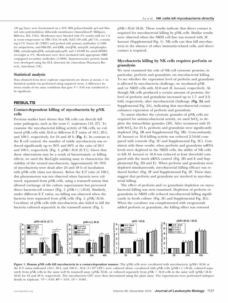
Contact-dependent killing of mycobacteria by pNKcellsPrevious studies have shown that NK cells can directly killsome pathogens, such as the yeast C. neoformans [19, 27]. Toexamine the mycobacterial killing activity of NK cells, we cul-tured pNK cells with M.tb at different E:T ratios of 10:1, 20:1,and 100:1, respectively, for 24 and 48 h (Fig. 1). In contrast tothe B cell control, the number of viable mycobacteria was re-duced significantly up to 30% and 60% at the ratio of 20:1and 100:1, respectively [Fig. 1; pNK�M.tb (E:T)]. Given thatthese observations may be a result of bacteriostatic or killingeffects, we used the BacLight staining assay to characterize theviability of the treated mycobacteria. Approximately 50–70%of mycobacteria were dead after 24 and 48 h of incubationwith pNK cells (data not shown). Below the E:T ratio of 100:1,this phenomenon was not observed when bacteria were cul-tured, separated from pNK cells, using a transwell system thatallowed exchange of the culture supernatants but preventeddirect bacteria-cell contact [Fig. 1; pNK�(/)M.tb]. Similarly,under different E:T ratios, no killing was observed when thebacteria were separated from pNK cells (Fig. 1; pNK/M.tb).Coculture of pNK cells with mycobacteria also failed to kill thebacteria cultured separately in the transwell system (Fig. 1;
pNK�M.tb/M.tb). These results indicate that direct contact isrequired for mycobacterial killing by pNK cells. Similar resultswere observed when the NK92 cell line was treated with M.kansasii (Supplemental Fig. 1). NK cells can thus kill mycobac-teria in the absence of other immunity-related cells, and directcontact is required.
Mycobacteria killing by NK cells requires perforin orgranulysinWe next examined the role of NK cell cytotoxic proteins, inparticular, perforin and granulysin, on mycobacterial killing.To see whether the expression level of perforin and granulysinis affected by mycobacteria challenge, we incubated pNKand/or NK92 cells with M.tb and M. kansasii, respectively. Al-though NK cells produced a certain amount of proteins, thelevel of perforin and granulysin increased up to 1.7- and 2.2-fold, respectively, after mycobacterial challenge (Fig. 2A andSupplemental Fig. 2A), indicating that mycobacterial contactenhances expression of perforin and granulysin.
To assess whether the cytotoxic granules of pNK cells arerequired for antimycobacterial activity, we used SrCl2 to de-plete the intracellular granules [28]. After treatment with 25mM SrCl2 for 24 h, perforin and granulysin were significantlydepleted (Fig. 2B and Supplemental Fig. 2B). Concomitantly,M. kansasii or M.tb killing activity was reduced 2.5-fold com-pared with controls (Fig. 2C and Supplemental Fig. 2C). Con-sistent with these results, when perforin and granulysin mRNAlevels were depleted in the NK92 cells, the ability of NK cellsto kill M. kansasii or M.tb was reduced at least threefold com-pared with the mock siRNA control (Fig. 2D and E and Sup-plemental Fig. 2D and E). When perforin and granulysin weredepleted simultaneously, mycobacterial killing efficacy was re-duced further (Fig. 2F and Supplemental Fig. 2F. These datasuggest that perforin and granulysin are involved in mycobac-terial killing.
The effect of perforin and/or granulysin depletion on myco-bacterial killing was next examined. Depletion of perforin orgranulysin in NK92 cells reduced mycobacterial killing signifi-cantly in broth culture (Fig. 2G and Supplemental Fig. 2G).When the coculture was complemented with exogenouslyadded perforin or granulysin, the killing effect was restored
Figure 1. Human pNK cells kill mycobacteria in a contact-dependent manner. The pNK cells were cocultured with mycobacteria (pNK�M.tb) atthe E:T ratios indicated (10:1, 20:1, and 100:1). M.tb (1�104 CFU) were cultured alone, cocultured with pNK cells [pNK�(/)M.tb], cultured sepa-rately from pNK cells in the same well by transwell assay (pNK/M.tb), or cultured separately from pNK � M.tb cells in the same well (pNK�M.tb/M.tb) for 24 and 48 h, respectively. The mycobacteria CFU were then determined using the plate assay. The experiments were performed indepen-dently in triplicate. *P � 0.05; #P � 0.01; †P � 0.001.
Lu et al. NK cells kill mycobacteria directly
www.jleukbio.org Volume 96, December 2014 Journal of Leukocyte Biology 1121
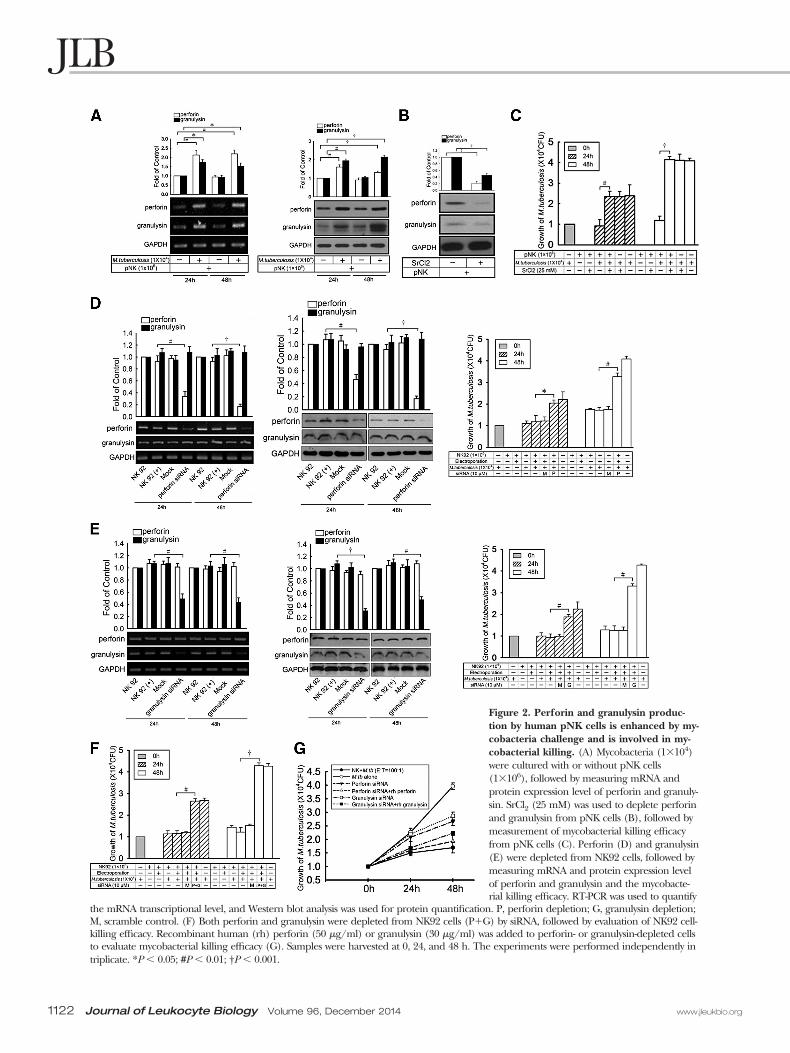
Figure 2. Perforin and granulysin produc-tion by human pNK cells is enhanced by my-cobacteria challenge and is involved in my-cobacterial killing. (A) Mycobacteria (1�104)were cultured with or without pNK cells(1�106), followed by measuring mRNA andprotein expression level of perforin and granuly-sin. SrCl2 (25 mM) was used to deplete perforinand granulysin from pNK cells (B), followed bymeasurement of mycobacterial killing efficacyfrom pNK cells (C). Perforin (D) and granulysin(E) were depleted from NK92 cells, followed bymeasuring mRNA and protein expression levelof perforin and granulysin and the mycobacte-rial killing efficacy. RT-PCR was used to quantify
the mRNA transcriptional level, and Western blot analysis was used for protein quantification. P, perforin depletion; G, granulysin depletion;M, scramble control. (F) Both perforin and granulysin were depleted from NK92 cells (P�G) by siRNA, followed by evaluation of NK92 cell-killing efficacy. Recombinant human (rh) perforin (50 �g/ml) or granulysin (30 �g/ml) was added to perforin- or granulysin-depleted cellsto evaluate mycobacterial killing efficacy (G). Samples were harvested at 0, 24, and 48 h. The experiments were performed independently intriplicate. *P � 0.05; #P � 0.01; †P � 0.001.
1122 Journal of Leukocyte Biology Volume 96, December 2014 www.jleukbio.org
(Fig. 2G and Supplemental Fig. 2G). These results indicatethat perforin and granulysin play a major role in NK cell-medi-ated killing of mycobacteria.
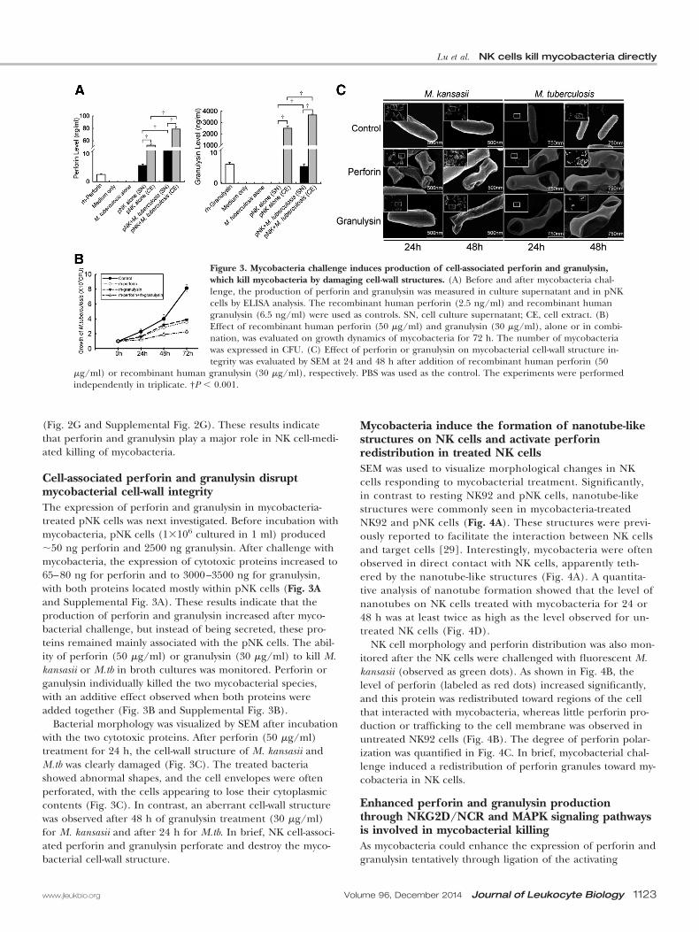
Cell-associated perforin and granulysin disruptmycobacterial cell-wall integrityThe expression of perforin and granulysin in mycobacteria-treated pNK cells was next investigated. Before incubation withmycobacteria, pNK cells (1�106 cultured in 1 ml) produced50 ng perforin and 2500 ng granulysin. After challenge withmycobacteria, the expression of cytotoxic proteins increased to65–80 ng for perforin and to 3000–3500 ng for granulysin,with both proteins located mostly within pNK cells (Fig. 3Aand Supplemental Fig. 3A). These results indicate that theproduction of perforin and granulysin increased after myco-bacterial challenge, but instead of being secreted, these pro-teins remained mainly associated with the pNK cells. The abil-ity of perforin (50 �g/ml) or granulysin (30 �g/ml) to kill M.kansasii or M.tb in broth cultures was monitored. Perforin organulysin individually killed the two mycobacterial species,with an additive effect observed when both proteins wereadded together (Fig. 3B and Supplemental Fig. 3B).
Bacterial morphology was visualized by SEM after incubationwith the two cytotoxic proteins. After perforin (50 �g/ml)treatment for 24 h, the cell-wall structure of M. kansasii andM.tb was clearly damaged (Fig. 3C). The treated bacteriashowed abnormal shapes, and the cell envelopes were oftenperforated, with the cells appearing to lose their cytoplasmiccontents (Fig. 3C). In contrast, an aberrant cell-wall structurewas observed after 48 h of granulysin treatment (30 �g/ml)for M. kansasii and after 24 h for M.tb. In brief, NK cell-associ-ated perforin and granulysin perforate and destroy the myco-bacterial cell-wall structure.
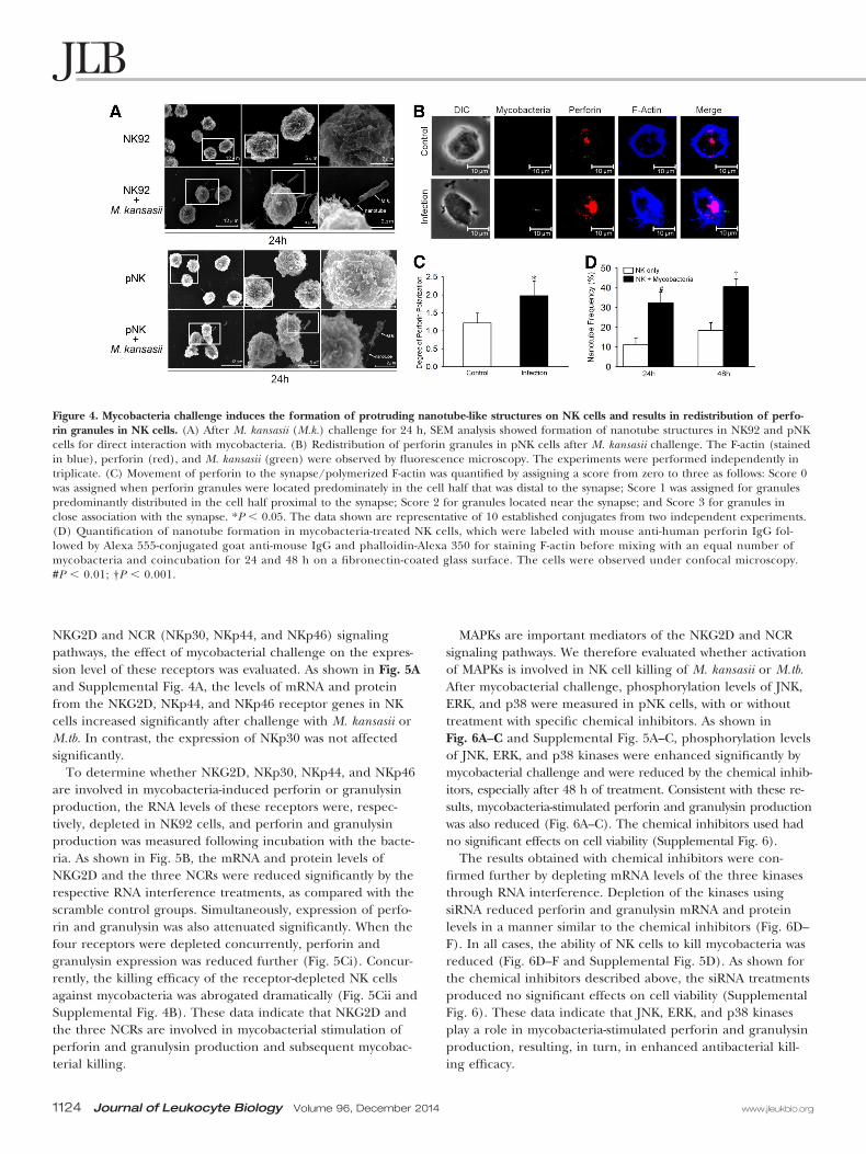
Mycobacteria induce the formation of nanotube-likestructures on NK cells and activate perforinredistribution in treated NK cellsSEM was used to visualize morphological changes in NKcells responding to mycobacterial treatment. Significantly,in contrast to resting NK92 and pNK cells, nanotube-likestructures were commonly seen in mycobacteria-treatedNK92 and pNK cells (Fig. 4A). These structures were previ-ously reported to facilitate the interaction between NK cellsand target cells [29]. Interestingly, mycobacteria were oftenobserved in direct contact with NK cells, apparently teth-ered by the nanotube-like structures (Fig. 4A). A quantita-tive analysis of nanotube formation showed that the level ofnanotubes on NK cells treated with mycobacteria for 24 or48 h was at least twice as high as the level observed for un-treated NK cells (Fig. 4D).
NK cell morphology and perforin distribution was also mon-itored after the NK cells were challenged with fluorescent M.kansasii (observed as green dots). As shown in Fig. 4B, thelevel of perforin (labeled as red dots) increased significantly,and this protein was redistributed toward regions of the cellthat interacted with mycobacteria, whereas little perforin pro-duction or trafficking to the cell membrane was observed inuntreated NK92 cells (Fig. 4B). The degree of perforin polar-ization was quantified in Fig. 4C. In brief, mycobacterial chal-lenge induced a redistribution of perforin granules toward my-cobacteria in NK cells.
Enhanced perforin and granulysin productionthrough NKG2D/NCR and MAPK signaling pathwaysis involved in mycobacterial killingAs mycobacteria could enhance the expression of perforin andgranulysin tentatively through ligation of the activating
Figure 3. Mycobacteria challenge induces production of cell-associated perforin and granulysin,which kill mycobacteria by damaging cell-wall structures. (A) Before and after mycobacteria chal-lenge, the production of perforin and granulysin was measured in culture supernatant and in pNKcells by ELISA analysis. The recombinant human perforin (2.5 ng/ml) and recombinant humangranulysin (6.5 ng/ml) were used as controls. SN, cell culture supernatant; CE, cell extract. (B)Effect of recombinant human perforin (50 �g/ml) and granulysin (30 �g/ml), alone or in combi-nation, was evaluated on growth dynamics of mycobacteria for 72 h. The number of mycobacteriawas expressed in CFU. (C) Effect of perforin or granulysin on mycobacterial cell-wall structure in-tegrity was evaluated by SEM at 24 and 48 h after addition of recombinant human perforin (50
�g/ml) or recombinant human granulysin (30 �g/ml), respectively. PBS was used as the control. The experiments were performedindependently in triplicate. †P � 0.001.
Lu et al. NK cells kill mycobacteria directly
www.jleukbio.org Volume 96, December 2014 Journal of Leukocyte Biology 1123
NKG2D and NCR (NKp30, NKp44, and NKp46) signalingpathways, the effect of mycobacterial challenge on the expres-sion level of these receptors was evaluated. As shown in Fig. 5Aand Supplemental Fig. 4A, the levels of mRNA and proteinfrom the NKG2D, NKp44, and NKp46 receptor genes in NKcells increased significantly after challenge with M. kansasii orM.tb. In contrast, the expression of NKp30 was not affectedsignificantly.
To determine whether NKG2D, NKp30, NKp44, and NKp46are involved in mycobacteria-induced perforin or granulysinproduction, the RNA levels of these receptors were, respec-tively, depleted in NK92 cells, and perforin and granulysinproduction was measured following incubation with the bacte-ria. As shown in Fig. 5B, the mRNA and protein levels ofNKG2D and the three NCRs were reduced significantly by therespective RNA interference treatments, as compared with thescramble control groups. Simultaneously, expression of perfo-rin and granulysin was also attenuated significantly. When thefour receptors were depleted concurrently, perforin andgranulysin expression was reduced further (Fig. 5Ci). Concur-rently, the killing efficacy of the receptor-depleted NK cellsagainst mycobacteria was abrogated dramatically (Fig. 5Cii andSupplemental Fig. 4B). These data indicate that NKG2D andthe three NCRs are involved in mycobacterial stimulation ofperforin and granulysin production and subsequent mycobac-terial killing.
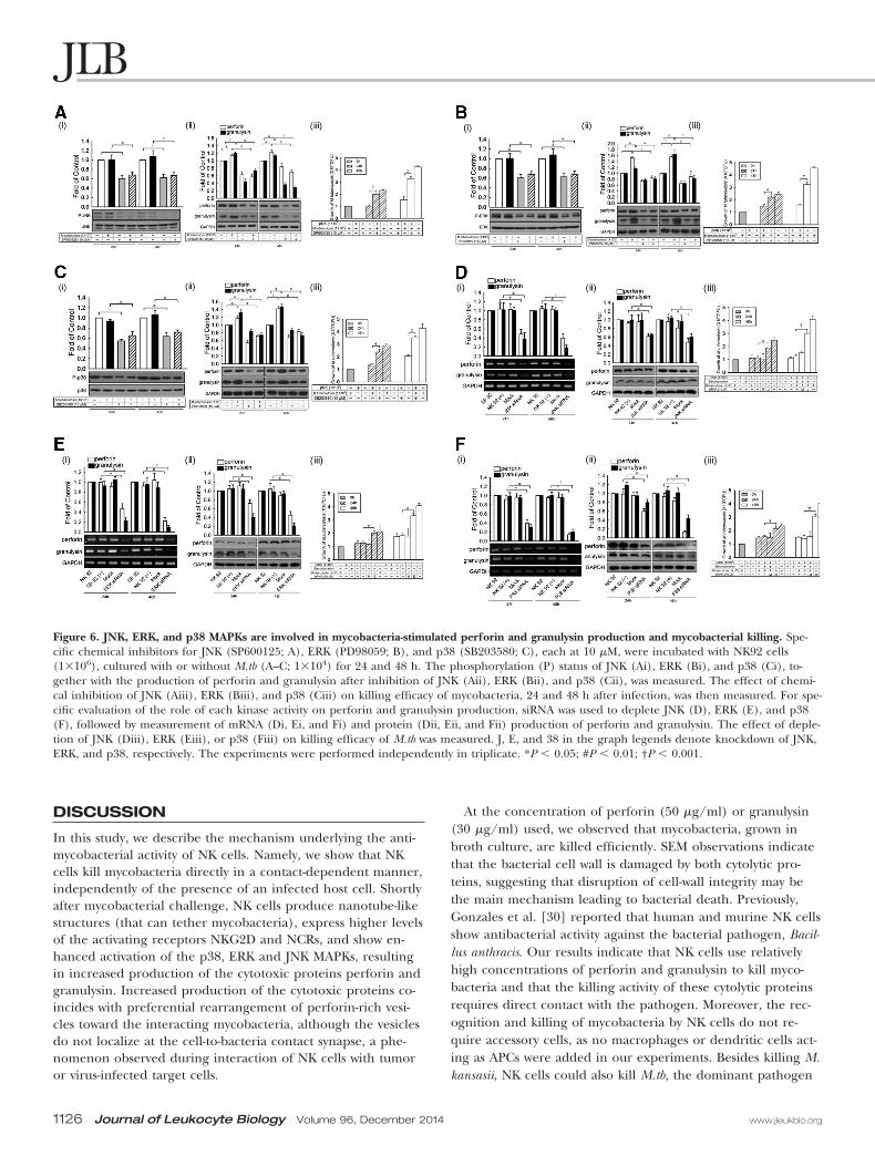
MAPKs are important mediators of the NKG2D and NCRsignaling pathways. We therefore evaluated whether activationof MAPKs is involved in NK cell killing of M. kansasii or M.tb.After mycobacterial challenge, phosphorylation levels of JNK,ERK, and p38 were measured in pNK cells, with or withouttreatment with specific chemical inhibitors. As shown inFig. 6A–C and Supplemental Fig. 5A–C, phosphorylation levelsof JNK, ERK, and p38 kinases were enhanced significantly bymycobacterial challenge and were reduced by the chemical inhib-itors, especially after 48 h of treatment. Consistent with these re-sults, mycobacteria-stimulated perforin and granulysin productionwas also reduced (Fig. 6A–C). The chemical inhibitors used hadno significant effects on cell viability (Supplemental Fig. 6).
The results obtained with chemical inhibitors were con-firmed further by depleting mRNA levels of the three kinasesthrough RNA interference. Depletion of the kinases usingsiRNA reduced perforin and granulysin mRNA and proteinlevels in a manner similar to the chemical inhibitors (Fig. 6D–F). In all cases, the ability of NK cells to kill mycobacteria wasreduced (Fig. 6D–F and Supplemental Fig. 5D). As shown forthe chemical inhibitors described above, the siRNA treatmentsproduced no significant effects on cell viability (SupplementalFig. 6). These data indicate that JNK, ERK, and p38 kinasesplay a role in mycobacteria-stimulated perforin and granulysinproduction, resulting, in turn, in enhanced antibacterial kill-ing efficacy.
Figure 4. Mycobacteria challenge induces the formation of protruding nanotube-like structures on NK cells and results in redistribution of perfo-rin granules in NK cells. (A) After M. kansasii (M.k.) challenge for 24 h, SEM analysis showed formation of nanotube structures in NK92 and pNKcells for direct interaction with mycobacteria. (B) Redistribution of perforin granules in pNK cells after M. kansasii challenge. The F-actin (stainedin blue), perforin (red), and M. kansasii (green) were observed by fluorescence microscopy. The experiments were performed independently intriplicate. (C) Movement of perforin to the synapse/polymerized F-actin was quantified by assigning a score from zero to three as follows: Score 0was assigned when perforin granules were located predominately in the cell half that was distal to the synapse; Score 1 was assigned for granulespredominantly distributed in the cell half proximal to the synapse; Score 2 for granules located near the synapse; and Score 3 for granules inclose association with the synapse. *P � 0.05. The data shown are representative of 10 established conjugates from two independent experiments.(D) Quantification of nanotube formation in mycobacteria-treated NK cells, which were labeled with mouse anti-human perforin IgG fol-lowed by Alexa 555-conjugated goat anti-mouse IgG and phalloidin-Alexa 350 for staining F-actin before mixing with an equal number ofmycobacteria and coincubation for 24 and 48 h on a fibronectin-coated glass surface. The cells were observed under confocal microscopy.#P � 0.01; †P � 0.001.
1124 Journal of Leukocyte Biology Volume 96, December 2014 www.jleukbio.org
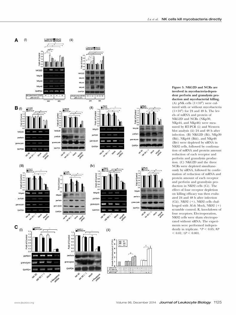
Figure 5. NKG2D and NCRs areinvolved in mycobacteria-depen-dent perforin and granulysin pro-duction and mycobacterial killing.(A) pNK cells (1�106) were cul-tured with or without mycobacteria(1�104) for 24 and 48 h. The lev-els of mRNA and protein ofNKG2D and NCRs (NKp30,NKp44, and NKp46) were mea-sured by RT-PCR (i) and Westernblot analysis (ii) 24 and 48 h afterinfection. (B) NKG2D (Bi), NKp30(Bii), NKp44 (Biii), and NKp46(Biv) were depleted by siRNA inNK92 cells, followed by confirma-tion of mRNA and protein amountreduction of each receptor andperforin and granulysin produc-tion. (C) NKG2D and the threeNCRs were depleted simultane-ously by siRNA, followed by confir-mation of reduction of mRNA andprotein amount of each receptorand perforin and granulysin pro-duction in NK92 cells (Ci). Theeffect of four receptor depletionon killing efficacy was then evalu-ated 24 and 48 h after infection(Cii). NK92 (�), NK92 cells chal-lenged with M.tb; Mock, NK92 (�)scramble control; R, knockdown offour receptors; Electroporation,NK92 cells were sham electropo-rated without siRNA. The experi-ments were performed indepen-dently in triplicate. *P � 0.05; #P� 0.01; †P � 0.001.
Lu et al. NK cells kill mycobacteria directly
www.jleukbio.org Volume 96, December 2014 Journal of Leukocyte Biology 1125
DISCUSSION
In this study, we describe the mechanism underlying the anti-mycobacterial activity of NK cells. Namely, we show that NKcells kill mycobacteria directly in a contact-dependent manner,independently of the presence of an infected host cell. Shortlyafter mycobacterial challenge, NK cells produce nanotube-likestructures (that can tether mycobacteria), express higher levelsof the activating receptors NKG2D and NCRs, and show en-hanced activation of the p38, ERK and JNK MAPKs, resultingin increased production of the cytotoxic proteins perforin andgranulysin. Increased production of the cytotoxic proteins co-incides with preferential rearrangement of perforin-rich vesi-cles toward the interacting mycobacteria, although the vesiclesdo not localize at the cell-to-bacteria contact synapse, a phe-nomenon observed during interaction of NK cells with tumoror virus-infected target cells.
At the concentration of perforin (50 �g/ml) or granulysin(30 �g/ml) used, we observed that mycobacteria, grown inbroth culture, are killed efficiently. SEM observations indicatethat the bacterial cell wall is damaged by both cytolytic pro-teins, suggesting that disruption of cell-wall integrity may bethe main mechanism leading to bacterial death. Previously,Gonzales et al. [30] reported that human and murine NK cellsshow antibacterial activity against the bacterial pathogen, Bacil-lus anthracis. Our results indicate that NK cells use relativelyhigh concentrations of perforin and granulysin to kill myco-bacteria and that the killing activity of these cytolytic proteinsrequires direct contact with the pathogen. Moreover, the rec-ognition and killing of mycobacteria by NK cells do not re-quire accessory cells, as no macrophages or dendritic cells act-ing as APCs were added in our experiments. Besides killing M.kansasii, NK cells could also kill M.tb, the dominant pathogen
Figure 6. JNK, ERK, and p38 MAPKs are involved in mycobacteria-stimulated perforin and granulysin production and mycobacterial killing. Spe-cific chemical inhibitors for JNK (SP600125; A), ERK (PD98059; B), and p38 (SB203580; C), each at 10 �M, were incubated with NK92 cells(1�106), cultured with or without M.tb (A–C; 1�104) for 24 and 48 h. The phosphorylation (P) status of JNK (Ai), ERK (Bi), and p38 (Ci), to-gether with the production of perforin and granulysin after inhibition of JNK (Aii), ERK (Bii), and p38 (Cii), was measured. The effect of chemi-cal inhibition of JNK (Aiii), ERK (Biii), and p38 (Ciii) on killing efficacy of mycobacteria, 24 and 48 h after infection, was then measured. For spe-cific evaluation of the role of each kinase activity on perforin and granulysin production, siRNA was used to deplete JNK (D), ERK (E), and p38(F), followed by measurement of mRNA (Di, Ei, and Fi) and protein (Dii, Eii, and Fii) production of perforin and granulysin. The effect of deple-tion of JNK (Diii), ERK (Eiii), or p38 (Fiii) on killing efficacy of M.tb was measured. J, E, and 38 in the graph legends denote knockdown of JNK,ERK, and p38, respectively. The experiments were performed independently in triplicate. *P � 0.05; #P � 0.01; †P � 0.001.
1126 Journal of Leukocyte Biology Volume 96, December 2014 www.jleukbio.org
responsible for human tuberculosis, indicating that a con-served killing mechanism may be used by NK cells for lysingall mycobacterial species.
The work of Vankayalapati et al. [31, 32] demonstratedthat NKp46, NKp30, NKp44, and NKG2D are involved inrecognition of M.tb-infected macrophages through interac-tion with specific ligands. However, the bacterial ligandsthat activate NK cells remain to be identified. A previousstudy indicated that several unidentified surface moleculesof M. bovis BCG may be recognized by NKp44 in human NKcells [17]. In addition, NKG2D on NK cells is involved inclearance of extracellular pathogens, such as Pseudomonasaeruginosa, in mouse models [33]. Furthermore, althoughhuman proteins, such as MHC class I chain-related proteinsA and B and UL16-binding proteins, have also been identi-fied as activating ligands of NKG2D [34, 35], no bacterialligand has been characterized so far. On the other hand,whereas the expression of NKp30 on NK cells was not in-creased by mycobacterial challenge, we found that depletionof NKp30 still resulted in reduced killing of mycobacteriaby NK cells, suggesting that NKp30 may be constitutivelyinvolved in defense against these bacteria. The depletion ofeach of the four NK cell receptors separately reduced kill-ing, and the depletion of all four receptors at the sametime further reduced killing efficiency, suggesting that allfour receptors may be used in concert by NK cells for myco-bacterial recognition. Alternatively, as NK cells also expresspattern recognition receptors, such as TLRs and NLRs,which may recognize bacterial products, activation of theNKG2D and NKp44 and NKp46 receptors may be an indi-rect effect, resulting from activation of TLRs or NLRs.
Previous studies had reported that granulysin reduces theviability of a broad spectrum of pathogenic bacteria, fungi,and parasites in vitro [36, 37]. Granulysin is a cytolytic proteinpresent in the granules of activated human CD4� and cyto-toxic CD8� CTLs, as well as NK cells [38, 39], and is a mem-ber of the saposin-like protein family [39]. The underlyingmechanism of granulysin’s cytotoxic effects may involve theinsertion of its positively charged domain into the negativelycharged surface of target microbes or cells, resulting in altera-tion of membrane permeability.
In addition to granulysin, our results indicate that perforinalone kills extracellular M. kansasii and M.tb. Perforin is apore-forming protein [40–42] that can directly damage thecytoplasmic membrane of cancer cells or virus-infected cells[10, 43]. However, before the present study, there was no re-port of direct bacterial killing by perforin. Previous studies hadshown that upon secretion by CTLs or NK cells, perforin bindsand inserts itself into the phospholipid bilayer of the targetcell plasma membrane and polymerizes to form a pore of 16nm in diameter that spans the target cell membrane [27]. Per-forin can synergize with other lytic granule components, suchas granzyme B, to induce apoptosis in target cells [44]. As my-cobacteria harbor a thick and waxy cell wall, which is consider-ably different from the cellular membrane of eukaryotic cells,whether perforin can interact directly with cell-wall compo-nents of mycobacteria or the bacterial membrane remains tobe examined.
The effects of granulysin on the M.tb membrane have beenobserved previously by SEM by Stenger et al. [36], whereas theeffect of perforin was not reported. The membrane morphol-ogy observed by this group appears to be different from ourresults. In addition, these authors observed that human perfo-rin was ineffective in reducing the viability of M.tb in cultureor intracellularly in macrophages [36]. These differencescould be a result of the variable susceptibility of the mycobac-terial species or strains used in the two studies or of the differ-ent duration of treatment. Previous studies have shown thatNK-lysin, the pig homologue of granulysin, can permeabilizelipid bilayers and the bacterial cell membrane by interactingwith lipid components [45, 46], providing a possible mecha-nism for the antibacterial effect of this protein.
CTLs and NK cells have been reported to kill C. neoformansdirectly. However, the underlying mechanism used by NK cellsto kill this pathogen may be different from the one used tokill mycobacteria. The antifungal activity of NK cells is medi-ated by a perforin-dependent mechanism under the control ofthe PI3K-dependent ERK1/2 signaling pathway [27]. More-over, granulysin is not involved in killing of C. neoformans. In-triguingly, it has been reported that granulysin but not perfo-rin is involved in CD8� T cell-mediated killing of C. neoformans[47]. The differential requirements for killing of yeast andbacteria by perforin and granulysin remain to be investigated.
Various E:T ratios have been used in previous studies. Forinstance, Gonzales and colleagues [30] demonstrated that hu-man NK cells used at a E:T ratio of 5:1 efficiently reduce B.anthracis CFU, whereas the cells were not active against spores.In another study, NK cells showed perforin-mediated anticryp-tococcal activity at E:T ratios of 100:1, 200:1, and 500:1 [28].These differences in E:T ratios may be explained by differentkilling mechanisms that depend on the composition of thetested bacteria. For instance, mycobacteria possess a thick layerof lipid in their cell wall, which may require a larger numberof effector NK cells compared with other bacteria.
Although the nanotube-like membrane structures wererarely observed when NK cells were cultured alone, thesestructures were observed protruding from NK cells followingchallenge with M. kansasii. The mycobacteria appeared to betethered by the nanotubes, and many bacteria were seen to betrapped and in close contact with NK cells via the nanotubes.Notably, nanotube-like membrane structures were observedpreviously when NK cells were coincubated with other immunecells or tumor cells [29], where the nanotubes were shown tofacilitate long-distance interactions between NK cells and theirtarget cells [29]. It was reported previously that these mem-brane structures could transfer cell-surface proteins across theNK cell-immune synapse [48]. It is thus possible that NK cellsmay use nanotubes to deliver perforin and granulysin to theirmycobacterial targets for direct killing. The use of nanotubeswould also explain the requirement for a contact-dependentmechanism for bacterial killing.
A possible limitation of the present study is the use of theNK92 cell line for the experiments requiring transfection, aspNK cells are not suitable for this purpose. NK92 cells main-tained in culture require treatment with rIL-2, which is an acti-vation signal for perforin and granulysin. This process may be
Lu et al. NK cells kill mycobacteria directly
www.jleukbio.org Volume 96, December 2014 Journal of Leukocyte Biology 1127
responsible for the inability of mycobacteria to induce perfo-rin and granulysin secretion in some experiments (see, for in-stance, Figs. 5 and 6).
In conclusion, we describe here a novel innate-immunefunction for NK cells—the direct killing of mycobacteria. Theunderlying mechanism requires activation of the signalingpathways downstream from NKG2D and NCRs. Perforin andgranulysin are involved in the killing process, and loss of theintegrity of the bacterial cell wall appears to be the majorcause of bacterial death. Modulation of NK cell-signaling path-ways may thus be an alternative strategy for enhancing hostdefense against mycobacterial infection.
AUTHORSHIP
C-C.L. designed and performed experiments, analyzed andinterpreted data, and wrote the manuscript. Y-J.H., C-J.C., andC-S.L. performed experiments. T-S.W., J-H.C., and T-L.W. per-formed experiments in P3 laboratories. T-T.H., D.M.O., andJ.M. assisted with the experimental design and manuscriptwriting. J.D.Y. and H-C.L. conceived of the study, supervisedexperiments, and composed the manuscript.
ACKNOWLEDGMENTS
This work was supported by the Primordia Institute of NewSciences and Medicine, by the University of California Presi-dential Chair, by grant NSC100-2320-B-030-001-MY2 from theNational Science Council of Taiwan (to C-C.L.), and by grantsCMRPD-190501 and NSC98-2320-B-182-007-MY3 from LinkouChang Gung Memorial Hospital and the National ScienceCouncil of Taiwan, respectively (to H-C.L.). We thank the Mi-croscope Core Laboratory at Linkou Chang Gung MemorialHospital for assistance with SEM and confocal microscopy.J.D.Y. dedicates this work in dear memory of both Drs. RalphSteinman and Zanvil A. Cohn, with whom the topic of NK cellkilling was often discussed over the years in the context of in-nate immunity and at times, bacterial infections.
REFERENCES
1. Caligiuri, M. A. (2008) Human natural killer cells. Blood 112, 461–469.2. Lanier, L. L. (2008) Up on the tightrope: natural killer cell activation
and inhibition. Nat. Immunol. 9, 495–502.3. Moretta, A., Bottino, C., Vitale, M., Pende, D., Cantoni, C., Mingari,
M. C., Biassoni, R., Moretta, L. (2001) Activating receptors and corecep-tors involved in human natural killer cell-mediated cytolysis. Annu. Rev.Immunol. 19, 197–223.
4. Moretta, L., Moretta, A. (2004) Unravelling natural killer cell function:triggering and inhibitory human NK receptors. EMBO J. 23, 255–259.
5. Vivier, E., Nunes, J. A., Vely, F. (2004) Natural killer cell signaling path-ways. Science 306, 1517–1519.
6. Wei, S., Gamero, A. M., Liu, J. H., Daulton, A. A., Valkov, N. I., Trapani,J. A., Larner, A. C., Weber, M. J., Djeu, J. Y. (1998) Control of lytic func-tion by mitogen-activated protein kinase/extracellular regulatory kinase2 (ERK2) in a human natural killer cell line: identification of perforinand granzyme B mobilization by functional ERK2. J. Exp. Med. 187,1753–1765.
7. Trotta, R., Fettucciari, K., Azzoni, L., Abebe, B., Puorro, K. A., Eisen-lohr, L. C., Perussia, B. (2000) Differential role of p38 and c-Jun N-ter-minal kinase 1 mitogen-activated protein kinases in NK cell cytotoxicity.J. Immunol. 165, 1782–1789.
8. Kumar, D., Hosse, J., von Toerne, C., Noessner, E., Nelson, P. J. (2009)JNK MAPK pathway regulates constitutive transcription of CCL5 by hu-man NK cells through SP1. J. Immunol. 182, 1011–1020.
9. Trapani, J. A., Smyth, M. J. (2002) Functional significance of the perfo-rin/granzyme cell death pathway. Nat. Rev. Immunol. 2, 735–747.
10. Smyth, M. J., Cretney, E., Kelly, J. M., Westwood, J. A., Street, S. E.,Yagita, H., Takeda, K., van Dommelen, S. L., Degli-Esposti, M. A., Hay-akawa, Y. (2005) Activation of NK cell cytotoxicity. Mol. Immunol. 42,501–510.
11. Podack, E. R., Young, J. D., Cohn, Z. A. (1985) Isolation and biochemi-cal and functional characterization of perforin 1 from cytolytic T-cellgranules. Proc. Natl. Acad. Sci. USA 82, 8629–8633.
12. Liu, C. C., Walsh, C. M., Young, J. D. (1995) Perforin: structure andfunction. Immunol. Today 16, 194–201.
13. Liu, C. C., Young, L. H., Young, J. D. (1996) Lymphocyte-mediated cy-tolysis and disease. N. Engl. J. Med. 335, 1651–1659.
14. Law, R. H., Lukoyanova, N., Voskoboinik, I., Caradoc-Davies, T. T.,Baran, K., Dunstone, M. A., D’Angelo, M. E., Orlova, E. V., Coulibaly,F., Verschoor, S., Browne, K. A., Ciccone, A., Kuiper, M. J., Bird, P. I.,Trapani, J. A., Saibil, H. R., Whisstock, J. C. (2010) The structural basisfor membrane binding and pore formation by lymphocyte perforin. Na-ture 468, 447–451.
15. Garcia-Penarrubia, P., Koster, F. T., Kelley, R. O., McDowell, T. D.,Bankhurst, A. D. (1989) Antibacterial activity of human natural killercells. J. Exp. Med. 169, 99–113.
16. Brill, K. J., Li, Q., Larkin, R., Canaday, D. H., Kaplan, D. R., Boom,W. H., Silver, R. F. (2001) Human natural killer cells mediate killing ofintracellular Mycobacterium tuberculosis H37Rv via granule-independentmechanisms. Infect. Immun. 69, 1755–1765.
17. Esin, S., Batoni, G., Pardini, M., Favilli, F., Bottai, D., Maisetta, G., Flo-rio, W., Vanacore, R., Wigzell, H., Campa, M. (2004) Functional charac-terization of human natural killer cells responding to Mycobacterium bovisbacille Calmette-Guerin. Immunology 112, 143–152.
18. Esin, S., Batoni, G., Counoupas, C., Stringaro, A., Brancatisano, F. L.,Colone, M., Maisetta, G., Florio, W., Arancia, G., Campa, M. (2008) Di-rect binding of human NK cell natural cytotoxicity receptor NKp44 tothe surfaces of mycobacteria and other bacteria. Infect. Immun. 76,1719–1727.
19. Marr, K. J., Jones, G. J., Zheng, C., Huston, S. M., Timm-McCann, M.,Islam, A., Berenger, B. M., Ma, L. L., Wiseman, J. C., Mody, C. H.(2009) Cryptococcus neoformans directly stimulates perforin productionand rearms NK cells for enhanced anticryptococcal microbicidal activity.Infect. Immun. 77, 2436–2446.
20. Wolinsky, E. (1992) Mycobacterial diseases other than tuberculosis. Clin.Infect. Dis. 15, 1–10.
21. Ellis, S. M. (2004) The spectrum of tuberculosis and non-tuberculousmycobacterial infection. Eur. Radiol. 14 (Suppl. 3), E34–E42.
22. Wu, T. S., Leu, H. S., Chiu, C. H., Lee, M. H., Chiang, P. C., Wu, T. L.,Chia, J. H., Su, L. H., Kuo, A. J., Lai, H. C. (2009) Clinical manifesta-tions, antibiotic susceptibility and molecular analysis of Mycobacteriumkansasii isolates from a university hospital in Taiwan. J. Antimicrob. Che-mother. 64, 511–514.
23. Han, S. H., Kim, K. M., Chin, B. S., Choi, S. H., Lee, H. S., Kim, M. S.,Jeong, S. J., Choi, H. K., Kim, C. O., Choi, J. Y., Song, Y. G., Kim, J. M.(2010) Disseminated Mycobacterium kansasii infection associated with skinlesions: a case report and comprehensive review of the literature. J. Ko-rean Med. Sci. 25, 304–308.
24. Gong, J. H., Maki, G., Klingemann, H. G. (1994) Characterization of ahuman cell line (NK-92) with phenotypical and functional characteris-tics of activated natural killer cells. Leukemia 8, 652–658.
25. Favier, B., Lemaoult, J., Lesport, E., Carosella, E. D. (2010) ILT2/HLA-G interaction impairs NK-cell functions through the inhibition ofthe late but not the early events of the NK-cell activating synapse. FASEBJ. 24, 689–699.
26. Lu, C. C., Lai, H. C., Hsieh, S. C., Chen, J. K. (2008) Resveratrol amelio-rates Serratia marcescens-induced acute pneumonia in rats. J. Leukoc. Biol.83, 1028–1037.
27. Wiseman, J. C., Ma, L. L., Marr, K. J., Jones, G. J., Mody, C. H. (2007)Perforin-dependent cryptococcal microbicidal activity in NK cells re-quires PI3K-dependent ERK1/2 signaling. J. Immunol. 178, 6456–6464.
28. Ma, L. L., Wang, C. L., Neely, G. G., Epelman, S., Krensky, A. M., Mody,C. H. (2004) NK cells use perforin rather than granulysin for anticrypto-coccal activity. J. Immunol. 173, 3357–3365.
29. Chauveau, A., Aucher, A., Eissmann, P., Vivier, E., Davis, D. M. (2010)Membrane nanotubes facilitate long-distance interactions between natu-ral killer cells and target cells. Proc. Natl. Acad. Sci. USA 107, 5545–5550.
30. Gonzales, C. M., Williams, C. B., Calderon, V. E., Huante, M. B., Moen,S. T., Popov, V. L., Baze, W. B., Peterson, J. W., Endsley, J. J. (2012) An-tibacterial role for natural killer cells in host defense to Bacillus anthra-cis. Infect. Immun. 80, 234–242.
31. Garg, A., Barnes, P. F., Porgador, A., Roy, S., Wu, S., Nanda, J. S., Grif-fith, D. E., Girard, W. M., Rawal, N., Shetty, S., Vankayalapati, R. (2006)Vimentin expressed on Mycobacterium tuberculosis-infected human mono-cytes is involved in binding to the NKp46 receptor. J. Immunol. 177,6192–6198.
32. Vankayalapati, R., Garg, A., Porgador, A., Griffith, D. E., Klucar, P., Safi,H., Girard, W. M., Cosman, D., Spies, T., Barnes, P. F. (2005) Role ofNK cell-activating receptors and their ligands in the lysis of mononu-
1128 Journal of Leukocyte Biology Volume 96, December 2014 www.jleukbio.org
clear phagocytes infected with an intracellular bacterium. J. Immunol.175, 4611–4617.
33. Wesselkamper, S. C., Eppert, B. L., Motz, G. T., Lau, G. W., Hassett,D. J., Borchers, M. T. (2008) NKG2D is critical for NK cell activation inhost defense against Pseudomonas aeruginosa respiratory infection. J. Im-munol. 181, 5481–5489.
34. Cosman, D., Mullberg, J., Sutherland, C. L., Chin, W., Armitage, R.,Fanslow, W., Kubin, M., Chalupny, N. J. (2001) ULBPs, novel MHC classI-related molecules, bind to CMV glycoprotein UL16 and stimulate NKcytotoxicity through the NKG2D receptor. Immunity 14, 123–133.
35. Salih, H. R., Antropius, H., Gieseke, F., Lutz, S. Z., Kanz, L., Ram-mensee, H. G., Steinle, A. (2003) Functional expression and release ofligands for the activating immunoreceptor NKG2D in leukemia. Blood102, 1389–1396.
36. Stenger, S., Hanson, D. A., Teitelbaum, R., Dewan, P., Niazi, K. R., Fro-elich, C. J., Ganz, T., Thoma-Uszynski, S., Melian, A., Bogdan, C., Por-celli, S. A., Bloom, B. R., Krensky, A. M., Modlin, R. L. (1998) An anti-microbial activity of cytolytic T cells mediated by granulysin. Science 282,121–125.
37. Ernst, W. A., Thoma-Uszynski, S., Teitelbaum, R., Ko, C., Hanson, D. A.,Clayberger, C., Krensky, A. M., Leippe, M., Bloom, B. R., Ganz, T., Mod-lin, R. L. (2000) Granulysin, a T cell product, kills bacteria by alteringmembrane permeability. J. Immunol. 165, 7102–7108.
38. Pena, S. V., Hanson, D. A., Carr, B. A., Goralski, T. J., Krensky, A. M.(1997) Processing, subcellular localization, and function of 519 (granu-lysin), a human late T cell activation molecule with homology to small,lytic, granule proteins. J. Immunol. 158, 2680–2688.
39. Clayberger, C., Krensky, A. M. (2003) Granulysin. Curr. Opin. Immunol.15, 560–565.
40. Liu, C. C., Perussia, B., Cohn, Z. A., Young, J. D. (1986) Identificationand characterization of a pore-forming protein of human peripheralblood natural killer cells. J. Exp. Med. 164, 2061–2076.
41. Young, J. D., Hengartner, H., Podack, E. R., Cohn, Z. A. (1986) Purifica-tion and characterization of a cytolytic pore-forming protein from gran-ules of cloned lymphocytes with natural killer activity. Cell 44, 849–859.
42. Lowin, B., Peitsch, M. C., Tschopp, J. (1995) Perforin and granzymes:crucial effector molecules in cytolytic T lymphocyte and natural killercell-mediated cytotoxicity. Curr. Top. Microbiol. Immunol. 198, 1–24.
43. Young, L. H., Foster, C. S., Young, J. D. (1990) In vivo expression ofperforin by natural killer cells during a viral infection. Studies on uveitisproduced by herpes simplex virus type I. Am. J. Pathol. 136, 1021–1030.
44. Pardo, J., Aguilo, J. I., Anel, A., Martin, P., Joeckel, L., Borner, C., Wal-lich, R., Mullbacher, A., Froelich, C. J., Simon, M. M. (2009) The biol-ogy of cytotoxic cell granule exocytosis pathway: granzymes have evolvedto induce cell death and inflammation. Microbes Infect. 11, 452–459.
45. Ruysschaert, J. M., Goormaghtigh, E., Homblé, F., Andersson, M.,Liepinsh, E., Otting, G. (1998) Lipid membrane binding of NK-lysin.FEBS Lett. 425, 341–344.
46. Miteva, M., Andersson, M., Karshikoff, A., Otting, G. (1999) Molecularelectroporation: a unifying concept for the description of membranepore formation by antibacterial peptides, exemplified with NK-lysin.FEBS Lett. 462, 155–158.
47. Ma, L. L., Spurrell, J. C., Wang, J. F., Neely, G. G., Epelman, S., Kren-sky, A. M., Mody, C. H. (2002) CD8 T cell-mediated killing of Cryptococ-cus neoformans requires granulysin and is dependent on CD4 T cells andIL-15. J. Immunol. 169, 5787–5795.
48. Williams, G. S., Collinson, L. M., Brzostek, J., Eissmann, P., Almeida,C. R., McCann, F. E., Burshtyn, D., Davis, D. M. (2007) Membranousstructures transfer cell surface proteins across NK cell immune synapses.Traffic 8, 1190–1204.
KEY WORDS:antibacterial activity � innate immunity � NKG2D � NCR � tuberculosis
Lu et al. NK cells kill mycobacteria directly
www.jleukbio.org Volume 96, December 2014 Journal of Leukocyte Biology 1129