nitrico xideand staphylococcus,aureus biofilms ir
TRANSCRIPT

Nitric Oxide and Staphylococcus aureus Biofilms: Defining their Intricate Relationship in Chronic Rhinosinusitis Camille Jardeleza BS Public Health, M.D. Submitted for the title of Doctor of Philosophy School of Medicine, Division of Surgery The University of Adelaide July 2014

ii
Dedicated to my parents Ted and Teret

iii
Table of Contents
Thesis Abstract ................................................................................................................... v
Declarations ...................................................................................................................... vii Acknowledgements ....................................................................................................... viii
Publications arising from this Thesis ....................................................................... ix Awards arising from this Thesis ................................................................................... x
Presentations arising from this Thesis ..................................................................... xi
Abbreviations Used in the Thesis ............................................................................. xiii Tables ................................................................................................................................. xvi
Figures .............................................................................................................................. xvii Chapter 1: Systematic Review of Literature ............................................................ 1 1.1 Chronic Rhinosinusitis ...................................................................................................... 1 A. Definition and Epidemiology ......................................................................................................... 1 B. Classification ......................................................................................................................................... 3 C. Aetiopathogenesis .............................................................................................................................. 7 C.1 The Host ........................................................................................................................................................... 7 C.2 The Environment ...................................................................................................................................... 10 C.3 The Microorganism .................................................................................................................................. 11
1.2 Biofilms ................................................................................................................................ 13 A. Bacterial biofilms: History, Discovery and Definition ..................................................... 13 B. Implications in Diseased States ................................................................................................. 15 C. Role in Otolaryngologic Conditions ......................................................................................... 16 D. Role in Chronic Rhinosinusitis .................................................................................................. 18
1.3 Staphylococcus aureus .................................................................................................... 19 A. Historical facts .................................................................................................................................. 19 B. Role in Human Disease .................................................................................................................. 21 B. 1 Components, products and host evasive mechanisms ............................................................ 21 B. 2 Role in major organ system infection ............................................................................................. 22 B. 3 Role in Chronic Rhinosinusitis ........................................................................................................... 24
C. Recalcitrance in biofilm form ..................................................................................................... 26 1.4 Nitric Oxide ......................................................................................................................... 28 A. Discovery and History ................................................................................................................... 29 B. The role of nitric oxide in human and bacterial physiology ......................................... 30 C. Nitric oxide in the Nose and Paranasal Sinuses .................................................................. 33 D. Nitric oxide as a Therapeutic Agent for CRS ........................................................................ 35 E. Nitric oxide delivery mechanisms ............................................................................................ 36
Summary of Literature Review .................................................................................. 39
Studies to be performed ............................................................................................... 41 Chapter 2: The Effects of Nitric Oxide on Staphylococcus aureus Biofilm Growth and its Implications in Chronic Rhinosinusitis ..................................... 42 Statement of Authorship ....................................................................................................... 43 2.1 Abstract ............................................................................................................................... 45 2.2 Introduction ....................................................................................................................... 46 2.3 Methodology ...................................................................................................................... 47 2.4 Results .................................................................................................................................. 52 2.5 Discussion ........................................................................................................................... 58

iv
2.6 Conclusion .......................................................................................................................... 61 Chapter 3: Gene Expression Differences in Nitric Oxide and Reactive Oxygen Species Regulation Point to an Altered Innate Immune Response in Chronic Rhinosinusitis .................................................................................................................. 63 Statement of Authorship ....................................................................................................... 64 3.1 Abstract ............................................................................................................................... 66 3.2 Introduction ....................................................................................................................... 67 3.3 Methodology ...................................................................................................................... 68 3.4 Results .................................................................................................................................. 71 3.5 Discussion ........................................................................................................................... 75 3.6 Conclusion .......................................................................................................................... 78
Chapter 4: Inflammasome Gene Expression Alterations in Staphylococcus aureus Biofilm-‐associated Chronic Rhinosinusitis .............................................. 80 Statement of Authorship ....................................................................................................... 81 4.1 Abstract ............................................................................................................................... 84 4.2 Introduction ....................................................................................................................... 85 4.3 Materials and methods ................................................................................................... 87 4.4 Results .................................................................................................................................. 90 4.5 Discussion ........................................................................................................................... 95 4.6 Conclusion ........................................................................................................................ 100
Chapter 5: Liposome-‐encapsulated ISMN: a Novel Nitric Oxide-‐Based Therapeutic Agent against Staphylococcus aureus Biofilms ......................... 101 Statement of Authorship ..................................................................................................... 102 5.1 Abstract ............................................................................................................................. 104 5.2 Introduction ..................................................................................................................... 105 5.3 Methodology .................................................................................................................... 107 5.4 Results ................................................................................................................................ 112 5.5 Discussion ......................................................................................................................... 122 5.6 Conclusion ........................................................................................................................ 125
Chapter 6: Liposomal Formulation of Nitric Oxide Donor: A Novel Topical Treatment for Staphylococcus aureus Biofilm-‐ associated Rhinosinusitis 126 Statement of Authorship ..................................................................................................... 127 6.1 Abstract ............................................................................................................................... 129 6.2 Introduction ..................................................................................................................... 130 6.3 Methodology .................................................................................................................... 132 6.4 Results ................................................................................................................................ 139 6.5 Discussion ......................................................................................................................... 147 6.6 Conclusion ........................................................................................................................ 151
Thesis Synopsis ............................................................................................................. 152 References ...................................................................................................................... 158

Statements of Authorship
NOTE: Statements of authorship appear in the print copy of the thesis held in
the University of Adelaide Library.

v
Thesis Abstract This thesis aims to address the relationship of Staphylococcus aureus (S. aureus)
biofilms to the endogenously produced gas nitric oxide (NO) in chronic rhinosinusitis
(CRS). While S. aureus biofilms are associated with recalcitrance and high severity in
CRS, the naturally elevated NO gas is significantly lower in sinuses of CRS patients.
However, the relationship of these 3 important factors in CRS aetiopathogenesis is
poorly defined. To further clarify this host-microbe-environment (NO) relationship, this
thesis first looks into the history of each factor, the roles they play in other disease
processes, and the most recent clinical findings and applications in current literature.
Building on this foundation, the projects emanating from this thesis hoped to fill in
some gaps in knowledge of these 3 components, identifying that all are linked to disease
manifestation, and that each can mutually contribute to CRS pathogenesis.
The first project was designed to establish a clearer description of the relationship
between NO and S. aureus biofilms. Utilizing S. aureus strains from CRS patients,
these were grown as biofilms and exposed to various NO concentrations mimicking NO
levels measured in healthy sinuses vs. CRS patients. We demonstrated the dualistic
effects of NO on biofilm growth: increased at lower NO concentrations mimicking
diseased sinuses, and anti-biofilm effects at higher concentrations similar to
measurements in healthy sinuses. These findings became a stepping stone for the
potential design of NO as a therapeutic agent in S. aureus-associated CRS.
But first, further characterization of NO’s role on the host immune response was
needed. The 2nd and 3rd projects aimed to define the host–NO relationship, focusing on
the genes involved in NO regulation within the sinonasal mucosa. Because NO is

vi
considered one of the reactive oxygen species (ROS), major players of the innate
immune response, genes involved in ROS/innate immunity were investigated. CRS
patients, with or without polyps, were sub-classified as either with or without S. aureus
biofilms, allowing a separate analysis of the role S. aureus biofilms play in the
alteration of gene expression. The results showed that S. aureus biofilm presence
associates with a significant difference in the certain gene expressions which have
specific roles in NO regulation. This indicates that the microorganism may alter or
contribute to an impaired localized innate immune response in the sinuses, or
alternatively favor growth in genetically susceptible individuals. Although the cause-
effect timeline was not established, these results will serve as baseline for future gene
and protein studies that will further increase our understanding of the NO-CRS
pathophysiology.
Lastly, building on the therapeutic potential of NO as an anti-biofilm agent, we aimed to
design a suitable NO-based topical agent against S. aureus biofilms. The 4th project
tested a multitude of liposome-encapsulated NO formulations in-vitro with the best
formulation tested for safety and efficacy in a sheep model of rhinosinusitis. These
projects were designed with an aim for future clinical trials, to test a novel NO-based
topical agent, which can be used as a safe and efficacious topical sinus rinse to benefit
CRS patients.

vii
Declarations
I certify that this work contains no material which has been accepted for the award of
any other degree or diploma in any university or other tertiary institution awarded to
Camille Jardeleza. To the best of my knowledge and belief, this work contains no
material previously published or written by another person, except where due reference
has been made in the text.
I certify that no part of this work will, in the future, be used in a submission in my name
for any other degree or diploma in any university or other tertiary institution without the
prior approval of the University of Adelaide and where applicable, any partner
institution responsible for the joint award of this degree.
I give consent to this copy of my thesis when deposited in the University Library, being
made available for loan and photocopying, subject to the provisions of the Copyright
Act 1968.
I acknowledge that copyright of published works contained within this thesis resides
with the copyright holders of those works.
Dr. Camille Jardeleza

viii
Acknowledgements Any conclusion of a long-term goal cannot be done alone. I have been fortunate to be part of an excellent team during my PhD. I would like to first thank Prof. Peter-John Wormald, my mentor in research and now in surgical training, who provided me the ideal work environment, and gave me the perfect blend of autonomy and supervision. In both actions and words he inspires me to continue to strive for excellence and to believe in my capabilities. It has been an honor Prof. to be part of your team. To the two chief research scientists during my time, Dr. Lor Wai Tan, thank you for ensuring that I fulfilled all my PhD requirements prior to your departure, and Dr. Sarah Vreugde for sharing your views and always having time to answer my concerns and review my articles. Thank you both for your valued supervision. To my co-authors Dr. Andrew Foreman and Dr. Sam Boase, and my friend Dr. Josh Jervis-Bardy, thank you for showing me the way to becoming an efficient Surgeon Scientist, and for being excellent role models in balancing the multiple tasks of research and surgical training. I am indebted to you all for the guidance. To my good friends and co-authors Dr. Sukanya Rajiv, Dr. Amanda Drilling, Dr. Ahmed Bassiouni, Dr. Daniel Cantero and Dr. Neil Tan, thank you for making my PhD time an enjoyable 3 years, for always being a great sounding board in the exchange of scientific ideas, and for the assistance in tasks which required team effort. It would not have been memorable times without your friendship. To the research scientists, especially Ms. Leonie Baker, Ms. Dijana Miljkovic, Dr. Clare Cooksley and Mr. Damien Jones, thank you for teaching me all the laboratory techniques I know today, and for being patient with me in the lab and institute. I was very lucky to have had you all in the department during my PhD. To our collaborators from the University of South Australia, Ian Wark Institute: Dr. Shasha Rao, for providing me the seemingly endless supply of liposomes, and Dr. Benjamin Thierry and Dr. Clive Prestidge for the manuscript reviews. To the Adelaide Microscopy staff, especially Ms. Lyn Waterhouse and the LARIF staff, especially Dr. Tim Kuchel and Ms. Loren Matthews, thank you for always having a welcoming environment as I visited and did my work in your facilities. The financial support during times of study is much needed to ensure successful completion. Thank you to The Garnett Passe and Rodney Williams Memorial Foundation and The University of Adelaide for my scholarships during this PhD. To Ms. Lyn Martin, who has known me even before my PhD started, thank you for always accommodating all my requests, from science to clinics, to books and journals, you have never failed to assist me, and for that I am grateful. And to Dr. Prue Cowled, my Post Graduate Coordinator, thank you for always having the right answers when I ask, and pointing me in the right direction when I get lost in the land of research. And lastly, to my parents, and sisters Steph and Kathryn, thank you for the support in all these tasks I find myself wanting to accomplish, for always being there for me regardless of circumstances, and pushing me to finish what I’ve started. Thank you for the continuous understanding.

ix
Publications arising from this Thesis
The effects of nitric oxide on Staphylococcus aureus biofilm growth and its
implications in chronic rhinosinusitis
Jardeleza C, Foreman A, Baker L, Paramasivan S, Field J, Tan LW, Wormald PJ.
International Forum of Allergy and Rhinology 2011, 1(6): 438-44.
Gene expression differences in nitric oxide and reactive oxygen species regulation
point to an altered innate immune response in chronic rhinosinusitis
Jardeleza C, Jones D, Baker L, Miljkovic D, Boase S, Tan NC, Vreugde S, Tan LW,
Wormald PJ.
International Forum of Allergy and Rhinology 2013, 3(3): 193-8.
Inflammasome gene expression alterations in Staphylococcus aureus biofilm-
associated Chronic Rhinosinusitis
Jardeleza C, Miljkovic D, Baker L, Boase S, Tan NC, Koblar SA, Zalewski P,
Rischmueller M, Lester S, Drilling A, Jones D, Tan LW, Wormald PJ, Vreugde S.
Rhinology 2013, 51(4): 315-22.
Liposome-encapsulated ISMN: A novel nitric oxide-based therapeutic agent
against Staphylococcus aureus biofilms
Jardeleza C, Rao S, Thierry B, Gajjar P, Vreugde S, Prestidge C, Wormald PJ.
Plos One 2014, 9(3): e92117.
Liposome-encapsulated nitric oxide donor: a novel topical treatment for
Staphylococcus aureus biofilm- associated rhinosinusitis
Jardeleza C, Thierry B, Rao S, Rajiv S, Drilling A, Miljkovic D, Paramasivan S, James
C, Vreugde S, Prestidge C, Wormald PJ
Prepared for submission

x
Awards arising from this Thesis
BEST ORAL PRESENTATION: 1st Year Higher Degree Student
Research Day, The Queen Elizabeth Hospital / Basil Hetzel Institute, Adelaide SA, Oct
2010
BASIC RESEARCH SCIENCE AWARD WINNER for best scientific paper
American Rhinologic Society, San Francisco Ca, USA, Sept 2011
RONALD GRISTWOOD MEDAL, Best South Australian ENT Registrar
Presentation for Research, Adelaide, SA, Nov 2012
NATIONAL HEALTH AND MEDICAL RESEARCH FOUNDATION, Successful
Project Grant, Application 1047576. A novel nitric oxide-based treatment for
recalcitrant Staphylococcus aureus-associated chronic rhinosinusitis, 2013

xi
Presentations arising from this Thesis The role of nitric oxide in the pathophysiology of Staphylococcus aureus biofilm formation in chronic rhinosinusitis Basil Hetzel Institute Post Graduate Seminar, Adelaide, July 2010 The role of nitric oxide in the pathophysiology of Staphylococcus aureus biofilm formation in chronic rhinosinusitis The Queen Elizabeth Hospital / Basil Hetzel Institute Research day, Adelaide Oct 2010 The Effects of Nitric Oxide on Staphylococcus aureus Biofilm Growth and its Implications in Chronic Rhinosinusitis ASOHNS, Annual Scientific Meeting, Melbourne, April 2011 The role of nitric oxide on Staphylococcus aureus biofilm formation in chronic rhinosinusitis Basil Hetzel Institute Post Graduate Seminar, Adelaide, May 2011 The Effects of Nitric Oxide on Staphylococcus aureus Biofilm Growth and its Implications in Chronic Rhinosinusitis American Rhinologic Society Annual Scientific Meeting, San Francisco Ca, USA September 2011 Gene expression differences in nitric oxide regulation point to an altered innate immune response in chronic rhinosinusitis The Australian Society for Medical Research SA Scientific Conference, Adelaide, June 2012 The role of nitric oxide on Staphylococcus aureus biofilm growth in chronic rhinosinusitis Endoscopic Sinus and Skull Base Surgery Course August 2-4 2012, St. Vincent’s Hospital, Sydney NSW, August 2012 - Invited speaker The Efficacy of Liposome-encapsulated Nitric Oxide on Staphylococcus aureus biofilms in Chronic Rhinosinusitis Basil Hetzel Institute Post Graduate Seminar, Adelaide, May 2012 Gene expression difference in nitric oxide and reactive oxygen species regulation point to an altered innate immune response in chronic rhinosinusitis American Rhinologic Society Annual Scientific Meeting, Washington DC, USA September 2012

xii
The Effects of Nitric Oxide on Staphylococcus aureus Biofilm Growth and its Implications in Chronic Rhinosinusitis Ronald Gristwood Medal, Adelaide, October 2012 Management of the recalcitrant sinus infection With NC Tan, 15th Advanced FESS course, Adelaide, November 2012 – Invited speaker Gene expression difference in nitric oxide and reactive oxygen species regulation point to an altered innate immune response in chronic rhinosinusitis ASOHNS, Annual Scientific Meeting, Perth, March 2013 Liposome-encapsulated Nitric Oxide against Staphylococcus aureus biofilms in a rhinosinusitis sheep model Basil Hetzel Institute Post Graduate Seminar, Adelaide, June 2013 The Effects of Nitric Oxide on Staphylococcus aureus Biofilm Growth and its Implications in Chronic Rhinosinusitis RP Jepson Medal, Royal Australasian College of Surgeons ASM, August 2013.

xiii
Abbreviations Used in the Thesis AdSA: Adenosine synthase A
AERD: Aspirin Exacerbated Respiratory
Disease
AFS: Allergic Fungal Sinusitis
AIM2: Absent in Melanoma 2
ASC: Apoptosis-associated Speck-like
protein with a CARD
bNOS: Bacterial Nitric Oxide Synthase
B– P-: Biofilm negative Polyp negative
B- P+: Biofilm negative Polyp positive
B+ P-: Biofilm positive Polyp negative
B+ P+: Biofilm positive Polyp positive
CARD: Caspase Recruitment Domain
CASP5: Caspase-5
CAT: Catalase
CCL2: Chemokine Ligand 2
CCL20: Chemokine Ligand 20
CF: Cystic Fibrosis
CFTR: Cystic Fibrosis Transmembrane
Conductance Regulator
CLSM: Confocal Scanning Laser
Microscopy
CRS: Chronic Rhinosinusitis
CRSsNP: Chronic Rhinosinusitis without
Nasal Polyps
CRSwNP: Chronic Rhinosinusitis with
Nasal Polyps
Cyclic GMP: Cyclic Guanosine
Monophosphate
DAMP: Danger-Associated Molecular
Patterns
DC: Dendritic Cells
DNA: Deoxyribonucleic Acid
DPPG: Dipalmitoylglycero-
phosphoglycerol
dsDNA: Double stranded DNA
DUOX1: Dual Oxidase 1
EDRF: Endothelium Derived Relaxing
Factor
EGFR: Epidermal growth factor
eNOS: Endothelial Nitric Oxide Synthase
EPS: Extracellular Polymeric Substance
ESS: Endoscopic Sinus Surgery
FISH: Fluorescence in situ hybridization
GTN: Glyceryl Trinitrate
H. influenzae: Haemophilus influenzae
HPLC: High Performance Liquid
Chromatography

xiv
HSP90AA1: Heat shock Protein 90 KDα
IE: Infective Endocarditis
IgE: Immunoglobulin E
IgG: Immunoglobulin G
IL-1β: Interleukin 1-β
IL-18: Interleukin 18
IFN-γ : Interferon-γ
iNOS: Inducible Nitric Oxide Synthase
ISMN: Isosorbide Mononitrate
LPS: Lipopolysaccharide
LTA: Lipoteichoic acid
LFNO: Liposomal-formulated nitric
oxide donor
MBEC: Minimum Biofilm Eradication
Concentration
M. catarrhalis: Moraxella catarrhalis
MLV: Multilamellar vesicles
MRSA: Methicillin Resistant
Staphylococcus aureus
N2O: Nitrous Oxide
NFκβ: Nuclear factor kappa beta
NADPH: Nicotinamide adenine
dinucleotide phosphate
NCF2: Neutrophil cytosoloic factor 2
NLR: NOD-like receptors
NLRP3: NOD-like receptor pyrin
domain containing protein
NME5: Non-metastatic cell 5
NO2: Nitrite
NO3: Nitrate
NOD: Nucleotide Oligomerization
Domain
nNOS: neuronal Nitric Oxide Synthase
NO: Nitric Oxide
NOS: Nitric Oxide Synthase
OME: Otitis Media with Effusion
ONOO-: Peroxynitrite
OXR1: Oxidoreductase 1
P. aeruginosa: Pseudomonas aeruginosa
PALS: Phase analysis light scattering
PAMP: Pathogen-associated molecular
patterns
PANX1: Pannexin 1
PDI: Polydispersity Index
PCD: Primary Ciliary Dyskinesia
PCR: Polymerase Chain Reaction
PRDX2: Peroxiredoxin 2
PRDX5: Peroxiredoxin 5
PRDX6: Peroxiredoxin 6

xv
PRNP: Prion protein
PRR: Pattern-recognition receptors
PSTPIP1: proline-serine- threonine
phosphatase interacting protein 1
PYCARD: PYD and CARD Domain
containing gene
qRT-PCR: Quantitative Real Time PCR
RANKL: Receptor activator of nuclear
factor kappa-β ligand
RAST: Radioallergosorbent test
RLH: RNA-sensing RIG-like helicases
RNS: Reactive Nitrigen Species
ROS: Reactive Oxygen Species
S. aureus: Staphylococcus aureus
SCV: Small Colony Variants
S. pneumoniae: Streptococcus
pneumoniae
TH1: T-Helper 1
TH2: T-Helper 2
TLR: Toll-Like Receptor
TNFSF11: Tumor necrosis factor
member 11
ULV: Unilamellar vesicles
XIAP: X-linked inhibitor of apoptosis
protein
ZBP1: Z-DNA-binding protein

xvi
Tables Table 1.1 Sub-classification of CRS based on allergy and asthma status. Table 2.1: Average biofilm biomass of the different S. aureus strains exposed to high (125-1000 µM) and low (0.975-1.96 µM.) NO donor concentrations. Table 3.1: Patient profile of CRS patients classified based on biofilm and polyp status. Table 3.2: Selected genes significantly changed in the B+P+ group when compared to controls. Table 3.3: Genes significantly and consistently changed upon comparison of the 4 groups based on S. aureus biofilm and polyp status. Table 4.1: Summary of patient demographics and clinical findings in the 3 groups classified based on S. aureus biofilm and polyp status. Table 4.2: Expression of inflammasome related genes in sinonasal tissue harvested from B+P+ patients compared with non-diseased control mucosa and their biological functions. Table 4.3: Summary of genes significantly changed upon comparison of the 3 groups classified based on S. aureus biofilm and polyp status. Table 5.1: Characteristics of the different anionic liposomes. Table 5.2: Drug encapsulation efficiency of anionic liposomes loaded with ISMN. Table 6.1: Mean, median and range (maximum – minimum) of ciliary preservation of LFNO vs. saline control. Table 6.2: Statistical results of ciliary preservation comparing control (saline) and treatment (LFNO) group. Table 6.3: Average biofilm biomass of control vs. treated sinus for each sheep classified to the different liposomal groups.

xvii
Figures Figure 1.1: Scanning electron micrograph of a bacterial biofilm with bacterial cells encased in the slimy EPS matrix. Figure 1.2: Stages of the biofilm lifecycle. Figure 1.3: Scanning electron micrograph of S. aureus. Figure 1.4: Pathway of NO synthesis in oxygen available states. Figure 1.5: Summary of the aims of the projects of this PhD addressing the host, environment and microbe component of CRS pathophysiology. Figure 2.1: NO levels released by the NO donor Deta NONOate, showing the predictability of NO release of Deta NONOate. Figure 2.2: Scanning electron microscopy images of S. aureus ATCC 25923 biofilms grown on the MBEC peg device taken at different magnifications. Figure 2.3: Baseline biofilm biomass of different S. aureus strains highlighting variability in biofilm growth across isolates. Figure 2.4: CSLM 3D projection images of biofilm growth of S. aureus ATCC 25923 exposed to various NO donor concentrations. Figure 2.5: CLSM 3D projection images of biofilm growth of S. aureus Clinical Isolate 1 exposed to different NO donor concentrations.
Figure 2.6: Scatter plot graphs of biofilm growth of the different S. aureus isolates exposed to various NO donor concentrations, plotted on a logarithmic scale.
Figure 2.7: Line graph of the average planktonic bacterial CFU counts released from the biofilms of different S. aureus isolates. Figure 3.1: CLSM images of S. aureus biofilm positive (B+) and negative (B-) tissue samples using fluorescence-in-situ hybridization. Figure 4.1. CLSM image of S. aureus biofilms on the sinonasal mucosa of a B+P+ patient using FISH.

xviii
Figure 5.1: Dose-response of pure ISMN on S. aureus biofilm biomass (A) and planktonic cell growth (B). Figure 5.2: Effects of ULV liposomal formulations on S. aureus biofilm biomass after a 24-hour exposure. Figure 5.3: Effects of ULV liposomal formulations on S. aureus biofilm biomass after a 5-minute exposure. Figure 5.4: Effects of MLV liposomal formulations of 25 mM lipid composition on S. aureus biofilm biomass after a 5-minute exposure. Figure 5.5: CLSM 3D projection images of S. aureus biofilms on pegs exposed for 5 minutes to MLV liposomes with a 25 mM lipid composition. Figure 6.1: Average heart rate (HR) and Mean arterial pressure (MAP) of control vs. treatment sheep at different sinus flushing times. Figure 6.2: Histologic analysis of the control vs. treatment sheep sinus (LFNO) comparing degree of inflammation, epithelial thickness and acute inflammation. Figure 6.3: Average biofilm biomass of control vs. treatment sinus within each sheep group showing only the LFNO group with statistical significance.

1
Chapter 1: Systematic Review of Literature
1.1 Chronic Rhinosinusitis
A. Definition and Epidemiology The definition of CRS is best summarized by the individual description of its
components. Rhinitis has been defined as inflammation of the nasal cavity while
sinusitis refers to the inflammation of the sinuses. Because it has been well documented
that the two co-exist, the correct terminology is now known as rhinosinusitis.1,2 For a
clinical diagnosis, the presence of inflammation of the nose and paranasal sinuses must
be accompanied by two or more clinical symptoms as described by the European
taskforce of 2012,3 one of which should include:
a. nasal blockage, obstruction or congestion or
b. nasal discharge: either anterior rhinorrhea or postnasal drip
+/- the presence of facial pain/pressure and +/- reduction or loss of smell.
These symptoms should be accompanied by clinical or radiologic findings, such as:
a. endoscopic signs of nasal polyps, mucopurulent discharge or oedema/mucosal
obstruction, usually in the middle meatus and/or
b. CT scan changes which are mucosal changes usually in the osteomeatal complex
or sinuses.
The duration of symptoms separates the definition to acute rhinosinusitis which is <12
weeks with complete resolution, and chronic which is >12 weeks without complete
symptom resolution. In essence, CRS is the inflammation of the nose and sinuses
characterized by symptoms such as nasal airway obstruction, anterior nasal discharge,
post nasal drip, facial pain or pressure and anosmia or hyposmia, accompanied by

2
radiologic changes and/or endoscopic signs as mentioned above, for a duration of more
than 12 weeks.
The incidence of CRS varies across literature from as low as 2% to as high as 16%.4,5 In
Australia, it affects as many as 1.8 million or 9.2% of the population and comparable to
asthma, is one of the most frequently reported health conditions.6,7 Despite the
localization of signs, the severity of symptoms can greatly affect quality of life and
functional capacity. In a study by Bhattacharya et al., it was found that CRS causes an
average of 4.8 missed workdays due to sinonasal symptoms, 2.7 episodes of acute
exacerbations, and 3.9 physician visits per patient every year.8
This high incidence of CRS and its detrimental effects on a patient’s well being
correspondingly has a large global socio-economic impact. In the US, it has been
observed that CRS results in an estimated 18-22 million physician visits, with direct
treatment costs of $8.6 billion per annum.9 These computed costs not only include
missed workdays and physician visits, but also the consumption of a variety of
prescription drugs such as intranasal corticosteroids, antihistamines and antibiotics. The
approximate yearly economic cost is between $15008-$250010 per patient. Furthermore,
when surgery is required, the cost average increases to over $7500 per patient, from the
time of surgery to 45 days post-procedure.10 From these figures, the potential of CRS to
consume a vast amount of economic resources cannot be ignored.

3
B. Classification
The simplest classification of CRS is based on the presence (CRSwNP) or absence
(CRSsNP) of nasal polyps. A nasal polyp is an oedematous mucosal membrane which
forms a pedunculated mass, with a base or a stalk usually originating from the narrow
regions of the nasal cavity, such as the osteomeatal complex or middle meatus.11 The
exact etiology of nasal polyps is still unknown, although the narrow slits of mucous
membranes where they often originate is postulated to play a role in its formation. It is
said that mechanical stimulation of adjacent epithelial cells on the mucosal surfaces
causes a release of pro-inflammatory cytokines inciting or contributing to a localized
inflammatory reaction and polyp formation.11 Most likely nasal polyposis is
multifactorial in cause, but despite its unclear pathologic process, many studies have
shown CRSwNP to be clinically, histologically and genetically distinct from its non-
polyp counterpart. Histologic characteristics of nasal polyps show greater stromal
oedema, plasma cells, and eosinophils with a higher degree of inflammation.12 This
reflects greater symptomatology and the need for more aggressive treatment in this
patient group. CRSsNP patients on the other hand, tend to have a generally more
neutrophilic predominance.13 Eosinophilia in itself has been strongly associated with
polypoid mucosal changes. Certainly, patients with nasal polyps clinically manifest with
more severe symptoms,14 and are more frequently associated with other diseases that
can contribute to disease severity such as asthma, cystic fibrosis, aspirin sensitivity and
primary ciliary dyskinesia.
With the emergence of new molecular and histologic techniques, discoveries of new
disease entities are challenging the traditional classification of CRS with and without
nasal polyps. Several studies have emerged describing other potential sub-

4
classifications.15-17 Allergic fungal sinusitis (AFS) is said to have its own distinct
characteristics, and sometimes thought of to be a separate classification. The presence
or absence of eosinophilic mucus can further sub-classify CRS to another 3 distinct
histologic entities.18 For example, in CRSwNP, three distinct histologic features were
further discovered based on cell predominance:
a. Eosinophilic
b. Neutrophilic
c. Non-eosinophilic, non-neutrophilic
The eosinophilic type is characterized by a T-helper 2 (TH2) response, while the non-
eosinophilic type has a T-helper 1 (TH1) polarization. This becomes clinically
important as the eosinophilic phenotype is associated with atopy, more extensive
disease and poorer prognosis.18,19 Further characterization of different CRS subtypes is
important, as they will likely dictate future sub-type specific treatment strategies.
Asthma, allergy and aspirin sensitivity have also been found to play a greater role in the
disease process, and thus certain sub-classifications take their presence into
consideration.20 Samter’s triad of aspirin sensitivity, asthma and nasal polyposis is a
particular disease entity associated with a more recalcitrant form of CRS. Han et al.20
proposed a new sub-grouping of CRS that stratifies the disease into 7 separate
categories:
a. Allergic fungal sinusitis (AFS)
b. Aspirin triad or Aspirin Exacerbated Respiratory Disease (AERD)
c. Asthmatic sinusitis with allergy (Asthma + Allergy +)
d. Asthmatic sinusitis without allergy (Asthma + Allergy -)

5
e. Non-asthmatic sinusitis with allergy (Asthma – Allergy +)
f. Non-asthmatic sinusitis without allergy (Asthma - Allergy -)
g. Cystic fibrosis (CF) associated sinusitis
CRS patients with asthma, allergy and aspirin sensitivity (AERD) or Samter’s triad,
have been shown to have the worse symptoms and more aggressive nasal polyposis, and
are associated with higher degrees of eosinophilia.21,22 The pattern often starts with
rhinitis symptoms at the 3rd decade, followed by months of nasal congestion, rhinorrhea
and eventually a diagnosis of nasal polyposis, followed shortly by asthmatic attacks and
aspirin sensitivity.23,24 Asthmatic sinusitis with allergy is also associated with a TH2
response, polyposis and allergy, but the asthma component commences during
childhood years. Non-asthmatic non-allergic sinusitis is deemed the mildest sub-type,
and likely caused by impairments in sinus drainage due to anatomic abnormalities. This
type is associated with localized hypoxia in the sinuses and bacterial infection, hence a
TH1 response, with non-asthmatic with allergy CRS halfway between the 2 in terms of
severity.20 Asthmatic CRS without allergy is similar although to a lesser severity than
AERD, with asthma developing during the adult years. AFS, although similar to
asthma and allergy associated CRS, has a more localized TH2 allergic reaction to
fungus, often confined to the sinuses. CF associated CRS is associated with higher
polymorphonuclear cell count, and thick mucopus production rather than polyposis,
marking an infectious inflammatory process rather than an allergic type of reaction.20
Table 1.1 summarizes these findings.
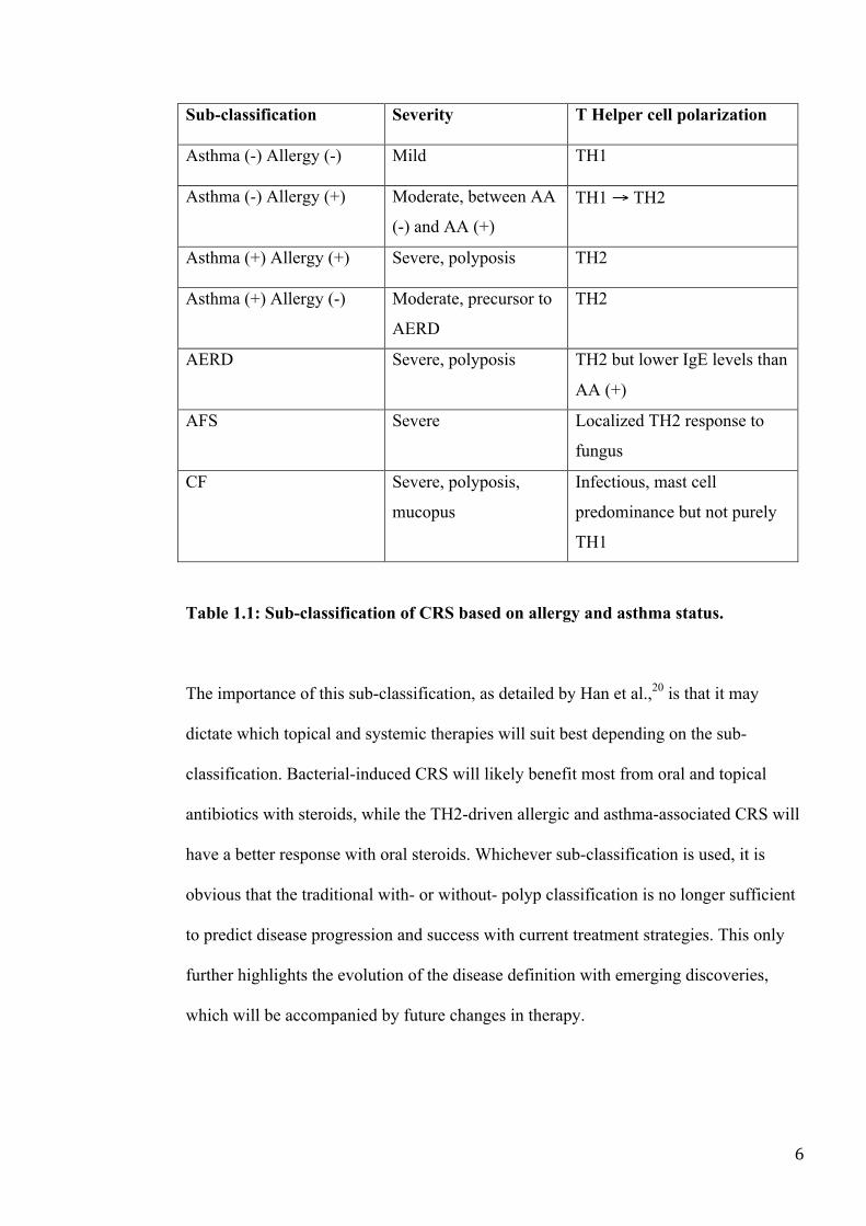
6
Sub-classification Severity T Helper cell polarization
Asthma (-) Allergy (-) Mild TH1
Asthma (-) Allergy (+) Moderate, between AA
(-) and AA (+)
TH1 → TH2
Asthma (+) Allergy (+) Severe, polyposis TH2
Asthma (+) Allergy (-) Moderate, precursor to
AERD
TH2
AERD Severe, polyposis TH2 but lower IgE levels than
AA (+)
AFS Severe Localized TH2 response to
fungus
CF Severe, polyposis,
mucopus
Infectious, mast cell
predominance but not purely
TH1
Table 1.1: Sub-classification of CRS based on allergy and asthma status.
The importance of this sub-classification, as detailed by Han et al.,20 is that it may
dictate which topical and systemic therapies will suit best depending on the sub-
classification. Bacterial-induced CRS will likely benefit most from oral and topical
antibiotics with steroids, while the TH2-driven allergic and asthma-associated CRS will
have a better response with oral steroids. Whichever sub-classification is used, it is
obvious that the traditional with- or without- polyp classification is no longer sufficient
to predict disease progression and success with current treatment strategies. This only
further highlights the evolution of the disease definition with emerging discoveries,
which will be accompanied by future changes in therapy.

7
C. Aetiopathogenesis One of the underlying reasons for the absence of a cure for CRS is due to its complex
multifactorial aetiology. The interplay of two main factors, the host and the
environment, leads to the manifestation of CRS at varying degrees of severity. Each of
these factors will be discussed in detail.
C.1 The Host
In the host, both general and localized immune responses to environmental stimuli have
been implicated in the disease process. The role of the general immune response is
evident in the association of CRS with immune deficiency and genetic disorders.25
Immunoglobulin deficiency, the most common of which is an IgG deficiency, has been
commonly found in CRS patients, and concluded by some to be the first sign of an
immunologic predisposition to persistent sinus infection.25 The frequency of CRS in
genetic diseases such as cystic fibrosis (CF) and primary ciliary dyskinesia (PCD) also
implicates the role of a possible genetic component in its etiology. For example, the
cause of CF, a Cystic Fibrosis Transmembrane Conductance Regulator (CFTR) gene
mutation, has been found to be higher in patients with CRS compared to non-diseased
patients.26 PCD, an autosomal recessive disorder, is characterized by ciliary dysfunction
and impaired mucociliary clearance, with sinusitis being one of its clinical
manifestations. Not isolated to a single genetic mutation, PCD is a genetically
heterogeneous disorder, implicating 8 genes involved in structural or functional defects
of the cilia.27 Because of the several anatomical and systemic factors involved in the
maintenance of normal sinonasal function, it is more likely multiple genes, rather than a
solitary gene, will be implicated in disease aetiopathogenesis. This is already evident in

8
a multitude of genes presented in current literature, most of which have roles in the host
immune response.28,29
The localized immune response in the sinonasal tract is greatly regulated by the innate
immune system, a first-line of defense against infection. Although a non-specific
response, this localized protective mechanism is crucial due to the regular exposure of
the sinonasal mucosa to pathogens and allergens from inhaled air. Many steps are
involved in the mounting of a successful innate immune response.30 Larger inhaled
particles, which are trapped in the superficial mucus, are swept away via a coordinated
mucocilary system, physically clearing pathogens from the sinonasal tract. For
pathogens that have evaded this primary barrier, receptors in the epithelium, called
pattern-recognition receptors (PRRs) can recognize certain microbial molecular
components called pathogen-associated molecular patterns (PAMPs). The binding of
PAMPs to epithelial PRRs can elicit the production of a myriad of antimicrobial
enzymes, peptides and small molecules such as NO by epithelial cells in an attempt to
neutralize infection.31 These secreted molecules may not only function as direct
antimicrobials but can also serve as chemoattractants and activators of other effector
cells.32 Specific PRRs such as toll-like receptors (TLRs), not only play key roles in the
initiation and orchestration of the innate immune response, but also activate parts of the
more specific adaptive immunity.30 TLRs can recognize and bind specific molecules of
gram negative and positive bacteria such as lipopolysaccharides (LPS), lipoteichoic acid
(LTA) and peptidoglycans and mount an appropriate inflammatory response for
bacterial eradication.

9
Cell surface receptors are also found in circulating inflammatory cells such as
macrophages, dendritic cells (DC) and polymorphonuclear cells, all of which are
involved in innate immunity. The recognition of non-self elements by these receptors
leads to either of the following: engulfment of the antigen and intracellular destruction,
engulfment and antigen presentation to another cell to incite either direct antigen
destruction or proliferation of other effector cells, and the release of inflammatory
mediators and cytokines to attract other effector cells and potentiate further
inflammation.30
The adaptive immune response has both general and localized immune functions in
CRS and is closely linked to innate immunity. In contrast to innate immunity, adaptive
immunity has the ability to mount a specific response to antigens based on memory
from a previous exposure to the same microbe. T, B and dendritic cells are the classic
cells involved, and although of a slower response, possess specific cell receptors, which
recognize particular antigens and propagate a more enhanced effect in pathogen
elimination. When the innate immune response fails to clear non-self and other
microbial particles, adaptive immunity can potentially be lifesaving as it dominates the
body’s attempt to eliminate antigens.
A localized adaptive response is said to occur with the epithelium being a key player in
the initiation and mediation of both innate and adaptive immunity. Epithelial cells can
trigger and modify the differentiation of T, B and DCs, via the release of sub-type
specific chemokines and expression of cell surface molecules which orchestrate the
action of nearby T and B cells including proliferation and differentiation.31 Activated
epithelial cells can induce dendritic cell migration via the chemokine CCL20 with

10
secondary antimicrobial properties, the recruitment of which is essential for an effective
adaptive immune response against viral and bacterial infection, as well as airway
inflammation.33 Airway epithelial cells can also chemoattract cells involved in both
TH1 and TH2 response, which, although possible to occur sequentially, function as
distinct pathways of adaptive immunity.
Apart from CRS, the role of the epithelium in immunity is also seen in closely
associated airway diseases such as asthma and rhinitis. This implicates a similarity in
the pathophysiology of these diseases, and highlights the importance of the sinonasal
epithelium in the analysis of gene regulation and protein expression involved in both
innate and adaptive immunity.
C.2 The Environment
Postulations have emerged as to the inherent susceptibility of certain individuals in
developing CRS. An exaggerated immune response is propagated by certain triggering
factors, which can potentiate an uncontrolled inflammatory process. Many
environmental factors have been associated with an increased incidence of CRS,
including environmental pollution,34 allergens and irritants which cause mucosal
oedema and narrowing of sinus drainage pathways leading to secondary bacterial
infection.35 This hyperactive mucosal reaction is more associated with the allergic sub-
type of CRS and a TH2 response, hence unlikely to be applicable to the entire spectrum
of the disease. The sequence of events of disease development, a concrete description of
underlying host susceptibility, and the role of external factors in CRS need to be more
clearly defined.

11
C.3 The Microorganism
The role of microbes in CRS pathogenesis has been given its place as evidenced by the
common use of antibiotics in disease treatment. Bacteria however, are not the only
pathogens implicated in the disease process. Clinical history of a significant proportion
of patients elicited CRS as being preceded by a common cold or flu-like symptoms,
pointing to a virus as the primary trigger, although this is not supported by documented
literature.3 Furthermore, attempts to identify any specific virus as the primary cause
have also been so far unsuccessful. PCR studies looking at viral genes found in polyp
mucosa of CRS patients have isolated a variety of viruses that cause flu-like symptoms
in these patients, but the results are inconclusive.36 Further studies with greater patient
numbers encompassing all the subtypes of CRS are needed to better define the role of
viruses in the etiology of CRS.
Fungus has been characterized to play a significant role in CRS pathogenesis. Its
presence puts patients under a different sub-classification of allergic fungal sinusitis
(AFS). This is attributed to a difference in an etiologic mechanism dominated by an IgE
mediated response, eosinophilic infiltration, the presence of eosinophilic mucus and
Charcot Leyden crystals in the absence of invasive fungal disease or
immunodeficiency.37 The presence of fungal elements or a positive culture may or may
not be present, but the importance of this distinction lies in its allergic component,
pointing to a distinct clinico-pathologic process and potentially different treatment
paradigm.38
Several recent research attempts have been made to delineate the differences between
AFS and CRS associated with the classical bacterial infection. Bacteria have been

12
thought of to play a significant role in the initiation and propagation of the disease.1
This has been supported by early theories that normal sinuses are sterile while CRS
sinuses are heavily colonized with bacteria. Recently this traditional thinking has
changed, with findings of healthy sinuses also colonized by bacteria,39 and that it is the
microbial type and relative abundance that differs between healthy and diseased
subjects. The polymicrobial nature of the infected sinuses and the change in microbial
flora as it transitions from an acute to chronic infection, further complicates our
understanding of the role of bacteria in the pathogenesis of the disease.40 Patient
demographics, location and differences in specimen collection, storage and culture
techniques may also influence bacterial culture findings, making inter-institutional
comparison of data difficult. Furthermore, with the advent of molecular techniques in
bacterial identification,41 even the minutest of microbial presence can now be identified.
Although increasing the possibility of identifying new pathogens, this expansion of
microbial knowledge can make interpretation even more difficult. Nonetheless the
opportunity to improve our understanding of the bacterial role in CRS and thus modify
treatment modalities is greatly enhanced with increasing sensitivity of new molecular
techniques.
The treatment of the traditional acute bacterial infection has shifted to the control of a
chronic infectious process.42 This has been attributed to the discovery of bacterial
biofilms in the sinuses of CRS patients. With this discovery, therapeutic strategies have
changed, owing to the more resistant nature of biofilms to complete eradication.
13
1.2 Biofilms
A. Bacterial biofilms: History, Discovery and Definition How were bacterial biofilms discovered? In the past, bacteria were thought to just exist
and flourish in free-floating forms. It was Anton Van Leeuwenhoek, the father of
microbiology, who first noticed in the 1600s the presence of aggregates of
microorganisms attached to his tooth scrapings. He examined his own dental plaque
under a microscope, and thus was attributed as the first to discover bacterial biofilms.43
With the advent of stronger microscopic techniques such as scanning electron
microscopy, bacterial cells were more clearly seen to cluster together, appearing to be
encased in a slime-like matrix on surfaces. Studies in the 1940s certainly showed that
bacterial growth was increased in the presence of a surface44 and grew more on surfaces
rather than surrounding medium,45 but it was only in the late 1970s that Costerton
described in detail and crystalized the concept of a “biofilm”.46 Over a short span of
time, clearer descriptions of biofilm formation, maturation, and dispersal occurred,
revolutionizing the concept of microbial existence and altering the approaches of
bacterial eradication.
A biofilm is thus defined as clusters of microbial cells in a structured community
surrounded by a self-producing polymeric matrix attaching to an inert or living
surface.47 Biofilm communities may be mono or polymicrobial in nature within the
matrix. This extracellular polymeric substance (EPS) matrix was initially thought to
comprise only of polysaccharides, but proteins, DNA, lipids and other biopolymers
were also found, all of which serve important functions for the enhanced survivability
and growth of the bacterial community.48 Horizontal gene transfer for example, can
occur between biofilm cells thru DNA transfer through the matrix, sharing genes of

14
resistance against antimicrobial agents. The matrix also serves as nutrient source for
bacterial cells, facilitates adhesion and contains enzymatic proteins that form or degrade
matrix and allow dispersal and spread of bacteria to other sites.
There is still much knowledge to be obtained in the understanding of biofilms, but an
abundance of information has been gained in the last decade. Its lifecycle and
mechanism of spread is distinctly different to its planktonic counterpart. Attachment of
free-floating microbial cells in a nutrient-rich environment on a surface is the initial step
in biofilm formation. Generating only weak physical forces, bacterial-surface
attachment can be transient, and only bacterial cells that do not immediately separate
adhere firmly to the surface. They subsequently undergo a phenotypic change and
microcolony aggregation.49 Biofilm growth occurs with further cell attraction to the
remaining surface or on top of the adhering cells through quorum sensing. The
surrounding EPS matrix forms and undergoes further phenotypic changes as the biofilm
matures. Complex channels within the matrix facilitate growth, the sharing of genetic
information, and nutrient and waste management, leading to biofilm maturation.
Detachment of bacterial cells from the surface of the mature biofilm is the last step,
these new planktonic cells dispersing to find a new surface to attach to. New biofilms
then form in new locations, aiding in the spread of microbial infection. In nutrient
Figure 1.1: Scanning electron micrograph of bacterial cells encased in the slimy EPS matrix: a bacterial biofilm.

15
deficient states however, bacterial cells in the biofilm can become quiescent and not
proceed directly to the detachment stage. It is during these periods where negative
culture rates are often observed, leading many to believe that infection has been
successfully treated. It is due to these discrepancies that there remains heavy interest in
understanding each of these stages of the biofilm lifecycle50 as attempts at creating
novel strategies for biofilm treatment are ongoing.
Figure 1.2: The biofilm lifecycle. Attachment of free-floating planktonic cells to a surface first occurs, followed by aggregation, further EPS matrix formation and biofilm maturation. Detachment of bacterial cells from the top part of the mature biofilm to become planktonic cells completes the cycle.
B. Implications in Diseased States
Biofilms have the potential to grow on any appropriate surface in both the natural
environment and human body as long as suitable conditions permit. A phenotypic
difference to its planktonic free-floating form has been well established, signifying a
more complex and thus more difficult process of eradication. They have host evasive
mechanisms, preventing effective phagocytosis. Nutrient gradients form certain pockets
of anoxic and acidic regions in the biofilm complex, specifically in deeper recesses. In

16
these areas of poor nutrient penetration, bacterial cells are in a relatively dormant state
and respond poorly to antimicrobial therapy. While destruction of the more superficial
active cells occurs, these dormant cells survive, serving as a nidus of infection as they
phenotypically change to become active when environmental conditions improve. The
EPS matrix also plays a role in slowing antibiotic diffusion, giving bacterial cells time
to express genes of resistance through cell-to-cell signaling via quorum-sensing.51
These and many other factors demonstrate the complexity of biofilm resistance
mechanisms, and contribute to its resilience to conventional antibiotic treatment.
In the human body, biofilms seem to prefer inert surfaces and dead tissue, as well as in-
dwelling medical devices such as catheters and implants, tissue fragments such as bone,
and live tissue such as heart valves.47 Its robustness and versatility to adapt to stressful
conditions are reflected in the chronicity of the infectious process where they are
involved. Osteomyelitis in the bone, infective endocarditis, dental caries, and in the
respiratory tract, cystic fibrosis, are some of the many diseases where biofilms have
been implicated in causing recurrent and persistent infection. When identified,
modifications of standard antibiotic schemes are often required. Ongoing
characterization of adaptive mechanisms with new molecular and genetic techniques
will hopefully enhance our understanding of biofilms in the near future.
C. Role in Otolaryngologic Conditions
In the field of otolaryngology, many chronic infections highlight the significant role of
biofilms in causing disease recurrence. The ear, nose and throat are all susceptible to
bacterial colonization due to its direct continuity and regular exposure to external
elements. Pathogens in the air for example, are inhaled on a regular basis, constantly

17
exposing the nasal cavity and nasopharynx to airborne pathogens. The oral cavity and
oropharynx are a natural home to bacterial oral flora, which can serve as an easy source
of biofilms in times of bacterial overgrowth, infection and host susceptibility. Biofilms
have been isolated in tonsillar crypts of patients with chronic tonsillitis, infective
cholesteatomas of the middle ear, and in in-dwelling devices such as endotracheal tubes,
ventilation tubes and voice prostheses, all of which are associated with chronic
infection.52 In recurrent otitis media and otitis media with effusion (OME), a
dysfunction of the Eustachian tube connecting the nasopharynx to the middle ear causes
an increased negative pressure and poor oxygenation.53 This, along with ciliary
denudation, increased secretory cells, and microbial ascension from the adenoids and
nasopharynx, serve as a rich environment for biofilm formation in the middle ear and
contributing significantly to many chronic middle ear conditions.
As expected, most biofilm-forming organisms associated with otolaryngologic
infections are natural colonizers of the upper respiratory tract. In acute otitis media, the
most common causative organisms Streptococcus pneumoniae (S. pneumoniae),
Haemophilus influenzae (H. influenzae) and Moraxella catarrhalis (M. catarrhalis) are
also the most common organisms seen in biofilm form in the middle ear of those with
recurrent otitis media and OME, but were not isolated in mucosa of cochlear implant
controls.54 The same organisms also colonize the nasopharynx and adenoidal pad, a
common reservoir of bacteria for otitis media,55 and also associated with adenoidal
hypertrophy.56 Potentially pathogenic bacteria such as S. aureus and β-hemolytic
Streptococcus, which can also colonize the nasal mucosa,57 have been found in biofilm
form in tonsillar tissue of patients with chronic tonsillitis and in tracheostomy tubes.58,59
The presence of these biofilm-forming bacteria during diseased state implies its

18
important role in recalcitrant chronic infections. The polymicrobial nature of the oral
and nasal flora is also reflected in the multiple bacterial components found within these
biofilms. The involvement of multiple microbes in ear nose and throat infections needs
to be taken into consideration when targeted anti-biofilm treatment is used.
D. Role in Chronic Rhinosinusitis
Recent evidence has suggested a major role biofilms play in CRS. Although the
sampling site and imaging modality may slightly alter the sensitivity of biofilm pick-up
in tissue research, it has been concluded that biofilms are present in sinuses of CRS
patients. Studies have shown biofilms to be present in nasal polyps,60 polypoid
mucosa61 and diseased maxillary sinus mucosa.62 Although some studies have isolated
biofilms in control tissue samples,62 they are more than often absent in normal healthy
sinus mucosa.63 However, the question still remains on whether biofilms are part of
disease causation or simply opportunistic residents favoring growth on inflamed
mucosa.64 Regardless, many studies show symptomatic and clinical improvement with
therapeutic goals of biofilm eradication. This points to biofilms having a distinct
pathologic role in CRS aetiopathogenesis.
The polymicrobial nature of infection associated with CRS is well documented.65,66
Recent gene sequencing techniques demonstrate a vast variety of microbes found in the
sinonasal cavity of CRS patients, more than what is isolated using conventional culture
techniques.67 However, microbial DNA is also abundantly found in non-CRS control
samples, highlighting the non-sterility of the sinonasal cavity, and the fact that not all
microbes necessarily play an important role in disease pathology. It is more likely that
microbial abundance and the relative predominance of pathologic bacteria are
19
associated with disease persistence,68 which correlates with the culturability of the
microorganism. Of the biofilm-forming bacteria, H. influenzae, Pseudomonas
aeruginosa (P. aeruginosa), S. aureus and anaerobes are found to be the most common
in CRS.63 Certain bacterial types are associated with greater disease severity and
recalcitrance. In a study by Foreman et al, they showed that biofilms with H. influenzae
are associated with milder forms of CRS, while those with S. aureus had greater disease
severity and poorer post-operative outcomes.69 P. aeruginosa biofilms have also been
linked to poor outcomes,70 but it is the microorganism S. aureus that is repeatedly
linked to unfavorable results, despite maximal medical and surgical treatment.65,71
Hence there is great focus of attempts to develop novel treatment strategies against this
particular biofilm-forming microorganism.72-74
1.3 Staphylococcus aureus
A. Historical facts
There is probably no other microorganism as well-known and as extensively studied as
Staphylococcus aureus. It was first discovered in 1880 by a Scottish surgeon Alexander
Ongston, who found clusters of “micrococcus” in pus from one of his patient’s
wounds.75 He demonstrated that pus from wound abscesses have the ability to form
further abscesses and cause septicemia when injected into guinea pigs and mice,
exhibiting a microbial causation of this infectious spread. In 1882, he later coined the
term “staphylococci”, meaning “bunch of grapes”, describing the morphologic clusters
of spherical bacteria.

20
It was the German surgeon, Anton Rosenbach who further described 2 types of
Staphylococci: “aureus” meaning gold as depicted by gold-colored bacterial colonies,
and “albus” (now epidermidis) for white.75,76 In the pre-antibiotic era, S. aureus was
found to cause severe infection and sepsis, with a mortality rate of as high as 80%.77 But
perhaps the most important discovery was that of a cure, found in the 1920s when
Alexander Fleming accidentally noticed the inhibited growth of S. aureus when grown
with the mold Penicillium notatum. The eventual isolation of this particular substance in
the mold was called Penicillin, the antibiotic which drastically improved the outcome of
patients with Staphylococcus infections in the 1940s.78 Pivotal in the treatment of
infection during World War II, the discovery of Penicillin paved the way for treatment
of Streptococcus and other gram-positive bacteria, and the synthesis of other Penicillin-
derived antibiotics.
The success of anti-Staphylococcus therapy however, was not long term for Penicillin.
Over a short period of time, resistant strains were already emerging, and by 1944, the
first Penicillin-resistant Staphylococcus was identified, a Penicillinase-producing S.
aureus strain.79 Even in the early course of history, this demonstrates the remarkable
Figure 1.3: Scanning electron micrograph of S. aureus, clusters of spherical shaped bacteria appearing in their characteristic grape-like pattern.

21
ability of S. aureus to mount an effective response against antibiotic therapy. With the
discovery of new antibiotics, the emergence of drug-resistant strains seemed to
inevitably follow. Up to 90% of S. aureus strains are now resistant to Penicillin, and
reports as high as 50% in certain communities have developed Methicillin-resistance.80-
82
B. Role in Human Disease Surprisingly S. aureus is considered a commensal microorganism existing in a non-
infectious state in several parts of the body. With humans serving as natural
reservoirs,83 its isolation in healthy skin and mucosa demonstrates its existence as part
of the normal flora. However, overgrowth can lead to significant disease burden both
locally and systemically, necessitating prompt treatment and eradication. Up to 60% of
individuals can be colonized by S. aureus, placing them at higher risk of obtaining S.
aureus-associated infection.84 Its ability to cause significant morbidity and propensity
for resistance still makes it one of the most important pathogens to date. These abilities
can be explained by several unique bacterial components and mechanisms of host
evasion.
B. 1 Components, products and host evasive mechanisms
The bacterial cell wall of gram-positive microbes such as S. aureus serves not only as a
protective mechanical and osmotic barrier, but also comprises and binds surface
molecules which are involved in host evasion and bacterial survival.85 A specific
polysaccharide microcapsule for example, inhibits phagocytosis, promoting mucosal
persistence and enhancing microbial virulence.86 Surface proteins such as Protein A can

22
bind the Fc portion of immunoglobulins and also prevent phagocytosis. Other related
proteins can bind extracellular matrix molecules, which enhance host colonization.83,87
Most recently, another S. aureus surface protein, adenosine synthase A (AdsA)
catalyzes the production of adenosine, which has anti-inflammatory effects via down-
regulation of the innate immune response and evading host defenses.88
Certain S. aureus strains also produce various toxins with specific virulence activities
enhancing the ability to cause significant morbidity and mortality. Pro-inflammatory
cytotoxins, pyrogenic toxin superantigens, and toxins responsible for toxic shock and
Staphylococcal scalded skin syndrome, are some of the toxins produced by certain S.
aureus strains,83 distinguishing their infectious features from other Staphylococcal
species. This highlights the variability of S. aureus manifestations, making them unique
from other bacterial strains.
Enzyme secretion is another pathogenic mechanism used by S. aureus to promote tissue
invasion. These include proteases, nucleases, lipases, hyaluronate lyase and
staphylokinase, causing tissue lysis and spread of infection.89 β-lactamase or
penicillinase is the enzyme responsible for Penicillin resistance, hydrolyzing the β-
lactam ring of the drug and leading to inactivation. Up to 90% of staphylococcal
isolates now produce Penicillinase,78 making S. aureus relatively resistant to this first
line and popular antibiotic.
B. 2 Role in major organ system infection Because of its ubiquitous nature, S. aureus can cause a multitude of severe infections
throughout the body. It can cause skin infections from as simple as furuncles and
23
carbuncles, to as life threatening as Staphylococcal scalded skin syndrome. It is also the
second most commonly isolated microorganism in burn wounds, next only to P.
aeruginosa.90 A great percentage of bone infections also involves S. aureus, with an
80% cause of osteomyelitis,91 the infectious process originating either from
hematogenous spread, direct inoculation through trauma or from an adjacent site, or
from surgical implantation such as prosthesis insertions. Without treatment, this can
lead to severe bony destruction, loss of limb function and death from sepsis and shock.
Implantation of other medical devices such as prosthetic heart valves, hemodialysis
catheters and pacemakers also place patients at risk of S. aureus contamination and
bacteremia with serious sequelae. S. aureus infective endocarditis (IE) is one major
complication, and although in the past was not the major cause, is now in recent years,
the most frequent cause of IE.92 Initially more commonly found in intravenous drug
users, S. aureus-associated IE is increasing in incidence with a change in demographic
prevalence. This is likely due to a greater number of those exposed and susceptible to
the infection. Factors such as spread via the community or via health care contact, along
with more frequent use of indwelling medical devices has increased the carrier rate and
the risk of infection.92
But perhaps the greatest global impact involving S. aureus infection to date is the
development of methicillin resistant S. aureus (MRSA) strains. Created in 1959,
Methicillin was developed to address the emerging Penicillin-resistant S. aureus strains.
In a short span of two years however, MRSA-resistant strains were already isolated in
the United Kingdom,93 with other European countries, Australia and the United states

24
soon following.94 Currently representing a major healthcare concern, risk factors for
MRSA colonization are increasing globally. The traditional risk factor for MRSA
exposure in health care institutions have now been joined by an increasing incidence of
risk factors in the community setting such as repeated antibiotic use, surgery and
exposure to MRSA colonizers.95 Methicillin resistance is explained by the presence of
the methicillin resistance gene (mecA), carried on a mobile genetic element and
encoding a methicillin-resistance Penicillin binding protein. Although the true origin is
still poorly understood, the initial theory was of MRSA resistance originating from a
single S. aureus strain that acquired the mecA gene.96 This theory though has been
recently disputed, with new suggestions that the mecA gene was transferred between S.
aureus lineages, implying a highly diversified organism with great ability for
environmental adaptation.97,98 MRSA infection complicates patient treatment and limits
appropriate antibiotic options. This creates a greater burden to both patient and the
health care professional in obtaining successful treatment outcomes.
B. 3 Role in Chronic Rhinosinusitis
Although CRS is polymicrobial in nature, only recently have organisms playing a more
pathogenic role been more clearly identified. Certainly S. aureus has been repeatedly
linked to disease recalcitrance in CRS.66,69 A study by Singhal et al. showed that the
presence of S. aureus in CRS sinuses, whether alone or co-inhabiting with other
bacteria, have worse symptom scores, quality of life and post-operative outcomes.65 Its
presence in the CRS sinonasal cavity has also been positively linked to the presence of
nasal polyps.99 S. aureus enterotoxins can act as superantigens which incite a hyper-
reactive IgE mediated response and eosinophilic inflammation, the classical immune-

25
mediated response found in patients with nasal polyposis.100 This indicates a role for S.
aureus in polyp formation. Although S. aureus enterotoxins may not be the only
element involved in polyp formation, its recent link to the subset of CRS patients with
the most severe disease type points to an important role in CRSwNP manifestation and
pathogenesis.
Demonstrations on the capability of S. aureus to reside intracellularly and within the
mucosal epithelium open another possible explanation of its recalcitrance and high
recurrence rates despite antibiotic treatment. When inside the cell, S. aureus is described
to be in a “small colony variant” (SCV) form, phenotypically distinct from its
extracellular counterpart. SCVs are metabolically less active and do not incite a host
response, are one tenth the size of the parent strain, and resist intracellular host
defenses.101 Furthermore, S. aureus seems to have the ability to reside in non-
phagocytic cells such as epithelial cells, avoiding host defenses successfully and
possibly serving as reservoirs of infection. This characteristic of S. aureus further adds
to its ability to persist within previously infected sinuses, serving as a nidus of infection
and thus a failure of complete bacterial eradication.
Antimicrobial resistance in CRS is also becoming a major concern as new topical
therapies emerge in attempts to lessen oral antibiotic use. Adding to the risk of
resistance is the vast array of physicians involved in the medical management of CRS.
From general practitioners, to allergy specialists and otolaryngologists, these patients
have often had a history of multiple or extended antibiotic use, putting them at greater
risk of developing drug resistant strains. The prevalence of MRSA for example,
approaches a 9% incidence in CRS,102 with up to 19% of isolated S. aureus species, and

26
more commonly seen in individuals who have had previous antimicrobial therapy.103
Erythromycin-resistant S. aureus strains are also increasing, with higher rates in MRSA
than non-MRSA strains.104 The emergence of multi-drug resistant strains, more so with
the commonly used antibiotics, strengthens the argument for the practice of culture-
directed antibiotic therapy. Despite this, ongoing surveillance of drug-resistant S.
aureus strains must continue, to allow us to tailor our treatment strategies and increase
the likelihood of a successful outcome.
C. Recalcitrance in biofilm form
The existence of polymicrobial biofilms in the sinuses of CRS patients is well
documented,65,66 and associations between the biofilm type and clinical outcomes have
been clearly demonstrated. This points to an essential role biofilms play in the
pathophysiology of CRS. H. influenzae, P. aeruginosa and S. aureus are some of the
more common organisms found, with evidence of fungal elements in some mucosal
samples indicating a co-existence of fungus within the biofilm matrix in some cases.105
However, it is the presence of S. aureus biofilms, which seems to be linked to more
unfavorable outcomes.70,71 Whether isolated alone or in combination with other
microbes at the time of surgery, the poorer patient outcomes in those with S. aureus
biofilms are consistently demonstrated via a higher rate of re-infection and more severe
patient symptoms. Reduced quality of life and greater symptom scores are reflected by
greater frequency of outpatient visits,65 long standing antibiotic use,73 and subsequently
a greater chance of requiring revision surgery. The high propensity to re-culture S.
aureus at the post-operative period despite a culture-directed 2 week oral antibiotic
regimen,71 demonstrates its extreme recalcitrance, and the insufficient ability of current

27
treatment paradigms to successfully eradicate the microorganism. This is further
reflected in our tertiary hospital clinical practice, where patients with S. aureus biofilms
often present with recurrence within weeks of completing antibiotic treatment.
Repeated antibiotic use will thus increase the risk of developing drug resistance. MRSA
strains in CRS are also more commonly encountered. With these concerns and obvious
limitations seen with oral antibiotic use, a visible shift in treatment schemes to include
topical antimicrobial therapy is emerging. Thus there is a resulting increase in attempts
to discover novel topical therapies or re-visit classical antimicrobial agents to address
these issues. Direct application of an antimicrobial agent theoretically improves the
chance of successful treatment due to exposure of bacteria to higher concentrations with
lesser chance of systemic side effects.
Several topical therapies are available off-prescription to treat bacterial exacerbations of
CRS. Chemical surfactants such as baby shampoo have been shown to not only lessen
thickened secretions and symptoms of post-nasal discharge, but also have potential
biocidal effects with prevention of biofilm formation.106 They however, failed to have
anti-biofilm effects against established Pseudomonas biofilms. Topical antibiotics such
as Moxifloxacin74 and Mupirocin107 have been shown to be effective in established S.
aureus biofilms as topical agents, but the risk of developing drug resistance still
remains, as well as the recurrence of infection when treatment is ceased.
A shift to the use of more natural products with antimicrobial properties will attempt to
attenuate the concerns of drug resistance development. Silver, a well-known
antimicrobial, appears to have significant anti-biofilm effects on S. aureus in vitro.

28
Although showing great promise as an alternative topical agent, a safety profile still
needs to be established,108 and further studies are needed before its clinical use in the
sinuses. Manuka honey has also been shown to have anti-S. aureus biofilm effects when
topically applied.72 Its relatively inexpensive cost and availability makes it an attractive
antimicrobial agent, as evidenced by a multitude of research looking into its potential
clinical application in CRS,109,110 but certain higher concentrations may cause ciliary
denudation and squamous metaplasia,110 limiting the safe dose range for topical use and
necessitating the need for monitoring of surface mucosal effects.
Thus, the quest for an ideal topical agent with anti-S. aureus biofilm effects continues.
A natural compound, existing normally in healthy sinuses, and locally safe at high
concentrations, with the capacity to penetrate the biofilm matrix and avoid systemic
effects is what is needed. This will allow regular topical use with minimal risk of side
effects and emergence of drug resistant strains. Ultimately this research on the role of
nitric oxide in the sinuses led to further exploration on its potential use as a novel
topical therapy for S. aureus biofilms in CRS.
1.4 Nitric Oxide Nitric Oxide is a naturally occurring free radical with several, but often conflicting roles
within living organ systems. Its dualistic nature, but also precise functions in specific
physiologic processes within the human body, creates a mysterious aura with much yet
to be explored in its vast array of functions and potential applications. This review
attempts to highlight NOs physiologic role in the sinuses, its role in S. aureus bacterial
physiology, and lastly, as a potential therapeutic agent in CRS against S. aureus biofilm
infection.

29
A. Discovery and History
Nitric oxide was discovered by a chemist and philosopher Joseph Priestley in 1772,
along with his discoveries of oxygen and nitrous oxide (N2O).111 Whereas N2O became
known as “laughing gas” and went on to be used in the fields of anesthetics,112 NO was
described further in 1979 for its vascular smooth muscle relaxant effects, demonstrated
first in bovine coronary arteries.113 In the 1980s the beginning of NOs rise to scientific
fame truly started when a potent vasodilating substance named endothelium derived
relaxation factor (EDRF) was found to be produced by intact endothelial cells in
response to acetylcholine and other agonists, causing vascular smooth muscle
relaxation.114,115 In 1987, EDRF was proven to be NO, synthesized from a protein
precursor L-arginine via an oxygen-dependent enzymatic pathway. This led to the
establishment of NO as an important signaling molecule in the cardiovascular system.
The discovery of NO as the active substance in nitrovasodilators was well after the start
of their therapeutic use in the treatment of chest pain and hypertension. Amyl nitrite
and nitroglycerin (GTN) have been used to treat angina chest pain as early as in the
1800s, but it was only in the 1970s where nitrate compounds and NO were both shown
to have a common pathway of inducing smooth muscle relaxation, through activating
guanylate cyclase and increasing cyclic GMP. Subsequently it was proven that NO was
generated by these organic nitrates and is the key component of their vasodilating
properties.116 This led to a massive range of therapeutic applications of nitrovasodilators
in the field of cardiology on conditions such as angina, hypertension, congestive cardiac
failure, pulmonary hypertension among others.

30
Over the years, the discovery of NO’s functions have broadened beyond the
cardiovascular system to encompass roles in both normal and pathologic processes,
implicated in both essential physiologic functions and diseased states. In 1992 it was
named molecule of the year and by the 20th century, an average of 6000 publications
annually are all in some form pertaining to NO.114 A reflection of newly described roles
within the human body, NO has now been shown to play key roles in
neurotransmission, melanogenesis,117 immunity,118 respiration and wound healing,119 as
well as implicated in conditions such as diabetes, cancer and recently in CRS.
B. The role of nitric oxide in human and bacterial physiology
NO is an unstable diatomic molecule containing an unpaired electron, which is
responsible for many of its functions. Its gaseous form and high reactivity give it a very
short half-life, thereby needing a constant source of production within requiring
physiologic systems. In humans, NO is mainly formed via a 2-step reaction from a
natural, semi-essential amino acid L-arginine, utilizing oxygen and NADPH to produce
NO and L-citrulline.
Figure 1.4: Pathway of NO synthesis in oxygen available states. L-arginine is the main substrate for NO generation.

31
This pathway is regulated by an enzyme nitric oxide synthase (NOS), coming in 3
isoforms: endothelial (eNOS, NOS-1), inducible (iNOS, NOS-2) and neuronal (nNOS,
NOS-3), named after their primary location. While eNOS and nNOS are constitutively
expressed and are calcium-calmodulin dependent on activation (cNOS), iNOS was
found to be expressed only in activated cells.120 Calcium-calmodulin independent, iNOS
is produced in response to inflammatory stimulants such as pro-inflammatory cytokines
and endotoxins.121 The iNOS–induced NO production also differs from its cNOS
counterparts, with NO produced in sustained and more elevated levels. So as long as
iNOS is expressed NO production will continue, in contrast to eNOS and nNOS –
produced NO which is generated in a pulsatile manner at significantly lower levels. In
essence, iNOS expression is induced in response to infection. Phagocytic cells such as
macrophages and their monocyte precursors express iNOS, with NO generation being
one of their major antimicrobial mechanisms.122 NO’s oxidation to reactive nitrogen
species (RNS) within macrophages also adds to these desired effects, and in concert
with ROS, can kill or prevent intracellular bacterial replication,123 thus displaying both
bactericidal and bacteriostatic effects. NO also plays an essential role in wound healing,
as evidenced by delayed wound closure in eNOS and iNOS knock-out mice.119 iNOS
expression in wound macrophages during the initial inflammatory phase of wound
healing implicates NO as an essential molecule for wound healing. Whether eNOS or
iNOS-derived, NO also has important roles in fibroblast collagen synthesis,
angiogenesis and keratinocyte proliferation. Conversely, uncontrolled inflammation has
also implicated iNOS-produced NO in cellular damage. Via its conversion to
peroxynitrite (ONOO-) and generation of other ROS, NO-mediated effects causing
cytotoxicity and tissue damage have also been reported.124

32
Due to its important and multifaceted physiologic roles, it is not surprising that there is
an alternative source of NO.125 In the absence of oxygen, NOS-independent NO
generation can occur from nitrate (NO3) and nitrite (NO2). Initially thought to only be
end-products of endogenous NO consumption, NO3 and NO2 can be an essential source
of NO in the absence of oxygen, serving as a back-up source during hypoxic states.
Exogenous sources of NO3 such as in food can also supplement NO requirements
within the body. The recycling of these by-products ensures another pathway of NO
generation to maintain the body’s physiologic functions despite diseased states which
cause or are characterized by low oxygen tensions.125
In contrast to its functions in cytoprotection and against microbial invasion in humans,
NO is also produced by bacteria as part of their protective mechanism against
antimicrobial destruction. Using bacterial NOS (bNOS), bacteria produce NO in a
manner similar to the eukaryotic NOS, synthesizing NO from the oxidation of L-
arginine to L-citrulline. In gram-positive bacteria, NO protects them against oxidative
stress generated by certain antibiotics whose bactericidal function are due in part to
forming ROS.126 In a study by Gusarov et al., it was demonstrated that NO produced by
bNOS can directly alter the active chemical structure of certain antibiotics, making them
less bactericidal and also directly protect bacteria from ROS-mediated effects of the
antibiotic. Two independent mechanisms were found to be used by bNOS-expressing
bacteria: by the suppression of free cysteine reduction which subsequently suppresses
the cytotoxic Fenton reaction, and by the reactivation of catalase, a strong
antioxidant.127 As bNOS is found only in gram-positive bacteria, there is additional
evidence that S. aureus can utilize NO as a form of adaptation to oxidative stress
induced by antibiotics and the host immune system. This further highlights the dualistic

33
nature of NO as having both pro- and anti-bacterial effects, most likely dependent on
specific NO concentrations. Furthermore, S. aureus has the ability to mount a strong
resistance to high levels of NO by altering the expression of an array of genes to battle
the host’s innate immune response.128 These must be taken into consideration when
attempts at using NO as an antimicrobial agent are made.
C. Nitric oxide in the Nose and Paranasal Sinuses
In comparison to other organ systems, a significant concentration and difference in the
mode of NO production was found in the nose and paranasal sinuses. It was Alving et.
al. who discovered that high concentrations of NO were measurable in exhaled air,
more when exhaled from the nasal cavity than from the mouth.129 Further experiments
showed that although the mucosal lining of the nasal cavity also produced NO, the
paranasal sinuses were the major source, enzymatically produced by an inducible-NOS
in the apex of the epithelial cells. A slight difference in this region is that iNOS is also
constitutively expressed, constantly generating high levels of NO within the sinuses.
Concentrations as high as maximum allowable air pollutant levels were measured in the
sinuses of healthy individuals,130 indicating that specific roles for NO should exist in
such confined spaces. Although the exact physiologic role of NO in the sinuses still
needs to be elucidated, studies have demonstrated possible roles in host defenses. Anti-
viral and anti-bacterial properties are depicted in the ability of macrophages to produce
NO to destroy invading microbes. These effects are further strengthened by the
demonstration of genetically engineered mice lacking iNOS being more susceptible to
bacterial and viral infections.131,132 Certainly diseases associated with low sinonasal NO
levels such as Kartagener’s syndrome and cystic fibrosis are more prone to recurrent

34
sinus infection, the symptoms so severe that sinusitis is classified as part of the
spectrum of these disorders.
Another potential function of NO is its inhalation from the upper airways to reach the
lungs where it exerts its desired effects of broncho- and vasodilation to improve
ventilation-perfusion.130 Localized regulation of blood flow and mucociliary activity by
NO is also another important role, demonstrated in both animal and human studies.133
An increase in ciliary beat frequency with the administration of a NO donor in a study
by Runer et al. strongly suggests that NO enhances mucociliary clearance,134
supplementing its antimicrobial properties in keeping the sinonasal cavity relatively
clean.
It is only recently that the sinuses have been shown to lack complete sterility with
molecular techniques identifying a multitude of microbial DNA in healthy sinus tissue.
It is more likely that a balance in microbial flora exists in normal sinuses, and so as long
there is no shift to allow a pathogenic organism to dominate and overgrow, then
infection is prevented. With NO seeming to have both pro- and anti-bacterial functions,
it is necessary to determine what specific concentration range prevents pathogen
overgrowth prior to therapeutic utilization. Measurable NO concentrations in patients
with acute and chronic rhinosinusitis are significantly lower than those of healthy
individuals,135 strengthening the theory behind NOs antimicrobial effects. Although it is
still unknown if the low NO levels are the cause or consequence of the disease process
within the sinuses, there remains a steady increase of explorative attempts to use NO as
a potential treatment for sinus infection.

35
D. Nitric oxide as a Therapeutic Agent for CRS
It is well documented that NO levels in the sinuses of CRS patients are significantly
lower than those of healthy subjects.136,137 Postulations on the causes of low NO levels
have been made. Although not yet established, ostial blockage from mucosal oedema
and inflammation may decrease the amount of NO emerging from the sinuses to the
nasal cavity, thus affecting NO measurements. The paradoxical increase in iNOS levels
in the sinonasal mucosa may signify localized attempts to increase nasal NO levels
during this inflammatory state. The lower NO levels in patients with nasal polyposis
than non-polyp CRS supports this hypothesis, further strengthened with a noticeable
increase in nasal NO after medical and surgical treatment.138
Highlighting all the physiologic functions of NO, its high concentrations in healthy
sinuses, and the lack of its protective mechanisms in CRS, it is not unreasonable to
determine if NO has potential therapeutic applications for CRS. Normal sinus NO levels
exceed that which is bacteriostatic to S. aureus growth,139 and NO’s anti-biofilm effects
have been demonstrated in the past, with metabolites having inhibitory effects on
biofilm growth.140 Enhanced Pseudomonas biofilm dispersal by NO has also been
previously shown,141 but there is a paucity of studies looking at the effect of NO on
established biofilms, more so with S. aureus, which is arguably the most pathogenic
organism involved in CRS recalcitrance. NOs anti-viral and anti-bacterial effects, its
enhancement of mucociliary clearance and wound healing, and its endogenous
production in the sinuses, give it great potential as a topical therapy in the context of
CRS. The appropriate anti-biofilm dose using a practical mode of delivery to the
sinuses is however required, if it is to be used as a topical anti-biofilm agent. Being
36
aware of its contradictory properties also requires the strict establishment of a safety
profile prior to clinical use.
E. Nitric oxide delivery mechanisms
The challenge of delivering NO in sustained amounts at correct concentrations is a
justifiable concern when planning a topical application to the sinuses. This is due to the
irregular configurations and variability in sinus anatomy. To ensure therapeutic success,
obtaining the desired anti-biofilm effects of NO at a specific duration and dose are
needed. Because of factors such as NO’s gaseous state and short half-life, mucus
accumulation and oedema associated with CRS, and the thick biofilm matrix impeding
drug penetration, adequate NO delivery on its own will be difficult to achieve for
effective biofilm treatment and eradication.
Delivery of NO into aqueous solutions has been investigated using devices that
incorporate a predictable concentration of NO into liquids,142 but these were designed
more for investigating its biological effects rather than a therapeutic application. In vitro
chambers delivering gaseous NO have also been used to demonstrate antibacterial
effects against more common pathogenic organisms including S. aureus,143 and attempts
to deliver at intermittent short term exposure for pulmonary infections have been made
to decrease risk of side effects from continuous exposure.144 These devices however,
will not be feasible for use in patients with CRS given their size and cost, and if used,
patients will most likely need to return to hospital for drug administration. A more
portable device using an aqueous solution similar to the topical washes currently being
used by CRS patients is needed.

37
NO was not spared in the potential applications of nanotechnology in the medical field.
NO-releasing silica nanoparticles have been recently used against a variety of biofilms
in vitro, including S. aureus, P. aeruginosa and Escherichia coli (E.coli).145 The anti-
biofilm eradication rate of over 99% for these organisms was reported, showing great
promise for these nanoparticles as anti-biofilm agents. Although the fibroblast toxicity
of these nanoparticles was comparable to more commonly used antiseptics, nanoparticle
application is not yet approved for clinical use, limiting the current applications to in-
vitro and animal experiments until a clearer safety profile is defined.
Nonetheless much research is currently being performed on NO to treat nosocomial
infections associated with implantable devices, many of which are in biofilm form.
Investigations on slow release NO highlight the importance of continuous exposure to
eradicate biofilms on implant surfaces. Slow-release NO designed on gel-coatings for
example, has been shown to decrease infection rates of S. aureus in a rat model.146
Utilizing the same principle of slow release in the sinuses, a safe drug delivery of NO at
the correct anti-biofilm dose can be achieved.
Drug encapsulation using liposomes has been investigated in the past and used for a
wide variety of clinical applications. Defined as “spherical vesicles with a bilayered
membrane composed of natural or synthetic ampiphilic molecules”,147 liposomes have
been used for many years in several industries. They have been used to encapsulate
substances such as antibiotics with high efficacy,148 with aims of enhancing drug
specificity, lessening side effect risks, prolonging drug release and decreasing the
required effective dose. Its potential for use as a topical agent to encapsulate NO for use
in CRS has not yet been fully explored.

38
To our knowledge, no application to date has been designed to use NO as a topical
therapeutic agent in the sinuses. With an abundance of literature documenting NO’s
antibacterial and anti-biofilm effects on many microorganisms including S. aureus,
there is great promise for its use as a potent anti-biofilm agent in the sinuses. However,
side effects of NO have also been reported,149 as well as its potential cytotoxicity.150 A
clear safety profile should accompany the high efficacy of a NO-based topical solution,
should it ever be designed.

39
Summary of Literature Review
CRS is a debilitating disease with symptoms severely affecting activities of daily living
and function. This is reflected in its large socio-economic impact as seen in the high use
of out patient services, drug prescription, and amount of time taken off work. Its
multifactorial etiology likely involves a complex interplay of host, microorganism and
environmental factors, leading to challenges in pinpointing a single etiologic cause. As
a result, treatment regimes are also complex and abundant, and no single therapeutic
modality is successful. Approaches to control rather than cure the disease is met with
some degree of success, as rhinologists, allergy specialists and scientists strive to better
understand this complex disease.
With disease control comes the identification of key players which are associated with
recalcitrance and symptom severity. Bacterial infection, whether a cause or
consequence of CRS, is consistently associated with disease exacerbation and increased
symptomatology. S. aureus is the microorganism repetitively isolated in this subset of
patients who seem to suffer most and have the highest risk of poorer outcomes despite
maximal medical and surgical treatment. Its existence in biofilm form makes
eradication even more challenging, leading to infection recurrence shortly after
cessation of oral or topical antibiotics.
With high NO levels in sinuses of healthy individuals and significantly lower levels in
sinuses of CRS patients, the use of NO as a safe and effective antimicrobial agent in the
sinuses is promising. Its anti-bacterial, anti-viral effects and enhancement of
mucociliary activity make it an attractive replacement to current antibiotics. Its
endogenous presence in concentrations as high as maximum allowable air pollutant

40
levels in normal sinuses points to a safe existence in the sinonasal cavity at high
concentrations, theoretically allowing the use of high doses with relative safety.
However, a further understanding of the role of NO in S. aureus biofilm growth is
needed to establish the correct anti-biofilm concentration. This is essential given NO’s
often contradictory functions within physiologic systems. From the host perspective, a
clearer description of gene regulation in association with NO in CRS patients is also
required. Its role in innate immunity, and the determination of gene expression
differences in healthy vs. CRS patients, will indicate if there are any discrepancies in
NO or ROS generation between groups. This will hopefully determine if there are any
host factors involved in NO dysregulation, which inevitably may make certain
individuals more prone to developing CRS.
The key issues that this thesis aims to address are:
1. What is the role of NO on S. aureus biofilm growth in the setting of CRS?
2. Are there gene-expression differences in NO and ROS regulation in CRS
patients, pointing to a host factor in the dysregulation of NO synthesis and
metabolism in the sinuses?
3. Given the deficiencies in effective topical therapies for CRS, is there a role for
NO as a therapeutic agent in the context of S. aureus- associated CRS?

41
Figure 1.5: Summary of the aims of the projects of this PhD addressing the host, environment and microbe component of CRS pathophysiology.
Studies to be performed
1. Determine the in-vitro effects of NO on established S. aureus biofilms isolated
from CRS patients mimicking concentrations in healthy and diseased sinuses.
2. Determine the localized expression of genes involved in NO and ROS regulation
in CRS patients with S. aureus biofilms vs. healthy individuals, including genes
involved in innate immunity.
3. Determine if NO can be delivered in an appropriate anti-biofilm concentration
effective against S. aureus biofilms.
4. To design a safe and effective NO-based therapeutic agent to be used as a
topical wash against S. aureus biofilm- associated CRS.

42
Chapter 2: The Effects of Nitric Oxide on Staphylococcus aureus
Biofilm Growth and its Implications in Chronic Rhinosinusitis
Conducted at the Department of Otolaryngology Head and Neck Surgery, University of
Adelaide, Adelaide, Australia
Financial assistance from the University of Adelaide, Australia

45
2.1 Abstract
Background: The relationship between sinonasal nitric oxide levels and the pathogenic
organism Staphylococcus aureus is yet to be established. High nitric oxide levels
measured in healthy sinuses likely contribute to maintenance of relative sterility. Lower
concentrations such as is found in the sinuses of chronic rhinosinusitis (CRS) patients
may decrease this effect. S. aureus in biofilm form has recently been implicated in
recalcitrant CRS, its isolation predicting a higher risk of post-treatment re-infection.
This in-vitro study aims to characterize the changes in S. aureus biofilm formation
when exposed to different NO levels mimicking the normal and diseased NO sinus
concentrations reported in previous literature in an in-vitro setting.
Methodology: S. aureus ATCC 25923 and seven clinical isolates were cultured in
biofilm form using the MBEC device and the established biofilms exposed to 1-1000
micromolar (µM) NO concentrations. Biofilms were visualized using LiveDead
Baclight stain and confocal scanning laser microscopy, and quantified using Comstat 2,
a biofilm quantification software.
Results: Biofilm biomass decreases from an average of 0.105 to 0.057 µm3/µm2 at
higher NO concentrations (125-1000 µM), but is increased to 0.470 µm3/µm2 at lower
NO concentrations (0.9-2 µM). The average biomass at high vs. low concentrations are
statistically significant (p<0.001).
Conclusion: S. aureus biofilm formation varies across exposure to different NO levels,
with anti-biofilm effects at higher concentrations, and enhanced biofilm formation at
lower or sub-physiologic concentrations. These results coincide with the often dualistic
function of NO, and have implications in its future use in the treatment of CRS.

46
2.2 Introduction
In a recent study performed on the Hawaiian bobtail squid, nitric oxide (NO) was found
to play a pivotal role in the selective colonization of its light organ with Vibrio fischeri,
the only organism with the ability to cause illumination through beneficial symbiosis.151
NO was found in high concentrations in parts of the light organ where the bacteria
aggregate, and has also been implicated in the maintenance of bacterial colonization.
This serves as an essential model for associations of hosts with their bacterial symbionts
using nitric oxide as a form of signaling molecule. Certain characteristics unique to
Vibrio fischeri may allow them to survive the high NO environment and therefore
selectively colonize the light organ.
Similarly, high concentrations of NO are present in the sinuses of healthy individuals.
Locally it has been shown to exert antimicrobial and antiviral effects, providing the
relative sterility of the sinuses and also enhancing mucociliary clearance.133 Patients
with chronic rhinosinusitis (CRS) however, have significantly lower levels of sinonasal
NO.135 Although the exact mechanism is not yet fully understood, this occurrence may
potentially explain the higher risk of sinus re-infection in CRS patients, contributing
greatly to the severity of symptoms and disease recalcitrance.
The isolation of Staphylococcus aureus in the sinuses of CRS patients at the time of
endoscopic sinus surgery has shown these patients to have a higher rate of re-infection,
persistent evidence of mucosal disease and increased difficulty in treatment eradication,
therefore having a predictably poorer post-operative outcome.66,69,152 The relationship
between S. aureus and NO levels in diseased sinuses need to be further defined,
although there have been recent publications showing that S. aureus growth is inhibited

47
by high NO levels.143,144 These in-vitro studies, however, have been performed on
bacteria in their planktonic form. It will be important to characterize the effects NO will
have on their biofilm counterparts, as S. aureus is known to exist in biofilm form in the
sinuses.52,153 A strong structural formation, extracellular polymeric substance (EPS)
matrix and distinct phenotypic characteristics provide biofilms greater resistance to
antimicrobial therapy. Whether NO will exert its classical antimicrobial function on S.
aureus biofilms, or act as a signaling molecule in a manner similar to the squid-Vibrio
model, attracting and enhancing bacterial colonization, remains to be seen. The aim of
this study is to therefore determine what effects NO will have on established biofilms of
S. aureus isolated from CRS patients when tested in an in-vitro setting.
2.3 Methodology
Bacterial strains and culture conditions
Staphylococcus aureus American Type Culture Collection (ATCC) 25923, a known
biofilm-forming strain, and seven S. aureus clinical strains isolated from CRS patients’
sinuses were used. Each strain was cultured for 18-24 hours in 2 mLs of Cerebrospinal
fluid (CSF) broth (Oxoid Australia, Thebarton South Australia) at 37oC with moderate
shaking (90-100 rpm). One loop of the CSF culture broth was streaked on horse blood
agar plates (Oxoid Australia SA) and allowed to grow at 37oC for 18-24 hours. 1-2
colonies were immersed in 2 mLs of 0.45% Sodium Chloride, adjusted to create a 1
McFarland unit solution (3 x 10⁸ colony forming units/mL). 400 µL of this solution was
mixed with 5.6 mLs of CSF broth to make a 1:15 dilution. This serves as the bacterial
solution to be used in the formation of biofilms.

48
Biofilm formation and exposure to Nitric Oxide
Bacterial biofilms were grown using the Calgary Biofilm or MBEC device as
previously described by the manufacturer (Innovotech Inc., Edmonton, AB Canada).
Briefly, the MBEC device contains 96 pegs attached on a lid that is designed to fit into a
standard 96 well plate. 150 µL of the bacterial solution was pipetted into each well of
the plate allowing the pegs to be immersed in the solution. The device was then
incubated at 35oC in a gyratory shaker at maximum speed of 70 rpm (Ratek
Instruments, Vic Australia) for 44 hours, to allow adequate biofilm formation on the
pegs’ surface.
The pegs were then washed for 1 minute with 1x Phosphate Buffered Saline (PBS) in a
new 96 well plate (Greiner Bio-one, Germany) to remove excess planktonic bacteria
and immersed into a new plate containing 180 µL/well of DetaNONOate, a nitric oxide
donor with a predictable NO molecular release (A.G. Scientific, San Diego CA USA) at
serial concentrations of 1-1000 µM, the range chosen as it encompasses the NO
concentrations measured in healthy and diseased sinuses based on previous
literature.130,137,154,155 DetaNONOate releases 2 moles of NO per moL of parent
compound, allowing computation of NO concentrations at a later stage. This is essential
as gaseous NO is generally measured in parts per million (ppm), and it will be necessary
to convert the NO donor concentrations from µM to ppm, allowing comparability of NO
levels created in-vitro to that measured in the sinuses based on previous literature. As
DetaNONOate instantaneously decomposes at an acidic pH, the NO solution was tested
to a pH of 7 prior to use to maintain predictability of NO release. To ensure the actual
presence of NO, the solution was tested using a Nitric oxide colorimetric assay
(Biovision, CA USA) which utilizes the Griess reaction to reflect the amount of NO in

49
the sample (Figure 2.1: Addendum graph). CSF broth was used as diluent for the
solution to ensure biofilm death is caused by NO rather than absence of media nutrient.
20 µL of 0.4 mM L-arginine (Musashi, Vic Australia) was also added per well to mimic
a bacterial culture environment156 and provide L-arginine for the bacteria to use as a
substrate in response to high NO levels.157 The pegs were then immersed in the NO
solution and incubated for another 24 hours, broken off the lid the following day and
washed twice with 0.9% Sodium Chloride at 1 minute and 10 seconds respectively,
prior to staining and quantification.
Figure 2.1: Addendum Graph: Comparison of NO concentrations of the NO donor Deta NONOate and its computation using the Greiss reaction. The near identical concentrations demonstrate the accuracy of the Greiss reaction in detecting NO concentrations in the solutions, and the predictability of NO release in Deta NONOate.
Comparison of NO concentrations of Deta NONOate and its Computation based on the Greiss reaction
0
200
400
600
800
1000
00.
976
1.95 3.
97.
8115
.63
31.2
562
.5 125
250
500
1000
NO concentration DetaNONOate (microM)
NO
co
nce
ntr
atio
n b
ased
on
Gre
iss
Rea
ctio
n (
mic
roM
)
NOconcentrationbased onGreissreaction
50
Image Acquisition & Quantification of Biofilms
The biofilm-coated pegs were fixed in 5% glutaraldehyde (Sigma-Aldrich MO USA)
for 30-60 minutes at room temperature and washed again with 0.9% NaCl for 10
seconds to remove excess fixative. They were immersed in individual tubes containing
1 mL sterile MQ water with aliquots of 1.5 µL each of component A (Syto 9) and B
(Propidium Iodide) of LiveDead BacLight stain. (Invitrogen Molecular Probes,
Mulgrave, VIC) The tubes were incubated in the dark at room temperature for 15
minutes. The pegs were removed from the tubes and washed with 0.9% NaCl to remove
excess stain and mounted on coverslips for viewing under the Leica TCS SP5 confocal
scanning laser microscope (Leica Microsystems, Wetzlar Germany). 2 z-stacks, each
containing 120 +/- 5 images were captured in the middle 2/3 of each peg and image
slices were taken at 0.7 µm apart. The length and width of each z-stack are 775 µm each
with a depth of 85 µm. Thus, each z-stack volume is approximately 5.1 x 107 µm3. Total
surface area per z-stack is ̴6 x 105 µm2, more than the recommended surface area by
Korber et al. to obtain representative data for Pseudomonas biofilms.158 Given a peg’s
surface area is 109 mm2 with 4-5 mm of the peg depth immersed in solution,159 2 z-
stacks captured for each peg represents ̴1/44th of a pegs total surface area. S. aureus
biofilms were identified as immotile coccal structures, irreversibly attached and of
appropriate size (0.5-2µm diameter) and morphology, existing in characteristic clusters
and towers of microcolonies.160 Visualization of the green to orange hazy matrix was
often seen surrounding coccal structures. A three-dimensional (3D) projection image,
which is a compression of all images in a z-stack to a single picture, was created for
each z-stack using the Leica Application Suite Advanced Fluorescence Software (LAS
AF). Biofilm biomass, which is the overall volume of the biofilm, was computed by the
biofilm quantification software Comstat 2, by obtaining the number of biomass pixels in

51
all images of the stack multiplied by the voxel size [(pixel sizex) x (pixel sizey) x (pixel
sizez)] and divided by the substratum area of the stack, resulting in a volume/area ratio
(µm3/µm2).161 The biomass of the live or green-staining (Syto-9) biofilms was
quantified. Thresholds were set by a single investigator to remove any remaining
background noise and artifact. The experiment was replicated in all 7 clinical isolates.
Planktonic Bacterial Quantification
After removal of the pegged lid of the MBEC device, planktonic bacteria at the bottom
of the 96 well plates were quantified to determine if NO had any effect on planktonic
detachment from mature biofilms, or if it caused planktonic death. The solution in each
well was collected into Eppendorf tubes labeled according to the corresponding NO
concentration in the wells. The tubes were centrifuged at 2000 rpm for 10 minutes,
(Heraeus Biofuge Fresco, UK), the supernatant extracted, and the bacterial pellets re-
suspended in the original volume of 200 µL using sterile 1x PBS. Serial 10-fold
dilutions were performed up to 10-6 and 20 µL of the diluted solutions were streaked in
triplicate on Horse Blood Agar Plates for colony-forming unit (CFU) counting. All 7
clinical isolates were run in duplicate.
Statistical Analysis
GraphPad Prism 5.0 (San Diego, CA USA) was used to convert data to a logarithmic
scale to remove differences between isolates and replicates and compute statistical
significance between NO levels using one-way analysis of variance (ANOVA).
Genstat 13th edition (VSN International Ltd., UK) was used to compute multi-stratum
ANOVA of biomass on a logarithmic scale to again remove differences between

52
isolates and replicates and allow comparison of biomass readings between at high (125-
1000 µM) vs. low (0.975-1.95 µM) NO concentrations.
2.4 Results
Biofilm Formation of Clinical Isolates
There was uneven growth of biofilm on the surface of the pegs, with the thickest growth
seen at the air-fluid interface or meniscus. This area was avoided when capturing
representative z-stacks. When assessing experimental reproducibility of biofilm growth
in flow chambers, Heydorn et al. recommended acquiring images in the middle two-
thirds of the flow channel and avoid the inlet which had typically greater biofilm growth
due to structural heterogeneities.162 The z-stacks were thus recorded at areas of highest
biofilm growth visualized in the middle 2/3rds of the pegs. Figure 2.2 shows scanning
electron microscopy images of a peg demonstrating high-powered magnification of the
S. aureus biofilms.

53
A. B.
C. D.
Figure 2.2: Scanning electron microscopy images of S. aureus ATCC 25923 biofilms on an untreated peg. A. 80x magnification B. 3500x, C. 50000X and D. 120000x magnification.
Biofilm forming capacity also differed across clinical isolates, with ATCC 25923 and
isolates 1, 4 and 5 being stronger biofilm formers. Figure 2.3 shows the average biofilm
biomass readings of the untreated (control) pegs of ATCC and the different isolates.

54
Baseline Biofilm Biomass of S. aureus Isolates
ATCC 1 2 3 4 5 6 70.0
0.2
0.4
0.6
S. aureus Clinical Isolate
Bio
film
Bio
mas
s (µ
m3 /µ
m2 )
Figure 2.3: Baseline biofilm biomass of ATCC and the 7 clinical isolates showing differences in biofilm formation across strains.
Effects of Nitric Oxide on Biofilm Formation
Figure 2.4 and Figure 2.5 are representative examples of 3D projection images of a peg
surface exposed to different NO concentrations.
A. B. C. D.
Figure 2.4: 3D projection images of Biofilm-forming reference strain S. aureus ATCC 25923. A. Un-treated control peg, B. Peg exposed to 0.976 µM NO donor, C. Peg exposed to 15.625µM NO donor & D. Peg exposed to 1000 µM NO donor.

55
A. B. C. D.
Figure 2.5: Confocal scanning laser microscope 3D projection images of S. aureus Clinical Isolate 1. A. Un-treated control peg, B. Peg exposed to 0.976 µM NO donor, C. 15.625 µM NO donor & D. 1000 µM NO donor.
The biofilm growth pattern is similar in all isolates, with an inverse relationship of
biofilm biomass to NO concentrations. One-way ANOVA of the logarithmic values
showed statistically significant differences between NO concentrations in all 7 clinical
isolates. Figure 2.6 shows the scatter plot graphs of the 7 isolates on a logarithmic scale
with the corresponding p values.

56
Isolate 1
0 1 2 4 7.8 16 31 62.5 125 250 500100010- 3
10- 2
10- 1
100
101p <0.0001
NO concentration
Bio
mas
s (µ
m3 /µ
m2 )
Isolate 2
0 1 2 4 7.8 16 31 62.5 125 250 500100010- 4
10- 3
10- 2
10- 1
100
101 p=0.038
NO concentration
Bio
mas
s (µ
m3 /µ
m2 )
Isolate 3
0 1 2 4 7.8 16 31 62.5 125 250 500100010- 4
10- 3
10- 2
10- 1
100
p=0.0001
NO concentration
Bio
mas
s (µ
m3 /µ
m2 )
Isolate 4
0 1 2 4 7.8 16 31 62.5 125 250 500100010- 3
10- 2
10- 1
100
p<0.0001
NO concentration
Bio
mas
s (µ
m3 /µ
m2 )
Isolate 5
0 1 2 4 7.8 16 31 62.5 125 250 500100010- 2
10- 1
100
101 p=0.0009
NO concentration
Bio
mas
s (µ
m3 /µ
m2 )
Isolate 6
0 1 2 4 7.8 16 31 62.5 125 250 500100010- 4
10- 3
10- 2
10- 1
100
p=0.0003
NO concentration
Bio
mas
s (µ
m3 /µ
m2 )
Isolate 7
0 1 2 4 7.8 16 31 62.5 125 250 500100010- 3
10- 2
10- 1
100
101p=0.0001
NO concentration
Biom
ass
(µm
3 /µm
2 )
S. aureus ATCC 25923
0 1 2 4 7.8 16 31 62.5 125 250 500100010- 3
10- 2
10- 1
100
101p<0.0001
NO concentration
Biom
ass
(µm
3 /µm
2 )

57
Figure 2.6: Scatter plot graphs of the S. aureus isolates. Biomass values are plotted on a logarithmic scale. Replicates were performed on the 7 clinical isolates.
Biomass on a logarithmic scale at high (125-1000 µM) vs. low (0.975-1.95 µM) NO
concentrations were then compared using multi-stratum ANOVA. The compared
concentrations were chosen based on the computed conversion of NO levels in µM NO
donor to ppm, measured in healthy vs. diseased sinuses based on previous literature.
The normal NO levels in the sinuses range from 6-25 ppm,130,154 equivalent to
approximately 100-450 µM of the NO donor concentration, while diseased maxillary
sinus levels range from 0.021-0.07 ppm,137,155 equivalent to 0.3-1.2 µM NO donor. The
results were statistically significant between the two groups in all isolates. Table 2.1
shows the p-values and average biofilm biomass readings of ATCC & the 7 clinical
isolates.
Isolate High NO Low NO p-value
ATCC 0.053 3.11
1 0.081 0.828 < 0.001
2 0.029 0.372 < 0.001
3 0.014 0.294 < 0.001
4 0.038 0.485 < 0.001
5 0.184 0.586 < 0.001
6 0.006 0.144 0.002
7 0.048 0.585 0.02
Ave Biomass 1-7 0.057 0.470
Table 2.1: Average biofilm biomass of ATCC & clinical isolates. The two groups are significantly different. High NO = 125-1000 µM, Low NO= 0.975-1.96 µM.

58
Colony forming unit counting of Planktonic Bacteria
There was no clear universal pattern in planktonic count between isolates. There were
also different trends between replicates of some isolates. The most common pattern
shared between isolates 1, 2, 6 and one repeat of isolate 7 was an increase in CFU
counts in the middle NO concentrations, with a drop in the higher and lower NO
concentrations. However, the opposite pattern was seen in isolates 3, 5 and the 2nd
repeat of isolate 7. Other patterns were also present. Figure 2.7 shows the linear graph
of the average CFU counts of ATCC & 7 clinical isolates.
Figure 2.7: Line graph of the average planktonic bacterial CFU counts released from the biofilms of S. aureus ATCC 25923 and the 7 clinical isolates exposed to various NO concentrations, showing no general pattern within or between strains, hence no further statistical testing was performed. Results include replicates performed on the 7 clinical isolates.
2.5 Discussion
In this study, we examined the effects NO has on established S. aureus biofilms in an in
vitro setting. Altering its concentrations to test for anti-biofilm effects has confirmed
0 10 20 30 40 50 60 70 80
Col
ony
Form
ing
Uni
t
NO Concentration (µM)
Planktonic CFU Counts released from S. aureus Biofilms in the Different Isolates
ATCC Isolate 1 Isolate 2 Isolate 3 Isolate 4 Isolate 5 Isolate 6 Isolate 7

59
NO’s ability to disrupt S. aureus biofilms at high concentrations, while at lower
concentrations, enhanced biofilm formation was observed. This highlights the
ubiquitous nature of nitric oxide, clearly exhibiting its multiple, often contradicting
functions in the human body. From neurotransmission to blood flow, cytoprotection to
host defense,125 this naturally occurring free radical can be found exerting its effects on
almost every major organ system. It can act locally, such as in vasodilation and wound
healing, or distantly such as in an aerocrine fashion, inhaled from the upper airways to
the lungs to improve ventilation-perfusion.120,136 Its diverging roles are clearly
demonstrated in its pro- and anti-inflammatory properties as it functions as a regulator
of the immune response.125
The results of this study exemplify this dualistic nature of NO, showing that changes in
concentration can alter its effect from anti- to pro-biofilm forming. NO’s anti-biofilm
capacity on other organisms has been illustrated in the past, such as in the study by
Barraud et al. on Pseudomonas biofilms using a different NO donor and concentration
range.141 A study by Shlag et. al. showed that nitrite, a known end-product of NO
metabolism, inhibited S. aureus biofilm formation,140 but it had yet to be determined
what effects NO had on mature & established S. aureus biofilms, more relevant to its
application in CRS. Surprisingly, our study showed that NO’s anti-biofilm range on
CRS-isolated S. aureus is similar to the NO concentrations measured in sinuses of
healthy individuals based on previous literature,130,154 while biofilm formation seems to
have an affiliation for lower NO concentrations, similar to the range measured in CRS
sinuses.137,155 Although these findings can in part, potentially explain the greater
incidence of sinus infection in CRS patients, who have measurably lower sinus NO
levels, the underlying reason for changes in S. aureus biofilm formation at varying NO

60
concentrations still need to be further explored. S. aureus itself produces its own NO for
metabolic use through the synthesis of bacterial nitric oxide synthase (bNOS) using L-
arginine as substrate,126 a mechanism similar to human NO production. This
endogenous NO was shown to protect S. aureus from anti-microbial therapy. Up-
regulation of certain genes in S. aureus upon NO exposure has also been
documented.128 These could be a few of potentially several combative mechanisms that
S. aureus exert when exposed to sub-lethal NO concentrations, hence leading to a
flourishing of bacterial biofilm growth in this environment.
Quantitative assessment of biofilms was performed in this study as an objective means
to compare biofilm growth across isolates and NO concentrations. The variations in
biomass readings between isolates and replicates were noted, but were not surprising.
Heydorn et al explains that biofilm development is in part a stochastic process, and
independent rounds of biofilm experiments will never be exactly the same despite
maintaining constant conditions.161 Nonetheless, the biofilm growth pattern and trend in
response to NO was similar in all isolates. Planktonic growth on the other hand, had no
distinct pattern across isolates, with pattern differences noted even between replicates of
the same isolate. Because of the absence of a general trend in planktonic growth, no
further statistical testing was done. These findings show that NO likely has no
associated effect on planktonic growth or release during the biofilm lifecycle.
In a study published by Kirihene et al, it was shown that enlargement of the maxillary
sinus ostial size significantly lowers measurable nasal and sinus NO levels in CRS
patients163 possibly preventing NO re-accumulation in the sinus cavities. Taking into
conjunction the results of this study, this may explain the existence of the subset of

61
patients who experience recurrent infection with S. aureus after endoscopic sinus
surgery (ESS) and are extremely difficult to treat. However, a conclusion as to the series
of events that lead to recurrent S. aureus infection in these patients is far from being
reached. It remains uncertain whether the low NO levels is the cause or consequence of
bacterial biofilm infection. Nasal NO levels have been demonstrated to increase with
symptom score improvement in CRS patients treated either medically or surgically138
but the role biofilm eradication plays in this correlation remains to be determined.
Further investigation is required to better understand this pathophysiologic relationship.
Incorporation of the host response is also crucial in the further elucidation of the NO-S.
aureus biofilm interaction, a limitation of a pure in-vitro study. Host factors will most
likely alter this interaction. Biofilm attachment to sinonasal mucosa, for example may
be more difficult to eradicate with NO due to the additional presence of mucus and pus
and higher concentrations may be required. Progression into in-vivo studies is needed
to further define the host-NO-biofilm triad.
2.6 Conclusion
Nitric oxide’s anti-microbial properties and the fact that it is naturally occurring and
endogenously produced in the sinuses give it great potential to be used as an efficacious
and safe anti-microbial agent. This study showed that NO has anti-biofilm effects on S.
aureus biofilms in an in-vitro setting, but only at higher concentrations. Enhanced
biofilm formation at lower NO concentrations may explain the difficulty in treating
recalcitrant CRS patients who are colonized with S. aureus. Challenges to attain and
sustain the key anti-biofilm concentration range in the sinuses will be expected, as well
as determining an effective and feasible delivery system. Further studies are needed to

62
address these issues, and determine what role host factors will play in this NO-biofilm
interaction.

63
Chapter 3: Gene Expression Differences in Nitric Oxide and
Reactive Oxygen Species Regulation Point to an Altered Innate
Immune Response in Chronic Rhinosinusitis
Conducted at the Department of Otolaryngology Head and Neck Surgery, University of
Adelaide, Adelaide, Australia
Financial assistance from The Garnett Passe and Rodney Williams Memorial
Foundation

66
3.1 Abstract
Background: The complex interplay between host, environment and microbe in the
aetiopathogenesis of chronic rhinosinusitis (CRS) remains unclear. This study focuses
on the host-microbe interaction, specifically the regulation of nitric oxide (NO) and
reactive oxygen species (ROS) against the pathogenic organism Staphylococcus aureus
(S. aureus). NO and ROS play crucial roles in innate immunity and in the first line
defense against microbial invasion.
Methodology: Sinonasal tissue samples were harvested from CRS and control patients
during surgery. CRS patients were classified S. aureus biofilm positive (B+) or negative
(B-) using Fluorescence in situ hybridization and clinically as polyp positive (P+) or
negative (P-). Samples were assessed using a Nitric oxide PCR array containing 84
genes involved in NO and ROS regulation and gene expression of all sub-groups were
compared to each other.
Results: 23 samples were analysed with 31 genes significantly changed, the greatest
seen in the B+P+ CRS patients. 4 genes consistently displayed differential expression
between the groups including the cytoprotective OXR1 (Oxidation Resistance 1) and
PRDX6 (Peroxiredoxin 6), NCF2 (Neutrophil cytosolic factor 2) and the PRNP (Prion
Protein) genes.
Conclusion: Alteration in gene expression points to impaired innate immune responses
differing among CRS sub-groups based on S. aureus biofilm and polyp status. The
consistent alteration of four genes among distinct groups demonstrates that S. aureus
biofilms and polyps are associated with specific changes in gene expression. Further
studies are required to validate these findings in a wider cohort of patients and correlate
this to protein expression and disease manifestation.

67
3.2 Introduction
The aetiopathogenesis of chronic rhinosinusitis (CRS) has yet to be completely
elucidated. As a chronic inflammatory condition, the role of the adaptive immune
response in CRS has been abundantly described in the literature.164,165 However, there is
increasing interest in investigating the role of innate immunity in this complex
disease.30,32,166 The sinonasal tract, due to its location, is exposed to a variety of inhaled
pathogens on a daily basis and serves as the primary barrier against noxious
substances.167 Its ability to combat and prevent infection from bacteria and viruses,
demonstrates an active innate immunosurveillance in normal individuals.30 In CRS
patients, where inflammation is continuous and uncontrolled, an impaired first-line
defense to pathogens can potentially be one of the first triggering mechanisms in the
disease process, inevitably leading to an ongoing cycle of chronic inflammation and
infection.
Staphylococcus aureus (S. aureus) may be the key microorganism in this host-pathogen
interaction. S. aureus has been associated with more severe and recalcitrant forms of
CRS, its isolation predicting poorer patient outcomes despite appropriate medical and
surgical therapy. 65,152 Its existence in biofilm form has been demonstrated in patient
sinuses, which makes successful treatment even more difficult.64,69 Evidence of S.
aureus biofilms’ role in the alteration of the innate immune response in CRS patients, if
present, has yet to be provided.
Nitric oxide (NO) and reactive oxygen species (ROS) are key players in host innate
immunity, involved in direct bacterial defense and inflammatory cell regulation. 168
However, they can also cause negative effects such as cell death and carcinogenesis

68
when dysregulation of production or elimination occurs.169 With this delicate balance
required, it is important to identify and further describe the genes involved in NO and
ROS regulation. This will determine if gene expressions are significantly altered in CRS
patients, and provide a crucial glimpse of the underlying innate immunity mechanisms
that are impaired in CRS.
This study therefore aims to determine if gene expression involved in nitric oxide and
reactive oxygen species regulation are altered in CRS patients and whether S. aureus
biofilm plays a role in this alteration. This will provide a baseline description of the
host-pathogen relationship and important clues to the further understanding this
complex disease process.
3.3 Methodology
Patient Clinical Data
After approval from the local Human Research Ethics Committee (Ethics number
2010055), consent for tissue collection was obtained from each patient prior to
endoscopic sinus surgery for CRS. Patient demographics including age, allergies,
medication and pre-operative symptom scores were recorded. Lund-MacKay scores
were recorded from patient sinus CT scans. CRS patients were classified at the time of
surgery as either with (CRSwNP) or without (CRSsNP) nasal polyps based on
published guidelines 170,171 with the addition of strict exclusion criteria. Patients who do
not fulfill the CRSwNP definition of polyps visible in the middle meatus were excluded
from the study. Patients positive for fungus based on microscopy or culture were also
excluded from the study.

69
Patients in the control group were also consented for tissue to be used for research
purposes. All were undergoing endoscopic sinus surgery (commonly an endoscopic
approach to non-secreting pituitary tumor) for reasons other than CRS. Those with
symptoms of CRS or evidence of sinus disease on imaging were excluded from the
study.
Tissue Collection and Storage
Mucosal tissue samples were harvested from the sinonasal cavity at the time of surgery
and placed in Dulbecco's Modified Eagle Medium (Gibco, Invitrogen Life
Technologies, VIC, Australia) for transport to the laboratory. Diseased ethmoidal tissue
was used from the CRS patients while non-diseased ethmoidal tissue was taken from
the control patients. Tissue samples are then removed of bone fragments and 8x8 mm2
pieces of tissue were cryofrozen for future biofilm analysis. Remaining tissue was
transferred into RNAlater solution (Ambion, Life Technologies, Grand Island, NY).
Samples were stored overnight at 4°C to allow complete saturation of tissue in solution,
followed by storage in -80°C for future use.
S. aureus Biofilm Analysis using Fluorescence-in-situ Hybridization
Tissue samples stored for biofilm assessment were defrosted and analyzed for the
presence or absence of S. aureus biofilms using Fluorescence-in-situ-hybridization as
per previously published departmental protocol 66 using a S. aureus-specific Alexa 488
probe (AdvanDx, Woburn, MA). Briefly, mucosal samples were washed three times in
milliQ water to remove any planktonic bacteria. Samples were then air dried at 55°C for
20 minutes on glass microscope slides and immersed in 90% alcohol for 10 minutes.
The S. aureus FISH probe was applied and hybridization occurred for 90 minutes at

70
55OC in a moisture chamber. Samples were then immersed in the prepared wash
solution for 30 minutes and again air dried, then mounting medium and coverslips
applied. These were then viewed under the Leica TCS SP5 confocal scanning laser
microscope (Leica Microsystems, Wetzlar Germany) at 20x and 40x magnification. An
excitation wavelength of 488nm with emission range of 495 – 540nm was used to detect
S. aureus biofilms with the entire sample systematically scanned. Representative z-
stacks were recorded of between 30-40 consecutive images with a slice thickness of 1-
2um. Biofilm status was assessed by a blinded investigator who was not aware of the
patient’s disease classification (CRSwNP, CRSsNP or control).
RNA Extraction and Quality Assessment
One-hundred milligrams (100 mg) of tissue stored in RNAlater was measured into 5 mL
tubes and homogenized using the TissueRuptor (Qiagen, VIC, Australia) for a
maximum of 20 minutes or until complete homogenization occurred. RNA was then
extracted using the RNeasy Lipid tissue Mini Kit-QIAzol method according to the
manufacturer's protocol (Qiagen). DNAse treatment was performed on all samples to
remove genomic DNA contamination using the RNase-Free DNase Set (Qiagen).
RNA quality was checked using the Experion Electrophoresis Station (BioRad, NSW,
Australia). Samples were run through using the Experion RNA StdSens Kit according
to manufacturer's instructions. RNA quantification was performed using the NanoDrop
1000 Spectrophotometer (Thermo Scientific, Wilmington, DE) prior to polymerase
chain reaction (PCR) array analysis.

71
Real-Time PCR Nitric Oxide Array
One microgram (1 µg) of total RNA was reverse transcribed to complementary DNA
(cDNA) using the RT2 First Strand Kit (SA Biosciences, Qiagen, VIC, Australia) with
the incubation steps performed by the MyCycler Thermal Cycler (BioRad). Synthesized
cDNA was then mixed with the RT2 qPCR Master Mix (SA Biosciences) and 25 µL of
the mixture aliquoted to each well of a 96-well formatted Nitric Oxide Human PCR
Array (SA Biosciences, Qiagen) as per manufacturer’s instructions. DNA amplification
was performed using the iCycler with iQ5 Software (BioRad, NSW, Australia).
Statistical Analysis
The CRS patient groups were each compared to the control group and to each other.
Data analysis to compare 2 groups was performed using the SA Biosciences RT²
Profiler PCR Array Data Analysis Template v3.3 (SABiosciences, Qiagen).
3.4 Results
Patient Demographics and Classification
Twenty-three patients were included in the study; 6 control and 17 CRS patients. Mean
age was 41.2 years (range 18-72) with a M:F ratio of 10:13. None of the CRS patients
were on antibiotics or oral steroids for at least 2 weeks from the date of surgery. CRS
patients were further classified based on polyp and S. aureus biofilm status with 6
patients belonging to the biofilm positive polyp positive (B+P+) group, 6 patients in the
biofilm negative polyp negative (B-P) group and 5 patients in the B+P- group. Patients
in the B+P+ group have undergone more extensive sinus surgery than the other 2 groups
with all undergoing revision surgery. A standardized clinical symptom scoring system
was also recorded using a scale of 1-5 for each CRS symptom, including anterior

72
rhinorrhoea, post nasal drip, facial pain, headache, sense of smell and nasal obstruction.
A score of 1 means the absence of symptoms and 5 being the most severe. Table 3.1
lists the patient profiles of the 3 CRS groups. All control patients had primary surgeries
for endoscopic resection of pituitary macroadenoma with no evidence of sinus disease,
mucosal inflammation or infection at the time of surgery.
Patient Group
Mean Pre-op Symptom Score
(Total 30)
Number of revision cases
Mean Lund-Mackay Score
B+ P+ 18.8 6/6 17.3 B+ P- 15.4 2/5 8.6 B- P- 14.8 0/6 8.1
Table 3.1: Patient profile of CRS patients classified base on biofilm and polyp status. B+P+ = Biofilm positive Polyp positive; B+P- = Biofilm positive Polyp negative; B- P- = Biofilm negative Polyp negative.
Patient Biofilm Status
Tissue samples stored for S. aureus biofilm analysis were viewed using FISH by a
single blinded investigator and defined as areas of clustered fluorescence with an
intense blush surrounding bacterial elements of 0.5-3 µm in size and shape arranged in a
characteristic 3 dimensional structure.66 Figure 3.1 shows representative examples of
patient biofilm status. Control patient tissue samples were also analyzed for S. aureus
biofilms to ensure the absence of incidental S. aureus biofilm presence/colonization. All
control patients in this study were biofilm negative.

73
A. B.
Figure 3.1: Representative images of S. aureus biofilm status using fluorescence-in-situ hybridization. Note the clusters of brightly fluorescent bacterial cocci with a surrounding blush in A. Biofilm positive (B+) vs. B. Biofilm negative (B-) tissue samples.
Gene Regulation Differences
A total of 31 genes were significantly altered when comparing 2 different patient groups
with the greatest number of changes between the B+P+ and Control groups, having 15
genes significantly altered. Table 3.2 lists these corresponding genes.
Gene Fold Regulation p-value Gene Description
NCF2 +2.8 0.03 Neutrophil Cytosolic Factor 2
PRNP -1.6 0.02 Prion Protein gene
PRDX5 -2.4 0.03 Peroxiredoxin 5
PRDX2 -2.5 0.04 Peroxiredoxin 2
PRDX6 -2.7 0.003 Peroxiredoxin 6
CAT -2.7 0.003 Catalase
EGFR -3.3 0.01 Epidermal Growth Factor
DUOX1 -3.7 0.01 Dual Oxidase 1
OXR1 -4.3 0.0001 Oxidation Resistance 1
NME5 -6.4 0.02 Non-metastatic cell 5
Table 3.2: Selected genes significantly changed in the B+P+ group when compared to controls. Fold regulation: (-) down-regulated; (+) up-regulated.

74
Of the 84 genes in the array, only 4 genes were consistently changed when comparing
the 4 patient groups. Table 3.3 summarizes the group comparisons, the 4 genes and
fold-regulation. Oxidation Resistance 1 or OXR1, involved in the protection of cells
against oxidative damage 172 is persistently down-regulated in the B+ groups regardless
of polyp status. PRDX6 is also persistently down-regulated in the B+ groups. Coding
for Peroxiredoxin 6, this gene is also involved in cellular protection against oxidative
stress. 173
B+P+ vs. Control B+P- vs. Control B-P- vs. Control
NCF2 á OXR1 â PRDX6 â PRNP â
OXR1 â PRDX6 â PRNP â
NCF2 â
B+P+ vs. B-P-
B+P+ vs. B+P- B+P- vs. B-P-
NCF2 á OXR1 â PRDX6 â PRNP â
NCF2 á PRNP á
OXR1 â PRDX6 â PRNP â
Table 3.3: Genes significantly and consistently changed upon comparison of the 4 groups. B+P+ = Biofilm positive Polyp positive; B+P- = Biofilm positive Polyp negative; B- P- = Biofilm negative Polyp negative. á: gene up-regulated; â: gene down-regulated. P value < 0.05.
Neutrophil Cytosolic Factor 2 or NCF2, a gene coding for part of the NADPH complex
and thus involved in the oxidative burst to combat bacterial invasion, 174 is consistently
up-regulated in the P+ group but down-regulated in the P- group. Lastly, PRNP, a gene
coding for the prion protein, was slightly but consistently and significantly down-
regulated in the B+ group, but becomes up-regulated in the B+P+ group relative to the

75
B+P- group, thereby showing up-regulation when the polyp status changes in B+
groups.
3.5 Discussion
In this study, we examined the genes involved in NO and ROS regulation in sinonasal
tissue samples of CRS patients compared to those without the disease using a NO
Human PCR microarray. NO and ROS are key players in the function of a normal
innate immune response, responsible for the first line defense in eliminating pathogens
and in the destruction of phagocytosed bacteria.168 As natural by-products of aerobic
respiration, they can also cause unwanted cellular damage when production overwhelms
the body’s antioxidant response, ultimately leading to cell death via apoptosis and
necrosis.175 This highlights that a delicate regulatory balance is needed to ensure that an
appropriately functioning innate immune response exists, and that regulation of pro and
anti-oxidant compounds form a critical component of an effective innate immune
response.
Surprisingly, out of the 84 genes evaluated by the NO array only 4 genes were
differentially expressed when comparing the specific patient groups. However, it must
be noted that the B-P+ group is not present for comparison, as the group could not be
completed. Only 1 patient fulfilled the category over a span of 2 years of tissue
collection. This is likely due to the extremely close association of polyp patients with S.
aureus biofilms in our patient population. In a recently published departmental study by
Foreman et. al.,66 17 out of the 22 CRSwNP patients (77%) were positive for bacterial
biofilms, 50% of which were S. aureus. Despite these supporting results, further studies
with greater patient numbers are needed to truly determine if S. aureus biofilms are

76
more prevalent in nasal polyposis, and what the clinical implications would be in terms
of patient management.
Two genes, OXR1 and PRDX6 were consistently down-regulated in S. aureus biofilm
positive patients. OXR1, a gene present in all eukaryote genomes, has been shown to
exhibit a response to oxidative stress in human cells with its corresponding protein
providing protection from oxidative damage.172 In a study by Oliver et al 175, it was
demonstrated that a lack of OXR1 increased neurodegeneration noting significant DNA
damage in a mouse model, while overexpression confers cytoprotection. PRDX6,
which codes for a protein found in all major mammalian organs, has been shown to
have highest level of expression in epithelial layers such as the olfactory epithelium and
lungs.176,177 Apart from functioning as an antioxidant enzyme, it also aids in the repair
of damaged cell membranes by reducing phospholipid peroxidation.177 PRDX6 knock-
out mice for instance, exhibited a greater sensitivity to oxidative stress with reduced
survival time upon exposure to 100% oxygen. This was accompanied with greater lung
injury, inflammation and oedema. 177
A significant down-regulation of OXR1 and PRDX6 in B+ patients regardless of polyp
status may be linked to an inherently impaired host innate immunity with low
antioxidant gene expression, thereby causing an increase in cellular death and apoptosis
at the sinonasal mucosal interface of these patients. This breach of mucosal barrier
function may serve as a rich surface for S. aureus biofilm formation, further promoting
pathological immune cell activation. Alternatively, the biofilms themselves may affect
the innate immune response directly, decreasing OXR1 and PRDX6 expression leading
to an increase in ROS to combat bacterial infection. Whichever the scenario, these

77
results point to the fact that S. aureus B+ CRS patients have significantly altered gene
expressions pertaining to ROS detoxification and cellular protection compared to B-
and control patients. This implies that S. aureus biofilms in CRS are playing a role in
triggering the host immune response which cannot effectively combat its presence. This
may potentially help explain why this subset of patients with S. aureus biofilms
continues to do poorly despite maximum medical or surgical therapy.
NCF2 is another gene identified in the NO array, to show a significant up-regulation in
patients who are both B+P+ while being down-regulated in B-P- patients compared to
control. NCF2 is one of the 4 genes coding for a subunit of NADPH oxidase, an
essential component of the innate immune response responsible for effective superoxide
production. 178 Gene defects in NADPH oxidase lead to recurrent bacterial and fungal
infection and granulomatous inflammation, a condition called chronic granulomatous
disease.179 An up-regulation of NCF2 mRNA in B+P+ patients, and a down-regulation
of NCF2 in B-P- patients compared to controls, indicates that the presence of biofilms
and polyps in these patients is associated with an increased mRNA expression of NCF2.
This suggests that B+P+ patients are attempting an appropriately directed, albeit
ineffective innate immune response against S. aureus biofilms. The up-regulation in
B+P+ patients but lack of significant difference when comparing expression of B+P- to
B-P- and controls may indicate that the presence of polyps is needed to elicit NCF2 up-
regulation in B+ patients. Alternatively, it may imply that the NCF2 mRNA up-
regulation correlates with the development of nasal polyps. Studies looking into the
association of biofilm status and nasal polyps have so far concluded that biofilm
presence is independent of nasal polyposis, 69 180 but further studies are needed to define
this pathophysiologic relationship.

78
PRNP, coding for the prion protein, is found to be slightly but consistently down-
regulated in B+ patients but up-regulated in P+ vs P- patients. The prion protein,
through a still unknown mechanism, is associated with the development of
neurodegenerative disorders when the human-encoded form is converted to its disease
isoform.181,182 Prion diseases also correlate with greater inflammation as evidenced by
the higher expression of pro-inflammatory cytokines.183,184 The role of PRNP gene
expression alterations in CRS has yet to be fully explored and needs to be clarified by
further research.
Surprisingly, gene expression of the NO synthase (NOS) genes, involved in NO
production, was not significantly changed in this study. This goes against the findings
of previous reports which show that the inducible NOS form is increased in CRS
patients especially in those with polyp disease, 185,186 This may be either due to a
difference of patient classification in this study, which incorporates S. aureus biofilm
status, or the small patient numbers. Future studies with greater patient numbers are
needed to investigate and confirm these findings.
3.6 Conclusion
Alterations of gene expression involved in ROS regulation are present in CRS patients,
with the greatest changes seen in those with the presence of S. aureus biofilms. This
may explain why the clinical observation of greater disease severity and poorer
outcomes are seen in this subset of patients, although the conclusion of whether S.
aureus biofilm is the cause or consequence of greater gene changes cannot be made.
Nasal polyposis also seems to play a role in gene regulation, associated with certain
gene expressions as evidenced in changes to NCF2 and PRNP. This implies that it may

79
be an independent factor affecting ROS regulation. 4 genes of interest have been
identified: OXR1, PRDX6, NCF2, and PRNP which play different, but important roles
in the innate immune response. Validation of this array data with greater patient
numbers using quantitative real-time (qRT)-PCR coupled with corresponding protein
analysis of these genes need to be performed to obtain a clearer picture of their role in
CRS.

80
Chapter 4: Inflammasome Gene Expression Alterations in
Staphylococcus aureus Biofilm-‐associated Chronic
Rhinosinusitis
Conducted at the Department of Otolaryngology Head and Neck Surgery, University of
Adelaide, Adelaide, Australia
Financial assistance from The Garnett Passe and Rodney Williams Memorial
Foundation and The Hospital Research Foundation, Basil Hetzel Institute

Jardeleza, C., Miljkovic, D., Baker, L., Boase, S., Tan, N.C.W., Koblar, S.A., Zalewski, P., Rischmueller, M., Lester, S., Drilling, A., Jones, D., Tan, L.W., Wormald, P-J. and Vreugde, S. (2013). Inflammasome gene expression alterations in Staphylococcus aureus biofilm-associated chronic rhinosinusitis. Rhinology, v. 51 (4), pp. 315-322
NOTE:
This publication is included on pages 81- 100 in the print copy of the thesis held in the University of Adelaide Library.
101
Chapter 5: Liposome-‐encapsulated ISMN: a Novel Nitric Oxide-‐
Based Therapeutic Agent against Staphylococcus aureus
Biofilms
Conducted at the Department of Otolaryngology Head and Neck Surgery, University of
Adelaide, and The Ian Wark Institute, University of South Australia, Adelaide,
Australia
Financial assistance from The Garnett Passe and Rodney Williams Memorial
Foundation and The National Health and Medical Research Foundation (Application
1047576)

104
5.1 Abstract
Background: Staphylococcus aureus in its biofilm form has been associated with
recalcitrant chronic rhinosinusitis with significant resistance to conventional therapies.
This study aims to determine if liposomal-encapsulation of a precursor of the naturally
occurring antimicrobial nitric oxide (NO) enhances its desired anti-biofilm effects
against S. aureus, in the hope that improving its efficacy can provide an effective
topical agent for future clinical use.
Methodology: S. aureus ATCC 25923 biofilms were grown in-vitro using the
Minimum Biofilm Eradication Concentration (MBEC) device and exposed to 3 and 60
mg/mL of the NO donor isosorbide mononitrate (ISMN) encapsulated into different
anionic liposomal formulations based on particle size (unilamellar ULV, multilamellar
MLV) and lipid content (5 and 25 mM) at 24 h and 5 min exposure times. Biofilms
were viewed using Live-Dead Baclight stain and confocal scanning laser microscopy
and quantified using the software COMSTAT2.
Results: At 3 and 60 mg/mL, ISMN-ULV liposomes had comparable and significant
anti-biofilm effects compared to untreated control at 24 h exposure (p=0.012 and 0.02
respectively). ULV blanks also had significant anti-biofilm effects at both 24 h and 5
min exposure (p=0.02 and 0.047 respectively). At 5 min exposure, 60 mg/mL ISMN-
MLV liposomes appeared to have greater anti-biofilm effects compared to pure ISMN
or ULV particles. Increasing liposomal lipid content improved the anti-biofilm efficacy
of both MLV and ULVs at 5 min exposure.
Conclusion: Liposome-encapsulated “nitric oxide” is highly effective in eradicating S.
aureus biofilms in-vitro, giving great promise for use in the clinical setting to treat this
burdensome infection. Further studies however are needed to assess its safety and
efficacy in-vivo before clinical translation is attempted.

105
5.2 Introduction
The battle against bacterial infections persists despite discoveries of new and broader
spectrum antibiotics. Over use of antibiotics promotes the emergence of drug-resistant
strains which makes successful treatment even more challenging. Thus the global quest
for better and more efficacious antimicrobial agents continues.
Staphylococcus aureus (S. aureus) remains one of the most commonly known
opportunistic pathogens colonizing up to 2 billion people worldwide. Ranging from
chronic skin infections216 and allergic dermatitis,217 to osteomyelitis218 and sepsis,83 S.
aureus has great versatility in contributing to diseased states in both healthy and
immunocompromised individuals219. Its existence in biofilm form has been well
documented,65,220 partially explaining the difficulty in eradication and its ability to cause
a repeated cycle of infection. Encased in an extra-polymeric substance matrix which
enhances microbial survival and impairs antimicrobial penetration,47 bacterial biofilms
require up to 1000x greater antibiotic dose for effective treatment compared to their
planktonic counterparts.221 This contributes to treatment failure and emphasizes the
need for the development of alternative antimicrobial treatment strategies.
In particular, the isolation of S. aureus biofilms in the sinuses of patients with chronic
rhinosinusitis (CRS) has been linked to poorer clinical outcomes and disease
prognosis66,69,152, further defining the importance of this microorganism in disease
severity. Although antibiotics initially improve patients’ signs and symptoms,
recurrence after treatment cessation is often the case, demonstrating the limitations of
the use of antimicrobial compounds.

106
In the sinuses of healthy individuals, the endogenous gas nitric oxide (NO) has been
documented to be highly concentrated, sometimes reaching maximum allowable air
pollutant levels,130 while the levels found in CRS patients are significantly
lower.135,137,155 NO has demonstrated antibacterial and antiviral properties154, plays a
role in innate immunity and mucociliary clearance133 and is thought to provide a
significant contribution to maintaining the balance of normal flora in non-diseased
sinuses. Recent evidence has shown NO to also have anti-biofilm effects against a wide
range of organisms222, including S. aureus.145 Its natural occurrence in the body in
conjunction with its antimicrobial properties gives it great potential for topical
application against S. aureus biofilm-associated infections, both within and outside the
confines of the paranasal sinuses.
Whereas high NO levels have been shown to have anti-biofilm effects on established S.
aureus biofilms, we have demonstrated that low NO levels increase the quantity of a S.
aureus biofilm biomass.223 The dualistic effect of NO on S. aureus biofilms indicates
that the desired anti-biofilm effects are critically dependent on the NO concentration
and exposure time.223 Therefore there is a need to improve NO’s formulation prior to
clinical application as a topical therapy. Isosorbide mononitrate (ISMN), a clinically
approved NO-donor, has a well-established safety profile in a range of
applications.224,225 Conversely, a number of liposome-based drugs are already clinically
approved thereby demonstrating safety for clinical use.226,227 Liposomes typically
increase drug specificity, lessen the risk of adverse drug reactions, decrease the required
dose and prolong drug-release, all ideal properties of a suitable topical anti-biofilm
agent. In addition, liposomes have been found to be effective in delivering antibiotics

107
and other therapeutics to various biofilms.228,229 Importantly, the inherent bactericidal
and anti-biofilm effects of cationic and anionic liposomes are well demonstrated.230-232
This motivated the design of a novel ISMN liposomal formulation towards the
development of a novel topical treatment against S. aureus biofilms with potential
clinical applications in S. aureus biofilm-associated diseases such as CRS. We
specifically aimed to determine the synergistic anti-biofilm effects obtained with
unilamellar and multilamellar anionic liposomal (ULV and MLV respectively)
formulations of ISMN. To this end, anionic ULV and MLV liposomes were prepared
and their efficacy tested in vitro.
5.3 Methodology
Nitric oxide donor
99% pure grade ISMN (Bosche Sci, NB, New Jersey) was used as the NO donor. ISMN
was chosen for liposomal encapsulation due to its well-documented side effect profile
and its established safety for human use in the field of cardiology.224,225
Characterization of the anti-biofilm and anti-planktonic effects of the free drug form
was first performed. Since the maximum aqueous solubility of ISMN was at 60 mg/mL,
serial dilutions down to concentrations of 3.75 mg/mL were tested for anti-biofilm
effects. CSF broth was used as the diluent to ensure that it was not the absence of
bacterial culture media that was causing the desired effect. Anti-planktonic properties
were obtained by reading the optical density (OD) at 540 nm at the bottom of each well
of the challenge plate.

108
Culture and formation of S. aureus biofilms
A biofilm forming reference strain S. aureus American Type Culture Collection
(ATCC) 25923 was used to test against the different liposomal-NO formulations. S.
aureus culture and biofilm formation was performed as previously described.223 Briefly,
a single loop of S. aureus glycerol stock was defrosted at 37°C in 2 mL of cerebrospinal
fluid (CSF) broth (Oxoid, Australia) for 18-24 hours under agitation. 1 loop of culture
was then plated onto a blood agar plate (Oxoid, Australia) and incubated for 18-24
hours at 37°C, following which 1-2 bacterial colonies were immersed in 0.45% saline to
create a 1 McFarland unit (MFU) solution (3 x 10⁸ colony forming units/mL). This
solution was then diluted to 1:15 in CSF broth, 150 µL of which was pipetted into each
well of a 96-well plate of the Minimum Biofilm Eradication Concentration (MBEC)
biofilm-forming device (Edmonton, Canada).
The MBEC device was used as per manufacturer’s instructions. The pegs suspended on
the lid were immersed into the 96-well plate containing the bacterial solution and
incubated for 44 hours at 35°C on a gyrorotary shaker (Ratek, Vic, Aus) at 70 rpm,
allowing the biofilms to form on each peg’s surface.
Liposomal preparation
Anionic liposomes were prepared containing egg lecithin:dipalmitoylglycero-
phosphoglycerol (DPPG) at 4:1 mol ratio. The required amount of lipids was weighed
into a 25 ml round bottom flask and dissolved in 5 ml of chloroform. The chloroform
was slowly removed under reduced pressure using a Buchi rotary evaporator (Buchi,
Germany) to deposit a thin film of dry lipid on the inner wall of the flask. The dry lipid
film was hydrated with 5 ml of blank phosphate-buffered saline (PBS) solution or PBS
solution containing either 3 mg/ml or 60 mg/ml of ISMN for at least 1.5 hours at a
109
temperature of 5˚C above the phase transition temperature of the main lipid to obtain
the MLV vesicles. ULV were produced from MLVs by extrusion through 800 nm, 400
nm and 200 nm pore size polycarbonate membranes in a Lipex 10 ml Thermobarrel
Extruder (Burnaby, BC Canada). In the anti-biofilm activity studies, freshly prepared
ULV and MLV ISMN liposomal formulations were used without purification. The latter
approach was selected to increase the translational potential of the proposed ISMN
liposomal formulations.
Liposome Characterization
Particle Size Analysis
The particle size of the blank liposomes and ISMN-loaded liposomes were
characterised using a dynamic light scattering (DLS) technique which has a size
detection range of 0.6 nm to 6 µm (Malvern Zetasizer Nano ZS, UK). Liposomes were
diluted 100-fold with 10 mM sodium chloride (NaCl) aqueous solution prior to
measurement at 25˚C. Water (refractive index = 1.33) was used as the dispersant in the
DLS analysis. A typical liposome refractive index of 1.45 was used.233 Size distribution
results are expressed as the z-average diameter (i.e. the intensity-weighted mean
hydrodynamic diameter) together with the polydispersity index (PDI) indicating the
width of the size distribution.
Determination of Zeta-potential
Liposomes were diluted 100-fold with 10 mM NaCl aqueous solution prior to the
measurement of zeta potentials. Zeta potentials were determined by a using phase
analysis light scattering (PALS) technique (Malvern Zetasizer Nano ZS, UK) at 25 °C,
with the detection limit of 5 nm to 10 nm particles. The mean zeta potential was

110
computed based on the electrophoretic mobility (i.e. the ratio of the velocity of particles
to the field strength) by applying the Smoluchowski or Huckel theories.
Determination of drug encapsulation efficacy
Liposomes were ultra-centrifuged at 30,000 rpm at 4˚C for one hour. The supernatant
was taken and diluted with mobile phase and analysed by high performance liquid
chromatography (HPLC) to determine the amount of free drug (Cfree). The pellet phase
was rehydrated in 1 mM PBS. 0.2% Triton X-100 was added to break the phospholipid
structure and free the entrapped drug. The mixture was sonicated (30 min) and
centrifuged at 20,000 rpm for 30min. The supernatant was taken, diluted and analysed
by HPLC to determine the amount of encapsulated drug (Cencapsulated). The
encapsulation efficiency (En%) was calculated using the following equation:
En%= encapsulatedCfreeC + encapsulatedC( )
x100
HPLC Analysis of ISMN
An HPLC method employing UV detection was used for quantification of ISMN-
containing samples (Shimadzu, Japan). Chromatographic separation was performed on
a LiChrospher RP C18 column (5 µm, 4.6 mm ID x 150 mm, Grace Davison Discovery
Science, Rowville, VIC) at a detection wavelength of 196 nm. The mobile phase was a
mixture of methanol and water (20:80 v/v), eluded at a flow rate of 1.5 ml/min. The
sample was injected at a volume of 50 µl at 40˚C. The average retention time was 3.4
min; detection limit of the method was 15 ng/ml. The linearity range of the method used
was 0.1–10 µg/ml with an R2 (correlation coefficient) value of 0.998. Within-day
precision was < 3% and between-day precision was < 4%.

111
Exposure of S. aureus biofilms to the Liposome-encapsulated ISMN
All liposomal experiments were repeated twice. The S. aureus biofilm-coated pegs
prepared using the MBEC device were washed for 1 minute in 1x PBS to remove
planktonic bacteria and exposed to a challenge plate containing 180 µL of the liposomal
test agent added with 20 µL of 0.4 mM L-Arginine (Musashi, Vic Aus) to mimic a
bacterial culture state156 for 24 hours at 37°C. Based on these results, the liposomal
formulations with the best anti-biofilm effects were selected and tested at a shorter
exposure time of 5 minutes, chosen to better simulate the rapid exposure time of topical
douching into the sinuses. After 5 minutes of exposure to the challenge plate, the pegs
were re-immersed into a new 96-well plate containing 180 µL of CSF broth with 20 µL
of 0.4 mM L-Arginine and incubated for 24h.
Biofilm Imaging and Quantification
After treatment exposure, the pegs were washed twice in 0.9% NaCl for 1 minute and
10 seconds respectively to remove planktonic bacteria as per manufacturer’s
instructions. These were then fixed in 5% glutaraldehyde (Sigma Aldrich, St Louis MO)
for 45 minutes, followed by a repeat wash in 0.9% NaCl for 10 seconds to remove
excess fixative. The pegs were then individually placed in 1 mL sterile milliQ water
(Millipore, Billerica, MA) containing 1.5 µL each of the LiveDead Baclight stains
(Invitrogen Molecular Probes, Vic Aus) Syto 9 and propidium iodide and incubated on
a rotating mixer at 10 rpm (Pelco, CA USA) in the dark at room temperature for 15
minutes. The pegs were again rinsed in 0.9% NaCl for 10 seconds and individually
mounted on cover slips for viewing under the Leica TCS SP5 confocal scanning laser
microscope (Leica Microsystems, Wetzlar, Germany).
112
2 representative z-stacks each containing 120 +/- 5 serial images set at 0.7 µm distance
between 2 images were obtained per peg. Biofilm quantification of each z-stack was
then calculated using the COMSTAT 2 software.161
Statistical Analysis
Graphpad Prism 5.0 (San Diego Ca) was used to calculate one-way analysis of variance
(ANOVA) when comparing biofilm biomass of more than 2 treatment groups with
Bonferroni multiple comparisons (95% confidence interval) as post hoc test using the R
statistical software (R Foundation for statistical computing, Vienna, Austria). Unpaired
t-test was used to compare 2 groups for the anti-biofilm effects of the pure drug ISMN.
A p value of < 0.05 was considered statistically significant.
5.4 Results
Characterization of Liposomes
Anionic liposomes were prepared from Egg lecithin and DPPG (4:1 molar ratio) using
the standard thin film hydration method. ULV liposomes were prepared from MLV’s
using standard extrusion. The physicochemical properties of the samples, namely
hydrodynamic diameter and zeta-potential, were obtained using dynamic light scattering
and are presented in Table 5.1. The average hydrodynamic diameter of the as prepared
MLV was 692 nm (PdI= 0.7). After membrane extrusion, ULV liposomes were
obtained with an average hydrodynamic diameter of 351 nm (PdI= 0.6). The
incorporation of ISMN during the hydration step had little effect on the size of the
liposomes, although a small reduction was observed for the ISMN loaded MLV
liposomes (536 nm, PdI= 0.8) and a small increase was observed for the ISMN loaded

113
ULV liposomes (384 nm, PDI= 0.2). The zeta potential was measured in 10-2 M NaCl
and as expected all liposomal preparations displayed negative potentials. The zeta
potential values have been shown to be sufficient with excellent colloidal stability.
Liposome Lipid Composition
Hydrodynamic diameter (nm) Zeta Potential (mV)
MLV ULV MLV ULV
Blank Egg lecithin : DPPG 4 : 1
692 351 -27.7 -38.5
ISMN 536 384 -32.9 -19.6
Table 5.1: Particle size and zeta-potential of the prepared anionic liposomes. ISMN: isosorbide mononitrate; MLV: multilamellar; ULV: unilamellar.
Next the ISMN encapsulation efficiency was determined using HPLC at two lipid
concentrations, 5 mM and 25 mM. As expected, the encapsulation efficiency was higher
at higher lipid concentrations for both ULV (1.3% at 5 mM vs. 6.3% at 25 mM) and
MLV (1.5% at 5 mM vs. 10.7% at 25 mM) liposomes (Table 5.2), while the increase in
the lipid concentration had no significant impact on the particle size.
Liposome En%
MLV ULV
Ani-ISMN-5mM 1.47 ± 0.32 1.30 ± 0.19
Ani-ISMN-25mM 10.73 ± 1.51 6.34 ± 0.19 Table 5.2: Drug encapsulation efficiency of anionic liposomes loaded with 60 mg/mL of isosorbide mononitrate. MLV: multilamellar; ULV: unilamellar; En%: percentage of drug encapsulated.

114
Anti-biofilm and anti-planktonic effects of ISMN (free-drug)
There was variability of biofilm growth of the untreated control pegs across all
experimental runs. These findings are similar to our previous in-vitro biofilm studies,223
and is attributed in part to the stochastic process of biofilm development despite
maintaining constant growth conditions.161 One-way ANOVA failed to show statistical
significance when comparing all treatment groups. A global overview however, shows
that biofilm growth at the lowest ISMN concentration of 3.75 mg/mL was increased,
compared to the untreated control peg. (Figure 5.1) The unpaired t-test was then used to
determine if statistical significance was present when comparing 2 groups. There was
still no statistical significance between the 3.75 mg/mL ISMN vs. untreated control
(unpaired t-test, p= 0.21). However, subsequent and increasing concentrations of ISMN
showed anti-biofilm effects, resulting in an almost complete eradication at 60 mg/mL
ISMN dose with statistical significance (unpaired t-test vs. control, p= 0.024). This
paradoxical pattern of enhanced biofilm growth at low NO concentrations and anti-
biofilm effects at higher concentrations is consistent with findings from our previous
studies using the NO donor DetaNONOate.223 The lowest ISMN concentration with
statistically significant anti-biofilm effects compared to the untreated control was at 15
mg/mL (unpaired t-test vs. control, p= 0.025).
The anti-planktonic effects were also tested in the free-drug form to completely
characterize ISMN’s antibacterial effects in order to obtain the best possible dose for
liposomal encapsulation. Planktonic growth is shown to be inversely proportional to
ISMN concentrations with the greatest anti-planktonic effect at 60 mg/mL (One way
ANOVA, p= <0.0001). Figure 5.1 summarizes these findings.

115
Figure 5.1: Dose-response of pure ISMN on S. aureus biofilm biomass (A) and planktonic cell growth (B). 4.64 is the biofilm biomass at 3.75 mg/mL ISMN concentration. ISMN: Isosorbide mononitrate. Data represents Mean +/- SD of a duplicate experiment. *, P < 0.05; **, P < 0.01, ***, P < 0.001 compared to untreated control; Two-tailed t test.

116
Anti-biofilm efficacy of ISMN liposomal formulations at 24-hour exposure
To establish the anti-biofilm potential of the ISMN-Liposomal formulations, anionic
ULV liposomes prepared with different ISMN doses (3 mg/mL and 60 mg/mL) were
tested using initially a contact time of 24 hours. Their anti-biofilm activity was
compared to untreated control pegs (S. aureus biofilms grown for 44 hours on pegs
immersed in pure CSF broth), corresponding ULV blanks, and liposome-free treated
control of pure drug 60 mg/mL ISMN.
The results are summarized in Figure 5.2. One-way ANOVA using Bonferroni
comparisons test showed that there was a significant decrease in biofilm biomass in
pegs treated with both the 3 mg/mL ISMN and 60 mg/mL ISMN ULV liposomal
formulations when compared to their corresponding untreated control (respectively 190
fold decrease- 0.024 µm3/µm2 vs. untreated control 4.56 µm3/µm2 p= 0.0013; and 2126
fold decrease- 0.005 µm3/µm2 vs. untreated control of 10.63 µm3/µm2, p= 0.003). Blank
ULV liposomes also resulted into a significant decrease in the biofilm biomass (152
fold decrease; 0.03 µm3/µm2 for 3 mg/mL ISMN p= 0.0013; and 2650 fold decrease;
0.004 µm3/µm2 for 60 mg/mL ISMN, p= 0.003 respectively) compared to the untreated
control. Comparing ULVs incorporating either 3 or 60 mg/mL ISMN vs. their
corresponding blanks however showed no statistical significance using unpaired t-test
(p> 0.05). In agreement with our mechanistic study, ISMN alone had a very strong
negative effect on the biofilm biomass at a dose of 60 mg/mL (26,575 fold decrease;
0.0004 µm3/µm2 p= 0.003). There was no significant difference in anti-biofilm effects
of ISMN alone compared to ULV at 3 or 60 mg/mL ISMN and their blanks (p> 0.05).
These results indicate that both ISMN alone, ULV alone and both compounds combined
diminish S. aureus biofilms in vitro after 24 hours.

117
Figure 5.2: Effects of ISMN, ULV Blank and ISMN-ULV liposomes on S. aureus biofilm biomass after 24 hours. The numbers above the bar represents the biofilm biomass of the untreated controls. ULV: Unilamellar liposome; ISMN: Isosorbide mononitrate. Data represents Mean +/- SD of a duplicate experiment. A replicate experiment showed the same trend. *, P < 0.05; **, P < 0.01, ***, P < 0.001 vs untreated control; Bonferroni’s multiple comparisons test.

118
Effects of ISMN-liposome formulations in short term (5 minutes) exposure
Based on these results, we wanted to see if the anti-biofilm effects of the above
formulations were maintained at a time of 5 min incubation mimicking the shorter
contact time of a topical wash into the nose and paranasal sinuses. One-way ANOVA
showed a significant decrease in biofilm biomass at 3 mg and 60 mg/mL ISMN ULV
liposomes compared to untreated control (respectively 33 fold at 0.25 µm3/µm2, p=
0.008; and 922 fold at 0.009 µm3/µm2, p= 0.007 vs. untreated control of 8.3 µm3/µm2).
Blank ULV liposomes also significantly reduced the S. aureus biofilm biomass
compared to the untreated control (15 fold, 0.55 µm3/µm2 vs. 8.3 µm3/µm2, p= 0.009).
Although the results did not reach statistical significance, ISMN-liposomal formulations
showed stronger anti-biofilm effects than blank liposomes, especially at the higher
ISMN dose. The 60 mg/mL liposomal formulation had a greater anti-biofilm effect than
the liposome-free ISMN at the same dose (922 fold; 0.009 µm3/µm2 for 60 mg/ml
ISMN ULV vs. 69 fold; 0.12 µm3/µm2 for 60 mg/ml ISMN alone, p> 0.05). (Figure
5.3) Together, these results demonstrate the anti-biofilm effects of ULV liposomes,
ISMN free drug and ISMN-ULV liposomes also at short (5 minutes) exposure times, in
particular for higher (60 mg/ml) ISMN concentrations.

119
Figure 5.3: Effects of ULV Blank liposomes, ISMN-ULV liposomes and ISMN on S. aureus biofilm biomass after 5 minutes. The number above the bar represents the biofilm biomass beyond the y-axis scale. ULV: Unilamellar liposome; ISMN: Isosorbide mononitrate. Data represents Mean +/- SD of a duplicate experiment. *, P < 0.05; **, P < 0.01, ***, P < 0.001 compared to untreated control; Bonferroni multiple comparisons test.
Building on the above results, 60 mg/mL ISMN liposomal formulations were now
selected to investigate the anti biofilm efficacy of this approach at a 5 min incubation
duration altering lipid composition. A non-statistically significant decrease was
observed in the biofilm biomass when treated with the 60 mg/mL ISMN ULV
liposomal formulation at a 5 mM lipid concentration compared to the blank ULV
liposome at the same lipid concentration (1.1 µm3/µm2 vs. 1.7 µm3/µm2, p> 0.05).
However, both groups showed a significantly reduced biomass compared to the
untreated control (4.4 µm3/µm2, p= 0.01 for ULV loaded and p= 0.04 for ULV Blank)
but no significant difference compared to the ISMN treated group (1.1 µm3/µm2). Next
a 60 mg/ml ISMN–ULV liposomal formulation at 25 mM lipid concentration was
tested. Unlike the lower lipid concentrations, both blank ULV and ISMN ULV

120
liposomal formulations significantly reduced the biofilm biomass (1.1 µm3/µm2, p=
0.01 for Blank and 0.6 µm3/µm2 for loaded, p= 0.004) compared to the untreated
control. No significant inhibiting effects were however observed compared to the pure-
drug ISMN control. Although no statistical difference was attained between the ISMN
liposomal formulations at 5 mM and 25 mM, the higher lipid concentration tended to
reduce the biofilm biomass to a greater extent than the lower concentration counterpart
(0.6 µm3/µm2 for 25 mM vs. 1.1 µm3/µm2 for 5 mM p > 0.5).
Next a 25 mM lipid MLV formulation loaded with 60 mg/mL ISMN was tested and
compared to the ULV formulations and untreated and pure-drug ISMN controls. As
shown in Figure 5.4, the 60 mg/mL ISMN loaded MLV formulation resulted into a
significant decrease in the biofilm biomass in comparison to the untreated control
(0.003 µm3/µm2 vs. 4.4 µm3/µm2, p= 0.002). Although the MLV formulation had
stronger anti-biofilm effects compared to its ULV counterpart at similar ISMN and lipid
concentration (0.003 µm3/µm2 for MLV vs. 0.6 µm3/µm2 for ULV) the results did not
reach statistical significance. This was the same when comparing the MLV formulation
with the pure-drug ISMN (0.003 µm3/µm2 vs. 1.14 µm3/µm2). The blank MLV also
resulted in a biofilm biomass decrease when compared to the untreated control (1.4
µm3/µm2 vs. 4.4 µm3/µm2, p= 0.02). Although there was a decrease in biofilm biomass
with the encapsulated 60 mg/mL ISMN MLV formulation compared to its blank (0.002
µm3/µm2 vs. 1.4 µm3/µm2), the results did not reach statistical significance with the
Bonferroni test. Figure 5.5 shows representative 3D projection images of S. aureus
biofilms exposed to the MLV liposomes.

121
Figure 5.4: Biofilm biomass of S. aureus ATCC 25923 exposed for 5 minutes to MLV liposomes of 25 mM lipid composition. Data shows MLV Blank and MLV liposomes encapsulating 60 mg/mL of the NO donor ISMN. MLV: Multilamellar liposome, ISMN: Isosorbide mononitrate. Data represents Mean +/- SD of a duplicate experiment. *, P < 0.05; **, P < 0.01, ***, P < 0.001; Bonferroni multiple comparisons test.
Figure 5.5: 3D projections of S. aureus biofilms on pegs exposed for 5 minutes to MLV liposomes with a 25 mM lipid composition. A. Untreated control peg, B. MLV blank liposomes, C. MLV 60 mg/mL ISMN loaded liposomes, and D. 60 mg/mL pure drug ISMN.

122
5.5 Discussion
In this study, the anti-biofilm effect of liposomal-formulations of ISMN against S.
aureus was demonstrated. In vitro experiments indeed showed that short exposure times
mimicking nasal flush induced a strong reduction in the biofilm biomass. These also
showed that modification of liposome size and lipid content dramatically alters its
efficacy. Our results may guide which liposomal characteristics are critical in the
formulation of liposomes encapsulating compounds for optimal topical delivery to S.
aureus biofilms.
There is a vast existence of S. aureus-related infections where effective anti-biofilm
therapy is needed. Infections of the skin,216 bone,218 heart (endocarditis),234 sinuses65,69
and device-related infections (catheters, implantable prosthetics)235,236 could be targeted
with specifically designed liposomes encapsulating the appropriate drug dose.
Liposomes, with their phospholipid bilayer, can be modified to deliver drugs to specific
physiologic targets to obtain the desired therapeutic efficiency.237 Alteration of the lipid
composition and concentration in the bilayer have been demonstrated in past studies to
have significant effects on the extent of adsorption into S. aureus biofilms.238 These
liposomal properties thus change the effectiveness of the encapsulated drugs in
performing their action. Given the degree of difficulty of biofilm matrix penetration by
topical antimicrobials, which originates in a combination of physical and metabolic
barriers49, ensuring that highly efficient drug doses diffuse into and reach embedded
bacterial cells, is key for effective biofilm eradication. Bacterial properties such as cell
wall hydrophobicity have also been shown to affect liposome penetration and hence
drug mobility through the biofilm matrix.239 In this study, the S. aureus biofilm-

123
liposome interaction is yet to be explored and further characterization of this
relationship is required.
We chose to use anionic liposomes by virtue of their well-documented anti-biofilm
properties against S. aureus,240,241 with an aim to obtain a synergistic effect with the NO
donor ISMN and improve overall efficacy. Furthermore, a comparison of ULV and
MLV formulations was carried out. ULV liposomes were demonstrated to have anti-
biofilm effects even without the encapsulated NO donor and these effects were more
pronounced compared to their MLV counterparts. Although beyond the scope of this
study, many factors may alter this antimicrobial effect of bare liposomes. A direct
interaction of the liposome with the bacterial cell wall may play a role in better biofilm
penetration.242 Bacterial cell wall properties, different for gram positive and negative
bacteria, have been shown to be an important factor in nanoparticle penetration of
biofilm matrix.243 Changing expression of cell-wall proteins can completely switch
bacterial surface properties from hydrophilic to hydrophobic without altering biofilm
structure, thus significantly altering susceptibility of bacteria to nanoparticles244 such as
liposomes. Thus, characterization of the physicochemical properties of the targeted
bacteria is needed to develop specifically designed liposomes in order to produce
desired anti-biofilm results. Although the liposomal-bacterial interaction was not
characterized in detail in this in-vitro study, a clearer description of this interaction is
therefore warranted prior to in-vivo and clinical application.
When comparing the ISMN liposomal formulations of 5 mM and 25 mM lipids,
liposomes of higher lipid concentration tended to be superior in reducing the biofilm

124
biomass in comparison to the lower concentration counterpart. In addition, according to
the encapsulation study, the drug encapsulation efficiency in liposomes of high lipid
concentration was at least 5-fold higher than that of the lower concentration counterpart.
It is likely that the encapsulation efficiency plays a significant role in defining the extent
of drug delivery to the biofilm. In this study, it was deliberately chosen not to purify the
ISMN liposomal formulations towards facilitating their translational uses against CRS.
However, this approach further increased the complexity of the system from a structure-
activity point of view since both free ISMN and encapsulated drug is present in the
formulation. Further mechanistic understanding of how the liposomal formulations
affect the delivery of the NO donor to the biofilm is required, as well as elucidating the
how the synergistic role of liposomes and ISMN in reducing the biofilm biomass.
Although ULV liposomes have been to date the preferred clinical option considering
their optimal pharmacokinetics in blood, in the context of topical treatment such as in
CRS, MLV liposomes present many advantages over their ULV counterparts. They are
indeed easier to manufacture, and are more stable with longer storage in liquid form.
MLV liposomes will thus most likely offer a more straightforward translational path to
the creation of a topical sinus rinse. In this study, it was demonstrated that MLV
liposomes with greater lipid composition are comparable to ULV liposomes in terms of
anti-S. aureus biofilm effects encapsulating the same ISMN concentration of 60
mg/mL. Thus in future in vivo studies, MLV liposomes will most likely be the approach
in the conversion to clinical application in the context of CRS.

125
Providing alternative antimicrobial agents that are safe and effective, will hopefully
contribute to decreasing antibiotic use, and consequently reduce the risk of developing
drug resistance. With MRSA emerging as a global concern and a shift in pharmaceutical
interest away from antibiotic therapy due to lesser profitability,245 the development of
novel antimicrobials is urgently needed. Despite the necessity to further improve the
proposed liposomal ISMN formulations and characterize their mechanisms of action,
the identification of the efficacy of high-lipid anionic MLV liposomes proves an
important first step in the successful topical utilization of liposomal-encapsulated nitric
oxide to treat S. aureus biofilm infection in CRS.
5.6 Conclusion
Liposomal formulation of ISMN has significant anti-biofilm effects against S. aureus,
showing greatest efficacy with higher lipid content in both ULV and MLV systems.
Future in-vivo studies are required however, to determine their safety prior to attempts
at a topical clinical application.

126
Chapter 6: Liposomal Formulation of Nitric Oxide Donor: A
Novel Topical Treatment for Staphylococcus aureus Biofilm-‐
associated Rhinosinusitis
Conducted at the Department of Otolaryngology Head and Neck Surgery, University of
Adelaide, and The Ian Wark Institute, University of South Australia, Adelaide,
Australia
Financial assistance from The Garnett Passe and Rodney Williams Memorial
Foundation and The National Health and Medical Research Foundation (Application
1047576)

129
6.1 Abstract
Background:
Refractory chronic rhinosinusitis (CRS) has been repeatedly associated with
Staphylococcus aureus biofilm infection. The endogenous anti-microbial agent nitric
oxide (NO) is found at extremely low levels in CRS sinuses and at high levels in
healthy sinuses. As a novel treatment modality, we have designed a liposomal-
formulation of nitric oxide (NO) donor (LFNO) using isosorbide mononitrate (ISMN)
as a topical sinus wash, and we report its safety and efficacy against S. aureus biofilms
in a rhinosinusitis sheep model.
Methodology:
24 sheep were included: 8 safety sheep: 4 saline controls, and 4 treatment flushed with
LFNO twice daily for 7 days. Heart rate (HR), Blood Pressure (BP) and mean arterial
pressure (MAP) were monitored, and tissue samples harvested for histology and ciliary
analysis. 16 sheep had frontal sinus S. aureus biofilms grown and treated with different
agents: 4 Saline, 4 LFNO, 4 pure drug ISMN and 4 sheep with liposomal blanks. Tissue
samples were harvested after 3 days and S. aureus biofilms quantified using Fluorescent
In Situ Hybridisation (FISH) and Comstat 2. Biofilm growth was compared using a
two-tailed unpaired t-test.
Results:
A transient increase in HR and decrease in MAP 10 minutes after flushing with LFNO
(p<0.001) was seen, with return to baseline levels in between flushing times. No
associated clinical side effects were observed. Greater inflammation was seen in the
control group (p=0.01) and greater epithelial thickness in the treatment group (p=0.021),
but no significant difference in ciliary preservation. 3 out of 4 sheep had significantly

130
lower biofilm biomass in the LFNO group, which was only seen in 1 other sheep in the
other treatment arms using ISMN solution.
Conclusion:
Liposomal formulation of ISMN appears to be a safe and effective topical therapy
against S. aureus biofilms in CRS, although improvements in formulation may be
needed to decrease the known risk of NO side effects such as tachycardia and
hypotension. Proceeding to well-controlled clinical trials will be the next crucial step.
6.2 Introduction
With the focus of current medical therapy on the treatment of widespread infections and
systemic diseases, it is not surprising that localized chronic conditions are seldom
prioritized. Chronic rhinosinusitis (CRS) is one of those debilitating long-standing
diseases with no cure even though it has significant prevalence and is associated with
poor quality of life. Patients suffer from persistent nasal congestion, nasal discharge and
post nasal drip, but loss of sense of smell and pain such as frontal headaches and facial
pain can also be a feature.1 Failure of treatment can result in complications, of which
acute exacerbations can lead to spread of infection to the orbit and brain, causing
blindness, meningitis and a high risk of mortality.3 Up to 16% of the western population
is affected,4 leading to an average of 18-22 million patient visits to a physician a year in
the United States (USA).246 Its protracted course means ongoing treatment, and whether
requiring medical or surgical therapy, puts significant burden to both patient and
society.
Recalcitrant sinus infection does not in most cases respond to normal therapies, and
recently, a substantial research effort has been directed to fill this gap. The emergence

131
of new topical washes such as manuka honey,72,110 chemical surfactants such as baby
shampoo,106 and topical antibiotics such as Moxifloxacin74 and Mupirocin,107 have
attempted to address infection associated with CRS with the aim to provide an anti-
microbial component to topical therapy. Research has found Staphylococcus aureus (S.
aureus) to be associated with greater disease severity and more unfavorable clinical
outcomes,69,70 equating to a higher frequency of post-operative visits and lower quality
of life scores.65 S. aureus existence in biofilm form in infected sinuses enhances its
survivability to antimicrobial therapy, making eradication even more challenging.247,248
In healthy human sinuses, nitric oxide (NO) is an endogenous molecule that exists in
extremely high concentrations.154 Proven to have anti-microbial, anti-viral, and anti-
fungal effects124,136, and to enhance mucociliary clearance,134,249 NO is typically found
at very low levels in sinuses of CRS patients in comparison to healthy sinuses.135,138 A
number of diseases associated with the more severe forms of CRS such as cystic
fibrosis and primary ciliary dyskinesia, have been observed to have very low to almost
negligible levels of sinonasal NO.250,251 Both planktonic and biofilm forms of S. aureus
have been shown to be susceptible to NO,143,145 but to our knowledge, no studies to date
have completely explored NO’s therapeutic use within the sinuses, and no NO-based
topical therapy exists that is designed specifically for use in CRS. Therapy utilizing
endogenous compounds such as NO to combat infection in the sinuses may aid in the
development of safe and effective topical antimicrobials whilst minimizing localized
and systemic adverse effects.
To overcome the challenge of topical delivery of NO, we have recently developed a
novel liposomal formulation of the NO-donor isosorbide mononitrate (ISMN).252

132
Liposomes are currently used in drug delivery for a variety of existing therapeutic
agents in the fields of cosmetics, cancer, gene therapy, as well as antibiotics.253,254 On
the other hand, ISMN, is a NO-donor already clinically approved for use in the fields of
Cardiology and Internal Medicine.224,225 Building on our successful in vitro studies
which have demonstrated the anti-biofilm potency of the ISMN liposomal
formulation,252 this study aims to determine the safety and the anti-biofilm effects of
this novel formulation in an established animal model of rhinosinusitis.255-257
6.3 Methodology
Preparation of the different treatment agents
Three formulations were developed for the different treatment groups for the in vivo
comparison of anti-biofilm efficacy. ISMN solution (Pure ISMN) was prepared by
dissolving 60 mg/mL of 99% pure grade ISMN (Bosche Sci, NB, New Jersey USA) in
1x phosphate-buffered saline (PBS). Liposomes formulation of the NO donor ISMN
(LFNO) were prepared based on a previous in-vitro study aimed at screening liposomal
formulations using the Minimum Biofilm Eradication Concentration (MBEC) device.252
Briefly, LFNO was prepared with 1,2-Distearoyl-sn-glycero-3-phosphocholine (DSPC):
dipalmitoylglycero-phosphoglycerol (DPPG) at an 8:2 mol ratio. The required amount
of lipids was weighed into a 500ml round bottom flask and dissolved in 80ml of
chloroform. The chloroform was slowly removed under reduced pressure using a rotary
evaporator (Buchi, Germany) to deposit a thin film of dry lipid on the inner wall of the
flask. The dry lipid film was hydrated with 80ml of PBS solution containing 60mg/ml
of ISMN for at least 1.5 hours at a temperature of 5˚C above the phase transition
temperature of the main lipid to obtain the LFNO. The blank liposomes (LB) were
prepared in a similar manner as the LFNO, but the dry lipid film was hydrated with pure

133
PBS solution. Both LFNO and LB appeared visually clear. Particle size analysis using
dynamic light scattering suggested the LFNO has a mean particle size of 969.6nm
(PdI=0.7), and the LB has a mean particle size of 1026nm (PdI=0.6). The ISMN
encapsulation efficiency as determined using HPLC suggested a 7.24% (w/w) drug
encapsulation in the LFNO.
Safety Study
Sheep telemetry implantation
Eight sheep were used in the safety component of the study. Each sheep was implanted
with the TL11M2-D70-PCT implant (DSI, MN, USA), which serves as the internal
device and monitors the sheep’s heart rate (HR) and blood pressure (BP). This was
placed surgically between the sheep’s upper lateral thigh and lower ribs on the flank in
a subcutaneous pocket. A pressure probe connected to the device was fed into the
femoral artery, which directly monitored the HR and systolic and diastolic pressures.
The sheep were then allowed to recover from the procedure for a minimum of 2 weeks
in single pens each with an attached transmitter to its wall to capture signals from the
internal device. Each transmitter was connected to a central computer located in a
separate room, equipped with a specific software (Ponemah Physiology Platform, DSI,
OH, USA) designed to record HR and BP readings continuously. The pens had a
distance of 6 meters from each other to prevent cross transmission of signals.
Sheep sinus minitrephination and instillation of agents
After the 2-week convalescence period, the sheep underwent a 2nd procedure to access
each frontal sinus. Each frontal sinus was accessed via a 1mm drill hole on the anterior
table of the forehead, using a minitrephination set from Medtronic, MN USA. A

134
cannula was inserted into the hole (minitrephination) t allow the instillation of agents.
This was capped in between flushes to prevent external contamination.
After 2 days of recovery and monitoring of baseline BP and HR, each sinus was flushed
with 20 mLs of the assigned agent twice daily for 7 days, with a minimum of 5 hours
between each flush.
Both frontal sinuses of sheep 1 to 4 were flushed with 20 mLs each of 0.9% NaCl
(Control), while the frontal sinuses of sheep 5 to 8 were flushed with 20 mLs in each
sinus of LFNO at a dose of 60 mg/mL (Treatment). Sheep were grouped into pairs
during monitoring (Sheep 1 and 2, 3 and 4, 5 and 6, 7 and 8). During this time,
continuous monitoring of vital signs via the transmitter occurred. The sheep were then
euthanized on the 8th day, when the frontal sinus mucosa were harvested and assessed
for inflammation, ciliary preservation and histological characteristics.
Observation of sheep and Heart rate and Blood pressure recording
Baseline heart rate (HR) in beats per minute (bpm), blood pressure (BP) and mean
arterial pressure (MAP) readings were recorded within the 2 days of the rest period
post-minitrephine insertion at a minimum of 2 hours of recordings. Subsequent readings
were obtained continuously for the 7 day flushing period of the paired sheep, recording
specifically the following time points clustered during flushing times:
Time point 1: 10 minutes before flushing 1st sheep
Time point 2: flushing of 1st sheep
Time point 3: flushing of 2nd sheep and
Time point 4: 10 minutes after flushing 2nd sheep

135
Time point 5: 2x 30 minute random readings taken after the morning and afternoon
flushing periods
An average of 2 random readings recorded between flushes within a day were also
recorded. Because sheep have been documented to react to NO in a similar manner to
humans such as a drop in BP and increase in HR,258 it would be expected that they
would show clinical and objective signs similar to humans should side effects develop.
Sheep were monitored for clinical signs of hypotension or general discomfort for at
least 1 hour after the last flush looking for: knee bending, fainting or teeth grinding.
Within the control and treatment groups, HR, BP and MAP readings were compared
between time points.
Ciliary count
A 1 x 1 cm representative piece of mucosa from each frontal sinus was taken and
processed in preparation for scanning electron microscopy (SEM). Briefly, samples
were placed in 1mL 1x PBS and placed in a water sonicator (Soniclean, SA Australia)
on high for 20 minutes twice to remove the surface mucous, changing the PBS in
between sonication times. The samples were then placed in 1 mL of electron
microscopy fixative (4% paraformaldehyde/1.25% glutaraldehyde in PBS with 4%
sucrose) for at least overnight. They were then washed in washing buffer, fixed in
osmium and dehydrated in 70%, 90% and 100% ethanol. Samples were then placed in a
critical point dryer (Tousimis AutosamdriÒ 931 Series Critical Point Dryer) and the
prepared samples mounted and coated with carbon. Specimens were observed using a
SEM (XL30, Philips, Eindhoven, Netherlands).

136
Four images at 2500x magnification were taken randomly from each sinus tissue
sample, and each image divided with gridlines into 100 equal squares using Adobe
Photoshop CC (version 14.0). Each square was given a grade of either 0, 0.5 or 1 based
on the amount of ciliary coverage in each square, with 0 virtually having less than 25%
ciliary coverage, 0.5 having 25-75% coverage, and 1 having >75% coverage. The total
highest score obtainable per image is 100. Four images were captured per sinus tissue
and each image scored over 100 based on cilia coverage of an image’s surface area.
Histology
Mucosal samples from each frontal sinus were stored immediately into 10% formalin
(Sigma-Aldrich, MO USA) and sent to an accredited pathology laboratory (Adelaide
Pathology Partners, SA Australia) for processing. A clinical pathologist blinded to the
sheep groups then characterized and compared each sample looking specifically at:
degree of inflammation, inflammatory cell predominance, goblet cell hyperplasia,
epithelial thickness and fibrosis.
The degree of inflammation was semi-quantified, given a score of 1-3, 1 being mild: <
30% of the superficial half of the stroma is occupied by inflammatory cells; a score of 2
for moderate: 30-60% occupied by inflammatory cells and 3 for severe: >60% occupied
by inflammatory cells. Both acute (neutrophils) and chronic inflammatory cells
(lymphocytes, histiocytes, plasma cells and mast cells) were assessed in the degree of
inflammation. Epithelial thickness was measured at an area of maximum thickness for
every sheep sinus sample under 10x magnification in millimeters (mm). Fibrosis was
also assessed using a scale of 0 to 3, with 0 having no fibrosis to 3 having severe
fibrosis.

137
Efficacy Study
S. aureus biofilm growth and sheep bacterial inoculation
The natural frontal sinus drainage ostium was accessed as per previously published
departmental studies.256,257 Firstly, the sheep underwent minitrephination of the frontal
sinus with identification of the natural opening by flushing 0.2% fluorescein dye
(Phebra, NSW Australia) through the frontal sinuses. This identified the frontal sinus
opening from within the nasal cavity which was then sealed with Vaseline ribbon gauze.
S. aureus ATCC 25923 (American Type Culture Collection) was used to inoculate the
sheep frontal sinuses. One milliliter of 0.5 MFU (1 MFU= 3 x 10⁸ colony forming
units/mL) of S. aureus solution was used to inoculate each frontal sinus. S. aureus
biofilms were allowed to flourish in the frontal sinuses of the live sheep for 7 days
ensuring adequate biofilm growth as per previous established protocols.256
Treatment of sheep
Sixteen sheep were classified into the different treatment groups with 4 sheep in each
group: Normal Saline (Control), Pure ISMN, LB, and LFNO. The Vaseline ribbon
gauze used to plug the natural frontal ostium was removed in all sheep sinuses prior to
treatment. Both sinuses of the control group were flushed with normal saline. In the
remaining three groups, one sinus was flushed with the agent, and one sinus flushed
with normal saline. 20 mLs of the assigned agent was flushed in each sinus through the
minitrephine, twice daily for 3 days, with a minimum of 5 hours in between flushes per
day. The sheep were euthanized on day 4 with harvesting of sinus tissue.

138
Imaging of S. aureus biofilms and quantification
A 1 x 1 cm sample from each sinus was taken adjacent to the natural frontal ostium.
This was to ensure that the flushed treatment contacted surface biofilm in every flush.
Each sample was assessed for S. aureus biofilms using fluorescence in situ
hybridization (FISH), in which a S. aureus specific probe Alexa 488 (AdvanDx,
Woburn, MA) was used as per manufacturer’s instructions. Biofilm identification using
this protocol has been performed and validated on our previous sheep studies.66,255
After processing, the samples were mounted on coverslips and viewed using a Leica
TCS SP5 confocal scanning laser microscope (Leica Microsystems, Wetzlar Germany)
using 20x magnification at an excitation wavelength of 488nm with an emission range
of 495 – 540nm. A systematic scanning of each sample was performed and 3 areas with
greatest biofilm growth were chosen. A z-stack of 100 sequential images with a slice
thickness of 0.7 µm was captured per area. Z-stacks were then inputted into the biofilm
quantification software, Comstat 2, assigning a threshold per z-stack to remove any
background artifact, and obtain biofilm biomass readings. This was performed by a
blinded investigator who was unaware of the treatment groups (AD).
Statistical Analysis
For ciliary preservation, data were initially summarized using means and medians
stratified by treatment group. Group differences were then assessed using a linear
generalized estimating equation (GEE) model to account for clustering of ciliary score
by sinus and sheep. All analyses were performed using SAS 9.3 (SAS Institute Inc.,
Cary, NC, USA).

139
HR, BP and MAP readings were analyzed using a mixed effects model with treatment
treated as a fixed effect and sheep, day and time within day (AM v PM) as random
factors. The treating sheep, day and time of day as random factors removes all
variability that is due to differences between sheep, differences due to day and
differences due to time of day (AM and PM). All analyses were completed using SAS
V9.3 (SAS Institute Inc., Cary, NC, USA).
Graphpad Prism 5 version 5 (San Diego, Ca) was used to compare biofilm biomass
readings of the control and treatment sinus for each sheep using a two-tailed unpaired t-
test. Mann-Whitney test was used to assess the degree of inflammation and epithelial
thickness between the control and treatment group. A p value of <0.05 was considered
stastistically significant.
6.4 Results
Safety: Analysis of systemic effects
Observation of sheep
There were no clinical signs of hypotension or differences in behavior seen in the
control or treatment sheep during or after flushing for 30 minutes after the last sinus
flush.
Heart rate, blood pressure, and mean arterial pressure
The average HR, systolic and diastolic BP, and MAP were monitored. Because there
were significant differences of baseline readings of the same time points between
Control and Treatment groups (e.g. MAP at time point 1 was significantly different
between the control and treatment sheep), comparisons between these 2 groups could

140
not be made. Thus we could only compare between 2 time points within the same group
and not between groups. The time points listed above were compared within the control
and within the treatment group: Time points: 1 vs. 2, 1 vs. 3, 1 vs. 4, 1 vs. 5, 2 vs. 3, 2
vs. 4, 2 vs. 5, 3 vs. 4, 3 vs. 5 and 4 vs. 5.
Systolic and diastolic BP changes were found to be reflective of the MAP changes using
the mixed effects model and thus only the HR and MAP were compared. In the control
group, the HR showed a significant increase from time point 1 (85 beats per minute or
bpm) to point 2 (98 bpm p=0.0002) during the flushing of sheep sinuses. Although the
HR at time point 3 was also increased, (91 bpm) it was not significantly different to
time point 1 (p=0.099). At time point 4 (10 minutes post-flushing, 85 bpm), the HR
returned to its pre-flush levels comparable to time point 1 (p=0.97). Although time point
4 had lower HR levels compared to time point 5 (92 bpm), the results were not
statistically significant (p =0.056).
Treatment sheep HR also showed an increase from time point 1 to time point 2 during
the flushing of the 1st of the sheep pair, the results also statistically significant (80 vs 93
bpm, p=0.0002). There was also an increase in HR at time point 3, but in contrast to the
control group, this was statistically significant compared to time point 1 (106 bpm,
p<0.0001). HR slightly dropped as it approached time point 4 (103 bpm), but was still
significantly higher compared to time point 1 (p<0.0001). HR at time point 5 (79 bpm)
was comparable to pre-flush levels of time point 1 (p=0.68). These measurements
demonstrate that a transient increase of HR immediately after treatment occurred but
that the HR returned to pre-flush level before the start of the next flushing. Figure 6.1
highlights this trend.

141
Because the baseline MAP of time point 1 of the treatment was greater than the control
sheep as also shown in Figure 6.1 (110 mmHg vs. 80 mmHg p <0.0001), inter-group
comparisons could not be performed. The control group during the flushing time points
of 2 and 3 showed a significant increase in MAP when compared to the pre-flush levels
(90 and 93, p <0.0001), and a return to comparable levels by time point 4 (82 , p= 0.33).
There was no significant difference in MAP between time point 5 vs. 1 and 4 (p= 0.9
and 0.27 respectively).
In the treatment group, an increase in MAP was also seen from time point 1 to time
point 2 (110 vs. 121, p<0.0001), but returned to almost pre-flush levels by time point 3
(113, p=0.16) and a decrease in MAP by an average of 10 at time point 4 (99,
p<0.0001). Time point 5 had a comparable MAP to time point 4 (97, p=0.44).

142
Figure 6.1: Average heart rate (HR) and Mean arterial pressure (MAP) of control vs. treatment sheep. Note the increase in heart rate in time points 3 and 4 and a higher baseline MAP of the treatment group. * indicates statistical significance compared to Time point 1 within the group (p <0.05). Time point 1: 10 minutes before start of flush; Time point 2: Flushing of the 1st of paired sheep; Time point 3: Flushing of the 2nd of the paired sheep; Time point 4: 10 minutes after the 2nd flush; Time point 5: 1-1.5 hours after the last flush.
0
20
40
60
80
100
120
140
10 min before Vlush
Flush of 1st sheep
Flush of 2nd sheep
10 min after last Vlush
1-‐1.5h after last Vlush
Average Heart Rate (bpm
)
Time Point
Control
Treatment
* * *
*
0
20
40
60
80
100
120
140
10 min before Vlush
Flush of 1st sheep
Flush of 2nd sheep
10 min after last Vlush
1-‐1.5h after last Vlush
Average Mear Arterial Pressure
Time Point
Control
Treatment
*
* * * *

143
Analysis of localized effects on sheep sinus mucosa
Ciliary preservation
The cilia scores over a total of 100, ranged between 24 to 96 with a mean of 62.6 in the
control and 64.3 in the treatment group. The mean ciliary score was higher in the 4
sheep treated with LFNO compared to the Control sheep (Table 6.1) however, the
difference was not statistically significant (Table 6.2: Mean Difference = 1.69; p =
0.86).
Group n Min Max Mean Median
Control 32 24.0 96 62.58 67.00
Treatment 32 33.5 96 64.27 67.25
Table 6.1: Mean, median and range (maximum – minimum) of ciliary preservation using Scanning electron microscopy
Parameter Level Estimate Standard
error Lower
Confidence Limit
Upper Confidence
Limit
Z p
Intercept 62.58 7.40 48.07 77.09 8.45 <.0001
Group Treatment 1.69* 9.37 -16.68 20.06 0.18 0.86 *Mean difference between the Treatment and Control group
Table 6.2: Parameter estimates for the Generalized Estimating Equation model of ciliary preservation comparing control (saline) and treatment (LFNO) group. LFNO: liposomal-formulated nitric oxide.
Histology
All of the control and treatment sheep samples had background inflammatory changes
which has been noted in all our previously published trials using this animal model,255

144
with all showing degrees of acute and chronic inflammation comprising of mostly
lymphocytes, plasma cells and neutrophils. The degree of inflammation was found to be
higher in the control than the treatment group (2.65 vs. 1.75, Mann-Whitney p = 0.01).
Although the percentage of acute inflammatory cells was higher in the control group,
the results were not statistically significant (16.25% vs. 9.375%, Mann-Whitney test p =
0.42).
All sheep sinuses had oedematous changes on histology, and all but sheep 8 had
evidence of goblet cell hyperplasia. The degree of fibrosis was comparable in both
groups, with only one sinus in each group showing mild fibrosis. Epithelial thickness
was found to be greater in the treatment than control group (0.0625mm vs. 0.0425mm,
Mann-Whitney test p= 0.021). Figure 6.2 summarizes these findings.
There was reactive atypia seen in one Control and two Treatment sinuses, but none of
the samples showed any evidence of dysplasia, malignancy, granuloma, prominent
scarring, mucosal ulcerations, polypoid or foreign body giant cell changes.

145
Figure 6.2: Histologic analysis of the control vs. treatment sheep sinus. Note the control group had significantly higher inflammation scores while the treatment group had significantly higher epithelial thickness. The percentage of acute inflammation was comparable between the 2 groups.
Anti-biofilm effects of treatment agents
Next a biofilm sheep model was used to determine the efficacy of the LFNO
formulation. There was no statistical significance in biofilm growth when comparing
each sinus within the four saline control sheep. In the LFNO group, three out of four
sheep had significantly lesser biofilm growth on the treated vs. saline flushed sinus. Of
the sheep treated with ISMN alone, one out of four had significantly lesser biomass in
the treated vs. saline sinus, while in the LB group, three sheep had greater biofilm
biomass on treated sinus, but none of them were statistically significant when compared
Degree of Inflammation
Infla
mm
atio
n s
core
Control Treatment0
1
2
3
4ControlTreatment
p = 0.01
Acute Inflammation
Pe
rce
nta
ge
(%
)
Control Group Treatment Group0
10
20
30
40Control GroupTreatment Groupp = 0.42
Epithelial thickness
Ep
ithe
lial t
hic
kne
ss (m
m)
Control Treatment0.00
0.05
0.10
0.15ControlTreatment
p = 0.021

146
to the control side. Figure 6.3 gives the average biofilm biomass for the treated vs.
control sinus of each sheep group. Table 6.3 gives the p-values for comparisons of
treated vs. control sinus of each sheep within a group.
Figure 6.3: Average biofilm biomass of control vs. treatment sinus within each sheep group. Only the LFNO group showed statistical significance using two-tailed unpaired t-test (P=0.044). LFNO: Liposomal-formulated ISMN; ISMN: pure drug Isosorbide mononitrate; LB: Liposome blank. * p <0.05.
0
0.01
0.02
0.03
0.04
0.05
0.06
0.07
0.08
0.09
0.1
Saline Control LENO ISMN LB
Average BioZilm
biomass (µm3/µm2)
Control sinus
Treated Sinus *

147
Sheep Group Control Sinus Treated Sinus p-‐value Control S1 0.043 0.042 0.92 S2 0.035 0.068 0.14 S3 0.034 0.051 0.28 S4 0.083 0.068 0.6 LFNO S5 0.04 0.028 0.35 S6 0.081 0.02 0.038* S7 0.054 0.019 0.044* S8 0.027 0.014 0.018* ISMN S9 0.017 0.015 0.27 S10 0.04 0.021 0.004* S11 0.024 0.022 0.75 S12 0.015 0.016 0.88 LB S13 0.02 0.175 0.26 S14 0.032 0.037 0.56 S15 0.009 0.013 0.27 S16 0.022 0.036 0.2 Table 6.3: Average biofilm biomass of control vs. treated sinus for each sheep classified to their specific treatment groups. The average is taken from 3 readings per sheep sinus. LFNO: Liposomal-formulated ISMN; ISMN: pure drug Isosorbide mononitrate; LB: Liposome blank.
6.5 Discussion
In this study, we determined the safety and efficacy of a liposomal formulation of the
NO donor ISMN against S. aureus biofilms in an established rhinosinusitis sheep
model. The formulation chosen was based on a recently concluded in-vitro project
comparing the anti-biofilm effects of anionic liposomes with varying lipid content and
ISMN doses.252 Based on this in vitro study, it was determined that a maximum
dissolvable dose of 60 mg/mL ISMN encapsulated into anionic unilamellar (ULV) and
MLV liposomes with a higher lipid content showed the best efficacy. In view of its
148
greater translational capabilities to a topical sinus wash, MLV liposomes were chosen
based on their greater stability in liquid form.
For any prospect of clinical application, the safety profile of this novel anti-biofilm
formulation needed to be thoroughly examined in a suitable animal model. Local
mucosal effects as well as systemic side effects were both assessed in a sheep model.
The comparability of mucosal ciliary and inflammatory cell count relative to the saline
control demonstrates the localized safe characteristics of the ISMN liposomal
formulation. The observed presence of inflammatory cells in both treatment and control
mucosa is well recognized in this animal model, and thought to be due to the high
prevalence of oestrus ovis infection.255 Although a histologic standardization of sheep
sinus mucosa should be performed in the future, a lesser degree of inflammation
compared to the control group demonstrates that LFNO has no significant localized pro-
inflammatory effects.
The greater degree of epithelial thickness in the treatment group did not correspond to
an increase in inflammatory cells. On the contrary, the inflammatory cell counts were
higher in the control arm. In addition, the increase in epithelial thickness may reflect an
epithelial hyperplastic response to bacterial eradication associated with decreased
inflammation in the treatment group. The safety of LFNO is further strengthened by the
absence of squamous metaplasia or dysplastic changes in the treatment specimens.
Nonetheless close monitoring of potential subtle histologic changes should be
performed in subsequent animal studies and clinical trials.

149
Vital signs were continuously monitored for 7 days in all safety sheep including direct
observation for at least 1 hour after sinus irrigation. The increase in HR and BP during
flushing times is expected given the increased animal anxiety associated with holding
and positioning of the head for sinus access. However, the noticeable increase in HR
and decrease in MAP observed ten minutes after flushing of the treatment sheep is
likely due to the systemic effects of ISMN. This however was not accompanied by any
observed common side effects such as headache, dizziness and syncope,259,260 which
can only be indirectly assessed in sheep such as teeth grinding when they are in pain, or
knee bending when feeling lightheaded. ISMN is a known cardiac drug, which acts as a
vasodilator to treat angina chest pain in patients with ischemic heart disease.261 The
vasodilatory effects can lead to a compensatory tachycardia in an effort to maintain
cardiac output. With a return to baseline in the random readings taken between the
flushing times, these effects on the treated sheep were noticeably transient. These
results are promising for the use of ISMN as a safe topical sinus wash given the trialed
maximum dissolvable dose of 60 mg/mL produces only small and transient changes in
HR. MAP in contrast, remained at an average of 10 mmHg less at the random readings
vs. ten minutes before flushing times. Although the MAP levels after flushing were still
within acceptable levels by human standards, and not accompanied by observable
clinical effects of hypotension on the sheep, it indicates that LFNO may have longer
effects of MAP compared to HR. Further improvement of the formulation aimed at
decreasing the overall ISMN dose while increasing encapsulation efficiency in the
liposome formulations will likely reduce these effects. However, as with any drug with
documented side effects, caution should be used in applying LFNO to patients with a
history of cardiac disease. Aims at reducing the ISMN concentration prior to clinical
trials should be performed in an attempt to further reduce the risk of these effects.

150
Anti-S. aureus anti-biofilm properties were only statistically significant in the LFNO
group and not in the liposomal blanks or pure-drug ISMN group. This indicates that the
formulation of ISMN into liposomes enhances the potency of the drug against S. aureus
biofilms. Further mechanistic studies to characterize the degree of interactions with the
biofilms are necessary.
Biofilms are known to be more difficult to treat compared to their planktonic
counterparts due to the protective extracellular polymeric substance matrix and
alterations in metabolic rate to enhance bacterial survivability.50-52 Phenotypic changes
of some bacteria within the biofilm render them more resistant to antibiotics.262 It is
therefore important that a designed topical anti-biofilm agent has the greatest efficacy
with a well-documented mechanism of action. Furthermore, potential improvements of
the liposomal formulation can be achieved once a better understanding of the biofilm-
liposomal interaction occurs.
The additional burden of drug resistance emerges in the wake of chronic and repeated
antibiotic use. The rapid ability of microorganisms to adapt and resist antibiotic therapy
is exemplified in S. aureus, its enhanced survivability well chronicled over the course of
history. Penicillin-resistant S. aureus emerged shortly after its popular use in the 1940’s,
with up to 90% of strains now being Penicillin resistant.80,81 Current global concerns of
Methicillin resistance are also emerging, with up to 50% resistance reported in certain
communities.82 This underpins the importance of discovering alternatives to current
available antibiotic regimens. In addition, limiting the use of antibiotics with alternative
topical antimicrobials can aid in preventing drug resistance. Liposomes have been
151
demonstrated in the past to be effective in delivering antibiotics to a variety of
biofilms,228 and have many essential characteristics to enhance a drug’s efficacy. The
ability to alter its encapsulation efficiency, lipid content and surface charge also gives
liposomes the flexibility to be designed based on the biological target. With ISMN also
currently approved for human use, there is great capacity for liposomal-formulation of
ISMN for clinical translation to a safe and effective sinus topical agent.
6.6 Conclusion
Liposomal formulation of ISMN is a promising anti-S. aureus biofilm agent in the
setting of CRS. Its portrayal as a relatively safe drug with no observable side effects at
the maximum dissolvable ISMN dose shows great promise towards the development of
a safe topical agent for clinical use. Further studies however are needed to determine its
mechanism of action against S. aureus biofilms and once established, further
refinements in the formulation can be made to create the optimal topical sinus irrigation
for clinical application.

152
Thesis Synopsis
There is still much to be learned about the intricate role of NO in the sinuses. With its
multiple functions within human physiologic processes and its successful therapeutic
applications in other medical fields, there is great opportunity to explore its potential
uses in the sinonasal cavity. Its natural effects within the sinuses have been described to
some degree in literature, with antibacterial, anti-viral and mucociliary properties, but a
clear link to its effects on CRS pathogenesis and the pathogenic organism S. aureus
have not yet been clearly defined. To date, there remains a paucity of studies describing
its effects on S. aureus-infected CRS patients, a subset with arguably the worst disease
outcomes. This thesis was created on the basic knowledge that extremely high levels of
NO are present in healthy human sinuses, while low to almost negligible levels exist in
CRS sinuses. We provided through the results of the first study, a simple but distinct
relationship of NO levels with S. aureus biofilm growth. Using NO concentrations
mimicking healthy and diseased sinus levels against established S. aureus biofilms, we
demonstrated a pro-biofilm effect of NO at diseased NO levels, vs. anti-biofilm effects
at healthy NO concentrations. The study design aimed to create, in a laboratory setting,
similarities to a sinus environment by using physiologic and pathologic NO levels, and
bacterial isolates from CRS patients who were documented to have S. aureus biofilms at
the time of surgery.
The results of the first study gave important insights on the relationship of NO and S.
aureus biofilm growth in the setting of CRS. Although we still do not know which
event supersedes the other, our findings imply that low NO levels may provide a
suitable environment for biofilm formation within CRS sinuses, while physiologically
high NO levels play a role in preventing S. aureus biofilm overgrowth. This certainly

153
fits into the pattern of NOs contradictory functions. The results of this study will
hopefully serve as a strong baseline for future research on NO and S. aureus biofilm
overgrowth, with focus on extracting the sequence of events that lead to their current
state in the disease process, and their exact roles in CRS pathogenesis and persistence.
After detailing NO’s relationship to the key microbe S. aureus, the second study aimed
to explore further the host- NO relationship. With CRS being a chronic debilitating
condition with predominantly inflammatory and infectious components, it is possible
that NO, an important molecule involved in innate immunity and cell signaling, will
also have key roles in the localized inflammatory response within the sinonasal cavity.
Taking into consideration that the classifications of CRS may have differences in
disease etiopathogenesis, patients were meticulously grouped based on the presence of
nasal polyposis or S. aureus biofilms, and along with a control group, utilized strict
criteria in patient selection. Hoping to obtain an overview of genetic expressions across
mucosal samples, genes involved in NO and ROS regulation were chosen and compared
between the different groups. The greatest number of gene expression changes was
found in mucosal samples positive for S. aureus biofilm growth. Although we could not
conclude whether the presence of S. aureus biofilms was the cause or effect of these
changes, the results indicate that there is a significant association of S. aureus biofilm
presence in altered gene expressions involved in innate immune regulation. Either one
of two potential scenarios can exist, requiring further investigations in future studies. S.
aureus may have an inherent ability to cause corruption of innate immune responses via
the alteration of NO and ROS regulation, thereby leading to a persistent inflammatory
response which is ineffective in microbial eradication. Alternatively their persistence
may be due to an inherently impaired innate immunity, as reflected by altered gene
154
expressions, allowing biofilms to grow and flourish in an environment with inadequate
microbial combative mechanisms. The augmentation of these gene expression findings
in the presence of nasal polyposis further strengthens the link between S. aureus
biofilms, impaired innate immunity via the degree of gene expression alterations, and
disease severity.
Delving further into the host component of the disease process, the third study looked
briefly into the role of the inflammasomes in CRS, a group of intracellular protein
complexes involved in innate immunity and the first response attempt at control of
microbial invasion. Keeping to the theme of defining the role of S. aureus biofilms in
CRS, our findings again showed that the greatest number of differentially expressed
genes were in the presence of polyps and S. aureus biofilms. Furthermore the gene
AIM2 was increased in expression in S. aureus biofilm positive patients, insinuating
that microbial DNA enters the host cells to incite this intracellular response. This
supports the theory of a major role S. aureus biofilms play in the dysregulation of innate
immunity and the propagation of CRS pathogenesis.
Lastly, building on the foundation of a better understanding of NO-S. aureus biofilm-
host relationship, we now aimed to translate our findings to a clinical application. The
first requirement was to overcome the limitations of NOs gaseous state as a possible
therapeutic antimicrobial agent. After an exhaustive literature review, the use of
liposomes to encapsulate a NO donor was chosen. With liposomes’ current use in the
effective encapsulation of many drugs including antibiotics and its approval for human
use, we have hoped to address the most of the obvious barriers of the clinical
application of NO. The fourth study aimed at identifying the best liposomal formulation

155
to effectively encapsulate the NO donor ISMN. This study served as proof of principle
that liposomal-encapsulated ISMN can be designed to have significant anti-biofilm
effects on established S. aureus biofilms. This in-vitro study showed that altering
liposome charges and lipid content can change the drug encapsulation efficacy of
ISMN, and that increasing its concentration and encapsulation enhances its anti-biofilm
effects. Although the smaller ULV liposomes generally had a better anti-biofilm
performance than the MLV liposomes, increasing the lipid composition of the MLV
improved the efficacy to comparable rates with its ULV counterparts. This was essential
to achieve early in the course of the agent’s design. MLVs will be predictably easier to
create into a topical solution due to its greater stability in long-term aqueous form.
An additional goal of this in-vitro study was to simulate the exposure time of a topical
solution onto S. aureus biofilms within the sinuses. The designed anti-biofilm agent
needed to exert its antimicrobial effects despite a short exposure time, mimicking
similar circumstances of an ejected topical wash from a squeeze bottle into the nose and
sinuses. Thus the liposomal formulation of ISMN was chosen based on its retained anti-
biofilm efficacy despite a short exposure time of 5 minutes.
In the realm of rhinology, much has yet to be explored in the successful management of
CRS. Discussions of significant improvement in therapeutic strategies are ongoing, and
needed in order to provide the best treatment for our patients. With an abundance of
new topical agents to combat mostly bacterial-associated infections, the risk of adverse
localized changes to the sensitive sinonasal surface and systemic side effects with
mucosal absorption increases. Thus the establishment of safety is an essential
requirement of every novel topical therapy. In the last project of this thesis, we aimed to

156
determine the most important characteristic of a novel topical solution: the
establishment of a clear safety profile to accompany the efficacy of our liposomal-
encapsulated NO donor.
Utilizing the departmental sheep model of biofilm-associated rhinosinusitis, this in vivo
study incorporated the monitoring of well-known systemic side effects of the NO donor
ISMN such as hypotension and tachycardia. Furthermore, mucosal samples were
harvested and assessed for histological mucosal changes and ciliary denudation. The
results of the study confirmed that the designed liposomal formulation has significant
anti-biofilm effects against S. aureus biofilms grown within sheep frontal sinuses. The
absence of significant differences in localized mucosal effects compared to saline
controls portrayed a safe agent within the confines of the sinuses. There were however,
slight changes in heart rate and blood pressure readings on sheep exposed to the
treatment agent, and although transient in nature, warrants further characterization in
subsequent animal studies prior to clinical application. More ideal would be further
refinement of the current liposomal formulation to enhance encapsulation efficacy, so
that a lower overall ISMN dose can be used. This will theoretically reduce the risk of
systemic effects without compromising the agent’s desired anti-biofilm properties.
Nonetheless, this fifth study proves that liposomal encapsulation of NO via the use of a
clinically approved NO donor such as ISMN can be a viable and suitable anti-biofilm
agent against S. aureus- associated CRS. Its promise of an efficacious and safe topical
agent for regular use is exemplified in the results of this last project, but haste should
not deter any attempts on the further improvement of this formulation. More animal
studies are recommended prior to clinical trials.

157
In summary, this thesis encompassed the three core components of the long-standing
debilitating disease of chronic rhinosinusitis. The host (patient), environment (NO) and
microbe (S. aureus) in one aspect or another, form the foundation of this disease’s
aetiopathogenesis. The complex interplay of all key elements has yet to be completely
described, attributable to the likely multiple players contributing to the disease process.
Yet the underlying goal of understanding such a complex disease is so that successful
treatment strategies can be provided, a gap quite evident in current treatment paradigms.
Perhaps in the near future, improved topical therapies to complement surgical
intervention will be made available to patients, propelled by ongoing research into this
difficult disease. It is with great hope that the result of this thesis has shined a brighter
light into the understanding and treatment of CRS, more so in those groups of patients
with the most burdensome of symptoms.

158
References 1. Benninger MS, Ferguson BJ, Hadley JAet al. Adult chronic rhinosinusitis:
definitions, diagnosis, epidemiology, and pathophysiology. Otolaryngol Head Neck Surg 2003; 129:S1-‐32.
2. Meltzer EO, Hamilos DL, Hadley JAet al. Rhinosinusitis: Establishing definitions for clinical research and patient care. Otolaryngol Head Neck Surg 2004; 131:S1-‐62.
3. Fokkens WJ, Lund VJ, Mullol Jet al. EPOS 2012: European position paper on rhinosinusitis and nasal polyps 2012. A summary for otorhinolaryngologists. Rhinology 2012; 50:1-‐12.
4. Blackwell DL, Collins JG, Coles R. Summary health statistics for U.S. adults: National Health Interview Survey, 1997. Vital and health statistics Series 10, Data from the National Health Survey 2002:1-‐109.
5. Halawi AM, Smith SS, Chandra RK. Chronic rhinosinusitis: epidemiology and cost. Allergy and asthma proceedings : the official journal of regional and state allergy societies 2013; 34:328-‐334.
6. Joe SA, Thakkar K. Chronic rhinosinusitis and asthma. Otolaryngol Clin North Am 2008; 41:297-‐309, vi.
7. Welfare AIoHa. Asthma, chronic obstructive pulmonary disease and other respiratory diseases in Australia. May 2010.
8. Bhattacharyya N. The economic burden and symptom manifestations of chronic rhinosinusitis. Am J Rhinol 2003; 17:27-‐32.
9. Bhattacharyya N. Incremental health care utilization and expenditures for chronic rhinosinusitis in the United States. Ann Otol Rhinol Laryngol 2011; 120:423-‐427.
10. Bhattacharyya N, Orlandi RR, Grebner J, Martinson M. Cost burden of chronic rhinosinusitis: a claims-‐based study. Otolaryngol Head Neck Surg 2011; 144:440-‐445.
11. Mygind N, Dahl R, Bachert C. Nasal polyposis, eosinophil dominated inflammation, and allergy. Thorax 2000; 55 Suppl 2:S79-‐83.
12. Polzehl D, Moeller P, Riechelmann H, Perner S. Distinct features of chronic rhinosinusitis with and without nasal polyps. Allergy 2006; 61:1275-‐1279.
13. Pawankar R, Nonaka M. Inflammatory mechanisms and remodeling in chronic rhinosinusitis and nasal polyps. Current allergy and asthma reports 2007; 7:202-‐208.
14. Deal RT, Kountakis SE. Significance of nasal polyps in chronic rhinosinusitis: symptoms and surgical outcomes. Laryngoscope 2004; 114:1932-‐1935.
15. Baraniuk JN, Maibach H. Pathophysiological classification of chronic rhinosinusitis. Respir Res 2005; 6:149.
16. Malekzadeh S, McGuire JF. The new histologic classification of chronic rhinosinusitis. Current allergy and asthma reports 2003; 3:221-‐226.
17. Rudack C, Sachse F, Alberty J. Chronic rhinosinusitis-‐-‐need for further classification? Inflamm Res 2004; 53:111-‐117.
18. Ikeda K, Shiozawa A, Ono Net al. Subclassification of chronic rhinosinusitis with nasal polyp based on eosinophil and neutrophil. Laryngoscope 2013; 123:E1-‐9.
19. Kountakis SE, Arango P, Bradley Det al. Molecular and cellular staging for the severity of chronic rhinosinusitis. Laryngoscope 2004; 114:1895-‐1905.

159
20. Han JK. Subclassification of chronic rhinosinusitis. Laryngoscope 2013; 123 Suppl 2:S15-‐27.
21. Mrowka-‐Kata K, Czecior E, Kata Det al. Current view on nasal polyps management in Samter's triad patients. Otolaryngologia polska The Polish otolaryngology 2012; 66:373-‐378.
22. Mullol J, Picado C. Rhinosinusitis and nasal polyps in aspirin-‐exacerbated respiratory disease. Immunology and allergy clinics of North America 2013; 33:163-‐176.
23. Szczeklik A, Nizankowska E. Clinical features and diagnosis of aspirin induced asthma. Thorax 2000; 55 Suppl 2:S42-‐44.
24. Pfaar O, Klimek L. Eicosanoids, aspirin-‐intolerance and the upper airways-‐-‐current standards and recent improvements of the desensitization therapy. Journal of physiology and pharmacology : an official journal of the Polish Physiological Society 2006; 57 Suppl 12:5-‐13.
25. Kennedy DW. Pathogenesis of chronic rhinosinusitis. The Annals of otology, rhinology & laryngology Supplement 2004; 193:6-‐9.
26. Wang X, Moylan B, Leopold DAet al. Mutation in the gene responsible for cystic fibrosis and predisposition to chronic rhinosinusitis in the general population. JAMA : the journal of the American Medical Association 2000; 284:1814-‐1819.
27. Leigh MW, Pittman JE, Carson JLet al. Clinical and genetic aspects of primary ciliary dyskinesia/Kartagener syndrome. Genetics in medicine : official journal of the American College of Medical Genetics 2009; 11:473-‐487.
28. Stankovic KM, Goldsztein H, Reh DDet al. Gene expression profiling of nasal polyps associated with chronic sinusitis and aspirin-‐sensitive asthma. Laryngoscope 2008; 118:881-‐889.
29. Richer SL, Truong-‐Tran AQ, Conley DBet al. Epithelial genes in chronic rhinosinusitis with and without nasal polyps. Am J Rhinol 2008; 22:228-‐234.
30. Ooi EH, Wormald PJ, Tan LW. Innate immunity in the paranasal sinuses: a review of nasal host defenses. Am J Rhinol 2008; 22:13-‐19.
31. Schleimer RP, Kato A, Kern Ret al. Epithelium: at the interface of innate and adaptive immune responses. J Allergy Clin Immunol 2007; 120:1279-‐1284.
32. Ramanathan M, Jr., Lane AP. Innate immunity of the sinonasal cavity and its role in chronic rhinosinusitis. Otolaryngol Head Neck Surg 2007; 136:348-‐356.
33. Kato A, Schleimer RP. Beyond inflammation: airway epithelial cells are at the interface of innate and adaptive immunity. Curr Opin Immunol 2007; 19:711-‐720.
34. Wolf C. Urban air pollution and health: an ecological study of chronic rhinosinusitis in Cologne, Germany. Health & place 2002; 8:129-‐139.
35. Krause HF. Allergy and chronic rhinosinusitis. Otolaryngol Head Neck Surg 2003; 128:14-‐16.
36. Wood AJ, Antoszewska H, Fraser J, Douglas RG. Is chronic rhinosinusitis caused by persistent respiratory virus infection? Int Forum Allergy Rhinol 2011; 1:95-‐100.
37. Sasama J, Sherris DA, Shin SHet al. New paradigm for the roles of fungi and eosinophils in chronic rhinosinusitis. Curr Opin Otolaryngol Head Neck Surg 2005; 13:2-‐8.

160
38. Hutcheson PS, Schubert MS, Slavin RG. Distinctions between allergic fungal rhinosinusitis and chronic rhinosinusitis. Am J Rhinol Allergy 2010; 24:405-‐408.
39. Abreu NA, Nagalingam NA, Song Yet al. Sinus microbiome diversity depletion and Corynebacterium tuberculostearicum enrichment mediates rhinosinusitis. Science translational medicine 2012; 4:151ra124.
40. Bhattacharyya N. The role of infection in chronic rhinosinusitis. Current allergy and asthma reports 2002; 2:500-‐506.
41. Stephenson MF, Mfuna L, Dowd SEet al. Molecular characterization of the polymicrobial flora in chronic rhinosinusitis. J Otolaryngol Head Neck Surg 2010; 39:182-‐187.
42. Harvey RJ, Lund VJ. Biofilms and chronic rhinosinusitis: systematic review of evidence, current concepts and directions for research. Rhinology 2007; 45:3-‐13.
43. Donlan RM. Biofilms: microbial life on surfaces. Emerg Infect Dis 2002; 8:881-‐890.
44. Heukelekian H, Heller A. Relation between Food Concentration and Surface for Bacterial Growth. J Bacteriol 1940; 40:547-‐558.
45. Zobell CE. The Effect of Solid Surfaces upon Bacterial Activity. J Bacteriol 1943; 46:39-‐56.
46. Costerton JW, Geesey GG, Cheng KJ. How bacteria stick. Scientific American 1978; 238:86-‐95.
47. Costerton JW, Stewart PS, Greenberg EP. Bacterial biofilms: a common cause of persistent infections. Science 1999; 284:1318-‐1322.
48. Flemming HC, Wingender J. The biofilm matrix. Nat Rev Microbiol 2010; 8:623-‐633.
49. Kilty SJ, Desrosiers MY. The role of bacterial biofilms and the pathophysiology of chronic rhinosinusitis. Current allergy and asthma reports 2008; 8:227-‐233.
50. Hall-‐Stoodley L, Costerton JW, Stoodley P. Bacterial biofilms: from the natural environment to infectious diseases. Nat Rev Microbiol 2004; 2:95-‐108.
51. Hall-‐Stoodley L, Stoodley P. Evolving concepts in biofilm infections. Cell Microbiol 2009; 11:1034-‐1043.
52. Post JC, Stoodley P, Hall-‐Stoodley L, Ehrlich GD. The role of biofilms in otolaryngologic infections. Curr Opin Otolaryngol Head Neck Surg 2004; 12:185-‐190.
53. Fireman P. Otitis media and eustachian tube dysfunction: connection to allergic rhinitis. J Allergy Clin Immunol 1997; 99:S787-‐797.
54. Hall-‐Stoodley L, Hu FZ, Gieseke Aet al. Direct detection of bacterial biofilms on the middle-‐ear mucosa of children with chronic otitis media. JAMA : the journal of the American Medical Association 2006; 296:202-‐211.
55. Nistico L, Kreft R, Gieseke Aet al. Adenoid reservoir for pathogenic biofilm bacteria. J Clin Microbiol 2011; 49:1411-‐1420.
56. Torretta S, Drago L, Marchisio Pet al. Topographic distribution of biofilm-‐producing bacteria in adenoid subsites of children with chronic or recurrent middle ear infections. Ann Otol Rhinol Laryngol 2013; 122:109-‐113.

161
57. Gluck U, Gebbers JO. Ingested probiotics reduce nasal colonization with pathogenic bacteria (Staphylococcus aureus, Streptococcus pneumoniae, and beta-‐hemolytic streptococci). Am J Clin Nutr 2003; 77:517-‐520.
58. Zautner AE. Adenotonsillar disease. Recent patents on inflammation & allergy drug discovery 2012; 6:121-‐129.
59. Solomon DH, Wobb J, Buttaro BAet al. Characterization of bacterial biofilms on tracheostomy tubes. Laryngoscope 2009; 119:1633-‐1638.
60. Zernotti ME, Angel Villegas N, Roques Revol Met al. Evidence of bacterial biofilms in nasal polyposis. J Investig Allergol Clin Immunol 2010; 20:380-‐385.
61. Mladina R, Poje G, Vukovic Ket al. Biofilm in nasal polyps. Rhinology 2008; 46:302-‐307.
62. Sanderson AR, Leid JG, Hunsaker D. Bacterial biofilms on the sinus mucosa of human subjects with chronic rhinosinusitis. Laryngoscope 2006; 116:1121-‐1126.
63. Foreman A, Jervis-‐Bardy J, Wormald PJ. Do biofilms contribute to the initiation and recalcitrance of chronic rhinosinusitis? Laryngoscope 2011; 121:1085-‐1091.
64. Foreman A, Boase S, Psaltis A, Wormald PJ. Role of bacterial and fungal biofilms in chronic rhinosinusitis. Current allergy and asthma reports 2012; 12:127-‐135.
65. Singhal D, Foreman A, Bardy JJ, Wormald PJ. Staphylococcus aureus biofilms: Nemesis of endoscopic sinus surgery. Laryngoscope 2011; 121:1578-‐1583.
66. Foreman A, Psaltis AJ, Tan LW, Wormald PJ. Characterization of bacterial and fungal biofilms in chronic rhinosinusitis. Am J Rhinol Allergy 2009; 23:556-‐561.
67. Feazel LM, Robertson CE, Ramakrishnan VR, Frank DN. Microbiome complexity and Staphylococcus aureus in chronic rhinosinusitis. Laryngoscope 2012; 122:467-‐472.
68. Boase S, Foreman A, Cleland Eet al. The microbiome of chronic rhinosinusitis: culture, molecular diagnostics and biofilm detection. BMC Infect Dis 2013; 13:210.
69. Foreman A, Wormald PJ. Different biofilms, different disease? A clinical outcomes study. Laryngoscope 2010; 120:1701-‐1706.
70. Bendouah Z, Barbeau J, Hamad WA, Desrosiers M. Biofilm formation by Staphylococcus aureus and Pseudomonas aeruginosa is associated with an unfavorable evolution after surgery for chronic sinusitis and nasal polyposis. Otolaryngol Head Neck Surg 2006; 134:991-‐996.
71. Jervis-‐Bardy J, Foreman A, Boase Set al. What is the origin of Staphylococcus aureus in the early postoperative sinonasal cavity? Int Forum Allergy Rhinol 2011; 1:308-‐312.
72. Jervis-‐Bardy J, Foreman A, Bray Set al. Methylglyoxal-‐infused honey mimics the anti-‐Staphylococcus aureus biofilm activity of manuka honey: potential implication in chronic rhinosinusitis. Laryngoscope 2011; 121:1104-‐1107.
73. Jervis-‐Bardy J, Wormald PJ. Microbiological outcomes following mupirocin nasal washes for symptomatic, Staphylococcus aureus-‐positive chronic rhinosinusitis following endoscopic sinus surgery. Int Forum Allergy Rhinol 2012; 2:111-‐115.

162
74. Desrosiers M, Bendouah Z, Barbeau J. Effectiveness of topical antibiotics on Staphylococcus aureus biofilm in vitro. Am J Rhinol 2007; 21:149-‐153.
75. Orenstein A. The Discovery and Naming of Staphylococcus aureus. Periodical [serial online]. Date. Available from.
76. Rosenbach A. Mikro-‐Qrganismen bei den Wund-‐Infections-‐Krankheiten des Menschen. . Wiesbaden, JF Bergmann 1884:p 18.
77. Skinner D, Keefer C. Significance of bacteremia caused by Staphylococcus aureus. Arch Intern Med 1941; 68:851-‐875.
78. Lowy FD. Antimicrobial resistance: the example of Staphylococcus aureus. J Clin Invest 2003; 111:1265-‐1273.
79. Kirby WM. Extraction of a Highly Potent Penicillin Inactivator from Penicillin Resistant Staphylococci. Science 1944; 99:452-‐453.
80. Huttner A, Harbarth S, Carlet Jet al. Antimicrobial resistance: a global view from the 2013 World Healthcare-‐Associated Infections Forum. Antimicrobial resistance and infection control 2013; 2:31.
81. Johnson AP, Davies J, Guy Ret al. Mandatory surveillance of methicillin-‐resistant Staphylococcus aureus (MRSA) bacteraemia in England: the first 10 years. J Antimicrob Chemother 2012; 67:802-‐809.
82. Klevens RM, Morrison MA, Nadle Jet al. Invasive methicillin-‐resistant Staphylococcus aureus infections in the United States. JAMA : the journal of the American Medical Association 2007; 298:1763-‐1771.
83. Lowy FD. Staphylococcus aureus infections. N Engl J Med 1998; 339:520-‐532.
84. Kluytmans J, van Belkum A, Verbrugh H. Nasal carriage of Staphylococcus aureus: epidemiology, underlying mechanisms, and associated risks. Clinical microbiology reviews 1997; 10:505-‐520.
85. Navarre WW, Schneewind O. Surface proteins of gram-‐positive bacteria and mechanisms of their targeting to the cell wall envelope. Microbiology and molecular biology reviews : MMBR 1999; 63:174-‐229.
86. O'Riordan K, Lee JC. Staphylococcus aureus capsular polysaccharides. Clinical microbiology reviews 2004; 17:218-‐234.
87. Patti JM, Allen BL, McGavin MJ, Hook M. MSCRAMM-‐mediated adherence of microorganisms to host tissues. Annual review of microbiology 1994; 48:585-‐617.
88. Thammavongsa V, Kern JW, Missiakas DM, Schneewind O. Staphylococcus aureus synthesizes adenosine to escape host immune responses. J Exp Med 2009; 206:2417-‐2427.
89. Archer GL. Staphylococcus aureus: a well-‐armed pathogen. Clin Infect Dis 1998; 26:1179-‐1181.
90. Mousa HA. Aerobic, anaerobic and fungal burn wound infections. The Journal of hospital infection 1997; 37:317-‐323.
91. Waldvogel FA, Medoff G, Swartz MN. Osteomyelitis: a review of clinical features, therapeutic considerations and unusual aspects. 3. Osteomyelitis associated with vascular insufficiency. N Engl J Med 1970; 282:316-‐322.
92. Fowler VG, Jr., Miro JM, Hoen Bet al. Staphylococcus aureus endocarditis: a consequence of medical progress. JAMA : the journal of the American Medical Association 2005; 293:3012-‐3021.
93. Jevons MP. " Celbenin"-‐resistant Staphylococci. British Medical Journal 1961.

163
94. Enright MC, Robinson DA, Randle Get al. The evolutionary history of methicillin-‐resistant Staphylococcus aureus (MRSA). Proc Natl Acad Sci U S A 2002; 99:7687-‐7692.
95. Chambers HF. The changing epidemiology of Staphylococcus aureus? Emerg Infect Dis 2001; 7:178-‐182.
96. Kreiswirth B, Kornblum J, Arbeit RDet al. Evidence for a clonal origin of methicillin resistance in Staphylococcus aureus. Science 1993; 259:227-‐230.
97. Fitzgerald JR, Sturdevant DE, Mackie SMet al. Evolutionary genomics of Staphylococcus aureus: insights into the origin of methicillin-‐resistant strains and the toxic shock syndrome epidemic. Proc Natl Acad Sci U S A 2001; 98:8821-‐8826.
98. Musser JM, Kapur V. Clonal analysis of methicillin-‐resistant Staphylococcus aureus strains from intercontinental sources: association of the mec gene with divergent phylogenetic lineages implies dissemination by horizontal transfer and recombination. J Clin Microbiol 1992; 30:2058-‐2063.
99. Van Zele T, Gevaert P, Watelet JBet al. Staphylococcus aureus colonization and IgE antibody formation to enterotoxins is increased in nasal polyposis. J Allergy Clin Immunol 2004; 114:981-‐983.
100. Zhang N, Gevaert P, van Zele Tet al. An update on the impact of Staphylococcus aureus enterotoxins in chronic sinusitis with nasal polyposis. Rhinology 2005; 43:162-‐168.
101. Sendi P, Proctor RA. Staphylococcus aureus as an intracellular pathogen: the role of small colony variants. Trends in microbiology 2009; 17:54-‐58.
102. Manarey CR, Anand VK, Huang C. Incidence of methicillin-‐resistant Staphylococcus aureus causing chronic rhinosinusitis. Laryngoscope 2004; 114:939-‐941.
103. Brook I, Foote PA, Hausfeld JN. Increase in the frequency of recovery of meticillin-‐resistant Staphylococcus aureus in acute and chronic maxillary sinusitis. J Med Microbiol 2008; 57:1015-‐1017.
104. Bhattacharyya N, Kepnes LJ. Assessment of trends in antimicrobial resistance in chronic rhinosinusitis. Ann Otol Rhinol Laryngol 2008; 117:448-‐452.
105. Healy DY, Leid JG, Sanderson AR, Hunsaker DH. Biofilms with fungi in chronic rhinosinusitis. Otolaryngol Head Neck Surg 2008; 138:641-‐647.
106. Chiu AG, Palmer JN, Woodworth BAet al. Baby shampoo nasal irrigations for the symptomatic post-‐functional endoscopic sinus surgery patient. Am J Rhinol 2008; 22:34-‐37.
107. Uren B, Psaltis A, Wormald PJ. Nasal lavage with mupirocin for the treatment of surgically recalcitrant chronic rhinosinusitis. Laryngoscope 2008; 118:1677-‐1680.
108. Goggin R, Jardeleza C, Wormald PJ, Vreugde S. Colloidal silver: a novel treatment for Staphylococcus aureus biofilms? Int Forum Allergy Rhinol 2014; 4:171-‐175.
109. Alandejani T, Marsan J, Ferris Wet al. Effectiveness of honey on Staphylococcus aureus and Pseudomonas aeruginosa biofilms. Otolaryngol Head Neck Surg 2009; 141:114-‐118.

164
110. Paramasivan S, Drilling AJ, Jardeleza Cet al. Methylglyoxal-‐augmented manuka honey as a topical anti-‐Staphylococcus aureus biofilm agent: safety and efficacy in an in vivo model. Int Forum Allergy Rhinol 2014; 4:187-‐195.
111. Smith WD. A history of nitrous oxide and oxygen anaesthesia. I. Joseph Priestley to Humphry Davy. British journal of anaesthesia 1965; 37:790-‐798.
112. Jay M. The atmosphere of heaven: the 1799 nitrous oxide researches reconsidered. Notes and records of the Royal Society of London 2009; 63:297-‐309.
113. Gruetter CA, Barry BK, McNamara DBet al. Relaxation of bovine coronary artery and activation of coronary arterial guanylate cyclase by nitric oxide, nitroprusside and a carcinogenic nitrosoamine. Journal of cyclic nucleotide research 1979; 5:211-‐224.
114. Yetik-‐Anacak G, Catravas JD. Nitric oxide and the endothelium: history and impact on cardiovascular disease. Vascular pharmacology 2006; 45:268-‐276.
115. Furchgott RF, Zawadzki JV. The obligatory role of endothelial cells in the relaxation of arterial smooth muscle by acetylcholine. Nature 1980; 288:373-‐376.
116. Moncada S, Palmer RM, Higgs EA. The discovery of nitric oxide as the endogenous nitrovasodilator. Hypertension 1988; 12:365-‐372.
117. Romero-‐Graillet C, Aberdam E, Biagoli Net al. Ultraviolet B radiation acts through the nitric oxide and cGMP signal transduction pathway to stimulate melanogenesis in human melanocytes. J Biol Chem 1996; 271:28052-‐28056.
118. Foley E, O'Farrell PH. Nitric oxide contributes to induction of innate immune responses to gram-‐negative bacteria in Drosophila. Genes & development 2003; 17:115-‐125.
119. Frank S, Kampfer H, Wetzler C, Pfeilschifter J. Nitric oxide drives skin repair: novel functions of an established mediator. Kidney international 2002; 61:882-‐888.
120. Maniscalco M, Sofia M, Pelaia G. Nitric oxide in upper airways inflammatory diseases. Inflamm Res 2007; 56:58-‐69.
121. Djupesland PG, Chatkin JM, Qian W, Haight JS. Nitric oxide in the nasal airway: a new dimension in otorhinolaryngology. Am J Otolaryngol 2001; 22:19-‐32.
122. MacMicking J, Xie QW, Nathan C. Nitric oxide and macrophage function. Annual review of immunology 1997; 15:323-‐350.
123. Vazquez-‐Torres A, Jones-‐Carson J, Mastroeni Pet al. Antimicrobial actions of the NADPH phagocyte oxidase and inducible nitric oxide synthase in experimental salmonellosis. I. Effects on microbial killing by activated peritoneal macrophages in vitro. J Exp Med 2000; 192:227-‐236.
124. Bogdan C. Nitric oxide and the immune response. Nat Immunol 2001; 2:907-‐916.
125. Lundberg JO, Weitzberg E, Gladwin MT. The nitrate-‐nitrite-‐nitric oxide pathway in physiology and therapeutics. Nat Rev Drug Discov 2008; 7:156-‐167.
126. Gusarov I, Shatalin K, Starodubtseva M, Nudler E. Endogenous nitric oxide protects bacteria against a wide spectrum of antibiotics. Science 2009; 325:1380-‐1384.

165
127. Gusarov I, Nudler E. NO-‐mediated cytoprotection: instant adaptation to oxidative stress in bacteria. Proc Natl Acad Sci U S A 2005; 102:13855-‐13860.
128. Richardson AR, Dunman PM, Fang FC. The nitrosative stress response of Staphylococcus aureus is required for resistance to innate immunity. Mol Microbiol 2006; 61:927-‐939.
129. Alving K, Weitzberg E, Lundberg JM. Increased amount of nitric oxide in exhaled air of asthmatics. The European respiratory journal : official journal of the European Society for Clinical Respiratory Physiology 1993; 6:1368-‐1370.
130. Lundberg JO. Nitric oxide and the paranasal sinuses. Anat Rec (Hoboken) 2008; 291:1479-‐1484.
131. Wei XQ, Charles IG, Smith Aet al. Altered immune responses in mice lacking inducible nitric oxide synthase. Nature 1995; 375:408-‐411.
132. MacMicking JD, Nathan C, Hom Get al. Altered responses to bacterial infection and endotoxic shock in mice lacking inducible nitric oxide synthase. Cell 1995; 81:641-‐650.
133. Runer T, Cervin A, Lindberg S, Uddman R. Nitric oxide is a regulator of mucociliary activity in the upper respiratory tract. Otolaryngol Head Neck Surg 1998; 119:278-‐287.
134. Runer T, Lindberg S. Effects of nitric oxide on blood flow and mucociliary activity in the human nose. Ann Otol Rhinol Laryngol 1998; 107:40-‐46.
135. Lindberg S, Cervin A, Runer T. Nitric oxide (NO) production in the upper airways is decreased in chronic sinusitis. Acta Oto-‐Laryngol 1997; 117:113-‐117.
136. Jorissen M, Lefevere L, Willems T. Nasal nitric oxide. Allergy 2001; 56:1026-‐1033.
137. Degano B, Genestal M, Serrano Eet al. Effect of treatment on maxillary sinus and nasal nitric oxide concentrations in patients with nosocomial maxillary sinusitis. Chest 2005; 128:1699-‐1705.
138. Ragab SM, Lund VJ, Saleh HA, Scadding G. Nasal nitric oxide in objective evaluation of chronic rhinosinusitis therapy. Allergy 2006; 61:717-‐724.
139. Mancinelli RL, McKay CP. Effects of nitric oxide and nitrogen dioxide on bacterial growth. Applied and environmental microbiology 1983; 46:198-‐202.
140. Schlag S, Nerz C, Birkenstock TAet al. Inhibition of staphylococcal biofilm formation by nitrite. J Bacteriol 2007; 189:7911-‐7919.
141. Barraud N, Hassett DJ, Hwang SHet al. Involvement of nitric oxide in biofilm dispersal of Pseudomonas aeruginosa. J Bacteriol 2006; 188:7344-‐7353.
142. Kavdia M, Nagarajan S, Lewis RS. Novel devices for the predictable delivery of nitric oxide to aqueous solutions. Chemical research in toxicology 1998; 11:1346-‐1351.
143. Ghaffari A, Miller CC, McMullin B, Ghahary A. Potential application of gaseous nitric oxide as a topical antimicrobial agent. Nitric oxide : biology and chemistry / official journal of the Nitric Oxide Society 2006; 14:21-‐29.
144. Miller C, McMullin B, Ghaffari Aet al. Gaseous nitric oxide bactericidal activity retained during intermittent high-‐dose short duration exposure. Nitric oxide : biology and chemistry / official journal of the Nitric Oxide Society 2009; 20:16-‐23.

166
145. Hetrick EM, Shin JH, Paul HS, Schoenfisch MH. Anti-‐biofilm efficacy of nitric oxide-‐releasing silica nanoparticles. Biomaterials 2009; 30:2782-‐2789.
146. Nablo BJ, Prichard HL, Butler RDet al. Inhibition of implant-‐associated infections via nitric oxide release. Biomaterials 2005; 26:6984-‐6990.
147. Zhang L, Gu FX, Chan JMet al. Nanoparticles in medicine: therapeutic applications and developments. Clin Pharmacol Ther 2008; 83:761-‐769.
148. Mozafari MR. Nanoliposomes: preparation and analysis. Methods Mol Biol 2010; 605:29-‐50.
149. Ichinose F, Roberts JD, Jr., Zapol WM. Inhaled nitric oxide: a selective pulmonary vasodilator: current uses and therapeutic potential. Circulation 2004; 109:3106-‐3111.
150. Pacher P, Beckman JS, Liaudet L. Nitric oxide and peroxynitrite in health and disease. Physiological reviews 2007; 87:315-‐424.
151. Davidson SK, Koropatnick TA, Kossmehl Ret al. NO means 'yes' in the squid-‐vibrio symbiosis: nitric oxide (NO) during the initial stages of a beneficial association. Cell Microbiol 2004; 6:1139-‐1151.
152. Jervis-‐Bardy J, Foreman A, Field J, Wormald PJ. Impaired mucosal healing and infection associated with Staphylococcus aureus after endoscopic sinus surgery. Am J Rhinol Allergy 2009; 23:549-‐552.
153. Hunsaker DH, Leid JG. The relationship of biofilms to chronic rhinosinusitis. Curr Opin Otolaryngol Head Neck Surg 2008; 16:237-‐241.
154. Lundberg JO, Farkas-‐Szallasi T, Weitzberg Eet al. High nitric oxide production in human paranasal sinuses. Nat Med 1995; 1:370-‐373.
155. Deja M, Busch T, Bachmann Set al. Reduced nitric oxide in sinus epithelium of patients with radiologic maxillary sinusitis and sepsis. Am J Respir Crit Care Med 2003; 168:281-‐286.
156. Chaturvedi R, Asim M, Lewis NDet al. L-‐arginine availability regulates inducible nitric oxide synthase-‐dependent host defense against Helicobacter pylori. Infect Immun 2007; 75:4305-‐4315.
157. Zhu Y, Weiss EC, Otto Met al. Staphylococcus aureus biofilm metabolism and the influence of arginine on polysaccharide intercellular adhesin synthesis, biofilm formation, and pathogenesis. Infect Immun 2007; 75:4219-‐4226.
158. Korber DL, J.; Hendry, M.; Caldwell, D. Analysis of spatial variability within Mot+ and Mot-‐ Pseudomonas fluorescens biofilms using representative elements. . Biofouling 1993; 7:339-‐358.
159. Harrison JC, H.; Yerly, J.; Stremick, C.; Hu, Y.; Martinuzzi, R.; Turner, R. The use of microscopy and three-‐dimensional visualization to evaluate the structure of microbial biofilms cultivated in the Calgary Biofilm Device. Biol Proced 2006; 8:194-‐215.
160. Psaltis AJ, Ha KR, Beule AGet al. Confocal scanning laser microscopy evidence of biofilms in patients with chronic rhinosinusitis. Laryngoscope 2007; 117:1302-‐1306.
161. Heydorn A, Nielsen AT, Hentzer Met al. Quantification of biofilm structures by the novel computer program COMSTAT. Microbiology 2000; 146 ( Pt 10):2395-‐2407.
162. Heydorn A, Ersboll BK, Hentzer Met al. Experimental reproducibility in flow-‐chamber biofilms. Microbiology 2000; 146 ( Pt 10):2409-‐2415.

167
163. Kirihene RK, Rees G, Wormald PJ. The influence of the size of the maxillary sinus ostium on the nasal and sinus nitric oxide levels. Am J Rhinol 2002; 16:261-‐264.
164. Foreman A, Holtappels G, Psaltis AJet al. Adaptive immune responses in Staphylococcus aureus biofilm-‐associated chronic rhinosinusitis. Allergy 2011; 66:1449-‐1456.
165. Sanchez-‐Segura A, Brieva JA, Rodriguez C. T lymphocytes that infiltrate nasal polyps have a specialized phenotype and produce a mixed TH1/TH2 pattern of cytokines. J Allergy Clin Immunol 1998; 102:953-‐960.
166. Ramanathan M, Jr., Lee WK, Dubin MGet al. Sinonasal epithelial cell expression of toll-‐like receptor 9 is decreased in chronic rhinosinusitis with polyps. Am J Rhinol 2007; 21:110-‐116.
167. Ooi EH, Psaltis AJ, Witterick IJ, Wormald PJ. Innate immunity. Otolaryngol Clin North Am 2010; 43:473-‐487, vii.
168. Kohchi C, Inagawa H, Nishizawa T, Soma G. ROS and innate immunity. Anticancer Res 2009; 29:817-‐821.
169. Nordberg J, Arner ES. Reactive oxygen species, antioxidants, and the mammalian thioredoxin system. Free Radic Biol Med 2001; 31:1287-‐1312.
170. Meltzer EO, Hamilos DL, Hadley JAet al. Rhinosinusitis: establishing definitions for clinical research and patient care. J Allergy Clin Immunol 2004; 114:155-‐212.
171. Meltzer EO, Hamilos DL, Hadley JAet al. Rhinosinusitis: developing guidance for clinical trials. J Allergy Clin Immunol 2006; 118:S17-‐61.
172. Elliott NA, Volkert MR. Stress induction and mitochondrial localization of Oxr1 proteins in yeast and humans. Mol Cell Biol 2004; 24:3180-‐3187.
173. Wang Y, Feinstein SI, Manevich Yet al. Peroxiredoxin 6 gene-‐targeted mice show increased lung injury with paraquat-‐induced oxidative stress. Antioxid Redox Signal 2006; 8:229-‐237.
174. Jacob CO, Eisenstein M, Dinauer MCet al. Lupus-‐associated causal mutation in neutrophil cytosolic factor 2 (NCF2) brings unique insights to the structure and function of NADPH oxidase. Proc Natl Acad Sci U S A 2012; 109:E59-‐67.
175. Oliver PL, Finelli MJ, Edwards Bet al. Oxr1 is essential for protection against oxidative stress-‐induced neurodegeneration. PLoS Genet 2011; 7:e1002338.
176. Novoselov SV, Peshenko IV, Popov VIet al. Localization of 28-‐kDa peroxiredoxin in rat epithelial tissues and its antioxidant properties. Cell Tissue Res 1999; 298:471-‐480.
177. Manevich Y, Fisher AB. Peroxiredoxin 6, a 1-‐Cys peroxiredoxin, functions in antioxidant defense and lung phospholipid metabolism. Free Radic Biol Med 2005; 38:1422-‐1432.
178. Matute JD, Arias AA, Wright NAet al. A new genetic subgroup of chronic granulomatous disease with autosomal recessive mutations in p40 phox and selective defects in neutrophil NADPH oxidase activity. Blood 2009; 114:3309-‐3315.
179. Heyworth PG, Cross AR, Curnutte JT. Chronic granulomatous disease. Curr Opin Immunol 2003; 15:578-‐584.

168
180. Zhang Z, Kofonow JM, Finkelman BSet al. Clinical factors associated with bacterial biofilm formation in chronic rhinosinusitis. Otolaryngol Head Neck Surg 2011; 144:457-‐462.
181. Kang SG, Roh YM, Lau Aet al. Establishment and characterization of Prnp knockdown neuroblastoma cells using dual microRNA-‐mediated RNA interference. Prion 2011; 5:93-‐102.
182. Collinge J. Medicine. Prion strain mutation and selection. Science 2010; 328:1111-‐1112.
183. Eikelenboom P, Bate C, Van Gool WAet al. Neuroinflammation in Alzheimer's disease and prion disease. Glia 2002; 40:232-‐239.
184. Perry VH, Cunningham C, Boche D. Atypical inflammation in the central nervous system in prion disease. Current opinion in neurology 2002; 15:349-‐354.
185. Watkins DN, Lewis RH, Basclain KAet al. Expression and localization of the inducible isoform of nitric oxide synthase in nasal polyp epithelium. Clin Exp Allergy 1998; 28:211-‐219.
186. Ramis I, Lorente J, Rosello-‐Catafau Jet al. Differential activity of nitric oxide synthase in human nasal mucosa and polyps. The European respiratory journal : official journal of the European Society for Clinical Respiratory Physiology 1996; 9:202-‐206.
187. Schroder K, Tschopp J. The inflammasomes. Cell 2010; 140:821-‐832. 188. Fink SL, Cookson BT. Apoptosis, pyroptosis, and necrosis: mechanistic
description of dead and dying eukaryotic cells. Infect Immun 2005; 73:1907-‐1916.
189. Bergsbaken T, Fink SL, Cookson BT. Pyroptosis: host cell death and inflammation. Nat Rev Microbiol 2009; 7:99-‐109.
190. Case CL, Shin S, Roy CR. Asc and Ipaf Inflammasomes direct distinct pathways for caspase-‐1 activation in response to Legionella pneumophila. Infect Immun 2009; 77:1981-‐1991.
191. Fink SL, Bergsbaken T, Cookson BT. Anthrax lethal toxin and Salmonella elicit the common cell death pathway of caspase-‐1-‐dependent pyroptosis via distinct mechanisms. Proc Natl Acad Sci U S A 2008; 105:4312-‐4317.
192. Suzuki T, Franchi L, Toma Cet al. Differential regulation of caspase-‐1 activation, pyroptosis, and autophagy via Ipaf and ASC in Shigella-‐infected macrophages. PLoS Pathog 2007; 3:e111.
193. Kim S, Bauernfeind F, Ablasser Aet al. Listeria monocytogenes is sensed by the NLRP3 and AIM2 inflammasome. Eur J Immunol 2010; 40:1545-‐1551.
194. Tran HB, Lewis MD, Tan LWet al. Immunolocalization of NLRP3 Inflammasome in Normal Murine Airway Epithelium and Changes following Induction of Ovalbumin-‐Induced Airway Inflammation. Journal of allergy 2012; 2012:819176.
195. Shaw PJ, McDermott MF, Kanneganti TD. Inflammasomes and autoimmunity. Trends in molecular medicine 2011; 17:57-‐64.
196. Foreman A, Singhal D, Psaltis AJ, Wormald PJ. Targeted imaging modality selection for bacterial biofilms in chronic rhinosinusitis. Laryngoscope 2010; 120:427-‐431.
197. Sun Y, Zhou B, Wang Cet al. Biofilm formation and Toll-‐like receptor 2, Toll-‐like receptor 4, and NF-‐kappaB expression in sinus tissues of patients with chronic rhinosinusitis. Am J Rhinol Allergy 2012; 26:104-‐109.

169
198. Schroder K, Muruve DA, Tschopp J. Innate immunity: cytoplasmic DNA sensing by the AIM2 inflammasome. Curr Biol 2009; 19:R262-‐265.
199. Fernandes-‐Alnemri T, Yu JW, Datta Pet al. AIM2 activates the inflammasome and cell death in response to cytoplasmic DNA. Nature 2009; 458:509-‐513.
200. Tan NC, Foreman A, Jardeleza Cet al. The multiplicity of Staphylococcus aureus in chronic rhinosinusitis: correlating surface biofilm and intracellular residence. Laryngoscope 2012; 122:1655-‐1660.
201. Wood AJ, Fraser JD, Swift Set al. Intramucosal bacterial microcolonies exist in chronic rhinosinusitis without inducing a local immune response. Am J Rhinol Allergy 2012; 26:265-‐270.
202. Mariathasan S, Weiss DS, Newton Ket al. Cryopyrin activates the inflammasome in response to toxins and ATP. Nature 2006; 440:228-‐232.
203. Craven RR, Gao X, Allen ICet al. Staphylococcus aureus alpha-‐hemolysin activates the NLRP3-‐inflammasome in human and mouse monocytic cells. PLoS One 2009; 4:e7446.
204. Mansson A, Bogefors J, Cervin Aet al. NOD-‐like receptors in the human upper airways: a potential role in nasal polyposis. Allergy 2011; 66:621-‐628.
205. Dinarello CA. Interleukin-‐1beta. Crit Care Med 2005; 33:S460-‐462. 206. Munday NA, Vaillancourt JP, Ali Aet al. Molecular cloning and pro-‐apoptotic
activity of ICErelII and ICErelIII, members of the ICE/CED-‐3 family of cysteine proteases. J Biol Chem 1995; 270:15870-‐15876.
207. Martinon F, Burns K, Tschopp J. The inflammasome: a molecular platform triggering activation of inflammatory caspases and processing of proIL-‐beta. Molecular cell 2002; 10:417-‐426.
208. Lin XY, Choi MS, Porter AG. Expression analysis of the human caspase-‐1 subfamily reveals specific regulation of the CASP5 gene by lipopolysaccharide and interferon-‐gamma. J Biol Chem 2000; 275:39920-‐39926.
209. Salskov-‐Iversen ML, Johansen C, Kragballe K, Iversen L. Caspase-‐5 expression is upregulated in lesional psoriatic skin. The Journal of investigative dermatology 2011; 131:670-‐676.
210. Daly C, Rollins BJ. Monocyte chemoattractant protein-‐1 (CCL2) in inflammatory disease and adaptive immunity: therapeutic opportunities and controversies. Microcirculation 2003; 10:247-‐257.
211. Wu L, Lin JH, Bao Ket al. In vitro effects of erythromycin on RANKL and nuclear factor-‐kappa B by human TNF-‐alpha stimulated Jurkat cells. International immunopharmacology 2009; 9:1105-‐1109.
212. Bharti AC, Aggarwal BB. Ranking the role of RANK ligand in apoptosis. Apoptosis : an international journal on programmed cell death 2004; 9:677-‐690.
213. Bernier LP. Purinergic regulation of inflammasome activation after central nervous system injury. The Journal of general physiology 2012; 140:571-‐575.
214. Birrell MA, Eltom S. The role of the NLRP3 inflammasome in the pathogenesis of airway disease. Pharmacol Ther 2011; 130:364-‐370.
215. Bogefors J, Rydberg C, Uddman Ret al. Nod1, Nod2 and Nalp3 receptors, new potential targets in treatment of allergic rhinitis? Allergy 2010; 65:1222-‐1226.

170
216. James GA, Swogger E, Wolcott Ret al. Biofilms in chronic wounds. Wound repair and regeneration : official publication of the Wound Healing Society [and] the European Tissue Repair Society 2008; 16:37-‐44.
217. Nakamura Y, Oscherwitz J, Cease KBet al. Staphylococcus delta-‐toxin induces allergic skin disease by activating mast cells. Nature 2013; 503:397-‐401.
218. Brady RA, Leid JG, Calhoun JHet al. Osteomyelitis and the role of biofilms in chronic infection. FEMS immunology and medical microbiology 2008; 52:13-‐22.
219. Kluytmans JA, Wertheim HF. Nasal carriage of Staphylococcus aureus and prevention of nosocomial infections. Infection 2005; 33:3-‐8.
220. Gotz F. Staphylococcus and biofilms. Mol Microbiol 2002; 43:1367-‐1378. 221. Ha KR, Psaltis AJ, Butcher ARet al. In vitro activity of mupirocin on clinical
isolates of Staphylococcus aureus and its potential implications in chronic rhinosinusitis. Laryngoscope 2008; 118:535-‐540.
222. Barraud N, Storey MV, Moore ZPet al. Nitric oxide-‐mediated dispersal in single-‐ and multi-‐species biofilms of clinically and industrially relevant microorganisms. Microbial biotechnology 2009; 2:370-‐378.
223. Jardeleza C, Foreman A, Baker Let al. The effects of nitric oxide on Staphylococcus aureus biofilm growth and its implications in chronic rhinosinusitis. Int Forum Allergy Rhinol 2011; 1:438-‐444.
224. Chrysant SG, Glasser SP, Bittar Net al. Efficacy and safety of extended-‐release isosorbide mononitrate for stable effort angina pectoris. The American journal of cardiology 1993; 72:1249-‐1256.
225. ISIS-‐4: a randomised factorial trial assessing early oral captopril, oral mononitrate, and intravenous magnesium sulphate in 58,050 patients with suspected acute myocardial infarction. ISIS-‐4 (Fourth International Study of Infarct Survival) Collaborative Group. Lancet 1995; 345:669-‐685.
226. Torchilin VP. Recent advances with liposomes as pharmaceutical carriers. Nat Rev Drug Discov 2005; 4:145-‐160.
227. Lian T, Ho RJ. Trends and developments in liposome drug delivery systems. J Pharm Sci 2001; 90:667-‐680.
228. Huh AJ, Kwon YJ. "Nanoantibiotics": a new paradigm for treating infectious diseases using nanomaterials in the antibiotics resistant era. Journal of controlled release : official journal of the Controlled Release Society 2011; 156:128-‐145.
229. Smith AW. Biofilms and antibiotic therapy: is there a role for combating bacterial resistance by the use of novel drug delivery systems? Advanced drug delivery reviews 2005; 57:1539-‐1550.
230. Campanha MT, Mamizuka EM, Carmona-‐Ribeiro AM. Interactions between cationic liposomes and bacteria: the physical-‐chemistry of the bactericidal action. J Lipid Res 1999; 40:1495-‐1500.
231. Obonyo M, Zhang L, Thamphiwatana Set al. Antibacterial activities of liposomal linolenic acids against antibiotic-‐resistant Helicobacter pylori. Molecular pharmaceutics 2012; 9:2677-‐2685.
232. Huang CM, Chen CH, Pornpattananangkul Det al. Eradication of drug resistant Staphylococcus aureus by liposomal oleic acids. Biomaterials 2011; 32:214-‐221.

171
233. Samadikhah H, Majidi A, Nikkhah M, Hosseinkhani S. Preparation, characterization, and efficient transfection of cationic liposomes and nanomagnetic cationic liposomes. Int J Nanomedicine 2011; 6:2275-‐2283.
234. Cunha BA, Gill MV, Lazar JM. Acute infective endocarditis. Diagnostic and therapeutic approach. Infectious disease clinics of North America 1996; 10:811-‐834.
235. Darouiche RO. Treatment of infections associated with surgical implants. N Engl J Med 2004; 350:1422-‐1429.
236. Thwaites GE, Edgeworth JD, Gkrania-‐Klotsas Eet al. Clinical management of Staphylococcus aureus bacteraemia. Lancet Infect Dis 2011; 11:208-‐222.
237. Maherani B, Arab-‐Tehrany E, R Mozafari Met al. Liposomes: a review of manufacturing techniques and targeting strategies. Current nanoscience 2011; 7:436-‐452.
238. Kim H-‐J, Michael Gias EL, Jones MN. The adsorption of cationic liposomes to< i> Staphylococcus aureus</i> biofilms. Colloids and Surfaces A: Physicochemical and Engineering Aspects 1999; 149:561-‐570.
239. Habimana O, Steenkeste K, Fontaine-‐Aupart MPet al. Diffusion of nanoparticles in biofilms is altered by bacterial cell wall hydrophobicity. Applied and environmental microbiology 2011; 77:367-‐368.
240. Jones MN. Use of liposomes to deliver bactericides to bacterial biofilms. Methods Enzymol 2005; 391:211-‐228.
241. Meng J, Wang H, Hou Zet al. Novel anion liposome-‐encapsulated antisense oligonucleotide restores susceptibility of methicillin-‐resistant Staphylococcus aureus and rescues mice from lethal sepsis by targeting mecA. Antimicrob Agents Chemother 2009; 53:2871-‐2878.
242. Peulen TO, Wilkinson KJ. Diffusion of nanoparticles in a biofilm. Environmental science & technology 2011; 45:3367-‐3373.
243. Baek YW, An YJ. Microbial toxicity of metal oxide nanoparticles (CuO, NiO, ZnO, and Sb2O3) to Escherichia coli, Bacillus subtilis, and Streptococcus aureus. The Science of the total environment 2011; 409:1603-‐1608.
244. Hajipour MJ, Fromm KM, Ashkarran AAet al. Antibacterial properties of nanoparticles. Trends in biotechnology 2012; 30:499-‐511.
245. Bradley JS, Guidos R, Baragona Set al. Anti-‐infective research and development-‐-‐problems, challenges, and solutions. Lancet Infect Dis 2007; 7:68-‐78.
246. Benninger MS, Sedory Holzer SE, Lau J. Diagnosis and treatment of uncomplicated acute bacterial rhinosinusitis: summary of the Agency for Health Care Policy and Research evidence-‐based report. Otolaryngol Head Neck Surg 2000; 122:1-‐7.
247. Cohen M, Kofonow J, Nayak JVet al. Biofilms in chronic rhinosinusitis: a review. Am J Rhinol Allergy 2009; 23:255-‐260.
248. Suh JD, Cohen NA, Palmer JN. Biofilms in chronic rhinosinusitis. Curr Opin Otolaryngol Head Neck Surg 2010; 18:27-‐31.
249. Yang B, Schlosser RJ, McCaffrey TV. Dual signal transduction mechanisms modulate ciliary beat frequency in upper airway epithelium. Am J Physiol 1996; 270:L745-‐751.
250. Lundberg JO, Weitzberg E, Nordvall SLet al. Primarily nasal origin of exhaled nitric oxide and absence in Kartagener's syndrome. The European

172
respiratory journal : official journal of the European Society for Clinical Respiratory Physiology 1994; 7:1501-‐1504.
251. Lundberg JO, Nordvall SL, Weitzberg Eet al. Exhaled nitric oxide in paediatric asthma and cystic fibrosis. Archives of disease in childhood 1996; 75:323-‐326.
252. Jardeleza C, Rao S, Thierry Bet al. Liposome-‐encapsulated ISMN: a novel nitric oxide-‐based therapeutic agent against Staphylococcus aureus biofilms. PLoS One 2014; 9:e92117.
253. Allen TM, Cullis PR. Liposomal drug delivery systems: from concept to clinical applications. Advanced drug delivery reviews 2013; 65:36-‐48.
254. Goyal P, Goyal K, Vijaya Kumar SGet al. Liposomal drug delivery systems-‐-‐clinical applications. Acta Pharm 2005; 55:1-‐25.
255. Boase S, Valentine R, Singhal Det al. A sheep model to investigate the role of fungal biofilms in sinusitis: fungal and bacterial synergy. Int Forum Allergy Rhinol 2011; 1:340-‐347.
256. Ha KR, Psaltis AJ, Tan L, Wormald PJ. A sheep model for the study of biofilms in rhinosinusitis. Am J Rhinol 2007; 21:339-‐345.
257. Le T, Psaltis A, Tan LW, Wormald PJ. The efficacy of topical antibiofilm agents in a sheep model of rhinosinusitis. Am J Rhinol 2008; 22:560-‐567.
258. Takahashi Y, Kobayashi H, Tanaka Net al. Nitrosyl hemoglobin in blood of normoxic and hypoxic sheep during nitric oxide inhalation. Am J Physiol 1998; 274:H349-‐357.
259. Abad-‐Perez D, Novella-‐Arribas B, Rodriguez-‐Salvanes FJet al. Effect of oral nitrates on pulse pressure and arterial elasticity in patients aged over 65 years with refractory isolated systolic hypertension: study protocol for a randomized controlled trial. Trials 2013; 14:388.
260. Nyberg G, Carlens P, Lindstrom Eet al. The effect of isosorbide-‐5-‐mononitrate (5-‐ISMN) Durules on exercise tolerance in patients with exertional angina pectoris. A placebo controlled study. European heart journal 1986; 7:835-‐842.
261. Rehnqvist N, Olsson G, Engvall Jet al. Abrupt withdrawal of isosorbide-‐5-‐mononitrate in Durules (Imdur) after long term treatment in patients with stable angina pectoris. European heart journal 1988; 9:1339-‐1347.
262. Stewart PS, Costerton JW. Antibiotic resistance of bacteria in biofilms. Lancet 2001; 358:135-‐138.