nitric oxide as a mediator of apoptosis mallika somayajulu
TRANSCRIPT
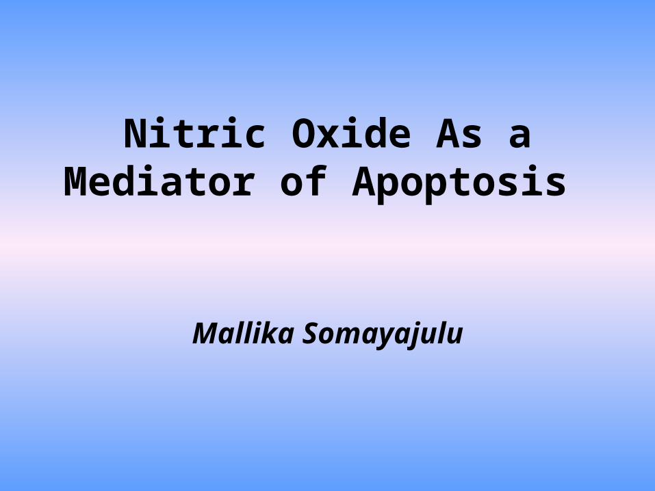
Nitric Oxide As a Mediator of Apoptosis
Mallika Somayajulu

N O
Introduction
• A diatomic free radical consisting of one atom of nitrogen and one atom of oxygen.
• Highly reactive• Very small so can easily pass between cell
membranes.
Nitric Oxide:

• Nitric oxide is synthesized from L-arginine. • This reaction is catalyzed by nitric oxide synthase.
COO-
C
(CH2)3
NH
C
H2N
H
NH2+
+H3N
Arginine
NOS
NADPH
+ O2
NADP+
COO-
C
(CH2)3
NH
C
H+H3N
N+
H2NH
OH
N-w-Hydroxyarginine
COO-
C
(CH2)3
NH
H+H3N + NO
NOS
C
O NH2
Citrulline
Synthesis of Nitric Oxide

NOS
n NOSe NOS i NOS
• Activated by increased Ca+2.
• Seen in macrophages after stimulation of inflammatory/immune reaction.

Effects of NO
Direct
-Is because of NO
•Metal complexes
-Can activate and inactivate many proteins
•Lipid radicals
Indirect
-Is due to formation of N203
And ONOO-
•Nitrosation
•DNA strand breaks
•Nitration

NO as an anti-apoptotic agent

Apoptosis
• Programmed cell death.
• A physiological process in response to a specific
stimuli.
• A cellular process regulated extrinsically and
intrinsically.
• An active process in which specific genes are
involved.
• May occur through specific signaling pathways.

Physiological Significance
• Excessive cell death causes neurodegenerative diseases such as Alzheimer’s and Parkinson’s diseases.• Inhibition of cell death can also lead to hyperproliferative disease such as cancer.

Apoptotic Pathway

NO Signaling in Apoptosis
(Biochemical and biophysical Research Communications 2001)

Nitric oxide induced apoptosis-
Is it p53 dependent?

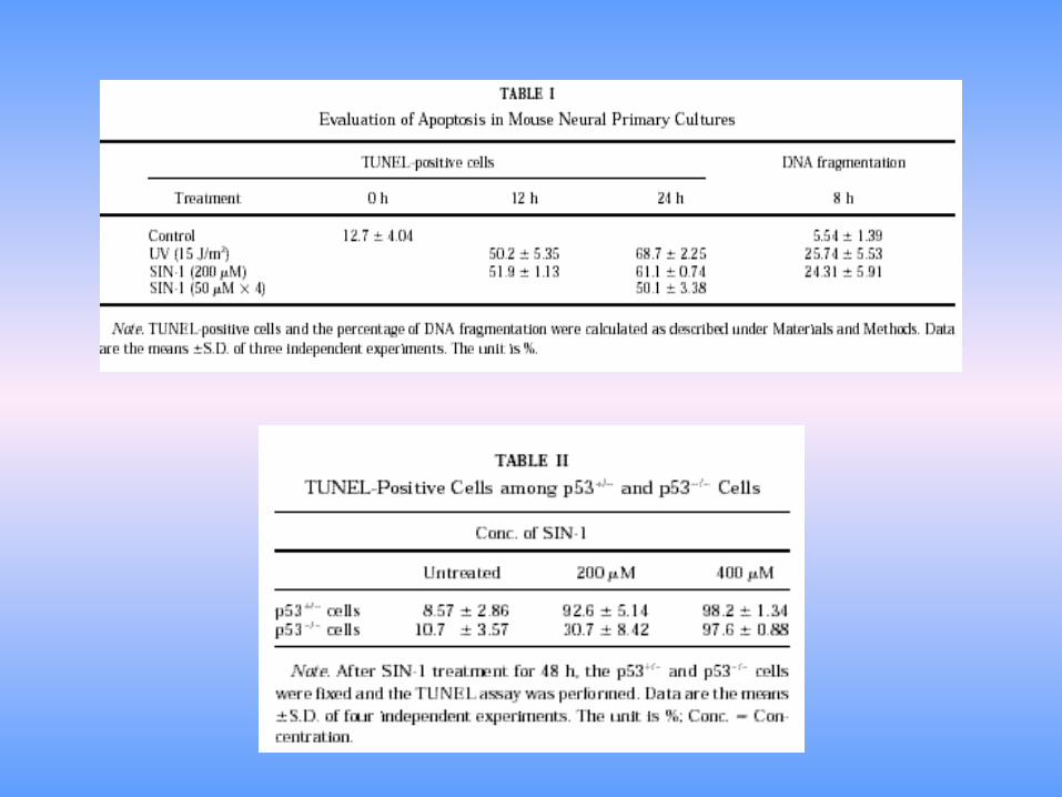
NO Induces Apoptosis in Murine Primary Neural
Cells by a p53 Dependent Pathway
Kaji T.,Kaieda I., HisatsuneT., Kaminogawa S., Nitric Oxide(2002)

•Neural cells from p53 positive and p53 knock out mice were treated with SIN-1 and apoptosis was studied.
•Mechanism for p53 accumulation by SIN-1 was analyzed.
•Western blot revealed that p53 accumulation didn’t require p53 phosphorylation.
•P53 accumulation by ras-MAPK-p19 pathway.

P53
+MDM2p53
Active
Phosphorylation
Inactive
• MDM2 binds to N-terminal region of p53 inhibits p53 mediated transcription

DNA damage and p53
•DNA damage activates the DNA dependent protein kinases.
•Phosphorylation of Ser (15) in the N terminal.
•Mdm2 dissociates from p53 , making it active.
•P53 induces the transcription of Bax and ultimately leads to apoptosis.
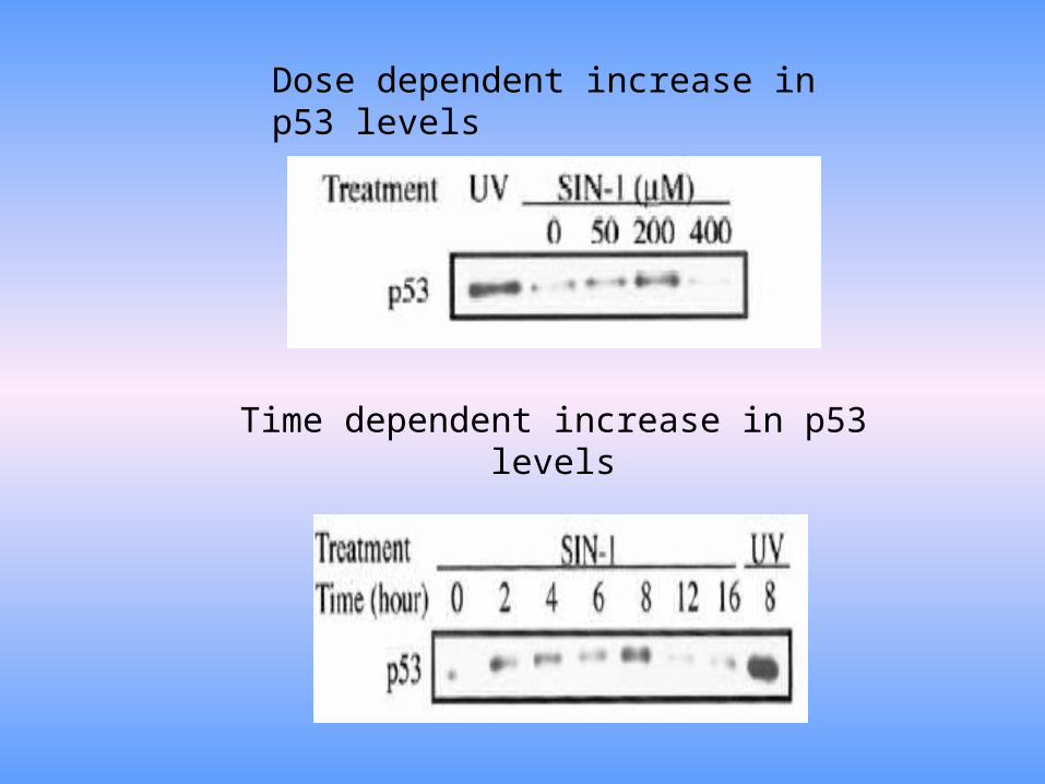
Dose dependent increase in p53 levels
Time dependent increase in p53 levels

• SIN-1 induced p53 accumulation is not through DNA damage.

Based on the reports:
• NO stimulates p21 ras and MAPK activity.(1)
• p21 ras serves as a signal target of reactive free radicals.(2)
• p21 ras induces p53 accumulation via p19.(3)
• p19 functions as a bridge between p21 ras and p53 accumulation.(4)
The Postulated Existence of Another Pathway

ERK activation U0126 and p53
Lovastatin and p53 Northern Blotting

Levels of expression of p53 – related mRNA after treatment with 200μM SIN-1

PATHWAYras
MEK
ERK1/2
p19
Mdm2-p53 p53 +Mdm2
Apoptosis

Apoptosis Occurs in p53 Deficient MG5 Microglial Cells by NO Through ER
Stress
Kawahara K., Oyadomari S. , Gotoh T, Koshsaka S., Nakayama H., Mori M., FEBS (2001)135-139

ER Mediated Stress
• NO inhibits Ca-ATPase activity of ER by tyrosine Nitration .
• Activation of ryanodine receptor Ca+2 channel by nitrosylation of cysteine residues.
• Cells overcome ER stress by unfolding the proteins in the ER lumen.
• Involves the up regulation of different ER chaperones like Bip, Grp78 and CHOP.

• C/EBP Homologous Protein.
• Transcription factor.
• Belongs to the family of Leucine zipper proteins.
• Have extensive amino acid homology with the DNA binding
domain of C/EBP.
• Expressed at undetectable levels in growing mammalian cells.
• Considerably high under stress and treatment with genotoxic
agents.
CHOP

Experiment
• Murine microglial cells were exposed to LPS(1μg/ml) and INF-γ (100U/ml) and mRNA and protein for iNOS and CHOP were induced and apoptosis occurred.
• CHOP and Bip mRNAs as well as proteins were expressed when cells were treated with SIN-1 and SNAP.
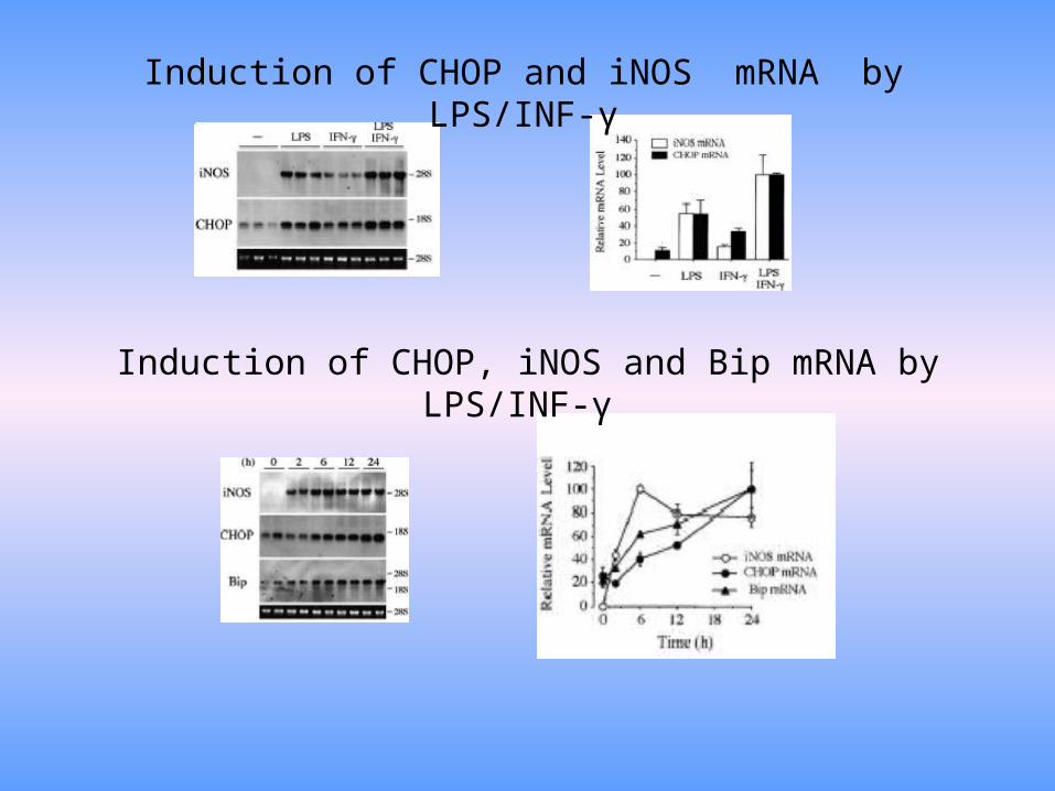
Induction of CHOP and iNOS mRNA by LPS/INF-γ
Induction of CHOP, iNOS and Bip mRNA by LPS/INF-γ

Induction of iNOS and CHOP Protein by LPS/INF-γ
Morphological changes induced by LPS/INF- γ
0 hours
24 hours
48 hours

DNA Fragmentation Caspase–3 like activity

Induction of CHOP and Bip by SNAP
Induction of CHOP and Bip by SIN-1

Morphological changes Caspase-3 like activity
0 hours
24 hours
48 hours

• Several genes on which CHOP could act downstream have been identified down stream of CHOP.
• But none is involved in programmed cell death.
• CHOP expression down regulates Bcl-2 expression, depletion of cellular glutathione, increases production of ROS.
(Mc Cullough et al, (2001)Mol.Cell.Biol.21, 1249-1259)
• Precise apoptotic cascade downstream has not been clarified.

Conclusions
• NO and peroxy nitrite induce apoptosis in different types of neural cells.
• Basis for new therapeutic intervention in various pathological states of the brain where NO plays an important role in development of cell injury.

References:1) Chung ,H.T. et al (2001)No as a bioregulator of apoptosis. Biochem.
Biophys.Res.Comm 282, 1075-1079.
2) Palmero,I.Pantoja,C.,and Serrano,M. (1998).p19ARFlinks the tumour suppressor p53 to ras.Nature 395, 125-126
3) Lander,H.M.et al(1995)p21 ras as a common signaling target of reactive free radicals and cellular redox stress.J.Biol.Chem 270,21195-21198
4) Sherr,C.J.,and Weber,J.D.(2000).The ARF/p53 pathway.Curr.Opin.Genet.Dev.10,94-99.
5) Ries,S.,Biederer,C.,Woods,D.,Shifman,O.Shirasawa,S.,Sasazuki,T.,McMahon,M.,Oren,M., and McCormick,F.(2000).Opposing effects of ras on p53 transcriptional activation of mdm2 and introduction of p19 ARF.Cell 103,321-330.
6) Matsumoto,M.Minami,M.,Takeda,K., Sakao,Y.,Akira,S.,(1996)Ectopic expression of Chop induces apoptosis in M1 leukemia cells.FEBS Letters 395, 123-147