nipah virus assays and animal models for vaccine development
TRANSCRIPT
Nipah Virus Assays and Animal Models for Vaccine Development
Landscape Analysis, January 2021
Author:Albert Price, IAVI
Secondary authors/reviewers:Donata Sizemore, IAVIThomas Hassell, IAVIRaúl Gómez Román, CEPIJohan Holst, CEPIPaul Kristiansen, CEPI

Nipah Virus Assays and Animal Models for Vaccine Development, Landscape Analysis, January 20212
I. Introduction 4
II. Background 5
1. Epidemiology 5
2. NiV Clinical Features and Pathogenesis in Humans 9
3. Diagnosis and Treatment 10
4. NiV Molecular Biology and Structure 11
5. Vaccine Development 13
III. Standardization of Assays and Animal Models 18
IV. NiV Serological Assays 21
1.Detectionofantigen–specificserumIgG 21
2. Detection of serum neutralizing antibodies 22
V. NiV Animal Models 27
1.SyrianGoldenHamster 29
2. Ferret 32
3.AfricanGreenMonkey(AGM) 34
VI. Conclusions 38
VII. References 40
VIII. Statement of Support 46
1. Article H.20. Publication And Publicity 46
TABLE OF CONTENTS
Nipah Virus Assays and Animal Models for Vaccine Development, Landscape Analysis, January 20213
Figure 1.GeographicdistributionofPteropidfruitbats andHenipavirusoutbreaks(EncheryandHorvat,2017) 5
Figure 2.NiVstructureandorganizationofthe18.2kBssRNA (-)genome(Sunetal.,2018). 11
Figure 3. The Henipavirus infection and replication cycle (AguilarandLee,2011). 12
Table 1.NiVOutbreaksbyYearandLocation* 7
Table 2.DifferencesinClinicalandEpidemiologicalCharacteristics BetweenNiVMalaysiaandBangladeshOutbreaks 8
Table 3.NiVViral-vectorVaccineCandidatesTestedinAnimals 15
Table 4. NiV Submit Vaccine Candidates Tested in Animals 16
Table 5.NiVvaccinecandidatessupportedbyCEPI 17
Table 6.BenefitsandPotentialChallengesofImplementing Biological Standards for NiV Vaccine Development 20
Table 7. Pros and Cons of NiV Serological Assays 24
Table 8.SerologicalAssaysUsedinNiVPre-ClinicalVaccineStudies 25
Table 9.SerologicalAssaysUsedinOtherNiVResearchStudies 26
Table 10. Summary of clinical signs and pathology in the NiV hamster, ferretandAGMchallengemodels 28
Table 11. NiV Hamster Model Challenge Studies 30
Table 12. NiV Hamster Model Challenge Studies, Continued 31
Table 13. NiV Ferret Model Challenge Studies 33
Table 14.NiVAfricanGreenMonkeyModelChallengeStudies 35
Table 15.NiVAfricanGreenMonkeyModelChallengeStudies,Continued 36
Table 16. Pros and Cons of the Major NiV Animal Challenge Models 37
LIST OF FIGURES
LIST OF TABLES
Nipah Virus Assays and Animal Models for Vaccine Development, Landscape Analysis, January 20214
1 https://www.who.int/blueprint/priority-diseases/key-action/Nipah_virus_vaccineTPP.pdf2 https://cepi.net/research_dev/priority-diseases/
I. INTRODUCTIONNipah is an emerging, zoonotic viral disease that causes severe neurologic and respiratory symptoms and has an overall case fatality rate of 59%.
Although relatively rare and currently confined to sporadic outbreaks in southern and southeast Asia (including Malaysia, Singapore, Bangladesh and India), the extreme virulence, lack of a vaccine or effective therapeutic options, broad species tropism and wide geographical distribution of the Nipah virus’ (NiV) primary animal reservoir (Pteropid fruit bats) led the World Health Organization (WHO) to label NiV a “Priority Pathogen” for the development of effective medical countermeasures (MCMs), and in 2017 developed a Target Product Profile (TPP) for a NiV vaccine.1 The Coalition for Epidemic Preparedness Innovations (CEPI) has selected NiV as one of seven emerging infectious diseases
currently targeted for development of prophylactic vaccines as an urgent priority2 and is actively supporting efforts toward a protective NiV vaccine.
Development of new vaccines against any disease is most efficient when there is standardization of key R&D tools, particularly analytical methods, reagents and animal models, so that experimental results from different investigators and developers can be directly and confidently compared. CEPI has identified a set of research and development activities needed to accelerate vaccine development by promoting standardization, transparency and comparability between vaccine candidates.
To this end, CEPI is focusing on developing biological standards, validating assays and supporting the development and refinement of animal models for three emerging diseases in its vaccine development portfolio: Nipah, MERS-CoV and Lassa. The purpose of this Landscape Analysis, supported by NIH/NIAID/DMID and prepared for CEPI, is to analyze the current state of NiV assays and animal models currently in use within the context of NiV biology, epidemiology, and vaccine development. This document will serve both as an internal resource at CEPI to guide scientific discussions and as an external resource to inform the Nipah scientific community.
Nipah Virus Assays and Animal Models for Vaccine Development, Landscape Analysis, January 20215
3 Epstein et al. PNAS 2020: https://doi.org/10.1073/pnas.2000429117
II. BACKGROUND
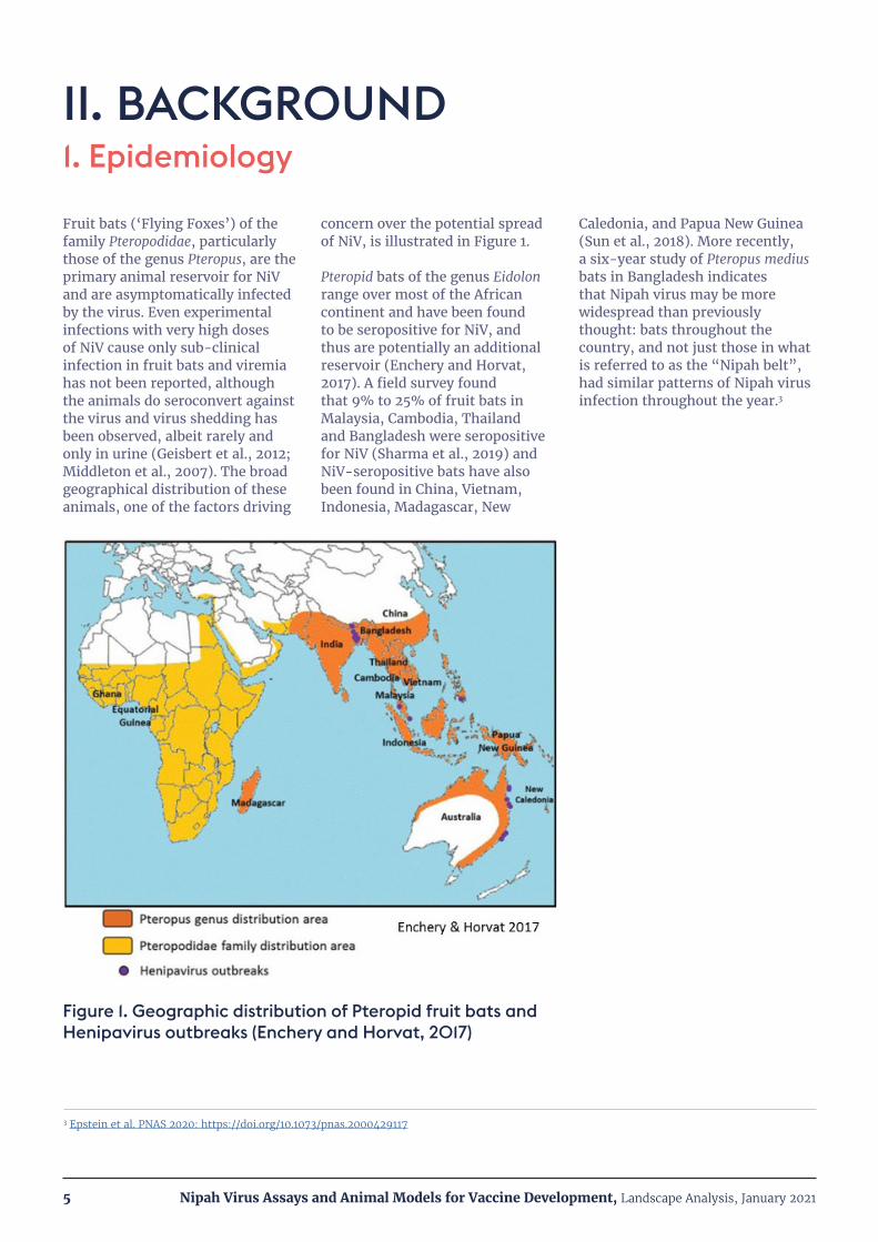
Fruit bats (‘Flying Foxes’) of the family Pteropodidae, particularly those of the genus Pteropus, are the primary animal reservoir for NiV and are asymptomatically infected by the virus. Even experimental infections with very high doses of NiV cause only sub-clinical infection in fruit bats and viremia has not been reported, although the animals do seroconvert against the virus and virus shedding has been observed, albeit rarely and only in urine (Geisbert et al., 2012; Middleton et al., 2007). The broad geographical distribution of these animals, one of the factors driving
concern over the potential spread of NiV, is illustrated in Figure 1.
Pteropid bats of the genus Eidolon range over most of the African continent and have been found to be seropositive for NiV, and thus are potentially an additional reservoir (Enchery and Horvat, 2017). A field survey found that 9% to 25% of fruit bats in Malaysia, Cambodia, Thailand and Bangladesh were seropositive for NiV (Sharma et al., 2019) and NiV-seropositive bats have also been found in China, Vietnam, Indonesia, Madagascar, New
Caledonia, and Papua New Guinea (Sun et al., 2018). More recently, a six-year study of Pteropus medius bats in Bangladesh indicates that Nipah virus may be more widespread than previously thought: bats throughout the country, and not just those in what is referred to as the “Nipah belt”, had similar patterns of Nipah virus infection throughout the year.3
1. Epidemiology
Figure 1. Geographic distribution of Pteropid fruit bats and Henipavirus outbreaks (Enchery and Horvat, 2017)

Nipah Virus Assays and Animal Models for Vaccine Development, Landscape Analysis, January 20216
A number of domesticated animals – pigs, dogs, cats and horses – can be infected by NiV (Geisbert et al., 2012), but the virulence and rate of infection is variable. Mortality in suckling pigs is high (40%) and 1-6 month-old pigs show respiratory and neurological symptoms, but mortality is less than 5%. Adult pigs show less serious respiratory signs and mortality is rare (McLean and Graham, 2019). During NiV outbreaks in Malaysia many dogs on pig farms were found to be NiV-seropositive, but only 2 had active disease (Hooper et al., 2001). Horses can be infected by NiV (Hooper et al., 2001), most likely from eating fruit contaminated by bats, and were intermediate hosts in an outbreak in the Philippines in 2014. During that outbreak investigations found disease among horses including neurological signs and 10 deaths. Four (4) cats and a dog were also likely infected by eating horse meat and died (Ching et al., 2015). However, no published reports of cats or dogs serving as intermediate hosts for transmission of NiV have been found.
The locations and human fatality rates of all NiV outbreaks to date are summarized in Table 1. The first reported NiV outbreaks occurred in Malaysia and Singapore in 1998-99. In those outbreaks the majority of human cases were due to contact with infected pigs that had acquired NiV by eating fruit contaminated by bat saliva, urine, or feces. Humans (mostly pig farmers and slaughterhouse workers) acquired NiV from infected pig urine or respiratory secretions. A majority of cases were characterized by an acute encephalitic syndrome. The case fatality rate was 40% (see Table 1) and, at the time, there was inconclusive evidence of human-to-human transmission (Ahmad
and Tan, 2014). In the 2014 NiV outbreak in the Philippines (Malaysia strain), the majority of cases were acquired by eating contaminated horse meat or participating in slaughtering horses, and the rest were likely due to human to human transmission. 65% of the cases presented with an acute encephalitic syndrome and the overall case fatality rate was 53% (Ching et al., 2015).
The NiV outbreaks in Bangladesh and India beginning in 2001 showed a distinct pattern of transmission and symptomology. Humans were infected by drinking raw date palm sap contaminated with bat saliva or urine, and there was no intermediate animal host. There was a higher incidence of respiratory illness (69% and a higher fatality rate (75%; see Table 1). Patients infected with the Bangladesh strain (NiV-B) had higher NiV RNA levels in the blood and more virus in oral secretions (Hossain et al., 2008). Finally, there was evidence of human-to-human transmission, primarily to healthcare workers or family caregivers, in the Indian and Bangladeshi outbreaks (Chadha et al., 2006). The mechanism(s) of human to human transmission has not been conclusively established, but exposure to bodily fluids (saliva, cough, vomit, blood) elevates the risk of transmission compared to physical contact alone or being near the patient (Kumar et al., 2019). Experiments in non-human primates have demonstrated infection via small and medium particle size aerosols (Cong et al., 2017; Hammoud et al., 2018). Comparisons in transmission rates between the Malaysia and Bangladesh outbreaks have not been found in the published literature.

Nipah Virus Assays and Animal Models for Vaccine Development, Landscape Analysis, January 20217
Month/Year Country Location Number of Cases
Number of Deaths
Case Fatality (%)
Sep 1998 – Apr 1999 Malaysia Perak, Selangor, Negeri Sebilan 265 105 40
Mar 1999 Singapore Singapore 11 1 9
Jan – Feb 2001 India Siliguri 66 45 68
Apr – May 2001
Bangladesh
Meherpur 13 9 69
Jan 2003 Naogaon 12 8 67
Jan 2004 Rajbari 31 23 74
Apr 2004 Faridpur 36 27 75
Jan – Mar 2005 Tangail 12 11 92
Jan – Feb 2007 Thakurgaon 7 3 43
Mar 2007 Kushtia, Pabna, Tatore 8 5 63
Apr 2007 Naogaon 3 1 33
Apr 2007 India Nadia 5 5 100
Feb 2008
Bangladesh
Manikgon 4 4 100
Apr 2008 Rajbari, Faridpur 7 5 71
Jan 2009 Gaibandha, Rangpur, Nilphamari 3 0 0
Jan 2009 Rajbari 1 1 100
Feb – Mar 2010 Faridpur, Rajbari, Gopalganj, Madaripur 16 14 87.5
Jan – Feb 2011 Lalmohirhat, Dinajpur, Comilla, Nilphamari , Rangpur 44 40 91
Feb 2012 Joypurhat, Rajshahi, Tatore, Rajbari, Gopalganj 12 10 83
Jan – Feb 2013 Gaibandha, Natore, Rajshahi,Naogaon, Rajbari, Pabna, Jhenaidah, Mymensingh 24 21 87.5
Feb 2014Manikganj, Magura, Faridpur, Rangpur, Shaariatpur, Kushtia, Rajshahi, Tatore, Dinajpur, Chapai Nawabganj, Naogaon
18 9 50
Mar – May 2014 Philippines Tinalon, Midtungok 17 9 53
Feb 2015 Bangladesh Nilphamari, Pnchoghor, Faridpur, Magura, Naugaon, Rajbari 9 6 67
May 2018 India Kozhikode, Malappuram 19 17 89
Overall Total 643 379 58.9
Malaysia/Singapore/Philippines Total 293 115 39.2
Bangladesh/India Total 350 264 75.4
*Adapted from (Thakur and Bailey, 2019) and (Sharma et al., 2019)
Table 1. NiV Outbreaks by Year and Location*

Nipah Virus Assays and Animal Models for Vaccine Development, Landscape Analysis, January 20218
A comparison of key clinical and epidemiological characteristics of NiV outbreaks in Malaysia and Bangladesh is compiled in Table 2. Three clinical characteristics between the outbreaks stand out. The first is the shorter time from disease onset to death (7 days vs. 16 days) and higher case fatality rate (74% vs. 38%; Table 1) in the Bangladesh vs. the Malaysia outbreaks (Ahmad and Tan, 2014; Ang et al., 2018; Hossain et al., 2008; Lo and Rota, 2008). The second is the higher incidence of respiratory involvement in
the Bangladesh outbreaks. This, coupled with the higher level of NiV-B RNA in oral secretions could be linked to the higher level of human to human transmission, which is likely via oral/respiratory secretions or bodily fluids (Ahmad and Tan, 2014). Finally, in the Malaysia outbreak there was a high incidence of segmented myoclonus (muscle jerking), which was not reported in the Bangladesh outbreaks, although other indications of encephalitis or neurological involvement (such as altered mental status, hyporeflexia
and convulsions) were seen in the Bangladesh outbreaks at rates comparable to the Malaysia cases (Ang et al., 2018; Hossain et al., 2008). This phenomenon is largely unexplained but could reflect involvement of different areas of the central nervous system (CNS) due to differences in virus tropism, differences in the route of infection or slower disease progression allowing infection of different neural tissues.
Characteristic Malaysia-Singapore Bangladesh-India
Transmission• Bat to pig → pig to human
• Rare human to human
• Bat to human via consumption of contaminated date palm juice and fruits.
• Human to human
Fever 95% 100%
Headache 75% 73%
Vomiting 32% 58%
Diarrhea 18% 29%
Respiratory involvement 14-29% 62-69%
Encephalitis/neurological involvement
• Segmental myoclonus 32-54%
• Hyporeflexia 60.5%
• Convulsion 23%
• Altered mental status 72%
• Segmental myoclonus not reported
• Hyporeflexia 65%
• Convulsion 23%
• Altered mental status 100%
MRI Disseminated small, high-signal-intensity lesions
Confluent high-signal brain lesions (limited MRIs were performed)
Relapsed and late-onset encephalitis ~5-10% 4 out of 22 patients (18%) in a follow-up study
Persistent neurological deficits ~20% ~30%
Incubation Period Mean = 10 days 6-11 days
Average (mean) time from disease onset to death 16 days 7 days
Table 2. Differences in Clinical and Epidemiological Characteristics Between NiV Malaysia and Bangladesh Outbreaks
Sources: (Ang et al., 2018; Hossain et al., 2008); (Ahmad and Tan, 2014; Chong et al., 2002; Lo and Rota, 2008)
Nipah Virus Assays and Animal Models for Vaccine Development, Landscape Analysis, January 20219
4 Conference Proceedings: https://cepi.net/wp-content/uploads/2020/06/2019-CEPI-Duke-WHO-NIAID-Nipah-Conference_FINAL.pdf.
Further clinical commonalities and differences in Nipah infections across countries were discussed during the NIAID co-sponsored Nipah@20 Conference in Singapore, December 20204. The cause of the differences in disease between the Malaysia/Singapore and Bangladesh/India outbreaks remains uncertain and is complicated by the possibility of differing diagnostic methods and case definitions. Experiments in animal models have demonstrated some differences in disease course and symptomology between the Malaysia and Bangladesh strains, suggesting a genetic component:
• Hamsters infected with NiV-M showed accelerated virus replication, pathology and death compared to hamsters infected with equivalent doses of NiV-B (DeBuysscher et al., 2013). This finding is opposite to the human fatality rates in the Malaysia and Bangladesh outbreaks.
• Ferrets infected with NiV-B showed comparable disease progression, and histopathology of the lungs and CNS compared to ferrets infected with NiV-M. However, NiV-B infected ferrets shed more virus in oral secretions than NiV-M infected animals (Clayton et al 2012). Increased human shedding of NiV-B was seen in the Bangladesh outbreaks (Hossain et al., 2008).
• African Green Monkeys (AGM) Lethality of NiV-B was 100%, compared to 50% for monkeys infected with an equivalent dose of NiV-M. NiV-B also causes more severe lung histopathology than NiV-M and has a shorter window for therapy with the monoclonal antibody m102.4 (Mire et al., 2016).
AGMs appear to reproduce the NiV strain differences in virulence and pathology seen in humans more faithfully than hamster and ferrets, and the results suggest that the distinct clinical characteristics and epidemiology seen in the Malaysia and Bangladesh outbreaks is at least partly genetic. However, geographic differences in virus transmission (including route of infection and dose), population health and the quality of subsequent health care may also play a role. More information on the major animal challenge models for NiV is presented in Section V.
The initial clinical presentation of NiV infection (by either strain) is non-specific and characterized by flu-like symptoms including fever, headache, dizziness, myalgia, and loose stools (Banerjee et al., 2019). Mild or asymptomatic infections have also been reported in various outbreaks, but the overall incidence is relatively low and appears to be strain dependent, with the Malaysia strain causing less severe illness, a lower case fatality rate and higher prevalence of asymptomatic infections than the Bangladesh strain (Kumar et al., 2019). The incubation period ranges from 7 to 40 days and from onset the disease progresses
rapidly to an encephalitic syndrome in approximately 60 percent of patients. The time course of disease progression from initial symptoms to the encephalitic syndrome has not been reported in detail. Neurological symptoms include meningismus (central nervous system inflammation) and seizures in approximately one-third of patients. A deterioration in consciousness, coma and death typically occur within an average of 7 days of disease onset (Bangladesh outbreaks) or an average of 16 days (Malaysia outbreak) (Ang et al., 2018; Hossain et al., 2008; Rahman and Chakraborty, 2012). Approximately 20-30% of NiV
encephalitis survivors suffer long-term neurologic dysfunction characterized by persistent seizures, disabling fatigue and behavioral abnormalities (Mazzola and Kelly-Cirino, 2019; Sejvar et al., 2007). In addition, some patients with initially mild, non-encephalitic disease develop a late-onset or recurrent neurological disease (Ramphul et al., 2018). Some patients also present with severe respiratory symptoms, and respiratory involvement has been more common in the Bangladesh outbreaks compared to the Malaysia outbreaks (see Epidemiology).
2. NiV Clinical Features and Pathogenesis in Humans
Nipah Virus Assays and Animal Models for Vaccine Development, Landscape Analysis, January 202110
Although the route of infection in humans has not been conclusively determined, work with experimental animal challenge models has shown that inhalation of NiV virus particles is sufficient to initiate infection (Cong et al., 2017; Hammoud et al., 2018). In humans, early infection appears to occur in lung epithelial cells, and in later stages moves to lung endothelial cells. Vasculitis in small blood vessels may be present
throughout the body, and the virus may then enter the blood stream and disseminate throughout the host, infecting the brain, spleen and kidneys (Escaffre et al., 2013). Experiments in hamsters suggest that entry to the central nervous system (CNS) may occur either through olfactory neurons or via the choroid plexus and cerebral blood vessels (Baseler et al., 2016; Munster et al., 2012). Infection of the human CNS is characterized
by severe vasculitis and syncytia formation, resulting in endothelial damage due to vasculitis-induced thrombosis and the presence of viral inclusion bodies. Necrotic plaques are found in both grey and white matter of the CNS (Escaffre et al., 2013). Lessons learned from pathology and disease course in humans were discussed in the Transmission/Case Management Session of the Nipah@20 Conference.
Laboratory diagnosis of NiV infection can be performed using a variety of nucleic acid-based or serological assays. The currently preferred methods for detecting active NiV infection are PCR-based tests such as conventional reverse transcriptase (RT) PCR, nested RT-PCR and real-time PCR (qPCR). PCR-based tests usually target the conserved N, M or P viral genes. ELISA assays detecting IgM against NiV antigens are typically the first-line serological tests for NiV infection (Mazzola and Kelly-Cirino, 2019). Progress and challenges in diagnostics, including a presentation on the WHO Nipah diagnostics Target Product Profile, were featured in Session 4 of the Nipah@20 Conference.
Treatment of NiV infection consists primarily of supportive care including maintaining fluids, anticonvulsants, treatment of secondary infection and mechanical ventilation. (Ang et al., 2018). No effective therapeutics for NiV infection are currently approved for use in humans. The antiviral drug ribavirin was administered to 140 patients during the 1998-99 NiV Malaysia outbreak, resulting in a 36% reduction in mortality compared to 52 untreated control patients (Chong et al., 2001). However, the treatment allocation
was not randomized and the treated patients may have received better overall care, thus making the outcome uncertain (Banerjee et al., 2019). Subsequent animal challenge studies in hamsters showed that ribavirin delayed, but did not prevent, death after NiV infection (Freiberg et al., 2010; Georges-Courbot et al., 2006). A similar result was obtained after infection of African Green Monkeys with the closely related Hendra Virus (HeV; (Rockx et al., 2010)).
The broad-spectrum antiviral drug Remdesivir (GS-5734) was recently shown to protect African Green Monkeys when administered 24 hours post-inoculation with a lethal dose of NiV (Bangladesh strain) (Lo et al., 2019). Treated animals developed mild respiratory symptoms, reduced appetite and showed local virus replication but no viremia. All the Remdesivir-treated animals recovered fully, while all the control-treated animals succumbed to the infection.
A human monoclonal antibody, m102.4, targeting the Ephrin-B2/B3 binding site on the NiV and HeV G glycoproteins (see NiV Molecular Biology and Structure), has been tested in NiV animal models for prophylactic and therapeutic use.
In a lethal challenge study in African Green Monkeys (AGMs), monkeys could be successfully treated up to 5 days post-infection with the NiV Malaysia strain (NiV-M). Although half of the treated monkeys developed overt clinical signs (fever, respiratory and neurological), all the animals fully recovered (Geisbert et al., 2014). The window for successful treatment with m102.4 is only 3 days post-infection when AGMs are challenged with the NiV Bangladesh strain (NiV-B) (Mire et al., 2016). These results suggest that m102.4 may have utility as a post-exposure prophylactic or therapeutic in humans. The m102.4 antibody has also been administered on an emergency basis as post-exposure prophylaxis to a handful of humans in cases of high risk of exposure to NiV or HeV (Broder et al., 2013). In all cases the patients did not become ill, but it is impossible to know if illness was prevented by the antibody.
3. Diagnosis and Treatment
Nipah Virus Assays and Animal Models for Vaccine Development, Landscape Analysis, January 202111
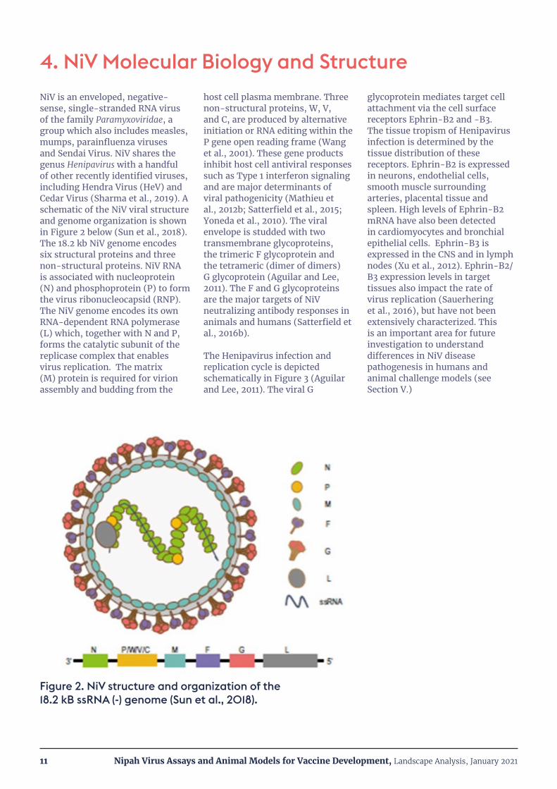
NiV is an enveloped, negative-sense, single-stranded RNA virus of the family Paramyxoviridae, a group which also includes measles, mumps, parainfluenza viruses and Sendai Virus. NiV shares the genus Henipavirus with a handful of other recently identified viruses, including Hendra Virus (HeV) and Cedar Virus (Sharma et al., 2019). A schematic of the NiV viral structure and genome organization is shown in Figure 2 below (Sun et al., 2018). The 18.2 kb NiV genome encodes six structural proteins and three non-structural proteins. NiV RNA is associated with nucleoprotein (N) and phosphoprotein (P) to form the virus ribonucleocapsid (RNP). The NiV genome encodes its own RNA-dependent RNA polymerase (L) which, together with N and P, forms the catalytic subunit of the replicase complex that enables virus replication. The matrix (M) protein is required for virion assembly and budding from the
host cell plasma membrane. Three non-structural proteins, W, V, and C, are produced by alternative initiation or RNA editing within the P gene open reading frame (Wang et al., 2001). These gene products inhibit host cell antiviral responses such as Type 1 interferon signaling and are major determinants of viral pathogenicity (Mathieu et al., 2012b; Satterfield et al., 2015; Yoneda et al., 2010). The viral envelope is studded with two transmembrane glycoproteins, the trimeric F glycoprotein and the tetrameric (dimer of dimers) G glycoprotein (Aguilar and Lee, 2011). The F and G glycoproteins are the major targets of NiV neutralizing antibody responses in animals and humans (Satterfield et al., 2016b).
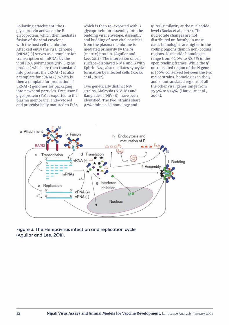
The Henipavirus infection and replication cycle is depicted schematically in Figure 3 (Aguilar and Lee, 2011). The viral G
glycoprotein mediates target cell attachment via the cell surface receptors Ephrin-B2 and -B3. The tissue tropism of Henipavirus infection is determined by the tissue distribution of these receptors. Ephrin-B2 is expressed in neurons, endothelial cells, smooth muscle surrounding arteries, placental tissue and spleen. High levels of Ephrin-B2 mRNA have also been detected in cardiomyocytes and bronchial epithelial cells. Ephrin-B3 is expressed in the CNS and in lymph nodes (Xu et al., 2012). Ephrin-B2/B3 expression levels in target tissues also impact the rate of virus replication (Sauerhering et al., 2016), but have not been extensively characterized. This is an important area for future investigation to understand differences in NiV disease pathogenesis in humans and animal challenge models (see Section V.)
4. NiV Molecular Biology and Structure
Figure 2. NiV structure and organization of the 18.2 kB ssRNA (-) genome (Sun et al., 2018).
Nipah Virus Assays and Animal Models for Vaccine Development, Landscape Analysis, January 202112
Following attachment, the G glycoprotein activates the F glycoprotein, which then mediates fusion of the viral envelope with the host cell membrane. After cell entry the viral genome [vRNA(-)] serves as a template for transcription of mRNAs by the viral RNA polymerase (NiV L gene product) which are then translated into proteins, the vRNA(-) is also a template for cRNA(+), which is then a template for production of vRNA(-) genomes for packaging into new viral particles. Precursor F glycoprotein (Fo) is exported to the plasma membrane, endocytosed and proteolytically matured to F1/2,
which is then re-exported with G glycoprotein for assembly into the budding viral envelope. Assembly and budding of new viral particles from the plasma membrane is mediated primarily by the M (matrix) protein. (Aguilar and Lee, 2011). The interaction of cell surface-displayed NiV F and G with Ephrin B2/3 also mediates syncytia formation by infected cells (Rockx et al., 2012).
Two genetically distinct NiV strains, Malaysia (NiV-M) and Bangladesh (NiV-B), have been identified. The two strains share 92% amino acid homology and
91.8% similarity at the nucleotide level (Rockx et al., 2012). The nucleotide changes are not distributed uniformly; in most cases homologies are higher in the coding regions than in non-coding regions. Nucleotide homologies range from 92.0% to 98.5% in the open reading frames. While the 5’ untranslated region of the N gene is 100% conserved between the two major strains, homologies in the 5’ and 3’ untranslated regions of all the other viral genes range from 75.5% to 91.4% (Harcourt et al., 2005).
Figure 3. The Henipavirus infection and replication cycle (Aguilar and Lee, 2011).

Nipah Virus Assays and Animal Models for Vaccine Development, Landscape Analysis, January 202113
A number of factors suggest that development of a safe, efficacious human prophylactic vaccine against NiV is scientifically feasible. Natural infection by other paramyxoviruses, such as measles and mumps, results in long-term immunity and vaccines for those diseases have been successfully developed. A vaccine protecting horses against the closely related Hendra Virus (HeV; Equivac®) has been approved for use in Australia (Tan et al., 2018). Passive immunization experiments in a NiV animal challenge model (hamsters) using immune sera and monoclonal antibodies have demonstrated that neutralizing antibodies confer protection against NiV challenge (Guillaume et al., 2004; Guillaume et al., 2006). Finally, as discussed below, multiple modes of active vaccination have resulted in protection from lethal NiV challenge in animal models.
There is broad consensus that neutralizing antibodies confer protection against NiV infection and all vaccine development efforts to date have focused on their elicitation (Broder et al., 2012; Prescott et al., 2012; Satterfield et al., 2016b). However, a correlate of protection based on neutralizing antibody titer has not been defined. Neutralizing antibody titers in animal vaccine challenge studies where protection was conferred are reported in Tables 3 and 4. However, since virtually all animals were protected in these studies a threshold of protection cannot be defined. The role of cell-mediated immune responses (CMI) in either natural immunity or vaccine-induced protection against NiV has received relatively
little attention. One challenge study conducted in pigs suggested that cellular immune responses may be important for achieving full protection, but the mechanism was not defined, and the conclusions are complicated by the fact that, unlike with other animal hosts, NiV infects a range of porcine immune cells (Pickering et al., 2016). More work is needed to elucidate the role of cellular immune responses in protection against NiV infection.
The WHO developed a Target Product Profile (TPP) for a human NiV vaccine, including preferred as well as critical or minimal product characteristics.1 Key vaccine performance attributes recommended in the TPP are:
• Intended use: For reactive use in outbreak settings
• Efficacy: ≥ 90% efficacy in preventing disease (preferred); ≥70% (minimal); rapid onset of protection, less than 2 weeks after the first dose (preferred); protection ≤ 2 weeks after the last dose (minimal).
• Dose Regimen: Single-dose primary series (preferred); no more than 2 doses, with some protection after the first dose (minimal).
• Durability of Protection: ≥ 1 year (preferred); ≥ 6 months (minimal).
• Product Stability and Storage: Shelf life of 5 years at 2-8oC (preferred); shelf life of at least 12 months at -20oC and demonstrated stability of ≥ 1 month at 2-8oC (minimal).
Due to safety issues associated with production (i.e., BSL-4 containment) and administration of a live-attenuated or inactivated NiV vaccine, and the need to elicit neutralizing antibodies, most attempts at NiV vaccine development have focused on recombinant viral vectors and adjuvanted protein subunit vaccines. In all cases the target antigen(s) have been the F and/or G glycoproteins (see Tables 3 and 4).
The NiV vaccines described below are all research-stage candidates focused on demonstrating immunogenicity and protection against lethal NiV challenge. No safety issues associated with vaccination or subsequent virus challenge (due to antibody-dependent disease enhancement; ADE) were reported. However, more in-depth safety studies will necessarily be performed on any NiV vaccine candidates prior to advancing into human clinical testing.
5. Vaccine Development

Nipah Virus Assays and Animal Models for Vaccine Development, Landscape Analysis, January 202114
Viral Vector Candidates
A number of viral vector platforms expressing the NiV F or G glycoproteins have been tested as vaccine candidates. The most widely used vector platform to date has been the Vesicular Stomatitis Virus (VSV). Three types of VSV vectors have been employed: 1) replication-incompetent VSV pseudotypes expressing NiV F or G (Lo et al., 2014); 2) VSV virions expressing NiV F or G that can undergo a single round of replication (Mire et al., 2019); and 3) replication-competent recombinant viruses in which the VSV-G protein is replaced by the Ebola glycoprotein (ZEBOV) and also co-expressing NiV F or G (DeBuysscher et al., 2014; DeBuysscher et al., 2016; Prescott et al., 2015). All three VSV-vaccine types, whether expressing NiV F or G antigens and administered singly, or co-administered, elicited neutralizing antibodies and fully protected immunized animals from clinical disease in at least one of the 3 major NiV lethal challenge models (hamsters, ferrets or non-human primates; see Section V). Additionally, all three VSV vaccine types conferred protection after a single dose (see Table 3). Additional details on the animal challenge models and their use in vaccine studies are given in Section V.
Other recombinant viral vector vaccine platforms expressing NiV F or G have been tested, including: vaccinia virus (Guillaume et al., 2004), canarypox (ALVAC) (Weingartl et al., 2006), Measles virus (Yoneda et al., 2013), Venezuelan Equine Encephalitis Virus (VEEV) (Defang et al., 2010), Rabies virus (Keshwara et al., 2019), Newcastle disease virus (Kong et al., 2012), Adeno Associated Virus (AAV) (Ploquin et al., 2013) and chimpanzee adenovirus (ChAd; (van Doremalen et al., 2019)). All these candidates conferred full protection against lethal challenge and/or elicited high titers of neutralizing antibodies. However, only the AAV and chimpanzee adenovirus (ChAd) vectored vaccines reported protection after a single vaccination. A summary of NiV viral vector vaccine candidates tested in animals is shown in Table 3.
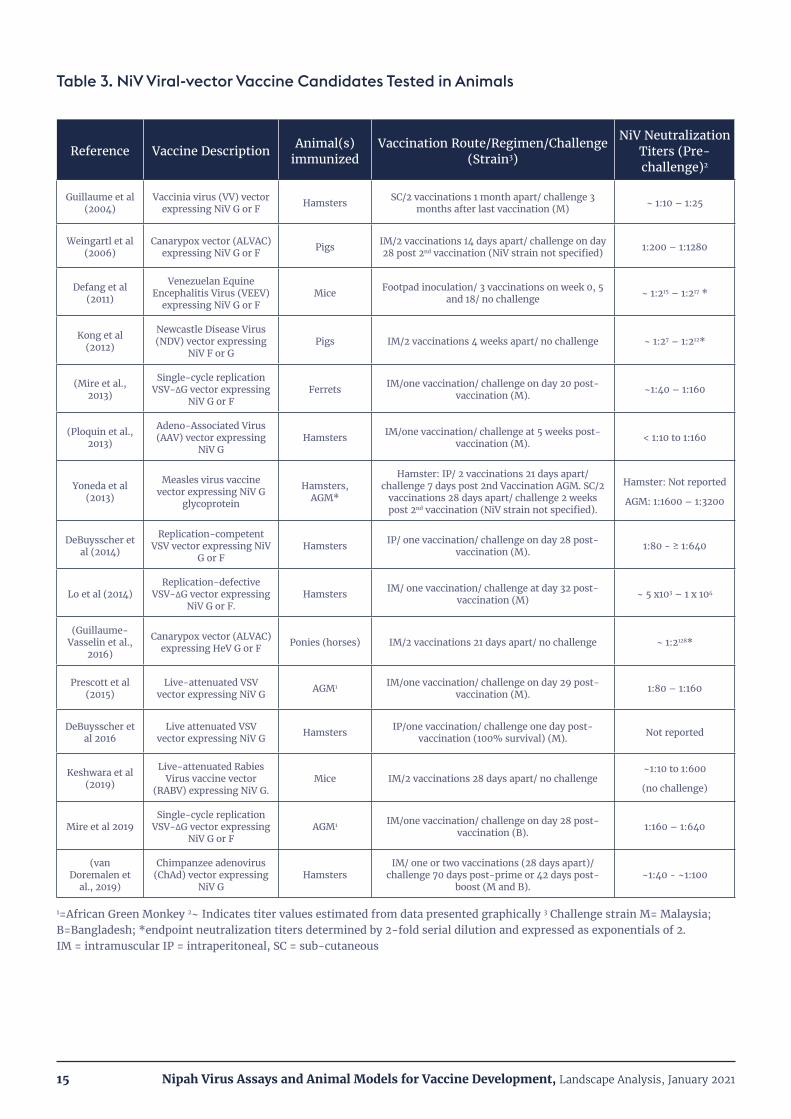
Nipah Virus Assays and Animal Models for Vaccine Development, Landscape Analysis, January 202115
Reference Vaccine Description Animal(s) immunized
Vaccination Route/Regimen/Challenge (Strain3)
NiV Neutralization Titers (Pre-challenge)2
Guillaume et al (2004)
Vaccinia virus (VV) vector expressing NiV G or F Hamsters SC/2 vaccinations 1 month apart/ challenge 3
months after last vaccination (M) ~ 1:10 – 1:25
Weingartl et al (2006)
Canarypox vector (ALVAC) expressing NiV G or F Pigs IM/2 vaccinations 14 days apart/ challenge on day
28 post 2nd vaccination (NiV strain not specified) 1:200 – 1:1280
Defang et al (2011)
Venezuelan Equine Encephalitis Virus (VEEV)
expressing NiV G or FMice Footpad inoculation/ 3 vaccinations on week 0, 5
and 18/ no challenge ~ 1:215 – 1:217 *
Kong et al (2012)
Newcastle Disease Virus (NDV) vector expressing
NiV F or GPigs IM/2 vaccinations 4 weeks apart/ no challenge ~ 1:27 – 1:212*
(Mire et al., 2013)
Single-cycle replication VSV-∆G vector expressing
NiV G or FFerrets IM/one vaccination/ challenge on day 20 post-
vaccination (M). ~1:40 – 1:160
(Ploquin et al., 2013)
Adeno-Associated Virus (AAV) vector expressing
NiV GHamsters IM/one vaccination/ challenge at 5 weeks post-
vaccination (M). < 1:10 to 1:160
Yoneda et al (2013)
Measles virus vaccine vector expressing NiV G
glycoprotein
Hamsters, AGM*
Hamster: IP/ 2 vaccinations 21 days apart/ challenge 7 days post 2nd Vaccination AGM. SC/2
vaccinations 28 days apart/ challenge 2 weeks post 2nd vaccination (NiV strain not specified).
Hamster: Not reported
AGM: 1:1600 – 1:3200
DeBuysscher et al (2014)
Replication-competent VSV vector expressing NiV
G or FHamsters IP/ one vaccination/ challenge on day 28 post-
vaccination (M). 1:80 - ≥ 1:640
Lo et al (2014)Replication-defective
VSV-∆G vector expressing NiV G or F.
Hamsters IM/ one vaccination/ challenge at day 32 post-vaccination (M) ~ 5 x10³ – 1 x 10⁴
(Guillaume-Vasselin et al.,
2016)
Canarypox vector (ALVAC) expressing HeV G or F Ponies (horses) IM/2 vaccinations 21 days apart/ no challenge ~ 1:2128*
Prescott et al (2015)
Live-attenuated VSV vector expressing NiV G AGM1 IM/one vaccination/ challenge on day 29 post-
vaccination (M). 1:80 – 1:160
DeBuysscher et al 2016
Live attenuated VSV vector expressing NiV G Hamsters IP/one vaccination/ challenge one day post-
vaccination (100% survival) (M). Not reported
Keshwara et al (2019)
Live-attenuated Rabies Virus vaccine vector
(RABV) expressing NiV G.Mice IM/2 vaccinations 28 days apart/ no challenge
~1:10 to 1:600
(no challenge)
Mire et al 2019Single-cycle replication
VSV-∆G vector expressing NiV G or F
AGM1 IM/one vaccination/ challenge on day 28 post-vaccination (B). 1:160 – 1:640
(van Doremalen et
al., 2019)
Chimpanzee adenovirus (ChAd) vector expressing
NiV GHamsters
IM/ one or two vaccinations (28 days apart)/ challenge 70 days post-prime or 42 days post-
boost (M and B).~1:40 - ~1:100
1=African Green Monkey 2~ Indicates titer values estimated from data presented graphically 3 Challenge strain M= Malaysia; B=Bangladesh; *endpoint neutralization titers determined by 2-fold serial dilution and expressed as exponentials of 2. IM = intramuscular IP = intraperitoneal, SC = sub-cutaneous
Table 3. NiV Viral-vector Vaccine Candidates Tested in Animals
Nipah Virus Assays and Animal Models for Vaccine Development, Landscape Analysis, January 202116
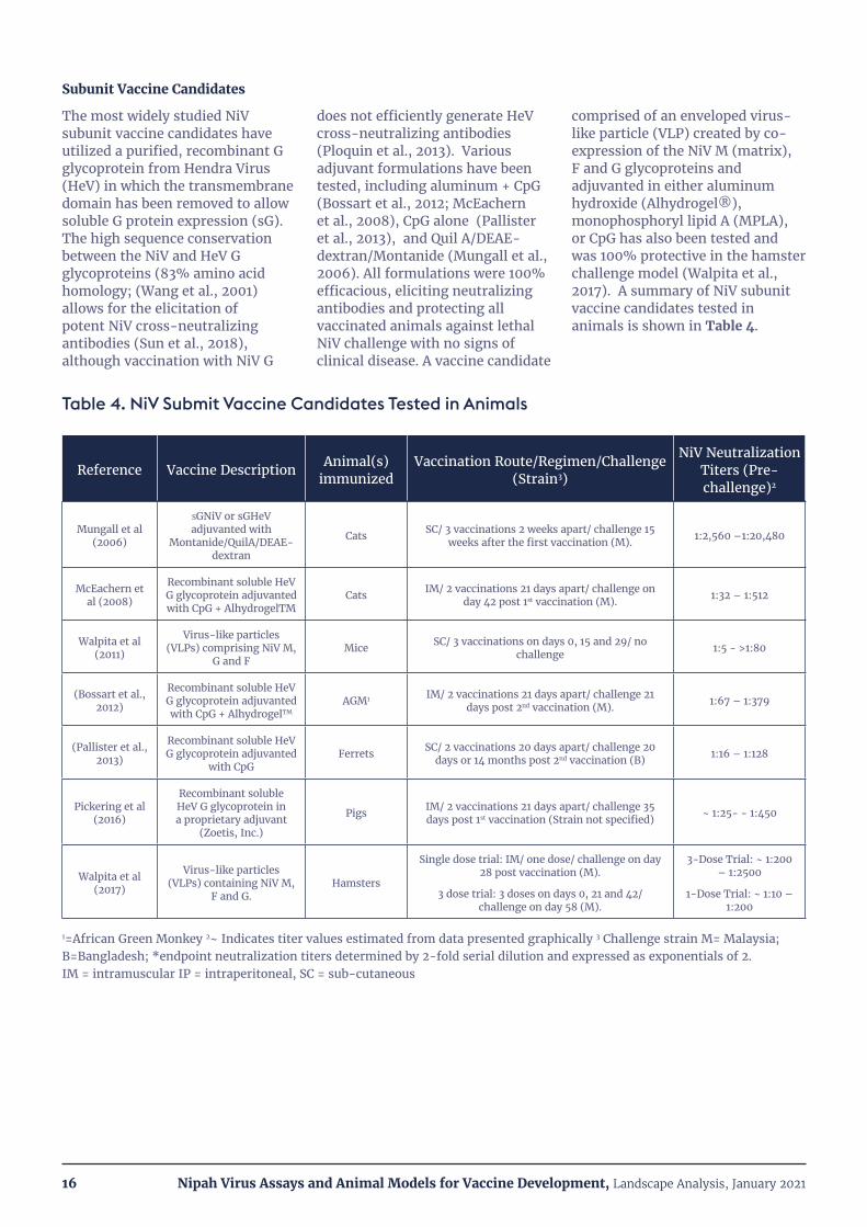
Subunit Vaccine Candidates
The most widely studied NiV subunit vaccine candidates have utilized a purified, recombinant G glycoprotein from Hendra Virus (HeV) in which the transmembrane domain has been removed to allow soluble G protein expression (sG). The high sequence conservation between the NiV and HeV G glycoproteins (83% amino acid homology; (Wang et al., 2001) allows for the elicitation of potent NiV cross-neutralizing antibodies (Sun et al., 2018), although vaccination with NiV G
does not efficiently generate HeV cross-neutralizing antibodies (Ploquin et al., 2013). Various adjuvant formulations have been tested, including aluminum + CpG (Bossart et al., 2012; McEachern et al., 2008), CpG alone (Pallister et al., 2013), and Quil A/DEAE-dextran/Montanide (Mungall et al., 2006). All formulations were 100% efficacious, eliciting neutralizing antibodies and protecting all vaccinated animals against lethal NiV challenge with no signs of clinical disease. A vaccine candidate
comprised of an enveloped virus-like particle (VLP) created by co-expression of the NiV M (matrix), F and G glycoproteins and adjuvanted in either aluminum hydroxide (Alhydrogel®), monophosphoryl lipid A (MPLA), or CpG has also been tested and was 100% protective in the hamster challenge model (Walpita et al., 2017). A summary of NiV subunit vaccine candidates tested in animals is shown in Table 4.
Reference Vaccine Description Animal(s) immunized
Vaccination Route/Regimen/Challenge (Strain3)
NiV Neutralization Titers (Pre-challenge)2
Mungall et al (2006)
sGNiV or sGHeV adjuvanted with
Montanide/QuilA/DEAE-dextran
Cats SC/ 3 vaccinations 2 weeks apart/ challenge 15 weeks after the first vaccination (M). 1:2,560 –1:20,480
McEachern et al (2008)
Recombinant soluble HeV G glycoprotein adjuvanted with CpG + AlhydrogelTM
Cats IM/ 2 vaccinations 21 days apart/ challenge on day 42 post 1st vaccination (M). 1:32 – 1:512
Walpita et al (2011)
Virus-like particles (VLPs) comprising NiV M,
G and FMice SC/ 3 vaccinations on days 0, 15 and 29/ no
challenge 1:5 - >1:80
(Bossart et al., 2012)
Recombinant soluble HeV G glycoprotein adjuvanted with CpG + AlhydrogelTM
AGM1 IM/ 2 vaccinations 21 days apart/ challenge 21 days post 2nd vaccination (M). 1:67 – 1:379
(Pallister et al., 2013)
Recombinant soluble HeV G glycoprotein adjuvanted
with CpG Ferrets SC/ 2 vaccinations 20 days apart/ challenge 20
days or 14 months post 2nd vaccination (B) 1:16 – 1:128
Pickering et al (2016)
Recombinant soluble HeV G glycoprotein in a proprietary adjuvant
(Zoetis, Inc.)
Pigs IM/ 2 vaccinations 21 days apart/ challenge 35 days post 1st vaccination (Strain not specified) ~ 1:25- - 1:450
Walpita et al (2017)
Virus-like particles (VLPs) containing NiV M,
F and G.Hamsters
Single dose trial: IM/ one dose/ challenge on day 28 post vaccination (M).
3 dose trial: 3 doses on days 0, 21 and 42/ challenge on day 58 (M).
3-Dose Trial: ~ 1:200 – 1:2500
1-Dose Trial: ~ 1:10 – 1:200
1=African Green Monkey 2~ Indicates titer values estimated from data presented graphically 3 Challenge strain M= Malaysia; B=Bangladesh; *endpoint neutralization titers determined by 2-fold serial dilution and expressed as exponentials of 2. IM = intramuscular IP = intraperitoneal, SC = sub-cutaneous
Table 4. NiV Submit Vaccine Candidates Tested in Animals
Nipah Virus Assays and Animal Models for Vaccine Development, Landscape Analysis, January 202117
More recently, novel antigen design options have been evaluated using a structure-based design.5 A stabilized prefusion F (pre-F), multimeric G constructs, and chimeric proteins containing both pre-F and G were developed as protein subunit candidate vaccines. The proteins were evaluated for antigenicity and structural integrity using kinetic binding assays, electron microscopy, and other biophysical properties. Immunogenicity of the vaccine antigens was evaluated in mice using aluminum hydroxide as adjuvant.
mRNA Vaccine Candidates
The US CDC has published proof-of-concept pre-clinical data on a Hendra virus glycoprotein mRNA vaccine in liquid nanoparticles.6 A single dose of the vaccine protected up to 70% of hamsters against a lethal, intraperitoneal
challenge with the Malaysian strain of Nipah virus. Authors noted immune responses were suboptimal. It is conceivable that the protection would have been superior under a two-dose regimen.
In addition, some of the structure-based designs described earlier for immunogen development5 are being evaluated in the mRNA platform in collaboration with Moderna, and clinical evaluation is planned.
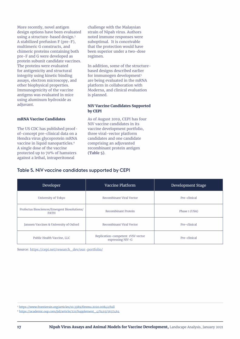
NiV Vaccine Candidates Supported by CEPI
As of August 2019, CEPI has four NiV vaccine candidates in its vaccine development portfolio, three viral-vector platform candidates and one candidate comprising an adjuvanted recombinant protein antigen (Table 5).
Developer Vaccine Platform Development Stage
University of Tokyo Recombinant Viral Vector Pre-clinical
Profectus Biosciences/Emergent Biosolutions/PATH Recombinant Protein Phase 1 (USA)
Janssen Vaccines & University of Oxford Recombinant Viral Vector Pre-clinical
Public Health Vaccine, LLC Replication-competent rVSV vector expressing NiV-G Pre-clinical
Source: https://cepi.net/research_dev/our-portfolio/
Table 5. NiV vaccine candidates supported by CEPI
5 https://www.frontiersin.org/articles/10.3389/fimmu.2020.00842/full6 https://academic.oup.com/jid/article/221/Supplement_4/S493/5637464
Nipah Virus Assays and Animal Models for Vaccine Development, Landscape Analysis, January 202118
III. STANDARDIZATION OF ASSAYS AND ANIMAL MODELSAssays and animal models to quantify or characterize immune responses elicited by vaccination are, by their nature, inherently variable.
The reasons for this include the molecular complexity of the samples (serum or other biological samples), the need to produce reagents in complex biological systems such as cell culture or in vivo, variability in composition and stability of these reagents, and the need to test immune responses in vivo. Nevertheless, modern vaccine development requires vaccines and samples from vaccinated humans and animals be tested with the highest possible precision. The task is further complicated by the collaborative and global nature of modern vaccine development. Multiple research laboratories, vaccine developers, non-governmental organizations, and regulatory agencies are often involved in the development process and vaccine candidates utilizing different platform technologies are often evaluated for the same disease indication. Thus, standardization of methods and reagents is important to facilitate development of new vaccines such as for NiV. The goal is to enable “like versus like” comparisons of data generated by different laboratories and derived from many assay types. Recognizing the value of phase-appropriate standardization early in the vaccine development process, CEPI is promoting assay, reagent, and animal model standardization to accelerate development of vaccines for NiV and other priority diseases in its portfolio.
Immune Serum Reference Standards
One of the most important tools for standardization of serological assays is immune reference serum. Even when similar assay formats are used for detection of antigen binding antibodies or virus neutralizing antibodies, the resulting data can be highly variable between laboratories due to differences in assay methods and reagents. For example, a 10-laboratory collaborative study assessing the precision of assays for detection of serum antibodies against Human Papillomavirus 16 (HPV 16) revealed inter-laboratory variations in anti-HPV titer of up to 25-fold for the same test sample (Ferguson et al., 2006). A similar, 15-laboratory collaborative study evaluating assays for serum antibodies against H5N1 influenza showed inter-laboratory variations in titer of 10 to 35-fold, depending on the sample and type of assay (Stephenson et al., 2009). The purpose of establishing immune reference standards is to provide a common, external control to improve the comparability of assay data between laboratories. With the standard in place, test results are reported relative to the activity of the reference standard. In the studies cited above, use of a common reference standard significantly reduced intra-laboratory assay variability. Reference standards have been developed for many vaccine indications, both in development
and commercial manufacturing (https://www.who.int/biologicals/vaccines/en/). Recent examples include HPV 16 (Ferguson et al., 2011), Typhoid Fever (Rijpkema et al., 2018), Respiratory Syncytial Virus (McDonald et al., 2018) and Zika (Source: WHO/BS/2018.2345).
Three key factors determine the fitness of material for use as a biological standard. First, the material must have similar composition and in vitro behavior to the human sera test articles. Second, the standard should be commutable, meaning it should work for a wide range of serological assays and vaccine platforms being tested. Finally, a blinded multi-laboratory collaborative study must demonstrate the utility of the standard for reducing intra-laboratory assay variability. (Source: CEPI 2nd Standards and Assays Workshop; June 2019).
Serum reference standards for a new vaccine are often established in a staged manner as the development process progresses. This mitigates the risk of producing exhaustively characterized materials which might not be required if vaccine development does not progress. For R&D and early clinical trials a working standard or interim standard may be established by a collaborative study involving a relatively limited number of laboratories, and relatively low volumes may be sufficient in the earlier stages.

Nipah Virus Assays and Animal Models for Vaccine Development, Landscape Analysis, January 202119
A number of different sources of immune sera may be considered. For example, a collaborative study for establishment of an interim standard for antibodies to Ebola virus (EBOV) tested plasma samples from patients who recovered from Ebola infection (convalescent sera), anti-EBOV IgG preparations from trans-chromosomal (Tc) cows immunized with experimental vaccines and plasma from vaccinated volunteers participating in an EBOV vaccine trial (Wilkinson et al., 2017). An interim standard for NiV will probably be generated from non-human primates infected with a sub-lethal dose of NiV and which generate high titers of neutralizing antibodies (Dhondt and Horvat, 2013). Obtaining convalescent sera from NiV survivors is also being considered (Source: CEPI 2nd Standards and Assays Workshop; June 2019). However, given the sporadic nature of NiV outbreaks and relatively small number of cases (and available survivors), obtaining sufficient quantities of convalescent sera for long-term use may be challenging. Therefore, in the case of emerging infections such as NiV one of the alternative approaches described above may need to be employed.
Establishment of an interim standard usually precedes establishment of an International Standard (IS) , often called an International Reference Preparation (IRP) under the endorsement of the WHO Expert Committee on Biological Standardization. This is a more formal process, taking up to 36 months and involving a larger and more in-depth collaborative study, often involving more than 25 laboratories and a wide geographical distribution. This is the main difference from a working or interim standard. Regulatory agencies generally expect an established International Standard to be used in pivotal clinical trials for vaccine approval, unless specifically justified
(Source: CEPI 2nd Standards and Assays Workshop; June 2019). A single, large lot of an antibody standard is preferred to avoid potential variability between multiple lots and the need for subsequent bridging studies. Once suitable standard sera candidates are available for evaluation, a collaborative study is performed to evaluate serological assays performed by a number of participating laboratories. A broad panel of test samples from different sources (e.g., sera from naturally infected humans, animals infected in the laboratory, and vaccinated humans or animals) is assayed and the intra-laboratory variability in assay results is assessed. Finally, the test sample absolute values (for example, geometric mean titers) are expressed relative to the activity of the candidate standard and the ability of the standard to improve intra-laboratory comparability of test results is assessed. Once the standard has been chosen a full storage stability program is conducted to ensure the quality of the material over time. Trending of assay performance over time is also performed. The International Standard itself is not intended for routine assay use. A working reference standard is established for routine use and a bridging study is conducted to calibrate the working standard to the International Standard (Source: CEPI 2nd Standards and Assays Workshop; June, 2019).
Standardization of Other Biological Assay Reagents and Methods
Many aspects of biological assays for vaccine testing may be standardized to improve the comparability of intra-laboratory data. Common reagents (reference sera, antigens, virus stocks) may be produced and standardized assay methods established and validated. For example, potency testing for release of subunit seasonal influenza vaccines is performed under a high degree of standardization using
common reagents (reference sera and antigens) provided by regulatory agencies and using a single, validated assay method to test and release new seasonal vaccine formulations. In general, standards may improve inter-laboratory test performance. However, the need, feasibility and level of standardization is typically considered on a case-by-case basis according to the stage of vaccine development, the types of assays in use and the potential of standards to facilitate development and licensure. Standardized reference sera are relatively easy to implement since they ideally should be commutable across many assay types and are broadly recognized for improving both intra- and inter-laboratory assay consistency. In contrast, standardized assay formats are more challenging to implement, especially for newer vaccines and those in development, since vaccine antigens may differ between candidates and there is less consensus on the ideal assay format. With these considerations in mind, the benefits and potential challenges of standardizing various assay and animal model components to accelerate NiV vaccine development are summarized in Table 6.
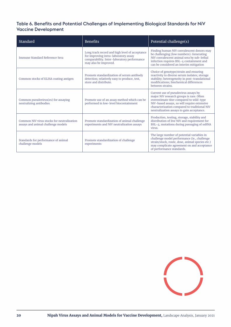
Nipah Virus Assays and Animal Models for Vaccine Development, Landscape Analysis, January 202120
Table 6. Benefits and Potential Challenges of Implementing Biological Standards for NiV Vaccine Development
Standard Benefits Potential challenge(s)
Immune Standard Reference Sera
Long track record and high level of acceptance for improving intra-laboratory assay comparability. Inter-laboratory performance may also be improved.
Finding human NiV convalescent donors may be challenging (low numbers). Generating NiV convalescent animal sera by sub-lethal infection requires BSL-4 containment and can be considered an interim mitigation
Common stocks of ELISA coating antigenPromote standardization of serum antibody detection; relatively easy to produce, test, store and distribute.
Choice of genotype/strain and ensuring reactivity to diverse serum isolates; storage stability; heterogeneity in post-translational modifications; biochemical differences between strains.
Common pseudovirus(es) for assaying neutralizing antibodies
Promote use of an assay method which can be performed in low-level biocontainment
Current use of pseudovirus assays by major NiV research groups is rare. Often overestimate titer compared to wild-type NiV-based assays, so will require extensive characterization compared to traditional NiV neutralization assays to gain acceptance.
Common NiV virus stocks for neutralization assays and animal challenge models
Promote standardization of animal challenge experiments and NiV neutralization assays
Production, testing, storage, stability and distribution of live NiV and requirement for BSL-4; mutations during passaging of ssRNA virus.
Standards for performance of animal challenge models
Promote standardization of challenge experiments
The large number of potential variables in challenge model performance (ie., challenge strain/stock, route, dose, animal species etc.) may complicate agreement on and acceptance of performance standards.
Nipah Virus Assays and Animal Models for Vaccine Development, Landscape Analysis, January 202121
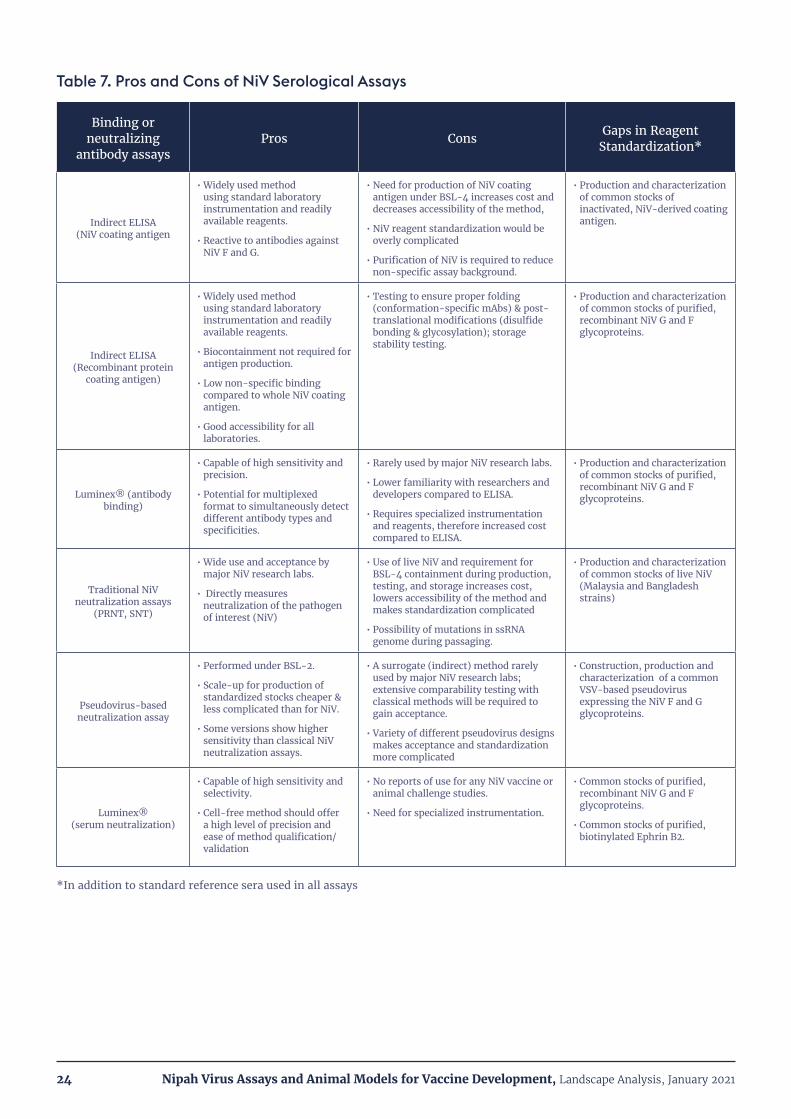
IV. NIV SEROLOGICAL ASSAYSRobust serological assays for quantifying and characterizing humoral immune responses in humans and animals are critical for vaccine development.
A number of methods have been developed for NiV serology, and the refinement and standardization of such methods will be essential for facilitating development of safe and effective human vaccines against NiV. This section describes the commonly used serological
assays for NiV, as well as newer assays in earlier stages of use and acceptance. An analysis of the pros and cons of different serological assays for NiV vaccine development is presented in Table 7. The usage of assays in a large number of NiV vaccine studies and other research
experiments is compiled in Table 8 and Table 9 to illustrate the prevalence of use, variables in assay performance, how reagents and methods have changed over time and opportunities for assay standardization.
Detection of antigen – specific serum IgG is essential to the vaccine development process to characterize the specificity and magnitude of the vaccine-induced humoral immune response. The most common assay method is the traditional “indirect” ELISA. In this assay a target antigen is plated (adsorbed) onto a 96- well microtiter plate. After blocking the plate to suppress non-specific binding, dilutions of immune or control sera are added to the wells. After washing away unbound antibody, bound IgG is usually detected by the addition of a species-specific anti-IgG secondary antibody conjugated to a chromogenic enzyme. While serum IgG is measured to elucidate vaccine responses, as well as for surveillance and epidemiology, measurement of NiV-specific serum IgM is usually performed for diagnosis of active infection (Mazzola and Kelly-Cirino, 2019). The advantages of the ELISA assay format include its broad use and familiarity throughout biomedical science, relatively low-tech and low-cost application and wide availability of reagents.
The earliest ELISAs for detection of NiV antibodies in sera were developed by the Centers for Disease Control (CDC; USA). Different ELISAs were developed for NiV-specific serum IgG and IgM. These ELISAs were used for surveillance and diagnosis of disease in humans and pigs, and used detergent and radiation-inactivated, NiV – infected Vero cell lysates as the target antigen (Daniels et al., 2001). Several of the NiV animal vaccination studies and research experiments detailed in Table 8 and Table 9 utilized inactivated crude NiV- infected Vero extracts or gradient-purified NiV as the target antigen. However, the use of NiV as an assay reagent is obviously problematic since the initial preparation requires BSL-4 containment. ELISAs using NiV-infected crude extracts also suffered from non-specific binding (Daniels et al., 2001). The discovery that NiV F and G glycoproteins are the major target of neutralizing antibodies and their use in vaccine formulations spurred the use of recombinant, soluble NiV F (sFNiV) and G (sGNiV) as the target antigens in ELISA assays. Six of
the 17 ELISAs in Tables 8 and 9 use recombinant G or F for target IgG capture. The sFNiV and sGNiV have been produced in a variety of recombinant expression systems (E. coli, insect cells, mammalian cells) and are usually epitope tagged for ease of purification (Eshaghi et al., 2005; Eshaghi et al., 2004; Keshwara et al., 2019; Kurup et al., 2015). The glycosylation and disulfide bonding in the NiV F and G glycoproteins make eukaryotic cells preferable for recombinant expression of these antigens. The many successful tests of vaccines targeting the G glycoprotein in NiV animal challenge models make it likely that this antigen will be used in human vaccine candidates.
1. Detection of antigen – specific serum IgG
Nipah Virus Assays and Animal Models for Vaccine Development, Landscape Analysis, January 202122
A newer assay format that has been used for detection of NiV antigen-specific serum IgG is the bead-based liquid protein array system commonly known as Luminex® (Vignali, 2000). In this assay (Bossart et al., 2007), purified sGNiV protein is covalently coupled to fluorescent microspheres. After binding to the analyte (NiV G- specific IgG) biotinylated Protein A is added, followed by streptavidin-phycoerythrin (PE), a fluorescence indicator that emits at a different wavelength than the microspheres. The bead mixture is analyzed on a dual laser flow-based detection instrument. One laser detects the
bead and determines the analyte being detected. The second laser detects the PE-derived signal, which is in direct proportion to the amount of anti-NiV G bound (Source: R&D Systems website). One vaccine study in Table 8 (Pallister et al., 2013) and two other research studies in Table 9 utilized this system for detection of antigen-specific serum IgG. Luminex®-based assays are capable of high sensitivity and specificity and the experimental manipulations are no more complicated than running ELISA-based assays. Another potential advantage of this platform is the
ability to develop multiplexed assays for discrimination of different antibody types and specificities. However, the utilization of this system for NiV vaccine development is limited by availability of the specialized instrumentation and relative unfamiliarity of the technology compared with ELISA-based assays. Nevertheless, standardization of such an assay would be similar to the ELISA and would involve use of standard immune sera and common stocks of target antigen.
The quantitation of serum neutralizing antibodies is essential for measuring vaccine potency and is often important for establishing correlates of protection. As shown in Tables 8 and 9, most published NiV vaccine studies and other research investigations have utilized traditional assays for virus neutralization based on inhibition of NiV-mediated killing or cytopathology in Vero cell cultures. One common assay format is the Plaque Reduction Neutralization Test (PRNT), of which there are a number of variations. In the original method developed for NiV (Crameri et al., 2002) the test sample (immune or control serum) is diluted and mixed with a NiV suspension. After an incubation period the mixture is applied to a confluent monolayer of Vero cells. After an adsorption period the virus mixture is removed and replaced with fresh culture medium. After incubating overnight, the cells are
fixed with methanol. Plaques are then detected by immune-assay using rabbit antisera specific for a NiV antigen. Serum titer is expressed as the reciprocal of the serum dilution that reduces the number of plaques to 75% of that of control untreated virus. The PRNT assay was used in 10 of the 24 NiV animal studies described in Tables 8 and 9.
The other common traditional assay for NiV neutralization is the Serum Neutralization Test (SNT) (Daniels et al., 2001) In this assay, serum dilutions are incubated with approximately 200 TCID50 NiV in 96-well microtiter plates prior to the addition of Vero cells. Cultures are visually (microscopically) examined for the presence or absence of cytopathic effect after a three-day incubation. The neutralization titer is expressed as the reciprocal of the highest dilution that prevents virus growth
in 50% of replicate wells (Crameri et al., 2002). This assay, or variants of it, was used in 11 of 24 NiV animal studies detailed in Tables 8 and 9.
The major drawback of both the PRNT and SNT assays is that they use live NiV and the procedures (or at least the first steps) must be performed in BSL-4 containment (Table 7). This would be a major hurdle to any attempts to standardize assays of this type since scaled-up production and testing of a common stock of NiV under BSL-4 containment would be extremely challenging. The need for BSL-4 containment is also a significant hinderance to developers lacking such facilities. Thus, other assay formats have been pursued that do not require high-level biocontainment, and that may thus facilitate NiV vaccine development.
2. Detection of serum neutralizing antibodies
Nipah Virus Assays and Animal Models for Vaccine Development, Landscape Analysis, January 202123
Pseudotype viruses (pseudoviruses) have been constructed for use in surrogate NiV neutralization assays which can be performed under BSL-2. A pseudovirus is an enveloped virus expressing one or more foreign virus envelope proteins that mediate cell attachment and membrane fusion. In most cases the pseudovirus also carries a reporter gene such as GFP or luciferase which serves as an indicator of infection. The most common parental viruses used for pseudovirus construction are vesicular stomatitis virus (VSV) and lentivirus (HIV); (Wang and Daniels, 2012). The first NiV pseudovirus assay (Tamin et al., 2009) utilized a VSV constructed to display both the NiV F and G glycoproteins on its envelope and carrying the luciferase gene as the reporter. This assay showed generally good correlation of titers with a classical PRNT assay, although some serum samples with very low titers showed reduced sensitivity in the pseudovirus assay. Another assay system developed by Kaku et al., (2009) also utilized VSV pseudotyped with NiV G and F but used green fluorescent protein (GFP) as the reporter gene (Kaku et al., 2009). This assay showed good specificity and higher sensitivity compared with the NiV SNT assay. A variation of this assay was developed (Kaku et al., 2012) in which the GFP reporter gene was replaced by secreted alkaline phosphatase (SEAP), which can be assayed from the culture supernatant using an ordinary ELISA plate reader. This assay was also generally more sensitive than the classical SNT assay. Three in vivo studies described in Tables 8 and 9 used NiV pseudovirus-based neutralization assays.
Overall, these and other results suggest that pseudovirus-based assay systems are a promising alternative to classical NiV neutralization assays for use in vaccine development. However, acceptance of NiV pseudovirus-based neutralization assays for vaccine development and licensure will require thorough characterization to understand the comparability in sensitivity and specificity with traditional NiV neutralization assays (Table 7). The ability of a single reference standard to harmonize pseudovirus-based assays as well as NiV-based assays will also play a major role in the acceptance pseudovirus-based assays.
Another promising NiV surrogate neutralization assay uses a variation of the Luminex® method described for detection of serum IgG binding. Like the binding antibody assay, this Luminex® assay (Bossart et al., 2007) uses sGNiV covalently conjugated to fluorescent microspheres. Biotinylated Ephrin B2, the cellular receptor for NiV G, is added and the binding interaction is detected with streptavidin-phycoerythrin. Neutralizing antibodies targeting NiV G disrupt the sGNiV – Ephrin B2 interaction and thus diminish the phycoerythrin fluorescence signal. The sensitivity and specificity of this assay appears to be comparable to the NiV SNT assay using control and immune sera from a variety of sources. In addition to standard immune reference sera, standardization of this assay format could benefit from the availability of common stocks of sGNiV, purified Ephrin B2 and a control anti-sGNiV mAb.
Nipah Virus Assays and Animal Models for Vaccine Development, Landscape Analysis, January 202124
Table 7. Pros and Cons of NiV Serological Assays
Binding or neutralizing
antibody assaysPros Cons Gaps in Reagent
Standardization*
Indirect ELISA (NiV coating antigen
• Widely used method using standard laboratory instrumentation and readily available reagents.
• Reactive to antibodies against NiV F and G.
• Need for production of NiV coating antigen under BSL-4 increases cost and decreases accessibility of the method,
• NiV reagent standardization would be overly complicated
• Purification of NiV is required to reduce non-specific assay background.
• Production and characterization of common stocks of inactivated, NiV-derived coating antigen.
Indirect ELISA (Recombinant protein
coating antigen)
• Widely used method using standard laboratory instrumentation and readily available reagents.
• Biocontainment not required for antigen production.
• Low non-specific binding compared to whole NiV coating antigen.
• Good accessibility for all laboratories.
• Testing to ensure proper folding (conformation-specific mAbs) & post-translational modifications (disulfide bonding & glycosylation); storage stability testing.
• Production and characterization of common stocks of purified, recombinant NiV G and F glycoproteins.
Luminex® (antibody binding)
• Capable of high sensitivity and precision.
• Potential for multiplexed format to simultaneously detect different antibody types and specificities.
• Rarely used by major NiV research labs.
• Lower familiarity with researchers and developers compared to ELISA.
• Requires specialized instrumentation and reagents, therefore increased cost compared to ELISA.
• Production and characterization of common stocks of purified, recombinant NiV G and F glycoproteins.
Traditional NiV neutralization assays
(PRNT, SNT)
• Wide use and acceptance by major NiV research labs.
• Directly measures neutralization of the pathogen of interest (NiV)
• Use of live NiV and requirement for BSL-4 containment during production, testing, and storage increases cost, lowers accessibility of the method and makes standardization complicated
• Possibility of mutations in ssRNA genome during passaging.
• Production and characterization of common stocks of live NiV (Malaysia and Bangladesh strains)
Pseudovirus-based neutralization assay
• Performed under BSL-2.
• Scale-up for production of standardized stocks cheaper & less complicated than for NiV.
• Some versions show higher sensitivity than classical NiV neutralization assays.
• A surrogate (indirect) method rarely used by major NiV research labs; extensive comparability testing with classical methods will be required to gain acceptance.
• Variety of different pseudovirus designs makes acceptance and standardization more complicated
• Construction, production and characterization of a common VSV-based pseudovirus expressing the NiV F and G glycoproteins.
Luminex® (serum neutralization)
• Capable of high sensitivity and selectivity.
• Cell-free method should offer a high level of precision and ease of method qualification/validation
• No reports of use for any NiV vaccine or animal challenge studies.
• Need for specialized instrumentation.
• Common stocks of purified, recombinant NiV G and F glycoproteins.
• Common stocks of purified, biotinylated Ephrin B2.
*In addition to standard reference sera used in all assays
Nipah Virus Assays and Animal Models for Vaccine Development, Landscape Analysis, January 202125
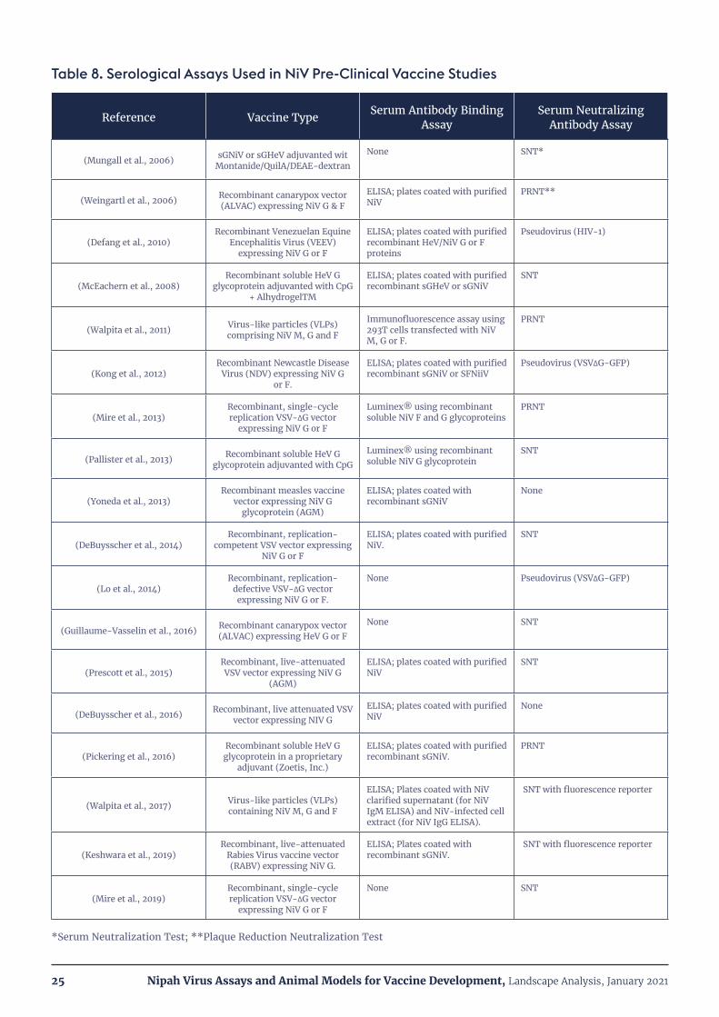
Table 8. Serological Assays Used in NiV Pre-Clinical Vaccine Studies
Reference Vaccine Type Serum Antibody Binding Assay
Serum Neutralizing Antibody Assay
(Mungall et al., 2006) sGNiV or sGHeV adjuvanted wit Montanide/QuilA/DEAE-dextran
None SNT*
(Weingartl et al., 2006) Recombinant canarypox vector (ALVAC) expressing NiV G & F
ELISA; plates coated with purified NiV
PRNT**
(Defang et al., 2010)Recombinant Venezuelan Equine
Encephalitis Virus (VEEV) expressing NiV G or F
ELISA; plates coated with purified recombinant HeV/NiV G or F proteins
Pseudovirus (HIV-1)
(McEachern et al., 2008)Recombinant soluble HeV G
glycoprotein adjuvanted with CpG + AlhydrogelTM
ELISA; plates coated with purified recombinant sGHeV or sGNiV
SNT
(Walpita et al., 2011) Virus-like particles (VLPs) comprising NiV M, G and F
Immunofluorescence assay using 293T cells transfected with NiV M, G or F.
PRNT
(Kong et al., 2012)Recombinant Newcastle Disease
Virus (NDV) expressing NiV G or F.
ELISA; plates coated with purified recombinant sGNiV or SFNiiV
Pseudovirus (VSV∆G-GFP)
(Mire et al., 2013)Recombinant, single-cycle replication VSV-∆G vector
expressing NiV G or F
Luminex® using recombinant soluble NiV F and G glycoproteins
PRNT
(Pallister et al., 2013) Recombinant soluble HeV G glycoprotein adjuvanted with CpG
Luminex® using recombinant soluble NiV G glycoprotein
SNT
(Yoneda et al., 2013)Recombinant measles vaccine
vector expressing NiV G glycoprotein (AGM)
ELISA; plates coated with recombinant sGNiV
None
(DeBuysscher et al., 2014)Recombinant, replication-
competent VSV vector expressing NiV G or F
ELISA; plates coated with purified NiV.
SNT
(Lo et al., 2014)Recombinant, replication-
defective VSV-∆G vector expressing NiV G or F.
None Pseudovirus (VSV∆G-GFP)
(Guillaume-Vasselin et al., 2016) Recombinant canarypox vector (ALVAC) expressing HeV G or F
None SNT
(Prescott et al., 2015)Recombinant, live-attenuated VSV vector expressing NiV G
(AGM)
ELISA; plates coated with purified NiV
SNT
(DeBuysscher et al., 2016) Recombinant, live attenuated VSV vector expressing NIV G
ELISA; plates coated with purified NiV
None
(Pickering et al., 2016)Recombinant soluble HeV G glycoprotein in a proprietary
adjuvant (Zoetis, Inc.)
ELISA; plates coated with purified recombinant sGNiV.
PRNT
(Walpita et al., 2017) Virus-like particles (VLPs) containing NiV M, G and F
ELISA; Plates coated with NiV clarified supernatant (for NiV IgM ELISA) and NiV-infected cell extract (for NiV IgG ELISA).
SNT with fluorescence reporter
(Keshwara et al., 2019) Recombinant, live-attenuated
Rabies Virus vaccine vector (RABV) expressing NiV G.
ELISA; Plates coated with recombinant sGNiV.
SNT with fluorescence reporter
(Mire et al., 2019)Recombinant, single-cycle replication VSV-∆G vector
expressing NiV G or F
None SNT
*Serum Neutralization Test; **Plaque Reduction Neutralization Test

Nipah Virus Assays and Animal Models for Vaccine Development, Landscape Analysis, January 202126
Table 9. Serological Assays Used in Other NiV Research Studies
Reference Study Type Serum Antibody Binding Assay
Serum Neutralizing Antibody Assay
(Wong et al., 2003) Development of hamster challenge model
ELISA; plates coated with crude extracts from NiV-infected Vero cells
None
(Guillaume et al., 2004) Antibody prophylaxis (hamster)ELISA; plates coated with crude extract from NiV-infected Vero cells
PRNT**
(Guillaume et al., 2006) Antibody prophylaxis (hamster
ELISA; plates coated with NiV clarified extract or recombinant NiV N protein produced in insect cells
SNT*
(de Wit et al., 2011) NiV transmission in hamsters ELISA; plates coated with purified NiV SNT
(Mathieu et al., 2012b)Impact of non-structural proteins on NiV virulence (hamster)
Purified NiV None
(de Wit et al., 2014) Foodborne transmission of NiV in hamsters
ELISA; plates coated with purified, inactivated NiV. None
(Geisbert et al., 2014) Therapeutic mAb treatment of NiV infection (AGM)
Multiplexed microsphere assay (Luminex) using recombinant soluble NiV F glycoprotein
SNT
(Johnston et al., 2015) Analysis of NiV AGM challenge model None Pseudovirus (VSV∆G-RFP)
(Satterfield et al., 2015)Impact of non-structural proteins on NiV disease course (ferret)
None PRNT
(Borisevich et al., 2016) Monoclonal antibody study (hamster) None PRNT
(Mire et al., 2016) NiV strain differences in AGMMultiplexed microsphere assay (Luminex) using recombinant soluble NiV F glycoprotein
PRNT
(Satterfield et al., 2016b) Impact of non-structural proteins on NiV virulence (ferret) None PRNT
(Dawes et al., 2018) Favipiravir prophylaxis study (hamster) None PRNT
(Mathieu et al., 2018) Peptide prophylaxis (AGM) None SNT
(Lo et al., 2019) Remdesivir prophylaxis (AGM) ELISA; plates coated with NiV antigen SNT
(Schountz et al., 2019) Immune response/pathogenesis study in hamsters None SNT
*Serum Neutralization Test; **Plaque Reduction Neutralization Test
Nipah Virus Assays and Animal Models for Vaccine Development, Landscape Analysis, January 202127
V. NIV ANIMAL MODELS
Well characterized, robust animal challenge models are another critical component for development of any vaccine and, as with assays, standardization of animal models can accelerate vaccine development by promoting like-versus-like comparisons between laboratories. In the case of NiV and other emerging diseases the refinement and standardization of animal models takes on additional importance regarding the pathway to vaccine licensure. Since NiV outbreaks are sporadic and infect relatively small numbers, performing controlled vaccine efficacy studies in humans becomes very challenging. A potential alternative licensure pathway is the FDA “Animal Rule”, a mechanism designed for disease indications for which efficacy studies would be infeasible or unethical. Under this mechanism Phase I/II safety and immunogenicity testing is conducted in humans, but efficacy is demonstrated in a well-established animal model(s) that provides substantial evidence of effectiveness when all of the following four criteria are met: 1) There is a reasonably well-understood pathophysiological mechanism of the toxicity of the substance (pathogen) and its prevention or substantial reduction by the product (vaccine); 2) The effect is demonstrated in more than one animal species expected to react with a response predictive for humans, unless the effect is demonstrated in a single animal species that represents a sufficiently well-characterized animal model for predicting the response in humans; 3) The animal
study endpoint is clearly related to the desired benefit in humans, generally the enhancement of survival or prevention of major morbidity; and 4) The data or information on the kinetics and pharmacodynamics of the product or other relevant data or information, in animals and humans, allows selection of an effective dose in humans. For vaccines, prediction of clinical benefit is determined by bridging from the human immunogenicity data to the animal model immunogenicity and efficacy data (Source: Draft FDA Guidance for Industry; Animal Models – Essential Elements to Address Efficacy Under the Animal Rule; January 2009). To date, one FDA vaccine approval has followed this pathway, a post-exposure prophylaxis indication for an anthrax vaccine (Beasley et al., 2016). The European Medicines Agency (EMA) does not currently have a detailed mechanism analogous to the FDA Animal Rule. However, current EMA guidance leaves open the possibility of using data from animal models to demonstrate vaccine efficacy (Source: EMEA/CHMP/VWP/164653/05 Rev.1; 2018)
According to the FDA guidance, animal model(s) should be highly refined and characterized with regard to understanding of disease etiology, progression and pathology and their performance (along with validated assays) should be standardized as much as possible ( (Williamson and Westlake, 2019); WHO Nipah R&D Roadmap Draft, 2018). Key
attributes of relevant animal challenge models to support FDA licensure of vaccines under the Animal Rule are summarized below (Golding et al., 2018):
1. Animal species should show key characteristics of the human disease following exposure to the challenge pathogen (time from exposure to onset of disease, time course/progression of disease, clinical manifestations, morbidity and lethality.
2. The challenge agent used in the animal study should be relevant to the human disease.
3. The immune marker(s) selected should reflect the protective immune responses generated by humans.
4. The vaccine dose and vaccination schedule chosen for adequate and well-controlled studies in animals should elicit an immune response in animals reflective of that in humans.
5. Ideally, the immunological assays should be species-independent.
6. There should be a robust statistical plan.
Vaccine Licensure under the FDA “Animal Rule”

Nipah Virus Assays and Animal Models for Vaccine Development, Landscape Analysis, January 202128
NiV infects a wide range of wild and domesticated animals, including: fruit bats, cats, dogs, swine, and horses (see Section II, Epidemiology). However, the symptomology is often variable, and in some cases does not closely resemble the human disease. Fruit bats are asymptomatically infected (Geisbert et al., 2012). In the laboratory setting mice are largely resistant to NiV infection unless delivered intracranially (Mungall et al., 2006), administered to aged mice (C57B6 or BALB/c), or given to Type I interferon knockout (IFNAR KO) transgenic mice
(Dhondt et al., 2013). No reports of NiV strains lab-adapted to mice or any other animal species have been found in the published literature. The NiV animal challenge models that most accurately recapitulate the symptomology and pathology of the human disease are Syrian Golden Hamsters, ferrets and African Green Monkeys. The following sections describe the key characteristics and use of the models. For each model a table compiling published challenge experiments is provided to illustrate experimental performance variables and
how the models have been used. Key variables relevant to standardization, such as challenge strain, dose, and route of infection are provided, along with other available details such as survival time, clinical signs, and pathology. A summary comparison of clinical signs and pathology of NiV infection in the hamster, ferret and AGM models is given in Table 10, and Table 16 provides an analysis of the pros and cons of each model vis a vis its usefulness in vaccine development.
NiV Animal Challenge Models
Table 10. Summary of clinical signs and pathology in the NiV hamster, ferret and AGM challenge models.
SpeciesClinical Signs
Gross lesions Histology Virus found in:Respiratory Neurological
HamsterLabored breathing, serosanguineous nasal exudates
Imbalance, muscle twitching, tremor, limb paralysis
Edema, hemorrhages, congestion
Vasculitis, meningitis, encephalitis, endothelial syncytia
Lung, nasal epithelium, CNS, heart, liver, spleen, kidney, bladder, urine
FerretDyspnea, cough, serous nasal discharge
Depression, tremors, myoclonus (muscle twitching), hind limb paresis
Edema, hemorrhages, enlarged lymph nodes
Necrotizing alveolitis, glomerular necrosis, vasculitis, endothelial syncytial, meningitis.
Brain, lung, lymphoid organs, adrenal, kidney, testes, uterus, liver pharyngeal and rectal swabs
African Green Monkey
Severe Dyspnea, open-mouth breathing, serosanguineous nasal discharge
Muscle twitches, seizures
Pulmonary consolidation, congestion of lungs, enlarged lymph nodes, congested liver, inflammation of gastrointestinal tract, congestion in the brain.
Alveolar hemorrhages, pulmonary edema and inflammation, alveolitis, fibrinoid necrosis, vasculitis, meningitis
Lung, lymph nodes, heart, liver, spleen, kidney, adrenal gland, brain, urinary bladder, sex organs
Adapted from (Dhondt and Horvat, 2013)
Nipah Virus Assays and Animal Models for Vaccine Development, Landscape Analysis, January 202129
The Syrian Golden Hamster (Mesocricetus auratus) is the most commonly used animal challenge model for NiV owing to its small size and economical housing. Tables 11 and 12 provide a compilation of most of the NiV hamster challenge studies found in the published literature, including challenge strain and available identifying information on the challenge virus, route of administration, dose and notes on the disease course. Hamsters develop clinical symptoms and pathologies which closely resemble NiV disease in humans, including respiratory and neurological signs (Guillaume et al., 2006). Infection by the intranasal (IN) or intraperitoneal (IP) routes induces similar symptoms and pathologies, but the disease course may vary with dose and route (Rockx et al., 2011; Wong et al., 2003), with IP inoculation causing a more rapid disease course than the IN route. Respiratory signs in the hamster include labored breathing and severe respiratory distress, bloody nasal discharge, pulmonary infiltrates (fluid build-up), and hemorrhagic lesions of the lung (Rockx, 2014). Neurological signs include behavioral deterioration, lethargy, imbalance, muscle twitching, tremors and limb paralysis (Dhondt and Horvat, 2013). Dose levels appear to play a role in NiV symptomology in the hamster. High doses, particularly when delivered IN, induce a primarily respiratory syndrome. Lower doses also induce respiratory symptoms, but neurological symptoms are also present (Rockx et al., 2011). Hamsters develop
encephalitis and lesions in multiple organs, including the lungs and brain, caused by vasculitis and thrombosis (Williamson and Torres-Velez, 2010). Some variances in NiV disease pathology have been reported in different hamster studies. The NiV (Malaysia strain) infection in hamsters reported by Wong et al., (2003) was dominated by neurological signs. In contrast, (Rockx et al., 2011) reported that infection by the same NiV strain, by the same route and at a similar dose caused a primarily respiratory disease. The cause of the discordance in results in these studies is not clear and suggests that further studies are needed to fully understand NiV infection in the hamster. This will be particularly important for standardization of the model. To our knowledge, a formal natural history study of the NiV hamster challenge model has not been performed. However, several publications have investigated the course of NiV infection and pathology in hamsters in detail, including: virus strain, routes of infection and dose, time to death, and virus replication and pathology/histopathology in major organs and tissues (Baseler et al., 2015; DeBuysscher et al., 2013; Rockx et al., 2011; Wong et al., 2003).
Both the Malaysia and Bangladesh NiV strains have been tested in the hamster model, but most studies have used Malaysia as the challenge virus (see Tables 11 and 12). A direct comparison of the two NiV strains in hamsters suggested that the Malaysia strain
had accelerated virus replication, pathology, and death compared to the Bangladesh strain, which had a more delayed disease progression. The accelerated disease course of the Malaysia strain in hamsters appears contrary to what has been observed in human outbreaks. However, the overall symptomology and viral distribution of the two strains were comparable (DeBuysscher et al., 2013).
Several key variables in performance of the NiV hamster model are apparent from Tables 11 and 12. A wide range of doses is reported, and two different routes of administration are used. Many of the published studies provide only limited information on the identity and origin of NiV challenge stocks. Most of the stocks appear to be different isolates from the same outbreaks: Malaysia 1999 and Bangladesh 2004 (Source: CEPI 2nd Standards and Assays Workshop, June 2019). However, the identity of many individual isolates, how their activities compare to one another, and how they have been stored, amplified, and tested is unclear. Establishment of common, well-characterized virus challenge stocks is important for standardization of this model. It is also unclear why the Malaysia strain has been the preferred challenge strain although the Bangladesh strain causes most human outbreaks. Many of the same gaps are also true for the ferret and African Green Monkey Models, as discussed below.
1. Syrian Golden Hamster
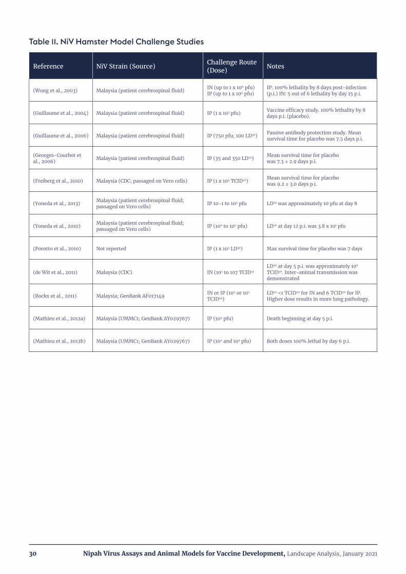
Nipah Virus Assays and Animal Models for Vaccine Development, Landscape Analysis, January 202130
Table 11. NiV Hamster Model Challenge Studies
Reference NiV Strain (Source) Challenge Route (Dose) Notes
(Wong et al., 2003) Malaysia (patient cerebrospinal fluid) IN (up to 1 x 10⁶ pfu) IP (up to 1 x 10⁴ pfu)
IP: 100% lethality by 8 days post-infection (p.i.) IN: 5 out of 6 lethality by day 15 p.i.
(Guillaume et al., 2004) Malaysia (patient cerebrospinal fluid) IP (1 x 10³ pfu) Vaccine efficacy study. 100% lethality by 8 days p.i. (placebo).
(Guillaume et al., 2006) Malaysia (patient cerebrospinal fluid) IP (750 pfu; 100 LD50) Passive antibody protection study. Mean survival time for placebo was 7.5 days p.i.
(Georges-Courbot et al., 2006) Malaysia (patient cerebrospinal fluid) IP (35 and 350 LD50) Mean survival time for placebo
was 7.3 ± 2.9 days p.i.
(Freiberg et al., 2010) Malaysia (CDC; passaged on Vero cells) IP (1 x 10⁴ TCID50) Mean survival time for placebo was 9.2 ± 3.0 days p.i.
(Yoneda et al., 2013) Malaysia (patient cerebrospinal fluid; passaged on Vero cells) IP 10-1 to 104 pfu LD50 was approximately 10 pfu at day 8
(Yoneda et al., 2010) Malaysia (patient cerebrospinal fluid; passaged on Vero cells) IP (100 to 105 pfu) LD50 at day 12 p.i. was 3.8 x 101 pfu
(Porotto et al., 2010) Not reported IP (1 x 10³ LD50) Max survival time for placebo was 7 days
(de Wit et al., 2011) Malaysia (CDC) IN (103 to 107 TCID50LD50 at day 5 p.i. was approximately 106 TCID50. Inter-animal transmission was demonstrated
(Rockx et al., 2011) Malaysia; GenBank AF017149 IN or IP (102 or 105 TCID50)
LD50 <1 TCID50 for IN and 6 TCID50 for IP. Higher dose results in more lung pathology.
(Mathieu et al., 2012a) Malaysia (UMMC1; GenBank AY029767) IP (104 pfu) Death beginning at day 5 p.i.
(Mathieu et al., 2012b) Malaysia (UMMC1; GenBank AY029767) IP (102 and 103 pfu) Both doses 100% lethal by day 6 p.i.

Nipah Virus Assays and Animal Models for Vaccine Development, Landscape Analysis, January 202131
Table 12. NiV Hamster Model Challenge Studies, Continued
Reference NiV Strain (Source) Challenge Route (Dose) Notes
(Munster et al., 2012) Malaysia (CDC) IN (105 TCID50) All animals developed neurological symptoms between day 7 and day 12 p.i.
(DeBuysscher et al., 2013)
Malaysia (1999 human CNS sample); Bangladesh (2004 human throat swab)
IP (1 to 105 TCID50) IN (105 TCID50)
First analysis of NiV-B in an animal model. NiV-M IP LD50 = 68 TCID50; NiV-B IP LD50 = 528 TCID50; Slower disease progression for NiV-B by IP or IN.
(Lo et al., 2014) Malaysia IP (105 TCID50; >103 LD50)
Vaccine study; 100% of moribund animals euthanized by day 6 p.i.)
(de Wit et al., 2014) Bangladesh/200401066 (CDC)
IN (107 TCID50); Oral gavage (107 TCID50); drinking palm sap (107 and 108)
IN 100% lethal by day 14; Oral gavage 20% lethal; Drinking 108 TCID50 was 40% lethal.
(DeBuysscher et al., 2014) Malaysia (CDC) IP (6.8 x 10⁴ TCID50;
103 LD50) Vaccine efficacy study
(Baseler et al., 2015) Malaysia (1999 human CNS sample); Bangladesh (2004 human throat swab) Oronasal (107 TCID50) Histological comparisons at days 2 & 4 p.i.;
lethality not reported.
(Borisevich et al., 2016) Malaysia (CDC) IP (105 TCID50) 100% lethality by day 7 p.i.
(Baseler et al., 2016)Malaysia (1999 human CNS sample); Bangladesh (2004 human throat swab). Both from CDC.
IN (5 x 10⁶ TCID50)
Histopathology study of early infection; lethality not reported. NiV-B disseminates within the nasal cavity and lung more slowly than NiV-M.
(Dawes et al., 2018) Malaysia (CDC) IP (104 pfu) Favipiravir efficacy study. Moribund animals euthanized at 5-6 days p.i.
Nipah Virus Assays and Animal Models for Vaccine Development, Landscape Analysis, January 202132
The domestic ferret (Mustela putorius furo) is a small carnivore which is susceptible to a range of respiratory viruses including influenza, paramyxoviruses such as NiV and HeV, pneumoviruses such as Respiratory Syncytial Virus and human metapneumovirus, coronaviruses, and morbillivirues such as Canine Distemper Virus (CDV). Compared to the smaller Syrian Golden Hamster, ferrets can be more easily monitored for physiological signs such as temperature, respiration, and balance, and have more easily observable behavioral characteristics (Enkirch and von Messling, 2015). The larger snouts of ferrets are also reported to make intranasal (IN) challenge easier to perform than on hamsters (R. Gomez-Roman, personal communication).
Table 13 provides a compilation of most of the NiV ferret challenge studies found in the published literature, including challenge strain and available identifying information on the challenge virus, route of administration, dose and notes on the disease course and pathology. Several publications have compiled detailed descriptions of the NiV disease course in ferrets, including dose range, symptoms, viral loads, tissue tropism and pathology/histopathology in major organs ((Bossart et al., 2009; Clayton et al., 2012) ; (Pallister et al., 2013). NiV-infected ferrets develop serious respiratory and neurological disease. NiV (500 – 5 x 10⁴ TCID50) is usually introduced via the oronasal or intranasal (IN) routes. Infected animals develop
clinical symptoms within six days of infection, including fever, severe depression, myoclonus (muscle spasms), and hind-limb paresis (partial paralysis); (Clayton et al., 2012). Respiratory symptoms include dyspnea (labored breathing), severe respiratory distress, cough, and serous nasal discharge. Pathology includes subcutaneous edema of the head, hemorrhagic lymphadenopathy (swollen lymph nodes), petechial (spotted) hemorrhages of the lung and kidney, and generalized vasculitis (Bossart et al., 2009). Vasculitis and pulmonary alveolitis are common and meningitis may be present, although encephalitis was not observed (de Wit and Munster, 2015). Lesions frequently contain syncytial cells (Williamson and Torres-Velez, 2010). Ferrets infected with the Malaysia and Bangladesh NiV strains show similar clinical signs and disease course, although Malaysia-infected animals show a more pronounced hemorrhagic state, while the Bangladesh strain induces more oral shedding (Clayton et al., 2012; Leon et al., 2018).
Intra-laboratory performance of the NiV ferret challenge model appears to be somewhat more consistent compared to the hamster model (see Tables 11 and 12). Most challenge studies used a similar dose, 5 x 10³ pfu or 5 x 10³ TCID50, delivered oronasally or IN, although the dose could be better standardized by using the same NiV titer assay. Disease progression and symptomology appear comparable between most of the studies, with animals
showing similar respiratory and neurologic signs and reaching the humane endpoint by days 7 – 10 post-infection. However, as with the hamster challenge model, a variety of Malaysia and Bangladesh strain isolates have been used and limited information is published regarding the identity and passage history of the challenge stocks. Most NiV challenge studies in the ferret have been performed with the Malaysia strain. Agreement on, and production of, a common, well characterized challenge stock(s) is critical for standardization of the ferret model. Performance of IN and oronasal administration methods should be reported in more detail since the amount of challenge virus which is delivered to the upper versus lower respiratory tract could be a significant variable.
2. Ferret
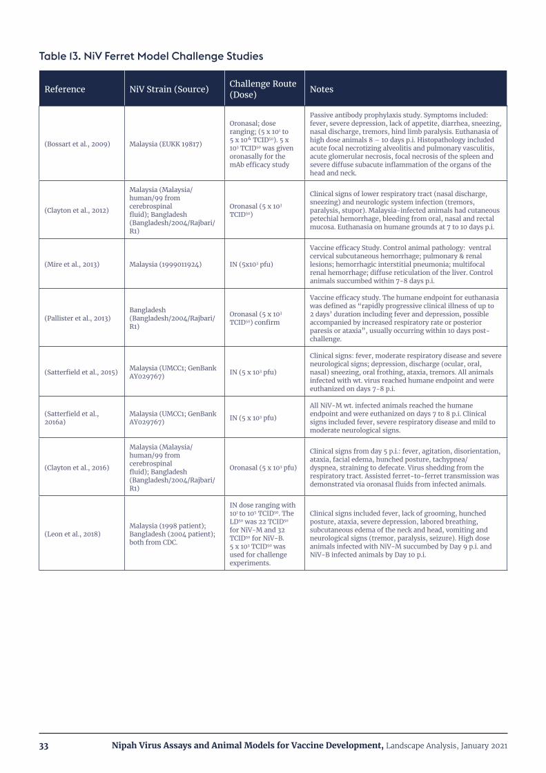
Nipah Virus Assays and Animal Models for Vaccine Development, Landscape Analysis, January 202133
Table 13. NiV Ferret Model Challenge Studies
Reference NiV Strain (Source) Challenge Route (Dose) Notes
(Bossart et al., 2009) Malaysia (EUKK 19817)
Oronasal; dose ranging; (5 x 101 to 5 x 10⁴ TCID50). 5 x 10³ TCID50 was given oronasally for the mAb efficacy study
Passive antibody prophylaxis study. Symptoms included: fever, severe depression, lack of appetite, diarrhea, sneezing, nasal discharge, tremors, hind limb paralysis. Euthanasia of high dose animals 8 – 10 days p.i. Histopathology included acute focal necrotizing alveolitis and pulmonary vasculitis, acute glomerular necrosis, focal necrosis of the spleen and severe diffuse subacute inflammation of the organs of the head and neck.
(Clayton et al., 2012)
Malaysia (Malaysia/human/99 from cerebrospinal fluid); Bangladesh (Bangladesh/2004/Rajbari/R1)
Oronasal (5 x 10³ TCID50)
Clinical signs of lower respiratory tract (nasal discharge, sneezing) and neurologic system infection (tremors, paralysis, stupor). Malaysia-infected animals had cutaneous petechial hemorrhage, bleeding from oral, nasal and rectal mucosa. Euthanasia on humane grounds at 7 to 10 days p.i.
(Mire et al., 2013) Malaysia (1999011924) IN (5x10³ pfu)
Vaccine efficacy Study. Control animal pathology: ventral cervical subcutaneous hemorrhage; pulmonary & renal lesions; hemorrhagic interstitial pneumonia; multifocal renal hemorrhage; diffuse reticulation of the liver. Control animals succumbed within 7-8 days p.i.
(Pallister et al., 2013)Bangladesh (Bangladesh/2004/Rajbari/R1)
Oronasal (5 x 10³ TCID50) confirm
Vaccine efficacy study. The humane endpoint for euthanasia was defined as “rapidly progressive clinical illness of up to 2 days’ duration including fever and depression, possible accompanied by increased respiratory rate or posterior paresis or ataxia”, usually occurring within 10 days post-challenge.
(Satterfield et al., 2015) Malaysia (UMCC1; GenBank AY029767) IN (5 x 10³ pfu)
Clinical signs: fever, moderate respiratory disease and severe neurological signs; depression, discharge (ocular, oral, nasal) sneezing, oral frothing, ataxia, tremors. All animals infected with wt. virus reached humane endpoint and were euthanized on days 7-8 p.i.
(Satterfield et al., 2016a)
Malaysia (UMCC1; GenBank AY029767) IN (5 x 10³ pfu)
All NiV-M wt. infected animals reached the humane endpoint and were euthanized on days 7 to 8 p.i. Clinical signs included fever, severe respiratory disease and mild to moderate neurological signs.
(Clayton et al., 2016)
Malaysia (Malaysia/human/99 from cerebrospinal fluid); Bangladesh (Bangladesh/2004/Rajbari/R1)
Oronasal (5 x 10³ pfu)
Clinical signs from day 5 p.i.: fever, agitation, disorientation, ataxia, facial edema, hunched posture, tachypnea/dyspnea, straining to defecate. Virus shedding from the respiratory tract. Assisted ferret-to-ferret transmission was demonstrated via oronasal fluids from infected animals.
(Leon et al., 2018)Malaysia (1998 patient); Bangladesh (2004 patient); both from CDC.
IN dose ranging with 101 to 105 TCID50. The LD50 was 22 TCID50 for NiV-M and 32 TCID50 for NiV-B. 5 x 10³ TCID50 was used for challenge experiments.
Clinical signs included fever, lack of grooming, hunched posture, ataxia, severe depression, labored breathing, subcutaneous edema of the neck and head, vomiting and neurological signs (tremor, paralysis, seizure). High dose animals infected with NiV-M succumbed by Day 9 p.i. and NiV-B infected animals by Day 10 p.i.

Nipah Virus Assays and Animal Models for Vaccine Development, Landscape Analysis, January 202134
The most relevant non-human primate model for NiV disease is the African Green Monkey (AGM; Chlorocebus sabaeus). Tables 14 and 15 provide a compilation of most of the published NiV AGM challenge studies, including challenge strain and available identifying information on the challenge virus, route of administration, dose and notes on disease course, clinical signs and pathology. Several publications have compiled detailed descriptions of NiV disease course in AGMs, including dose range, symptoms, viral loads, tissue tropism and pathology/histopathology in major organs (Geisbert et al., 2010; Johnston et al., 2015; Mire et al., 2016). Of particular interest, Johnston et al., (2015) conducted a detailed characterization study of NiV infection in AGMs with the aim of supporting future therapeutic and vaccine development, although this study only used the NiV Malaysia strain.
Intra-tracheal (IT) inoculation of AGMs with NiV is lethal, with death occurring in 9-12 days. Higher challenge doses generally accelerate the disease course and reduce survival time (Geisbert et al., 2012). Animals initially show signs of depression, lethargy, fever, and loss of appetite. NiV causes an acute respiratory disease in AGMs, characterized by labored breathing and bloody nasal discharge. The lungs become enlarged with edema and areas of congestion and hemorrhage (Geisbert et al., 2010). Histopathology includes alveolar hemorrhage in the lungs, prominent syncytia formation in lung endothelial cells, and generalized vasculitis. Neurological signs are less
consistent in the AGM than in the hamster and ferret models. Some animals show muscle twitches and partial paralysis (Geisbert et al., 2014), histopathological signs of encephalitis and tremors (Johnston et al., 2015), but overt brain abnormalities are usually not seen. One study identified lesions by brain imaging (Hammoud et al., 2018) while another did not (Cong et al., 2017). The different findings from these studies may be due to differences in dose or route of administration.
Infection with NiV Malaysia and Bangladesh strains has been directly compared in the AGM model, and in that study morbidity and mortality was accelerated in the NiV Bangladesh-infected animals by approximately three days compared with the NiV Malaysia-infected animals. This acceleration of disease progression appears distinct from what is seen in hamsters and ferrets, but consistent with human NiV disease. Otherwise, symptomology between the two groups of AGMs was comparable (Mire et al., 2016).
Several different isolates of NiV Malaysia have been used in AGM challenge studies, while the same isolate of NiV Bangladesh has been used (Tables 14 and 15). As with the hamster and ferret challenge models, production of a common, well-characterized stock of virus for AGM challenge studies would help to standardize performance of the model. There appears to be an overall trend toward lower challenge doses since the first AGM challenge experiments (Tables 14 and 15). However, the route of administration is not entirely settled. While most
experiments have used the intra-tracheal (IT) route, and some studies have included IN as well, recent experiments have tested aerosolized delivery (Cong et al., 2017; Lo et al., 2014). This form of administration may best mimic human-to-human transmission. The question of the utility, general feasibility and necessity of this route of administration may need to be settled to fully standardize performance of the NiV AGM model.
3. African Green Monkey (AGM)
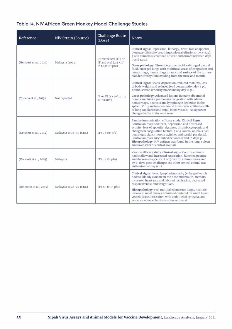
Nipah Virus Assays and Animal Models for Vaccine Development, Landscape Analysis, January 202135
Table 14. NiV African Green Monkey Model Challenge Studies
Reference NiV Strain (Source) Challenge Route (Dose) Notes
(Geisbert et al., 2010) Malaysia (1999)Intratracheal (IT) or IT and oral (2.5 x10³
to 1.3 x 10⁶ pfu)
Clinical signs: Depression, lethargy, fever, loss of appetite, dyspnea (difficulty breathing), pleural effusions (by x-ray). 7 of 8 animals succumbed or were euthanized between days 9 and 12 p.i.
Gross pathology: Thrombocytopenia, blood-tinged pleural fluid, enlarged lungs with multifocal areas of congestion and hemorrhage, hemorrhage on mucosal surface of the urinary bladder, frothy fluid exuding from the nose and mouth.
(Yoneda et al., 2013) Not reported IP or IN (1 x 10⁶ or 1 x 10⁸ TCID50)
Clinical Signs: Severe depression, reduced mobility, loss of body weight and reduced food consumption day 5 p.i. Animals were seriously moribund by day 14 p.i.
Gross pathology: Advanced lesions in many abdominal organs and lungs, pulmonary congestion with edema, hemorrhage, necrosis and lymphocyte depletion in the spleen. Virus antigen was found in vascular epithelial cells of lung capillaries and small blood vessels. No apparent changes in the brain were seen.
(Geisbert et al., 2014) Malaysia 1998-99 (CDC) IT (5 x 10⁵ pfu)
Passive immunization efficacy study. Clinical Signs: Control animals had fever, depression and decreased activity, loss of appetite, dyspnea, thrombocytopenia and changes in coagulation factors. 3 of 4 control animals had neurologic signs (muscle twitches and partial paralysis). Control animals succumbed between 8 and 10 days p.i. Histopathology: NiV antigen was found in the lung, spleen and brainstem of control animals
(Prescott et al., 2015) Malaysia IT (1 x 10⁵ pfu)
Vaccine efficacy study. Clinical signs: Control animals had shallow and increased respiration, hunched posture and decreased appetite. 2 of 3 control animals recovered by 15 days post-challenge; the other control animal was euthanized at day 9 p.i.
(Johnston et al., 2015) Malaysia 1998-99 (CDC) IT (2.5 x 10⁴ pfu)
Clinical signs: fever, lymphadenopathy (enlarged lymph nodes), bloody exudate in the nose and mouth, tremors, increased heart rate and labored respiration, decreased responsiveness and weight loss.
Histopathology: red, mottled edematous lungs, necrotic lesions in most tissues examined centered on small blood vessels (vasculitis) often with endothelial syncytia, and evidence of encephalitis is some animals/

Nipah Virus Assays and Animal Models for Vaccine Development, Landscape Analysis, January 202136
Table 15. NiV African Green Monkey Model Challenge Studies, Continued
Reference NiV Strain (Source) Challenge Route (Dose) Notes
(Mire et al., 2016) Malaysia 199912916; Bangladesh 200401066 IT + IN (5 x 10⁵ pfu)
Morbidity and mortality was accelerated for animals infected with NiV-B compared to NiV-M by approximately 3 days. The incidence of respiratory and neurologic symptoms was comparable between the NiV-B and NiV-M infected groups, as was overall symptomology. Animals in both groups showed primarily respiratory symptoms, and one in each group showed some neurological signs (tremors)
(Cong et al., 2017) MalaysiaIT or small-particle aerosol inhalation (1 x 10⁴ pfu)
Animals challenged by either method showed similar symptomology. Clinical signs: Lethargy, cough, difficulty breathing, decreased fluid and food consumption. There were no overt signs of neurological disease or hemorrhage, or bloody froth from the mouth/nose. There was no weight loss and only half of the animals had elevated temperature. Survival time for all animals was approximately 8 days. IT administration induced a significant congestion and loss in lung volume, while small-particle aerosol confined infection to the lower respiratory tract. Imaging did not reveal significant lesions in the brain.
(Mathieu et al., 2018) Malaysia (UMMC1, GenBank AY029767) IT (2 x 10⁷ pfu)
Peptide prophylaxis study. Clinical signs: Depression, loss of appetite, dyspnea, labored breathing, epistaxis (hemorrhage from nose or pharynx), paresis of hindlimbs, uncoordinated motor movements and brain hemorrhage. Control animals succumbed to infection by day 13 post-challenge.
(Hammoud et al., 2018) Malaysia 1998 (GenBank AF212302)
Medium-large particle aerosol inhalation (102 or 103pfu)
Clinical signs: lethargy, reduced appetite, cough. No animals showed overt neurological symptoms. Average survival time was 12.5 days p.i. There were no clear differences between the dose groups. Gross pathology showed congestion and hemorrhage in the lungs, but no overt brain abnormalities. Imaging showed loss of lung volume and brain lesions.
(Lo et al., 2019) Bangladesh/200401066 IN (1 x 10⁵ TCID50)Remdesivir therapeutic efficacy study. Clinical signs: Control animals developed severe respiratory symptoms and were euthanized on day 7 or 8 p.i.
(Mire et al., 2019) Bangladesh/200401066 IT + IN (5 x 10⁵ pfu)
Vaccine challenge study. Clinical signs: control animal showed loss of appetite, labored breathing & succumbed on day 8 p.i.
Clinical and gross pathological findings: lymphopenia (low lymphocyte count); serosanguinous oral & nasal discharge; severely enlarged lungs with severe congestion and hemorrhage; hemorrhage of the mucosal surface of the urinary bladder.
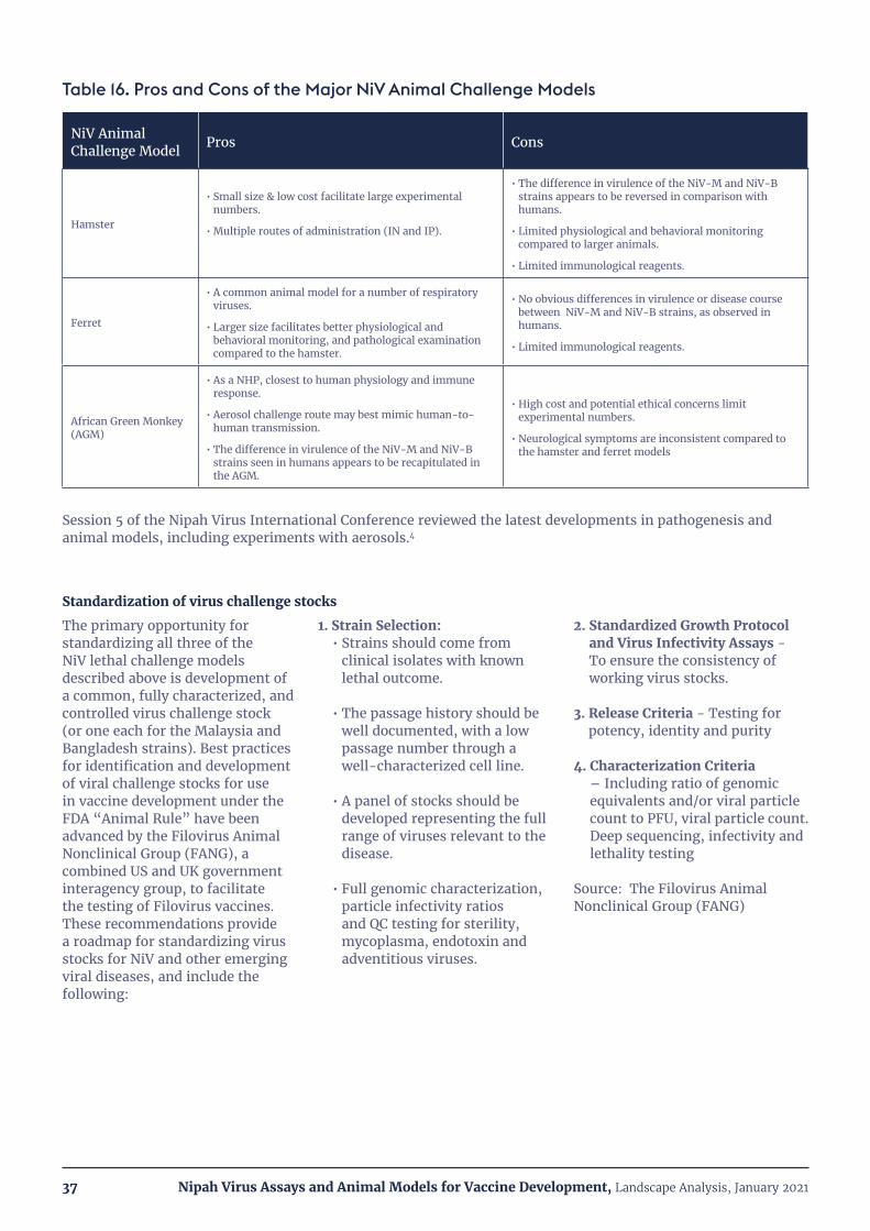
Nipah Virus Assays and Animal Models for Vaccine Development, Landscape Analysis, January 202137
Table 16. Pros and Cons of the Major NiV Animal Challenge Models
NiV Animal Challenge Model Pros Cons
Hamster
• Small size & low cost facilitate large experimental numbers.
• Multiple routes of administration (IN and IP).
• The difference in virulence of the NiV-M and NiV-B strains appears to be reversed in comparison with humans.
• Limited physiological and behavioral monitoring compared to larger animals.
• Limited immunological reagents.
Ferret
• A common animal model for a number of respiratory viruses.
• Larger size facilitates better physiological and behavioral monitoring, and pathological examination compared to the hamster.
• No obvious differences in virulence or disease course between NiV-M and NiV-B strains, as observed in humans.
• Limited immunological reagents.
African Green Monkey (AGM)
• As a NHP, closest to human physiology and immune response.
• Aerosol challenge route may best mimic human-to-human transmission.
• The difference in virulence of the NiV-M and NiV-B strains seen in humans appears to be recapitulated in the AGM.
• High cost and potential ethical concerns limit experimental numbers.
• Neurological symptoms are inconsistent compared to the hamster and ferret models
Session 5 of the Nipah Virus International Conference reviewed the latest developments in pathogenesis and animal models, including experiments with aerosols.4
The primary opportunity for standardizing all three of the NiV lethal challenge models described above is development of a common, fully characterized, and controlled virus challenge stock (or one each for the Malaysia and Bangladesh strains). Best practices for identification and development of viral challenge stocks for use in vaccine development under the FDA “Animal Rule” have been advanced by the Filovirus Animal Nonclinical Group (FANG), a combined US and UK government interagency group, to facilitate the testing of Filovirus vaccines. These recommendations provide a roadmap for standardizing virus stocks for NiV and other emerging viral diseases, and include the following:
1. Strain Selection:• Strains should come from
clinical isolates with known lethal outcome.
• The passage history should be well documented, with a low passage number through a well-characterized cell line.
• A panel of stocks should be developed representing the full range of viruses relevant to the disease.
• Full genomic characterization, particle infectivity ratios and QC testing for sterility, mycoplasma, endotoxin and adventitious viruses.
2. Standardized Growth Protocol and Virus Infectivity Assays - To ensure the consistency of working virus stocks.
3. Release Criteria - Testing for potency, identity and purity
4. Characterization Criteria – Including ratio of genomic equivalents and/or viral particle count to PFU, viral particle count. Deep sequencing, infectivity and lethality testing
Source: The Filovirus Animal Nonclinical Group (FANG)
Standardization of virus challenge stocks

Nipah Virus Assays and Animal Models for Vaccine Development, Landscape Analysis, January 202138
VI. CONCLUSIONSDevelopment of an effective prophylactic vaccine against NiV appears to be scientifically feasible, and the basic R&D components and knowledge are in place to enable successful development programs.
A large number of vaccine candidates using a range of platform technologies have been tested and showed very good efficacy in a variety of lethal animal challenge models. There is broad consensus in the NiV field that neutralizing antibodies are the mechanism of protection and will form the basis for defining a correlate of protection once appropriate studies are performed. A variety of assay platforms exist to quantitate NiV serum binding antibodies and virus neutralizing antibodies. While standardization of assays is in an early stage, there is broad acceptance of the pathways this process will follow. Finally, three animal challenge models (hamsters, ferrets and African Green Monkeys) appear to faithfully replicate many aspects of NiV infection and pathology in humans. Nevertheless, gaps, challenges and further questions remain which must be resolved to accelerate NiV vaccine development and ensure success. Below are three key, forward-looking topics and questions to be addressed:
Defining a correlate of protectionMany types of vaccines tested in NiV lethal animal challenge models elicited serum neutralizing antibodies. However, none of the studies determined a correlate of protection based on neutralizing antibody titer because in most studies all the animals were protected. Encouragingly, anecdotal evidence from NiV researchers suggests that even very low neutralizing antibody titers may be protective in animal models (Source: CEPI 2nd Assays and Standards Workshop; June
2019). Nevertheless, it is important to generate data on the protective threshold for several reasons. The first reason is for evaluating vaccine candidates in early development. What minimal titers are needed for protection and what dose(s) of a particular candidate are needed to elicit them, and in which animal model(s)? Which candidates elicit minimal protective titers after a single dose, as is the preference in the WHO Target Product Profile? Answers to these questions are critical for vaccine candidate evaluation and advancement. The second reason is to anticipate what neutralizing antibody responses will be needed in formal animal studies for vaccine licensure under the “Animal Rule”, in which antibody responses in animal challenge models will be bridged back to those seen in human Phase I/II clinical studies. Finally, knowing the correlate of protection will make it possible to determine the durability of protection after vaccination in humans.
Choice of assay(s) for serum neutralizing antibodiesAs discussed earlier, there are well-established live NiV-based assays for serum neutralizing antibodies (SNT, PRNT) that are widely accepted and have been used in the majority of published NiV animal challenge studies. In theory, it should be possible to standardize and validate these assays, and use them for vaccine development, human clinical studies and animal challenge studies for licensure under the “Animal Rule”. However, the use of live NiV and the requirement for performing these assays under
BSL-4 containment would create serious technical and logistical challenges for vaccine developers needing to perform the assays on large numbers of samples, often under cGMP (current Good Manufacturing Practices) for lot release. Are research institutions with BSL-4 capability willing to collaborate with vaccine developers in this way, and are they capable of processing large numbers of samples under the quality systems necessary for clinical development and licensure? Could a centralized BSL-4 facility be created/funded to support all the NiV vaccine developers in this effort? Alternatively, surrogate assays for NiV serum neutralizing antibodies have been developed which can be performed by vaccine developers without high-level biocontainment, most notably those using pseudoviruses expressing the NiV F and G glycoproteins. However, these assays have not been used extensively by major NiV research laboratories, and even the most recent animal challenge and vaccine studies continue to use the live NiV-based assays almost exclusively. At a minimum, extensive testing will be required to characterize the sensitivity and specificity of these assays in comparison with the live NiV-based assays and evaluate their potential for validation. Who would perform these studies? And what else needs to be done (and by whom) to facilitate acceptance of these assays by NiV researchers and regulatory bodies? This is an area in which major stakeholders in NiV vaccine development (such as CEPI) have a key role to play.
Nipah Virus Assays and Animal Models for Vaccine Development, Landscape Analysis, January 202139
Characterization of animal challenge models
FDA draft guidance on the use of animal challenge models for licensure under the “Animal Rule” emphasizes the requirement for a high degree of characterization of the animal model, such as identity and characterization of the challenge agent, route and quantification of exposure and the course and pathophysiology of infection compared to humans. The three well established animal
challenge models for NiV infection – hamsters, ferrets and the African Green Monkey – have all been characterized in multiple publications, in some cases in a high degree of detail. However, is the existing characterization sufficient to support use under the “Animal Rule”? If not, what additional studies are needed to close the gaps, and who would perform them? Ultimately, these questions should be addressed in consultation with FDA,
which recommends early and frequent engagement when use of the “Animal Rule” is being contemplated. This is another area in which leadership by major stakeholders, such as CEPI, is needed to facilitate vaccine development and licensure by ensuring these issues are resolved and the animal models are ready for use when vaccine candidates are ready for clinical testing.
Nipah Virus Assays and Animal Models for Vaccine Development, Landscape Analysis, January 202140
VII. REFERENCESAguilar, H.C., and Lee, B. (2011). Emerging paramyxoviruses: molecular mechanisms and antiviral strategies. Expert Rev Mol Med 13, e6.
Ahmad, S.B., and Tan, C.T. (2014). Nipah encephalitis - an update. Med J Malaysia 69, 103-111.
Ang, B.S.P., Lim, T.C.C., and Wang, L. (2018). Nipah Virus Infection. J Clin Microbiol 56.
Banerjee, S., Gupta, N., Kodan, P., Mittal, A., Ray, Y., Nischal, N., Soneja, M., Biswas, A., and Wig, N. (2019). Nipah virus disease: A rare and intractable disease. Intractable Rare Dis Res 8, 1-8.
Baseler, L., de Wit, E., Scott, D.P., Munster, V.J., and Feldmann, H. (2015). Syrian hamsters (Mesocricetus auratus) oronasally inoculated with a Nipah virus isolate from Bangladesh or Malaysia develop similar respiratory tract lesions. Vet Pathol 52, 38-45.
Baseler, L., Scott, D.P., Saturday, G., Horne, E., Rosenke, R., Thomas, T., Meade-White, K., Haddock, E., Feldmann, H., and de Wit, E. (2016). Identifying Early Target Cells of Nipah Virus Infection in Syrian Hamsters. PLoS Negl Trop Dis 10, e0005120.
Beasley, D.W.C., Brasel, T.L., and Comer, J.E. (2016). First vaccine approval under the FDA Animal Rule. NPJ Vaccines 1, 16013.
Borisevich, V., Lee, B., Hickey, A., DeBuysscher, B., Broder, C.C., Feldmann, H., and Rockx, B. (2016). Escape From Monoclonal Antibody Neutralization Affects Henipavirus Fitness In Vitro and In Vivo. J Infect Dis 213, 448-455.
Bossart, K.N., McEachern, J.A., Hickey, A.C., Choudhry, V., Dimitrov, D.S., Eaton, B.T., and Wang, L.F. (2007). Neutralization assays for differential henipavirus serology using Bio-Plex protein array systems. J Virol Methods 142, 29-40.
Bossart, K.N., Rockx, B., Feldmann, F., Brining, D., Scott, D., LaCasse, R., Geisbert, J.B., Feng, Y.R., Chan, Y.P., Hickey, A.C., et al. (2012). A Hendra virus G glycoprotein subunit vaccine protects African Green Monkeys from Nipah virus challenge. Sci Transl Med 4, 146ra107.
Bossart, K.N., Zhu, Z., Middleton, D., Klippel, J., Crameri, G., Bingham, J., McEachern, J.A., Green, D., Hancock, T.J., Chan, Y.P., et al. (2009). A neutralizing human monoclonal antibody protects against lethal disease in a new ferret model of acute nipah virus infection. PLoS Pathog 5, e1000642.
Broder, C.C., Geisbert, T.W., Xu, K., Nikolov, D.B., Wang, L.F., Middleton, D., Pallister, J., and Bossart, K.N. (2012). Immunization strategies against henipaviruses. Curr Top Microbiol Immunol 359, 197-223.
Broder, C.C., Xu, K., Nikolov, D.B., Zhu, Z., Dimitrov, D.S., Middleton, D., Pallister, J., Geisbert, T.W., Bossart, K.N., and Wang, L.F. (2013). A treatment for and vaccine against the deadly Hendra and Nipah viruses. Antiviral Res 100, 8-13.
Chadha, M.S., Comer, J.A., Lowe, L., Rota, P.A., Rollin, P.E., Bellini, W.J., Ksiazek, T.G., and Mishra, A.C. (2006). Nipah virus-associated encephalitis outbreak, Siliguri, India. Emerg Infect Dis 12, 235-240.
Ching, P.K., de los Reyes, V.C., Sucaldito, M.N., Tayag, E., Columna-Vingno, A.B., Malbas, F.F., Jr., Bolo, G.C., Jr., Sejvar, J.J., Eagles, D., Playford, G., et al. (2015). Outbreak of henipavirus infection, Philippines, 2014. Emerg Infect Dis 21, 328-331.
Chong, H.T., Kamarulzman, A., Tan, C.T., Goh, K.J., Thayaparan, T., Kunjapan, S.R., Chew, N.K., Chua, K.B., and Lam, S.K. (2001). Treatment of acute Nipah encephalitis with ribavirin. Ann Neurol 49, 810-813.
Chong, H.T., Kunjapan, S.R., Thayaparan, T., Tong, J., Petharunam, V., Jusoh, M.R., and Tan, C.T. (2002). Nipah encephalitis outbreak in Malaysia, clinical features in patients from Seremban. Can J Neurol Sci 29, 83-87.
Clayton, B.A., Middleton, D., Arkinstall, R., Frazer, L., Wang, L.F., and Marsh, G.A. (2016). The Nature of Exposure Drives Transmission of Nipah Viruses from Malaysia and Bangladesh in Ferrets. PLoS Negl Trop Dis 10, e0004775.
Clayton, B.A., Middleton, D., Bergfeld, J., Haining, J., Arkinstall, R., Wang, L., and Marsh, G.A. (2012). Transmission routes for nipah virus from Malaysia and Bangladesh. Emerg Infect Dis 18, 1983-1993.
Cong, Y., Lentz, M.R., Lara, A., Alexander, I., Bartos, C., Bohannon, J.K., Hammoud, D., Huzella, L., Jahrling, P.B., Janosko, K., et al. (2017). Loss in lung volume and changes in the immune response demonstrate disease progression in African green monkeys infected by small-particle aerosol and intratracheal exposure to Nipah virus. PLoS Negl Trop Dis 11, e0005532.
Nipah Virus Assays and Animal Models for Vaccine Development, Landscape Analysis, January 202141
Crameri, G., Wang, L.F., Morrissy, C., White, J., and Eaton, B.T. (2002). A rapid immune plaque assay for the detection of Hendra and Nipah viruses and anti-virus antibodies. Journal of Virological Methods 99, 41-51.
Daniels, P., Ksiazek, T., and Eaton, B.T. (2001). Laboratory diagnosis of Nipah and Hendra virus infections. Microbes Infect 3, 289-295.
Dawes, B.E., Kalveram, B., Ikegami, T., Juelich, T., Smith, J.K., Zhang, L., Park, A., Lee, B., Komeno, T., Furuta, Y., et al. (2018). Favipiravir (T-705) protects against Nipah virus infection in the hamster model. Sci Rep 8, 7604.
de Wit, E., Bushmaker, T., Scott, D., Feldmann, H., and Munster, V.J. (2011). Nipah virus transmission in a hamster model. PLoS Negl Trop Dis 5, e1432.
de Wit, E., and Munster, V.J. (2015). Animal models of disease shed light on Nipah virus pathogenesis and transmission. J Pathol 235, 196-205.
de Wit, E., Prescott, J., Falzarano, D., Bushmaker, T., Scott, D., Feldmann, H., and Munster, V.J. (2014). Foodborne transmission of nipah virus in Syrian hamsters. PLoS Pathog 10, e1004001.
DeBuysscher, B.L., de Wit, E., Munster, V.J., Scott, D., Feldmann, H., and Prescott, J. (2013). Comparison of the pathogenicity of Nipah virus isolates from Bangladesh and Malaysia in the Syrian hamster. PLoS Negl Trop Dis 7, e2024.
DeBuysscher, B.L., Scott, D., Marzi, A., Prescott, J., and Feldmann, H. (2014). Single-dose live-attenuated Nipah virus vaccines confer complete protection by eliciting antibodies directed against surface glycoproteins. Vaccine 32, 2637-2644.
DeBuysscher, B.L., Scott, D., Thomas, T., Feldmann, H., and Prescott, J. (2016). Peri-exposure protection against Nipah virus disease using a single-dose recombinant vesicular stomatitis virus-based vaccine. NPJ Vaccines 1.
Defang, G.N., Khetawat, D., Broder, C.C., and Quinnan, G.V., Jr. (2010). Induction of neutralizing antibodies to Hendra and Nipah glycoproteins using a Venezuelan equine encephalitis virus in vivo expression system. Vaccine 29, 212-220.
Dhondt, K.P., and Horvat, B. (2013). Henipavirus infections: lessons from animal models. Pathogens 2, 264-287.
Dhondt, K.P., Mathieu, C., Chalons, M., Reynaud, J.M., Vallve, A., Raoul, H., and Horvat, B. (2013). Type I interferon signaling protects mice from lethal henipavirus infection. J Infect Dis 207, 142-151.
Enchery, F., and Horvat, B. (2017). Understanding the interaction between henipaviruses and their natural host, fruit bats: Paving the way toward control of highly lethal infection in humans. Int Rev Immunol 36, 108-121.
Enkirch, T., and von Messling, V. (2015). Ferret models of viral pathogenesis. Virology 479-480, 259-270.
Escaffre, O., Borisevich, V., and Rockx, B. (2013). Pathogenesis of Hendra and Nipah virus infection in humans. J Infect Dev Ctries 7, 308-311.
Eshaghi, M., Tan, W.S., Chin, W.K., and Yusoff, K. (2005). Purification of the extra-cellular domain of Nipah virus glycoprotein produced in Escherichia coli and possible application in diagnosis. J Biotechnol 116, 221-226.
Eshaghi, M., Tan, W.S., Mohidin, T.B., and Yusoff, K. (2004). Nipah virus glycoprotein: production in baculovirus and application in diagnosis. Virus Res 106, 71-76.
Ferguson, M., Heath, A., Johnes, S., Pagliusi, S., Dillner, J., and Collaborative Study, P. (2006). Results of the first WHO international collaborative study on the standardization of the detection of antibodies to human papillomaviruses. Int J Cancer 118, 1508-1514.
Ferguson, M., Wilkinson, D.E., Heath, A., and Matejtschuk, P. (2011). The first international standard for antibodies to HPV 16. Vaccine 29, 6520-6526.
Freiberg, A.N., Worthy, M.N., Lee, B., and Holbrook, M.R. (2010). Combined chloroquine and ribavirin treatment does not prevent death in a hamster model of Nipah and Hendra virus infection. J Gen Virol 91, 765-772.
Geisbert, T.W., Daddario-DiCaprio, K.M., Hickey, A.C., Smith, M.A., Chan, Y.P., Wang, L.F., Mattapallil, J.J., Geisbert, J.B., Bossart, K.N., and Broder, C.C. (2010). Development of an acute and highly pathogenic nonhuman primate model of Nipah virus infection. PLoS One 5, e10690.
Geisbert, T.W., Feldmann, H., and Broder, C.C. (2012). Animal challenge models of henipavirus infection and pathogenesis. Curr Top Microbiol Immunol 359, 153-177.
Geisbert, T.W., Mire, C.E., Geisbert, J.B., Chan, Y.P., Agans, K.N., Feldmann, F., Fenton, K.A., Zhu, Z., Dimitrov, D.S., Scott, D.P., et al. (2014). Therapeutic treatment of Nipah virus infection in nonhuman primates with a neutralizing human monoclonal antibody. Sci Transl Med 6, 242ra282.
Nipah Virus Assays and Animal Models for Vaccine Development, Landscape Analysis, January 202142
Georges-Courbot, M.C., Contamin, H., Faure, C., Loth, P., Baize, S., Leyssen, P., Neyts, J., and Deubel, V. (2006). Poly(I)-poly(C12U) but not ribavirin prevents death in a hamster model of Nipah virus infection. Antimicrob Agents Chemother 50, 1768-1772.
Golding, H., Khurana, S., and Zaitseva, M. (2018). What Is the Predictive Value of Animal Models for Vaccine Efficacy in Humans? The Importance of Bridging Studies and Species-Independent Correlates of Protection. Cold Spring Harb Perspect Biol 10.
Guillaume-Vasselin, V., Lemaitre, L., Dhondt, K.P., Tedeschi, L., Poulard, A., Charreyre, C., and Horvat, B. (2016). Protection from Hendra virus infection with Canarypox recombinant vaccine. NPJ Vaccines 1, 16003.
Guillaume, V., Contamin, H., Loth, P., Georges-Courbot, M.C., Lefeuvre, A., Marianneau, P., Chua, K.B., Lam, S.K., Buckland, R., Deubel, V., et al. (2004). Nipah virus: vaccination and passive protection studies in a hamster model. J Virol 78, 834-840.
Guillaume, V., Contamin, H., Loth, P., Grosjean, I., Courbot, M.C., Deubel, V., Buckland, R., and Wild, T.F. (2006). Antibody prophylaxis and therapy against Nipah virus infection in hamsters. J Virol 80, 1972-1978.
Hammoud, D.A., Lentz, M.R., Lara, A., Bohannon, J.K., Feuerstein, I., Huzella, L., Jahrling, P.B., Lackemeyer, M., Laux, J., Rojas, O., et al. (2018). Aerosol exposure to intermediate size Nipah virus particles induces neurological disease in African green monkeys. PLoS Negl Trop Dis 12, e0006978.
Harcourt, B.H., Lowe, L., Tamin, A., Liu, X., Bankamp, B., Bowden, N., Rollin, P.E., Comer, J.A., Ksiazek, T.G., Hossain, M.J., et al. (2005). Genetic characterization of Nipah virus, Bangladesh, 2004. Emerg Infect Dis 11, 1594-1597.
Hooper, P., Zaki, S., Daniels, P., and Midleton, D. (2001). Comparative pathology of the diseases caused by Hendra and Nipah viruses. Microbes Infect 3, 315-322.
Hossain, M.J., Gurley, E.S., Montgomery, J.M., Bell, M., Carroll, D.S., Hsu, V.P., Formenty, P., Croisier, A., Bertherat, E., Faiz, M.A., et al. (2008). Clinical presentation of nipah virus infection in Bangladesh. Clin Infect Dis 46, 977-984.
Johnston, S.C., Briese, T., Bell, T.M., Pratt, W.D., Shamblin, J.D., Esham, H.L., Donnelly, G.C., Johnson, J.C., Hensley, L.E., Lipkin, W.I., et al. (2015). Detailed analysis of the African green monkey model of Nipah virus disease. PLoS One 10, e0117817.
Kaku, Y., Noguchi, A., Marsh, G.A., Barr, J.A., Okutani, A., Hotta, K., Bazartseren, B., Fukushi, S., Broder, C.C., Yamada, A., et al. (2012). Second generation of pseudotype-based serum neutralization assay for Nipah virus antibodies: sensitive and high-throughput analysis utilizing secreted alkaline phosphatase. J Virol Methods 179, 226-232.
Kaku, Y., Noguchi, A., Marsh, G.A., McEachern, J.A., Okutani, A., Hotta, K., Bazartseren, B., Fukushi, S., Broder, C.C., Yamada, A., et al. (2009). A neutralization test for specific detection of Nipah virus antibodies using pseudotyped vesicular stomatitis virus expressing green fluorescent protein. J Virol Methods 160, 7-13.
Keshwara, R., Shiels, T., Postnikova, E., Kurup, D., Wirblich, C., Johnson, R.F., and Schnell, M.J. (2019). Rabies-based vaccine induces potent immune responses against Nipah virus. NPJ Vaccines 4, 15.
Kong, D., Wen, Z., Su, H., Ge, J., Chen, W., Wang, X., Wu, C., Yang, C., Chen, H., and Bu, Z. (2012). Newcastle disease virus-vectored Nipah encephalitis vaccines induce B and T cell responses in mice and
long-lasting neutralizing antibodies in pigs. Virology 432, 327-335.
Kumar, C.P.G., Sugunan, A.P., Yadav, P., Kurup, K.K., Aarathee, R., Manickam, P., Bhatnagar, T., Radhakrishnan, C., Thomas, B., Kumar, A.t., et al. (2019). Infections among Contacts of Patients with Nipah Virus, India. Emerg Infect Dis 25, 1007-1010.
Kurup, D., Wirblich, C., Feldmann, H., Marzi, A., and Schnell, M.J. (2015). Rhabdovirus-based vaccine platforms against henipaviruses. J Virol 89, 144-154.
Leon, A.J., Borisevich, V., Boroumand, N., Seymour, R., Nusbaum, R., Escaffre, O., Xu, L., Kelvin, D.J., and Rockx, B. (2018). Host gene expression profiles in ferrets infected with genetically distinct henipavirus strains. PLoS Negl Trop Dis 12, e0006343.
Lo, M.K., Bird, B.H., Chattopadhyay, A., Drew, C.P., Martin, B.E., Coleman, J.D., Rose, J.K., Nichol, S.T., and Spiropoulou, C.F. (2014). Single-dose replication-defective VSV-based Nipah virus vaccines provide protection from lethal challenge in Syrian hamsters. Antiviral Res 101, 26-29.
Lo, M.K., Feldmann, F., Gary, J.M., Jordan, R., Bannister, R., Cronin, J., Patel, N.R., Klena, J.D., Nichol, S.T., Cihlar, T., et al. (2019). Remdesivir (GS-5734) protects African green monkeys from Nipah virus challenge. Sci Transl Med 11, eaau9242.
Lo, M.K., and Rota, P.A. (2008). The emergence of Nipah virus, a highly pathogenic paramyxovirus. J Clin Virol 43, 396-400.
Mathieu, C., Guillaume, V., Sabine, A., Ong, K.C., Wong, K.T., Legras-Lachuer, C., and Horvat, B. (2012a). Lethal Nipah virus infection induces rapid overexpression of CXCL10. PLoS One 7, e32157.
Nipah Virus Assays and Animal Models for Vaccine Development, Landscape Analysis, January 202143
Mathieu, C., Guillaume, V., Volchkova, V.A., Pohl, C., Jacquot, F., Looi, R.Y., Wong, K.T., Legras-Lachuer, C., Volchkov, V.E., Lachuer, J., et al. (2012b). Nonstructural Nipah virus C protein regulates both the early host proinflammatory response and viral virulence. J Virol 86, 10766-10775
Mathieu, C., Porotto, M., Figueira, T.N., Horvat, B., and Moscona, A. (2018). Fusion Inhibitory Lipopeptides Engineered for Prophylaxis of Nipah Virus in Primates. J Infect Dis 218, 218-227.
Mazzola, L.T., and Kelly-Cirino, C. (2019). Diagnostics for Nipah virus: a zoonotic pathogen endemic to Southeast Asia. BMJ Glob Health 4, e001118.
McDonald, J.U., Rigsby, P., Dougall, T., Engelhardt, O.G., and Study, P. (2018). Establishment of the first WHO International Standard for antiserum to Respiratory Syncytial Virus: Report of an international collaborative study. Vaccine 36, 7641-7649.
McEachern, J.A., Bingham, J., Crameri, G., Green, D.J., Hancock, T.J., Middleton, D., Feng, Y.R., Broder, C.C., Wang, L.F., and Bossart, K.N. (2008). A recombinant subunit vaccine formulation protects against lethal Nipah virus challenge in cats. Vaccine 26, 3842-3852.
McLean, R.K., and Graham, S.P. (2019). Vaccine Development for Nipah Virus Infection in Pigs. Front Vet Sci 6, 16.
Middleton, D.J., Morrissy, C.J., van der Heide, B.M., Russell, G.M., Braun, M.A., Westbury, H.A., Halpin, K., and Daniels, P.W. (2007). Experimental Nipah virus infection in pteropid bats (Pteropus poliocephalus). J Comp Pathol 136, 266-272.
Mire, C.E., Geisbert, J.B., Agans, K.N., Versteeg, K.M., Deer, D.J., Satterfield, B.A., Fenton, K.A.,
and Geisbert, T.W. (2019). Use of Single-Injection Recombinant Vesicular Stomatitis Virus Vaccine to Protect Nonhuman Primates Against Lethal Nipah Virus Disease. Emerg Infect Dis 25, 1144-1152.
Mire, C.E., Satterfield, B.A., Geisbert, J.B., Agans, K.N., Borisevich, V., Yan, L., Chan, Y.P., Cross, R.W., Fenton, K.A., Broder, C.C., et al. (2016). Pathogenic Differences between Nipah Virus Bangladesh and Malaysia Strains in Primates: Implications for Antibody Therapy. Sci Rep 6, 30916.
Mire, C.E., Versteeg, K.M., Cross, R.W., Agans, K.N., Fenton, K.A., Whitt, M.A., and Geisbert, T.W. (2013). Virol J 10, e353.
Mungall, B.A., Middleton, D., Crameri, G., Bingham, J., Halpin, K., Russell, G., Green, D., McEachern, J., Pritchard, L.I., Eaton, B.T., et al. (2006). Feline model of acute nipah virus infection and protection with a soluble glycoprotein-based subunit vaccine. J Virol 80, 12293-12302.
Munster, V.J., Prescott, J.B., Bushmaker, T., Long, D., Rosenke, R., Thomas, T., Scott, D., Fischer, E.R., Feldmann, H., and de Wit, E. (2012). Rapid Nipah virus entry into the central nervous system of hamsters via the olfactory route. Sci Rep 2, 736.
Pallister, J.A., Klein, R., Arkinstall, R., Haining, J., Long, F., White, J.R., Payne, J., Feng, Y.R., Wang, L.F., Broder, C.C., et al. (2013). Vaccination of ferrets with a recombinant G glycoprotein subunit vaccine provides protection against Nipay virus disease for over 12 months. Virol J 10, e237.
Pickering, B.S., Hardham, J.M., Smith, G., Weingartl, E.T., Dominowski, P.J., Foss, D.L., Mwangi, D., Broder, C.C., Roth, J.A., and Weingartl, H.M. (2016). Protection against henipaviruses in swine requires both, cell-mediated and humoral immune response.
Vaccine 34, 4777-4786.
Ploquin, A., Szecsi, J., Mathieu, C., Guillaume, V., Barateau, V., Ong, K.C., Wong, K.T., Cosset, F.-L., Horvat, B., and Salvetti, A. (2013). Protection against Henipavirus infection using recombinant AAV vector vaccines. J Infect Dis 207, 469-478.
Porotto, M., Rockx, B., Yokoyama, C.C., Talekar, A., Devito, I., Palermo, L.M., Liu, J., Cortese, R., Lu, M., Feldmann, H., et al. (2010). Inhibition of Nipah virus infection in vivo: targeting an early stage of paramyxovirus fusion activation during viral entry. PLoS Pathog 6, e1001168.
Prescott, J., de Wit, E., Feldmann, H., and Munster, V.J. (2012). The immune response to Nipah virus infection. Arch Virol 157, 1635-1641.
Prescott, J., DeBuysscher, B.L., Feldmann, F., Gardner, D.J., Haddock, E., Martellaro, C., Scott, D., and Feldmann, H. (2015). Single-dose live-attenuated vesicular stomatitis virus-based vaccine protects African green monkeys from Nipah virus disease. Vaccine 33, 2823-2829.
Rahman, M., and Chakraborty, A. (2012). Nipah virus outbreaks in Bangladesh: a deadly infectious disease. WHO South-East Asia Journal of Public Health 1, 208-212.
Ramphul, K., Mejias, S.G., Agumadu, V.C., Sombans, S., Sonaye, R., and Lohana, P. (2018). The Killer Virus Called Nipah: A Review. Cureus 10, e3168.
Rijpkema, S., Hockley, J., Logan, A., Rigsby, P., Atkinson, E., Jin, C., Goldblatt, D., Liang, H., Bachtiar, N.S., Yang, J.S., et al. (2018). Establishment of the first International Standard for human anti-typhoid capsular Vi polysaccharide IgG. Biologicals 56, 29-38.
Nipah Virus Assays and Animal Models for Vaccine Development, Landscape Analysis, January 202144
Rockx, B. (2014). Recent developments in experimental animal models of Henipavirus infection. Pathog Dis 71, 199-206.
Rockx, B., Bossart, K.N., Feldmann, F., Geisbert, J.B., Hickey, A.C., Brining, D., Callison, J., Safronetz, D., Marzi, A., Kercher, L., et al. (2010). A novel model of lethal Hendra virus infection in African green monkeys and the effectiveness of ribavirin treatment. J Virol 84, 9831-9839.
Rockx, B., Brining, D., Kramer, J., Callison, J., Ebihara, H., Mansfield, K., and Feldmann, H. (2011). Clinical outcome of henipavirus infection in hamsters is determined by the route and dose of infection. J Virol 85, 7658-7671.
Rockx, B., Winegar, R., and Freiberg, A.N. (2012). Recent progress in henipavirus research: molecular biology, genetic diversity, animal models. Antiviral Res 95, 135-149.
Satterfield, B.A., Cross, R.W., Fenton, K.A., Agans, K.N., Basler, C.F., Geisbert, T.W., and Mire, C.E. (2015). The immunomodulating V and W proteins of Nipah virus determine disease course. Nat Commun 6, 7483.
Satterfield, B.A., Cross, R.W., Fenton, K.A., Borisevich, V., Agans, K.N., Deer, D.J., Graber, J., Basler, C.F., Geisbert, T.W., and Mire, C.E. (2016a). Nipah Virus C and W Proteins Contribute to Respiratory Disease in Ferrets. J Virol 90, 6326-6343.
Satterfield, B.A., Dawes, B.E., and Milligan, G.N. (2016b). Status of vaccine research and development of vaccines for Nipah virus. Vaccine 34, 2971-2975.
Sauerhering, L., Zickler, M., Elvert, M., Behner, L., Matrosovich, T., Erbar, S., Matrosovich, M., and Maisner, A. (2016). Species-specific and individual differences in Nipah virus replication in porcine and human airway epithelial cells. J Gen Virol 97, 1511-1519.
Schountz, T., Campbell, C., Wagner, K., Rovnak, J., Martellaro, C., DeBuysscher, B.L., Feldmann, H., and Prescott, J. (2019). Differential Innate Immune Responses Elicited by Nipah Virus and Cedar Virus Correlate with Disparate In Vivo Pathogenesis in Hamsters. Viruses 11.
Sejvar, J.J., Hossain, J., Saha, S.K., Gurley, E.S., Banu, S., Hamadani, J.D., Faiz, M.A., Siddiqui, F.M., Mohammad, Q.D., Mollah, A.H., et al. (2007). Long-term neurological and functional outcome in Nipah virus infection. Ann Neurol 62, 235-242.
Sharma, V., Kaushik, S., Kumar, R., Yadav, J.P., and Kaushik, S. (2019). Emerging trends of Nipah virus: A review. Rev Med Virol 29, e2010.
Stephenson, I., Heath, A., Major, D., Newman, R.W., Hoschler, K., Junzi, W., Katz, J.M., Weir, J.P., Zambon, M.C., and Wood, J.M. (2009). Reproducibility of serologic assays for influenza virus A (H5N1). Emerg Infect Dis 15, 1252-1259.
Sun, B., Jia, L., Liang, B., Chen, Q., and Liu, D. (2018). Phylogeography, Transmission, and Viral Proteins of Nipah Virus. Virol Sin 33, 385-393.
Tamin, A., Harcourt, B.H., Lo, M.K., Roth, J.A., Wolf, M.C., Lee, B., Weingartl, H., Audonnet, J.C., Bellini, W.J., and Rota, P.A. (2009). Development of a neutralization assay for Nipah virus using pseudotype particles. J Virol Methods 160, 1-6.
Tan, R., Hodge, A., Klein, R., Edwards, N., Huang, J.A., Middleton, D., and Watts, S.P. (2018). Virus-neutralising antibody responses in horses following vaccination with Equivac(R) HeV: a field study. Aust Vet J 96, 161-166.
Thakur, N., and Bailey, D. (2019). Advances in diagnostics, vaccines and therapeutics for Nipah virus. Microbes Infect.
van Doremalen, N., Lambe, T., Sebastian, S., Bushmaker, T., Fischer, R., Feldmann, F., Haddock, E., Letko, M., Avanzato, V.A., Rissanen, I., et al. (2019). A single-dose ChAdOx1-vectored vaccine provides complete protection against Nipah Bangladesh and Malaysia in Syrian golden hamsters. PLoS Negl Trop Dis 13, e0007462.
Vignali, D.A.A. (2000). Multiplexed particle-based flow cytometric assays. Journal of Immunological Methods 243, 243-255.
Walpita, P., Barr, J., Sherman, M., Basler, C.F., and Wang, L. (2011). Vaccine potential of Nipah virus-like particles. PLoS One 6, e18437.
Walpita, P., Cong, Y., Jahrling, P.B., Rojas, O., Postnikova, E., Yu, S., Johns, L., and Holbrook, M.R. (2017). A VLP-based vaccine provides complete protection against Nipah virus challenge following multiple-dose or single-dose vaccination schedules in a hamster model. NPJ Vaccines 2, 21.
Wang, L.-F., Harcourt, B.H., Yu, M., Tamin, A., Rota, P.A., Bellini, W.J., and Eaton, B.T. (2001). Molecular biology of Hendra and Nipah viruses. Microbes Infect 3, 279-287.
Wang, L.F., and Daniels, P. (2012). Diagnosis of henipavirus infection: current capabilities and future directions. Curr Top Microbiol Immunol 359, 179-196.
Weingartl, H.M., Berhane, Y., Caswell, J.L., Loosmore, S., Audonnet, J.C., Roth, J.A., and Czub, M. (2006). Recombinant nipah virus vaccines protect pigs against challenge. J Virol 80, 7929-7938.
Wilkinson, D.E., Page, M., Mattiuzzo, G., Hassall, M., Dougall, T., Rigsby, P., Stone, L., and Minor, P. (2017). Comparison of platform technologies for assaying antibody to Ebola virus. Vaccine 35, 1347-1352.
Nipah Virus Assays and Animal Models for Vaccine Development, Landscape Analysis, January 202145
Williamson, E.D., and Westlake, G.E. (2019). Vaccines for emerging pathogens: prospects for licensure. Clin Exp Immunol.
Williamson, M.M., and Torres-Velez, F.J. (2010). Henipavirus: a review of laboratory animal pathology. Vet Pathol 47, 871-880.
Wong, K.T., Grosjean, I., Brisson, C., Blanquier, B., Fevre-Montange, M., Bernard, A., Loth, P., Georges-Courbot, M.-C., Chevallier, M., Akaoka, H., et al. (2003). A Golden Hamster Model for Human Acute Nipah Virus Infection. The American Journal of Pathology 163, 2127-2137.
Xu, K., Broder, C.C., and Nikolov, D.B. (2012). Ephrin-B2 and ephrin-B3 as functional henipavirus receptors. Semin Cell Dev Biol 23, 116-123.
Yoneda, M., Georges-Courbot, M.C., Ikeda, F., Ishii, M., Nagata, N., Jacquot, F., Raoul, H., Sato, H., and Kai, C. (2013). Recombinant measles virus vaccine expressing the Nipah virus glycoprotein protects against lethal Nipah virus challenge. PLoS One 8, e58414.
Yoneda, M., Guillaume, V., Sato, H., Fujita, K., Georges-Courbot, M.C., Ikeda, F., Omi, M., Muto-Terao, Y., Wild, T.F., and Kai, C. (2010). The nonstructural proteins of Nipah virus play a key role in pathogenicity in experimentally infected animals. PLoS One 5, e12709.

Nipah Virus Assays and Animal Models for Vaccine Development, Landscape Analysis, January 202146
VIII. STATEMENT OF SUPPORT1. ARTICLE H.20. PUBLICATION AND PUBLICITY
In addition to the requirements set forth in HHSAR Clause 352.227-70, Publications and Publicity incorporated by reference in SECTION I of this contract, the Contractor shall acknowledge the support of the National Institutes of Health whenever publicizing the work under this contract in any media by including an acknowledgment substantially as follows:
"This project has been funded in whole or in part with Federal funds from the Division of Microbiology and Infectious Diseases, National Institutes of Health, Department of Health and Human Services, under Contract No. HHSN272201800010l".
AcknowledgementsDan Stoughton, NIAID/DMID
Janet Lathey, NIAID/DMID
Larry Wolfraim, NIAID/DMID
Colleen Sico, NIAID/DMID
Eun-Chung Park, NIAID/DMID
Sara Woodson, NIAID/DMID
www.cepi.net@CEPIvaccines
#OutsmartEpidemics
Oslo,Norway(HQ)Marcus Thranes gate 2,
0473 Oslo, Norway
London, UKGibbs building, 215 Euston Rd,
Bloomsbury, London NW1 2BE, UK
Washington, USA1901 Pennsylvania Ave, NW; Suite 1003; Washington, DC 20006; USA