niosomes as a novel pharmaceutical formulation encapsulating the
TRANSCRIPT
Received 2 Sep 2011 Revised and Accepted 26 Oct 2011
ABSTRACT
Silymarin is a purified extract isolated from seeds of the milk thistle Silybum marianum It has been used for more than 2000 years to treat liver and gallbladder disorders Based on the poor bioavailability of silymarin and on the advantages of niosomes the objective of this research is to develop a silymarin niosomal preparation with enhanced activity and limited side effects Silymarin loaded niosomes were prepared using different non-ionic surfactants (NIS) cholesterol (Ch) and different charge inducing agents (CIA) in molar ratios (1101) and (21025) The effect of components molar ratio and effect of surface charges on the percentage drug encapsulated were investigated Characterization of prepared niosomes was performed via transmission electron microscopy (TEM) differential scanning calorimetry (DSC) particle size analysis and also investigation of the in-vitro release profiles Selected silymarin niosomal formulations were evaluated for their hepatoprtective activity against carbon tetrachloride (CCl4) induced oxidative stress in albino rats Biochemical parameters like serum glutamate oxaloacetic transaminase (SGOT) serum glutamate pyruvate transaminase (SGPT) and serum alkaline phosphatase (SALP) were used to measure the degree of liver protection Silymarin niosomal formulations produced a significant decrease in both transaminase levels as well as in SALP level in comparison with administered silymarin suspension This improvement was also proven histopathologically
Research Article
NIOSOMES AS A NOVEL PHARMACEUTICAL FORMULATION ENCAPSULATING THE HEPATOPROTECTIVE DRUG SILYMARIN
MOHAMED S EL-RIDY A ALIA A BADAWI B MARWA M SAFAR C AND AMIRA M MOHSEN A aPharmaceutical Technology Department National Research Centre (NRC) Dokki Cairo Egypt bPharmaceutics Department Faculty of Pharmacy Cairo University Cairo Egypt cParmacology and Toxicology Department Faculty of Pharmacy Cairo University Cairo Egypt
Email dr_elridyyahoocom
Keywords Niosomes Silymarin Hepatoprotective Biochemical Histopathology
INTRODUCTION
Over the past three decades significant advances have been made in drug delivery technology Drug delivery system (DDS) is an important component of drug development and therapeutics 1 The low cost greater stability and ease of storage of non-ionic surfactants led to the exploitation of these compounds as alternative to phospholipids the main constituent of liposomes 2
Niosomes are microscopic lamellar structures formed on admixture of a non ionic surfactant cholesterol and a charge inducing agent with subsequent hydration in aqueous media 3 and 4 Niosomes possess an infrastructure consisting of hydrophobic and hydrophilic moieties together and as a result can accommodate drug molecules with a wide range of solubilities 5 Niosomes have been evaluated in many pharmaceutical applications In such therapeutic applications important advantages of using niosomes include their ability to reduce systemic toxicity by encapsulation of treatment agents and minimize clearance of such agents from the body by slow drug release 6
Silymarin is a polyphenolic flavonoid isolated from seeds of the milk thistle Silybum marianum (Family Asteraceae) It has been used to treat liver and gallbladder disorders including hepatitis cirrhosis and jaundice and to protect the liver against poisoning from chemical and environmental toxins including snake bites insect stings Amanita phalloides mushroom poisoning and alcohol 7-9 Silymarin has also been reported to provide liver protection against CCl4 and paracetamol-induced liver damage in rat models 10-11
Silymarins effects are accomplished via several mechanisms It prevents lipid peroxidation 12 protects the cell membrane from radical-induced damage 13 blocks the uptake of toxins such as Amanita phalloides toxin 14-15 and stimulates ribosomal RNA polymerase thereby increases protein synthesis 16 Other mechanisms include anti-inflammation 17 antifibrosis 18 and anticarcinogenesis 19
Silymarin absorption rate levels vary between 20 and 50 20 Several reasons have been attributed for this poor bioavailability eg poor enteral absorption 21 degradation by gastric fluid 22 or its poor solubility 23-25 Several pharmaceutical approaches have been employed to improve the bioavailability of silymarin These approaches include complexation of silymarin with phosphatidylcholine (Siliphos) 26-28 complexation with cyclodextrins 29 complexation with phospholipids 24 provision of silymarin in the
form of salts of polyhydroxyphenyl chromanones 23 and other more soluble derivatives 30
Based on the successes and the advantages of niosomes and the poor bioavailability of silymarin the objective of this research is to develop silymarin niosomal preparation with enhanced activity extended over a prolonged period and with limited side effects
MATERIALS AND METHODS
Materials
Silymarin was kindly supplied by Medical Union Pharmaceutical Co Abu Sultan (Egypt) Sorbitan monostearate (Sp 60) was purchased from Merck Schuchardit OHG (Germany) Sorbitan monopalmitate (Sp 40) Cholesterol (Ch) and Dicetyl Phosphate (DCP) Sigma Aldrich Co (Germany) Stearyl amine (SA) Sigma Chemical Co (USA) Methanol Alliance Bio California (USA) Chloroform RPS Chemicals Co London (England) Sodium chloride Potassium dihydrogen phosphate disodium hydrogen phosphate and Propylene glycol Oxford-Laboratory Mumbai (India) Heavy Liquid Paraffin ElNasr Pharmaceutical Chemicals Co Abu Zaabal (Egypt) Carbon tetrachloride (CCl4) Alpha Chemicka Mumbai (India)
Preparation of niosomes
Plain niosomes and silymarin niosomes were prepared using the Hand Shaking Method 31 Accurately weighed quantities of the drug the non-ionic surfactant (either Sp 40 or Sp 60) and Cholesterol (either alone or mixed with a CIA) in different molar ratios were dissolved in chloroformmethanol mixture (1 1 vv) in a round-bottom flask 32 The organic solvents were slowly evaporated under reduced pressure using the rotary evaporator at 58-60oC 32-33 After evaporation of the organic solvents the thin film formed on the inner wall of the rotating flask is then hydrated with 10 ml phosphate buffered saline (PBS) pH=74 pre-warmed to 58degC-60degC The drug containing niosomes were separated from un-entrapped drug by cooling centrifugation of the niosomal formulation using 5400 xg at -4degC The pellets formed were washed twice with phosphate buffered saline (pH=74) and re-centrifuged again for 30 min
Determination of Silymarin Entrapment Efficiency in Niosomes
The concentration of the entrapped drug was determined by lysis of the niosomal pellet with methanol and sonication to obtain a clear
International Journal of Pharmacy and Pharmaceutical Sciences
ISSN- 0975-1491 Vol 4 Issue 1 2012
AAccaaddeemmiicc SScciieenncceess
El-ridy et al Int J Pharm Pharm Sci Vol 4 Issue 1 549-559
550
solution 34-35 The concentration of drug in methanol after filtration using 045 um Millipore filter was determined spectrophotometrically by measuring the UV absorbance at λ= 288nm which is the maximum absorption of silymarin in methanol 36 Further dilution was made if necessary The encapsulation or entrapment efficiency was calculated relative to the original drug amount through the following relationship Entrapment efficiency percentage (E ) = EDTD 100 where ED is the amount of encapsulated drug and TD is the total amount of drug added 37
The following factors affecting entrapment efficiency were investigated
a) Effect of niosomal surface charge on the percentage of drug entrapped
Surface charges were imparted to drug niosomal preparations using charge inducing agents Charge inducers are used to impart charge on the vesicles to increase its stability by preventing fusion of vesicles 38 For inducing a negative charge DCP was added while for inducing a positive charge SA was incorporated
b) Effect of niosomes components molar ratio on the percentage of drug entrapped
Two molar ratios were used for the prepatation of negatively and positively charged niosomes namely NIS Ch CIA (1101) and (21025)
Transmission Electron Microscopy (TEM)
All niosomal systems prepared were examined under TEM A drop of the niosome sample was transferred into the copper mesh grids After the sample was adsorbed (about 15~20 min) the staining dye (potassium phosphotungstate) was dripped onto the film The staining time was about 1~2 min 39 After drying the copper mesh grids the morphology of the investigated niosomes was clearly observed by transmission electron microscopy
Differential Scanning Calorimetry (DSC)
DSC was carried out for silymarin powder as well as for the dehydrated pellets of the niosomal formulations The apparatus employed for the thermal analysis was Shimadzu-DSC 50 Differential Scanning Calorimeter Computer presentations of the DSC thermograms were provided using the same apparatus The temperature scan ranged from 20degC to 120degC with a scan rate of 5degCmin The analysis was performed under nitrogen atmosphere using aluminum pans The weights used for the niosomal preparations were equivalent to l mg of the non-ionic surfactant (NIS) investigated viz either span60 or span 40
Particle Size Distribution Measurements of Niosomal Vesicles
The particle size of the prepared silymarin vesicles was measured by dynamic light scattering (DLS) based apparatus (NICOMP 380 ZLS PSS-Nicomp Particle Sizing Systems) at the National Center for Radiation Research and Technology (NCRRT) Nasr City Cairo (Egypt)
In-vitro Release Profiles of Silymarin Niosomes
This experiment was conducted using neutral and negative niosomal formulations Positively charged niosomes were omitted in this experiment due to the reported toxicity 40 and aggregation 41 The amount of silymarin entrapped at zero time was considered as the total amount of the drug (100 ) Dilution of the pellets of each preparation was then carried out to exactly 10 ml using PBS (pH 74) Thus the preparations were ready to undergo the hydrodynamic stress conditions (rotation at a rate of 150 strokes min and adjusting the temperature to 37degC) One ml sample from each of the niosomal suspensions was taken at different time intervals namely at 3 6 24 48 and 72 hours after the start of the experiment After separation and washing of the samples the amount of silymarin retained inside niosomal vesicles was determined at each time interval spectrophotometricaly at λmax 288 nm The mean amount of silymarin retained was then calculated at each time interval for each of the eight formulations investigated
In-Vivo Study on Silymarin Niosomes
Experimental animals
The study was carried out on female Albino Wistar rats weighing 110-150 gm The animals were housed in clean cages and maintained in controlled temperature (23 plusmn 2C) and light cycle (12 h light and 12 h dark) They were fed with standard diet and water
Assessment of hepatoprotective activity
Animals were divided into six groups each of six rats Group I was kept as a control group and received only vehicle [(Propylene glycol PBS (31)] 42 via the subcutaneous route (sc) Group II acted as toxin control and received vehicle for five consecutive days Also CCl4 in liquid paraffin (11) at a dose of 2mlkg bw intraperitoneally (ip) was injected on 4th day 43 to induce hepatic damage Groups III IV V and VI received plain niosomal suspension silymarin suspension (silymarin in vehicle) (100 mgkg body weight (bw)) neutral silymarin loaded niosomes of the molar ratio Sp60 Ch (11) (100mg kg bw) (N1) and neutral silymarin loaded niosomes of the molar ratio Sp40 Ch (21) (100 mgkg bw) (N2) respectively via the subcutaneous (sc) route for five consecutive days as well as CCl4 in liquid paraffin (11) 2mlkg bw on 4th day intraperitoneally (ip) On the sixth day the blood was collected from the retro orbital plexus of each animal and serum was separated Collected serum was biochemically tested for transaminase levels of both types ie SGOT and SGPT as well as SALP level
Histopathological Study
After collecting the blood from each animal animals were sacrificed Liver was immediately separated fixed in 10 formalin serially sectioned and microscopically examined after staining with hematoxylin and eosin to analyze any pathological changes
Statistical Analysis
All data are presented as the arithmetic mean values plusmnstandard deviation (meanplusmnSD) Statistical analysis was performed using one-way analysis of variance (ANOVA) followed by LSD or independent sample t test using SPSSreg software Difference at Plt005 was considered to be significant
RESULTS AND DISCUSSION
Silymarin Entrapment Efficiency in Niosomes
Table 1 illustrates the drug entrapment percentages in Sp60 and Sp40 niosomes prepared using different molar ratios and different surface charges The prepared silymarin loaded niosomes formed of Sp 60 and Ch at 11 molar ratio showed promising drug encapsulation efficiency of 7061 plusmn 1832
Effect of surface charge on the percentage of drug entrapped in Sp60 and Sp40 niosomes
Figures (1 and 2) and table (1) reveal clearly the effect of noisome surface charge on silymarin entrapped percentages for sp60 and sp40 niosomes respectively Statistical analysis reveals a significant difference (Plt0001) between neutral and negatively charged silymarin niosomes of the molar ratios Sp 60 Ch (11) and Sp Ch DCP (1101) respectively where incorporation of the negative charge inducer (DCP) significantly decreased the entrapment percentage from 7061 plusmn 1832 to 6249 plusmn 1756 Also by the incorporation of the positive charge inducer (SA) has led to a significant decrease (Plt0001) in the percentage of silymarin entrapment to reach 5262 plusmn 207 Considering the molar ratio Sp60 Ch (21) statistical analysis (Table 1) also reveals a significant difference between neutral and negatively charged niosomes of the molar ratios Sp 60 Ch (21) and Sp 60 Ch DCP (21025) respectively where incorporation of DCP led to a significant decrease (P=0025) in entrapment percentage from 4914 plusmn 1268 to 438 plusmn 3202 Table I also show an insignificant difference (P=0490) between the entrapment percentages of neutral (4914 plusmn 1268) and positively charged silymarin niosomes (4772 plusmn 3418) of the molar ratios Sp 60 Ch (21) and Sp 60 Ch SA (21025) respectively
El-ridy et al Int J Pharm Pharm Sci Vol 4 Issue 1 549-559
551
Considering Sp40 niosomes statistical analysis of the data (Table 1) reveals a significant difference (P=0006) between neutral (6133 plusmn 1806) and negatively charged niosomes (5378 plusmn 3641) of the two molar ratios Sp40 Ch (11) and Sp40 Ch DCP (1101) respectively
Upon investigating the data of the molar ratio Sp40 Ch (21) same conclusions were obtained Results reveal a significant difference (P=0001) between neutral (6282 plusmn 2321) and negatively (5618 plusmn 2472) charged niosomes of the two molar raios Sp
Table 1 Effect of surface charge and cholesterol content on percentage of silymarin entrapped in niosomes prepared using the NIS Sp60 and Sp40
Molar Ratio (NIS Ch CIA) Mean drug entrapment plusmn SD Neutral Negative Positive
Sp 60 Ch CIA (1101) 7061 plusmn 1832 6249 plusmn 1756 a 5262 plusmn 207 a Sp 60 Ch CIA (21025) 4914 plusmn 1268 a 438 plusmn 3202 b e 4772 plusmn 3418 f Sp 40 Ch CIA (1101) 6133 plusmn 1806 5378 plusmn 3641 c 5893 plusmn 3304 Sp 40 Ch CIA (21025) 6282 plusmn 2321 5618 plusmn 2472 d 6648 plusmn 2697 g Values of a exhibit significant difference from neutral niosomes Sp 60 Ch (11) P lt 005 Values of b exhibit significant difference from neutral niosomes Sp 60 Ch (21) P lt 005 Values of c exhibit significant difference from neutral niosomes Sp 40 Ch (11) P lt 005 Values of d exhibit significant difference from neutral niosomes Sp 40 Ch (21) P lt 005 Values of e exhibit significant difference from negative niosomes Sp 60 Ch CIA (1101) P lt 005 Values of f exhibit significant difference from positive niosomes Sp 60 Ch CIA (1101) P lt 005 Values of g exhibit significant difference from positive niosomes Sp 40 Ch CIA (1101) P lt 005
Fig 1 Effect of surface charge on percentage of silymarin entrapped in sp 60 niosomes
Fig 2 Effect of surface charge on percentage of silymarin entrapped in sp 40 niosomes
40 Ch (21) and Sp 40 Ch DCP (21025) respectively
El-ridy et al Int J Pharm Pharm Sci Vol 4 Issue 1 549-559
552
Table 1 reveals the results of incorporation of the positive charge inducer SA where an insignificant difference (P=0268) was revealed between the entrapment efficiencies of neutral (6133 plusmn 1806) and positively (5893 plusmn 3304) charged niosomes of the molar ratios Sp 40 Ch (11) and Sp40 Ch SA (1101) respectively Same was noticed upon comparing the entrapment percentage of neutral (6282 plusmn 2321) and positively (6648 plusmn 2697) charged niosomes of the molar ratios Sp 40 Ch (21) and Sp 40 Ch DCP (21025) respectively where the difference between them appeared to be insignificant (P=0051)
Effect of niosome components molar ratio on the percentage of drug entrapped in Sp60 and Sp40 niosomes
Figures 3 and 4 reveal the effect of niosomes components molar ratios on the amount of drug entrapped in Sp60 and Sp40 niosomes respectively Table I reveals the statistical analysis of
the data indicating that increasing the amount of cholesterol content from 33 in the molar ratio Sp 60 Ch (21) to 50 in the molar ratio Sp 60 Ch (11) significantly increased (Plt0001) the drug entrapment efficiency from 4914 plusmn 1268 to 7061 plusmn 1832 Regarding negatively and positively charged Sp 60 niosomes same was noticed Increasing cholesterol content from 33 in the molar ratio Sp 60 Ch CIA (21025) to 50 in the molar ratio Sp 60 Ch CIA (1101) significantly increased the drug entrapment efficiency from 438 plusmn 3202 to 6249 plusmn 1756 (Plt0001) and from 4772 plusmn 3418 to 5262 plusmn 207 (P=0049) for negatively and positively charged niosomes respectively Cholesterol alters the fluidity of chains in bilayers and when present in sufficient concentration abolishes the gel to liquid phase transition of surfactant bilayers 3 33 and 44 It also increases the microviscosity of niosomal membrane conferring more rigidity 45
Fig 3 Effect of niosomes components molar ratio on percentage of silymarin entrapped in sp60 niosomes
Fig 4 Effect of niosomes components molar ratio on percentage of silymarin entrapped in sp40 niosomes
El-ridy et al Int J Pharm Pharm Sci Vol 4 Issue 1 549-559
553
Considering neutral and negatively charged silymarin niosomes prepared using the NIS Sp 40 Statistical analysis of the data reveals an insignificant difference (Pgt005)between the drug entrapment efficiencies of the molar ratios Sp 40 Ch DCP (21025) and Sp40 Ch DCP (1101) Regarding the positively charged silymarin Sp40 niosomes statistical analysis reveals a significant difference (P=0008) between the drug entrapment efficiencies in positively charged niosomes prepared using the molar ratios Sp 40 Ch SA (1101) and Sp 40 Ch SA (21025) Thus we can conclude that the effect of increasing surface charge inducer content in the molar ratio Sp 40 Ch SA (21025) overcame that of the higher cholesterol content in the molar ratio Sp 40 Ch SA (1101) Increasing SA content significantly increased the drug entrapment efficiency from 5893 plusmn 3304 to 6648 plusmn 2697
Characterization of prepared silymarin niosomes
Transmission Electron Microscopy (TEM)
Figures 5 and 6 show selected micrographs prepared using different surface charges and different molar ratios As observed the micrographs reveal the spherical shape and the bilayered structure of the prepared niosomes that exist in disperse or in aggregate
collections
Differential Scanning Calorimetry (DSC)
DSC thermograms of silymarin plain (drug-free) niosomes of the molar ratio Sp 40 Ch (21) as well as silymarin loaded niosomes of the same molar ratio are illustrated in Figure 7
A DSC thermogram of silymarin showed an endothermic peak at 2188оC Plain (drug-free) and drug loaded niosomal formulations showed broad transitions which are characteristic for lipid mixtures containing cholesterol signifying good interaction of all components forming the bilayers of niosomes 46 A DSC thermogram of plain neutral niosomes prepared using the molar ratio Sp 40 Ch (21) show an endothermic peak at 4426 оC A DSC thermogram of silymarin loaded niosomes of the same molar ratio show disappearance of the melting endotherm of silymarin and shifting of the endothermic peak at 3526 оC The absence of the melting endotherm of silymarin and shifting andor broadening of the endotherms of surfactant bilayers of niosomes suggest possible interaction of silymarin with bilayer components and can account for the enhanced entrapment of silymarin into these formulations 47-48
Fig 5 Electron micrograph of positive drug-free niosomal suspensions of the molar ratio Sp40 Ch SA (1101) at magnification power of 20000x
Fig 6 Electron micrograph of negative silymarin niosomal suspensions of the molar ratio Sp40 Ch DCP (21025) Magnification 50000
El-ridy et al Int J Pharm Pharm Sci Vol 4 Issue 1 549-559
554
Fig 7 DSC thermograms of silymarin plain (drug-free) niosomes and silymarin loaded Sp40 niosomes
In-vitro release profiles
The results tabulated in table (2) and shown in figures (8 and 9) illustrate the amount of silymarin retained in span 60 and span 40 niosomes respectively after the periods of time investigated The results reveal that the release of silymarin from either span 60 or span 40 niosomes was biphasic with an initial faster release followed by a period of slow release This biphasic release pattern seems to be a characteristic of bilayered vesicles Similar results were reported in case of liposomes 49 and in case of niosomes 50-51 Rapid drug leakage was observed during the initial phase where about 15ndash35 of the entrapped drug was released from various formulations in the first six hours However during the following 66 hrs slow release occurred in which only further 6ndash10 of silymarin was released from different niosomal preparations
This could be explained by that the drug is mainly incorporated between the bilayers of niosomal vesicles which lead to rapid release upon dispersing niosomes in buffer until reaching equilibrium 50
Effect of cholesterol content on silymarin release from span 60 and span 40 niosomes
Considering Span 60 niosomes Table 2 reveals that the increase in cholesterol content from 33 in neutral niosomes of the molar ratio Sp 60 Ch (21) to 50 in the molar ratio Sp 60 Ch (11) significantly increased (Plt005) the percentages of silymarin retained inside niosomes at all times investigated Upon comparing the two molar ratios Span60 Cholesterol DCP (21025) and Span60 Cholesterol DCP (1101) the same could be noticed
Table 2 In-vitro release profiles of silymarin loaded Span 60 and Span 40 niosomes of different charges and molar ratio
Time (hr) Mean Silymarin Retained () plusmn SD Neutral Sp 60 Ch Negative Sp 60 Ch DCP Neutral Sp 40 Ch Negative Sp 40 Ch DCP 11 21 1101 21025 11 21 1101 21025
Zero 100 plusmn 0 100 plusmn 0 100 plusmn 0 100 plusmn 0 100 plusmn 0 100 plusmn 0 100 plusmn 0 100 plusmn 0 3 909 plusmn 171 7968 plusmn 044 9236 plusmn 107 838 plusmn 201 9218 plusmn 2401 8344 plusmn 0500 8229 plusmn 1947 9285 plusmn 0714 6 7998 plusmn 193 7112 plusmn 037 8063 plusmn 114 663 plusmn 164 8664 plusmn 1734 7515 plusmn 0241 7617 plusmn 1798 8015 plusmn 2791 24 7799plusmn 206 6813 plusmn 063 7840 plusmn 140 6312 plusmn 177 7907 plusmn 217 7035 plusmn 1344 7183 plusmn 2058 7594 plusmn 2531 48 7246 plusmn 273 6649 plusmn 063 7363 plusmn 102 6176 plusmn 221 7716 plusmn 186 6854 plusmn 1010 6965 plusmn 1613 7504 plusmn 2475 72 7086 plusmn 264 6233 plusmn 137 7169 plusmn 092 6022 plusmn 207 7592 plusmn173 6770 plusmn 1010 6846 plusmn 1474 7450 plusmn 2336
Fig 8 In-vitro release profiles of silymarin loaded Span 60 niosomes of different charges and molar ratio
El-ridy et al Int J Pharm Pharm Sci Vol 4 Issue 1 549-559
555
Fig 9 In-vitro release profiles of silymarin loaded Span 40 niosomes of different charges and molar ratios
The increase in cholesterol content significantly increased the percentage of silymarin retained inside niosomes from 663 plusmn 164 to 8063 plusmn 114 and from 6312 plusmn 177 to 7840 plusmn 140 after
6 hours (P=0005) and 24 hours (P=0001) respectively Investigating span 40 niosomes the results (table 2) reveal that increasing cholesterol content from 33 in neutral niosomes of the molar ratio Sp60 Ch (21) to 50 in neutral niosomes of the molar ratio Sp 60 Ch (11) significantly increased (P=0045) the drug retained percentage from 7515 plusmn 0241 to 8664 plusmn 1734 at t=6 hours These results are in accordance with cholesterol membrane stabilizing ability and space filling action 3 52 Cholesterol is known to increase the rigidity of niosomes and abolish the gel-to-liquid phase transition of niosomal systems resulting in niosomes that are less leaky 53 thus decreasing the drug release from niosomes 54 Cholesterol also alters the fluidity of chains in bilayers and increases the degree of orientation order leading to decreased permeability 55
On the other hand comparing the two molar ratios Sp40 Ch DCP (21025) and Sp40 Ch DCP (1101) the opposite could be noticed (table 2) The increase in cholesterol content did not lead to an increase in silymarin retained percentage This maybe attributed to the effect of charge inducer where in this case the effect of charge overcame that of cholesterol content This confirms that DCP stabilizes the structure of niosomes and renders it less permeable 56
Effect of charge inducing agents on silymarin release from span 60 and span 40 niosomes
Table 2 show that the difference between the amount of drug retained inside neutral niosomes prepared using the molar ratio Sp 60 Ch (11) and that from negative niosomes of the molar ratio Sp60 Ch DCP (1101) appeared to be insignificant (Pgt005) at all times investigated Upon comparing the amount of silymarin retained inside neutral niosomes of the molar ratio Sp60 Ch (21) and that in negatively charged niosomes of the molar ratio Sp60 Ch DCP (21025) it can be concluded that the inclusion of DCP significantly increased (Plt005) the amount of silymarin retained at all times investigated Same was noticed on comparing the release extent of
drug from neutral niosomes of the molar ratio Sp40 Ch (21) and that from negatively charged niosomes of the molar ratio Sp40 Ch DCP (21025) where the inclusion of DCP significantly increased (Plt005) drug retained percentage at all times investigated This comes in accordance with the effect of DCP in stabilizing the niosomal membrane structure rendering it less permeable 56
Particle size and distribution of niosomal vesicles
Particle size distribution of span 60 and span 40 niosomes are illustrated in table 3 and table 4 respectively Particle size distribution of silymarin niosomes appeared to be bimodal with the 1st peaks showing the highest contribution
Effect of charge inducers on particle size diameter
The results illustrated in Table 3 reveal that charged niosomes exhibited mean particle diameter higher than that of the neutral ones viz 4912 plusmn 680 nm and 5411 plusmn 733 for negatively and positively charged niosomes of the molar ratio Sp 60 Ch CIA (1101) compared to 319 plusmn 362 nm for neutral silymarin niosomes of the same molar ratio This could also be noticed upon comparing the mean particle diameter of charged niosomes of the molar ratios Sp 60 Ch CIA (21025) to that of neutral niosomes of the molar ratio Sp 60 Ch (21) where charged niosomes exhibited mean particle diameter higher than that of the neutral ones viz 3326 plusmn 473nm and 3976 plusmn 425 nm for negatively and positively charged niosomes respectively of the molar ratio Sp 60 Ch CIA (21025) compared to 2562 plusmn29 nm for neutral silymarin niosomes of the molar ratio Sp 60 Ch(21) The results illustrated in Table 4 also show that negatively charged niosomes of the molar ratio Sp 40 Ch DCP (21025) exhibited higher mean particle diameter than that of the neutral niosomes of the molar ratio Sp 40 Ch (21) viz 2772 plusmn 438 nm for negatively charged niosomes of the molar ratio Sp 40 Ch DCP (21025) compared to 2190 plusmn 325 nm for neutral silymarin niosomes of the molar ratio Sp 40 Ch (21) These results could be attributed to that the inclusion of a charge inducer in niosomes leads to an increase in the spacing between adjacent bilayers 57-58
Table 3 Particle size distributions of silymarin span 60 niosomes
Formulation Particle diameter (nm) plusmn SD ( Area of the peak indicating the volume of particle population) 1st Peak 2nd Peak
Sp 60 Ch (11) 319 plusmn 362 (740) 21862 plusmn 3194 (26) Sp 60 Ch DCP (1101) 4912 plusmn 680 (948) 87333 plusmn 12593 (52) Sp 60 Ch SA (1101) 5411 plusmn 733 (951) 88030 plusmn 6586 (49) Sp 60 Ch (21) 2562 plusmn 29 (879) 30824 plusmn 3605 (121) Sp 60 Ch DCP (21025) 3326 plusmn 473 (897) 29725 plusmn 4130 (103) Sp 60 Ch SA (21025) 3976 plusmn 425 (922) 89603 plusmn 5787 (78)
El-ridy et al Int J Pharm Pharm Sci Vol 4 Issue 1 549-559
556
Table 4 Particle size distributions of silymarin span 40 niosomes
Formulation Particle diameter (nm) plusmn SD ( Area of the peak indicating the volume of particle population) 1st Peak 2nd Peak
Sp 40 Ch (11) 339 plusmn 423 (768 ) 25810 plusmn 3353 (232) Sp 40 Ch DCP (1101) 1851 plusmn 316 (945) 20105 plusmn 3910 (55 ) Sp 40 Ch SA (1101) 2348 plusmn 411 (930) 29980 plusmn 6634 (62) Sp 40 Ch (21) 2190 plusmn 325 (890) 18708 plusmn 2949 (110) Sp 40 Ch DCP(21025) 2772 plusmn 438 (932) 29441 plusmn 3873 (132) Sp 40 Ch SA (21025) 1577 plusmn 191 (979) 71601 plusmn 11445 (21 )
Effect of cholesterol content on particle size diameter
Table 3 show that the increase in cholesterol content from 33 in the molar ratio span60 Cholesterol (21) to 50 in the molar ratio Span60 Cholesterol (11) led to an increase in the particle size diameter viz 2562 plusmn 29 nm for neutral silymarin niosomes of the molar ratio Span 60 Cholesterol (21) compared to 319 plusmn 362 nm for neutral silymarin niosomes of the molar ratio Span 60 Cholesterol (11) Same conclusions could be depicted upon comparing the mean particle diameter of charged niosomes of the molar ratio Sp60 Ch CIA (21025) to that of charged niosomes of the molar ratio Sp60 Ch CIA (1101) where increasing the cholesterol content led to an increase in the particle diameter viz 3326 plusmn 473 nm and 3976 plusmn 425 nm for negatively and positively charged niosomes of the molar ratio Span 60 Cholesterol CIA (21025) compared to 4912 plusmn 680 nm and 5411 plusmn 733 nm for negatively and positively charged noisomes of the molar ratio Span 60 Cholesterol CIA (1101) respectively Cholesterol increases the width of the bilayers and consequently increases the vesicle size 59
Considering Sp 40 niosomes the results (table 4) reveal that an increase in cholesterol content in neutral niosomes led to an increase in the particle size diameter viz 2190 plusmn 325 nm for neutral silymarin niosomes of the molar ratio Sp 40 Ch (21) compared to 339 plusmn 423 nm for neutral silymarin niosoems of the molar ratio Sp 40 Ch (11) Same conclusion could be observed upon comparing the mean particle diameter of the positively charged niosomes where increasing the cholesterol content led to an increase in the particle diameter from 1577 plusmn 191 nm for positively charged niosomes of the molar ratio Sp 40 Ch SA (21025) to 2348 plusmn 311 nm for positively charged noisomes of the molar ratio Sp 40 ChSA(1101)
In-Vivo Study
The effect of silymarin silymarin niosomal formulations and plain niosomes on activities of serum SGPT SGOT and SALP in rats after
induction of liver damage by CCl4 is tabulated in table 5 and illustrated in figure 10 The in vivo results revealed that the administration of the drug loaded niosomal suspensions N1 and N2 (groups V and VI) did not show any change when compared to control group I
Acute CCl4 administration resulted in a significant (P lt 0001) increase in SGPT to 5516 plusmn 1053UL compared to normal value which was 2404 plusmn 783 UL Administration of plain niosomes produced a non significant decrease in serum GPT to 4556 plusmn 1010 UL (P = 0063) Administration of silymarin suspensions N1 and N2 produced a significant decrease in SGPT levels (P lt 0001) to reach 3596 plusmn 942 2449 plusmn 56 and 2547 plusmn 154 UL respectively At the same time both niosomal formulations (N1 and N2) showed a significant decrease in SGPT levels in comparison to silymarin suspension (P lt 005)
Concerning the biochemical parameter SGOT acute CCl4 administration resulted in a significant (P lt 0001) increase in SGOT to 13802 plusmn 1788 UL compared to normal value which was 5512 plusmn 1739 UL Plain niosomes produced an insignificant change in serum SGOT to reach 12365 plusmn 140 UL (P = 0631) Significant decrease in SGOT levels upon administration of silymarin suspension N1 and N2 to reach 8911 plusmn 1026 5006 plusmn 627 and 5462 plusmn 833 UL respectively (P lt 0001) was found At the same time both silymarin niosomal suspensions N1 and N2 showed a significant decrease in SGOT levels relative to that of silymarin suspension (P lt 0001)
Concerning the SALP analysis results acute CCl4 administration resulted in a significant (P lt 0001) increase in serum SALP to 66699 plusmn 1887 UL compared to normal value which was analyzed as 2651 plusmn 1876 UL Plain niosomes produced a significant change in serum SALP to reach 3815 plusmn 1925 UL (P lt 005) Significant decrease in SALP levels upon administration of silymarin suspensions N1 and N2 to reach 3024 plusmn 1878 2712 plusmn 1592 and 2665 plusmn 733 UL respectively (P lt 0001) was produced Meanwhile these suspensions viz N1 and N2 showed a significant decrease in SGOT levels relative to that of silymarin suspension (P lt 001)
[
Fig 10 Effect of silymarin silymarin niosomal formulations and plain niosomes on activities of serum GPT GOT and ALP in rats after induction of liver damage by CCl4
El-ridy et al Int J Pharm Pharm Sci Vol 4 Issue 1 549-559
557
Table 5 Effect of silymarin silymarin niosomal formulations and plain niosomes on activities of serum GPT GOT and ALP in rats after induction of liver damage by CCl4
Groups GPT (Ul) GOT (Ul) SALP (Ul) Control 2404 plusmn 783 (319) 5512 plusmn 1739 (778) 2651 plusmn 1876 (839) CCl4 5516 plusmn 1053 (470) 13802 plusmn 1788 (799) 66699 plusmn 1887 (844) CCl4 + Plain N 4556 plusmn 1010 (452) 12365 plusmn 140 (0628) 3815 plusmn 1925 (861) dagger CCl4 + drug 3596 plusmn 942 (412) dagger 8911 plusmn 1026 (459) dagger 3024 plusmn 1878 (840) dagger CCl4 + N1 2449 plusmn 56 (211) dagger para 5006 plusmn 627 (256) dagger para 2712 plusmn 1592 (712) dagger para CCl4 + N2 2547 plusmn 154 (063) dagger para 5462 plusmn 833 (340) dagger para 2665 plusmn 733 (327) dagger para
Values are mean plusmn SD (SE)
Values of exhibit significant changes from control group P lt 005
Values of dagger exhibit significant changes when compared to CCl4 group P lt 005
Values of para exhibit significant changes when compared to (CCl4 + drug suspension) group P lt 005
Histopathological Studies
Histopathological studies (Fig 11) show that CCl4 induced vacuolar degenerative changes and necrosis in the hepatocytes surrounding the central veins and portal area Administration of plain niosomes also caused degeneration in the hepatocytes allover the hepatic parenchyma By examination under the microscope the total area of necrosis and hepatic lesions induced by CCl4 were reduced by administration of silymarin suspension Administration of both silymarin niosomal formulations N1 and N2 showed more improvement than silymarin suspension in the hepatocytes structure and degenerative areas Both silymarin loaded niosomal formulations N1 and N2 succeeded to minimize the vacuolar degeneration and necrosis These results are in accordance with the result of the serum SGPT SGOT and SALP levels in which administration of N1 and N2 showed better protection against CCl4 induced damage in comparison with silymarin suspension (table 5 and fig 10)
CONCLUSION
This study showed that the niosomal formulation could be one of the promising delivery systems for the hepatoprotective drug silymarin It provided successful preparation with efficient encapsulation of silymarin Niosomal formulations characterization using TEM showed the spherical shape and the bilayered structure of the prepared niosomes Studies using DSC gave evidence of possible interaction of silymarin with bilayer components In-vitro release profiles were biphasic with an initial faster release followed by a period of slow release In-vivo study performed on rats proved that silymarin is an efficient hepatoprotective drug and that the investigated niosomal formulations significantly improved the hepatoprotective efficiency Accordingly subcutaneous administration of niosomal silymarin formulations is expected to increase drug bioavailability Drug niosomal formulations were also proved to be safe according to the histopathological investigation
El-ridy et al Int J Pharm Pharm Sci Vol 4 Issue 1 549-559
558
Fig 11 Photomicrographs of histological sections (hematoxylin and eosin stained) representing (a) liver of normal rat (64 x) treated with (b) CCl4 (160 x) (c) CCl4 and plain niosomes (64 x) (d) CCl4 and silymarin suspension (160 x) (e) CCl4 and silymarin loaded
niosomes N1 (64 x) (f) CCl4 and silymarin loaded niosomes N2 (64 x) central vein (C ) portal area (P) hepatocytes (h) necrosis (n) vacuolar degeneration (d) hydropic degeneration of hepatocytes (reversible) (rarr)
REFERENCES
1 Jain KK Methods in Molecular Biology Drug Delivery Systems Edited by Kewal K Jain Humana Press Totowa NJ 2008 437
2 Uchegbu IF Vyas SP Non-ionic surfactant based vesicles (niosomes) in drug delivery Int J Pharm 1998 172 33-70
3 Uchegbu IF Florence AT Non-ionic surfactant vesicles (niosomes) physical and pharmaceutical chemistry Adv Coll Interf Sci 1995 58 1-55
4 DSouza SA Ray J Pandey S Udupa N Absorption of ciprofloxacin and norfloxacin when adminstered as niosomes-encapsulated inclusion complexes J Pharm Pharmacol 1997 49 145-149
5 Namdeo A Jain N K Niosomes as Drug Carriers Ind J Pharm Sci 1996 58 Suppl 2 41-46
6 Sahin NO In Nanomaterials and Nanosystems for Biomedical Applications Mozafari M R Ed Springer Berlin 2007 p 67
7 Ramasamy K Agarwal R Multitargeted therapy of cancer by silymarin Cancer Letters 2008 269 352ndash362
8 Wu J W Lin L C Tsai T H Drug-drug interactions of silymarin on the perspective of pharmacokinetics Journal of Ethnopharmacology 2009 121 181-193
9 Polyak SJ Morishima C Lohmannd V Pala S Leee DYW Liue Y Graf TN Oberlies NF 2010 Identification of hepatoprotective flavonolignans from silymarin PNAS 2010 107 Suppl 13 5995ndash5999
10 Mourelle M MurielP Favari L Franco T 1989 Prevention of CCl4-induced liver cirrhosis by silymarin Fundam Clin Pharmacol 1989 3 183ndash191
11 Muriel P Garciapina T Perez-Alvarez V Mourelle M Silymarin protects against paracetamol-induced lipid peroxidation and liver damage J Appl Toxicol 1992 12 439ndash442
12 Muriel P Mourelle M Prevention by silymarin of membrane alterations in acute CCl4 liver damage J Appl Toxicol 1990 10 275ndash279
13 Mira L Silva M and Manso C F Scavenging of reactive oxygen species by silibin dihemisuccinate Biochem Pharmacol 1994 48 753-759
14 Hruby KCsomosGFuhrmannMThalerH Chemotherapy of Amanita phalloides poisoning with intravenous silibininHumToxicol 1983 2 138ndash195
15 Desplace A Choppin J Vogel G Trost W The effects of silymarin on experimental phalloidine poisoning Arzneim Forsch 1975 25 89ndash96
16 Sonnenbichler J Zetl I Biochemical effects of the flavolignane silibinin on RNA protein and DNA synthesis in rat liver Prog Clin Biol Res 1986 213 319ndash331
17 Jumaa KM Ahmed ZA Numan IT Hussain SA Dose-dependent anti-inflammatory effect of silymarin in experimental
animal model of chronic inflammation African Journal of Pharmacy and Pharmacology 2009 3 Suppl 5 242-247
18 Fuchs EC Weyhenmeyer R Weiner OH Effects of silibinin and of a synthetic analogue on isolated rat hepatic stellate cells and myofibroblasts Arzneimittelforschung 1997 47 1383-7
19 Manna SK Mukhopadhyay A Van NT Aggarwal BB 1999 Silymarin suppresses TNF-induced activation of NF-kappaB c-jun N-terminal kinase and apoptosis Journal of Immunology 1999 163 6800-6809
20 Blumenthal M Goldberg A Brinkmann J Herbal Medicine Integrative Medicine Communications Newton 2000
21 Giacomelli S Gallo D Apollonio P Ferlini C Distefano M Morazzoni P Riva A Bombardelli E Mancuso S Scambia G Silybin and its bioavailable phospholipid complex (IdB 1016) potentiate in vitro and in vivo activity of cisplatin Life Sci 2002 70 Suppl 12 1447-59
22 Fraschini F Demartini G Esposti D 2002 Pharmacology of Silymarin Clin Drug Invest 2002 22 Suppl 1 51-65
23 Madaus R Halbach G Trost W Salt of silymarin group with aminopolyhydroxy alcohols US patent 3994925 1976
24 Gabetta B Bombardelli E Pifferi G Complexes of flavanolignans with phospholipids preparation thereof and associated pharmaceutical compositions US patent 4764508 1988
25 Wachter W Zaeske H Process for the manufacture of flavanolignan preparation with improved release and absorbability compositions obtainable thereby and their use for the preparation of pharmaceuticals US patent 6020384 2000
26 Kidd P Head K A review of the bioavailability and clinical efficacy of milk thistle phytosome A silybin-phosphatidylcholine complex (Siliphos) Altern Med Rev 2005 10 193-203
27 Li W Gao J Zhao H Z Liu CX Development of a HPLC-UV assay for silybin-phosphatidylcholine complex (silybinin capsules) and its pharmacokinetic study in healthy male Chinese volunteers European Journal of Drug Metabolism and Pharmacokinetics 2006 31 265ndash270
28 Filburn CR Kettenacker R Griffin DW Bioavailability of a silybin phosphatidylcholine complex in dogs Journal of Veterinary Pharmacology and Therapeutics 2007 30 132ndash138
29 Valcavi U Monterosso V Caponi R Bosone E Wachter W Szejtli J Inclusion complexes with silybinin their preparation and pharmaceutical compositions containing them US patent 5198430 1993
30 Giorgi R Conti M Pifferi G Soluble derivative of silybin a method of preparing them and pharmaceutical composition containing them US patent 4886791 1999
31 Azmin MN Florence AT Handjani-Vila RM Stuart JFB Vanlerberghe G Whittaker JS The effect of non-ionic surfactant vesicle (niosome) entrapment on the absorption and distribution of methotrexate in mice J Pharm Pharmacol 1985 37 237ndash242
El-ridy et al Int J Pharm Pharm Sci Vol 4 Issue 1 549-559
559
32 Jigar V Puja V Krutika S Formulation and evaluation of topical niosomal gel of erythromycin Int J Pharmacy Pharm Sci 2011 3 Suppl 1 123-126
33 Hao Y Zhao F Li N Yang T Li K Studies on a high encapsulation of colchicine by a noisome system Int J of Pharm 2002 244 73-80
34 Law SL Lo WY Lin M Increase of liposome stability by incorporation of bovine serum albumin Drug Dev Ind Pharm 1994 20 1411ndash1423
35 Fang JY Yu SY Wu PC Huang YB and Tsai YH In vitro skin permeation of estradiol from various proniosome formulations Int J Pharm 2001 215 Suppl 1-2 91-99
36 OrsquoNeil MJ Smith A Heckelman PE The Merck Index 13th ed Merck and Company Whitehouse Station 2001
37 Aggarwal D Kaur IP Improved pharmacodynamics of timolol maleate from a mucoadhesive niosomal ophthalmic drug delivery system IntJ Pharm 2005 290 Suppl 1-2 155-159
38 PaulS Mondol R Ranjit S Maiti S Antilaucomatic niosomal system recent trend in ocular drug delivery Int J Pharmacy Pharm Sci 2010 2 Suppl 2 15-18
39 Liu T Guo R Investigation of PEG 6000Tween 80Span 80H2O niosome microstructure Colloid Polym Sci 2007 285 711-713
40 Juliano RL Stamp D Pharmacokinetics of liposome-encapsulated anti-tumor drugs Studies with vinblastine actinomycin D cytosine arabinoside and daunomycin Biochem Pharmacol 1978 27 21
41 El-Ridy MS Kassem M Akbarieh M and Tawashi R The effect of surface charge of liposomes on aggregation in the buccal cavity proceeding 15th Intern Symp Control Rel Bioact Mater Basel Switzerland 1988 344 Suppl 200
42 Mansour HH Hafez HF Fahmy NM Silymarin Modulates Cisplatin-Induced Oxidative Stress and Hepatotoxicity in Rats Journal of Biochemistry and Molecular Biology 2006 39 Suppl 6 656-661
43 Yadav N P Pal A Shanker K Bawankule DU Gupta AK Darokar MP and Khanuja S P S Synergistic effect of silymarin and standardized extractof Phyllanthus amarus against CCl4-induced hepatotoxicityin Rattus norvegicus Phytomedicine 2008 15 1053ndash1061
44 Devaraj GN Parakh SR Devraj R Apte SS Rao BR Rambhau D Release studies on niosomes containing fatty alcohols as bilayer stabilizers instead of cholesterol J Coll Int Sci 2002 251 360ndash 365
45 Manosroi A Wongtrakul P Manosroi J Sakai H Sugawara F Yuasa M Abe M Characterization of vesicles prepared with
various non-ionic surfactants mixed with cholesterol Colloids and Surfaces B Biointerfaces 2003 30 129ndash138
46 Nagarsenker MS Londhea VY Nadkarnib GD Preparation and evaluation of liposomal formulations of tropicamide for ocular delivery Int J Pharm 1999 190 Suppl 1 63-71
47 Nagarsenker MS Joshi AA Preparation Characterization and Evaluation of Liposomal Dispersions of Lidocaine Drug Dev Ind Pharmacy 1997 23 Suppl 12 1159-1165
48 Hathout RM Mansour S Mortada ND Guinedi A S Liposomes as an Ocular Delivery System for Acetazolamide In Vitro and In Vivo Studies AAPS Pharm Sci Tech 2007 8 Suppl 1 Article 1
49 Mokhtar M Sammour OA Hammad MA Megrab NA Effect of some formulation parameters on flurbiprofen encapsulation and release rates of niosomes prepared from proniosomes Int J Pharm 2008 361 104ndash111
50 Baillie AJ Florence AT Hume LR Muirhead GT Rogerson A The preparation and properties of niosomes-non ionic surfactant vesicles J Pharm Pharmacol 1985 37 863-868
51 Wissing SA Kayser O and Muumlller R H Solid lipid nanoparticles for parenteral drug delivery Advanced Drug Delivery Reviews 2004 56 Suppl 9
52 Namdeo A Jain NK Niosomal delivery of 5-flourouracil J Microencap 1999 16 Suppl 6 731-740
53 Rogerson A Cummings J Florence AT Adriamycin-loaded niosomes ndashdrug entrapment stability and release J Microencap 1987 4 Suppl 4 321-328
54 Alsarra IA Bosela AA Ahmed SM Mahrous GM Proniosomes as a drug carrier for transdermal delivery of ketorolac EurJ Pharm Biopharm 2005 59 Suppl 3 485-490
55 Bayindir ZS Yuksel AN Characterization of Niosomes Prepared With Various Nonionic Surfactants for Paclitaxel Oral Delivery J Pharm Sci 2010 99 Suppl 4 2049ndash2060
56 Abdelbary G El-gendy N Niosome-Encapsulated Gentamicin for Ophthalmic Controlled Delivery AAPS Pharm Sci Tech 2008 9 Suppl 3 740-7
57 Taylor KMG Taylor G Kellaway IW Stevens J Drug entrapment and release from multilamellar and reverse-phase evaporation liposomes Int J Pharm 1999 58 49ndash55
58 Vyas SP Singh R Asati R K Liposomally encapsulated diclofenac for sonophoresis induced systemic delivery J Microencapsulation 1995 12 149ndash154
59 Mclntosh TJ The effect of cholesterol content on the structure of phosphatidylcholine bilayers Biochim Biophys Acta 1978 51 43-58

El-ridy et al Int J Pharm Pharm Sci Vol 4 Issue 1 549-559
551
Considering Sp40 niosomes statistical analysis of the data (Table 1) reveals a significant difference (P=0006) between neutral (6133 plusmn 1806) and negatively charged niosomes (5378 plusmn 3641) of the two molar ratios Sp40 Ch (11) and Sp40 Ch DCP (1101) respectively
Upon investigating the data of the molar ratio Sp40 Ch (21) same conclusions were obtained Results reveal a significant difference (P=0001) between neutral (6282 plusmn 2321) and negatively (5618 plusmn 2472) charged niosomes of the two molar raios Sp
Table 1 Effect of surface charge and cholesterol content on percentage of silymarin entrapped in niosomes prepared using the NIS Sp60 and Sp40
Molar Ratio (NIS Ch CIA) Mean drug entrapment plusmn SD Neutral Negative Positive
Sp 60 Ch CIA (1101) 7061 plusmn 1832 6249 plusmn 1756 a 5262 plusmn 207 a Sp 60 Ch CIA (21025) 4914 plusmn 1268 a 438 plusmn 3202 b e 4772 plusmn 3418 f Sp 40 Ch CIA (1101) 6133 plusmn 1806 5378 plusmn 3641 c 5893 plusmn 3304 Sp 40 Ch CIA (21025) 6282 plusmn 2321 5618 plusmn 2472 d 6648 plusmn 2697 g Values of a exhibit significant difference from neutral niosomes Sp 60 Ch (11) P lt 005 Values of b exhibit significant difference from neutral niosomes Sp 60 Ch (21) P lt 005 Values of c exhibit significant difference from neutral niosomes Sp 40 Ch (11) P lt 005 Values of d exhibit significant difference from neutral niosomes Sp 40 Ch (21) P lt 005 Values of e exhibit significant difference from negative niosomes Sp 60 Ch CIA (1101) P lt 005 Values of f exhibit significant difference from positive niosomes Sp 60 Ch CIA (1101) P lt 005 Values of g exhibit significant difference from positive niosomes Sp 40 Ch CIA (1101) P lt 005
Fig 1 Effect of surface charge on percentage of silymarin entrapped in sp 60 niosomes
Fig 2 Effect of surface charge on percentage of silymarin entrapped in sp 40 niosomes
40 Ch (21) and Sp 40 Ch DCP (21025) respectively
El-ridy et al Int J Pharm Pharm Sci Vol 4 Issue 1 549-559
552
Table 1 reveals the results of incorporation of the positive charge inducer SA where an insignificant difference (P=0268) was revealed between the entrapment efficiencies of neutral (6133 plusmn 1806) and positively (5893 plusmn 3304) charged niosomes of the molar ratios Sp 40 Ch (11) and Sp40 Ch SA (1101) respectively Same was noticed upon comparing the entrapment percentage of neutral (6282 plusmn 2321) and positively (6648 plusmn 2697) charged niosomes of the molar ratios Sp 40 Ch (21) and Sp 40 Ch DCP (21025) respectively where the difference between them appeared to be insignificant (P=0051)
Effect of niosome components molar ratio on the percentage of drug entrapped in Sp60 and Sp40 niosomes
Figures 3 and 4 reveal the effect of niosomes components molar ratios on the amount of drug entrapped in Sp60 and Sp40 niosomes respectively Table I reveals the statistical analysis of
the data indicating that increasing the amount of cholesterol content from 33 in the molar ratio Sp 60 Ch (21) to 50 in the molar ratio Sp 60 Ch (11) significantly increased (Plt0001) the drug entrapment efficiency from 4914 plusmn 1268 to 7061 plusmn 1832 Regarding negatively and positively charged Sp 60 niosomes same was noticed Increasing cholesterol content from 33 in the molar ratio Sp 60 Ch CIA (21025) to 50 in the molar ratio Sp 60 Ch CIA (1101) significantly increased the drug entrapment efficiency from 438 plusmn 3202 to 6249 plusmn 1756 (Plt0001) and from 4772 plusmn 3418 to 5262 plusmn 207 (P=0049) for negatively and positively charged niosomes respectively Cholesterol alters the fluidity of chains in bilayers and when present in sufficient concentration abolishes the gel to liquid phase transition of surfactant bilayers 3 33 and 44 It also increases the microviscosity of niosomal membrane conferring more rigidity 45
Fig 3 Effect of niosomes components molar ratio on percentage of silymarin entrapped in sp60 niosomes
Fig 4 Effect of niosomes components molar ratio on percentage of silymarin entrapped in sp40 niosomes
El-ridy et al Int J Pharm Pharm Sci Vol 4 Issue 1 549-559
553
Considering neutral and negatively charged silymarin niosomes prepared using the NIS Sp 40 Statistical analysis of the data reveals an insignificant difference (Pgt005)between the drug entrapment efficiencies of the molar ratios Sp 40 Ch DCP (21025) and Sp40 Ch DCP (1101) Regarding the positively charged silymarin Sp40 niosomes statistical analysis reveals a significant difference (P=0008) between the drug entrapment efficiencies in positively charged niosomes prepared using the molar ratios Sp 40 Ch SA (1101) and Sp 40 Ch SA (21025) Thus we can conclude that the effect of increasing surface charge inducer content in the molar ratio Sp 40 Ch SA (21025) overcame that of the higher cholesterol content in the molar ratio Sp 40 Ch SA (1101) Increasing SA content significantly increased the drug entrapment efficiency from 5893 plusmn 3304 to 6648 plusmn 2697
Characterization of prepared silymarin niosomes
Transmission Electron Microscopy (TEM)
Figures 5 and 6 show selected micrographs prepared using different surface charges and different molar ratios As observed the micrographs reveal the spherical shape and the bilayered structure of the prepared niosomes that exist in disperse or in aggregate
collections
Differential Scanning Calorimetry (DSC)
DSC thermograms of silymarin plain (drug-free) niosomes of the molar ratio Sp 40 Ch (21) as well as silymarin loaded niosomes of the same molar ratio are illustrated in Figure 7
A DSC thermogram of silymarin showed an endothermic peak at 2188оC Plain (drug-free) and drug loaded niosomal formulations showed broad transitions which are characteristic for lipid mixtures containing cholesterol signifying good interaction of all components forming the bilayers of niosomes 46 A DSC thermogram of plain neutral niosomes prepared using the molar ratio Sp 40 Ch (21) show an endothermic peak at 4426 оC A DSC thermogram of silymarin loaded niosomes of the same molar ratio show disappearance of the melting endotherm of silymarin and shifting of the endothermic peak at 3526 оC The absence of the melting endotherm of silymarin and shifting andor broadening of the endotherms of surfactant bilayers of niosomes suggest possible interaction of silymarin with bilayer components and can account for the enhanced entrapment of silymarin into these formulations 47-48
Fig 5 Electron micrograph of positive drug-free niosomal suspensions of the molar ratio Sp40 Ch SA (1101) at magnification power of 20000x
Fig 6 Electron micrograph of negative silymarin niosomal suspensions of the molar ratio Sp40 Ch DCP (21025) Magnification 50000
El-ridy et al Int J Pharm Pharm Sci Vol 4 Issue 1 549-559
554
Fig 7 DSC thermograms of silymarin plain (drug-free) niosomes and silymarin loaded Sp40 niosomes
In-vitro release profiles
The results tabulated in table (2) and shown in figures (8 and 9) illustrate the amount of silymarin retained in span 60 and span 40 niosomes respectively after the periods of time investigated The results reveal that the release of silymarin from either span 60 or span 40 niosomes was biphasic with an initial faster release followed by a period of slow release This biphasic release pattern seems to be a characteristic of bilayered vesicles Similar results were reported in case of liposomes 49 and in case of niosomes 50-51 Rapid drug leakage was observed during the initial phase where about 15ndash35 of the entrapped drug was released from various formulations in the first six hours However during the following 66 hrs slow release occurred in which only further 6ndash10 of silymarin was released from different niosomal preparations
This could be explained by that the drug is mainly incorporated between the bilayers of niosomal vesicles which lead to rapid release upon dispersing niosomes in buffer until reaching equilibrium 50
Effect of cholesterol content on silymarin release from span 60 and span 40 niosomes
Considering Span 60 niosomes Table 2 reveals that the increase in cholesterol content from 33 in neutral niosomes of the molar ratio Sp 60 Ch (21) to 50 in the molar ratio Sp 60 Ch (11) significantly increased (Plt005) the percentages of silymarin retained inside niosomes at all times investigated Upon comparing the two molar ratios Span60 Cholesterol DCP (21025) and Span60 Cholesterol DCP (1101) the same could be noticed
Table 2 In-vitro release profiles of silymarin loaded Span 60 and Span 40 niosomes of different charges and molar ratio
Time (hr) Mean Silymarin Retained () plusmn SD Neutral Sp 60 Ch Negative Sp 60 Ch DCP Neutral Sp 40 Ch Negative Sp 40 Ch DCP 11 21 1101 21025 11 21 1101 21025
Zero 100 plusmn 0 100 plusmn 0 100 plusmn 0 100 plusmn 0 100 plusmn 0 100 plusmn 0 100 plusmn 0 100 plusmn 0 3 909 plusmn 171 7968 plusmn 044 9236 plusmn 107 838 plusmn 201 9218 plusmn 2401 8344 plusmn 0500 8229 plusmn 1947 9285 plusmn 0714 6 7998 plusmn 193 7112 plusmn 037 8063 plusmn 114 663 plusmn 164 8664 plusmn 1734 7515 plusmn 0241 7617 plusmn 1798 8015 plusmn 2791 24 7799plusmn 206 6813 plusmn 063 7840 plusmn 140 6312 plusmn 177 7907 plusmn 217 7035 plusmn 1344 7183 plusmn 2058 7594 plusmn 2531 48 7246 plusmn 273 6649 plusmn 063 7363 plusmn 102 6176 plusmn 221 7716 plusmn 186 6854 plusmn 1010 6965 plusmn 1613 7504 plusmn 2475 72 7086 plusmn 264 6233 plusmn 137 7169 plusmn 092 6022 plusmn 207 7592 plusmn173 6770 plusmn 1010 6846 plusmn 1474 7450 plusmn 2336
Fig 8 In-vitro release profiles of silymarin loaded Span 60 niosomes of different charges and molar ratio
El-ridy et al Int J Pharm Pharm Sci Vol 4 Issue 1 549-559
555
Fig 9 In-vitro release profiles of silymarin loaded Span 40 niosomes of different charges and molar ratios
The increase in cholesterol content significantly increased the percentage of silymarin retained inside niosomes from 663 plusmn 164 to 8063 plusmn 114 and from 6312 plusmn 177 to 7840 plusmn 140 after
6 hours (P=0005) and 24 hours (P=0001) respectively Investigating span 40 niosomes the results (table 2) reveal that increasing cholesterol content from 33 in neutral niosomes of the molar ratio Sp60 Ch (21) to 50 in neutral niosomes of the molar ratio Sp 60 Ch (11) significantly increased (P=0045) the drug retained percentage from 7515 plusmn 0241 to 8664 plusmn 1734 at t=6 hours These results are in accordance with cholesterol membrane stabilizing ability and space filling action 3 52 Cholesterol is known to increase the rigidity of niosomes and abolish the gel-to-liquid phase transition of niosomal systems resulting in niosomes that are less leaky 53 thus decreasing the drug release from niosomes 54 Cholesterol also alters the fluidity of chains in bilayers and increases the degree of orientation order leading to decreased permeability 55
On the other hand comparing the two molar ratios Sp40 Ch DCP (21025) and Sp40 Ch DCP (1101) the opposite could be noticed (table 2) The increase in cholesterol content did not lead to an increase in silymarin retained percentage This maybe attributed to the effect of charge inducer where in this case the effect of charge overcame that of cholesterol content This confirms that DCP stabilizes the structure of niosomes and renders it less permeable 56
Effect of charge inducing agents on silymarin release from span 60 and span 40 niosomes
Table 2 show that the difference between the amount of drug retained inside neutral niosomes prepared using the molar ratio Sp 60 Ch (11) and that from negative niosomes of the molar ratio Sp60 Ch DCP (1101) appeared to be insignificant (Pgt005) at all times investigated Upon comparing the amount of silymarin retained inside neutral niosomes of the molar ratio Sp60 Ch (21) and that in negatively charged niosomes of the molar ratio Sp60 Ch DCP (21025) it can be concluded that the inclusion of DCP significantly increased (Plt005) the amount of silymarin retained at all times investigated Same was noticed on comparing the release extent of
drug from neutral niosomes of the molar ratio Sp40 Ch (21) and that from negatively charged niosomes of the molar ratio Sp40 Ch DCP (21025) where the inclusion of DCP significantly increased (Plt005) drug retained percentage at all times investigated This comes in accordance with the effect of DCP in stabilizing the niosomal membrane structure rendering it less permeable 56
Particle size and distribution of niosomal vesicles
Particle size distribution of span 60 and span 40 niosomes are illustrated in table 3 and table 4 respectively Particle size distribution of silymarin niosomes appeared to be bimodal with the 1st peaks showing the highest contribution
Effect of charge inducers on particle size diameter
The results illustrated in Table 3 reveal that charged niosomes exhibited mean particle diameter higher than that of the neutral ones viz 4912 plusmn 680 nm and 5411 plusmn 733 for negatively and positively charged niosomes of the molar ratio Sp 60 Ch CIA (1101) compared to 319 plusmn 362 nm for neutral silymarin niosomes of the same molar ratio This could also be noticed upon comparing the mean particle diameter of charged niosomes of the molar ratios Sp 60 Ch CIA (21025) to that of neutral niosomes of the molar ratio Sp 60 Ch (21) where charged niosomes exhibited mean particle diameter higher than that of the neutral ones viz 3326 plusmn 473nm and 3976 plusmn 425 nm for negatively and positively charged niosomes respectively of the molar ratio Sp 60 Ch CIA (21025) compared to 2562 plusmn29 nm for neutral silymarin niosomes of the molar ratio Sp 60 Ch(21) The results illustrated in Table 4 also show that negatively charged niosomes of the molar ratio Sp 40 Ch DCP (21025) exhibited higher mean particle diameter than that of the neutral niosomes of the molar ratio Sp 40 Ch (21) viz 2772 plusmn 438 nm for negatively charged niosomes of the molar ratio Sp 40 Ch DCP (21025) compared to 2190 plusmn 325 nm for neutral silymarin niosomes of the molar ratio Sp 40 Ch (21) These results could be attributed to that the inclusion of a charge inducer in niosomes leads to an increase in the spacing between adjacent bilayers 57-58
Table 3 Particle size distributions of silymarin span 60 niosomes
Formulation Particle diameter (nm) plusmn SD ( Area of the peak indicating the volume of particle population) 1st Peak 2nd Peak
Sp 60 Ch (11) 319 plusmn 362 (740) 21862 plusmn 3194 (26) Sp 60 Ch DCP (1101) 4912 plusmn 680 (948) 87333 plusmn 12593 (52) Sp 60 Ch SA (1101) 5411 plusmn 733 (951) 88030 plusmn 6586 (49) Sp 60 Ch (21) 2562 plusmn 29 (879) 30824 plusmn 3605 (121) Sp 60 Ch DCP (21025) 3326 plusmn 473 (897) 29725 plusmn 4130 (103) Sp 60 Ch SA (21025) 3976 plusmn 425 (922) 89603 plusmn 5787 (78)
El-ridy et al Int J Pharm Pharm Sci Vol 4 Issue 1 549-559
556
Table 4 Particle size distributions of silymarin span 40 niosomes
Formulation Particle diameter (nm) plusmn SD ( Area of the peak indicating the volume of particle population) 1st Peak 2nd Peak
Sp 40 Ch (11) 339 plusmn 423 (768 ) 25810 plusmn 3353 (232) Sp 40 Ch DCP (1101) 1851 plusmn 316 (945) 20105 plusmn 3910 (55 ) Sp 40 Ch SA (1101) 2348 plusmn 411 (930) 29980 plusmn 6634 (62) Sp 40 Ch (21) 2190 plusmn 325 (890) 18708 plusmn 2949 (110) Sp 40 Ch DCP(21025) 2772 plusmn 438 (932) 29441 plusmn 3873 (132) Sp 40 Ch SA (21025) 1577 plusmn 191 (979) 71601 plusmn 11445 (21 )
Effect of cholesterol content on particle size diameter
Table 3 show that the increase in cholesterol content from 33 in the molar ratio span60 Cholesterol (21) to 50 in the molar ratio Span60 Cholesterol (11) led to an increase in the particle size diameter viz 2562 plusmn 29 nm for neutral silymarin niosomes of the molar ratio Span 60 Cholesterol (21) compared to 319 plusmn 362 nm for neutral silymarin niosomes of the molar ratio Span 60 Cholesterol (11) Same conclusions could be depicted upon comparing the mean particle diameter of charged niosomes of the molar ratio Sp60 Ch CIA (21025) to that of charged niosomes of the molar ratio Sp60 Ch CIA (1101) where increasing the cholesterol content led to an increase in the particle diameter viz 3326 plusmn 473 nm and 3976 plusmn 425 nm for negatively and positively charged niosomes of the molar ratio Span 60 Cholesterol CIA (21025) compared to 4912 plusmn 680 nm and 5411 plusmn 733 nm for negatively and positively charged noisomes of the molar ratio Span 60 Cholesterol CIA (1101) respectively Cholesterol increases the width of the bilayers and consequently increases the vesicle size 59
Considering Sp 40 niosomes the results (table 4) reveal that an increase in cholesterol content in neutral niosomes led to an increase in the particle size diameter viz 2190 plusmn 325 nm for neutral silymarin niosomes of the molar ratio Sp 40 Ch (21) compared to 339 plusmn 423 nm for neutral silymarin niosoems of the molar ratio Sp 40 Ch (11) Same conclusion could be observed upon comparing the mean particle diameter of the positively charged niosomes where increasing the cholesterol content led to an increase in the particle diameter from 1577 plusmn 191 nm for positively charged niosomes of the molar ratio Sp 40 Ch SA (21025) to 2348 plusmn 311 nm for positively charged noisomes of the molar ratio Sp 40 ChSA(1101)
In-Vivo Study
The effect of silymarin silymarin niosomal formulations and plain niosomes on activities of serum SGPT SGOT and SALP in rats after
induction of liver damage by CCl4 is tabulated in table 5 and illustrated in figure 10 The in vivo results revealed that the administration of the drug loaded niosomal suspensions N1 and N2 (groups V and VI) did not show any change when compared to control group I
Acute CCl4 administration resulted in a significant (P lt 0001) increase in SGPT to 5516 plusmn 1053UL compared to normal value which was 2404 plusmn 783 UL Administration of plain niosomes produced a non significant decrease in serum GPT to 4556 plusmn 1010 UL (P = 0063) Administration of silymarin suspensions N1 and N2 produced a significant decrease in SGPT levels (P lt 0001) to reach 3596 plusmn 942 2449 plusmn 56 and 2547 plusmn 154 UL respectively At the same time both niosomal formulations (N1 and N2) showed a significant decrease in SGPT levels in comparison to silymarin suspension (P lt 005)
Concerning the biochemical parameter SGOT acute CCl4 administration resulted in a significant (P lt 0001) increase in SGOT to 13802 plusmn 1788 UL compared to normal value which was 5512 plusmn 1739 UL Plain niosomes produced an insignificant change in serum SGOT to reach 12365 plusmn 140 UL (P = 0631) Significant decrease in SGOT levels upon administration of silymarin suspension N1 and N2 to reach 8911 plusmn 1026 5006 plusmn 627 and 5462 plusmn 833 UL respectively (P lt 0001) was found At the same time both silymarin niosomal suspensions N1 and N2 showed a significant decrease in SGOT levels relative to that of silymarin suspension (P lt 0001)
Concerning the SALP analysis results acute CCl4 administration resulted in a significant (P lt 0001) increase in serum SALP to 66699 plusmn 1887 UL compared to normal value which was analyzed as 2651 plusmn 1876 UL Plain niosomes produced a significant change in serum SALP to reach 3815 plusmn 1925 UL (P lt 005) Significant decrease in SALP levels upon administration of silymarin suspensions N1 and N2 to reach 3024 plusmn 1878 2712 plusmn 1592 and 2665 plusmn 733 UL respectively (P lt 0001) was produced Meanwhile these suspensions viz N1 and N2 showed a significant decrease in SGOT levels relative to that of silymarin suspension (P lt 001)
[
Fig 10 Effect of silymarin silymarin niosomal formulations and plain niosomes on activities of serum GPT GOT and ALP in rats after induction of liver damage by CCl4
El-ridy et al Int J Pharm Pharm Sci Vol 4 Issue 1 549-559
557
Table 5 Effect of silymarin silymarin niosomal formulations and plain niosomes on activities of serum GPT GOT and ALP in rats after induction of liver damage by CCl4
Groups GPT (Ul) GOT (Ul) SALP (Ul) Control 2404 plusmn 783 (319) 5512 plusmn 1739 (778) 2651 plusmn 1876 (839) CCl4 5516 plusmn 1053 (470) 13802 plusmn 1788 (799) 66699 plusmn 1887 (844) CCl4 + Plain N 4556 plusmn 1010 (452) 12365 plusmn 140 (0628) 3815 plusmn 1925 (861) dagger CCl4 + drug 3596 plusmn 942 (412) dagger 8911 plusmn 1026 (459) dagger 3024 plusmn 1878 (840) dagger CCl4 + N1 2449 plusmn 56 (211) dagger para 5006 plusmn 627 (256) dagger para 2712 plusmn 1592 (712) dagger para CCl4 + N2 2547 plusmn 154 (063) dagger para 5462 plusmn 833 (340) dagger para 2665 plusmn 733 (327) dagger para
Values are mean plusmn SD (SE)
Values of exhibit significant changes from control group P lt 005
Values of dagger exhibit significant changes when compared to CCl4 group P lt 005
Values of para exhibit significant changes when compared to (CCl4 + drug suspension) group P lt 005
Histopathological Studies
Histopathological studies (Fig 11) show that CCl4 induced vacuolar degenerative changes and necrosis in the hepatocytes surrounding the central veins and portal area Administration of plain niosomes also caused degeneration in the hepatocytes allover the hepatic parenchyma By examination under the microscope the total area of necrosis and hepatic lesions induced by CCl4 were reduced by administration of silymarin suspension Administration of both silymarin niosomal formulations N1 and N2 showed more improvement than silymarin suspension in the hepatocytes structure and degenerative areas Both silymarin loaded niosomal formulations N1 and N2 succeeded to minimize the vacuolar degeneration and necrosis These results are in accordance with the result of the serum SGPT SGOT and SALP levels in which administration of N1 and N2 showed better protection against CCl4 induced damage in comparison with silymarin suspension (table 5 and fig 10)
CONCLUSION
This study showed that the niosomal formulation could be one of the promising delivery systems for the hepatoprotective drug silymarin It provided successful preparation with efficient encapsulation of silymarin Niosomal formulations characterization using TEM showed the spherical shape and the bilayered structure of the prepared niosomes Studies using DSC gave evidence of possible interaction of silymarin with bilayer components In-vitro release profiles were biphasic with an initial faster release followed by a period of slow release In-vivo study performed on rats proved that silymarin is an efficient hepatoprotective drug and that the investigated niosomal formulations significantly improved the hepatoprotective efficiency Accordingly subcutaneous administration of niosomal silymarin formulations is expected to increase drug bioavailability Drug niosomal formulations were also proved to be safe according to the histopathological investigation
El-ridy et al Int J Pharm Pharm Sci Vol 4 Issue 1 549-559
558
Fig 11 Photomicrographs of histological sections (hematoxylin and eosin stained) representing (a) liver of normal rat (64 x) treated with (b) CCl4 (160 x) (c) CCl4 and plain niosomes (64 x) (d) CCl4 and silymarin suspension (160 x) (e) CCl4 and silymarin loaded
niosomes N1 (64 x) (f) CCl4 and silymarin loaded niosomes N2 (64 x) central vein (C ) portal area (P) hepatocytes (h) necrosis (n) vacuolar degeneration (d) hydropic degeneration of hepatocytes (reversible) (rarr)
REFERENCES
1 Jain KK Methods in Molecular Biology Drug Delivery Systems Edited by Kewal K Jain Humana Press Totowa NJ 2008 437
2 Uchegbu IF Vyas SP Non-ionic surfactant based vesicles (niosomes) in drug delivery Int J Pharm 1998 172 33-70
3 Uchegbu IF Florence AT Non-ionic surfactant vesicles (niosomes) physical and pharmaceutical chemistry Adv Coll Interf Sci 1995 58 1-55
4 DSouza SA Ray J Pandey S Udupa N Absorption of ciprofloxacin and norfloxacin when adminstered as niosomes-encapsulated inclusion complexes J Pharm Pharmacol 1997 49 145-149
5 Namdeo A Jain N K Niosomes as Drug Carriers Ind J Pharm Sci 1996 58 Suppl 2 41-46
6 Sahin NO In Nanomaterials and Nanosystems for Biomedical Applications Mozafari M R Ed Springer Berlin 2007 p 67
7 Ramasamy K Agarwal R Multitargeted therapy of cancer by silymarin Cancer Letters 2008 269 352ndash362
8 Wu J W Lin L C Tsai T H Drug-drug interactions of silymarin on the perspective of pharmacokinetics Journal of Ethnopharmacology 2009 121 181-193
9 Polyak SJ Morishima C Lohmannd V Pala S Leee DYW Liue Y Graf TN Oberlies NF 2010 Identification of hepatoprotective flavonolignans from silymarin PNAS 2010 107 Suppl 13 5995ndash5999
10 Mourelle M MurielP Favari L Franco T 1989 Prevention of CCl4-induced liver cirrhosis by silymarin Fundam Clin Pharmacol 1989 3 183ndash191
11 Muriel P Garciapina T Perez-Alvarez V Mourelle M Silymarin protects against paracetamol-induced lipid peroxidation and liver damage J Appl Toxicol 1992 12 439ndash442
12 Muriel P Mourelle M Prevention by silymarin of membrane alterations in acute CCl4 liver damage J Appl Toxicol 1990 10 275ndash279
13 Mira L Silva M and Manso C F Scavenging of reactive oxygen species by silibin dihemisuccinate Biochem Pharmacol 1994 48 753-759
14 Hruby KCsomosGFuhrmannMThalerH Chemotherapy of Amanita phalloides poisoning with intravenous silibininHumToxicol 1983 2 138ndash195
15 Desplace A Choppin J Vogel G Trost W The effects of silymarin on experimental phalloidine poisoning Arzneim Forsch 1975 25 89ndash96
16 Sonnenbichler J Zetl I Biochemical effects of the flavolignane silibinin on RNA protein and DNA synthesis in rat liver Prog Clin Biol Res 1986 213 319ndash331
17 Jumaa KM Ahmed ZA Numan IT Hussain SA Dose-dependent anti-inflammatory effect of silymarin in experimental
animal model of chronic inflammation African Journal of Pharmacy and Pharmacology 2009 3 Suppl 5 242-247
18 Fuchs EC Weyhenmeyer R Weiner OH Effects of silibinin and of a synthetic analogue on isolated rat hepatic stellate cells and myofibroblasts Arzneimittelforschung 1997 47 1383-7
19 Manna SK Mukhopadhyay A Van NT Aggarwal BB 1999 Silymarin suppresses TNF-induced activation of NF-kappaB c-jun N-terminal kinase and apoptosis Journal of Immunology 1999 163 6800-6809
20 Blumenthal M Goldberg A Brinkmann J Herbal Medicine Integrative Medicine Communications Newton 2000
21 Giacomelli S Gallo D Apollonio P Ferlini C Distefano M Morazzoni P Riva A Bombardelli E Mancuso S Scambia G Silybin and its bioavailable phospholipid complex (IdB 1016) potentiate in vitro and in vivo activity of cisplatin Life Sci 2002 70 Suppl 12 1447-59
22 Fraschini F Demartini G Esposti D 2002 Pharmacology of Silymarin Clin Drug Invest 2002 22 Suppl 1 51-65
23 Madaus R Halbach G Trost W Salt of silymarin group with aminopolyhydroxy alcohols US patent 3994925 1976
24 Gabetta B Bombardelli E Pifferi G Complexes of flavanolignans with phospholipids preparation thereof and associated pharmaceutical compositions US patent 4764508 1988
25 Wachter W Zaeske H Process for the manufacture of flavanolignan preparation with improved release and absorbability compositions obtainable thereby and their use for the preparation of pharmaceuticals US patent 6020384 2000
26 Kidd P Head K A review of the bioavailability and clinical efficacy of milk thistle phytosome A silybin-phosphatidylcholine complex (Siliphos) Altern Med Rev 2005 10 193-203
27 Li W Gao J Zhao H Z Liu CX Development of a HPLC-UV assay for silybin-phosphatidylcholine complex (silybinin capsules) and its pharmacokinetic study in healthy male Chinese volunteers European Journal of Drug Metabolism and Pharmacokinetics 2006 31 265ndash270
28 Filburn CR Kettenacker R Griffin DW Bioavailability of a silybin phosphatidylcholine complex in dogs Journal of Veterinary Pharmacology and Therapeutics 2007 30 132ndash138
29 Valcavi U Monterosso V Caponi R Bosone E Wachter W Szejtli J Inclusion complexes with silybinin their preparation and pharmaceutical compositions containing them US patent 5198430 1993
30 Giorgi R Conti M Pifferi G Soluble derivative of silybin a method of preparing them and pharmaceutical composition containing them US patent 4886791 1999
31 Azmin MN Florence AT Handjani-Vila RM Stuart JFB Vanlerberghe G Whittaker JS The effect of non-ionic surfactant vesicle (niosome) entrapment on the absorption and distribution of methotrexate in mice J Pharm Pharmacol 1985 37 237ndash242
El-ridy et al Int J Pharm Pharm Sci Vol 4 Issue 1 549-559
559
32 Jigar V Puja V Krutika S Formulation and evaluation of topical niosomal gel of erythromycin Int J Pharmacy Pharm Sci 2011 3 Suppl 1 123-126
33 Hao Y Zhao F Li N Yang T Li K Studies on a high encapsulation of colchicine by a noisome system Int J of Pharm 2002 244 73-80
34 Law SL Lo WY Lin M Increase of liposome stability by incorporation of bovine serum albumin Drug Dev Ind Pharm 1994 20 1411ndash1423
35 Fang JY Yu SY Wu PC Huang YB and Tsai YH In vitro skin permeation of estradiol from various proniosome formulations Int J Pharm 2001 215 Suppl 1-2 91-99
36 OrsquoNeil MJ Smith A Heckelman PE The Merck Index 13th ed Merck and Company Whitehouse Station 2001
37 Aggarwal D Kaur IP Improved pharmacodynamics of timolol maleate from a mucoadhesive niosomal ophthalmic drug delivery system IntJ Pharm 2005 290 Suppl 1-2 155-159
38 PaulS Mondol R Ranjit S Maiti S Antilaucomatic niosomal system recent trend in ocular drug delivery Int J Pharmacy Pharm Sci 2010 2 Suppl 2 15-18
39 Liu T Guo R Investigation of PEG 6000Tween 80Span 80H2O niosome microstructure Colloid Polym Sci 2007 285 711-713
40 Juliano RL Stamp D Pharmacokinetics of liposome-encapsulated anti-tumor drugs Studies with vinblastine actinomycin D cytosine arabinoside and daunomycin Biochem Pharmacol 1978 27 21
41 El-Ridy MS Kassem M Akbarieh M and Tawashi R The effect of surface charge of liposomes on aggregation in the buccal cavity proceeding 15th Intern Symp Control Rel Bioact Mater Basel Switzerland 1988 344 Suppl 200
42 Mansour HH Hafez HF Fahmy NM Silymarin Modulates Cisplatin-Induced Oxidative Stress and Hepatotoxicity in Rats Journal of Biochemistry and Molecular Biology 2006 39 Suppl 6 656-661
43 Yadav N P Pal A Shanker K Bawankule DU Gupta AK Darokar MP and Khanuja S P S Synergistic effect of silymarin and standardized extractof Phyllanthus amarus against CCl4-induced hepatotoxicityin Rattus norvegicus Phytomedicine 2008 15 1053ndash1061
44 Devaraj GN Parakh SR Devraj R Apte SS Rao BR Rambhau D Release studies on niosomes containing fatty alcohols as bilayer stabilizers instead of cholesterol J Coll Int Sci 2002 251 360ndash 365
45 Manosroi A Wongtrakul P Manosroi J Sakai H Sugawara F Yuasa M Abe M Characterization of vesicles prepared with
various non-ionic surfactants mixed with cholesterol Colloids and Surfaces B Biointerfaces 2003 30 129ndash138
46 Nagarsenker MS Londhea VY Nadkarnib GD Preparation and evaluation of liposomal formulations of tropicamide for ocular delivery Int J Pharm 1999 190 Suppl 1 63-71
47 Nagarsenker MS Joshi AA Preparation Characterization and Evaluation of Liposomal Dispersions of Lidocaine Drug Dev Ind Pharmacy 1997 23 Suppl 12 1159-1165
48 Hathout RM Mansour S Mortada ND Guinedi A S Liposomes as an Ocular Delivery System for Acetazolamide In Vitro and In Vivo Studies AAPS Pharm Sci Tech 2007 8 Suppl 1 Article 1
49 Mokhtar M Sammour OA Hammad MA Megrab NA Effect of some formulation parameters on flurbiprofen encapsulation and release rates of niosomes prepared from proniosomes Int J Pharm 2008 361 104ndash111
50 Baillie AJ Florence AT Hume LR Muirhead GT Rogerson A The preparation and properties of niosomes-non ionic surfactant vesicles J Pharm Pharmacol 1985 37 863-868
51 Wissing SA Kayser O and Muumlller R H Solid lipid nanoparticles for parenteral drug delivery Advanced Drug Delivery Reviews 2004 56 Suppl 9
52 Namdeo A Jain NK Niosomal delivery of 5-flourouracil J Microencap 1999 16 Suppl 6 731-740
53 Rogerson A Cummings J Florence AT Adriamycin-loaded niosomes ndashdrug entrapment stability and release J Microencap 1987 4 Suppl 4 321-328
54 Alsarra IA Bosela AA Ahmed SM Mahrous GM Proniosomes as a drug carrier for transdermal delivery of ketorolac EurJ Pharm Biopharm 2005 59 Suppl 3 485-490
55 Bayindir ZS Yuksel AN Characterization of Niosomes Prepared With Various Nonionic Surfactants for Paclitaxel Oral Delivery J Pharm Sci 2010 99 Suppl 4 2049ndash2060
56 Abdelbary G El-gendy N Niosome-Encapsulated Gentamicin for Ophthalmic Controlled Delivery AAPS Pharm Sci Tech 2008 9 Suppl 3 740-7
57 Taylor KMG Taylor G Kellaway IW Stevens J Drug entrapment and release from multilamellar and reverse-phase evaporation liposomes Int J Pharm 1999 58 49ndash55
58 Vyas SP Singh R Asati R K Liposomally encapsulated diclofenac for sonophoresis induced systemic delivery J Microencapsulation 1995 12 149ndash154
59 Mclntosh TJ The effect of cholesterol content on the structure of phosphatidylcholine bilayers Biochim Biophys Acta 1978 51 43-58
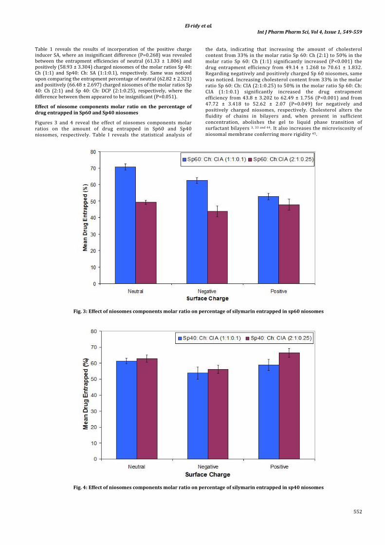
El-ridy et al Int J Pharm Pharm Sci Vol 4 Issue 1 549-559
554
Fig 7 DSC thermograms of silymarin plain (drug-free) niosomes and silymarin loaded Sp40 niosomes
In-vitro release profiles
The results tabulated in table (2) and shown in figures (8 and 9) illustrate the amount of silymarin retained in span 60 and span 40 niosomes respectively after the periods of time investigated The results reveal that the release of silymarin from either span 60 or span 40 niosomes was biphasic with an initial faster release followed by a period of slow release This biphasic release pattern seems to be a characteristic of bilayered vesicles Similar results were reported in case of liposomes 49 and in case of niosomes 50-51 Rapid drug leakage was observed during the initial phase where about 15ndash35 of the entrapped drug was released from various formulations in the first six hours However during the following 66 hrs slow release occurred in which only further 6ndash10 of silymarin was released from different niosomal preparations
This could be explained by that the drug is mainly incorporated between the bilayers of niosomal vesicles which lead to rapid release upon dispersing niosomes in buffer until reaching equilibrium 50
Effect of cholesterol content on silymarin release from span 60 and span 40 niosomes
Considering Span 60 niosomes Table 2 reveals that the increase in cholesterol content from 33 in neutral niosomes of the molar ratio Sp 60 Ch (21) to 50 in the molar ratio Sp 60 Ch (11) significantly increased (Plt005) the percentages of silymarin retained inside niosomes at all times investigated Upon comparing the two molar ratios Span60 Cholesterol DCP (21025) and Span60 Cholesterol DCP (1101) the same could be noticed
Table 2 In-vitro release profiles of silymarin loaded Span 60 and Span 40 niosomes of different charges and molar ratio
Time (hr) Mean Silymarin Retained () plusmn SD Neutral Sp 60 Ch Negative Sp 60 Ch DCP Neutral Sp 40 Ch Negative Sp 40 Ch DCP 11 21 1101 21025 11 21 1101 21025
Zero 100 plusmn 0 100 plusmn 0 100 plusmn 0 100 plusmn 0 100 plusmn 0 100 plusmn 0 100 plusmn 0 100 plusmn 0 3 909 plusmn 171 7968 plusmn 044 9236 plusmn 107 838 plusmn 201 9218 plusmn 2401 8344 plusmn 0500 8229 plusmn 1947 9285 plusmn 0714 6 7998 plusmn 193 7112 plusmn 037 8063 plusmn 114 663 plusmn 164 8664 plusmn 1734 7515 plusmn 0241 7617 plusmn 1798 8015 plusmn 2791 24 7799plusmn 206 6813 plusmn 063 7840 plusmn 140 6312 plusmn 177 7907 plusmn 217 7035 plusmn 1344 7183 plusmn 2058 7594 plusmn 2531 48 7246 plusmn 273 6649 plusmn 063 7363 plusmn 102 6176 plusmn 221 7716 plusmn 186 6854 plusmn 1010 6965 plusmn 1613 7504 plusmn 2475 72 7086 plusmn 264 6233 plusmn 137 7169 plusmn 092 6022 plusmn 207 7592 plusmn173 6770 plusmn 1010 6846 plusmn 1474 7450 plusmn 2336
Fig 8 In-vitro release profiles of silymarin loaded Span 60 niosomes of different charges and molar ratio
El-ridy et al Int J Pharm Pharm Sci Vol 4 Issue 1 549-559
555
Fig 9 In-vitro release profiles of silymarin loaded Span 40 niosomes of different charges and molar ratios
The increase in cholesterol content significantly increased the percentage of silymarin retained inside niosomes from 663 plusmn 164 to 8063 plusmn 114 and from 6312 plusmn 177 to 7840 plusmn 140 after
6 hours (P=0005) and 24 hours (P=0001) respectively Investigating span 40 niosomes the results (table 2) reveal that increasing cholesterol content from 33 in neutral niosomes of the molar ratio Sp60 Ch (21) to 50 in neutral niosomes of the molar ratio Sp 60 Ch (11) significantly increased (P=0045) the drug retained percentage from 7515 plusmn 0241 to 8664 plusmn 1734 at t=6 hours These results are in accordance with cholesterol membrane stabilizing ability and space filling action 3 52 Cholesterol is known to increase the rigidity of niosomes and abolish the gel-to-liquid phase transition of niosomal systems resulting in niosomes that are less leaky 53 thus decreasing the drug release from niosomes 54 Cholesterol also alters the fluidity of chains in bilayers and increases the degree of orientation order leading to decreased permeability 55
On the other hand comparing the two molar ratios Sp40 Ch DCP (21025) and Sp40 Ch DCP (1101) the opposite could be noticed (table 2) The increase in cholesterol content did not lead to an increase in silymarin retained percentage This maybe attributed to the effect of charge inducer where in this case the effect of charge overcame that of cholesterol content This confirms that DCP stabilizes the structure of niosomes and renders it less permeable 56
Effect of charge inducing agents on silymarin release from span 60 and span 40 niosomes
Table 2 show that the difference between the amount of drug retained inside neutral niosomes prepared using the molar ratio Sp 60 Ch (11) and that from negative niosomes of the molar ratio Sp60 Ch DCP (1101) appeared to be insignificant (Pgt005) at all times investigated Upon comparing the amount of silymarin retained inside neutral niosomes of the molar ratio Sp60 Ch (21) and that in negatively charged niosomes of the molar ratio Sp60 Ch DCP (21025) it can be concluded that the inclusion of DCP significantly increased (Plt005) the amount of silymarin retained at all times investigated Same was noticed on comparing the release extent of
drug from neutral niosomes of the molar ratio Sp40 Ch (21) and that from negatively charged niosomes of the molar ratio Sp40 Ch DCP (21025) where the inclusion of DCP significantly increased (Plt005) drug retained percentage at all times investigated This comes in accordance with the effect of DCP in stabilizing the niosomal membrane structure rendering it less permeable 56
Particle size and distribution of niosomal vesicles
Particle size distribution of span 60 and span 40 niosomes are illustrated in table 3 and table 4 respectively Particle size distribution of silymarin niosomes appeared to be bimodal with the 1st peaks showing the highest contribution
Effect of charge inducers on particle size diameter
The results illustrated in Table 3 reveal that charged niosomes exhibited mean particle diameter higher than that of the neutral ones viz 4912 plusmn 680 nm and 5411 plusmn 733 for negatively and positively charged niosomes of the molar ratio Sp 60 Ch CIA (1101) compared to 319 plusmn 362 nm for neutral silymarin niosomes of the same molar ratio This could also be noticed upon comparing the mean particle diameter of charged niosomes of the molar ratios Sp 60 Ch CIA (21025) to that of neutral niosomes of the molar ratio Sp 60 Ch (21) where charged niosomes exhibited mean particle diameter higher than that of the neutral ones viz 3326 plusmn 473nm and 3976 plusmn 425 nm for negatively and positively charged niosomes respectively of the molar ratio Sp 60 Ch CIA (21025) compared to 2562 plusmn29 nm for neutral silymarin niosomes of the molar ratio Sp 60 Ch(21) The results illustrated in Table 4 also show that negatively charged niosomes of the molar ratio Sp 40 Ch DCP (21025) exhibited higher mean particle diameter than that of the neutral niosomes of the molar ratio Sp 40 Ch (21) viz 2772 plusmn 438 nm for negatively charged niosomes of the molar ratio Sp 40 Ch DCP (21025) compared to 2190 plusmn 325 nm for neutral silymarin niosomes of the molar ratio Sp 40 Ch (21) These results could be attributed to that the inclusion of a charge inducer in niosomes leads to an increase in the spacing between adjacent bilayers 57-58
Table 3 Particle size distributions of silymarin span 60 niosomes
Formulation Particle diameter (nm) plusmn SD ( Area of the peak indicating the volume of particle population) 1st Peak 2nd Peak
Sp 60 Ch (11) 319 plusmn 362 (740) 21862 plusmn 3194 (26) Sp 60 Ch DCP (1101) 4912 plusmn 680 (948) 87333 plusmn 12593 (52) Sp 60 Ch SA (1101) 5411 plusmn 733 (951) 88030 plusmn 6586 (49) Sp 60 Ch (21) 2562 plusmn 29 (879) 30824 plusmn 3605 (121) Sp 60 Ch DCP (21025) 3326 plusmn 473 (897) 29725 plusmn 4130 (103) Sp 60 Ch SA (21025) 3976 plusmn 425 (922) 89603 plusmn 5787 (78)
El-ridy et al Int J Pharm Pharm Sci Vol 4 Issue 1 549-559
556
Table 4 Particle size distributions of silymarin span 40 niosomes
Formulation Particle diameter (nm) plusmn SD ( Area of the peak indicating the volume of particle population) 1st Peak 2nd Peak
Sp 40 Ch (11) 339 plusmn 423 (768 ) 25810 plusmn 3353 (232) Sp 40 Ch DCP (1101) 1851 plusmn 316 (945) 20105 plusmn 3910 (55 ) Sp 40 Ch SA (1101) 2348 plusmn 411 (930) 29980 plusmn 6634 (62) Sp 40 Ch (21) 2190 plusmn 325 (890) 18708 plusmn 2949 (110) Sp 40 Ch DCP(21025) 2772 plusmn 438 (932) 29441 plusmn 3873 (132) Sp 40 Ch SA (21025) 1577 plusmn 191 (979) 71601 plusmn 11445 (21 )
Effect of cholesterol content on particle size diameter
Table 3 show that the increase in cholesterol content from 33 in the molar ratio span60 Cholesterol (21) to 50 in the molar ratio Span60 Cholesterol (11) led to an increase in the particle size diameter viz 2562 plusmn 29 nm for neutral silymarin niosomes of the molar ratio Span 60 Cholesterol (21) compared to 319 plusmn 362 nm for neutral silymarin niosomes of the molar ratio Span 60 Cholesterol (11) Same conclusions could be depicted upon comparing the mean particle diameter of charged niosomes of the molar ratio Sp60 Ch CIA (21025) to that of charged niosomes of the molar ratio Sp60 Ch CIA (1101) where increasing the cholesterol content led to an increase in the particle diameter viz 3326 plusmn 473 nm and 3976 plusmn 425 nm for negatively and positively charged niosomes of the molar ratio Span 60 Cholesterol CIA (21025) compared to 4912 plusmn 680 nm and 5411 plusmn 733 nm for negatively and positively charged noisomes of the molar ratio Span 60 Cholesterol CIA (1101) respectively Cholesterol increases the width of the bilayers and consequently increases the vesicle size 59
Considering Sp 40 niosomes the results (table 4) reveal that an increase in cholesterol content in neutral niosomes led to an increase in the particle size diameter viz 2190 plusmn 325 nm for neutral silymarin niosomes of the molar ratio Sp 40 Ch (21) compared to 339 plusmn 423 nm for neutral silymarin niosoems of the molar ratio Sp 40 Ch (11) Same conclusion could be observed upon comparing the mean particle diameter of the positively charged niosomes where increasing the cholesterol content led to an increase in the particle diameter from 1577 plusmn 191 nm for positively charged niosomes of the molar ratio Sp 40 Ch SA (21025) to 2348 plusmn 311 nm for positively charged noisomes of the molar ratio Sp 40 ChSA(1101)
In-Vivo Study
The effect of silymarin silymarin niosomal formulations and plain niosomes on activities of serum SGPT SGOT and SALP in rats after
induction of liver damage by CCl4 is tabulated in table 5 and illustrated in figure 10 The in vivo results revealed that the administration of the drug loaded niosomal suspensions N1 and N2 (groups V and VI) did not show any change when compared to control group I
Acute CCl4 administration resulted in a significant (P lt 0001) increase in SGPT to 5516 plusmn 1053UL compared to normal value which was 2404 plusmn 783 UL Administration of plain niosomes produced a non significant decrease in serum GPT to 4556 plusmn 1010 UL (P = 0063) Administration of silymarin suspensions N1 and N2 produced a significant decrease in SGPT levels (P lt 0001) to reach 3596 plusmn 942 2449 plusmn 56 and 2547 plusmn 154 UL respectively At the same time both niosomal formulations (N1 and N2) showed a significant decrease in SGPT levels in comparison to silymarin suspension (P lt 005)
Concerning the biochemical parameter SGOT acute CCl4 administration resulted in a significant (P lt 0001) increase in SGOT to 13802 plusmn 1788 UL compared to normal value which was 5512 plusmn 1739 UL Plain niosomes produced an insignificant change in serum SGOT to reach 12365 plusmn 140 UL (P = 0631) Significant decrease in SGOT levels upon administration of silymarin suspension N1 and N2 to reach 8911 plusmn 1026 5006 plusmn 627 and 5462 plusmn 833 UL respectively (P lt 0001) was found At the same time both silymarin niosomal suspensions N1 and N2 showed a significant decrease in SGOT levels relative to that of silymarin suspension (P lt 0001)
Concerning the SALP analysis results acute CCl4 administration resulted in a significant (P lt 0001) increase in serum SALP to 66699 plusmn 1887 UL compared to normal value which was analyzed as 2651 plusmn 1876 UL Plain niosomes produced a significant change in serum SALP to reach 3815 plusmn 1925 UL (P lt 005) Significant decrease in SALP levels upon administration of silymarin suspensions N1 and N2 to reach 3024 plusmn 1878 2712 plusmn 1592 and 2665 plusmn 733 UL respectively (P lt 0001) was produced Meanwhile these suspensions viz N1 and N2 showed a significant decrease in SGOT levels relative to that of silymarin suspension (P lt 001)
[
Fig 10 Effect of silymarin silymarin niosomal formulations and plain niosomes on activities of serum GPT GOT and ALP in rats after induction of liver damage by CCl4
El-ridy et al Int J Pharm Pharm Sci Vol 4 Issue 1 549-559
557
Table 5 Effect of silymarin silymarin niosomal formulations and plain niosomes on activities of serum GPT GOT and ALP in rats after induction of liver damage by CCl4
Groups GPT (Ul) GOT (Ul) SALP (Ul) Control 2404 plusmn 783 (319) 5512 plusmn 1739 (778) 2651 plusmn 1876 (839) CCl4 5516 plusmn 1053 (470) 13802 plusmn 1788 (799) 66699 plusmn 1887 (844) CCl4 + Plain N 4556 plusmn 1010 (452) 12365 plusmn 140 (0628) 3815 plusmn 1925 (861) dagger CCl4 + drug 3596 plusmn 942 (412) dagger 8911 plusmn 1026 (459) dagger 3024 plusmn 1878 (840) dagger CCl4 + N1 2449 plusmn 56 (211) dagger para 5006 plusmn 627 (256) dagger para 2712 plusmn 1592 (712) dagger para CCl4 + N2 2547 plusmn 154 (063) dagger para 5462 plusmn 833 (340) dagger para 2665 plusmn 733 (327) dagger para
Values are mean plusmn SD (SE)
Values of exhibit significant changes from control group P lt 005
Values of dagger exhibit significant changes when compared to CCl4 group P lt 005
Values of para exhibit significant changes when compared to (CCl4 + drug suspension) group P lt 005
Histopathological Studies
Histopathological studies (Fig 11) show that CCl4 induced vacuolar degenerative changes and necrosis in the hepatocytes surrounding the central veins and portal area Administration of plain niosomes also caused degeneration in the hepatocytes allover the hepatic parenchyma By examination under the microscope the total area of necrosis and hepatic lesions induced by CCl4 were reduced by administration of silymarin suspension Administration of both silymarin niosomal formulations N1 and N2 showed more improvement than silymarin suspension in the hepatocytes structure and degenerative areas Both silymarin loaded niosomal formulations N1 and N2 succeeded to minimize the vacuolar degeneration and necrosis These results are in accordance with the result of the serum SGPT SGOT and SALP levels in which administration of N1 and N2 showed better protection against CCl4 induced damage in comparison with silymarin suspension (table 5 and fig 10)
CONCLUSION
This study showed that the niosomal formulation could be one of the promising delivery systems for the hepatoprotective drug silymarin It provided successful preparation with efficient encapsulation of silymarin Niosomal formulations characterization using TEM showed the spherical shape and the bilayered structure of the prepared niosomes Studies using DSC gave evidence of possible interaction of silymarin with bilayer components In-vitro release profiles were biphasic with an initial faster release followed by a period of slow release In-vivo study performed on rats proved that silymarin is an efficient hepatoprotective drug and that the investigated niosomal formulations significantly improved the hepatoprotective efficiency Accordingly subcutaneous administration of niosomal silymarin formulations is expected to increase drug bioavailability Drug niosomal formulations were also proved to be safe according to the histopathological investigation
El-ridy et al Int J Pharm Pharm Sci Vol 4 Issue 1 549-559
558
Fig 11 Photomicrographs of histological sections (hematoxylin and eosin stained) representing (a) liver of normal rat (64 x) treated with (b) CCl4 (160 x) (c) CCl4 and plain niosomes (64 x) (d) CCl4 and silymarin suspension (160 x) (e) CCl4 and silymarin loaded
niosomes N1 (64 x) (f) CCl4 and silymarin loaded niosomes N2 (64 x) central vein (C ) portal area (P) hepatocytes (h) necrosis (n) vacuolar degeneration (d) hydropic degeneration of hepatocytes (reversible) (rarr)
REFERENCES
1 Jain KK Methods in Molecular Biology Drug Delivery Systems Edited by Kewal K Jain Humana Press Totowa NJ 2008 437
2 Uchegbu IF Vyas SP Non-ionic surfactant based vesicles (niosomes) in drug delivery Int J Pharm 1998 172 33-70
3 Uchegbu IF Florence AT Non-ionic surfactant vesicles (niosomes) physical and pharmaceutical chemistry Adv Coll Interf Sci 1995 58 1-55
4 DSouza SA Ray J Pandey S Udupa N Absorption of ciprofloxacin and norfloxacin when adminstered as niosomes-encapsulated inclusion complexes J Pharm Pharmacol 1997 49 145-149
5 Namdeo A Jain N K Niosomes as Drug Carriers Ind J Pharm Sci 1996 58 Suppl 2 41-46
6 Sahin NO In Nanomaterials and Nanosystems for Biomedical Applications Mozafari M R Ed Springer Berlin 2007 p 67
7 Ramasamy K Agarwal R Multitargeted therapy of cancer by silymarin Cancer Letters 2008 269 352ndash362
8 Wu J W Lin L C Tsai T H Drug-drug interactions of silymarin on the perspective of pharmacokinetics Journal of Ethnopharmacology 2009 121 181-193
9 Polyak SJ Morishima C Lohmannd V Pala S Leee DYW Liue Y Graf TN Oberlies NF 2010 Identification of hepatoprotective flavonolignans from silymarin PNAS 2010 107 Suppl 13 5995ndash5999
10 Mourelle M MurielP Favari L Franco T 1989 Prevention of CCl4-induced liver cirrhosis by silymarin Fundam Clin Pharmacol 1989 3 183ndash191
11 Muriel P Garciapina T Perez-Alvarez V Mourelle M Silymarin protects against paracetamol-induced lipid peroxidation and liver damage J Appl Toxicol 1992 12 439ndash442
12 Muriel P Mourelle M Prevention by silymarin of membrane alterations in acute CCl4 liver damage J Appl Toxicol 1990 10 275ndash279
13 Mira L Silva M and Manso C F Scavenging of reactive oxygen species by silibin dihemisuccinate Biochem Pharmacol 1994 48 753-759
14 Hruby KCsomosGFuhrmannMThalerH Chemotherapy of Amanita phalloides poisoning with intravenous silibininHumToxicol 1983 2 138ndash195
15 Desplace A Choppin J Vogel G Trost W The effects of silymarin on experimental phalloidine poisoning Arzneim Forsch 1975 25 89ndash96
16 Sonnenbichler J Zetl I Biochemical effects of the flavolignane silibinin on RNA protein and DNA synthesis in rat liver Prog Clin Biol Res 1986 213 319ndash331
17 Jumaa KM Ahmed ZA Numan IT Hussain SA Dose-dependent anti-inflammatory effect of silymarin in experimental
animal model of chronic inflammation African Journal of Pharmacy and Pharmacology 2009 3 Suppl 5 242-247
18 Fuchs EC Weyhenmeyer R Weiner OH Effects of silibinin and of a synthetic analogue on isolated rat hepatic stellate cells and myofibroblasts Arzneimittelforschung 1997 47 1383-7
19 Manna SK Mukhopadhyay A Van NT Aggarwal BB 1999 Silymarin suppresses TNF-induced activation of NF-kappaB c-jun N-terminal kinase and apoptosis Journal of Immunology 1999 163 6800-6809
20 Blumenthal M Goldberg A Brinkmann J Herbal Medicine Integrative Medicine Communications Newton 2000
21 Giacomelli S Gallo D Apollonio P Ferlini C Distefano M Morazzoni P Riva A Bombardelli E Mancuso S Scambia G Silybin and its bioavailable phospholipid complex (IdB 1016) potentiate in vitro and in vivo activity of cisplatin Life Sci 2002 70 Suppl 12 1447-59
22 Fraschini F Demartini G Esposti D 2002 Pharmacology of Silymarin Clin Drug Invest 2002 22 Suppl 1 51-65
23 Madaus R Halbach G Trost W Salt of silymarin group with aminopolyhydroxy alcohols US patent 3994925 1976
24 Gabetta B Bombardelli E Pifferi G Complexes of flavanolignans with phospholipids preparation thereof and associated pharmaceutical compositions US patent 4764508 1988
25 Wachter W Zaeske H Process for the manufacture of flavanolignan preparation with improved release and absorbability compositions obtainable thereby and their use for the preparation of pharmaceuticals US patent 6020384 2000
26 Kidd P Head K A review of the bioavailability and clinical efficacy of milk thistle phytosome A silybin-phosphatidylcholine complex (Siliphos) Altern Med Rev 2005 10 193-203
27 Li W Gao J Zhao H Z Liu CX Development of a HPLC-UV assay for silybin-phosphatidylcholine complex (silybinin capsules) and its pharmacokinetic study in healthy male Chinese volunteers European Journal of Drug Metabolism and Pharmacokinetics 2006 31 265ndash270
28 Filburn CR Kettenacker R Griffin DW Bioavailability of a silybin phosphatidylcholine complex in dogs Journal of Veterinary Pharmacology and Therapeutics 2007 30 132ndash138
29 Valcavi U Monterosso V Caponi R Bosone E Wachter W Szejtli J Inclusion complexes with silybinin their preparation and pharmaceutical compositions containing them US patent 5198430 1993
30 Giorgi R Conti M Pifferi G Soluble derivative of silybin a method of preparing them and pharmaceutical composition containing them US patent 4886791 1999
31 Azmin MN Florence AT Handjani-Vila RM Stuart JFB Vanlerberghe G Whittaker JS The effect of non-ionic surfactant vesicle (niosome) entrapment on the absorption and distribution of methotrexate in mice J Pharm Pharmacol 1985 37 237ndash242
El-ridy et al Int J Pharm Pharm Sci Vol 4 Issue 1 549-559
559
32 Jigar V Puja V Krutika S Formulation and evaluation of topical niosomal gel of erythromycin Int J Pharmacy Pharm Sci 2011 3 Suppl 1 123-126
33 Hao Y Zhao F Li N Yang T Li K Studies on a high encapsulation of colchicine by a noisome system Int J of Pharm 2002 244 73-80
34 Law SL Lo WY Lin M Increase of liposome stability by incorporation of bovine serum albumin Drug Dev Ind Pharm 1994 20 1411ndash1423
35 Fang JY Yu SY Wu PC Huang YB and Tsai YH In vitro skin permeation of estradiol from various proniosome formulations Int J Pharm 2001 215 Suppl 1-2 91-99
36 OrsquoNeil MJ Smith A Heckelman PE The Merck Index 13th ed Merck and Company Whitehouse Station 2001
37 Aggarwal D Kaur IP Improved pharmacodynamics of timolol maleate from a mucoadhesive niosomal ophthalmic drug delivery system IntJ Pharm 2005 290 Suppl 1-2 155-159
38 PaulS Mondol R Ranjit S Maiti S Antilaucomatic niosomal system recent trend in ocular drug delivery Int J Pharmacy Pharm Sci 2010 2 Suppl 2 15-18
39 Liu T Guo R Investigation of PEG 6000Tween 80Span 80H2O niosome microstructure Colloid Polym Sci 2007 285 711-713
40 Juliano RL Stamp D Pharmacokinetics of liposome-encapsulated anti-tumor drugs Studies with vinblastine actinomycin D cytosine arabinoside and daunomycin Biochem Pharmacol 1978 27 21
41 El-Ridy MS Kassem M Akbarieh M and Tawashi R The effect of surface charge of liposomes on aggregation in the buccal cavity proceeding 15th Intern Symp Control Rel Bioact Mater Basel Switzerland 1988 344 Suppl 200
42 Mansour HH Hafez HF Fahmy NM Silymarin Modulates Cisplatin-Induced Oxidative Stress and Hepatotoxicity in Rats Journal of Biochemistry and Molecular Biology 2006 39 Suppl 6 656-661
43 Yadav N P Pal A Shanker K Bawankule DU Gupta AK Darokar MP and Khanuja S P S Synergistic effect of silymarin and standardized extractof Phyllanthus amarus against CCl4-induced hepatotoxicityin Rattus norvegicus Phytomedicine 2008 15 1053ndash1061
44 Devaraj GN Parakh SR Devraj R Apte SS Rao BR Rambhau D Release studies on niosomes containing fatty alcohols as bilayer stabilizers instead of cholesterol J Coll Int Sci 2002 251 360ndash 365
45 Manosroi A Wongtrakul P Manosroi J Sakai H Sugawara F Yuasa M Abe M Characterization of vesicles prepared with
various non-ionic surfactants mixed with cholesterol Colloids and Surfaces B Biointerfaces 2003 30 129ndash138
46 Nagarsenker MS Londhea VY Nadkarnib GD Preparation and evaluation of liposomal formulations of tropicamide for ocular delivery Int J Pharm 1999 190 Suppl 1 63-71
47 Nagarsenker MS Joshi AA Preparation Characterization and Evaluation of Liposomal Dispersions of Lidocaine Drug Dev Ind Pharmacy 1997 23 Suppl 12 1159-1165
48 Hathout RM Mansour S Mortada ND Guinedi A S Liposomes as an Ocular Delivery System for Acetazolamide In Vitro and In Vivo Studies AAPS Pharm Sci Tech 2007 8 Suppl 1 Article 1
49 Mokhtar M Sammour OA Hammad MA Megrab NA Effect of some formulation parameters on flurbiprofen encapsulation and release rates of niosomes prepared from proniosomes Int J Pharm 2008 361 104ndash111
50 Baillie AJ Florence AT Hume LR Muirhead GT Rogerson A The preparation and properties of niosomes-non ionic surfactant vesicles J Pharm Pharmacol 1985 37 863-868
51 Wissing SA Kayser O and Muumlller R H Solid lipid nanoparticles for parenteral drug delivery Advanced Drug Delivery Reviews 2004 56 Suppl 9
52 Namdeo A Jain NK Niosomal delivery of 5-flourouracil J Microencap 1999 16 Suppl 6 731-740
53 Rogerson A Cummings J Florence AT Adriamycin-loaded niosomes ndashdrug entrapment stability and release J Microencap 1987 4 Suppl 4 321-328
54 Alsarra IA Bosela AA Ahmed SM Mahrous GM Proniosomes as a drug carrier for transdermal delivery of ketorolac EurJ Pharm Biopharm 2005 59 Suppl 3 485-490
55 Bayindir ZS Yuksel AN Characterization of Niosomes Prepared With Various Nonionic Surfactants for Paclitaxel Oral Delivery J Pharm Sci 2010 99 Suppl 4 2049ndash2060
56 Abdelbary G El-gendy N Niosome-Encapsulated Gentamicin for Ophthalmic Controlled Delivery AAPS Pharm Sci Tech 2008 9 Suppl 3 740-7
57 Taylor KMG Taylor G Kellaway IW Stevens J Drug entrapment and release from multilamellar and reverse-phase evaporation liposomes Int J Pharm 1999 58 49ndash55
58 Vyas SP Singh R Asati R K Liposomally encapsulated diclofenac for sonophoresis induced systemic delivery J Microencapsulation 1995 12 149ndash154
59 Mclntosh TJ The effect of cholesterol content on the structure of phosphatidylcholine bilayers Biochim Biophys Acta 1978 51 43-58
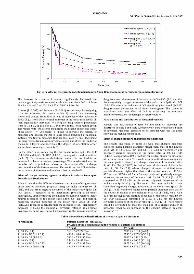
El-ridy et al Int J Pharm Pharm Sci Vol 4 Issue 1 549-559
559
32 Jigar V Puja V Krutika S Formulation and evaluation of topical niosomal gel of erythromycin Int J Pharmacy Pharm Sci 2011 3 Suppl 1 123-126
33 Hao Y Zhao F Li N Yang T Li K Studies on a high encapsulation of colchicine by a noisome system Int J of Pharm 2002 244 73-80
34 Law SL Lo WY Lin M Increase of liposome stability by incorporation of bovine serum albumin Drug Dev Ind Pharm 1994 20 1411ndash1423
35 Fang JY Yu SY Wu PC Huang YB and Tsai YH In vitro skin permeation of estradiol from various proniosome formulations Int J Pharm 2001 215 Suppl 1-2 91-99
36 OrsquoNeil MJ Smith A Heckelman PE The Merck Index 13th ed Merck and Company Whitehouse Station 2001
37 Aggarwal D Kaur IP Improved pharmacodynamics of timolol maleate from a mucoadhesive niosomal ophthalmic drug delivery system IntJ Pharm 2005 290 Suppl 1-2 155-159
38 PaulS Mondol R Ranjit S Maiti S Antilaucomatic niosomal system recent trend in ocular drug delivery Int J Pharmacy Pharm Sci 2010 2 Suppl 2 15-18
39 Liu T Guo R Investigation of PEG 6000Tween 80Span 80H2O niosome microstructure Colloid Polym Sci 2007 285 711-713
40 Juliano RL Stamp D Pharmacokinetics of liposome-encapsulated anti-tumor drugs Studies with vinblastine actinomycin D cytosine arabinoside and daunomycin Biochem Pharmacol 1978 27 21
41 El-Ridy MS Kassem M Akbarieh M and Tawashi R The effect of surface charge of liposomes on aggregation in the buccal cavity proceeding 15th Intern Symp Control Rel Bioact Mater Basel Switzerland 1988 344 Suppl 200
42 Mansour HH Hafez HF Fahmy NM Silymarin Modulates Cisplatin-Induced Oxidative Stress and Hepatotoxicity in Rats Journal of Biochemistry and Molecular Biology 2006 39 Suppl 6 656-661
43 Yadav N P Pal A Shanker K Bawankule DU Gupta AK Darokar MP and Khanuja S P S Synergistic effect of silymarin and standardized extractof Phyllanthus amarus against CCl4-induced hepatotoxicityin Rattus norvegicus Phytomedicine 2008 15 1053ndash1061
44 Devaraj GN Parakh SR Devraj R Apte SS Rao BR Rambhau D Release studies on niosomes containing fatty alcohols as bilayer stabilizers instead of cholesterol J Coll Int Sci 2002 251 360ndash 365
45 Manosroi A Wongtrakul P Manosroi J Sakai H Sugawara F Yuasa M Abe M Characterization of vesicles prepared with
various non-ionic surfactants mixed with cholesterol Colloids and Surfaces B Biointerfaces 2003 30 129ndash138
46 Nagarsenker MS Londhea VY Nadkarnib GD Preparation and evaluation of liposomal formulations of tropicamide for ocular delivery Int J Pharm 1999 190 Suppl 1 63-71
47 Nagarsenker MS Joshi AA Preparation Characterization and Evaluation of Liposomal Dispersions of Lidocaine Drug Dev Ind Pharmacy 1997 23 Suppl 12 1159-1165
48 Hathout RM Mansour S Mortada ND Guinedi A S Liposomes as an Ocular Delivery System for Acetazolamide In Vitro and In Vivo Studies AAPS Pharm Sci Tech 2007 8 Suppl 1 Article 1
49 Mokhtar M Sammour OA Hammad MA Megrab NA Effect of some formulation parameters on flurbiprofen encapsulation and release rates of niosomes prepared from proniosomes Int J Pharm 2008 361 104ndash111
50 Baillie AJ Florence AT Hume LR Muirhead GT Rogerson A The preparation and properties of niosomes-non ionic surfactant vesicles J Pharm Pharmacol 1985 37 863-868
51 Wissing SA Kayser O and Muumlller R H Solid lipid nanoparticles for parenteral drug delivery Advanced Drug Delivery Reviews 2004 56 Suppl 9
52 Namdeo A Jain NK Niosomal delivery of 5-flourouracil J Microencap 1999 16 Suppl 6 731-740
53 Rogerson A Cummings J Florence AT Adriamycin-loaded niosomes ndashdrug entrapment stability and release J Microencap 1987 4 Suppl 4 321-328
54 Alsarra IA Bosela AA Ahmed SM Mahrous GM Proniosomes as a drug carrier for transdermal delivery of ketorolac EurJ Pharm Biopharm 2005 59 Suppl 3 485-490
55 Bayindir ZS Yuksel AN Characterization of Niosomes Prepared With Various Nonionic Surfactants for Paclitaxel Oral Delivery J Pharm Sci 2010 99 Suppl 4 2049ndash2060
56 Abdelbary G El-gendy N Niosome-Encapsulated Gentamicin for Ophthalmic Controlled Delivery AAPS Pharm Sci Tech 2008 9 Suppl 3 740-7
57 Taylor KMG Taylor G Kellaway IW Stevens J Drug entrapment and release from multilamellar and reverse-phase evaporation liposomes Int J Pharm 1999 58 49ndash55
58 Vyas SP Singh R Asati R K Liposomally encapsulated diclofenac for sonophoresis induced systemic delivery J Microencapsulation 1995 12 149ndash154
59 Mclntosh TJ The effect of cholesterol content on the structure of phosphatidylcholine bilayers Biochim Biophys Acta 1978 51 43-58