nih public accessamy berrington de gonzalez stephen balter...
TRANSCRIPT

OCCUPATIONAL RADIATION DOSES TO OPERATORSPERFORMING FLUOROSCOPICALLY-GUIDED PROCEDURES
Kwang Pyo Kim*, Donald L. Miller†, Amy Berrington de Gonzalez‡, Stephen Balter§, Ruth A.Kleinerman‡, Evgenia Ostroumova‡, Steven L. Simon‡, and Martha S. Linet‡*Department of Nuclear Engineering, Kyung Hee University, Gyeonggi-do, Republic of Korea†Center for Devices and Radiological Health, Food and Drug Administration, Silver Spring, MD‡Division of Cancer Epidemiology and Genetics, National Cancer Institute, National Institutes ofHealth, Bethesda, MD§Departments of Radiology and Medicine, Columbia University Medical Center, New York City,NY
AbstractIn the past 30 years, the numbers and types of fluoroscopically-guided (FG) procedures haveincreased dramatically. The objective of the present study is to provide estimated radiation dosesto physician specialists, other than cardiologists, who perform FG procedures. We searchedMedline to identify English-language journal articles reporting radiation exposures to thesephysicians. We then identified several primarily therapeutic FG procedures that met specificcriteria: well-defined procedures for which there were at least five published reports of estimatedradiation doses to the operator, procedures performed frequently in current medical practice, andinclusion of physicians from multiple medical specialties. These procedures were percutaneousnephrolithotomy (PCNL), vertebroplasty, orthopedic extremity nailing for treatment of fractures,biliary tract procedures, transjugular intrahepatic portosystemic shunt creation (TIPS), head/neckendovascular therapeutic procedures, and endoscopic retrograde cholangiopancreatography(ERCP). We abstracted radiation doses and other associated data, and estimated effective dose tooperators. Operators received estimated doses per patient procedure equivalent to doses receivedby interventional cardiologists. The estimated effective dose per case ranged from 1.7 – 56μSv forPCNL, 0.1 – 101 μSv for vertebroplasty, 2.5 – 88μSv for orthopedic extremity nailing, 2.0 –46μSv for biliary tract procedures, 2.5 – 74μSv for TIPS, 1.8 – 53μSv for head/neck endovasculartherapeutic procedures, and 0.2 – 49μSv for ERCP. Overall, mean operator radiation dose per casemeasured over personal protective devices at different anatomic sites on the head and body rangedfrom 19 – 800 (median = 113) μSv at eye level, 6 – 1180 (median = 75)μSv at the neck, and 2 –1600 (median = 302) μSv at the trunk. Operators’ hands often received greater doses than theeyes, neck or trunk. Large variations in operator doses suggest that optimizing procedure protocolsand proper use of protective devices and shields might reduce occupational radiation dosesubstantially.
Keywordsinterventional procedure; fluoroscopically-guided procedure; occupational exposure; radiationprotection
For reprints and correspondence contact: Kwang Pyo Kim, PhD, Department of Nuclear Engineering, Kyung Hee University, 1Seocheon-dong, Giheung-gu, Yongin-si, Gyeonggi-do, Republic of Korea, Phone: +82 (31) 201-2560 Fax: +82 (31) 202-8106,[email protected].
NIH Public AccessAuthor ManuscriptHealth Phys. Author manuscript; available in PMC 2014 March 12.
Published in final edited form as:Health Phys. 2012 July ; 103(1): 80–99. doi:10.1097/HP.0b013e31824dae76.
NIH
-PA Author Manuscript
NIH
-PA Author Manuscript
NIH
-PA Author Manuscript

INTRODUCTIONThe term “fluoroscopically-guided (FG) procedures” refers to procedures where real-timeradiological images (fluoroscopy) of a patient’s internal structures are used for diagnostic ortherapeutic purposes. FG procedures are utilized to treat a growing range of diseases andinjuries by a variety of physician specialists, including interventional radiologists,neuroradiologists, cardiologists, electrophysiologists, orthopedic surgeons, urologists andgastroenterologists. Examples of structural or functional conditions treated using FGprocedures include disorders of the heart, blood vessels, gastrointestinal system, biliary tract,bladder, ureters and kidneys. Fluoroscopic imaging has also been employed in minimallyinvasive hip fracture plating, nailing, external fixation and other orthopedic procedures.
The National Council on Radiation Protection and Measurements (NCRP) has estimated thatan average of 17 million interventional fluoroscopic procedures were performed in 2006,including 4.6 million cardiac procedures, 3.4 million vascular non-cardiac procedures, and8.6 million nonvascular procedures (NCRP 2009). Not included in these estimates wereradiographic fluoroscopy procedures (e.g., barium enemas). The number of FG procedureshas increased by about 8.5% annually (Bhargavan 2008), increasing approximately 4.7-foldbetween 1986 and 2005, with cardiac procedures (16% annual increase) and spinalprocedures (15% annual increase) demonstrating the greatest growth. Of the FG procedurescarried out in 2005, 33% were vascular procedures, 29% cardiac, 23% spinal 3.1%gastrointestinal, 1.8% urinary, 0.8% extremity procedures and 9.9% all others.
In contrast to other radiological modalities, such as conventional radiography, computedtomography and nuclear medicine, operators who perform FG procedures stand in closeproximity to the patient and the x-ray tube, and are therefore exposed to substantial scatteredradiation from the patient. Although radiation doses to operators from scattered radiation aremuch smaller than patient doses (Koenig et al. 2001, Vano et al. 2001, Miller et al. 2003a,Miller et al. 2003b, Neofotistou et al. 2003), the cumulative dose from many proceduresperformed over an operator’s career may be substantial. In addition, there appears to be anincreasing workload per operator, as the number of practitioners performing FG procedureshas not kept pace with the substantial increases in the numbers of FG procedures (Vano etal. 1998b)
Clinical reports and a case-control epidemiologic study have suggested an increased risk ofbrain tumors and skin cancers in physicians who use fluoroscopy (Finkelstein 1998, Hardellet al. 2001, Eagan and Jones 2010). Clinical and epidemiologic studies have also suggested apossible excess occurrence of radiation-related cataracts in physicians who perform FGprocedures (Vano et al. 1998a, RSNA 2004, Ciraj-Bjelac et al. 2010, Shore et al. 2010,ICRP 2011b).
Recently, we reported estimated radiation doses to cardiologists who perform the mostcommon FG cardiac procedures, based on a comprehensive assessment of the literature(Kim et al. 2008). Our review of exposure data demonstrated notable variations, ranging upto 1000-fold from minimum to maximum, in estimated radiation doses for each procedure -diagnostic cardiac catheterization (DC), percutaneous coronary intervention (PCI),radiofrequency ablation, and implantable cardioverter defibrillator (ICD) and pacemaker(PM) placement. Patient, operator, fluoroscopic equipment, equipment operation andshielding factors all influenced operator dose to different degrees (Kim and Miller 2009). Anassessment of temporal trends revealed absent to modest dose reductions over time, likelyreflecting dose increases due to the increasing complexity of medical procedures that offsetdose reductions due to technological improvements. The International Atomic EnergyAgency (IAEA) has begun an Information System on Occupational Exposure in Medicine,
Kim et al. Page 2
Health Phys. Author manuscript; available in PMC 2014 March 12.
NIH
-PA Author Manuscript
NIH
-PA Author Manuscript
NIH
-PA Author Manuscript

Industry and Research (ISEMIR). Its Working Group on Interventional Cardiology (WGIC)has proposed establishment of an international database of occupational exposures of staffworking in interventional cardiology facilities (Padovani et al. 2011).
The objectives of the present study are to provide a comprehensive and systematic summaryof estimated radiation doses received by operators performing non-cardiac FG proceduresand to identify the primary factors influencing occupational radiation dose for theseprocedures.
MATERIALS AND METHODSWe carried out a preliminary review of the literature on radiation dose to operatorsperforming non-cardiac FG procedures. We identified several procedures, primarilytherapeutic in nature, which met the following criteria: well-defined procedures for whichthere were at least five published reports of estimated radiation doses to the operator,procedures performed frequently in current medical practice, and inclusion of physiciansfrom multiple medical specialties. The procedures selected for this review werepercutaneous nephrolithotomy (PCNL), vertebroplasty, orthopedic extremity nailing (fortreatment of fractures), biliary tract procedures, transjugular intrahepatic portosystemicshunt creation (TIPS), head/neck endovascular therapeutic procedures, and endoscopicretrograde cholangiopancreatography (ERCP). We excluded studies for which it wasdifficult to interpret the reported data or to estimate dose on a per case basis. An example ofa reason for exclusion was because the published report grouped together differentprocedures in one general category (i.e., peripheral arteriography and renal arteriographywere grouped together as vascular procedures).
We conducted a comprehensive literature search using Medline to identify articles inEnglish on occupational radiation dose from the selected procedures. We used broad searchterms such as “(dos* or exposure or radiation) and (occupational or personnel or staff oroperator or physician or doctor) and (fluoroscop* or intervention)”. The references cited ineach useful publication were traced to locate other relevant publications.
From each publication we abstracted the total number of procedures reported within eachmajor procedure category, dose assessment methods, reported doses to various anatomicsites on the operator, fluoroscopy time, kerma area product (KAP), and other data associatedwith radiation doses. Radiation doses to operators can be assessed by direct personnelmonitoring during clinical procedures (Cohen et al. 1997, Derdeyn et al. 1999) or by indirectmethods such as dose rate measurement or computer simulations (Schultz et al. 2003,Siiskonen et al. 2007). Our previous investigation demonstrated that dose estimates usingindirect methods generally deviated more from the observed trend than did doses estimatedfrom direct dose measurements (Kim et al. 2008). Therefore, in the current study we onlyabstracted dose data from direct monitoring.
Different dosimetric quantities and units have been used in the literature to describeoccupational doses. To simplify our data analysis and to reduce the data to a singleconsistent metric of exposure, we transformed the different units and quantities to personaldose equivalent HP(10) and HP(0.07), as defined by the International Commission onRadiation Units and Measurements (ICRU) (ICRU 1993). Measurements obtained frompersonal monitors under a lead apron were converted to personal dose equivalent HP(10) anddoses obtained from personal monitors near the operator’s eye and hand were converted topersonal dose equivalent HP(0.07). HP(0.07) is more appropriate for the skin and the eyethan HP(10) because doses to the skin and the lens of the eye are defined at a depth of 0.07mm and 3 mm in tissue, respectively. The difference between HP(10) and HP(0.07) for a
Kim et al. Page 3
Health Phys. Author manuscript; available in PMC 2014 March 12.
NIH
-PA Author Manuscript
NIH
-PA Author Manuscript
NIH
-PA Author Manuscript
given procedure was minor because all procedures studied involved x-ray energies fromfluoroscopy (Simon et al. 2006).
Effective doses were estimated using a systematic approach for conversion of the reporteddoses to comparable measures. Many strategies have been developed to estimate effectivedose using personal monitors (Niklason et al. 1994, NCRP 1995, von Boetticher et al. 2003,Clerinx et al. 2008). A comprehensive review of different dosimetry algorithms used todetermine effective doses for interventional radiology staff revealed that the Niklasonalgorithm estimated effective dose well and could provide good estimates of dose foroperators regardless of whether or not they wore a thyroid shield (Niklason et al. 1994,Jarvinen et al. 2008). According to a review study, there were significant differences in theeffective dose estimations by different algorithms (Jarvinen et al. 2008). The algorithmswere generally developed for radiation protection purposes and thus resulting inconservatively high dose estimation (NCRP 1995). For this study we used the Niklasonalgorithm to well estimate effective dose based on two dosimeter readings, with one dosemeasured under the lead apron and the other measured over the lead apron or thyroid shield.When the dosimeter reading under the apron was not available, a modified Niklasonapproach was employed (Padovani and Rodella 2001).
The conversion algorithms are given below:
(1)
(2)
and
(3)
(4)
where E is effective dose, Hos is shallow dose measured over the thyroid shield at the neck,and Hu is the under apron dose. If the badge dose at the neck was not available, then the eyedose or trunk dose measured over the apron was substituted. There were only smalldifferences among radiation doses over protective devices at the neck, at the eye and at thetrunk for a given procedure. Radiation dose measured at the hand was not used to estimateeffective dose because the radiation dose measured at the hand may be much greater thandoses at the neck, eye, or trunk for these procedures. The use of hand doses tends tosubstantially overestimate effective doses to operators.
Since the operator’s head and neck are generally unshielded or poorly shielded during FGprocedures, organs and tissues in the head and neck receive high radiation doses (Kuon et al.2003, Ciraj-Bjelac et al. 2010). Effective dose is substantially affected by the use of athyroid shield, because the thyroid shield protects the underlying skin, esophagus, vertebrae,and bone marrow as well as the thyroid gland. According to the Niklason algorithm,effective dose is reduced by about 50 percent when a thyroid shield is used. In our analysis,effective dose was calculated assuming no use of a thyroid shield. The assumption was madeto facilitate comparisons of different studies. Most reports of occupational radiationexposure from fluoroscopic procedures lack detailed information about radiation protectionmeasures, and especially about use of a thyroid shield. Forty-five of the publications
Kim et al. Page 4
Health Phys. Author manuscript; available in PMC 2014 March 12.
NIH
-PA Author Manuscript
NIH
-PA Author Manuscript
NIH
-PA Author Manuscript
reviewed reported that physicians wore lead aprons during procedures. Of those 45publications, only 17 reported use of a thyroid shield.
Absorbed doses to the lens of the eye, thyroid, brain, and bone marrow were estimatedassuming the operators wore a lead apron but no thyroid shield or leaded glasses. Organabsorbed doses were reconstructed with dose measurements at different anatomic sites basedon an organ dose conversion algorithm (Simon 2011):
(5)
where DT is tissue or organ dose, Hp(d) is personal dose equivalent, and Ka is air kerma.Calculated dose conversion coefficients (DT per Ka) for the lens of the eye, thyroid, andbrain for the general x-ray beam quality of fluoroscopy systems were 1.26, 1.17, and 0.262,respectively. Bone marrow dose was estimated based on the bone marrow fraction thatmight be assumed to be protected by a lead apron using the bone marrow distributionreported by Cristy (Cristy 1981, ICRP 1995). About 17% of bone marrow was found to beunprotected by standard lead aprons (Simon 2011).
Aprons of different lead equivalent thicknesses, ranging from 0.25 to 0.5 mm leadequivalence, were reported in the reviewed literature. We assumed that an apron with 0.5mm lead equivalent thickness was worn most commonly. Our assumption was based on 26publications (included in Tables 1–7) of which 17 reported an apron thickness of 0.5 mmlead equivalence.
Radiation doses measured at eye level were converted to absorbed dose to the brain and thelens of the eye; doses measured at the neck were converted to absorbed dose to the thyroid.If measurements were not available for either site, measurement data from one site wereused to estimate absorbed doses to all of these organs because there were small differencesin radiation doses measured at eye level versus those measured at the neck. Average ratio ofradiation doses measured at eye and neck was 1.1 ± 0.5 (see results).
The radiation dose data from the literature for non-cardiac procedures were tabulated byprocedure type. For each procedure type, the reported radiation doses were designatedaccording to the anatomic sites where dosimeters were placed. From the anatomic site-specific dose measurements, effective doses were estimated. Patient doses (as KAP and asfluoroscopy time) were also abstracted because occupational dose is strongly related topatient dose. Some dosimetry studies reported dose results under different conditions andcompared the findings to determine if there were differences. The detailed data collected forvarious aspects of the FG procedures were evaluated to identify and quantify effects of dose-influencing factors.
The large variations in radiation intensity at different points around the periphery of thepatient table and at different heights above the floor during a FG procedure may result insubstantial variations in dose at different anatomic sites on the operator (Schueler et al.2006). Ratios of doses measured at different pairs of anatomic sites were calculated for thosestudies that provided measurement data over personal protective shields at more than twodifferent anatomic sites.
RESULTSIn general, there were substantially fewer reports of occupational doses associated with non-cardiac procedures than we had identified in our earlier study of occupational doses fromcardiac procedures.
Kim et al. Page 5
Health Phys. Author manuscript; available in PMC 2014 March 12.
NIH
-PA Author Manuscript
NIH
-PA Author Manuscript
NIH
-PA Author Manuscript

Table 1 summarizes radiation doses to operators during PCNL, a procedure for removinglarge renal calculi (kidney stones). Under fluoroscopic guidance, a needle is insertedpercutaneously and guided to the renal stone. A tract is created and the stone is manipulatedand removed or broken into very small fragments (Ko et al. 2008). The procedure istypically performed by a urologist or a radiologist. Although the initial needle placementand tract dilation results in high operator exposure, PCNL is generally associated with lowor moderate radiation exposure unless the fluoroscope is placed in an oblique position. Ingeneral, the operator is usually positioned within 25 – 60 cm of the patient.
Mean fluoroscopy times were relatively short, usually ranging from 2 – 28 (median = 13)min per case (Table 1). Radiation doses to the hand (33 – 5800 μSv per case) were greaterthan radiation doses measured at the trunk or head (25 – 800μSv per case). Effective doseestimates ranged from 1.7 – 56 (median = 6.2) μSv per case. High radiation doses at thelevel of the eye, and thus high effective dose estimates, were reported in some studies inwhich over-couch fluoroscopy systems (tube-over-table geometry) were used (Rao et al.1987, Ramsdale et al. 1990, Bowsher et al. 1992). A comparison of measured radiationdoses using over-couch versus under-couch systems revealed that radiation doses to theforehead and finger were about 5–6-fold greater for over-couch than for under-couchsystems (Bowsher et al. 1992). Yang et al. measured radiation dose with and without aleaded screen shield between the patient and the operator and found that the shield reducedthe radiation dose to the forehead (about 50 cm from the radiation source) by an average of70 percent (Yang et al. 2002).
Table 2 summarizes radiation doses to operators from vertebroplasty, a procedure involvinginjection of bone cement through a needle into an abnormal vertebral body, usingfluoroscopy for guidance (Garfin et al. 2001). The procedure is generally performed byorthopedic surgeons or radiologists. Vertebroplasty, which has become widely used in thepast decade, generally results in low or moderate exposure to operators. The operator istypically about 40 cm from the operative field (Ortiz et al. 2006), and the operator’s handsare approximately 25 – 30 cm from the surgical site (Kruger and Faciszewski 2003). Withlateral fluoroscopy guidance, a cement injection system allows operators’ hands to remain34 cm outside the fluoroscopy field (Komemushi et al. 2005).
Mean fluoroscopy times for vertebroplasty were relatively short, ranging from 2 – 35(median = 8) min per case, but the operator’s hands may be within the x-ray field duringneedle placement. Radiation doses measured at the level of the body and the head rangedfrom 2 – 1600 μSv per case. Radiation doses measured at the hands ranged from 74 – 4500μSv per case. The range of effective dose estimates was 0.1 – 101 (median = 14.3) μSv percase. Comparison of operator radiation exposure when using syringes versus other cementdelivery systems has shown inconsistent findings as to which approach was associated withgreater radiation doses (Kallmes et al. 2003, Ortiz et al. 2006). The inconsistency may bedue to differences in hand location during the procedure. Use of leaded gloves reducedradiation dose by 75% (Synowitz and Kiwit 2006). Kruger et al. evaluated the effect ofmodified practice habits and use of radiation shielding (exposure-reducing fluoroscopyequipment configurations, fluoroscopy operational modes and dose rate considerations;minimization of fluoroscopy time; maximization of operator distance from the primarybeam; improvements in placement of leaded shields and use of lead aprons) on occupationaldose (Kruger and Faciszewski 2003). Implementation of multiple modifications to reduceradiation doses reduced operator whole-body dose per vertebroplasty procedure from 1440μSv to 4 μSv.
Table 3 summarizes radiation doses to operators from orthopedic extremity nailing, whichhas been widely used for 30 years to treat long bone shaft fractures (Miller et al. 1983).
Kim et al. Page 6
Health Phys. Author manuscript; available in PMC 2014 March 12.
NIH
-PA Author Manuscript
NIH
-PA Author Manuscript
NIH
-PA Author Manuscript

Fluoroscopic guidance is required to reduce the fracture, place nails, and fix screws. Theprocedure is performed by orthopedic surgeons. Of concern is radiation exposure to theoperator’s hands, which are in close proximity to the direct x-ray beam during the procedure(Hafez et al. 2005). Radiation doses measured at the surgeon’s hands within the direct beamwere 100 times greater than doses to the operator’s hands at 15 cm from the beam (Arnsteinet al. 1994, Blattert et al. 2004). Mean fluoroscopy times were shortest among the varioustypes of procedures reviewed in the present study, and ranged from 1.2 – 15 (median = 4)minutes per case. Radiation doses to the hands (37 – 2100 [median = 553] μSv per case)were greater than the measured doses at the level of the body and the head (19 – 1180[median = 70] μSv per case). Despite the short fluoroscopy times, effective dose estimateswere relative high, ranging from 2.5–88 (median = 9.8) μSv per case, likely due to theproximity of the operator to the patient during the procedure. Comparison of radiation dosesto trainees versus experienced operators revealed significantly greater radiation doses totrainees, perhaps resulting from closer proximity of the trainee’s hands to the x-ray beamrather than the differences in procedure length (Hafez et al. 2005). Mean fluoroscopy timefor moderately experienced orthopedic surgeons was more than 2-fold longer thanfluoroscopy time of senior surgeons (Madan and Blakeway 2002). Radiation dose to thesurgeons’ hands was 4-fold greater for femoral nailing than for tibial nailing. An increasefrom 15 cm to 60 cm in the distance of the operator’s hands from the patient resulted in amore than 10-fold decrease in operator hand dose. Fluoroscopy time associated with use ofthe Marchetti-Vincenzi nail was significantly shorter than that associated with use of theRussell-Taylor nail (Madan and Blakeway 2002).
Table 4 summarizes radiation dose to operators during biliary tract procedures, includingdrainage, stenting or both. Biliary tract procedures are commonly performed for treatment ofbile duct occlusion or stenosis. These procedures are performed by radiologists. Fluoroscopytimes were relatively short, ranging from 5 – 23 (median = 9.5) min per case. Radiationdoses measured at the hands (105 – 1290 [median = 460] μSv per case) were much higherthan those at the level of the body and the head (20 – 660 [median = 103] μSv per case).Effective dose estimates ranged from 2 – 46 (median = 5) μSv per case. Use of leadedunder-couch shield decreased occupational radiation exposure at the level of the abdomen 8-fold (Stratakis et al. 2006). A comparison of radiation doses to the operator’s hands duringbiliary tract procedures, TIPS, angioplasty, stent placement, embolization, angiography, andcardiac procedures revealed that biliary tract procedures resulted in the highest hand doses.This was attributed to the proximity of the operators’ hands to the x-ray field during cathetermanipulation (Martin and Whitby 2003). Radiation dose to the operator’s neck, normalizedto KAP, was 7.4 times greater for biliary tract drainage procedures than for otherprocedures. Again, this was attributed to the very close proximity of the operator’s head andneck to the x-ray field (Williams et al, 1997).
Table 5 summarizes radiation dose to operators during TIPS, a procedure in which a newvascular channel is created in the liver between the portal vein and a hepatic vein. Theprocedure is performed by interventional radiologists under fluoroscopic guidance. Theprocedure requires long fluoroscopy times, ranging from 32 – 78 (median = 59) min percase. As a result, effective dose estimates are among the highest for the procedures reviewedin this study, ranging from 2.5 – 74 (median = 17) μSv per case. Although the operator’shands are relatively far from the x-ray field, the long fluoroscopy time for the procedureresults in substantial hand doses, e.g., 447 –1350 (median = 935) μSv per case. The range ofradiation doses measured at the level of the body and the head was 35 – 589 (median = 205)μSv. Comparison of radiation doses for two different fluoroscopy systems, where manualadjustment of fluoroscopy peak potential and tube current setting was possible for onesystem but not the other, demonstrated that increasing tube potential and lowering tubecurrent resulted in a significant dose reduction for patient and staff (Zweers et al. 1998).
Kim et al. Page 7
Health Phys. Author manuscript; available in PMC 2014 March 12.
NIH
-PA Author Manuscript
NIH
-PA Author Manuscript
NIH
-PA Author Manuscript

Table 6 summarizes radiation dose to operators for head/neck endovascular therapeuticprocedures. These procedures are performed by neuroradiologists and neurosurgeons andinclude vascular embolization to treat tumors and some vascular disorders (e.g., aneurysms,arteriovenous malformations), and thrombolytic and other procedures to treat other vasculardisorders (e.g., arterial stenosis, stroke). Vascular procedures performed in the head andneck can be diagnostic or therapeutic. Both kinds of procedures demonstrate substantialvariability in radiation dose to the operator (data not shown for diagnostic procedures). Inone study, radiation doses for embolization were approximately 2-fold greater than forcerebral angiography (Marshall et al. 1995). There are limited dosimetry data on theradiation exposure of operators who perform therapeutic head and neck vascular procedures.The complexity of many of these procedures results in lengthy fluoroscopy time, with meanfluoroscopy times ranging from 35 –100 (median = 60) min per case. The operator’s handsare located relatively far from the x-ray field. Radiation doses measured at the level of thehand ranged from 71 to 208 (median = 197)μSv per case. Radiation doses measured at thelevel of the body and the head ranged from 25 to 337 (median = 98) μSv per case. Effectivedose estimates ranged from 1.8 – 53 (median = 5.2)μSv per case.
Table 7 summarizes radiation dose to operators for ERCP, which combines the use ofendoscopy and fluoroscopy to diagnose and treat certain obstructions and other disorders ofthe biliary and pancreatic ductal systems. These procedures are performed by endoscopists,primarily gastroenterologists. The operator can visualize the stomach and duodenum throughthe endoscope, and can inject contrast material into the biliary and pancreatic ducts so thatthey can be seen on x-rays. ERCP can be diagnostic or therapeutic. Radiation doses tooperators and patients are higher for therapeutic than for diagnostic ERCP procedures,because the former are more complex, and require more fluoroscopy time (Chen et al. 1996,Olgar et al. 2009). In the dosimetry studies examined, fluoroscopy time was relatively short,ranging from 5 to 12 (median = 8) min per case. Radiation doses measured at the level of thehands ranged from <30 to 835 (median = 640) μSv per case and doses measured at the levelof the body and the head ranged from 3 to 550 (median = 32) μSv per case. The limitednumber of studies revealed 10-fold differences in hand dose compared with doses to thebody and head (Buls et al. 2002, Olgar et al. 2009). Substantially higher radiation doseswere reported in studies in which over-couch fluoroscopy systems were used (Buls et al.2002, Naidu et al. 2005). Effective dose estimates ranged from 0.2 – 49 (median = 1.1) μSvper case.
Figure 1 presents effective dose estimates, by procedure type, for the non-cardiac proceduresincluded in the current study. For comparison, we also include radiation doses for cardiacprocedures from our earlier study (Kim et al. 2008). The length of each line represents thenumber of cases in each report. We did not find any relationship between radiation dose andstudy size. Reported radiation doses varied by 1 – 3 orders of magnitude among studies.Since the data shown are the mean effective dose estimates from each study, not the range ofindividual measurements, the variation for individual measurements is even greater. Evenwithin the same institution, for a given procedure there was a wide variation in individualmeasurements. Not uncommonly this variation was as much as 10-fold. Comparisons ofmean values should be made with caution because the exposure conditions are specific toeach procedure type and each published report. Direct comparisons are most appropriatewhen comparing doses for the same procedure and under similar exposure conditions.
Figure 2 presents operator effective dose normalized by patient dose (as fluoroscopy time).Even with normalization, wide variations in operator dose were observed. Operator effectivedose normalized by fluoroscopy time varied by several orders of magnitude, ranging from0.02μSv min−1 to 73μSv min−1depending on the study. The median values for meaneffective dose rate were 1.6 μSv min−1 for orthopedic extremity nailing, 1.4μSv min−1 for
Kim et al. Page 8
Health Phys. Author manuscript; available in PMC 2014 March 12.
NIH
-PA Author Manuscript
NIH
-PA Author Manuscript
NIH
-PA Author Manuscript

vertebroplasty, 1.0 μSv min−1 for ERCP, 0.7 μSv min−1 for PCNL, 0.5 μSv min−1 for biliarytract procedures, 0.5 μSv min−1 for TIPS, and 0.1 μSv min−1 for head/neck endovasculartherapeutic procedures. In comparison, the median values for mean effective dose rate forcardiac procedures are generally lower (0.4 μSv min−1 for DC, 0.4 μSv min−1 for PCI andimplant, and 0.1 μSv min−1 for ablation) than those for non-cardiac procedures (Kim et al.2008).
Some studies provided patient dose as KAP. Operator dose normalized by KAP also showedwide variation, ranging from 0.01 μSv Gy−1cm−2 to 0.63μSv Gy−1 cm−2, with the exceptionof a single outlier (Figure 3). Although the available data are limited, the normalizedoperator doses for non-cardiac procedures, except for TIPS and head/neck endovasculartherapeutic procedures, appear higher than those for cardiac procedures (ranging from0.006μSv Gy−1 cm−2 to 0.4μSv Gy−1 cm−2).
Figure 4 presents operator hand dose normalized by patient dose (as fluoroscopy time).Radiation dose rates to the operator’s hands for non-cardiac procedures demonstrated widevariation, ranging from 1.5 μGy min−1 to 905μGy min−1. The mean hand dose rates forcertain non-cardiac procedures (i.e., vertebroplasty, nailing, ERCP, and biliary procedures)exceeded the dose rates for cardiac procedures. Median values of hand dose rates for non-cardiac procedures were 130 μGy min−1 for vertebroplasty, 110μGy min−1 for nailing, 54μGy min−1 for ERCP, 49μGy min−1 for biliary procedures, 24 μGy min−1 for PCNL, and 3μGy min−1 for head/neck procedures while the median values for cardiac procedures were22μGy min−1 for pacemaker implant, 9μGy min−1 for DC, 8μGy min−1 for PCI, and 1μGymin−1 for ablation.
Fluoroscopy time varied with procedure type (Tables 1–7). In general, head/neckendovascular therapeutic procedures (35 – 100 [median = 60] minutes) and TIPS (32 – 78[median = 59] min) were characterized by relatively long fluoroscopy time whereas PCNL(2 –28 [median = 13] min), biliary tract procedures (5 – 23 [median = 9] min),vertebroplasty (2 – 35 [median = 8] min), ERCP (5 – 12 [median = 8] min), and orthopedicextremity nailing (1 – 15 [median = 4] min) required less fluoroscopy time.
Patient dose, measured as KAP, generally showed a similar relationship with procedure typeas did patient dose measured as fluoroscopy time (Tables 1–7). Reported mean KAP valueswere high for head/neck endovascular therapeutic procedures (120 – 250 [median = 230]Gy·cm2) and TIPS (77 – 450 [median = 230] Gy·cm2) and substantially less for the otherprocedures: 35 –50 (median=43) Gy·cm2 for ERCP, 17 – 51 (median=20) Gy·cm2 for biliarytract procedures, and 11 – 28 (median=13) Gy·cm2 for vertebroplasty.
Overall, mean operator radiation dose per case measured over personal protective devices atdifferent anatomic sites on the head and body ranged from 19 – 800 (median = 113) μSv ateye level, 6 – 1180 (median = 75) μSv at the neck, and 2 – 1600 (median = 302) μSv at thetrunk (Tables 1–7). Radiation doses measured at the hand were notably higher, ranging from30 – 5800 (median = 450) μSv per case. Under-apron measurements at the trunk yielded thelowest doses, ranging from 0 to 240 (median = 9) μSv per case. The ratios of radiation dosesbetween various anatomic sites were 1.1 ± 0.5 (±1σ) for eye to neck and 1.0 ± 0.5 (±1σ) fortrunk to neck. However, the dose ratio between the hand and the eye, neck or trunk wassubstantially greater, e.g., 5.2 ± 5.7 (±1σ). Especially large differences between hand doseand eye, neck or trunk dose were observed frequently for PCNL, vertebroplasty, orthopedicnailing, and biliary tract procedures. For cardiac procedures, we previously reported that thecorresponding average ratios between anatomic sites of the reported doses measured on eyeto neck, trunk to neck, and hand to neck were 0.9, 1.0, and 1.3, respectively (Kim et al.2008).
Kim et al. Page 9
Health Phys. Author manuscript; available in PMC 2014 March 12.
NIH
-PA Author Manuscript
NIH
-PA Author Manuscript
NIH
-PA Author Manuscript

Figure 5 presents data on radiation dose to the brain, the lens of the eye, the thyroid, thehand, and bone marrow. Radiation dose was highest for the hand. Radiation doses to the lensof the eye and the thyroid were comparable to each other and much greater than effectivedose, ranging from 1.5 to 1300μSv per case. The radiation dose to the brain was about 5times smaller than the radiation dose to the lens of the eye, but still an order of magnitudegreater than effective dose. The radiation dose to bone marrow was comparable to effectivedose because most bone marrow (about 83%) is well protected by lead aprons (Boothroydand Russell 1987, Simon 2011). The small fraction of bone marrow (about 17%)unprotected by the lead apron receives relatively high radiation doses.
DISCUSSIONOur comprehensive literature search for reports on radiation dose to operators who performFG procedures revealed relatively few reports for non-cardiac FG procedures. Substantiallymore studies assessing occupational radiation doses have been reported for cardiac than fornon-cardiac FG procedures (Padovani and Rodella 2001, Tsapaki et al. 2004, Lange and vonBoetticher 2006). Cardiac FG procedures are more commonly performed than most non-cardiac FG procedures.
The non-cardiac procedures that met our criteria included PCNL, vertebroplasty, orthopedicextremity nailing, biliary tract procedures, TIPS, head/neck endovascular therapeuticprocedures and ERCP. Radiation doses to operators performing these non-cardiac FGprocedures varied by 1 to 3 orders of magnitude (10s – 1000s of times), depending on thetype of procedure. While the average operator dose was quantitatively related to the averagepatient dose, we observed much greater variation in operator doses than in patient doses, aswe previously reported for cardiac procedures (Kim et al. 2008). Longer fluoroscopy timesand greater KAP were observed for head/neck endovascular therapeutic procedures andTIPS as compared with the other non-cardiac procedures. Radiation doses measured at theeye, neck, and trunk outside protective equipment were comparable. Radiation doses tooperators’ hands were often much higher than those to the operator’s head or trunk.
We observed wide variations in operator dose within published reports as well as amongreports. For a given procedure, the radiation dose to the operator varies, depending onfactors such as patient characteristics, lesion characteristics, the experience and skill of theoperator, and characteristics of the fluoroscopic equipment and its operation (Pantos et al.2009). These dose-influencing factors may result in differences in fluoroscopy time,variation in the need for imaging during a procedure, and other determinants of differingradiation exposure to patients and associated differences in levels of radiation exposure tooperators. For individual procedure types, occupational dose from FG procedures is stronglyrelated to patient dose as fluoroscopy time (Delichas et al. 2003, Vano et al. 2009).However, variations in patient dose, as KAP or as fluoroscopy time, do not fully explain thegreater variation in operator dose. For the same fluoroscopy time or KAP, data from ourreview revealed that occupational dose still varied widely (Figures 2 and 3).
Some dose-influencing factors affect both patient and operator dose (patient characteristics,lesion characteristics, the experience and skill of the operator, characteristics of thefluoroscopic equipment and its operation) and some factors affect only operator dose(operator position, use of protective measures such as protective garments and shielding). Ashorter fluoroscopy time for certain non-cardiac FG procedures does not necessarily result ina lower radiation dose to the operator if the procedure requires the operator to stay in veryclose proximity to the x-ray field (Whitby and Martin 2005). In general, the distancebetween the operator and the patient during cardiac procedures is greater than that for manynon-cardiac procedures (Vano et al. 1998b).
Kim et al. Page 10
Health Phys. Author manuscript; available in PMC 2014 March 12.
NIH
-PA Author Manuscript
NIH
-PA Author Manuscript
NIH
-PA Author Manuscript

Factors that affect only operator dose are the principal causes for the wide variation inoperator dose normalized by patient dose. Kim and Miller determined that operator dosecould change several-fold depending on the operator’s position with respect to the patient,and up to an order of magnitude depending on the use of radiation shielding (Kim and Miller2009). In addition, an operator’s awareness of radiation exposure could result in a markeddecrease in his or her occupational dose (Kim et al. 2010).
We observed variation in KAP and in fluoroscopy time for the same procedure, althoughKAP data were limited in the reports we evaluated. This is consistent with the findings inother, larger studies of patient radiation dose. In an observational study of patient doses ininterventional radiology procedures carried out at seven academic medical centers in theU.S. (Miller et al. 2003a, Miller et al. 2003b, Balter et al. 2004), Miller et al. found widevariations in KAP and fluoroscopy time. For example, the fluoroscopy time for TIPS rangedfrom 3.5 to 153 (mean = 39) min for 135 cases and KAP ranged from 14 to 1364 (mean =335) Gy·cm2 for 135 cases. Based on KAP and fluoroscopy time in the current review,patient doses from FG procedures can be grouped into two patient dose groups. Head/necktherapeutic procedures and TIPS were associated with greater patient dose, while PCNL,vertebroplasty, nailing, biliary procedures, and ERCP were associated with low or moderatepatient dose. However, it should be noted that the same KAP or fluoroscopy time may resultin orders of magnitude differences in radiation doses to operators, depending on the effect offactors that influence operator dose (Hirshfeld et al. 2004, Kim and Miller 2009).
Radiation doses to the eye, neck, and trunk measured outside aprons or shields during FGprocedures were comparable. The higher doses to operators’ hands observed for PCNL,vertebroplasty, orthopedic nailing, and biliary tract procedures can be attributed to thelocation of the operator’s hands with respect to the primary x-ray beam during these FGprocedures. Operators perform these procedures with their hands relatively close to the x-rayfield, in contrast to the location of the operator’s hands during head and neck procedures,TIPS, and cardiac procedures, where they are relatively far from the x-ray field. DuringPCNL, vertebroplasty, orthopedic nailing and biliary tract procedures, the operator mayplace his or her hands within the primary beam. The radiation dose to hands placed withinthe primary beam is substantially greater than the radiation dose to hands exposed for thesame period of time to scatter radiation.
In this study, effective doses to operators were estimated using dose measurements andalgorithms derived from the literature. Although estimated effective dose is useful forcomparing doses from different FG procedures and for comparing radiation doses reportedin different publications for the same types of FG procedures, effective dose does notdescribe the actual dose received by any particular organ or tissue. Because the operator’sorgans and tissues receive heterogeneous radiation exposure during an FG procedure,radiation doses to specific organs are generally not well represented by effective dose. As aresult, cancer risk to any specific tissue cannot be estimated. During FG procedures, theradiation dose to the hands, brain, lens of the eye, thyroid, and skin of the head and neck canbe high. The hands are located close to or within the x-ray field and the other organs andtissues are generally unshielded or only partially shielded. Because of the relatively largedoses to these organs and structures, they are at greater risk of stochastic effects than issuggested by the operator’s effective dose. The hands and lens of the eye are also at risk fordeterministic effects (Dauer et al. 2010).
Radiation dose to the lens of the eye has been a topic of interest and concern. Recentpublications have highlighted epidemiologic evidence supporting a lower threshold dose(and potentially no dose threshold) for radiation-induced cataracts than previously suspected(Kleiman 2007, Shore et al. 2010). The IAEA has coordinated surveys in Latin America and
Kim et al. Page 11
Health Phys. Author manuscript; available in PMC 2014 March 12.
NIH
-PA Author Manuscript
NIH
-PA Author Manuscript
NIH
-PA Author Manuscript

Asia of cardiologists and support staff working in catheterization laboratories. These surveysfound that a high percentage of cardiologists and support staff had lens opacitiescharacteristic of radiation exposure and attributable to occupational radiation exposure(Vano et al. 2008, Ciraj-Bjelac et al. 2010, Vano et al. 2010). As a result, the InternationalCommission on Radiological Protection (ICRP) recently lowered the recommended annualdose limit for the lens of the eye (ICRP 2011b).
A limitation of our study was the difficulty of comparing dosimetry results from differentstudies. We found differences in the dosimetry methods used and often an absence ofinformation associated with operator dose. Future studies on occupational exposure from FGprocedure could benefit from standardization of dose estimation methods and detailedreporting of related information. It would be helpful for characterization of operator dosesand for radiation protection purposes if there was standardization in the placement andnumbers of personal dosimeters used. Another limitation was the paucity of KAP data in thestudies we reviewed. As a result, the graph on operator doses normalized by KAP containsrelatively little data.
Another potential limitation of our study is our assumption regarding the use of thyroidshields. We estimated effective dose and organ doses in order to compare doses reported indifferent studies. To do this, we assumed that thyroid shields were not used. This assumptioncould be a potential source of error in dose estimation. If a thyroid shield is used during aprocedure, the radiation dose to the thyroid is substantially reduced and effective dose isreduced by about 50% (Niklason et al. 1994). For typical fluoroscopy beam energies, a 0.5mm lead equivalent thyroid shield provides a reduction in thyroid exposure of more than95% (Yaffe et al. 1991, Murphy et al. 1993, von Boetticher et al. 2009).
The number of cases in the studies included in our review of operator doses from non-cardiac procedures ranged from 2 to 136. We found that reported radiation doses variedwidely. The data do not permit characterization of operator dose on a national orinternational basis. Larger dosimetry studies are needed to provide sufficient information tounderstand exposure conditions under different working conditions. A well-quantifiedrelationship between dose-influencing factors and occupational dose could provide valuableinsights to help optimize radiation protection. This quantification can be achieved throughdosimetry standardization and systematic collection of data on dose-influencing factors.
Our finding of large variations in operator doses associated with the same patient dosesuggests that radiation doses to operators during FG procedures could be substantiallyreduced with improved radiation protection practices. Operators who perform FGprocedures with their hands close to the x-ray field should be careful to avoid positioningtheir hands within the primary beam during the procedure. Extremity dosimeters can provideuseful information about doses to operators’ hands.
The studies we identified did not provide data that would enable estimation of cumulativedose, thereby impeding our ability to estimate typical annual or lifetime doses. We foundthat most studies provided radiation dose per case rather than annual or cumulative dose. Akey difficulty in estimating physicians’ annual or lifetime cumulative doses from personalbadges is the absence of a nationwide radiation dose registry or repository for badgereadings. In the absence of such a registry, it is often not possible to obtain complete filmbadge data for individual operators. Another critical problem limiting determination ofcumulative doses is the likely underestimation of doses for the unknown but non-trivialproportion of physicians who do not wear film badges consistently (Marx et al. 1992,Padovani et al. 2011).
Kim et al. Page 12
Health Phys. Author manuscript; available in PMC 2014 March 12.
NIH
-PA Author Manuscript
NIH
-PA Author Manuscript
NIH
-PA Author Manuscript
Despite the increasing number of FG procedures, the high radiation dose from FGprocedures, and the wide variation in radiation dose for the same type of procedure, nationaland international radiation protection organizations recommend that physicians who performFG procedures be trained in radiation protection and radiation management, with regularrefresher training (ICRP 2009, NCRP 2010). Currently, however, this training may not beeasily available or provided to physician specialists other than radiologists. A growingnumber of non-radiologist physicians are performing FG procedures (ICRP 2011a). Thesephysicians often lack knowledge in key areas of radiation science, including radiation dosemanagement and radiation protection. These physicians need to be informed about theirradiation dose, the key factors influencing their dose and those radiation protection measuresthat can reduce their dose. It has been shown that increasing operator awareness can lead tomarked decreases in occupational dose (Pitney et al. 1994, Huyskens and Hummel 1995).Increasing physicians’ awareness of radiation dose levels, determinants of dose, andprotective measures to reduce dose can be improved by providing regular training inradiation protection.
CONCLUSIONOccupational radiation dose to operators who perform selected non-cardiac FG proceduresvaried over a range of one to three orders of magnitude for a given procedure. The estimatedoccupational effective doses per case for these physicians were equivalent to those receivedby interventional cardiologists. Radiation doses to the operator’s hands, brain, lens of theeye and thyroid from non-cardiac procedures are much greater than the operator’s effectivedose because the operator’s hands are often close to or within the direct beam, and the brain,lens of the eye and (if no thyroid shield is worn) the thyroid are typically less well shieldedduring FG procedures. Because of the relatively larger doses to these organs and structures,they are at greater risk of stochastic effects than is suggested by the operator’s effectivedose. Large variations in operator dose for the same type of procedure suggest thatoptimizing procedure protocols and the use of protective measures might reduceoccupational radiation doses substantially. Optimization and improved radiation protectionmeasures can be achieved through continuing education and training of physicians inradiation physics and radiation protection.
AcknowledgmentsFunding
This study was supported by the Intramural Research Program of the Division of Cancer Epidemiology andGenetics, National Cancer Institute, National Institutes of Health
ReferencesArnstein PM, Richards AM, Putney R. The Risk from Radiation Exposure during Operative X-Ray
Screening in Hand Surgery. Journal of Hand Surgery-British and European. 1994; 19B:393–396.
Balter S, Schueler BA, Miller DL, Cole PE, Lu HT, Berenstein A, Albert R, Georgia JD, Noonan PT,Russell EJ, Malisch TW, Vogelzang RL, Geisinger M, Cardella JF, St George J, Miller GL,Anderson J. Radiation doses in interventional radiology procedures: The RAD-IR study Part III:Dosimetric performance of the interventional fluoroscopy units. Journal of Vascular andInterventional Radiology. 2004; 15:919–926. [PubMed: 15361559]
Berthelsen B, Cederblad A. Radiation-Doses to Patients and Personnel Involved in Embolization ofIntracerebral Arteriovenous-Malformations. Acta Radiologica. 1991; 32:492–497. [PubMed:1742131]
Bhargavan M. Trends in the utilization of medical procedures that use ionizing radiation. HealthPhysics. 2008; 95:612–27. [PubMed: 18849695]
Kim et al. Page 13
Health Phys. Author manuscript; available in PMC 2014 March 12.
NIH
-PA Author Manuscript
NIH
-PA Author Manuscript
NIH
-PA Author Manuscript

Blattert TR, Fill UA, Kunz E, Panzer W, Weckbach A, Regulla DF. Skill dependence of radiationexposure for the orthopaedic surgeon during interlocking nailing of long-bone shaft fractures: aclinical study. Archives of Orthopaedic and Trauma Surgery. 2004; 124:659–664. [PubMed:15365718]
Boothroyd AE, Russell JG. The lead apron: room for improvement? Br J Radiol. 1987; 60:203–4.[PubMed: 3815020]
Bowsher WG, Blott P, Whitfield HN. Radiation Protection in Percutaneous Renal Surgery. BritishJournal of Urology. 1992; 69:231–233. [PubMed: 1568094]
Buls N, Pages J, Mana F, Osteaux M. Patient and staff exposure during endoscopic retrogradecholangiopancreatography. British Journal of Radiology. 2002; 75:435–443. [PubMed: 12036837]
Burgess AE, Burhenne HJ. Finger Doses in Special Procedures. British Journal of Radiology. 1984;57:650–651. [PubMed: 6733413]
Bush WH, Brannen GE, Gibbons RP, Correa RJ, Elder JS. Radiation Exposure to Patient andUrologist during Percutaneous Nephrostolithotomy. Journal of Urology. 1984; 132:1148–1152.[PubMed: 6502806]
Bush WH, Jones D, Brannen GE. Radiation-Dose to Personnel during Percutaneous Renal CalculusRemoval. American Journal of Roentgenology. 1985; 145:1261–1264. [PubMed: 3877433]
Chen MYM, VanSwearingen FL, Mitchell R, Ott DJ. Radiation exposure during ERCP: Effect of aprotective shield. Gastrointestinal Endoscopy. 1996; 43:1–5. [PubMed: 8903808]
Ciraj-Bjelac O, Rehani MM, Sim KH, Liew HB, Vano E, Kleiman NJ. Risk for radiation-inducedcataract for staff in interventional cardiology: is there reason for concern? Catheter CardiovascInterv. 2010; 76:826–34. [PubMed: 20549683]
Clerinx P, Buls N, Bosmans H, de Mey J. Double-dosimetry algorithm for workers in interventionalradiology. Radiat Prot Dosimetry. 2008; 129:321–7. [PubMed: 18426849]
Coetzee JC, van der Merwe EJ. Exposure of surgeons-in-training to radiation during intramedullaryfixation of femoral shaft fractures. S Afr Med J. 1992; 81:312–4. [PubMed: 1570580]
Cohen G, Brodmerkel GJ, Lynn S. Absorbed doses to patients and personnel from endoscopicretrograde cholangiopancreatographic (ERCP) examinations. Radiology. 1979; 130:773–775.[PubMed: 424556]
Cohen RV, Aldred MA, Paes WS, Fausto AM, Nucci JR, Yoshimura EM, Okuno E, Garcia MEM,Tolosa LMEM. How safe is ERCP to the endoscopist? Surgical Endoscopy. 1997; 11:615–617.[PubMed: 9171117]
Cristy M. Active bone marrow distribution as a function of age in humans. Physics in Medicine andBiology. 1981; 26:389–400. [PubMed: 7243876]
Dauer LT, Thornton RH, Solomon SB, St Germain J. Unprotected operator eye lens doses in oncologicinterventional radiology are clinically significant: estimation from patient kerma-area-productdata. Journal of Vascular and Interventional Radiology. 2010; 21:1859–61. [PubMed: 20970356]
Delichas M, Psarrakos K, Molyvda-Athanassopoulou E, Giannoglou G, Sioundas A, Hatziioannou K,Papanastassiou E. Radiation exposure to cardiologists performing interventional cardiologyprocedures. European Journal of Radiology. 2003; 48:268–273. [PubMed: 14652145]
Derdeyn CP, Moran CJ, Eichling JO, Cross DT. Radiation dose to patients and personnel duringintraoperative digital subtraction angiography. American Journal of Neuroradiology. 1999;20:300–305. [PubMed: 10094359]
Eagan JT Jr, Jones CT. Cutaneous cancers in an interventional cardiologist: a cautionary tale. J IntervCardiol. 2010; 24:49–55. [PubMed: 21114530]
Finkelstein MM. Is brain cancer an occupational disease of cardiologists? Canadian Journal ofCardiology. 1998; 14:1385–8. [PubMed: 9854520]
Fitousi NT, Efstathopoulos EP, Delis HB, Kottou S, Kelekis AD, Panayiotakis GS. Patient and staffdosimetry in vertebroplasty. Spine (Phila Pa 1976). 31:E884–9. discussioin E890; 2006. [PubMed:17077725]
Fuchs M, Schmid A, Eiteljorge T, Modler M, Sturmer KM. Exposure of the surgeon to radiationduring surgery. International Orthopaedics. 1998; 22:153–156. [PubMed: 9728306]
Kim et al. Page 14
Health Phys. Author manuscript; available in PMC 2014 March 12.
NIH
-PA Author Manuscript
NIH
-PA Author Manuscript
NIH
-PA Author Manuscript

Garfin SR, Yuan HA, Reiley MA. New technologies in spine: kyphoplasty and vertebroplasty for thetreatment of painful osteoporotic compression fractures. Spine (Phila Pa 1976). 2001; 26:1511–5.[PubMed: 11462078]
Geterud K, Larsson A, Mattsson S. Radiation-Dose to Patients and Personnel during Fluoroscopy atPercutaneous Renal Stone Extraction. Acta Radiologica. 1989; 30:201–206. [PubMed: 2923746]
Goldstone KE, Wright IH, Cohen B. Radiation exposure to the hands of orthopaedic surgeons duringprocedures under fluoroscopic X-ray control. Br J Radiol. 1993; 66:899–901. [PubMed: 8220973]
Hafez MA, Smith RM, Matthews SJ, Kalap G, Sherman KP. Radiation exposure to the hands oforthopaedic surgeons: are we underestimating the risk? Arch Orthop Trauma Surg. 2005;125:330–5. [PubMed: 15843947]
Hardell L, Mild KH, Pahlson A, Hallquist A. Ionizing radiation, cellular telephones and the risk forbrain tumours. European Journal of Cancer Prevention. 2001; 10:523–529. [PubMed: 11916351]
Harstall R, Heini PF, Mini RL, Orler R. Radiation exposure to the surgeon during fluoroscopicallyassisted percutaneous vertebroplasty - A prospective study. Spine. 2005; 30:1893–1898. [PubMed:16103862]
Hellawell GO, Mutch SJ, Thevendran G, Wells E, Morgan RJ. Radiation exposure and the urologist:What are the risks? Journal of Urology. 2005; 174:948–952. [PubMed: 16094003]
Heyd RL, Kopecky KK, Sherman S, Lehman GA, Stockberger SM. Radiation exposure to patients andpersonnel during interventional ERCP at a teaching institution. Gastrointestinal Endoscopy. 1996;44:287–292. [PubMed: 8885348]
Hidajat N, Wust P, Felix R, Schroder RJ. Radiation exposure to patient and staff in hepaticchemoembolization: risk estimation of cancer and deterministic effects. Cardiovasc InterventRadiol. 2006; 29:791–6. [PubMed: 16802077]
Hirshfeld JW Jr, Balter S, Brinker JA, Kern MJ, Klein LW, Lindsay BD, Tommaso CL, Tracy CM,Wagner LK, Creager MA, Elnicki M, Lorell BH, Rodgers GP, Weitz HH. ACCF/AHA/HRS/SCAIclinical competence statement on physician knowledge to optimize patient safety and imagequality in fluoroscopically guided invasive cardiovascular procedures. A report of the AmericanCollege of Cardiology Foundation/American Heart Association/American College of PhysiciansTask Force on Clinical Competence and Training. Journal of the American College of Cardiology.2004; 44:2259–82. [PubMed: 15582335]
Huyskens CJ, Hummel WA. Data-Analysis on Patient Exposures in Cardiac Angiography. RadiationProtection Dosimetry. 1995; 57:475–480.
ICRP. Anatomical and physiological data for use in radiological protection. Part 1. Skeleton.International Commission on Radiological Protection. ICRP Publication 70. Ann ICRP. 1995;25(2)
ICRP. Annals of the ICRP. Vol. 39. New York, NY: International Commission on RadiologicalProtection; 2009. Education and training in radiological protection for diagnostic andinterventional procedures. ICRP 113
ICRP. [Accessed December 10, 2011.] Draft report for consultation: radiological protection influoroscopically guided procedures performed outside the imaging department. [online]. Availableat: http://www.icrp.org/page.asp?id=126
ICRP. [Accessed December 10, 2011.] ICRP statement on tissue reactions. [online]. Available at:http://icrp.org/news.asp
ICRU. Quantities and units in radiation protection dosimetry. Vol. 51. Bethesda, MD: InternationalCommission on Radiation Units and Measurements ICRU; 1993.
Inglis JA, Tolley DA, Law J. Radiation safety during percutaneous nephrolithotomy. Br J Urol. 1989;63:591–3. [PubMed: 2752251]
Jarvinen H, Buls N, Clerinx P, Jansen J, Miljanic S, Nikodemova D, Ranogajec-Komor M, d’Errico F.Overview of double dosimetry procedures for the determination of the effective dose to theinterventional radiology staff. Radiat Prot Dosimetry. 2008; 129:333–9. [PubMed: 18483007]
Kallmes DF, OE, Roy SS, Piccolo RG, Marx WF, Lee JK, Jensen ME. Radiation dose to the operatorduring vertebroplasty: Prospective comparison of the use of 1-cc syringes versus an injectiondevice. American Journal of Neuroradiology. 2003; 24:1257–1260. [PubMed: 12812967]
Kim et al. Page 15
Health Phys. Author manuscript; available in PMC 2014 March 12.
NIH
-PA Author Manuscript
NIH
-PA Author Manuscript
NIH
-PA Author Manuscript

Kemerink GJ, Frantzen MJ, Oei K, Sluzewski M, van Rooij WJ, Wilmink J, van Engelshoven JMA.Patient and occupational dose in neurointerventional procedures. Neuroradiology. 2002; 44:522–528. [PubMed: 12070727]
Kim C, Vasaiwala S, Haque F, Pratap K, Vidovich MI. Radiation safety among cardiology fellows.American Journal of Cardiology. 2010; 106:125–128. [PubMed: 20609659]
Kim KP, Miller DL. Minimising radiation exposure to physicians performing fluoroscopically guidedcardiac catheterisation procedures: a review. Radiat Prot Dosimetry. 2009; 133:227–33. [PubMed:19329511]
Kim KP, Miller DL, Balter S, Kleinerman RA, Linet MS, Kwon D, Simon SL. Occupational radiationdoses to operators performing cardiac catheterization procedures. Health Physics. 2008; 94:211–27. [PubMed: 18301095]
Kirousis G, Delis H, Megas P, Lambiris E, Panayiotakis G. Dosimetry during intramedullary nailing ofthe tibia. Acta Orthop. 2009; 80:568–72. [PubMed: 19916691]
Kleiman NJ. Radiation cataract. Radiation Protection 145 - EU Scientific Seminar 2006 New insightsin radiation risk and basic safety standards. 2007:81–95.
Ko R, Soucy F, Denstedt JD, Razvi H. Percutaneous nephrolithotomy made easier: a practical guide,tips and tricks. BJU Int. 2008; 101:535–9. [PubMed: 17922862]
Koenig TR, Mettler FA, Wagner LK. Skin injuries from fluoroscopically guided procedures: Part 2,Review of 73 cases and recommendations for minimizing dose delivered to patient. AmericanJournal of Roentgenology. 2001; 177:13–20. [PubMed: 11418390]
Komemushi A, Tanigawa N, Kariya S, Kojima H, Shomura Y, Sawada S. Radiation exposure tooperators during vertebroplasty. Journal of Vascular and Interventional Radiology. 2005; 16:1327–1332. [PubMed: 16221903]
Krueger KJ, Hoffman BJ. Radiation Exposure during Gastroenterologic Fluoroscopy - RiskAssessment for Pregnant Workers. American Journal of Gastroenterology. 1992; 87:429–431.[PubMed: 1553929]
Kruger R, Faciszewski T. Radiation dose reduction to medical staff during vertebroplasty - A reviewof techniques and methods to mitigate occupational dose. Spine. 2003; 28:1608–1613. [PubMed:12865853]
Kumari G, Kumar P, Wadhwa P, Aron M, Gupta NP, Dogra PN. Radiation exposure to the patient andoperating room personnel during percutaneous nephrolithotomy. Int Urol Nephrol. 2006; 38:207–10. [PubMed: 16868684]
Kuon E, Birkel J, Schmitt M, Dahm JB. Radiation exposure benefit of a lead cap in invasivecardiology. Heart. 2003; 89:1205–10. [PubMed: 12975420]
Kuwayama N, Takaku A, Endo S, Nishijima M, Kamei T. Radiation Exposure in EndovascularSurgery of the Head and Neck. American Journal of Neuroradiology. 1994; 15:1801–1808.[PubMed: 7863926]
Lange HW, von Boetticher H. Randomized comparison of operator radiation exposure during coronaryangiography and intervention by radial or femoral approach. Catheterization and CardiovascularInterventions. 2006; 67:12–16. [PubMed: 16331696]
Levin PE, Schoen RW, Browner BD. Radiation Exposure to the Surgeon during Closed InterlockingIntramedullary Nailing. Journal of Bone and Joint Surgery-American Volume. 1987; 69A:761–766.
Lowe FC, Auster M, Beck TJ, Chang R, Marshall FF. Monitoring radiation exposure to medicalpersonnel during percutaneous nephrolithotomy. Urology. 1986; 28:221–6. [PubMed: 3750603]
Madan S, Blakeway C. Radiation exposure to surgeon and patient in intramedullary nailing of thelower limb. Injury-International Journal of the Care of the Injured. 2002; 33:723–727.
Marshall NW, Noble J, Faulkner K. Patient and staff dosimetry in neuroradiological procedures.British Journal of Radiology. 1995; 68:495–501. [PubMed: 7788235]
Martin CJ, Whitby M. Application of ALARP to extremity doses for hospital workers. Journal ofRadiological Protection. 2003; 23:405–421. [PubMed: 14750688]
Marx MV, Niklason L, Mauger EA. Occupational radiation exposure to interventional radiologists: aprospective study. Journal of Vascular and Interventional Radiology. 1992; 3:597–606. [PubMed:1446120]
Kim et al. Page 16
Health Phys. Author manuscript; available in PMC 2014 March 12.
NIH
-PA Author Manuscript
NIH
-PA Author Manuscript
NIH
-PA Author Manuscript

Mehdizade A, Lovblad KO, Wilhelm KE, Somon T, Wetzel SG, Kelekis AD, Yilmaz H, Abdo G,Martin JB, Viera JM, Rufenacht DA. Radiation dose in vertebroplasty. Neuroradiology. 2004;46:243–245. [PubMed: 14968269]
Miller DL, Balter S, Cole PE, Lu HT, Berenstein A, Albert R, Schueler BA, Georgia JD, Noonan PT,Russell EJ, Malisch TW, Vogelzang RL, Geisinger M, Cardella JF, St George J, Miller GL,Anderson J. Radiation doses in interventional radiology procedures: The RAD-IR study - Part II:Skin dose. Journal of Vascular and Interventional Radiology. 2003a; 14:977–990. [PubMed:12902555]
Miller DL, Balter S, Cole PE, Lu HT, Schueler BA, Geisinger M, Berenstein A, Albert R, Georgia JD,Noonan PT, Cardella JF, George JS, Russell EJ, Malisch TW, Vogelzang RL, Miller GL,Anderson J. Radiation doses in interventional radiology procedures: The RAD-IR Study - Part I:Overall measures of dose. Journal of Vascular and Interventional Radiology. 2003b; 14:711–727.[PubMed: 12817038]
Miller ME, Davis ML, Macclean CR, Davis JG, Smith BL, Humphries JR. Radiation Exposure andAssociated Risks to Operating-Room Personnel during Use of Fluoroscopic Guidance for SelectedOrthopedic Surgical-Procedures. Journal of Bone and Joint Surgery-American Volume. 1983;65:1–4.
Moritake T, Matsumaru Y, Takigawa T, Nishizawa K, Matsumura A, Tsuboi K. Dose measurement onboth patients and operators during neurointerventional procedures using photoluminescence glassdosimeters. AJNR Am J Neuroradiol. 2008; 29:1910–7. [PubMed: 18719037]
Muller LP, Suffner J, Wenda K, Mohr W, Rommens PM. Radiation exposure to the hands and thethyroid of the surgeon during intramedullary nailing. Injury-International Journal of the Care ofthe Injured. 1998; 29:461–468.
Murphy PH, Wu Y, Glaze SA. Attenuation properties of lead composite aprons. Radiology. 1993;186:269–72. [PubMed: 8416577]
Muzaffar TS, Imran Y, Iskandar MA, Zakaria A. Radiation exposure to the surgeon during femoralinterlocking nailing under fluoroscopic imaging. Med J Malaysia. 2005; 60(Suppl 26–9)
Naidu LS, Singhal S, Preece DE, Vohrah A, Loft DE. Radiation exposure to personnel performingendoscopic retrograde cholangiopancreatography. Postgraduate Medical Journal. 2005; 81:660–662. [PubMed: 16210465]
NCRP. Use of personal monitors to estimate effective dose equivalent and effective dose to workersfor external exposure to low-LET radiation. Bethesda, MD: National Council on RadiationProtection and Measurements; 1995. NCRP report No. 122
NCRP. Ionizing radiation exposure of the population of the United States. Bethesda, MD: NationalCouncil on Radiation Protection and Measurements; 2009. NCRP Report No. 160
NCRP. Radiation dose management for fluoroscopically-guided interventional medical procedures.Bethesda, MD: National Council on Radiation Protection and Measurements; 2010. NCRP ReportNo. 168
Neofotistou V, Vano E, Padovani R, Kotre J, Dowling A, Toivonen M, Kottou S, Tsapaki V, Willis S,Bernardi G, Faulkner K. Preliminary reference levels in interventional cardiology. EuropeanRadiology. 2003; 13:2259–2263. [PubMed: 14534803]
Niklason LT, Marx MV, Chan HP. The Estimation of Occupational Effective Dose in Diagnostic-Radiology with 2 Dosimeters. Health Physics. 1994; 67:611–615. [PubMed: 7960781]
Nowak B, Jankowski J. Occupational exposure in operational radiology. Pol J Occup Med EnvironHealth. 1991; 4:169–74. [PubMed: 1799642]
Olgar T, Bor D, Berkmen G, Yazar T. Patient and staff doses for some complex x-ray examinations. JRadiol Prot. 2009; 29:393–407. [PubMed: 19690354]
Oonsiri S, Jumpangern C, Sanghangthum T, Krisanachinda A, Suriyapee S. Radiation dose to medicalstaff in interventional radiology. J Med Assoc Thai. 2007; 90:823–8. [PubMed: 17487142]
Ortiz AO, Natarajan V, Gregorius DR, Pollack S. Significantly reduced radiation exposure to operatorsduring kyphoplasty and vertebroplasty procedures: methods and techniques. AJNR Am JNeuroradiol. 2006; 27:989–94. [PubMed: 16687529]
Kim et al. Page 17
Health Phys. Author manuscript; available in PMC 2014 March 12.
NIH
-PA Author Manuscript
NIH
-PA Author Manuscript
NIH
-PA Author Manuscript

Padovani R, Le Heron J, Cruz-Suarez R, Duran A, Lefaure C, Miller DL, Sim HK, Vano E, Rehani M,Czarwinski R. International project on individual monitoring and radiation exposure levels ininterventional cardiology. Radiat Prot Dosimetry. 2011; 144:437–441. [PubMed: 21051431]
Padovani R, Rodella CA. Staff dosimetry in interventional cardiology. Radiation ProtectionDosimetry. 2001; 94:99–103. [PubMed: 11487853]
Pantos I, Patatoukas G, Katritsis DG, Efstathopoulos E. Patient radiation doses in interventionalcardiology procedures. Curr Cardiol Rev. 2009; 5:1–11. [PubMed: 20066141]
Persliden J. Patient and staff doses in interventional X-ray procedures in Sweden. Radiation ProtectionDosimetry. 2005; 114:150–157. [PubMed: 15933098]
Pinto NGV, Braz D, Vallim MA, Filho LGP, Azevedo FS, Barroso RC, Lopes RT. Radiation exposurein interventional radiology. Nuclear Instruments and Methods in Physics Research Section A.2007; 580:586–590.
Pitney MR, Allan RM, Giles RW, Mclean D, Mccredie M, Randell T, Walsh WF. ModifyingFluoroscopic Views Reduces Operator Radiation Exposure during Coronary Angioplasty. Journalof the American College of Cardiology. 1994; 24:1660–1663. [PubMed: 7963112]
Ramsdale ML, Walker WJ, Horton PW. Extremity Doses during Interventional Radiology. ClinicalRadiology. 1990; 41:34–36. [PubMed: 2297965]
Rao PN, Faulkner K, Sweeney JK, Asbury DL, Sambrook P, Blacklock NJ. Radiation-Dose to Patientand Staff during Percutaneous Nephrostolithotomy. British Journal of Urology. 1987; 59:508–512.[PubMed: 3690178]
RSNA. RSNA news - Interventional radiologies carries occupational risk for cataracts. RANA News.2004; 14:5–6.
Safak M, Olgar T, Bor D, Berkmen G, Gogus C. Radiation doses of patients and urologists duringpercutaneous nephrolithotomy. J Radiol Prot. 2009; 29:409–15. [PubMed: 19690355]
Sanders R, Koval KJ, DiPasquale T, Schmelling G, Stenzler S, Ross E. Exposure of the orthopaedicsurgeon to radiation. J Bone Joint Surg Am. 1993; 75:326–330. [PubMed: 8444910]
Schueler BA, Vrieze TJ, Bjarnason H, Stanson AW. An investigation of operator exposure ininterventional radiology. Radiographics. 2006; 26:1533–41. [PubMed: 16973780]
Schultz FW, Geleijns J, Spoelstra FM, Zoetelief J. Monte Carlo calculations for assessment ofradiation dose to patients with congenital heart defects and to staff during cardiac catheterizations.British Journal of Radiology. 2003; 76:638–647. [PubMed: 14500279]
Shore RE, Neriishi K, Nakashima E. Epidemiological Studies of Cataract Risk at Low to ModerateRadiation Doses: (Not) Seeing is Believing. Radiation Research. 2010:889–894. [PubMed:21128813]
Siiskonen T, Tapiovaara M, Kosunen A, Lehtinen M, Vartiainen E. Monte Carlo simulations ofoccupational radiation doses in interventional radiology. Br J Radiol. 2007; 80:460–8. [PubMed:17151067]
Simon SL. Organ-specific external dose coefficients and protective apron transmission factors forhistorical dose reconstruction for medical personnel. Health Physics. 2011; 101:13–27. [PubMed:21617389]
Simon SL, Weinstock RM, Doody MM, Neton J, Wenzl T, Stewart P, Mohan AK, Yoder RC,Hauptmann M, Freedman DM, Cardarelli J, Feng HA, Bouville A, Linet M. Estimating historicalradiation doses to a cohort of U.S. radiologic technologists. Radiation Research. 2006; 166:174–92. [PubMed: 16808606]
Stratakis J, Damilakis J, Hatzidakis A, Theocharopoulos N, Gourtsoyiannis N. Occupational radiationexposure from fluoroscopically guided percutaneous transhepatic biliary procedures. Journal ofVascular and Interventional Radiology. 2006; 17:863–871. [PubMed: 16687753]
Synowitz M, Kiwit J. Surgeon’s radiation exposure during percutaneous vertebroplasty. Journal ofNeurosurgery-Spine. 2006; 4:106–109. [PubMed: 16506476]
Tappero C, Barbero S, Costantino S, Bergui M, Ropolo R, Bradac G, Gandini G. Patient and operatorexposure during percutaneous vertebroplasty. Radiol Med. 2009; 114:595–607. [PubMed:19444592]
Kim et al. Page 18
Health Phys. Author manuscript; available in PMC 2014 March 12.
NIH
-PA Author Manuscript
NIH
-PA Author Manuscript
NIH
-PA Author Manuscript
Tsapaki V, Kottou S, Vano E, Komppa T, Padovani R, Dowling A, Molfetas M, Neofotistou V.Occupational dose constraints in interventional cardiology procedures: the DIMOND approach.Physics in Medicine and Biology. 2004; 49:997–1005. [PubMed: 15104322]
Vano E, Gonzalez L, Beneytez F, Moreno F. Lens injuries induced by occupational exposure in non-optimized interventional radiology laboratories. British Journal of Radiology. 1998a; 71:728–733. [PubMed: 9771383]
Vano E, Gonzalez L, Fernández JM, Haskal ZJ. Eye lens exposure to radiation in interventional suites:caution is warranted. Radiology. 2008; 248:945–53. [PubMed: 18632529]
Vano E, Gonzalez L, Guibelalde E, Fernandez JM, Ten JI. Radiation exposure to medical staff ininterventional and cardiac radiology. British Journal of Radiology. 1998b; 71:954–960.[PubMed: 10195011]
Vano E, Gonzalez L, Ten JI, Fernandez JM, Guibelalde E, Macaya C. Skin dose and dose-area productvalues for interventional cardiology procedures. British Journal of Radiology. 2001; 74:48–55.[PubMed: 11227777]
Vano E, Kleiman NJ, Duran A, Rehani MM, Echeverri D, Cabrera M. Radiation cataract risk ininterventional cardiology personnel. Radiation Research. 2010; 174:490–495. [PubMed:20726724]
Vano E, Ubeda C, Leyton F, Miranda P, Gonzalez L. Staff radiation doses in interventionalcardiology: correlation with patient exposure. Pediatr Cardiol. 2009; 30:409–13. [PubMed:19184174]
Vehmas T. What Factors Influence Radiologists Finger Doses during Percutaneous Drainages underFluoroscopic Guidance. Health Physics. 1993; 65:161–163. [PubMed: 8330962]
Vehmas T, Tikkanen H. Measuring radiation exposure during percutaneous drainages: can shoulderdosemeters be used to estimate finger doses? Br J Radiol. 1992; 65:1007–1010. [PubMed:1450814]
von Boetticher H, Lachmund J, Hoffmann W. Cardiac catheterization: impact of face and neckshielding on new estimates of effective dose. Health Physics. 2009; 97:622–7. [PubMed:19901597]
von Boetticher H, Meenen C, Lachmund J, Hoffmann W, Engel HJ. [Radiation exposure to personnelin cardiac catheterization laboratories]. Z Med Phys. 2003; 13:251–6. [PubMed: 14732954]
Whitby M, Martin CJ. Radiation doses to the legs of radiologists performing interventionalprocedures: are they a cause for concern? British Journal of Radiology. 2003; 76:321–327.[PubMed: 12763947]
Whitby M, Martin CJ. A study of the distribution of dose across the hands of interventionalradiologists and cardiologists. British Journal of Radiology. 2005; 78:219–229. [PubMed:15730986]
Williams JR. The interdependence of staff and patient doses in interventional radiology. BritishJournal of Radiology. 1997; 70:498–503. [PubMed: 9227232]
Yaffe MJ, Mawdsley GE, Lilley M, Servant R, Reh G. Composite materials for x-ray protection.Health Physics. 1991; 60:661–4. [PubMed: 2019497]
Yang RM, Morgan T, Bellman GC. Radiation protection during percutaneous nephrolithotomy: a newurologic surgery radiation shield. J Endourol. 2002; 16:727–31. [PubMed: 12542875]
Zweers D, Geleijns J, Aarts NJM, Hardam LJ, Lameris JS, Scultz FW, Kool LJS. Patient and staffradiation dose in fluoroscopy-guided TIPS procedures and dose reduction, using dedicatedfluoroscopy exposure settings. British Journal of Radiology. 1998; 71:672–676. [PubMed:9849393]
Kim et al. Page 19
Health Phys. Author manuscript; available in PMC 2014 March 12.
NIH
-PA Author Manuscript
NIH
-PA Author Manuscript
NIH
-PA Author Manuscript

Figure 1.Mean effective dose estimates per case for operators performing various types of FGprocedures. Each line represents the mean value from one published study under similarexposure conditions. The length of each line represents the number of cases in each study.Effective dose estimates for cardiac procedures are also depicted for comparison. PCNL(percutaneous nephrolithotomy), VP (Vertebroplasty), TIPS (transjugular intrahepaticportosystemic shunt creation), HN (Head/neck endovascular therapeutic procedures), ERCP(endoscopic retrograde cholangiopancreatography), DC (diagnostic catheterization), PCI(percutaneous coronary intervention).
Kim et al. Page 20
Health Phys. Author manuscript; available in PMC 2014 March 12.
NIH
-PA Author Manuscript
NIH
-PA Author Manuscript
NIH
-PA Author Manuscript
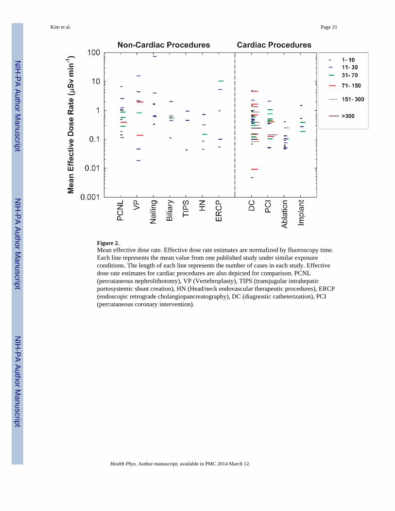
Figure 2.Mean effective dose rate. Effective dose rate estimates are normalized by fluoroscopy time.Each line represents the mean value from one published study under similar exposureconditions. The length of each line represents the number of cases in each study. Effectivedose rate estimates for cardiac procedures are also depicted for comparison. PCNL(percutaneous nephrolithotomy), VP (Vertebroplasty), TIPS (transjugular intrahepaticportosystemic shunt creation), HN (Head/neck endovascular therapeutic procedures), ERCP(endoscopic retrograde cholangiopancreatography), DC (diagnostic catheterization), PCI(percutaneous coronary intervention).
Kim et al. Page 21
Health Phys. Author manuscript; available in PMC 2014 March 12.
NIH
-PA Author Manuscript
NIH
-PA Author Manuscript
NIH
-PA Author Manuscript

Figure 3.Mean effective dose normalized by patient radiation dose (as kerma area product). Each linerepresents the mean value from one published study under similar exposure conditions. Thelength of each line represents the number of cases in each study. Data for cardiac proceduresare also depicted for comparison. PCNL (percutaneous nephrolithotomy), VP(Vertebroplasty), TIPS (transjugular intrahepatic portosystemic shunt creation), HN (Head/neck endovascular therapeutic procedures), ERCP (endoscopic retrogradecholangiopancreatography), DC (diagnostic catheterization), PCI (percutaneous coronaryintervention).
Kim et al. Page 22
Health Phys. Author manuscript; available in PMC 2014 March 12.
NIH
-PA Author Manuscript
NIH
-PA Author Manuscript
NIH
-PA Author Manuscript

Figure 4.Mean hand dose rate. Radiation dose rates measured at the operator’s hand are normalizedby fluoroscopy time. Each line represents the mean value from one published study undersimilar exposure conditions. The length of each line represents the number of cases in eachstudy. Data for cardiac procedures are also depicted for comparison. PCNL (percutaneousnephrolithotomy), VP (Vertebroplasty), TIPS (transjugular intrahepatic portosystemic shuntcreation), HN (Head/neck endovascular therapeutic procedures), ERCP (endoscopicretrograde cholangiopancreatography), DC (diagnostic catheterization), PCI (percutaneouscoronary intervention).
Kim et al. Page 23
Health Phys. Author manuscript; available in PMC 2014 March 12.
NIH
-PA Author Manuscript
NIH
-PA Author Manuscript
NIH
-PA Author Manuscript

Figure 5.Mean organ dose estimates per case for operators performing various types of FGprocedures. Each line represents the mean value from one published study under similarexposure conditions. The length of each line represents the number of cases in each study.Organ dose estimates for cardiac procedures are also depicted for comparison. PCNL(percutaneous nephrolithotomy), VP (Vertebroplasty), TIPS (transjugular intrahepaticportosystemic shunt creation), HN (Head/neck endovascular therapeutic procedures), ERCP(endoscopic retrograde cholangiopancreatography), DC (diagnostic catheterization), PCI(percutaneous coronary intervention).
Kim et al. Page 24
Health Phys. Author manuscript; available in PMC 2014 March 12.
NIH
-PA Author Manuscript
NIH
-PA Author Manuscript
NIH
-PA Author Manuscript

NIH
-PA Author Manuscript
NIH
-PA Author Manuscript
NIH
-PA Author Manuscript
Kim et al. Page 25
Table 1
Mean exposure and effective dose to the operator per case from percutaneous nephrolithotomy
Author(Publication
Year) aPhysician b
No ofCases
cFluoroscopy
Time d (min)
Protective
Measures eMean Badge Dose
per Case (μSv) d, fEffective
Dose
k (μSv)
Note
Apron (mm) ThyroidShield (mm) Hand g
Eye
Level h Neck iTrunk j
OverApron
UnderApron
Safak et al.(2009)
Urologist 20 11.7 (1.5–31.2) 0.5 0.5 33 26 48 - 12 14.2
Kumari etal. (2006)
Urologist 50 6.0 (1.8–12.2) 0.5 0.5 280 (±130) - - 24.9 (7.4–50.2) - 1.7
Hellawell etal. (2005)
Urologist 6 6.8–23 0.35 0.35 48 (±12) 40 (±10) - - - 2.8
Yang et al.(2002)
Urologist 6 12.8 O - - - 88 - - 6.2 Without ceiling-suspended shield
Yang et al.(2002)
Urologist 6 12.8 O - - - 25 - - 1.8 With ceiling-suspended shield
Bowsher etal. (1992)
Urologist 6 2.0 (0.3–2.8) - - 50 (±40) 30 (±15) - - - 2.1 Under-couch system
Bowsher etal. (1992)
Urologist 8 2.0 (0.3–2.8) - - 230 (±120) 190 (±120) - - - 13.3 Over-couch system
Nowak andJankowski(1991)
NS 54 - 0.25 - 41 34 - - - 2.4
Ramsdale etal. (1990)
Radiologist 42 22 (±13) - - 520 (±750) 320 (±360) 270 (±220) - - 18.9 Over-couch system
Geterud etal. (1989)
Urologist + Radiologist 11 14 (3.0–29) 0.3 X 210 (14–710) - 99 (15–260) - 8.6 (2.1–18) 14.0
Inglis et al.(1989)
Urologist 55 4.4 (1.2–13) - - 342 - 35 - - 2.5
Rao et al.(1987)
Urologist + Radiologist 18 22 (0.9–45) O - 5800 800 - - - 56.0 Over-couch system
Lowe et al.(1986)
Urologist + Radiologist 15 28 0.5 O 83 (±84) - 45 (±48) - - 3.2
Bush et al.(1985)
Urologist + Radiologist 94 18 (4–65) 0.5 0.5 300 (100–2000) - 100 (20–320) - - 7.0
Bush et al.(1984)
Urologist 51 8 (2–30) 0.5 X - - 100 (10–380) - - 7.0
There is no column for kerma-area product (KAP) data because no KAP data were included in these reports.
aReferences are arranged by publication year.
bNS: Not specified.
cNumber of cases in the report.
dNumbers in parenthesis are standard deviation (±) or minimum-maximum (−). Superscripts of ‘md’, and ‘iq’ indicate median value and inter-
quartile.
eLead-equivalent thickness of protective measures. O indicates that protective measures were used but their thicknesses were not given. X indicates
that protective measures were not used.
fExposure unit (R) in some studies was converted into Hp(10) for trunk dose under apron (conversion factor 11,600 μSv R−1) and into Hp(0.07)
for doses outside shield (conversion factor 11,900 μSv R−1).
gMeasurements obtained at the wrist, hand, or finger outside shield.
hMeasurements obtained at the eye, forehead, glabella, maxilla, or temple outside shield.
iMeasurements obtained at the neck, collar, clavicle, or shoulder outside shield.
jMeasurements obtained at the chest, sternum, umbilicus, waist, or abdomen over or under apron.
Health Phys. Author manuscript; available in PMC 2014 March 12.

NIH
-PA Author Manuscript
NIH
-PA Author Manuscript
NIH
-PA Author Manuscript
Kim et al. Page 26
kEffective doses were calculated using the Niklason (2 dosimeters) and Padovani et al. (1 dosimeter) algorithms, assuming no use of a thyroid
shield (see text for details). If no measurement over the thyroid shield was available, the measurement at eye level or outside the apron at trunklevel was used, in that order of preference.
Health Phys. Author manuscript; available in PMC 2014 March 12.
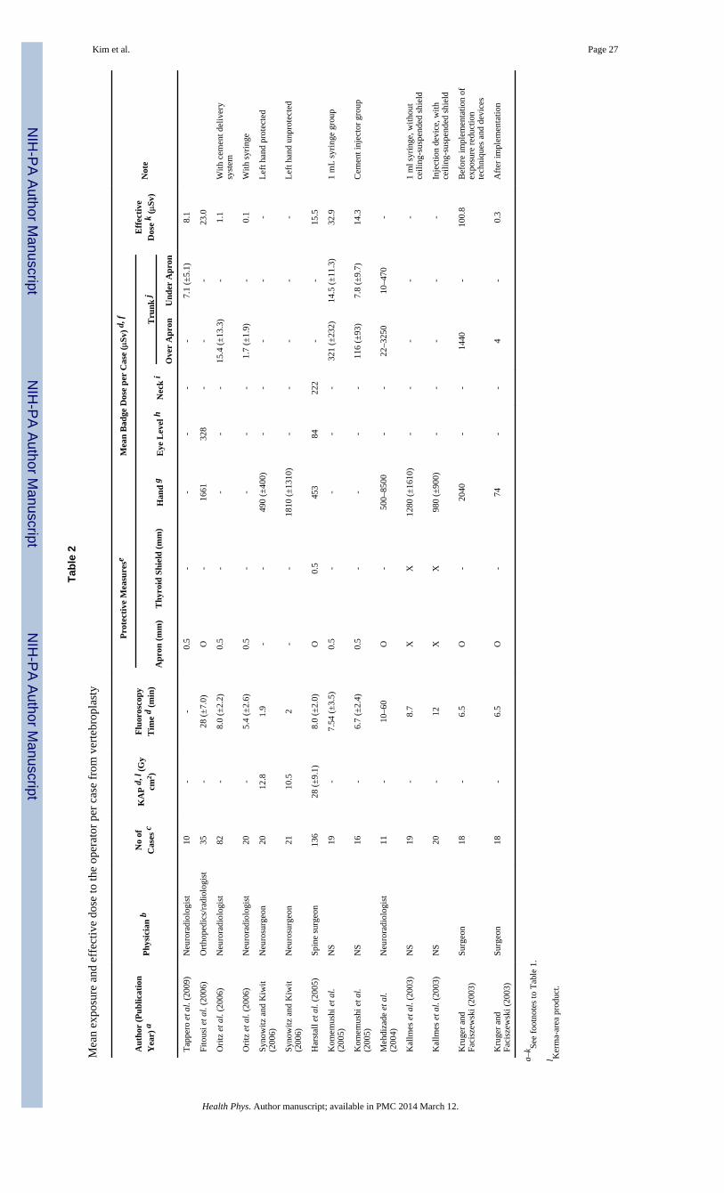
NIH
-PA Author Manuscript
NIH
-PA Author Manuscript
NIH
-PA Author Manuscript
Kim et al. Page 27
Tabl
e 2
Mea
n ex
posu
re a
nd e
ffec
tive
dose
to th
e op
erat
or p
er c
ase
from
ver
tebr
opla
sty
Aut
hor
(Pub
licat
ion
Yea
r) a
Phy
sici
an b
No
ofC
ases
cK
AP
d, l
(G
ycm
2 )
Flu
oros
copy
Tim
e d
(min
)
Pro
tect
ive
Mea
sure
seM
ean
Bad
ge D
ose
per
Cas
e (μ
Sv)
d, f
Eff
ecti
veD
ose
k (μ
Sv)
Not
e
Apr
on (
mm
)T
hyro
id S
hiel
d (m
m)
Han
d g
Eye
Lev
el h
Nec
k i
Tru
nk j
Ove
r A
pron
Und
er A
pron
Tap
pero
et a
l. (2
009)
Neu
rora
diol
ogis
t10
--
0.5
--
--
-7.
1 (±
5.1)
8.1
Fito
usi e
t al.
(200
6)O
rtho
pedi
cs/r
adio
logi
st35
-28
(±
7.0)
O-
1661
328
--
-23
.0
Ori
tz e
t al.
(200
6)N
euro
radi
olog
ist
82-
8.0
(±2.
2)0.
5-
--
-15
.4 (
±13
.3)
-1.
1W
ith c
emen
t del
iver
ysy
stem
Ori
tz e
t al.
(200
6)N
euro
radi
olog
ist
20-
5.4
(±2.
6)0.
5-
--
-1.
7 (±
1.9)
-0.
1W
ith s
yrin
ge
Syno
witz
and
Kiw
it(2
006)
Neu
rosu
rgeo
n20
12.8
1.9
--
490
(±40
0)-
--
--
Lef
t han
d pr
otec
ted
Syno
witz
and
Kiw
it(2
006)
Neu
rosu
rgeo
n21
10.5
2-
-18
10 (
±13
10)
--
--
-L
eft h
and
unpr
otec
ted
Har
stal
l et a
l. (2
005)
Spin
e su
rgeo
n13
628
(±
9.1)
8.0
(±2.
0)O
0.5
453
8422
2-
-15
.5
Kom
emus
hi e
t al.
(200
5)N
S19
-7.
54 (
±3.
5)0.
5-
--
-32
1 (±
232)
14.5
(±
11.3
)32
.91
mL
syr
inge
gro
up
Kom
emus
hi e
t al.
(200
5)N
S16
-6.
7 (±
2.4)
0.5
--
--
116
(±93
)7.
8 (±
9.7)
14.3
Cem
ent i
njec
tor
grou
p
Meh
diza
de e
t al.
(200
4)N
euro
radi
olog
ist
11-
10–6
0O
-50
0–85
00-
-22
–325
010
–470
-
Kal
lmes
et a
l. (2
003)
NS
19-
8.7
XX
1280
(±
1610
)-
--
--
1 m
l syr
inge
, with
out
ceili
ng-s
uspe
nded
shi
eld
Kal
lmes
et a
l. (2
003)
NS
20-
12X
X98
0 (±
900)
--
--
-In
ject
ion
devi
ce, w
ithce
iling
-sus
pend
ed s
hiel
d
Kru
ger
and
Faci
szew
ski (
2003
)Su
rgeo
n18
-6.
5O
-20
40-
-14
40-
100.
8B
efor
e im
plem
enta
tion
ofex
posu
re r
educ
tion
tech
niqu
es a
nd d
evic
es
Kru
ger
and
Faci
szew
ski (
2003
)Su
rgeo
n18
-6.
5O
-74
--
4-
0.3
Aft
er im
plem
enta
tion
a–k Se
e fo
otno
tes
to T
able
1.
l Ker
ma-
area
pro
duct
.
Health Phys. Author manuscript; available in PMC 2014 March 12.
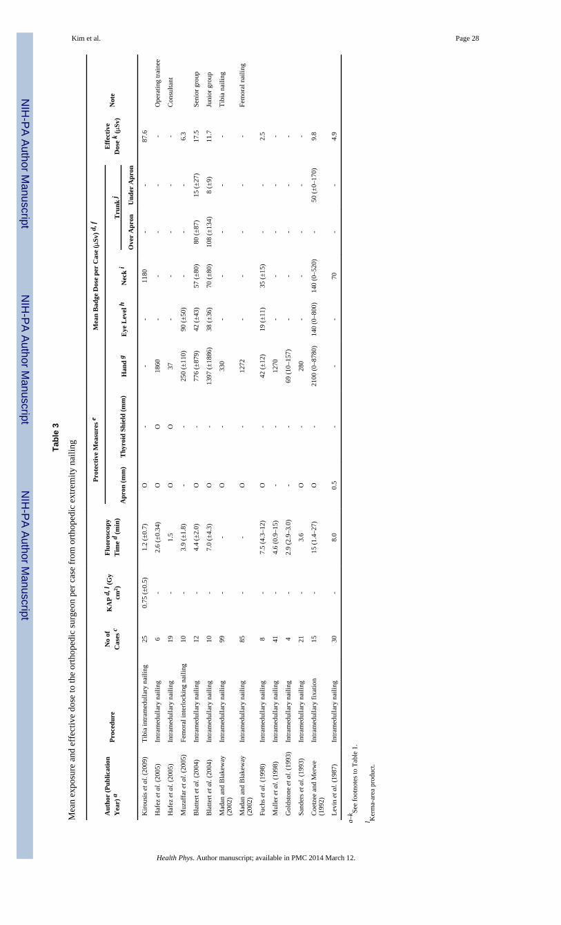
NIH
-PA Author Manuscript
NIH
-PA Author Manuscript
NIH
-PA Author Manuscript
Kim et al. Page 28
Tabl
e 3
Mea
n ex
posu
re a
nd e
ffec
tive
dose
to th
e or
thop
edic
sur
geon
per
cas
e fr
om o
rtho
pedi
c ex
trem
ity n
ailin
g
Aut
hor
(Pub
licat
ion
Yea
r) a
Pro
cedu
reN
o of
Cas
es c
KA
P d
, l (
Gy
cm2 )
Flu
oros
copy
Tim
e d
(min
)
Pro
tect
ive
Mea
sure
s e
Mea
n B
adge
Dos
e pe
r C
ase
(μSv
) d,
f
Eff
ecti
veD
ose
k (μ
Sv)
Not
e
Apr
on (
mm
)T
hyro
id S
hiel
d (m
m)
Han
d g
Eye
Lev
el h
Nec
k i
Tru
nk j
Ove
r A
pron
Und
er A
pron
Kir
ousi
s et
al.
(200
9)T
ibia
intr
amed
ulla
ry n
ailin
g25
0.75
(±
0.5)
1.2
(±0.
7)O
--
-11
80-
-87
.6
Haf
ez e
t al.
(200
5)In
tram
edul
lary
nai
ling
6-
2.6
(±0.
34)
OO
1860
--
--
-O
pera
ting
trai
nee
Haf
ez e
t al.
(200
5)In
tram
edul
lary
nai
ling
19-
1.5
OO
37-
--
--
Con
sulta
nt
Muz
affa
r et
al.
(200
5)Fe
mor
al in
terl
ocki
ng n
ailin
g10
-3.
9 (±
1.8)
--
250
(±11
0)90
(±
50)
--
-6.
3
Bla
ttert
et a
l. (2
004)
Intr
amed
ulla
ry n
ailin
g12
-4.
4 (±
2.0)
O-
776
(±87
9)42
(±
43)
57 (
±80
)80
(±
87)
15 (
±27
)17
.5Se
nior
gro
up
Bla
ttert
et a
l. (2
004)
Intr
amed
ulla
ry n
ailin
g10
-7.
0 (±
4.3)
O-
1397
(±
1886
)38
(±
36)
70 (
±80
)10
8 (±
134)
8 (±
9)11
.7Ju
nior
gro
up
Mad
an a
nd B
lake
way
(200
2)In
tram
edul
lary
nai
ling
99-
-O
-33
0-
--
--
Tib
ia n
ailin
g
Mad
an a
nd B
lake
way
(200
2)In
tram
edul
lary
nai
ling
85-
-O
-12
72-
--
--
Fem
oral
nai
ling
Fuch
s et
al.
(199
8)In
tram
edul
lary
nai
ling
8-
7.5
(4.3
–12)
O-
42 (
±12
)19
(±
11)
35 (
±15
)-
-2.
5
Mul
ler
et a
l. (1
998)
Intr
amed
ulla
ry n
ailin
g41
-4.
6 (0
.9–1
5)-
-12
70-
--
--
Gol
dsto
ne e
t al.
(199
3)In
tram
edul
lary
nai
ling
4-
2.9
(2.9
–3.0
)-
-69
(10
–157
)-
--
--
Sand
ers
et a
l. (1
993)
Intr
amed
ulla
ry n
ailin
g21
-3.
6O
-28
0-
--
--
Coe
tzee
and
Mer
we
(199
2)In
tram
edul
lary
fix
atio
n15
-15
(1.
4–27
)O
-21
00 (
0–87
80)
140
(0–8
00)
140
(0–5
20)
-50
(±
0–17
0)9.
8
Lev
in e
t al.
(198
7)In
tram
edul
lary
nai
ling
30-
8.0
0.5
--
-70
--
4.9
a–k Se
e fo
otno
tes
to T
able
1.
l Ker
ma-
area
pro
duct
.
Health Phys. Author manuscript; available in PMC 2014 March 12.

NIH
-PA Author Manuscript
NIH
-PA Author Manuscript
NIH
-PA Author Manuscript
Kim et al. Page 29
Tabl
e 4
Mea
n ex
posu
re a
nd e
ffec
tive
dose
to th
e op
erat
or p
er c
ase
from
bili
ary
trac
t pro
cedu
res
Aut
hor
(Pub
licat
ion
Yea
r) a
Phy
sici
an b
No
of C
ases
cK
AP
d, l
(G
y cm
2 )F
luor
osco
py T
ime
d (m
in)
Pro
tect
ive
Mea
sure
s e
Mea
n B
adge
Dos
e pe
r C
ase
(μSv
) d,
f
Eff
ecti
ve D
ose
k (μ
Sv)
Not
e
Apr
on (
mm
)T
hyro
id S
hiel
d (m
m)
Han
d g
Eye
Lev
el h
Nec
k i
Tru
nk j
Ove
r A
pron
Und
er A
pron
Oon
siri
et a
l. (2
007)
Rad
iolo
gist
918
(2.
8–32
.7)
1.9–
14O
O-
110
(23–
282)
63 (
1–20
0)-
-4.
4
Stra
taki
s et
al.
(200
6)R
adio
logi
st35
207.
80.
50.
543
083
6023
-4.
2D
rain
age
only
, with
und
er-c
ouch
shi
eld
Stra
taki
s et
al.
(200
6)R
adio
logi
st-
--
0.5
0.5
-18
013
518
2-
9.5
Dra
inag
e on
ly, w
ithou
t und
er-c
ouch
shi
eld
Stra
taki
s et
al.
(200
6)R
adio
logi
st17
2511
0.5
0.5
507
9670
27-
4.9
Dra
inag
e +
ste
ntin
g, w
ith u
nder
-cou
ch s
hiel
d
Stra
taki
s et
al.
(200
6)R
adio
logi
st-
--
0.5
0.5
-21
216
021
5-
11.2
Dra
inag
e +
ste
ntin
g, w
ithou
t und
er-c
ouch
shi
eld
Stra
taki
s et
al.
(200
6)R
adio
logi
st19
175.
70.
50.
527
872
5220
-3.
6St
entin
g on
ly, w
ith u
nder
-cou
ch s
hiel
d
Stra
taki
s et
al.
(200
6)R
adio
logi
st-
--
0.5
0.5
-15
912
016
2-
8.4
Sten
ting
only
, with
out u
nder
-cou
ch s
hiel
d
Mar
tin a
nd W
hitb
y (2
003)
Rad
iolo
gist
17-
-O
-80
0 (4
00–5
50)
--
--
-B
iliar
y pr
oced
ure
Whi
tby
and
Mar
tin (
2003
)R
adio
logi
st11
--
O-
950
--
--
-B
iliar
y pr
oced
ure
Will
iam
s (1
997)
Rad
iolo
gist
8643
(19
–61)
iq-
0.35
/0.5
m-
105
-38
-2.
14.
3B
iliar
y dr
aina
ge
Will
iam
s (1
997)
Rad
iolo
gist
7451
(15
–63)
iq-
0.35
/0.5
m-
124
-45
-2.
55.
1B
iliar
y dr
aina
ge +
ste
nt
Veh
mas
(19
93)
Rad
iolo
gist
418
19-
-22
8-
--
--
Veh
mas
and
Tik
kane
n (1
992)
Rad
iolo
gist
2-
18O
-36
7-
28-
-2.
0
Now
ak a
nd J
anko
wsk
i (19
91)
NS
29-
-0.
25-
488
213
--
-14
.9X
-ray
con
trol
of
bilia
ry r
oute
Ram
sdal
e et
al.
(199
0)R
adio
logi
st16
-23
(±
16)
--
1290
(±
1980
)31
0 (±
400)
660
(±10
00)
--
46.2
Bili
ary
drai
nage
and
ste
nt
Bur
gess
and
Bur
henn
e (1
984)
Rad
iolo
gist
33-
5-
-60
0-
--
--
Bili
ary
proc
edur
es
a–k Se
e fo
otno
tes
to T
able
1.
l Ker
ma-
area
pro
duct
.
mO
ne o
pera
tor
wor
e an
apr
on o
f 0.
5 m
m le
ad-e
quiv
alen
t thi
ckne
ss. T
he o
ther
s w
ore
apro
ns o
f 0.
35 m
m le
ad-e
quiv
alen
t thi
ckne
ss.
Health Phys. Author manuscript; available in PMC 2014 March 12.
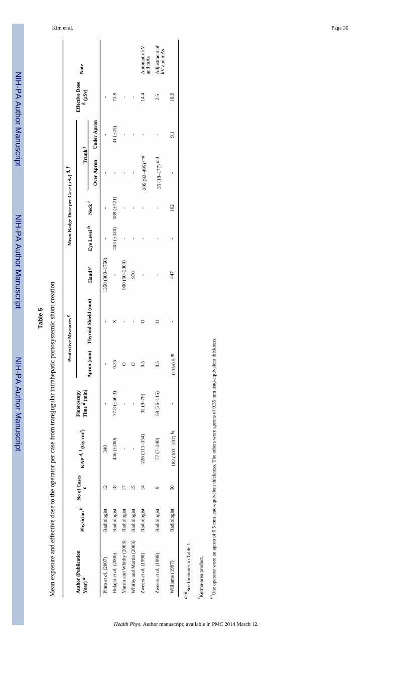
NIH
-PA Author Manuscript
NIH
-PA Author Manuscript
NIH
-PA Author Manuscript
Kim et al. Page 30
Tabl
e 5
Mea
n ex
posu
re a
nd e
ffec
tive
dose
to th
e op
erat
or p
er c
ase
from
tran
sjug
ular
intr
ahep
atic
por
tosy
stem
ic s
hunt
cre
atio
n
Aut
hor
(Pub
licat
ion
Yea
r) a
Phy
sici
an b
No
of C
ases
cK
AP
d, l
(G
y cm
2 )F
luor
osco
pyT
ime
d (m
in)
Pro
tect
ive
Mea
sure
s e
Mea
n B
adge
Dos
e pe
r C
ase
(μSv
) d,
f
Eff
ecti
ve D
ose
k (μ
Sv)
Not
e
Apr
on (
mm
)T
hyro
id S
hiel
d (m
m)
Han
d g
Eye
Lev
el h
Nec
k i
Tru
nk j
Ove
r A
pron
Und
er A
pron
Pint
o et
al.
(200
7)R
adio
logi
st12
340
--
-13
50 (
900–
1750
)-
--
--
Hid
ajat
et a
l. (2
006)
Rad
iolo
gist
1844
6 (±
280)
77.8
(±
66.3
)0.
35X
-40
3 (±
328)
589
(±72
1)-
41 (
±25
)73
.9
Mar
tin a
nd W
hitb
y (2
003)
Rad
iolo
gist
17-
-O
-90
0 (5
0–20
00)
--
--
-
Whi
tby
and
Mar
tin (
2003
)R
adio
logi
st15
--
O-
970
--
--
-
Zw
eers
et a
l. (1
998)
Rad
iolo
gist
1422
6 (1
11–3
54)
32 (
9–79
)0.
5O
--
-20
5 (9
2–49
5) m
d-
14.4
Aut
omat
ic k
Van
d m
As
Zw
eers
et a
l. (1
998)
Rad
iolo
gist
977
(7–
240)
59 (
26–1
15)
0.5
O-
--
35 (
18–1
77)
md
-2.
5A
djus
tmen
t of
kV a
nd m
As
Will
iam
s (1
997)
Rad
iolo
gist
5618
2 (1
03 –
237)
iq-
0.35
/0.5
m-
447
-16
2-
9.1
18.9
a–k Se
e fo
otno
tes
to T
able
1.
l Ker
ma-
area
pro
duct
.
mO
ne o
pera
tor
wor
e an
apr
on o
f 0.
5 m
m le
ad-e
quiv
alen
t thi
ckne
ss. T
he o
ther
s w
ore
apro
ns o
f 0.
35 m
m le
ad-e
quiv
alen
t thi
ckne
ss.
Health Phys. Author manuscript; available in PMC 2014 March 12.
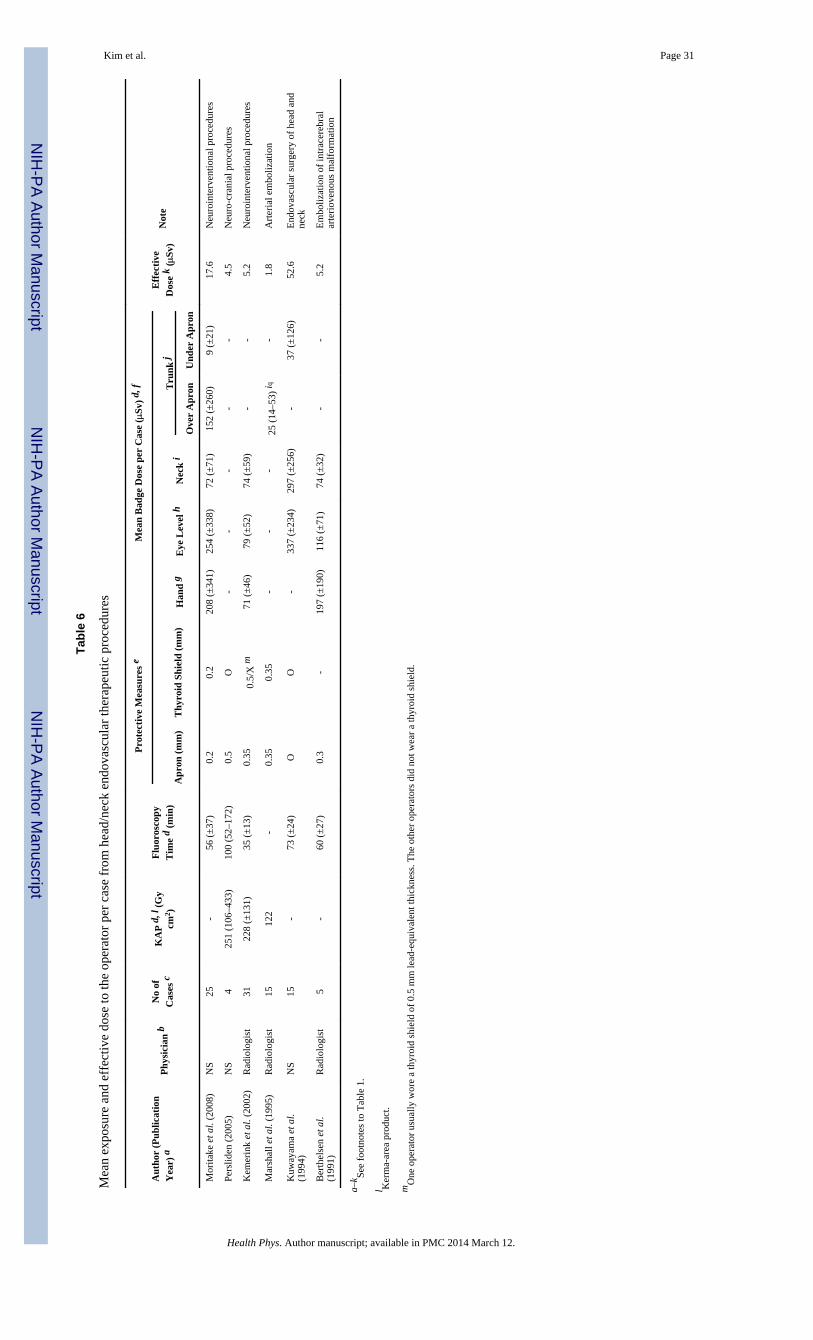
NIH
-PA Author Manuscript
NIH
-PA Author Manuscript
NIH
-PA Author Manuscript
Kim et al. Page 31
Tabl
e 6
Mea
n ex
posu
re a
nd e
ffec
tive
dose
to th
e op
erat
or p
er c
ase
from
hea
d/ne
ck e
ndov
ascu
lar
ther
apeu
tic p
roce
dure
s
Aut
hor
(Pub
licat
ion
Yea
r) a
Phy
sici
an b
No
ofC
ases
cK
AP
d, l
(G
ycm
2 )
Flu
oros
copy
Tim
e d
(min
)
Pro
tect
ive
Mea
sure
s e
Mea
n B
adge
Dos
e pe
r C
ase
(μSv
) d,
f
Eff
ecti
veD
ose
k (μ
Sv)
Not
e
Apr
on (
mm
)T
hyro
id S
hiel
d (m
m)
Han
d g
Eye
Lev
el h
Nec
k i
Tru
nk j
Ove
r A
pron
Und
er A
pron
Mor
itake
et a
l. (2
008)
NS
25-
56 (
±37
)0.
20.
220
8 (±
341)
254
(±33
8)72
(±
71)
152
(±26
0)9
(±21
)17
.6N
euro
inte
rven
tiona
l pro
cedu
res
Pers
liden
(20
05)
NS
425
1 (1
06–4
33)
100
(52–
172)
0.5
O-
--
--
4.5
Neu
ro-c
rani
al p
roce
dure
s
Kem
erin
k et
al.
(200
2)R
adio
logi
st31
228
(±13
1)35
(±
13)
0.35
0.5/
X m
71 (
±46
)79
(±
52)
74 (
±59
)-
-5.
2N
euro
inte
rven
tiona
l pro
cedu
res
Mar
shal
l et a
l. (1
995)
Rad
iolo
gist
1512
2-
0.35
0.35
--
-25
(14
–53)
iq-
1.8
Art
eria
l em
boliz
atio
n
Kuw
ayam
a et
al.
(199
4)N
S15
-73
(±
24)
OO
-33
7 (±
234)
297
(±25
6)-
37 (
±12
6)52
.6E
ndov
ascu
lar
surg
ery
of h
ead
and
neck
Ber
thel
sen
et a
l.(1
991)
Rad
iolo
gist
5-
60 (
±27
)0.
3-
197
(±19
0)11
6 (±
71)
74 (
±32
)-
-5.
2E
mbo
lizat
ion
of in
trac
ereb
ral
arte
riov
enou
s m
alfo
rmat
ion
a–k Se
e fo
otno
tes
to T
able
1.
l Ker
ma-
area
pro
duct
.
mO
ne o
pera
tor
usua
lly w
ore
a th
yroi
d sh
ield
of
0.5
mm
lead
-equ
ival
ent t
hick
ness
. The
oth
er o
pera
tors
did
not
wea
r a
thyr
oid
shie
ld.
Health Phys. Author manuscript; available in PMC 2014 March 12.
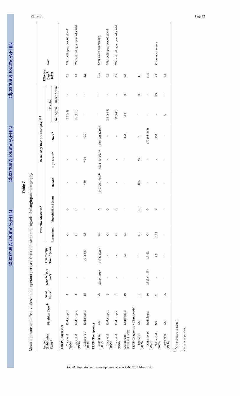
NIH
-PA Author Manuscript
NIH
-PA Author Manuscript
NIH
-PA Author Manuscript
Kim et al. Page 32
Tabl
e 7
Mea
n ex
posu
re a
nd e
ffec
tive
dose
to th
e op
erat
or p
er c
ase
from
end
osco
pic
retr
ogra
de c
hola
ngio
panc
reat
ogra
phy
Aut
hor
(Pub
licat
ion
Yea
r) a
Phy
sici
an T
ype
bN
o of
Cas
es c
KA
P d
, l (
Gy
cm2 )
Flu
oros
copy
Tim
e d
(min
)
Pro
tect
ive
Mea
sure
s e
Mea
n B
adge
Dos
e pe
r C
ase
(μSv
) d,
f
Eff
ecti
veD
ose
k(μ
Sv)
Not
e
Apr
on (
mm
)T
hyro
id S
hiel
d (m
m)
Han
d g
Eye
Lev
el h
Nec
k i
Tru
nk j
Ove
r A
pron
Und
er A
pron
ER
CP
(D
iagn
osti
c)
C
hen
et a
l.(1
996)
End
osco
pist
4-
-O
O-
--
2.5
(±5)
-0.
2W
ith c
eilin
g-su
spen
ded
shie
ld
C
hen
et a
l.(1
996)
End
osco
pist
4-
-O
O-
--
15 (
±19
)-
1.1
With
out c
eilin
g-su
spen
ded
shie
ld
C
ohen
et a
l.(1
979)
End
osco
pist
15-
10 (
±4.
4)0.
5-
<30
<30
<30
--
2.1
ER
CP
(T
hera
peut
ic)
B
uls
et a
l.(2
002)
NS
2550
(24–
60)
iq6
(3.6
–8.3
) iq
0.5
X64
0 (2
00–8
80)i
q55
0 (1
60–6
60)i
q45
0 (1
70–6
00)i
q-
-31
.5O
ver-
couc
h fl
uoro
scop
y
C
hen
et a
l.(1
996)
End
osco
pist
6-
-O
O-
--
2.8
(±4.
4)-
0.2
With
cei
ling-
susp
ende
d sh
ield
C
hen
et a
l.(1
996)
End
osco
pist
6-
-O
O-
--
32 (
±45
)-
2.2
With
out c
eilin
g-su
spen
ded
shie
ld
Kru
eger
and
Hof
fman
(19
92)
End
osco
pist
10-
7.5
0.5
--
-6.
23.
30
0.4
ER
CP
(D
iagn
osti
c +
The
rape
utic
)
O
lgar
et a
l.(2
009)
NS
31-
-0.
50.
583
594
75-
04.
5
O
onsi
ri e
t al.
(200
7)R
adio
logi
st10
35 (
9.6–
105)
1.7–
23O
O-
-17
0 (9
8–31
8)-
-11
.9
N
aidu
et a
l.(2
005)
NS
61-
4.8
0.25
X-
-45
7-
2349
Ove
r-co
uch
syst
em
H
eyd
et a
l.(1
996)
NS
25-
--
--
--
6-
0.4
a–k Se
e fo
otno
tes
to T
able
1.
l Ker
ma-
area
pro
duct
.
Health Phys. Author manuscript; available in PMC 2014 March 12.