nih public access biomaterials for neural stem cell culture … publications... · 2019-12-22 ·...
TRANSCRIPT

Exploiting Bacterial Peptide Display Technology to EngineerBiomaterials for Neural Stem Cell Culture
Lauren Little1, Karen Dane2, Patrick Daugherty2, Kevin Healy3,4,*, and DavidSchaffer1,3,5,*1 Department of Chemical Engineering, University of California at Berkeley, Berkeley, California94720, United States2 Department of Chemical Engineering, University of California at Santa Barbara, Santa Barbara,California 93106, United States3 Department of Bioengineering, University of California at Berkeley, Berkeley, California 94720,United States4 Department of Material Science and Engineering, University of California at Berkeley, Berkeley,California 94720, United States5 The Helen Wills Neuroscience Institute, University of California at Berkeley, Berkeley, California94720, United States
AbstractStem cells are often cultured on substrates that present extracellular matrix (ECM) proteins;however, the heterogeneous and poorly defined nature of ECM proteins presents challenges bothfor basic biological investigation of cell-matrix investigations and translational applications ofstem cells. Therefore, fully synthetic, defined materials conjugated with bioactive ligands, such asadhesive peptides, are preferable for stem cell biology and engineering. However, identifyingnovel ligands that engage cellular receptors can be challenging, and we have thus developed a highthroughput approach to identify new adhesive ligands. We selected an unbiased bacterial peptidedisplay library for the ability to bind adult neural stem cells (NSCs), and 44 bacterial clonesexpressing peptides were identified and found to bind to NSCs with high avidity. Of these clones,four contained RGD motifs commonly found in integrin binding domains, and three exhibitedhomology to ECM proteins. Three peptide clones were chosen for further analysis, and theirsynthetic analogs were adsorbed on tissue culture polystyrene (TCPS) or grafted onto aninterpenetrating polymer network (IPN) for cell culture. These three peptides were found tosupport neural stem cell self-renewal in defined medium as well as multi-lineage differentiation.Therefore, bacterial peptide display offers unique advantages to isolate bioactive peptides fromlarge, unbiased libraries for applications in biomaterials engineering.
KeywordsBiomimetic material; ECM; Peptide; Stem cell
*Corresponding authors: David Schaffer, 274 Stanley Hall, Berkeley, CA 94720-3220, [email protected], (510) 643-5963, (510)642-4778 (fax), Kevin Healy, 370 Hearst MMB, Berkeley, CA 94720-1760, Email: [email protected], (510) 643-5963, (510)643-5792 (fax).Publisher's Disclaimer: This is a PDF file of an unedited manuscript that has been accepted for publication. As a service to ourcustomers we are providing this early version of the manuscript. The manuscript will undergo copyediting, typesetting, and review ofthe resulting proof before it is published in its final citable form. Please note that during the production process errors may bediscovered which could affect the content, and all legal disclaimers that apply to the journal pertain.
NIH Public AccessAuthor ManuscriptBiomaterials. Author manuscript; available in PMC 2012 February 1.
Published in final edited form as:Biomaterials. 2011 February ; 32(6): 1484–1494. doi:10.1016/j.biomaterials.2010.10.032.
NIH
-PA Author Manuscript
NIH
-PA Author Manuscript
NIH
-PA Author Manuscript

IntroductionMany cell types, including stem cells, require interactions with extracellular matrix (ECM)molecules to support and regulate their survival and proliferation.[1,2] In addition to servingas important structural elements in formations such as basement membrane, the ECMexhibits a number of bioactivities via engaging with cellular adhesion receptors such asintegrins, thereby activating a number of downstream signaling pathways including crosstalkwith growth factor dependent pathways.[3–6] As a result, ECM molecules regulate anumber of downstream cell behaviors, including adhesion, survival, proliferation, migration,and differentiation.[7] These many active role of ECM molecules explains why manysurfaces do not support the growth of stem cells.[8,9]
Although animal- and human-derived ECM molecules are often necessary for current stemcell culture systems, their use is highly problematic for numerous reasons. Natural ECMmolecules are very large (e.g. ~500,000 MW fibronectin and ~850,000 MW laminin), areextremely complex (with numerous isoforms, splice variants, and glycoforms), and havenumerous signaling motifs that are not yet fully understood.[10] In addition, there isconsiderable lot-to-lot variability in animal- and human-derived ECM because of the manyisoforms present and difficulty in purifying such proteins to homogeneity,[11,12] and suchprotein preparations run the risk of being contaminated with pathogens and immunogens.[13] The development of stem cell culture systems that are robust, reproducible, andscaleable can benefit both scientific studies and clinical therapies.
One promising approach is to design and develop synthetic stem cell culture platforms thatmimic the physical and biochemical properties of the natural ECM. For example, materialscan be functionalized with short (9–15mer) synthetic peptide ligands designed to engagewith cell-surface receptors and thereby functionally replace ECM proteins typically used instem cell culture.[9,12,14–17] In particular, it is important to develop surfaces that canengage with integrins—heterodimeric transmembrane receptors that bind ECM molecules,anchor cells to the matrix, and act as bidirectional transducers to transmit both mechanicaland chemical stimuli into the cell. [18–20] Most ligands for integrins that have beenexploited in biomaterials research have employed the canonical Arg-Gly-Asp (RGD)sequence that binds a subset of these adhesion receptors.[21–24] The flanking residues tothe RGD sequence can affect specificity to the RGD-binding integrins such as the αvβ3integrin.[22,24] However, identifying new ligands that bind these and other receptors couldaid in engineering biomaterials and production of cell-based therapies for the clinic, as wellas in gaining a better understanding of signaling mechanisms that regulate cell function.[23,25–28]
However, while various cell-binding domains—such as RGD and IKVAV motifs—[29,30]have been identified in many ECM proteins, many peptides mimicking those domains arenot as active as the full ECM proteins, as evidenced by the fact that few of them support theculture of cells to the extent of the native protein.[12,29–36] In addition, many of theimportant signaling domains in ECM proteins may not be known, and for example onlyrecently has the αv binding domain in fibronectin been identified.[37] Finally, in generalthere is no guarantee that the optimal peptide ligand for a given receptor exactly matches aportion of the “linear” sequence of its natural ECM ligand, because the three-dimensionalstructure of proteins that typically contain more than one chain (e.g., laminin, collagen).Therefore, there is a need to develop a robust method to identify new candidate peptides thatcan be used to modify or create biomaterials to replace ECM proteins.
Little et al. Page 2
Biomaterials. Author manuscript; available in PMC 2012 February 1.
NIH
-PA Author Manuscript
NIH
-PA Author Manuscript
NIH
-PA Author Manuscript

Due to of the vast number of possible peptide sequences, even for a very short polypeptide,rationally designing such a ligand is very challenging; however, library-based screening andselection methods have potential to address this problem. With library strategies such asbacterial display, many peptides with random sequences can be tested simultaneously andthus can facilitate the discovery of peptides even with little prior mechanistic knowledge ofthe motifs required for receptor engagement.[38,39] While phage display libraries have beenused in a number of prior studies, these have focused primarily on finding cell adhesionreceptors antagonists, including for integrins, rather than identifying peptides agonists forthese receptors.[40–44] In addition, newer library systems using bacteria as a displayplatform offer numerous advantages. For example, they are easier to generate since thelibraries involve straightforward plasmid manipulations, do not require bacteriophageproduction or purification, do not involve viral infection steps for amplification during theselection process, and can readily be analyzed after selection by simple plasmid isolationand sequencing.[38,39,45–47] In addition, the levels of surface-displayed peptide can inprinciple be varied to modulate selection stringency, and fluorescent protein expressingbacteria can be used to aid library selection and analysis.[48–50]
In this study, we have used a bacterial display library to identify new peptides that bind adulthippocampal neural stem cells (NSCs). Several of these peptides were examined for theirability to support neural stem cells in two different contexts: adsorbed on tissue culturepolystyrene (TCPS) or grafted to an interpenetrating polymer network surface. On bothsurfaces, neural stem cells retained their ability to self-renew or differentiate into multiplelineages, depending on the soluble factors given to the cells.
Materials and MethodsCell Culture
Neural stem cells isolated from the hippocampal region of the adult rat brain were culturedon polyornithine/laminin adsorbed to tissue culture polystyrene plates as describedelsewhere.[1,51] These cells were grown in DMEM/F12 media (Invitrogen) supplementedwith N-2 (Invitrogen) and 20 ng/mL recombinant human fibroblast growth factor 2 (FGF-2)(Peprotech).
Bacterial Peptide Display LibrariesThe bacterial display libraries were expressed in MC1061 E. coli carrying a pBAD33plasmid encoding alajGFP and the CPX membrane protein - a circularly permutated form ofthe common bacterial protein OmpX to locate the N- and C-terminus on its extracellularface[52] -with random peptides of the forms X15 (15mer) and X2CX7CX2 (7C) displayedfrom the N-terminus.[50] Three libraries were used: one containing only 15mer clones (15),one containing only 7C clones (7C), and a third with initially equal parts of 15mer and 7Cclones (combined). alajGFP is a bright fluorescent protein engineered for high expression inE. coli,[48] and both genes were expressed under the control of an arabinose-induciblepromoter.
Bacterial Peptide Display SelectionsThe central approach of this study (Figure 1) integrates bacterial peptide display libraryselection with biomaterials engineering. Peptide selections were performed in three roundsbased on the method of Dane et al.[50] In each round, a frozen stock of the bacterial librarywas grown overnight in Luria Broth (LB) supplemented with 34 μg/mL chloramphenicol(Sigma) and 0.2% D-glucose (Sigma). The library was then sub-cultured 1:50 with LB and34 μg/mL chloramphenicol. After two hours, it was induced at 30°C with 0.02% L-arabinose (Sigma) to initiate expression of alajGFP[48] and CPX. Neural stem cells were
Little et al. Page 3
Biomaterials. Author manuscript; available in PMC 2012 February 1.
NIH
-PA Author Manuscript
NIH
-PA Author Manuscript
NIH
-PA Author Manuscript

removed from their plates with 2 mM Na2EDTA (Fisher) in phosphate-buffered saline(PBS) and then co-incubated with the bacteria in a shaker for 1 hour in DMEM/F12 media.
For the first round, 100-fold more bacterial cells than NSCs were used, and 50-fold morewere used for the latter two rounds. Washing steps were performed by centrifuging thesamples at 3500 rpm for 4 min for the first round and 1600 rpm for 30 s for the subsequentrounds. The resulting pellet was then grown overnight in LB supplemented with 34 μg/mLchloramphenicol and 0.2% D-glucose. For the third round selections, FACS was performedon the samples after the washing using a Beckman Coulter Elite Sorter instrument in theBerkeley Flow Cytometry Lab. Clonal and library analysis was performed with flowcytometry after the bacteria population was panned against NSCs similar to third roundselections. All libraries were analyzed by expanding 108 clones of each library.Representative flow cytometry data were analyzed with FlowJo software. The resultingpopulations from these selections were then either plated or frozen for further selection orlater analysis. After all selections, some of the resulting bacterial clones with high avidity tothe mammalian cell surface of interest were sequenced.
Synthetic PeptidesSynthetic peptides for all studies were purchased from American Peptide Company, Inc.Linear peptides had an additional cysteine residue on the N-terminus to allow forconjugation to the IPN surfaces.[53] Cyclic peptides from the 7C library were ordered aseither CX7G -denoted as (9) - or CX7GG - denoted as (10) - where X7 are the residues in themiddle of the cysteine-cysteine loop from the sequenced library clones; these peptides werecyclized through an amide linkage between the N- and C-termini of the peptides (forstructures, see Supplementary Figure 1). The peptides bsp-RGD(15) and bsp-RGE(15),which were used as positive and negative controls in this study, have the sequencesCGGNGEPRGDTYRAY and CGGNGEPRGETYRAY, respectively.[12,54]
Adsorbed Peptide SurfacesPeptides were dissolved at 100 μM in synthesis grade water, or DMSO for peptide 15–2. Foradsorption to TCPS plates, solutions were sterile filtered and dried onto the plates for 3hours at room temperature in a sterile biohazard hood. NSCs were detached from lamininplates with Accutase (Innovative Cell Technologies, Inc.) and added at 30,000 cells/cm2 inDMEM/F12 supplemented with 20 ng/mL FGF-2. Cells were incubated at 37ºC for 5 dayswith media replacement every other day. After 5 days, all media were removed, and plateswere frozen at −80°C for Cyquant (Invitrogen) cell counting, as per the manufacturer’sinstructions.
Interpenetrating Polymer Network SynthesisInterpenetrating network surfaces were synthesized as described previously.[53,55] Briefly,TCPS plates were first cleaned with 1.5 M sodium hydroxide (Sigma) dissolved in 70%ethanol (Sigma) for 1 hour, followed by washing with synthesis grade water and sonicationfor 30 minutes. Before synthesis, plates were functionalized in an oxygen plasma for 5 minat 75W power and 0.5 Torr O2 partial pressure. The first network of the IPN wassynthesized by addition of 0.1485 g/mL acrylamide (Polysciences) and 0.0015 g/mL N,N-ethylenebisacrylamide (Polysciences) as a cross-linker initiated with 0.01 g/mL [3-(3,4-Dimethyl-9-oxo-9H-thioxanthen-2-yloxy)-2-hydroxypropyl]trimethylammonium chloride(QTX, Sigma Aldrich) as a photoinitiator dissolved in 97% isopropyl alcohol (Sigma) and3% synthesis grade water. After pipeting 500 μL volume into each well of a 12-well plate,plates were illuminated on a UV light table for 4.5 min. The second network was formedwith 0.02 g/mL poly(ethylene glycol) monomethyl ether (MW 1000) (Polysciences) and0.01 g/mL N,N-ethylenebisacrylamide as a crosslinker with 0.005 g/mL QTX as a
Little et al. Page 4
Biomaterials. Author manuscript; available in PMC 2012 February 1.
NIH
-PA Author Manuscript
NIH
-PA Author Manuscript
NIH
-PA Author Manuscript

photoinitiator. Acrylic acid (Polysciences) at 0.0162 mL per mL solution was also added toprovide a functional site for subsequent peptide conjugation. The second network waspolymerized on a UV light table for 6 min. Chains of amine-terminated PEG (MW 3400)(Laysan) at 0.150 g/mL were then grafted to the acrylic acid in the second network with0.0025 g/mL N-hydroxysulfosuccinimide (Sulfo-NHS, Pierce) and 0.005 g/mL 1-Ethyl-3-[3-dimethylaminopropyl]carbiodiimide hydrochloride (EDC, Pierce) dissolved in 0.5 M 2-(N-Morpholino)ethanesulfonic acid buffer at pH 7.0. Sulfosuccinimidyl 4-[N-maleimidomethyl]cyclohexane-1-carboxylate (Sulfo-SMCC, Pierce) was attached at 0.0005g/mL in Sodium Borate Buffer at pH 7.5. Peptides were then attached in 0.1 M sodiumphosphate buffer at pH 6.6 overnight at 4ºC via the free thiol on the terminal cysteineresidue to the Sulfo-SMCC.[53] Surfaces were washed with 0.1 M sodium phosphate bufferto remove unattached peptide.
For peptide 15-2, succinimidyl 4-[N-maleimidomethyl]cyclohexane-1-carboxylate (SMCC)was first dissolved in DMSO and then diluted 1:5 in sodium borate buffer to a finalconcentration of 0.0005 g/mL. The peptide 15-2, which was insoluble in water, wassimilarly dissolved first in DMSO and then diluted 1:5 in sodium phosphate buffer. Surfaceswere then washed with 1% SDS in synthesis grade water to remove unattached peptide.
Peptide Density DeterminationFor peptide surface density determination, peptides with a fluorescein isothiocyanate (FITC)tag, synthesized by American Peptide Company, were attached to IPN surfaces as describedabove. The fluorescent peptide sequences used were CCFDK(FITC)GYYG,CDHKFGLVMLNK(FITC)YAYAG, and CGGNGEPRGDTYRAYK(FITC)GG for7C-9(9), 15-2, and bsp-RGD(15), respectively, where the FITC residue is attached to thelysine side chain and where the 7C-9 peptide was cyclized through the N- and C-termini.After washing to remove unattached peptide, surfaces were incubated for 2 hours with 1,546U/mL chymotrypsin (Calbiochem) in 10 mM Tris-HCl buffer (Invitrogen) supplementedwith 1.47 mg/mL CaCl2•2H2O (Fisher) adjusted to pH 8.0. Fluorescence measurementswere then taken on a Spectra MAX Gemini XS to calculate a final peptide concentration foreach surface using an appropriate calibration curve.
Cell ProliferationFor cell studies, surfaces were sterilized with 1% penicillin/streptomycin (Invitrogen) inPBS. Immediately before use, IPN surfaces were further sterilized in 70% ethanol andwashed with PBS four times to remove any traces of ethanol. For proliferation studies,NSCs were detached from laminin plates and added at 30,000 cells/cm2 in DMEM/F12supplemented with 20 ng/mL FGF-2. Cells were incubated at 37ºC for 5 days with mediachanges every other day. After 5 days, plates were frozen at −80ºC and then assayed withCyQuant (Invitrogen).
Stem Cell Differentiation and ImmunocytochemistryNSCs were seeded at 10,000 cells/cm2 in media supplemented with 20 ng/mL FGF-2 toanalyze immature neural markers, or at 18,000 cells/cm2 in media supplemented with 1 μMretinoic acid (Calbiochem) and 1% fetal bovine serum (Invitrogen) for differentiatingconditions. Cells were incubated at 37ºC for 5 days with media changes every other day.Cells were then stained as previously described.[56,57] Primary antibodies were incubatedwith cells for 48 hours at 4ºC at with the following dilutions: nestin antibody (BDPharmingen) at 1:500 for proliferating conditions, and GFAP (Advanced Immunochemical,Inc.) and β-tubulin III (Sigma) at 1:1000 and 1:250 for differentiating conditions,respectively. Goat anti-mouse Alexa 488 and goat anti-rabbit Alexa 546 secondary
Little et al. Page 5
Biomaterials. Author manuscript; available in PMC 2012 February 1.
NIH
-PA Author Manuscript
NIH
-PA Author Manuscript
NIH
-PA Author Manuscript

antibodies were used at a 1:250 dilution. All images were taken on a Nikon Eclipse TE2000-E microscope.
Quantification of Differentiation PicturesAll cells in an image were identified and counted via DAPI staining. Cells were thenmanually scored as either β-tubulin III positive, GFAP positive, or indeterminate incomparison to control cells.
StatisticsAll statistical analysis was carried out using ANOVA with a Tukey HSD post-hoc test. Theresults were considered significant at p < 0.05.
ResultsPeptides that Bind to Neural Stem Cells
Bacterial peptide display selections were performed on adult rat hippocampal neural stemcells (NSCs) by incubation of bacteria with NSCs and subsequent separation of themammalian cells from unbound bacteria by centrifugation or fluorescence activated cellsorting.[1,51] Two types of peptides were used: linear X15 (i.e. random 15mer peptides) andlooped X2CX7CX2 (i.e. 7C peptides), consisting of a random 7mer constrained by twocysteine residues that form a disulfide bond to yield a looped peptide. Both types of peptideswere displayed on an engineered outer membrane protein, CPX.[52] In total, three librarieswere pursued: one composed of only linear X15 clones, one with only 7C cyclic clones, anda combined library containing initially equal proportions of each peptide type. To examinethe binding of various library bacterial populations to the neural stem cells after eachselection step, these populations were incubated with NSCs, and the levels of bound bacteriawere assessed by flow cytometry analysis of GFP, which is expressed by the E. coli.Representative histograms of GFP fluorescence are given for a control (empty CPX lackinga peptide insert), the unselected 7C library, and the 7C library populations after each of thethree selection rounds (Figure 2a). For instance, after three rounds of selection, the 7Clibrary showed two clear populations, NSCs without bacteria bound at low GFP levels andNSCs with bound bacteria at high GFP levels, indicating that multiple bacteria were bindingmany of the NSCs. When the percentages of NSCs with bound bacteria were quantified forthe various library populations with flow cytometry (Figure 2b), we observed low bindinglevels for empty CPX with no peptide, unselected libraries, post round 1 libraries, and thepost round 2 15mer library. However, for all other library populations, a statistical increasein binding was observed relative to bacteria expressing empty CPX. For instance, for thepost round 3 libraries, there was a significant difference between the three libraries, and the7C library had the most NSCs with bacteria bound followed by the combined library and the15mer library.
To determine whether individual peptides from these selected pools similarly exhibited highbinding levels, the binding capacities of individual bacterial clonal populations were nextanalyzed. After examining 60 clones, 44 were determined to have high binding, i.e. ones thathave at least 30% of the NSCs with bacteria bound after co-incubation. Next, the plasmidsencoding the CPX protein displaying the peptides were isolated and subjected to DNAsequencing (see Supplementary Table 1 for a complete list). Eighteen clones with a range ofbinding capacities were predicted to be water soluble, and thus attractive for biomaterialsynthesis and modification, (Table 1). In addition, several clones expressed peptides withhomology to known ECM proteins based on small sequence protein basic local alignmentsearch tool (BLAST) searches. These clones had various binding capacities ranging from36.6% to 86.3%. Interestingly, clones 7C-15, 15–52, and 7C-24 had homology to collagen,
Little et al. Page 6
Biomaterials. Author manuscript; available in PMC 2012 February 1.
NIH
-PA Author Manuscript
NIH
-PA Author Manuscript
NIH
-PA Author Manuscript

fibrinogen, and fibronectin, respectively. In addition, four clones contained an RGD motif,well known as an important ligand motif for a subset of integrins.[18, 20] These clonal datasuggest that the library-based ligand selections, which were unbiased towards any ECM orintegrin binding domains, yield peptides that bind to cell receptors, including integrins.Future analysis may investigate their potential receptors.
Synthetic Peptides Adsorbed on TCPSSynthetic versions of three of the peptides (indicated in grey in Table 1), chosen for theirhigh binding to NSCs and predicted water-solubility based on the ratio of charged tohydrophobic residues in the sequence, were commercially synthesized to assess their abilityto support attachment and growth of NSCs. All three were tested both as adsorbed peptideson TCPS and chemically conjugated to an interpenetrating polymer network.[53,58] Twolooped peptides, 7C-9 and 7C-24, were synthesized as a cyclic peptide — formed through abond attaching the N- and C-termini — of the form CX7G where X7 are the amino acidsfrom the middle of the sequenced clone (Supplementary Figure 1). The cysteine residue wasleft to allow for the conjugation to the IPN, and the glycine residue was added to mimic thesize of the loop in the 7C peptide clones. The residues flanking the loop from the 7C peptideclones were not incorporated synthetically, as the sterically constrained loop likelyconstituted the most important region of the peptide for binding. These synthetic peptideswere defined as 7C-9(9) and 7C-24(9).
A linear peptide, 15-2, was synthesized with a cysteine residue on the N-terminus to allowfor conjugation to various materials. bsp-RGD(15) — a peptide previously shown to mimicNSC behavior on laminin[12]—and bsp-RGE(15)—a peptide that does not bind NSCs dueto the insertion of an additional methylene in the D to E substitution that renders the peptideunable to bind integrins[12]—were used as positive and negative controls, respectively.Each of these synthetic peptides was adsorbed onto tissue culture polystyrene (TCPS), andNSCs were cultured on the resulting surfaces for 5 days (Figure 3a). Surfaces with the threeselected peptides and bsp-RGD(15) encouraged NSC attachment as clumps, while bsp-RGE(15) and TCPS with no adsorbed peptide had little cell attachment. When proliferationafter 5 days was quantified (Figure 3b), there was little cell expansion on bsp-RGE(15) andTCPS, whereas the other peptide-coated surfaces had similar levels of proliferation.
In addition to expansion, neural stem cells can undergo multipotent differentiation, acapacity tested on the peptides. Neural stem cells were cultured on the adsorbed peptidesunder “mixed” differentiation media conditions that encouraged differentiation into amixture of neurons and astrocytes. After 5 days, both neurons and asytrocytes were found onall surfaces as determined by β-tubulin III and glial fibrillary acid protein (GFAP) staining,respectively (Figure 3c). When the percentages of neurons and astrocytes were quantified,all library-selected peptides were found to yield similar percentages of neurons andastrocytes to that seen on bsp-RGD(15) and laminin surfaces. In contrast, the TCPS and bsp-RGE(15) surfaces had fewer neurons and more astrocytes than laminin and the library-selected peptides (Figure 3d). In addition, under the differentiating conditions, bsp-RGE(15)had the lowest number of cells, even in comparison to plastic surfaces with no peptide(Supplementary Figure 2), indicating that the results observed on the peptides were entirelydistinguishable from those on a peptide that does not support NSC culture. This initial studyindicated that all three peptides obtained using biomimetic selections were able to encourageNSC proliferation while maintaining the NSCs in a multipotent state similar to laminin, aswell as support their differentiation, making them good candidate peptides for biomaterialconjugation.
Little et al. Page 7
Biomaterials. Author manuscript; available in PMC 2012 February 1.
NIH
-PA Author Manuscript
NIH
-PA Author Manuscript
NIH
-PA Author Manuscript

Synthetic Peptides on a Model BiomaterialTo investigate their ability to biofunctionalize a polymer hydrogel, the activity of thepeptides grafted to an interpenetrating polymer network (IPN) composed of acrylamide andpoly ethylene glycol (PEG) networks was also determined.[53,55] To ensure that the peptidesurface concentration on the IPN was consistent for all peptides, fluorescently taggedpeptides were used to determine the peptide concentration at which the peptide surfaceconcentration becomes saturated (Figure 4a).[59] The linear peptides 15-2 and bsp-RGD(15)had a saturation level of approximately 20–25 pmol/cm2. In contrast, the looped peptide7C-9(9) had a maximum surface peptide density of 8 pmol/cm2, as anticipated since loopedpeptides cannot pack as tightly as linear peptides. For all subsequent surface experiments, allpeptides were used at 8 pmol/cm2 or below, thereby presenting the same concentration ofligands to the cells for all samples.
The three peptides chosen from the libraries — 7C-9(9), 15-2, and 7C-24(9) — along withbsp-RGD(15) and bsp-RGE(15) were grafted onto IPN surfaces at concentrations rangingfrom 2 to 8 pmol/cm2 (Figure 4b and Supplementary Figure 3). NSCs grown on IPNs withpeptides 7C-9(9) or 15-2 formed neurospheres, aggregates of cells in suspension, with a fewclumps of cells attached to the surface. However, the RGD-containing peptide 7C-24(9) hadNSCs attached as a monolayer similar to laminin and bsp-RGD(15). IPNs conjugated withbsp-RGE(15) had very few cells attached. When proliferation after 5 days was quantified,two peptides showed very little proliferation, while the bsp-RGE(15) surfaces were belowthe detection limit of the assay (Figure 4c). 7C-24(9) had proliferation at or above the levelof proliferation on laminin, and bsp-RGD(15) surfaces exhibited similar extents of cellproliferation to 7C-24(9). Thus, while two of the library-selected peptides did not supportproliferation to the same extent as laminin, 7C-24(9) was able to support proliferation at orabove the level of laminin, indicating the selection process successfully identified peptidesequences allowing cell adhesion and expansion.
For the looped peptides, we also investigated how the size of the loop would affect cellattachment and proliferation. Although the looped peptides expressed on the bacteria in thepeptide display libraries utilized a disulfide bond to generate the loop, the synthetic peptidesused in these studies were cyclized via an amide bond connecting the N- and C-termini, asthe conjugation chemistry utilized a free thiol on a cysteine residue (Supplementary Figure1). Looped peptides of the form CX7GG, denoted as (10) at the end of the peptide name,were also tested. Both 7C-9(10) and 7C-24(10) had similar cell attachment behavior as theirsmaller looped counterparts. That is, NSCs attached on 7C-9(10) surfaces formedneurospheres with some cell clumps attached, whereas cells on 7C-24(10) surfaces attachedas a monolayer (Supplementary Figure 4a). The cell proliferation after 5 days on thesepeptide-conjugated IPNs was compared with the smaller looped peptides (SupplementaryFigure 4b). The smaller loops supported proliferation at or above the levels of the largerloops, indicating that the smaller loops provided a better biomimetic surface.
The expression of lineage markers under proliferating and differentiating conditions wasmeasured for NCSs cultured on the peptide-modified IPNs, with the exception of the RGEpeptide that was unable to sustain cell attachment during this longer study (Figure 5a).Under proliferating conditions with 20 ng/mL FGF-2, most NSCs on all peptide-conjugatedIPNs expressed nestin, a cytoskeletal maker characteristic of an immature neural cell.[60] Inaddition, all peptide-conjugated IPNs supported both neuronal and glial differentiation ofNSCs as indicated by expression of β-tubulin III and GFAP (glial fibrillary acidic protein),respectively, when NSCs were exposed to 1 μM retinoic acid and 1% fetal bovine serum.When the lineage distributions were quantified under differentiating conditions (Figure 6b),there was no appreciable difference in the numbers of astroctyes on different ligands.However, laminin surfaces had more neurons than all IPNs, and all IPNs had similar
Little et al. Page 8
Biomaterials. Author manuscript; available in PMC 2012 February 1.
NIH
-PA Author Manuscript
NIH
-PA Author Manuscript
NIH
-PA Author Manuscript

amounts of neurons. These data indicate that NSCs seeded on IPN surfaces conjugated withpeptides obtained from the biomimetic ligand selections were able to self-renew underproliferative conditions and differentiate into multiple lineages under differentiatingconditions. In comparison to the peptides adsorbed on TCPS, only the 7C-24(9) peptidesupported the proliferation of NSCs when conjugated to the IPN, though 15-2, 7C-24(9),and 7C-9(9) each supported differentiation similar to laminin surfaces.
DiscussionStem cells in general, and neural stem cells in particular, often require ECM proteins forexpansion and differentiation in cell culture.[4–6,61] As the cellular signals that ECMmolecules elicit are not fully or in many cases even well understood, developing abiomaterials that present isolated ligands in a controlled conformation could elucidate moreabout their roles in cellular signaling processes and may help identify unknown signalingdomains on ECM proteins. Furthermore, the resulting fully defined systems may have utilityfor regenerative medicine applications. However, in part because such ECM signaling is notcompletely understood, designing peptides to mimic ECM proteins is difficult and oftentime consuming.
Fibronectin is an illustrative example of the inherent difficulty in identifying peptide ligandsthat recapitulate the function of the entire protein. While fibronectin has been known formany years to contain binding domains for integrins, only recently was the domain for αvintegrin binding found in this large ECM protein.[4,37] Additionally, even when a receptor-binding region within an ECM protein is known, very few peptides that match the sequenceof the ECM motif are capable of serving as agonists for that receptor.[29,30,34] By using anunbiased library approach to find peptides with binding properties similar to ECM proteins,we have sampled a large peptide sequence space in a manner that does not require priorknowledge of the binding motifs required for cell growth and maintenance.
To find peptide candidates to replace ECM proteins, we used bacterial peptide display,which is a versatile method for peptide ligand selection for several reasons. Unlikebacteriophage libraries that require viral infections for amplification, a selected populationof bacteria can be amplified by simple culturing, and the peptide valency on bacteria can beadjusted to change the selection stringency.[39,49,50] Bacterial plasmids are also easy tomanipulate, allowing for easy library creation with each library containing bacteria withrandom peptide sequences on the bacterial surfaces. In addition, with the expression offluorescent proteins in the library bacteria, mammalian cells with bound bacteria can beanalyzed or sorted with flow cytometry for library analysis and rapid selection.[48–50]
Although phage peptide display libraries have been used to find many peptides that bind tovarious purified integrins, most of these peptides were shown to bind to, but not to activate,these target receptors.[24,40–44,62] Cell experiments with phage display-derived peptidesthat bind to integrins have either focused on supporting short term cell spreading orinhibiting cell binding to ECM proteins, but longer-term assessment of the capacity of thesepeptides to support the culture of differentiated or stem cells have not been performed.[41,44], [62] A recent study with phage display libraries found several peptide sequencesthat bound to human embryonic stem cells,[62] but these peptides neither supportedproliferation similar to Matrigel nor engaged with integrins as observed with other studiesemploying hES cells.[14] In addition the peptides were displayed on self-assembledmonolayer surfaces that lose stability over time and contain surface defects where cells canattach non-specifically.[62–66] Yeast peptide display libraries have been used to target theαvβ3 integrin with RGD peptides,[24] but a method that is unbiased towards any particular
Little et al. Page 9
Biomaterials. Author manuscript; available in PMC 2012 February 1.
NIH
-PA Author Manuscript
NIH
-PA Author Manuscript
NIH
-PA Author Manuscript

motif is desirable, since it can allow the investigation of peptides for non-RGD bindingcellular receptors.
With adult neural stem cells, our bacterial peptide display and selection approach yieldedpeptides with homology to small segments of ECM proteins such as collagen, fibronectin,and fibrinogen (Table 1). Furthermore, four of the selected peptides had an RGD motif, wellknown to bind to a subset of integrins with high affinity.[20,29] Since our biomimetic ligandselections yielded peptides with homology to ECM proteins, we hypothesize that some ofthe peptides that were selected may also target integrins or other important cell adhesionreceptors, which future work may investigate.
In addition to finding a list of peptides that bind to NSCs, we have also explored the abilityof these peptides on two different biomaterial platforms to emulate the effects of laminin,the ECM protein typically used in NSC culture. Synthetic peptides based on the sequencesobtained using the selection process encouraged attachment and proliferation of NSCs todifferent extents in two different contexts: adsorbed on TCPS and conjugated to IPNsurfaces. We used a IPN due to its modularity in controlling material properties, such asligand density, ligand orientation, and mechanical modulus, as well as because previousstudies have shown that IPNs conjugated with bsp-RGD(15) were able to mimic the effectsof laminin in NSC culture.[12] In addition, the IPN has been used to support the growth ofmany other cell types including osteoblasts and mesenchymal stem cells, and it has alsobeen used as a coating on model orthopedic implants, angioplasty balloons, and stents.[12,26,58,67–69]
The three synthetic peptides studied in detail were also able to support short term self-renewal and differentiation of the NSCs as indicated by studies under proliferating anddifferentiating conditions, respectively. All three library-selected peptides supported bothself-renewal and differentiation of NSCs when adsorbed on TCPS, but only one supportedboth processes when conjugated on the IPN. This differential behavior observed between thesame peptides when adsorbed on TCPS and conjugated on IPNs seems to indicate that theorientation of the peptide is important, as all peptides have the same orientation on the IPN,as constrained by the linkage chemistry, while the adsorbed peptide surfaces theoreticallyallow all possible orientations. However, in comparison to surfaces with an RGE peptidethat did not support proliferation of NSCs, all of the library-selected peptides supportedproliferation of NSCs and thus likely engage with cell adhesion receptors. The peptide thatperformed the best on the IPN contained RGD residues. This agrees with other studies thatfound RGD containing peptides support NSC growth similar to laminin, indicating that theRGD-binding integrins are important for NSC engagement with matrix.[12] For example, β1integrins are thought to control neural stem cell self-renewal and differentiation since theyact as sensors for NSCs in the changing extracellular environment during development.[3]However, different RGD peptides likely have different specificities for the different RGD-binding integrins, such that there is value for identifying ligands. Thus, we have shown thatthe bacterial peptide display selection is a very powerful tool for identifying promisingcandidate peptides that can be conjugated to biomaterials surfaces for the growth of adesired cell type, including stem cells, indicating that these peptides may serve as receptoragonists.
ConclusionsBacterial peptide display libraries are versatile and efficient tools for finding peptides forvarious biomaterials. The general method presented here can be applied to any desired celltype for the development of a biomaterials grafted with peptides. Many peptides that boundadult neural stem cells at high levels were identified, and three of these peptides supported
Little et al. Page 10
Biomaterials. Author manuscript; available in PMC 2012 February 1.
NIH
-PA Author Manuscript
NIH
-PA Author Manuscript
NIH
-PA Author Manuscript

self-renewal and differentiation to different extents when presented either as adsorbedpeptides on TCPS or conjugated to an IPN. This general platform should be helpful indiscovering candidate peptides that mimic ECM signals for more complex systems such ashuman pluripotent stem cells.
Supplementary MaterialRefer to Web version on PubMed Central for supplementary material.
AcknowledgmentsThis work was supported by funding from a graduate research fellowship from the National Science Foundation (toLL), Training Grant Number T1-00007 from the California Institute for Regenerative Medicine (to LL), and a grantfrom the National Institutes of Health (NIH R21DE18044).
References1. Palmer TD, Takahashi J, Gage FH. The adult rat hippocampus contains primordial neural stem cells.
Mol Cell Neurosci 1997;8:389–404. [PubMed: 9143557]2. Thomson JA, Itskovitz-Eldor J, Shapiro SS, Waknitz MA, Swiergiel JJ, Marshall VS, et al.
Embryonic stem cell lines derived from human blastocysts. Science 1998;282:1145–7. [PubMed:9804556]
3. Campos LS. Beta1 integrins and neural stem cells: making sense of the extracellular environment.Bioessays 2005;27:698–707. [PubMed: 15954093]
4. Leiss M, Beckmann K, Giros A, Costell M, Fassler R. The role of integrin binding sites infibronectin matrix assembly in vivo. Curr Opin Cell Biol 2008;20:502–7. [PubMed: 18586094]
5. Tzu J, Marinkovich MP. Bridging structure with function: structural, regulatory, and developmentalrole of laminins. Int J Biochem Cell Biol 2008;40:199–214. [PubMed: 17855154]
6. Yu WM, Yu H, Chen ZL. Laminins in peripheral nerve development and muscular dystrophy. MolNeurobiol 2007;35:288–97. [PubMed: 17917117]
7. Sastry SK, Lakonishok M, Thomas DA, Muschler J, Horwitz AF. Integrin alpha subunit ratios,cytoplasmic domains, and growth factor synergy regulate muscle proliferation and differentiation. JCell Biol 1996;133:169–84. [PubMed: 8601606]
8. Hakala H, Rajala K, Ojala M, Panula S, Areva S, Kellomaki M, et al. Comparison of biomaterialsand extracellular matrices as a culture platform for multiple, independently derived humanembryonic stem cell lines. Tissue Eng Part A 2009;15:1775–85. [PubMed: 19132919]
9. Little L, Healy KE, Schaffer D. Engineering biomaterials for synthetic neural stem cellmicroenvironments. Chem Rev 2008;108:1787–96. [PubMed: 18476674]
10. Ghosh K, Ren XD, Shu XZ, Prestwich GD, Clark RA. Fibronectin functional domains coupled tohyaluronan stimulate adult human dermal fibroblast responses critical for wound healing. TissueEng 2006;12:601–13. [PubMed: 16579693]
11. Martin MJ, Muotri A, Gage F, Varki A. Human embryonic stem cells express an immunogenicnonhuman sialic acid. Nat Med 2005;11:228–32. [PubMed: 15685172]
12. Saha K, Irwin EF, Kozhukh J, Schaffer DV, Healy KE. Biomimetic interfacial interpenetratingpolymer networks control neural stem cell behavior. J Biomed Mater Res A 2007;81A:240–9.[PubMed: 17236216]
13. Carlson, JA.; Garg, R.; Compton, SR.; Zeiss, C.; Uchio, E. Poliomyelitis in SCID mice followinginjection of basement membrane matrix contaminated with lactate dehydrogenase-elevating virus.Proceedings of the 59th AALAS National Meeting; 2008. p. S35
14. Meng Y, Eshghi S, Li YJ, Schmidt R, Schaffer DV, Healy KE. Characterization of integrinengagement during defined human embryonic stem cell culture. FASEB J. 2009
15. Rodin S, Domogatskaya A, Strom S, Hansson EM, Chien KR, Inzunza J, et al. Long-term self-renewal of human pluripotent stem cells on human recombinant laminin-511. Nat Biotechnol2010;28:611–5. [PubMed: 20512123]
Little et al. Page 11
Biomaterials. Author manuscript; available in PMC 2012 February 1.
NIH
-PA Author Manuscript
NIH
-PA Author Manuscript
NIH
-PA Author Manuscript

16. Melkoumian Z, Weber JL, Weber DM, Fadeev AG, Zhou Y, Dolley-Sonneville P, et al. Syntheticpeptide-acrylate surfaces for long-term self-renewal and cardiomyocyte differentiation of humanembryonic stem cells. Nat Biotechnol 2010;28:606–10. [PubMed: 20512120]
17. Villa-Diaz LG, Nandivada H, Ding J, Nogueira-de-Souza NC, Krebsbach PH, O’Shea KS, et al.Synthetic polymer coatings for long-term growth of human embryonic stem cells. Nat Biotechnol2010;28:581–3. [PubMed: 20512122]
18. Humphries JD, Byron A, Humphries MJ. Integrin ligands at a glance. J Cell Sci 2006;119:3901–3.[PubMed: 16988024]
19. Arnaout MA, Mahalingam B, Xiong J-P. Integrin structure, allostery, and bidirectional signaling.Annu Rev Cell Dev Biol 2005;21:381–410. [PubMed: 16212500]
20. Hynes RO. Integrins: bidirectional, allosteric signaling machines. Cell 2002;110:673–87.[PubMed: 12297042]
21. Petrie TA, Capadona JR, Reyes CD, Garcia AJ. Integrin specificity and enhanced cellular activitiesassociated with surfaces presenting a recombinant fibronectin fragment compared to RGDsupports. Biomaterials 2006;27:5459–70. [PubMed: 16846640]
22. Lee MH, Adams CS, Boettiger D, Degrado WF, Shapiro IM, Composto RJ, et al. Adhesion ofMC3T3-E1 cells to RGD peptides of different flanking residues: detachment strength andcorrelation with long-term cellular function. J Biomed Mater Res A 2007;81:150–60. [PubMed:17111408]
23. Drumheller PD, Hubbell JA. Polymer networks with grafted cell adhesion peptides for highlybiospecific cell adhesive substrates. Anal Biochem 1994;222:380–8. [PubMed: 7864362]
24. Silverman AP, Levin AM, Lahti JL, Cochran JR. Engineered cystine-knot peptides that bindalpha(v)beta(3) integrin with antibody-like affinities. J Mol Biol 2009;385:1064–75. [PubMed:19038268]
25. Rezania A, Healy KE. Integrin subunits responsible for adhesion of human osteoblast-like cells tobiomimetic peptide surfaces. J Orthop Res 1999;17:615–23. [PubMed: 10459771]
26. Bearinger JP, Castner DG, Healy KE. Biomolecular modification of p(AAm-co-EG/AA) IPNssupports osteoblast adhesion and phenotypic expression. J Biomater Sci Polym Ed 1998;9:629–52.[PubMed: 9686332]
27. Rezania A, Thomas CH, Branger AB, Waters CM, Healy KE. The detachment strength andmorphology of bone cells contacting materials modified with a peptide sequence found withinbone sialoprotein. J Biomed Mater Res 1997;37:9–19. [PubMed: 9335344]
28. Massia SP, Hubbell JA. Covalent surface immobilization of Arg-Gly-Asp- and Tyr-Ile-Gly-Ser-Arg-containing peptides to obtain well-defined cell-adhesive substrates. Anal Biochem1990;187:292–301. [PubMed: 2382830]
29. Ruoslahti E, Pierschbacher MD. New perspectives in cell adhesion: RGD and integrins. Science1987;238:491–7. [PubMed: 2821619]
30. Tashiro K, Sephel GC, Weeks B, Sasaki M, Martin GR, Kleinman HK, et al. A synthetic peptidecontaining the IKVAV sequence from the A chain of laminin mediates cell attachment, migration,and neurite outgrowth. J Biol Chem 1989;264:16174–82. [PubMed: 2777785]
31. Aota S, Nomizu M, Yamada KM. The short amino acid sequence Pro-His-Ser-Arg-Asn in humanfibronectin enhances cell-adhesive function. J Biol Chem 1994;269:24756–61. [PubMed:7929152]
32. Nomizu M, Kim WH, Yamamura K, Utani A, Song SY, Otaka A, et al. Identification of cellbinding sites in the laminin alpha 1 chain carboxyl-terminal globular domain by systematicscreening of synthetic peptides. J Biol Chem 1995;270:20583–90. [PubMed: 7657636]
33. Ponce ML, Kleinman HK. Identification of redundant angiogenic sites in laminin alpha1 andgamma1 chains. Exp Cell Res 2003;285:189–95. [PubMed: 12706114]
34. Knight CG, Morton LF, Peachey AR, Tuckwell DS, Farndale RW, Barnes MJ. The collagen-binding A-domains of integrins alpha(1)beta(1) and alpha(2)beta(1) recognize the same specificamino acid sequence, GFOGER, in native (triple-helical) collagens. J Biol Chem 2000;275:35–40.[PubMed: 10617582]
35. Li YJ, Chung EH, Rodriguez RT, Firpo MT, Healy KE. Hydrogels as artificial matrices for humanembryonic stem cell self-renewal. J Biomed Mater Res A 2006;79:1–5. [PubMed: 16741988]
Little et al. Page 12
Biomaterials. Author manuscript; available in PMC 2012 February 1.
NIH
-PA Author Manuscript
NIH
-PA Author Manuscript
NIH
-PA Author Manuscript

36. Reyes CD, Garcia AJ. Engineering integrin-specific surfaces with a triple-helical collagen-mimeticpeptide. J Biomed Mater Res A 2003;65:511–23. [PubMed: 12761842]
37. Takahashi S, Leiss M, Moser M, Ohashi T, Kitao T, Heckmann D, et al. The RGD motif infibronectin is essential for development but dispensable for fibril assembly. J Cell Biol2007;178:167–78. [PubMed: 17591922]
38. Hartley O. The use of phage display in the study of receptors and their ligands. J Recept SignalTransduct Res 2002;22:373–92. [PubMed: 12503628]
39. Lee S, Choi J, Xu Z. Microbial cell-surface display. Trends Biotechnol 2003;21:45–52. [PubMed:12480350]
40. Koivunen E, Arap W, Rajotte D, Lahdenranta J, Pasqualini R. Identification of receptor ligandswith phage display peptide libraries. J Nucl Med 1999;40:883–8. [PubMed: 10319765]
41. Koivunen E, Gay DA, Ruoslahti E. Selection of peptides binding to the alpha 5 beta 1 integrinfrom phage display library. J Biol Chem 1993;268:20205–10. [PubMed: 7690752]
42. Li R, Hoess RH, Bennett JS, DeGrado WF. Use of phage display to probe the evolution of bindingspecificity and affinity in integrins. Protein Eng 2003;16:65–72. [PubMed: 12646694]
43. Healy JM, Murayama O, Maeda T, Yoshino K, Sekiguchi K, Kikuchi M. Peptide ligands forintegrin alpha v beta 3 selected from random phage display libraries. Biochemistry 1995;34:3948–55. [PubMed: 7535098]
44. Murayama O, Nishida H, Sekiguchi K. Novel peptide ligands for integrin alpha 6 beta 1 selectedfrom a phage display library. J Biochem 1996;120:445–51. [PubMed: 8889832]
45. Schreuder MP, Brekelmans S, van den Ende H, Klis FM. Targeting of a heterologous protein to thecell wall of saccharomyces cerevisiae. Yeast 1993;9:399–409. [PubMed: 8390128]
46. Schreuder MP, Mooren AT, Toschka HY, Verrips CT, Klis FM. Immobilizing proteins on thesurface of yeast cells. Trends Biotechnol 1996;14:115–20. [PubMed: 8936431]
47. Kondo A, Ueda M. Yeast cell-surface display--applications of molecular display. Appl MicrobiolBiotechnol 2004;64:28–40. [PubMed: 14716465]
48. Bessette P, Daugherty P. Flow Cytometric screening of cDNA expression libraries for fluorescentproteins. Biotechnol Prog 2004;20:963–7. [PubMed: 15176905]
49. Bessette P, Rice J, Daugherty P. Rapid isolation of high-affinity protein binding peptides usingbacterial display. Protein Eng Des Sel 2004;17:731–9. [PubMed: 15531628]
50. Dane K, Chan L, Rice J, Daugherty P. Isolation of cell specific peptide ligands using fluorescentbacterial display libraries. J Immunol Methods 2006;309:120–9. [PubMed: 16448666]
51. Ray J, Peterson DA, Schinstine M, Gage FH. Proliferation, differentiation, and long-term cultureof primary hippocampal neurons. Proc Natl Acad Sci U S A 1993;90:3602–6. [PubMed: 8475109]
52. Rice J, Schohn A, Bessette P, Boulware K, Daugherty P. Bacterial display using circularlypermuted outer membrane protein OmpX yields high affinity peptide ligands. Protein Sci2006;15:825–36. [PubMed: 16600968]
53. Bearinger JPC, DG, Golledge SL, Rezania A, Hubchak S, Healy KE. P(AAm-co-EG)Interpenetrating polymer networks grafted to oxide surfaces: surface characterization, proteinadsoprtion, and cell detachment studies. Langmuir 1997;13:5175–83.
54. Harbers, GaH; Kevin. The effect of ligand type and density on osteoblast adhesion, proliferation,and matrix mineralization. J Biomed Mater Res A 2005;75A:855–69. [PubMed: 16121356]
55. Harbers GM, Gamble LJ, Irwin EF, Castner DG, Healy KE. Development and characterization of ahigh-throughput system for assessing cell-surface receptor-ligand engagement. Langmuir2005;21:8374–84. [PubMed: 16114945]
56. Hsieh J, Aimone JB, Kaspar BK, Kuwabara T, Nakashima K, Gage FH. IGF-I instructs multipotentadult neural progenitor cells to become oligodendrocytes. J Cell Biol 2004;164:111–22. [PubMed:14709544]
57. Palmer TD, Markakis EA, Willhoite AR, Safar F, Gage FH. Fibroblast growth factor-2 activates alatent neurogenic program in neural stem cells from diverse regions of the adult CNS. J Neurosci1999;19:8487–97. [PubMed: 10493749]
Little et al. Page 13
Biomaterials. Author manuscript; available in PMC 2012 February 1.
NIH
-PA Author Manuscript
NIH
-PA Author Manuscript
NIH
-PA Author Manuscript

58. Barber TA, Gamble LJ, Castner DG, Healy KE. In vitro characterization of peptide-modifiedp(AAm-co-EG/AAc) IPN-coated titanium implants. J Orthop Res 2006;24:1366–76. [PubMed:16732610]
59. Barber TA, Harbers GM, Park S, Gilbert M, Healy KE. Ligand density characterization of peptide-modified biomaterials. Biomaterials 2005;26:6897–905. [PubMed: 16045984]
60. Palmer TD, Ray J, Gage FH. FGF-2-responsive neuronal progenitors reside in proliferative andquiescent regions of the adult rodent brain. Mol Cell Neurosci 1995;6:474–86. [PubMed:8581317]
61. Hubert T, Grimal S, Carroll P, Fichard-Carroll A. Collagens in the developing and diseasednervous system. Cell Mol Life Sci 2009;66:1223–38. [PubMed: 19031044]
62. Derda R, Musah S, Orner BP, Klim JR, Li L, Kiessling LL. High-throughput discovery ofsynthetic surfaces that support proliferation of pluripotent cells. J Am Chem Soc 2010;132:1289–95. [PubMed: 20067240]
63. Flynn N, Tran TNT, Cima MJ, Langer R. Long-term stability of self-assembled monolayers inbiological media. Langmuir 2003;19:10909–15.
64. Vericat C, Vela ME, Salvarezza RC. Self-assembled monolayers of alkanethiols on Au(111):surface structures, defects and dynamics. Phys Chem Chem Phys 2005;7:3258–68. [PubMed:16240039]
65. Love JC, Estroff LA, Kriebel JK, Nuzzo RG, Whitesides GM. Self-assembled monolayers ofthiolates on metals as a form of nanotechnology. Chem Rev 2005;105:1103–69. [PubMed:15826011]
66. Li Q, Chau Y. Neural differentiation directed by self-assembling peptide scaffolds presentinglaminin-derived epitopes. J Biomed Mater Res A 2010;94:688–99. [PubMed: 20730926]
67. Saha K, Keung AJ, Irwin EF, Li Y, Little L, Schaffer DV, et al. Substrate modulus directs neuralstem cell behavior. Biophys J 2008;95:4426–38. [PubMed: 18658232]
68. Barber TA, Ho JE, De Ranieri A, Virdi AS, Sumner DR, Healy KE. Peri-implant bone formationand implant integration strength of peptide-modified p(AAM-co-EG/AAC) interpenetratingpolymer network-coated titanium implants. J Biomed Mater Res A 2007;80:306–20. [PubMed:16960836]
69. Park S, Bearinger JP, Lautenschlager EP, Castner DG, Healy KE. Surface modification ofpoly(ethylene terephthalate) angioplasty balloons with a hydrophilic poly(acrylamide-co-ethyleneglycol) interpenetrating polymer network coating. J Biomed Mater Res 2000;53:568–76.[PubMed: 10984706]
Little et al. Page 14
Biomaterials. Author manuscript; available in PMC 2012 February 1.
NIH
-PA Author Manuscript
NIH
-PA Author Manuscript
NIH
-PA Author Manuscript
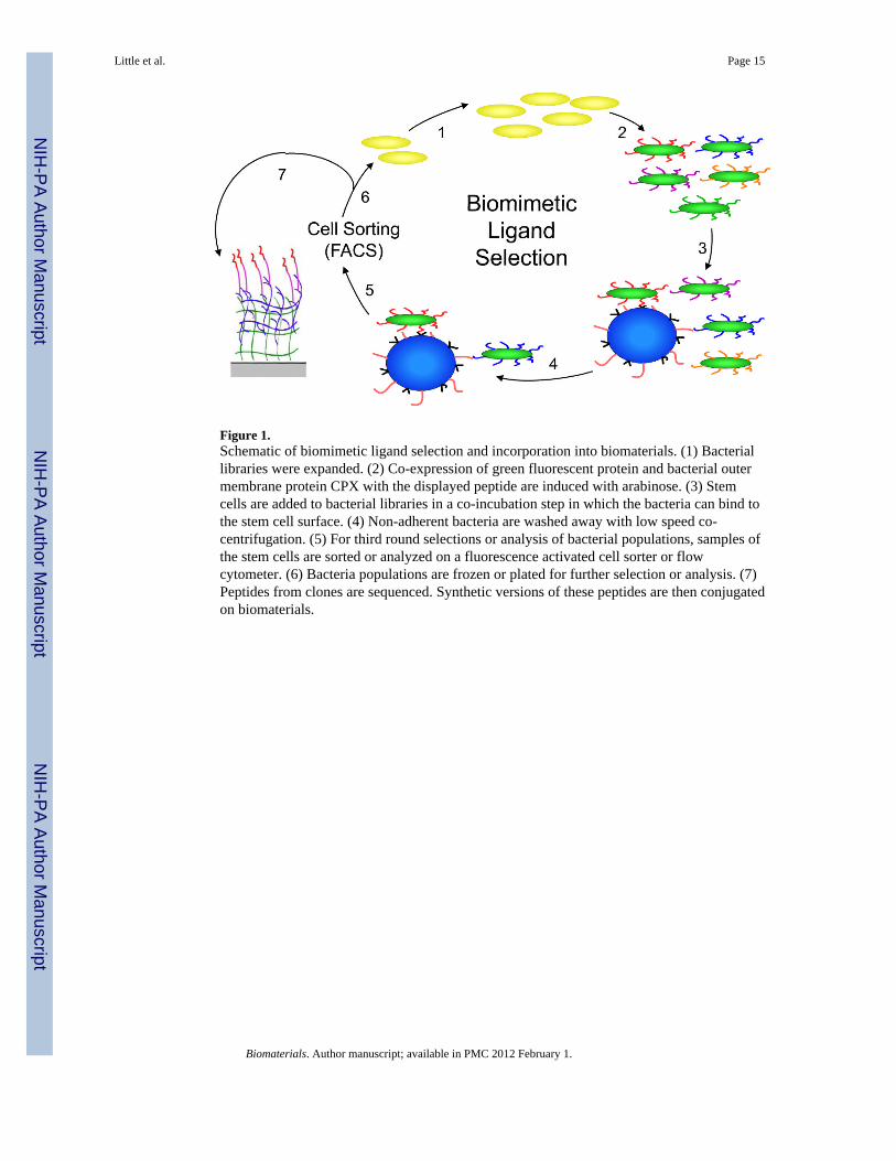
Figure 1.Schematic of biomimetic ligand selection and incorporation into biomaterials. (1) Bacteriallibraries were expanded. (2) Co-expression of green fluorescent protein and bacterial outermembrane protein CPX with the displayed peptide are induced with arabinose. (3) Stemcells are added to bacterial libraries in a co-incubation step in which the bacteria can bind tothe stem cell surface. (4) Non-adherent bacteria are washed away with low speed co-centrifugation. (5) For third round selections or analysis of bacterial populations, samples ofthe stem cells are sorted or analyzed on a fluorescence activated cell sorter or flowcytometer. (6) Bacteria populations are frozen or plated for further selection or analysis. (7)Peptides from clones are sequenced. Synthetic versions of these peptides are then conjugatedon biomaterials.
Little et al. Page 15
Biomaterials. Author manuscript; available in PMC 2012 February 1.
NIH
-PA Author Manuscript
NIH
-PA Author Manuscript
NIH
-PA Author Manuscript

Figure 2.Binding capacity of peptide display library populations with neural stem cells. (a) Examplehistograms of library and clonal populations of the bacterial peptide display libraries bindingto neural stem cells: (1) CPX (no peptide), (2) unselected 7C library, (3) 7C library afterRound 1, (4) 7C library after Round 2, (5) 7C library after Round 3, and (6) high affinityclone 15-2. (b) Quantification of library populations. Three libraries were tested: (■) 15merlibrary composed of peptides with the sequence X15, (▨) 7C library composed of peptideswith the sequence X2CX7CX2, and (□) a combined library containing both types of peptideclones. All unselected and post round 1 library populations showed similar binding asbacteria expressing CPX, the outer membrane display protein, but no peptide. After rounds 2and 3, there were significantly more bacteria binding to the neural stems, with the 7C libraryhaving the highest amount of binding. The 15mer and in particular the combined librariesexhibited the highest binding. Data represent mean ± standard deviation. Library populationsnot in the same group (#, $, or ‡) were statistically different from one another (p < 0.05using ANOVA between groups with Tukey-Kramer significant difference post hoc test).
Little et al. Page 16
Biomaterials. Author manuscript; available in PMC 2012 February 1.
NIH
-PA Author Manuscript
NIH
-PA Author Manuscript
NIH
-PA Author Manuscript

Figure 3.Neural stem cells on peptide-adsorbed TCPS surfaces. Peptides, including (■) 7C-9(9), (▨)7C-24(9), (▩) 15-2, (□) bsp-RGD(15), (▤) Laminin, (▥) bsp-RGE(15), and ( ) TCPSalone, were dissolved at 100 μM in synthesis-grade water or DMSO for 15-2, and thenpeptides were dried on TCPS. (a). Brightfield micrographs of the neural stem cells after 4days of culture on the adsorbed surfaces exhibited similar attachment and clumping of cellson the surface on the 7C-9(9), 7C-24(9), 15-2, and bsp-RGD(15) surfaces, while the bsp-RGE(15) and TCPS surfaces had significantly fewer cells. The scale bar represents 250 μm.(b) Quantification of the number of cells on the surface with the Cyquant cell counting assayshowed similar numbers of cells on all surfaces except the bsp-RGE(15) and TCPS surfaces,which had significantly fewer cells. (c) NSCs grown under differentiating conditions wereassessed for expression of GFAP (red), a cytoskeletal marker for astrocytes, and β-Tubulin
Little et al. Page 17
Biomaterials. Author manuscript; available in PMC 2012 February 1.
NIH
-PA Author Manuscript
NIH
-PA Author Manuscript
NIH
-PA Author Manuscript

III (green), a cytoskeletal marker for neurons. Nuclei were stained with DAPI (blue). Allpeptide surfaces had both astrocytes and neurons under differentiating conditions. All scalebars represent 100 μm. (d) Quantification of differentiation markers, β-Tubulin III andGFAP, on peptide-adsorbed surfaces. All library-selected and bsp-RGD(15) peptide surfaceshad similar percentages of neurons and astrocytes compared to laminin, while bsp-RGE(15)and TCPS surfaces had fewer neurons and more astrocytes. (Data represent mean ± standarddeviation. Library populations not in the same group (*) were statistically different from oneanother (p < 0.05 using ANOVA between groups with Tukey-Kramer significant differencepost hoc test).
Little et al. Page 18
Biomaterials. Author manuscript; available in PMC 2012 February 1.
NIH
-PA Author Manuscript
NIH
-PA Author Manuscript
NIH
-PA Author Manuscript

Figure 4.Peptide grafting and cell proliferation on IPN surfaces. (a) Peptide density oninterpenetrating polymer network (IPN) surfaces. Peptide densities on IPN surfaces weredetermined by grafting on a FITC-tagged peptide and digesting the FITC from the peptidewith chymotrypsin. Fluorescent measurements then allowed for the calculation of thesurface peptide concentration. Three peptides – (●) bsp-RGD(15), (□) 15-2 and (◆) 7C-9(9)– were examined. bsp-RGD(15) and 15-2 exhibited saturation at approximately 25 and 20pmol/cm2, respectively, while the looped 7C-9(9) peptide showed saturation around 8 pmol/cm2. (b) Neural stem cells were cultured on interpenetrating polymer networks (IPNs)conjugated with peptides including (■) 7C-9(9), (▨) 7C-24(9), (▩) 15-2, (□) bsp-RGD(15).For comparison, neural stems were also cultured on (▤) laminin. Brightfield images after 4days illustrated that cells on the 7C-9(9) and 15-2 surfaces either attached in clumps or
Little et al. Page 19
Biomaterials. Author manuscript; available in PMC 2012 February 1.
NIH
-PA Author Manuscript
NIH
-PA Author Manuscript
NIH
-PA Author Manuscript

remained as non-adherent neurospheres. 7C-24(9) and bsp-RGD(15) surfaces showedsimilar cell morphology and growth to the laminin control surface. The bsp-RGE(15)-conjugated surface showed little cell attachment. All surfaces had peptides at 8 pmol/cm2,and the scale bar represents 250 μm. (c) The amount of cells on each surface after 5 dayswas quantified with Cyquant. 7C-24(9) and bsp-RGD(15) surfaces supported cellproliferation at or above the amount of laminin at all peptide surface concentrations. The7C-9(9) and 15-2 surfaces had substantially fewer cells than all other surfaces, as anticipatedsince the cells primarily formed neurospheres rather than attaching to the surface. Resultsfrom the bsp-RGE(15) and unconjugated IPN surfaces were below the detection limit of theassay. Data represent mean ± standard deviation. Library populations not in the same group(* or **) were statistically different from one another (p < 0.05 using ANOVA betweengroups with Tukey-Kramer significant difference post hoc test).
Little et al. Page 20
Biomaterials. Author manuscript; available in PMC 2012 February 1.
NIH
-PA Author Manuscript
NIH
-PA Author Manuscript
NIH
-PA Author Manuscript

Figure 5.Expression of lineage markers under proliferative and differentiating conditions on peptide-conjugated interpenetrating polymer networks (IPNs). Neural stem cells were cultured onthe surfaces for 5 days either under proliferative conditions with 20 ng/mL basic FibroblastGrowth Factor (FGF-2) or with 1% fetal bovine serum and 1 μM retinoic acid. (a). NSCsgrown under proliferative conditions were assessed for the expression of nestin (green), acytoskeletal marker for a neural stem cell, while NSCs grown under differentiatingconditions were assessed for expression of GFAP (red), a cytoskeletal marker for astrocytes,and β-tubulin III (green), a cytoskeletal marker for neurons. All cells were stained withDAPI (blue) for the nucleus. All surfaces had most of the cells staining for nestin underproliferative conditions, and all surfaces had astrocytes and neurons under differentiatingconditions. All scale bars represent 100 μm. (b) Quantification of differentiation markers, β-
Little et al. Page 21
Biomaterials. Author manuscript; available in PMC 2012 February 1.
NIH
-PA Author Manuscript
NIH
-PA Author Manuscript
NIH
-PA Author Manuscript

tubulin III and GFAP, on (■) 7C-9(9), (▩) 15-2, (▨) 7C-24(9), (□) bsp-RGD(15), or (▤)laminin. Laminin had significantly more cells expressing β-tubulin III than any othersurface, but all other surfaces had similar β-tubulin III expression. With GFAP expressionthere was no significant difference in expression on any surface. Data represent mean ±standard deviation. Library populations not in the same group (*) were statistically differentfrom one another (p < 0.05 using ANOVA between groups with Tukey-Kramer significantdifference post hoc test).
Little et al. Page 22
Biomaterials. Author manuscript; available in PMC 2012 February 1.
NIH
-PA Author Manuscript
NIH
-PA Author Manuscript
NIH
-PA Author Manuscript

NIH
-PA Author Manuscript
NIH
-PA Author Manuscript
NIH
-PA Author Manuscript
Little et al. Page 23
Table 1
List of clones found with high binding for neural stem cells. Clones were analyzed with flow cytometry toquantify the percentage of neural stem cells that had bacteria bound after coincubation with the clonal bacteriapopulation. Peptide sequences were determined via sequencing of the plasmid DNA from the bacteria. Severalof the peptides exhibited homology to ECM proteins, including 7C-15, 15–52, and 7C-24 with homology tocollagen, fibrinogen, and fibronectin, respectively. Also, the integrin-binding motif arginine-glycine-asparticacid (RGD) was found in several peptides and is denoted in bold. The peptides used in further studies arehighlighted in gray. The clone name indicates the library containing the clone and the data represent mean ±standard deviation.
Clone % NSCs with Bacteria (average ± S.D.) Peptide Sequence
15–50 86.3 ± 0.8 GFVLVWSYTCRCWGK
7C-15 83.4 ± 0.8 QCCQLRGDAVCNC
15–52 82.3 ± 5.0 ESGLKVMCMKYYCMA
15–32 80.5 ± 2.0 RRELVRMTDWVWVSG
7C-1 79.4 ± 1.0 WYCFREN KYVCVM
7C-24 78.9 ± 1.7 WWCDMRGDSRCSG
Co-21 77.3 ± 5.6 MYCERDSKYWCIH
Co-17 76.3 ± 3.0 WECAEESKFWCVF
Co-22 75.7 ± 0.8 VWCGMFGKRRCVT
15-2 73.7 ± 5.8 DHKFGLVMLNKYAYAG
7C-9 73.4 ± 1.2 KLCCFDKGYYCMR
Co-18 65.6 ± 5.9 WWCKKPEYWYCIW
7C-7 65.5 ± 2.3 LECTERGDFNCFV
Co-8 64.6 ± 5.2 WTWESAFAGRWEVGD
Co-12 54.7 ± 2.1 WVCLWRHRGDCSI
7C-20 52.4 ± 3.3 WVCIWERFKSCNE
Co-15 45.2 ± 7.2 WVCNDLIHHFCVW
7C-19 36.6 ± 3.7 WVCNKLGVYACEY
Biomaterials. Author manuscript; available in PMC 2012 February 1.