nih public access 1 john c. tan 3 , and insect nerve cord
TRANSCRIPT

A Requirement for Commissureless2 Function during DipteranInsect Nerve Cord Development
Joseph Sarro1, Emily Andrews2, Longhua Sun1, Susanta K. Behura1, John C. Tan1, ErliangZeng3, David W. Severson1,2, and Molly Duman-Scheel1,2,4
1Eck Institute for Global Health and Department of Biological Sciences, University of Notre Dame,Notre Dame, Indiana, 465562Department of Medical and Molecular Genetics, Indiana University School of Medicine, SouthBend, Indiana, 466173Department of Computer Science and Engineering, University of Notre Dame, Notre Dame, IN,46556
AbstractBackground—In Drosophila melanogaster, commissureless (comm) function is required forproper nerve cord development. Although comm orthologs have not been identified outside ofDrosophila species, some insects possess orthologs of Drosophila comm2, which may alsoregulate embryonic nerve cord development. Here, this hypothesis is explored throughcharacterization of comm2 genes in two disease vector mosquitoes.
Results—Culex quinquefasciatus (West Nile and lymphatic filiariasis vector) has three comm2genes that are expressed in the developing nerve cord. Aedes aegypti (dengue and yellow fevervector) has a single comm2 gene that is expressed in commissural neurons projecting axonstoward the midline. Loss of comm2 function in both A. aegypti and D. melanogaster was found toresult in loss of commissure defects that phenocopy the frazzled (fra) loss of function phenotypesobserved in both species. Loss of fra function in either insect was found to result in decreasedcomm2 transcript levels during nerve cord development.
Conclusions—The results of this investigation suggest that Fra downregulates repulsion inprecrossing commissural axons by regulating comm2 levels in both A. aegypti and D.melanogaster, both of which require Comm2 function for proper nerve cord development,
Keywordscommissureless; development; nervous system; mosquito; Aedes aegypti; Culex quinquefasciatus;Drosophila melanogaster; frazzled
INTRODUCTIONIn recent years, knockdown studies have suggested that axon guidance gene function hasdiverged in the insect embryonic ventral nerve cord (Clemons et al., 2011; Haugen et al.,2011; Evans and Bashaw, 2012). For example, an siRNA-mediated knockdown strategy wasused to study the role of the Netrin receptor frazzled (fra) during Aedes aegypti embryonicnerve cord development (Clemons et al., 2011). The results of this investigation, the firsttargeted knockdown of a gene during vector mosquito embryogenesis, suggest that although
4To whom correspondence should be addressed: [email protected], Indiana University School of Medicine-South Bend at NotreDame, Raclin-Carmichael Hall, 1234 Notre Dame Ave., South Bend, IN 46617, Phone: (574) 631-7194, Fax: (574) 631-7821.
NIH Public AccessAuthor ManuscriptDev Dyn. Author manuscript; available in PMC 2014 December 01.
Published in final edited form as:Dev Dyn. 2013 December ; 242(12): 1466–1477. doi:10.1002/dvdy.24059.
NIH
-PA Author Manuscript
NIH
-PA Author Manuscript
NIH
-PA Author Manuscript
Fra plays a critical role during development of the A. aegypti ventral nerve cord,mechanisms regulating embryonic commissural axon guidance have diverged in dipteraninsects. Specifically, loss of Aae fra results in thin and missing commissural axons, defectswhich are qualitatively similar to those observed in D. melanogaster fra null mutants.However, complete commissure loss is observed in A. aegypti fra knockdown embryos,which bear more resemblance to the Drosophila commissureless (comm) loss of functionphenotype (Seeger et al., 1993) than the Drosophila fra loss of function phenotype(Kolodziej et al., 1996).
Mutations in the D. melanogaster comm gene were initially uncovered from a screen formutations that affect development of the Drosophila embryonic CNS axons pathways(Seeger et al., 1993). The comm gene is the most extensively studied of the three commfamily genes (comm, comm2, and comm3) in D. melanogaster (Keleman et al., 2002).Mutants for comm display a unique phenotype, the complete absence of most axoncommissures. Cloning and sequencing of the comm gene revealed that it encodes a noveltransmembrane protein (Tear et al., 1996). Subsequent studies revealed that Comm functionsin neurons to regulate the intracellular trafficking and surface levels of Robo (Keleman etal., 2002; McGovern and Seeger, 2003; Keleman et al., 2005), receptor for the midlinerepellent Slit. The choice of crossing (commissural) or noncrossing (ipsilateral) axonpathways is regulated by the differential sensitivity of axons to Slit (reviewed by (Dickson,2002). Ipsilateral axons lacking Comm expression are repelled by Slit-Robo signaling andfail to cross the midline. Commissural axons, which express Comm and consequently lackRobo surface expression, are initially insensitive to Slit-mediated repulsion and respond toNet attractive midline cues. Once commissural axons reach the midline, Comm expression isdownregulated, and these neurons are repelled from the midline by Slit-Robo signaling(Kidd et al., 1999; Stein and Tessier-Lavigne, 2001; Dickson and Gilestro, 2006). Thus, theability of Comm to control Robo levels is critical for proper nerve cord formation.
Despite the importance of comm gene function in the D. melanogaster nerve cord, no commgene ortholog has been detected outside of the Drosophila species (Waterhouse et al., 2013).However, orthologs of the Drosophila comm2 and comm3 genes, neither of which have beenextensively characterized in Drosophila, have been identified in other arthropods, includingvector mosquitoes (Behura et al., 2011). It was hypothesized that these comm family genesmight function during vector mosquito nerve cord development. In this investigation, weperform phylogenetic and expression analyses of comm family orthologs in C.quinquefasciatus and A. aegypti. The role of the A. aegypti comm2 gene, the only commfamily ortholog in this species, was assessed through siRNA-mediated silencing. Thefunction of Aae comm2 was compared to that of its closest D. melanogaster comm familyortholog, D. melanogaster comm2, which was also assessed for comparison.
RESULTS AND DISCUSSIONMosquito comm family genes
Previous comparative genomic analyses of developmental genes in the D. melanogaster, A.aegypti, C. quinquefasciatus, and A. gambiae genomes revealed changes in the number ofaxon guidance genes in these species (Behura et al., 2011). While three comm family genes(comm, comm2, and comm3) are found in D. melanogaster, no comm family genes havebeen identified in A. gambiae. However, three comm family genes were identified in C.quinquefasciatus (CPIJ016283, CPIJ016285, CPIJ017280), while a single comm familygene has been identified in the A. aegypti genome (AAEL007250). These genes were notedto be Drosophila comm2/comm3 orthologs (Behura et al., 2011). Sequence analyses wereperformed to further explore the relationship between these mosquito comm family geneswith each other and with those of other arthropods (Fig. 1).
Sarro et al. Page 2
Dev Dyn. Author manuscript; available in PMC 2014 December 01.
NIH
-PA Author Manuscript
NIH
-PA Author Manuscript
NIH
-PA Author Manuscript
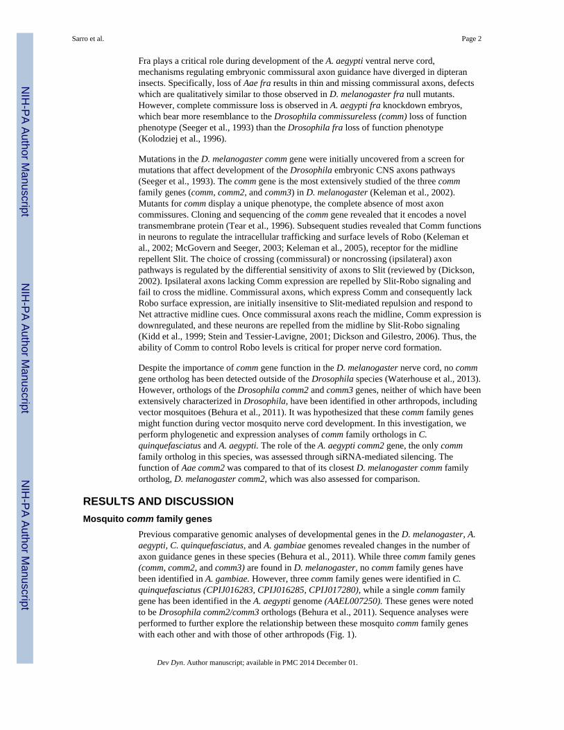
The amino acid sequence conservation pattern among Drosophila and mosquito Commfamily proteins is shown in Fig. 1A, C. Phylogenetic comparisons suggest that the A. aegyptiand C. quinquefasciatus Comm family proteins more closely resemble each other than theydo any of the Drosophila Comm family proteins (Fig. 1B, C). D. melanogaster Commfamily proteins bear a highly conserved 22 amino acid sequence (residues 215–236 in D.melanogaster Comm; Keleman et al., 2002). This sequence is well conserved inAAEL007250, CPIJ16283, and CPIJ16285 (Fig. 1A). However, comparison of the motifmaps of CPIJ017280 with D. melanogaster Comm2 (FBgn0041160) based on Dayhoffweight estimates of local sequence similarities (Leontovich et al. 1993) revealed that theconserved cytoplasmic domain is either lost or extensively mutated in CPIJ017280 protein(Fig. 1A and data not shown). The D. melanogaster Comm protein also contains aheterotetrameric adaptor protein binding site motif LPSY (Wolf et al., 1998) which isconserved in D. melanogaster Comm2, but not Comm3. This domain is required forendosomal sorting of Comm and Comm2, as well as downregulation of Robo and thepromotion of midline crossing (Keleman et al., 2002; Choi et al., 2003). An LPSY motif isfound in all of the A. aeygpti and C. quinquefasciatus Comm family proteins, suggestingthat with reference to this functionally significant motif, these proteins are more comparableto D. melanogaster Comm2 than Comm3. This observation suggests that the A. aegypti andC. quinquefasciatus genes are comm2 orthologs.
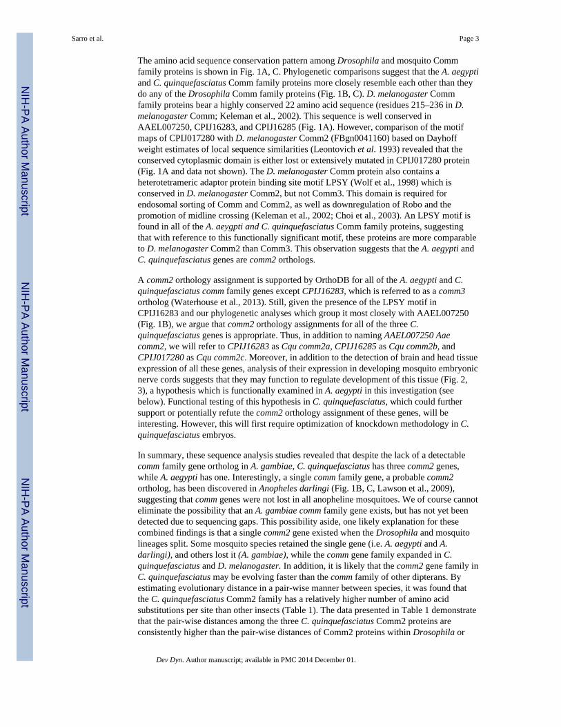
A comm2 orthology assignment is supported by OrthoDB for all of the A. aegypti and C.quinquefasciatus comm family genes except CPIJ16283, which is referred to as a comm3ortholog (Waterhouse et al., 2013). Still, given the presence of the LPSY motif inCPIJ16283 and our phylogenetic analyses which group it most closely with AAEL007250(Fig. 1B), we argue that comm2 orthology assignments for all of the three C.quinquefasciatus genes is appropriate. Thus, in addition to naming AAEL007250 Aaecomm2, we will refer to CPIJ16283 as Cqu comm2a, CPIJ16285 as Cqu comm2b, andCPIJ017280 as Cqu comm2c. Moreover, in addition to the detection of brain and head tissueexpression of all these genes, analysis of their expression in developing mosquito embryonicnerve cords suggests that they may function to regulate development of this tissue (Fig. 2,3), a hypothesis which is functionally examined in A. aegypti in this investigation (seebelow). Functional testing of this hypothesis in C. quinquefasciatus, which could furthersupport or potentially refute the comm2 orthology assignment of these genes, will beinteresting. However, this will first require optimization of knockdown methodology in C.quinquefasciatus embryos.
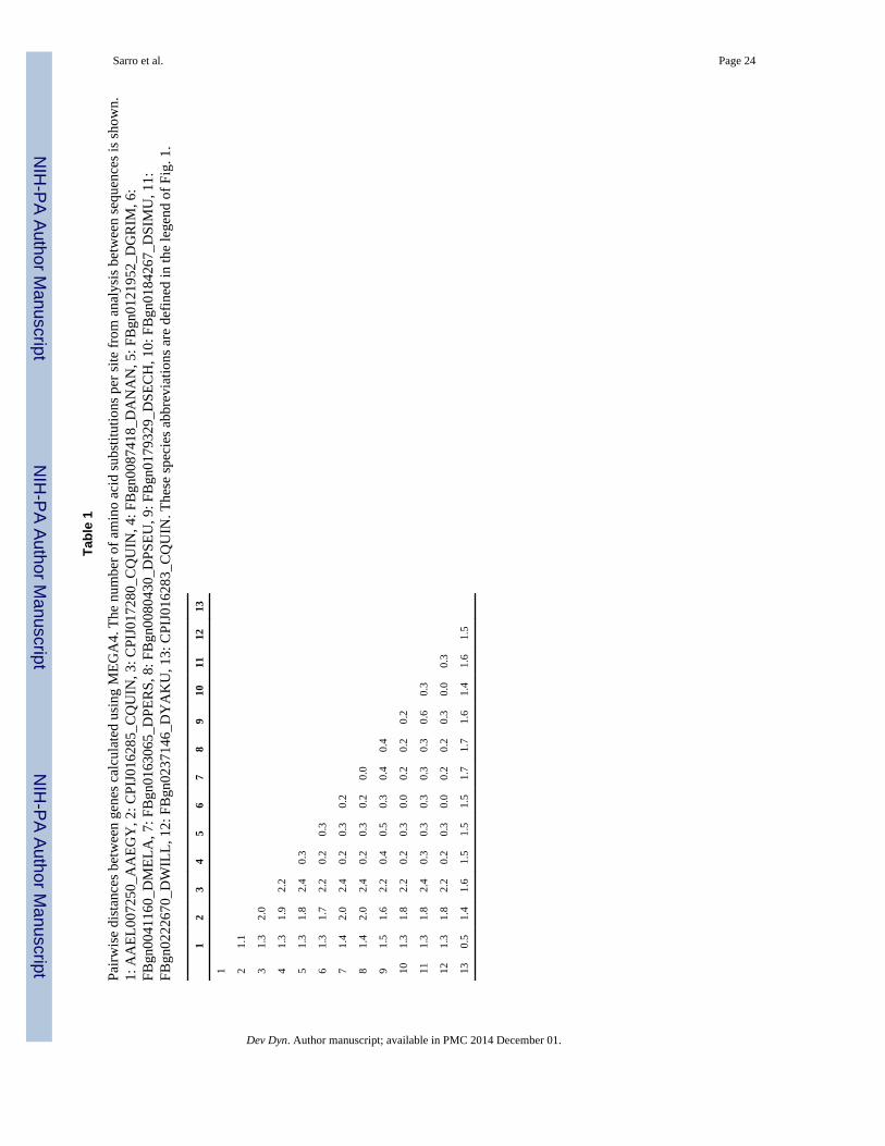
In summary, these sequence analysis studies revealed that despite the lack of a detectablecomm family gene ortholog in A. gambiae, C. quinquefasciatus has three comm2 genes,while A. aegypti has one. Interestingly, a single comm family gene, a probable comm2ortholog, has been discovered in Anopheles darlingi (Fig. 1B, C, Lawson et al., 2009),suggesting that comm genes were not lost in all anopheline mosquitoes. We of course cannoteliminate the possibility that an A. gambiae comm family gene exists, but has not yet beendetected due to sequencing gaps. This possibility aside, one likely explanation for thesecombined findings is that a single comm2 gene existed when the Drosophila and mosquitolineages split. Some mosquito species retained the single gene (i.e. A. aegypti and A.darlingi), and others lost it (A. gambiae), while the comm gene family expanded in C.quinquefasciatus and D. melanogaster. In addition, it is likely that the comm2 gene family inC. quinquefasciatus may be evolving faster than the comm family of other dipterans. Byestimating evolutionary distance in a pair-wise manner between species, it was found thatthe C. quinquefasciatus Comm2 family has a relatively higher number of amino acidsubstitutions per site than other insects (Table 1). The data presented in Table 1 demonstratethat the pair-wise distances among the three C. quinquefasciatus Comm2 proteins areconsistently higher than the pair-wise distances of Comm2 proteins within Drosophila or
Sarro et al. Page 3
Dev Dyn. Author manuscript; available in PMC 2014 December 01.
NIH
-PA Author Manuscript
NIH
-PA Author Manuscript
NIH
-PA Author Manuscript
between Drosophila and A. aegypti. Hence, the C. quinquefasciatus proteins distinctly differin the rate of amino acid substitutions compared to that of A. aegypti or Drosophila.
Detailed analysis of comm2 expression during A. aegypti nerve cord developmentGiven the lack of a detectable comm gene outside the drosophilids and the apparent lack ofany comm family (comm, comm2, or comm3) gene in some insects (Behura et al., 2011;Evans and Bashaw, 2012), it is possible that the requirement for comm family gene functionis not retained outside of Drosophila. However, given the expansion of C. quinquefasciatuscomm2 genes, all of which are expressed in the developing nerve cord (Fig. 2), it is alsopossible that comm family gene function is in fact required outside of D. melanogaster, aconcept which has never been experimentally tested. To explore this, we opted to assesscomm2 gene function in A. aegypti because it has retained a single comm family ortholog(Fig. 1), and also due to the fact that we recently optimized embryonic knockdown strategiesin this species (Clemons et al., 2010a; Clemons et al., 2011; Haugen et al., 2011; Nguyen etal., 2013), which is to our knowledge the only mosquito species in which embryonic RNAiknockdown studies have been published to date.
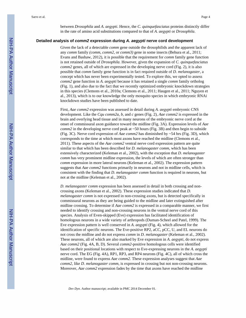
First, Aae comm2 expression was assessed in detail during A. aegypti embryonic CNSdevelopment. Like the Cqu comm2a, b, and c genes (Fig. 2), Aae comm2 is expressed in thebrain and overlying head tissue and in many neurons of the embryonic nerve cord at theonset of commissural axon guidance toward the midline (Fig. 3A). Expression levels of Aaecomm2 in the developing nerve cord peak at ~50 hours (Fig. 3B) and then begin to subside(Fig. 3C). Nerve cord expression of Aae comm2 has diminished by ~54 hrs (Fig. 3D), whichcorresponds to the time at which most axons have reached the midline (Clemons et al.,2011). These aspects of the Aae comm2 ventral nerve cord expression pattern are quitesimilar to that which has been described for D. melanogaster comm, which has beenextensively characterized (Keleman et al., 2002), with the exception that D. melanogastercomm has very prominent midline expression, the levels of which are often stronger thancomm expression in more lateral neurons (Keleman et al., 2002). The expression patternsuggests that Aae comm2 functions primarily in neurons and not in midline cells, which isconsistent with the finding that D. melanogaster comm function is required in neurons, butnot at the midline (Keleman et al., 2002).
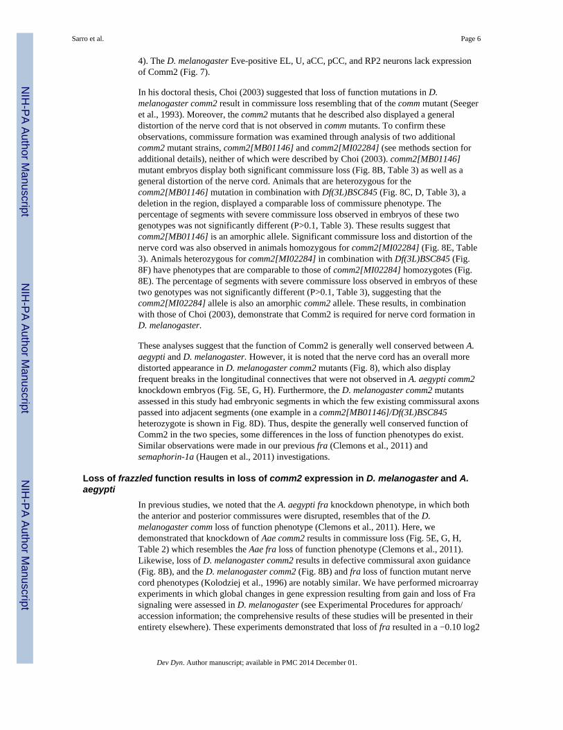
D. melanogaster comm expression has been assessed in detail in both crossing and non-crossing axons (Keleman et al., 2002). These expression studies indicated that D.melanogaster comm is not expressed in non-crossing axons, but is detected specifically incommissural neurons as they are being guided to the midline and later extinguished aftermidline crossing. To determine if Aae comm2 is expressed in a comparable manner, we firstneeded to identify crossing and non-crossing neurons in the ventral nerve cord of thisspecies. Analysis of Even-skipped (Eve) expression has facilitated identification ofhomologous neurons in a wide variety of arthropods (Duman-Scheel and Patel, 1999). TheEve expression pattern is well conserved in A. aegypti (Fig. 4), which allowed for theidentification of specific neurons. The Eve-positive RP2, aCC, pCC, U, and EL neurons donot cross the midline and do not express comm in D. melanogaster (Keleman et al., 2002).These neurons, all of which are also marked by Eve expression in A. aegypti, do not expressAae comm2 (Fig. 4A, B, D). Several comm2-positive homologous cells were identifiedbased on their positional locations with respect to Eve-expressing neurons in the A. aegyptinerve cord. The EG (Fig. 4A), RP1, RP3, and RP4 neurons (Fig. 4C), all of which cross themidline, were found to express Aae comm2. These expression analyses suggest that Aaecomm2, like D. melanogaster comm, is expressed in crossing but not non-crossing neurons.Moreover, Aae comm2 expression fades by the time that axons have reached the midline
Sarro et al. Page 4
Dev Dyn. Author manuscript; available in PMC 2014 December 01.
NIH
-PA Author Manuscript
NIH
-PA Author Manuscript
NIH
-PA Author Manuscript

(Fig. 3D), which presumably facilitates Slit-Robo mediated axon repulsion from the midlineas it does in Drosophila (Keleman et al., 2002).
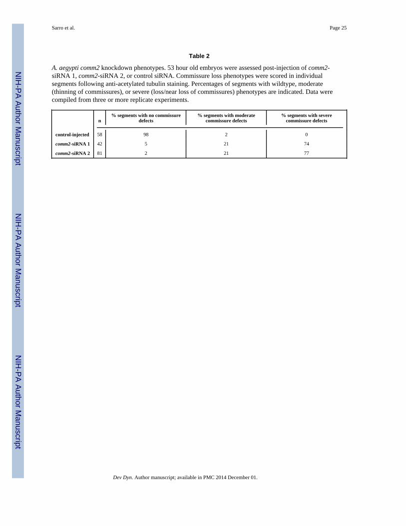
Knockdown of comm2 in A. aegypti results in a commissureless phenotypeExpression data (Fig. 3,4) support the notion that Aae comm2 function is required duringnerve cord development. To examine this possibility, the putative role of Aaecomm2 wasassessed through siRNA-mediated knockdown experiments, which were recently shown tobe an effective method of inhibiting gene expression during embryonic development of A.aegypti (Clemons et al., 2011; Haugen et al., 2011; Nguyen et al., 2013). Experimental orcontrol siRNAs were microinjected at the pre-cellular blastoderm stage. Two siRNAscorresponding to different regions of the Aae comm2 gene, comm2-siRNA 1 and comm2-siRNA 2, were used to target this gene. Control experiments were performed usingpreviously designed control siRNA (Nguyen et al., 2013) which does not bear high sequencesimilarity to other genes in the A. aegypti genome. 53 hour old embryos were assessed post-injection of comm2-siRNA 1, comm2-siRNA 2, or control siRNA. In situ hybridizationassays demonstrated that a majority of comm2-siRNA 1 (79%, n=33) and comm2-siRNA 2(74%, n=31) injected embryos lacked comm2 expression in the developing ventral nervecord (Fig. 5B, C), while control-injected embryos displayed comm2 expression levels whichresemble those of wild-type embryos (Fig. 5A, n=46).
The impact of comm2 knockdown on A. aegypti embryonic nerve cord development wasassessed through anti-acetylated tubulin staining at 53 hours of development. Althoughcontrol-injected animals had normal nerve cord development (n=71, Fig. 5D, F, Table 2),commissure formation was disrupted in embryos injected with either comm2-siRNA 1(n=35, Fig. 5E, G) or siRNA 2 (n=51, Fig. 5H). Comparable phenotypes were generatedthrough injection of comm2- siRNA 1 or 2 (Fig. 5E, G, H), suggesting that thecommissureless phenotype observed was not the result of off-site targeting by either siRNA.71% of comm2-siRNA 1 injected embryos and 51% of comm2-siRNA 2 injected embryosdisplayed severe nerve cord phenotypes in which most segments had commissure defects.Individual segments were scored in a subset of these animals, and the percentages ofsegments with moderate (thinning of commissures) or severe (loss/near loss) phenotypes areprovided in Table 2. These data indicate that Aae Comm2 function is required for propercommissure formation in A. aegypti.
D. melanogaster comm2 is also required for nerve cord developmentThe results of the A. aegypti knockdown experiments suggested that D. melanogasterComm2, the ortholog of Aae Comm2, may also function during nerve cord development.Expression of D. melanogaster comm2 transcript is detected in the developing embryonicnerve cord (Fig. 6A, B), brain, and head tissue (not shown) in a pattern which resembles thatof Aae comm2 (Fig. 3). High levels of D. melanogaster comm2 transcript are detected in thenerve cord at stage 13 (Fig. 6A), and by Stage 15, expression of comm2 is observed in amore restricted set of D. melanogaster neurons (Fig. 6B). Like Aae comm2, expression of D.melanogaster comm2 diminishes once the majority of commissural axons have crossed themidline (Fig. 6B). It has been suggested both in the discussion of Keleman et al. (2002) andalso in the doctoral thesis of Choi (2003) that D. melanogaster Comm2, like D.melanogaster Comm, prevents Robo from reaching the cell surface. If this is true, then D.melanogaster Comm2, like Comm (Keleman et al., 2002) should not be expressed inneurons which do not project axons toward the midline. This was assessed in D.melanogaster in the same manner in which it was examined in A. aegypti (Fig. 4), throughthe combined analysis of Eve and Comm2 expression during nerve cord development. Theresults of these experiments (Fig. 7) were comparable to those obtained for A. aegypti (Fig.
Sarro et al. Page 5
Dev Dyn. Author manuscript; available in PMC 2014 December 01.
NIH
-PA Author Manuscript
NIH
-PA Author Manuscript
NIH
-PA Author Manuscript
4). The D. melanogaster Eve-positive EL, U, aCC, pCC, and RP2 neurons lack expressionof Comm2 (Fig. 7).
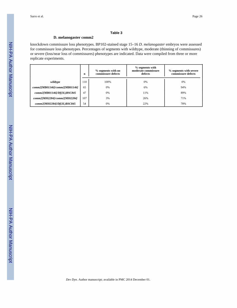
In his doctoral thesis, Choi (2003) suggested that loss of function mutations in D.melanogaster comm2 result in commissure loss resembling that of the comm mutant (Seegeret al., 1993). Moreover, the comm2 mutants that he described also displayed a generaldistortion of the nerve cord that is not observed in comm mutants. To confirm theseobservations, commissure formation was examined through analysis of two additionalcomm2 mutant strains, comm2[MB01146] and comm2[MI02284] (see methods section foradditional details), neither of which were described by Choi (2003). comm2[MB01146]mutant embryos display both significant commissure loss (Fig. 8B, Table 3) as well as ageneral distortion of the nerve cord. Animals that are heterozygous for thecomm2[MB01146] mutation in combination with Df(3L)BSC845 (Fig. 8C, D, Table 3), adeletion in the region, displayed a comparable loss of commissure phenotype. Thepercentage of segments with severe commissure loss observed in embryos of these twogenotypes was not significantly different (P>0.1, Table 3). These results suggest thatcomm2[MB01146] is an amorphic allele. Significant commissure loss and distortion of thenerve cord was also observed in animals homozygous for comm2[MI02284] (Fig. 8E, Table3). Animals heterozygous for comm2[MI02284] in combination with Df(3L)BSC845 (Fig.8F) have phenotypes that are comparable to those of comm2[MI02284] homozygotes (Fig.8E). The percentage of segments with severe commissure loss observed in embryos of thesetwo genotypes was not significantly different (P>0.1, Table 3), suggesting that thecomm2[MI02284] allele is also an amorphic comm2 allele. These results, in combinationwith those of Choi (2003), demonstrate that Comm2 is required for nerve cord formation inD. melanogaster.
These analyses suggest that the function of Comm2 is generally well conserved between A.aegypti and D. melanogaster. However, it is noted that the nerve cord has an overall moredistorted appearance in D. melanogaster comm2 mutants (Fig. 8), which also displayfrequent breaks in the longitudinal connectives that were not observed in A. aegypti comm2knockdown embryos (Fig. 5E, G, H). Furthermore, the D. melanogaster comm2 mutantsassessed in this study had embryonic segments in which the few existing commissural axonspassed into adjacent segments (one example in a comm2[MB01146]/Df(3L)BSC845heterozygote is shown in Fig. 8D). Thus, despite the generally well conserved function ofComm2 in the two species, some differences in the loss of function phenotypes do exist.Similar observations were made in our previous fra (Clemons et al., 2011) andsemaphorin-1a (Haugen et al., 2011) investigations.
Loss of frazzled function results in loss of comm2 expression in D. melanogaster and A.aegypti
In previous studies, we noted that the A. aegypti fra knockdown phenotype, in which boththe anterior and posterior commissures were disrupted, resembles that of the D.melanogaster comm loss of function phenotype (Clemons et al., 2011). Here, wedemonstrated that knockdown of Aae comm2 results in commissure loss (Fig. 5E, G, H,Table 2) which resembles the Aae fra loss of function phenotype (Clemons et al., 2011).Likewise, loss of D. melanogaster comm2 results in defective commissural axon guidance(Fig. 8B), and the D. melanogaster comm2 (Fig. 8B) and fra loss of function mutant nervecord phenotypes (Kolodziej et al., 1996) are notably similar. We have performed microarrayexperiments in which global changes in gene expression resulting from gain and loss of Frasignaling were assessed in D. melanogaster (see Experimental Procedures for approach/accession information; the comprehensive results of these studies will be presented in theirentirety elsewhere). These experiments demonstrated that loss of fra resulted in a −0.10 log2
Sarro et al. Page 6
Dev Dyn. Author manuscript; available in PMC 2014 December 01.
NIH
-PA Author Manuscript
NIH
-PA Author Manuscript
NIH
-PA Author Manuscript

fold decrease in comm2 expression, while activation of Net-Fra signaling resulted in a 0.20log2 fold increase in comm2 transcript levels. Although these fold changes were notstatistically significant, these observations, when combined with the notable similarity of thefra and comm2 mutant phenotypes in both D. melanogaster and A. aegypti, suggested thatcomm2 expression levels may change in response to Fra signaling. Such a phenomenon hasbeen described in relation to the Drosophila comm gene, the expression of which decreasesin Drosophila fra mutants (Yang et al., 2009). Thus, comm2 transcript levels were assessedin fra[3] loss of function D. melanogaster embryos through whole-mount in situhybridization. These experiments demonstrated that loss of fra function results in substantialloss of comm2 expression in the developing nerve cord (Fig. 9B, n=36). These resultssuggest that Fra downregulates repulsion in commissural axons that are being guided to themidline by regulating not only comm, but also comm2 levels. Thus, in addition to mediatingthe response of Netrin attractive cues secreted from the midline, Fra signaling alsodownregulates responses to repulsive Slit-Robo cues by promoting high levels of bothComm and Comm2.
Assessment of comm2 expression in A. aegypti fra knockdown embryos (Fig. 9D)demonstrated that the ability of Fra signaling to regulate comm2 transcript levels isconserved between D. melanogaster and A. aegypti. Loss of comm2 nerve cord expressionwas observed in 85% of fra knockdown siRNA-injected mosquito embryos (n=105). Thus,regulation of comm family gene transcript levels by Fra is observed outside of Drosophila.These findings demonstrate that although some insects lack an apparent comm familyortholog, the function of Comm2 is generally well conserved between D. melanogaster andA. aegypti, as evidenced through the well conserved expression patterns of comm2 in A.aegypti (Figs. 3 and 4) and D. melanogaster (Figs. 6 and 7), the commissure loss phenotypesobserved in the comm2 loss of function nerve cords of both species (Figs. 5,8, Tables 2,3),and the finding that comm2 levels are regulated by Fra signaling in both insects (Fig. 9).
Given that Comm2 plays an essential role in the developing D. melanogaster and A. aegyptinerve cord, it is interesting that A. gambiae and T. castaneum appear to lack a comm familygene (Behura et al., 2011; Evans and Bashaw, 2012). It is possible that genome sequencinggaps or the notably low level of amino acid conservation among Comm family proteins hashindered the identification of a comm family ortholog in these insects, and that they dopossess a comm family gene that cannot be identified through sequence comparisons.Interestingly, although no comm family genes have yet been identified in vertebrates,RabGDI was recently found to regulate Robo1 surface expression levels in chickcommissural axons (Philipp et al., 2012). These findings suggest that other proteins, perhapseven Rab GDI which has been described in insects (Ricard et al., 2001), may function toregulate Robo surface levels in animals which lack apparent Comm family proteins.
EXPERIMENTAL PROCEDURESSequence analyses
Mosquito comm orthologs were previously identified (Behura et al., 2011) from Biomart(Smedley et al., 2009) and Vectorbase (Lawson et al., 2009). The amino acid sequences ofComm family proteins were obtained from Vectorbase (Lawson et al., 2009) and Flybase(Tweedie et al., 2009). ClustalW (Chenna et al., 2003) was used for multiple sequencealignment. The phylogenetic tree was drawn using the Neighbor-Joining method (Saitou andNei, 1987), and a bootstrap consensus tree was inferred from 1000 replicates (Felsenstein,1992). The Poisson correction method (Zuckerkandl and Pauling, 1965) was used tocompute evolutionary distances which correspond to the number of amino acid substitutionsper site. Phylogenetic analyses were conducted using MEGA4 (Tamura et al., 2007). Thepairwise distance between genes was also calculated using MEGA4. The local sequence
Sarro et al. Page 7
Dev Dyn. Author manuscript; available in PMC 2014 December 01.
NIH
-PA Author Manuscript
NIH
-PA Author Manuscript
NIH
-PA Author Manuscript

alignments for constructing motif maps of CPIJ016285 and CPIJ017280 in comparison withD. melanogaster Comm2 (FBgn0041160) was based on Dayhoff weight matrices(Leontovich et al. 1993) implemented in the GeneBee program (Brodsky et al. 1995).
Mosquito Rearing, Egg Collection, and FixationA. aegypti Liverpool- IB12 (LVP-IB12) and C. quinquefasciatus Johannesburg (JHB)strains, which were utilized in the genome sequencing efforts (Nene et al., 2007;Arensburger et al., 2010), were used in these studies. Mosquitoes were reared as previouslydescribed (Clemons et al., 2010b), except that an artificial membrane blood feeding systemwas employed. Procedures for A. aegypti embryo fixation (Clemons et al., 2010c) have beendescribed, and these methods were applied to both A. aegypti and C. quinquefasciatus in thisinvestigation. C. quinquefasciatus embryos were staged as previously described (Davis,1967).
Drosophila GeneticsThe following D. melanogaster mutant stocks were used in this investigation:comm2[MB01146] (Bloomington Stock Center, #23000), comm2[MI02284] (BloomingtonStock Center, #33190), Df(3L)BSC845 (Bloomington Stock Center, #27888), and fra[3](Bloomington Stock Center #8813). Detailed information about these stocks is available atFlybase (Tweedie et al., 2009). In summary, comm2[MB01146] and comm2[MI02284] aretransgenic insertion stocks in the comm2 locus produced by the Drosophila Gene DisruptionProject that were derived by transposable element mobilization with the Dhyd\Minos-basedconstructs Mi{ET1} and Mi{MIC}, respectively (Metaxakis et al., 2005; Bellen et al., 2011).Levels of comm2 transcript were not observed to be reduced in either strain, as is typical forMinos-generated alleles (Metaxakis et al., 2005). However, genetic characterization of thesemutations described herein indicates that they behave as amorphic comm2 alleles.Df(3L)BSC845 is a large deletion of cytogenic region 71E1 which covers sequences 3L:15,559,113 to 15,569,413 and results in the deletion of numerous genes, including comm2(Cook et al., 2012).
In situ hybridization, Immunohistochemistry, and ImagingRiboprobes (~500 bp in size) corresponding to the following genes were synthesized asdescribed (Patel, 1996): Aae comm2 (AAEL007250), Cqu comm2 genes CPIJ016283,CPIJ016285, and CPIJ017280, and D. melanogaster comm2 (CG7554). A. aegypti in situhybridization experiments were performed as discussed previously (Haugen et al., 2010),and this method was applied to C. quinquefasciatus. D. melanogaster in situ hybridizationexperiments were performed as described previously (Patel, 1996; VanZomeren-Dohm etal., 2008).
Immunohistochemistry was performed in A. aegypti using previously publishedmethodology (Clemons et al., 2010d), and this protocol was applied to C. quinquefasciatus.Drosophila embryos were prepared and stained as described by Patel (Patel, 1994), whodiscusses the use of BP102 and anti-Even-skipped (Eve) 2B8 antibodies which he graciouslyprovided. Anti-acetylated tubulin antibody was obtained from Zymed (San Francisco, CA).Anti-GFP antibody was purchased from Sigma-Aldrich (St. Louis, MO). The HRP-conjugated secondary antibodies used in this investigation were purchased from JacksonImmunoresearch (West Grove, PA). Use of GFP marked balancers permitted genotypescoring of Drosophila embryos in immunohistochemical assays.
Samples were imaged using a Zeiss Axioimager equipped with a Spot Flex camera or with aZeiss LSM 710 Confocal Microscope. Images were processed with Adobe Photoshop andZeiss ZEN Software.
Sarro et al. Page 8
Dev Dyn. Author manuscript; available in PMC 2014 December 01.
NIH
-PA Author Manuscript
NIH
-PA Author Manuscript
NIH
-PA Author Manuscript

RNAi knockdown experimentsKnockdown of Aae comm2 was performed through embryonic microinjection of siRNAscorresponding to this gene as previously described in a protocol (Clemons et al., 2010a)which has been successfully utilized in three recent embryonic knockdown investigations(Clemons et al., 2011; Haugen et al., 2011; Nguyen et al., 2013). The following siRNAscorresponding to Ae. aeygpti comm2 were synthesized by Dharmacon RNAi Technologies(Lafayette, CO): siRNA-1 sense: CGCCAGUGAUUUCAACCUGUU and antisense:CAGGUUGAAAUCACUGGCGUU (corresponds to base pairs 265–283 of Aae comm2)and siRNA-2 sense: CGACCUUCGGUCAUCAAACUU and antisense:GUUUGAUGACCGAAGGUCGUU (corresponds to base pairs 677–695 of Aae comm2).Previously described control siRNA (Nguyen et al., 2013) was used in these experiments;BLAST searches have confirmed that this siRNA does not have any known targets in the A.aegypti genome. For nerve cord phenotype assessments, at least eight replicate experimentswere performed with each siRNA. Knockdown of Aae comm2 was confirmed through in situhybridization experiments in two separate replicate experiments. Knockdown of Aae fra wasperformed as previously described in two separate replicate experiments (Clemons et al.,2011).
Microarray experimentsTwo complementary microarray experiments were used to assess the impacts of Frasignaling on global gene expression. In the first experiment, global gene expression profilesfor stage 13 fra[3]/fra[3] mutant Drosophila vs. wild-type embryos were compared. Fourreplicates, each with 10 embryos for each condition (fra[3] mutant or wild-type) wereprepared. The second experiment examined effects of activated NetA-Fra overexpression inthe third instar wing imaginal disc. GFP-marked NetA+Fra overexpression wing discs wereprepared as previously described (Flannery et al., 2010), and age matched wild type discswith GFP clones were used as controls. Four replicates with 15 discs for each condition(NetA+Fra or wild-type) were prepared. For each of the two microarray experiments, totalRNA was isolated using Trizol LS Reagent (Invitrogen, Carlsbad, CA) according to themanufacturer’s instructions. Total RNA was quantified by OD260, and RNA quality wasassessed using an Agilent 2100 Bioanalyzer.
RNA samples were sent to the University of Notre Dame Genomics and BioinformaticsCore Facility which performed hybridization experiments using the Affymetrix GeneChipDrosophila Genome 2.0 arrays (Affymetrix, Inc., Santa Clara, CA). RNA samples wereprocessed and hybridized using the GeneChip 3′IVT Express kit (Affymetrix, Inc., SantaClara, CA) according to the manufacturer’s instructions. Briefly, reverse transcriptase andan oligo(dT) primer were used to synthesize first-strand cDNA. Second-strand cDNA wassynthesized using DNA polymerase. Biotin-modified amplified RNA (aRNA) was generatedthrough in vitro transcription. The aRNA was purified, fragmented, and then hybridized in aGeneChip Hybridization Oven 640 at 45° C for 16 hours. Finally, the microarrays werewashed and stained in a GeneChip Fluidics Station 450 and scanned in a GeneChip 3000 7Gscanner. Microarray data pre-processing and normalization were performed using theBioconductor packages in R. Microarray data were deposited in the National Center forBiotechnology Information Gene Expression Omnibus under accession numbers GSE47112(http://www.ncbi.nlm.nih.gov/geo/query/acc.cgi?acc=GSE47112, fra loss of function study)and GSE47113 (http://www.ncbi.nlm.nih.gov/geo/query/acc.cgi?acc=GSE47113; Net-Fraactivation study).
AcknowledgmentsGrant sponsors: NIH/NIAID Award R01-AI081795
Sarro et al. Page 9
Dev Dyn. Author manuscript; available in PMC 2014 December 01.
NIH
-PA Author Manuscript
NIH
-PA Author Manuscript
NIH
-PA Author Manuscript
University of Notre Dame Genomics and Bioinformatics Pilot Project Award
We are grateful to Ping Le who assisted with riboprobe preparation, as well as Charles Tessier and Lucy Shi whoassisted with the Drosophila genetic crosses. We are grateful to Brent Harker for his assistance with the microarrayexperiments. Thanks to members of the lab for their advice and comments on the manuscript. M.D.S. and D.W.S.were funded by the National Institutes of Health. M.D.S., J.T., and E.Z. were funded by a University of NotreDame Genomics and Bioinformatics Pilot Project Award.
ReferencesArensburger P, Megy K, Waterhouse RM, Abrudan J, Amedeo P, Antelo B, Bartholomay L, Bidwell
S, Caler E, Camara F, Campbell CL, Campbell KS, Casola C, Castro MT, Chandramouliswaran I,Chapman SB, Christley S, Costas J, Eisenstadt E, Feschotte C, Fraser-Liggett C, Guigo R, Haas B,Hammond M, Hansson BS, Hemingway J, Hill SR, Howarth C, Ignell R, Kennedy RC, Kodira CD,Lobo NF, Mao C, Mayhew G, Michel K, Mori A, Liu N, Naveira H, Nene V, Nguyen N, PearsonMD, Pritham EJ, Puiu D, Qi Y, Ranson H, Ribeiro JM, Roberston HM, Severson DW, ShumwayM, Stanke M, Strausberg RL, Sun C, Sutton G, Tu ZJ, Tubio JM, Unger MF, Vanlandingham DL,Vilella AJ, White O, White JR, Wondji CS, Wortman J, Zdobnov EM, Birren B, Christensen BM,Collins FH, Cornel A, Dimopoulos G, Hannick LI, Higgs S, Lanzaro GC, Lawson D, Lee NH,Muskavitch MA, Raikhel AS, Atkinson PW. Sequencing of Culex quinquefasciatus establishes aplatform for mosquito comparative genomics. Science. 2010; 330:86–88. [PubMed: 20929810]
Behura SK, Haugen M, Flannery E, Sarro J, Tessier CR, Severson DW, Duman-Scheel M.Comparative genomic analysis of Drosophila melanogaster and vector mosquito developmentalgenes. PLoS One. 2011; 6:e21504. [PubMed: 21754989]
Bellen, et al. The Drosophila gene disruption project: progress using transposons with distinctive sitespecificities. Genetics. 2011; 188(3):731–743. [PubMed: 21515576]
Chenna R, Sugawara H, Koike T, Lopez R, Gibson TJ, Higgins DG, Thompson JD. Multiple sequencealignment with the Clustal series of programs. Nucleic Acids Res. 2003; 31:3497–3500. [PubMed:12824352]
Choi, Y-J. Dissertation. The Ohio State University; 2003. Function of commissureless and relatedgenes in Drosophila neural development.
Clemons A, Haugen M, Severson D, Duman-Scheel M. Functional analysis of genes in Aedes aegyptiembryos. Cold Spring Harb Protoc. 2010a; 2010:2010. pdb prot5511.
Clemons, A.; Mori, A.; Haugen, M.; Severson, DW.; Duman-Scheel, M. Cold Spring Harb Protoc.2010b. Culturing and egg collection of Aedes aegypti. 2010:pdb prot5507
Clemons A, Haugen M, Flannery E, Kast K, Jacowski C, Severson D, Duman-Scheel M. Fixation andpreparation of developing tissues from Aedes aegypti. Cold Spring Harb Protoc. 2010c; 2010:2010.pdb prot5508.
Clemons A, Flannery E, Kast K, Severson D, Duman-Scheel M. Immunohistochemical analysis ofprotein expression during Aedes aegypti development. Cold Spring Harb Protoc. 2010d 2010:pdbprot5510.
Clemons A, Haugen M, Le C, Mori A, Tomchaney M, Severson DW, Duman-Scheel M. siRNA-mediated gene targeting in Aedes aegypti embryos reveals that frazzled regulates vector mosquitoCNS development. PLoS One. 2011; 6:e16730. [PubMed: 21304954]
Cook RK, Christensen SJ, Deal JA, Coburn RA, Deal ME, Gresens JM, Kaufman TC, Cook KR. Thegeneration of chromosomal deletions to provide extensive coverage and subdivision of theDrosophila melanogaster genome. Genome Biol. 2012; 13(3):R21. [PubMed: 22445104]
Davis C. A comparative study of larval embryogenesis in the mosquito Culex fatigans wiedemann(diptera: culicidae) and the sheep-fly Lucilia sericata meigen (diptera: calliphoridae). AustralianJournal of Zoology. 1967; 15:547–579.
Dickson BJ. Molecular mechanisms of axon guidance. Science. 2002; 298:1959–1964. [PubMed:12471249]
Dickson BJ, Gilestro GF. Regulation of commissural axon pathfinding by slit and its Robo receptors.Annu Rev Cell Dev Biol. 2006; 22:651–675. [PubMed: 17029581]
Duman-Scheel M, Patel NH. Analysis of molecular marker expression reveals neuronal homology indistantly related arthropods. Development. 1999; 126:2327–2334. [PubMed: 10225992]
Sarro et al. Page 10
Dev Dyn. Author manuscript; available in PMC 2014 December 01.
NIH
-PA Author Manuscript
NIH
-PA Author Manuscript
NIH
-PA Author Manuscript
Evans TA, Bashaw GJ. Slit/Robo-mediated axon guidance in Tribolium and Drosophila: divergentgenetic programs build insect nervous systems. Dev Biol. 2012; 363:266–278. [PubMed:22245052]
Felsenstein J. Estimating effective population size from samples of sequences: a bootstrap MonteCarlo integration method. Genet Res. 1992; 60:209–220. [PubMed: 1286805]
Flannery E, Vanzomeren-Dohm A, Beach P, Holland WS, Duman-Scheel M. Induction of cellulargrowth by the axon guidance regulators netrin A and semaphorin-1a. Dev Neurobiol. 2010;70:473–484. [PubMed: 20162636]
Haugen M, Flannery E, Tomchaney M, Mori A, Behura SK, Severson DW, Duman-Scheel M.Semaphorin-1a is required for Aedes aegypti embryonic nerve cord development. PLoS One.2011; 6:e21694. [PubMed: 21738767]
Haugen M, Tomchaney M, Kast K, Flannery E, Clemons A, Jacowski C, Simanton Holland W, Le C,Severson D, Duman-Scheel M. Whole-mount in situ hybridization for analysis of gene expressionduring Aedes aegypti development. Cold Spring Harbor Protocols. 2010 pdb prot5509.
Keleman K, Rajagopalan S, Cleppien D, Teis D, Paiha K, Huber LA, Technau GM, Dickson BJ.Comm sorts robo to control axon guidance at the Drosophila midline. Cell. 2002; 110:415–427.[PubMed: 12202032]
Keleman K, Ribeiro C, Dickson BJ. Comm function in commissural axon guidance: cell-autonomoussorting of Robo in vivo. Nat Neurosci. 2005; 8:156–163. [PubMed: 15657595]
Kidd T, Bland KS, Goodman CS. Slit is the midline repellent for the robo receptor in Drosophila. Cell.1999; 96:785–794. [PubMed: 10102267]
Kolodziej PA, Timpe LC, Mitchell KJ, Fried SR, Goodman CS, Jan LY, Jan YN. frazzled encodes aDrosophila member of the DCC immunoglobulin subfamily and is required for CNS and motoraxon guidance. Cell. 1996; 87:197–204. [PubMed: 8861904]
Kriventseva EV, Rahman N, Espinosa O, Zdobnov EM. OrthoDB: the hierarchical catalog ofeukaryotic orthologs. Nucleic Acids Res. 2008; 36:D271–275. [PubMed: 17947323]
Lawson D, Arensburger P, Atkinson P, Besansky NJ, Bruggner RV, Butler R, Campbell KS,Christophides GK, Christley S, Dialynas E, Hammond M, Hill CA, Konopinski N, Lobo NF,MacCallum RM, Madey G, Megy K, Meyer J, Redmond S, Severson DW, Stinson EO, Topalis P,Birney E, Gelbart WM, Kafatos FC, Louis C, Collins FH. VectorBase: a data resource forinvertebrate vector genomics. Nucleic Acids Res. 2009; 37:D583–587. [PubMed: 19028744]
McGovern VL, Seeger MA. Mosaic analysis reveals a cell-autonomous, neuronal requirement forCommissureless in the Drosophila CNS. Dev Genes Evol. 2003; 213:500–504. [PubMed:12928898]
Metaxakis A, Oehler S, Kliakis A, Savakis C. Minos as a genetic and genomic tool in Drosophilamelanogaster. Genetics. 2005; 171(2):571–581. [PubMed: 15972463]
Nene V, Wortman JR, Lawson D, Haas B, Kodira C, et al. Genome sequence of Aedes aegypti, amajor arbovirus vector. Science. 2007; 316:1718–1723. [PubMed: 17510324]
Nguyen C, Andrews E, Le C, Sun L, Annan Z, Clemons A, Severson DW, Duman-Scheel M.Functional genetic characterization of salivary gland development in Aedes aegypti. Evodevo.2013; 4:9. [PubMed: 23497573]
Patel, N. In situ hybridization to whole mount Drosophila embryos. New York: Wiley-Liss; 1996. p.357-370.
Patel NH. Imaging neuronal subsets and other cell types in whole-mount Drosophila embryos andlarvae using antibody probes. Methods Cell Biol. 1994; 44:445–487. [PubMed: 7707967]
Philipp M, Niederkofler V, Debrunner M, Alther T, Kunz B, Stoeckli ET. RabGDI controls axonalmidline crossing by regulating Robo1 surface expression. Neural Dev. 2012; 7:36. [PubMed:23140504]
Ricard CS, Jakubowski JM, Verbsky JW, Barbieri MA, Lewis WM, Fernandez GE, Vogel M, Tsou C,Prasad V, Stahl PD, Waksman G, Cheney CM. Drosophila rab GDI mutants disrupt developmentbut have normal Rab membrane extraction. Genesis. 2001; 31:17–29. [PubMed: 11668674]
Saitou N, Nei M. The neighbor-joining method: a new method for reconstructing phylogenetic trees.Mol Biol Evol. 1987; 4:406–425. [PubMed: 3447015]
Sarro et al. Page 11
Dev Dyn. Author manuscript; available in PMC 2014 December 01.
NIH
-PA Author Manuscript
NIH
-PA Author Manuscript
NIH
-PA Author Manuscript
Seeger M, Tear G, Ferres-Marco D, Goodman CS. Mutations affecting growth cone guidance inDrosophila: genes necessary for guidance toward or away from the midline. Neuron. 1993;10:409–426. [PubMed: 8461134]
Smedley D, Haider S, Ballester B, Holland R, London D, Thorisson G, Kasprzyk A. BioMart--biological queries made easy. BMC Genomics. 2009; 10:22. [PubMed: 19144180]
Stein E, Tessier-Lavigne M. Hierarchical organization of guidance receptors: silencing of netrinattraction by slit through a Robo/DCC receptor complex. Science. 2001; 291:1928–1938.[PubMed: 11239147]
Tamura K, Dudley J, Nei M, Kumar S. MEGA4: Molecular Evolutionary Genetics Analysis (MEGA)software version 4.0. Mol Biol Evol. 2007; 24:1596–1599. [PubMed: 17488738]
Tear G, Harris R, Sutaria S, Kilomanski K, Goodman CS, Seeger MA. commissureless controlsgrowth cone guidance across the CNS midline in Drosophila and encodes a novel membraneprotein. Neuron. 1996; 16:501–514. [PubMed: 8785048]
Tweedie S, Ashburner M, Falls K, Leyland P, McQuilton P, Marygold S, Millburn G, Osumi-Sutherland D, Schroeder A, Seal R, Zhang H. FlyBase: enhancing Drosophila Gene Ontologyannotations. Nucleic Acids Res. 2009; 37:D555–559. [PubMed: 18948289]
VanZomeren-Dohm A, Flannery E, Duman-Scheel M. Whole-mount in situ hybridization detection ofmRNA in GFP-marked Drosophila imaginal disc mosaic clones. Fly (Austin). 2008; 2:323–325.[PubMed: 19029797]
Wolf B, Seeger MA, Chiba A. Commissureless endocytosis is correlated with initiation ofneuromuscular synaptogenesis. Development. 1998; 125:3853–3863. [PubMed: 9729493]
Yang L, Garbe DS, Bashaw GJ. A frazzled/DCC-dependent transcriptional switch regulates midlineaxon guidance. Science. 2009; 324:944–947. [PubMed: 19325078]
Zuckerkandl, E.; Pauling, L. Evolutionary divergence and convergence in the proteins. In: Vogel,VBaHJ., editor. Evolving Genes and Proteins. New York: Academic Press; 1965. p. 97-166.
Sarro et al. Page 12
Dev Dyn. Author manuscript; available in PMC 2014 December 01.
NIH
-PA Author Manuscript
NIH
-PA Author Manuscript
NIH
-PA Author Manuscript

Key Findings
• The vector mosquitoes Aedes aegypti and Culex quinquefasciatus possesscommissureless2 (comm2) genes (one and three, respectively) that are expressedby commissural axons during embryonic nerve cord development.
• Knockdown of A. aegypti commissureless2 (Aae comm2), the single commfamily ortholog in A. aegypti, results in a commissureless phenotype thatphenocopies the frazzled (fra) loss of function phenotype in this species.
• Mutation of D. melanogaster comm2, an ortholog of Aae comm2, also results ina commissureless phenotype which bears resemblance to the fra loss of functionmutant.
• Loss of Frazzled signaling in A. aegypti or D. melanogaster results in decreasedcomm2 expression, suggesting that Comm2 functions to mediate the conservedability of Fra to downregulate repulsion in precrossing commissural axons ofboth species.
Sarro et al. Page 13
Dev Dyn. Author manuscript; available in PMC 2014 December 01.
NIH
-PA Author Manuscript
NIH
-PA Author Manuscript
NIH
-PA Author Manuscript
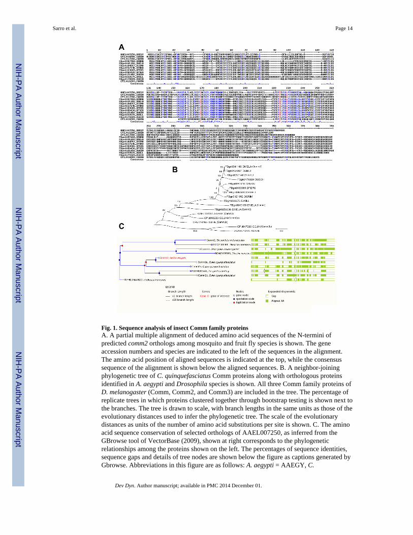
Fig. 1. Sequence analysis of insect Comm family proteinsA. A partial multiple alignment of deduced amino acid sequences of the N-termini ofpredicted comm2 orthologs among mosquito and fruit fly species is shown. The geneaccession numbers and species are indicated to the left of the sequences in the alignment.The amino acid position of aligned sequences is indicated at the top, while the consensussequence of the alignment is shown below the aligned sequences. B. A neighbor-joiningphylogenetic tree of C. quinquefasciatus Comm proteins along with orthologous proteinsidentified in A. aegypti and Drosophila species is shown. All three Comm family proteins ofD. melanogaster (Comm, Comm2, and Comm3) are included in the tree. The percentage ofreplicate trees in which proteins clustered together through bootstrap testing is shown next tothe branches. The tree is drawn to scale, with branch lengths in the same units as those of theevolutionary distances used to infer the phylogenetic tree. The scale of the evolutionarydistances as units of the number of amino acid substitutions per site is shown. C. The aminoacid sequence conservation of selected orthologs of AAEL007250, as inferred from theGBrowse tool of VectorBase (2009), shown at right corresponds to the phylogeneticrelationships among the proteins shown on the left. The percentages of sequence identities,sequence gaps and details of tree nodes are shown below the figure as captions generated byGbrowse. Abbreviations in this figure are as follows: A. aegypti = AAEGY, C.
Sarro et al. Page 14
Dev Dyn. Author manuscript; available in PMC 2014 December 01.
NIH
-PA Author Manuscript
NIH
-PA Author Manuscript
NIH
-PA Author Manuscript

quinquefasciatus = CQUIN, D. ananassae = DANAN, D. melanogaster = DMELA, D.simulans = DSIMU, D. yakuba = DYAKU, D. persimilis = DPERS, D. pseudoobscura =DPSEU, D. grimshawi = DGRIM, D. willistoni = DWILL, D. sechellia = DSECH.
Sarro et al. Page 15
Dev Dyn. Author manuscript; available in PMC 2014 December 01.
NIH
-PA Author Manuscript
NIH
-PA Author Manuscript
NIH
-PA Author Manuscript
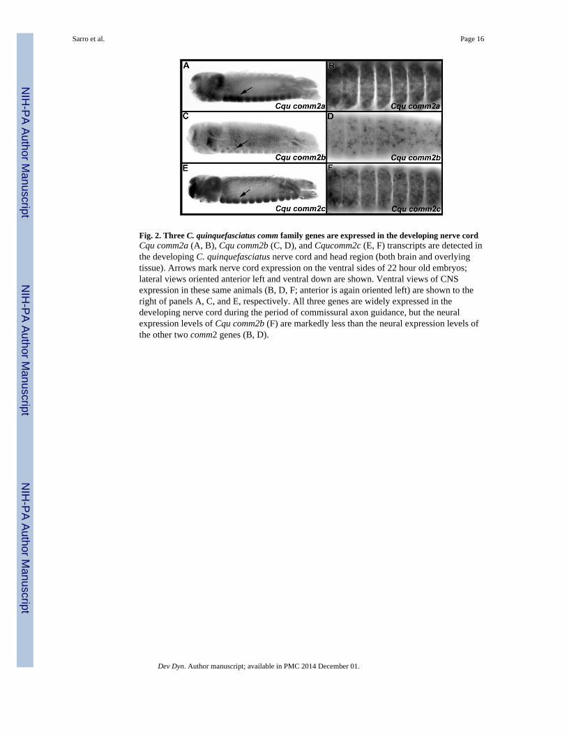
Fig. 2. Three C. quinquefasciatus comm family genes are expressed in the developing nerve cordCqu comm2a (A, B), Cqu comm2b (C, D), and Cqucomm2c (E, F) transcripts are detected inthe developing C. quinquefasciatus nerve cord and head region (both brain and overlyingtissue). Arrows mark nerve cord expression on the ventral sides of 22 hour old embryos;lateral views oriented anterior left and ventral down are shown. Ventral views of CNSexpression in these same animals (B, D, F; anterior is again oriented left) are shown to theright of panels A, C, and E, respectively. All three genes are widely expressed in thedeveloping nerve cord during the period of commissural axon guidance, but the neuralexpression levels of Cqu comm2b (F) are markedly less than the neural expression levels ofthe other two comm2 genes (B, D).
Sarro et al. Page 16
Dev Dyn. Author manuscript; available in PMC 2014 December 01.
NIH
-PA Author Manuscript
NIH
-PA Author Manuscript
NIH
-PA Author Manuscript
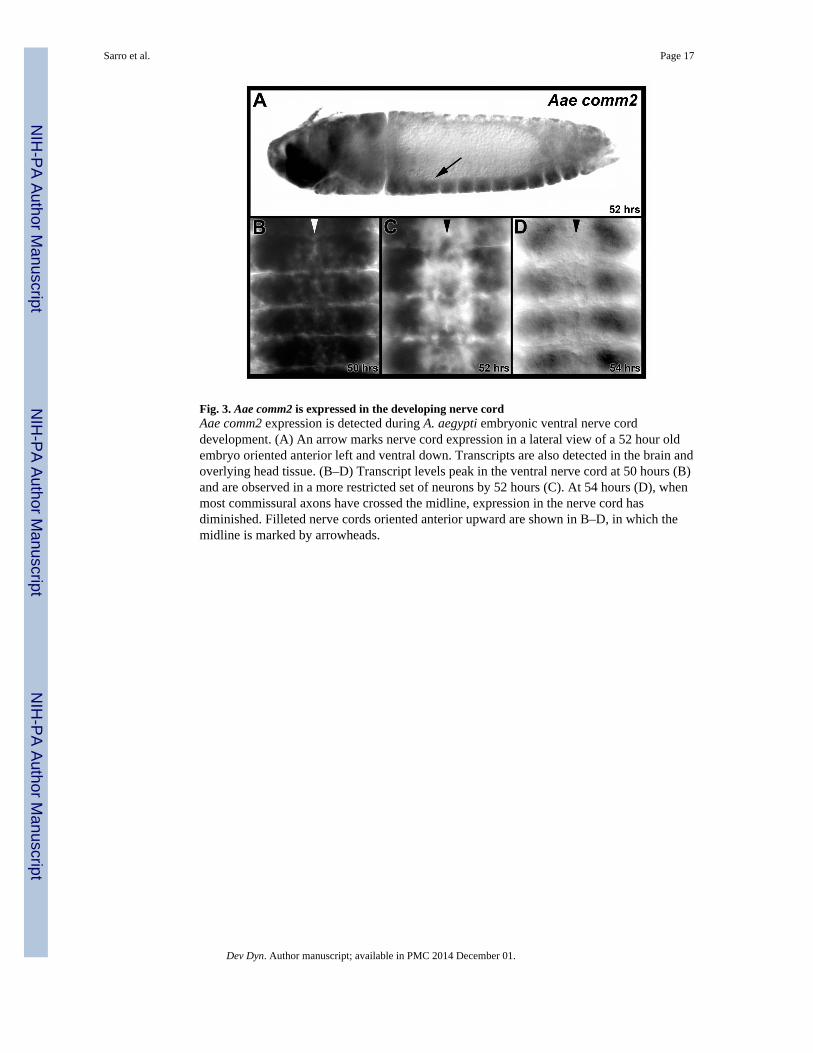
Fig. 3. Aae comm2 is expressed in the developing nerve cordAae comm2 expression is detected during A. aegypti embryonic ventral nerve corddevelopment. (A) An arrow marks nerve cord expression in a lateral view of a 52 hour oldembryo oriented anterior left and ventral down. Transcripts are also detected in the brain andoverlying head tissue. (B–D) Transcript levels peak in the ventral nerve cord at 50 hours (B)and are observed in a more restricted set of neurons by 52 hours (C). At 54 hours (D), whenmost commissural axons have crossed the midline, expression in the nerve cord hasdiminished. Filleted nerve cords oriented anterior upward are shown in B–D, in which themidline is marked by arrowheads.
Sarro et al. Page 17
Dev Dyn. Author manuscript; available in PMC 2014 December 01.
NIH
-PA Author Manuscript
NIH
-PA Author Manuscript
NIH
-PA Author Manuscript
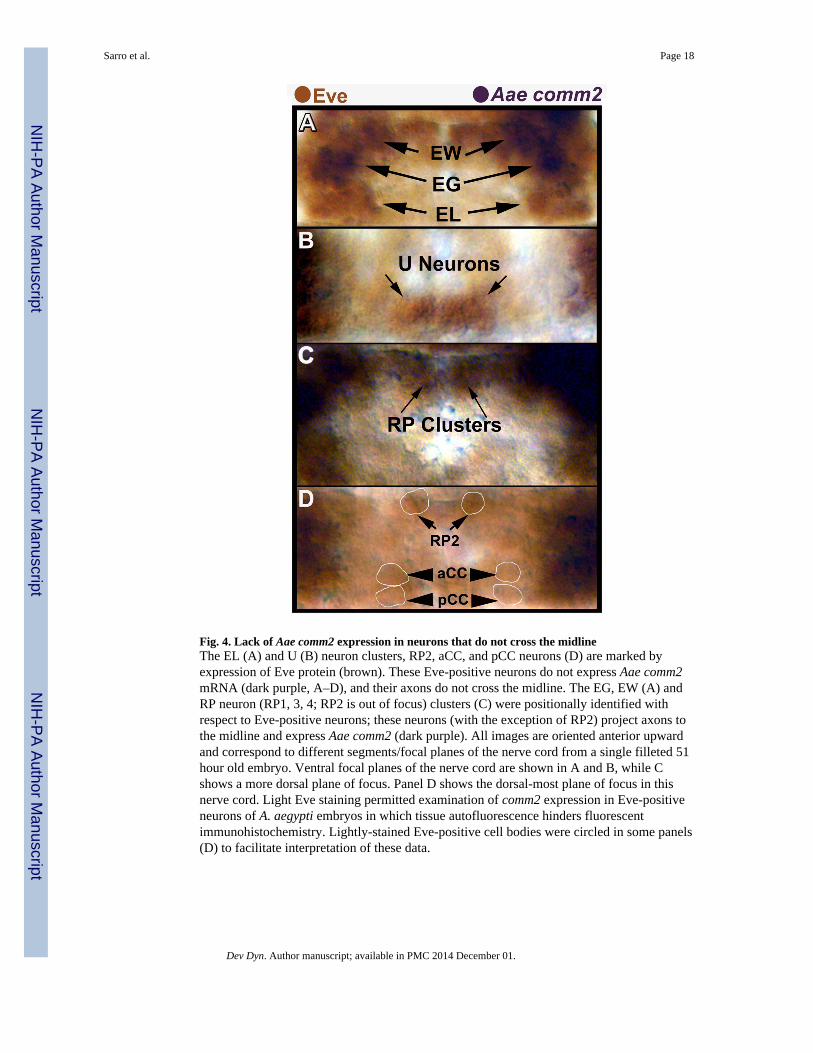
Fig. 4. Lack of Aae comm2 expression in neurons that do not cross the midlineThe EL (A) and U (B) neuron clusters, RP2, aCC, and pCC neurons (D) are marked byexpression of Eve protein (brown). These Eve-positive neurons do not express Aae comm2mRNA (dark purple, A–D), and their axons do not cross the midline. The EG, EW (A) andRP neuron (RP1, 3, 4; RP2 is out of focus) clusters (C) were positionally identified withrespect to Eve-positive neurons; these neurons (with the exception of RP2) project axons tothe midline and express Aae comm2 (dark purple). All images are oriented anterior upwardand correspond to different segments/focal planes of the nerve cord from a single filleted 51hour old embryo. Ventral focal planes of the nerve cord are shown in A and B, while Cshows a more dorsal plane of focus. Panel D shows the dorsal-most plane of focus in thisnerve cord. Light Eve staining permitted examination of comm2 expression in Eve-positiveneurons of A. aegypti embryos in which tissue autofluorescence hinders fluorescentimmunohistochemistry. Lightly-stained Eve-positive cell bodies were circled in some panels(D) to facilitate interpretation of these data.
Sarro et al. Page 18
Dev Dyn. Author manuscript; available in PMC 2014 December 01.
NIH
-PA Author Manuscript
NIH
-PA Author Manuscript
NIH
-PA Author Manuscript
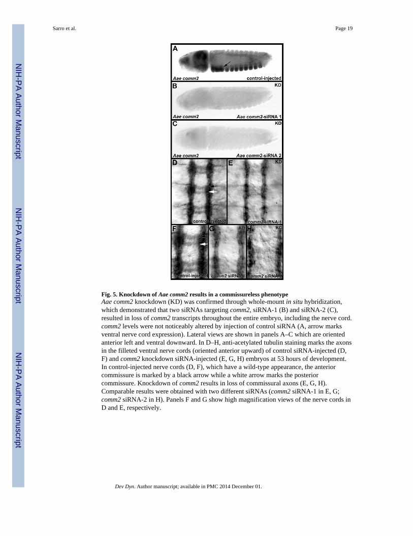
Fig. 5. Knockdown of Aae comm2 results in a commissureless phenotypeAae comm2 knockdown (KD) was confirmed through whole-mount in situ hybridization,which demonstrated that two siRNAs targeting comm2, siRNA-1 (B) and siRNA-2 (C),resulted in loss of comm2 transcripts throughout the entire embryo, including the nerve cord.comm2 levels were not noticeably altered by injection of control siRNA (A, arrow marksventral nerve cord expression). Lateral views are shown in panels A–C which are orientedanterior left and ventral downward. In D–H, anti-acetylated tubulin staining marks the axonsin the filleted ventral nerve cords (oriented anterior upward) of control siRNA-injected (D,F) and comm2 knockdown siRNA-injected (E, G, H) embryos at 53 hours of development.In control-injected nerve cords (D, F), which have a wild-type appearance, the anteriorcommissure is marked by a black arrow while a white arrow marks the posteriorcommissure. Knockdown of comm2 results in loss of commissural axons (E, G, H).Comparable results were obtained with two different siRNAs (comm2 siRNA-1 in E, G;comm2 siRNA-2 in H). Panels F and G show high magnification views of the nerve cords inD and E, respectively.
Sarro et al. Page 19
Dev Dyn. Author manuscript; available in PMC 2014 December 01.
NIH
-PA Author Manuscript
NIH
-PA Author Manuscript
NIH
-PA Author Manuscript
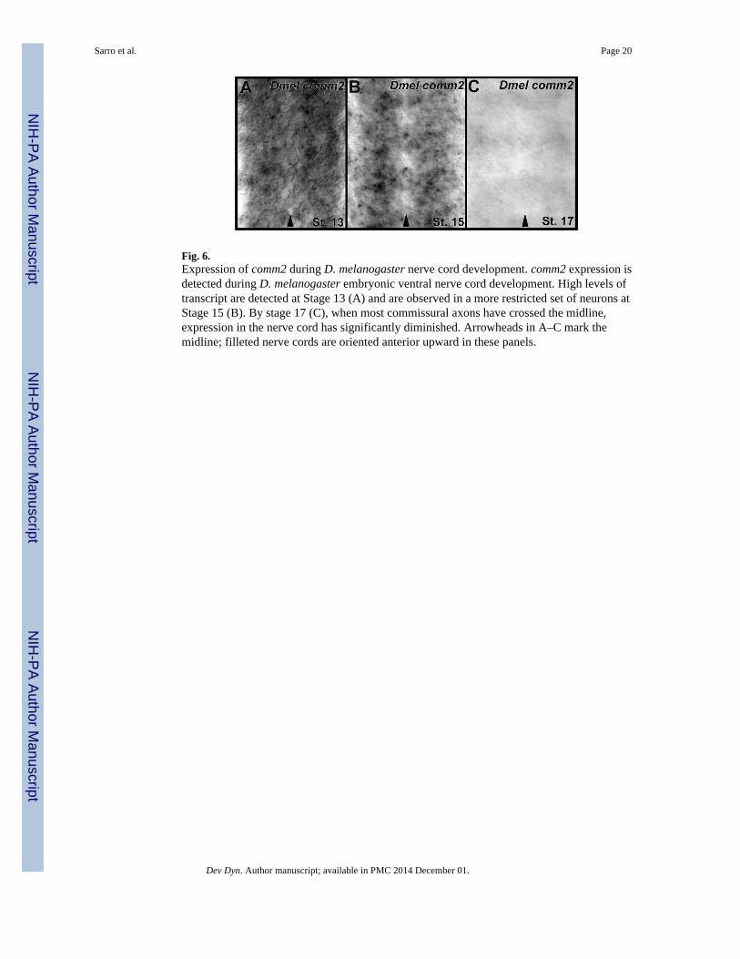
Fig. 6.Expression of comm2 during D. melanogaster nerve cord development. comm2 expression isdetected during D. melanogaster embryonic ventral nerve cord development. High levels oftranscript are detected at Stage 13 (A) and are observed in a more restricted set of neurons atStage 15 (B). By stage 17 (C), when most commissural axons have crossed the midline,expression in the nerve cord has significantly diminished. Arrowheads in A–C mark themidline; filleted nerve cords are oriented anterior upward in these panels.
Sarro et al. Page 20
Dev Dyn. Author manuscript; available in PMC 2014 December 01.
NIH
-PA Author Manuscript
NIH
-PA Author Manuscript
NIH
-PA Author Manuscript
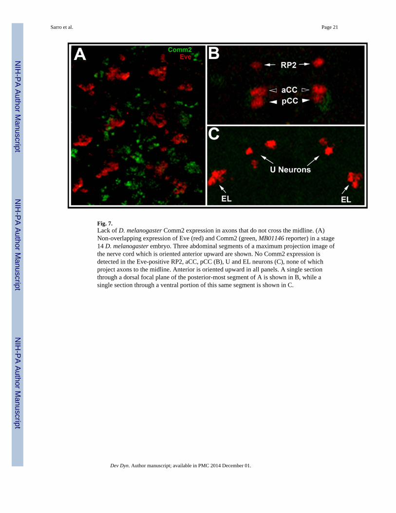
Fig. 7.Lack of D. melanogaster Comm2 expression in axons that do not cross the midline. (A)Non-overlapping expression of Eve (red) and Comm2 (green, MB01146 reporter) in a stage14 D. melanogaster embryo. Three abdominal segments of a maximum projection image ofthe nerve cord which is oriented anterior upward are shown. No Comm2 expression isdetected in the Eve-positive RP2, aCC, pCC (B), U and EL neurons (C), none of whichproject axons to the midline. Anterior is oriented upward in all panels. A single sectionthrough a dorsal focal plane of the posterior-most segment of A is shown in B, while asingle section through a ventral portion of this same segment is shown in C.
Sarro et al. Page 21
Dev Dyn. Author manuscript; available in PMC 2014 December 01.
NIH
-PA Author Manuscript
NIH
-PA Author Manuscript
NIH
-PA Author Manuscript
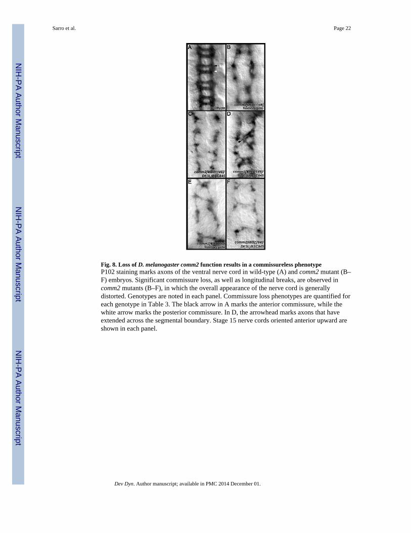
Fig. 8. Loss of D. melanogaster comm2 function results in a commissureless phenotypeP102 staining marks axons of the ventral nerve cord in wild-type (A) and comm2 mutant (B–F) embryos. Significant commissure loss, as well as longitudinal breaks, are observed incomm2 mutants (B–F), in which the overall appearance of the nerve cord is generallydistorted. Genotypes are noted in each panel. Commissure loss phenotypes are quantified foreach genotype in Table 3. The black arrow in A marks the anterior commissure, while thewhite arrow marks the posterior commissure. In D, the arrowhead marks axons that haveextended across the segmental boundary. Stage 15 nerve cords oriented anterior upward areshown in each panel.
Sarro et al. Page 22
Dev Dyn. Author manuscript; available in PMC 2014 December 01.
NIH
-PA Author Manuscript
NIH
-PA Author Manuscript
NIH
-PA Author Manuscript
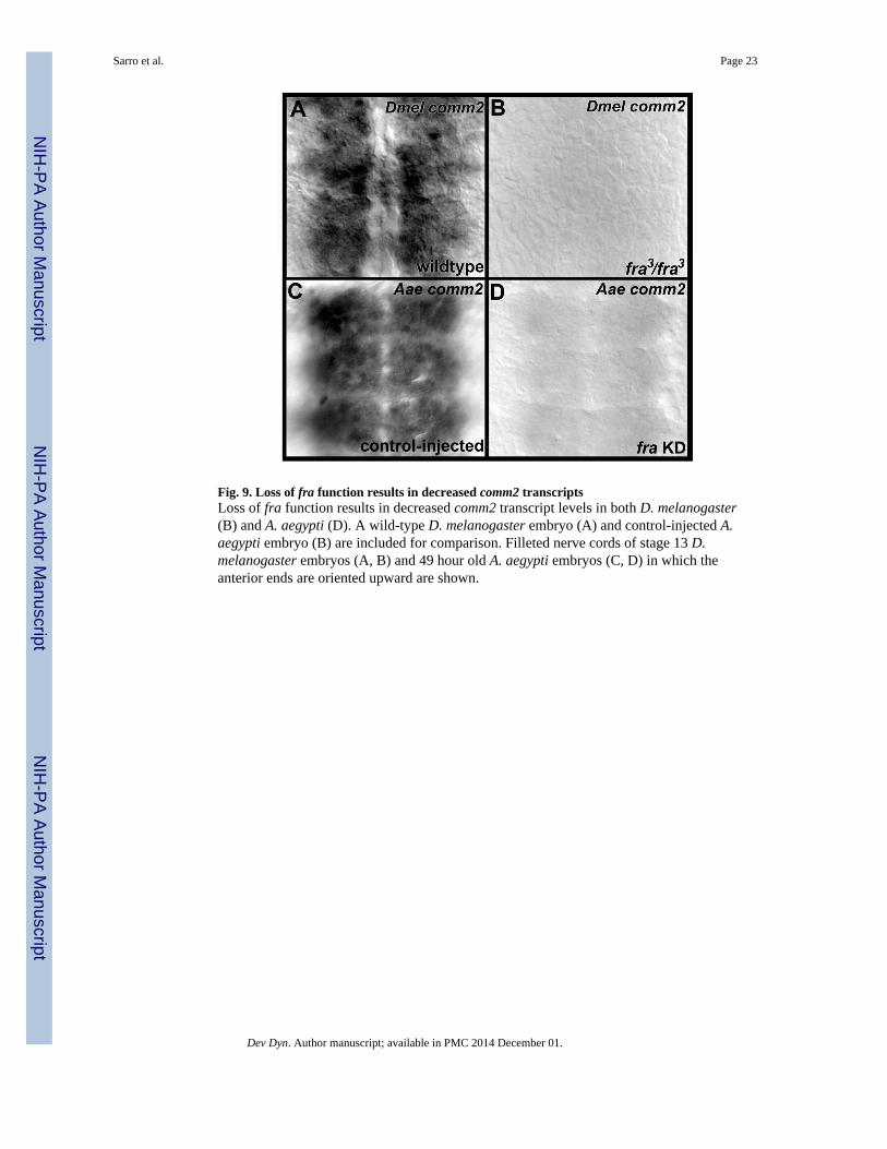
Fig. 9. Loss of fra function results in decreased comm2 transcriptsLoss of fra function results in decreased comm2 transcript levels in both D. melanogaster(B) and A. aegypti (D). A wild-type D. melanogaster embryo (A) and control-injected A.aegypti embryo (B) are included for comparison. Filleted nerve cords of stage 13 D.melanogaster embryos (A, B) and 49 hour old A. aegypti embryos (C, D) in which theanterior ends are oriented upward are shown.
Sarro et al. Page 23
Dev Dyn. Author manuscript; available in PMC 2014 December 01.
NIH
-PA Author Manuscript
NIH
-PA Author Manuscript
NIH
-PA Author Manuscript
NIH
-PA Author Manuscript
NIH
-PA Author Manuscript
NIH
-PA Author Manuscript
Sarro et al. Page 24
Tabl
e 1
Pair
wis
e di
stan
ces
betw
een
gene
s ca
lcul
ated
usi
ng M
EG
A4.
The
num
ber
of a
min
o ac
id s
ubst
itutio
ns p
er s
ite f
rom
ana
lysi
s be
twee
n se
quen
ces
is s
how
n.1:
AA
EL
0072
50_A
AE
GY
, 2: C
PIJ0
1628
5_C
QU
IN, 3
: CPI
J017
280_
CQ
UIN
, 4: F
Bgn
0087
418_
DA
NA
N, 5
: FB
gn01
2195
2_D
GR
IM, 6
:FB
gn00
4116
0_D
ME
LA
, 7: F
Bgn
0163
065_
DPE
RS,
8: F
Bgn
0080
430_
DPS
EU
, 9: F
Bgn
0179
329_
DSE
CH
, 10:
FB
gn01
8426
7_D
SIM
U, 1
1:FB
gn02
2267
0_D
WIL
L, 1
2: F
Bgn
0237
146_
DY
AK
U, 1
3: C
PIJ0
1628
3_C
QU
IN. T
hese
spe
cies
abb
revi
atio
ns a
re d
efin
ed in
the
lege
nd o
f Fi
g. 1
.
12
34
56
78
910
1112
13
1 21.
1
31.
32.
0
41.
31.
92.
2
51.
31.
82.
40.
3
61.
31.
72.
20.
20.
3
71.
42.
02.
40.
20.
30.
2
81.
42.
02.
40.
20.
30.
20.
0
91.
51.
62.
20.
40.
50.
30.
40.
4
101.
31.
82.
20.
20.
30.
00.
20.
20.
2
111.
31.
82.
40.
30.
30.
30.
30.
30.
60.
3
121.
31.
82.
20.
20.
30.
00.
20.
20.
30.
00.
3
130.
51.
41.
61.
51.
51.
51.
71.
71.
61.
41.
61.
5
Dev Dyn. Author manuscript; available in PMC 2014 December 01.
NIH
-PA Author Manuscript
NIH
-PA Author Manuscript
NIH
-PA Author Manuscript
Sarro et al. Page 25
Table 2
A. aegypti comm2 knockdown phenotypes. 53 hour old embryos were assessed post-injection of comm2-siRNA 1, comm2-siRNA 2, or control siRNA. Commissure loss phenotypes were scored in individualsegments following anti-acetylated tubulin staining. Percentages of segments with wildtype, moderate(thinning of commissures), or severe (loss/near loss of commissures) phenotypes are indicated. Data werecompiled from three or more replicate experiments.
n% segments with no commissure
defects% segments with moderate
commissure defects% segments with severe
commissure defects
control-injected 58 98 2 0
comm2-siRNA 1 42 5 21 74
comm2-siRNA 2 81 2 21 77
Dev Dyn. Author manuscript; available in PMC 2014 December 01.
NIH
-PA Author Manuscript
NIH
-PA Author Manuscript
NIH
-PA Author Manuscript
Sarro et al. Page 26
Table 3D. melanogaster comm2
knockdown commissure loss phenotypes. BP102-stained stage 15–16 D. melanogaster embryos were assessedfor commissure loss phenotypes. Percentages of segments with wildtype, moderate (thinning of commissures)or severe (loss/near loss of commissures) phenotypes are indicated. Data were compiled from three or morereplicate experiments.
n% segments with nocommissure defects
% segments withmoderate commissure
defects% segments with severe
commissure defects
wildtype 110 100% 0% 0%
comm2[MB01146]/comm2[MB01146] 65 0% 6% 94%
comm2[MB01146]/Df(3L)BSC845 47 0% 11% 89%
comm2[MI02284]/comm2[MI02284] 107 3% 26% 71%
comm2[MI02284]/Df(3L)BSC845 54 0% 22% 78%
Dev Dyn. Author manuscript; available in PMC 2014 December 01.