nih public access 1 javier lópez-garrido h. velocity ... · peptidoglycan transformations during...
TRANSCRIPT
Peptidoglycan transformations during Bacillus subtilissporulation
Elitza I. Tocheva1, Javier López-Garrido3, H. Velocity Hughes4, Jennifer Fredlund3, ErkinKuru5, Michael S. VanNieuwenhze5, Yves V. Brun4, Kit Pogliano3, and Grant J. Jensen1,2,*
1Division of Biology, California Institute of Technology, 1200 E California Blvd., Pasadena, CA91125, USA2Howard Hughes Medical Institute, California Institute of Technology, 1200 E California Blvd.,Pasadena, CA 91125, USA3Division of Biological Sciences, University of California at San Diego, La Jolla, California, USA4Department of Biology, Indiana University, Bloomington, IN 47405, USA5Department of Chemistry, Indiana University, Bloomington, IN 47405, USA
SummaryWhile vegetative Bacillus subtilis cells and mature spores are both surrounded by a thick layer ofpeptidoglycan (PG, a polymer of glycan strands cross-linked by peptide bridges), it has remainedunclear whether PG surrounds prespores during engulfment. To clarify this issue, we generated aslender ΔponA mutant that enabled high-resolution electron cryotomographic imaging. Three-dimensional reconstructions of whole cells in near-native states revealed a thin PG-like layerextending from the lateral cell wall around the prespore throughout engulfment. Cryotomographyof purified sacculi and fluorescent labelling of PG in live cells confirmed that PG surrounds theprespore. The presence of PG throughout engulfment suggests new roles for PG in sporulation,including a new model for how PG synthesis might drive engulfment, and obviates the need tosynthesize a PG layer de novo during cortex formation. In addition, it reveals that B. subtilis cansynthesize thin, Gram-negative-like PG layers as well as its thick, archetypal Gram-positive cellwall. The continuous transformations from thick to thin and back to thick during sporulationsuggest that both forms of PG have the same basic architecture (circumferential). Endopeptidaseactivity may be the main switch that governs whether a thin or a thick PG layer is assembled.
IntroductionThe bacterial cell envelope is a complex multilayered structure (Silhavy et al., 2010). Itprotects the cell from harsh environments, facilitates transport of molecules in and out of thecell, and is used to maintain a proton gradient. More than 100 years ago Christian Gramdeveloped a staining procedure that classified bacterial cells into two major groups – Gram‘-positive’ and ‘-negative’ – based on their ability to retain the chemical crystal violet(Gram, 1884). With the advent of electron microscopy, the fundamental structuraldifferences between the two classes of bacteria were clarified: canonical Gram-negativebacterial cell envelopes consist of two membranes and a thin layer of peptidoglycan (PG)
© 2013 John Wiley & Sons Ltd*For correspondence. [email protected], Tel. (+1) 626 395 8827, Fax (+1) 626 395 5730.
Supporting informationAdditional supporting information may be found in the online version of this article at the publisher’s web-site.
NIH Public AccessAuthor ManuscriptMol Microbiol. Author manuscript; available in PMC 2014 May 01.
Published in final edited form as:Mol Microbiol. 2013 May ; 88(4): 673–686. doi:10.1111/mmi.12201.
NIH
-PA Author Manuscript
NIH
-PA Author Manuscript
NIH
-PA Author Manuscript
between them, and Gram-positive cells have one membrane surrounded by a much thickerlayer of PG (Chapman and Hillier, 1953). Despite the obvious importance, many aspects ofthe structure and function of the cell envelope remain poorly understood.
Peptidoglycan is a large polymer that surrounds the cell. It consists of long glycan strandsformed by repeating units of N-acetyl glucosamine-N-acetyl muramic acid, which are cross-linked through pentapeptide chains (Typas et al., 2011). PG adds mechanical strength to thecell envelope and maintains cell shape (Thwaites and Mendelson, 1991; Tuson et al., 2012).The structure of PG from Gram-negative bacteria has been extensively studied and electroncryotomography (ECT) studies showed by direct imaging that the glycan chains inEscherichia coli and Caulobacter crescentus lie parallel to the cell membrane roughlyperpendicular to the long axis of the cell (an architecture that we call here ‘circumferential’)(Gan et al., 2008). This ultrastructural analysis also indicated that the cell walls were only asingle layer thick, consistent with the thin appearance of the PG in earlier cryo-sections of E.coli and Pseudomonas aeruginosa (Matias et al., 2003).
Even though the PG layer of Gram-positive cells is composed of the same building blocks asGram-negative cells, it appears much thicker in electron micrographs. It is possible that themolecular structure is similar to that of Gram-negative bacteria, but is multi-layered. Insupport of this idea, studies of purified sacculi from the classic Gram-positive model speciesBacillus subtilis have already suggested circumferential orientation of the glycan strands(Verwer and Nanninga, 1976), and movements of various components of the cell wallsynthetic machinery have also been shown to be circumferential (Dominguez-Escobar et al.,2011; Garner et al., 2011). Recent literature proposes other models, however. NMR studiesled to a ‘scaffold’ model, where the glycan strands are oriented perpendicular to the cellsurface (Dmitriev et al., 2003). Atomic force microscopy (AFM) studies of purifiedStaphylococcus aureus sacculi showed a fibrous network with many pores (Touhami et al.,2004), but AFM images of purified B. subtilis sacculi looked quite different, and led to a‘coiled-coil’ model where glycan strands are bundled together, coiled tightly to form ~ 50nm hollow cables, and finally wrapped around cells (like a telephone cord wrapped around abarrel) (Hayhurst et al., 2008).
When growth conditions become unfavourable, some members of the phylum Firmicutesincluding B. subtilis undergo a complex morphological transformation called sporulationwhich has been used as a basic system to study membrane movements and cell–cellcommunication (Errington, 2010, Errington, 2003). Sporulation begins with the formation ofan asymmetric septum that divides the cell into a smaller ‘prespore’ and a larger mother cell.Next, in a process similar to phagocytosis, the mother cell membranes migrate around theprespore until the engulfing membranes meet and fuse, releasing the ‘forespore’ into themother cell cytoplasm. Transmission electron microscopy (TEM) images have shown thatsporulation septa are formed by the inward growth of a thick disk of septal PG andcytoplasmic membrane. After septation, this thick PG layer is thinned (Holt et al., 1975) bysporulation-specific enzymes that degrade septal PG and then localize to the leading edgesof the engulfing membranes (Abanes-De Mello et al., 2002; Chastanet and Losick, 2007;Gutierrez et al., 2010; Morlot et al., 2010). PG degradation is rate-limiting for membranemigration throughout engulfment (Abanes-De Mello et al., 2002; Gutierrez et al., 2010) andit has been proposed that these membrane-anchored enzymes processively degrade PG andthereby move the mother cell membranes around the prespore (Abanes-De Mello et al.,2002; Morlot et al., 2010). While it has remained unclear if these enzymes completelydegrade the septal PG, TEM images have not shown any clear PG layer between the mothercell and prespore membranes during engulfment (Holt et al., 1975; Aronson and Fitz-James,1976).
Tocheva et al. Page 2
Mol Microbiol. Author manuscript; available in PMC 2014 May 01.
NIH
-PA Author Manuscript
NIH
-PA Author Manuscript
NIH
-PA Author Manuscript

It has long been known that after engulfment, new PG is synthesized to produce the cortex,the thick modified PG layer that protects the spore (Tipper and Linnett, 1976; Sekiguchi etal., 1995; Atrih et al., 1998; Meador-Parton and Popham, 2000; McPherson et al., 2001;Vasudevan et al., 2007). More recently, it has been found that PG synthesis also likelyoccurs during engulfment. Specifically, fluorescently labelled PG precursors localize to theleading edges of engulfing membranes (Meyer et al., 2010). Furthermore, blocking PGsynthesis with fosfomycin inhibits membrane migration in cells lacking the SpoIIQ–SpoIIIAH backup engulfment proteins and blocks engulfment membrane fission in wild typecells (Meyer et al., 2010). Studies of specific enzymes are complicated, however, by theredundancy of the PG biosynthetic machinery and the requirement for PG synthesis duringseptum formation (Buchanan and Sowell, 1983; McPherson and Popham, 2003; Korsak etal., 2005; Scheffers, 2005; Sauvage et al., 2008). The large amounts of vegetative andcortical PG present throughout and after engulfment, respectively, have also complicatedsearches for engulfment-specific PG (Atrih et al., 1996; Meador-Parton and Popham, 2000).Thus, it has remained unclear if the prespore is always surrounded by a layer of PG, or ifseptal PG is first completely degraded and later re-synthesized during cortex formation.More recently, ECT of whole cells and purified sacculi of an unusual Firmicute that isGram-negative and produces endospores, Acetonema longum, showed that a layer of PG wassynthesized between the prespore and mother cell membrane during engulfment (Tocheva etal., 2011), but it is unclear whether this also occurs in the more well-studied, Gram-positiveendospore-forming bacteria.
Traditional EM preparation methods disrupt membranes and other macromolecules throughcross-linking and dehydration. In contrast, ECT begins by rapidly plunge-freezing cells intheir growth medium (Dubochet et al., 1983; Iancu et al., 2006), reducing specimenpreparation artefacts and allowing three-dimensional images of cells to be obtained intact ina near-native state to ~ 4 nm resolution (Tocheva et al., 2010). To address the role of PGduring engulfment in the archetypal sporulating bacterium B. subtilis, we generated a ΔponAmutant strain that was thin enough to be imaged directly with ECT. The ponA gene encodesfor a class A penicillin-binding protein (PBP1), but previous studies have shown that sincemultiple PBPs exhibit redundant functions, deletion of the ponA gene has no significanteffect on rod-shape cell morphology, cell division, sporulation, spore heat resistance, orspore germination except that cells are thinner than wild type (Popham and Setlow, 1995;Meador-Parton and Popham, 2000). Here we demonstrate that this strain is sufficiently thinfor ECT, and cryotomograms of vegetative, sporulating and germinating ΔponA cells revealthat a thin PG-like layer persists between engulfing membranes throughout engulfment.ECT of purified sacculi and light microscopy of fluorescently labelled PG confirm that alayer of PG persists around the prespore throughout engulfment. This layer likely serves asthe foundation for assembly of the thick inner and outer cortices of the mature spore. Upongermination, the outer cortex is degraded and the inner cortex (germ cell wall) remains asthe vegetative PG of outgrowing cells (Santo and Doi, 1974). B. subtilis therefore maintainsPG around the spore continually throughout engulfment, maturation, and germination, andtransforms its PG from thick to thin and back to thick. The implications of these transitionsand the possible roles of PG during engulfment are discussed.
ResultsECT characterization of the ΔponA mutant
Wild type B. subtilis cells are typically too thick (~ 1200 nm) for high-resolution ECTimaging, so a mutation in the ponA gene was introduced. To test whether the PG layers ofthe ΔponA mutant were similar to those of wild type cells, cryotomograms of ΔponA B.subtilis cells were compared with the small number of lower-quality cryotomograms of wild
Tocheva et al. Page 3
Mol Microbiol. Author manuscript; available in PMC 2014 May 01.
NIH
-PA Author Manuscript
NIH
-PA Author Manuscript
NIH
-PA Author Manuscript

type cells we could obtain. The cell walls of both were uniform around the cell with anaverage thickness of 40–50 nm (Fig. 1A and B), in good agreement with results producedwith other forms of electron microscopy (Matias and Beveridge, 2005). In order to permitslightly higher resolution, sacculi from both wild type and ΔponA vegetative B. subtilis cellswere purified and imaged. The PG in both types of sacculi were again uniformly thick (40–50 nm), with smooth inner surfaces and ‘fuzzy’ outer surfaces (Fig. S1). High contrast, thin,planar ‘patch-like’ densities within the PG parallel to the membrane were occasionallyobserved in both wild type and ΔponA cells (Fig. S1, black arrows). Unfortunately, purifiedsacculi from vegetative cells did not completely flatten on the EM grids due to cellulardebris aggregates, limiting the resolution to less than that obtained previously for Gram-negative sacculi (Gan et al., 2008) (Fig. S1, white arrows).
To further characterize the ΔponA mutant, dividing cells were also imaged using ECT (Fig.1C–F) and fluorescence microscopy. On average the cells were 700 nm wide and 2 μm long.Just as in wild type B. subtilis, vegetative septa formed in the middle of dividing cells, ~ 2μm from a cell pole, and exhibited symmetric ingression of the cytoplasmic membrane (Fig.1C) with a thick layer of PG (Fig. 1D). Upon closure, the thickness of the PG in the divisionseptum was double the thickness of the PG on the lateral cell wall (~ 80 nm, Fig. 1E), asexpected as the septal PG later splits to generate two cell poles. The division septa were flatbut nascent cell poles became rounder as daughter cells separated (Fig. 1E–F). The natureand transformations of the cell wall in the ΔponA mutant therefore appear to mimic those ofwild type cells, in keeping with the minor changes in muropeptide composition detected bymass spectrometry (Popham et al., 1996).
ECT of sporulating ΔponA B. subtilisBecause the ΔponA cell wall resembled and behaved like that of wild type cells, and themutant completed sporulation to nearly wild type levels (Popham and Setlow, 1995), weused it to obtain high-resolution images of sporulation. Sporulation in ΔponA B. subtilisbegan with the formation of an asymmetrically positioned septum ~ 500 nm from one cellpole that contained PG that was roughly half the thickness of a vegetative septum (Fig. 2A).Some flat sporulation septa had only a thin layer of material continuous with and resemblingPG between the inner and outer spore membranes (Fig. 2A, red dotted line in inset);presumably because these were undergoing or had already completed septal thinning. Later,the septal membranes became convex around the prespore (Fig. 2B) and migrated aroundthe prespore toward the cell pole (Fig. 2C–D). The pole of the mother cell did not enlargeduring engulfment, but rather retained its original diameter. As engulfment proceeded, theforespore also maintained its width but increased in length, as also evident in timelapsemicroscopy of this process (Pogliano et al., 1999). The thin, continuous layer of PG-likematerial between the inner and outer spore membranes persisted throughout engulfment,always connected to and continuous with the cell wall (Fig. 2B–D, red dotted line in insets).The distance between the inner and outer spore membranes during engulfment was ~ 22 nm(measured ‘peak’ to ‘peak’). During engulfment, an additional layer of density was observedon the mother side of the septum (Fig. 2D) that likely represents an early assembly stage ofthe multilayered protein coat (Fig. 2H) (McKenney et al., 2012). After engulfment theforespore of the ΔponA B. subtilis appeared elliptical with typical dimensions of ~ 0.6 μm ×0.9 μm, and was completely surrounded by coat material (Fig. 2E–F, arrows).
Mature spores were ellipsoidal with final dimensions of ~ 1 μm × 1.5 μm. Two thick layersof cortex were apparent between the inner and outer spore membranes. The inner cortex(germ cell wall) was denser and more uniform in thickness (~ 50–70 nm) than the outercortex (50–100 nm). The inner and outer spore membrane and various coat layers were alsoclearly discernable (Fig. 2G–H inset). During germination, the outer cortex and coat were
Tocheva et al. Page 4
Mol Microbiol. Author manuscript; available in PMC 2014 May 01.
NIH
-PA Author Manuscript
NIH
-PA Author Manuscript
NIH
-PA Author Manuscript

shed from the remaining inner cortex and spore (Fig. 2H–J). A thin interfacial layer ofhigher density was observed between the inner and outer cortex (Fig. 2J, arrows). The innercortex appeared intact and resembled the thickness and density of vegetative PG (~ 50 nm).During outgrowth, the inner cortex remained associated with the newly emerging cells astheir thick vegetative cell wall.
ECT of purified sacculi from sporulating wild type and ΔponA B. subtilisTo test whether the observed layer of PG-like material between the engulfing mother cellmembrane and the prespore membrane in the cryotomograms was PG, sacculi from wildtype B. subtilis cells at different stages of sporulation were purified. As with sacculi fromvegetative cells, some cellular debris persisted inside throughout the purification, preventingthe sacculi from flattening on the EM grids. The septal PG in the sacculi of sporulating cellsvaried in thickness, with some appearing 40 nm thick, similar to the cell wall of the mothercell (Fig. S2A, arrows). Others showed septal PG ~ 20 nm thick at the edges and thinner inthe middle (Fig. S2B), consistent with observations that septal PG degradation commencesat the septal midpoint (Perez et al., 2000). Other sacculi showed undulating layers of verythin (~ 2 nm) septal PG that could be traced across the cell, connecting the two sides of themother cell wall (Figs 3A and S2). Cellular debris was often seen stuck to or penetrating themiddle of the thin septal PG. Late stages of engulfment were not seen, perhaps because ofincreased fragility.
Fluorescent labelling of PG in sporulating wild type and ΔdacA B. subtilisIn order to further confirm that a layer of PG surrounds the prespore throughout engulfment,we labelled sporulating cells with fluorescent derivatives of D-amino acids (FDAAs), whichcan be covalently incorporated at the fifth position of the stem peptide in PG chains,substituting for terminal D-alanine and labelling PG in living cells (Kuru et al., 2012). Wehypothesized that, if the layer of dense material observed between the inner and the outerspore membranes was PG, it would incorporate FDAAs and a continuous fluorescent signalwould appear around pre-spores throughout engulfment in B. subtilis.
A fluorescein-conjugated derivative of D-lysine (FDL) (Kuru et al., 2012) was used tolocalize PG within sporulating wild type B. subtilis cells. As shown in Fig. 3B (upper panel)bright FDL foci are observed near the leading edge of the engulfing membranes, consistentwith the localization of PG biosynthetic intermediates which localize to this position (Meyeret al., 2010). However, no signal was observed in the septal region around the pre-spore.Given the fact that the PG layer of interest is very thin, we considered the possibility that thesensitivity of the labelling was not high enough to detect it. Typically, the terminal D-alanine of one of the two stem peptides forming a peptide bridge is removed in the cross-linking reaction and most of the remaining terminal D-alanine is typically removed by PBP5(encoded by the gene dacA), a D,D-carboxypeptidase that cleaves the terminal D-alanine ofthe pentapeptides in nascent PG (Lawrence and Strominger, 1970; Todd et al., 1986; Atrihet al., 1999). Incorporation of FDAA into B. subtilis PG is significantly higher in ΔdacAmutants (Kuru et al., 2012), so we decided to use ΔdacA B. subtilis mutant to maximizelabelling during engulfment. While ΔdacA mutants are known to exhibit normal growth,spore development, resistance and germination (Todd et al., 1986; Buchanan and Gustafson,1992; Popham et al., 1999), cryotomograms were recorded to confirm that the cellsexhibited a typical sporulation septum with the same thin layer of PG-like material betweenthe inner and outer spore membranes (Fig. S3B–D, again with reduced resolution due to thegreater thickness of otherwise wild type cells).
Sporangia of ΔdacA B. subtilis with flat septa showed variable FDL intensities in theseptum, ranging from > 10-fold to 3-fold higher than background (Fig. S4C). This variable
Tocheva et al. Page 5
Mol Microbiol. Author manuscript; available in PMC 2014 May 01.
NIH
-PA Author Manuscript
NIH
-PA Author Manuscript
NIH
-PA Author Manuscript

staining likely reflects the loss of septal PG during septal thinning, since sporangia in earlystages of prespore chromosome segregation showed higher FDL staining than those in laterstages (Fig. 3C) (Illing and Errington, 1991). As engulfment proceeded, the septum curvedaround the prespore, retaining a faint and continuous FDL signal (Fig. 3C). Cells that hadcompleted membrane migration but not membrane fission showed a fluorescent signal allaround the prespore and often contained bright foci (Fig. 3C, bottom). Similar FDL labellingpatterns were observed in cells imaged with or without the membrane stain (Fig. S5A) butnot in cells incubated with free fluorescein (Fig. S5B), suggesting that the staining was infact the result of FDL incorporation in PG chains. Altogether, FDL staining of sporulatingwild type and ΔdacA B. subtilis cells suggests that, although PG may be actively synthesizedat the leading edge of the engulfing membrane, a layer of PG is continuously presentbetween the mother cell and pre-spore membranes throughout engulfment.
DiscussionA thin layer of PG surrounds prespores throughout engulfment
Exploiting a slender B. subtilis mutant amenable to high-resolution ECT, here we haveshown that a thin layer of material continuous with and resembling the cell wall surroundsprespores throughout engulfment. This thin layer was likely missed in previous EM studies(Holt et al., 1975; Aronson and Fitz-James, 1976; Illing and Errington, 1991) because of thefixation, plastic embedding, sectioning and staining inherent in traditional approaches(Matias and Beveridge, 2005). ECT of purified wild type sacculi during sporulation anddirect fluorescent labelling of PG in live cells confirmed that the observed thin layer was infact PG (Figs 3, S2 and S4).
The presence of a thin PG between engulfing membranes in B. subtilis could be due to oneof the following two possibilities: (1) an innermost layer of PG is separated from the thickmother PG and directed into the septum, or (2) a new, thin layer of PG is synthesized at theleading edge of the engulfing membranes. Comparison of the PG transformations in B.subtilis and A. longum allows us to distinguish between these two possibilities. In A.longum, the vegetative cell wall is only a layer thick and follows the membranes into thesporulation septum. The single layer of PG observed between the engulfing membranes,therefore, must be newly synthesized. Because the PG synthetic machinery of these twospecies are so similar, B. subtilis likely also synthesizes a thin layer of PG duringengulfment. Furthermore, fluorescent labelling of wild type and ΔdacA B. subtilis showdifferent labelling patterns, indicating that the septal PG layer is modified by PBPs similarlyto the vegetative cell wall. The incorporation of FDAA therefore labels newly synthesizedPG. Lastly, the PG just ahead of the engulfing membranes (Fig. 4B, black arrow) is thickerthan just behind (white arrow), and the PG behind the junction is not thinner than the rest ofthe cell wall, all consistent with synthesis of new, additional PG at the leading edge of thejunction rather than separation of existing PG (Fig. 4B).
These observations suggest three potential functions of the thin layer of PG duringengulfment. First, it may provide mechanical strength as the genome is packed within theprespore. Second, PG synthesis may drive membrane movement during engulfment. Third,it may serve as a template for elaboration of the cortex. Concerning membrane movement,immediately after septal thinning, the thin layer of PG between the septal membranes is flat.During engulfment, the septum becomes convex and then the perimeter migrates towards thecell pole. As suggested previously (Lopez-Diaz et al., 1986; Smith et al., 1993; Frandsenand Stragier, 1995), septal thinning might be required to give the septum enough flexibilityto undergo these morphological changes. The hydrolytic enzymes that mediate septalthinning are also rate limiting for membrane migration and it has been further proposed thatthey move the membranes around the prespore by a burnt-bridge ratchet mechanism, pulling
Tocheva et al. Page 6
Mol Microbiol. Author manuscript; available in PMC 2014 May 01.
NIH
-PA Author Manuscript
NIH
-PA Author Manuscript
NIH
-PA Author Manuscript

the membrane towards the pole by binding and hydrolysing glycan strands along the path(Abanes-De Mello et al., 2002; Gutierrez et al., 2010; Morlot et al., 2010). The resultsdescribed here demonstrate that septal curving and membrane migration towards the cellpole must also require PG synthesis, since the septal PG must cover an increasingly largesurface. This rationalizes why fluorescently labelled PG building blocks were observedlocalizing at the leading edge of the engulfing membrane (Meyer et al., 2010). Moreover,because synthesis of new PG is known to drive cell shape changes (such as division (Cabeenand Jacobs-Wagner, 2005)) and move PG synthetic enzymes across membranes(Dominguez-Escobar et al., 2011; Garner et al., 2011), we suggest that synthesis of the thinlayer of septal PG may help drive engulfment (Meyer et al., 2010). Specifically, addition ofnew glycan strands at the front (pole-facing) side of the junction between the septal PG andthe cell wall may push the inner spore membrane inward, and create new cross-links withthe cell wall in front of the junction (Fig. 4B, red strands). Hydrolysis at the back (mother-cell-facing) side of the junction may cleave older cross-links, freeing the trailing surface ofthe growing septal PG from the cell wall. PG-synthetic enzymes could be localized to thefront face by expression in the prespore (Fig. 4, green squares). PG-degrading enzymescould be localized to the back face by expression in the mother cell (Fig. 4, blue circles). Asthe cross-links between the septal PG and the cell wall are released on the back of thejunction, the outer spore membrane could explore the newly empty space by Brownianmotion and be captured in an advanced position by membrane-embedded proteins that bindto PG. Thus co-ordinated PG synthesis and hydrolysis could gradually move the sporulationseptum towards the pole.
Sporulating bacteria go through both ‘Gram-positive’ and ‘Gram-negative’ phasesThe growth of the thin septal PG suggests that in addition to its thick, archetypal Gram-positive cell wall, B. subtilis can also synthesize thin layers of PG. Moreover, B. subtilisinterconverts the two forms: the thick cell wall is gradually thinned within the nascentsporulation septum, and the thin layer is later elaborated into the thick cortices that surroundthe mature spore. Upon germination, the inner spore cortex becomes the cell wall of thevegetative cell. These are the same PG transformations that were seen previously in thesporulating Gram-negative Firmicute A. longum (Tocheva et al., 2011): both A. longum andB. subtilis transform thick PG layers into thin PG layers that eventually surround theforespore and are later elaborated into a thick cortex. The only difference is the timing of thePG transformations: whereas in B. subtilis, the thick septal PG thins at the beginning ofsporulation and then the thick inner cortex remains during germination, in A. longum, thethick cortical PG is thinned during germination, remaining thin during spore outgrowth,vegetative growth, and into the next sporulation cycle (Fig. 5).
As a clearly Gram-negative species, A. longum showed that the thin septal PG is ‘Gram-negative’ since it was physically continuous with its Gram-negative vegetative cell wall(Tocheva et al., 2011). As the archetypal Gram-positive species, B. subtilis shows that thethick inner spore cortex can be considered ‘Gram-positive’, since it becomes the cell wallduring germination. Thus assuming the homologous genes in the two organisms aresynthesizing PG layers with similar architectures (Vollmer et al., 2008), both B. subtilis andA. longum go through thin, ‘Gram-negative’ and thick, ‘Gram-positive’ phases. Both speciescan synthesize both forms of PG, and the two are furthermore gradually interconverted. Thedifference between the species is that the vegetative B. subtilis cells emerge in the thick,‘Gram-positive’ phase and vegetative A. longum cells emerge in the thin, ‘Gram-negative’phase, following germination. Our observation that in both species, spores and cells appearto be continuously surrounded by PG throughout their development, suggests that de novountemplated synthesis of new PG layers is not necessary.
Tocheva et al. Page 7
Mol Microbiol. Author manuscript; available in PMC 2014 May 01.
NIH
-PA Author Manuscript
NIH
-PA Author Manuscript
NIH
-PA Author Manuscript
Similar to thin PG, thick PG is likely ‘circumferential’As explained in the Introduction, there are currently three models for the architecture ofGram-positive PG: scaffold, coiled-coil, and circumferential. Our observation that the thickPG in the B. subtilis sporulation septum is gradually thinned to a thin, Gram-negative-likelayer argues strongly against the coiled-coil model, since it is unclear how 50 nm coils couldbe ‘thinned’ by hydrolysis to just ~ 2 nm thick without jeopardizing the integrity of thecable. It is nicely consistent, however, with the possibility that the architecture of Gram-positive PG is basically the same as Gram-negative (circumferential), differing only in thenumber of layers, since such related forms could be gradually interconverted. The coiled-coil model also predicts that the Gram-positive cell wall should appear as a row of hollowtubes, but in our cryotomograms of wild type, ΔponA and ΔdacA B. subtilis cells it appearedflat and almost uniformly dense, without any indication of cables or coils (Figs 1 and S3).The only deviations from uniform density were a few planar patches of high densityobserved within the cell wall parallel to its faces. These were seen in whole cells, purifiedsacculi of wild type and ΔponA B. subtilis, and in mature spores (Figs 1–2 and Fig. S1).Since sacculi lack proteins, these patches are likely PG, and therefore further support thecircumferential model. The patches were more prominent at the cell poles, in the ΔponAmutant, and at the interface between the inner and outer cortex. Since these are all placesreported to experience slower growth (Popham and Setlow, 1996; de Pedro et al., 1997),patches may reflect variations in packing density. Through ECT of purified sacculi andmulti-scale computational modelling, Beeby et al. provide additional evidence that likeGram-negative PG, the architecture of Gram-positive PG is neither scaffold nor coiled-coil,but rather circumferential (see companion article in this issue – Beeby et al., 2013).
Mechanistic implications for cell wall synthesisThe current model for PG synthesis in Gram-positive bacteria is that while new material isinserted on the inner face of the cell wall, old material is removed on the outside byautolysins, resulting in ‘inside-to-out’ growth (Holtje and Glauner, 1990). Given theevidence presented here and by Beeby et al. that the basic architecture of Gram-positive andGram-negative PG is the same (circumferential), and the numerous biochemical and geneticstudies that have shown that the enzymes responsible for synthesizing PG in Gram-negativeand Gram-positive species are highly homologous (McPherson et al., 2001; Foster andPopham, 2002; Sauvage et al., 2008) and redundant (Sauvage et al., 2008), it is possible thatthe main difference in their assembly is as simple as turning on or off a peptidase: if existingpeptides are not cleaved, new layers of glycan strands must accumulate inside of old layers,resulting in thick, Gram-positive PG. If, however, existing peptides are cleaved as newglycan strands are added, the existing layer will split and incorporate the new strands,resulting in thin, Gram-negative PG. Of course another major difference between Gram-negative and Gram-positive PG is the presence of teichoic acids. Other differences includeslight modifications in the peptide composition, the degree of peptide cross-links, and thelength of the PG chains (Vollmer et al., 2008). It is interesting to note that since theperiplasmic space of Gram-negative bacteria structurally mimics the inter-membrane spaceof the sporulation septum (two membranes separated by a thin layer of PG), it is possiblethat this physical constraint drives the initial synthesis of thin PG in both situations.
Experimental proceduresConstruction of the ΔponA B. subtilis mutant
DNA regions upstream and downstream of ponA coding sequence in B. subtilis PY79 wereamplified with the following primers: Forward primer: 5′-ggaattcctgtctctcacggagtc caag-3′(EcoRI restriction site underlined) and Reverse primer: 5′-cgcggatccgatctgacataacatctcaacctttcg-3′ (BamHI restriction site underlined) resulting in a
Tocheva et al. Page 8
Mol Microbiol. Author manuscript; available in PMC 2014 May 01.
NIH
-PA Author Manuscript
NIH
-PA Author Manuscript
NIH
-PA Author Manuscript

939 bp fragment. The downstream region of the ponA gene was amplified with the Forwardprimer: 5′-aataatctcgaggaatgattcaacaggttctgac acga-3′ (XhoI restriction site underlined) andReverse primer: 5′-aataatgcatgcttgaagattacggcggagaagtg-3′ (SphI restriction site underlined)which resulted in a 1052 bp fragment. The upstream and downstream DNA fragments weredigested with EcoRI–BamHI and XhoI–SphI respectively, and subsequently cloned inpEB71 (a pUC plasmid derivative with loxP sites flanking a KanR cassette). Naturallycompetent B. subtilis PY79 cells were transformed with the resulting plasmid and selectedfor with kanamycin. The construct was confirmed by PCR using the following primers:Forward: 5′-ccagttcgctctttcataggct-3′ and Reverse: 5′-gagcttcagcaggat attaaatcaatcg-3′.
Culture growth and sample preparationWild type B. subtilis, ΔponA and ΔdacA (Kuru et al., 2012) mutant cells were grown in LBfor vegetative growth and germination. Sporulation was induced by resuspension aspreviously described (Becker and Pogliano, 2007). Briefly, cells were grown to OD600 =0.5–0.7 in ¼ diluted LB at 37°C. Cells were spun down at 3000 g for 5 min, resuspended insporulation medium and incubated at 37°C with shaking. One litre of sporulation mediumcontains 1 ml of 3 μM FeCl3·6H2O, 40 μM MgCl2·6H2O, 38 μM MnCl2·4H2O; 10 ml of 1M NH4Cl, 75 mM Na2SO4, 0.12 M NH4NO3, 0.05 M KH2PO4, 50 ml of 0.5 M MOPS pH7.5, 2 ml of 10% glutamic acid, 1 ml 0.1 M CaCl2, 4 ml 1 M MgSO4. Cells were grown insporulation medium for 1.5 h to observe septum formation, 2–3 h to observe engulfment and4–5 h for engulfment completion. Mature spores were purified as described previously(Tocheva et al., 2011).
Sacculi purificationSacculi from vegetative ΔponA and wild type B. subtilis were purified as describedpreviously (Tocheva et al., 2011). To minimize the cellular debris in sacculi fromsporulating wild type B. subtilis the following optimized procedure was used. Cells weregrown as described above. After 2.5 h of growth in sporulation medium cells were washedin cold 50 mM Tris, pH 7.5 and resuspended in 4% SDS. The cells were then shaken at 150rpm at 30°C for 2 h and subsequently sonicated 5× for 30 s at 50% amplitude. Aftersonication, the cells were boiled in a water bath for 1 h and centrifuged at 37 000 g at roomtemperature. The cells were washed once with 0.1% Triton X-100 and 5× with H2O. Thesacculi were treated with DNase, RNase and peptidases to remove remaining respectivemacromolecules. Teichoic acids were not removed from either ΔponA or wild type B.subtilis sacculi since their removal destroys the cellular shape and rigidity of the sacculi(Matias and Beveridge, 2005).
ECT data collection and processingTilt-series of vegetative, sporulating and germinating ΔponA B. subtilis cells, purifiedsacculi of wild type and ΔponA B. subtilis, mature spores of ΔponA B. subtilis andsporulating ΔdacA B. subtilis were collected with an FEI Polara (FEI Company, Hillsboro,OR) 300 kV FEG transmission electron microscope equipped with a Gatan energy filter anda lens-coupled 4k × 4k UltraCam or K2 Summit™ counting direct detector camera (Gatan,Pleasanton, CA). Data were collected with Leginon (Suloway et al., 2005) or UCSFTomo(Zheng et al., 2007). The tomograms were obtained by using 22.5 × K magnification, 10 μmdefocus, 120–200 electrons per Å2 total dose, ± 65° total tilt, and 1° increments. Three-dimensional reconstructions were calculated with IMOD using the weighted back-projectionmethod (Kremer et al., 1996).
Tocheva et al. Page 9
Mol Microbiol. Author manuscript; available in PMC 2014 May 01.
NIH
-PA Author Manuscript
NIH
-PA Author Manuscript
NIH
-PA Author Manuscript

Fluorescence microscopy and image analysisFluorescently labelled amino acids were recently developed and successfully incorporated invegetative PG of wild type and ΔdacA B. subtilis at position 5 of the muramyl pentapeptide(Cava et al., 2011). In order to visualize the presence of newly synthesized PG betweenmother cell and prespore membranes we first labelled wild type B. subtilis cells with FDL, afluorescein-conjugated derivative of D-lysine. To increase the number of PG sites to belabelled during sporulation we next used ΔdacA B. subtilis cells and labelled them with FDLsimilarly to wild type. Briefly, cells were induced for sporulation as described above.Samples were collected 1.5 and 2.5 h after resuspension in sporulation medium, incubatedwith 500 μM FDL or non-conjugated fluorescein for 5 min and washed 3 times withsporulation medium. The cells were then added to agarose pads supplemented with 1 μgml−1 FM 4–64 for membrane visualization and 40 ng ml−1 DAPI for chromosomevisualization. Images were collected using an Applied Precision Spectris optical sectioningmicroscope equipped with a Photometrix CoolsnapHQ charge coupled device camera.Exposure times were 0.3–0.4 s for visualizing FDL, 0.1–0.3 s for FM 4–64 and 0.1–0.3 forDAPI. Images were deconvolved with SoftWoRx software (Applied Precision, Inc.). FDL-specific fluorescence in the sporulation septum was quantified for cells in different stages ofengulfment. Net septal fluorescence of FDL treated cells was made relative to the septalfluorescence of cells in the same sporulation stage, but not treated with FDL. The resultingvalues (corrected fluorescence intensity) were plotted for cells in different stages. Thebackground value is the relative septal fluorescence of cells not treated with FDL, andcorresponds to 1 in every case. Fluorescence values above the background are considered tobe the FDL signal.
Supplementary MaterialRefer to Web version on PubMed Central for supplementary material.
AcknowledgmentsWe thank Poochit Nonejuie for the construction of ΔponA B. subtilis mutant, and Dr Tim Baker and Norm Olsenfor training J.F. in microscopy. This work was supported by the Howard Hughes Medical Foundation, gifts toCaltech from the Gordon and Betty Moore Foundation (to G.J.J.), GM57045 (to K.P.), AI059327 (to M.S.V.) andGM051986 (to Y.V.B.).
ReferencesAbanes-De Mello A, Sun YL, Aung S, Pogliano K. A cytoskeleton-like role for the bacterial cell wall
during engulfment of the Bacillus subtilis forespore. Genes Dev. 2002; 16:3253–3264. [PubMed:12502745]
Aronson AI, Fitz-James P. Structure and morphogenesis of the bacterial spore coat. Bacteriol Rev.1976; 40:360–402. [PubMed: 786255]
Atrih A, Zollner P, Allmaier G, Foster SJ. Structural analysis of Bacillus subtilis 168 endosporepeptidoglycan and its role during differentiation. J Bacteriol. 1996; 178:6173–6183. [PubMed:8892816]
Atrih A, Zollner P, Allmaier G, Williamson MP, Foster SJ. Peptidoglycan structural dynamics duringgermination of Bacillus subtilis 168 endospores. J Bacteriol. 1998; 180:4603–4612. [PubMed:9721302]
Atrih A, Bacher G, Allmaier G, Williamson M, Foster SJ. Analysis of peptidoglycan structure fromvegetative cells of Bacillus subtilis 168 and role of PBP 5 in peptidoglycan maturation. J Bacteriol.1999; 181:3956–3966. [PubMed: 10383963]
Becker E, Pogliano K. Cell-specific SpoIIIE assembly and DNA translocation polarity are dictated bychromosome orientation. Mol Microbiol. 2007; 66:1066–1079. [PubMed: 18001347]
Tocheva et al. Page 10
Mol Microbiol. Author manuscript; available in PMC 2014 May 01.
NIH
-PA Author Manuscript
NIH
-PA Author Manuscript
NIH
-PA Author Manuscript

Beeby M, Gumbart JC, Roux B, Jensen GJ. Architecture and assembly of the Gram-positive cell wall.Mol Microbiol. 2013; 88:664–672. [PubMed: 23600697]
Buchanan CE, Gustafson A. Mutagenesis and mapping of the gene for a sporulation-specificpenicillin-binding protein in Bacillus subtilis. J Bacteriol. 1992; 174:5430–5435. [PubMed:1644769]
Buchanan CE, Sowell MO. Stability and synthesis of the penicillin-binding proteins duringsporulation. J Bacteriol. 1983; 156:545–551. [PubMed: 6415034]
Cabeen MT, Jacobs-Wagner C. Bacterial cell shape. Nat Rev Microbiol. 2005; 3:601–610. [PubMed:16012516]
Cava F, de Pedro MA, Lam H, Davis BM, Waldor MK. Distinct pathways for modification of thebacterial cell wall by non-canonical D-amino acids. EMBO J. 2011; 30:3442–3453. [PubMed:21792174]
Chapman GB, Hillier J. Electron microscopy of ultra-thin sections of bacteria I. Cellular division inBacillus cereus. J Bacteriol. 1953; 66:362–373. [PubMed: 13096487]
Chastanet A, Losick R. Engulfment during sporulation in Bacillus subtilis is governed by a multi-protein complex containing tandemly acting autolysins. Mol Microbiol. 2007; 64:139–152.[PubMed: 17376078]
Dmitriev BA, Toukach FV, Schaper KJ, Holst O, Rietschel ET, Ehlers S. Tertiary structure of bacterialmurein: the scaffold model. J Bacteriol. 2003; 185:3458–3468. [PubMed: 12754246]
Dominguez-Escobar J, Chastanet A, Crevenna AH, Fromion V, Wedlich-Soldner R, Carballido-LopezR. Processive movement of MreB-associated cell wall biosynthetic complexes in bacteria. Science.2011; 333:225–228. [PubMed: 21636744]
Dubochet J, McDowall AW, Menge B, Schmid EN, Lickfeld KG. Electron microscopy of frozen-hydrated bacteria. J Bacteriol. 1983; 155:381–390. [PubMed: 6408064]
Errington J. Regulation of endospore formation in Bacillus subtilis. Nat Rev Microbiol. 2003; 1:117–126. [PubMed: 15035041]
Errington J. From spores to antibiotics via the cell cycle. Microbiology. 2010; 156:1–13. [PubMed:19892764]
Foster, S.; Popham, D. Structure and synthesis of cell wall, spore cortex, teichoic acids, S-layers, andcapsules. In: Sonenshein, AL.; Hoch, JA.; Losick, R., editors. Bacillus Subtilis and Its CloseRelatives: From Genes to Cells. Washington, DC: American Society for Microbiology; 2002. p.21-41.
Frandsen N, Stragier P. Identification and characterization of the Bacillus subtilis spoIIP locus. JBacteriol. 1995; 177:716–722. [PubMed: 7836306]
Gan L, Chen S, Jensen GJ. Molecular organization of Gram-negative peptidoglycan. Proc Natl AcadSci USA. 2008; 105:18953–18957. [PubMed: 19033194]
Garner EC, Bernard R, Wang W, Zhuang X, Rudner DZ, Mitchison T. Coupled, circumferentialmotions of the cell wall synthesis machinery and MreB filaments in B. subtilis. Science. 2011;333:222–225. [PubMed: 21636745]
Gram C. Ueber die isolirte Firbung der Schizomyceten iu Schnitt-und Trockenpriparate. FortschitteMedicin. 1884; 2:185–189.
Gutierrez J, Smith R, Pogliano K. SpoIID peptidoglycan hydrolase activity is required throughoutengulfment during Bacillus subtilis sporulation. J Bacteriol. 2010; 192:3174–3186. [PubMed:20382772]
Hayhurst EJ, Kailas L, Hobbs JK, Foster SJ. Cell wall peptidoglycan architecture in Bacillus subtilis.Proc Natl Acad Sci USA. 2008; 105:14603–14608. [PubMed: 18784364]
Holt SC, Gauther JJ, Tipper DJ. Ultrastructural studies of sporulation in Bacillus sphaericus. JBacteriol. 1975; 122:1322–1338. [PubMed: 1097399]
Holtje JV, Glauner B. Structure and metabolism of the murein sacculus. Res Microbiol. 1990; 141:75–89. [PubMed: 2194253]
Iancu CV, Tivol WF, Schooler JB, Dias DP, Henderson GP, Murphy GE, et al. Electroncryotomography sample preparation using the Vitrobot. Nat Protocols. 2006; 1:2813–2819.
Tocheva et al. Page 11
Mol Microbiol. Author manuscript; available in PMC 2014 May 01.
NIH
-PA Author Manuscript
NIH
-PA Author Manuscript
NIH
-PA Author Manuscript

Illing N, Errington J. Genetic regulation of morphogenesis in Bacillus subtilis: roles of sigma E andsigma F in prespore engulfment. J Bacteriol. 1991; 173:3159–3169. [PubMed: 1902463]
Korsak D, Liebscher S, Vollmer W. Susceptibility to antibiotics and beta-lactamase induction inmurein hydrolase mutants of Escherichia coli. Antimicrob Agents Chemother. 2005; 49:1404–1409. [PubMed: 15793119]
Kremer JR, Mastronarde DN, McIntosh JR. Computer visualization of three-dimensional image datausing IMOD. J Struct Biol. 1996; 116:71–76. [PubMed: 8742726]
Kuru E, Hughes HV, Brown PJ, Hall E, Tekkam S, Cava F, et al. In situ probing of newly synthesizedpeptidoglycan in live bacteria with fluorescent D-amino acids. Angew Chem Int Ed Engl. 2012;51:12519–12523. [PubMed: 23055266]
Lawrence PJ, Strominger JL. Biosynthesis of the peptidoglycan of bacterial cell walls. XVI. Thereversible fixation of radioactive penicillin G to the D-alanine carboxypeptidase of Bacillussubtilis. J Biol Chem. 1970; 245:3660–3666. [PubMed: 4248525]
Lopez-Diaz I, Clarke S, Mandelstam J. spoIID operon of Bacillus subtilis: cloning and sequence. JGen Microbiol. 1986; 132:341–354. [PubMed: 3011962]
McKenney PT, Driks A, Eichenberger P. The Bacillus subtilis endospore: assembly and functions ofthe multilayered coat. Nat Rev Microbiol. 2012; 11:33–44. [PubMed: 23202530]
McPherson DC, Popham DL. Peptidoglycan synthesis in the absence of class A penicillin-bindingproteins in Bacillus subtilis. J Bacteriol. 2003; 185:1423–1431. [PubMed: 12562814]
McPherson DC, Driks A, Popham DL. Two class A high-molecular-weight penicillin-binding proteinsof Bacillus subtilis play redundant roles in sporulation. J Bacteriol. 2001; 183:6046–6053.[PubMed: 11567005]
Matias VR, Beveridge TJ. Cryo-electron microscopy reveals native polymeric cell wall structure inBacillus subtilis 168 and the existence of a periplasmic space. Mol Microbiol. 2005; 56:240–251.[PubMed: 15773993]
Matias VR, Al-Amoudi A, Dubochet J, Beveridge TJ. Cryo-transmission electron microscopy offrozen-hydrated sections of Escherichia coli and Pseudomonas aeruginosa. J Bacteriol. 2003;185:6112–6118. [PubMed: 14526023]
Meador-Parton J, Popham DL. Structural analysis of Bacillus subtilis spore peptidoglycan duringsporulation. J Bacteriol. 2000; 182:4491–4499. [PubMed: 10913082]
Meyer P, Gutierrez J, Pogliano K, Dworkin J. Cell wall synthesis is necessary for membrane dynamicsduring sporulation of Bacillus subtilis. Mol Microbiol. 2010; 76:956–970. [PubMed: 20444098]
Morlot C, Uehara T, Marquis KA, Bernhardt TG, Rudner DZ. A highly coordinated cell walldegradation machine governs spore morphogenesis in Bacillus subtilis. Genes Dev. 2010; 24:411–422. [PubMed: 20159959]
de Pedro MA, Quintela JC, Holtje JV, Schwarz H. Murein segregation in Escherichia coli. J Bacteriol.1997; 179:2823–2834. [PubMed: 9139895]
Perez AR, Abanes-De Mello A, Pogliano K. SpoIIB localizes to active sites of septal biogenesis andspatially regulates septal thinning during engulfment in Bacillus subtilis. J Bacteriol. 2000;182:1096–1108. [PubMed: 10648537]
Pogliano J, Osborne N, Sharp MD, Abanes-De Mello A, Perez A, Sun YL, Pogliano K. A vital stainfor studying membrane dynamics in bacteria: a novel mechanism controlling septation duringBacillus subtilis sporulation. Mol Microbiol. 1999; 31:1149–1159. [PubMed: 10096082]
Popham DL, Setlow P. Cloning, nucleotide sequence, and mutagenesis of the Bacillus subtilis ponAoperon, which codes for penicillin-binding protein (PBP) 1 and a PBP-related factor. J Bacteriol.1995; 177:326–335. [PubMed: 7814321]
Popham DL, Setlow P. Phenotypes of Bacillus subtilis mutants lacking multiple class A high-molecular-weight penicillin-binding proteins. J Bacteriol. 1996; 178:2079–2085. [PubMed:8606187]
Popham DL, Helin J, Costello CE, Setlow P. Analysis of the peptidoglycan structure of Bacillussubtilis endospores. J Bacteriol. 1996; 178:6451–6458. [PubMed: 8932300]
Popham DL, Gilmore ME, Setlow P. Roles of low-molecular-weight penicillin-binding proteins inBacillus subtilis spore peptidoglycan synthesis and spore properties. J Bacteriol. 1999; 181:126–132. [PubMed: 9864321]
Tocheva et al. Page 12
Mol Microbiol. Author manuscript; available in PMC 2014 May 01.
NIH
-PA Author Manuscript
NIH
-PA Author Manuscript
NIH
-PA Author Manuscript

Santo LY, Doi RH. Ultrastructural analysis during germination and outgrowth of Bacillus subtilisspores. J Bacteriol. 1974; 120:475–481. [PubMed: 4213660]
Sauvage E, Kerff F, Terrak M, Ayala JA, Charlier P. The penicillin-binding proteins: structure and rolein peptidoglycan biosynthesis. FEMS Microbiol Rev. 2008; 32:234–258. [PubMed: 18266856]
Scheffers DJ. Dynamic localization of penicillin-binding proteins during spore development inBacillus subtilis. Microbiology. 2005; 151:999–1012. [PubMed: 15758244]
Sekiguchi J, Akeo K, Yamamoto H, Khasanov FK, Alonso JC, Kuroda A. Nucleotide sequence andregulation of a new putative cell wall hydrolase gene, cwlD, which affects germination in Bacillussubtilis. J Bacteriol. 1995; 177:5582–5589. [PubMed: 7559346]
Silhavy TJ, Kahne D, Walker S. The bacterial cell envelope. Cold Spring Harb Perspect Biol. 2010;2:a000414. [PubMed: 20452953]
Smith K, Bayer ME, Youngman P. Physical and functional characterization of the Bacillus subtilisspoIIM gene. J Bacteriol. 1993; 175:3607–3617. [PubMed: 8501064]
Suloway C, Pulokas J, Fellmann D, Cheng A, Guerra F, Quispe J, et al. Automated molecularmicroscopy: the new Leginon system. J Struct Biol. 2005; 151:41–60. [PubMed: 15890530]
Thwaites JJ, Mendelson NH. Mechanical behaviour of bacterial cell walls. Adv Microb Physiol. 1991;32:173–222. [PubMed: 1882728]
Tipper DJ, Linnett PE. Distribution of peptidoglycan synthetase activities between sporangia and fore-spores in sporulating cells of Bacillus sphaericus. J Bacteriol. 1976; 126:213–221. [PubMed:1262302]
Tocheva EI, Li Z, Jensen GJ. Electron cryotomography. Cold Spring Harb Perspect Biol. 2010;2:a003442. [PubMed: 20516135]
Tocheva EI, Matson EG, Morris DM, Moussavi F, Leadbetter JR, Jensen GJ. Peptidoglycanremodeling and conversion of an inner membrane into an outer membrane during sporulation.Cell. 2011; 146:799–812. [PubMed: 21884938]
Todd JA, Roberts AN, Johnstone K, Piggot PJ, Winter G, Ellar DJ. Reduced heat resistance of mutantspores after cloning and mutagenesis of the Bacillus subtilis gene encoding penicillin-bindingprotein 5. J Bacteriol. 1986; 167:257–264. [PubMed: 3087956]
Touhami A, Jericho MH, Beveridge TJ. Atomic force microscopy of cell growth and division inStaphylococcus aureus. J Bacteriol. 2004; 186:3286–3295. [PubMed: 15150213]
Tuson HH, Auer GK, Renner LD, Hasebe M, Tropini C, Salick M, et al. Measuring the stiffness ofbacterial cells from growth rates in hydrogels of tunable elasticity. Mol Microbiol. 2012; 84:874–891. [PubMed: 22548341]
Typas A, Banzhaf M, Gross CA, Vollmer W. From the regulation of peptidoglycan synthesis tobacterial growth and morphology. Nat Rev Microbiol. 2011; 10:123–136. [PubMed: 22203377]
Vasudevan P, Weaver A, Reichert ED, Linnstaedt SD, Popham DL. Spore cortex formation in Bacillussubtilis is regulated by accumulation of peptidoglycan precursors under the control of sigma K.Mol Microbiol. 2007; 65:1582–1594. [PubMed: 17714441]
Verwer RW, Nanninga N. Electron microscopy of isolated cell walls of Bacillus subtilis var. niger.Arch Microbiol. 1976; 109:195–197. [PubMed: 822796]
Vollmer W, Blanot D, de Pedro MA. Peptidoglycan structure and architecture. FEMS Microbiol Rev.2008; 32:149–167. [PubMed: 18194336]
Zheng SQ, Keszthelyi B, Branlund E, Lyle JM, Braunfeld MB, Sedat JW, Agard DA. UCSFtomography: an integrated software suite for real-time electron microscopic tomographic datacollection, alignment, and reconstruction. J Struct Biol. 2007; 157:138–147. [PubMed: 16904341]
Tocheva et al. Page 13
Mol Microbiol. Author manuscript; available in PMC 2014 May 01.
NIH
-PA Author Manuscript
NIH
-PA Author Manuscript
NIH
-PA Author Manuscript

Fig. 1. ΔponA B. subtilis cells undergo regular binary fission. Tomographic slices throughA. Wild type B. subtilis PY79 cell with typical Gram-positive, 40–50 nm thick cell wall. PG,peptidoglycan; CM, cytoplasmic membrane.B. Vegetative ΔponA B. subtilis cell with an average cell width of ~ 700 nm also displays atypical Gram-positive cell envelope with 40- to 50-nm-thick cell wall.C. A dividing ΔponA B. subtilis cell during an early stage of vegetative septum formation.D. A more advanced stage of vegetative septum formation in ΔponA B. subtilis showing thepresence of ~ 80-nm-thick PG between ingressing membranes.E. Two ΔponA B. subtilis daughter cells during septal PG hydrolysis.F. Dividing ΔponA B. subtilis cells at a final stage of binary fission. Scale bar 200 nm.
Tocheva et al. Page 14
Mol Microbiol. Author manuscript; available in PMC 2014 May 01.
NIH
-PA Author Manuscript
NIH
-PA Author Manuscript
NIH
-PA Author Manuscript

Fig. 2. ECT of sporulating and germinating ΔponA B. subtilis
Tocheva et al. Page 15
Mol Microbiol. Author manuscript; available in PMC 2014 May 01.
NIH
-PA Author Manuscript
NIH
-PA Author Manuscript
NIH
-PA Author Manuscript

A. A sporulation septum is formed by the cytoplasmic membrane (CM) and separates theprespore (S) from the mother cell (M); PG, peptidoglycan; IsM, inner spore membrane;OsM, outer spore membrane.B. A septum after septal thinning and during early stages of engulfment.C and D. Septa during later stages of engulfment. A protein coat assembles on the motherside of the OsM.E. Complete engulfment.F. Protein coat synthesis is observed all around the forespore.Dashed boxes indicate the areas enlarged in insets. Insets show that a layer of material (red,dotted line) is retained between the IsM and OsM (solid blue lines) throughout engulfment.G. A mature spore is ellipsoidal and is surrounded by numerous protective layers: IsM,OsM, ICx (inner cortex or germ cell wall), OCx (outer cortex) and spore coat.H. A spore during an early stage of germination shows degradation of the OCx and openingof the coat. The inset shows the multi-layered structure of the spore coat: IsM and OsM areshown in blue, ICx is shown in red dashed lines, protein coat layers are shown in dark green.I. A germinating spore shows that the ICx becomes the vegetative PG of outgrowing cells.J. A spore during later stage of germination shows that the OCx and coat are shed. Whitearrows point to a ‘layered patch’ of PG in the ICx. Scale bar 200 nm.
Tocheva et al. Page 16
Mol Microbiol. Author manuscript; available in PMC 2014 May 01.
NIH
-PA Author Manuscript
NIH
-PA Author Manuscript
NIH
-PA Author Manuscript
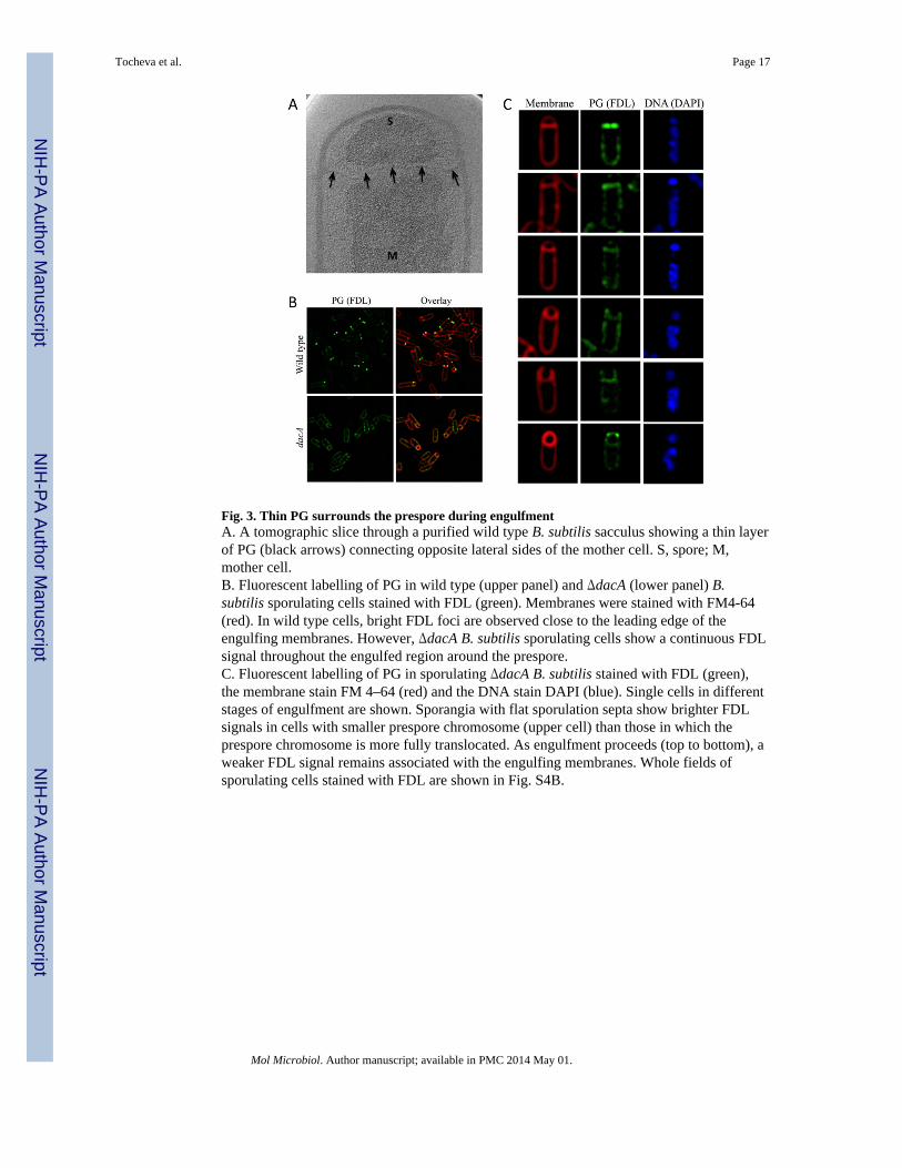
Fig. 3. Thin PG surrounds the prespore during engulfmentA. A tomographic slice through a purified wild type B. subtilis sacculus showing a thin layerof PG (black arrows) connecting opposite lateral sides of the mother cell. S, spore; M,mother cell.B. Fluorescent labelling of PG in wild type (upper panel) and ΔdacA (lower panel) B.subtilis sporulating cells stained with FDL (green). Membranes were stained with FM4-64(red). In wild type cells, bright FDL foci are observed close to the leading edge of theengulfing membranes. However, ΔdacA B. subtilis sporulating cells show a continuous FDLsignal throughout the engulfed region around the prespore.C. Fluorescent labelling of PG in sporulating ΔdacA B. subtilis stained with FDL (green),the membrane stain FM 4–64 (red) and the DNA stain DAPI (blue). Single cells in differentstages of engulfment are shown. Sporangia with flat sporulation septa show brighter FDLsignals in cells with smaller prespore chromosome (upper cell) than those in which theprespore chromosome is more fully translocated. As engulfment proceeds (top to bottom), aweaker FDL signal remains associated with the engulfing membranes. Whole fields ofsporulating cells stained with FDL are shown in Fig. S4B.
Tocheva et al. Page 17
Mol Microbiol. Author manuscript; available in PMC 2014 May 01.
NIH
-PA Author Manuscript
NIH
-PA Author Manuscript
NIH
-PA Author Manuscript

Fig. 4.A model for PG hydrolysis and synthesis during (A) vegetative septation and (B)engulfment. Schematics represent the boxed areas in the tomographic slices. Membranes areshown in black, glycan strands are viewed end-on (circled X’s) interconnected by peptidebonds (lines). Older glycan strands and cross-links are shown in grey, new glycan strandsand cross-links are shown in red, PG synthases are green and PG hydrolases are blue.During vegetative septation, new PG synthesis is thought to push the cytoplasmic membranetowards the middle of the cell. Hydrolysis occurs after septation from the outside. Similarly,we propose that during engulfment, co-ordinated PG synthesis at the ‘front’ and hydrolysisat the ‘back’ of the septal junction may drive membrane migration towards the pole. Blackarrow indicates PG synthesis, white arrow points to average thickness of vegetative cellwall.
Tocheva et al. Page 18
Mol Microbiol. Author manuscript; available in PMC 2014 May 01.
NIH
-PA Author Manuscript
NIH
-PA Author Manuscript
NIH
-PA Author Manuscript

Fig. 5.Continuous PG and cortex model during sporulation in A. longum (dashed line) and B.subtilis (solid line). The Gram-negative A. longum synthesizes a thin layer of PG duringvegetative growth and engulfment. During spore maturation, the thin PG is elaborated into athick cortex which gets degraded back to thin PG during outgrowth. B. subtilis has a thickcell wall during vegetative growth, which thins during engulfment. The thin PG iselaborated into a thick cortex during spore maturation and remains thick during outgrowth(where the ICx becomes the vegetative PG). The two cells overlap in their PG/cortexmorphologies during engulfment and spore maturation and the major mechanistic differencebetween the two cell types likely occurs upon germination (asterisk). Tomographic slicesfrom B. subtilis (top) and A. longum (bottom) cells at different stages of sporulationrepresent the major PG transformations. PG is marked as dotted red line and membranes aretraced as solid blue lines.
Tocheva et al. Page 19
Mol Microbiol. Author manuscript; available in PMC 2014 May 01.
NIH
-PA Author Manuscript
NIH
-PA Author Manuscript
NIH
-PA Author Manuscript