nidcd strategic plan 2017-2021 · nidcd strategic plan 2017-2021 hearing ... earlier devices based...
TRANSCRIPT
NIDCD Strategic Plan2017-2021
Hearing and Balance
Taste and Smell
Voice, Speech, and Language
2017-2021 NIDCD Strategic Plan2
Table of Contents
WELCOME FROM THE DIRECTOR 3
Science Capsule: Advances in Hearing Aid Research 4
INTRODUCTION 6
NIDCD Overview 6
NIDCD Strategic Plan and Priority Setting 7
Enhance Scientific Stewardship at the NIDCD 8
Shared Databases, Registries, and Metrics on Communication Disorders 9
Trans-NIH Efforts Encourage Innovation Through Partnerships 10
Excel as a Federal Science Agency by Managing for Results 10
FUTURE DIRECTIONS IN NIDCD PROGRAM AREAS 12
HEARING AND BALANCE RESEARCH 13
Why the NIDCD Supports Hearing and Balance Research 13
The Hearing and Balance Program 16
Recent Advances in Hearing and Balance Research 16
Science Capsule: Balance or Vestibular Disorders in Adults 21
Priority Areas in Hearing and Balance Research 22
TASTE AND SMELL RESEARCH 27
Why the NIDCD Supports Taste and Smell Research 27
The Taste and Smell Program 31
Recent Advances in Taste and Smell Research 31
Science Capsule: How Mosquitoes Target their Human Hosts 34
Priority Areas in Taste and Smell Research 35
VOICE, SPEECH, AND LANGUAGE RESEARCH 38
Why the NIDCD Supports Voice, Speech, and Language Research 38
The Voice, Speech, and Language Program 41
Recent Advances in Voice, Speech, and Language Research 41
Science Capsule: Spasmodic Dysphonia 46
Priority Areas in Voice, Speech, and Language Research 47
SUMMARY 51
APPENDIX A: NIDCD FUNDING HISTORY 52
APPENDIX B: THE NIDCD 2017-2021 STRATEGIC PLAN: THE PROCESS 54
APPENDIX C: NIDCD’S TRANS-NIH AND TRANS-AGENCY ACTIVITIES 57
APPENDIX D: GLOSSARY AND ACRONYM LIST 61
APPENDIX E: BIBLIOGRAPHY 66
2017-2021 NIDCD Strategic Plan3
Welcome from the Director
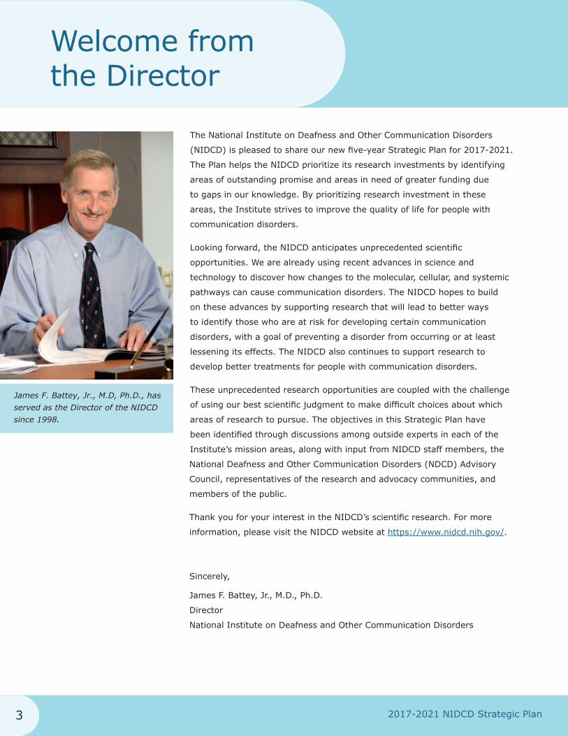
James F. Battey, Jr., M.D, Ph.D., has served as the Director of the NIDCD since 1998.
The National Institute on Deafness and Other Communication Disorders
(NIDCD) is pleased to share our new five-year Strategic Plan for 2017-2021.
The Plan helps the NIDCD prioritize its research investments by identifying
areas of outstanding promise and areas in need of greater funding due
to gaps in our knowledge. By prioritizing research investment in these
areas, the Institute strives to improve the quality of life for people with
communication disorders.
Looking forward, the NIDCD anticipates unprecedented scientific
opportunities. We are already using recent advances in science and
technology to discover how changes to the molecular, cellular, and systemic
pathways can cause communication disorders. The NIDCD hopes to build
on these advances by supporting research that will lead to better ways
to identify those who are at risk for developing certain communication
disorders, with a goal of preventing a disorder from occurring or at least
lessening its effects. The NIDCD also continues to support research to
develop better treatments for people with communication disorders.
These unprecedented research opportunities are coupled with the challenge
of using our best scientific judgment to make difficult choices about which
areas of research to pursue. The objectives in this Strategic Plan have
been identified through discussions among outside experts in each of the
Institute’s mission areas, along with input from NIDCD staff members, the
National Deafness and Other Communication Disorders (NDCD) Advisory
Council, representatives of the research and advocacy communities, and
members of the public.
Thank you for your interest in the NIDCD’s scientific research. For more
information, please visit the NIDCD website at https://www.nidcd.nih.gov/.
Sincerely,
James F. Battey, Jr., M.D., Ph.D.
Director
National Institute on Deafness and Other Communication Disorders
Science Capsule:Advances in Hearing Aid Research
Nearly 15 percent of American adults (37.5 million) aged 18 and over report some trouble
hearing, making this one of the most prevalent disabling conditions in the U.S. Hearing loss
can be hereditary, or it can result from disease, trauma, medications, or long-term exposure to
damaging noise. The condition can vary from a mild but important loss of sensitivity to a total
loss of hearing.
Sensorineural hearing loss is caused by a problem in the cochlea or the auditory nerve, which
are parts of the ear that help sound impulses reach the brain. Hearing loss affects people of all
ages, in all segments of the population, and across all socioeconomic levels. It can interfere with
an individual’s physical, cognitive, behavioral, and social functions, and hearing aids are the main
form of treatment. However, of adults aged 70 and older with hearing loss who could benefit
from wearing hearing aids, fewer than 30 percent have ever used them. Of adults aged 20 to 69
who could benefit from hearing aids, the proportion that has used them is even lower (only about
16 percent).
A hearing aid works by amplifying sound to allow people to hear sounds that would not be
audible. In specially equipped movie theaters, auditoriums, lecture halls, places of worship,
and other areas, people can use a hearing aid to access “hearing loop” wireless signals that are
beamed directly to the aid to bypass background noises. A vast array of hearing aid technology
is available to provide additional features, such as the telecoil needed to pick up the hearing loop
wireless signal.
Although the development of microelectronic components
has enabled new digital hearing aid technology to replace
earlier devices based on analog circuits, the underlying
damage to the inner ear remains a limitation when the user
is confronted by multiple speakers or background noise.
Hearing aid users often complain of straining to focus on a
single speech sound among competing sources at meetings,
banquets, and sporting events. One solution to this problem
is to move the hearing aid user closer to the person speaking
and farther from the noise sources. Directional microphones
offer another approach to do the same thing simply by
pointing a device.
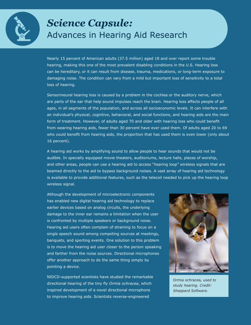
Ormia ochracea, used to study hearing. Credit: Sheppard Software.
NIDCD-supported scientists have studied the remarkable
directional hearing of the tiny fly Ormia ochracea, which
inspired development of a novel directional microphone
to improve hearing aids. Scientists reverse-engineered
the physics and biology behind the fly’s abilities to localize sound and provided engineers with
strategies to improve directional microphones that are small enough to use in hearing aids and
help focus the aid on one sound source at a time.
Capitalizing on the knowledge learned from studying Ormia, another group of NIDCD-supported
scientists successfully completed design and testing of a novel microphone based on these design
elements. The scientists used silicon microfabrication technology to build the critical sensing
elements needed for a functional microphone, characterize its function, and prove it had the
capability to provide performance gains over existing designs.
Other NIDCD-supported scientists have continued research and development efforts based
on this proof of concept prototype by adapting the microphone design into a form that could
be more readily incorporated in a hearing aid. The scientists are the first to use piezoelectric
materials, which turn mechanical pressure into electrical signals (voltage) and allow the
microphone to operate with very little power.
Because hearing aids rely on batteries, minimizing
power consumption is a crucial design requirement.
The NIDCD recognizes that the needs of the
majority of adults with hearing loss are not being
met, and the cost and accessibility of hearing aids
are considered part of the barriers to care. In
response, the NIDCD is working to fill this need
by supporting research or infrastructure that will
lead to more accessible and affordable hearing
health care for adults. The NIDCD cosponsored a
consensus development study with the National
Academies of Sciences, Engineering, and
Medicine to consider hearing health care from the
health care and population health perspectives,
including the regulatory environment, access,
and affordability. By identifying the research gaps
related to effective and affordable hearing health
care, devices, and compliance, and by developing
novel strategies to overcome these gaps, NIDCD
clinical and translational research will endeavor to
improve the quality of life for millions of Americans
with hearing loss.
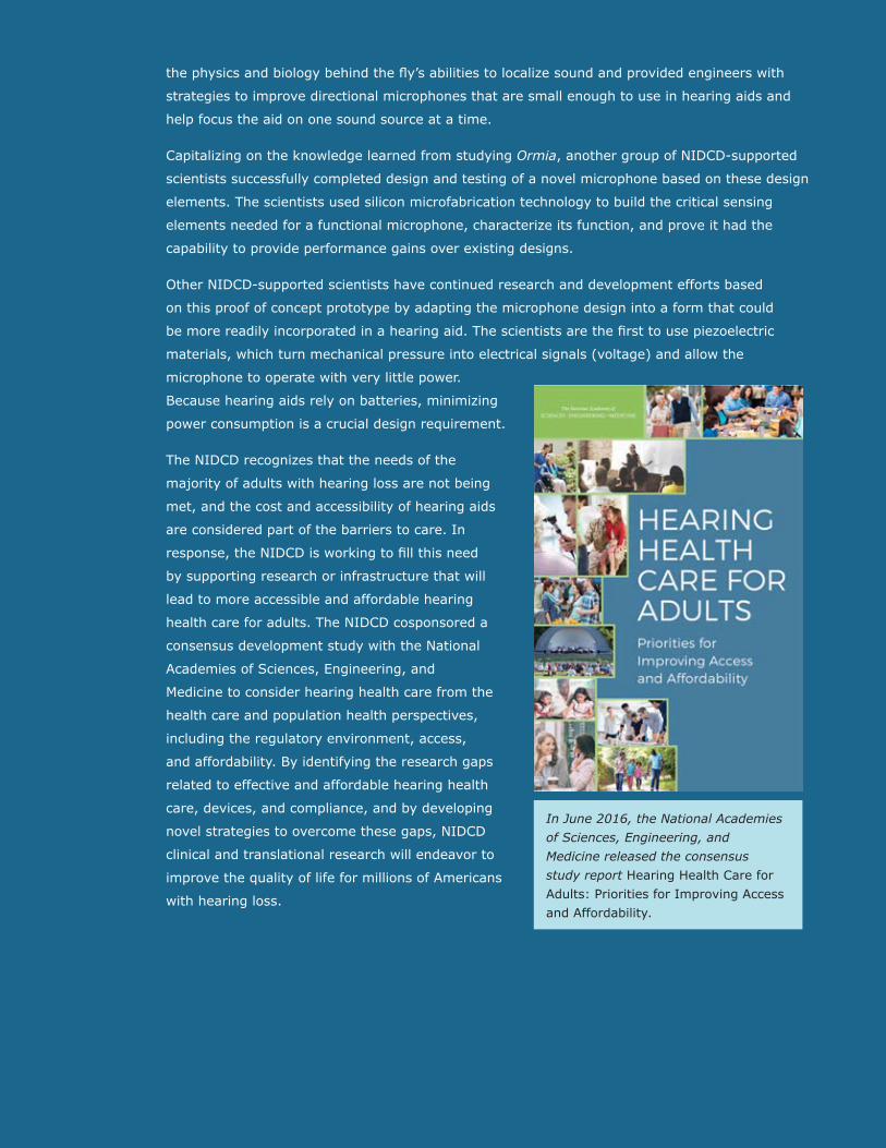
In June 2016, the National Academies of Sciences, Engineering, and Medicine released the consensus study report Hearing Health Care for Adults: Priorities for Improving Access and Affordability.
2017-2021 NIDCD Strategic Plan6
Introduction
NIDCD Overview
Approximately 46 million Americans experience some form of communication disorder. Communication
disorders make the basic components of communication (sensing, interpreting, and responding to people and
things in our environment) challenging. In addition, communication disorders not only compromise physical
health, but also affect the emotional, social, recreational, educational, and vocational aspects of life. The
effects often ripple outward to affect families and social networks, including those at work and school. The
total economic impact of these disorders in regards to quality of life and unfulfilled potential is substantial.
Furthermore, the prevalence of communication disorders is expected to increase as the population ages, and
as survival rates improve for medically fragile infants and people affected by traumatic injuries and diseases.
In October 1988, Congress established the National Institute on Deafness and Other Communication Disorders
(NIDCD) as one of the institutes that compose the National Institutes of Health (NIH), part of the U.S.
Department of Health and Human Services. The NIH is the federal government’s focal point for the support of
biomedical research and is among the leading
biomedical research funding institutions in the
world. NIH’s mission is to seek fundamental
knowledge about the nature and behavior of
living systems and to apply that knowledge to
enhance health, lengthen life, and reduce the
burdens of illness and disability. NIDCD’s focus
within this broad mission is to bring national
attention to the disorders and dysfunctions of
human communication and to contribute to
advances in biomedical and behavioral research
that will improve the lives of the millions of
people with a communication disorder.
The NIDCD mission is to conduct and support
biomedical research, behavioral research, and
research training in the normal and disordered
processes of hearing, balance, taste, smell,
voice, speech, and language.
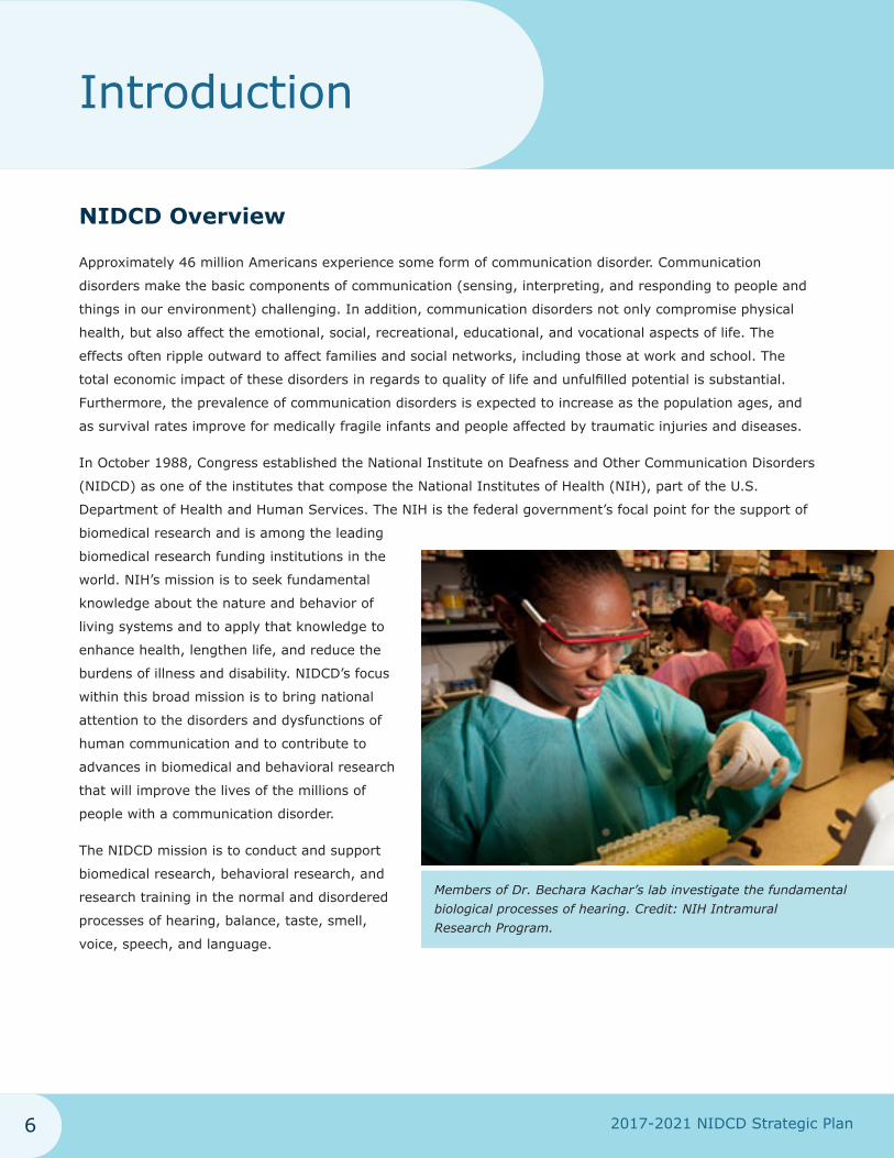
Members of Dr. Bechara Kachar’s lab investigate the fundamental biological processes of hearing. Credit: NIH Intramural Research Program.
2017-2021 NIDCD Strategic Plan7
Introduction
The Institute conducts and supports research and
research training related to disease prevention and
health promotion; addresses special biomedical and
behavioral problems associated with people who have
communication impairments or disorders; supports
research evaluating approaches to the identification
and treatment of communication disorders and
patient outcomes; and supports efforts to create
devices that substitute for lost and impaired sensory
and communication function. A group of NIDCD clinician-scientists discuss findings.
To accomplish these goals, the NIDCD manages a
broad portfolio of both basic and clinical research. The
portfolio is organized into three program areas: hearing and balance; taste and smell; and voice, speech, and
language. The three program areas seek to answer fundamental scientific questions about normal function
and disorders and to identify patient-oriented scientific discoveries for preventing, screening, diagnosing, and
treating disorders of human communication. See Appendix A for the NIDCD Funding History.
The NIDCD accomplishes its research mission through three divisions: the Division of Intramural Research
(DIR), the Division of Scientific Programs (DSP), and the Division of Extramural Activities (DEA). The DIR
conducts research and related support activities in laboratories and clinics housed at the NIH. The DSP and
DEA manage complementary aspects of the NIDCD’s Extramural Research Program, a program of research
grants, career development awards, individual and institutional research training awards, center grants, and
contracts to public and private research institutions and organizations throughout the U.S. and abroad. As
a whole, the Institute supported approximately 1,300 research grants, training awards, and research and
development contracts in Fiscal Year (FY) 2016. Through research and education, the NIDCD strives to reduce
both the direct and indirect economic burden of communication disorders on individuals, families, and society,
thereby improving the quality of life for people living with a communication disorder.
NIDCD Strategic Plan and Priority Setting
The NIDCD uses the NIH system of peer review to evaluate research grant applications. The system depends
on scientists to submit their best research ideas to drive the spectrum of supported research. The NIH is
committed to a transparent, evidence-based process of structured peer review. A panel of scientific experts
from outside of the NIH (who work in the same or a related academic field) scrutinize grant applications. To
identify research ideas with the highest overall potential impact, the panel evaluates applications for approach,
significance, innovation, investigator(s), and quality of the academic environment. This system helps NIH
select the most promising ideas to receive federal funding. To learn more about the NIH peer review process,
see http://grants.nih.gov/grants/peer/peer.htm. To learn how NIH continuously reviews and updates its peer
review process, see http://grants.nih.gov/grants/peer/continuous_review.htm.
2017-2021 NIDCD Strategic Plan8
Introduction
The NIDCD values investigator-initiated applications submitted to NIH that help achieve the NIDCD mission.
In particular, the Institute encourages investigators to submit applications for research projects that directly
address priorities within the NIDCD Strategic Plan (Plan). The NIDCD also uses the Plan to develop targeted
Funding Opportunity Announcements (FOAs) to stimulate research applications that address a particular and
much-needed area of science.
The NIDCD Strategic Plan helps the Institute (including NIDCD staff and the NDCD Advisory Council) prioritize
research investment. The Plan helps identify investigator-initiated research proposals for High Program Priority
(HPP) funding so that these projects, if funded, will address a significant research need in the NIDCD portfolio.
The NIDCD uses its HPP process to fill scientific gaps in the research portfolio, foster the entry of new
investigators, encourage innovative research, and increase the diversity of the scientists who lead a research
team, known as Principal Investigators (PIs).
NIDCD staff distribute the Plan to the research community at workshops and scientific conferences to increase
awareness of Institute priorities. Additionally, the Plan informs the public about the state of the science and
advances in diagnosis and treatment of communication disorders, while creating a vision for the future. To
develop the 2017-2021 Plan, the NIDCD convened a series of working group meetings and solicited input from
scientific experts, the NDCD Advisory Council, NIDCD staff, and the public. See Appendix B for more details on
the Plan process.
Enhance Scientific Stewardship at the NIDCD
Research Training and Career Development at the NIDCD
The number of Americans with communication disorders is expected to rise as the nation’s older population
increases and as survival rates improve for a wide range of medical conditions associated with communication
disorders. As such, the NIDCD recognizes the importance of research training and career development
opportunities to ensure a productive, creative, and innovative cadre of qualified scientists in basic, clinical,
and translational research. The NIDCD is continuously adapting its research training and career development
efforts to help new scientists establish careers in our mission areas, encourage clinicians to pursue
opportunities in translational research, and build shared research resources.
The field of human communication sciences needs interdisciplinary research teams of clinicians and
basic scientists to bridge the gap between laboratory research and patient care. Clinicians need a deeper
understanding of the latest research discoveries to bring new diagnostic and treatment approaches into the
clinic. Basic researchers need a thorough understanding of the needs, challenges, and opportunities faced by
clinicians. The NIDCD believes that cross training these scientists could spark new ways to better prevent,
detect, and treat communication and chemosensory disorders. Interdisciplinary teams of basic scientists and
clinicians—including physicians, surgeons, and audiologists—will then be able to initiate and support new
directions for scientific discovery, conduct hypothesis-driven clinical trials, assess new diagnostic tools and
interventions, and improve public health and well-being.
2017-2021 NIDCD Strategic Plan9
Introduction
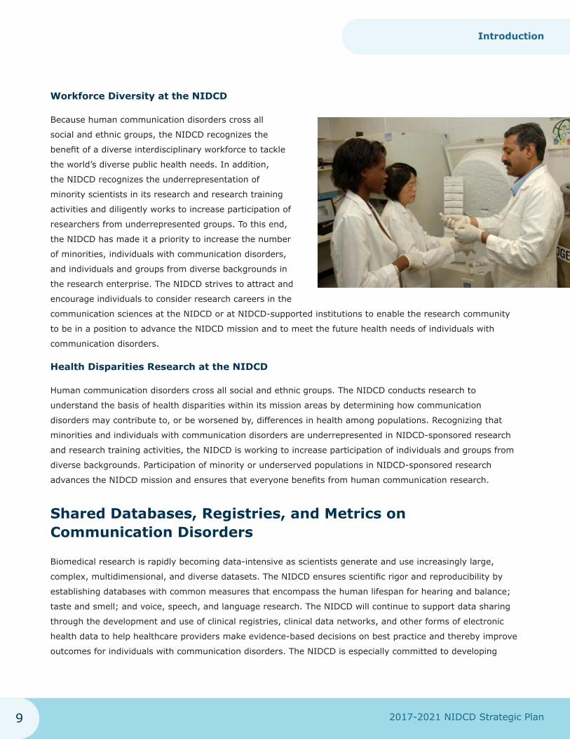
Workforce Diversity at the NIDCD
Because human communication disorders cross all
social and ethnic groups, the NIDCD recognizes the
benefit of a diverse interdisciplinary workforce to tackle
the world’s diverse public health needs. In addition,
the NIDCD recognizes the underrepresentation of
minority scientists in its research and research training
activities and diligently works to increase participation of
researchers from underrepresented groups. To this end,
the NIDCD has made it a priority to increase the number
of minorities, individuals with communication disorders,
and individuals and groups from diverse backgrounds in
the research enterprise. The NIDCD strives to attract and
encourage individuals to consider research careers in the
communication sciences at the NIDCD or at NIDCD-supported institutions to enable the research community
to be in a position to advance the NIDCD mission and to meet the future health needs of individuals with
communication disorders.
Health Disparities Research at the NIDCD
Human communication disorders cross all social and ethnic groups. The NIDCD conducts research to
understand the basis of health disparities within its mission areas by determining how communication
disorders may contribute to, or be worsened by, differences in health among populations. Recognizing that
minorities and individuals with communication disorders are underrepresented in NIDCD-sponsored research
and research training activities, the NIDCD is working to increase participation of individuals and groups from
diverse backgrounds. Participation of minority or underserved populations in NIDCD-sponsored research
advances the NIDCD mission and ensures that everyone benefits from human communication research.
Shared Databases, Registries, and Metrics on Communication Disorders
Biomedical research is rapidly becoming data-intensive as scientists generate and use increasingly large,
complex, multidimensional, and diverse datasets. The NIDCD ensures scientific rigor and reproducibility by
establishing databases with common measures that encompass the human lifespan for hearing and balance;
taste and smell; and voice, speech, and language research. The NIDCD will continue to support data sharing
through the development and use of clinical registries, clinical data networks, and other forms of electronic
health data to help healthcare providers make evidence-based decisions on best practice and thereby improve
outcomes for individuals with communication disorders. The NIDCD is especially committed to developing
2017-2021 NIDCD Strategic Plan10
Introduction
and implementing infrastructure to identify: 1) investigators with expertise in epidemiology, data registry,
clinical trials, and other clinical research and 2) academic- and community-based clinical practice settings with
geographical, racial, and ethnic diversity to facilitate rigorous, cost-effective clinical research and maximize
human subjects’ protection.
By establishing standard metrics in anatomical, acoustical, and physiological measures, researchers can
better define functional communication abilities under real-world conditions. The NIDCD will support new
and enhance existing centralized tissue and cell banks to aid access to biological source materials. Standard
metrics and centralized tissue banks also help researchers to differentiate clinical subtypes and to identify
early preclinical pathology. To improve communication among scientists and clinicians with different
specialties, the NIDCD supports development of better measures of performance, communication abilities,
disease-specific quality of life instruments, assessment of communication impairments, and outcomes of
individuals with communication disorders.
Trans-NIH Efforts Encourage Innovation Through Partnerships
While the NIDCD focuses its research efforts on programs that support its mission areas, breakthroughs in
related areas, such as neuroscience, genetics, and animal model development, improve our understanding
of communication disorders and encourage innovation through partnerships. To support these discoveries,
the NIDCD participates in many Trans-NIH initiatives and programs. See Appendix C for examples of
Trans-NIH activities.
Excel as a Federal Science Agency by Managing for Results
The NIDCD is a public science agency supported by federal funds. As part of the NIH, the NIDCD is obligated
to base its decisions on science, and to make its decision-making process transparent. The NIDCD upholds
its accountability to the American public by managing its scientific endeavors with an eye towards achieving
results that improve the health of individuals with communication disorders. The NIDCD approaches this
responsibility in several different ways, from its reporting as required by a U.S. Law called the Government
Performance and Results Act (GPRA), to developing an administrative strategic plan to complement this NIDCD
Strategic Plan, and by mitigating the risks involved with administering the NIDCD mission.
GPRA is a U.S. law enacted in 1993. It is designed to improve government performance management,
and it requires agencies to manage their performance by setting goals, measuring results, and reporting
their progress. To comply with GPRA, the NIH develops an annual plan proposing goals that provide a
representative sample of NIH’s activities for each year and describes how these goals will be met, and later
in the fiscal year, NIH provides evidence to support any claims for successful achievement of the goals. Each
Institute and Center at NIH participates in the GPRA reporting process, including the NIDCD.
2017-2021 NIDCD Strategic Plan11
Introduction
The NIDCD’s goal represents only one snapshot of NIDCD’s entire portfolio, but aligns with our Mission to
improve the lives of people with communication disorders. The current NIDCD GPRA goal began in FY 2015
and states: By 2020, increase the number of potential treatment options for communication disorders that are
being tested in clinical trials by adding one new treatment option per year. To comply with GPRA obligations
for this particular goal under the law, the NIDCD proposes a distinct new treatment option that will be tested
each fiscal year and then, at the end of that fiscal year, the NIDCD submits evidence that we have tested a
new treatment option for a communication disorder. The NIH compiles NIDCD’s annual submission with those
from all of the other NIH Institutes and Centers and presents it to the Office of Management and Budget
(OMB). OMB includes the NIH information in an annual report on government agency performance that
accompanies the President’s annual budget request.
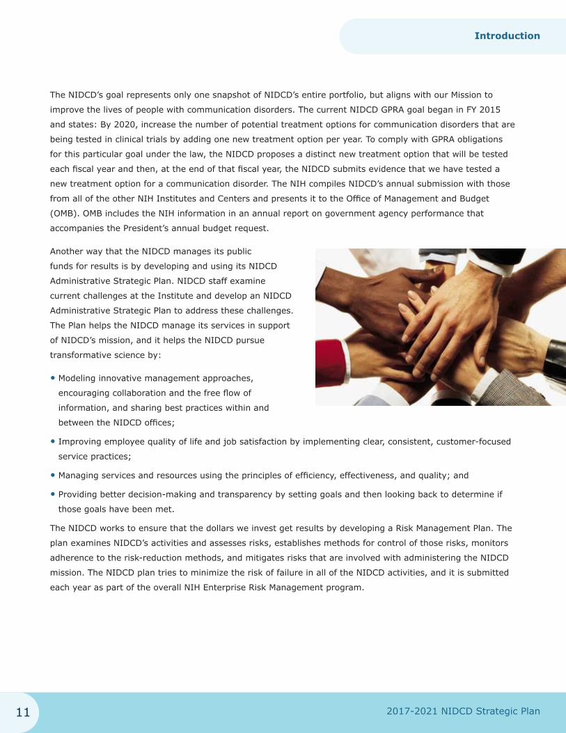
Another way that the NIDCD manages its public
funds for results is by developing and using its NIDCD
Administrative Strategic Plan. NIDCD staff examine
current challenges at the Institute and develop an NIDCD
Administrative Strategic Plan to address these challenges.
The Plan helps the NIDCD manage its services in support
of NIDCD’s mission, and it helps the NIDCD pursue
transformative science by:
• Modeling innovative management approaches,
encouraging collaboration and the free flow of
information, and sharing best practices within and
between the NIDCD offices;
• Improving employee quality of life and job satisfaction by implementing clear, consistent, customer-focused
service practices;
• Managing services and resources using the principles of efficiency, effectiveness, and quality; and
• Providing better decision-making and transparency by setting goals and then looking back to determine if
those goals have been met.
The NIDCD works to ensure that the dollars we invest get results by developing a Risk Management Plan. The
plan examines NIDCD’s activities and assesses risks, establishes methods for control of those risks, monitors
adherence to the risk-reduction methods, and mitigates risks that are involved with administering the NIDCD
mission. The NIDCD plan tries to minimize the risk of failure in all of the NIDCD activities, and it is submitted
each year as part of the overall NIH Enterprise Risk Management program.
2017-2021 NIDCD Strategic Plan12
Future Directions in NIDCD Program Areas
In consultation with communication research scientists and the public, the NIDCD has identified four
Priority Areas that have the potential to increase our understanding of the normal and disordered processes
of hearing, balance, taste, smell, voice, speech, and language and to further our knowledge in human
communication sciences.
Priority Area 1: Understanding Normal Function
Deepen our understanding of the mechanisms underlying normal function of the systems of human
communication. By defining what is normal in both animal models and humans, we can better understand
mechanisms of disease.
Priority Area 2: Understanding Diseases and Disorders
Increase our knowledge of the mechanisms of diseases, disorders, and dysfunctions that impair human
communication and health. Understanding mechanisms that underlie diseases and disorders is an important
step in developing better prevention and treatment strategies.
Priority Area 3: Improving Diagnosis, Treatment, and Prevention
Develop, test, and improve diagnosis, treatment, and prevention of diseases, disorders, and dysfunctions
of human communication and health. Diagnosis considers normal function and provides targets for
prevention and treatment. Improvements in prevention and treatment lead to better outcomes and guide
treatment options.
Priority Area 4: Improving Outcomes for Human Communication
Accelerate the translation of research discoveries into practice; increase access to health care; and enhance
the delivery, quality, and effectiveness of care to improve personal and public health. Scientifically validated
prevention and treatment models will lead to better personal and public health only after adoption into
routine practice.
Although the Priority Areas described in this Plan will help the NIDCD identify promising scientific
opportunities to advance human communication research over the next five years, the Plan is not meant to
be a comprehensive list of all research areas that the NIDCD is currently supporting or plans to support in
the future.
The NIDCD will continue to fund as much meritorious research as possible within our program areas of hearing
and balance; taste and smell; and voice, speech, and language. Basic and clinical research being supported
by the NIDCD will continue to be given high priority. The Institute is committed to supporting new, innovative,
hypothesis-driven, meritorious research that can enhance the overall health and quality of life of people with
communication disorders.
2017-2021 NIDCD Strategic Plan13
Hearing and Balance Research
Hearing and Balance Research
Why the NIDCD Supports Hearing and Balance Research
Loss of hearing or balance negatively impacts quality of life and imposes a significant social and
economic burden upon individuals, their families, and the communities in which they live. Millions of
Americans experience a hearing or balance disorder at some point in their life, especially as young
children or older adults. Common examples include middle-ear infections (otitis media), noise-
induced hearing loss, tinnitus, age-related hearing loss, dizziness, and vertigo. Hearing and balance
disorders cross all ethnic and socioeconomic lines. Approximately 37.5 million American adults report
some degree of hearing loss and 33.4 million adults report a problem during the past 12 months with
dizziness or balance, such as vertigo, unsteadiness, or blurred vision after moving the head.1, 2 Among
the younger age group, an additional 5.3 percent of American children (3.3 million) also experienced
balance and dizziness problems in the last 12 months, as reported by their parents or other adult
caregivers.3-6 About two to three of every 1,000 children in the U.S. are born with a detectable level of
hearing loss in one or both ears that can affect speech, language, social, and cognitive development.4, 5
In 2014, one in six U.S. adults aged 18 and older reports trouble hearing without a hearing aid.6
2017-2021 NIDCD Strategic Plan14
Hearing and Balance Research
Noise-Induced Hearing Loss
Excess noise is a major contributor to hearing loss in the U.S. Based on nationally representative hearing
exam surveys (1999-2004), an estimated 15 percent of Americans aged 20 to 69, or 26 million Americans,
reported a history of loud noise exposure and also had high-frequency audiogram results suggesting exposure
to excess noise.7 Recent animal studies suggest that noise exposure causing temporary measurable hearing
loss may also cause permanent hearing loss that is not readily detectable using standard audiometric testing.
Such damage may underlie the common complaint of having difficulty in understanding speech in noisy
situations. The NIDCD encourages research to better understand noise-induced auditory damage to inform
potential therapies.
Otitis Media
Otitis media (OM), or middle ear infection, is a condition that affects most young children before three years of
age. Repeated episodes of OM can contribute to hearing loss and possibly delay language and cognitive skills
development. NIDCD-supported research is improving our understanding of susceptibility and pathogenesis of
OM. In the future, this research might identify immune pathways to guide effective OM vaccine development.
Age-Related Hearing Loss
Age-related hearing loss (presbycusis) is the loss of hearing that gradually occurs during aging. It is one of the
most common conditions affecting older and elderly adults with approximately one in three people in the U.S.
aged 65 to 74 exhibiting a hearing loss, and nearly half of those older than 75 have difficulty hearing.8 There
are many causes of age-related hearing loss. Most commonly, it arises from changes in the inner ear, but it
can also result from complex changes along the nerve pathways from the ear to the brain. Understanding the
cause of age-related hearing loss and finding ways to prevent it are important research areas supported by
the NIDCD.
Tinnitus
Tinnitus, or ringing in the ears, is a disorder that affects approximately 25 million Americans, many of whom
also have hearing loss. Severity can range from a mild condition, which requires no intervention, to a severe
debilitating disease with significant emotional, social, and economic impact. NIDCD-supported research aims
to determine the neural basis of tinnitus, and to develop effective interventions for affected people.
Technology Interventions for Hearing Loss
Individuals with mild-to-severe hearing loss can benefit from using a hearing aid, and many with severe to
profound hearing loss benefit from having a cochlear implant. Advances in both hearing aid and cochlear
implant technology are improving treatment options for many people with various degrees of hearing loss.
For example, individuals may be fitted with hearing aids or cochlear implants on both ears instead of only
one ear to improve sound localization and discrimination. In recent years, some people with residual hearing
for low-frequency sounds have received both a cochlear implant, to aid them in hearing higher frequency
2017-2021 NIDCD Strategic Plan15
Hearing and Balance Research
sounds, and a hearing aid to allow them to take advantage of their residual low-frequency hearing. In many
cases, this combination (‘hybrid’) strategy results in a significant improvement when listening to speech in
background noise.
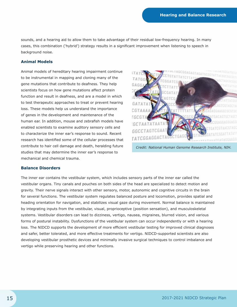
Animal Models
Animal models of hereditary hearing impairment continue
to be instrumental in mapping and cloning many of the
gene mutations that contribute to deafness. They help
scientists focus on how gene mutations affect protein
function and result in deafness, and are a model in which
to test therapeutic approaches to treat or prevent hearing
loss. These models help us understand the importance
of genes in the development and maintenance of the
human ear. In addition, mouse and zebrafish models have
enabled scientists to examine auditory sensory cells and
to characterize the inner ear’s response to sound. Recent
research has identified some of the cellular processes that
contribute to hair cell damage and death, heralding future
studies that may determine the inner ear’s response to
mechanical and chemical trauma.
Credit: National Human Genome Research Institute, NIH.
Balance Disorders
The inner ear contains the vestibular system, which includes sensory parts of the inner ear called the
vestibular organs. Tiny canals and pouches on both sides of the head are specialized to detect motion and
gravity. Their nerve signals interact with other sensory, motor, autonomic and cognitive circuits in the brain
for several functions. The vestibular system regulates balanced posture and locomotion, provides spatial and
heading orientation for navigation, and stabilizes visual gaze during movement. Normal balance is maintained
by integrating inputs from the vestibular, visual, proprioceptive (position sensation), and musculoskeletal
systems. Vestibular disorders can lead to dizziness, vertigo, nausea, migraines, blurred vision, and various
forms of postural instability. Dysfunctions of the vestibular system can occur independently or with a hearing
loss. The NIDCD supports the development of more efficient vestibular testing for improved clinical diagnoses
and safer, better tolerated, and more effective treatments for vertigo. NIDCD-supported scientists are also
developing vestibular prosthetic devices and minimally invasive surgical techniques to control imbalance and
vertigo while preserving hearing and other functions.
2017-2021 NIDCD Strategic Plan16
Hearing and Balance Research
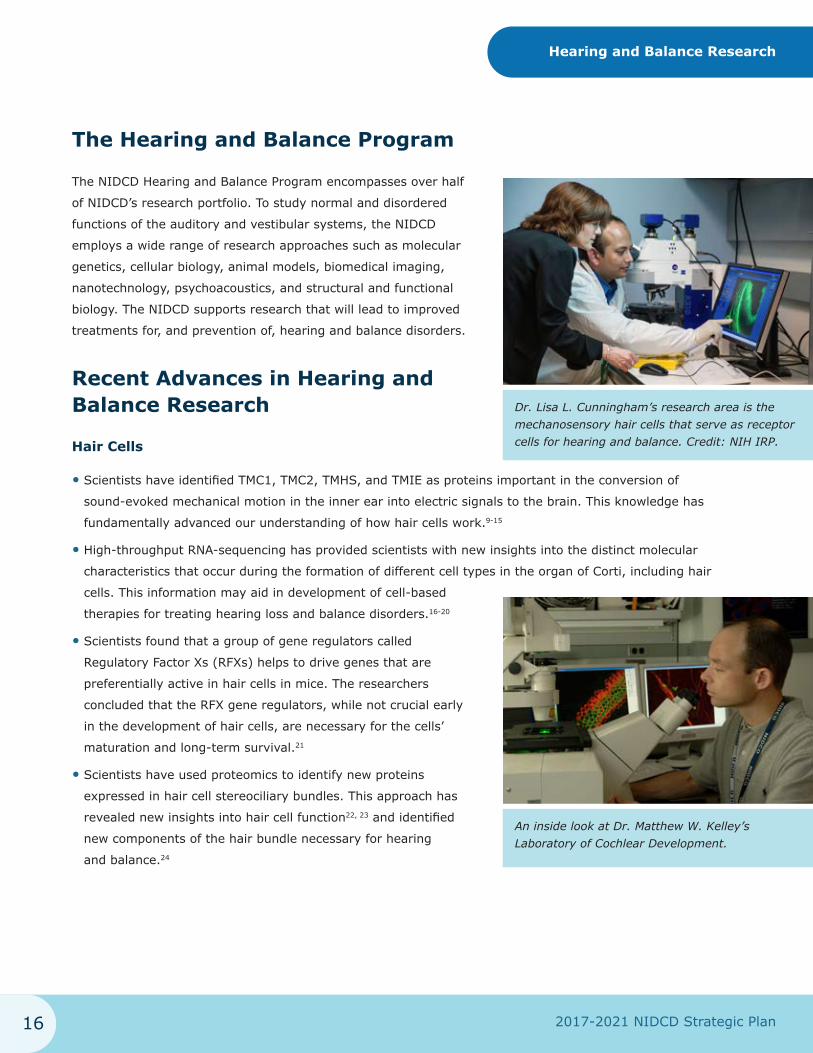
The Hearing and Balance Program
The NIDCD Hearing and Balance Program encompasses over half
of NIDCD’s research portfolio. To study normal and disordered
functions of the auditory and vestibular systems, the NIDCD
employs a wide range of research approaches such as molecular
genetics, cellular biology, animal models, biomedical imaging,
nanotechnology, psychoacoustics, and structural and functional
biology. The NIDCD supports research that will lead to improved
treatments for, and prevention of, hearing and balance disorders.
Dr. Lisa L. Cunningham’s research area is the mechanosensory hair cells that serve as receptor cells for hearing and balance. Credit: NIH IRP.
Recent Advances in Hearing and Balance Research
Hair Cells
• Scientists have identified TMC1, TMC2, TMHS, and TMIE as proteins important in the conversion of
sound-evoked mechanical motion in the inner ear into electric signals to the brain. This knowledge has
fundamentally advanced our understanding of how hair cells work.9-15
• High-throughput RNA-sequencing has provided scientists with new insights into the distinct molecular
characteristics that occur during the formation of different cell types in the organ of Corti, including hair
cells. This information may aid in development of cell-based
therapies for treating hearing loss and balance disorders.16-20
• Scientists found that a group of gene regulators called
Regulatory Factor Xs (RFXs) helps to drive genes that are
preferentially active in hair cells in mice. The researchers
concluded that the RFX gene regulators, while not crucial early
in the development of hair cells, are necessary for the cells’
maturation and long-term survival.21
• Scientists have used proteomics to identify new proteins
expressed in hair cell stereociliary bundles. This approach has
revealed new insights into hair cell function22, 23 and identified
new components of the hair bundle necessary for hearing
and balance.24
An inside look at Dr. Matthew W. Kelley’s Laboratory of Cochlear Development.
2017-2021 NIDCD Strategic Plan17
Hearing and Balance Research
Development and Regeneration
• Wnt signaling and Lgr5-expression have been shown to be key for the generation of hair cells in the
developing cochlea.25, 26
• Scientists have developed an in vitro technique to turn embryonic stem cells into inner ear hair cells
and supporting cells. This technique is well suited for high-throughput screening of drugs for hair cell
regeneration.27
• Antisense oligonucleotides have been used to rescue hearing and balance function in a mouse model of
human deafness.28
• In the research laboratory, it is now possible to prevent hearing loss and stimulate repair or regenerate
sensory cells of the inner ear by transdifferentiating or directly reprogramming cells, or by using gene
therapy in animal models.29-31
Hearing Loss
• Damage to spiral ganglion neurons or their synapses in the inner ear may contribute to hearing loss.
Scientists have discovered that the synapses between cochlear nerve fibers and inner hair cells are
the most vulnerable elements in noise-induced and age-related hearing loss and nerve fibers with high
response thresholds are the first to degenerate, which likely contributes to problems with hearing in noisy
environments.32-37
• Scientists have determined that unmyelinated type II sensory fibers innervating outer hair cells respond to
cellular damage resulting from loud sound and thus may serve as the nociceptors of the inner ear.38, 39
• Dozens of new gene defects responsible for hereditary hearing
loss have been identified in recent years, including mutations in
the first microRNA (miR-96) involved in hearing loss.40, 41
• The combination of using whole exome sequencing (a technique
for sequencing all the expressed genes in a genome) and hearing
testing is ushering in a new area of personalized diagnoses,
opportunity for earlier intervention, and ultimately, treatment for
individuals with hearing loss.42-50
• Gene therapy is being used to correct gene defects that cause
hereditary hearing loss and restore auditory function in animal
models.51-53
• The use of high-throughput screening in zebrafish is leading
to the discovery of new protective compounds that will
help diminish or prevent noise- or drug-induced hearing
impairment.54-57
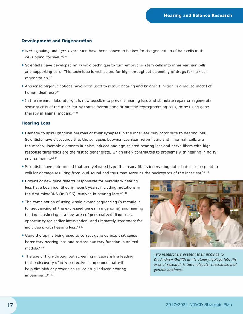
Two researchers present their findings to Dr. Andrew Griffith in his otolaryngology lab. His area of research is the molecular mechanisms of genetic deafness.
2017-2021 NIDCD Strategic Plan18
Hearing and Balance Research
• Proof-of-principle studies have shown that small molecules delivered to the cochlea after noise damage can
lead to some hair cell regeneration and some functional recovery.58
• Preliminary studies suggest that, in older adults, hearing impairment is associated with cognitive decline,
dementia, and depression. Estimated declines are greatest in participants who do not wear a hearing aid.
Although data do not currently support a causative relationship, they support future research on causation
and potential for reversal with interventions for treatment of hearing loss.59, 60
• Scientists have identified the genetic bases for accelerated
age-related hearing loss in humans.61
• Research has shown that genetically producing
overexpression of proteins called neurotrophins in the inner
ear can elicit regeneration of cochlear synapses after noise
damage.62
Otitis Media
• Research has advanced understanding of cell signaling and
gene expression patterns of the innate immune system in
response to an ear infection (otitis media).63, 64
• The study of microbial genomes has provided a cost-effective
and high-throughput tool to determine genome content of a
bacterium that causes ear infections.65
• Scientists have identified and characterized new vaccine candidates with the potential for preventing
ear infections.66-68
• To better treat ear infections, scientists have developed a new, noninvasive drug delivery system for the
administration of antibiotics and anti-inflammatory agents across the eardrum.69, 70
• Researchers have described how the inflammation induced
by bacterial infections treated with aminoglycoside antibiotics
potentiates the undesirable side effect of hearing loss.71
Hearing Aids
• Advanced digital technology hearing aids provide noise
reduction, directional hearing, and feedback suppression.
Binaural hearing aids further improve sound source
localization and spatial separation.72 Credit: The Ohio State University.
2017-2021 NIDCD Strategic Plan19
Hearing and Balance Research
Cochlear Implants and Other Implantable Hearing Devices
• Hybrid devices that combine both electric and acoustic stimulation
allow individuals with preserved low-frequency hearing and un-aidable
high frequency loss to utilize a combination device that includes a
cochlear implant for stimulation of high frequencies and a hearing aid
to enhance residual low frequency hearing.73-75
• Scientists are studying further expansion of cochlear implant candidacy
in individuals with unilateral deafness who received a cochlear
implant. They showed significant improvement in speech perception
performance in quiet and in noise after implantation.76 Another study
has shown the benefit of cochlear implants in reducing tinnitus in
individuals with unilateral hearing loss.77
• More focused electrical stimulation can improve performance for
existing cochlear implant users by limiting the overlap between the
number of neurons stimulated by different sound frequencies.78, 79
• For individuals in whom cochlear implantation is not an option,
auditory brainstem implants now offer an alternative.80
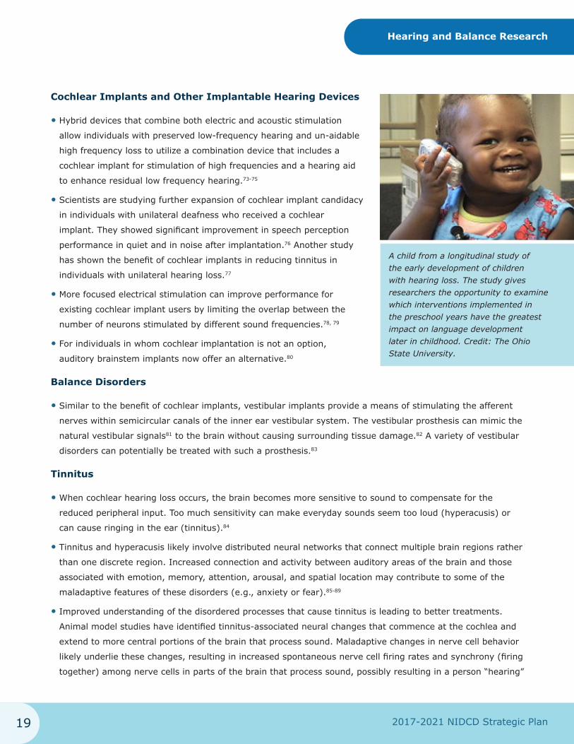
A child from a longitudinal study of the early development of children with hearing loss. The study gives researchers the opportunity to examine which interventions implemented in the preschool years have the greatest impact on language development later in childhood. Credit: The Ohio State University.
Balance Disorders
• Similar to the benefit of cochlear implants, vestibular implants provide a means of stimulating the afferent
nerves within semicircular canals of the inner ear vestibular system. The vestibular prosthesis can mimic the
natural vestibular signals81 to the brain without causing surrounding tissue damage.82 A variety of vestibular
disorders can potentially be treated with such a prosthesis.83
Tinnitus
• When cochlear hearing loss occurs, the brain becomes more sensitive to sound to compensate for the
reduced peripheral input. Too much sensitivity can make everyday sounds seem too loud (hyperacusis) or
can cause ringing in the ear (tinnitus).84
• Tinnitus and hyperacusis likely involve distributed neural networks that connect multiple brain regions rather
than one discrete region. Increased connection and activity between auditory areas of the brain and those
associated with emotion, memory, attention, arousal, and spatial location may contribute to some of the
maladaptive features of these disorders (e.g., anxiety or fear).85-89
• Improved understanding of the disordered processes that cause tinnitus is leading to better treatments.
Animal model studies have identified tinnitus-associated neural changes that commence at the cochlea and
extend to more central portions of the brain that process sound. Maladaptive changes in nerve cell behavior
likely underlie these changes, resulting in increased spontaneous nerve cell firing rates and synchrony (firing
together) among nerve cells in parts of the brain that process sound, possibly resulting in a person “hearing”
2017-2021 NIDCD Strategic Plan20
Hearing and Balance Research
a sound when no sound stimulus is present. Scientists are currently conducting clinical trials to test the
effectiveness of drugs that change the way nerve cells fire to treat acute tinnitus in people. Other new
approaches including brain stimulation, such as rTMS (repetitive transcranial magnetic stimulation),90 hold
some promise. Scientists have also had some success with vagal nerve stimulation to eliminate or minimize
abnormal nerve cell circuits in individuals with tinnitus. Research has shown that, after cochlear damage,
upregulation of somatosensory input to the cochlear nucleus may follow reduction in auditory nerve input,
resulting in heightened cochlear nucleus cell responses to somatosensory stimulation. Animals known to
have tinnitus have been shown to demonstrate changes in auditory-somatosensory integration, providing a
possible mechanism for the treatment of individuals with tinnitus.91, 92
Auditory and Vestibular Processing
• Scientists have been able to determine which speech stimuli cause brain activity by making
electrophysiological recordings from electrodes placed on the human brain’s surface. This advance has high
significance for the future development of objective ways to measure ability in the parts of the brain that
produce and process speech in individuals with normal hearing and hearing impairment.93-95
• Several studies have established that the auditory cortex represents only the sounds of interest and is less
affected by the presence of background noise than peripheral auditory neurons in the ears. These findings
are crucial for understanding the mechanisms for signal detection in unfavorable listening conditions and the
detrimental consequences of even mild hearing loss on those capacities.96-98
• Scientists have made important discoveries to describe the ion channels responsible for transmitting signals
to the brain that help us detect our balance and orientation in space.99, 100
• Scientists have integrated their study of auditory and vestibular activity with other sensory systems to
advance our understanding of how the nervous system combines and jointly encodes input of sound, sight,
and position to improve the ability to orient ourselves with objects around us, while maintaining gaze
and posture.101-106

Science Capsule: Balance or Vestibular Disorders in Adults
Balance disorders can result from trauma, disease, or the effects of aging on all the balance-
related systems. Vestibular dysfunction can lead to dizziness, vertigo, nausea, migraines, blurred
vision, and various forms of postural instability. Episodes of vestibular dizziness or nausea may
be relatively brief, but when present can be profoundly disturbing, including disorientation,
falling, or even complete incapacitation from physical activity. About 15 percent of American
adults (33 million) had a balance or dizziness problem during the past year.2 NIDCD research is
supporting the development of more efficient vestibular testing for improved clinical diagnoses
and effective pharmacological treatments for vertigo.
A common balance disorder affecting more than one-half million Americans is Ménière’s
disease. It can develop at any age, but most often occurs in adults aged 40 to 60. Characteristic
symptoms include a combination of vertigo, hearing loss, nausea, tinnitus, and a feeling of
fullness in the ear. Ménière’s disease usually affects only one ear. At worst, intense vertigo
causes a fall, called a “drop attack,” with possible injury. Because episodes can be repetitive
(recurring several times a day, coming and receding over weeks or months) and intense, it can
be very debilitating.
Dysfunctions of the vestibular system
can occur independently or with
a hearing loss, from causes like
pharmacotoxicity or head trauma.
NIDCD Intramural scientists, at the
NIH Clinical Center, evaluate both
hearing and vestibular function by
testing individuals with and without
balance disorders. The goal of the
studies is to determine the best
way to perform the testing and
understand the variations among
the test and different individuals.
Examples of ongoing research include
examining auditory or vestibular
function in individuals with neurofibromatosis type 2, Usher syndrome, enlarged vestibular
aqueducts, Niemann-Pick type C, xeroderma pigmentosum, and Moebius syndrome.
Dr. Christopher Zalewski performs the Epley maneuver to treat a patient with a balance disorder.
Balance disorders are associated, as mentioned, with falling, which is the leading cause of injury
deaths among older adults. One in three Americans aged 65 and older falls each year,107-110
and falls can result in severe trauma and even loss of life. Each year, more than 4 million older
2017-2021 NIDCD Strategic Plan22
Hearing and Balance ResearchU.S. adults go to emergency departments for fall-related injuries at a cost of $4 billion.111, 112
The NIDCD supports a longitudinal study that measures vestibular function in older adults. The
NIDCD is also sponsoring the AVERT (Acute video-oculography for Vertigo in Emergency Rooms
for rapid Triage) clinical trial to help diagnose vertigo, dizziness, and other balance problems.
The team of researchers is using a diagnostic medical device (video-oculography or VOG) in
the triage of patients who go to emergency room with complaints of vertigo and/or dizziness.
The device measures abnormal eye movements to differentiate benign causes of the dizziness
or imbalance from dangerous causes (like stroke). This study offers the potential for improving
standard of care in the diagnosis and treatment of patients with vertigo or dizziness, leading to
better outcomes at lower cost.
Priority Areas in Hearing and Balance Research
Priority Area 1: Understanding Normal Function
• Development of the Auditory and Vestibular System: Identify the molecules and the genetic and
epigenetic changes involved in development of the peripheral and central auditory and vestibular pathways.
Understand how auditory neurons establish tonotopic and other organized sensory representations.
• Homeostasis and Microenvironment: Increase understanding of homeostasis in the inner ear (e.g.,
ionic composition and maintenance, inflammatory response and toxin elimination, blood-labyrinth barrier,
microcirculation, hormonal and other control systems), transport of macromolecules through the round
window and in the middle ear (e.g., gas exchange, fluid regulation, innate immunity, and gene expression)
and how these homeostatic mechanisms are established developmentally.
• Mechanics: Expand knowledge of three-dimensional mechanics in the cochlea (e.g., interaction of hair cell
membranes and stereocilia with supporting structures); in the middle ear (e.g., resolve important issues of
middle ear mechanics, including tympanic membrane/ossicular coupling and modes of stapes motion); and
in the vestibular system (e.g., cupular and otolithic maintenance of posture and equilibrium).
• Sensory Cell Transduction and Innervation: Identify all the molecular constituents of the hair cell
transduction process: nanomechanical properties, molecular motors in hair cell membranes and stereocilia,
ion channels and pumps; and their integration for hair cell tuning and maintenance. Identify the factors that
promote and maintain hair cell afferent synapses.
• Single Cell Analysis: Define the gene expression profile at the single cell level for multiple different cell
types and regions in the cochlea over multiple different time points.
• Functional Connectivity: Clarify how afferent and efferent neural circuits process auditory and vestibular
peripheral input. Understand how coding schemes influence plasticity and enable attention, cognition,
and stress. Incorporate advanced techniques of functional and structural neural imaging and connectivity,
ranging from molecular to systems scale. Bridge non-invasive lower-resolution assessments (imaging and
electrophysiological methods – ECoG) of complex sounds (speech) obtained in humans with combined
invasive/non-invasive higher-resolution assessments in animal models.
2017-2021 NIDCD Strategic Plan23
Hearing and Balance Research
• Perception:
¡ Auditory System: Determine how sound detection, discrimination, and recognition interact with learning,
memory, and attention as well as with vision, tactile sensation, and balance to better understand auditory
perception in real-world listening environments, especially in conditions with unfavorable low signal-to-
noise ratios.
¡ Vestibular System: Determine how vestibular, visual, and proprioceptive (the sensing of motion or
position) systems interact to perceive space and motion and to maintain orientation.
Priority Area 2: Understanding Diseases and Disorders
• Epidemiology: Investigate natural history; genetic and environmental risk factors; racial, ethnic, and
gender differences; and practical objective metrics for subpopulations to inform the development of
evidence-based treatment strategies. Explore how complex comorbidities create differences in disease
phenotypes and treatment outcomes.
• Genetic Causes of Hearing Loss: Leverage new genetic tools and big data to study genotype and
phenotype relationships, e.g., genetic risk factors in noise-induced and age-related hearing loss. Test
emerging ideas with animal models using cutting-edge gene-editing technologies (CRISPR). Define the
spectrum of genetic contributions to inherited, noise-induced and age-related hearing loss and understand
the structural and functional consequences of such mutations. Identify the spectrum of mutations in non-
coding sequences that contribute to hereditary hearing loss.
• Single Cell Analysis: Define the gene expression profile at the single cell level for multiple different cell
types and regions in the cochlea over multiple different time points in diseased or disordered tissue.
• Otitis Media: Improve understanding of susceptibility and pathogenesis related to genetics, prior
upper respiratory infection, eustachian tube dysfunction and reflux, bacterial biofilms and microbiome,
polymicrobial infections, dysregulation of innate immunity, inflammation and mucus production, mucosal
hyperplasia, and dysregulation of the resolution of inflammation and tissue repair. Define immune pathways
for effective middle ear protection by vaccines and for identification of new therapeutic targets. Develop
animal models of acute and chronic otitis media. Determine impact of vaccination on disease prevalence and
infection by other microbes.
• Inflammatory and Autoimmune Responses of the Inner Ear: Identify and characterize first responders
to injury in the inner ear. Determine how molecules and cells cross the blood-labyrinth barriers to initiate
immune response and autoimmune disease. Identify genetic and epigenetic risk factors. Investigate innate
and cognate immunity in resolution of otitis media.
• Tinnitus and Hyperacusis: Validate assays for tinnitus and hyperacusis in animal models. Couple behavior
and neurophysiology in animals to probe mechanisms. Use human brain imaging to identify networks that
are involved in tinnitus and hyperacusis.
2017-2021 NIDCD Strategic Plan24
Hearing and Balance Research
• Other Acquired Disorders: Improve understanding of the pathogenesis and processes of noise-induced,
age-related, traumatic, idiopathic, ototoxic, neurotoxic, metabolic, and hereditary and non-hereditary
auditory and vestibular dysfunction. Acquired disorders of interest include Ménière’s disease, otosclerosis,
idiopathic sudden sensorineural hearing impairment, and the slow hearing decline after hearing-preservation
cochlear implantation. Leverage the use of human temporal bones to better understand the clinical
progression of disease and disease treatment.
• Pathways and Damage: Determine how the peripheral and central auditory and vestibular pathways are
reorganized following injury. Define the long-term changes resulting from sensory cell or neuronal loss.
Identify molecular, genetic, and anatomical underpinnings of plasticity in normal and hearing-impaired
models. Use human imaging and electrophysiological methods to assess effects of hearing loss on central
speech representations. Research the central neural pathways to better understand the relevance of hearing
loss to balance disorders.
• Changes in Perception with Disease:
¡ Auditory System: Identify sources of variance contributing to large individual differences in response to
similar intervention strategies among people with hearing loss. Improve understanding of the time course,
sensitive periods, and complications of hearing loss across the lifespan. Clarify the aspects of perceptual
impairment that are primarily caused by cochlear synaptopathy rather than by cochlear hair cell loss.
¡ Vestibular System: Understand how disease affects perception of motion and spatial orientation,
including connections with limbic and autonomic systems.
Priority Area 3: Improving Diagnosis, Treatment, and Prevention
• Genetic Testing: Improve comprehensive genetic testing by developing more affordable and faster
Targeted Genomic Enrichment and Massively Parallel Sequencing Platforms integrating single nucleotide
(SNV) and copy number (CNV) variation detection in coding and non-coding regions. Develop better variant
annotating and pathogenicity prediction tools.
• Regeneration: Develop in vitro systems to identify genes and factors that promote regeneration of specific
cellular phenotypes (e.g., hair cells, supporting cells, spiral ganglion neurons, cells of the stria vascularis);
understand factors that promote or inhibit hair cell regeneration spiral ganglion neurite extension and hair
cell synaptogenesis; and determine which genes and extracellular factors control cell-specific differentiation.
• Pharmacotherapeutics: Develop targeted delivery of viral vectors for gene therapy and gene repair/
correction and site-specific, controlled, sustained molecular therapy for both developing and dysfunctional
pathways. Develop therapies to improve neuronal stimulation, resist cell damage, and enhance cell repair.
Determine rules governing the diffusion or transport of small molecules, macromolecules, and viruses across
the round window membrane.
• Gene Therapy and Gene Delivery: Develop therapies to prevent progression of hearing loss and/or
restore function after hearing loss has occurred; identify and catalog viral and non-viral vectors with cell-
specific inner ear tropism.
2017-2021 NIDCD Strategic Plan25
Hearing and Balance Research
• Tinnitus and Hyperacusis: Apply advanced imaging techniques to provide measures of changed neural
activity in people with tinnitus and hyperacusis. Identify pharmacologic agents to prevent tinnitus resulting
from traumatic, ototoxic, degenerative, and other acquired disorders. Identify behavioral, pharmacological,
surgical, and device-based treatments for improving tinnitus and hyperacusis.
• Otitis Media: Develop new vaccines including polyvalent vaccines for middle ear bacterial and viral
infections including polymicrobial infections. Develop new therapeutic agents to enhance innate immunity
and host defense, suppress uncontrolled inflammation, mucus production, and tissue repair and speed
resolution of inflammation for the treatment of otitis media. Develop new drug delivery systems to the
middle ear to treat both middle ear and inner ear diseases.
• Noise-Induced Hearing Loss: Use evidence-based research to develop strategies for preventing noise-
induced hearing loss for workers in construction and agriculture and from recreational noise exposure.
• Interventions for Hearing Loss:
¡ Expand or combine databases for high-resolution molecular, neurophysiological, and psychophysical
diagnostics for evidence-based therapeutic approaches.
¡ Examine existing and develop better aural rehabilitation strategies across the lifespan. Investigate how
aural rehabilitation strategies are affected by treating comorbid conditions that influence success, such as
co-occurring issues in children with hearing impairment, dementia, or diabetes.
¡ Improve the performance of traditional (external) hearing aids in background noise and other real-
world settings.
¡ Improve the efficacy of bilateral auditory implants, short electrode implants, and hybrid cochlear
implant/hearing aids in the same or opposite ear in conjunction with auditory/aural rehabilitation,
assistive devices, and sign language in home and educational environments. Develop alternative means
of stimulating the auditory nerve to provide greater channel resolution of auditory implants. Improve
prediction of outcome and maintenance of outcome over time.
• Interventions for Dizziness and Balance Disorders: Develop safe and effective pharmacological
treatments for vertigo. Develop vestibular prosthetic devices and minimally invasive surgery for better
control of imbalance and vertigo while preserving hearing and other functions. Develop improved behavioral
approaches for the rehabilitation of chronic vestibular disorders. Develop improved methods of systematic
diagnosis and delineation of subtypes of dizziness and vertigo to identify subpopulations that might respond
best to targeted therapies. Further research is needed to determine the impact of aural therapies on balance
disorders, such as the effect of a cochlear implant or hearing aids on balance function, and the connection
with vestibular migraines.
• Management of Infants and Children with Hearing Impairment: Improve early hearing detection
and intervention (EHDI) and hearing loss management, including screening, treatment, and rehabilitation.
Define the underserved population of infants and children for hearing health care. Determine if early access
to hearing health care changes health outcomes later in life. Develop and evaluate the effectiveness of
screening methods. Test the effectiveness of various types of intervention strategies.
2017-2021 NIDCD Strategic Plan26
Hearing and Balance Research
• Management of Older Adults: Improve hearing loss management, including screening, treatment, and
rehabilitation. Define the underserved population of older adults for hearing health care. Determine if early
access to hearing health care changes health outcomes later in life. Develop and evaluate the effectiveness
of screening methods. Reduce risk of falls in older adults due to imbalance. Develop assistive balance aids,
remote sensing feedback devices, and training programs to improve stability and posture in the elderly.
Priority Area 4: Improving Outcomes for Human Communication
• Identifying Impact of Hearing Loss and of Hearing Health Care: Identify factors that influence
a person’s motivation and perceived need for hearing health care. Examine the impact of organization,
financing, and management of health care services on the delivery, cost, access to, and outcomes
of services. Develop innovative delivery systems (e.g., mHealth) to increase awareness, access, and
affordability. Identify cost-effective approaches for diagnosis and treatment. Determine the impact of
hearing loss on quality of life and general physical and mental health and impact of intervention—including
hearing aids and other technologies and communication strategies—on the same outcome measures in
real-world environment. In addition, the research recommendations from the 2016 National Academies
of Sciences, Engineering, and Medicine report on Hearing Health Care for Adults: Priorities for Improving
Access and Affordability continue to be a high priority.
• Auditory Ecology: Use mobile technologies to better understand the real-life listening and communication
needs of children and adults with mild to profound hearing loss.
• Comparative Effectiveness Research and Evidence-Based Medicine: Through clinical trials and
epidemiological studies, identify best treatments for a given medical condition for a defined set of
individuals. Develop and use clinical registries, clinical data networks, and other forms of electronic health
data to inform the conscientious, explicit, and judicious use of current best evidence in making decisions
about hearing health care options. Develop generalizable quality of life measures that allow us to compete
with other health care priorities.
• Implementation and Dissemination Research: Improve implementation of “best practices” among
health care providers to translate advances into routine community practice. Increase dissemination
of health information to the public to promote healthy behaviors, including the need for intervention in
individuals with hearing loss and the dangers of acoustic overexposure to the long-term health of the ear.
• Community-Based Participation in Research: Promote community-based research to identify factors
that influence outcomes for people with hearing and balance disorders in diverse real-world settings.
Engage deaf and hard of hearing individuals in community-based research to aid in developing behavioral
interventions to improve their quality of life. Develop methods to address communication disorders in
diverse populations, considering variations in care and practice settings.

2017-2021 NIDCD Strategic Plan27
Hearing and Balance ResearchTaste and Smell Research
Taste and Smell Research
Why the NIDCD Supports Taste and Smell Research
The chemical senses—more commonly known as taste, smell, and chemesthesis (chemically provoked
irritation)—enable us to use chemical signals to communicate with the environment and each other. For
people, memories of taste and smell experiences are vivid and long lasting, and play an important role
in our enjoyment of life. The chemical senses accomplish three major purposes:
• Nutrition: Seeking out safe and nourishing food.
• Protection: Helping us to avoid spoiled food and toxic chemicals.
• Communication: Conveying important information to others.
Specialized cells in the human oral cavity can detect at least five basic taste qualities: sweet, sour,
bitter, salty, and savory (umami). Taste cells may also respond to components of fat, to calcium, to
complex carbohydrates, and perhaps to other chemical substances found in foods and beverages.
Together with the nose, the oral cavity also plays a role in signaling temperature and touch sensations,
and in chemesthesis, a multimodal chemical sensitivity of burning sensations that signals the presence
of chemical irritants such as capsaicin in hot peppers and toxic chemicals in the air.
2017-2021 NIDCD Strategic Plan28
Hearing and Balance ResearchTaste and Smell Research
Sensory neurons in the nose can detect a wide array of odors, and the sense of smell plays an important
role in the perception of food flavor as well. In 1991, Linda Buck and Richard Axel described a very large
family of about 1,000 mouse genes that give rise to an equivalent number of olfactory receptor types.113
These receptors are located on olfactory sensory neurons that occupy a small area in the upper part of
the nasal epithelium. Drs. Buck and Axel received the 2004 Nobel Prize in Physiology or Medicine for this
groundbreaking research, which established a foundation for understanding how odorant molecules interact
with their odor receptors.
Each year, more than 200,000 people visit a physician for chemosensory problems such as taste and smell
disorders.114 Many more taste and smell disorders go unreported. About 19 percent of U.S. adults aged 40
and older report having had a problem with their ability to taste, and approximately 23 percent report having
had a problem with their ability to smell. The likelihood
that a person will report a diminished sense of taste and/
or smell increases with age. In adults aged 80 and older,
nearly 31 percent report a problem with their sense of smell,
and more than 27 percent have a problem with their sense
of taste.115
Nutrition
The chemical senses are important for regulating food
preferences and intake. They evolved to help humans and
other animals survive in environments in which required
nutrients were scarce and many plants contained poisonous,
bitter compounds. Consequently, we seek out sweet, fatty
foods and tend to reject the bitterness that characterizes
many nutritious vegetables. Although this behavior made
sense as humans were evolving, an almost limitless
availability of high-calorie foods today can cause the normal
function of taste and smell to lead to overconsumption and
obesity. More than 2 of every 3 adults are considered to be
overweight or obese, and more than 1 of every 3 adults is
considered to be obese.116 Individuals who are overweight or
obese are at risk of numerous serious conditions (e.g., Type 2
diabetes, heart disease, and sleep apnea).117
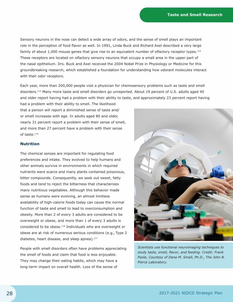
Scientists use functional neuroimaging techniques to study taste, smell, flavor, and feeding. Credit: Frank Poole, Courtesy of Dana M. Small, Ph.D., The John B. Pierce Laboratory.
People with smell disorders often have problems appreciating
the smell of foods and claim that food is less enjoyable.
They may change their eating habits, which may have a
long-term impact on overall health. Loss of the sense of
2017-2021 NIDCD Strategic Plan29
Hearing and Balance ResearchTaste and Smell Research
smell may also cause a person to add too much sugar or
salt to make food taste better. This can be a problem for
people with certain medical conditions such as diabetes
or high blood pressure. In addition, cancer treatments
such as radiation and chemotherapy may result in taste
and smell loss and an associated decrease in appetite,
complicating treatment.
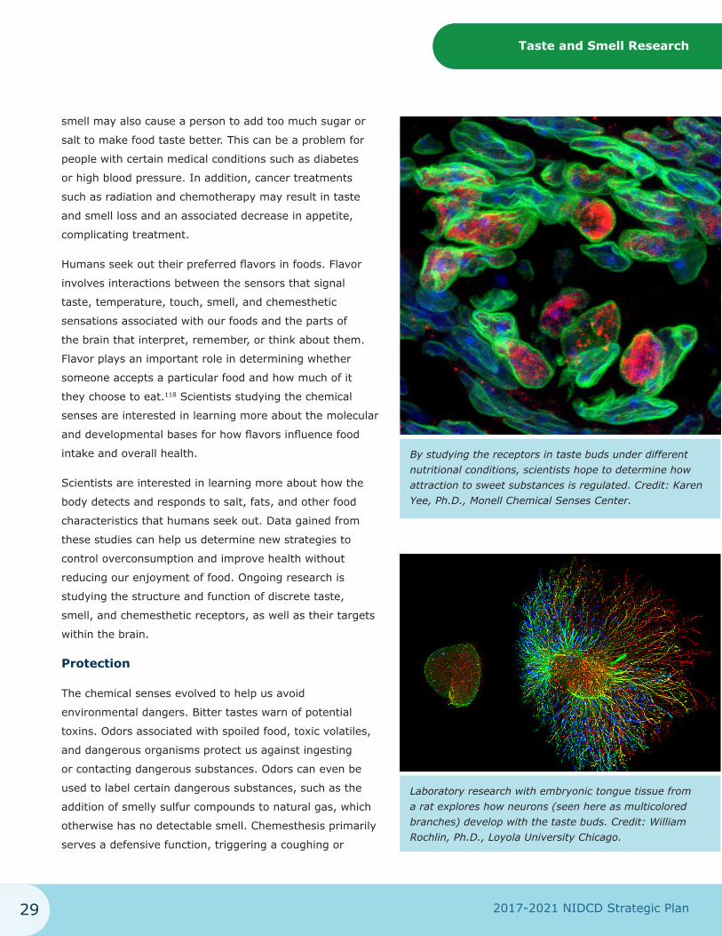
By studying the receptors in taste buds under different nutritional conditions, scientists hope to determine how attraction to sweet substances is regulated. Credit: Karen Yee, Ph.D., Monell Chemical Senses Center.
Humans seek out their preferred flavors in foods. Flavor
involves interactions between the sensors that signal
taste, temperature, touch, smell, and chemesthetic
sensations associated with our foods and the parts of
the brain that interpret, remember, or think about them.
Flavor plays an important role in determining whether
someone accepts a particular food and how much of it
they choose to eat.118 Scientists studying the chemical
senses are interested in learning more about the molecular
and developmental bases for how flavors influence food
intake and overall health.
Scientists are interested in learning more about how the
body detects and responds to salt, fats, and other food
characteristics that humans seek out. Data gained from
these studies can help us determine new strategies to
control overconsumption and improve health without
reducing our enjoyment of food. Ongoing research is
studying the structure and function of discrete taste,
smell, and chemesthetic receptors, as well as their targets
within the brain.
Laboratory research with embryonic tongue tissue from a rat explores how neurons (seen here as multicolored branches) develop with the taste buds. Credit: William Rochlin, Ph.D., Loyola University Chicago.
Protection
The chemical senses evolved to help us avoid
environmental dangers. Bitter tastes warn of potential
toxins. Odors associated with spoiled food, toxic volatiles,
and dangerous organisms protect us against ingesting
or contacting dangerous substances. Odors can even be
used to label certain dangerous substances, such as the
addition of smelly sulfur compounds to natural gas, which
otherwise has no detectable smell. Chemesthesis primarily
serves a defensive function, triggering a coughing or
2017-2021 NIDCD Strategic Plan30
Hearing and Balance ResearchTaste and Smell Research
gagging reaction that allows us to
avoid chemical irritants that cause
tissue damage. Loss of chemesthesis
results in the inability to detect
toxic chemicals in our environment,
possibly leading to increased exposure
and greater risk of serious health
effects. This loss of detection ability
persists in people involved in the
early rescue, recovery, demolition, or
cleanup efforts after the collapse of
the World Trade Center towers.119
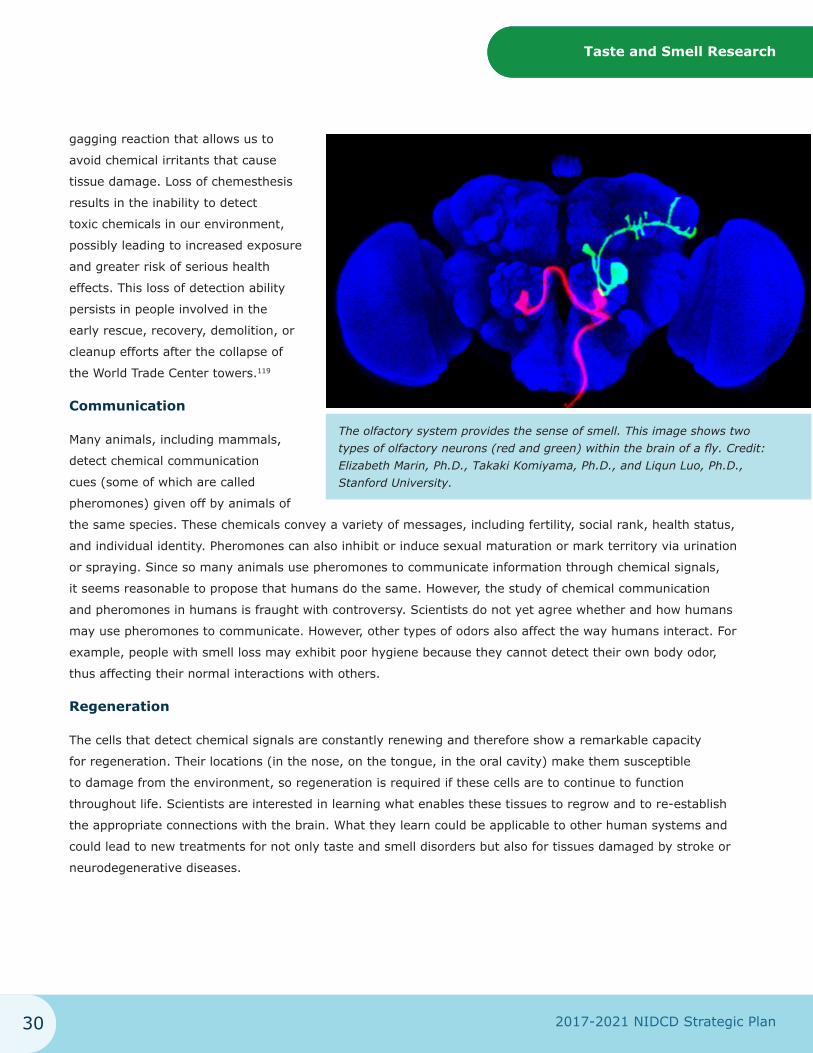
The olfactory system provides the sense of smell. This image shows two types of olfactory neurons (red and green) within the brain of a fly. Credit: Elizabeth Marin, Ph.D., Takaki Komiyama, Ph.D., and Liqun Luo, Ph.D., Stanford University.
Communication
Many animals, including mammals,
detect chemical communication
cues (some of which are called
pheromones) given off by animals of
the same species. These chemicals convey a variety of messages, including fertility, social rank, health status,
and individual identity. Pheromones can also inhibit or induce sexual maturation or mark territory via urination
or spraying. Since so many animals use pheromones to communicate information through chemical signals,
it seems reasonable to propose that humans do the same. However, the study of chemical communication
and pheromones in humans is fraught with controversy. Scientists do not yet agree whether and how humans
may use pheromones to communicate. However, other types of odors also affect the way humans interact. For
example, people with smell loss may exhibit poor hygiene because they cannot detect their own body odor,
thus affecting their normal interactions with others.
Regeneration
The cells that detect chemical signals are constantly renewing and therefore show a remarkable capacity
for regeneration. Their locations (in the nose, on the tongue, in the oral cavity) make them susceptible
to damage from the environment, so regeneration is required if these cells are to continue to function
throughout life. Scientists are interested in learning what enables these tissues to regrow and to re-establish
the appropriate connections with the brain. What they learn could be applicable to other human systems and
could lead to new treatments for not only taste and smell disorders but also for tissues damaged by stroke or
neurodegenerative diseases.
2017-2021 NIDCD Strategic Plan31
Hearing and Balance ResearchTaste and Smell Research
The Taste and Smell Program
The NIDCD Taste and Smell Program supports studies of the chemical senses known as taste, smell, and
chemesthesis (chemically provoked irritation) to enhance our understanding of how individuals communicate
with their environment and how human chemosensory disorders can be diagnosed and treated. NIDCD-
supported research on molecular and cellular biology, animal models, biophysics, and biochemistry of the
olfactory and gustatory systems is paving the way for improved diagnosis, prevention, and treatment of
chemosensory disorders.
Recent Advances in Taste and Smell Research
Transduction Mechanisms
• The body uses chemosensory transduction mechanisms—processes that enable the conversion of detection
into an electrical signal—throughout the oral and nasal cavities. These transduction mechanisms play a
major role in the regulation of food intake and the protection of the airways. Scientists have discovered new
families of chemosensory receptors (trace amine-associated receptors, formyl peptide receptors) that could
detect chemical cues used for communication of odors that signal disease.120
• Scientists have discovered new chemosensory receptors and transduction mechanisms in the gustatory
(taste)121-126 and olfactory systems.127, 128
• Scientists are using novel single cell techniques to make numerous copies of the DNA expressed in a
single cell as it progresses through early development to explore how olfactory receptor cells choose which
receptor to express.129
• Bacteria release quorum signaling molecules to coordinate behaviors such as biofilm formation, virulence,
and antibiotic resistance, based on the local density of the bacterial population. Taste receptors expressed
in solitary chemosensory cells and ciliated cells of the respiratory epithelium detect irritants and quorum
signaling molecules of pathogenic bacteria, evoking protective airway reflexes and inflammatory responses
to rid the airways of infection.130, 131
• The use of novel methods132 is rapidly expanding our identification of the ions or molecules (ligands) that
bind to a receptor for the diverse set of identified chemosensory receptors.133-136
How Genes and Environment Affect Food Preference
• Experience, internal state, and genetic variation in taste and smell receptor genes affect chemosensory likes
and dislikes137-143 Thus, the chemical senses play key roles in the regulation of food intake that underlies
major health issues such as obesity and diabetes.144-146
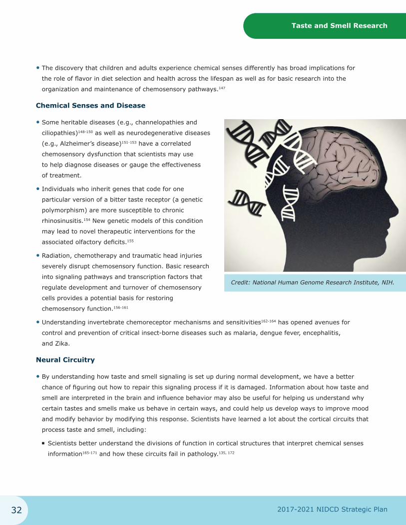
2017-2021 NIDCD Strategic Plan32
Hearing and Balance ResearchTaste and Smell Research
• The discovery that children and adults experience chemical senses differently has broad implications for
the role of flavor in diet selection and health across the lifespan as well as for basic research into the
organization and maintenance of chemosensory pathways.147
Chemical Senses and Disease
• Some heritable diseases (e.g., channelopathies and
ciliopathies)148-150 as well as neurodegenerative diseases
(e.g., Alzheimer’s disease)151-153 have a correlated
chemosensory dysfunction that scientists may use
to help diagnose diseases or gauge the effectiveness
of treatment.
• Individuals who inherit genes that code for one
particular version of a bitter taste receptor (a genetic
polymorphism) are more susceptible to chronic
rhinosinusitis.154 New genetic models of this condition
may lead to novel therapeutic interventions for the
associated olfactory deficits.155
• Radiation, chemotherapy and traumatic head injuries
severely disrupt chemosensory function. Basic research
into signaling pathways and transcription factors that
regulate development and turnover of chemosensory
cells provides a potential basis for restoring
chemosensory function.156-161
• Understanding invertebrate chemoreceptor mechanisms and sensitivities162-164 has opened avenues for
control and prevention of critical insect-borne diseases such as malaria, dengue fever, encephalitis,
and Zika.
Neural Circuitry
• By understanding how taste and smell signaling is set up during normal development, we have a better
chance of figuring out how to repair this signaling process if it is damaged. Information about how taste and
smell are interpreted in the brain and influence behavior may also be useful for helping us understand why
certain tastes and smells make us behave in certain ways, and could help us develop ways to improve mood
and modify behavior by modifying this response. Scientists have learned a lot about the cortical circuits that
process taste and smell, including:
¡ Scientists better understand the divisions of function in cortical structures that interpret chemical senses
information165-171 and how these circuits fail in pathology.135, 172
Credit: National Human Genome Research Institute, NIH.
2017-2021 NIDCD Strategic Plan33
Hearing and Balance ResearchTaste and Smell Research
¡ They are learning how cortical circuits create and read odor patterns and the basic circuitry and physiology
of these circuits.173-176
¡ They are using artificial neural networks and optical imaging to define and dissect the circuitry and coding
in the chemical senses.177-181
¡ They have figured out how adult-born neurons can be functionally and synaptically integrated into neural
circuits.182
¡ They have better insight into how activity within these neural circuits translates to chemosensory
perception and stimulus identification171, 183-188 and guide such behaviors as emotional response189 and
parenting behavior.190
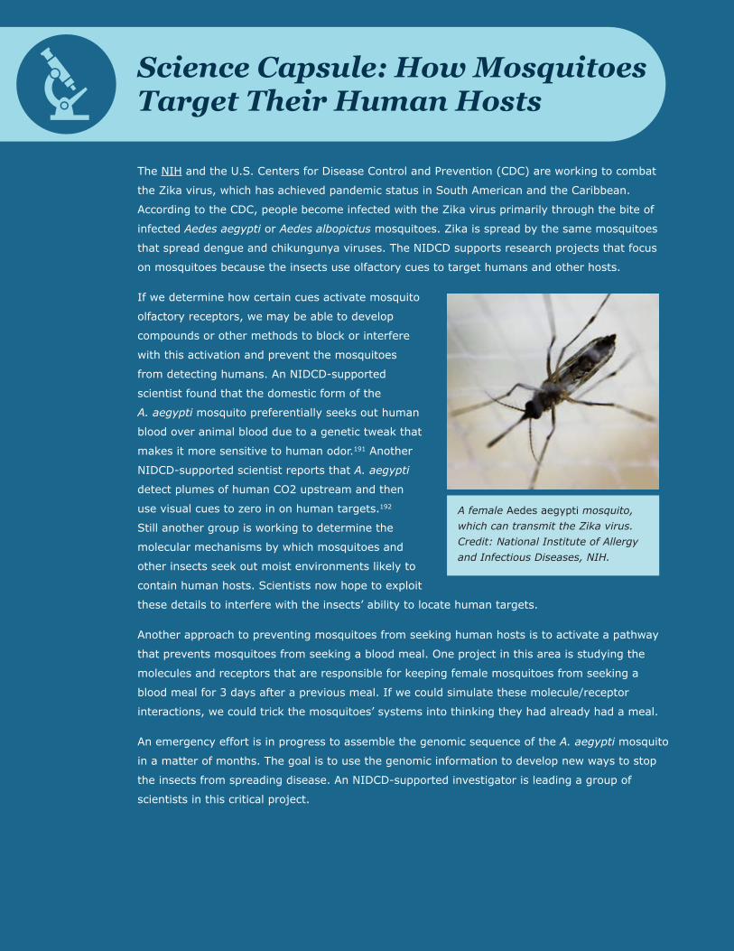
Science Capsule: How Mosquitoes Target Their Human Hosts
The NIH and the U.S. Centers for Disease Control and Prevention (CDC) are working to combat
the Zika virus, which has achieved pandemic status in South American and the Caribbean.
According to the CDC, people become infected with the Zika virus primarily through the bite of
infected Aedes aegypti or Aedes albopictus mosquitoes. Zika is spread by the same mosquitoes
that spread dengue and chikungunya viruses. The NIDCD supports research projects that focus
on mosquitoes because the insects use olfactory cues to target humans and other hosts.
If we determine how certain cues activate mosquito
olfactory receptors, we may be able to develop
compounds or other methods to block or interfere
with this activation and prevent the mosquitoes
from detecting humans. An NIDCD-supported
scientist found that the domestic form of the
A. aegypti mosquito preferentially seeks out human
blood over animal blood due to a genetic tweak that
makes it more sensitive to human odor.191 Another
NIDCD-supported scientist reports that A. aegypti
detect plumes of human CO2 upstream and then
use visual cues to zero in on human targets.192
Still another group is working to determine the
molecular mechanisms by which mosquitoes and
other insects seek out moist environments likely to
contain human hosts. Scientists now hope to exploit
these details to interfere with the insects’ ability to locate human targets.
A female Aedes aegypti mosquito, which can transmit the Zika virus. Credit: National Institute of Allergy and Infectious Diseases, NIH.
Another approach to preventing mosquitoes from seeking human hosts is to activate a pathway
that prevents mosquitoes from seeking a blood meal. One project in this area is studying the
molecules and receptors that are responsible for keeping female mosquitoes from seeking a
blood meal for 3 days after a previous meal. If we could simulate these molecule/receptor
interactions, we could trick the mosquitoes’ systems into thinking they had already had a meal.
An emergency effort is in progress to assemble the genomic sequence of the A. aegypti mosquito
in a matter of months. The goal is to use the genomic information to develop new ways to stop
the insects from spreading disease. An NIDCD-supported investigator is leading a group of
scientists in this critical project.
2017-2021 NIDCD Strategic Plan35
Hearing and Balance ResearchTaste and Smell Research
Priority Areas in Taste and Smell Research
In developing Priority Areas, the NIDCD took into consideration areas of research that are within the mission
of other NIH Institutes, Centers, and Offices (ICO) and are not primarily supported by the NIDCD but that
have relevance to the study of chemical senses. These research areas include dietary intake, infectious
diseases, and neurological diseases.
• Dietary Intake: The NIDCD supports basic research on chemosensory factors controlling flavor perception,
food selection, and related neural pathways. However, research studies that focus exclusively on the
consequences of overconsumption or poor diet, including type 2 diabetes, metabolic disorders, stroke,
cancer, cardiovascular disease, hypertension, and obesity, are supported by several other NIH ICOs.
• Infectious Diseases: The NIDCD supports studies of basic neural mechanisms of insect olfaction, including
olfaction of insects that serve as disease vectors for encephalitis, dengue fever, and malaria. However, the
funding of studies focusing exclusively on the infectious nature of these diseases fall outside of NIDCD’s
mission area.
• Neurological Diseases: The NIDCD supports studies on alteration and loss of olfactory function, including
potential diagnostic significance of such changes, in neurological diseases such as Parkinson’s and
Alzheimer’s disease. However, studies focusing exclusively on causes and treatments of these diseases fall
within the mission of areas of other NIH ICOs.
Priority Area 1: Understanding Normal Function
• Fundamental Biology of Chemosensory Function: Continue to develop and apply new tools and
approaches to delineate the organization of molecules, cells, and neural circuits underlying the function
of the chemesthetic (trigeminal), gustatory and olfactory systems, including development, cell turnover,
regeneration, and plasticity.
• Peripheral and Central Bases of Flavor: Understand the complex interactions between peripheral and
central aspects of flavor perception, including retronasal or orthonasal olfaction, oral chemesthesis (chemical
irritation), taste, oral somesthesis (temperature, texture), memory, and motivational state (such as hunger).
• Sentinel/Sensory Functions: Describe how chemical senses help us avoid dangers such as spoiled or
contaminated foods, how they detect potentially toxic chemicals in the environment and in our bodies, and
how these protective functions can be damaged and regenerated.
• Genetic Aspects of Chemosensory Sensitivity:
¡ Genomics: Identify genes involved in the development and normal function of the taste and smell
systems, including the use of single-cell profiling approaches.
¡ Variation: Describe the normal variation in taste and smell sensitivity. Identify the genes involved in
order to understand what is outside the range of normal function. Describe how such variation may relate
to susceptibility for human communication disorders.
2017-2021 NIDCD Strategic Plan36
Hearing and Balance ResearchTaste and Smell Research
¡ Experience: Identify genes involved with storing memories of taste and smell. Determine how experience
influences future diet.
¡ Epigenetics: Describe how external factors (e.g., sensory experience, diet, stress) activate and
deactivate genes.
• Central Control of Taste and Smell: Characterize top-down control within the central nervous system that
modulates sensory input, sensory processing and perception, and determine how such activity may change
depending on internal state, motivational or cognitive factors.
• Developing Tools to Measure Taste and Smell Function: Refine, develop, and apply appropriate
psychophysical and behavioral methods for assessing taste and smell functions in animal models and
humans. Provide practicing physicians with standardized tools to test taste and smell during physical exams
or routine office visits. Develop criteria and metrics for the range of “normal” taste and smell by analogy to
hearing and vision.
• Develop Novel Approaches to Alter Taste Function: Alter the levels of salt, sugar, and fat intake using
innovative methods such as using artificial substitutes or changing learned flavor preferences.
Priority Area 2: Understanding Diseases and Disorders
• Genetic Disorders: Clarify and classify taste and smell disorders caused mainly by significant genetic
alterations (e.g., ciliopathies and channelopathies). Determine the normal range of variation of function in
the chemical senses as related to genetic polymorphisms.
• Environmental Insults on Taste and Smell: Identify the mechanisms that contribute to taste and smell
loss and/or dysfunction resulting from radiation, chemotherapy, head trauma, and toxins.
• Sinusitis/Rhinitis: Identify the molecular and cellular bases for loss of olfaction following nasal cavity or
sinus infection, the most common cause of temporary and permanent olfactory loss.
• Understanding How the Activity of the Chemical Senses Can Lead to Excessive Consumption or
Malnutrition: Determine whether calorie intake is affected by normal variation or altered function of taste
and smell activity.
• Epidemiology: Describe the incidence and prevalence of taste and smell loss and dysfunction. For
example, as the population ages, determine how many more people report taste and smell problems
that affect quality of life. Enable practical approaches for wider integration of standardized chemosensory
measurements into large-scale epidemiological and clinical studies.
Priority Area 3: Improving Diagnosis, Treatment, and Prevention
• Improved Diagnostic Tools and Pharmacological Treatments: Develop and validate tests to evaluate
taste and smell function that are practical and affordable for use in the office setting. Develop drugs to treat
taste and smell dysfunction, especially drugs which slow apoptosis (cell death) and promote regeneration.
2017-2021 NIDCD Strategic Plan37
Hearing and Balance ResearchTaste and Smell Research
• Regenerative Medicine/Tissue Engineering: Increase understanding of the properties that enable stem
cells in the peripheral taste and smell pathways to proliferate and differentiate, providing insights not only
for the treatment of taste and smell loss but also for the treatment of other neurological diseases.
• Enhancing the Clinical Enterprise: Promote clinical training in the chemical senses to encourage
development of animal models of relevant disorders and promote clinical and translational research,
involving interdisciplinary teams of clinicians and basic scientists.
Priority Area 4: Improving Outcomes for Human Communication
• Translational Research: Translational research in the chemical senses is in its infancy, due in part
to the modest amount of clinical research that has been conducted. Currently, no evidence-based
preventive measures, interventions, or treatments are applied to taste and smell dysfunction. Comparative
effectiveness research is premature because of the lack of intervention and treatment strategies and
decisions. Because taste and smell loss become increasingly common in a population with a growing
proportion of older adults, the NIDCD has identified translational research in the chemical senses as a critical
gap area.
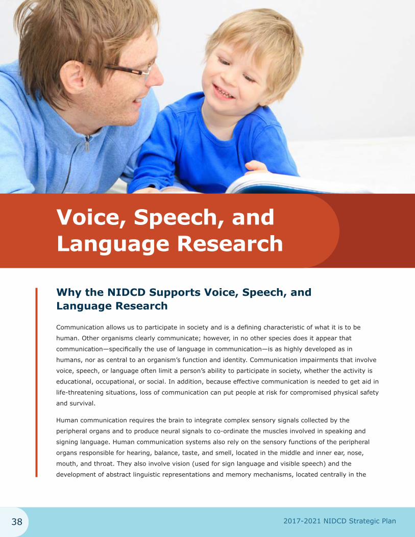
2017-2021 NIDCD Strategic Plan38
Hearing and Balance ResearchTaste and Smell Research
Voice, Speech, and Language Research
Why the NIDCD Supports Voice, Speech, and Language Research
Communication allows us to participate in society and is a defining characteristic of what it is to be
human. Other organisms clearly communicate; however, in no other species does it appear that
communication—specifically the use of language in communication—is as highly developed as in
humans, nor as central to an organism’s function and identity. Communication impairments that involve
voice, speech, or language often limit a person’s ability to participate in society, whether the activity is
educational, occupational, or social. In addition, because effective communication is needed to get aid in
life-threatening situations, loss of communication can put people at risk for compromised physical safety
and survival.
Human communication requires the brain to integrate complex sensory signals collected by the
peripheral organs and to produce neural signals to co-ordinate the muscles involved in speaking and
signing language. Human communication systems also rely on the sensory functions of the peripheral
organs responsible for hearing, balance, taste, and smell, located in the middle and inner ear, nose,
mouth, and throat. They also involve vision (used for sign language and visible speech) and the
development of abstract linguistic representations and memory mechanisms, located centrally in the
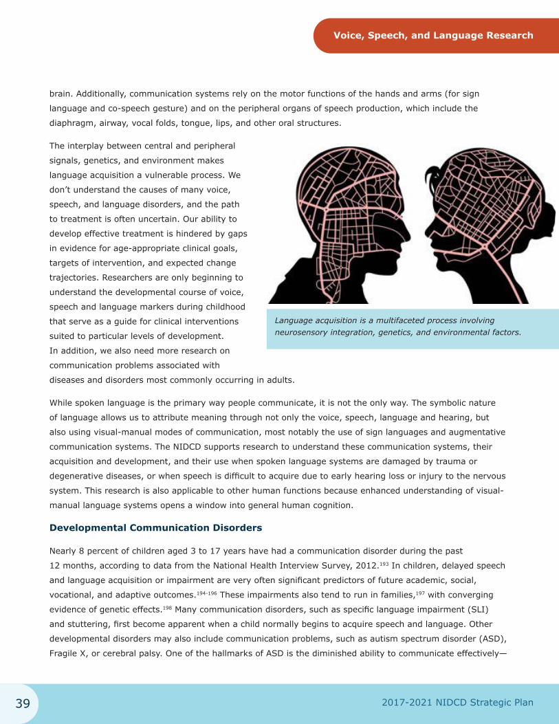
2017-2021 NIDCD Strategic Plan39
Voice, Speech, and Language Research
brain. Additionally, communication systems rely on the motor functions of the hands and arms (for sign
language and co-speech gesture) and on the peripheral organs of speech production, which include the
diaphragm, airway, vocal folds, tongue, lips, and other oral structures.
The interplay between central and peripheral
signals, genetics, and environment makes
language acquisition a vulnerable process. We
don’t understand the causes of many voice,
speech, and language disorders, and the path
to treatment is often uncertain. Our ability to
develop effective treatment is hindered by gaps
in evidence for age-appropriate clinical goals,
targets of intervention, and expected change
trajectories. Researchers are only beginning to
understand the developmental course of voice,
speech and language markers during childhood
that serve as a guide for clinical interventions
suited to particular levels of development.
In addition, we also need more research on
communication problems associated with
diseases and disorders most commonly occurring in adults.
Language acquisition is a multifaceted process involving neurosensory integration, genetics, and environmental factors.
While spoken language is the primary way people communicate, it is not the only way. The symbolic nature
of language allows us to attribute meaning through not only the voice, speech, language and hearing, but
also using visual-manual modes of communication, most notably the use of sign languages and augmentative
communication systems. The NIDCD supports research to understand these communication systems, their
acquisition and development, and their use when spoken language systems are damaged by trauma or
degenerative diseases, or when speech is difficult to acquire due to early hearing loss or injury to the nervous
system. This research is also applicable to other human functions because enhanced understanding of visual-
manual language systems opens a window into general human cognition.
Developmental Communication Disorders
Nearly 8 percent of children aged 3 to 17 years have had a communication disorder during the past
12 months, according to data from the National Health Interview Survey, 2012.193 In children, delayed speech
and language acquisition or impairment are very often significant predictors of future academic, social,
vocational, and adaptive outcomes.194-196 These impairments also tend to run in families,197 with converging
evidence of genetic effects.198 Many communication disorders, such as specific language impairment (SLI)
and stuttering, first become apparent when a child normally begins to acquire speech and language. Other
developmental disorders may also include communication problems, such as autism spectrum disorder (ASD),
Fragile X, or cerebral palsy. One of the hallmarks of ASD is the diminished ability to communicate effectively—
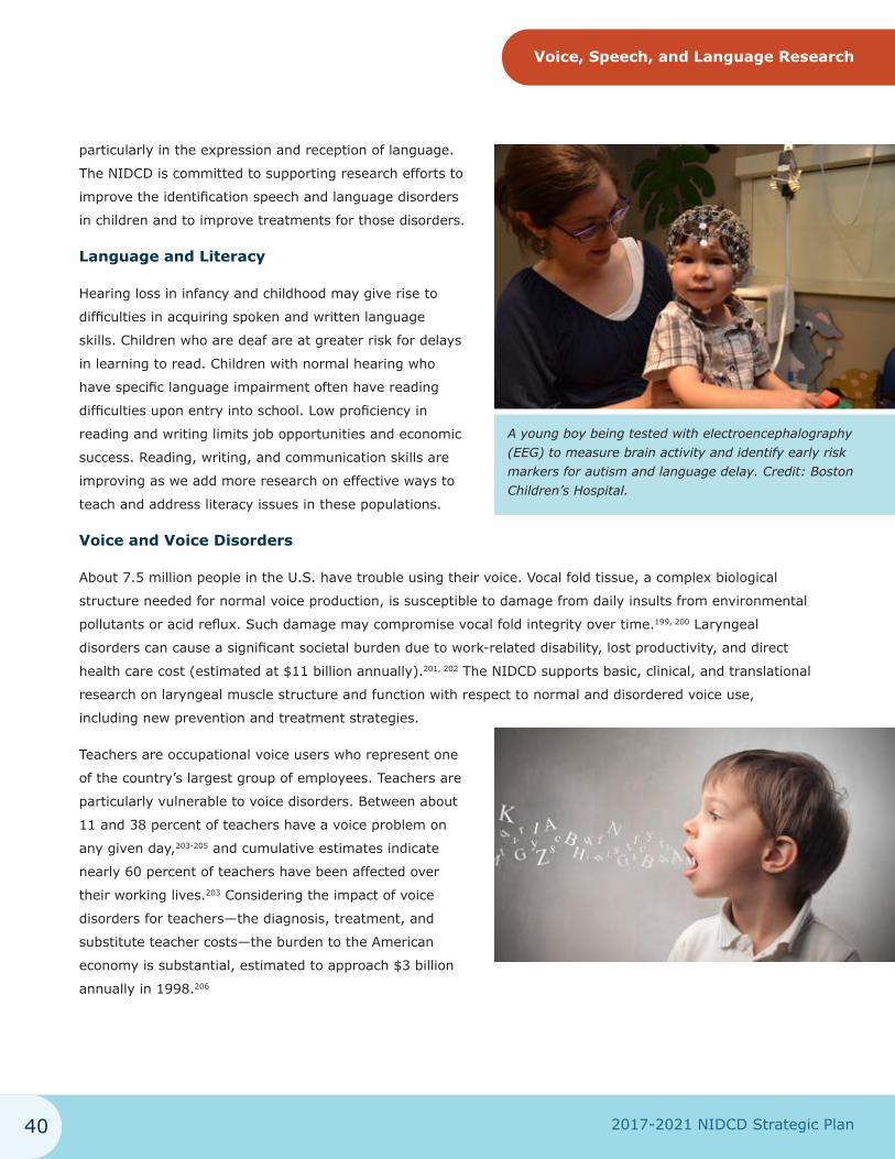
2017-2021 NIDCD Strategic Plan40
Voice, Speech, and Language Research
particularly in the expression and reception of language.
The NIDCD is committed to supporting research efforts to
improve the identification speech and language disorders
in children and to improve treatments for those disorders.
Language and Literacy
Hearing loss in infancy and childhood may give rise to
difficulties in acquiring spoken and written language
skills. Children who are deaf are at greater risk for delays
in learning to read. Children with normal hearing who
have specific language impairment often have reading
difficulties upon entry into school. Low proficiency in
reading and writing limits job opportunities and economic
success. Reading, writing, and communication skills are
improving as we add more research on effective ways to
teach and address literacy issues in these populations.
A young boy being tested with electroencephalography (EEG) to measure brain activity and identify early risk markers for autism and language delay. Credit: Boston Children’s Hospital.
Voice and Voice Disorders
About 7.5 million people in the U.S. have trouble using their voice. Vocal fold tissue, a complex biological
structure needed for normal voice production, is susceptible to damage from daily insults from environmental
pollutants or acid reflux. Such damage may compromise vocal fold integrity over time.199, 200 Laryngeal
disorders can cause a significant societal burden due to work-related disability, lost productivity, and direct
health care cost (estimated at $11 billion annually).201, 202 The NIDCD supports basic, clinical, and translational
research on laryngeal muscle structure and function with respect to normal and disordered voice use,
including new prevention and treatment strategies.
Teachers are occupational voice users who represent one
of the country’s largest group of employees. Teachers are
particularly vulnerable to voice disorders. Between about
11 and 38 percent of teachers have a voice problem on
any given day,203-205 and cumulative estimates indicate
nearly 60 percent of teachers have been affected over
their working lives.203 Considering the impact of voice
disorders for teachers—the diagnosis, treatment, and
substitute teacher costs—the burden to the American
economy is substantial, estimated to approach $3 billion
annually in 1998.206
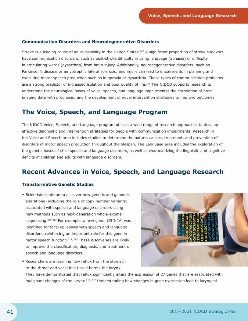
2017-2021 NIDCD Strategic Plan41
Voice, Speech, and Language Research
Communication Disorders and Neurodegenerative Disorders
Stroke is a leading cause of adult disability in the United States.207 A significant proportion of stroke survivors
have communication disorders, such as post-stroke difficulty in using language (aphasia) or difficulty
in articulating words (dysarthria) from brain injury. Additionally, neurodegenerative disorders, such as
Parkinson’s disease or amyotrophic lateral sclerosis, and injury can lead to impairments in planning and
executing motor speech production such as in apraxia or dysarthria. These types of communication problems
are a strong predictor of increased isolation and poor quality of life.208 The NIDCD supports research to
understand the neurological bases of voice, speech, and language impairments; the correlation of brain
imaging data with prognosis; and the development of novel intervention strategies to improve outcomes.
The Voice, Speech, and Language Program
The NIDCD Voice, Speech, and Language program utilizes a wide range of research approaches to develop
effective diagnostic and intervention strategies for people with communication impairments. Research in
the Voice and Speech area includes studies to determine the nature, causes, treatment, and prevention of
disorders of motor speech production throughout the lifespan. The Language area includes the exploration of
the genetic bases of child speech and language disorders, as well as characterizing the linguistic and cognitive
deficits in children and adults with language disorders.
Recent Advances in Voice, Speech, and Language Research
Transformative Genetic Studies
• Scientists continue to discover new genetic and genomic
alterations (including the role of copy number variants)
associated with speech and language disorders using
new methods such as next-generation whole-exome
sequencing.209-213 For example, a new gene, GRIN2A, was
identified for focal epilepsies with speech and language
disorders, reinforcing an important role for this gene in
motor speech function.214, 215 These discoveries are likely
to improve the classification, diagnosis, and treatment of
speech and language disorders.
• Researchers are learning how reflux from the stomach
to the throat and vocal fold tissue harms the larynx.
They have demonstrated that reflux significantly alters the expression of 27 genes that are associated with
malignant changes of the larynx.216, 217 Understanding how changes in gene expression lead to laryngeal

2017-2021 NIDCD Strategic Plan42
Voice, Speech, and Language Research
injury provides a comprehensive model for identifying novel diagnostic and therapeutic targets to treat
reflux-related injury.
• Researchers generated a transcriptome dataset to capture the complexity of genes responsible for wound
healing of the vocal folds. This dataset serves as a resource in developing new studies that would accelerate
the identification of novel therapeutic targets to treat reflux-related injury.218
Behavioral Phenotyping
• Studies demonstrated that children with developmental
speech and language problems are at a considerable risk
for learning disabilities and other psychosocial problems
that emerge during adolescence or adulthood.219-221
• Some families with high incidence of stuttering may also
have high incidence of other fluency disorders and other
speech production difficulties. This finding can lead to
new genetic studies across multiple families to define the
characteristics of stuttering.222
• Scientists are using new imaging technology to study
structural and mechanical characteristics of laryngeal
scarring.223 This could provide the foundation for
developing improved treatments for one of the most
common causes of voice disorders.
• Researchers have identified distinct and viable
characteristics of language disorders, extending the
research to new populations, such as children who are
deaf or minimally verbal children with autism, and to language disorders shared across different populations
that may be used in future genetic and treatment studies.224-226 The development of these classification
systems will guide future investigations into the genetic, neurologic, and other causal factors that contribute
to voice, speech, and language impairments.
Dr. Dennis Drayna is the Chief of the Section on Genetics of Communication Disorders. His area of research is focused on the genetic linkage and positional cloning in human communication disorders, such as stuttering.
Interventions
• Researchers suggest that self-administered computer therapy with single word production improved
chronic apraxia of speech. This method shows promise for delivering high-intensity speech and language
rehabilitation for individuals recovering from stroke.227
• Scientists have developed a wearable monitoring device to accurately measure voice disorders during daily
activities and provide real-time feedback.228, 229 When combined with knowledge of gene expression changes
related to vocal fold vibration exposure,230 ambulatory monitoring has shown the potential to revolutionize
treatment that could facilitate healthier vocal function and enhance diagnosis and treatment options.
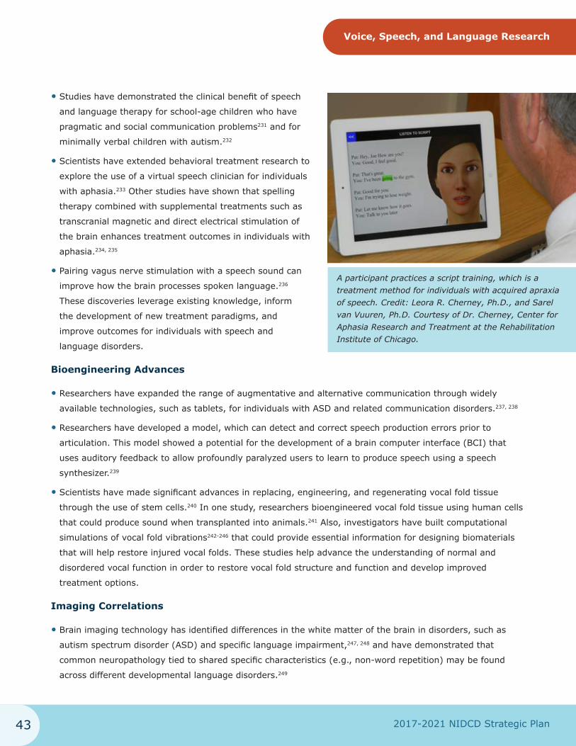
2017-2021 NIDCD Strategic Plan43
Voice, Speech, and Language Research
• Studies have demonstrated the clinical benefit of speech
and language therapy for school-age children who have
pragmatic and social communication problems231 and for
minimally verbal children with autism.232
• Scientists have extended behavioral treatment research to
explore the use of a virtual speech clinician for individuals
with aphasia.233 Other studies have shown that spelling
therapy combined with supplemental treatments such as
transcranial magnetic and direct electrical stimulation of
the brain enhances treatment outcomes in individuals with
aphasia.234, 235
• Pairing vagus nerve stimulation with a speech sound can
improve how the brain processes spoken language.236
These discoveries leverage existing knowledge, inform
the development of new treatment paradigms, and
improve outcomes for individuals with speech and
language disorders.
A participant practices a script training, which is a treatment method for individuals with acquired apraxia of speech. Credit: Leora R. Cherney, Ph.D., and Sarel van Vuuren, Ph.D. Courtesy of Dr. Cherney, Center for Aphasia Research and Treatment at the Rehabilitation Institute of Chicago.
Bioengineering Advances
• Researchers have expanded the range of augmentative and alternative communication through widely
available technologies, such as tablets, for individuals with ASD and related communication disorders.237, 238
• Researchers have developed a model, which can detect and correct speech production errors prior to
articulation. This model showed a potential for the development of a brain computer interface (BCI) that
uses auditory feedback to allow profoundly paralyzed users to learn to produce speech using a speech
synthesizer.239
• Scientists have made significant advances in replacing, engineering, and regenerating vocal fold tissue
through the use of stem cells.240 In one study, researchers bioengineered vocal fold tissue using human cells
that could produce sound when transplanted into animals.241 Also, investigators have built computational
simulations of vocal fold vibrations242-246 that could provide essential information for designing biomaterials
that will help restore injured vocal folds. These studies help advance the understanding of normal and
disordered vocal function in order to restore vocal fold structure and function and develop improved
treatment options.
Imaging Correlations
• Brain imaging technology has identified differences in the white matter of the brain in disorders, such as
autism spectrum disorder (ASD) and specific language impairment,247, 248 and have demonstrated that
common neuropathology tied to shared specific characteristics (e.g., non-word repetition) may be found
across different developmental language disorders.249
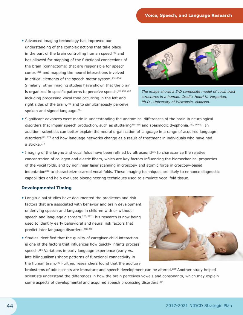
2017-2021 NIDCD Strategic Plan44
Voice, Speech, and Language Research
• Advanced imaging technology has improved our
understanding of the complex actions that take place
in the part of the brain controlling human speech95 and
has allowed for mapping of the functional connections of
the brain (connectome) that are responsible for speech
control250 and mapping the neural interactions involved
in critical elements of the speech motor system.251-254
Similarly, other imaging studies have shown that the brain
is organized in specific patterns to perceive speech,93, 255-262
including processing vocal tone occurring in the left and
right sides of the brain,263 and to simultaneously perceive
spoken and signed language.264
The image shows a 3-D composite model of vocal tract structures in a human. Credit: Houri K. Vorperian, Ph.D., University of Wisconsin, Madison.
• Significant advances were made in understanding the anatomical differences of the brain in neurological
disorders that impair speech production, such as stuttering265-268 and spasmodic dysphonia.222, 269-271 In
addition, scientists can better explain the neural organization of language in a range of acquired language
disorders272, 273 and how language networks change as a result of treatment in individuals who have had
a stroke.274
• Imaging of the larynx and vocal folds have been refined by ultrasound275 to characterize the relative
concentration of collagen and elastic fibers, which are key factors influencing the biomechanical properties
of the vocal folds, and by nonlinear laser scanning microscopy and atomic force microscopy-based
indentation223 to characterize scarred vocal folds. These imaging techniques are likely to enhance diagnostic
capabilities and help evaluate bioengineering techniques used to simulate vocal fold tissue.
Developmental Timing
• Longitudinal studies have documented the predictors and risk
factors that are associated with behavior and brain development
underlying speech and language in children with or without
speech and language disorders.276, 277 This research is now being
used to identify early behavioral and neural risk factors that
predict later language disorders.278-280
• Studies identified that the quality of caregiver-child interaction
is one of the factors that influences how quickly infants process
speech.281 Variations in early language experience (early vs.
late bilingualism) shape patterns of functional connectivity in
the human brain.282 Further, researchers found that the auditory
brainstems of adolescents are immature and speech development can be altered.283 Another study helped
scientists understand the differences in how the brain perceives vowels and consonants, which may explain
some aspects of developmental and acquired speech processing disorders.284
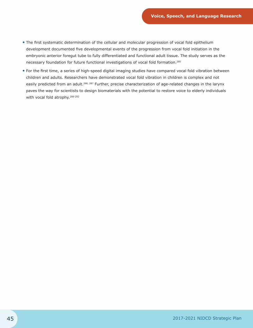
2017-2021 NIDCD Strategic Plan45
Voice, Speech, and Language Research
• The first systematic determination of the cellular and molecular progression of vocal fold epithelium
development documented five developmental events of the progression from vocal fold initiation in the
embryonic anterior foregut tube to fully differentiated and functional adult tissue. The study serves as the
necessary foundation for future functional investigations of vocal fold formation.285
• For the first time, a series of high-speed digital imaging studies have compared vocal fold vibration between
children and adults. Researchers have demonstrated vocal fold vibration in children is complex and not
easily predicted from an adult.286, 287 Further, precise characterization of age-related changes in the larynx
paves the way for scientists to design biomaterials with the potential to restore voice to elderly individuals
with vocal fold atrophy.288-292
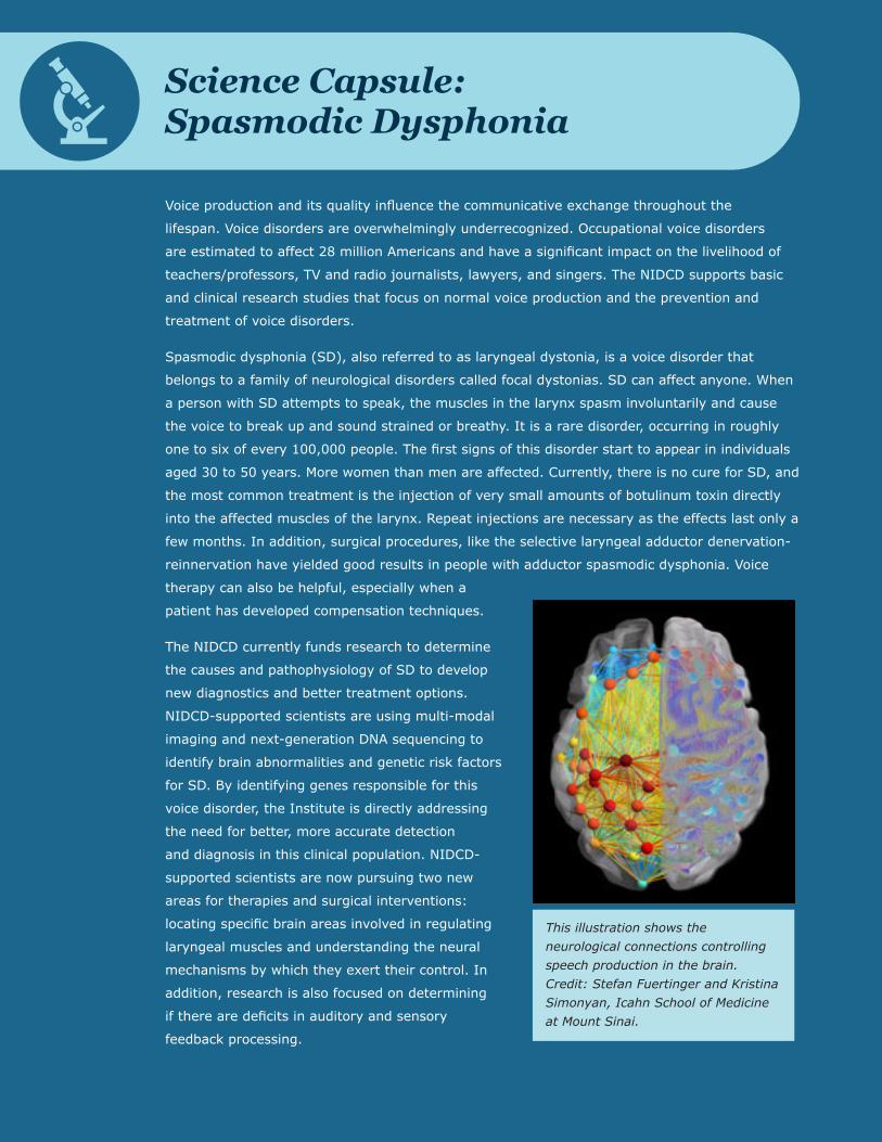
Science Capsule: Spasmodic Dysphonia
Voice production and its quality influence the communicative exchange throughout the
lifespan. Voice disorders are overwhelmingly underrecognized. Occupational voice disorders
are estimated to affect 28 million Americans and have a significant impact on the livelihood of
teachers/professors, TV and radio journalists, lawyers, and singers. The NIDCD supports basic
and clinical research studies that focus on normal voice production and the prevention and
treatment of voice disorders.
Spasmodic dysphonia (SD), also referred to as laryngeal dystonia, is a voice disorder that
belongs to a family of neurological disorders called focal dystonias. SD can affect anyone. When
a person with SD attempts to speak, the muscles in the larynx spasm involuntarily and cause
the voice to break up and sound strained or breathy. It is a rare disorder, occurring in roughly
one to six of every 100,000 people. The first signs of this disorder start to appear in individuals
aged 30 to 50 years. More women than men are affected. Currently, there is no cure for SD, and
the most common treatment is the injection of very small amounts of botulinum toxin directly
into the affected muscles of the larynx. Repeat injections are necessary as the effects last only a
few months. In addition, surgical procedures, like the selective laryngeal adductor denervation-
reinnervation have yielded good results in people with adductor spasmodic dysphonia. Voice
therapy can also be helpful, especially when a
patient has developed compensation techniques.
The NIDCD currently funds research to determine
the causes and pathophysiology of SD to develop
new diagnostics and better treatment options.
NIDCD-supported scientists are using multi-modal
imaging and next-generation DNA sequencing to
identify brain abnormalities and genetic risk factors
for SD. By identifying genes responsible for this
voice disorder, the Institute is directly addressing
the need for better, more accurate detection
and diagnosis in this clinical population. NIDCD-
supported scientists are now pursuing two new
areas for therapies and surgical interventions:
locating specific brain areas involved in regulating
laryngeal muscles and understanding the neural
mechanisms by which they exert their control. In
addition, research is also focused on determining
if there are deficits in auditory and sensory
feedback processing.
This illustration shows the neurological connections controlling speech production in the brain. Credit: Stefan Fuertinger and Kristina Simonyan, Icahn School of Medicine at Mount Sinai.
2017-2021 NIDCD Strategic Plan47
Voice, Speech, and Language ResearchThe NIDCD will continue to support voice disorders research, guided by recommendations from
a 2013 NIDCD-sponsored workshop on voice sciences and disorders. Leading experts in the
field agreed that it is essential to strengthen the pipeline of future voice scientists by creating
collaborative teams to address lingering research questions. Accordingly, the NIDCD issued
two Funding Opportunity Announcements (FOAs) on Advancing Research in Voice Disorders.
The initiatives seek cutting-edge research proposals such as the development of biomaterials
for engineering vocal fold tissue and development of ambulatory biofeedback approaches for
management of patients with voice disorders. Additionally, the FOAs encourage patient outcomes
research, health services research, and community-based research with special attention to
the needs of individuals with low socio-economic status, disparities, rural, second language
populations, and women’s health.
Priority Areas in Voice, Speech, and Language Research
The NIDCD Voice, Speech, and Language Program contains areas of research that overlap with mission areas
of other NIH ICOs. In particular:
• Language: NIDCD research focuses on language acquisition in the presence of dysfunctions, diseases,
and disorders that alter the traditional developmental course such as hearing loss, ASD, SLI, and aphasia.
Research on the normal acquisition of language and on normal language decline from normal aging is within
the mission of other NIH ICOs.
• Literacy: As with language, the normal acquisition of literacy skills and individual outcomes in educational
settings are within the mission of other NIH ICOs. The NIDCD supports research into literacy for people
who are deaf and hard of hearing, the acquisition of written language for people with pre-existing language
disorders, improving reading and writing deficits often associated with stroke, and educational interventions
to support improved individual outcomes.
• Swallowing: Speech and swallowing functions have shared anatomy, leading the NIDCD to fund some
research on swallowing and disordered swallowing (dysphagia). Dysphagia often occurs after head and neck
cancer or certain neurological conditions. Many NIH ICOs may have an interest in research on swallowing
and dysphagia.
Priority Area 1: Understanding Normal Function
• Modeling: Improve and validate physical, computational, and theoretical modeling of human
communication, including vibratory properties of the larynx, neural and speech motor control, and speech
language processing.
2017-2021 NIDCD Strategic Plan48
Voice, Speech, and Language Research
• Laryngeal System: Examine impact on vocal health from changes to laryngeal muscle function and
structure, such as muscle fiber and mucosal changes at the cellular and molecular level. Determine effects
from development, the environment, aging, and voice use (voice training and vocal dose—amount, intensity,
and distribution).
• Motor Speech Production: Determine the similarities and differences in development and functioning
of neural and musculoskeletal systems for human voice and speech production vs. non-speech oral motor
control to identify the sensorimotor principles underlying typical speech development and adult speech
motor control, and to understand overlapping sensorimotor mechanisms of the larynx.
• Developmental and Neural Plasticity: Identify the developmental course of sensory and motor plasticity
and the underlying neural mechanisms associated with voice and speech motor learning in children and
adults (e.g., sensorimotor adaptation).
• Sign Language Research: Investigate the acquisition, processing, and neural underpinnings of languages
within the visual-manual modality.
• Literacy and Deafness: Identify central and peripheral factors associated with the successful
comprehension and use of written language for people who use sign language as their primary
way of communication.
Priority Area 2: Understanding Diseases and Disorders
• Natural History and Epidemiology: Identify genetic, neural, sensorimotor, cognitive, linguistic,
behavioral, demographic, and environmental factors associated with the progression, developmental course,
and long-term outcomes of voice, speech, and language impairments. Determine the relative contribution of
those factors to the development of and the recovery from impairment.
• Pathophysiology: Identify the pathophysiologic and cognitive mechanisms underlying both common and
rare voice, speech, and language impairments.
• Genetics: Identify genetic and epigenetic factors that contribute to voice, speech, and language
impairments, including studies that identify prenatal factors that can modify genetic and epigenetic
expression in offspring.
• Developmental and Neural Plasticity: Examine changes in brain structure and functioning in response to
behavioral, pathologic, or environmental insult as a basis for voice, speech, and language impairments with
an emphasis on developmental timing.
• Co-Occurring Conditions: Examine factors (e.g., social context, inflammatory response, co-morbid
conditions) that interact or coexist with primary voice, speech, and language impairments. Examine
diagnostic and treatment strategies for voice, speech, and language impairments that may coexist in
individuals with deafness, and in individuals with communication disorders. Examine cross-system deficits
and their influence on communication health and responsiveness to treatment.
2017-2021 NIDCD Strategic Plan49
Voice, Speech, and Language Research
Priority Area 3: Improving Diagnosis, Treatment, and Prevention
• Detection, Diagnosis and Hypothesis-Driven Interventions: Develop biomarkers (e.g., genetic,
imaging, behavioral) of objective diagnosis, prognosis, treatment monitoring for developmental and acquired
voice, speech, and language impairments. Develop models of intervention informed by cognitive, linguistic,
biological, or neurophysiological processes, accounting for cultural and linguistic variation and including
predictors of response to treatment. Develop and refine techniques, technology, and instrumentation for
improved diagnosis to aid in treatment and prevention.
• Efficacy: Using outcomes-based clinical studies and randomized clinical trials, determine the efficacy of
proposed interventions for the prevention and treatment of voice, speech, and language impairments, which
can include accounting for cultural and linguistic variation.
• Prevention: Develop and expand programs that prevent the onset or limit the severity of developmental
and acquired voice, speech, and language impairments for people with genetic, occupational, environmental,
or other risks.
• Understudied Populations: Identify the cause and pathophysiology for understudied populations, such
as school-aged children, minimally verbal children with ASD, health disparity groups, and multicultural
groups, or understudied conditions, such as stuttering and apraxia of speech in children and adults. Develop
methods of assessing and new effective interventions or approaches tailored for understudied populations
or conditions.
• Rare Disorders: Develop biomarkers for improved diagnosis, prediction of risk, and treatment response
for patients with rare voice, speech and language disorders (e.g., spasmodic dysphonia, paradoxical vocal
fold motion).
• Bioengineering, including Assistive Technologies: Harness recent advances in bioengineering to
inform the development and evaluate efficacy of wearable monitoring devices, imaging procedures, tissue
engineering, bioreactors, and novel augmentative and alternative communication approaches. Enhance
brain-computer interface technologies for communication.
• Literacy Skills: Develop methods that promote the acquisition of literacy skills during childhood and
improve the reading and writing abilities of people who are deaf and native American Sign Language users.
Priority Area 4: Improving Outcomes for Human Communication
• Novel Delivery: Translate and evaluate efficacy of conventional interventions into new delivery models
(e.g., group, family, telehealth, cell-based therapies, and emerging technology platforms).
• Screening: Develop effective and efficient clinical screening tools for use in health and community settings
such as schools, primary care physician offices, and senior centers. Develop novel screening tools to
document treatment outcomes, to determine communication status, and to improve clinical outcomes in
real-world settings. Determine efficacy of screening for improving clinical outcomes.
2017-2021 NIDCD Strategic Plan50
Voice, Speech, and Language Research
• Comparative Effectiveness Research and Evidence-Based Medicine: Through clinical trials and
epidemiological comparative effectiveness research, identify best treatments for a given communication
disorder for a defined set of individuals.
• Patient-Oriented Research: Conduct research to help define the impact of voice, speech, and language
communication problems and the desirable/reasonable expectation for quality of life outcome from the
individual’s perspective.
• Community-Based Research:
¡ Promote community-based research and data collection to identify factors that influence outcomes for
people with voice, speech, or language impairments, and to inform the development of public policy
recommendations.
¡ Examine community-level health promotion strategies to prevent the occurrence of voice, speech, and
language impairments, reduce risk, and improve adherence with treatment.
• Bridging the Gap Between Research and Practice: Determine effective dissemination and
implementation strategies that enhance the adoption of voice, speech, and language clinical discoveries
into routine community practice.

2017-2021 NIDCD Strategic Plan51
Summary
The mission of the NIDCD is to conduct and support biomedical and behavioral research and research training
in the normal and disordered processes of hearing, balance, taste, smell, voice, speech, and language.
The Institute also conducts and supports research and research training related to disease prevention and
health promotion; addresses special biomedical and behavioral problems associated with people who have
communication impairments or disorders; and supports efforts to create devices that substitute for lost and
impaired sensory and communication function.
The goals listed in the NIDCD Strategic Plan are an assessment of research areas that present the greatest
scientific opportunities and public health needs over the next five years for the three program areas: hearing
and balance; taste and smell; and voice, speech, and language. The goals in the Strategic Plan’s Priority Areas
are a guide for the following groups:
• Scientists: To better understand the directions that NIDCD research may take in the future;
• The NIDCD: To assist in developing FOAs and to identify projects for HPP nomination; and
• The Public: To understand the state of communication sciences and to discover the scientific breakthroughs
that are possible with sustained investments in biomedical research.
The Plan is not a complete list of all research areas that the NIDCD is currently supporting or plans to support
in the future. The NIDCD is committed to supporting new, innovative, hypothesis-driven, meritorious research.
The Plan will assist us in identifying research areas that have a great opportunity to help the NIDCD improve
the health and quality of life of people with communication disorders.
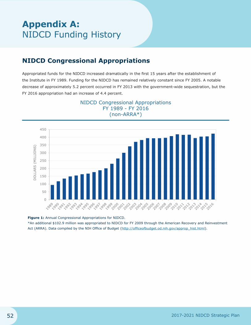
2017-2021 NIDCD Strategic Plan52
Appendix A: NIDCD Funding History
NIDCD Congressional Appropriations
Appropriated funds for the NIDCD increased dramatically in the first 15 years after the establishment of
the Institute in FY 1989. Funding for the NIDCD has remained relatively constant since FY 2005. A notable
decrease of approximately 5.2 percent occurred in FY 2013 with the government-wide sequestration, but the
FY 2016 appropriation had an increase of 4.4 percent.
NIDCD Congressional Appropriations FY 1989 - FY 2016
(non-ARRA*
*An additional $102.9 million was appropriated to NIDCD for FY 2009 through the American Recovery and Reinvestment Act (ARRA). Data compiled by the NIH Office of Budget (http://officeofbudget.od.nih.gov/approp_hist.html).
)
0
50
100
150
200
250
300
350
400
450
DO
LLARS (
MIL
LIO
NS)
Figure 1: Annual Congressional Appropriations for NIDCD.
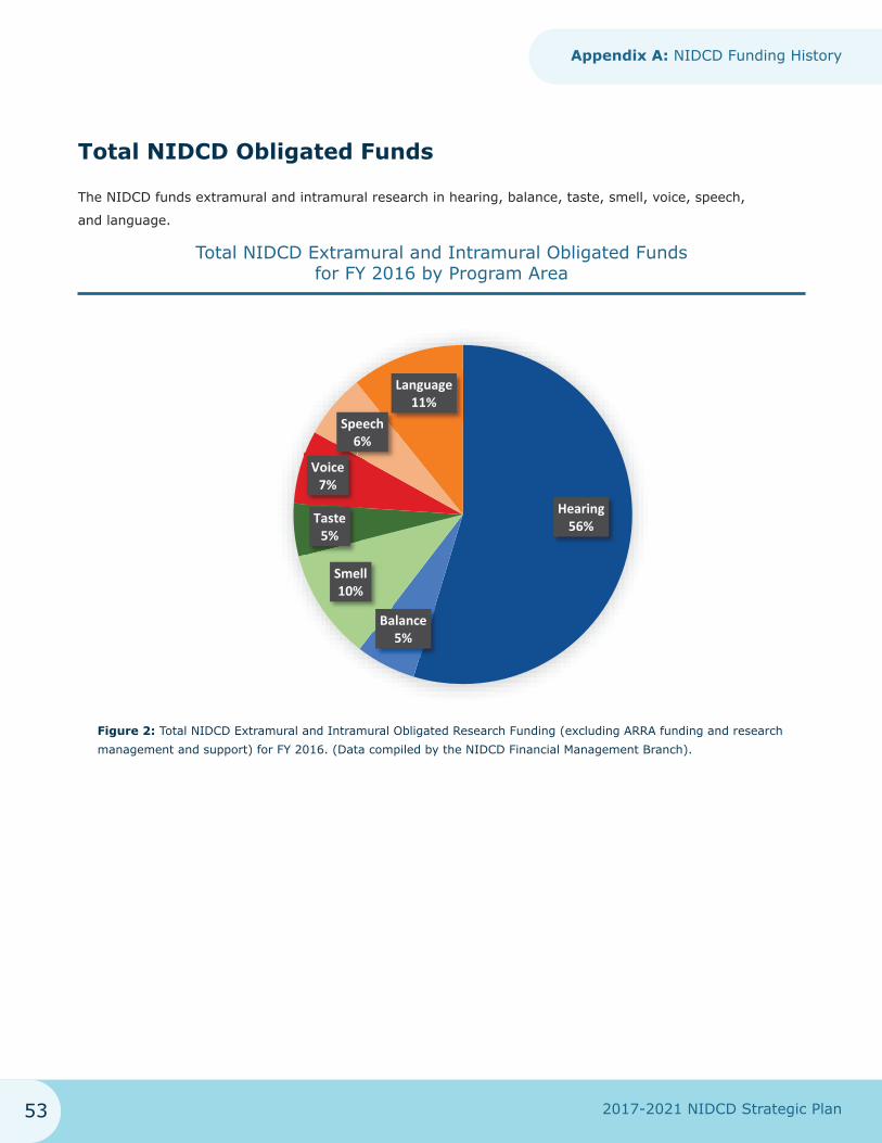
2017-2021 NIDCD Strategic Plan53
Appendix A: NIDCD Funding History
Total NIDCD Obligated Funds
The NIDCD funds extramural and intramural research in hearing, balance, taste, smell, voice, speech,
and language.
Total NIDCD Extramural and Intramural Obligated Funds for FY 2016 by Program Area
Hearing56%
Smell10%
Taste5%
Speech6%
Language11%
Voice7%
Balance5%
Figure 2: Total NIDCD Extramural and Intramural Obligated Research Funding (excluding ARRA funding and research management and support) for FY 2016. (Data compiled by the NIDCD Financial Management Branch).
2017-2021 NIDCD Strategic Plan54
Appendix B: The NIDCD 2017-2021 Strategic Plan: The Process
In the fall of 2015, NIDCD’s Science Policy and Planning Branch (SPPB) began the process of updating the
current NIDCD Strategic Plan for research, which was expiring in 2016. SPPB took the following steps to
update the previous strategic plan:
1. Established a Scientific Expert Working Group: In January 2016, the NIDCD identified and invited 12 outside
scientific experts to serve on the Working Group to update the NIDCD Strategic Plan. The experts
represented NIDCD’s mission areas (Hearing and Balance; Taste and Smell; Voice, Speech, and Language).
A roster of the Working Group is below. NIDCD staff also selected a working group chairperson, who is a
current NDCD Advisory Council member, and will serve as Council Liaison. SPPB and other NIDCD staff
served as resource persons.
2. Convened Scientific Expert Working Group via Teleconference: The Working Group held its first conference
call in February 2016. SPPB hosted separate working group teleconferences for each of the three main
program areas in March 2016. Prior to the teleconferences, working group participants received instructions
(including a current NIDCD portfolio analysis, templates with the previous science advances and scientific
objectives, and roster/contact information of working group members and NIDCD staff). The Working Group
discussed scientific advances that they considered suitable and identified areas of outstanding opportunity
and unmet need within their areas of expertise.
3. Face to Face Working Group Meeting: The Working Group met in Bethesda, Maryland, at the NIDCD to
finalize the draft science advances and research objectives in May 2016.
4. Presentation to NDCD Advisory Council: Dr. Charles Liberman, chair of the Working Group, presented the
Working Group’s recommendations for research objectives in all three program areas of the NIDCD Plan
at the May 20, 2016, meeting. Council members had the opportunity to comment on the draft objectives. In
June 2016, the NIDCD SPPB sent the first draft of the Plan to Council for review and comment. At the
September 2016 NDCD Advisory Council meeting, NIDCD staff announced that the draft Plan would be made
available for public comment.
5. Solicited Public Comments: The draft Plan was made available for a 30-day Public Comment Period on
the NIDCD website in the fall of 2016. To announce the public comment period, the NIDCD published the
Notice (NOT-DC-16-006) in the NIH Guide for Grants and Contracts on September 1, 2016. The NIDCD also
published a Notice in the Federal Register on September 1, 2016. The NIDCD received 224 comments from
the public.
6. Finalized and Posted the Plan on the NIDCD Website: Once appropriate Public Comments were incorporated
into the draft approved by NIDCD staff, SPPB finalized the Plan and published it on the NIDCD website in
early 2017.
2017-2021 NIDCD Strategic Plan55
Appendix B: The NIDCD 2017-2021 Strategic Plan: The Process
Working Group Roster
Hearing and Balance
M. Charles Liberman, Ph.D. (Chair)
Director, Eaton-Peabody Laboratories
Massachusetts Eye and Ear
Andrew K. Groves, Ph.D.
Professor and Co-Director, Program in Developmental Biology
Departments of Neuroscience and Molecular and Human Genetics
Baylor College of Medicine
Jian-Dong Li, M.D., Ph.D.
Professor and Director, Institute for Biomedical Sciences
Georgia Research Alliance Eminent Scholar
Georgia State University
Jay T. Rubinstein, M.D., Ph.D.
Professor and Director, Department of Otolaryngology
Virginia Merrill Bloedel Hearing Research Center
University of Washington
Christoph E. Schreiner, M.D., Ph.D.
Professor and Vice Chairman of Otolaryngology – Head and Neck Surgery
Kavli Neuroscience Center
University of California San Francisco
Richard J. Smith, M.D.
Professor and Director, Iowa Institute of Human Genetics, Department of Otolaryngology
University of Iowa
Debara L. Tucci, M.D.
Professor of Surgery, Division of Otolaryngology – Head and Neck Surgery
Duke University Medical Center
2017-2021 NIDCD Strategic Plan56
Appendix B: The NIDCD 2017-2021 Strategic Plan: The Process
Taste and Smell
Sue C. Kinnamon, Ph.D.
Professor, Department of Otolaryngology
University of Colorado, Denver
Donald A. Wilson, Ph.D.
Senior Research Scientist and Deputy Director, Emotional Brain Institute
New York University School of Medicine
Voice, Speech, and Language
Diane M. Bless, Ph.D.
Professor Emeritus, Departments of Communicative Disorders and Surgery
University of Wisconsin – Madison
School of Medicine and Public Health
Kristina Simonyan, M.D., Ph.D.
Associate Professor of Neurology and Otolaryngology
Icahn School of Medicine at Mount Sinai
Helen Tager-Flusberg, Ph.D.
Professor, Department of Psychology
Boston University
NIDCD Staff Participants
James F. Battey, Jr., M.D., Ph.D.
Kathy Bainbridge, Ph.D.
Laura Cole, Ph.D.
Judith Cooper, Ph.D.
Janet Cyr, Ph.D.
Amy Donahue, Ph.D.
Nancy Freeman, Ph.D.
Andrew Griffith, M.D., Ph.D.
Steven Hirschfeld, M.D.
Howard Hoffman, M.A.
Craig Jordan, Ph.D.
Lisa Kennedy, Ph.D.
Chuan-Ming Li, Ph.D.
Roger Miller, Ph.D.
Geri Piazza, M.A.
Christopher Platt, Ph.D.
Amy Poremba, Ph.D.
Alberto Rivera-Rentas, Ph.D.
Elka Scordalakes, Ph.D.
Lana Shekim, Ph.D.
Susan Sullivan, Ph.D.
Bracie Watson, Ph.D.
Ginger Webb, M.S.
Baldwin Wong, B.S.
2017-2021 NIDCD Strategic Plan57
Appendix C: NIDCD’s Trans-NIH and Trans-Agency Activities
For the most up-to-date listing of NIDCD’s trans-NIH activities, see https://dpcpsi.nih.gov/collaboration/index.
Trans-NIH Activities
NIH Autism Coordinating Committee (NIH/ACC): Formed by the NIH in 1997 at the request of Congress,
the NIH/ACC has been instrumental in planning trans-NIH research initiatives to advance the understanding
of autism. The mission is to enhance the quality, pace, and coordination of autism research efforts at the NIH.
In addition to program staff from seven ICOs, the NIMH Office of Autism Research Coordination and the NIMH
National Database for Autism Research Office participate in NIH/ACC meetings, keeping NIH program offices
apprised of their activities and coordinating projects of mutual interest. The NIH/ACC continually monitors the
NIH autism research portfolio and the agency’s progress toward meeting the goals of the Interagency Autism
Coordinating Committee Strategic Plan for ASD Research.
The Brain Research through Advancing Innovative Neurotechnologies (BRAIN) Initiative: Part of
a Presidential goal aimed at revolutionizing our understanding of the human brain, the BRAIN Initiative aims
to accelerate the development and application of innovative technologies so that researchers will be able to
produce a revolutionary new dynamic picture of the brain. For the first time, investigators will be able to show
how individual cells and complex neural circuits interact in both time and space. Long desired by researchers
seeking new ways to treat, cure, and even prevent brain disorders, this picture will address major gaps in
our current knowledge and provide unprecedented opportunities for exploring exactly how the brain enables
the human body to record, process, utilize, store, and retrieve vast quantities of information, all at the speed
of thought.
National Advisory Board on Medical Rehabilitation Research: The advisory board was stablished by the
Director of NIH to advise the directors of NIH ICOs, the Eunice Kennedy Shriver National Institute of Child
Health and Human Development (NICHD), and NICHD’s National Center for Medical Rehabilitation Research on
matters and policies relating to the Center’s programs. The Board is comprised of 12 members representing
health and scientific disciplines related to medical rehabilitation and six members representing persons
with disabilities.
NIH Medical Rehabilitation Coordinating Committee: Established by the NIH Director to comply with
Public Law 101-613, the coordinating committee will make recommendations with respect to the content of
the Research Plan and the activities of the NIH Clinical Center that are carried out in conjunction with other
components of NIH and with other federal government agencies.
2017-2021 NIDCD Strategic Plan58
Appendix C: NIDCD’s Trans-NIH and Trans-Agency Activities
The NIH Human Connectome Project: An ambitious effort to map the neural pathways that underlie
human brain function, the Project aims to acquire and share data about the structural and functional
connectivity of the human brain. It will greatly advance the capabilities for imaging and analyzing brain
connections, resulting in improved sensitivity, resolution, and utility, thereby accelerating progress in the
emerging field of human connectomics.
NIH Neuroprosthesis Group: The program officers and staff in this trans-NIH group share an interest in
neural prosthetics and neuroengineering research. The group hosts discussions about funding opportunities,
meetings, and ongoing projects.
NIH Obesity Research Task Force: The task force was established to accelerate progress in obesity
research across NIH in view of the importance of the obesity epidemic as a public health crisis. The task force
has been instrumental in fostering trans-NIH collaboration on obesity research, including basic, clinical, and
population studies. The task force also sponsors an NIH seminar series on obesity research topics.
Prevention Research Coordinating Committee: The committee serves as a venue for exchanging
information on recent scientific advances in disease prevention; examining the impact of new policies on
research; planning new or discussing ongoing initiatives; and highlighting program accomplishments. The
trans-NIH, trans-agency committee provides a broad perspective on the current state-of-the-science and
actively disseminates information about prevention-related activities sponsored by federal and non-federal
organizations to the NIH ICOs.
Trans-NIH Rare Diseases Working Group: The working group aims to develop an integrated NIH-wide plan
for research in rare diseases that addresses basic, translational, and clinical aspects aimed at the prevention
and cure of rare diseases.
NIHSeniorHealth.gov: The senior-friendly NIH website is specially formatted for optimal use by seniors
seeking health information. It features health information on a variety of topics pertinent to older adults and
includes videos, interactive quizzes, and FAQs to reinforce learning.
Trans-NIH Zebrafish Coordinating Committee: In 1997, the committee was established in response to the
scientific community’s recommendation to promote the use of zebrafish as a model organism for the study of
vertebrate development and disease. The committee developed a website to provide information about major
NIH-organized zebrafish meetings, funding opportunities for zebrafish genomics and genetic resources, major
resources generated from grants funded in response to Trans-NIH zebrafish initiatives, training courses and
scientific meetings related to the zebrafish initiatives, and selected reports and publications.

2017-2021 NIDCD Strategic Plan59
Appendix C: NIDCD’s Trans-NIH and Trans-Agency Activities
Trans-Agency Efforts
Early Hearing Detection and Intervention: The NIDCD’s collaboration with the Centers for Disease Control
and Prevention (CDC) and HRSA focuses on a bringing together federal agencies that are interested in issues
related to screening infants for hearing loss and providing early intervention.
It’s a Noisy Planet. Protect Their Hearing® Public Education Campaign: The NIDCD sponsors It’s a
Noisy Planet. Protect Their Hearing®, a national public education campaign to increase awareness among
parents of children aged 8 to 12 about the causes and prevention of noise-induced hearing loss. Our federal
partner is the National Institute for Occupational Safety and Health (NIOSH) at the CDC, and several
nonfederal organizations are involved in the campaign also.
Interagency Autism Coordinating Committee (IACC): Established in accordance with the Autism
Collaboration, Accountability, Research, Education, and Support (CARES) Act of 2014, the IACC federal
advisory committee is charged with coordinating all efforts with the Department of HHS and across member
federal agencies concerning autism spectrum disorder. The committee was established to accelerate progress
in autism spectrum disorder biomedical research and services efforts by improving coordination and
communication across the federal government and by working in partnership with the autism community.
NIDCD-Supported Epidemiological Studies with the Centers for Disease Control and Prevention
(CDC): Several CDC studies are supported by NIDCD.
• NIOSH Audiometric Examinations for Population-Based Surveys: NIDCD provides funding for
scientific and technical support as well as quality assurance of three large audiometric examination surveys:
¡ The National Health and Nutrition Examination Survey (NHANES)
¡ Age, Gene/Environment Susceptibility Study–Reykjavik Study (AGES–RS)
¡ The Early Childhood Longitudinal Study
• National Center for Health Statistics (NCHS) Balance/Dizziness Problem Examinations: NIDCD
provides funding for the inclusion of Balance/Dizziness Examinations for a representative sample of U.S.
adults aged 18 and older and children aged 3 to 17 in the 2016 National Health Interview Survey (NHIS).
• NHIS Hearing Testing: The NIDCD provides funding for a hearing component to the National Health
Interview Survey (NHIS) by sponsoring inclusion of many additional questions on hearing loss and tinnitus.
Department of Education Early Childhood Longitudinal Study: The NIDCD supports hearing screening
examinations in the Early Childhood Longitudinal Study for the Kindergarten cohort.
2017-2021 NIDCD Strategic Plan60
Appendix C: NIDCD’s Trans-NIH and Trans-Agency Activities
Advanced Electrode Microfabrication for Neural Prostheses at the Department of Energy: The
NIDCD provides funding support for the Lawrence Livermore National Laboratory to develop precise and rapid
construction micromachining techniques and construct arrays of microelectrodes suitable for recording and
stimulating neural tissue. These devices will be specifically optimized for use in the NIDCD mission areas of
voice, speech, hearing, and balance.
Interagency Committee on Disability Research (ICDR): NIDCD takes part in this government-wide group
that meets monthly to discuss issues related to people with disabilities and to coordinate research in this area.
2017-2021 NIDCD Strategic Plan61
Appendix D: Glossary and Acronym List
Glossary
afferent: conducting toward the center; for neurons, conducting nerve impulses toward the spinal cord
and brain
aphasia: total or partial loss of the ability to use or understand language; usually caused by stroke, brain
disease, or injury
apraxia of speech: a speech disorder, also known as verbal apraxia or dyspraxia, in which a person has
trouble speaking because of inability to execute a voluntary movement despite normal muscle function
assistive technologies: products, devices, or equipment that help maintain, increase, or improve the
functional capabilities of people with disabilities
auditory nerve: eighth cranial nerve that connects the inner ear to the brainstem and is responsible for
hearing and balance
auditory system: the outer, middle, and inner ear, along with the neurons and brain regions involved
in hearing
autism spectrum disorders: a spectrum of developmental disorders that begin in early childhood and
persists throughout adulthood; autism spectrum disorders affect three crucial areas of development:
communication, social interaction, and creative or imaginative play
biofilm: communities of bacteria, such as the potentially antibiotic-resistant bacterial communities that are
present in the middle ears of most children with chronic ear infections
biomarker: a specific physical trait or a measurable biologically produced change in the body connected with
a disease or health condition
chemesthesis: the “feel” of a chemical; the term describes chemically provoked irritation
chemical senses: taste and smell
cochlea: the organ of hearing
cochlear implant: a medical device that bypasses damaged structures in the inner ear and directly
stimulates the auditory nerve, allowing some people who are deaf or hard of hearing to learn to hear and
interpret sounds and speech
comorbid: the existence of one or more co-occurring disorders in addition to a primary disorder
2017-2021 NIDCD Strategic Plan62
Appendix D: Glossary and Acronym List
efferent: conducting away from the center; for neurons, conducting outward from the spinal cord and brain
embryonic stem cells: cells that are derived from the inner cell mass of blastocyst stage embryos, are
capable of dividing without differentiating for a prolonged period in culture, and are known to develop into
cells and tissues of the three primary germ layers
epidemiology: the branch of medical science that investigates all the factors that determine the presence or
absence of diseases and disorders in a population
epigenetics: the study of heritable changes caused by the activation and deactivation of genes without any
change in the underlying DNA sequence of the organism
eustachian tube: a small passageway that connects the upper part of the throat to the middle ear; its job is
to supply fresh air to the middle ear, drain fluid, and keep air pressure at a steady level between the nose and
the ear
gene expression: the process by which the information encoded in a gene is used to direct the assembly of a
protein molecule; different subsets of genes are expressed in different cell types or under different conditions
genetics: the study of particular genes, DNA, and heredity
genomics: the study of the genome (the entire genetic makeup) of an organism
hair cells: sensory cells of the inner ear, which are topped with hair-like structures (stereocilia) and which
transform the mechanical energy of sound waves into nerve impulses
hearing aid: an electronic device that brings amplified sound to the ear; it usually consists of a microphone,
amplifier, and receiver
idiopathic: relating to a disease or disorder that arises spontaneously or without a known cause
inner ear: part of the ear that contains both the organ of hearing (the cochlea) and the organ of balance
(the labyrinth)
knockout: an organism that has been genetically engineered to lack one or more specific genes; scientists
study knockout organisms to determine the impact of the missing gene, which helps determine its the function
larynx: valve structure between the trachea (windpipe) and the pharynx (the upper throat) that is the
primary organ of voice production
model organism: animal species used in medical research to mimic aspects of a disease found in humans
mutation: a change in a DNA sequence that can result from DNA copying mistakes made during cell division,
exposure to ionizing radiation, exposure to chemical mutagens, or infection by viruses
2017-2021 NIDCD Strategic Plan63
Appendix D: Glossary and Acronym List
neural prostheses: devices such as the cochlear implant that substitute for an injured or diseased part of the
nervous system
nociceptors: the relatively unspecialized nerve cell endings that initiate the sensation of pain
olfaction: the sense of smell; to perceive odor or scent through stimuli affecting the olfactory nerves
otitis media: inflammation of the middle ear caused by infection
ototoxic: a drug or compound such as a special class of antibiotics, aminoglycoside antibiotics, that can
damage the hearing and balance organs located in the inner ear for some individuals
pathogenesis: the development of a disease or condition, particularly the cellular and molecular origins and
causes of disease development
phenotype: an individual’s physical and behavioral characteristics
pheromone: chemical substance secreted by an animal that elicits a specific behavioral or physiological
response in another animal of the same species
polymorphism: one of two or more variants of a particular DNA sequence that can correlate with disease,
drug response, and other phenotypes; the most common type of polymorphism involves variation at a single
base pair (single nucleotide polymorphism) of DNA
psychoacoustics: the study of sound perception
rhinitis: inflammation of the mucous membranes of the nose, generally accompanied by discharge
(runny nose) and usually caused by a virus infection (e.g., the common cold) or by an allergic reaction (e.g.,
hay fever)
sinusitis: inflammation or infection of one of the air-filled nasal sinuses
spasmodic dysphonia: momentary disruption of voice caused by involuntary movements of one or more
muscles of the larynx
spiral ganglion: the group of nerve cells that serve the sense of hearing by sending a representation of
sound from the cochlea to the brain; the cell bodies of the spiral ganglion neurons are found in the spiral
structure of the cochlea
stereocilia: see “hair cells”
stria vascularis: specialized epithelium lining the cochlear duct that maintains the ion homeostasis of the
fluid within the cochlea
stuttering: a speech disorder in which sounds, syllables, or words are repeated or prolonged, disrupting the
normal flow of speech
2017-2021 NIDCD Strategic Plan64
Appendix D: Glossary and Acronym List
synapse: a junction between two nerve cells
tinnitus: sensation of a ringing, roaring, or buzzing sound in the ears or head when no actual sound stimulus
is present in the environment
tonotopic: the spatial arrangement of where sounds of different frequency are processed in the brain. For
example, the auditory nerves that carry signals from adjacent portions of the cochlea project their information
to adjacent portions of the auditory cortex
transduction: the process by which stimuli in the environment are converted into electrical (neural) signals
by sensory receptors
vertigo: illusion of movement; a sensation as if the external world were revolving around an individual
(objective vertigo) or as if the individual were revolving in space (subjective vertigo)
vestibular system: system in the body that is responsible for maintaining balance, posture, and the body’s
orientation in space; this system also regulates locomotion and other movements and keeps objects in visual
focus as the body moves
2017-2021 NIDCD Strategic Plan65
Appendix D: Glossary and Acronym List
Acronyms
ACC Autism Coordinating Committee
ARRA American Recovery and Reinvestment Act
ASD Autism Spectrum Disorder
DEA Division of Extramural Activities
DIR Division of Intramural Research
DNA Deoxyribonucleic Acid
DSP Division of Scientific Programs
EHDI Early Hearing Detection and Intervention
FY Fiscal Year
HHS Department of Health and Human Services
HPP High Program Priority
IACC Interagency Autism Coordinating Committee
ICOs Institutes, Centers, and Offices
M.D. Doctor of Medicine
NDCD National Deafness and Other Communication Disorders
NF2 Neurofibromatosis 2
NICHD Eunice Kennedy Shriver National Institute of Child Health and Human Development
NIDCD National Institute on Deafness and Other Communication Disorders
NIH National Institutes of Health
Ph.D. Doctor of Philosophy
Plan NIDCD Strategic Plan
SLI Specific Language Impairment
SPPB Science Policy and Planning Branch
T2R Type 2 Taste Receptors
2017-2021 NIDCD Strategic Plan66
Appendix E: Bibliography
1 Blackwell DL, Lucas JW, Clarke TC. Summary health statistics for U.S. adults: National Health Interview Survey, 2012. National Center for Health Statistics. Vital Health Stat 10(260). 2014 Contract No.: 260.
2 Ward BK, Agrawal Y, Hoffman HJ, Carey JP, Della Santina CC. Prevalence and impact of bilateral vestibular hypofunction: results from the 2008 US National Health Interview Survey. JAMA Otolaryngol Head Neck Surg. 2013;139(8):803-10. doi: 10.1001/jamaoto.2013.3913. PubMed PMID: 23949355; PMCID: PMC4839981.
3 Li CM, Hoffman HJ, Ward BK, Cohen HS, Rine RM. Epidemiology of Dizziness and Balance Problems in Children in the United States: A Population-Based Study. J Pediatr. 2016;171:240-7 e3. doi: 10.1016/j.jpeds.2015.12.002. PubMed PMID: 26826885.
4 CDC. Identifying infants with hearing loss - United States, 1999-2007. MMWR Morb Mortal Wkly Rep. 2010;59(8):220-3. PubMed PMID: 20203554.
5 Vohr B. Overview: Infants and children with hearing loss-part I. Ment Retard Dev Disabil Res Rev. 2003;9(2):62-4. doi: 10.1002/mrdd.10070. PubMed PMID: 12784222.
6 Zelaya CE LJ, Hoffman HJ. Self-reported hearing trouble in adults aged 18 and over: United States, 2014. NCHS data brief, no 214. 2015.
7 Hoffman HJ, Ko C-W, Themann CL, Dillon CF, Franks JR. Reducing noise-induced hearing loss (NIHL) in adults to achieve U.S. Healthy People 2010 goals. Am J Epidemiol. 2006;(Suppl S); 163(11):S122.
8 CDC/NHANES. Based on calculations performed by NIDCD Epidemiology and Statistics Program staff: (1) using data from the 1999-2010 National Health and Nutrition Examination Survey (NHANES); (2) applying the definition of disabling hearing loss used by the 2010 Global Burden of Disease Expert Hearing Loss Team (hearing loss of 35 decibels or more in the better ear, the level at which adults could generally benefit from hearing aids) 2010.
9 Kawashima Y, Geleoc GS, Kurima K, Labay V, Lelli A, Asai Y, Makishima T, Wu DK, Della Santina CC, Holt JR, Griffith AJ. Mechanotransduction in mouse inner ear hair cells requires transmembrane channel-like genes. J Clin Invest. 2011;121(12):4796-809. doi: 10.1172/JCI60405. PubMed PMID: 22105175; PMCID: PMC3223072.
10 Kurima K, Ebrahim S, Pan B, Sedlacek M, Sengupta P, Millis BA, Cui R, Nakanishi H, Fujikawa T, Kawashima Y, Choi BY, Monahan K, Holt JR, Griffith AJ, Kachar B. TMC1 and TMC2 Localize at the Site of Mechanotransduction in Mammalian Inner Ear Hair Cell Stereocilia. Cell Rep. 2015;12(10):1606-17. doi: 10.1016/j.celrep.2015.07.058. PubMed PMID: 26321635; PMCID: PMC4569002.
11 Liedtke W. A precisely defined role for the tip link-associated protein TMIE in the mechanoelectrical transduction channel complex of inner ear hair cells. Neuron. 2014;84(5):889-91. doi: 10.1016/j.neuron.2014.11.015. PubMed PMID: 25475183.
2017-2021 NIDCD Strategic Plan67
Appendix E: Bibliography
12 Maeda R, Kindt KS, Mo W, Morgan CP, Erickson T, Zhao H, Clemens-Grisham R, Barr-Gillespie PG, Nicolson T. Tip-link protein protocadherin 15 interacts with transmembrane channel-like proteins TMC1 and TMC2. Proc Natl Acad Sci U S A. 2014;111(35):12907-12. doi: 10.1073/pnas.1402152111. PubMed PMID: 25114259; PMCID: PMC4156717.
13 Pan B, Geleoc GS, Asai Y, Horwitz GC, Kurima K, Ishikawa K, Kawashima Y, Griffith AJ, Holt JR. TMC1 and TMC2 are components of the mechanotransduction channel in hair cells of the mammalian inner ear. Neuron. 2013;79(3):504-15. doi: 10.1016/j.neuron.2013.06.019. PubMed PMID: 23871232; PMCID: PMC3827726.
14 Zhao B, Wu Z, Grillet N, Yan L, Xiong W, Harkins-Perry S, Muller U. TMIE is an essential component of the mechanotransduction machinery of cochlear hair cells. Neuron. 2014;84(5):954-67. doi: 10.1016/j.neuron.2014.10.041. PubMed PMID: 25467981; PMCID: PMC4258123.
15 Xiong W, Grillet N, Elledge HM, Wagner TF, Zhao B, Johnson KR, Kazmierczak P, Muller U. TMHS is an integral component of the mechanotransduction machinery of cochlear hair cells. Cell. 2012;151(6):1283-95. doi: 10.1016/j.cell.2012.10.041. PubMed PMID: 23217710; PMCID: PMC3522178.
16 Burns JC, Kelly MC, Hoa M, Morell RJ, Kelley MW. Single-cell RNA-Seq resolves cellular complexity in sensory organs from the neonatal inner ear. Nat Commun. 2015;6:8557. doi: 10.1038/ncomms9557. PubMed PMID: 26469390; PMCID: PMC4634134.
17 Cai T, Jen HI, Kang H, Klisch TJ, Zoghbi HY, Groves AK. Characterization of the transcriptome of nascent hair cells and identification of direct targets of the Atoh1 transcription factor. J Neurosci. 2015;35(14):5870-83. doi: 10.1523/JNEUROSCI.5083-14.2015. PubMed PMID: 25855195; PMCID: PMC4388939.
18 Scheffer DI, Shen J, Corey DP, Chen ZY. Gene Expression by Mouse Inner Ear Hair Cells during Development. J Neurosci. 2015;35(16):6366-80. doi: 10.1523/JNEUROSCI.5126-14.2015. PubMed PMID: 25904789; PMCID: PMC4405555.
19 Shen J, Scheffer DI, Kwan KY, Corey DP. SHIELD: an integrative gene expression database for inner ear research. Database (Oxford). 2015;2015:bav071. doi: 10.1093/database/bav071. PubMed PMID: 26209310; PMCID: PMC4513695.
20 Waldhaus J, Durruthy-Durruthy R, Heller S. Quantitative High-Resolution Cellular Map of the Organ of Corti. Cell Rep. 2015;11(9):1385-99. doi: 10.1016/j.celrep.2015.04.062. PubMed PMID: 26027927; PMCID: PMC4465070.
21 Elkon R, Milon B, Morrison L, Shah M, Vijayakumar S, Racherla M, Leitch CC, Silipino L, Hadi S, Weiss-Gayet M, Barras E, Schmid CD, Ait-Lounis A, Barnes A, Song Y, Eisenman DJ, Eliyahu E, Frolenkov GI, Strome SE, Durand B, Zaghloul NA, Jones SM, Reith W, Hertzano R. RFX transcription factors are essential for hearing in mice. Nat Commun. 2015;6:8549. doi: 10.1038/ncomms9549. PubMed PMID: 26469318; PMCID: PMC4634137.
2017-2021 NIDCD Strategic Plan68
Appendix E: Bibliography
22 Shin JB, Krey JF, Hassan A, Metlagel Z, Tauscher AN, Pagana JM, Sherman NE, Jeffery ED, Spinelli KJ, Zhao H, Wilmarth PA, Choi D, David LL, Auer M, Barr-Gillespie PG. Molecular architecture of the chick vestibular hair bundle. Nat Neurosci. 2013;16(3):365-74. doi: 10.1038/nn.3312. PubMed PMID: 23334578; PMCID: PMC3581746.
23 Wilmarth PA, Krey JF, Shin JB, Choi D, David LL, Barr-Gillespie PG. Hair-bundle proteomes of avian and mammalian inner-ear utricles. Sci Data. 2015;2:150074. doi: 10.1038/sdata.2015.74. PubMed PMID: 26645194; PMCID: PMC4672683.
24 Francis SP, Krey JF, Krystofiak ES, Cui R, Nanda S, Xu W, Kachar B, Barr-Gillespie PG, Shin JB. A short splice form of Xin-actin binding repeat containing 2 (XIRP2) lacking the Xin repeats is required for maintenance of stereocilia morphology and hearing function. J Neurosci. 2015;35(5):1999-2014. doi: 10.1523/JNEUROSCI.3449-14.2015. PubMed PMID: 25653358; PMCID: PMC4315831.
25 Chai R, Kuo B, Wang T, Liaw EJ, Xia A, Jan TA, Liu Z, Taketo MM, Oghalai JS, Nusse R, Zuo J, Cheng AG. Wnt signaling induces proliferation of sensory precursors in the postnatal mouse cochlea. Proc Natl Acad Sci U S A. 2012;109(21):8167-72. doi: 10.1073/pnas.1202774109. PubMed PMID: 22562792; PMCID: PMC3361451.
26 Shi F, Kempfle JS, Edge AS. Wnt-responsive Lgr5-expressing stem cells are hair cell progenitors in the cochlea. J Neurosci. 2012;32(28):9639-48. doi: 10.1523/JNEUROSCI.1064-12.2012. PubMed PMID: 22787049; PMCID: PMC3417821.
27 Koehler KR, Hashino E. 3D mouse embryonic stem cell culture for generating inner ear organoids. Nat Protoc. 2014;9(6):1229-44. doi: 10.1038/nprot.2014.100. PubMed PMID: 24784820.
28 Lentz JJ, Jodelka FM, Hinrich AJ, McCaffrey KE, Farris HE, Spalitta MJ, Bazan NG, Duelli DM, Rigo F, Hastings ML. Rescue of hearing and vestibular function by antisense oligonucleotides in a mouse model of human deafness. Nat Med. 2013;19(3):345-50. doi: 10.1038/nm.3106. PubMed PMID: 23380860; PMCID: PMC3657744.
29 Bermingham-McDonogh O, Reh TA. Regulated reprogramming in the regeneration of sensory receptor cells. Neuron. 2011;71(3):389-405. doi: 10.1016/j.neuron.2011.07.015. PubMed PMID: 21835338; PMCID: PMC4403668.
30 Conde de Felipe MM, Feijoo Redondo A, Garcia-Sancho J, Schimmang T, Duran Alonso MB. Cell- and gene-therapy approaches to inner ear repair. Histol Histopathol. 2011;26(7):923-40. PubMed PMID: 21630222.
31 Shibata SB, Ranum PT, Moteki H, Pan B, Goodwin AT, Goodman SS, Abbas PJ, Holt JR, Smith RJ. RNA Interference Prevents Autosomal-Dominant Hearing Loss. Am J Hum Genet. 2016. doi: 10.1016/j.ajhg.2016.03.028. PubMed PMID: 27236922.
32 Hickox AE, Liberman MC. Is noise-induced cochlear neuropathy key to the generation of hyperacusis or tinnitus? J Neurophysiol. 2014;111(3):552-64. doi: 10.1152/jn.00184.2013. PubMed PMID: 24198321; PMCID: PMC3921399.
2017-2021 NIDCD Strategic Plan69
Appendix E: Bibliography
33 Kujawa SG, Liberman MC. Synaptopathy in the noise-exposed and aging cochlea: Primary neural degeneration in acquired sensorineural hearing loss. Hear Res. 2015;330(Pt B):191-9. doi: 10.1016/j.heares.2015.02.009. PubMed PMID: 25769437; PMCID: PMC4567542.
34 Liberman MC. Hidden Hearing Loss. Sci Am. 2015;313(2):48-53. PubMed PMID: 26349143.
35 Sergeyenko Y, Lall K, Liberman MC, Kujawa SG. Age-related cochlear synaptopathy: an early-onset contributor to auditory functional decline. J Neurosci. 2013;33(34):13686-94. doi: 10.1523/JNEUROSCI.1783-13.2013. PubMed PMID: 23966690; PMCID: PMC3755715.
36 Furman AC, Kujawa SG, Liberman MC. Noise-induced cochlear neuropathy is selective for fibers with low spontaneous rates. J Neurophysiol. 2013;110(3):577-86. doi: 10.1152/jn.00164.2013. PubMed PMID: 23596328; PMCID: PMC3742994.
37 Viana LM, O’Malley JT, Burgess BJ, Jones DD, Oliveira CA, Santos F, Merchant SN, Liberman LD, Liberman MC. Cochlear neuropathy in human presbycusis: Confocal analysis of hidden hearing loss in post-mortem tissue. Hear Res. 2015;327:78-88. doi: 10.1016/j.heares.2015.04.014. PubMed PMID: 26002688; PMCID: PMC4554812.
38 Flores EN, Duggan A, Madathany T, Hogan AK, Marquez FG, Kumar G, Seal RP, Edwards RH, Liberman MC, Garcia-Anoveros J. A non-canonical pathway from cochlea to brain signals tissue-damaging noise. Curr Biol. 2015;25(5):606-12. doi: 10.1016/j.cub.2015.01.009. PubMed PMID: 25639244; PMCID: PMC4348215.
39 Liu C, Glowatzki E, Fuchs PA. Unmyelinated type II afferent neurons report cochlear damage. Proc Natl Acad Sci U S A. 2015;112(47):14723-7. doi: 10.1073/pnas.1515228112. PubMed PMID: 26553995; PMCID: PMC4664349.
40 Kuhn S, Johnson SL, Furness DN, Chen J, Ingham N, Hilton JM, Steffes G, Lewis MA, Zampini V, Hackney CM, Masetto S, Holley MC, Steel KP, Marcotti W. miR-96 regulates the progression of differentiation in mammalian cochlear inner and outer hair cells. Proc Natl Acad Sci U S A. 2011;108(6):2355-60. doi: 10.1073/pnas.1016646108. PubMed PMID: 21245307; PMCID: PMC3038748.
41 Mencia A, Modamio-Hoybjor S, Redshaw N, Morin M, Mayo-Merino F, Olavarrieta L, Aguirre LA, del Castillo I, Steel KP, Dalmay T, Moreno F, Moreno-Pelayo MA. Mutations in the seed region of human miR-96 are responsible for nonsyndromic progressive hearing loss. Nat Genet. 2009;41(5):609-13. doi: 10.1038/ng.355. PubMed PMID: 19363479.
42 Shearer AE, DeLuca AP, Hildebrand MS, Taylor KR, Gurrola J, 2nd, Scherer S, Scheetz TE, Smith RJ. Comprehensive genetic testing for hereditary hearing loss using massively parallel sequencing. Proc Natl Acad Sci U S A. 2010;107(49):21104-9. doi: 10.1073/pnas.1012989107. PubMed PMID: 21078986; PMCID: PMC3000272.
43 Sloan-Heggen CM, Babanejad M, Beheshtian M, Simpson AC, Booth KT, Ardalani F, Frees KL, Mohseni M, Mozafari R, Mehrjoo Z, Jamali L, Vaziri S, Akhtarkhavari T, Bazazzadegan N, Nikzat N, Arzhangi S, Sabbagh F, Otukesh H, Seifati SM, Khodaei H, Taghdiri M, Meyer NC, Daneshi A, Farhadi M, Kahrizi K, Smith RJ, Azaiez H, Najmabadi H. Characterising the spectrum of autosomal recessive hereditary hearing loss in Iran. J Med Genet. 2015;52(12):823-9. doi: 10.1136/jmedgenet-2015-103389. PubMed PMID: 26445815; PMCID: PMC4733363.
2017-2021 NIDCD Strategic Plan70
Appendix E: Bibliography
44 Alford RL, Arnos KS, Fox M, Lin JW, Palmer CG, Pandya A, Rehm HL, Robin NH, Scott DA, Yoshinaga-Itano C, Loss AWGoUoGEGftEDoCH, Professional P, Guidelines C. American College of Medical Genetics and Genomics guideline for the clinical evaluation and etiologic diagnosis of hearing loss. Genet Med. 2014;16(4):347-55. doi: 10.1038/gim.2014.2. PubMed PMID: 24651602.
45 Kimberling WJ, Hildebrand MS, Shearer AE, Jensen ML, Halder JA, Trzupek K, Cohn ES, Weleber RG, Stone EM, Smith RJ. Frequency of Usher syndrome in two pediatric populations: Implications for genetic screening of deaf and hard of hearing children. Genet Med. 2010;12(8):512-6. doi: 10.1097/GIM.0b013e3181e5afb8. PubMed PMID: 20613545; PMCID: PMC3131500.
46 Declau F, Boudewyns A, Van den Ende J, Peeters A, van den Heyning P. Etiologic and audiologic evaluations after universal neonatal hearing screening: analysis of 170 referred neonates. Pediatrics. 2008;121(6):1119-26. doi: 10.1542/peds.2007-1479. PubMed PMID: 18519481.
47 Choi BY, Park G, Gim J, Kim AR, Kim BJ, Kim HS, Park JH, Park T, Oh SH, Han KH, Park WY. Diagnostic application of targeted resequencing for familial nonsyndromic hearing loss. PLoS One. 2013;8(8):e68692. doi: 10.1371/journal.pone.0068692. PubMed PMID: 23990876; PMCID: PMC3750053.
48 Vona B, Muller T, Nanda I, Neuner C, Hofrichter MA, Schroder J, Bartsch O, Lassig A, Keilmann A, Schraven S, Kraus F, Shehata-Dieler W, Haaf T. Targeted next-generation sequencing of deafness genes in hearing-impaired individuals uncovers informative mutations. Genet Med. 2014;16(12):945-53. doi: 10.1038/gim.2014.65. PubMed PMID: 24875298; PMCID: PMC4262760.
49 Jayawardena AD, Shearer AE, Smith RJ. Sensorineural Hearing Loss: A Changing Paradigm for Its Evaluation. Otolaryngol Head Neck Surg. 2015;153(5):843-50. doi: 10.1177/0194599815596727. PubMed PMID: 26216887; PMCID: PMC4730883.
50 Sloan-Heggen CM, Bierer AO, Shearer AE, Kolbe DL, Nishimura CJ, Frees KL, Ephraim SS, Shibata SB, Booth KT, Campbell CA, Ranum PT, Weaver AE, Black-Ziegelbein EA, Wang D, Azaiez H, Smith RJ. Comprehensive genetic testing in the clinical evaluation of 1119 patients with hearing loss. Hum Genet. 2016;135(4):441-50. doi: 10.1007/s00439-016-1648-8. PubMed PMID: 26969326; PMCID: PMC4796320.
51 Akil O, Seal RP, Burke K, Wang C, Alemi A, During M, Edwards RH, Lustig LR. Restoration of hearing in the VGLUT3 knockout mouse using virally mediated gene therapy. Neuron. 2012;75(2):283-93. doi: 10.1016/j.neuron.2012.05.019. PubMed PMID: 22841313; PMCID: PMC3408581.
52 Askew C, Rochat C, Pan B, Asai Y, Ahmed H, Child E, Schneider BL, Aebischer P, Holt JR. Tmc gene therapy restores auditory function in deaf mice. Sci Transl Med. 2015;7(295):295ra108. doi: 10.1126/scitranslmed.aab1996. PubMed PMID: 26157030.
53 Di Domenico M, Ricciardi C, Martone T, Mazzarella N, Cassandro C, Chiarella G, D’Angelo L, Cassandro E. Towards gene therapy for deafness. J Cell Physiol. 2011;226(10):2494-9. doi: 10.1002/jcp.22617. PubMed PMID: 21792906.
54 Ou HC, Keating S, Wu P, Simon JA, Raible DW, Rubel EW. Quinoline ring derivatives protect against aminoglycoside-induced hair cell death in the zebrafish lateral line. J Assoc Res Otolaryngol. 2012;13(6):759-70. doi: 10.1007/s10162-012-0353-0. PubMed PMID: 23053627; PMCID: PMC3505584.
2017-2021 NIDCD Strategic Plan71
Appendix E: Bibliography
55 Ou H, Simon JA, Rubel EW, Raible DW. Screening for chemicals that affect hair cell death and survival in the zebrafish lateral line. Hear Res. 2012;288(1-2):58-66. doi: 10.1016/j.heares.2012.01.009. PubMed PMID: 22310494; PMCID: PMC3371178.
56 Stawicki TM, Esterberg R, Hailey DW, Raible DW, Rubel EW. Using the zebrafish lateral line to uncover novel mechanisms of action and prevention in drug-induced hair cell death. Front Cell Neurosci. 2015;9:46. doi: 10.3389/fncel.2015.00046. PubMed PMID: 25741241; PMCID: PMC4332341.
57 Thomas AJ, Wu P, Raible DW, Rubel EW, Simon JA, Ou HC. Identification of small molecule inhibitors of cisplatin-induced hair cell death: results of a 10,000 compound screen in the zebrafish lateral line. Otol Neurotol. 2015;36(3):519-25. doi: 10.1097/MAO.0000000000000487. PubMed PMID: 25687728; PMCID: PMC4332566.
58 Mizutari K, Fujioka M, Hosoya M, Bramhall N, Okano HJ, Okano H, Edge AS. Notch inhibition induces cochlear hair cell regeneration and recovery of hearing after acoustic trauma. Neuron. 2013;77(1):58-69. doi: 10.1016/j.neuron.2012.10.032. PubMed PMID: 23312516; PMCID: PMC3573859.
59 Deal JA, Sharrett AR, Albert MS, Coresh J, Mosley TH, Knopman D, Wruck LM, Lin FR. Hearing impairment and cognitive decline: a pilot study conducted within the atherosclerosis risk in communities neurocognitive study. Am J Epidemiol. 2015;181(9):680-90. doi: 10.1093/aje/kwu333. PubMed PMID: 25841870; PMCID: PMC4408947.
60 Whitson HE, Lin FR. Hearing and vision care for older adults: sensing a need to update Medicare policy. JAMA. 2014;312(17):1739-40. doi: 10.1001/jama.2014.13535. PubMed PMID: 25369486; PMCID: PMC4260264.
61 Van Laer L, Huyghe JR, Hannula S, Van Eyken E, Stephan DA, Maki-Torkko E, Aikio P, Fransen E, Lysholm-Bernacchi A, Sorri M, Huentelman MJ, Van Camp G. A genome-wide association study for age-related hearing impairment in the Saami. Eur J Hum Genet. 2010;18(6):685-93. doi: 10.1038/ejhg.2009.234. PubMed PMID: 20068591; PMCID: PMC2987344.
62 Wan G, Gomez-Casati ME, Gigliello AR, Liberman MC, Corfas G. Neurotrophin-3 regulates ribbon synapse density in the cochlea and induces synapse regeneration after acoustic trauma. Elife. 2014;3. doi: 10.7554/eLife.03564. PubMed PMID: 25329343; PMCID: PMC4227045.
63 Kurabi A, Pak K, Ryan AF, Wasserman SI. Innate Immunity: Orchestrating Inflammation and Resolution of Otitis Media. Curr Allergy Asthma Rep. 2016;16(1):6. doi: 10.1007/s11882-015-0585-2. PubMed PMID: 26732809.
64 Kerschner JE, Hong W, Taylor SR, Kerschner JA, Khampang P, Wrege KC, North PE. A novel model of spontaneous otitis media with effusion (OME) in the Oxgr1 knock-out mouse. Int J Pediatr Otorhinolaryngol. 2013;77(1):79-84. doi: 10.1016/j.ijporl.2012.09.037. PubMed PMID: 23200873; PMCID: PMC3535456.
65 Eutsey RA, Hiller NL, Earl JP, Janto BA, Dahlgren ME, Ahmed A, Powell E, Schultz MP, Gilsdorf JR, Zhang L, Smith A, Murphy TF, Sethi S, Shen K, Post JC, Hu FZ, Ehrlich GD. Design and validation of a supragenome array for determination of the genomic content of Haemophilus influenzae isolates. BMC Genomics. 2013;14:484. doi: 10.1186/1471-2164-14-484. PubMed PMID: 23865594; PMCID: PMC3723446.
2017-2021 NIDCD Strategic Plan72
Appendix E: Bibliography
66 Novotny LA, Jurcisek JA, Ward MO, Jr., Jordan ZB, Goodman SD, Bakaletz LO. Antibodies against the majority subunit of type IV Pili disperse nontypeable Haemophilus influenzae biofilms in a LuxS-dependent manner and confer therapeutic resolution of experimental otitis media. Mol Microbiol. 2015;96(2):276-92. doi: 10.1111/mmi.12934. PubMed PMID: 25597921; PMCID: PMC4423401.
67 Murphy TF. Vaccines for Nontypeable Haemophilus influenzae: the Future Is Now. Clin Vaccine Immunol. 2015;22(5):459-66. doi: 10.1128/CVI.00089-15. PubMed PMID: 25787137; PMCID: PMC4412935.
68 Yildirim I, Shea KM, Pelton SI. Pneumococcal Disease in the Era of Pneumococcal Conjugate Vaccine. Infect Dis Clin North Am. 2015;29(4):679-97. doi: 10.1016/j.idc.2015.07.009. PubMed PMID: 26610421; PMCID: PMC4662776.
69 Khoo X, Simons EJ, Chiang HH, Hickey JM, Sabharwal V, Pelton SI, Rosowski JJ, Langer R, Kohane DS. Formulations for trans-tympanic antibiotic delivery. Biomaterials. 2013;34(4):1281-8. doi: 10.1016/j.biomaterials.2012.10.025. PubMed PMID: 23146430; PMCID: PMC3511665.
70 Lee JY, Komatsu K, Lee BC, Miyata M, O’Neill Bohn A, Xu H, Yan C, Li JD. Vinpocetine inhibits Streptococcus pneumoniae-induced upregulation of mucin MUC5AC expression via induction of MKP-1 phosphatase in the pathogenesis of otitis media. J Immunol. 2015;194(12):5990-8. doi: 10.4049/jimmunol.1401489. PubMed PMID: 25972475; PMCID: PMC4456630.
71 Koo JW, Quintanilla-Dieck L, Jiang M, Liu J, Urdang ZD, Allensworth JJ, Cross CP, Li H, Steyger PS. Endotoxemia-mediated inflammation potentiates aminoglycoside-induced ototoxicity. Sci Transl Med. 2015;7(298):298ra118. doi: 10.1126/scitranslmed.aac5546. PubMed PMID: 26223301; PMCID: PMC4534720.
72 Ahlstrom JB, Horwitz AR, Dubno JR. Spatial separation benefit for unaided and aided listening. Ear Hear. 2014;35(1):72-85. doi: 10.1097/AUD.0b013e3182a02274. PubMed PMID: 24121648; PMCID: PMC3872487.
73 Roland JT, Jr., Gantz BJ, Waltzman SB, Parkinson AJ, Multicenter Clinical Trial G. United States multicenter clinical trial of the cochlear nucleus hybrid implant system. Laryngoscope. 2016;126(1):175-81. doi: 10.1002/lary.25451. PubMed PMID: 26152811; PMCID: PMC4704985.
74 Quesnel AM, Nakajima HH, Rosowski JJ, Hansen MR, Gantz BJ, Nadol JB, Jr. Delayed loss of hearing after hearing preservation cochlear implantation: Human temporal bone pathology and implications for etiology. Hear Res. 2016;333:225-34. doi: 10.1016/j.heares.2015.08.018. PubMed PMID: 26341474; PMCID: PMC4775460.
75 Reiss LA, Turner CW, Karsten SA, Gantz BJ. Plasticity in human pitch perception induced by tonotopically mismatched electro-acoustic stimulation. Neuroscience. 2014;256:43-52. doi: 10.1016/j.neuroscience.2013.10.024. PubMed PMID: 24157931; PMCID: PMC3893921.
76 Friedmann DR, Ahmed OH, McMenomey SO, Shapiro WH, Waltzman SB, Roland JT, Jr. Single-sided Deafness Cochlear Implantation: Candidacy, Evaluation, and Outcomes in Children and Adults. Otol Neurotol. 2016;37(2):e154-60. doi: 10.1097/MAO.0000000000000951. PubMed PMID: 26756150.
2017-2021 NIDCD Strategic Plan73
Appendix E: Bibliography
77 Mertens G, De Bodt M, Van de Heyning P. Cochlear implantation as a long-term treatment for ipsilateral incapacitating tinnitus in subjects with unilateral hearing loss up to 10 years. Hear Res. 2016;331:1-6. doi: 10.1016/j.heares.2015.09.016. PubMed PMID: 26433053.
78 Bierer JA, Bierer SM, Kreft HA, Oxenham AJ. A fast method for measuring psychophysical thresholds across the cochlear implant array. Trends Hear. 2015;19. doi: 10.1177/2331216515569792. PubMed PMID: 25656797; PMCID: PMC4324086.
79 van den Honert C, Kelsall DC. Focused intracochlear electric stimulation with phased array channels. J Acoust Soc Am. 2007;121(6):3703-16. doi: 10.1121/1.2722047. PubMed PMID: 17552721.
80 Noij KS, Kozin ED, Sethi R, Shah PV, Kaplan AB, Herrmann B, Remenschneider A, Lee DJ. Systematic Review of Nontumor Pediatric Auditory Brainstem Implant Outcomes. Otolaryngol Head Neck Surg. 2015;153(5):739-50. doi: 10.1177/0194599815596929. PubMed PMID: 26227469.
81 van de Berg R, Guinand N, Nguyen TA, Ranieri M, Cavuscens S, Guyot JP, Stokroos R, Kingma H, Perez-Fornos A. The vestibular implant: frequency-dependency of the electrically evoked vestibulo-ocular reflex in humans. Front Syst Neurosci. 2014;8:255. doi: 10.3389/fnsys.2014.00255. PubMed PMID: 25653601; PMCID: PMC4299437.
82 Sun DQ, Lehar M, Dai C, Swarthout L, Lauer AM, Carey JP, Mitchell DE, Cullen KE, Della Santina CC. Histopathologic Changes of the Inner ear in Rhesus Monkeys After Intratympanic Gentamicin Injection and Vestibular Prosthesis Electrode Array Implantation. J Assoc Res Otolaryngol. 2015;16(3):373-87. doi: 10.1007/s10162-015-0515-y. PubMed PMID: 25790951; PMCID: PMC4417088.
83 Phillips JO, Ling L, Nie K, Jameyson E, Phillips CM, Nowack AL, Golub JS, Rubinstein JT. Vestibular implantation and longitudinal electrical stimulation of the semicircular canal afferents in human subjects. J Neurophysiol. 2015;113(10):3866-92. doi: 10.1152/jn.00171.2013. PubMed PMID: 25652917; PMCID: PMC4480623.
84 Auerbach BD, Rodrigues PV, Salvi RJ. Central gain control in tinnitus and hyperacusis. Front Neurol. 2014;5:206. doi: 10.3389/fneur.2014.00206. PubMed PMID: 25386157; PMCID: PMC4208401.
85 Rauschecker JP, Leaver AM, Muhlau M. Tuning out the noise: limbic-auditory interactions in tinnitus. Neuron. 2010;66(6):819-26. doi: 10.1016/j.neuron.2010.04.032. PubMed PMID: 20620868; PMCID: PMC2904345.
86 Hayes SH, Radziwon KE, Stolzberg DJ, Salvi RJ. Behavioral models of tinnitus and hyperacusis in animals. Front Neurol. 2014;5:179. doi: 10.3389/fneur.2014.00179. PubMed PMID: 25278931; PMCID: PMC4166233.
87 Gu JW, Halpin CF, Nam EC, Levine RA, Melcher JR. Tinnitus, diminished sound-level tolerance, and elevated auditory activity in humans with clinically normal hearing sensitivity. J Neurophysiol. 2010;104(6):3361-70. doi: 10.1152/jn.00226.2010. PubMed PMID: 20881196; PMCID: PMC3007631.
2017-2021 NIDCD Strategic Plan74
Appendix E: Bibliography
88 Chen YC, Li X, Liu L, Wang J, Lu CQ, Yang M, Jiao Y, Zang FC, Radziwon K, Chen GD, Sun W, Krishnan Muthaiah VP, Salvi R, Teng GJ. Tinnitus and hyperacusis involve hyperactivity and enhanced connectivity in auditory-limbic-arousal-cerebellar network. Elife. 2015;4:e06576. doi: 10.7554/eLife.06576. PubMed PMID: 25962854; PMCID: PMC4426664.
89 Roberts LE, Eggermont JJ, Caspary DM, Shore SE, Melcher JR, Kaltenbach JA. Ringing ears: the neuroscience of tinnitus. J Neurosci. 2010;30(45):14972-9. doi: 10.1523/JNEUROSCI.4028-10.2010. PubMed PMID: 21068300; PMCID: PMC3073522.
90 Piccirillo JF. Transcranial Magnetic Stimulation for Chronic Tinnitus. JAMA. 2016;315(5):506-7. doi: 10.1001/jama.2016.0075. PubMed PMID: 26836733.
91 Shore SE, Roberts LE, Langguth B. Maladaptive plasticity in tinnitus - triggers, mechanisms and treatment. Nat Rev Neurol. 2016;12(3):150-60. doi: 10.1038/nrneurol.2016.12. PubMed PMID: 26868680; PMCID: PMC4895692.
92 Hays SA, Rennaker RL, Kilgard MP. Targeting plasticity with vagus nerve stimulation to treat neurological disease. Prog Brain Res. 2013;207:275-99. doi: 10.1016/B978-0-444-63327-9.00010-2. PubMed PMID: 24309259; PMCID: PMC4615598.
93 Pasley BN, David SV, Mesgarani N, Flinker A, Shamma SA, Crone NE, Knight RT, Chang EF. Reconstructing speech from human auditory cortex. PLoS Biol. 2012;10(1):e1001251. doi: 10.1371/journal.pbio.1001251. PubMed PMID: 22303281; PMCID: PMC3269422.
94 Mesgarani N, Cheung C, Johnson K, Chang EF. Phonetic feature encoding in human superior temporal gyrus. Science. 2014;343(6174):1006-10. doi: 10.1126/science.1245994. PubMed PMID: 24482117; PMCID: PMC4350233.
95 Bouchard KE, Mesgarani N, Johnson K, Chang EF. Functional organization of human sensorimotor cortex for speech articulation. Nature. 2013;495(7441):327-32. doi: 10.1038/nature11911. PubMed PMID: 23426266; PMCID: PMC3606666.
96 Mesgarani N, David SV, Fritz JB, Shamma SA. Mechanisms of noise robust representation of speech in primary auditory cortex. Proc Natl Acad Sci U S A. 2014;111(18):6792-7. doi: 10.1073/pnas.1318017111. PubMed PMID: 24753585; PMCID: PMC4020083.
97 Schneider DM, Woolley SM. Sparse and background-invariant coding of vocalizations in auditory scenes. Neuron. 2013;79(1):141-52. doi: 10.1016/j.neuron.2013.04.038. PubMed PMID: 23849201; PMCID: PMC3713513.
98 Rabinowitz NC, Willmore BD, King AJ, Schnupp JW. Constructing noise-invariant representations of sound in the auditory pathway. PLoS Biol. 2013;11(11):e1001710. doi: 10.1371/journal.pbio.1001710. PubMed PMID: 24265596; PMCID: PMC3825667.
99 Holt JC, Kewin K, Jordan PM, Cameron P, Klapczynski M, McIntosh JM, Crooks PA, Dwoskin LP, Lysakowski A. Pharmacologically distinct nicotinic acetylcholine receptors drive efferent-mediated excitation in calyx-bearing vestibular afferents. J Neurosci. 2015;35(8):3625-43. doi: 10.1523/JNEUROSCI.3388-14.2015. PubMed PMID: 25716861; PMCID: PMC4339364.
2017-2021 NIDCD Strategic Plan75
Appendix E: Bibliography
100 Liu XP, Wooltorton JR, Gaboyard-Niay S, Yang FC, Lysakowski A, Eatock RA. Sodium channel diversity in the vestibular ganglion: NaV1.5, NaV1.8, and tetrodotoxin-sensitive currents. J Neurophysiol. 2016;115(5):2536-55. doi: 10.1152/jn.00902.2015. PubMed PMID: 26936982.
101 Chen G, King JA, Burgess N, O’Keefe J. How vision and movement combine in the hippocampal place code. Proc Natl Acad Sci U S A. 2013;110(1):378-83. doi: 10.1073/pnas.1215834110. PubMed PMID: 23256159; PMCID: PMC3538268.
102 Kaski D, Quadir S, Nigmatullina Y, Malhotra PA, Bronstein AM, Seemungal BM. Temporoparietal encoding of space and time during vestibular-guided orientation. Brain. 2016;139(Pt 2):392-403. doi: 10.1093/brain/awv370. PubMed PMID: 26719385; PMCID: PMC4805090.
103 Fan RH, Liu S, DeAngelis GC, Angelaki DE. Heading Tuning in Macaque Area V6. J Neurosci. 2015;35(50):16303-14. doi: 10.1523/JNEUROSCI.2903-15.2015. PubMed PMID: 26674858; PMCID: PMC4679817.
104 Dokka K, DeAngelis GC, Angelaki DE. Multisensory Integration of Visual and Vestibular Signals Improves Heading Discrimination in the Presence of a Moving Object. J Neurosci. 2015;35(40):13599-607. doi: 10.1523/JNEUROSCI.2267-15.2015. PubMed PMID: 26446214; PMCID: PMC4595618.
105 Yau JM, DeAngelis GC, Angelaki DE. Dissecting neural circuits for multisensory integration and crossmodal processing. Philos Trans R Soc Lond B Biol Sci. 2015;370(1677):20140203. doi: 10.1098/rstb.2014.0203. PubMed PMID: 26240418; PMCID: PMC4528815.
106 Carriot J, Jamali M, Cullen KE. Rapid adaptation of multisensory integration in vestibular pathways. Front Syst Neurosci. 2015;9:59. doi: 10.3389/fnsys.2015.00059. PubMed PMID: 25932009; PMCID: PMC4399207.
107 CDC. Important Facts about Falls 2016. Available from: http://www.cdc.gov/homeandrecreationalsafety/falls/adultfalls.html.
108 NIH. Falls and Older Adults 2016. Available from: http://nihseniorhealth.gov/falls/aboutfalls/01.html.
109 NIH. Preventing Falls 2014; Winter 2014:[Available from: https://www.nlm.nih.gov/medlineplus/magazine/issues/winter14/articles/winter14pg12-13.html.
110 NIH. Dizziness Can Be a Drag: Coping with Balance Disorders 2012. Available from: https://newsinhealth.nih.gov/issue/aug2012/feature1.
111 Orces CH, Alamgir H. Trends in fall-related injuries among older adults treated in emergency departments in the USA. Injury Prev. 2014;20(6):421-3. doi: 10.1136/injuryprev-2014-041268. PubMed PMID: WOS:000345276000009.
112 CDC. National Center for Injury Prevention and Control. Web–based Injury Statistics Query and Reporting System (WISQARS) [online] [cited 2013 August 15].
113 Buck L, Axel R. A novel multigene family may encode odorant receptors: a molecular basis for odor recognition. Cell. 1991;65(1):175-87. PubMed PMID: 1840504.
2017-2021 NIDCD Strategic Plan76
Appendix E: Bibliography
114 NIDCD. NIDCD Taste and Smell Statistics 2016. Available from: https://www.nidcd.nih.gov/health/taste-smell.
115 Rawal S, Hoffman HJ, Bainbridge KE, Huedo-Medina TB, Duffy VB. Prevalence and Risk Factors of Self-Reported Smell and Taste Alterations: Results from the 2011-2012 US National Health and Nutrition Examination Survey (NHANES). Chem Senses. 2016;41(1):69-76. doi: 10.1093/chemse/bjv057. PubMed PMID: 26487703; PMCID: PMC4715252.
116 NIDDK. Overweight and Obesity Statistics2016.
117 NIDDK. Do You Know Some of the Health Risks of Being Overweight? 2016.
118 Beauchamp GK, Mennella JA. Flavor perception in human infants: development and functional significance. Digestion. 2011;83 Suppl 1:1-6. doi: 10.1159/000323397. PubMed PMID: 21389721; PMCID: PMC3202923.
119 Dalton PH, Opiekun RE, Gould M, McDermott R, Wilson T, Maute C, Ozdener MH, Zhao K, Emmett E, Lees PS, Herbert R, Moline J. Chemosensory loss: functional consequences of the world trade center disaster. Environ Health Perspect. 2010;118(9):1251-6. doi: 10.1289/ehp.1001924. PubMed PMID: 20478761; PMCID: PMC2944085.
120 Johnson MA, Tsai L, Roy DS, Valenzuela DH, Mosley C, Magklara A, Lomvardas S, Liberles SD, Barnea G. Neurons expressing trace amine-associated receptors project to discrete glomeruli and constitute an olfactory subsystem. Proc Natl Acad Sci U S A. 2012;109(33):13410-5. doi: 10.1073/pnas.1206724109. PubMed PMID: 22837392; PMCID: PMC3421222.
121 Yee KK, Sukumaran SK, Kotha R, Gilbertson TA, Margolskee RF. Glucose transporters and ATP-gated K+ (KATP) metabolic sensors are present in type 1 taste receptor 3 (T1r3)-expressing taste cells. Proc Natl Acad Sci U S A. 2011;108(13):5431-6. doi: 10.1073/pnas.1100495108. PubMed PMID: 21383163; PMCID: PMC3069197.
122 Yasumatsu K, Ogiwara Y, Takai S, Yoshida R, Iwatsuki K, Torii K, Margolskee RF, Ninomiya Y. Umami taste in mice uses multiple receptors and transduction pathways. J Physiol. 2012;590(5):1155-70. doi: 10.1113/jphysiol.2011.211920. PubMed PMID: 22183726; PMCID: PMC3381822.
123 Ye W, Chang RB, Bushman JD, Tu YH, Mulhall EM, Wilson CE, Cooper AJ, Chick WS, Hill-Eubanks DC, Nelson MT, Kinnamon SC, Liman ER. The K+ channel KIR2.1 functions in tandem with proton influx to mediate sour taste transduction. Proc Natl Acad Sci U S A. 2016;113(2):E229-38. doi: 10.1073/pnas.1514282112. PubMed PMID: 26627720; PMCID: PMC4720319.
124 Lewandowski BC, Sukumaran SK, Margolskee RF, Bachmanov AA. Amiloride-Insensitive Salt Taste Is Mediated by Two Populations of Type III Taste Cells with Distinct Transduction Mechanisms. J Neurosci. 2016;36(6):1942-53. doi: 10.1523/JNEUROSCI.2947-15.2016. PubMed PMID: 26865617; PMCID: PMC4748077.
2017-2021 NIDCD Strategic Plan77
Appendix E: Bibliography
125 Taruno A, Vingtdeux V, Ohmoto M, Ma Z, Dvoryanchikov G, Li A, Adrien L, Zhao H, Leung S, Abernethy M, Koppel J, Davies P, Civan MM, Chaudhari N, Matsumoto I, Hellekant G, Tordoff MG, Marambaud P, Foskett JK. CALHM1 ion channel mediates purinergic neurotransmission of sweet, bitter and umami tastes. Nature. 2013;495(7440):223-6. doi: 10.1038/nature11906. PubMed PMID: 23467090; PMCID: PMC3600154.
126 Chandrashekar J, Kuhn C, Oka Y, Yarmolinsky DA, Hummler E, Ryba NJ, Zuker CS. The cells and peripheral representation of sodium taste in mice. Nature. 2010;464(7286):297-301. doi: 10.1038/nature08783. PubMed PMID: 20107438; PMCID: PMC2849629.
127 Li Q, Tachie-Baffour Y, Liu Z, Baldwin MW, Kruse AC, Liberles SD. Non-classical amine recognition evolved in a large clade of olfactory receptors. Elife. 2015;4:e10441. doi: 10.7554/eLife.10441. PubMed PMID: 26519734; PMCID: PMC4695389.
128 Koh TW, He Z, Gorur-Shandilya S, Menuz K, Larter NK, Stewart S, Carlson JR. The Drosophila IR20a clade of ionotropic receptors are candidate taste and pheromone receptors. Neuron. 2014;83(4):850-65. doi: 10.1016/j.neuron.2014.07.012. PubMed PMID: 25123314; PMCID: PMC4141888.
129 Hanchate NK, Kondoh K, Lu Z, Kuang D, Ye X, Qiu X, Pachter L, Trapnell C, Buck LB. Single-cell transcriptomics reveals receptor transformations during olfactory neurogenesis. Science. 2015;350(6265):1251-5. doi: 10.1126/science.aad2456. PubMed PMID: 26541607.
130 Tizzano M, Gulbransen BD, Vandenbeuch A, Clapp TR, Herman JP, Sibhatu HM, Churchill ME, Silver WL, Kinnamon SC, Finger TE. Nasal chemosensory cells use bitter taste signaling to detect irritants and bacterial signals. Proc Natl Acad Sci U S A. 2010;107(7):3210-5. doi: 10.1073/pnas.0911934107. PubMed PMID: 20133764; PMCID: PMC2840287.
131 Lee RJ, Kofonow JM, Rosen PL, Siebert AP, Chen B, Doghramji L, Xiong G, Adappa ND, Palmer JN, Kennedy DW, Kreindler JL, Margolskee RF, Cohen NA. Bitter and sweet taste receptors regulate human upper respiratory innate immunity. J Clin Invest. 2014;124(3):1393-405. doi: 10.1172/JCI72094. PubMed PMID: 24531552; PMCID: PMC3934184.
132 McClintock TS, Adipietro K, Titlow WB, Breheny P, Walz A, Mombaerts P, Matsunami H. In vivo identification of eugenol-responsive and muscone-responsive mouse odorant receptors. J Neurosci. 2014;34(47):15669-78. doi: 10.1523/JNEUROSCI.3625-14.2014. PubMed PMID: 25411495; PMCID: PMC4236398.
133 Munger SD, Leinders-Zufall T, McDougall LM, Cockerham RE, Schmid A, Wandernoth P, Wennemuth G, Biel M, Zufall F, Kelliher KR. An olfactory subsystem that detects carbon disulfide and mediates food-related social learning. Curr Biol. 2010;20(16):1438-44. doi: 10.1016/j.cub.2010.06.021. PubMed PMID: 20637621; PMCID: PMC2929674.
134 Jiang Y, Gong NN, Hu XS, Ni MJ, Pasi R, Matsunami H. Molecular profiling of activated olfactory neurons identifies odorant receptors for odors in vivo. Nat Neurosci. 2015;18(10):1446-54. doi: 10.1038/nn.4104. PubMed PMID: 26322927; PMCID: PMC4583814.
135 Cameron P, Hiroi M, Ngai J, Scott K. The molecular basis for water taste in Drosophila. Nature. 2010;465(7294):91-5. doi: 10.1038/nature09011. PubMed PMID: 20364123; PMCID: PMC2865571.
2017-2021 NIDCD Strategic Plan78
Appendix E: Bibliography
136 Kaur AW, Ackels T, Kuo TH, Cichy A, Dey S, Hays C, Kateri M, Logan DW, Marton TF, Spehr M, Stowers L. Murine pheromone proteins constitute a context-dependent combinatorial code governing multiple social behaviors. Cell. 2014;157(3):676-88. doi: 10.1016/j.cell.2014.02.025. PubMed PMID: 24766811; PMCID: PMC4051225.
137 Kass MD, Rosenthal MC, Pottackal J, McGann JP. Fear learning enhances neural responses to threat-predictive sensory stimuli. Science. 2013;342(6164):1389-92. doi: 10.1126/science.1244916. PubMed PMID: 24337299; PMCID: PMC4011636.
138 Root CM, Ko KI, Jafari A, Wang JW. Presynaptic facilitation by neuropeptide signaling mediates odor-driven food search. Cell. 2011;145(1):133-44. doi: 10.1016/j.cell.2011.02.008. PubMed PMID: 21458672; PMCID: PMC3073827.
139 Domingos AI, Vaynshteyn J, Voss HU, Ren X, Gradinaru V, Zang F, Deisseroth K, de Araujo IE, Friedman J. Leptin regulates the reward value of nutrient. Nat Neurosci. 2011;14(12):1562-8. doi: 10.1038/nn.2977. PubMed PMID: 22081158; PMCID: PMC4238286.
140 Debiec J, Sullivan RM. Intergenerational transmission of emotional trauma through amygdala-dependent mother-to-infant transfer of specific fear. Proc Natl Acad Sci U S A. 2014;111(33):12222-7. doi: 10.1073/pnas.1316740111. PubMed PMID: 25071168; PMCID: PMC4142995.
141 Yoshida R, Ohkuri T, Jyotaki M, Yasuo T, Horio N, Yasumatsu K, Sanematsu K, Shigemura N, Yamamoto T, Margolskee RF, Ninomiya Y. Endocannabinoids selectively enhance sweet taste. Proc Natl Acad Sci U S A. 2010;107(2):935-9. doi: 10.1073/pnas.0912048107. PubMed PMID: 20080779; PMCID: PMC2818929.
142 Hayes JE, Sullivan BS, Duffy VB. Explaining variability in sodium intake through oral sensory phenotype, salt sensation and liking. Physiol Behav. 2010;100(4):369-80. doi: 10.1016/j.physbeh.2010.03.017. PubMed PMID: 20380843; PMCID: PMC2874635.
143 Mathes CM, Bueter M, Smith KR, Lutz TA, le Roux CW, Spector AC. Roux-en-Y gastric bypass in rats increases sucrose taste-related motivated behavior independent of pharmacological GLP-1-receptor modulation. Am J Physiol Regul Integr Comp Physiol. 2012;302(6):R751-67. doi: 10.1152/ajpregu.00214.2011. PubMed PMID: 22170618; PMCID: PMC3774488.
144 Pepino MY, Mennella JA. Habituation to the pleasure elicited by sweetness in lean and obese women. Appetite. 2012;58(3):800-5. doi: 10.1016/j.appet.2012.01.026. PubMed PMID: 22326885; PMCID: PMC3340467.
145 Rudenga KJ, Small DM. Amygdala response to sucrose consumption is inversely related to artificial sweetener use. Appetite. 2012;58(2):504-7. doi: 10.1016/j.appet.2011.12.001. PubMed PMID: 22178008; PMCID: PMC3289048.
146 Thiebaud N, Johnson MC, Butler JL, Bell GA, Ferguson KL, Fadool AR, Fadool JC, Gale AM, Gale DS, Fadool DA. Hyperlipidemic diet causes loss of olfactory sensory neurons, reduces olfactory discrimination, and disrupts odor-reversal learning. J Neurosci. 2014;34(20):6970-84. doi: 10.1523/JNEUROSCI.3366-13.2014. PubMed PMID: 24828650; PMCID: PMC4019806.
2017-2021 NIDCD Strategic Plan79
Appendix E: Bibliography
147 Mennella JA, Finkbeiner S, Lipchock SV, Hwang LD, Reed DR. Preferences for salty and sweet tastes are elevated and related to each other during childhood. PLoS One. 2014;9(3):e92201. doi: 10.1371/journal.pone.0092201. PubMed PMID: 24637844; PMCID: PMC3956914.
148 McIntyre JC, Davis EE, Joiner A, Williams CL, Tsai IC, Jenkins PM, McEwen DP, Zhang L, Escobado J, Thomas S, Szymanska K, Johnson CA, Beales PL, Green ED, Mullikin JC, Program NCS, Sabo A, Muzny DM, Gibbs RA, Attie-Bitach T, Yoder BK, Reed RR, Katsanis N, Martens JR. Gene therapy rescues cilia defects and restores olfactory function in a mammalian ciliopathy model. Nat Med. 2012;18(9):1423-8. doi: 10.1038/nm.2860. PubMed PMID: 22941275; PMCID: PMC3645984.
149 Tadenev AL, Kulaga HM, May-Simera HL, Kelley MW, Katsanis N, Reed RR. Loss of Bardet-Biedl syndrome protein-8 (BBS8) perturbs olfactory function, protein localization, and axon targeting. Proc Natl Acad Sci U S A. 2011;108(25):10320-5. doi: 10.1073/pnas.1016531108. PubMed PMID: 21646512; PMCID: PMC3121838.
150 Weiss J, Pyrski M, Jacobi E, Bufe B, Willnecker V, Schick B, Zizzari P, Gossage SJ, Greer CA, Leinders-Zufall T, Woods CG, Wood JN, Zufall F. Loss-of-function mutations in sodium channel Nav1.7 cause anosmia. Nature. 2011;472(7342):186-90. doi: 10.1038/nature09975. PubMed PMID: 21441906; PMCID: PMC3674497.
151 Cramer PE, Cirrito JR, Wesson DW, Lee CY, Karlo JC, Zinn AE, Casali BT, Restivo JL, Goebel WD, James MJ, Brunden KR, Wilson DA, Landreth GE. ApoE-directed therapeutics rapidly clear beta-amyloid and reverse deficits in AD mouse models. Science. 2012;335(6075):1503-6. doi: 10.1126/science.1217697. PubMed PMID: 22323736; PMCID: PMC3651582.
152 Cao L, Schrank BR, Rodriguez S, Benz EG, Moulia TW, Rickenbacher GT, Gomez AC, Levites Y, Edwards SR, Golde TE, Hyman BT, Barnea G, Albers MW. Abeta alters the connectivity of olfactory neurons in the absence of amyloid plaques in vivo. Nat Commun. 2012;3:1009. doi: 10.1038/ncomms2013. PubMed PMID: 22910355; PMCID: PMC3529477.
153 Li W, Howard JD, Gottfried JA. Disruption of odour quality coding in piriform cortex mediates olfactory deficits in Alzheimer’s disease. Brain. 2010;133(9):2714-26. doi: 10.1093/brain/awq209. PubMed PMID: 20724290; PMCID: PMC2948816.
154 Lee RJ, Xiong G, Kofonow JM, Chen B, Lysenko A, Jiang P, Abraham V, Doghramji L, Adappa ND, Palmer JN, Kennedy DW, Beauchamp GK, Doulias PT, Ischiropoulos H, Kreindler JL, Reed DR, Cohen NA. T2R38 taste receptor polymorphisms underlie susceptibility to upper respiratory infection. J Clin Invest. 2012;122(11):4145-59. doi: 10.1172/JCI64240. PubMed PMID: 23041624; PMCID: PMC3484455.
155 Lane AP, Turner J, May L, Reed R. A genetic model of chronic rhinosinusitis-associated olfactory inflammation reveals reversible functional impairment and dramatic neuroepithelial reorganization. J Neurosci. 2010;30(6):2324-9. doi: 10.1523/JNEUROSCI.4507-09.2010. PubMed PMID: 20147558; PMCID: PMC2957830.
156 Guo Z, Packard A, Krolewski RC, Harris MT, Manglapus GL, Schwob JE. Expression of pax6 and sox2 in adult olfactory epithelium. J Comp Neurol. 2010;518(21):4395-418. doi: 10.1002/cne.22463. PubMed PMID: 20852734; PMCID: PMC2940252.
2017-2021 NIDCD Strategic Plan80
Appendix E: Bibliography
157 Wang YZ, Yamagami T, Gan Q, Wang Y, Zhao T, Hamad S, Lott P, Schnittke N, Schwob JE, Zhou CJ. Canonical Wnt signaling promotes the proliferation and neurogenesis of peripheral olfactory stem cells during postnatal development and adult regeneration. J Cell Sci. 2011;124(Pt 9):1553-63. doi: 10.1242/jcs.080580. PubMed PMID: 21486944; PMCID: PMC3078819.
158 Yee KK, Li Y, Redding KM, Iwatsuki K, Margolskee RF, Jiang P. Lgr5-EGFP marks taste bud stem/progenitor cells in posterior tongue. Stem Cells. 2013;31(5):992-1000. doi: 10.1002/stem.1338. PubMed PMID: 23377989; PMCID: PMC3637415.
159 Nguyen HM, Reyland ME, Barlow LA. Mechanisms of taste bud cell loss after head and neck irradiation. J Neurosci. 2012;32(10):3474-84. doi: 10.1523/JNEUROSCI.4167-11.2012. PubMed PMID: 22399770; PMCID: PMC3320161.
160 Ren W, Lewandowski BC, Watson J, Aihara E, Iwatsuki K, Bachmanov AA, Margolskee RF, Jiang P. Single Lgr5- or Lgr6-expressing taste stem/progenitor cells generate taste bud cells ex vivo. Proc Natl Acad Sci U S A. 2014;111(46):16401-6. doi: 10.1073/pnas.1409064111. PubMed PMID: 25368147; PMCID: PMC4246268.
161 Kumari A, Ermilov AN, Allen BL, Bradley RM, Dlugosz AA, Mistretta CM. Hedgehog pathway blockade with the cancer drug LDE225 disrupts taste organs and taste sensation. J Neurophysiol. 2015;113(3):1034-40. doi: 10.1152/jn.00822.2014. PubMed PMID: 25392175; PMCID: PMC4312875.
162 Pellegrino M, Steinbach N, Stensmyr MC, Hansson BS, Vosshall LB. A natural polymorphism alters odour and DEET sensitivity in an insect odorant receptor. Nature. 2011;478(7370):511-4. doi: 10.1038/nature10438. PubMed PMID: 21937991; PMCID: PMC3203342.
163 DeGennaro M, McBride CS, Seeholzer L, Nakagawa T, Dennis EJ, Goldman C, Jasinskiene N, James AA, Vosshall LB. orco mutant mosquitoes lose strong preference for humans and are not repelled by volatile DEET. Nature. 2013;498(7455):487-91. doi: 10.1038/nature12206. PubMed PMID: 23719379; PMCID: PMC3696029.
164 Lee Y, Kim SH, Montell C. Avoiding DEET through insect gustatory receptors. Neuron. 2010;67(4):555-61. doi: 10.1016/j.neuron.2010.07.006. PubMed PMID: 20797533; PMCID: PMC2929391.
165 Sosulski DL, Bloom ML, Cutforth T, Axel R, Datta SR. Distinct representations of olfactory information in different cortical centres. Nature. 2011;472(7342):213-6. doi: 10.1038/nature09868. PubMed PMID: 21451525; PMCID: PMC3354569.
166 Chapuis J, Cohen Y, He X, Zhang Z, Jin S, Xu F, Wilson DA. Lateral entorhinal modulation of piriform cortical activity and fine odor discrimination. J Neurosci. 2013;33(33):13449-59. doi: 10.1523/JNEUROSCI.1387-13.2013. PubMed PMID: 23946403; PMCID: PMC3742931.
167 Haddad R, Lanjuin A, Madisen L, Zeng H, Murthy VN, Uchida N. Olfactory cortical neurons read out a relative time code in the olfactory bulb. Nat Neurosci. 2013;16(7):949-57. doi: 10.1038/nn.3407. PubMed PMID: 23685720; PMCID: PMC3695490.
2017-2021 NIDCD Strategic Plan81
Appendix E: Bibliography
168 Jezzini A, Mazzucato L, La Camera G, Fontanini A. Processing of hedonic and chemosensory features of taste in medial prefrontal and insular networks. J Neurosci. 2013;33(48):18966-78. doi: 10.1523/JNEUROSCI.2974-13.2013. PubMed PMID: 24285901; PMCID: PMC3841457.
169 Miller P, Katz DB. Stochastic transitions between neural states in taste processing and decision-making. J Neurosci. 2010;30(7):2559-70. doi: 10.1523/JNEUROSCI.3047-09.2010. PubMed PMID: 20164341; PMCID: PMC2851230.
170 Moran A, Katz DB. Sensory cortical population dynamics uniquely track behavior across learning and extinction. J Neurosci. 2014;34(4):1248-57. doi: 10.1523/JNEUROSCI.3331-13.2014. PubMed PMID: 24453316; PMCID: PMC3898286.
171 Olofsson JK, Hurley RS, Bowman NE, Bao X, Mesulam MM, Gottfried JA. A designated odor-language integration system in the human brain. J Neurosci. 2014;34(45):14864-73. doi: 10.1523/JNEUROSCI.2247-14.2014. PubMed PMID: 25378154; PMCID: PMC4220022.
172 Wesson DW, Borkowski AH, Landreth GE, Nixon RA, Levy E, Wilson DA. Sensory network dysfunction, behavioral impairments, and their reversibility in an Alzheimer’s beta-amyloidosis mouse model. J Neurosci. 2011;31(44):15962-71. doi: 10.1523/JNEUROSCI.2085-11.2011. PubMed PMID: 22049439; PMCID: PMC3417321.
173 Rokni D, Hemmelder V, Kapoor V, Murthy VN. An olfactory cocktail party: figure-ground segregation of odorants in rodents. Nat Neurosci. 2014;17(9):1225-32. doi: 10.1038/nn.3775. PubMed PMID: 25086608; PMCID: PMC4146660.
174 Poo C, Isaacson JS. A major role for intracortical circuits in the strength and tuning of odor-evoked excitation in olfactory cortex. Neuron. 2011;72(1):41-8. doi: 10.1016/j.neuron.2011.08.015. PubMed PMID: 21982367; PMCID: PMC3190137.
175 Kato HK, Chu MW, Isaacson JS, Komiyama T. Dynamic sensory representations in the olfactory bulb: modulation by wakefulness and experience. Neuron. 2012;76(5):962-75. doi: 10.1016/j.neuron.2012.09.037. PubMed PMID: 23217744; PMCID: PMC3523713.
176 Doucette W, Gire DH, Whitesell J, Carmean V, Lucero MT, Restrepo D. Associative cortex features in the first olfactory brain relay station. Neuron. 2011;69(6):1176-87. doi: 10.1016/j.neuron.2011.02.024. PubMed PMID: 21435561; PMCID: PMC3064824.
177 Bazhenov M, Huerta R, Smith BH. A computational framework for understanding decision making through integration of basic learning rules. J Neurosci. 2013;33(13):5686-97. doi: 10.1523/JNEUROSCI.4145-12.2013. PubMed PMID: 23536082; PMCID: PMC3667960.
178 Harris DT, Kallman BR, Mullaney BC, Scott K. Representations of Taste Modality in the Drosophila Brain. Neuron. 2015;86(6):1449-60. doi: 10.1016/j.neuron.2015.05.026. PubMed PMID: 26051423; PMCID: PMC4474761.
179 Wu A, Dvoryanchikov G, Pereira E, Chaudhari N, Roper SD. Breadth of tuning in taste afferent neurons varies with stimulus strength. Nat Commun. 2015;6:8171. doi: 10.1038/ncomms9171. PubMed PMID: 26373451; PMCID: PMC4573454.
2017-2021 NIDCD Strategic Plan82
Appendix E: Bibliography
180 Wilson DM, Lemon CH. Modulation of central gustatory coding by temperature. J Neurophysiol. 2013;110(5):1117-29. doi: 10.1152/jn.00974.2012. PubMed PMID: 23761701; PMCID: PMC3763089.
181 Barretto RP, Gillis-Smith S, Chandrashekar J, Yarmolinsky DA, Schnitzer MJ, Ryba NJ, Zuker CS. The neural representation of taste quality at the periphery. Nature. 2015;517(7534):373-6. doi: 10.1038/nature13873. PubMed PMID: 25383521; PMCID: PMC4297533.
182 Wang W, Lu S, Li T, Pan YW, Zou J, Abel GM, Xu L, Storm DR, Xia Z. Inducible activation of ERK5 MAP kinase enhances adult neurogenesis in the olfactory bulb and improves olfactory function. J Neurosci. 2015;35(20):7833-49. doi: 10.1523/JNEUROSCI.3745-14.2015. PubMed PMID: 25995470; PMCID: PMC4438129.
183 Howard JD, Gottfried JA. Configural and elemental coding of natural odor mixture components in the human brain. Neuron. 2014;84(4):857-69. doi: 10.1016/j.neuron.2014.10.012. PubMed PMID: 25453843; PMCID: PMC4254622.
184 Fortis-Santiago Y, Rodwin BA, Neseliler S, Piette CE, Katz DB. State dependence of olfactory perception as a function of taste cortical inactivation. Nat Neurosci. 2010;13(2):158-9. doi: 10.1038/nn.2463. PubMed PMID: 20023656; PMCID: PMC2834247.
185 Dus M, Lai JS, Gunapala KM, Min S, Tayler TD, Hergarden AC, Geraud E, Joseph CM, Suh GS. Nutrient Sensor in the Brain Directs the Action of the Brain-Gut Axis in Drosophila. Neuron. 2015;87(1):139-51. doi: 10.1016/j.neuron.2015.05.032. PubMed PMID: 26074004; PMCID: PMC4697866.
186 Gaudry Q, Hong EJ, Kain J, de Bivort BL, Wilson RI. Asymmetric neurotransmitter release enables rapid odour lateralization in Drosophila. Nature. 2013;493(7432):424-8. doi: 10.1038/nature11747. PubMed PMID: 23263180; PMCID: PMC3590906.
187 Gutierrez R, Simon SA, Nicolelis MA. Licking-induced synchrony in the taste-reward circuit improves cue discrimination during learning. J Neurosci. 2010;30(1):287-303. doi: 10.1523/JNEUROSCI.0855-09.2010. PubMed PMID: 20053910; PMCID: PMC2831544.
188 Peng Y, Gillis-Smith S, Jin H, Trankner D, Ryba NJ, Zuker CS. Sweet and bitter taste in the brain of awake behaving animals. Nature. 2015;527(7579):512-5. doi: 10.1038/nature15763. PubMed PMID: 26580015; PMCID: PMC4712381.
189 Root CM, Denny CA, Hen R, Axel R. The participation of cortical amygdala in innate, odour-driven behaviour. Nature. 2014;515(7526):269-73. doi: 10.1038/nature13897. PubMed PMID: 25383519; PMCID: PMC4231015.
190 Dulac C, O’Connell LA, Wu Z. Neural control of maternal and paternal behaviors. Science. 2014;345(6198):765-70. doi: 10.1126/science.1253291. PubMed PMID: 25124430; PMCID: PMC4230532.
191 van Breugel F, Riffell J, Fairhall A, Dickinson MH. Mosquitoes Use Vision to Associate Odor Plumes with Thermal Targets. Curr Biol. 2015;25(16):2123-9. doi: 10.1016/j.cub.2015.06.046. PubMed PMID: 26190071; PMCID: PMC4546539.
2017-2021 NIDCD Strategic Plan83
Appendix E: Bibliography
192 McBride CS, Baier F, Omondi AB, Spitzer SA, Lutomiah J, Sang R, Ignell R, Vosshall LB. Evolution of mosquito preference for humans linked to an odorant receptor. Nature. 2014;515(7526):222-7. doi: 10.1038/nature13964. PubMed PMID: 25391959; PMCID: PMC4286346.
193 CDC. Communication Disorders and Use of Intervention Services Among Children Aged 3–17 Years: United States, 2012 2012. Available from: http://www.cdc.gov/nchs/data/databriefs/db205.htm.
194 Catts HW, Bridges MS, Little TD, Tomblin JB. Reading achievement growth in children with language impairments. J Speech Lang Hear Res. 2008;51(6):1569-79. doi: 10.1044/1092-4388(2008/07-0259). PubMed PMID: 18695010; PMCID: PMC3763805.
195 Durkin K, Conti-Ramsden G. Language, social behavior, and the quality of friendships in adolescents with and without a history of specific language impairment. Child Dev. 2007;78(5):1441-57. doi: 10.1111/j.1467-8624.2007.01076.x. PubMed PMID: 17883441.
196 Clegg J, Hollis C, Mawhood L, Rutter M. Developmental language disorders--a follow-up in later adult life. Cognitive, language and psychosocial outcomes. J Child Psychol Psychiatry. 2005;46(2):128-49. doi: 10.1111/j.1469-7610.2004.00342.x. PubMed PMID: 15679523.
197 Rice ML, Haney KR, Wexler K. Family histories of children with SLI who show extended optional infinitives. J Speech Lang Hear Res. 1998;41(2):419-32. PubMed PMID: 9570593.
198 Rice ML, Smith SD, Gayan J. Convergent genetic linkage and associations to language, speech and reading measures in families of probands with Specific Language Impairment. J Neurodev Disord. 2009;1(4):264-82. doi: 10.1007/s11689-009-9031-x. PubMed PMID: 19997522; PMCID: PMC2788915.
199 Erickson E, Sivasankar M. Simulated reflux decreases vocal fold epithelial barrier resistance. Laryngoscope. 2010;120(8):1569-75. doi: 10.1002/lary.20983. PubMed PMID: 20564752; PMCID: PMC2927501.
200 Levendoski EE, Sivasankar MP. Vocal fold ion transport and mucin expression following acrolein exposure. J Membr Biol. 2014;247(5):441-50. doi: 10.1007/s00232-014-9651-2. PubMed PMID: 24648011; PMCID: PMC4306594.
201 Cohen SM, Kim J, Roy N, Asche C, Courey M. The impact of laryngeal disorders on work-related dysfunction. Laryngoscope. 2012;122(7):1589-94. doi: 10.1002/lary.23197. PubMed PMID: 22549455.
202 Cohen SM, Kim J, Roy N, Asche C, Courey M. Direct health care costs of laryngeal diseases and disorders. Laryngoscope. 2012;122(7):1582-8. doi: 10.1002/lary.23189. PubMed PMID: 22544473.
203 Roy N, Merrill RM, Thibeault S, Parsa RA, Gray SD, Smith EM. Prevalence of voice disorders in teachers and the general population. J Speech Lang Hear Res. 2004;47(2):281-93. doi: 10.1044/1092-4388(2004/023). PubMed PMID: 15157130.
204 Smith E, Lemke J, Taylor M, Kirchner HL, Hoffman H. Frequency of voice problems among teachers and other occupations. J Voice. 1998;12(4):480-8. PubMed PMID: 9988035.
2017-2021 NIDCD Strategic Plan84
Appendix E: Bibliography
205 Thibeault SL, Merrill RM, Roy N, Gray SD, Smith EM. Occupational risk factors associated with voice disorders among teachers. Ann Epidemiol. 2004;14(10):786-92. doi: 10.1016/j.annepidem.2004.03.004. PubMed PMID: 15519901.
206 Verdolini K, Ramig LO. Review: occupational risks for voice problems. Logoped Phoniatr Vocol. 2001;26(1):37-46. PubMed PMID: 11432413.
207 Roger VL, Go AS, Lloyd-Jones DM, Adams RJ, Berry JD, Brown TM, Carnethon MR, Dai S, de Simone G, Ford ES, Fox CS, Fullerton HJ, Gillespie C, Greenlund KJ, Hailpern SM, Heit JA, Ho PM, Howard VJ, Kissela BM, Kittner SJ, Lackland DT, Lichtman JH, Lisabeth LD, Makuc DM, Marcus GM, Marelli A, Matchar DB, McDermott MM, Meigs JB, Moy CS, Mozaffarian D, Mussolino ME, Nichol G, Paynter NP, Rosamond WD, Sorlie PD, Stafford RS, Turan TN, Turner MB, Wong ND, Wylie-Rosett J, American Heart Association Statistics C, Stroke Statistics S. Heart disease and stroke statistics--2011 update: a report from the American Heart Association. Circulation. 2011;123(4):e18-e209. doi: 10.1161/CIR.0b013e3182009701. PubMed PMID: 21160056; PMCID: PMC4418670.
208 Bays CL. Quality of life of stroke survivors: a research synthesis. J Neurosci Nurs. 2001;33(6):310-6. PubMed PMID: 11776713.
209 Eicher JD, Powers NR, Miller LL, Mueller KL, Mascheretti S, Marino C, Willcutt EG, DeFries JC, Olson RK, Smith SD, Pennington BF, Tomblin JB, Ring SM, Gruen JR. Characterization of the DYX2 locus on chromosome 6p22 with reading disability, language impairment, and IQ. Hum Genet. 2014;133(7):869-81. doi: 10.1007/s00439-014-1427-3. PubMed PMID: 24509779; PMCID: PMC4053598.
210 Fedorenko E, Morgan A, Murray E, Cardinaux A, Mei C, Tager-Flusberg H, Fisher SE, Kanwisher N. A highly penetrant form of childhood apraxia of speech due to deletion of 16p11.2. Eur J Hum Genet. 2016;24(2):302-6. doi: 10.1038/ejhg.2015.149. PubMed PMID: 26173965; PMCID: PMC4717199.
211 Graham SA, Deriziotis P, Fisher SE. Insights into the genetic foundations of human communication. Neuropsychol Rev. 2015;25(1):3-26. doi: 10.1007/s11065-014-9277-2. PubMed PMID: 25597031.
212 Kornilov SA, Rakhlin N, Koposov R, Lee M, Yrigollen C, Caglayan AO, Magnuson JS, Mane S, Chang JT, Grigorenko EL. Genome-Wide Association and Exome Sequencing Study of Language Disorder in an Isolated Population. Pediatrics. 2016;137(4). doi: 10.1542/peds.2015-2469. PubMed PMID: 27016271.
213 Worthey EA, Raca G, Laffin JJ, Wilk BM, Harris JM, Jakielski KJ, Dimmock DP, Strand EA, Shriberg LD. Whole-exome sequencing supports genetic heterogeneity in childhood apraxia of speech. J Neurodev Disord. 2013;5(1):29. doi: 10.1186/1866-1955-5-29. PubMed PMID: 24083349; PMCID: PMC3851280.
214 Lesca G, Rudolf G, Bruneau N, Lozovaya N, Labalme A, Boutry-Kryza N, Salmi M, Tsintsadze T, Addis L, Motte J, Wright S, Tsintsadze V, Michel A, Doummar D, Lascelles K, Strug L, Waters P, de Bellescize J, Vrielynck P, de Saint Martin A, Ville D, Ryvlin P, Arzimanoglou A, Hirsch E, Vincent A, Pal D, Burnashev N, Sanlaville D, Szepetowski P. GRIN2A mutations in acquired epileptic aphasia and related childhood focal epilepsies and encephalopathies with speech and language dysfunction. Nat Genet. 2013;45(9):1061-6. doi: 10.1038/ng.2726. PubMed PMID: 23933820.
2017-2021 NIDCD Strategic Plan85
Appendix E: Bibliography
215 Turner SJ, Mayes AK, Verhoeven A, Mandelstam SA, Morgan AT, Scheffer IE. GRIN2A: an aptly named gene for speech dysfunction. Neurology. 2015;84(6):586-93. doi: 10.1212/WNL.0000000000001228. PubMed PMID: 25596506; PMCID: PMC4335991.
216 Durkes A, Sivasankar MP. Bicarbonate availability for vocal fold epithelial defense to acidic challenge. Ann Otol Rhinol Laryngol. 2014;123(1):71-6. doi: 10.1177/0003489414521143. PubMed PMID: 24574427; PMCID: PMC4000062.
217 Wood JM, Hussey DJ, Woods CM, Watson DI, Carney AS. Biomarkers and laryngopharyngeal reflux. J Laryngol Otol. 2011;125(12):1218-24. doi: 10.1017/S0022215111002234. PubMed PMID: 21914248.
218 Welham NV, Ling C, Dawson JA, Kendziorski C, Thibeault SL, Yamashita M. Microarray-based characterization of differential gene expression during vocal fold wound healing in rats. Dis Model Mech. 2015;8(3):311-21. doi: 10.1242/dmm.018366. PubMed PMID: 25592437; PMCID: PMC4348567.
219 Lewis BA, Patton E, Freebairn L, Tag J, Iyengar SK, Stein CM, Taylor HG. Psychosocial co-morbidities in adolescents and adults with histories of communication disorders. J Commun Disord. 2016;61:60-70. doi: 10.1016/j.jcomdis.2016.03.004. PubMed PMID: 27032038; PMCID: PMC4880534.
220 Redmond SM. Language Impairment in the Attention-Deficit/Hyperactivity Disorder Context. J Speech Lang Hear Res. 2016;59(1):133-42. doi: 10.1044/2015_JSLHR-L-15-0038. PubMed PMID: 26502026; PMCID: PMC4867926.
221 Snowling MJ, Duff FJ, Nash HM, Hulme C. Language profiles and literacy outcomes of children with resolving, emerging, or persisting language impairments. J Child Psychol Psychiatry. 2015. doi: 10.1111/jcpp.12497. PubMed PMID: 26681150.
222 Stager SV, Freeman FJ, Braun A. Characteristics of Fluency and Speech in Two Families With High Incidences of Stuttering. J Speech Lang Hear Res. 2015;58(5):1440-51. doi: 10.1044/2015_JSLHR-S-14-0080. PubMed PMID: 26126023; PMCID: PMC4686306.
223 Heris HK, Miri AK, Ghattamaneni NR, Li NY, Thibeault SL, Wiseman PW, Mongeau L. Microstructural and mechanical characterization of scarred vocal folds. J Biomech. 2015;48(4):708-11. doi: 10.1016/j.jbiomech.2015.01.014. PubMed PMID: 25648495; PMCID: PMC4337956.
224 Kasari C, Brady N, Lord C, Tager-Flusberg H. Assessing the minimally verbal school-aged child with autism spectrum disorder. Autism Res. 2013;6(6):479-93. doi: 10.1002/aur.1334. PubMed PMID: 24353165; PMCID: PMC4139180.
225 Szarkowski A, Mood D, Shield A, Wiley S, Yoshinaga-Itano C. A summary of current understanding regarding children with autism spectrum disorder who are deaf or hard of hearing. Semin Speech Lang. 2014;35(4):241-59. doi: 10.1055/s-0034-1389097. PubMed PMID: 25321849.
226 Thurm A, Manwaring SS, Swineford L, Farmer C. Longitudinal study of symptom severity and language in minimally verbal children with autism. J Child Psychol Psychiatry. 2015;56(1):97-104. doi: 10.1111/jcpp.12285. PubMed PMID: 24961159; PMCID: PMC4581593.
2017-2021 NIDCD Strategic Plan86
Appendix E: Bibliography
227 Varley R, Cowell PE, Dyson L, Inglis L, Roper A, Whiteside SP. Self-Administered Computer Therapy for Apraxia of Speech: Two-Period Randomized Control Trial With Crossover. Stroke. 2016;47(3):822-8. doi: 10.1161/STROKEAHA.115.011939. PubMed PMID: 26797664.
228 Mehta DD, Van Stan JH, Zanartu M, Ghassemi M, Guttag JV, Espinoza VM, Cortes JP, Cheyne HA, 2nd, Hillman RE. Using Ambulatory Voice Monitoring to Investigate Common Voice Disorders: Research Update. Front Bioeng Biotechnol. 2015;3:155. doi: 10.3389/fbioe.2015.00155. PubMed PMID: 26528472; PMCID: PMC4607864.
229 Mehta DD, Zanartu M, Feng SW, Cheyne HA, 2nd, Hillman RE. Mobile voice health monitoring using a wearable accelerometer sensor and a smartphone platform. IEEE Trans Biomed Eng. 2012;59(11):3090-6. doi: 10.1109/TBME.2012.2207896. PubMed PMID: 22875236; PMCID: PMC3539821.
230 Kojima T, Valenzuela CV, Novaleski CK, Van Deusen M, Mitchell JR, Garrett CG, Sivasankar MP, Rousseau B. Effects of phonation time and magnitude dose on vocal fold epithelial genes, barrier integrity, and function. Laryngoscope. 2014;124(12):2770-8. doi: 10.1002/lary.24827. PubMed PMID: 25073715; PMCID: PMC4241156.
231 Adams C, Lockton E, Freed J, Gaile J, Earl G, McBean K, Nash M, Green J, Vail A, Law J. The Social Communication Intervention Project: a randomized controlled trial of the effectiveness of speech and language therapy for school-age children who have pragmatic and social communication problems with or without autism spectrum disorder. Int J Lang Commun Disord. 2012;47(3):233-44. doi: 10.1111/j.1460-6984.2011.00146.x. PubMed PMID: 22512510.
232 Kasari C, Kaiser A, Goods K, Nietfeld J, Mathy P, Landa R, Murphy S, Almirall D. Communication interventions for minimally verbal children with autism: a sequential multiple assignment randomized trial. J Am Acad Child Adolesc Psychiatry. 2014;53(6):635-46. doi: 10.1016/j.jaac.2014.01.019. PubMed PMID: 24839882; PMCID: PMC4030683.
233 van Vuuren S, Cherney LR. A Virtual Therapist for Speech and Language Therapy. Intell Virtual Agents. 2014;8637:438-48. doi: 10.1007/978-3-319-09767-1_55. PubMed PMID: 25938137; PMCID: PMC4415876.
234 Meinzer M, Darkow R, Lindenberg R, Floel A. Electrical stimulation of the motor cortex enhances treatment outcome in post-stroke aphasia. Brain. 2016;139(Pt 4):1152-63. doi: 10.1093/brain/aww002. PubMed PMID: 26912641.
235 Tsapkini K, Frangakis C, Gomez Y, Davis C, Hillis AE. Augmentation of spelling therapy with transcranial direct current stimulation in primary progressive aphasia: Preliminary results and challenges. Aphasiology. 2014;28(8-9):1112-30. doi: 10.1080/02687038.2014.930410. PubMed PMID: 26097278; PMCID: PMC4470615.
236 Engineer CT, Engineer ND, Riley JR, Seale JD, Kilgard MP. Pairing Speech Sounds With Vagus Nerve Stimulation Drives Stimulus-specific Cortical Plasticity. Brain Stimul. 2015;8(3):637-44. doi: 10.1016/j.brs.2015.01.408. PubMed PMID: 25732785; PMCID: PMC4461522.
2017-2021 NIDCD Strategic Plan87
Appendix E: Bibliography
237 Boyd TK, Barnett JEH, More CM. Evaluating iPad Technology for Enhancing Communication Skills of Children With Autism Spectrum Disorders. Interv Sch Clin. 2015;51(1):19-27. doi: 10.1177/1053451215577476. PubMed PMID: WOS:000359419100003.
238 McNaughton D, Light J. The iPad and mobile technology revolution: benefits and challenges for individuals who require augmentative and alternative communication. Augment Altern Commun. 2013;29(2):107-16. doi: 10.3109/07434618.2013.784930. PubMed PMID: 23705813.
239 Guenther FH, Hickok G. Role of the auditory system in speech production. Handb Clin Neurol. 2015;129:161-75. doi: 10.1016/B978-0-444-62630-1.00009-3. PubMed PMID: 25726268.
240 Fishman JM, Long J, Gugatschka M, De Coppi P, Hirano S, Hertegard S, Thibeault SL, Birchall MA. Stem cell approaches for vocal fold regeneration. Laryngoscope. 2016. doi: 10.1002/lary.25820. PubMed PMID: 26774977.
241 Ling C, Li Q, Brown ME, Kishimoto Y, Toya Y, Devine EE, Choi KO, Nishimoto K, Norman IG, Tsegyal T, Jiang JJ, Burlingham WJ, Gunasekaran S, Smith LM, Frey BL, Welham NV. Bioengineered vocal fold mucosa for voice restoration. Sci Transl Med. 2015;7(314):314ra187. doi: 10.1126/scitranslmed.aab4014. PubMed PMID: 26582902; PMCID: PMC4669060.
242 Bhattacharya P, Siegmund T. Computational modeling of vibration-induced systemic hydration of vocal folds over a range of phonation conditions. Int J Numer Method Biomed Eng. 2014;30(10):1019-43. doi: 10.1002/cnm.2642. PubMed PMID: 24760548.
243 Ikuma T, Kunduk M, McWhorter AJ. Mitigation of temporal aliasing via harmonic modeling of laryngeal waveforms in high-speed videoendoscopy. J Acoust Soc Am. 2012;132(3):1636-45. doi: 10.1121/1.4742730. PubMed PMID: 22978892.
244 Kvit AA, Devine EE, Jiang JJ, Vamos AC, Tao C. Characterizing liquid redistribution in a biphasic vibrating vocal fold using finite element analysis. J Voice. 2015;29(3):265-72. doi: 10.1016/j.jvoice.2014.08.010. PubMed PMID: 25619469; PMCID: PMC4439368.
245 Samlan RA, Story BH, Lotto AJ, Bunton K. Acoustic and perceptual effects of left-right laryngeal asymmetries based on computational modeling. J Speech Lang Hear Res. 2014;57(5):1619-37. doi: 10.1044/2014_JSLHR-S-12-0405. PubMed PMID: 24845730; PMCID: PMC4495963.
246 Yang A, Stingl M, Berry DA, Lohscheller J, Voigt D, Eysholdt U, Dollinger M. Computation of physiological human vocal fold parameters by mathematical optimization of a biomechanical model. J Acoust Soc Am. 2011;130(2):948-64. doi: 10.1121/1.3605551. PubMed PMID: 21877808; PMCID: PMC3195891.
247 Port RG, Anwar AR, Ku M, Carlson GC, Siegel SJ, Roberts TP. Prospective MEG biomarkers in ASD: pre-clinical evidence and clinical promise of electrophysiological signatures. Yale J Biol Med. 2015;88(1):25-36. PubMed PMID: 25745372; PMCID: PMC4345535.
248 Roberts TP, Heiken K, Zarnow D, Dell J, Nagae L, Blaskey L, Solot C, Levy SE, Berman JI, Edgar JC. Left hemisphere diffusivity of the arcuate fasciculus: influences of autism spectrum disorder and language impairment. AJNR Am J Neuroradiol. 2014;35(3):587-92. doi: 10.3174/ajnr.A3754. PubMed PMID: 24335547; PMCID: PMC3970288.
2017-2021 NIDCD Strategic Plan88
Appendix E: Bibliography
249 Lu C, Qi Z, Harris A, Weil LW, Han M, Halverson K, Perrachione TK, Kjelgaard M, Wexler K, Tager-Flusberg H, Gabrieli JD. Shared neuroanatomical substrates of impaired phonological working memory across reading disability and autism. Biol Psychiatry Cogn Neurosci Neuroimaging. 2016;1(2):169-77. doi: 10.1016/j.bpsc.2015.11.001. PubMed PMID: 26949750; PMCID: PMC4776338.
250 Fuertinger S, Horwitz B, Simonyan K. The Functional Connectome of Speech Control. PLoS Biol. 2015;13(7):e1002209. doi: 10.1371/journal.pbio.1002209. PubMed PMID: 26204475; PMCID: PMC4512708.
251 Behroozmand R, Shebek R, Hansen DR, Oya H, Robin DA, Howard MA, 3rd, Greenlee JD. Sensory-motor networks involved in speech production and motor control: an fMRI study. Neuroimage. 2015;109:418-28. doi: 10.1016/j.neuroimage.2015.01.040. PubMed PMID: 25623499; PMCID: PMC4339397.
252 Lametti DR, Rochet-Capellan A, Neufeld E, Shiller DM, Ostry DJ. Plasticity in the human speech motor system drives changes in speech perception. J Neurosci. 2014;34(31):10339-46. doi: 10.1523/JNEUROSCI.0108-14.2014. PubMed PMID: 25080594; PMCID: PMC4115140.
253 Simmonds AJ, Leech R, Collins C, Redjep O, Wise RJ. Sensory-motor integration during speech production localizes to both left and right plana temporale. J Neurosci. 2014;34(39):12963-72. doi: 10.1523/JNEUROSCI.0336-14.2014. PubMed PMID: 25253845; PMCID: PMC4172797.
254 Simonyan K, Fuertinger S. Speech networks at rest and in action: interactions between functional brain networks controlling speech production. J Neurophysiol. 2015;113(7):2967-78. doi: 10.1152/jn.00964.2014. PubMed PMID: 25673742; PMCID: PMC4416556.
255 Correia JM, Jansma BM, Bonte M. Decoding Articulatory Features from fMRI Responses in Dorsal Speech Regions. J Neurosci. 2015;35(45):15015-25. doi: 10.1523/JNEUROSCI.0977-15.2015. PubMed PMID: 26558773.
256 Humphries C, Sabri M, Lewis K, Liebenthal E. Hierarchical organization of speech perception in human auditory cortex. Front Neurosci. 2014;8:406. doi: 10.3389/fnins.2014.00406. PubMed PMID: 25565939; PMCID: PMC4263085.
257 Hurschler MA, Liem F, Oechslin M, Stampfli P, Meyer M. fMRI reveals lateralized pattern of brain activity modulated by the metrics of stimuli during auditory rhyme processing. Brain Lang. 2015;147:41-50. doi: 10.1016/j.bandl.2015.05.004. PubMed PMID: 26025759.
258 Leaver AM, Rauschecker JP. Functional Topography of Human Auditory Cortex. J Neurosci. 2016;36(4):1416-28. doi: 10.1523/JNEUROSCI.0226-15.2016. PubMed PMID: 26818527; PMCID: PMC4728734.
259 Mesgarani N, Chang EF. Selective cortical representation of attended speaker in multi-talker speech perception. Nature. 2012;485(7397):233-6. doi: 10.1038/nature11020. PubMed PMID: 22522927; PMCID: PMC3870007.
2017-2021 NIDCD Strategic Plan89
Appendix E: Bibliography
260 Nourski KV, Brugge JF, Reale RA, Kovach CK, Oya H, Kawasaki H, Jenison RL, Howard MA, 3rd. Coding of repetitive transients by auditory cortex on posterolateral superior temporal gyrus in humans: an intracranial electrophysiology study. J Neurophysiol. 2013;109(5):1283-95. doi: 10.1152/jn.00718.2012. PubMed PMID: 23236002; PMCID: PMC3602837.
261 Nourski KV, Steinschneider M, McMurray B, Kovach CK, Oya H, Kawasaki H, Howard MA, 3rd. Functional organization of human auditory cortex: investigation of response latencies through direct recordings. Neuroimage. 2014;101:598-609. doi: 10.1016/j.neuroimage.2014.07.004. PubMed PMID: 25019680; PMCID: PMC4430832.
262 Sedley W, Gander PE, Kumar S, Kovach CK, Oya H, Kawasaki H, Howard MA, Griffiths TD. Neural signatures of perceptual inference. Elife. 2016;5. doi: 10.7554/eLife.11476. PubMed PMID: 26949254; PMCID: PMC4841773.
263 Sammler D, Grosbras MH, Anwander A, Bestelmeyer PE, Belin P. Dorsal and Ventral Pathways for Prosody. Curr Biol. 2015;25(23):3079-85. doi: 10.1016/j.cub.2015.10.009. PubMed PMID: 26549262.
264 Weisberg J, McCullough S, Emmorey K. Simultaneous perception of a spoken and a signed language: The brain basis of ASL-English code-blends. Brain Lang. 2015;147:96-106. doi: 10.1016/j.bandl.2015.05.006. PubMed PMID: 26177161.
265 Beal DS, Gracco VL, Brettschneider J, Kroll RM, De Nil LF. A voxel-based morphometry (VBM) analysis of regional grey and white matter volume abnormalities within the speech production network of children who stutter. Cortex. 2013;49(8):2151-61. doi: 10.1016/j.cortex.2012.08.013. PubMed PMID: 23140891; PMCID: PMC3617061.
266 Chang SE, Zhu DC. Neural network connectivity differences in children who stutter. Brain. 2013;136(Pt 12):3709-26. doi: 10.1093/brain/awt275. PubMed PMID: 24131593; PMCID: PMC3859219.
267 Chang SE, Zhu DC, Choo AL, Angstadt M. White matter neuroanatomical differences in young children who stutter. Brain. 2015;138(Pt 3):694-711. doi: 10.1093/brain/awu400. PubMed PMID: 25619509; PMCID: PMC4339778.
268 Neef NE, Anwander A, Friederici AD. The Neurobiological Grounding of Persistent Stuttering: from Structure to Function. Curr Neurol Neurosci Rep. 2015;15(9):63. doi: 10.1007/s11910-015-0579-4. PubMed PMID: 26228377.
269 Kirke DN, Battistella G, Kumar V, Rubien-Thomas E, Choy M, Rumbach A, Simonyan K. Neural correlates of dystonic tremor: a multimodal study of voice tremor in spasmodic dysphonia. Brain Imaging Behav. 2016. doi: 10.1007/s11682-016-9513-x. PubMed PMID: 26843004.
270 Simonyan K, Berman BD, Herscovitch P, Hallett M. Abnormal striatal dopaminergic neurotransmission during rest and task production in spasmodic dysphonia. J Neurosci. 2013;33(37):14705-14. doi: 10.1523/JNEUROSCI.0407-13.2013. PubMed PMID: 24027271; PMCID: PMC3771037.
2017-2021 NIDCD Strategic Plan90
Appendix E: Bibliography
271 Termsarasab P, Ramdhani RA, Battistella G, Rubien-Thomas E, Choy M, Farwell IM, Velickovic M, Blitzer A, Frucht SJ, Reilly RB, Hutchinson M, Ozelius LJ, Simonyan K. Neural correlates of abnormal sensory discrimination in laryngeal dystonia. Neuroimage Clin. 2016;10:18-26. doi: 10.1016/j.nicl.2015.10.016. PubMed PMID: 26693398; PMCID: PMC4660380.
272 Mesulam MM, Weintraub S, Rogalski EJ, Wieneke C, Geula C, Bigio EH. Asymmetry and heterogeneity of Alzheimer’s and frontotemporal pathology in primary progressive aphasia. Brain. 2014;137(Pt 4):1176-92. doi: 10.1093/brain/awu024. PubMed PMID: 24574501; PMCID: PMC3959558.
273 Mirman D, Chen Q, Zhang Y, Wang Z, Faseyitan OK, Coslett HB, Schwartz MF. Neural organization of spoken language revealed by lesion-symptom mapping. Nat Commun. 2015;6:6762. doi: 10.1038/ncomms7762. PubMed PMID: 25879574; PMCID: PMC4400840.
274 Kiran S, Meier EL, Kapse KJ, Glynn PA. Changes in task-based effective connectivity in language networks following rehabilitation in post-stroke patients with aphasia. Front Hum Neurosci. 2015;9:316. doi: 10.3389/fnhum.2015.00316. PubMed PMID: 26106314; PMCID: PMC4460429.
275 Tsui PH, Wan YL, Chen CK. Ultrasound imaging of the larynx and vocal folds: recent applications and developments. Curr Opin Otolaryngol Head Neck Surg. 2012;20(6):437-42. doi: 10.1097/MOO.0b013e32835896b4. PubMed PMID: 23000732.
276 Lewis BA, Freebairn L, Tag J, Ciesla AA, Iyengar SK, Stein CM, Taylor HG. Adolescent outcomes of children with early speech sound disorders with and without language impairment. Am J Speech Lang Pathol. 2015;24(2):150-63. doi: 10.1044/2014_AJSLP-14-0075. PubMed PMID: 25569242; PMCID: PMC4477798.
277 Rice ML, Hoffman L. Predicting vocabulary growth in children with and without specific language impairment: a longitudinal study from 2;6 to 21 years of age. J Speech Lang Hear Res. 2015;58(2):345-59. doi: 10.1044/2015_JSLHR-L-14-0150. PubMed PMID: 25611623; PMCID: PMC4398600.
278 Ellis Weismer S, Kover ST. Preschool language variation, growth, and predictors in children on the autism spectrum. J Child Psychol Psychiatry. 2015;56(12):1327-37. doi: 10.1111/jcpp.12406. PubMed PMID: 25753577; PMCID: PMC4565784.
279 Tager-Flusberg H. Risk Factors Associated With Language in Autism Spectrum Disorder: Clues to Underlying Mechanisms. J Speech Lang Hear Res. 2016;59(1):143-54. doi: 10.1044/2015_JSLHR-L-15-0146. PubMed PMID: 26502110; PMCID: PMC4867927.
280 Woynaroski T, Watson L, Gardner E, Newsom CR, Keceli-Kaysili B, Yoder PJ. Early Predictors of Growth in Diversity of Key Consonants Used in Communication in Initially Preverbal Children with Autism Spectrum Disorder. J Autism Dev Disord. 2016;46(3):1013-24. doi: 10.1007/s10803-015-2647-7. PubMed PMID: 26603885; PMCID: PMC4747804.
281 Elsabbagh M, Hohenberger A, Campos R, Van Herwegen J, Serres J, de Schonen S, Aschersleben G, Karmiloff-Smith A. Narrowing perceptual sensitivity to the native language in infancy: exogenous influences on developmental timing. Behav Sci (Basel). 2013;3(1):120-32. doi: 10.3390/bs3010120. PubMed PMID: 25379229; PMCID: PMC4217615.
2017-2021 NIDCD Strategic Plan91
Appendix E: Bibliography
282 Berken JA, Chai X, Chen JK, Gracco VL, Klein D. Effects of Early and Late Bilingualism on Resting-State Functional Connectivity. J Neurosci. 2016;36(4):1165-72. doi: 10.1523/JNEUROSCI.1960-15.2016. PubMed PMID: 26818505.
283 Krizman J, Tierney A, Fitzroy AB, Skoe E, Amar J, Kraus N. Continued maturation of auditory brainstem function during adolescence: A longitudinal approach. Clin Neurophysiol. 2015;126(12):2348-55. doi: 10.1016/j.clinph.2015.01.026. PubMed PMID: 25801342; PMCID: PMC4550576.
284 Perez CA, Engineer CT, Jakkamsetti V, Carraway RS, Perry MS, Kilgard MP. Different timescales for the neural coding of consonant and vowel sounds. Cereb Cortex. 2013;23(3):670-83. doi: 10.1093/cercor/bhs045. PubMed PMID: 22426334; PMCID: PMC3563339.
285 Lungova V, Verheyden JM, Herriges J, Sun X, Thibeault SL. Ontogeny of the mouse vocal fold epithelium. Dev Biol. 2015;399(2):263-82. doi: 10.1016/j.ydbio.2014.12.037. PubMed PMID: 25601450; PMCID: PMC4352410.
286 Patel R, Donohue KD, Unnikrishnan H, Kryscio RJ. Kinematic measurements of the vocal-fold displacement waveform in typical children and adult populations: quantification of high-speed endoscopic videos. J Speech Lang Hear Res. 2015;58(2):227-40. doi: 10.1044/2015_JSLHR-S-13-0056. PubMed PMID: 25652615; PMCID: PMC4675116.
287 Patel R, Dubrovskiy D, Dollinger M. Characterizing vibratory kinematics in children and adults with high-speed digital imaging. J Speech Lang Hear Res. 2014;57(2):S674-86. doi: 10.1044/2014_JSLHR-S-12-0278. PubMed PMID: 24686982.
288 Branco A, Rodrigues SA, Fabro AT, Fonseca-Alves CE, Martins RH. Hyaluronic acid behavior in the lamina propria of the larynx with advancing age. Otolaryngol Head Neck Surg. 2014;151(4):652-6. doi: 10.1177/0194599814544673. PubMed PMID: 25096358.
289 Branco A, Todorovic Fabro A, Goncalves TM, Garcia Martins RH. Alterations in extracellular matrix composition in the aging larynx. Otolaryngol Head Neck Surg. 2015;152(2):302-7. doi: 10.1177/0194599814562727. PubMed PMID: 25645525.
290 Martins RH, Benito Pessin AB, Nassib DJ, Branco A, Rodrigues SA, Matheus SM. Aging voice and the laryngeal muscle atrophy. Laryngoscope. 2015;125(11):2518-21. doi: 10.1002/lary.25398. PubMed PMID: 26154530.
291 Moore J, Thibeault S. Insights into the role of elastin in vocal fold health and disease. J Voice. 2012;26(3):269-75. doi: 10.1016/j.jvoice.2011.05.003. PubMed PMID: 21708449; PMCID: PMC3190022.
292 Roberts T, Morton R, Al-Ali S. Microstructure of the vocal fold in elderly humans. Clin Anat. 2011;24(5):544-51. doi: 10.1002/ca.21114. PubMed PMID: 21647958.
293 NIDCD. Spasmodic Dysphonia Fact Sheet 2010. Available from https://www.nidcd.nih.gov/health/spasmodic-dysphonia.
NIDCD Strategic Plan 2017-2021
January 2017