ni sepharose high performance - fred hutchresearch.fhcrc.org/content/dam/stripe/hahn/methods/...ni...
TRANSCRIPT
Instructions 71-5027-67 AC Affinity media
GE Healthcare
Ni Sepharose High PerformanceImmobilized metal ion affinity chromatography (IMAC) exploits the interaction between chelated transition metal ions and side-chains of certain amino acids (mainly histidine) on proteins. In general, Ni2+ is the preferred metal ion for purification of histidine-tagged proteins. The IMAC medium Ni Sepharose™ High Performance consists of 34 µm beads of highly cross-linked agarose, to which a chelating group has been coupled. This chelating group has then been charged with nickel (Ni2+) ions.
Ni Sepharose High Performance has low Ni2+ leakage, high protein-binding capacity and stability, and is compatible with a wide range of additives used in protein purification. This make Ni Sepharose High Performance the first-choice medium for high performance purification of histidine-tagged proteins.
Ni Sepharose High Performance is available in 25 and 100 ml lab packs and prepacked in 1 and 5-ml HisTrap™ HP columns.
Caution!Contains nickel. May produce an allergic reaction.
Please read these instructions carefully before use. Pay extra attention to the chapters “General considerations” and “Preparation before purification”.
p. 2
Table of ContentsProduct description 2
General considerations 5
Column packing 6
Preparation before purification 8
Purification procedure for a packed column 11
Optimization 13
Troubleshooting 14
Regenerating the medium 18
Cleaning-in-Place 19
Storage 20
Further information 20
Ordering information 21
Licensing information 24
Product descriptionNi Sepharose High Performance is highly stable and compatible with a wide range of common additives. This helps to maintain biological activity and increase product yield, while at the same time greatly expanding the range of suitable operating conditions.
In addition, the medium is easy to pack and use.
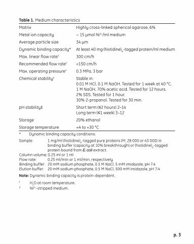
The key characteristics of the medium are listed in Table 1.
A variety of compounds that are compatible with Ni Sepharose High Performance are listed in Table 2.
p. 3
Table 1. Medium characteristics
Matrix Highly cross-linked spherical agarose, 6%
Metal ion capacity ~ 15 µmol Ni2+/ml medium
Average particle size 34 µm
Dynamic binding capacity* At least 40 mg (histidine)6-tagged protein/ml medium
Max. linear flow rate† 300 cm/h
Recommended flow rate† <150 cm/h
Max. operating pressure† 0.3 MPa, 3 bar
Chemical stability‡ Stable in: 0.01 M HCl, 0.1 M NaOH. Tested for 1 week at 40 °C. 1 M NaOH, 70% acetic acid. Tested for 12 hours. 2% SDS. Tested for 1 hour. 30% 2-propanol. Tested for 30 min.
pH stability‡ Short term (≤2 hours) 2–14 Long term (≤1 week) 3–12
Storage 20% ethanol
Storage temperature +4 to +30 °C* Dynamic binding capacity conditions:
Sample: 1 mg/ml (histidine)6-tagged pure proteins (Mr 28 000 or 43 000) in binding buffer (capacity at 10% breakthrough) or (histidine)6-tagged protein bound from E. coli extract.Column volume: 0.25 ml or 1 mlFlow rate: 0.25 ml/min or 1 ml/min, respectivelyBinding buffer: 20 mM sodium phosphate, 0.5 M NaCl, 5 mM imidazole, pH 7.4Elution buffer: 20 mM sodium phosphate, 0.5 M NaCl, 500 mM imidazole, pH 7.4
Note: Dynamic binding capacity is protein-dependent.† H2O at room temperature.‡ Ni2+-stripped medium.

p. 4
Table 2. Ni Sepharose High Performance is compatible with the following compounds, at the concentrations given.
Reducing agents* 5 mM DTE 5 mM DTT 20 mM ß-mercaptoethanol 5 mM TCEP 10 mM reduced glutathione
Denaturing agents 8 M urea†
6 M Gua-HCl†
Detergents 2% Triton™ X-100 (nonionic) 2% Tween™ 20 (nonionic) 2% NP-40 (nonionic) 2% cholate (anionic) 1% CHAPS (zwitterionic)
Other additives 20% ethanol 50% glycerol 100 mM Na2SO4 1.5 M NaCl 1 mM EDTA‡ 60 mM citrate‡
Buffer substances 50 mM sodium phosphate, pH 7.4 100 mM Tris-HCl, pH 7.4 100 mM Tris-acetate, pH 7.4 100 mM HEPES, pH 7.4 100 mM MOPS, pH 7.4 100 mM sodium acetate, pH 4†
* For best results it is recommended to perform a blank run before including reducing agents in the sample/buffers. See General considerations and Blank run, p. 12
† Tested for 1 week at +40 °C.‡ The strong chelator EDTA has been used succesfully in some cases at 1 mM.
Generally, chelating agents should be used with caution (and only in the sample, not in buffers). Any metal-ion stripping may be counteracted by addition of a small excess of MgCl2 before centrifugation/filtration of the sample. Note that strippingeffects may vary with applied sample volume.
p. 5
General considerationsNi Sepharose High Performance is supplied precharged with Ni2+ ions. In general, Ni2+ is the preferred metal ion for purification of recombinant histidine-tagged proteins and imidazole is used for elution.
Imidazole at low concentrations is commonly used in the binding and wash buffer to minimize binding of unwanted host cell proteins. For the same reason, it is important to also include imidazole in the sample (generally, at the same concentration as in the wash buffer). At somewhat higher concentrations, imidazole may also decrease the binding of histidine-tagged proteins.
The imidazole concentration must therefore be optimized to ensure the best balance of high purity (low binding of unwanted proteins) and high yield (binding of all of the histidine-tagged protein). The concentration of imidazole that will give optimal purification results is protein-dependent, and is usually slightly higher for Ni Sepharose High Performance than for similar IMAC media on the market. (see Data File 18-1174-40 and Optimization).
Leakage of Ni2+ from Ni Sepharose High Performance is low under all normal conditions, lower than for other IMAC media tested. For applications where very low leakage during purification is critical, leakage can be diminished even further by performing a blank run (see Purification procedure for packed column, page 11.)
Ni Sepharose High Performance is compatible with reducing agents (see Table 2). However, we recommend removing any weakly bound metal ions by performing a blank run without reducing agents (see “Blank run” on p. 12) before applying buffer/sample containing reducing agents. Do not leave Ni Sepharose High Performance with buffers containing reducing agents when not in use.
p. 6
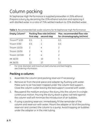
Column packingNi Sepharose High Performance is supplied preswollen in 20% ethanol. Prepare a slurry by decanting the 20% ethanol solution and replacing it with distilled water in a ratio of 75% settled medium to 25% distilled water.
Table 3. Recommended lab-scale columns for Ni Sepharose High Performance
Empty Column* Packing flow rate (ml/min) Max. recommended flow rate first step second step for chromatography (ml/min)
Tricorn™ 5/20 0.5 1 0.5
Tricorn 5/50 0.5 1 0.5
Tricorn 10/20 2 4 2
Tricorn 10/50 2 4 2
Tricorn 10/100 2 4 2
XK 16/20 5 10 5
XK 26/20 13 27 13 * For inner diameter and maximum bed volumes and bed heights,
see Ordering information.
Packing a column:1. Assemble the column (and packing reservoir if necessary).
2. Remove air from the end-piece and adapter by flushing with water. Make sure no air has been trapped under the column bed support. Close the column outlet leaving the bed support covered with water.
3. Resuspend the medium and pour the slurry into the column in a single continuous motion. Pouring the slurry down a glass rod held against the column wall will minimize the introduction of air bubbles.
4. If using a packing reservoir, immediately fill the remainder of the column and reservoir with water. Mount the adapter or lid of the packing reservoir and connect the column to a pump. Avoid trapping air bubbles under the adapter or in the inlet tubing.
p. 7
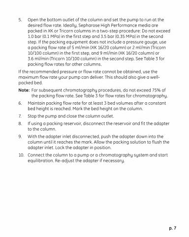
5. Open the bottom outlet of the column and set the pump to run at the desired flow rate. Ideally, Sepharose High Performance media are packed in XK or Tricorn columns in a two-step procedure: Do not exceed 1.0 bar (0.1 MPa) in the first step and 3.5 bar (0.35 MPa) in the second step. If the packing equipment does not include a pressure gauge, use a packing flow rate of 5 ml/min (XK 16/20 column) or 2 ml/min (Tricorn 10/100 column) in the first step, and 9 ml/min (XK 16/20 column) or 3.6 ml/min (Tricorn 10/100 column) in the second step. See Table 3 for packing flow rates for other columns.
If the recommended pressure or flow rate cannot be obtained, use the maximum flow rate your pump can deliver. This should also give a well-packed bed.
Note: For subsequent chromatography procedures, do not exceed 75% of the packing flow rate. See Table 3 for flow rates for chromatography.
6. Maintain packing flow rate for at least 3 bed volumes after a constant bed height is reached. Mark the bed height on the column.
7. Stop the pump and close the column outlet.
8. If using a packing reservoir, disconnect the reservoir and fit the adapter to the column.
9. With the adapter inlet disconnected, push the adapter down into the column until it reaches the mark. Allow the packing solution to flush the adapter inlet. Lock the adapter in position.
10. Connect the column to a pump or a chromatography system and start equilibration. Re-adjust the adapter if necessary.
p. 8
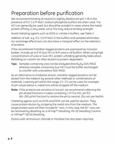
Preparation before purifi cationWe recommend binding at neutral to slightly alkaline pH (pH 7–8) in the presence of 0.5–1.0 M NaCl. Sodium phosphate buffers are often used. Tris-HCl can generally be used, but should be avoided in cases where the metal-protein affinity is very weak, since Tris may reduce binding strength.
Avoid chelating agents such as EDTA or citrate in buffers, see Table 2.
Addition of salt, e.g. 0.5–1.0 M NaCl in the buffers and samples eliminates ion-exchange effects but can also have a marginal effect on the retention of proteins.
If the recombinant histidine-tagged proteins are expressed as inclusion bodies, include up to 6 M Gua-HCl or 8 M urea in all buffers. When using high concentrations of urea or Gua-HCl, protein unfolding generally takes place. Refolding on-column (or after elution) is protein-dependent.
Tips: Samples containing urea can be analyzed directly by SDS-PAGE whereas samples containing Gua-HCl must be buffer-exchanged to a buffer with urea before SDS-PAGE.
As an alternative to imidazole elution, histidine-tagged proteins can be eluted from the medium by several other methods or combinations of methods. Lowering pH within the range 2.5–7.5 can be used, for example. At pH values below 4, metal ions will be stripped off the medium.
Note: If the proteins are sensitive to low pH, we recommend collecting the pH-eluted fractions in tubes containing 1 M Tris-HCl, pH 9.0 (60–200 µl/ml fraction) to restore the pH to neutral. (Do not use NaOH).
Chelating agents such as EGTA and EDTA can be used for elution. They cause protein elution by stripping the metal ions from the medium. The target protein pool will then include Ni2+ ions. In this case, the Ni2+ ions can be removed by desalting on a HiTrap™ Desalting, PD-10 Desalting Column, or HiPrep™ 26/10 Desalting.
Elution with ammonium chloride or histidine has also been reported.

p. 9
Imidazole concentraton in binding buff erThe purity of recombinant histidine-tagged proteins can often be increased by washing with binding buffer containing as high a concentration of imidazole as possible. However, care must be taken not to use a wash concentration of imidazole that causes elution of the histidine-tagged protein.
To obtain highest purity, first determine the optimal concentration of imidazole for sample loading, washing and elution. This can be done by eluting with a linear or stepwise gradient of imidazole from 20 to 500 mM, and testing fractions by SDS-PAGE and/or Western blotting for the presence of target protein and impurities. See Optimization.
When maximum binding and yield of the histidine-tagged protein (rather than purity) is the main objective, choose a low imidazole concentration for binding and wash, even if that concentration (in some cases) may lead to suboptimal purity.

p. 10
Buff er preparationWater and chemicals used for buffer preparation should be of high purity. Filter buffers through a 0.45 µm filter before use.
Use a high purity imidazole as this will give a very low or no absorbance at 280 nm.
Recommended buff ersBinding buffer: 20 mM sodium phosphate, 0.5 M NaCl, 20–40 mM imidazole, pH 7.4 (The optimal imidazole concentration is protein-dependent; 20–40 mM is suitable for many proteins).
Elution buffer: 20 mM sodium phosphate, 0.5 M NaCl, 500 mM imidazole, pH 7.4 (The imidazole concentration required for elution is protein-dependent).
Sample preparationFor optimal growth, induction and cell lysis conditions, please refer to established protocols.
The sample should be fully dissolved. To avoid column clogging, we recommend centrifugation and filtration through a 0.45 µm or 0.22 µm filter to remove cell debris or other particulate material. If the sample is dissolved in a buffer other than 20 mM phosphate buffer with 0.5 M NaCl pH 7.4, adjust its NaCl concentration to 0.5 M and pH to 7–8. This can be achieved by addition of concentrated stock solutions, by dilution with the binding buffer, or by buffer exchange (on HiTrap Desalting, PD-10 Desalting Column, or HiPrep 26/10 Desalting, depending on the sample volume). Do not use strong bases or acids for pH-adjustments (precipitation risk).
To prevent binding of host cell proteins with exposed histidine, add the same concentration of imidazole to the sample as to the binding buffer.
p. 11
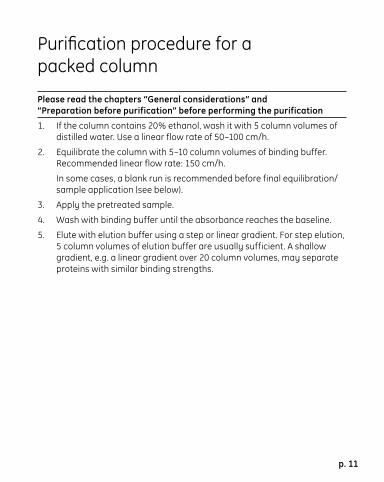
Purifi cation procedure for a packed column
Please read the chapters “General considerations” and “Preparation before purification” before performing the purification
1. If the column contains 20% ethanol, wash it with 5 column volumes of distilled water. Use a linear flow rate of 50–100 cm/h.
2. Equilibrate the column with 5–10 column volumes of binding buffer. Recommended linear flow rate: 150 cm/h.
In some cases, a blank run is recommended before final equilibration/sample application (see below).
3. Apply the pretreated sample.
4. Wash with binding buffer until the absorbance reaches the baseline.
5. Elute with elution buffer using a step or linear gradient. For step elution, 5 column volumes of elution buffer are usually sufficient. A shallow gradient, e.g. a linear gradient over 20 column volumes, may separate proteins with similar binding strengths.
p. 12
Note: Use the elution buffer as blank when measuring absorbance manually. If imidazole needs to be removed from the protein, use HiTrap Desalting, a PD-10 Desalting Column, or HiPrep 26/10 Desalting.
Leakage of Ni2+ from Ni Sepharose High Performance is low under all normal conditions. For very critical applications, leakage during purification can be even further decreased by performing a blank run (as described below) before loading sample. For best results, performing a blank run before including reducing agents in samples/buffers is also recommended.
Blank run:
Use binding buffer and elution buffer without reducing agents.
1. Wash the column with 5 column volumes of distilled water.
2. Wash with 5 column volumes of elution buffer.
3. Equilibrate with 10 column volumes of binding buffer.

p. 13
OptimizationConcentration of imidazole in binding/wash buff erImidazole at low concentrations is commonly used in the binding and wash buffers to minimize binding of unwanted host cell proteins. It is important to include imidazole also in the sample (generally, at the same concentration as in the wash buffer). At somewhat higher concentrations, imidazole may also decrease the binding of histidine-tagged proteins. The imidazole concentration must therefore be optimized to ensure the best balance of high purity (low binding of unwanted proteins), and high yield (binding of all of the histidine-tagged protein). This optimal concentration is different for different histidine-tagged proteins. Note that Ni Sepharose High Performance often requires a slightly higher concentration of imidazole in the wash buffer than similar IMAC media on the market. Finding the optimal imidazole concentration for a specific histidine-tagged protein is a trial-and-error effort, but 20–40 mM in the binding and wash buffers is a good starting point for many proteins. Prepacked HisTrap HP columns (1 or 5 ml) are ideal for optimization.
Choice of metal ionNi2+ is usually the first choice metal ion for purifying most (histidine)6-tagged recombinant proteins from cellular contaminants.
The strength of binding between a protein and a metal ion is affected by several factors, including the length, position, and exposure of the affinity tag on the protein, the type of ion used, and the pH of buffers, so some proteins may be easier to purify with ions other than Ni2+.
Prepacked HiTrap Chelating HP and HiTrap IMAC HP columns or IMAC Sepharose High Performance (not metal-ion charged) can be used to test this possibility. These products can be charged with different metal ions, e.g, Cu2+, Co2+, Zn2+, Ca2+, Ni2+ or Fe3+.
p. 14
TroubleshootingThe following tips may be of assistance. If you have any further questions about Ni Sepharose High Performance, please visit www.chromatography.amershambiosciences.com or contact our technical support, or your local GE Healthcare representative.
Column has clogged:• Cell debris in the sample may clog the column. Clean the column
according to the section Cleaning-in-Place.
• Centrifuge and/or filter the sample through a 0.22 µm or a 0.45 µm filter, see Sample preparation.
Sample is too viscous:• If the lysate is very viscous due to high concentration of host nucleic
acid, continue sonication until the viscosity is reduced, and/or add DNase I to 5 µg/ml, Mg2+ to 1 mM, and incubate on ice for 10–15 minutes. Alternatively, draw the lysate through a syringe needle several times.
Protein is diffi cult to dissolve or precipitates duringpurifi cation:• The following additives may be used: 2% Triton X-100, 2% Tween 20,
2% NP-40, 2% cholate, 1% CHAPS, 1.5 M NaCl, 50% glycerol, 20 mM ß-mercaptoethanol, 1–3 mM DTT or DTE (up to 5 mM is possible but depends on the sample and the sample volume), 5 mM TCEP, 10 mM reduced glutathione, 8 M urea or 6 M Gua-HCl. Mix gently for 30 minutes to aid solubilization of the tagged protein (inclusion bodies may require much longer mixing). Note that Triton X-100 and NP-40 (but not Tween) have a high absorbance at 280 nm. Furthermore, detergents cannot be easily removed by buffer exchange.
p. 15
Histidine-tagged protein found in the pellet:SDS-PAGE analysis of samples collected during the preparation of the bacterial lysate may indicate that most of the histidine-tagged protein is located in the centrifugation pellet. Possible causes and solutions are:
• Sonication may be insufficient: Check cell disruption by microscopic examination or monitor by measuring the release of nucleic acids at A260. Addition of lysozyme (up to 0.1 volume of a 10 mg/ml lysozyme solution in 25 mM Tris-HCl, pH 8.0) prior to sonication may improve results. Avoid frothing and overheating as this may denature the target protein. Oversonication can also lead to co-purification of host proteins with the target protein.
• The protein may be insoluble (inclusion bodies): The protein can usually be solubilized (and unfolded) from inclusion bodies using common denaturants such as 4–6 M Gua-HCl, 4–8 M urea or strong detergents.
Prepare buffers containing 20 mM sodium phosphate, 8 M urea or 6 M Gua-HCl, and suitable imidazole concentrations, pH 7.4–7.6. Use these buffers for sample preparation, as binding buffer, and as elution buffer. For sample preparation and binding buffer, use 10 mM imidazole or the concentration selected during optimization trials (including urea or Gua-HCl). To minimize sample dilution, solid urea or Gua-HCl can be added.
p. 16
Histidine-tagged protein is found in the fl owthrough and purifi ed fractions:• Capacity of Ni Sepharose High Performance is exceeded: Increase the
volume of Ni Sepharose High Performance used for your purification.
No histidine-tagged protein in the purifi ed fractions:• Elution conditions are too mild (histidine-tagged protein still bound):
Elute with an increasing imidazole gradient or decreasing pH to determine the optimal elution conditions.
• The protein has precipitated in the column: Try detergents or changed NaCl concentration or elute under denaturing (unfolding) conditions (use 4–8 M urea or 4–6 M Gua-HCl) to remove precipitated proteins. For the next experiment, decrease amount of sample, or decrease protein concentration by eluting with a linear imidazole gradient instead of imidazole steps.
• Nonspecific hydrophobic or other interaction: Add a nonionic detergent to the elution buffer (e.g. 0.2% Triton X-100) or increase the NaCl concentration.
• Concentration of imidazole in the sample and/or binding buffer is too high: The protein is found in the flowthrough material. Decrease the imidazole concentration.
• Target protein may not be histidine-tagged as expected: Verify DNA sequence of the gene. Analyze samples taken before and after induction of expression with, e.g., anti-His antibodies in Western blotting.
• Histidine-tag may be insufficiently exposed: The protein is found in the flowthrough material. Perform purification of unfolded protein in urea or Gua-HCl as for inclusion bodies.
To minimize dilution, solid urea or Gua-HCl can be added to the sample.
• Buffer/sample composition is incorrect: The protein is found in the flowthrough material. Check pH and composition of sample and binding buffer. Ensure that the concentration of chelating or strong reducing agents, as well as imidazole, in the solution is not too high.
p. 17
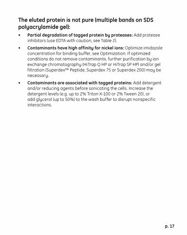
The eluted protein is not pure (multiple bands on SDS polyacrylamide gel):• Partial degradation of tagged protein by proteases: Add protease
inhibitors (use EDTA with caution, see Table 2).
• Contaminants have high affinity for nickel ions: Optimize imidazole concentration for binding buffer, see Optimization. If optimized conditions do not remove contaminants, further purification by ion exchange chromotography (HiTrap Q HP or HiTrap SP HP) and/or gel filtration (Superdex™ Peptide, Superdex 75 or Superdex 200) may be necessary.
• Contaminants are associated with tagged proteins: Add detergent and/or reducing agents before sonicating the cells. Increase the detergent levels (e.g. up to 2% Triton X-100 or 2% Tween 20), or add glycerol (up to 50%) to the wash buffer to disrupt nonspecific interactions.
p. 18
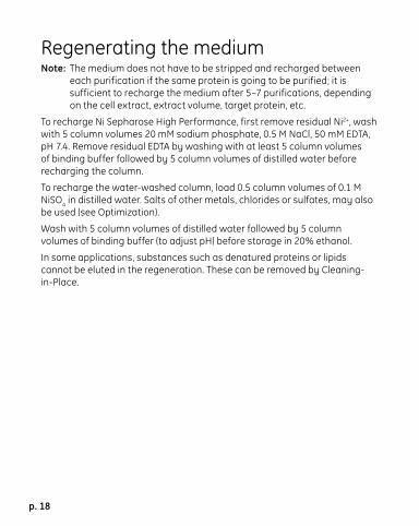
Regenerating the mediumNote: The medium does not have to be stripped and recharged between
each purification if the same protein is going to be purified; it is sufficient to recharge the medium after 5–7 purifications, depending on the cell extract, extract volume, target protein, etc.
To recharge Ni Sepharose High Performance, first remove residual Ni2+, wash with 5 column volumes 20 mM sodium phosphate, 0.5 M NaCl, 50 mM EDTA, pH 7.4. Remove residual EDTA by washing with at least 5 column volumes of binding buffer followed by 5 column volumes of distilled water before recharging the column.
To recharge the water-washed column, load 0.5 column volumes of 0.1 M NiSO4 in distilled water. Salts of other metals, chlorides or sulfates, may also be used (see Optimization).
Wash with 5 column volumes of distilled water followed by 5 column volumes of binding buffer (to adjust pH) before storage in 20% ethanol.
In some applications, substances such as denatured proteins or lipids cannot be eluted in the regeneration. These can be removed by Cleaning-in-Place.

p. 19
Cleaning-in-Place (CIP)When an increase in back-pressure is seen, the column should be cleaned. Before cleaning, strip off the Ni2+ ions using the recommended procedure (see Regenerating the medium). Use reversed flow direction for cleaning.
After cleaning, store in 20% ethanol or recharge with Ni2+ prior to storage in ethanol.
The Ni2+ stripped column can be cleaned by the following methods:
• Remove ionically bound proteins by washing with several column volumes of 1.5 M NaCl. Then wash with several column volumes of distilled water.
• Remove precipitated proteins, hydrophobically bound proteins, and lipoproteins by washing the column with 1 M NaOH, contact time usually 1–2 hours (12 hours or more to remove endotoxins). Then wash with approx. 10 column volumes of binding buffer, followed by 10 column volumes of distilled water.
• Remove hydrophobically bound proteins, lipoproteins and lipids by washing with 5–10 column volumes of 30% isopropanol for about 15–20 minutes. Then wash with approx. 10 column volumes of distilled water.
Alternatively, wash with 2 column volumes of detergent in a basic or acidic solution. Use, for example, 0.1–0.5% nonionic detergent in 0.1 M acetic acid, contact time 1–2 hours. After treatment, always remove residual detergent by washing with 5–10 column volumes of 70% ethanol. Then wash with approx. 10 column volumes of distilled water.

p. 20
StorageStore the medium for longer periods of time in 20% ethanol at +4 to +30 °C.
Further informationCheck www.chromatography.amershambiosciences.com for further information. Several handbooks also contain useful information, see Ordering information.
p. 21
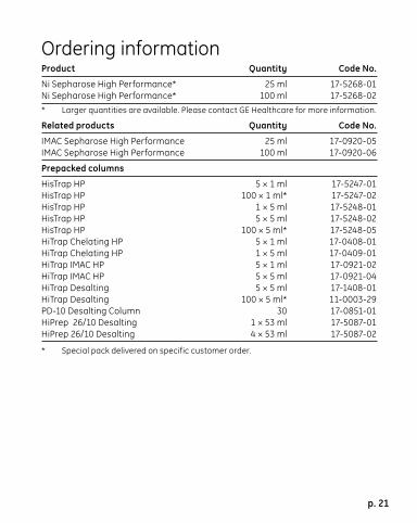
Ordering informationProduct Quantity Code No.
Ni Sepharose High Performance* 25 ml 17-5268-01Ni Sepharose High Performance* 100 ml 17-5268-02* Larger quantities are available. Please contact GE Healthcare for more information.
Related products Quantity Code No.
IMAC Sepharose High Performance 25 ml 17-0920-05IMAC Sepharose High Performance 100 ml 17-0920-06
Prepacked columns
HisTrap HP 5 × 1 ml 17-5247-01HisTrap HP 100 × 1 ml* 17-5247-02HisTrap HP 1 × 5 ml 17-5248-01HisTrap HP 5 × 5 ml 17-5248-02HisTrap HP 100 × 5 ml* 17-5248-05HiTrap Chelating HP 5 × 1 ml 17-0408-01HiTrap Chelating HP 1 × 5 ml 17-0409-01HiTrap IMAC HP 5 × 1 ml 17-0921-02HiTrap IMAC HP 5 × 5 ml 17-0921-04HiTrap Desalting 5 × 5 ml 17-1408-01HiTrap Desalting 100 × 5 ml* 11-0003-29PD-10 Desalting Column 30 17-0851-01HiPrep 26/10 Desalting 1 × 53 ml 17-5087-01HiPrep 26/10 Desalting 4 × 53 ml 17-5087-02
* Special pack delivered on specific customer order.
p. 22
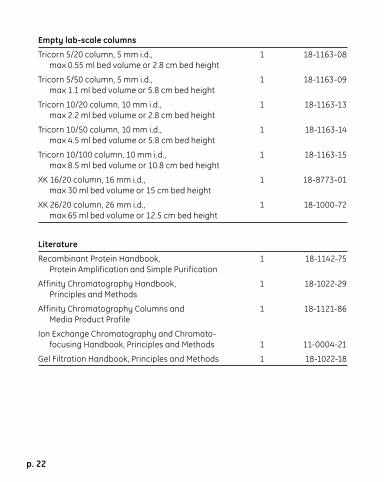
Empty lab-scale columns
Tricorn 5/20 column, 5 mm i.d., 1 18-1163-08 max 0.55 ml bed volume or 2.8 cm bed height
Tricorn 5/50 column, 5 mm i.d., 1 18-1163-09 max 1.1 ml bed volume or 5.8 cm bed height
Tricorn 10/20 column, 10 mm i.d., 1 18-1163-13 max 2.2 ml bed volume or 2.8 cm bed height
Tricorn 10/50 column, 10 mm i.d., 1 18-1163-14 max 4.5 ml bed volume or 5.8 cm bed height
Tricorn 10/100 column, 10 mm i.d., 1 18-1163-15 max 8.5 ml bed volume or 10.8 cm bed height
XK 16/20 column, 16 mm i.d., 1 18-8773-01 max 30 ml bed volume or 15 cm bed height
XK 26/20 column, 26 mm i.d., 1 18-1000-72 max 65 ml bed volume or 12.5 cm bed height
Literature
Recombinant Protein Handbook, 1 18-1142-75 Protein Amplification and Simple Purification
Affinity Chromatography Handbook, 1 18-1022-29 Principles and Methods
Affinity Chromatography Columns and 1 18-1121-86 Media Product Profile
Ion Exchange Chromatography and Chromato- focusing Handbook, Principles and Methods 1 11-0004-21
Gel Filtration Handbook, Principles and Methods 1 18-1022-18

HisTrap, HiTrap, Sepharose, Superdex, HiPrep, HisPrep, Drop Design, and Tricorn are trademarks of Amersham plc, a General Electric Company going to market as GE Healthcare. GE tagline and GE monogram are trademarks of General Electric Company. Triton is a trademark of Union Carbide Chemicals and Plastics Co. Tween is a trademark of ICIAmericas Inc.
All goods and services are sold subject to the terms and conditions of sale of the company within GE Healthcare which supplies them. GE Healthcare reserves the right, subject to any regulatory and contractual approval, if required, to make changes in specifi cations and features shown herein, or discontinue the product described at any time without notice or obligation. Contact your local GE Healthcare representative for the most current information.
Licensing informationPurification and preparation of fusion proteins and affinity peptides comprising at least two adjacent histidine residues may require a license under US pat 5.284.933 and US pat 5.310.663, including corresponding foreign patents (assigne: Hoffman La Roche, Inc).
GE Healthcare Bio-Sciences AB, a General Electric Company.
© 2005 General Electric Company – All rights reserved.
General Electric reserves the right to make changes in specifications and features shown herein, or discontinue the product described at any time without notice or obligation. Contact your GE representant for the most currentinformation.
GE HealthcareMunzinger Strasse 9D-79111 FreiburgGermany
GE HealthcareAmersham PlaceLittle ChalfontBuckinghamshire, HP7 9NA UK
GE Healthcare800 Centennial AvenueP.O. Box 1327Piscataway, NJ 08855-1327USA
GE HealthcareSanken Bldg. 3-25-1, HyakuninchoShinjuku-ku, Tokyo 169-0073Japan
www.amershambiosciences.com/hiswww.gehealthcare.com
GE Healthcare Bio-Sciences ABBjörkgatan 30751 84 UppsalaSweden
71-5027-67 AC
Pro
duce
d by
Wik
strö
ms,
Sw
eden
105
0880
, 11.
2005
Prin
ted
mat
ter.
Lice
nce
341
051