ni hms 29820
DESCRIPTION
123TRANSCRIPT
The Role of the Adventitia in Vascular Inflammation
Kathryn Maiellaro1 and W. Robert Taylor1,2,3
1Wallace H. Coulter Department of Biomedical Engineering, Georgia Institute of Technology andEmory University, Atlanta, GA 303322Division of Cardiology, Department of Medicine, Emory University, Atlanta, GA 303223Veterans Affairs Medical Center, Atlanta, GA 30033
AbstractTraditional concepts of vascular inflammation are considered “inside-out” responses centered onthe monocyte adhesion and lipid oxidation hypotheses. These mechanisms likely operate inconcert, holding the central tenet that the inflammatory response is initiated at the luminal surface.However, growing evidence supports a new paradigm of an “outside-in” hypothesis, in whichvascular inflammation is initiated in the adventitia and progresses inward toward the intima.Hallmarks of the outside-in hypothesis include population of the adventitia with exogenous celltypes, including monocytes, macrophages, and lymphocytes, the phenotypic switch of adventitialfibroblasts into migratory myofibroblasts, and increased vasa vasorum neovascularization. Theresident and migrating cells deposit collagen and matrix components, respond to and upregulateinflammatory chemokines and/or antigens, and regulate the local redox state of the adventitia. Bcells and T cells generate local humoral immune responses against local antigen presentation byfoam cells and antigen presenting cells. These events result in increased local expression ofcytokines and growth factors, evoking an inflammatory response that propagates inward towardthe intima. Ultimately, it appears that the basic mechanisms of cellular activation and migration invascular inflammation are highly conserved across a variety of cardiovascular disease states andthat major inflammatory events begin in the adventitia.
The Adventitia in the Inflammatory Response to Vascular InjuryVascular inflammation has traditionally been considered an “inside-out” response, centeredon the monocyte adhesion to the intima of blood vessels [1] and the oxidative lipidhypotheses [2]. In the former, injured vascular cells on the intimal surface of the bloodvessel express surface adhesion molecules and inflammatory mediators that participate inmonocyte homing to the endothelium and eventual transmigration into the media. In thelatter hypothesis, oxidized lipids in the circulation accumulate in macrophages on the intimalsurface, injure the endothelium, and initiate an inflammatory process (Figure 1). Thesemechanisms invoke a central role for vascular inflammation and indeed, it is likely that theyoperate in concert, holding the central tenet that inflammatory responses are initiated at the
© 2007 European Society of Cardiology. Published by Elsevier B.V. All rights reserved.W. Robert Taylor, CORRESPONDING AUTHOR, Emory University School of Medicine, Cardiology Division, 101 WoodruffCircle, Suite 319 WMB, Atlanta, GA 30322, Phone: 404-727-8921, Fax: 404-727-3572, [email protected] Maiellaro, Emory University School of Medicine, Cardiology Division, 101 Woodruff Circle, Suite 319 WMB, Atlanta, GA30322, Phone: 404-712-9912, Fax: 404-727-3330, [email protected]'s Disclaimer: This is a PDF file of an unedited manuscript that has been accepted for publication. As a service to ourcustomers we are providing this early version of the manuscript. The manuscript will undergo copyediting, typesetting, and review ofthe resulting proof before it is published in its final citable form. Please note that during the production process errors may bediscovered which could affect the content, and all legal disclaimers that apply to the journal pertain.
NIH Public AccessAuthor ManuscriptCardiovasc Res. Author manuscript; available in PMC 2012 January 22.
Published in final edited form as:Cardiovasc Res. 2007 September 1; 75(4): 640–648. doi:10.1016/j.cardiores.2007.06.023.
NIH
-PA Author Manuscript
NIH
-PA Author Manuscript
NIH
-PA Author Manuscript

luminal surface. As a result, the luminal surface is where the majority of investigation intocardiovascular disease (CVD) pathogenesis has been focused.
However, growing evidence supports a new paradigm of an “outside-in” hypothesis, inwhich vascular inflammation is initiated in the adventitia and progresses inward toward theintima. The functional significance of the adventitia in vascular disease has been exploredintermittently for many years. However, the hypothesis that he adventitia is a functionalhomeostatic regulator in CVD pathogenesis was only postulated about a decade ago in thesetting of restenosis after percutaneous coronary angioplasty [3, 4], and more recently in thesetting of abdominal aortic aneurysm (AAA) development [5]. Indeed, studies show that invarious presentations of CVD, the adventitia not only becomes populated with multipleexogenous cell types including monocytes, macrophages and T cells [6–8], but also showsincreased replication and differentiation of fibroblasts and into myofibroblasts [9]. Theseevents result in increased local expression of inflammatory cytokines and growth factors in amotif that is similar to that seen with inside-out mechanisms [10]. It is the objective of thisreview to explore the data supporting this non-traditional view of CVD-associatedinflammation occurring in the adventitia and to consider the implications of this alternativepathogenic paradigm in our understanding of the basic mechanisms of vascular disease.
The Cellular Composition of the Active Adventitiaa) Fibroblast and Myofibroblasts
Many observations on the role of the adventitia in response to injury were first reported inthe setting of restenosis after coronary angioplasty. Stent placement or balloon angioplastyinjures the endothelium and produces a focal rupture in the internal elastic lamina. Inresponse, vascular cells proliferate and migrate to the intimal region, secreting extracellularmatrix (ECM) proteins and subsequently generating a neointima. Recent studies have shownthat nearly half of the neointimal cells originate from proliferating adventitial cells [3],whereas previous studies hypothesized that most neointimal cells originated from theadjacent smooth muscle cells in the media [11]. Importantly, neointimal formation isaccompanied by a significant increase in the size of the adventitia as well as a change in itscellular components, providing morphological evidence that the adventitia is an activeparticipant in the wound healing response.
In the post-angioplasty setting, adventitial cells in regions near the medial tear demonstratean increase in α-smooth muscle (SM) actin and desmin positive staining within 2 weeks.This is indicative of a phenotypic switch of fibroblasts into myofibroblasts (Figure 1), aconversion considered the hallmark of adventitial response to injury in this model [4, 12,13]. A myofibroblast is a modified fibroblast that has contractile properties as indicated bythe presence of stress fibers and cytoskeletal proteins, which are upregulated in thetransformation from the non-proliferative fibroblast [14]. Myofibroblasts stain for α-SMactin, whereas fibroblasts do not [15]. Demonstration of myofibroblasts in the neointima ofexperimental angioplasty-induced intimal lesions was the first published evidence of themigratory capacity of the adventitial fibroblast. In addition, these studies provide thefoundation for the hypothesis that the adventitia holds a fundamental role in intimal vascularlesion formation [4, 12].
The activation and signaling mechanism of the myofibroblast has received much attentionfor the intriguing reason that it presents with the same phenotype and function regardless oftissue residence. Much of what is known about myofibroblast function was pioneered in thesetting of wounding healing [16]. Powell et al provided an elegant review of myofibroblastactivity in intestinal cells [17, 18]. Although specific myofibroblast activity in thevasculature has not been thoroughly detailed, there is most likely a strong correlation
Maiellaro and Taylor Page 2
Cardiovasc Res. Author manuscript; available in PMC 2012 January 22.
NIH
-PA Author Manuscript
NIH
-PA Author Manuscript
NIH
-PA Author Manuscript

between the wound healing mechanisms occurring in gastrointestinal cells, and thoseoccurring in the setting of CVD.
The lifespan of the myofibroblast appears to be controlled from the time of phenotypicswitch until apoptotic death. Of particular interest to this discussion is growing evidence thatfibroblast and myofibroblast regulation of collagen influences their role in the inflammatoryresponse. In vitro studies have shown that ECM deposited by the myofibroblast has adistinct composition, implying the potential to uniquely control fibrolytic collagendeposition in the wounded vessel [18]. It appears that the myofibroblast may first deposit thebasement membrane collagen type IV, which supports cell migration and attachment. Thecollagen is subsequently converted into mature and mechanically stable collagen type I.After myofibroblasts migrate and participate in wound healing and contraction, theyundergo apoptosis which presumably regulates contractile remodeling by self-limitingexcessive ECM deposition. Though not yet clearly defined, studies have shown that thisapoptotic signal is dependent upon transforming grow factor-β (TGF-β), an establishedactivator of fibroblast differentiation into myofibroblasts (Figure 1) [19]. Somewhatparadoxically TGF-β also serves as a sustained chemoattractant for subsequentmyofibroblast infiltration [20], illustrating the inherent theme of inflammatory balancewherein inflammatory mediators that enhance one process may serve to mitigate another.
The presence of non-migratory, but proliferating fibroblasts illustrates a second regulatoryadventitial cell phenotype. Responding to either intimal or adventitial injury, these non-migratory fibroblasts impart an increased cellularity and cell density in the adventitia [21].Fibroblasts themselves have been shown to be rich in caveolae content, suggesting thepotential of the adventitial fibroblast to modulate protein trafficking mechanisms involved invascular response to injury [22]. In the setting of injury, adventitial fibroblasts depositprocollagen alpha 1(I) [23] and presumably other ECM proteins into the adventitia (Figure1) [14, 24]. As a homeostatic mechanism, these same cells also control excessive collagendeposition by upregulating collagen triple helix repeat containing 1, a novel regulatoryprotein that reduces collagen type I expression [24]. The concept that fibroblasts andmyofibroblasts process and organize collagen was tested by inhibiting TGF-β in the settingof balloon angioplasty [25]. In addition to activating the fibroblast to myofibroblastconversion, TGF-β has also been shown to regulate ECM deposition by vascular cells [26].The result of TGF-β inhibition was decreased constrictive remodeling associated with areduction in post-angioplasty restenosis, preserved lumen, and dense collagen deposition inthe adventitia. In addition, the collagen content in restenotic vessels was lower than innonstenotic vessels. These data suggest that the non-migratory fibroblast influences the localadventitial collagen matrix, while the migratory myofibroblast organizes the collagen matrixalong its migratory path toward the intima, where it becomes fully contractile, promotingrestenosis and constrictive remodeling.
The phenotypic switch of fibroblasts to myofibroblast is certainly not a solitary event in theadventitial response. Adventitial activation is likely a result of a traditional inflammatorycascade whereby monocytic infiltration of the adventitia is preceded by migration ofneutrophils and lymphocytes into the adventitia. Similarly, there are likely multiple locallyreleased chemoattractants responsible for the migration of cells into the adventitia includingplatelet derived growth factor (PDGF) and monocyte chemoattractant protein-1 (MCP-1).Indeed it has been shown that after balloon injury myofibroblasts, along with otherinflammatory cells, are found in granulation tissue. In the vasculature, adventitialgranulation tissue is associated with altered ECM protein expression, promoting adventitialand peri-adventitial contraction and scarring [14]. This ultimately results in constrictiveremodeling of the vessel and associated lumen loss.
Maiellaro and Taylor Page 3
Cardiovasc Res. Author manuscript; available in PMC 2012 January 22.
NIH
-PA Author Manuscript
NIH
-PA Author Manuscript
NIH
-PA Author Manuscript
The ability of the myofibroblast and non-migratory fibroblast to respond to injury in such anelegantly self-regulated manner is evidence that the adventitia plays a critical role in thevascular inflammatory cascade. It is clear that by directing the composition, function, andlifespan of its cellular composition, the adventitia has the potential to orchestrate pivotalmatrix remodeling mechanisms in the response to injury. The fibroblast to myofibroblastconversion is such a fundamental mechanism in the adventitial role in vascular inflammationthat it receives singular attention in Adventitial Fibroblast Reactive Oxygen Species asAutacrine and Paracrine Mediators of Remodeling: Bellwether for Vascular Disease? byHaurani and Pagano elsewhere in this issue.
b) Adventitial LymphocytesImmune control of vascular pathology has been investigated since T cells were firstobserved in atherosclerotic lesions two decades ago [27]. As was the case with the monocyteadhesion hypothesis, many studies of lymphocyte involvement attempted to either removewhat was considered adventitial artifact or simply overlooked the adventitial contributionaltogether. Recent efforts have provided numerous reports of the role of lymphocytes in bothhealthy and diseased tissue [4, 28–33], and while abundant data on the adventitial fibroblastrole has been obtained in the setting of balloon angioplasty, the majority of literature onlymphocytes in CVD has been in the setting of atherosclerosis. Indeed the mechanisms of Tcell action in atherogenesis was recently thoroughly reviewed [34] and it has beenhypothesized that circulating CD4+/CD31+ T cells may regulate local T cell action in thearterial wall [35]. Proatherogenic Th1 cells are shown to limit lesion size, but not lesioninitiation [36]. In contrast Th2 cells are considered anti-atherogenic in that they have beenshown to promote antibody-generating B cell production, as well mitigate cholesteroluptake.
While the presence of T cells within the adventitia of atherosclerotic lesions has beenconfirmed by multiple investigators[37, 38], the presence of B cells is more enigmatic and ispossibly related to the stage of lesion advancement [6, 37, 39, 40]. The protective immunityconveyed by the B cell against atherosclerosis has however been corroborated by numerousinvestigators. In both apolipoprotein E (apoE) knockout mice and low density lipoprotein(LDL) receptor null mice, B cell depletion via splenectomy results in increasedatherogenesis compared to unsplenectomized controls [41, 42]. In the hypercholesterolemicapoE knockout model, post-splenectomy B cell transfer reduced both ultimate lesion sizeand CD4+ T cell infiltration, yet did not yield any B cell populated lesions [42].Interestingly, the studies that do report B cells within the atherosclerotic lesions, show thecells localized in basal areas rich in vascular cell adhesion molecule (VCAM) expression,suggesting the B cell has a role in activation of atherogenic pathogenesis in the injuryresponse [38]. Taken together these findings suggest the existence of localized vascular Bcells, fundamental to protection from atherogenic pathogenesis, that work in concert withthe circulating B cells, which serve to reduce lesion formation in part by limiting T cellactivity [41].
What is most intriguing for this discussion is not the presence of T and B cells, but rathertheir location and organization. These lymphocytes have been found in the adventitia andmedia of wild type mice preceding any intimal lesion formation [31, 33, 39]. T/B cellclusters have been found in the setting of murine hyperlipidemia in the adventitia of non-atherosclerotic upper abdominal aorta, while complex lymphoid-like structures appear in thenon-atherosclerotic abdominal aorta below the diaphragm [33]. This finding is particularlyrelevant to AAA which predominately form in the same subdiaphragmatic portion of theaorta. The spatial distribution of resident lymphatic cells in the adventitia of aortic regionvulnerable to disease may imply an adventitially localized feed-forward immune responsethat is spatially predictive of disease initiation.
Maiellaro and Taylor Page 4
Cardiovasc Res. Author manuscript; available in PMC 2012 January 22.
NIH
-PA Author Manuscript
NIH
-PA Author Manuscript
NIH
-PA Author Manuscript
In the setting of vascular injury, adventitial nodules with germinal centers composedprimarily of B cells and associated macrophages and T cells have been shown near sites ofadvanced lesions in humans. Similar follicle-like structures are seen in murine lesions aswell [33]. It is thought that these are antigen-driven B cell maturation centers (Figure 1),where humoral immune responses are generated. Staining of microvessels near humanplaques shows cell clusters with lymphoid organ-like pathology that are not found innonatherosclerotic controls [40]. Such lymphoid clusters are typically associated withchronic inflammation states, and as such the observation of these structures in the adventitiaof acutely inflamed arteries is unique. Cluster organization appears to depend upon the stageof plaque progression, but overall these adventitial nodular infiltrates are composed of well-organized B cells, T cells, and macrophages, within which the B cells mature and generateantibodies against inflammatory antigens. The specific antigens that trigger these responsesare still undetermined, but stress proteins (heat shock proteins) [6], modified lipoproteins,and other surface antigens have been implicated.
Taken together, multiple studies link the origin of locally generated immune responses to theadventitia. Importantly, in both acute traumatic angioplastic injury and chronic vasculardisease, coordination among resident fibroblasts, adventitial lymphocytes and inflammatorymediators orchestrate an inflammatory cascade that travels from the outside-in toward theintima to the site of injury (Figure 1). Indeed the adventitial stimulus for post-angioplastyrestenosis is acute trauma. In this setting, adventitially derived myofibroblasts may appear tobe the predominant cell type regulating post-angioplasty constrictive remodeling. However,the role of the lymphocytes in post-angioplasty restenosis is currently undefined and couldpossibly play a comparable role to the myofibroblast in the inflammation response. In thesame manner, it may be speculated in the setting of atherosclerosis that chronic stimuli,perhaps paracrine signaling from a dysfunctional endothelium, may provide the pro-inflammatory stimuli to activate adventitially residing fibroblasts and lymphocytes. At earlyatherosclerotic stages these cells can maintain an open lumen via outward remodeling.However, at later stages the myofibroblast is stimulated to migrate and promote neointimalformation and eventual lumen constriction. Underscoring the complexity of CVDpathophysiology, each disease pathogenesis has its own adventitial pro-inflammatorystimuli, both acute and chronic, as well as its own cellular activation pattern with controlsthe intensity and time course of the inflammatory response,
The Outside-In Hypothesis and the Adventitial Signaling Pathwaya) Signaling Events after Balloon Angioplasty
The above discussion presents evidence that the adventitia is homeostatically regulated andresponds to vascular injury from the outside-in through function as 1) the source ofmigratory myofibroblasts responding to medial or intimal injury and 2) a depot for non-migratory fibroblasts and inflammatory cells that regulate the local adventitial environment,provide structural integrity to the entire vessel, and generate local humoral immuneresponses against injury. The first line of evidence for the outside-in hypothesis is that aftervessel injury, but prior to established neointimal development, the adventitia andperivascular tissue become highly populated with neutrophils, macrophages and apoptoticcells [12, 43]. Once the recruited cells are inside the adventitia, a number of genes andinflammatory molecules are expressed. Although it is not yet clear which chemotacticsignals activate the adventitia during vessel injury and which subsequent signals secreted bythe adventitia complete the wound healing process, a recent study found CCL2, also knownas monocyte chemoattractant protein-1 (MCP-1), a target inflammatory molecule and potentleukocyte chemoattractant, highly expressed in myofibroblasts at the site of injury in theadventitial response (Figure 1) [10]. CCL2 is known to induce macrophage recruitment andpromote their subsequent activation. The authors also found temporal action and
Maiellaro and Taylor Page 5
Cardiovasc Res. Author manuscript; available in PMC 2012 January 22.
NIH
-PA Author Manuscript
NIH
-PA Author Manuscript
NIH
-PA Author Manuscript

upregulation of other chemokine and chemokinereceptor expression in the adventitia afterballoon angioplasty. Temporally, the cytokine CXCL2 was transiently upregulated in theadventitia at early time points less than 3 days and subsequently upregulated again in theneointima at 7 days, further supporting the progression of the inflammatory response fromthe adventitia toward the intima.
Other balloon angioplasty studies also confirm early adventitial action. Okamoto et al foundthat within hours after angioplasty, P-selectin and VCAM-1 expression are upregulated inthe endothelial cells of the vasa vasorum [44]. As early as 2 hours after injury, neutrophilsaccumulate in the adventitia and perivascular tissues. Indeed at 14 days, adventitialVCAM-1 expression diminishes, while medial and neointimal VCAM-1 expression appears,providing additional evidence for the spatial propagation of the inflammatory response fromthe adventitia toward the intima.
The adventitial response to vessel homeostasis, like other proinflammatory events, appearsto be redox sensitive as well. In healthy tissue the adventitia has been shown to regulate theoxidative state of the vessel wall by regulating the bioavailability of adventitially generatednitric oxide to the tunica media [45], most likely via increased production of superoxideanion [46]. It should be noted that reactive oxygen species (ROS) and chemoattractants,such as TGF-β and MCP-1, range in size up to 15 kD and therefore can easily diffusethrough elastic laminae fenestrations which are approximately 2–7 microns in size [47]. Inthe balloon angioplasty model, perivascular delivery of a NADPH oxidase inhibitor, andsubsequent reduction of adventitially derived ROS, reduced neointimal growth after injuryof the rat carotid artery [48]. It was hypothesized that the lack of in vivo redox cuesprevented the myofibroblast phenotypic switch into a migrating cell, a hypothesis supportedby in vitro studies demonstrating the ROS are able to induce the phenotypic switch fromfibroblast into myofibroblast [48]. In addition, data from other studies support the ability ofROS to modulate the apoptotic regulation of cellular migration and proliferation that occursduring wound repair and remodeling [49, 50]. Redox modulation may therefore, regulate theapoptotic death of the myofibroblast after its activation and migration to vascular injury.
Following the outside-in hypothesis, activated adventitial fibroblasts generate chemotacticsignals in the form of cytokines and chemokines to recruit a milieu of inflammatorymediators from the perivascular tissue. Non-migratory adventitial cells modify the matrixcomposition and redox state of the local environment and simultaneously express surfaceadhesion makers, such as VCAM-1, to recruit other cell types including macrophages andlymphocytes. In this manner, the adventitia can affect the stability of the injured vessel,control the cellular infiltrate, and provide the critical signals for the fibroblast tomyofibroblast phenotypic switch. [47].
Also of importance are the distant effects of vascular inflammation which extend outside thevessel proper. Indeed multiple studies have observed that the inflammatory and proliferativeresponse to balloon injury extends beyond the adventitia into the perivascular tissues,suggesting the potential for paracrine recruitment of leukocytes, lymphocytes, and otherinflammatory mediators. Studies to control neointimal hyperplasia in the angioplasty modelshow that uptake of proteins and viral vectors into the medial layer is more effective whendelivered perivascularly rather than via the lumen. Uptake of heparin [51], basic fibroblastgrowth factor [52, 53], urokinase plasminogen activator [54], and viral constructs [55] intothe vessel wall has also been shown to be more efficient when delivered perivascularly,indicating the involvement of the perivascular tissues and vasa vasorum in cellular trafficinto and presumably out of the adventitia. Moreover, when whole aortic rings weretransduced with endothelial nitric oxide synthase (eNOS), the vector preferentiallypartitioned into the adventitia [56]. It is also important to note that microvascular flow (as
Maiellaro and Taylor Page 6
Cardiovasc Res. Author manuscript; available in PMC 2012 January 22.
NIH
-PA Author Manuscript
NIH
-PA Author Manuscript
NIH
-PA Author Manuscript
compared to bulk luminal flow) in the outside of the vessel may offer a more favorable localenvironment for inflammatory cell recruitment, activation, and subsequent signaling.
The Active Adventitia: Evidence from Other Models of VascularInflammation
Much of the data presented up to this point has been in the setting of balloon angioplastywherein the adventitia is clearly injured and activated to elicit an inflammatory response toinjury. However in most CVD states such clear adventitial provocation is absent. Diseasesmanifesting phenotypically in the intima and media, such as atherosclerosis, and those likearteriosclerosis and AAA that consume the entire artery clearly evoke the chicken-or-the-egg- which is stimulated first, the intima or the adventitia? It is clear that the complexity ofCVD requires appreciation of both intimal and adventitial events as the outside-in andinside-out responses coincide in various disease pathogenesis. Monocyte recruitment andmigration through the endothelium is an important event in CVD and has indisputableclinical implications. However, based on recent studies in atherosclerosis it appears thatoutside-in mechanisms may have broad applicability in other vascular diseases [57–59].
a) AtherosclerosisStrongly indicating that outside-in mechanisms favor the initiation of atherosclerotic disease,recent studies have shown increased vasa vasorum neovascularization [57, 58] andmacrophage presence [59] in early atherosclerotic lesions before plaque neovascularization.These hallmark studies indicate that in a hypercholesterolemic setting endothelialdysfunction in the lumen is preceded by increased vasa vasorum, creating a conduit forinflammatory cell transport into the vessel wall to promote chronic inflammation and plaqueneovascularization. Furthermore, it is possible that endothelial dysfunction in the largevessel lumen may actually be a marker for the endothelial dysfunction taking place in thevasa vasorum. A thorough review of the role of the vasa vasorum in vascular inflammationis developed elsewhere in this issue (see The Dynamic Vasa Vasorum by Ritman andLerman), however, the hallmark studies presented here highlight the applicability of theoutside-in hypothesis to vascular atherosclerotic disease canonically considered an inside-out progression.
Studies have shown that after surgical removal of the vasa vasorum to promote adventitialischemia the endothelium overlying the ressected areas becomes injured and the vesselultimately becomes denuded [60, 61]. In the setting of hyperlipidemia however,atherosclerotic foam cell containing lesions form as a result of the dysfunctional adventitiabeneath [60, 62]. Ligation of intercostal arteries in order to restrict vasa vasorum flow to theadventitia of the aorta induced patchy intimal necrosis and cell loss in the areas withrestricted vasa flow, further suggesting that the intimal endothelium is activated in part viathe adventitia. [63]. These studies suggest an antiatherosclerotic role of the vasa vasorum inthe setting of normal lipid levels. However, recent data from our laboratory corroborates thepro-atherosclerotic effect of the vasa vasorum in hyperlipidemic conditions. The descendingaortas harvested from hyperlipidemic apolipoprotein E knockout mice treated with eithergranulocyte colonystimulating factor or granulocyte macrophage colony-stimulating factorfor eight weeks demonstrated an increase in both adventitial vascularity and atheroscleroticlesion extent [64]. These data suggest that the adventitial vascularization may have amechanistic role in inflammation of the aortic wall and subsequent atherosclerosis. It can bespeculated then that the provocation for adventitial inflammation response in atherosclerosisis paracrine signaling from dysfunctional endothelium activated by lipoproteins, nutrientstarved SMCs, and chronic inflammation. The adventitia responds in concert with the vasavasorum by increasing vascularity, subsequently opening new and larger avenues through
Maiellaro and Taylor Page 7
Cardiovasc Res. Author manuscript; available in PMC 2012 January 22.
NIH
-PA Author Manuscript
NIH
-PA Author Manuscript
NIH
-PA Author Manuscript
which lymphocytes and leukocytes can infiltrate. These studies underline the synergybetween the entire blood vessel and the vasa vasorum. However, they also indicate thecomplexity of the pathophysiology in that disruption of perivascular blood flow can causedeleterious effects in the intima, whereas augmented vascularity is associated with increasedvascular inflammation and atherosclerosis.
b) HypertensionIn models of hypertension, it has been shown that macrophage infiltration into the adventitiaof large conduit arteries occurs in parallel with the development of hypertrophy of thevascular wall [8, 65]. This occurs in the absence of significant recruitment of macrophagesinto the media or intima. Our group has proposed that mechanical strain imparted on thearterial wall as a result of an increase in blood pressure may be an important hypertensivesignal. We have shown that the application of mechanical strain to cultured vascular smoothmuscle cells results in an increase in MCP-1 expression [66]. Furthermore, we showed thatin mice deficient in the MCP-1 receptor, CCR2, hypertension-induced adventitialmacrophage infiltration of the arterial wall and subsequent vascular hypertrophy wassignificantly reduced [7]. These data suggest that there is physiologically relevant linkagebetween hypertension and macrophage infiltration of the adventitia and that macrophageinfiltration of the adventitia is a necessary prerequisite for vascular hypertrophy.
c) Abdominal Aortic AneurysmAAA are characterized in part by an inflammatory response in the aortic wall that includesdramatic remodeling of the adventitia [67]. In light of this adventitial remodeling the tenetsof the outside-in hypothesis are more easily illustrated. Indeed the pathogenesis of AAA hasbeen shown to initiate with macrophage recruitment into the adventitia followed bysubsequent presentation in the media [68]. In AAA, there are dramatic changes in theadventitia during development of the aneurysm with the adventitia thickening as the vesselwall expands. The adventitial becomes highly populated with neutrophils, macrophages, andlymphocytes. The adventitia of larger AAA also demonstrates increased expression ofmatrix metalloproteinase-3 (MMP-3) and MMP-9 [69, 70]. A possible pathogenicmechanism of AAA formation was recently elegantly studied in a hyperlipidemic mousemodel [5]. Supporting the outside-in hypothesis, the authors postulated that proinflammatoryleukotrienes may impact CVD and AAA pathogenesis by decreasing MMP activity andECM degrading enzymes that normally regulate vascular remodeling. Specifically theyfound that that 5-lipoxygenase pathway is active in the adventitia and that it may beinvolved in AAA formation via upregulation of plasma macrophage inflammatoryprotein-1α (MIP-1α). Macrophages are presumably recruited into the adventitia, where theyparticipate in the progressive aneurysmal inflammatory pathway.
It is clear that CVD pathologies progress in differential spatial and temporal manners,adding to the complexity of delineating mechanistic progression of the individual diseasestates. However, considering the outside-in hypothesis, we see that the basic mechanisms ofcellular activation and migration are highly conserved across a variety of disease states andthat the inflammatory events appear to initiate in the adventitia.
DiscussionThe ability to develop new therapeutics and ultimately prevent vascular disease lies partiallyin our understanding of the critical signals and events involved in the associatedinflammatory cascades of vascular remodeling. Traditionally considered an inside-outresponse, the current framework of vascular disease pathogenesis centers itself on themonocyte adhesion to the intima and the oxidative lipid hypotheses. We have presented data
Maiellaro and Taylor Page 8
Cardiovasc Res. Author manuscript; available in PMC 2012 January 22.
NIH
-PA Author Manuscript
NIH
-PA Author Manuscript
NIH
-PA Author Manuscript

that lends consideration to a new outside-in hypothesis for vascular inflammation. In thishypothesis the inflammatory response is orchestrated from the adventitial side of the vessel.Resident adventitial fibroblasts respond to inflammatory signals by modulating normalhomeostatic responses. A subpopulation of fibroblasts undergoes a phenotypic switch intomigratory myofibroblasts. These cells travel to the site of injured vasculature to mitigatevessel damage. At the same time non-migratory fibroblasts maintain the local adventitialenvironment during the inflammatory period. They simultaneously deposit additionalcollagen and matrix components, respond to and upregulate inflammatory chemokines and/or antigens, and regulate the local redox state of the adventitia. Clusters of lymphocytesreside in the vascular adventitia, some in arterial areas predisposed to disease. These B cellsand T cells generate local humoral immune responses to produce antibodies against localantigen presentation by foam cells and antigen presenting cells. The cell populations work inconcert to evoke an inflammatory response that propagates inward toward the intima.
It is clear that vascular inflammatory responses overlap among different vascular diseases.Vascular pathogenic mechanisms necessarily overlap in the same manner, conferringintimate interplay between the outside-in and the inside-out hypotheses. Formation of earlyatheromata on the surface of the intima minimally affects, if at all, the pathology of theadventitia below, but at the same time can signal neovascularization of the neighboring vasavasorum. These lesions presumably develop under the tenets of monocyte homing to aninjury endothelium and formation of the superficial lipid containing lesion and may generateparacrine signals to activate the adventitia at later atherosclerotic stages. In contrast, at theearliest stages of development AAA show dramatic changes in medial and adventitialpathology occurring around an intact intima and open lumen. It is likely that vascularpathology is ultimately mediated by complex mechanisms originated from both the intimaland adventitial surfaces of the arterial tree. Ultimately, the ability to delineate howinflammation affects vessel homeostasis lies in part by considering the adventitia as anactive component of the arterial wall that confers structurally integrity as well as a source ofinflammatory mediators.
References1. Li AC, Glass CK. The macrophage foam cell as a target for therapeutic intervention. Nature
Medicine. 2002; 8:1235–1242.2. Palinski W, Ord VA, Plump AS, Breslow JL, Steinberg D, Witztum JL. ApoE-deficient mice are a
model of lipoprotein oxidation in atherogenesis. Demonstration of oxidation-specific epitopes inlesions and high titers of autoantibodies to malondialdehyde-lysine in serum. Arterioscler andThrombosis. 1994; 14:605–616.
3. Wilcox JN, Waksman R, King SB, Scott NA. The role of the adventitia in the arterial response toangioplasty: The effect of intravascular radiation. International Journal of RadiationOncology*Biology*Physics Interdisciplinary Radiation Medicine for Nonmalignant Diseases. 1996;36:789–796.
4. Shi Y, O'Brien JE, Fard A, Mannion JD, Wang D, Zalewski A. Adventitial MyofibroblastsContribute to Neointimal Formation in Injured Porcine Coronary Arteries. Circulation. 1996;94:1655–1664. [PubMed: 8840858]
5. Zhao L, Moos MPW, Grabner R, Pedrono F, Fan J, Kaiser B, et al. The 5-lipoxygenase pathwaypromotes pathogenesis of hyperlipidemia-dependent aortic aneurysm. 2004; 10:966–973.
6. Wick G, Romen M, Amberger A, Metzler B, Mayr M, Falkensammer G, et al. Atherosclerosis,autoimmunity, and vascular-associated lymphoid tissue. FASEB J. 1997; 11:1199–1207. [PubMed:9367355]
7. Bush E, Maeda N, Kuziel WA, Dawson TC, Wilcox JN, DeLeon H, et al. CC chemokine receptor 2is required for macrophage infiltration and vascular hypertrophy in angiotensin II-inducedhypertension. Hypertension. 2000; 36:360–633. [PubMed: 10988265]
Maiellaro and Taylor Page 9
Cardiovasc Res. Author manuscript; available in PMC 2012 January 22.
NIH
-PA Author Manuscript
NIH
-PA Author Manuscript
NIH
-PA Author Manuscript

8. Capers IVQ, Alexander RW, Lou P, DeLeon H, Wilcox JN, Ishizaka N, et al. Monocytechemoattractant protein-1 expression in aortic tissues of hypertenisve rats. Hypertension. 1997;30:1397–1402. [PubMed: 9403559]
9. Chatelain RE, Dardik BN. Increased DNA replication in the arterial adventitia after aortic ligation.Hypertension. 1988; 11 I130-I4.
10. Jabs A, Okamoto E-i, Vinten-Johansen J, Bauriedel G, Wilcox JN. Sequential patterns ofchemokine- and chemokine receptor-synthesis following vessel wall injury in porcine coronaryarteries. Atherosclerosis. 2007; 192:75–84. [PubMed: 16926016]
11. Clowes AW, Schwartz SM. Significance of quiescent smooth muscle migration in the injured ratcarotid artery. Circ Res. 1985; 56:139–145. [PubMed: 3967343]
12. Scott NA, Cipolla GD, Ross CE, Dunn B, Martin FH, Simonet L, et al. Identification of a PotentialRole for the Adventitia in Vascular Lesion Formation After Balloon Overstretch Injury of PorcineCoronary Arteries. Circulation. 1996; 93:2178–2187. [PubMed: 8925587]
13. Desmouliere A, Gabbiani G. Myofibroblast differentiation during fibrosis. ExperimentalNephrology. 1995; 3:134–139. [PubMed: 7773633]
14. Desmouliere A, Badid C, Bochaton-Piallat ML, Gabbiani G. Apoptosis during wound healing,fibrocontractive diseases and vascular wall injury. International Journal of Biochemistry andCellular Biology. 1997; 29:19–30.
15. Arora PD, McCulloch MC. Dependence of collagen remodelling on alpha-smooth muscle actinexpression by fibroblasts. Journal of Cellular Physiology. 1994; 159:161–175. [PubMed: 8138584]
16. Gabbiani G, Ryan GB, Majne G. Presence of modified fibroblasts in granulation tissue and theirpossible role in wound contraction. Experientia. 1971; 27:549–550. [PubMed: 5132594]
17. Powell DW, Mifflin RC, Valentich JD, Crowe SE, Saada JI, West AB. Myofibroblasts. II.Intestinal subepithelial myofibroblasts. Am J Physiol Cell Physiol. 1999; 277:C183–C201.
18. Powell DW, Mifflin RC, Valentich JD, Crowe SE, Saada JI, West AB. Myofibroblasts. I. Paracrinecells important in health and disease. Am J Physiol Cell Physiol. 1999; 277:C1–C19.
19. Shi Y, O'Brien J, Fard A, Zalewski A. Transforming growth factor- beta1 expression andmyofibroblast formation during arterial repair. Arterioscler Thromb Vasc Biol. 1996; 16:1298–1305. [PubMed: 8857928]
20. Majesky MW, Lindner V, Twardzik DR, Schwartz SM, Reidy MA. Production of transforminggrowth factor beta 1 during repair of arterial injury. Journal of Clinical Investigation. 1991;88:904–910. [PubMed: 1832175]
21. Arribas SM, Hillier C, Gonzalez C, McGrory S, Dominiczak AF, McGrath JC. Cellular Aspects ofVascular Remodeling in Hypertension Revealed by Confocal Microscopy. Hypertension. 1997;30:1455–1464. [PubMed: 9403567]
22. Garcia-Cardena G, Oh P, Liu J, Schnitzer JE, Sessa WC. Targeting of nitric oxide synthase toendothelial cell caveolae via palmitoylation: Implications for nitric oxide signaling. PNAS. 1996;93:6448–6453. [PubMed: 8692835]
23. Zalewski A, Shi Y. Vascular Myofibroblasts : Lessons From Coronary Repair and Remodeling.Arterioscler Thromb Vasc Biol. 1997; 17:417–422. [PubMed: 9102158]
24. Pyagay P, Heroult M, Wang Q, Lehnert W, Belden J, Liaw L, et al. Collagen Triple Helix RepeatContaining 1, a Novel Secreted Protein in Injured and Diseased Arteries, Inhibits CollagenExpression and Promotes Cell Migration. Circ Res. 2005; 96:261–268. [PubMed: 15618538]
25. Kingston PA, Sinha S, David A, Castro MG, Lowenstein PR, Heagerty AM. Adenovirus-MediatedGene Transfer of a Secreted Transforming Growth Factor- {beta} Type II Receptor InhibitsLuminal Loss and Constrictive Remodeling After Coronary Angioplasty and Enhances AdventitialCollagen Deposition. Circulation. 2001; 104:2595–2601. [PubMed: 11714656]
26. Liu JM, Davidson JM. The elastogenic effect of recombinant transforming growth factor-beta onporcine aortic smooth muscle cells. Biochemical and Biophysical Research Communications.1988; 154:895–901. [PubMed: 3165637]
27. Jonasson L, Holm J, Skalli O, Bondjers G, Hansson GK. Regional accumulations of T cells,macrophages, and smooth muscle cells in the human atherosclerotic plaque. Arteriosclerosis.1986; 6:131–138. [PubMed: 2937395]
Maiellaro and Taylor Page 10
Cardiovasc Res. Author manuscript; available in PMC 2012 January 22.
NIH
-PA Author Manuscript
NIH
-PA Author Manuscript
NIH
-PA Author Manuscript
28. Emeson E, Robertson A Jr. T lymphocytes in aortic and coronary intimas. Their potential role inatherogenesis. Am J Pathol. 1988; 130:369–376. [PubMed: 3257650]
29. Hansson GK, Jonasson L, Lojsthed B, Stemme S, Kocher O, Gabbiani G. Localization of Tlymphocytes and macrophages in fibrous and complicated human atherosclerotic plaques.Atherosclerosis. 1988; 72:135–141. [PubMed: 3063267]
30. Katsuda S, Boyd H, Fligner C, Ross R, Gown A. Human atherosclerosis. III. Immunocytochemicalanalysis of the cell composition of lesions of young adults. Am J Pathol. 1992; 140:907–914.[PubMed: 1562051]
31. Sainz J, Al Haj Zen A, Caligiuri G, Demerens C, Urbain D, Lemitre M, et al. Isolation of "SidePopulation" Progenitor Cells From Healthy Arteries of Adult Mice. Arterioscler Thromb VascBiol. 2006; 26:281–286. [PubMed: 16306431]
32. Torsney E, Hu Y, Xu Q. Adventitial Progenitor Cells Contribute to Arteriosclerosis. Trends inCardiovascular Medicine. 2005; 15:64–68. [PubMed: 15885572]
33. Moos MPW, John N, Grabner R, Nossmann S, Gunther B, Vollandt R, et al. The LaminaAdventitia Is the Major Site of Immune Cell Accumulation in Standard Chow-Fed ApolipoproteinE-Deficient Mice. Arterioscler Thromb Vasc Biol. 2005; 25:2386–2391. [PubMed: 16179593]
34. Robertson A-KL, Hansson GK. T Cells in Atherogenesis: For Better or For Worse? ArteriosclerThromb Vasc Biol. 2006; 26:2421–2432. [PubMed: 16973967]
35. Caligiuri G, Rossignol P, Julia P, Groyer E, Mouradian D, Urbain D, et al. ReducedImmunoregulatory CD31+ T Cells in Patients With Atherosclerotic Abdominal Aortic Aneurysm.Arterioscler Thromb Vasc Biol. 2006; 26:618–623. [PubMed: 16357310]
36. Khallou-Laschet J, Caligiuri G, Groyer E, Tupin E, Gaston A-T, Poirier B, et al. TheProatherogenic Role of T Cells Requires Cell Division and Is Dependent on the Stage of theDisease. Arterioscler Thromb Vasc Biol. 2006; 26:353–358. [PubMed: 16322528]
37. Roselaar SE, Kakkanathu PX, Daugherty A. Lymphocyte Populations in Atherosclerotic Lesionsof ApoE −/− and LDL Receptor −/− Mice: Decreasing Density With Disease Progression.Arterioscler Thromb Vasc Biol. 1996; 16:1013–1018. [PubMed: 8696940]
38. Zhou, Hansson. Detection of B Cells and Proinflammatory Cytokines in Atherosclerotic Plaques ofHypercholesterolaemic Apolipoprotein E Knockout Mice. Scandinavian Journal of Immunology.1999; 50:25–30. [PubMed: 10404048]
39. Galkina E, Kadl A, Sanders J, Varughese D, Sarembock IJ, Ley K. Lymphocyte recruitment intothe aortic wall before and during development of atherosclerosis is partially L-selectin dependent.J. Exp. Med. 2006; 203:1273–1282. [PubMed: 16682495]
40. Houtkamp MA, de Boor OJ, van der Loos CM, van der Wal AC, Becker Anton E. Adventitialinfiltrates associated with advanced atherosclerotic plaques: structural organization suggestsgeneration of local humoral immune responses. The Journal of Pathology. 2001; 193:263–269.[PubMed: 11180175]
41. Major AS, Fazio S, Linton MF. B-Lymphocyte Deficiency Increases Atherosclerosis in LDLReceptor-Null Mice. Arterioscler Thromb Vasc Biol. 2002; 22:1892–1898. [PubMed: 12426221]
42. Caligiuri G, Nicoletti A, Poirier B, Hansson GK. Protective immunity against atherosclerosiscarried by B cells of hypercholesterolemic mice. J. Clin. Invest. 2002; 109:745–753. [PubMed:11901183]
43. Best PJM, Hasdai D, Sangiorgi G, Schwartz RS, Holmes DR, Simari RD, et al. Apoptosis : BasicConcepts and Implications in Coronary Artery Disease. Arterioscler Thromb Vasc Biol. 1999;19:14–22. [PubMed: 9888861]
44. Okamoto, E-i; Couse, T.; De Leon, H.; Vinten-Johansen, J.; Goodman, RB.; Scott, NA., et al.Perivascular Inflammation After Balloon Angioplasty of Porcine Coronary Arteries. Circulation.2001; 104:2228–2235. [PubMed: 11684636]
45. Steinhorn RHM, Frederick C III, Russell James A. The Adventitia May Be a Barrier Specific toNitric Oxide in Rabbit Pulmonary Artery. Journal of Clinical Investigation. 1994; 94:1883–1888.[PubMed: 7962533]
46. Di Wang H, Pagano PJ, Du Y, Cayatte AJ, Quinn MT, Brecher P, et al. Superoxide Anion Fromthe Adventitia of the Rat Thoracic Aorta Inactivates Nitric Oxide. Circ Res. 1998; 82:810–818.[PubMed: 9562441]
Maiellaro and Taylor Page 11
Cardiovasc Res. Author manuscript; available in PMC 2012 January 22.
NIH
-PA Author Manuscript
NIH
-PA Author Manuscript
NIH
-PA Author Manuscript
47. Campbell G, Roach M. Fenestrations in the internal elastic lamina at bifurcations of humancerebral arteries. Stroke. 1981; 12:489–496. [PubMed: 7314171]
48. Dourron HM, Jacobson GM, Park JL, Liu J, Reddy DJ, Scheel ML, et al. Perivascular genetransfer of NADPH oxidase inhibitor suppresses angioplastyinduced neointimal proliferation of ratcarotid artery. Am J Physiol Heart Circ Physiol. 2005; 288:H946–H953. [PubMed: 15388496]
49. Geng, Y-j; Hellstrand, K.; Wennmalm, A.; Hansson, GK. Apoptotic Death of Human LeukemicCells Induced by Vascular Cells Expressing Nitric Oxide Synthase in Response to {gamma}-Interferon and Tumor Necrosis Factor-{alpha}. Cancer Res. 1996; 56:866–874. [PubMed:8631026]
50. Geng Y-J, Wu Q, Muszynski M, Hansson GK, Libby P. Apoptosis of Vascular Smooth MuscleCells Induced by In Vitro Stimulation With Interferon-{gamma}, Tumor Necrosis Factor–{alpha},and Interleukin-1β. Arterioscler Thromb Vasc Biol. 1996; 16:19–27. [PubMed: 8548421]
51. Edelman ER, Adams DH, Karnovsky MJ. Effect of controlled adventitial heparin delivery onsmooth-muscle cell proliferation following endothelial injury. PNAS. 1990; 87:3773–3777.[PubMed: 2339120]
52. Edelman ER, Nugent MA, Karnovsky MJ. Perivascular and intravenous administration of basicfibroblast growth factor: Vascular and solid organ deposition. PNAS. 1993; 90:1513–1517.[PubMed: 8434012]
53. Edelman ER, Nugen MA, Smith LT, Karnovsky MJ. Basic fibroblast growth factor enhances thecoupling of intimal hyperplasia and proliferation of vasa vasorum in injured rat arteries. Journal ofClinical Investigation. 1992; 89:465–478. [PubMed: 1371124]
54. Plekhanova OS, Stepanova VV, Ratner EI, Bobik A, Tkachuk VA, Parfyonova. Urokinaseplasminogen activator in injured adventitia increases the number of myofibroblasts and augmentsearly proliferation. J Vasc Res. 2006; 43:437–446. [PubMed: 16899994]
55. Wilcox JN, Okamoto EI, Nakahara KI, Vinten-Johansen J. Perivascular responses after angioplastywhich may contribute to postangioplasty restenosis. Ann N Y Acad Sci. 2001; 947:68–92.[PubMed: 11795311]
56. Tsutsui M, Chen AFY, O'Brien T, Crotty TB, Katusic ZS. Adventitial Expression of RecombinanteNOS Gene Restores NO Production in Arteries Without Endothelium. Arterioscler Thromb VascBiol. 1998; 18:1231–1241. [PubMed: 9714129]
57. Gossl M, Versari D, Mannheim D, Ritman EL, Lerman LO, Lerman A. Increased spatial vasavasorum density in the proximal LAD in hypercholesterolemia� Implications for vulnerableplaque-development. Atherosclerosis. 2007; 192:246–252. [PubMed: 16919638]
58. Herrmann J, Lerman LO, Rodriguez-Porcel M, Holmes DR, Richardson DM, Ritman EL, et al.Coronary vasa vasorum neovascularization precedes epicardial endothelial dysfunction inexperimental hypercholesterolemia. Cardiovascular Research. 2001; 51:762–766. [PubMed:11530109]
59. Moulton KS, Vakili K, Zurakowski D, Soliman M, Butterfield C, Sylvin E, et al. Inhibition ofplaque neovascularization reduces macrophage accumulation and progression of advancedatherosclerosis. PNAS. 2003; 100:4736–4741. [PubMed: 12682294]
60. Barker SG, Tilling LC, Miller GC, Beesley JE, Fleetwood G, Stavri GT, et al. The adventitia andatherogenesis: removal initiates intimal proliferation in the rabbit which regresses on generation ofa 'neoadventitia'. Atherosclerosis. 1994; 105:131–144. [PubMed: 8003089]
61. Chignier ER, Eloy R. Adventitial resection of small artery provokes endothelial loss and intimalhyperplasia. Surgery, Gynecology, and Obstetrics. 1986; 163:327–334.
62. Barker SG, Beesley JE, Baskerville PA, Martin JF. The influence of the adventitia on the presenceof smooth muscle cells and macrophages in the arterial intima. European Journal of EndovascularSurgery. 1995; 9:222–227.
63. Heistad DD, Marcus ML, Larsen GE, Armstrong ML. Role of vasa vasorum in nourishment of theaortic wall. Am J Physiol Heart Circ Physiol. 1981; 240:H781–H787.
64. Haghighat A, Weiss D, Whalin MK, Cowan DP, Taylor WR. Granulocyte colony-stimulatingfactor and granulocyte macrophage colony-stimulating factor exacerbate atherosclerosis inapolipoprotein E-deficient mice. Circulation. 2007 in press.
Maiellaro and Taylor Page 12
Cardiovasc Res. Author manuscript; available in PMC 2012 January 22.
NIH
-PA Author Manuscript
NIH
-PA Author Manuscript
NIH
-PA Author Manuscript
65. Carnell PH, Vito RP, Taylow WR. Characterizing intramural stress and inflammation inhypertensive arterial bifurcations. Biomechanical Model and Mechanbiolgy. 2006
66. Guest TM, Vlastos G, Alameddine FM, Taylor WR. Mechanoregulation of monocytechemoattractant protein-1 expression in rat vascular smooth muscle cells. Antioxidants and RedoxSignaling. 2006; 8:1461–1471. [PubMed: 16987003]
67. Daugherty A, Manning MW, Cassis LA. Angiotensin II promotes atherosclerotic lesions andaneurysms in apolipoprotein E-deficient mice. J. Clin. Invest. 2000; 105:1605–1612. [PubMed:10841519]
68. Gavrila D, Li WG, McCormick ML, Thomas M, Daugherty A, Cassis LA, et al. Vitamin E InhibitsAbdominal Aortic Aneurysm Formation in Angiotensin II-Infused Apolipoprotein E-DeficientMice. Arterioscler Thromb Vasc Biol. 2005; 25:1671–1677. [PubMed: 15933246]
69. McMillan WD, Tamarina NA, Cipollone M, Johnson DA, Parker MA, Pearce WH. Size Matters :The Relationship Between MMP-9 Expression and Aortic Diameter. Circulation. 1997; 96:2228–2232. [PubMed: 9337194]
70. Silence J, Lupu F, Collen D, Lijnen HR. Persistence of Atherosclerotic Plaque but ReducedAneurysm Formation in Mice With Stromelysin-1 (MMP-3) Gene Inactivation. ArteriosclerThromb Vasc Biol. 2001; 21:1440–1445. [PubMed: 11557669]
Maiellaro and Taylor Page 13
Cardiovasc Res. Author manuscript; available in PMC 2012 January 22.
NIH
-PA Author Manuscript
NIH
-PA Author Manuscript
NIH
-PA Author Manuscript
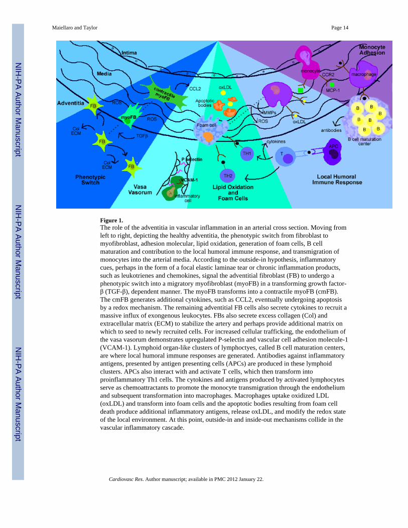
Figure 1.The role of the adventitia in vascular inflammation in an arterial cross section. Moving fromleft to right, depicting the healthy adventitia, the phenotypic switch from fibroblast tomyofibroblast, adhesion molecular, lipid oxidation, generation of foam cells, B cellmaturation and contribution to the local humoral immune response, and transmigration ofmonocytes into the arterial media. According to the outside-in hypothesis, inflammatorycues, perhaps in the form of a focal elastic laminae tear or chronic inflammation products,such as leukotrienes and chemokines, signal the adventitial fibroblast (FB) to undergo aphenotypic switch into a migratory myofibroblast (myoFB) in a transforming growth factor-β (TGF-β), dependent manner. The myoFB transforms into a contractile myoFB (cmFB).The cmFB generates additional cytokines, such as CCL2, eventually undergoing apoptosisby a redox mechanism. The remaining adventitial FB cells also secrete cytokines to recruit amassive influx of exongenous leukocytes. FBs also secrete excess collagen (Col) andextracellular matrix (ECM) to stabilize the artery and perhaps provide additional matrix onwhich to seed to newly recruited cells. For increased cellular trafficking, the endothelium ofthe vasa vasorum demonstrates upregulated P-selectin and vascular cell adhesion molecule-1(VCAM-1). Lymphoid organ-like clusters of lymphoctyes, called B cell maturation centers,are where local humoral immune responses are generated. Antibodies against inflammatoryantigens, presented by antigen presenting cells (APCs) are produced in these lymphoidclusters. APCs also interact with and activate T cells, which then transform intoproinflammatory Th1 cells. The cytokines and antigens produced by activated lymphocytesserve as chemoattractants to promote the monocyte transmigration through the endotheliumand subsequent transformation into macrophages. Macrophages uptake oxidized LDL(oxLDL) and transform into foam cells and the apoptotic bodies resulting from foam celldeath produce additional inflammatory antigens, release oxLDL, and modify the redox stateof the local environment. At this point, outside-in and inside-out mechanisms collide in thevascular inflammatory cascade.
Maiellaro and Taylor Page 14
Cardiovasc Res. Author manuscript; available in PMC 2012 January 22.
NIH
-PA Author Manuscript
NIH
-PA Author Manuscript
NIH
-PA Author Manuscript