nhs sickle cell and thalassaemia screening … sickle cell and thalassaemia screening programme 3...
TRANSCRIPT

NHS Sickle Cell and Thalassaemia Screening Programme
Handbook for antenatal laboratories
October 2017
Public Health England leads the NHS Screening Programmes

NHS Sickle Cell and Thalassaemia Screening Programme
Introduction 3
Haemoglobinopathies 4
Sickle cell disease 4
Beta thalassaemia major 5
Screening helpline 5
Antenatal screening 6Testing in subsequent pregnancies 6
Provision of screening 6
Organisation of screening services 7
Family origin questionnaire 8
Situations requiring particular care 9
Laboratory screening techniques 11
General laboratory considerations 12
Selecting an analytical system 13
Action Values 15
Good practice and troubleshooting 16
Problems with the measurement and interpretation of HbA2 17
Iron deficiency 18
Screening for carriers of a0 thalassaemia 18
Further investigation of a0 thalassaemia 19
The approach to screening and a0 thalassaemia 20
Risk assessment for antenatal screening in high prevalence areas 21
Risk assessment for antenatal screening in low prevalence areas 23
Interpretation and reporting of antenatal screening results 25
Screening shows the presence of HbA only: possible scenarios 26
Interpretation of relative proportions 33
ContentsVariant specific considerations 33
Homozygotes and compound heterozygotes 34
Reporting antenatal screening results 35
Recommended report formats 36Quality assurance and accreditation 43
Referral of antenatal samples for mutation analysis 44Testing of the baby’s biological father 44
Haemoglobin variants 45
Beta thalassaemias 46
Alpha thalassaemias 46
Alpha zero thalassaemia (a0 thalassaemia (--/aa)) 47
Quality assurance and accreditation 47
References 48
Appendix 1 – Carrying out a look back with particular reference to HbA2 49
Appendix 2 – Best practice guidelines for analysing liquid capillary blood samples 51
Appendix 3 – Table of biological parental carrier state combinations 53
Useful organisations and websites 54

NHS Sickle Cell and Thalassaemia Screening Programme
3
Introduction This is the fourth edition of the laboratory handbook, updating the edition published in October 2012. This handbook is for staff working in screening laboratories. It describes the policies and guidance that set out the requirements for laboratories involved in antenatal screening and DNA referral. Information for laboratories is regularly updated on GOV.UK[1] and laboratories must check this section frequently. The policies described relate to a screening programme, not a diagnostic service.
Providers must comply with the national guidance for the management of safety concerns and incidents in screening programmes and NHS England guidance for the management of serious incidents[2].
The British Committee for Standards in Haematology (BCSH) has published guidance about screening[3].
We would like to thank all those who have contributed to the handbook, which has been produced in accordance with the PHE style guide.
If you wish to reference or acknowledge this document, please use the following format: NHS Sickle Cell and Thalassaemia Screening Programme, Handbook for Laboratories, 4th edition, September 2017.
At the time of going to print, all information and contact details were correct. Please note this handbook is available on GOV.UK[1].

NHS Sickle Cell and Thalassaemia Screening Programme
4
HaemoglobinopathiesThe haemoglobinopathies are a heterogeneous group of more than 1,000 mutations, which are categorised into 2 main groups: the haemoglobin variants and the thalassaemias. The haemoglobin variants (also called the abnormal haemoglobins) arise from an alteration in the globin protein structure. The thalassaemias arise from inadequate production of structurally normal globin. There are also thalassaemic haemoglobinopathies that are produced when a structurally abnormal haemoglobin is synthesised at a reduced rate, for example HbE. Simple biochemical procedures can easily detect some of the abnormalities, but others are biochemically silent.
The frequency of different haemoglobinopathies varies in different ethnic groups and certain haemoglobinopathies are often associated with a family history. However, it is important to remember that no haemoglobinopathy is exclusive to any single ethnic group; so all individuals are theoretically at risk. It is not unusual for people to inherit more than one haemoglobin abnormality and many populations are at risk of a range of affected genes. Many haemoglobin mutations have no associated clinical significance. Others are associated with severe morbidity and mortality, most notably sickle cell disease and beta (β) thalassaemia major. Carriers are usually asymptomatic.
Sickle cell diseaseSickle haemoglobin (HbS) is a haemoglobin variant in which valine replaces glutamic acid, which is the sixth amino acid in the β globin chain. Other much rarer haemoglobins have been reported that have this same glutamic acid to valine substitution, but also an additional substitution in cis. All these variants will cause sickle cell disease in the situations described below for HbS.
Sickle cell disease results from the inheritance of certain genotypes, including:
• homozygosity for HbS (sickle cell anaemia)
• compound heterozygosity for HbS and an interacting mutation, such as HbC (Hb SC disease)
• β thalassaemia (Hb S/β thalassaemia)
The sickling disorders are associated with severe life-threatening vaso-occlusive crises, overwhelming sepsis, splenic sequestration, aplastic crises, stroke, priapism, pulmonary hypertension, proliferative retinopathy, chronic organ damage and avascular necrosis of the hips and shoulders.

NHS Sickle Cell and Thalassaemia Screening Programme
5
Beta thalassaemia majorBeta thalassaemias are a group of hereditary disorders that result in reduced (β+) or absent (β0) synthesis of β globin chains. These are an essential component of the main adult haemoglobin, HbA. Beta thalassaemias are usually caused by point mutations, small insertions or deletions and less frequently large deletions of the β globin gene located on chromosome 11. Inheritance is predominantly autosomal recessive although some dominant mutations have been reported. Mutation type and inheritance patterns result in variable phenotypes, which range from severe anaemia to clinically asymptomatic carriers. Thalassaemia major patients require lifelong transfusion therapy. This therapy has associated complications of iron overload, including endocrine dysfunction, cardiomyopathy, liver fibrosis and cirrhosis. A demanding daily regime of therapy with iron chelators that enable iron excretion from the body is required to treat these complications. Insufficient red cell replacement results in symptoms of anaemia, jaundice, poor growth and muscular development and skeletal changes from expansion of the bone marrow due to extramedullary haemopoiesis. Thalassaemia intermedia is a highly variable condition. Although some cases are transfusion-dependent it is usually a less severe disease than β thalassaemia major.
Screening helplineLaboratories may have questions about screening policy or interpretation of results that cannot be answered easily by reference to this handbook or textbooks. Oxford University Hospitals NHS Trust provides a support service for screening laboratories via designated telephone helplines and secure email. PHE funds the service and supports both antenatal and newborn screening enquiries.
Telephone: 01865 572 767Email: [email protected]: 01865 572 775

NHS Sickle Cell and Thalassaemia Screening Programme
6
Antenatal screeningThe aim of the antenatal screening programme is to offer timely antenatal sickle cell and thalassaemia screening to all women (and couples), to enable informed decision-making. We aim to:
• screen women by 10 weeks + 0 days gestation
• offer prenatal diagnosis (PND) to at risk women and couples by 12 weeks + 0 days gestation
• perform PND before 12 weeks + 6 days
Testing in subsequent pregnancies
If a woman is booked for antenatal care for a subsequent pregnancy, the healthcare professional must complete the Family Origin Questionnaire (FOQ), irrespective of previous screening. If the woman consents to screening, the healthcare professional must take the blood tests. They must ensure the sample is clearly labelled and sent to the laboratory accompanied by the completed FOQ. This blood sample must be tested according to the algorithm, however, if on repeat testing a variant with identical characteristics to one previously identified and confirmed is detected the result from the first line screen is sufficient.
If a carrier or affected woman is identified, the baby’s biological father must be offered a screening test, irrespective of previous screening history (Box 1).
If it is not possible to test the baby’s biological father in every pregnancy and a previous result is being used then this fact must be recorded in the woman’s notes for the current pregnancy.
The previous result must be from a laboratory accredited by the UK Accreditation Service (UKAS), and be consistent, unequivocal, well documented and interpreted and reported with the algorithms in this handbook.
Due to the complexities of the testing algorithm, screening for sickle cell and thalassaemia must be repeated in every pregnancy. Laboratories must be aware that serious incidents have resulted from failure to follow this policy.
Provision of screening
There are 2 approaches to the provision of the screening programme. The approach depends on whether a trust is defined as high prevalence or low prevalence. Each laboratory must use only one screening algorithm. Where high and low prevalence

NHS Sickle Cell and Thalassaemia Screening Programme
7
trusts merge the high prevalence algorithm should be used. In all cases a failsafe system must be in place to make sure all eligible women have been offered screening, and those that accept are tested.
High prevalence
Trusts are considered high prevalence if 2% or more of the booking bloods are screen positive. The high prevalence algorithm is viewed as the gold standard. In this case, laboratory testing for haemoglobin variants and thalassaemia must be carried out on all women who have accepted screening (see pages 21 and 22).
Low prevalence
Trusts are considered low prevalence where less than 1% of the booking bloods received by the laboratory are screen positive. Areas defined as low prevalence must perform screening for thalassaemia on all women who have accepted screening using the routine Hb and red cell indices. Further laboratory testing must be carried out on all women with defined abnormalities of the full blood count; those with high-risk family origins in either biological parent as determined by the FOQ and those women who request testing (see pages 23 and 24).
Intermediate prevalence
We advise trusts that have a prevalence between these 2 values to continue to use their current algorithm.
Organisation of screening services
Providers must have a risk assessment of the entire pathway and protocols defining where responsibility falls for all aspects of the process. Laboratories must have protocols detailing receipt of samples, analytical procedures and reporting of results in a timely manner. If some or all samples are referred to or received from other laboratories, there must be service level agreements covering all of these aspects. Laboratory staff must follow the analytical guidelines, algorithms and service specifications produced by the NHS Sickle Cell and Thalassaemia Screening Programme. Additionally, there must be an agreement outlining who is responsible for the provision of key performance indicators (KPI), annual return data and data returns to National Congenital and Rare Disorders Registration Service.
Where opinions on results obtained are sought from third parties, laboratories must ensure they meet and comply with screening programme requirements. This is particularly important when using any service provided by a commercial organisation. When reports received from third parties are transcribed into internal

NHS Sickle Cell and Thalassaemia Screening Programme
8
laboratory information systems, a full and exact copy of the report must be made. The transcribed report must be checked by an appropriate second person to ensure accuracy. Laboratories should scan these reports if possible.
Family origin questionnaire
The FOQ is used in both low and high prevalence areas. In low prevalence areas it is the basis for determining which women must be tested for haemoglobin variants. The woman’s sample must be tested if she or the baby’s biological father is in a high risk group. In high prevalence areas all consenting women must be tested. If a woman has declined screening then testing should not be carried out and this must be documented.
All women must be screened for thalassaemia using the mean cell haemoglobin (MCH) measurement, which has been shown to be stable for up to 4 to 5 days at room temperature[4,5]. The family origin also assists laboratories with the interpretation of laboratory screening results, particularly the interpretation of results indicating possible alpha (a) thalassaemia. People originating from certain areas of the Far East and Eastern Mediterranean are at higher risk of a0 thalassaemia, which in the homozygous state results in hydrops fetalis. Local policy must be in place for situations where an FOQ has not been received or is incomplete to ensure test reporting is not delayed. The FOQ must not be rejected if the only missing information is the gestational age.
The FOQ was developed from the initial literature review and research, funded by the programme centre. It was further developed after an evaluation by Ethnos Research and Consultancy during the first phase of implementation of antenatal screening in low prevalence areas. The questions aim to determine family origins and are different from the census based self-assigned ethnic group questions. If the FOQ is incorporated into an electronic requesting system, the number of options available must not be fewer than those on the 3-part paper version. If any modifications of the FOQ are being considered, for instance as part of a combined antenatal screening request form, the FOQ data fields must be consistent with the current version.

NHS Sickle Cell and Thalassaemia Screening Programme
9
Situations requiring particular care
Fertility treatment – donor gametes
If the pregnancy has been achieved using a donor egg then the screening results on the woman will not be informative. The baby’s biological father must be tested and if screen positive the report must recommend that the fertility clinic is contacted to obtain the biological mother’s haemoglobinopathy results.
If donor sperm has been used and the woman has a positive screening result then the report must recommend that the fertility clinic is contacted to obtain the biological father’s haemoglobinopathy results.
In the case of surrogacy the report must recommend that the fertility clinic is contacted to obtain haemoglobinopathy results of both biological parents.
It is best practice to test the pregnant woman in all circumstances to ensure optimal maternal care. The report must provide the appropriate advice for the circumstances of the pregnancy.
Adoption
If either biological parent has been adopted, the FOQ information may not accurately reflect the true family origins. Such cases must be treated as high risk and have full laboratory screening.
Bone marrow transplants
Where either biological parent has had a bone marrow transplant (BMT) it is likely the results obtained will reflect the BMT donor and will not accurately represent the genetic status of the fetus. If the biological mother has had a BMT, the baby’s biological father must be tested to ensure this is not a high risk pregnancy. If DNA confirmation of the biological mother’s status is required or if the baby’s biological father is post BMT and requires testing, then pre-transplant DNA or DNA obtained from hair follicles should be used.

NHS Sickle Cell and Thalassaemia Screening Programme
10
Box 1: Maternal conditions requiring testing of the baby’s biological father.
Significant maternal haemoglobinopathies
The following maternal haemoglobinopathies should be detected by antenatal screening and are important for maternal care:
• Hb SS
• Hb SC
• Hb SDPunjab
• Hb SE
• Hb SOArab
• Hb S/Lepore and Hb Lepore/β thalassaemia
• Hb S/β thalassaemia
• Hb S/δβ thalassaemia
• HbH disease (--/-a)
• β thalassaemia major/intermedia
• Hb E/β thalassaemia
Carrier states in biological mother:
• HbS
• HbC
• HbDPunjab
• HbE
• HbOArab
• Hb Lepore
• β thalassaemia
• δβ thalassaemia
• a0 thalassaemia (--/aa)
• Hereditary Persistance of Fetal Haemoglobin (HPFH)
Any compound heterozygote state including one or more of the above conditions.Any homozygous state of the above conditions.

NHS Sickle Cell and Thalassaemia Screening Programme
11
Laboratory screening techniques
In low prevalence areas the first-line screen for thalassaemia is the full blood count.
The following techniques are suitable for use in first-line screening for haemoglobin variants and for HbA2 quantitation:
• high performance liquid chromatography (HPLC), preferably with continuous gradient elution
• capillary electrophoresis (CE)
Both these techniques provide provisional results. Abnormal results must have further testing with a minimum of one alternate technique with a different analytical principle to the original and which is appropriate for the suspected variant (Table 1). Minimum requirements for quality control material for these techniques can be found on GOV.UK[1]. In some cases more than one technique will be required. If results from all techniques are interpreted together laboratories can achieve a result which is sufficiently reliable for screening purposes. Only mass spectrometry and DNA provide a definitive diagnosis.
Table 1: Acceptable laboratory techniques.
Initial method Alternate method
HPLC Alkaline electrophoresis
Acid electrophoresis
Isoelectric focusing (IEF)
CE
Mass spectrometry
DNA
CE Acid electrophoresis
Alkaline electrophoresisa
HPLC
IEF
Mass spectrometry
DNAaResults from alkaline electrophoresis are very similar to CE, so this should not be used as the only alternate test. In the absence of IEF it may be useful in combination with acid electrophoresis.

NHS Sickle Cell and Thalassaemia Screening Programme
12
In most circumstances sickle solubility testing can be used as confirmation of an initial screen that suggests the presence of HbS. There is an inherent unreliability in the sickle solubility test with the risk of false positive and negative results. Special care must be taken when using an instrument where HbS co-elutes with another haemoglobin variant, such as HbC. For example, in cases of co-eluting haemoglobin variants S and C, a negative solubility test cannot be presumed to indicate HbC and a positive solubility alone cannot be presumed to indicate HbS. In all cases where haemoglobins co-elute, it is necessary to use an additional technique, for example alkaline electrophoresis or IEF.
HbA2 measurement
HPLC and CE methods are acceptable. IEF and scanning densitometry are not acceptable.
HbF measurement
HPLC, CE or 2 minute alkali denaturation are acceptable. The Kleihauer test is not appropriate for measurement but is useful to support the identification of HbF.
General laboratory considerations
Users must be aware that this laboratory handbook highlights common analytical and diagnostic issues but every laboratory must follow the principles of good laboratory practice. Laboratories must satisfy themselves that they understand the capabilities and limitations of their chosen technique. The equipment and protocol chosen must fulfil the requirements of the screening programme and demonstrate suitable performance on external quality assurance (EQA). The manufacturer’s published recommendations should be followed. Where an instrument operates with more than one analytical programme designed for haemoglobin variant analysis both programmes must be validated to ensure there are no significant differences where a numerical value is to be reported. If significant differences are found a single programme should be chosen based on the validation data, from which all results are reported.
It may be necessary to return to the original specimen tube to check the identification details. For this reason it should be standard practice to ensure that any labels affixed when the specimen is received in the laboratory do not obscure the written identity or identity label already attached.
For all analytical techniques, appropriate controls must be included wherever possible. If IEF or electrophoresis is used, then control haemoglobins must be run with each plate.

NHS Sickle Cell and Thalassaemia Screening Programme
13
Minimum requirements for quality control material for these techniques can be found at GOV.UK[1].
If a column is used this must be replaced once it has performed the recommended number of analyses as stated in writing by the manufacturer or if the performance is observed to deteriorate. If software rules are used to screen samples for further action and reporting, it is essential that the process is risk assessed and that there are failsafe mechanisms in place. Raw data, including analytical traces, must always be reviewed and any post analytical procedures, including algorithms, must be fully documented and traceable to ensure consistency of quality. All raw data, including analytical traces and interpretative comments, must be reviewed by 2 people, one of whom must be a registered scientist. Final validation must be by a person with suitable expertise, as demonstrated by current competency testing.
Selecting an analytical system
Co-ordination of pathology services across chemistry and haematology can allow the sharing of equipment, for example with that used for measurement of HbA1c. In all situations, it is essential that HbA2 is analysed by a protocol that calibrates and clearly separates HbA2 from HbA. The required antenatal turnaround time of 3 working days for the issue of a report must be considered when this approach is used.
High performance liquid chromatography
HPLC uses an ion exchange resin, held in a column, in conjunction with a buffer gradient. As the ionic strength and/or pH of the buffer changes, certain haemoglobins are eluted from the column and the presence of haemoglobin is detected using a spectrophotometric technique. The time from injection to the point at which the haemoglobin fraction elutes is known as the retention time of the haemoglobin and is a reproducible measurement for a particular column, buffer, exchange resin and temperature.
However, it is quite common for different haemoglobins to elute at the same retention time. Thus the retention time is not a unique identifier. HbF and HbA2 elute separately and the relative proportion is calculated with the use of calibrators. Ideally haemoglobins S, C, D, E and OArab should have separate retention times and characteristic chromatographic profiles. In addition, the relative proportions of the different haemoglobins are recorded. HPLC analysers that use step-wise buffer gradients are not recommended if they produce co-elution of common haemoglobin variants which can lead to misinterpretation.

NHS Sickle Cell and Thalassaemia Screening Programme
14
When selecting an analyser for quantitation of HbA2 the first consideration is good separation. It is important to ensure the analyser clearly separates peaks to ensure accurate quantitation as shown in Figure 1. Laboratories should understand how the integration takes place and be aware that peaks measured on sloping baselines or on shoulders of adjacent peaks are likely to be less reliable. Sophisticated integration and the use of calibration factors cannot make up for poor chromatography.
Figure 1. Examples of integration.

NHS Sickle Cell and Thalassaemia Screening Programme
15
Capillary electrophoresis
CE uses a combination of ion migration and electro-osmotic flow to separate protein molecules. When a voltage is applied across the capillary tube filled with an electrolyte solution, the solution begins to move towards one of the electrodes due to electro-osmotic flow. This drives the bulk flow of materials past the detector in the same way that a pump pushes the liquid in HPLC. The haemoglobin molecules move towards the detector at different speeds depending on their ionic charge and electrophoretic mobility. Both electro-osmotic flow and electrophoretic mobility are occurring at the same time, working in opposite directions to provide greater resolution.
This method of separation should not be confused with simple electrophoretic mobility as seen in cellulose acetate electrophoresis. Combining electro-osmotic flow and electrophoretic mobility is a separate phenomenon and is exploited in CE for maximum separation power. Even so, it is quite common for different haemoglobins to migrate at the same rate and appear at the same position, so position is not a unique identifier. HbF and HbA2 elute separately and the relative proportion of each is calculated with the use of calibrators. Haemoglobins S, C, D, E and OArab also have separate retention times and characteristic profiles. In addition, the relative proportions of the different haemoglobins are recorded.
Optical density (OD) levels greater than 0.07 and the presence of sufficient HbA are required to determine the migration position and thus permit ‘zoning’ and a provisional identification of haemoglobins present in the sample. If failure to zone is due to low OD, this is usually related to the amount of haemoglobin in the sample. This should be corrected by increasing the haemoglobin to diluent ratio. Extreme care is needed if extraneous haemoglobin is added to a clinical sample to allow zoning. The addition of haemoglobins which were not present in the initial sample may lead to misinterpretation of the results.
Action Values
A single value for HbA2 of equal to or greater than 3.5% has been set as the action value for carriers of β thalassaemia. There are 2 action values for HbF. If the MCH is greater than or equal to 27pg, the action value is greater than 10%. However, if the MCH is less than 27pg, the action value is greater than or equal to 5.0%. The chosen system must be able to measure HbA2 and HbF with accuracy and precision at these action values and detect the haemoglobin variants as specified by the antenatal screening programme. Instrument validation protocols must assess these requirements. Quantitation at different levels may be needed for other clinical purposes.

NHS Sickle Cell and Thalassaemia Screening Programme
16
UK national external quality assurance scheme (UK NEQAS) data and published literature have demonstrated biases between different analysers for HbA2. The International Federation of Clinical Chemistry and Laboratory Medicine (IFCC) and the International Council for Standardization in Haematology (ICSH) have recognised the need for a new certified reference material for HbA2, which is under development. The screening programme supports the objectives of this work and continues to work with UK NEQAS to monitor the impact of inter-method variability.
Laboratories in the UK should be aware of these problems and ensure they have optimised their methods. When considering replacement purchases, laboratories should review all available evidence, including UK NEQAS data.
Analytical systems should be able to detect and measure at least the most common HbA2 variant (HbA2Prime (A2’)) to enable an approximate calculation of total HbA2 (for example HbA2 plus A2’).
If using equipment or an elution programme for more than one analyte, for example HbA2 and HbA1c, laboratories should ensure that the quantitation of HbA2 and HbF is not compromised.
Good practice and troubleshooting
When operating analytical systems the following points of good practice should be followed and may be used as a troubleshooting guide if problems are identified by internal and/or external quality assurance.
1. Ensure the HPLC column count is within manufacturer’s written guaranteed limit. 2. Make sure the calibration factor does not drift significantly or fall outside
recommended limits. 3. Monitor internal quality control for drift.4. Follow the manufacturer’s recommended maintenance and cleaning protocols.5. Ensure that reagents, controls and calibrators are stored in the correct conditions,
are within date, have appropriate lot numbers and are in the correct positions. 6. Avoid pooling and mixing reagents, do not pool reagents with different lot
numbers. 7. When reviewing analytical traces, ensure the appearance is as it should be with
the correct peak shape, baseline and separation from adjacent haemoglobin peaks.
8. Ensure maintenance visits are carried out at the recommended intervals. 9. If the machine is moved or laboratory conditions change, appropriate revalidation
procedures must be performed.10. If problems occur that cannot be resolved contact the instrument manufacturer.

NHS Sickle Cell and Thalassaemia Screening Programme
17
If samples require modification by the addition of extraneous haemoglobin an aliquot must be used. This must be analysed with a unique identifier distinguishable from both the original specimen and from any other clinical sample.
In the event of an out of consensus EQA result a look back exercise must be considered. Look back may also be required when internal quality assurance falls outside acceptable limits or when indicated for any other reason. Guidance is provided in Appendix 1.
Problems with the measurement and interpretation of HbA2
In the presence of an a chain variant it can be difficult to obtain a reliable HbA2 value. For this reason, where the biological mother has an MCH of less than 27pg and is found to have a suspected a chain variant or a haemoglobin variant that co-elutes with, or compromises the HbA2, the baby’s biological father must be offered screening. If the biological father has an MCH of less than 27pg with a suspected a chain variant or a variant which co-elutes with, or compromises the HbA2 and the biological mother is known to have β thalassaemia, or has Hb S, E, OArab or Lepore and/or is at risk of alpha zero (a0) thalassaemia, DNA studies should be performed.
If any small peaks which could be an HbA2 variant elute separately from the main HbA2 peak in a patient with an MCH below the action value (< 27pg), further investigation will be required if the addition of these peaks brings the total HbA2
greater than or equal to 3.5%.
HbA2 values of 4.0% and above with normal indices may indicate a carrier of β thalassaemia. In this case the findings including the FBC should be confirmed. If the results remain the same then the baby’s biological father must be tested. Other factors such as HIV infection, B12/folate deficiency or liver disease/alcohol can increase the HbA2.
HbA2 values less than 4.0% with normal red cell indices and an HbF level of less than or equal to 10% are regarded as not significant for screening purposes.
With many HPLC systems, HbA2 is overestimated in the presence of HbS. This is not a problem where the percentage of HbA is greater than HbS. See section ‘interpretation of relative proportions’ on page 33.
With many HPLC systems, HbA2 is underestimated in the presence of HbD. This is not a problem where the percentage of HbA is greater than HbD but may make discrimination between Hb DD and D/β0 thalassaemia more difficult. Ensure red cell indices are reviewed.

NHS Sickle Cell and Thalassaemia Screening Programme
18
When using CE, HbA2 is underestimated in the presence of HbC. This is not a problem where the percentage of HbA is greater than HbC but may make discrimination between Hb CC and C/β0 thalassaemia more difficult. Ensure red cell indices are reviewed.
For analysers that separate and measure HbA2 in the presence of HbE, caution should be used when interpreting this value.
Iron deficiency
The HbA2 level may be lowered by up to 0.5% in cases of severe iron deficiency anaemia[6] but screening for haemoglobin variants and thalassaemia should proceed without regard to iron deficiency, suspected or proven.
Any decrease in MCH should be regarded as potentially due to a haemoglobinopathy and the HbA2 should be measured. It may be appropriate to simultaneously investigate pregnant women for iron deficiency, using ferritin or zinc protoporphyrin but this is not specifically part of the screening protocols.
In pregnant women there is no justification for delaying the investigation of haemoglobinopathies while treating iron deficiency, as this will delay the process of identifying at-risk carrier couples who should be offered PND.
Screening for carriers of a0 thalassaemia
Methods of screening
The lack of a specific biomarker for the detection of a thalassaemia carriers creates problems, particularly in the context of a screening programme. In this context a+ thalassaemia is not regarded as significant and policies are designed to detect only couples at risk of hydrops fetalis. Policy has been designed to increase the positive predictive value of the screening algorithm and reduce false positives.
Diagnosis
Molecular techniques must be used for the confirmation and diagnosis of a0 thalassaemia when suspected in both biological parents. a thalassaemia mutations may be both deletional and non-deletional, requiring the use of different diagnostic techniques. For the common deletional mutations laboratories using GAP PCR based techniques will only detect those mutations included in their screening panels.

NHS Sickle Cell and Thalassaemia Screening Programme
19
Population estimates and ethnic distribution of a0 thalassaemia
It is estimated that only a small number of cases of a thalassaemia major can be expected each year in England, with approximately half of these of Chinese family origin.
Alpha zero thalassaemia is most commonly found in those of Southeast Asian origin (China including Hong Kong, Thailand, Taiwan, Cambodia, Laos, Vietnam, Indonesia, Burma, Malaysia, Singapore or Philippines) and East Mediterranean (Cyprus, Greece, Sardinia or Turkey). There are 2 common Mediterranean (--MED, –a(20.5)) and 3 common Southeast Asian deletional mutations (--SEA, --THAI, --FIL).
Alpha zero thalassaemia has also been reported to occur at low frequencies in some Middle Eastern countries: the --MED allele in the UAE, Iran, Yemen, Kuwait, and Jordan; the –a(20.5) allele in Iran; the --YEM allele in the Yemen. a0 thalassaemia is rarely reported in patients of African, Pakistani and Indian origin. a0 thalassaemia is rarely observed in patients of British origin and no couple at risk of Hb Bart’s hydrops fetalis has been reported. In these family origin groups the risk is small and in the context of screening, the cost benefit ratio is poor. Therefore the FOQ does not identify these groups as at high risk for a0 thalassaemia[7].
Further investigation of a0 thalassaemia
Alpha thalassaemia major is invariably fatal without treatment, resulting in hydrops fetalis due to severe fetal anaemia. If not detected, it can result in a stillbirth. A mother carrying a fetus with a thalassaemia major is at risk of obstetric complications, such as toxaemia and hypertension, particularly in the third trimester of pregnancy. If a fetus with a thalassaemia major is transfused in utero, it is possible that it will survive and be born as a transfusion-dependent infant. Usually a baby only has Hb Bart’s hydrops fetalis (a thalassaemia major) if both biological parents are carriers of a0 thalassaemia.
Table 2 shows the a thalassaemia genotypes.
Genotype Phenotype
aa/aa Normal
-a/aa Alpha plus (a+) thalassaemia (heterozygote)
-a/-a Alpha plus (a+) thalassaemia (homozygote)
--/aa Alpha zero (a0) thalassaemia (heterozygote)
--/-a Haemoglobin H disease
--/-- Alpha thalassaemia major (homozygote)Hb Bart’s hydrops fetalis

NHS Sickle Cell and Thalassaemia Screening Programme
20
The approach to screening and a0 thalassaemia
Two sets of information from the pregnant woman are combined as the screen:
1. Is the MCH <25pg?
2. Is the woman’s family origin identified as high risk from the FOQ: China (including Hong Kong), Southeast Asia (especially Thailand, Taiwan, Cambo-dia, Laos, Vietnam, Burma, Malaysia, Singapore, Indonesia or Philippines), Cyprus, Greece, Sardinia, Turkey, or unknown?
If the answer to both questions is yes, testing of the baby’s biological father must be offered if he is also from a high risk area or unknown.
If the baby’s biological father is suspected of having a0 thalassaemia, the samples on both biological parents must be sent for DNA analysis for a0 thalassaemia mutations.
If one biological parent is a suspected carrier of a0 thalassaemia and the other is a carrier of a thalassaemia and is also from one of the high risk groups for a0 thalassaemia with an MCH < 25pg, both biological parents should be screened for a0 thalassaemia by DNA analysis.
DNA analysis of samples and a0 thalassaemia
The policy guidance developed by the screening programme should mean that DNA analysis is only done in a limited number of cases and not usually in cases where a+ thalassaemia is suspected.
Evidence for above approach
Published studies have shown that 99% of a0 thalassaemia cases have an MCH <25pg. In a series of 270 carriers of a0 thalassaemia diagnosed by DNA analysis in the UK, only 2 patients had an MCH between 25 and 26pg – one with liver disease[8]. Findings in a study from Sheffield[9] which undertook DNA analysis in 425 pregnant women with an MCH <27pg showed that all cases of a0 thalassaemia had an MCH <25pg and would have been detected using an FOQ alone, which supports the screening programme’s approach.

NHS Sickle Cell and Thalassaemia Screening Programme
21
Risk assessment for antenatal screening in high prevalence areas
It is inevitable there will be some false positives and negatives as a result of screening because of the way screening programmes are designed, particularly with the use of action values. False positives have a positive result from the screening test but, when further tests have been performed, do not have one of the designated haemoglobins or thalassaemia. Potential causes of false negatives are shown below.
Conditions that may be missed using the algorithm (assuming that the FOQ has been completed accurately) include:
• ‘silent’ or ‘near silent’ β thalassaemia carriers. Some β thalassaemia carrier genotypes are associated with borderline HbA2 levels and an action value of 3.5% with an MCH of < 27pg will miss some cases. Examples of such mutations include the c.-50 A>C [CAP+1 (A>C)], c.92+6 T>C [IVSI-6 (T>C)], c.-151C>T [-101 (C>T)] and c.*111 A>G or c.*110 T>C [Poly A (A>G) or (T>C)]
• some β thalassaemia carriers obscured by severe iron deficiency anaemia
• thalassaemia where the MCH is raised, for example B12/folate deficiency, liver disease, HIV therapy
• β thalassaemia carriers with a coexisting δ chain mutation which is silent with the first line screening technique, or who have coexisting delta thalassaemia
• β thalassaemia carriers with coexisting HbH Disease as some cases have normal HbA2 values
• a0 thalassaemia occurring outside the defined at risk family origins (see page 20) or in those women with an MCH ≥25pg
• δβ thalassaemia carriers with HbF <5%
• γδβ thalassaemia carriers
• dominant haemoglobinopathies in the baby’s biological father when the woman is Hb AA, these are very rare and should be suggested by the family history
• any significant condition silent with the first line screening
• any significant condition masked by a blood transfusion
• any significant condition masked by an unreported bone marrow transplant
• any significant condition present in donor egg or sperm where the donor is undeclared or untested
• incorrect family origins assigned due to undeclared adoption

NHS Sickle Cell and Thalassaemia Screening Programme
22
Figure 2 - Testing algorithm for laboratory screening in high prevalence areas.

NHS Sickle Cell and Thalassaemia Screening Programme
23
Risk assessment for antenatal screening in low prevalence areas
It is inevitable there will be some false positives and negatives as a result of screening because of the way screening programmes are designed, particularly with the use of action values. False positives have a positive result from the screening test but, when further tests have been performed, do not have one of the designated haemoglobins or thalassaemia. Potential causes of false negatives are shown below.
Conditions that may be missed using the algorithm (assuming that the FOQ has been completed accurately) include:
• ‘silent’ or ‘near silent’ β thalassaemia carriers. Some β thalassaemia carrier genotypes are associated with borderline HbA2 levels and an action value of 3.5% in conjunction with an MCH of < 27pg will miss some cases. Examples of such mutations include the c.-50 A>C [CAP+1 (A>C)], c.92+6 T>C [IVSI-6 (T>C)], c.-151C>T [-101 (C>T)] and c.*111 A>G or c.*110 T>C [Poly A (A>G) or (T>C)].
• some β thalassaemia carriers obscured by severe iron deficiency anaemia
• thalassaemia where the MCH is raised eg. B12/folate deficiency, liver disease, HIV therapy
• β thalassaemia carriers with a coexisting delta chain mutation which is silent with the first line screening technique, or who have coexisting delta thalassaemia.
• β thalassaemia carriers with coexisting HbH Disease as some cases have normal HbA2 values.
• a0 thalassaemia occurring outside the defined at risk family origins (see page 20) or in those women with an MCH ≥25pg.
• δβ thalassaemia carriers with HbF <5%
• γδβ thalassaemia carriers
• dominant haemoglobinopathies in the baby’s biological father when the woman is Hb AA, these are very rare and should be suggested by the family history
• Hb S, C, DPunjab, E, OArab outside the defined at risk family origins
• any significant condition silent with the first line variant screening technique
• any significant condition masked by a blood transfusion
• any significant condition masked by an unreported bone marrow transplant
• any significant condition present in donor egg or sperm where the donor is undeclared or untested
• incorrect family origins assigned due to undeclared adoption

NHS Sickle Cell and Thalassaemia Screening Programme
24
Figure 3 - Testing algorithm for laboratory screening in low prevalence areas.

NHS Sickle Cell and Thalassaemia Screening Programme
25
Interpretation and reporting of antenatal screening results
These guidelines should provide sufficient information to allow all normal, and over 95% of abnormal, cases to be reported in a standardised manner. There will always be some situations that require further tests, or family studies, before a useful clinical result can be achieved because of the diversity of haemoglobin variants and thalassaemia syndromes. Appropriate clinical referral is required in these cases to ensure that the risk to the pregnancy is fully and correctly assessed.
If an individual has had a blood transfusion and any of the transfused red cells are still present, misleading data and conclusions may result. It is essential that clinicians realise this fact and it is best practice to have a universal footnote on all haemoglobinopathy results, such as: ‘unless otherwise stated, result assumes no recent transfusion’ or ‘result valid if not transfused’.
Laboratories should treat haemoglobin values of less than 80g/L with caution. There are multiple conditions that can cause severe anaemia in pregnancy, including iron deficiency, haemolytic anaemia, HbH disease, or β thalassaemia intermedia. Laboratories should review these on a case-by-case basis. If necessary, they should discuss such cases with a senior scientist or clinician to consider appropriate further action, including testing of the baby’s biological father.
It is not possible to separate all haemoglobin variants using screening methods and a second haemoglobin variant may be migrating/eluting with any other haemoglobin including HbA. This is a particular issue in HPLC instruments which use a stepwise buffer gradient, notably the co-elution of Hbs S and C. Conversely such gradients may also split other haemoglobin variants into 2 peaks, for example HbDPunjab. In other cases, such as when thalassaemia and haemoglobin variants are co-inherited, one condition may be masking another.
HPLC instruments separate adducted fractions of haemoglobins, these fractions must not be added to the assumed parent peak. Addition of assumed peaks can lead to erroneous results. No additional testing is necessary if the HbA2 is raised (but not greater than 8%) and the red cell indices are typical of a carrier of β thalassaemia. It is important to be aware that HbF levels are highly variable in β thalassaemia carriers.

NHS Sickle Cell and Thalassaemia Screening Programme
26
Screening shows the presence of HbA only: possible scenarios
Scenario 1
If screening tests show only a single major Hb peak (band) in the HbA position and the:
• MCH is ≥ 27pg
• HbA2 is < 4.0%
• HbF is < 10%
• Hb is ≥ 80g/L
Then no further testing is required. There is negligible risk in the pregnancy associated with an abnormal haemoglobin or thalassaemia. Use report format 1
Scenario 2
If screening tests show only a single major Hb peak (band) in the HbA position and the:
• MCH is < 27pg
• HbA2 is ≥ 4.0% but < 8.0%
Then there is a risk in the pregnancy associated with the presence of β thalassaemia. Use report format 4a. If the HbA2 is ≥ 8.0% check for other variants, particularly Hb Lepore.
Scenario 3
If screening tests show only a single major Hb peak (band) in the HbA position and the:
• MCH is ≥ 27pg
• HbA2 is ≥ 4.0% but < 8.0%
Then there is a risk in the pregnancy associated with the presence of β thalassaemia. Use report format 4b. If the HbA2 is ≥ 8.0% check for other variants using an alternate technique.

NHS Sickle Cell and Thalassaemia Screening Programme
27
Scenario 4
If screening tests show only a single major Hb peak (band) in the HbA positionand the: • MCH is < 27pg
• HbA2 is ≥ 3.5% but < 4.0%
Then there is a risk in the pregnancy associated with the presence of β thalassaemia. Use report format 4b.
Scenario 5
If screening tests show only a single major Hb peak (band) in the HbA position and the:
• MCH is < 27pg
• Hb is < 80g/L
• HbA2 is ≥ 3.0% but < 4.0%
Then there is a risk in the pregnancy associated with the presence of β thalassaemia. Use report format 4b.
Scenario 6
If screening tests show only a single major Hb peak (band) in the HbA position and the:
• MCH is < 27pg
• HbA2 is < 3.5%
Consider whether this is the measurement of the total HbA2. If the patient has a delta chain variant or an a chain variant, a second HbA2 peak/band may be present and must be included in the total HbA2 where appropriate. If total HbA2 (HbA2 plus HbA2 variant) ≥ 3.5% then there may be a risk in the pregnancy associated with the presence of β thalassaemia. If β thalassaemia is suspected use report format 4a or 4b as described in scenarios 3 to 5.
If the HbA2 measurement is correct, < 3.5% please see scenarios 7 to 9. In these scenarios the FOQ forms part of the screening tests.

NHS Sickle Cell and Thalassaemia Screening Programme
28
Scenario 7
If screening tests show only a single major Hb peak (band) in the HbA position and the:
• MCH is < 27pg but ≥ 25pg
• HbA2 is < 3.5%
• HbF is < 5.0%
• Hb is ≥ 80g/L
This indicates possible iron deficiency or a+ thalassaemia. There is negligible risk to the pregnancy. Use report format 7b.
Scenario 8
If screening tests show only a single major Hb peak (band) in the HbA position and the:
• MCH is < 25pg
• HbA2 is < 3.5%
• HbF is < 5.0%
• biological parents are both of high risk family origins for a0 thalassaemia or unknown or one has family origins of high risk and the other is unknown
This indicates possible a0 thalassaemia or iron deficiency. Use report format 6a. If the woman has HbH disease, use report format 6b.

NHS Sickle Cell and Thalassaemia Screening Programme
29
Scenario 9
If screening tests show only a single major Hb peak (band) in the HbA position and the:
• MCH is < 25pg
• HbA2 is < 3.5%
• HbF is < 5.0%
• neither, or only one, biological parent has family origins of high risk for a0 thalassaemia
This indicates possible iron deficiency, homozygous a+ thalassaemia, or heterozygous a0 thalassaemia (use report format 7a) or HbH disease (use report format 6b).
Scenario 10
If screening tests show only a single major Hb peak (band) in the HbA position and the:
• MCH is ≥ 27pg
• HbA2 is < 4.0%
• HbF is ≥ 10%
• Hb is ≥ 80g/L
There is a risk to the pregnancy associated with the presence of HPFH. Use report format 5a. If Hb is < 80g/L ensure that all other potential risks have been considered.

NHS Sickle Cell and Thalassaemia Screening Programme
30
Scenario 11
If screening tests show only a single major Hb peak (band) in the HbA position and the:
• MCH is < 27pg
• HbA2 is < 3.5%
• HbF is ≥ 5.0%
There is a risk to the pregnancy associated with the presence of δβ thalassaemia. Use report format 5b
The possible scenarios are summarised in Table 3.
Table 3 needs to be read in conjunction with ‘interpretation and reporting of antenatal screening results’ on page 25 and recommended report formats page 36.

NHS Sickle Cell and Thalassaemia Screening Programme
31
Table 3. Summary of interpretation and reporting guidelines. [Where screening shows a single peak in the position of HbA.]
Family origin MCH pg Total A2 % F % Hb (g/L)
Conclusion Report format
Any ≥ 27 < 4.0 < 10 ≥ 80 No evidence of abnormal haemoglobin or thalassaemia 1
Any ≥ 27 ≥ 4 but < 8.0 — — Possible β thalassaemia carrier 4b
Any ≥ 27 ≥ 8.0 — — Check for variant haemoglobins —
Any < 27 ≥ 4 but < 8.0 — — β thalassaemia carriera 4a
Any < 27 ≥ 8.0 — — Check for variant haemoglobins, particularly Hb Leporea
—
Any < 27 ≥ 3.5 but < 4.0 — — Possible β thalassaemia carriera 4b
Any < 27 > 3.0 but < 4.0 — < 80 Possible β thalassaemia carriera 4b
Any < 27 < 3.5 — — Check for a second HbA2, due to either an a or a δ chain varianta
—
Any < 27 but ≥ 25
< 3.5 < 5 ≥ 80 Possible iron deficiency or a thalassaemia 7b
Both biological parents high risk for a0 thalassaemia or unknown or one high risk and one unknown
< 25 < 3.5 < 5 ≥ 80b Possible a0 thalassaemia or iron deficiency or HbH Disease. Baby’s biological father requires testing.
6a or 6b
Neither, or only one, biological parent has family origins of high risk for a0 thalassaemia
< 25 < 3.5 < 5 ≥ 80b Possible iron deficiency or homozygous a+ thalassaemia or heterozygous a0 thalassaemia or HbH Disease. Testing of baby’s biological father not required.
7a or 6b
Any ≥ 27 < 4 ≥ 10 ≥ 80 Hereditary persistence of fetal haemoglobin 5a
Any < 27 < 3.5 ≥ 5 — δβ thalassaemia carriera 5baConsider coexisting a0 thalassaemia when MCH < 25pg and the biological parents have appropriate family origins (see algorithm)bUnless proven HbH Disease where Hb may be lower

NHS Sickle Cell and Thalassaemia Screening Programme
32
First test suggests HbA and either HbS, HbC, HbD, HbE or HbOArab
Further tests required:Sickle solubility testIt is best practice to perform a sickle solubility on all haemoglobin variants detected. If the variant has features suggestive of HbS and the sickle solubility is positive no further testing is usually required. However, see note on page 12 about the need for caution when using the sickle solubility test for confirmatory purposes with certain instruments. If a sickling haemoglobin is present at < 20% the sickle test may not be positive and in this case further investigations will be required.
For all other variants see Table 4
Table 4 gives the minimum testing requirements to support the presumed identity of the listed haemoglobin variants when using HPLC or CE as the first test. Laboratories may choose to perform additional or more specific techniques but must use a method with a different scientific principle to the primary method (Table 1). Minimum requirements for quality control material for these techniques can be found at GOV.UK[1]
Table 4. Minimum testing requirements.
Suspected variant Additional techniques
HbC Either acid electrophoresis or IEF
HbD aEither alkaline electrophoresis or IEF and acid electrophoresis
HbE Either alkaline electrophoresis or IEF and acid electrophoresis
HbOArab Acid electrophoresis
Hb Lepore Either alkaline electrophoresis or IEF and acid electrophoresis
aThere are several D haemoglobins, but the screening programme aims to detect HbDPunjab. When HbD is detected and it cannot be differentiated, testing of the baby’s biological father will identify the couples at potential risk. The HbD will need to be confirmed as HbDPunjab if the baby’s biological father is found to have HbS. This will also apply if the biological mother has HbS and the baby’s biological father is found to have HbD. In such cases confirmatory testing must be either DNA or mass spectrometry.

NHS Sickle Cell and Thalassaemia Screening Programme
33
Interpretation of relative proportions
Hb Variant (V) between 40 and 50% (35 and 50% if HbV is HbE). Is the band/peak in the HbA position actually HbA? If the HbV > HbA the possibility exists of Hb V/β+ thalassaemia with an unusually high expression of HbA. Unless β thalassaemia is also present the antenatal risk is only for HbV.
HbV > 50% – consider Hb V/β+ thalassaemia. Cases of Hb V/β+ thalassaemia will usually have a reduced MCH. Consider the HbA2, although the presence of a haemoglobin variant may affect the values obtained on many systems. Ensure that red cell indices are reviewed and where V/β+ thalassaemia is suspected further investigation is required. In the case of V/β+ thalassaemia the antenatal risk is for both HbV and β thalassaemia.
HbV < 30% (< 25% if HbV is HbE) and MCH < 25pg – HbV carrier with possible coexisting a thalassaemia or iron deficiency. The a thalassaemia risk needs to be considered in the light of the family origin of both biological parents – see algorithm for a thalassaemia. The major risk is for HbV, but the risk of hydrops fetalis must not be overlooked especially if HbV is HbE.
In all the above cases use report format 2 or 8 as appropriate.
Variant specific considerations
Refer to section ‘Problems with the measurement and interpretation of HbA2’, page 17.
HbS
Be aware that with some HPLC systems adducts of HbS may elute in the HbA window and may be mistaken for a small amount of HbA, so a person with sickle cell disease may appear to have Hb S/β+ thalassaemia. If there is doubt as to the identity of this small peak an alternate method to confirm or refute the presence of HbA must be used.
HbC
The position of HbC relative to other haemoglobins can vary considerably depending upon the system used. On some HPLC systems, HbC gives a small additional peak of adducts of HbC in the HbS window.

NHS Sickle Cell and Thalassaemia Screening Programme
34
HbD
The position of HbD will depend upon the system in use. Some systems underestimate the HbA2 level in the presence of HbDPunjab. This should not cause a diagnostic problem except when differentiating between Hb D/β0 thalassaemia and Hb DD when it is essential to review the red cell indices.
HbE
With some HPLC methods, HbE appears as a peak in the HbA2 window, so the levels of both HbE and HbA2 cannot be estimated accurately. The percentages given above can vary depending on the analyser in use. The HbE mutation creates an alternate splicing site in the messenger RNA resulting in reduced expression. This, together with the instability of HbE, means there is less of the variant when compared with other β chain mutations phenotypically. There is often associated microcytosis and slightly raised HbA2. Where they are measured separately the reference range for HbA2 is unlikely to apply.
HbOArab
HbOArab appears in different positions on different analysers but frequently appears in close proximity to HbC.
Hb Lepore
Hb Lepore appears in different positions depending on the analyser used. Quite often it appears as an oddly shaped elevated HbA2 peak with values up to 15% and therefore the levels of neither Hb Lepore nor HbA2 can be estimated accurately. By IEF there is a band in HbGPhiladelphia position and by alkaline electrophoresis, a band just faster than HbS, but this may be difficult to separate from HbS. The MCH is usually < 27pg.
The a thalassaemia risk needs to be considered in the light of the family origin of both biological parents – see algorithm for a thalassaemia. The major risk is for Hb Lepore, but the risk of hydrops fetalis must not be overlooked.
There is a risk in the pregnancy associated with the presence of Hb Lepore. Use report format 2 or 8 as appropriate.
Homozygotes and compound heterozygotes
Where a single major peak (band) is detected, coexisting β thalassaemia must always be considered. The red cell indices and HbA2 level need to be reviewed. Remember to allow for the variable nature of HbA2 levels in the presence of Hb variants. Refer to

NHS Sickle Cell and Thalassaemia Screening Programme
35
section ‘Problems with the measurement and interpretation of HbA2’, page 17. Once coexisting β thalassaemia has been excluded, the presence of single peak/band is most likely due to homozygosity for a Hb variant. Compound heterozygotes for haemoglobin variants usually have 2 peaks/bands equating to the 2 haemoglobin variants present, but it is possible for these to co-migrate.
The a thalassaemia risk needs to be considered in the light of the family origin of both biological parents – see algorithm for a thalassaemia.
The risk in these pregnancies is associated with the haemoglobin variant detected and possible coexisting thalassaemia. Use report format 8.
Reporting antenatal screening resultsAll reports must include a sample date. The FBC must be reviewed and included in the haemoglobinopathy report as it may be the only indication of thalassaemia. Where decisions are based on information derived from the FOQ or from FBC parameters from an alternate source, for example from a referring laboratory, it is best practice to include that information on the report. If you are reporting the standard abbreviations, for example HbAS, you must follow the convention for the haemoglobins present to be reported in the order of greatest to least percentage. You must separate analytical fact from interpretative opinion. The factual results should be given and then a clear conclusion given in full text, which must include recommendations. If a final result cannot be produced within the 3 working day standard for reporting, an interim report, sufficient for the woman’s clinical care and recommending the testing of the baby’s biological father if necessary, must be issued.
If an individual has had a blood transfusion and any of the transfused red cells are still present, misleading data and conclusions may result. It is essential clinicians realise this fact and it is best practice to have a universal footnote on all haemoglobinopathy results such as: ‘unless otherwise stated, result assumes no recent transfusion’ or ‘result valid if not transfused’.

NHS Sickle Cell and Thalassaemia Screening Programme
36
Recommended report formatsLaboratory reporting can be simplified by considering the conditions that are likely to be encountered in the antenatal screening programme. These will comprise:
• those with no evidence of a haemoglobin variant or thalassaemia
• carriers of a haemoglobin variant
• thalassaemia carriers
• homozygote and compound heterozygote conditions
The form of words in the report may differ depending on the local protocols of the screening laboratory. It is possible that the comment about testing the baby’s biological father will not be needed in all laboratories if alternative protocols are used by the screening service to initiate such requests. Comments highlighted in bold are intended as prompts for the laboratory and should not be reported verbatim.
Care should be taken when reporting coexisting conditions to ensure that all risks to the baby are considered when the results of both biological parents are reviewed.
Report format 0: for specimens screened by red cell indices only (low prevalence areas).
Report the red cell indices with the comments:
No evidence of thalassaemia.
Not tested for haemoglobin variants as family origin questionnaire indicates both biological parents are of low risk family origins.
Testing of baby’s biological father not required.
Report format 1: no abnormality detected.
Report the red cell indices and other results as appropriate together with the comments:
No evidence of an abnormal haemoglobin or thalassaemia. Testing of baby’s biological father not required.
NHS Sickle Cell and Thalassaemia Screening Programme
37
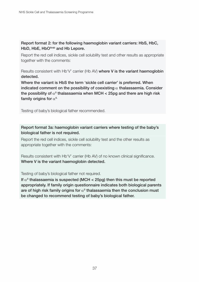
Report format 2: for the following haemoglobin variant carriers: HbS, HbC, HbD, HbE, HbOArab and Hb Lepore.
Report the red cell indices, sickle cell solubility test and other results as appropriate together with the comments:
Results consistent with Hb‘V’ carrier (Hb AV) where V is the variant haemoglobin detected.
Where the variant is HbS the term ‘sickle cell carrier’ is preferred. When indicated comment on the possibility of coexisting a thalassaemia. Consider the possibility of a0 thalassaemia when MCH < 25pg and there are high risk family origins for a0.
Testing of baby’s biological father recommended.
Report format 3a: haemoglobin variant carriers where testing of the baby’s biological father is not required.
Report the red cell indices, sickle cell solubility test and the other results as appropriate together with the comments:
Results consistent with Hb‘V’ carrier (Hb AV) of no known clinical significance. Where V is the variant haemoglobin detected.
Testing of baby’s biological father not required.
If a0 thalassaemia is suspected (MCH < 25pg) then this must be reported appropriately. If family origin questionnaire indicates both biological parents are of high risk family origins for a0 thalassaemia then the conclusion must be changed to recommend testing of baby’s biological father.

NHS Sickle Cell and Thalassaemia Screening Programme
38
Report format 3b: haemoglobin variant carriers where testing of the baby’s biological father is required.
Report the red cell indices, sickle cell solubility test and the other results as appropriate together with the comments:
Results consistent with Hb‘V’ carrier (Hb AV) - where V is the variant haemoglobin detected. This must be used for variants of clinical significance (not covered in report format 2) or variants of unknown clinical significance.If the variant is unidentified recommended wording is carrier of unidentified variant. When indicated comment on the possibility of coexisting a thalassaemia. Consider the possibility of a0 thalassaemia when MCH < 25pg and there are high risk family origins for a0.
Testing of baby’s biological father recommended.
Report format 4a: for β thalassaemia carriers.
Report the red cell indices and the other results as appropriate together with the comments:
Results consistent with β thalassaemia carrier.Comment on possibility of coexisting a0 thalassaemia when MCH <25pg and there are high risk family origins for a0.
Testing of baby’s biological father recommended.
Report format 4b: for possible β thalassaemia carriers.Report the red cell indices and the other results as appropriate together with the comments:
Results consistent with possible β thalassaemia carrier.Comment on possibility of coexisting a0 thalassaemia when MCH < 25pg and there are high risk family origins for a0.
Testing of baby’s biological father recommended.

NHS Sickle Cell and Thalassaemia Screening Programme
39
Report format 5a: for HPFH.
Report the red cell indices and the other results as appropriate together with the comments:
Results consistent with carrier of Hereditary Persistence of Fetal Haemoglobin.
Testing of baby’s biological father recommended.
Report format 5b: for δβ thalassaemia carrier.Report the red cell indices and the other results as appropriate together with the comments:
Results consistent with δβ thalassaemia carrier.
Comment on the possibility of coexisting a0 thalassaemia if the MCH < 25pg and there are high risk family origins for a0.
Testing of baby’s biological father recommended.
Report format 6a – for possible a0 thalassaemia carriers when both the biological mother and the baby’s biological father are of high risk family origins for a0.Report the red cell indices and the other results as appropriate together with the comments:
Results consistent with possible a thalassaemia carrier and/or iron deficiency.
Family origin questionaire indicates both biological parents are of high risk family origins for a0.
Testing of baby’s biological father recommended.

NHS Sickle Cell and Thalassaemia Screening Programme
40
Report format 6b: for HbH diseaseReport the red cell indices and the other results as appropriate together with the comments:
Results consistent with HbH disease.
Testing of baby’s biological father recommended if he is of high risk family origins for a0 thalassaemia.
Report format 7a: for possible a thalassaemia carriers when either biological parent is not of high risk a0 thalassaemia family origins (MCH < 25pg)Report the red cell indices and the other results as appropriate together with the comments:
Results consistent with possible a thalassaemia carrier and/or iron deficiency.
Testing of baby’s biological father not required as one or both biological parents are of low risk family origins for a0 thalassaemia.
Report format 7b: for possible a thalassaemia carriers (MCH ≥ 25 but < 27pg)Report the red cell indices and the other results as appropriate together with the comments:
Results consistent with possible iron deficiency and/or a thalassaemia carrier.
Testing of baby’s biological father not required.

NHS Sickle Cell and Thalassaemia Screening Programme
41
Report format 8: for homozygote and compound heterozygote conditionsReport the red cell indices, sickle cell solubility test and the other results as appropriate together with the comments:
Results consistent with:
Sickle cell anaemia (Hb SS)
Hb SC disease
Hb SD disease Hb SE disease
Hb SOArab disease Hb S/Lepore Sickle/β thalassaemia Hb variant/β thalassaemia
Or adapt this format appropriately to fit the haemoglobins present.
Potential clinically significant conditions must be referred to a Consultant Haematologist if not already in follow up.
Testing of baby’s biological father recommended.
Comment on the risk of coexisting a0 thalassaemia if the MCH <25pg and there are high risk family origins for a0.
Guidance on referral of samples for DNA Table 5 summarises the main genetic risk combinations that require antenatal screening actions, according to the antenatal screening recommendations, and shows which cases require referral of samples for further studies by DNA analysis. For other haemoglobinopathy combinations, refer results for an expert opinion.

NHS Sickle Cell and Thalassaemia Screening Programme
42
Table 5. Referral guidelines for antenatal screening specimens.
Biological mother carrier state Biological father carrier state Further studies by DNA analysis
No abnormalities detected Testing of baby’s biological father not required None required
Any abnormal Hb or thalassaemia No abnormality detected None required
HbS HbS or HbC None required until PND
HbS HbOArab, DPunjab, E, Lepore, β thalassaemia or δβ thalassaemia
If PND is being considered send bloods for mutation confirmation
HbS HPFH Send bloods for mutation confirmation. PND is not usually indicated when HPFH has been confirmed
HbS + a thalassaemia Assess risk as per HbS alone, unless family origins indicate a high risk of a0 thalassaemia
Assess risk as per HbS alone, unless family origins indicate a high risk of a0 thalassaemia
HbC HbS None required until PND
HbD HbS If PND is being considered send bloods for mutation confirmation
HbOArab HbS, β thalassaemia or δβ thalassaemia If PND is being considered send bloods for mutation confirmation
Hb Lepore HbS, E, OArab, Lepore, β thalassaemia or δβ thalassaemia If PND is being considered send bloods for mutation confirmation
HbE β thalassaemia, Hb Lepore, δβ thalassaemia, HbS If PND is being considered send bloods for mutation confirmation
HbE a thalassaemia (MCH < 25pg) Send bloods for mutation confirmation if high risk family origins for a0 thalassaemia
β or δβ thalassaemia HbS, E, OArab, Lepore, β thalassaemia or δβ thalassaemia If PND is being considered send bloods for mutation confirmation
β or δβ thalassaemia a thalassaemia (MCH < 25pg) Send bloods for mutation confirmation if high risk family origins for a0 thalassaemia
HPFH HbS, E, OArab, Lepore, β or δβ thalassaemia Send bloods for mutation confirmation. PND is not usually indicated when HPFH has been confirmed
a thalassaemia (MCH < 27pg but ≥ 25pg)
Testing of baby’s biological father not required None required
a thalassaemia (MCH < 25pg)1) Low risk a0 thal family origins in either biological parent2) High risk a0 thal family origins in both biological parents or unknown
Testing of baby’s biological father not required
Test baby’s biological father if MCH < 25pg irrespective of any other phenotype detected
None required
Send maternal and paternal bloods for mutation confirmation

NHS Sickle Cell and Thalassaemia Screening Programme
43
Quality assurance and accreditation
All commissioners and service providers must refer to the public health functions agreement (Section 7A) service specification (No 18), and supporting standards and handbook to ensure a service is set up correctly and meets the standards set by the national screening programme.
Laboratories offering screening for the NHS Sickle Cell and Thalassaemia Screening Programme must:
• be accredited by the UK Accreditation Service (UKAS) to ISO. ‘Medical laboratories – Requirements for quality and competence (ISO 15189) or be CPA accredited and actively transitioning towards ISO 15189
• participate in EQA schemes accredited to ISO. ‘Conformity assessment. General requirements for proficiency testing schemes (ISO 17043)’
• meet the screening programme quality assurance requirements mapped to ISO 15189
• use ISO 15189 accredited reference laboratories
The UK Accreditation Service (UKAS) will assess both the ISO and the screening requirements on behalf of PHE Screening Quality Assurance Services and the NHS Sickle Cell and Thalassaemia Screening Programme.

NHS Sickle Cell and Thalassaemia Screening Programme
44
Referral of antenatal samples for mutation analysis The majority of couples at risk of having a child affected with β thalassaemia or sickle cell disease are identified initially through the antenatal screening programme. The diagnosis of a thalassaemia is more complicated because DNA analysis is the only way to distinguish between a+ (-a/aa), homozygous a+ (-a/-a) and a0 (--/aa) thalassaemia. Due to the high frequency of a+ thalassaemia in some populations and the low cost benefit ratio in a screening programme, DNA confirmation is not required for all possible cases of a thalassaemia. The screening programme relies on a strategy for diagnosis of a0 thalassaemia by combining haematological tests and details of family origin.
Although the antenatal screening programme guidelines are designed to identify most carriers for serious haemoglobinopathies, including thalassaemias, the screening protocols will not identify every couple at risk for every haemoglobinopathy. For example, they are not designed to pick up couples at risk for HbH disease or the extremely rare cases of couples of low risk family origins on the FOQ who are at risk for Hb Bart’s hydrops fetalis syndrome.
Testing of the baby’s biological father
This must be performed according to screening protocols as for maternal phenotype testing. If the baby’s biological father has a phenotype that can interact with the maternal phenotype as depicted in Table 5, then the couple should be counselled. If PND is being considered, then blood samples on both biological parents must be sent to a DNA referral laboratory.
If the baby’s biological father is unavailable for testing or his haemoglobinopathy status is unknown, then a risk assessment should be done. The programme supports the woman being offered PND in this situation. PND can be undertaken without the DNA of the baby’s biological father, although in some circumstances the diagnosis will not be able to be given with such a high degree of certainty as when the baby’s biological father’s mutation is known.
Guidelines on the referral of blood samples for DNA analysis
The blood samples must be accompanied by an appropriately completed DNA laboratory referral form and the laboratory notified prior to sending samples.

NHS Sickle Cell and Thalassaemia Screening Programme
45
Haemoglobin variants
If both biological parents carry HbS, or one carries S and one carries C then:• blood samples are not required immediately to confirm the mutation by DNA
analysis
If PND has been accepted, fresh blood samples from both biological parents should be sent at the same time as the fetal sample.
If one biological parent carries HbS and the other is a suspected carrier of HbOArab, HbDPunjab, HbE, Hb Lepore or a type of β thalassaemia (including δβ thalassaemia) then:
• if time permits blood samples from both biological parents should be sent to confirm the mutations prior to fetal sampling
• if PND has been accepted, a fresh maternal sample (and paternal sample if not previously tested as above) should be sent at the same time as the fetal sample
If one biological parent carries HbS and the other is thought to carry deletional HPFH then:
• PND is not usually indicated but it is important to ensure that deletional HPFH is differentiated from δβ thalassaemia (these cases may require mutational analysis)
Carriers of deletional HPFH have a raised HbF level of approximately 20 to 30% and are usually associated with normal red cell indices. It is important to differentiate deletional HPFH from δβ thalassaemia. The carrier state for δβ thalassaemia is associated with a reduced MCH and an HbF level usually in the range of 5 to 15%. Non-deletional HPFH usually results in a more modest increase of HbF (less than 10%) in adults and is found in many populations. It is usually associated with normal red cell indices.
If one biological parent carries HbE and the other carries β thalassaemia, Hb Lepore, δβ thalassaemia or is thought to carry a0 thalassaemia then:
• if time permits blood samples from both biological parents should be sent to confirm the mutations prior to fetal sampling
• if PND has been accepted, a fresh maternal sample (and paternal sample if not previously tested as above) should be sent at the same time as the fetal sample

NHS Sickle Cell and Thalassaemia Screening Programme
46
Note in people of high risk family origins for a0 thalassaemia, there is a possibility of a hidden risk to the fetus of homozygous a0 thalassaemia, as the carrier state for a0 thalassaemia can be masked. It is important to determine the a genotype by DNA analysis.
Beta thalassaemias
If one biological parent carries β thalassaemia, Hb Lepore, δβ thalassaemia and the other carries β thalassaemia, Hb Lepore, δβ thalassaemia, HbOArab or HbS then:• if time permits blood samples from both biological parents should be sent to
confirm the mutations prior to fetal sampling
• if PND has been accepted, a fresh maternal sample (and paternal sample if not previously tested as above) should be sent at the same time as the fetal sample
Note in people of high risk family origins for a0 thalassaemia, the carrier state for a0 thalassaemia may also be present. It is important to determine the a genotype by DNA analysis.
Alpha thalassaemias
Coinheritance of a thalassaemia with other haemoglobin variants
Blood samples should not be sent to confirm and identify a thalassaemia, unless both biological parents are of high risk family origins for a0 thalassaemia. There is rarely clinical interaction between a thalassaemia and other haemoglobin variants. Unless there are high risk family origins, the type of a thalassaemia is almost always the a+ thalassaemia type, which poses no serious genetic risk to the fetus.
Alpha plus thalassaemia (a+ thalassaemia or -a/aa and -a/-a)
This is found in all ethnic groups, with a high (10 to 30%) carrier frequency in some parts of Africa, in Afro-Caribbeans and in South and Southeast Asia. Even if both biological parents are carriers, there is no risk to the fetus. Homozygous a+ thalassaemia (-a/-a) is not a clinically significant disorder with respect to genetic or obstetric complications, but can cause diagnostic confusion with a0 thalassaemia or iron deficiency.
Heterozygotes, (-a/aa) generally have an MCH of 25 to 28pg and a normal HbA2 level. Approximately one third of cases will be silent.
Homozygotes, (-a/-a) generally have an MCH below 25pg and some have HbH inclusions, as do carriers for a0 thalassaemia (--/aa).

NHS Sickle Cell and Thalassaemia Screening Programme
47
Individuals may be heterozygous for an a gene deletion (carrier genotype: -a/aa) or a rarer point mutation in one gene affecting gene expression (carrier genotype: aTa/aa), commonly called a non-deletional mutation. There are 2 common deletions (-a3.7 and –a4.2) and a number of less common non-deletional mutations.
Alpha zero thalassaemia (a0 thalassaemia (--/aa))
This carries the potential for a clinically significant disorder. If both biological parents are carriers of a0 thalassaemia (--/aa), the couple has a 1 in 4 risk of having a fetus with Hb Bart’s hydrops fetalis syndrome (--/--) and the mother is at risk of obstetric complications, particularly in the third trimester of pregnancy. The mutations leading to Hb Bart’s hydrops fetalis are almost always gene deletions.
If one biological parent carries a0 thalassaemia (--/aa) and the other a+ thalassaemia (either -a/aa or -a/-a), there is a risk of having a child with HbH disease (--/-a). PND is not usually indicated for HbH disease unless there are known higher risk mutations such as Hb Constant Spring.
Quality assurance and accreditation
All commissioners and service providers must refer to the public health functions agreement (Section 7A) service specification (No 18), and supporting standards and handbook to ensure a service is set up correctly and is meeting the standards set by the national screening programme.
DNA laboratories must:• be accredited by the UK Accreditation Service (UKAS) to ISO. ‘Medical
laboratories – Requirements for quality and competence (ISO 15189) or be CPA accredited and actively transitioning towards ISO 15189
• participate in EQA schemes accredited to ISO. ‘Conformity assessment. General requirements for proficiency testing schemes (ISO 17043)’
• meet the screening programme quality assurance requirements mapped to ISO 15189 and use ISO 15189 accredited reference laboratories
UKAS will assess both the ISO and the screening requirements on behalf of PHE Screening Quality Assurance Services and the NHS Sickle Cell and Thalassaemia Screening Programme.

NHS Sickle Cell and Thalassaemia Screening Programme
48
References1. GOV.UK [Internet]. Sickle cell and thalassaemia screening: handbook for
laboratories. Available from: www.gov.uk/government/publications/sickle-cell-and-thalassaemia-screening-handbook-for-laboratories (viewed on 15 August 2017)
2. NHS Sickle Cell and Thalassaemia Screening Programme, ‘Managing Safety Incidents in NHS Screening Programmes’, October 2015 (viewed on 15 August 2017)
3. Ryan K, Bain B, Worthington D, James J, Plews D, Mason A, et al. Significant haemoglobinopathies: guidelines for screening and diagnosis. British Journal of Haematology 2010: volume 149, pages 35 to 49
4. de Baca ME, Gulati G, Kocher W, Schwarting R. Effects of Storage of Blood at Room Temperature on Hematologic Parameters Measured on Sysmex XE-2100. Laboratory Medicine 2006: volume 37, issue 1, pages 28 to 35
5. Cohle S, Saleem A, Maddaoui D. Effects of storage of blood on stability of hematologic parameters. American Journal of Clinical Pathology 1981: volume 76, issue 1, pages 67 to 69
6. Mosca A, Paleari R, Ivaldi G, Galanello R, Giordano P. The role of haemoglobin A(2) in the diagnosis of thalassaemias and related haemoglobinopathies. Journal of Clinical Pathology 2009: volume 62, pages 13 to 17
7. Piel F, Weatherall D. The a-Thalassaemias, New England Journal of Medicine 2014: volume 371, issue 20, pages 1908 to 1916
8. England J, Rowan R, Dawson D, Lewis S, Shinton N, Stephens A, et al. Guidelines for haemoglobinopathy screening. Clinical Laboratory Haematology 1988: volume 10, pages 87 to 94
9. Sprour Y, Heppinstall S, Porter N, Wilson G, Goodeve A, Rees D, Wright J. Is routine molecular screening for common a thalassaemia deletions necessary as part of an antenatal screening programme? Journal of Medical Screening 2007: volume 14, pages 60 to 61

NHS Sickle Cell and Thalassaemia Screening Programme
49
Appendix 1 – Carrying out a look back with particular reference to HbA2
A lookback is a review of screening records to identify individuals harmed or at risk of harm as a result of a screening safety incident or serious incident. Look backs or case review may also be done as part of fact finding to establish whether there has been a screening incident. Look backs may result in a patient notification exercise/recall.
If your external quality control shows the HbA2 is out of consensus then a look back exercise must be considered. HbA2 values which fall below consensus are of particular concern as in this situation β thalassaemia carriers may have been missed. The following steps are a guide to carrying out a look back exercise. Whilst not mathematically exact, the calculation given is suitable for this purpose. It should be noted that every situation is unique and you may wish to obtain advice from your external quality control provider and the NHS Sickle Cell and Thalassaemia Screening Programme.
If a look back exercise is required report this as a suspected safety incident in accordance with Managing safety incidents in NHS Screening Programmes. This means informing the Screening QA Service (region) and the host organisation responsible for the laboratory in addition to relevant clinical teams (including midwifery). The accountable organisation will need to decide if the Duty of Candour applies. Reference should be made to PHE guidelines on Duty of Candour (see www.gov.uk/government/publications/nhs-screening-programmes-duty-of-candour).
Method
1. Identify the point in time the results were last within consensus. 2. Look back must review patient results from that time point until results are proven
to be back in consensus. If results are not yet proven to be within consensus then the actions described should continue prospectively.
3. All patient results must be reviewed, however, the following formula will help identify patients requiring more stringent scrutiny and possible recall.
Formula: Discrepancy value = consensus HbA2 minus HbA2 obtained by your laboratory.

NHS Sickle Cell and Thalassaemia Screening Programme
50
Patients requiring stringent review = HbA2 action value (3.5%) minus your calculated discrepancy value. For example: consensus = 3.6% and obtained = 3.2% therefore 3.6 - 3.2 = 0.4% (discrepancy value = 0.4%). In this scenario all patients with an HbA2 greater than or equal to 3.1% from the time point identified in (1) should be carefully reviewed.
4. Patients with an HbA2 greater than the value identified using the above formula and with an MCH less than 27pg not already identified as a β thalassaemia carrier, should be recalled and retested.
5. Patients with an MCH greater than or equal to 27pg and with an HbA2 which would have been greater than 4.0% when the discrepancy value identified using the above formula is added to the original result obtained should be recalled and retested.
6. All other patients do not need recalling or retesting.

NHS Sickle Cell and Thalassaemia Screening Programme
51
Appendix 2 – Best practice guidelines for analysing liquid capillary blood samples This service should be seen as a special ‘on-demand’ service to alleviate parental anxiety and not as a fail-safe or substitute for the newborn blood spot screening programme, nor to gain a more rapid entry into clinical care. If local arrangements can be implemented to obtain fast track results from the routine blood spot screening programme, then this approach should be encouraged. Those laboratories that perform second-line testing for the newborn blood spot screening programme would already satisfy most of these criteria.
Laboratory tests may be requested on newborn babies for clinical reasons but these best practice guidelines have been developed specifically for those babies born to known high risk couples. To be a ‘1 in 4’ high risk pregnancy, the haemoglobinopathy results must be known on both biological parents. Specimens taken for other clinical diagnostic purposes fall outside these guidelines, although the requester may wish to assure themselves that the laboratory has sufficient competence and expertise to analyse specimens from newborns.
1. The analytical technique for initial testing of the capillary specimen and the second line test must be appropriate for specimens from the newborn period. Some analytical protocols and instruments are designed for testing adult blood and are not optimal for newborns.
2. The laboratory must participate in an appropriate EQA scheme that assesses the analysis and interpretation of results from newborn specimens. UK NEQAS (H) provides suitable quality control specimens for this work.
3. Samples must be transported to the analysing laboratory in a reliable and swift manner, using a courier service if the specimen has to be referred, rather than risking delays using the postal service.
4. Both biological parents’ results must be reviewed before reporting the result of the liquid capillary sample. Since this is a service for pregnancies known to be at a 1 in 4 risk of a significant haemoglobinopathy, the requester must record the results of both biological parents on the request form.
5. The laboratory analysing the liquid specimen is responsible for notifying the newborn screening laboratory of the result. There must be documented local systems in place for this process. Any discrepancies between the 2 results need

NHS Sickle Cell and Thalassaemia Screening Programme
52
to be investigated rapidly at local level by both laboratories and reported to the NHS SCT Screening Programme and regional team within one week of the discrepancy being detected.
6. Laboratories offering this service must collate data for audit purposes on the number of specimens tested, turnaround times and consistency with newborn screening results.
7. If the results from the liquid capillary specimen and the bloodspot screening programme are in accord with each other then a further confirmatory specimen is not needed.
8. The laboratory must have standard operating procedures (SOPs) for the analytical methods and the rapid local reporting processes to the relevant healthcare professionals for the capillary specimen result. The SOPs must include senior level checking of analytical results before reporting and follow up of the subsequent bloodspot screening result.
9. A mother and baby who are ready to be sent home should not be kept in hospital awaiting the results of these tests.
10. The laboratory must be accredited as per screening programme requirements.

NHS Sickle Cell and Thalassaemia Screening Programme
53
Appendix 3 – Table of biological parental carrier state combinationsBiological mother
Carrier of: HbS β thalassaemia δβ thalassaemia Hb Lepore HbE HbOArab HbC HbDPunjab HPFH Not identified as a carrier
HbS
β thalassaemia
δβ thalassaemia
Hb Lepore
HbE
HbOArab
HbC
HbDPunjab
HPFH
Not identified as a carrier
Key
Bio
log
ical
fat
her
Less serious risk - refer couple for counselling - further investigation may be required
Serious risk - refer couple for counselling - prenatal diagnosis to be offered
Minimal risk

NHS Sickle Cell and Thalassaemia Screening Programme
54
Useful organisations and websitesThe Oxford Laboratory provides, on behalf of the screening programme, a dedicated sickle cell and thalassaemia laboratory support service for antenatal and newborn laboratories and other healthcare professionals involved in the screening programme.
Telephone: 01865 572 767Email: [email protected]: 01865 572775
NHS Sickle Cell and Thalassaemia Screening Programme enquiries:
Telephone: 020 3682 0890Email: [email protected] Website: gov.uk/phe/screening
British Committee for Standards in Haematology
SCT screening e-learning for health care professionals
Laboratory e-learning modules
NHS Choices: screening for sickle cell and thalassaemia
National Sickle Cell/Thalassaemia Centres
Family origin questionnaire

NHS Sickle Cell and Thalassaemia Screening Programme
55

About Public Health EnglandPublic Health England exists to protect and improve the nation’s health and wellbeing, and reduce health inequalities. It does this through world-class science, knowledge and intelligence, advocacy, partnerships and the delivery of specialist public health services. PHE is an operationally autonomous executive agency of the Department of Health.
Public Health England, Wellington House, 133-155 Waterloo Road, London SE1 8UGTel: 020 7654 8000 www.gov.uk/phe Twitter: @PHE_uk Facebook: www.facebook.com/PublicHealthEngland
About PHE ScreeningScreening identifies apparently healthy people who may be at increased risk of a disease or condition, enabling earlier treatment or better informed decisions. National population screening programmes are implemented in the NHS on the advice of the UK National Screening Committee (UK NSC), which makes independent, evidence-based recommendations to ministers in the four UK countries. The Screening Quality Assurance Service ensures programmes are safe and effective by checking that national standards are met. PHE leads the NHS Screening Programmes and hosts the UK NSC secretariat.
PHE Screening, Floor 2, Zone B, Skipton House, 80 London Road, London SE1 6LHwww.gov.uk/topic/population-screening-programmes Twitter: @PHE_Screening Blog: phescreening.blog.gov.uk
Prepared by: Dr Yvonne Daniel and Joan Henthorn. Endorsed by: Laboratory Subgroup of the NHS Sickle Cell and Thalassaemia Screening Programme.
For queries relating to this document, please contact: [email protected]
Front cover image: extender_01/Shutterstock
© Crown copyright 2017You may re-use this information (excluding logos) free of charge in any format or medium, under the terms of the Open Government Licence v3.0. To view this licence, visit OGL or email [email protected]. Where we have identified any third party copyright information you will need to obtain permission from the copyright holders concerned.
Published October 2017 (version 4.2)PHE publications gateway number: 2017345