nhs forth valley wound management formulary 1s edition … · 2020-01-07 · the principle aim is...
TRANSCRIPT
Version 5.4 April 2019 UNCONTROLLED WHEN PRINTED Page 1 of 115
NHS FORTH VALLEY WWoouunndd MMaannaaggeemmeenntt FFoorrmmuullaarryy
11sstt EEddiittiioonn VV55..44 AApprriill 22001199
Date of First Issue 01/05/2002 Approved 10/02/2015 Current Issue Date 30/04/2019 Review Date 31/12/2020 (Or as required after each Wound Management)
meeting Version 1st Edition Version 5.4 EQIA YES Author / Contact Heather Macgowan Group Committee – Final Approval
Area Drug and Therapeutics Committee
This document can, on request, be made available in alternative formats
Version 5.4 April 2019 UNCONTROLLED WHEN PRINTED Page 2 of 115
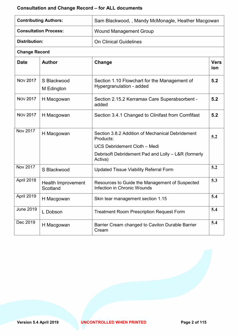
Consultation and Change Record – for ALL documents
Contributing Authors: Sam Blackwood, , Mandy McMonagle, Heather Macgowan
Consultation Process: Wound Management Group
Distribution: On Clinical Guidelines
Change Record
Date
Author Change Version
NNOOVV 22001177
S Blackwood M Edington
Section 1.10 Flowchart for the Management of Hypergranulation - added
5.2
NNOOVV 22001177 H Macgowan Section 2.15.2 Kerramax Care Superabsorbent - added
5.2
NNOOVV 22001177 H Macgowan Section 3.4.1 Changed to Clinifast from Comfifast
5.2
Nov 2017 H Macgowan Section 3.8.2 Addition of Mechanical Debridement Products: UCS Debridement Cloth – Medi Debrisoft Debridement Pad and Lolly – L&R (formerly Activa)
5.2
Nov 2017 S Blackwood Updated Tissue Viability Referral Form 5.2
April 2018 Health Improvement Scotland
Resources to Guide the Management of Suspected Infection in Chronic Wounds
5.3
April 2019 H Macgowan Skin tear management section 1.15 5.4
June 2019 L Dobson Treatment Room Prescription Request Form 5.4
Dec 2019 H Macgowan Barrier Cream changed to Cavilon Durable Barrier Cream
5.4
Version 5.4 April 2019 UNCONTROLLED WHEN PRINTED Page 3 of 115
CCOONNTTEENNTTSS Section 1
• 1.1 Introduction to the formulary……………………………………………..
Page 6
• 1.2 Accountability and Responsibility ..................................................... Page 7
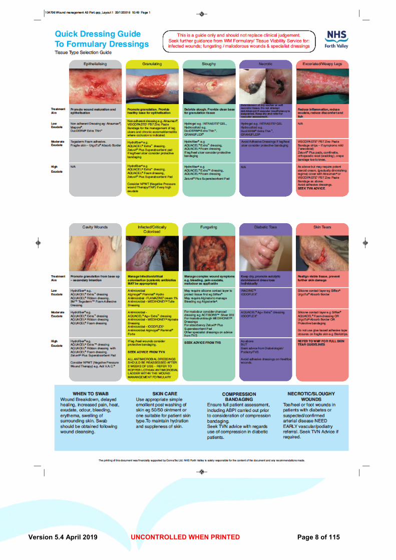
• 1.3 Quick Reference Guide...................………………………………………
Page 8
• 1.4 Holistic approach to wound healing…………………………………….. 1.4.1 Wound Photography...............................................................
Page 9 Page 9-10
• 1.5 The Physiology of wound healing………………………………………..
Page 11
• 1.6 Moist Wound Healing………………………………………………………. 1.6.1 Moist Wound Healing in Ischaemic Wounds………………..
Page 12 Page 12
• 1.7 Wound cleansing……………………………………………………………
Page 13
• 1.8 Wound swabbing……………………………………………………………
Page 14
• 1.9 Wound Infection……………………………………………………………..
Page 15-16 1.9.1 Resources to Guide the Management of Suspected
Infection in Chronic Wounds 1.9.2 Algorithm for assessment and Management of Chronic
Wounds Ropper Lothian Ladder
1.10 Hypergranulation – treatment 1.10.1 Treatment of Hypergranulation flowchart
Page 17 Page 18 Page 19 Page 20-22 Page 23
• 1.11 Reducing pain at dressing changes……………………………………
Page 24
• 1.12 Nutrition and wound management……………………………………..
Page 25-26
• 1.13 Non Formulary Dressings………………………………………………..
Page 27
• 1.14 Skin Care…………………………………………………………………….
Page 28-29
• 1.15 Skin Tears – NATVNS – best practice in the prevention, Assessment and management of skin tears………………………... 1.14.1 Skin tear Management Flowchart…………….....
Page 30-33 Page 34
• 1.16 Thermal Injury Guideline…………………………………………………
Page 35-38 1.16.1 Care of Burns In Scotland (COBIS)………………………….
Page 39
• 1.17 Sterile Dressing packs……………………………………………………
Page 40
• 1.18 Tissue Viability Service Referral………………………………………..
Page 41
Version 5.4 April 2019 UNCONTROLLED WHEN PRINTED Page 4 of 115
Section 2 – Dressings
• 2.1 Low adherent Dressing 2.1.1 Tricotex…………………………………………………………… 2.1.2 Atrauman…………………………………………………………. 2.1.3 Mepore…………………………………………………………….
Page 42 Page 43 Page 44
• 2.2 Alginate Dressing 2.2.1 Algosteril………………….…………………………………….
Page 45
• 2.3 Antibacterial Impregnated Dressing 2.3.1 Inadine ……………………………………………………………. 2.3.2 Iodoflex…………………………………………………………… 2.3.3 Flamazine 1% Cream……………………………………………
Page 46 Page 47-48 Page 49
• 2.4 Barrier Products 2.4.1 Cavilon Durable Barrier Cream……………………………… 2.4.2 Medline No Sting Barrier Film ……………………………….
Page 50-51 Page 52-53
• 2.5 Charcoal Dressings 2.5.1 Actisorb Silver 220 ……………………………………………
Page 54
• 2.6 Emollient 2.6.1 Olive Oil……………………………........................................... 2.6.2 Epaderm…………… ……………………………………………. 2.6.3 Liquid soft paraffin 50%/White soft paraffin50%................
Page 55 Page 56-57 Page 58
• 2.7 Foam Dressings > 2.7.1 Tegaderm Foam Adhesive ………………………………. > 2.7.2 Allevyn Non Adhesive…………………………………….
Page 59-60 Page 61
• 2.8 Honey Dressing……………………………………………………………. 2.8.1 MedihoneyTulle………………………………………………..... 2.8.2 Medihoney Antibacterial Medical Honey (tube) 2.8.3 Medihoney – Apinate Dressing……………………………….
Page 62 Page 63 Page 64 Page 65
• 2.9 Hydrocolloid 2.9.1 Duoderm Extra Thin / Granuflex…………………………………
Page 66-67
• 2.10 Hydrofibre 2.10.1 Aquacel Extra………………………………………………………. 2.10.2 Aquacel Ag + Extra………………………………………………...
Page 68 Page 69
• 2.11 Hydrogel 2.11.1 Intrasite Gel…………………………………………………….
Page 70-71
• 2.12 Paraffin Gauze Dressing 2.12.1 Jelonet…………………………………………………………..
Page 72
• 2.13 Paste Bandage……………………………………………………………
Page 73
• 2.14 Semi – permeable film dressing 2.14.1Tegaderm…………………………………………… …………..
Page 74
• 2.15 Super Absorbent dressing pads 2.15.1 Zetuvit Plus……………………………………………………... 2.15.2 Kerramax Care…………………………………………
Page 75 Page 76
Version 5.4 April 2019 UNCONTROLLED WHEN PRINTED Page 5 of 115
Section 3 – Specialist Products
• 3.1 Antimicrobial Enzyme Alginogel 3.1.1 Flaminal Hydro / Flaminal Forte………………………………
Page 77
• 3.2 Dermatology 3.2.1 Silflex…………………………………………………………….. 3.2.2 Urgotul Absorb Border ……………………………………......
3.3 Specialist Foam – Aquacel Foam Adhesive.....................................
Page 78 Page 79 Page 80
• 3.4 The Four Layer Bandage System………………………………………. 3.4.1 Clinifast………………………………………………………….
Page 81-82 Page 83
• 3.5 Larvae Therapy……………………………………………………………. 3.5.1 Larvae Therapy – Patient Information Leaflet……………...
Page 84-85 Page 86
• 3.6 Topical Steroid Preparations 3.6.1 Dermovate……………………………………………………….. 3.6.2 Elocon Ointment 3.6.3 Fludroxycortide Cream/Ointment (Haelan) 3.6.4 Fludroxycortide Tape (Haelan) 3.6.5 The ‘Fingertip unit’ of Topical Steroids…………………….. 3.6.6 Steroid Ladder…………………………………………………..
Page 87 Page 88 Page 89 Page 90 Page 91 Page 92
• 3.7 Topical Negative Pressure Therapy…………………………………… 3.7.1 Patient Information Leaflet……………………………………. 3.7.2 TNP Procedure for Order/Cancellation ……………………..
Page 93-94 Page 95 Page 96
• 3.8 Wound Bed Preparation 3.8.1 Prontosan Irrigation Solution and Gel………………………. 3.8.2 Mechanical Debridement Products: Medi – UCS Debridement cloth L&R – Debrisoft Debridement Pad and Lolly
Page 97 Page 98 Page 99-100
Section 4- Appendices
1. Non Formulary Request Form……………..………………………………. Page 101
2. Wound Assessment and Treatment Chart………………………………. Page 102-105
3. Photographic Consent Form ………………………………………………. Page 106
4. TNP (KCI) order form for VAC and consumables……………………….. Page 107
5. TNP Discharge Protocol………………………………………………………
6. Wound Management Product evaluation process
7a. Care Home Referral Form to TVS (Care Home Use Only)
7b. Tissue Viability Referral Form
8. Treatment Room Patient Prescription Request
Page 108 Page 109 Page 110 Page 111 Page 112 - 113
Version 5.4 April 2019 UNCONTROLLED WHEN PRINTED Page 6 of 115
SSEECCTTIIOONN 11 11..11 -- IINNTTRROODDUUCCTTIIOONN TTOO TTHHEE WWOOUUNNDD MMAANNAAGGEEMMEENNTT FFOORRMMUULLAARRYY
The Wound Management Formulary was developed by members of the NHS Forth Valley Wound Management Group. This group consists of specialist practitioners, nursing and pharmacy staff from the Acute and Primary Care settings; taking into account patient comfort, safety, cost effectiveness and clinical benefits of the product. These products should be the first choice for both Primary and Acute Care. The Wound Management Formulary has been approved by the Director of Nursing, NHS FV and the Drug and Therapeutics Committees. The principle aim is to rationalise and standardise wound care products throughout NHS Forth Valley, encouraging seamless care and to assist nursing staff in the selection of appropriate dressings. New products can be evaluated as they are developed and the need arises – the process for this can be seen on Appendix 6. Please note the sizes of dressings chosen for inclusion to the formulary are the current most popular sizes stocked by stores, there may be other sizes available if required, in certain circumstances. All products listed in the formulary are GP/DN prescribable unless otherwise stated. Please remember to prescribe generically as prescriptions cannot be dispensed when trade names alone have been written. Please note: Products highlighted in this formulary may only be used within the licensed indications within the SPC (specific product characteristics) i.e. only use each product under the guidance of the product information leaflet. SPC are available at www.medicines.org.uk On discharge from hospital one weeks supply of dressings should accompany the patient home, or if treatment is of short duration, enough to complete treatment. Likewise those patients with a planned admission to hospital should bring to the ward a small supply of current dressings. It is recognised that nursing and clinical practice is an evolving process, and members of the working group would welcome any information and advice which is considered necessary to update the Formulary in light of changes in practice and developments in wound care. Please contact the under noted for advice, guidance or any comments you may have: - Tissue Viability Service NHS Forth Valley Tel 01324 673747 or [email protected] Grateful thanks to Wound Management group Core members and to all other group members who attended meetings and contributed to the development of the formulary document.
Version 5.4 April 2019 UNCONTROLLED WHEN PRINTED Page 7 of 115
SSEECCTTIIOONN 11 11..22 -- AACCCCOOUUNNTTAABBIILLIITTYY AANNDD RREESSPPOONNSSIIBBIILLIITTYY As healthcare professionals using this formulary you must:
• Use your professional knowledge, judgement and skills to make a decision based on evidence for best practice and the person’s best interests. You need to be able to justify the decisions that you make
• Ensure any advice you give is evidence based when suggesting healthcare products or services
• Have the knowledge and skills for safe and effective practice when working without direct supervision
• Recognise and work within the limits of your competence • Keep your knowledge and skills up to date throughout your working life • Take part in appropriate learning and practice activities that maintain and develop your
competence and performance • Keep clear and accurate records of the discussions you have, the assessments you
make, the treatment and medicines you give and how effective these have been • Complete records as soon as possible after an event has occurred • Complete NATVNS Wound care assessment and treatment chart (Appendix 1A -1D) • Ensure any entries made in someone's paper records are clearly and legibly signed,
dated and timed • Ensure any entries made in someone's electronic records are clearly attributable to you • Where wound care is multi-professional and shared, ensure all involved are informed of
any significant change in status and/or dressing regime as soon as possible after the contact has occurred
Version 5.4 April 2019 UNCONTROLLED WHEN PRINTED Page 8 of 115
Version 5.4 April 2019 UNCONTROLLED WHEN PRINTED Page 9 of 115
Section 1
11..44 -- HHOOLLIISSTTIICC AAPPPPRROOAACCHH TTOO WWOOUUNNDD HHEEAALLIINNGG
Wound healing is a complex and interlinked series of biochemical processes that encompass the actions of various cells in different cellular environments involving oxygen, temperature, pH, growth factors and enzymes.
When the cells are healthy and the environment is within normal homeostatic parameters, the healing processes proceed predictably and without incident.
When the cells and their environments are compromised by alterations in local or systemic conditions, the healing process is impaired and the wound does not heal in an orderly or timely fashion.
Wound management and the selection of cleansing agents and dressing products is an important part of the healing process. It is, however important to remember that although this is a necessary component, it is not in itself the only part to consider when dealing with a patient with a non healing wound.
An awareness of the physical and psychosocial factors which delay wound healing is necessary. The assessment process should extend to identifying the intrinsic ( e.g., nutrition, chronic disease processes) and extrinsic factors (e.g. pressure, friction, shear) which may influence an individual’s healing rate. Some of these factors will be easily identified and corrected, others may not.
Nurses are in a unique position within the multi-disciplinary team to be able to holistically assess patients and their wounds and to develop realistic treatment objectives. The Wound Assessment must be completed by a registered nurse or other healthcare professional with appropriate knowledge and experience. The individual needs to take into account whether the wound needs cleansed, the size, the tissue type, the exudate levels, odour, expected wear time of dressings. Findings and decisions made should be documented in the wound chart – use a separate chart for each wound. (See appendix 2A-2D).
11..44 ..11 WWOOUUNNDD PPHHOOTTOOGGRRAAPPHHYY Photographs can be an important part of effective wound assessment – they can provide objective visual confirmation to the written record and can provide evidence of healing rates, capturing therapeutic efficacy. Current legal opinion recommends that written informed consent should be sought from individual patients (or carer if appropriate) or parent/guardian in the case of children under 16, when seeking to take photographs for the purpose of monitoring wounds. See appendix 3 for consent form. Consideration needs to be given to ensure all images remain confidential and stored in such a way that confidentiality is not breached, this includes sharing of images with the wider team. When photographing a wound it is important to be able to assess the dimensions of the wound also. Disposable measuring tapes should be placed on the skin next to the wound prior to taking photograph. If close up photographs are required of specific wounds, there should be a secondary photograph which enables the particular body part to be identified.

Version 5.4 April 2019 UNCONTROLLED WHEN PRINTED Page 10 of 115
The patient has the right to withdraw consent for wound photography at any time. The withdrawal should be fully documented in the patient records and any historic images that have been kept should be removed from electronic records or struck through if on paper form making it clear that the images cannot be used. REFERENCES Dealey, C. (1999) The Care of Wounds - A Guide for Nurses. 2nd Edn. Oxford, Blackwell Science Ltd. Flanagan, M. (1997) Wound Management Edinburgh, Churchill Livingstone. Morison, M (Ed) (2001) The Prevention and Treatment of Pressure Ulcers. London, Harcourt Publishers Ltd. www.tissueviabilityonline.com Institute of Medical illustrators, (2007) Clinical Photography in wound Management Guidelines.
Version 5.4 April 2019 UNCONTROLLED WHEN PRINTED Page 11 of 115
SSEECCTTIIOONN 11 11..55 -- TTHHEE PPHHYYSSIIOOLLOOGGYY OOFF WWOOUUNNDD HHEEAALLIINNGG Acute and chronic wounds have distinct differences. Some of the basic differences (excluding the microbiological/cellular differences) are:
AACCUUTTEE WWOOUUNNDDSS CCHHRROONNIICC WWOOUUNNDDSS • Short duration Unhealed within 6 weeks of formation • No underlying pathology Underlying pathology • Normal inflammatory stage Prolonged inflammatory Stage • Usually heals without Variety of complications may arise Complication Chronic wound fluid does not support cell • Acute wound fluid supports cell proliferation Proliferation (Cutting & Tong 2003) The literature cites many descriptive models of healing. Whichever model is followed, it is essential to have an understanding of the basic process as this will influence decisions made in the day to day management of the wound. Most models suggest that the mechanics of dermal wound healing fall largely into four overlapping phases:
1. Haemostasis
Bleeding starts the process of haemostasis. Blood vessels contract, platelets aggregate and a clot is formed. Leucocytes are attracted to the injured area.
2. Inflammation Prostaglandins and proteins are released, which cause vasodilation and inflammation. Neutrophils (whose function is phagocytosis of bacteria) and macrophages (which control the healing process) proliferate in the wound.
3. Granulation New supporting tissue is formed like a scaffold, along with new blood vessel development, which is known as angiogenesis, and the wound begins to contract.
4. Epithelialisation New skin cells emerge from the dermal edge and hair follicles, slowly bringing the wound edges together. Healing By Primary or Secondary Intention Wound healing by primary intention is when the edges of the wound can be brought together, eg a surgical wound which has been sutured, clipped or glued. The first three phases of healing are usually short but scar maturation may take a few months. Wound healing by secondary intention occurs when the edges of a wound cannot be approximated, eg a leg ulcer. This type of wound heals by a combination of proliferation and wound contraction. The granulation and epithelialisation phases of this type of wound may take months to complete.
Version 5.4 April 2019 UNCONTROLLED WHEN PRINTED Page 12 of 115
SSEECCTTIIOONN 11 11..66 -- MMOOIISSTT WWOOUUNNDD HHEEAALLIINNGG This concept dates back to the 1940s but did not gain credibility until 1962 when George Winter’s now infamous experiment examined the healing time of wounds exposed to air, compared with wounds covered with polyurethane. The wounds which were covered healed almost twice as fast as those exposed to air. Although this theory was applied to acute wounds, the significance of these findings in chronic wounds has been debated with little agreement about healing rates in the literature (Miller, 1998; Parnham, 2002). However, other benefits for creating a moist environment in chronic wound healing have been cited, such as enhancement of autolytic debridement and reduction in pain during wear and on removal of dressings (Hollinworth, 2005). Maceration may occur where there is excessive moisture on the wound bed. Excessive moisture can excoriate the surrounding skin and cause extension of the wound. Correct choice of dressing is essential to achieve a balance between a wound that is too wet and one that is too dry. Wound fluid contains essential growth factors necessary for epidermal growth. Proteolytic enzymes found in wound fluid have been shown to be beneficial to wound healing but are thought to be present in excessive numbers in chronic wounds (Wysocki et al. 1993). At present, there is no biochemical test to measure an excess of proteases in order to prove this is the cause of delayed healing. 11..66 ..11 -- MMOOIISSTT WWOOUUNNDD HHEEAALLIINNGG IINN IISSCCHHAAEEMMIICC WWOOUUNNDDSS It is important, when attempting to promote moist wound healing in ischaemic wounds, to be aware that wounds with an underlying ischaemic cause are prone to infection. The presence of necrotic/sloughy tissue, which contain greater quantities of bacteria, increase the risk of infection (Leaper & Ellis, 2002) when moistened and rehydration of the tissue is attempted. Where there is underlying ischaemic disease, and revascularization or restoration of the blood supply is not suitable, moist wound healing may not be appropriate. Devitalised necrotic tissue has a propensity to continually accumulate and may be impossible to resolve (Falanga, 2002) particularly with additional pathophysiology such as Diabetes. The bacterial release can overwhelm the wound, causing deterioration and expansion to the wound itself as well as risking systemic infection. Where individuals have severe arterial impairment, moist wound healing is often best avoided and the area kept as dry as possible. EG with the use of inadine or iodoflex dressing. In the case of ulceration to digits, it is advisable to separate digits from one another to prevent the spread of inter-digit ulceration particularly between the toes. REFERENCES Cutting, K. & Tong, A. (2003) Wound Physiology and Moist Wound Healing. Holsworthy: Medical Communications UK Ltd. Falanga, V. (2002) Wound bed preparation and the role of enzymes: a case for multiple actions of therapeutic agents. Wounds: A Compendium of Clinical Research and Practice 2002; 14:2. Hollinworth, H (2005)The management of patients’ pain in wound care. Nursing Standard Tissue Viability Supplement.20(7) 65-73. Leaper, D. and Ellis, S. (2002) “Managing Infection”. In: Harding, K. and Harker, J. (2002) Essential Wound Management for Day –To-Day Practice, Medical Education Partnership, London: Halcyon Print. Miller, M. (1998) Moist wound healing: the evidence. Nursing Times 94, 74-76. Parnham, A. (2002) Moist wound healing: does the theory apply to chronic wounds? Journal of Wound Care 11, 143-146. Wysocki, A.B., Staianocoico, L., Grinnell, F. (1993) Wound fluid from chronic leg ulcers contains elevated levels of metallopreinases MMP-2 and MMP-9. Journal of Investigative Dermatology 101, 64-68.
Version 5.4 April 2019 UNCONTROLLED WHEN PRINTED Page 13 of 115
SSEECCTTIIOONN 11
11..77 -- WWOOUUNNDD CCLLEEAANNSSIINNGG MMOODDEE OOFF AACCTTIIOONN Wounds may be irrigated with a gentle stream of warm tap water or warm normal saline. The purpose of wound irrigation is to gently remove loose debris and surface contamination from the wound bed. As a general rule, routine cleansing of wounds to remove bacteria or to reduce infection is unlikely to be effective (Miller and Gilchrist 1997) A study by Griffiths et al (2001) confirms that there is no statistically significant difference between the healing and infection rates in wounds cleansed with tap water or Normal saline. It is recognised that wound healing requires the bactericidal activity and growth factors present in wound exudate. Removal of this fluid and drying of wounds can deplete the healing tissue of vital components and contradicts the principles of moist wound healing (Davies 1998). IINNDDIICCAATTIIOONNSS Chronic wounds - if wound exudate is excessive, gentle removal of the exudate
surrounding the wound using a gentle stream of warm saline and removal of debris with a soft gauze swab is all that is required.
Surgical wounds - showering or bathing is usually adequate to cleanse a simple surgical wound (Neues 2000).
Leg Ulcers - Frequently patients with leg ulcer have bandages insitu for up to a week at a time. It is good practice and therapeutic for patients to soak their legs and feet in a basin of warm tap water, before redressing. This promotes patient comfort, removes exudate and allows reviewing of the wound for accurate assessment. (SIGN 2010)
Acute wound - Wound cleansing using a gentle stream of warm normal saline to clear the wound of visible debris to enable proper assessment is all that is required.
If it is not necessary to clean a wound – don’t CCOONNTTRRAA--IINNDDIICCAATTIIOONNSS
Irrigating wounds does not completely remove bacteria. Cotton wool/gauze should not be used over the wound surface as fibres can be shed which may adhere to the wound surface and become incorporated into wound tissue, acting as a foreign body which may impede healing. MMEETTHHOODD OOFF UUSSEE Warm fluid to body temperature as this promotes comfort. Cooling the wound reduces mitotic cell activity and delays healing. Presentation Sodium Chloride 0.9% Normasol sachets, 25ml sterile topical irrigation solution – containing 0.9% sodium chloride. REFERENCES Davies C (1998) Cleansing rites and wrongs Nursing Times 27 (95) 12-15 Griffiths P, Hall S (2001) Saline v Tap water Journal of Wound Care 78 (4) 57 Miller M, & Gilchrist B. (1997) Understanding Wound Cleaning and Infection. London. Macmillan. Neues C, Haas P (2000) Influence of early postoperative water contact on healing Journal of Wound Care 71 2 15-18 www.sign.ac.uk/pdf/sign120.pdf. Management of Chronic Venous Leg Ulcers Fletcher J, Ivinis N (2015) Is it time to review how we clean leg ulcers? Wounds Uk 1 (4) 42-48
Version 5.4 April 2019 UNCONTROLLED WHEN PRINTED Page 14 of 115
SSEECCTTIIOONN 11 11..88 -- WWOOUUNNDD SSWWAABBBBIINNGG
Swabs should only be sent for laboratory analysis when the wound displays clinical signs of infection. i.e. Increased pain, exudate, odour or increased size of wound, unhealthy wound bed e.g. greyish/dusky appearance. Infection delays wound healing and on a more serious level can lead to further tissue breakdown, extension of the wound, increased patient discomfort and septicaemia.
This concurs with the SIGN Guidelines (2010).
Infection is defined as “A higher level of bacteria sufficient to cause an observable tissue reaction” Ayton (1985)
It is important to distinguish between infection and colonisation, Ayton (1985) Colonisation is an increased level of bacteria, but insufficient to cause a tissue response ie. No observable evidence of infection.
There is no justification for taking a swab “just to see what is there”, results will always display organisms which may not be necessarily causing harm or having any adverse effects on healing. Therefore routine microbiological investigation is not justified. The exception to this is when screening for a specific organism e.g. MRSA
TTOO CCLLEEAANNSSEE OORR NNOOTT TTOO CCLLEEAANNSSEE?? In a search of available literature, the majority of the authors advocate the cleansing of wounds prior to obtaining a swab. This concurs with recommendations from microbiologists at FVRH. The rationale for this is to remove any excess surface exudate, revealing underlying bacteria. If a wound was not cleansed before swabbing, results would display colonised bacteria. PPRROOCCEEDDUURREE FFOORR OOBBTTAAIINNIINNGG WWOOUUNNDD SSWWAABBSS.. Rotate swab gently over infected area, place swab into transport (charcoal) medium, send to laboratory as soon as possible – this helps prevents early demise of bacteria. Once obtained wound swabs should not be stored in refrigerator, if they cannot be sent to laboratory straight away, they should be stored at room temperature for no longer than 24 hours. On the laboratory form include all clinical details about patient, wound, recent treatment and exact site of wound, to enable accurate processing and reporting of the specimen. REFERENCES Scottish Intercollegiate Guidelines Network (SIGN 120) The care of Patients with Chronic Leg Ulcer(2010) Ayton M (1985) Wounds that won’t heal. Nursing Times 81(Suppl):16-19 Cooper R (2010) Infection: assessment & Diagnosis – Ten Tips for Taking a Wound Swab. Wounds International 1 (3) Pattern H (2010) Identifying Wound Infection: Taking a wound swab. Wound essentials 5 (p64) Fernandez R, Griffiths R, Ussis C. (2002) The Cochrane Database of systematic Reviews.
Hansson C, Hoborn J, Moller A, Swanbeck G. The microbial flora in venous leg ulcers without clinical signs of infection. Acta Derm Venereol 1995; 75(1): 24-30.
Bowler PG, Duerden BI, Armstrong DG. Wound microbiology and associated approaches to wound management. Clin Microbiol Rev 2001; 14(2): 244-69.
Version 5.4 April 2019 UNCONTROLLED WHEN PRINTED Page 15 of 115
SSEECCTTIIOONN 11
11..99 -- WWOOUUNNDD IINNFFEECCTTIIOONN Infection may be defined as the invasion of living tissue by micro-organisms. The number of micro-organisms and their degree of pathogenicity determine the establishment of infection. Infection delays healing. Nosocomial (hospital-acquired) infections are associated with virulent organisms and are a great cause for concern. Misuse or overuse of antibiotics leads to resistance of these and to the emergence of new bacterial strains (Bale, Harding & Leaper 2000). Host defences usually resist all but the most pathogenic organisms but such defences are often depressed by systemic factors such as shock, immunosuppression, poor nutrition, and local factors such as ischaemia, trauma or implantation of foreign material. Rodeheaver (2001) stated that the single most important parameter to reduce the level of bacterial contamination in the chronic wound is the removal of devitalised tissue. This may be carried out by:
• Surgical debridement which is fast and effective but may be complicated by local pain • Autolytic debridement using moist interactive dressings which liquefy slough and
simultaneously promote granulation tissue. This process may be slow to achieve debridement.
• Biosurgical debridement, which uses sterile larvae to breakdown and remove dead tissue.
This is a fairly fast, effective method of debridement but may not be accepted by some patients (see specialist product section). BBAACCTTEERRIIAALL CCOOLLOONNIISSAATTIIOONN The mere presence of bacteria does not always indicate that a wound is infected. All chronic wounds are colonised with bacteria, usually of more than one species, and often in very large numbers (Hutchinson 1992). When healing progresses normally, these wound inhabitants rarely attract attention. • Many patients who have chronic wounds which are colonised by bacteria progress to complete healing without any setbacks • Some colonised wounds may become ‘indolent’ (where there is delayed healing) although there is no visible deterioration • Over-use of systemic antibiotics has resulted in resistance and this has prompted a return to the debate of using topical antiseptics. Iodine and silver in their contemporary formats appear to be of clinical benefit particularly where there is heavy or ‘critical’ colonisation and delayed healing (White et al. 2001). Critical colonisation refers to the point where a wound is unable to maintain a balance between the number of microbes and the defence systems available (White et al. 2001). Kingsley (2001) incorporates this notion into a wound infection continuum, extending from sterility to infection. Sterility ↓ Contamination ↓ Colonisation ↓ Infection
Version 5.4 April 2019 UNCONTROLLED WHEN PRINTED Page 16 of 115
At the point of critical colonisation, a wound may not show the multiple classical signs of infection but may cease to heal and become recalcitrant or indolent. For the observer to differentiate between contamination, colonisation and critical colonisation is almost impossible as there are often no visible clues. Due to the overuse and resistance problems of systemic antibiotics, researchers have been prompted to revisit the use of antiseptics. The antibacterial action of silver and its effect on indolent wounds and burns have been established (Demling & De Santi 2001; White & Cooper 2003). For cadexomer iodine, the consensus is in favour of its use in non-healing and infected chronic wounds (Gilchrist1997; White & Cooper 2003). Once the infection or critical colonisation is reduced and the wound shows signs of healing, the dressing should be changed for one which does not have antimicrobial properties and is appropriate to the wound type. Clinical infection is determined by whether the bacteria cause a ‘host reaction’ or not. The current standard infection criteria for wound infection suggested by Cutting and Harding (1994) are: • Abscess • Cellulitis • Discharge • Delayed healing • Discolouration • Friable, bleeding granulation tissue • Unexpected pain/tenderness • Pocketing/bridging at the base of the wound • Abnormal smell • Wound breakdown. The above criteria have been supported by Gardner et al. (2001), who found increasing pain and wound breakdown to be the most sensitive indicators of wound infection. REFERENCES Bale, S., Harding, K., Leaper, D. (2000) an Introduction to Wounds. London: Emap Healthcare Ltd.. Cutting, K. & Harding, K. (1994) Criteria for identifying wound infection. Journal of Wound Care 3, 198-201. Demling, R.H. & De Santi, L. (2001) as cited by White, R.J. & Cooper, R. (2003) The use of topical antimicrobials in wound bioburden control. In: The Silver Book. Bath: Quay Books, MA Healthcare Ltd, Bath Press. Gardner, S. et al. (2001) The validity of the clinical signs and symptoms used to identify localised wound infection. Wound Repair Regeneration 9, 178-186. Gilchrist, B. (1997) The use of topical antimicrobials in wound bioburden control. In: White, R.J. & Cooper, R. (eds) The Silver Book. Bath: Quay Books, MA Healthcare Ltd, Bath Press. Hutchinson, J. (1992) Influence of occlusive dressings on wound microbiology: interim results of a multi-centre clinical trial of an occlusive hydrocolloid dressing. In: Harding, K. et al. (eds) Proceedings of the First European Conference on Advances in Wound Management. London: Macmillan. Kingsley, A. (2001) A proactive approach to wound infection. Nursing Standard 15, 50-58. Rodeheaver,G.T. Wound cleansing, wound irrigation, wound disinfection. In: Krasner, D., Rodeheaver, G.T., Sibbald, R.G. (eds) (2001) Chronic Wound Care, 3rd edn. Wayne: HMP Communications. White, R.J. & Cooper, R. (2003) The use of topical antimicrobials in wound bioburden control. In: White, R.J. & Cooper, R. (eds) The Silver Book. Bath: Quay Books, MA Healthcare Ltd, Bath Press. White, R., Cooper, R., Kingsley, A. (2001) Wound colonisation and infection: the role of topical antimicrobials and guidelines in management. British Journal of Nursing 10, 563-578.
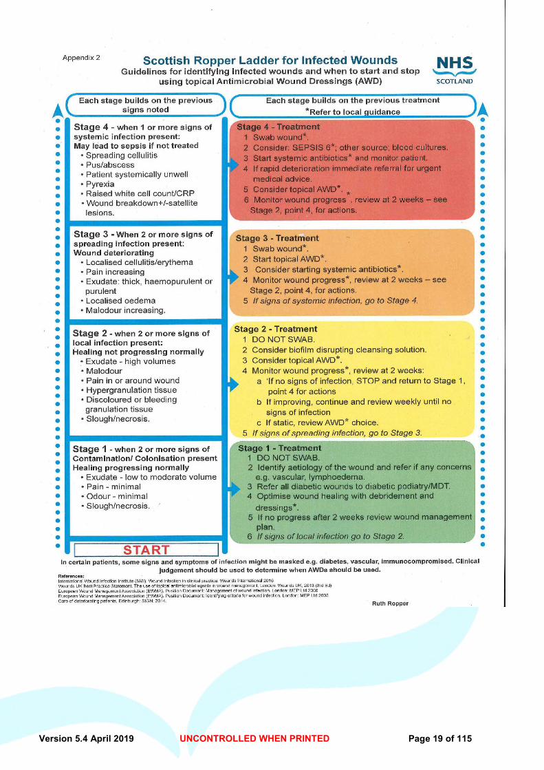
Version 5.4 April 2019 UNCONTROLLED WHEN PRINTED Page 17 of 115
SSEECCTTIIOONN 11
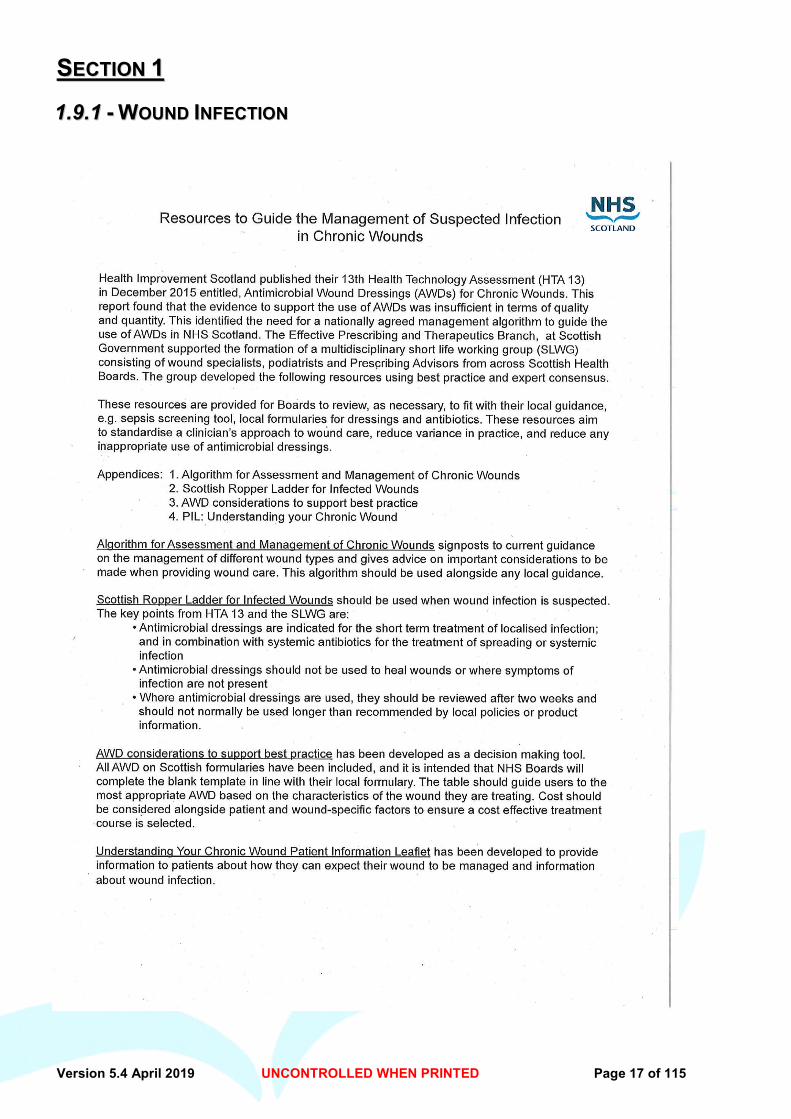
11..99..11 -- WWOOUUNNDD IINNFFEECCTTIIOONN
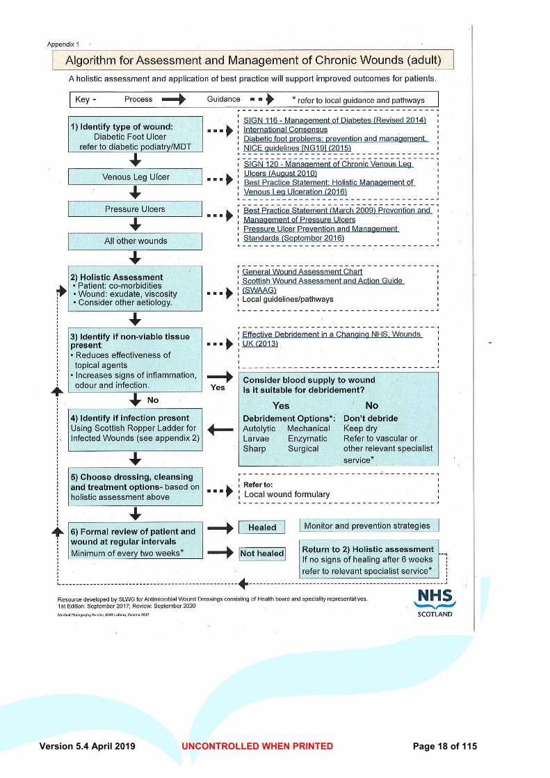
Version 5.4 April 2019 UNCONTROLLED WHEN PRINTED Page 18 of 115
-
Version 5.4 April 2019 UNCONTROLLED WHEN PRINTED Page 19 of 115
Version 5.4 April 2019 UNCONTROLLED WHEN PRINTED Page 20 of 115
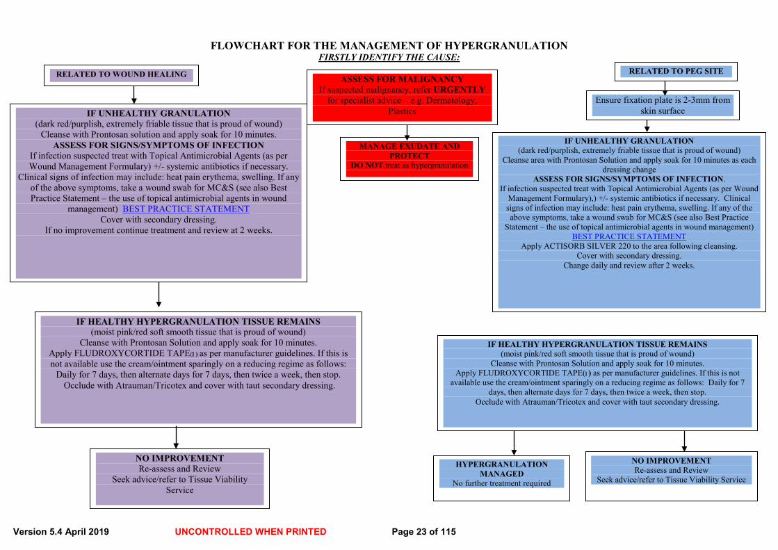
SSEECCTTIIOONN 11 11..1100 -- HHYYPPEERRGGRRAANNUULLAATTIIOONN When granulation tissue ‘over grows’ beyond the surface of the wound, this is known as ‘overgranulation’. This is also referred to as hypergranulation or ‘proud’ flesh. It can be present in wounds healing by secondary intention and is clinically recognised by a friable red, often shiny and soft appearance which is raised above the level of the skin. Once it has developed, hypergranulation is a difficult condition to deal with. Hypergranulation tissue can be classed as ‘healthy’ or ‘unhealthy’. Healthy hypergranulation tissue presents as an overgrowth of moist, pink/red tissue that may bleed easily. Healthy granulation tissue can reduce naturally and heal without intervention, although this may take longer if left untreated as the surface is moist and provides an ideal environment for bacterial colonisation and biofilm development. Unhealthy hypergranulation tissue presents as either a dark red or a pale bluish-purple uneven mass rising above the level of the skin and can also bleed easily. Whether the hypergranulation tissue is healthy or unhealthy the wound will not heal when the tissue is ‘proud’ because the epithelial tissue will be impeded from migrating across the wound’s surface. Causes The exact aetiology of overgranulation is unknown. The literature often links infection with overgranulation but it is not clear which occurs first. Vuolo (2010) suggests there are 3 types of overgranulation:
• Type 1: inflammatory with excessive exudate due to continued minor trauma or friction from mobility
• Type 2: occluded wound environment (possibly due to infection or chronic colonisation) (Bannerjee, 1999; Vandeputte and Hoekstra, 2006)
• Type 3: cellular imbalance – an imbalance between collagen synthesis and degradation due to the patients’ pathology.
Prevention
Overgranulation is recognised as a clinical problem. The limited evidence regarding the development and management of overgranulation means that clinical judgement must be exercised in the management of each patient to ensure that removal of the tissue is not harmful.
Infection is thought to be a cause of overgranulation and a preventative measure would be to try and prevent the wound from becoming chronically colonised or infected.
Continued reassessment of the wound will alert clinicians to changes in the granulation status and immediate intervention and treatment can then be applied.
Overgranulation tissue is a common problem encountered in wound care. There are several potential options for treatment. The steroid-impregnated tapes indicate that these can be an efficient and cost-effective treatment for overgranulation in a variety of wound types.
Version 5.4 April 2019 UNCONTROLLED WHEN PRINTED Page 21 of 115
A number of options are available to treat overgranulation tissue, but clinical effectiveness, patient safety and comfort should be a consideration. A strategic approach for preventing and treating overgranulation tissue ensures that patients receive the most effective and safe care. For those presenting with an overgranulating wound it is essential to undertake a differential diagnosis, to exclude malignancy and to assess and manage infection.
Treatment
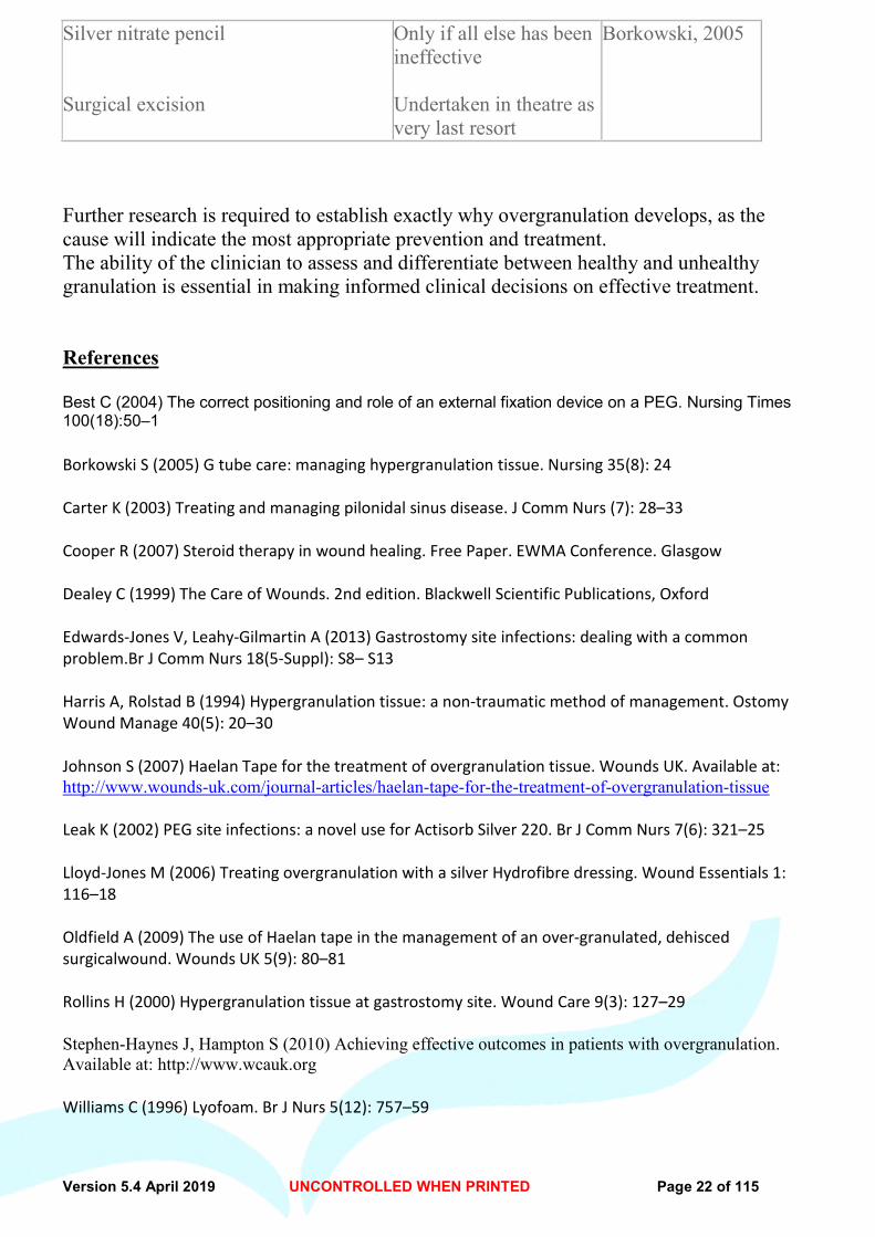
There are many treatment options for overgranulation, although research to support their use or to clearly suggest which is most effective is limited. The treatments reported in the table 2 below attempt to eliminate the causative factor and focus on reducing any bacteria present, applying pressure, reducing the occlusiveness of the dressings used, removing any overgranulation tissue and the use of steroid therapy. Dealey (2007) states that the use of silver nitrate directly reduces fibroblast production and should never be considered as a first line therapy.
Table 2: Treatment for overgranulation Treatment option Objective of treatment Evidence base Application of foam dressing To flatten and absorb
moisture Harris and Rolstad, 1994; Williams, 1996; Rollins, 2000; Carter, 2003
Change from an occlusive to a non-occlusive dressing
To reduce moisture Carter, 2003
Use of antimicrobials To reduce bacteria Leak, 2002; Lloyd Jones, 2006
Fludroxycortide Tape (Previously known as Haelan)
To reduce the production of new granulation cells. (licensed product)
Johnson, 2007; Oldfield, 2009
Fludroxycortide Cream/ ointment (previously known as Haelan)
To reduce the production of new granulation cells (although not licensed for use in overgranulation)
Johnson, 2007; Oldfield, 2009
Topical corticosteroid Reduces the cell division and production of granulation tissue
Carter, 2003; Cooper 2007
Use of a fixative device on PEG/Gastrostomy tubes.
To reduce movement and stimulation of new granulation tissue
Best, 2004; Edwards-Jones and Leahy-Gilmartin, 2013
Version 5.4 April 2019 UNCONTROLLED WHEN PRINTED Page 22 of 115
Silver nitrate pencil Surgical excision
Only if all else has been ineffective Undertaken in theatre as very last resort
Borkowski, 2005
Further research is required to establish exactly why overgranulation develops, as the cause will indicate the most appropriate prevention and treatment. The ability of the clinician to assess and differentiate between healthy and unhealthy granulation is essential in making informed clinical decisions on effective treatment. References Best C (2004) The correct positioning and role of an external fixation device on a PEG. Nursing Times 100(18):50–1 Borkowski S (2005) G tube care: managing hypergranulation tissue. Nursing 35(8): 24 Carter K (2003) Treating and managing pilonidal sinus disease. J Comm Nurs (7): 28–33 Cooper R (2007) Steroid therapy in wound healing. Free Paper. EWMA Conference. Glasgow Dealey C (1999) The Care of Wounds. 2nd edition. Blackwell Scientific Publications, Oxford Edwards-Jones V, Leahy-Gilmartin A (2013) Gastrostomy site infections: dealing with a common problem.Br J Comm Nurs 18(5-Suppl): S8– S13 Harris A, Rolstad B (1994) Hypergranulation tissue: a non-traumatic method of management. Ostomy Wound Manage 40(5): 20–30 Johnson S (2007) Haelan Tape for the treatment of overgranulation tissue. Wounds UK. Available at: http://www.wounds-uk.com/journal-articles/haelan-tape-for-the-treatment-of-overgranulation-tissue Leak K (2002) PEG site infections: a novel use for Actisorb Silver 220. Br J Comm Nurs 7(6): 321–25 Lloyd-Jones M (2006) Treating overgranulation with a silver Hydrofibre dressing. Wound Essentials 1: 116–18 Oldfield A (2009) The use of Haelan tape in the management of an over-granulated, dehisced surgicalwound. Wounds UK 5(9): 80–81 Rollins H (2000) Hypergranulation tissue at gastrostomy site. Wound Care 9(3): 127–29 Stephen-Haynes J, Hampton S (2010) Achieving effective outcomes in patients with overgranulation. Available at: http://www.wcauk.org Williams C (1996) Lyofoam. Br J Nurs 5(12): 757–59
Version 5.4 April 2019 UNCONTROLLED WHEN PRINTED Page 23 of 115
IF HEALTHY HYPERGRANULATION TISSUE REMAINS (moist pink/red soft smooth tissue that is proud of wound)
Cleanse with Prontosan Solution and apply soak for 10 minutes. Apply FLUDROXYCORTIDE TAPE(1) as per manufacturer guidelines. If this is not available use the cream/ointment sparingly on a reducing regime as follows:
Daily for 7 days, then alternate days for 7 days, then twice a week, then stop. Occlude with Atrauman/Tricotex and cover with taut secondary dressing.
NO IMPROVEMENT Re-assess and Review
Seek advice/refer to Tissue Viability Service
IF UNHEALTHY GRANULATION (dark red/purplish, extremely friable tissue that is proud of wound)
Cleanse area with Prontosan Solution and apply soak for 10 minutes as each dressing change
ASSESS FOR SIGNS/SYMPTOMS OF INFECTION. If infection suspected treat with Topical Antimicrobial Agents (as per Wound
Management Formulary),) +/- systemic antibiotics if necessary. Clinical signs of infection may include: heat pain erythema, swelling. If any of the above symptoms, take a wound swab for MC&S (see also Best Practice
Statement – the use of topical antimicrobial agents in wound management) BEST PRACTICE STATEMENT
Apply ACTISORB SILVER 220 to the area following cleansing. Cover with secondary dressing.
Change daily and review after 2 weeks.
FLOWCHART FOR THE MANAGEMENT OF HYPERGRANULATION FIRSTLY IDENTIFY THE CAUSE:
ASSESS FOR MALIGNANCY If suspected malignancy, refer URGENTLY
for specialist advice – e.g. Dermatology, Plastics
RELATED TO PEG SITE
MANAGE EXUDATE AND PROTECT
DO NOT treat as hypergranulation
IF UNHEALTHY GRANULATION (dark red/purplish, extremely friable tissue that is proud of wound)
Cleanse with Prontosan solution and apply soak for 10 minutes. ASSESS FOR SIGNS/SYMPTOMS OF INFECTION
If infection suspected treat with Topical Antimicrobial Agents (as per Wound Management Formulary) +/- systemic antibiotics if necessary.
Clinical signs of infection may include: heat pain erythema, swelling. If any of the above symptoms, take a wound swab for MC&S (see also Best Practice Statement – the use of topical antimicrobial agents in wound
management) BEST PRACTICE STATEMENT Cover with secondary dressing.
If no improvement continue treatment and review at 2 weeks.
Ensure fixation plate is 2-3mm from skin surface
NO IMPROVEMENT Re-assess and Review
Seek advice/refer to Tissue Viability Service
HYPERGRANULATION MANAGED
No further treatment required
IF HEALTHY HYPERGRANULATION TISSUE REMAINS (moist pink/red soft smooth tissue that is proud of wound)
Cleanse with Prontosan Solution and apply soak for 10 minutes. Apply FLUDROXYCORTIDE TAPE(1) as per manufacturer guidelines. If this is not
available use the cream/ointment sparingly on a reducing regime as follows: Daily for 7 days, then alternate days for 7 days, then twice a week, then stop.
Occlude with Atrauman/Tricotex and cover with taut secondary dressing.
RELATED TO WOUND HEALING
Version 5.4 April 2019 UNCONTROLLED WHEN PRINTED Page 24 of 115
SSEECCTTIIOONN 11 11..1111 -- RREEDDUUCCIINNGG PPAAIINN AATT DDRREESSSSIINNGG CCHHAANNGGEESS
Patients’ requirement for analgesia must be accurately assessed prior to the removal of the dressing. A specialist referral may be required to treat pain from underlying pathologies and wound pain. However basic principles of good pain management should be utilised until specialist advice is available.
The use of entonox, a self administered analgesic gas comprising oxygen and nitrous oxide may be favoured for its rapid onset analgesia. This gas is used for the duration of the procedure and not recommended for prolonged use or general pain relief (EWMA position document)
MMEETTHHOODDSS TTOO RREEDDUUCCEE AANNXXIIEETTYY AATT DDRREESSSSIINNGG CCHHAANNGGEE Provide adequate analgesia prior to dressing change Explanation to patient of what to expect Identify what the patient recognises to be triggers of pain Invite the patient to be involved as much as he/she wishes i.e. removal of dressing
themselves Encourage slow rhythmic breathing during the procedure Offer the patient “time out” during the procedure and negotiate a signal e.g. raise hand,
clap Use of distraction e.g. music Ensure all materials are ready and easily accessible, prior to dressing change.
DDRREESSSSIINNGG SSEELLEECCTTIIOONN CCRRIITTEERRIIAA Appropriate for the type of wound Maintains moist wound healing whilst managing exudate Does not adhere to the wound bed Reduce friction at the wound surface Minimises pain and trauma on removal Reduce the need for frequent dressing changes Provides patient comfort
(EWMA position document 2003, TVNA Best Practice Statement 2004)
DDRREESSSSIINNGG RREEMMOOVVAALL Avoid unnecessary stimulus, such as drafts from windows, poking/prodding, unnecessary
touch Ensure ” Methods” detailed above are employed. Consider the manufacturer’s instructions to release the adhesive Soaking an adherent dressing with warm saline or tap water prior to removal, (patient
may do this themselves or in the shower if feasible). Patient may wish to remove own dressing
AAPPPPLLIICCAATTIIOONN OOFF NNEEWW DDRREESSSSIINNGG Ensure wound cleansing methods, as detailed above, are employed Work gently and swiftly to apply dressing
REFERENCES European Wound Management Association EWMA (2003) Position Document - Pain at wound dressing changes EWMA London Tissue Viability Nurses Association TVNA (2004) Best practice Statement - Minimising Trauma and Pain in Wound Management issue 1 TVNA Scotland
Version 5.4 April 2019 UNCONTROLLED WHEN PRINTED Page 25 of 115
SSEECCTTIIOONN 11 11..1122 -- NNUUTTRRIITTIIOONN IINN WWOOUUNNDD MMAANNAAGGEEMMEENNTT Nutrients contain a composition of chemicals which are obtained from the foods we eat in order to provide our cells with growth, maintenance and repair. Therefore good nutrition is vital in wound healing and one of the essential components when considering the prevention and management of wounds. Malnutrition Malnutrition is the condition in which a deficiency or excess of energy, protein and other nutrients cause significant effects on body tissue, body function and clinical result. Unintentional weight loss, the need for consistency altered diet or oral problems with eating are all predictors of malnutrition which could hinder wound healing.
Malnutrition and specific nutrient deficiencies can lead to poor health, a reduced immune response and subsequent tissue damage with delayed wound healing and increased susceptibility to wound or systemic infection. Physical appearance alone will not always identify such patients.
A deficit in nutritional intake means that energy, protein, vitamin and mineral intakes are low at a time when requirements are increased. Muscle mass is lost as protein breakdown occurs to provide energy. Protein can also be lost in vast amounts through exuding wounds, dependant on the size and amount of fluid exuded.
A decreased nutritional intake can result in weight loss and the loss of the protective cushioning effect of fat. Dehydration can also cause dry fragile skin which again further impacts on the body’s ability to repair wounds.
A low energy intake is associated with a reduced availability of other nutrients such as Vitamin C, Zinc and Iron which have a vital role in wound healing.
Providing sufficient energy will prevent dietary or tissue protein being used as an energy source and therefore reduce these effects and adequate protein will optimise wound healing. To promote wound healing patients should choose foods high in energy and protein. Foods rich in protein include, milk, eggs, cheese, yoghurt, lean meat or poultry and fresh or tinned fish. Nutritional Screening
On admission to hospital patients should undergo initial nutritional assessment using the Malnutrition Universal screening tool (MUST). MUST is a 5 step process to identify adults who are undernourished, at risk of malnutrition or obese. It also includes guidelines that can be used to develop a care plan.
Where risk of malnutrition is detected, appropriate care plans must be initiated and be repeated at least weekly if there is any significant change in condition. Any nutritional inadequacy should be identified and corrected at an early stage to minimise the complications associated with them. Patients identified at risk of nutritional inadequacy with a high MUST score should be referred to the Dietetic Department for further assessment regarding their nutritional needs and management
The MUST screening tool was updated in 2013 and is used in Forth Valley Royal and all out-lying community hospitals. The next page details the updated version of MUST.
Version 5.4 April 2019 UNCONTROLLED WHEN PRINTED Page 26 of 115
Obesity Being obese can cause additional issues associated with wound healing. These patients will likely have reduced mobility, increased pressure placed on the wound along with reduced vascular supply within the adipose tissue making the wound healing process increasingly difficult. However, as good nutrition is required to aid wound healing it is not recommenced to reduce protein and calorie intake in obese and overweight patients.
Diabetes
It is important that people with diabetes ensure their blood sugars are well controlled. Hyperglycaemia increases the risk of localised and systemic infection which in turn delays the wound healing process. REFERENCES. Collins, C. Nutrition and wound healing. Care of the Critically Ill. 1996, 12, 97-90. Dickerson, JWT. Ascorbic acid, zinc and wound healing. Journal of Wound Care. 1993, 2. 350-353. Meyer, MA, Muller, MJ, Herndon, DN. Nutrient support of the healing wound. New Horizons. 1994, 2, 202-214.Ondrey, FG and Holm, DB. Effects of nutrition on wound healing. Otolaryngology – Head and Neck Surgery. 1994, 110, 557-559. MUST – BAPEN Elia M. Guidelines for Detection and management of malnutrition. Malnutrition Advisory Group (MAG), Standing committee of Bapen. Maidenhead: Bapen, 2000. Terranova A. The Effects of Diabetes Mellitus on wound healing. Plast Surg Nurs. 1991, 1, 20-5.
Version 5.4 April 2019 UNCONTROLLED WHEN PRINTED Page 27 of 115
SSEECCTTIIOONN 11 11..1133 NNOONN--FFOORRMMUULLAARRYY WWOOUUNNDD CCAARREE -- DDRREESSSSIINNGGSS The aim of the wound formulary is to standardise products and wound care throughout Forth Valley, based on best practice evidence. The list of products within the formulary is not exhaustive and there may be occasions where products available through the Formulary are not appropriate for individual patients or departments. Staff may wish to access certain specialised products which are not listed, or not available on prescription. In these circumstances, staff should involve the Tissue Viability Service or District Nurse caseload holder, for advice and non formulary products may then be recommended these products can then be ordered through central supplies using the Non-formulary request form (Appendix 1). Or following authorisation of the Non Formulary request form in the community, may be issued on prescription. Please note non-formulary items are not stocked by hospital pharmacy or central stores. This should be taken into account when initiating treatment, as delays will be inevitable when ordering non-formulary dressings. Forth Valley Wound Management Group continues to evaluate new products for inclusion to the formulary.
Version 5.4 April 2019 UNCONTROLLED WHEN PRINTED Page 28 of 115
SSEECCTTIIOONN 11 11..1144 -- SSKKIINN CCAARREE Care of the Surrounding Skin The principles of good skin care depend on:
• Keeping the skin clean and dry
• Avoiding the excessive use of soap
• Using showers in preference to baths where possible
• Keeping the skin moisturised
Assessment The state of the skin surrounding a wound should be assessed at each wound dressing change. Refer to assessment form - appendix 2A – 2D Observe for the following:
• Dry skin: may break down and provide a portal for infection
• Maceration: caused by poor management of exudates
• Inflammation: consider contact sensitivity to dressings or infection
Emollients Emollients are moisturisers that soothe and hydrate the skin. They are indicated for all dry or scaling disorders. Most are best applied after washing but their effects are short-lived so they must be applied frequently and regularly to maintain improvement. They should continue to be applied even after improvement occurs. NB- Emollients should be applied in the direction of hair growth to prevent folliculitis. Some ingredients may rarely cause sensitisation and this should be suspected if an eczematous reaction occurs. There are different types of products available. These include ointments, creams, lotions and gels. Effectiveness depends upon the correct choice of product and correct use. Choice will depend on:
• The severity of the condition
• Patient preference
• The site of application
• Cost of preparation
Ointments: Ointments are greasy and generally insoluble in water so can be difficult to wash off and do not suit all patients. They are recommended as the first choice for formulation in most skin conditions and are particularly useful for chronic dry conditions. Examples: Liquid Paraffin/White Soft Paraffin Ointment 50:50%. Creams: Creams are emulsions of oil and water and often contain an antimicrobial preservative. They are therefore more likely to cause both irritant and allergic reactions. For this reason creams are best avoided first line but are often more cosmetically acceptable for some patients. Creams can be better than ointments for some acute conditions due to a cooling effect as they evaporate from the skin. Example: Diprobase. Gels: Gels also have a high water content and produce a cooling effect on evaporation from the skin. They are suitable for use on the face and scalp. Example: Doublebase. Barrier Preparations:
Version 5.4 April 2019 UNCONTROLLED WHEN PRINTED Page 29 of 115
Wounds which are heavily exuding or have friable surrounding skin are at risk of excoriation, epidermal stripping and maceration. A barrier can be used on the surrounding skin prophylactically to protect the skin. Barrier preparations should be reapplied at dressing changes. Examples: Sorbaderm cream or Sure prep barrier spray N.B. Sudocrem should not be used as a barrier against incontinence. REFERENCES Penzer R. (2012) A Best Practice Statement for Emollient Therapy. Dermatological Nursing 11 (4) White R et al (2012) Best Practice Statement – Care of the Older Persons Skin (2nd edition) London. Wounds UK, 2012
Version 5.4 April 2019 UNCONTROLLED WHEN PRINTED Page 30 of 115
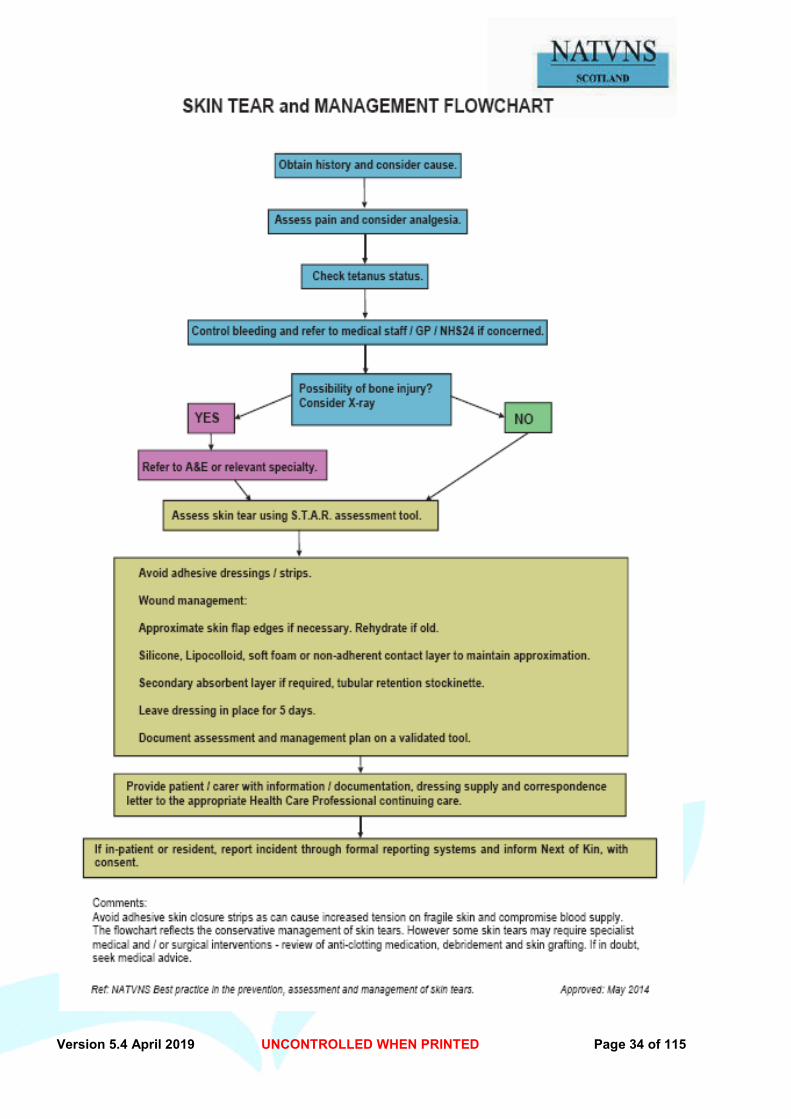
SSEECCTTIIOONN 11 11..1155 NNAATTVVNNSS BBEESSTT PPRRAACCTTIICCEE IINN TTHHEE PPRREEVVEENNTTIIOONN,, AASSSSEESSSSMMEENNTT AANNDD MMAANNAAGGEEMMEENNTT OOFF SSKKIINN TTEEAARRSS IINNTTRROODDUUCCTTIIOONN Skin tears are viewed as an increasing problem by healthcare practitioners and if appropriate treatment is not given, these injuries may become chronic wounds with prolonged healing subsequently causing unnecessary pain and distress (Jones & Millman 1983). Traditional management of skin tears can cause new damage and slow down the healing process (Meuleneire, 2002). This type of injury usually occurs in immature skin (neonatal) and in the elderly. As our population changes and the number of elderly people increases, therefore whether we are caring for patients in their own home, a care home or hospital, we need to be aware of best practice in prevention, assessment and management of skin tears. This document summarises the current evidence. IINNTTEERRNNAATTIIOONNAALL CCOONNSSEENNSSUUSS An international consensus panel have defined skin tears as “A skin tear is a traumatic wound caused by mechanical forces, including the removal of adhesives” (International skin Tears Advisory Panel ISTAP 2018). They most commonly occur at the extremes of age, in critically ill or medically compromised individuals and in those who require assistance with personal care (Carvell et al, 2007, Irving et al 2006, Belton 2008). Prevention of skin tears where possible should be our priority. When skin tears occur, accurate assessment and appropriate management will minimise further trauma and preserve viable tissue.
PPRREEVVAALLEENNCCEE OOFF SSKKIINN TTEEAARRSS The evidence on prevalence and incidence of skin tears is limited and generally dated.
In long term care: 2.23-92%, although estimates vary and may be lower (Strazzieri et al, 2017; LeBlanc, 2017; LeBlanc et al, 2013; Sanada et al, 2015; Skiveren et al, 2017; Woo et al, 2015)
In the community: 4.5-19.5% in known wounds in all age groups (Carville and Lewin, 1998; LeBlanc et al, 2008)
In acute care: 6.2-11.1% (Chang et al, 2016; Hsu and Chang, 2010; McErlean, 2004; Santamaria et al, 2009)
In palliative care: 3.3-14.3% (Amaral et al, 2012; Maida et al, 2012)
In intensive care and operative theatres: prevalence is unknown
The work carried out in Australia led by Carville et al (2007) to state that skin tears are perceived to be common wounds and occur more frequently than pressure ulcers. To date there are no prevalence data available for the UK therefore the true extent of patients requiring hospital attendance or the resource impact or cost to the patient of the NHS due to skin tears is still not fully known.
Version 5.4 April 2019 UNCONTROLLED WHEN PRINTED Page 31 of 115
AAGGEE RREELLAATTEEDD SSKKIINN CCHHAANNGGEESS AASSSSOOCCIIAATTEEDD WWIITTHH SSKKIINN TTEEAARRSS Changes to the skin due to the ageing process make the skin more vulnerable. These changes include: Thinning of the epidermis (top layer of the skin) and dermis (middle layer of the skin)
Shrinkage of subcutaneous / fatty tissue (bottom layer of skin)
Small blood vessel walls widen, shrink and become disorganised
Decrease in collagen (natural protein component of the skin) amount and quality
Reduced sebum (natural lubricant) production Pre-term and newborn infants have immature skin and are also vulnerable to skin tears. OOTTHHEERR FFAACCTTOORRSS TTOO CCOONNSSIIDDEERR Immunological status and malnutrition, circulation and oxygen intake may also impact on fragility of the skin (Meulenire, 2002.)
BBEESSTT PPRRAACCTTIICCEE IINN PPRREEVVEENNTTIIOONN OOFF SSKKIINN TTEEAARRSS Prevention of skin tears starts with early identification of individuals who are at risk. Based on available evidence the consensus statement of an international panel suggests the following strategies should be part of prevention 1 Assess for risk upon admission to healthcare service and whenever the individuals condition changes and document in care plan 2 Implement a systematic prevention protocol (points 3-10) 3 Have individuals at risk wear long sleeves, long trousers or knee high socks 4 Provide shin guards/leg protectors for those individuals who experience repeat skin tears on shins 5 Ensure safe patient handling techniques and equipment/environment 6 Involve individuals and families in prevention strategies 7 Educate registered and non registered staff and care givers to ensure proper techniques for providing care without causing skin tears 8 Consult dietician to ensure adequate nutrition and hydration 9 Keep skin well lubricated by applying hypoallergenic moisturiser at least 2 times per day. Encourage the patient or their carers to apply emollient. 10 Protect individuals at high risk of trauma during routine care from self-injury LeBlanc & Baranoski (2011) Stephen-Hayes & Carville (2011) also give practical advice on maintaining a safe environment to minimise the risk of skin tears which includes Ensure adequate lighting and position small furniture (night tables, chairs) to avoid bumps or knocks. Remove rugs and excessive furniture. Upholster or pad sharp borders of furniture or bed surroundings with padding and soft material Use appropriate aids when transferring patients and adopt good manual handling techniques according to local policy Never use bed sheets to move patients as this can contribute to damage by causing dragging effect on the skin. Always use lifting device or slide sheet Where possible reduce or eliminate pressure, shear and friction using pressure relieving devices and positioning techniques. Include these points where relevant in the patients care plan
Version 5.4 April 2019 UNCONTROLLED WHEN PRINTED Page 32 of 115
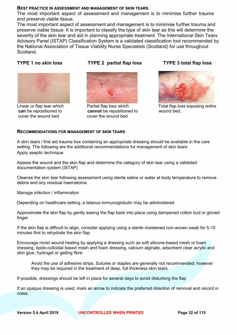
BBEESSTT PPRRAACCTTIICCEE IINN AASSSSEESSSSMMEENNTT AANNDD MMAANNAAGGEEMMEENNTT OOFF SSKKIINN TTEEAARRSS The most important aspect of assessment and management is to minimise further trauma and preserve viable tissue. The most important aspect of assessment and management is to minimise further trauma and preserve viable tissue. It is important to classify the type of skin tear as this will determine the severity of the skin tear and aid in planning appropriate treatment. The International Skin Tears Advisory Panel (ISTAP) Classification System is a validated classification tool recommended by the National Association of Tissue Viability Nurse Specialists (Scotland) for use throughout Scotland. TYPE 1 no skin loss TYPE 2 partial flap loss TYPE 3 total flap loss
Linear or flap tear which Partial flap loss which Total flap loss exposing entire can be repositioned to cannot be repositioned to wound bed. cover the wound bed cover the wound bed RREECCOOMMMMEENNDDAATTIIOONNSS FFOORR MMAANNAAGGEEMMEENNTT OOFF SSKKIINN TTEEAARRSS A skin tears / first aid trauma box containing an appropriate dressing should be available in the care setting. The following are the additional recommendations for management of skin tears: Apply aseptic technique
Assess the wound and the skin flap and determine the category of skin tear using a validated documentation system (ISTAP)
Cleanse the skin tear following assessment using sterile saline or water at body temperature to remove debris and any residual haematoma
Manage infection / inflammation
Depending on healthcare setting, a tetanus immunoglobulin may be administered
Approximate the skin flap by gently easing the flap back into place using dampened cotton bud or gloved finger
If the skin flap is difficult to align, consider applying using a sterile moistened non-woven swab for 5-10 minutes first to rehydrate the skin flap
Encourage moist wound healing by applying a dressing such as soft silicone-based mesh or foam dressing, lipido-colloidal based mesh and foam dressing, calcium alginate, adsorbent clear acrylic and skin glue, hydrogel or gelling fibre
Avoid the use of adhesive strips. Sutures or staples are generally not recommended; however they may be required in the treatment of deep, full thickness skin tears
If possible, dressings should be left in place for several days to avoid disturbing the flap
If an opaque dressing is used, mark an arrow to indicate the preferred direction of removal and record in notes
Version 5.4 April 2019 UNCONTROLLED WHEN PRINTED Page 33 of 115
Compression therapy should be considered if wound is on the lower leg. Before applying compression, a full leg assessment including vascular assessment e.g. ankle-brachial pressure index (ABPI) – should be carried out
Dressings should be held in place with stocking-like products (e.g. tubular viscose retention bandage)
Pain assessment should be carried out and appropriate analgesia should be provided
Complete formal wound assessment form
Document in care plan, complete accident / incident documentation and where relevant
WWHHEENN TTOO RREEFFEERR If the skin tear is extensive or associated with a full thickness injury, significant and or uncontrolled bleeding or haematoma formation, a surgical/plastic surgery review may be required (Stephens-Hayes & Carville, 2011). If the skin tear is on the lower leg and fails to progress consider early referral to local leg ulcer clinic or vascular nurse specialist for leg ulcer assessment. Referral to Tissue viability specialists may also be indicated if the wound fails to progress to healing.
BBEESSTT PPRRAACCTTIICCEE IINN OONNGGOOIINNGG MMAANNAAGGEEMMEENNTT At each dressing change the dressing should be gently removed in the direction indicated by the arrow. If it does not remove easily, consider the use of saline soaks or silicone-based adhesive removers Mudge & Orsted (2010). The wound flap may be friable so care should be taken to prevent disturbing it. The wound should be observed for signs of infection and any changes in the colour of the tissue of the flap which may indicate that it is becoming non-viable (Stephen-Hayes & Carville, 2011).
CCOONNCCLLUUSSIIOONN Skin tears are common wounds, particularly at the extremes of age. We should be aware of the risk factors associated with skin tears and where ever possible minimise risk to patients. When a patient develops a skin tear, the use of a skin tear classification system will aid our decision making, and ensure we are all using the same language to describe lesions. Treatment regimes should be structured on best available evidence. References Baharestani MM (2007) An overview of neonatal and paediatric wound care knowledge and considerations. Ostomy Wound Management 53;6:34-40 Baranoski S (2003) How to prevent and manage skin tears. Adv Skin Wound Care 16:268 Beldon, P. (2008) Classifying and managing pretibial lacerations in older people. British Journal of Nursing Tissue Viability Supp, 17; 11: S4 -S18 Carville C, Lewin G (1998) Caring in the community: a prevalence study. Prim Intent 6:54-
Carville K, Smith JA (2004) Report on the effectiveness of comprehensive wound assessment and documentation in the community. Prim Intent 12:41-48 Carville K, Lewis G, Newall N et al (2007) STAR: a consensus for skin tear classifications. Prim Intent 15;1:18-28 Everett S, Powell T (1994) Skin tears-the underestimated wound. Prim Intent 2;8:8-30 Irving V, Bethell E, Burtin F (2006) Neonatal wound care: minimising pain and trauma. Wounds 2;1:33-41 Jones P, Millman, A. (1983) Wound healing and the aged patient. Journal of Surgical Res 35:1428. LeBlanc K, Baranoski S (2011) Skin Tears: State of Science: Consensus statement of the prevention, prediction, assessment and treatment of skin tears. Advances in Skin and Wound Care 24; 9: 2-15 LeBlanc K, Baranoski S, Regan M (2011) Skin Tear Survey (unpublished data) Payne RL, Martin MC (1993) Defining and classifying skin tears:need for a common language. Ostomy and Wound Management 39;5:16-26 Meuleneire F (2002) Using a soft silicone-coated net dressing to manage skin tears. Journal of Wound Care 11;10:... Malone ML, Rozario N, Gavinski M, Goodwin J (1991) The epidemiology of skin tears in the institutionalised elderly. J Am Geriat Soc 39;6:591-595 O’Regan A (2002) Skin tears; a review of the literature. Wound Counc Enterostomal Ther J 22;2:26-31 Sibbald RG, Krasner DL, Lutz JB, et al (2009) The SCALE Expert Panel: Skin Changes At Life’s End. Final Consensus Document. October 1. Stephen-Hayes J, Carville K (2011) Skin tears Made Easy. Wounds International 2;4. Available from http:/www.woundsinternational.com Voegell D (2010) Basic essentials: why elderly skin requires special treatment. Nurs Res Care 12;9:422-429 White MW, Karman S, Cowell B (1994) Skin tears in frail elders: a practical approach to prevention. Geriat Nurs 15;2:95-99 WoundsWest wound survey 2009: key results at a glance. Government of Western Australia Department of Health.
Version 5.4 April 2019 UNCONTROLLED WHEN PRINTED Page 34 of 115
Version 5.4 April 2019 UNCONTROLLED WHEN PRINTED Page 35 of 115
SSEECCTTIIOONN 11 11..1166-- TTHHEERRMMAALL IINNJJUURRYY GGUUIIDDEELLIINNEESS
SSUUPPEERRFFIICCIIAALL PPAARRTTIIAALL TTHHIICCKKNNEESSSS BBUURRNNSS Description
• Pink, wet, small blisters, intact sensation • Blanches on pressure with normal capillary return
Management Aims
• To protect from infection • To absorb exudates • To encourage healing.
Treatment
• Initial first aid – cold water, about 15°C, for 20 minutes. Do not use ice or iced water If greater than three hours since time of injury cold water will have no beneficial effect (EMSB 1996)
• De-roof any blisters (larger than 1cm diameter) with sterile scissors • Apply wide mesh paraffin impregnated gauze (where larger volumes of fluid are • exuding) or narrow mesh paraffin impregnated gauze (where smaller volumes of fluid are
exuding) or silicone contact layer (children). Apply super absorbent secondary dressing or gauze +/- gamgee (depending on exudate levels) plus a bandage or tape to secure
• When exudate levels drop change to hydrocolloid or foam dressing. • If wound is not showing signs of improvement within three days post injury, refer to • Wallace Burns Unit Clinic at St. John’s Hospital (adult) or Royal Hospital for Sick • Children (children) for advice.
DO NOT APPLY FLAMAZINE® CREAM TO SUPERFICIAL BURNS (Flamazine® should only be used on infected small burns or for prevention of infection in larger burns after full assessment by a specialist) Comments
• Superficial burns should heal within two weeks • If healing is delayed it means the burn is deeper than originally diagnosed • Apply simple emollient 2 x daily when healed, washing off with water before
reapplying emollient • Avoid wearing nylon next to recently healed areas • Will need protected from sunlight/UV light for life with a factor 25+ sun screen. • Children will require at least factor 30+ sun screen.
For further copies of guideline contact: Wallace Burns Unit, St. John’s Hospital, Livingston, West Lothian, EH54 6PP It is important to realise that a burn wound is dynamic and continues to change up to 24 hours after injury. Do not assume that all areas of the burn are equally deep (EMSB 1996).
Version 5.4 April 2019 UNCONTROLLED WHEN PRINTED Page 36 of 115
SSEECCTTIIOONN 11 MMIIDD AANNDD DDEEEEPP PPAARRTTIIAALL TTHHIICCKKNNEESSSS BBUURRNNSS Description
• Mottled red/white patchy appearance • Blisters may be present, white in appearance on hands/feet • Capillary return is sluggish or absent • Reduced sensation or no sensation.
Management Aims
• To protect from infection • To manage exudates • To assess depth for conservative management or surgical management.
Treatment
• Initial first aid – cold water, about 15°C, for 20 minutes. Do not use ice or iced water. If greater than three hours since time of injury, cold water will have no beneficial effect (EMSB 1996)
• Apply superabsorbent dressing if available or simple conservative dressing of either wide mesh paraffin impregnated gauze (where larger volumes of fluid are exuding) or narrow mesh (where smaller volumes of fluid are exuding) or silicone contact layer (children) and gamgee padding plus bandage
• Change outer padding as required leaving paraffin gauze intact for two days or change super absorbent as required due to exudate
DO NOT APPLY FLAMAZINE® CREAM BEFORE ASSESSMENT AT 48 HOURS POST INJURY
• After 48 hours reassess: • If sensation and blanching, treat as for superficial burn • If no blanching refer to Wallace Burns Unit at St. John’s Hospital (adults) or Royal
Hospital for Sick Children (children) for full assessment and treatment regime. Comments
• If in any doubt as to depth of burn please refer to Wallace Burns Unit at St. John’s Hospital (adults) or Royal Hospital for Sick Children (children) to prevent delay in preparing for surgery if this is required
• Apply simple emollient 2 x daily when healed, washing off with water before reapplying emollient
• Avoid wearing nylon next to recently healed areas • Will need protected from sunlight/UV light for life with a factor 25+ sun screen. • Children will require at least factor 30+ sun screen.
Version 5.4 April 2019 UNCONTROLLED WHEN PRINTED Page 37 of 115
SSEECCTTIIOONN 11 FFUULLLL TTHHIICCKKNNEESSSS BBUURRNN Description
• Dry black/white/brown, leathery appearance • No sensation, no capillary return • If old burn may have thick layer of slough/eschar present
Management Aims
• To protect from infection • To manage exudate • To prepare for surgery for excision and grafting
Initial Treatment and Assessment
• Initial first aid – cold water, about 15°C, for 20 minutes. Do not use ice or iced water. If greater than three hours since time of injury cold water will have no beneficial effects (EMSB 1996)
• Phone the Wallace Burns Unit at St. John’s Hospital (adults) or Royal Hospital for Sick Children (children) for advice or to arrange transfer.
If patient is for Transfer
• If patient is for transfer, cover all burned areas with cling film to prevent infection and allow for ease of assessment. Then wrap patient in sterile/clean sheets/covers to prevent heat loss
• If transfer is delayed for any reason, or journey will be greater than 2-3 hours, apply super absorbent dressings or conservative dressings of paraffin impregnated gauze (use Silicone contact layer in children), gauze, gamgee and bandages to manage fluid loss.
DO NOT APPLY FLAMAZINE® CREAM AS IT WILL MASK THE BURN INJURY AND MAKE IT DIFFICULT TO ASSESS Treatment if not for transfer
• If the patient is not for transfer to specialist unit due to smaller size of burn, apply Flamazine® cream to the burn wound, cover with paraffin impregnated gauze, gauze, gamgee and bandages to manage fluid loss
• Change dressings daily until thick eschar lifts, then reduce to every two days depending on exudate levels
• Once a healthy granulating wound bed is present, change to hydrocolloid or foam dressing, depending on level of exudate.
Comments • If healing is delayed it means the burn may require skin grafting; please contact the
Regional Unit • Apply simple emollient 2 x daily when healed, washing off with water before reapplying
emollient • Avoid wearing nylon next to recently healed areas • Will need protected from sunlight/UV light for life with a factor 25+ sun screen. Children
will require at least factor 30+ sun screen.
Version 5.4 April 2019 UNCONTROLLED WHEN PRINTED Page 38 of 115
SSEECCTTIIOONN 11 CCRRIITTEERRIIAA FFOORR IIDDEENNTTIIFFYYIINNGG BBUURRNNSS RREEQQUUIIRRIINNGG RREEFFEERRRRAALL TTOO AA RREEGGIIOONNAALL BBUURRNNSS UUNNIITT:: IIDDEENNTTIIFFYYIINNGG BBUURRNNSS RREEQQUUIIRRIINNGG RREEFFEERRRRAALL TTHHEE BBRRIITTIISSHH BBUURRNN AASSSSOOCCIIAATTIIOONN HHAASS IIDDEENNTTIIFFIIEEDD TTHHEE FFOOLLLLOOWWIINNGG IINNJJUURRIIEESS AASS TTHHOOSSEE RREEQQUUIIRRIINNGG RREEFFEERRRRAALL TTOO AA BBUURRNN UUNNIITT::
• Burns greater than 10% Total Body Surface Area (TBSA) in adults • Burns greater than 5% TBSA in children • Burns of special areas – face, hands, feet, genitalia, perineum and major joints • Full thickness burns greater than 5% TBSA • Electrical burns • Chemical burns • Burns with an associated inhalation injury • Circumferential burns of the limbs or chest • Burns at the extremes of age – children and the elderly • Burn injury in patients with pre-existing medical disorders which complicate
management, prolong recovery or effect mortality • Any burn patient with associated trauma • Suspected ‘non accidental injury’ (children or elderly).
Ref: Emergency Management of Severe Burns (EMSB) Course Manual (1996) UK version for The British Burn Association Contact Details When phoning please ask for Specialist Registrar for Burns:
• Wallace Burns Unit at St. John’s Hospital, Livingston (adults): 01506 523000 • Royal Hospital for Sick Children (RHSC), Edinburgh (children): 0131 536 0000
For advice
• Wallace Burns Unit at St. John’s Hospital, Livingston (adults): 01506 524120 RHSC Nurse Led Dressing Clinic (children) Mon, Wed, Thu, Fri: 0131 536 0743 Further Information www.britishburnassociation.com British Burn Association www.baps.co.uk British Association of Plastic Surgeons www.cobis.scot.nhs.uk Care of Burns in Scotland
Version 5.4 April 2019 UNCONTROLLED WHEN PRINTED Page 39 of 115
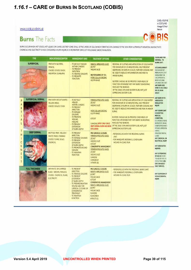
11..1166..11 –– CCAARREE OOFF BBUURRNNSS IINN SSCCOOTTLLAANNDD ((CCOOBBIISS))
Version 5.4 April 2019 UNCONTROLLED WHEN PRINTED Page 40 of 115
SSEECCTTIIOONN 11 11..1177-- SSTTEERRIILLEE DDRREESSSSIINNGG PPAACCKKSS
When considering prescribing/recommending a dressing pack, remember: Dressing packs should only be used where clinically indicated e.g. aseptic technique, non touch technique and immediate post surgical wounds (NICE Guidelines 74, 2008) and all components of the pack are necessary. Sterile dressing packs are available to order through central stores/ PECOS. The brand and contents supplied will be allocated by central stores. PPRRIIMMAARRYY CCAARREE PPRREESSCCRRIIBBIINNGG OOFF SSTTEERRIILLEE DDRREESSSSIINNGG PPAACCKKSS When a dressing pack is to be prescribed for a patient select the most appropriate dressing pack based on the contents required. The pack should be prescribed by brand name. CCLLEEAANN TTEECCHHNNIIQQUUEE Evidence has shown that a clean procedure is a safe way of dressing chronic wounds as they are all bacterially colonised with environmental micro organisms. The aim here is prevent harmful contamination to the wound. Indications for clean technique:
- any wound that has not been created surgically within 48hrs - if the wound does not connect to a deep body cavity/organ - if the patient is not immunocompromised/neutropenic.
eg pressure ulcers, leg ulcers, dehisced abdominal wounds. AASSEEPPTTIICC TTEECCHHNNIIQQUUEE Sterile dressing packs should be used for patients with acute surgical wounds (within 48hrs), dressings relating to insertion of or cleansing of peripheral access devices, urinary catheters and immunocompromised patients. (Refer to local policies for full guidance) Aim is to prevent/reduce the risk of microbial contamination into a wound healing by primary intention. SSTTEERRIILLEE DDRREESSSSIINNGG PPAACCKKSS
Nurse It (per pack 52p) Powder/Latex Free Nitrile Gloves S/M 7 Non-Woven Swabs 4-ply 10cm x 10cm 1 Compartment Tray 12cm x 11cm 1 Laminated Paper Sterile Field 40cm x 40cm 1 Large Apron 80cm x 130cm *1 Disposable Forceps 11cm 1 White Polythene Disposable Bag 46cm x 26cm *1 Paper Towel 35cm x 40cm 1 paper measuring tape * shows different components in packs Note: Forceps may be obtained through PECOS. (Scottish Drug Tariff January 2015)
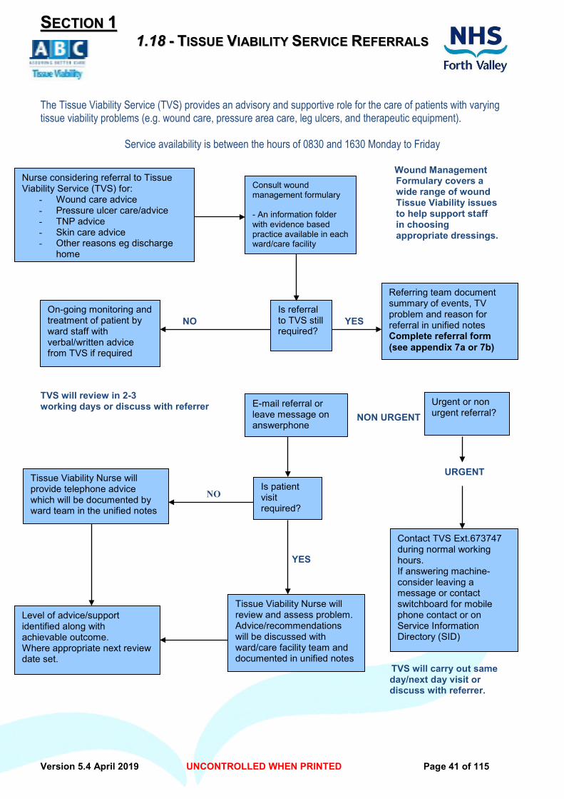
Version 5.4 April 2019 UNCONTROLLED WHEN PRINTED Page 41 of 115
SSEECCTTIIOONN 11 11..1188 -- TTIISSSSUUEE VVIIAABBIILLIITTYY SSEERRVVIICCEE RREEFFEERRRRAALLSS
The Tissue Viability Service (TVS) provides an advisory and supportive role for the care of patients with varying tissue viability problems (e.g. wound care, pressure area care, leg ulcers, and therapeutic equipment).
Service availability is between the hours of 0830 and 1630 Monday to Friday
Wound Management Formulary covers a wide range of wound Tissue Viability issues to help support staff in choosing appropriate dressings. # NO YES TVS will review in 2-3 working days or discuss with referrer NON URGENT URGENT NO
YES TVS will carry out same day/next day visit or discuss with referrer.
Consult wound management formulary - An information folder with evidence based practice available in each ward/care facility
Nurse considering referral to Tissue Viability Service (TVS) for:
- Wound care advice - Pressure ulcer care/advice - TNP advice - Skin care advice - Other reasons eg discharge
home
On-going monitoring and treatment of patient by ward staff with verbal/written advice from TVS if required
Is referral to TVS still required?
Referring team document summary of events, TV problem and reason for referral in unified notes Complete referral form (see appendix 7a or 7b)
E-mail referral or leave message on answerphone
Tissue Viability Nurse will provide telephone advice which will be documented by ward team in the unified notes
Is patient visit required?
Level of advice/support identified along with achievable outcome. Where appropriate next review date set.
Tissue Viability Nurse will review and assess problem. Advice/recommendations will be discussed with ward/care facility team and documented in unified notes
Urgent or non urgent referral?
Contact TVS Ext.673747 during normal working hours. If answering machine-consider leaving a message or contact switchboard for mobile phone contact or on Service Information Directory (SID)
Version 5.4 April 2019 UNCONTROLLED WHEN PRINTED Page 42 of 115
SSEECCTTIIOONN 22 –– DDRREESSSSIINNGGSS 22..11 -- LLOOWW AADDHHEERREENNTT DDRREESSSSIINNGG
•• 22..11..11 –– TTRRIICCOOTTEEXX A sterile knitted viscose non/low adherent wound contact layer. MMOODDEE OOFF AACCTTIIOONN Reduces adherence of the dressing to the surface of the wound preventing trauma on removal.
IINNDDIICCAATTIIOONNSS Primary dressing for use on dry or lightly exuding wounds . Secondary dressing covering hydrogel when desloughing wounds. CCOONNTTRRAA--IINNDDIICCAATTIIOONNSS Not suitable for wounds producing large quantities of exudate. MMEETTHHOODD OOFF UUSSEE Apply directly to the wound surface, with a secondary dressing. FFRREEQQUUEENNCCYY OOFF DDRREESSSSIINNGG CCHHAANNGGEESS Dressings should be changed depending on the nature and the condition of the wound. Removal can be assisted by the use of warm saline/tap water. SSIIZZEESS AAVVAAIILLAABBLLEE TTHHRROOUUGGHH FFOORRMMUULLAARRYY Available Sizes Pack size 9.5 x 9.5cm 50 dressings References: Macmillan S, Wells M, Macbride S, Raab G, Munro A, McDougall S (2007) RANDOMIZED COMPARISON OF DRY DRESSINGS VERSUS HYDROGEL IN MANAGEMENT OF RADIATION-INDUCED MOIST DESQUAMATION International. Journal of Radiation Oncology 68 (3) 864–872
Version 5.4 April 2019 UNCONTROLLED WHEN PRINTED Page 43 of 115
SSEECCTTIIOONN 22 –– DDRREESSSSIINNGGSS 22..11 -- LLOOWW AADDHHEERREENNTT DDRREESSSSIINNGG
•• 22..11..22 –– AATTRRAAUUMMAANN -- HHAARRTTMMAANNNN MMOODDEE OOFF AACCTTIIOONN A low adherent non-medicated knitted polyester dressing, impregnated with neutral triglycerides which prevents adherence to the wound bed. It does not contain vaseline or paraffins. IINNDDIICCAATTIIOONNSS Primary dressing for use on dry or lightly exuding, superficial wounds such as lacerations or abrasions. CCOONNTTRRAA--IINNDDIICCAATTIIOONNSS Not suitable for heavily exuding wounds MMEETTHHOODD OOFF UUSSEE Apply directly to the wound surface, with a secondary dressing. (Atrauman can be used as a secondary dressing over hydrogel, when attempting to de-slough wounds). FFRREEQQUUEENNCCYY OOFF UUSSEE Dressings should be changed according to the nature and condition of the wound. Removal can be assisted by the use of warm saline/tap water. SSIIZZEESS AAVVAAIILLAABBLLEE TTHHRROOUUGGHH FFOORRMMUULLAARRYY Sizes Pack size 5 cm x 5 cm 10 7.5 cm x 10 cm 10 10 cm x 20 cm 30 NNBB DDRREESSSSIINNGGSS RREEQQUUIIRREE TTOO BBEE SSTTOORREEDD FFLLAATT References: Burton F (2004)An evaluation of non-adherent wound contact layer for acute traumatic and surgical wounds . Journal of wound Care 13 (9); 371-3 Stephen-Haynes J (2009) the use of Atrauman non-adherent wound dressing in tissue viability. British Journal of Community Nursing, 14 (3).
Version 5.4 April 2019 UNCONTROLLED WHEN PRINTED Page 44 of 115
SSEECCTTIIOONN 22 –– DDRREESSSSIINNGGSS 22..11 -- LLOOWW AADDHHEERREENNTT DDRREESSSSIINNGG
•• 22..11..33 –– MMEEPPOORREE -- MMOOLLNNLLYYCCKKEE An absorbent, Perforated, viscose non-woven gauze dressing with an adhesive border. It has low adherence, is permeable, flexible and conformable.
MMOODDEE OOFF AACCTTIIOONN Consists of absorbent, low adherent pad with adhesive border. IINNDDIICCAATTIIOONNSS Covering for lightly exuding wounds such as surgical wounds, incisions, cuts and abrasions. Secondary dressing to retain other wound management products. CCOONNTTRRAA--IINNDDIICCAATTIIOONNSS Known sensitivity to acrylic adhesives Burns. Exuding wounds Fragile skin.
MMEETTHHOODD OOFF UUSSEE
1. Remove overlapping films from adhesive border. 2. Position dressing on skin. Without stretching, gently smooth over skin.
FFRREEQQUUEENNCCYY OOFF DDRREESSSSIINNGG CCHHAANNGGEESS On a post-operative wound, may be left in-situ for 3-4 days. Requires to be changed when any “strike-through” is visible on the dressing. PPLLEEAASSEE NNOOTTEE All adhesive dressings should be removed with care, to prevent trauma/skin stripping particularly when used on fragile oedematous skin SSIIZZEESS AAVVAAIILLAABBLLEE TTHHRROOUUGGHH FFOORRMMUULLAARRYY Sizes Pack sizes 7cm x 8cm 55 9cm x 20cm 30 9cm x 25cm 30 9cm x 30cm 30 9cm x 35cm 30 10cm x 11cm 40 11cm x 15 cm 40 ADDITIONAL SIZES ARE AVAILABLE FOR HOSPITAL USE ONLY References:
David Gray, Richard White, Fiona Russell, Pam Cooper (2002) Achieving Value for Money? Evaluation of two wound dressings. British Journal of Community Nursing 7(10) : 535 - 540

Version 5.4 April 2019 UNCONTROLLED WHEN PRINTED Page 45 of 115
Section 2 – Dressings
22..22-- AALLGGIINNAATTEE DDRREESSSSIINNGG •• 22..22..11 –– AALLGGOOSSTTEERRIILL-- SSMMIITTHH&&NNEEPPHHEEWW
Algosteril is a natural, pure, non-woven dressing made of calcium alginate fibres, that promotes granulation MMOODDEE OOFF AACCTTIIOONN Algosteril absorbs wound fluid, which enables the dressing to form a hydrophilic gel over the wound providing a moist warm environment. It is capable of absorbing moderate to high levels of exudate. Once the gel is formed over the wound it can assist in the autolytic debridement of soft slough / necrosis. Algosteril can also act as a haemostat, when in contact with blood it accelerates platelet aggregation, enhancing the haemostatic process. Can be used to treat infected wounds but may require to be changed more frequently. On removal of the dressing wounds would be irrigated with Normal Saline or tap water. IINNDDIICCAATTIIOONNSS Primary dressing for moderate to highly exuding wounds including pressure sores, leg ulcers, donor sites, malignant and fungating tumours, infected wounds which can be sloughy or granulating. Can be used on bleeding wounds for its mild haemostatic properties.
CCOONNTTRRAA –– IINNDDIICCAATTIIOONNSS If dressing adheres to wound, it can be loosened with normal saline/tap water. Low exuding wounds: Some patients may complain of a burning sensation if the dressing is applied to wounds with little or no exudate. MMEETTHHOODD OOFF UUSSEE Flat dressings should be placed directly onto wound surface. The ribbon dressings are recommended for use in cavity wounds. Care should be taken not to pack the dressing too firmly in the cavity as this impairs the free drainage of exudate. A secondary dressing is required to hold the dressings in place. FFRREEQQUUEENNCCYY OOFF DDRREESSSSIINNGG CCHHAANNGGEESS Dressing changes should be dictated by the level of exudate or if there is any strike through to the outer dressing. Daily dressing changes are recommended for infected wounds. SSIIZZEESS AAVVAAIILLAABBLLEE TTHHRROOUUGGHH FFOORRMMUULLAARRYY Sizes Pack sizes Product Code 5 x 5 cm 10 66076937 10 x 10cm 10 66076934 10cm x 20cm 10 66076935 OOTTHHEERR SSIIZZEESS AARREE AAVVAAIILLAABBLLEE OOUUTT WWIITTHH TTHHEE FFOORRMMUULLAARRYY References: Thomas S (2000) Alginate Dressings in Surgery and Wound Management Part 1. Journal of Wound Care 9 (2) : 56-60 Costagliola M, Castede JC, Chaouat M, et al. Algosteril dressing (calcium alginate) versus Flammazine (silver sulphadiazine) in the treatment of second-degree burns. Fourth Australian Wound Management Association Conference, South Australia; 2002 Thomas S (2010) Surgical Dressings and Wound Management. Biomedical Materials.
Version 5.4 April 2019 UNCONTROLLED WHEN PRINTED Page 46 of 115
SSEECCTTIIOONN 22 –– DDRREESSSSIINNGGSS
22..33 -- AANNTTIIMMIICCRROOBBIIAALL IIMMPPRREEGGNNAATTEEDD DDRREESSSSIINNGG •• 22..33..11 –– IINNAADDIINNEE –– KKCCII
A sterile, low-adherent knitted viscose dressing impregnated with Povidone Iodine 10% in a water soluble polyethylene glycol base. The dressing is yellow / brown in colour and individually wrapped.
MMOODDEE OOFF AACCTTIIOONN Povidone Iodine is a broad spectrum antiseptic which is present to prevent colonization of micro-organisms. As the base is water soluble, it is easily removed from the wound surface. IINNDDIICCAATTIIOONNSS Used as a primary wound contact layer, for prophylaxis and treatment against a wide range of micro-organisms in superficial burns and superficial traumatic skin loss injuries. Inadine is intended for short term use and no more than four dressings should be used at one time.
CCOONNTTRRAAIINNDDIICCAATTIIOONNSS Should not be used before and after the use of radio-iodine (until permanent healing); if the patient is being treated for kidney problems, is pregnant or breastfeeding; in cases of Duhring's herpetiform dermatitis (a rare skin disease). Must be used under medical supervision: in patients with any thyroid diseases; in newborn babies and infants up to the age of 6 months as povidone-iodine may be absorbed through unbroken skin; when treating deep ulcerative wounds, burns or large injuries.
Not suitable for: • Heavily exuding wounds. • Patients with known or suspected sensitivity to Iodine or Povidone-Iodine. • Deep wounds or wounds covering a large area as absorption of significant quantities of
Iodine may occur. • Medical advice should be sought in patients with thyroid disorders. • Children under 6 months.
FFRREEQQUUEENNCCYY OOFF DDRREESSSSIINNGG CCHHAANNGGEESS The Inadine dressing should be changed when the distinctive yellow-brown colour changes to white, as this is a sign of loss of antiseptic activity. SSIIZZEESS AAVVAAIILLAABBLLEE TTHHRROOUUGGHH FFOORRMMUULLAARRYY Sizes Pack sizes 5 x 5 cm 25 9.5 x 9.5 cm 10 References Campbell N, Campbell D (2013) Evaluation of a non-adherent povidon-iodine dressing in a case series of chronic wounds. Journal of Wound Care 22(8)
Version 5.4 April 2019 UNCONTROLLED WHEN PRINTED Page 47 of 115
SSEECCTTIIOONN 22 –– DDRREESSSSIINNGGSS 22..33 -- AANNTTIIMMIICCRROOBBIIAALL IIMMPPRREEGGNNAATTEEDD DDRREESSSSIINNGG
•• 22..33..22 –– IIOODDOOFFLLEEXX –– SSMMIITTHH&&NNEEPPHHEEWW
DDEESSCCRRIIPPTTIIOONN Iodoflex consists of individual applications of a cadexomer iodine paste which is presented between two layers of gauze fabric which act as carriers and facilitate application. IODOFLEX is a dual action wound management product. It offers the benefits of a broad spectrum slow release antimicrobial agent in combination with desloughing and fluid handling properties. IODOFLEX is effective at treating infection and preparing the wound bed to heal, in highly exuding chronic wounds. In the presence of aqueous solutions or wound fluid, the beads in the ointment/paste take up liquid and swell, holding up to 6 times its own weight. Iodine is slowly released into the wound imparting antibacterial properties. IINNDDIICCAATTIIOONNSS Iodoflex is used for the treatment of chronic exuding wounds such as leg ulcers, pressure ulcers and diabetic ulcers, particularly when infection is present or suspected. CCOONNTTRRAA--IINNDDIICCAATTIIOONNSS As Iodoflex contains iodine it should not be used in patients with known or suspected iodine sensitivity. Usage is also contraindicated in patients with Hashimoto's Thyroiditis and in the case of non-toxic nodular goitre. Iodine is absorbed systemically and patients with severely impaired renal function or with a past history of any thyroid disorder are more susceptible to alteration in thyroid metabolism with chronic Iodoflex therapy. In endemic goitre there have been isolated reports of hyperthyroidism associated with Iodoflex. MMEETTHHOODD OOFF UUSSEE Prior to application of the dressing, one of the carrier layers is removed and the paste is placed directly in contact with the wound. The second carrier is then generally removed although this can be left in place if required. The Iodoflex wafer is then covered with a dry dressing or absorbent pad, which is secured in the normal manner. Removal is best accomplished by irrigating the wound with water or normal saline. Once the wound has been cleansed, a second dressing is applied while the area is still moist. FFRREEQQUUEENNCCYY OOFF CCHHAANNGGEE The frequency of dressing changes will depend upon the nature of the wound. Daily changes may be required initially, but after the first few days the interval between changes can be extended until eventually the dressing is changed about three times per week. More frequent changes will be required if the paste becomes saturated with exudate as indicated by a loss of colour. WWAARRNNIINNGGSS Iodine is absorbed systemically especially when applied to large wounds and therefore Iodoflex should be used with care on patients who have a history of thyroid disorders. Iodoflex should not be used on children and as iodine can cross the placental barrier and is secreted into milk Iodoflex should not be applied to pregnant women or lactating mothers. There is a potential interaction of iodine with lithium and therefore co-administration is not recommended. Iodoflex should not be used concomitantly with mercurial antiseptics, e.g. mercurochrome and thiomersal, or taurolidine. A single application should not exceed 50 grams and not more than 150 grams of Iodoflex should be applied during the course of one week. A single course of treatment with Iodoflex should not exceed 3 months. Iodoflex should not be used on dry wounds.
Version 5.4 April 2019 UNCONTROLLED WHEN PRINTED Page 48 of 115
Presentation Iodoflex dressings are available individual wrapped in aluminium foil, sterilised by irradiation. SSIIZZEESS AAVVAAIILLAABBLLEE TTHHRROOUUGGHH FFOORRMMUULLAARRYY SIZES PACK SIZES 5g (3.5cm x 6cm approx) 5 in a pack References:
Sibbald RG, Leaper DJ, Queen D. (2011) Iodine Made Easy. Wounds International 2 (2) S1-S6.

Version 5.4 April 2019 UNCONTROLLED WHEN PRINTED Page 49 of 115
SSEECCTTIIOONN 22 –– DDRREESSSSIINNGGSS 22..33 -- AANNTTIIMMIICCRROOBBIIAALL IIMMPPRREEGGNNAATTEEDD DDRREESSSSIINNGG
•• 22..33..33 -- FFLLAAMMAAZZIINNEE CCRREEAAMM 11%% -- SSMMIITTHH&&NNEEPPHHEEWW A PGD for this product is available on the NHS Forth Valley Intranet for Wound Management/Dermatology specialist nurses. Otherwise this product must be prescribed by a doctor or independent prescriber.
MMOODDEE OOFF AACCTTIIOONN Flamazine –is a whitish cream containing 1%w/w Silver Sulfadiazine. It is an antibacterial cream used to treat bacterial infections in burns and other wounds IINNDDIICCAATTIIOONNSS Primary dressing for wounds known, or suspected to be infected. CCOONNTTRRAA--IINNDDIICCAATTIIOONNSS Should not be used in patients with a known sensitivity to this product, or its components, during pregnancy or breast-feeding or on newborn or premature babies during the first few months of life. Use with caution in patients with significant renal and hepatic impairment or G6 phospate dehydrogenase deficiency MMEETTHHOODD OOFF UUSSEE Flamazine should be placed directly on to the wound surface, in a layer of approx 3-5mm over the wound surface. A secondary dressing should be applied to retain the dressing, paraffin tulle is helpful. In the event of a burn to the hand or finger tips after applying a layer of Flamazine apply a plastic bag or non-sterile surgical glove and hold in place with tape. Patient should be encouraged to move hand and fingers. Irrigation with saline or tap water may be required to facilitate easy removal of this dressing. FFRREEQQUUEENNCCYY OOFF DDRREESSSSIINNGG CCHHAANNGGEESS This dressing should be changed every two to three days. Once opened pots should be disposed of after 24 hours, tubes can be used for up to seven days. In burns replace dressings after 24 hours. PPRREESSEENNTTAATTIIOONN AANNDD CCOOSSTT In Community - Available on GP/Nurse prescription only. SSIIZZEESS AAVVAAIILLAABBLLEE TTHHRROOUUGGHH FFOORRMMUULLAARRYY Tubes of 20g or 50g References Smith and Nephew 2005 Flamazine cream 1.0% product information leaflet Smith and Nephew 2004 Flamazine Cream 1.0% Summary of product characteristics United Kingdom Edwards J (2002) Product Focus – Flamazine. Journal of Community Nursing 16 (2) 22-24
Version 5.4 April 2019 UNCONTROLLED WHEN PRINTED Page 50 of 115
SSEECCTTIIOONN 22 –– DDRREESSSSIINNGGSS 22..44 -- BBAARRRRIIEERR CCRREEAAMM
•• 22..44..11-- CCAAVVIILLOONN DDUURRAABBLLEE BBAARRRRIIEERR CCRREEAAMM –– 33MM A highly concentrated moisturising barrier cream for intact skin providing long lasting protection from bodily fluids. MMOODDEE OOFF AACCTTIIOONN IINNDDIICCAATTIIOONNSS
• As a barrier against irritation from bodily fluids
• Protection of intact skin from damage associated with incontinence
• As a moisturiser for severely dry skin
• Does not clog incontinence pads and will allow medical adhesives to adhere to the skin
• Easily absorbed allowing essential monitoring of the skin
CCOONNTTRRAA-- IINNDDIICCAATTIIOONNSS May increase the adherence of some adhesive products. Persons with fragile skin should avoid using the cream under adhesive products Not to be used on infected areas of the skin - for example fungal infections. Not to be used if there is a known allergic sensitivity to the ingredients. MMEETTHHOODD OOFF UUSSEE
• Skin should be clean and dry prior to application of Cavilon barrier cream
• Apply Cavilon barrier cream sparingly (in pea sized amounts) to spread a thin layer over entire affected area.
• If the skin feels oily, too much has been applied.
FFRREEQQUUEENNCCYY OOFF DDRREESSSSIINNGG CCHHAANNGGEESS
• When incontinence is a problem, it should be re-applied after every third or fourth incontinent episode.
• When used to moisturise severely dry skin, apply daily or as needed
SSIIZZEESS AAVVAAIILLAABBLLEE TTHHRROOUUGGHH FFOORRMMUULLAARRYY Product code 2g sachet – box of 20 3026 HOSPITAL USE 28g tube – single tube 3027 COMMUNITY USE 92g tube – single tube 3028 COMMUNITY USE REFERENCES:
Version 5.4 April 2019 UNCONTROLLED WHEN PRINTED Page 51 of 115
Bale S, Tebble N, Jones V, Price P. (2004) The benefits of implementing a new skin care protocol in nursing homes. Journal of Tissue Viability 2004; 14(2)44-50 Grove G Houser, T, Zerweck C (2010) Comparison of the effectiveness and wash-off resisatance of four dimethicone skin barrier creams. Poster presentation Clinical symposium on Advanced Wound Care. Hart J, (2002) Assessment of the incontinence pad blocking potential of Cavilon DBC compared with Sudocrem and Zinc and Castor oil. Nursing Scotland 2002, Issue July/August
Version 5.4 April 2019 UNCONTROLLED WHEN PRINTED Page 52 of 115
SSEECCTTIIOONN 22 –– DDRREESSSSIINNGGSS 22..44 -- NNOO SSTTIINNGG SSKKIINN PPRROOTTEECCTTIIOONN FFIILLMM -- MMEEDDLLIINNEE
•• 22..44..22 -- SSUURREEPPRREEPP
Water-based no sting formula contains no alcohol or synthetic solvents, for painless application on damaged or compromised skin. It can provide up to 72 hours protection and is available in either a foam applicator or spray bottle. MMOODDEE OOFF AACCTTIIOONN Forms a long lasting clear, vapour-permeable, water-resistant coating. For use on all skin types, including neonatal skin. IINNDDIICCAATTIIOONNSS To be applied to intact or damaged skin in order to provide a primary barrier against: -Bodily wastes, including wound exudate - Fluids - Adhesives CCOONNTTRRAA--IINNDDIICCAATTIIOONNSS Not to be used - On infected areas of skin , Near the eyes, As the only covering in situations that require additional dressing protection from bacterial contaminatin/penetration e.g intravenous therapy catheter sites and full- or partial thickness wounds, Should redness or other signs of irritation appear, discontinue use. Sureprep liquid is flammable, to be used in well-ventilated areas and should not be used near flames and sources of ignition. MMEETTHHOODD OOFF UUSSEE
• Skin should be clean and dry prior to application. • Foam applicator:- apply a uniform coating of film over entire treatment area. • Spray:- hold spray bottle 10-15cm from skin and apply a smooth, uniform coating over
treatment area while moving the spray in a sweeping motion. • If application is to an area with skin folds or other skin-to-skin contact, make sure that
skin contact is separated and allow the coating to dry before returning to normal position. • When used under adhesive tapes, dressings or devices, allow the barrier film to dry
thoroughly. FFRREEQQUUEENNCCYY OOFF UUSSEE Reapplication is necessary each time a dressing and/or adhesive products are changed. When used as a protectant against body fluids, reapplication is recommended every 48-72 hours. In extreme cases with very frequent cleaning, every 24 hours. SSIIZZEESS AAVVAAIILLAABBLLEE TTHHRROOUUGGHH FFOORRMMUULLAARRYY Item number Description Size MSC1510 Applicator Wand 1ml MSC1513 Applicator Wand 3ml MSC1528 Spray 28ml
Version 5.4 April 2019 UNCONTROLLED WHEN PRINTED Page 53 of 115
References Chakravarthy D, Falconio-West M. (2007) A randomised controlled trial of two sting free polymeric skin barrier products, one water based, the other Solvent Based. .Shannon R J. Chakravarthey D. (2009) Affect of water based no sting protector barrier formulation and a solvent containing similar formulation on skin protection from medical adhesive trauma Int Wound Journal 6(1): 82-85 data on file
Version 5.4 April 2019 UNCONTROLLED WHEN PRINTED Page 54 of 115
SSEECCTTIIOONN 22 –– DDRREESSSSIINNGGSS
22..55 -- CCHHAARRCCOOAALL DDRREESSSSIINNGG
•• 22..55..11 -- AACCTTIISSOORRBB SSIILLVVEERR 222200 -- SSYYSSTTAAGGEENNIIXX An activated charcoal dressing impregnated with silver. MMOODDEE OOFF AACCTTIIOONN Charcoal attracts and traps bacteria and odour within the dressing. The silver component has antimicrobial properties which kill bacteria. IINNDDIICCAATTIIOONNSS Malodourous wounds Infected wounds (useful in managing MRSA) Fungating lesions Superficial or deep cavity wounds Effective in wet or dry wound conditions
CCOONNTTRRAA--IINNDDIICCAATTIIOONNSS No known side effects. MMEETTHHOODD OOFF UUSSEE May be applied directly to the wound surface with secondary dressing Gently packed into cavity wound Secondary dressing to absorb malodour
FFRREEQQUUEENNCCYY OOFF DDRREESSSSIINNGG CCHHAANNGGEESS Initially may require changing daily. Can remain in situ for 3-7 days, with secondary dressing changed as required. SSIIZZEESS AAVVAAIILLAABBLLEE TTHHRROOUUGGHH FFOORRMMUULLAARRYY Sizes Pack size 10.5cm x 10.5cm 10 AADDDDIITTIIOONNAALL SSIIZZEESS AAVVAAIILLAABBLLEE OONNLLYY FFOORR NNUURRSSEE PPRREESSCCRRIIBBEERR IINN TTHHEE CCOOMMMMUUNNIITTYY 6.5 x 9.5 cm 10.5 x 19 cm White r (2001) A charcoal dressing with silver in wound Infection: clinical evidence. British Journal of Community Nursing 6 (supp 3): 4-11 International Case Series (2012) Using Actisorb silver Case Studies. London. Wounds International. White R (2013) Wound Malodour and the role of Actisorb Silver 220. Wounds UK 9 (1) 101-104
Version 5.4 April 2019 UNCONTROLLED WHEN PRINTED Page 55 of 115
SSEECCTTIIOONN 22 –– DDRREESSSSIINNGGSS 22..66 - EEMMOOLLLLIIEENNTT
•• 22..66..11 -- OOLLIIVVEE OOIILL An emollient, which has a runny consistency. MMOODDEE OOFF AACCTTIIOONN Forms a film preventing moisture loss and restoring skin flexibility. IINNDDIICCAATTIIOONNSS Use as an emollient, suitable for patients with dry skin. CCOONNTTRRAA-- IINNDDIICCAATTIIOONNSS May cause sensitisation and should be discontinued if reaction occurs. A note of any allergic reaction should be documented in patient records. MMEETTHHOODD OOFF UUSSEE Apply sparingly as can be messy and result in soiling of clothing. SSIIZZEE AAVVAAIILLAABBLLEE TTHHRROOUUGGHH FFOORRMMUULLAARRYY Pack Size – 92ml bottle
Version 5.4 April 2019 UNCONTROLLED WHEN PRINTED Page 56 of 115
SSEECCTTIIOONN 22 –– DDRREESSSSIINNGGSS 22..66 - EEMMOOLLLLIIEENNTT
•• 22..66..22 –– EEPPAADDEERRMM -- MMOOLLNNLLYYCCKKEE Epaderm is an emollient which can be used as a moisturiser and soap substitute in patients with dry skin conditions. MODE OF ACTION EPADERM CREAM is a 2-in-1 emollient formulated to moisturise and soften the skin. Used for the management of eczema, psoriasis and other dry skin conditions. Epaderm Cream works by creating a barrier which helps keep moisture retention within the skin. It helps soothe the skin, reduce the itch and prevent moisture loss and contains glycerine which EPADERM OINTMENT is a 3-in-1 emollient that provides complete emollient therapy for all dry skin conditions. It is used for the management of eczema, psoriasis and dry skin conditions. Epaderm Ointment creates a barrier which aids moisture retention within the skin as well as helping to soothe the skin and reduce the itch. Ointment can feel greasy and may not be cosmetically acceptable all of the time. They are highly effective on very dry or cracked skin when used at night for long-term moisturising. Epaderm is a highly effective, greasier emollient (ointment) that provides an excellent film barrier over the skin. It should be applied generously and frequently - even when the skin has improved. INDICATIONS Epaderm ointment and cream can be used on any areas of very dry skin, as an emollient, cleanser or bath additive. Can be used in the management of eczema, psoriasis and all other dry skin conditions. Particularly useful when cleansing the legs of patients with leg ulcers. Suitable for all ages, including babies. Ointments can feel greasy and may not be cosmetically acceptable all of the time. They are highly effective on very dry or cracked skin when used at night for long-term moisturisation. METHOD OF USE Apply directly onto the skin or mix with water to form a skin cleanser. Epaderm Cream is especially effective if applied immediately after washing to counteract the loss of essential oils from the skin. Emollient cream should be applied generously and frequently even when the skin has improved. Using Epaderm Cream as a skin cleanser ensures that dry skin can be properly washed, without losing additional moisture or having to deal with the problems that common soap additives might produce. The emollient should be smoothed gently onto the skin in a downward motion to prevent folliculitis. FREQUENCY Can be used twice a day in very dry skin conditions, daily or when bandages require to be changed. CONTRA-INDICATIONS Known allergy to lanolin or other ingredients. PLEASE NOTE
Version 5.4 April 2019 UNCONTROLLED WHEN PRINTED Page 57 of 115
Patients having this product applied should be informed of its potential flammable properties and must not sit close to fires or sources of heat and refrain from smoking. SIZES CREAM 50G & 500G PUMP PACKS. OINTMENT 125G & 500G CONTAINERS. REFERENCES: Wingfield,C. .(2011) SKIN CARE IN THE OLDER PERSON: A FOCUS ON THE USE OF EMOLLIENTS. 16, 10 PG 470-478. Best Practice in Emollient therapy- a statement for healthcare professionals (2012) 11, (4) s17.
Version 5.4 April 2019 UNCONTROLLED WHEN PRINTED Page 58 of 115
SSEECCTTIIOONN 22 –– DDRREESSSSIINNGGSS 22..66 - EEMMOOLLLLIIEENNTT
•• 22..66..33 -- LLIIQQUUIIDD PPAARRAAFFFFIINN 5500%% && WWHHIITTEE SSOOFFTT PPAARRAAFFFFIINN 5500%% OOIINNTTMMEENNTT
AANN EEMMOOLLLLIIEENNTT,, WWHHIICCHH HHAASS AA TTHHIICCKK GGRREEAASSYY CCOONNSSIISSTTEENNCCYY..
MMOODDEE OOFF AACCTTIIOONN Restores the epidermal barrier and prevent allergens from entering the skin. IINNDDIICCAATTIIOONNSS Suitable for many patients with dry skin conditions (Anon 1997) * CCOONNTTRRAA-- IINNDDIICCAATTIIOONNSS May cause sensitisation and should be discontinued if an eczematous reaction occurs. A note of any allergic reaction should be documented in patient records. MMEETTHHOODD OOFF UUSSEE Should be used sparingly and applied thinly over dry areas. For external use only. PPLLEEAASSEE NNOOTTEE Patients having this product applied should be informed of its potential flammable properties and must not sit close to fires or sources of heat and refrain from smoking. SSIIZZEESS AAVVAAIILLAABBLLEE TTHHRROOUUGGHH FFOORRMMUULLAARRYY 200g containers References * Anonymous (1997) “Introduction: New perspectives on emollient therapy! Journal of Dermatological Treatment 8 (Suppl.1):S1 Emollients Factsheet (2016) National Eczema Society. Available at www.eczema.org Accessed September 2016
Version 5.4 April 2019 UNCONTROLLED WHEN PRINTED Page 59 of 115
Section 2 – Dressings 22..77 -- FFOOAAMM DDRREESSSSIINNGGSS
•• 22..77..11 –– TTEEGGAADDEERRMM FFOOAAMM AADDHHEESSIIVVEE –– 33MM HHEEAALLTTHHCCAARREE _ A highly absorbent breathable wound dressing, made from a conformable polyurethane foam pad, additional absorbent nonwoven layers, and a top layer of transparent adhesive film. MMOODDEE OOFF AACCTTIIOONN When placed in contact with the wound, the polyurethane island absorbs exudate. The wicking layer transports moisture away from the wound bed and peri- wound skin and allows evaporation through the permeable backing of the dressing. Quickly absorbs exudate and prevents pooling and leakage onto surrounding skin, avoiding maceration. The dressings conform well to skin contours and is not easily dislodged. It is formulated to adapt to changing levels of exudate – high breathability when the wound is highly exuding and low breathability when the wound is dry. IINNDDIICCAATTIIOONNSS Low to highly exuding partial and full thickness wounds such as pressure ulcers, leg ulcers, skin tears and abrasions, first and second degree burns, donor sites, neuropathic ulcers. Can be used as a primary or secondary dressing as necessary. CCOONNTTRRAA--IINNDDIICCAATTIIOONNSS Not for use if patient is allergic to any ingredients. Dry necrotic wounds Clinically infected wounds, although can be used as a secondary dressing. MMEETTHHOODD OOFF UUSSEE Ensure the surrounding skin is dry. Remove the backing paper to expose the adhesive border. Holding the two end tabs, centre over the wound area and apply the dressing over the wound. It is important to ensure that the wound is completely covered by the central island of the dressing and that there is good adhesion to the surrounding skin by gently pressing into place. Beginning at the centre, peel away the “spoke “system paper applicators outwards whilst gently pressing down the film border. There is no need to apply a secondary dressing. FFRREEQQUUEENNCCYY OOFF DDRREESSSSIINNGG CCHHAANNGGEESS May stay in place for up to 7 days depending on amount of exudate. To remove use the stretch and release technique by gently stretching the adhesive border away from the centre of the dressing. This releases the adhesion and aids atraumatic removal. If there is difficulty lifting the edge of the dressing, apply tape to the edge and use this tape to lift the edge. Care is required when removing the dressing especially the adhesive border. If removal is difficult the dressing may require irrigation or soaking with warm tap water to assist removal and avoid trauma. PLEASE NOTE All adhesive dressings should be removed with care, to prevent trauma/skin stripping particularly when used on fragile oedematous skin Sizes Available Through Formulary Available Sizes Pack Size Product Code 88..88XX88..88CCMM -- OOVVAALL 1100 9900661100 AAVVAAIILLAABBLLEE IINN AACCUUTTEE HHOOSSPPIITTAALL OONNLLYY 1100XX1111CCMM -- OOVVAALL 1100 9900661111 1144XX1144CCMM -- SSQQUUAARREE 1100 9900661122

Version 5.4 April 2019 UNCONTROLLED WHEN PRINTED Page 60 of 115
1144XX1155CCMM -- OOVVAALL 1100 9900661133 1199XX2222..22CCMM -- OOVVAALL 55 9900661166
References: Gray D, Cooper P, Russell F, Stringfellow S (2011) 3M™ Tegaderm™ HP Foam Adhesive Dressing: a case report series. Wounds UK 7 (3). Zehrer C, Holm D, Solfest S, Walters SA. 2014. A comparison of the in vitro moisture vapour transmission rate and in vivo fluid-handling capacity of six adhesive foam dressings to a newly reformulated adhesive foam dressing. International Wound Journal 2014. Volume 11, Issue 6, 681–690. Peach V. Evaluating adhesive foam wound care dressings in clinical practice. 2012. Vol 8, No 3 53
Version 5.4 April 2019 UNCONTROLLED WHEN PRINTED Page 61 of 115
SSEECCTTIIOONN 22 –– DDRREESSSSIINNGGSS 22..77 -- FFOOAAMM DDRREESSSSIINNGGSS
•• 22..77..22 -- AALLLLEEVVYYNN ––NNOONN AADDHHEESSIIVVEE –– SSMMIITTHH&&NNEEPPHHEEWW A non adhesive polyurethane foam dressing that combines an absorbent hydrocellular pad between a wound contact layer and highly permeable waterproof outer film. This provides an effective barrier to water or wound exudate and also prevents the passage of microorganisms through the back of the dressing. When applied to an exuding wound, the dressing will absorb excess fluid but maintain the wound surface in a moist condition providing a micro-environment that is conducive to healing.
MMOODDEE OOFF AACCTTIIOONN When placed in contact with the wound, the polyurethane foam absorbs exudates. The wicking layer transports moisture away from the wound and allows evaporation through the backing of the dressing. IINNDDIICCAATTIIOONNSS Moderate to highly exuding wounds such as pressure ulcers, leg ulcers, and abrasions when a non-adhesive dressing is required. Can be cut to suit different body contours. CCOONNTTRRAAIINNDDIICCAATTIIOONNSS No absolute contra-indications to the use of Allevyn have been reported, but the dressing will be of limited value if applied to dry wounds such as those covered with a scab or hard black necrotic tissue. METHOD OF USE Can be used as primary contact layer or a secondary dressing.A size of Allevyn should be chosen that will overlap the edges of the wound by 2-3 cm. It is placed with the perforated side next to the skin and the pink surface facing outwards. The dressing may be secured with tape, or held in position with a suitable compression bandage, as appropriate. If necessary, Allevyn may be cut or shaped with a pair of sterile scissors FFRREEQQUUEENNCCYY OOFF DDRREESSSSIINNGG CCHHAANNGGEESS The frequency with which Allevyn should be changed depends upon the nature of the wound and the amount of exudate produced. On a clean non-infected wound, it may be left in position for up to four or five days but more frequent changes will be required on infected or very heavily exuding wounds. SSIIZZEESS AAVVAAIILLAABBLLEE TTHHRROOUUGGHH FFOORRMMUULLAARRYY .Size Product code Dressings per box 5cm X 5cm 66157643 10 10cm x 10cm 66157637 10 10cm x 20cm 66157335 10 20cm x 20cm 66157638 10 References Leonard S, McCluskey P et al (2009) An evaluation of Allevyn Adhesive and Non Adhesive foam Dressing. Wounds UK 5, 1 p 17-28 White, r Hartwell S, Brown S (2007) Interim report on a study to assess the effectiveness and improved fluid uptake of new Allevyn. Wounds UK 3, 4 121-127.
Version 5.4 April 2019 UNCONTROLLED WHEN PRINTED Page 62 of 115
Section 2 – Dressings
22..88 HHOONNEEYY DDRREESSSSIINNGGSS
The use of honey in wound care has been recorded since the earliest times and in recent years has been rediscovered by the medical profession. (Zumla & Lulat 1989). The exact mode of action on wound healing is not fully understood but honey is described as being beneficial to wounds through its anti-inflammatory, antimicrobial and deodorising properties and also by providing a moist wound healing environment. The sugar in honey attracts water which in turn denies microbes the water they require to survive. The glucose in honey is converted into gluconic acid which makes honey acidic, ph 3.5, which also inhibits the growth of microbes. Hydrogen peroxide is also produced by this reaction. These factors are all useful in suppressing microbial growth when honey is applied to a wound. The effectiveness of honey in wound healing has been demonstrated in trials on experimental animals and has also had positive findings in randomised controlled trials. (Molan 2006) Until recently, the honey which has been used in wound management has been non-sterile and not intended for medical use. This has now changed and the honey dressing used are sterile and approved as Medical Devices for use on wounds. REFERENCES Molan P. C. International Journal of Lower Extremity Wounds. The evidence supporting honey as a wound dressing 2006 Mar; 5(1): 40-54. Zumla S, Lulat A (1989) Honey – A Remedy Rediscovered. J R Soc Med 82(7): 384-5
Version 5.4 April 2019 UNCONTROLLED WHEN PRINTED Page 63 of 115
Section 2 – Dressings 22..88 –– HHOONNEEYY DDRREESSSSIINNGGSS
•• 22..88..11 -- MMEEDDIIHHOONNEEYY TTUULLLLEE-- DDEERRMMAASSCCIIEENNCCEESS A sterile, non-adherent wound dressing comprising of a strong woven 3 ply dressing impregnated with Manuka honey. (minimum 20g) MODE OF ACTION protects the wound by creating a barrier against wound pathogens, including antibiotic resistant strains, and therefore reducing the risk of infection. The osmotic action produces an outflow of body fluid which assists the removal of wound bacteria, endotoxins, debris and slough, providing a cleaner wound, rapidly removing malodour and helping to reduce the inflammatory response, oedema and exudate levels. Granulation and epithelialisation are enhanced through provision of the optimal healing environment. INDICATIONS Use on lightly exuding,shallow wounds, with a suspected biofilm. Can also be used on first and second degree burns, donor sites and superficial wounds CONTRA-INDICATIONS Not to be used if patient allergic to bee venom. It is advisable to monitor the blood sugar levels of diabetic patients. Not to be used in patients known to have a sensitivity to honey, calcium alginate or sodium alginate. Contraindicated in deep wounds or if there is any undermining or tracking with a sinus. Some patients may experience pain and if this continues the dressing should be discontinued. Maceration may occur around the wound due to high levels of exudates if this happens dressings should be renewed more frequently or changed to a more absorbent dressing. METHOD OF USE A primary dressing which is cut to size and placed on the wound surface. Can be placed either side down. A secondary dressing will be required to absorb exudates. Protect wound edges with suitable barrier cream/film FREQUENCY OF USE Will depend on levels of exudates but can be left in place for up to 7 days. Removal may be assisted by irrigating with warm tap water or a saline solution. SSIIZZEESS AAVVAAIILLAABBLLEE TTHHRROOUUGGHH FFOORRMMUULLAARRYY Sizes Pack sizes Product code 5 x 5cm 5 797 10x10cm 5 796 References: Cooper R et al (2011) Inhibition of biofilms through the use of Manuka Honey. Wounds uk. 7 (11) Grothier L. & Cooper R., (2011) Medihoney Dressings made easy. Wounds UK 6 (2) pp 1-4
Version 5.4 April 2019 UNCONTROLLED WHEN PRINTED Page 64 of 115
2.8 – Honey Dressings
•• 22..88..22 –– MMEEDDIIHHOONNEEYY--AANNTTIIBBAACCTTEERRIIAALL MMEEDDIICCAALL HHOONNEEYY ((TTUUBBEE)) -- DDEERRMMAASSCCIIEENNCCEESS
DDEESSCCRRIIPPTTIIOONN Antibacterial medical honey with Active Leptospermum Honey INDICATIONS Can be used directly on the wound, or as a top up to any other honey dressings. It can be used to debride and de-slough, eliminate odours and provides a moist wound healing environment. Suitable for use on infected wounds or where bacterial resistance is suspected. Suitable for using in cavities: can be washed out with warm water or saline solution. CCOONNTTRRAAIINNDDIICCAATTIIOONNSS If the patient has a known allergy to bee venom. Blood sugar levels should be monitored in patients with diabetes. A stinging sensation may be experienced when applying the honey, if this unacceptable remove dressing and discontinue use.
SSIIZZEESS AAVVAAIILLAABBLLEE TTHHRROOUUGGHH FFOORRMMUULLAARRYY Product code 20g tube – 5 in box 398 50g tube – 1 in box 405
References:
www.dermasciences.com/medihoney-education
Molan. P December 2001 Honeyas a topical antibacterial agent for treatment of infected wounds. http://worlwidewounds.com/1998/march/Odour-Absorbing-Dressings/odour-absorbing-dressings.html
S Natarajan, D Williams, J Grey, KG Harding and RA Cooper (2001) Healing of an MRSA-Colonised, hydroxyurea induced leg ulcer with Honey. Journal of dermatological Treatment (2001) 12, 33-66.
Thomas P 1998 April 1998 Odour Absorbing Dressings: A comparative Laboratory study. (On-line). Available http://www.worldwidewounds.com/1998/march/Odour-Absorbing-dressings/odour-absorbing-dressings.html
Version 5.4 April 2019 UNCONTROLLED WHEN PRINTED Page 65 of 115
Section 2 – Dressings 22..88 –– HHOONNEEYY DDRREESSSSIINNGGSS
•• 22..88..33 –– MMEEDDIIHHOONNEEYY AAPPIINNAATTEE –– AANNTTIIBBAACCTTEERRIIAALL HHOONNEEYY DDRREESSSSIINNGG-- DDEERRMMAASSCCIIEENNCCEESS
Apinate is an absorbent, sterile, non adherent wound contact dressing which comprises of calcium alginate and medical grade honey. (leptospermum sp, including manuka) MMOODDEE OOFF AACCTTIIOONN The dressing provides an effective antibacterial barrier that inhibits bacterial growth including antibiotic resistant strains. It provides a moist wound environment which will absorb exudate and aid desloughing. In the presence of exudate fibres in the dressing will swell to form a gel which prolongs the action of honey at the wound site. IINNDDIICCAATTIIOONNSS For use on a wide variety of wounds that are exuding, sloughy and/or malodorous. To help in the management of wounds that are locally infected or colonised by antibiotic resistant bacteria. CCOONNTTRRAA--IINNDDIICCAATTIIOONNSS Do not use in heavily bleeding wounds or on any patients that have a known sensitivity to honey, calcium alginate or sodium alginate. Patients with diabetes should have their blood glucose monitored. MMEETTHHOODD OOFF UUSSEE A primary dressing which is cut to size to fit the shape of the wound, use a suitable barrier cream/film to peri wound edges. A suitable absorbent secondary dressing is required. FFRREEQQUUEENNCCYY OOFF DDRREESSSSIINNGG CCHHAANNGGEESS Medihoney Apinate is licensed to remain in place for up to 7 days. The frequency of changes depends on how rapidly the honey is diluted by the wound exudate and may require daily dressings initially. If strikethrough occurs on the secondary dressing this may need to be changed more often to prevent maceration of peri wound skin SSIIZZEESS AAVVAAIILLAABBLLEE TTHHRROOUUGGHH FFOORRMMUULLAARRYY Sizes Pack Sizes 5cm x 5cm 10 10cm x 10cm 5 1.9cm x 30cm rope 10 References: Bateman S, Graham T (2007) The Use of MEDIHONEY on surgical wounds post-CABG. WOUNDS UK. Vol 3. 76 – 83. Grothier L, cooper R (2014) Medihoney dressings made easy – Products for Practice. Wounds UK 10 (13)
Version 5.4 April 2019 UNCONTROLLED WHEN PRINTED Page 66 of 115
Section 2 – Dressings 22..99-- HHYYDDRROOCCOOLLLLOOIIDD DDRREESSSSIINNGG
•• 22..99..11 -- GGRRAANNUUFFLLEEXX//DDUUOODDEERRMM EEXXTTRRAA TTHHIINN-- CCOONNVVAATTEECC A sterile, non-adherent polyurethane foam, bonded onto a polyurethane film, which acts as a carrier for the hydrocolloid base. It is an occlusive dressing which is impermeable to water, water vapour, oxygen and bacteria. Patient can therefore bathe with the dressing in-situ.
MMOODDEE OOFF AACCTTIIOONN When the dressing comes into contact with wound exudate, the matrix of hydrocolloid particles absorb liquid and swell to form a gel at the wound interface. The moist environment created promotes removal of sloughy tissue and encourages granulation. The dressing also provides pain relief by keeping nerve endings moist. The choice of dressing will depend upon type and characteristics of the wound on assessment. 1. Duoderm Extra-Thin Superficial wounds. Prevention of skin breakdown at pressure areas.
2. Granuflex / Bordered Granuflex Partial or full thickness wounds, e.g. Leg ulcers, pressure ulcers, minor burns, donor
sites. Bordered granuflex, required for application to “difficult” region e.g. Sacrum.
IINNDDIICCAATTIIOONNSS Suitable for management of light to moderately exuding wounds. Removal of soft yellow slough Assisting in the removal of dry, black, necrotic tissue Treatment of granulating wounds
CCOONNTTRRAA--IINNDDIICCAATTIIOONNSS Heavily infected wounds. Wounds containing small sinuses or tracts. Very heavily exuding wounds, at least initially, as the dressing will require frequent
changing MMEETTHHOODD OOFF UUSSEE Irrigate the wound with warm tap water or sterile normal saline. Ensure the surrounding skin is dry. Lightly press the dressing into position over the wound. Allow a minimum overlap of 2-3cm from the wound margin, to ensure good adhesion to the surrounding skin. Smooth and mould the dressing into place. Granuflex and Duoderm are adhesive and impermeable, no secondary dressing is required.
FFRREEQQUUEENNCCYY OOFF DDRREESSSSIINNGG CCHHAANNGGEESS As the dressing interacts with the wound a gel forms. The gel is yellow and may resemble pus. It also has a characteristic odour, which may be confused with infection. The nurse and patient should be aware of this and should not be concerned as both should disappear on cleansing to reveal a healthy wound bed. The dressing should be changed when leakage occurs. Dressings can be left in place for up to 7 days, depending on exudate levels.
Version 5.4 April 2019 UNCONTROLLED WHEN PRINTED Page 67 of 115
Sizes available through formulary
GGRRAANNUUFFLLEEXX 10 x 10cm
BBOORRDDEERREEDD GGRRAANNUUFFLLEEXX Please note size includes 2cm Border Sizes Pack sizes Square 10 x 10 cm 10 Triangular 15 x 18 cm 5
DDUUOODDEERRMM EEXXTTRRAA TTHHIINN Sizes Pack sizes 10 x 10cm 10 15 x 15 cm 10 AADDDDIITTIIOONNAALL SSIIZZEESS AARREE AAVVAAIILLAABBLLEE OONNLLYY FFOORR NNUURRSSEE PPRREESSCCRRIIBBEERRSS IINN TTHHEE CCOOMMMMUUNNIITTYY
Burgos A, Gómez MJ, García L et al. Cost, efficacy, efficiency and tolerability of collagenase ointment versus hydrocolloid occlusive dressing in the treatment of pressure ulcers: a comparative, randomised, multicentre study. Clin Drug Invest. 2000 May;19(5):357-365
Sieggreen MY, Kline RA. Arterial insufficiency and ulceration: diagnosis and treatment options. Adv Skin Wound Care: 2004 Jun;17(5):242-251
Version 5.4 April 2019 UNCONTROLLED WHEN PRINTED Page 68 of 115
SSEECCTTIIOONN 22 –– DDRREESSSSIINNGGSS
22..1100 -- HHYYDDRROOFFIIBBRREE DDRREESSSSIINNGG •• 22..1100..11 –– AAQQUUAACCEELL EEXXTTRRAA -- CCOONNVVAATTEECC
MMOODDEE OOFF AACCTTIIOONN It is a soft sterile non-woven pad or ribbon dressing, composed of hydrocolloid fibres. It is highly absorbent and absorbs fluid within the structure of its fibres. As exudate is absorbed it converts to a soft gel sheet, providing a moist wound healing environment. It maintains its integrity and doesn’t disintegrate during handling, allowing easy, atraumatic removal. IINNDDIICCAATTIIOONNSS Primary dressing for moderate to heavily exuding wounds. Suitable for pressure ulcers, leg ulcers, lacerations and burns. Likelihood of skin maceration around the wound is reduced, due to the high absorbency of the product. Aquacel facilitates the control of minor bleeding. The ribbon is suitable for cavities, fistulae and sinus tracts. CCOONNTTRRAA IINNDDIICCAATTIIOONNSS Should not be used in patients with a known sensitivity to this product or its components. If wound infection is present this dressing may be used, but with frequent evaluation. MMEETTHHOODD OOFF UUSSEE Should be placed directly on to the wound surface, overlapping the surrounding skin by 1cm Cavity wounds should be loosely packed with ribbon, allowing a tail of at least 2.5cm to facilitate its easy removal. A secondary dressing should be applied to retain the dressing. Irrigation with saline or tap water may be required to facilitate easy removal of this dressing.
FFRREEQQUUEENNCCYY OOFF DDRREESSSSIINNGG CCHHAANNGGEESS The absorptive properties are greater than alginates, allowing increased wear time and subsequently fewer dressing changes (Armstrong and Ruckley, 1997). Dressings may be left insitu up to 7 days, but requires more frequent changes if exudate is heavy. SSIIZZEESS AAVVAAIILLAABBLLEE TTHHRROOUUGGHH FFOORRMMUULLAARRYY Sizes Pack sizes 5cm x 5cm 10 10cm x 10cm 10 2cm x 45cm (ribbon) 5 Aquacel Ribbon is not aquacel ribbon Extra When prescribing please ensure standard Aquacel Extra is selected and NOT Aquacel Ag Extra. If the silver product is intended, the formulary choice is Aquacel Ag+ Extra (see next page). References Armstrong S, Ruckley CV (1997) Use of a fibrous dressing in exuding leg ulcers. Journal of Wound Care. 6, (7): 322-4 Convatec wound care - Reference guide Nov 2000 Aquacell products information. Fife C E, (2012) Aquacel Extra Hydrofibre dressing, A novel CMC Dressing. Poster Presentation. Ostomy Wound Management 1-9.
Version 5.4 April 2019 UNCONTROLLED WHEN PRINTED Page 69 of 115
SSEECCTTIIOONN 22 –– DDRREESSSSIINNGGSS 22..1100 -- HHYYDDRROOFFIIBBRREE DDRREESSSSIINNGG
•• 22..1100..22 –– AAQQUUAACCEELL AAGG ++ EEXXTTRRAA -- CCOONNVVAATTEECC MMOODDEE OOFF AACCTTIIOONN Similar mode of action to Aquacel outlined above. In addition when the sodium ions from wound exudate come in contact with the dressing, the silver ions are released from the NacCMC to exert a sustained antimicrobial effect against a wide range of organisms including methicillin-resistant and Staphylococcus aureus (MRSA) and vancomycin-resistant enteroccocus (VRA), thus preventing colonisation of the dressing and providing an antimicrobial barrier to protect the wound. The addition of the + technology is aimed at disrupting biofilm. Biofilm protects the bacteria, and by breaking down the protective slime, the antimicrobial effect of the ionic silver is exerted more quickly and more efficiently. IINNDDIICCAATTIIOONNSS Primary dressing for moderate to heavily exuding wounds. Suitable for pressure ulcers, leg ulcers, lacerations and burns. Likelihood of skin maceration around the wound is reduced, due to the high absorbency of the product. Aquacel facilitates the control of minor bleeding. The ribbon is suitable for cavities, fistulae and sinus tracts.
CCOONNTTRRAA--IINNDDIICCAATTIIOONNSS Should not be used in patients with a known sensitivity to this product or its components. Should not be used on wounds that are very dry, covered with hard black necrotic tissue. MMEETTHHOODD OOFF UUSSEE Should be placed directly on to the wound surface, overlapping the surrounding skin by 1cm Cavity wounds should be loosely packed with ribbon, allowing a tail of at least 2.5cm to facilitate its easy removal. A secondary dressing should be applied to retain the dressing. Irrigation with saline or tap water may be required to facilitate easy removal of this dressing. FFRREEQQUUEENNCCYY OOFF DDRREESSSSIINNGG CCHHAANNGGEESS The frequency of dressing changes depends on the wound characteristics and the absorbency of the secondary dressing. Dressings may be left in situ up to 7 days, but requires more frequent changes if exudate is heavy. SSIIZZEESS AAVVAAIILLAABBLLEE TTHHRROOUUGGHH FFOORRMMUULLAARRYY 5cm x 5cm 10cm x 10cm 2cm x 45cm (ribbon) References Armstrong SH, Ruckley CV (1997) Use of a fibrous dressing in exuding leg ulcers. Journal of Wound Care 6(7):322-4 Convatec wound care - Reference guide Nov 2000 Convatec Product Sheet 2013 Aquacell products information.
Version 5.4 April 2019 UNCONTROLLED WHEN PRINTED Page 70 of 115
SSEECCTTIIOONN 22 –– DDRREESSSSIINNGGSS 22..1111 -- HHYYDDRROOGGEELL
- 2.11.1 – INTRASITE – Smith&Nephew INTRASITE Gel is an amorphous hydrogel which gently re-hydrates necrotic tissue, facilitating autolytic debridement, while being able to loosen and absorb slough and exudate. It can also be used to provide the optimum moist wound management environment during the later stages of wound closure. It is non-adherent and does not harm viable tissue or the skin surrounding the wound.
MMOODDEE OOFF AACCTTIIOONN • Debridement of a wound depends on the wound being kept moist (Winter 1962,
Dyson 1988). INTRASITE Gel's partially hydrated formulation allows the gel to donate moisture to drier environments and absorb in wetter environments, creating a moist wound management environment.
• Debriding action
INTRASITE Gel rehydrates necrotic tissue and, by its gentle yet effective action, aids debridement.
• Desloughing action INTRASITE Gel loosens and absorbs slough and exudate without damaging fragile granulation tissue.
• Creates a moist wound healing environment INTRASITE Gel increases moisture content at the wound surface, helping prevent eschar formation. By keeping fragile granulation tissue moist and allowing the migration of epithelialising cells, INTRASITE Gel helps to create an optimal moist wound management environment. which facilitates healing and can be utilized during the entire healing process
IINNDDIICCAATTIIOONNSS • INTRASITE Gel is used to create a moist wound environment for the treatment of minor
conditions such as minor burns, superficial lacerations, cuts and abrasions (partial thickness wounds) and skin tears.
• Under the direction of a healthcare professional, INTRASITE Gel is used to create a moist wound environment for the management of: Venous ulcers (leg ulcers) Surgical incisions Diabetic foot ulcers Pressure ulcers (including stage IV)
• Creates a moist wound environment, which assists in autolytic debridement of wounds covered with necrotic tissues.
CCOONNTTRRAA--IINNDDIICCAATTIIOONNSS Known sensitivity to INTRASITE Gel or any of its ingredients. INTRASITE Gel should be used with care in the vicinity of the eyes and in deep wounds with narrow openings (e.g. fistulas) where removal of the gel may be difficult. INTRASITE Gel is for external use only and should not be taken internally.
Version 5.4 April 2019 UNCONTROLLED WHEN PRINTED Page 71 of 115
MMEETTHHOODD OOFF UUSSEE Remove secondary dressing. Irrigate wound with warmed sterile saline solution/tap water to clean wound site, as appropriate.
- Remove protective cap from the nozzle. - Swab the snap-off tip and nozzle of the pack with a suitable antiseptic swab. - Snap the patterned tip off the nozzle. - Keeping the nozzle tip clear of the wound surface, gently press the bowl of the pack to
dispense gel into the wound. Smooth INTRASITE Gel over surface of wound to a depth of approximately 5mm (0.2in.).
- Discard any unused gel. - Cover with a secondary dressing of choice - INTRASITE Gel can be removed from the wound by rinsing.
FFRREEQQUUEENNCCYY OOFF DDRREESSSSIINNGGSS CCHHAANNGGEE On necrotic and sloughy wounds, it is recommended that the dressing is changed at least every three days. On clean granulating wounds, the frequency of dressing changes depends on the clinical condition of the wound and the amount of exudate produced. If applied to a necrotic wound, daily changes are required to optimise rehydration of the wound, unless occluded with a hydrocolloid. If exudate leakage occurs through the secondary dressing, this indicates the dressing should be changed.
SSIIZZEESS TTHHRROOUUGGHH FFOORRMMUULLAARRYY Sizes Pack sizes Product code 8 g Applipak 10 66027308 Dyson, M et al(1998) Comparison of the effects of moist and dry conditions on dermal repair. Journal of Investigative Dermatology 91:5, 435-439 Roberts A (2006) A retrospective review of two wounds debrided with Acivheal Hydrogel. Poster Presentation. The Great Western Hospital, Swindon Winter, GD (1962) Formation of the scab and rate of epithelialisation of superficial wounds in the skin of a young domestic pig. Nature 193: Smith and Nephew. (2016) found at: http://www.smith-nephew.com/professional/products/advanced-wound-management/intrasite-gel/
Version 5.4 April 2019 UNCONTROLLED WHEN PRINTED Page 72 of 115
SSEECCTTIIOONN 22 –– DDRREESSSSIINNGGSS
22..1122 -- PPAARRAAFFFFIINN GGAAUUZZEE DDRREESSSSIINNGG •• 22..1122..11 –– JJEELLOONNEETT –– SSMMIITTHH&&NNEEPPHHEEWW
A sterile, semi-occlusive dressing consisting of a weave fabric of cotton and viscose impregnated with white soft paraffin. The dressing contains not less than 175g of paraffin base per square metre of cloth. Jelonet dressings are individually wrapped in aluminium peel pouches. MMOODDEE OOFF AACCTTIIOONN The dressing is used as a primary wound contact layer, the paraffin being present to reduce adherence of the dressing to the surface of the wound. Can also be used to keep hydrogel in-situ. IINNDDIICCAATTIIOONNSS Used for treatment of minor abrasions/clean superficial wounds eg. partial thickness burns, skin grafts. Also used as a transfer medium for skin during grafting. CCOONNTTRRAA--IINNDDIICCAATTIIOONNSS Not suitable for moderate or heavily exuding wounds. Do not use in cavity wounds MMEETTHHOODD OOFF UUSSEE Irrigate the wound with warmed normal saline/tap water. Open the aluminium peel pouch and remove the paraffin gauze dressing. Apply the dressing directly on to the surface of the wound. Several layers can be used to prevent dressing drying out and adhering to wound bed. Cover the paraffin gauze with a secondary non-adherent dressing and secure with tape
or a bandage as appropriate.
FFRREEQQUUEENNCCYY OOFF DDRREESSSSIINNGG CCHHAANNGGEESS Frequent changes are necessary to prevent drying out and incorporation in granulation tissue. There may be some adherence of the dressing to the wound due to the sticky nature of the paraffin. This should be removed from the wound by irrigation with warmed sterile normal saline before application of a new dressing. SSIIZZEESS AAVVAAIILLAABBLLEE TTHHRROOUUGGHH FFOORRMMUULLAARRYY Sizes 10 x 10 cm Pack of 10 dressings References McIntosh CD, Thomson C E (2006) Honey dressing versus paraffin tulle gras following toe nail surgery. Journal of /wound Care 13 (3)
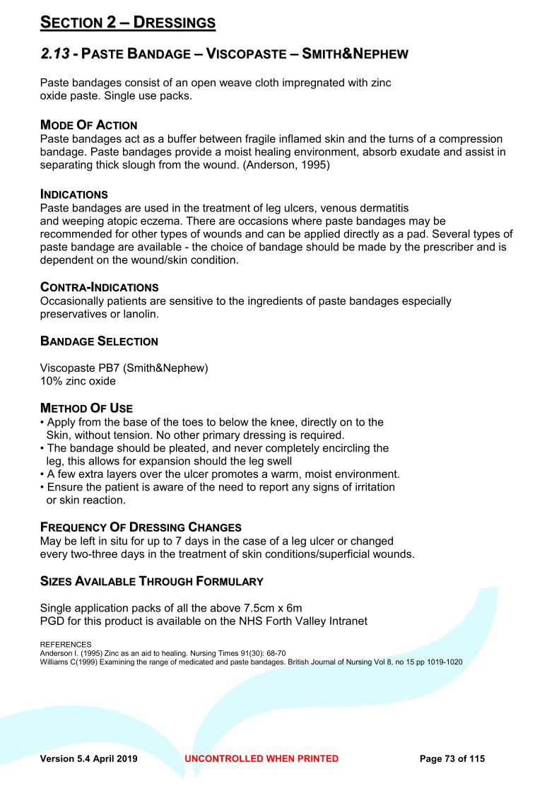
Version 5.4 April 2019 UNCONTROLLED WHEN PRINTED Page 73 of 115
SSEECCTTIIOONN 22 –– DDRREESSSSIINNGGSS 22..1133 -- PPAASSTTEE BBAANNDDAAGGEE –– VVIISSCCOOPPAASSTTEE –– SSMMIITTHH&&NNEEPPHHEEWW
Paste bandages consist of an open weave cloth impregnated with zinc oxide paste. Single use packs.
MMOODDEE OOFF AACCTTIIOONN Paste bandages act as a buffer between fragile inflamed skin and the turns of a compression bandage. Paste bandages provide a moist healing environment, absorb exudate and assist in separating thick slough from the wound. (Anderson, 1995)
IINNDDIICCAATTIIOONNSS Paste bandages are used in the treatment of leg ulcers, venous dermatitis and weeping atopic eczema. There are occasions where paste bandages may be recommended for other types of wounds and can be applied directly as a pad. Several types of paste bandage are available - the choice of bandage should be made by the prescriber and is dependent on the wound/skin condition. CCOONNTTRRAA--IINNDDIICCAATTIIOONNSS Occasionally patients are sensitive to the ingredients of paste bandages especially preservatives or lanolin. BBAANNDDAAGGEE SSEELLEECCTTIIOONN Viscopaste PB7 (Smith&Nephew) 10% zinc oxide MMEETTHHOODD OOFF UUSSEE • Apply from the base of the toes to below the knee, directly on to the Skin, without tension. No other primary dressing is required. • The bandage should be pleated, and never completely encircling the leg, this allows for expansion should the leg swell • A few extra layers over the ulcer promotes a warm, moist environment. • Ensure the patient is aware of the need to report any signs of irritation or skin reaction. FFRREEQQUUEENNCCYY OOFF DDRREESSSSIINNGG CCHHAANNGGEESS May be left in situ for up to 7 days in the case of a leg ulcer or changed every two-three days in the treatment of skin conditions/superficial wounds. SSIIZZEESS AAVVAAIILLAABBLLEE TTHHRROOUUGGHH FFOORRMMUULLAARRYY Single application packs of all the above 7.5cm x 6m PGD for this product is available on the NHS Forth Valley Intranet REFERENCES Anderson I. (1995) Zinc as an aid to healing. Nursing Times 91(30): 68-70 Williams C(1999) Examining the range of medicated and paste bandages. British Journal of Nursing Vol 8, no 15 pp 1019-1020
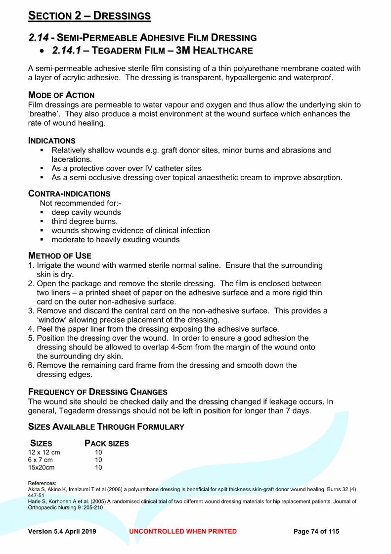
Version 5.4 April 2019 UNCONTROLLED WHEN PRINTED Page 74 of 115
SSEECCTTIIOONN 22 –– DDRREESSSSIINNGGSS
22..1144 -- SSEEMMII--PPEERRMMEEAABBLLEE AADDHHEESSIIVVEE FFIILLMM DDRREESSSSIINNGG •• 22..1144..11 –– TTEEGGAADDEERRMM FFIILLMM –– 33MM HHEEAALLTTHHCCAARREE
A semi-permeable adhesive sterile film consisting of a thin polyurethane membrane coated with a layer of acrylic adhesive. The dressing is transparent, hypoallergenic and waterproof. MMOODDEE OOFF AACCTTIIOONN Film dressings are permeable to water vapour and oxygen and thus allow the underlying skin to ‘breathe’. They also produce a moist environment at the wound surface which enhances the rate of wound healing. IINNDDIICCAATTIIOONNSS Relatively shallow wounds e.g. graft donor sites, minor burns and abrasions and
lacerations. As a protective cover over IV catheter sites As a semi occlusive dressing over topical anaesthetic cream to improve absorption.
CCOONNTTRRAA--IINNDDIICCAATTIIOONNSS Not recommended for:- deep cavity wounds third degree burns. wounds showing evidence of clinical infection moderate to heavily exuding wounds
MMEETTHHOODD OOFF UUSSEE 1. Irrigate the wound with warmed sterile normal saline. Ensure that the surrounding skin is dry. 2. Open the package and remove the sterile dressing. The film is enclosed between two liners – a printed sheet of paper on the adhesive surface and a more rigid thin card on the outer non-adhesive surface. 3. Remove and discard the central card on the non-adhesive surface. This provides a ‘window’ allowing precise placement of the dressing. 4. Peel the paper liner from the dressing exposing the adhesive surface. 5. Position the dressing over the wound. In order to ensure a good adhesion the dressing should be allowed to overlap 4-5cm from the margin of the wound onto the surrounding dry skin. 6. Remove the remaining card frame from the dressing and smooth down the dressing edges. FFRREEQQUUEENNCCYY OOFF DDRREESSSSIINNGG CCHHAANNGGEESS The wound site should be checked daily and the dressing changed if leakage occurs. In general, Tegaderm dressings should not be left in position for longer than 7 days.
SSIIZZEESS AAVVAAIILLAABBLLEE TTHHRROOUUGGHH FFOORRMMUULLAARRYY
SIZES PACK SIZES 12 x 12 cm 10 6 x 7 cm 10 15x20cm 10 References: Akita S, Akino K, Imaizumi T et al (2006) a polyurethane dressing is beneficial for split thickness skin-graft donor wound healing. Burns 32 (4) 447-51 Harle S, Korhonen A et al. (2005) A randomised clinical trial of two different wound dressing materials for hip replacement patients. Journal of Orthopaedic Nursing 9 :205-210
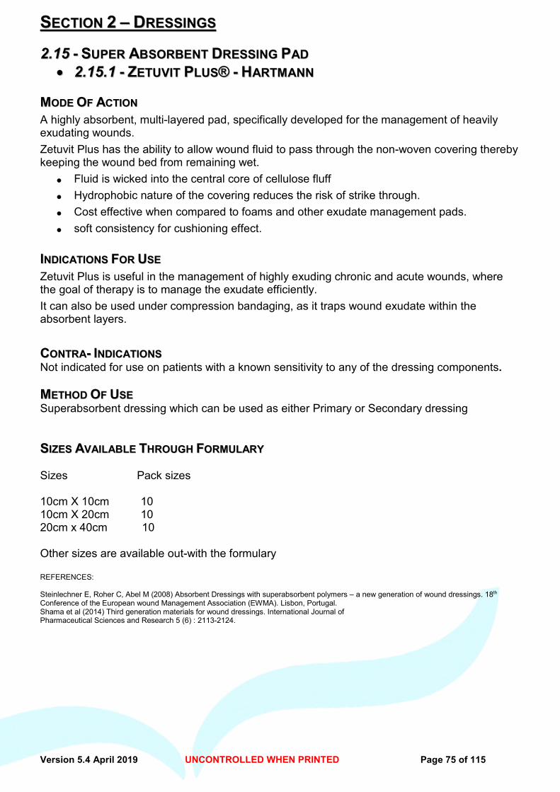
Version 5.4 April 2019 UNCONTROLLED WHEN PRINTED Page 75 of 115
SSEECCTTIIOONN 22 –– DDRREESSSSIINNGGSS 22..1155 -- SSUUPPEERR AABBSSOORRBBEENNTT DDRREESSSSIINNGG PPAADD
•• 22..1155..11 -- ZZEETTUUVVIITT PPLLUUSS®® -- HHAARRTTMMAANNNN
MMOODDEE OOFF AACCTTIIOONN A highly absorbent, multi-layered pad, specifically developed for the management of heavily exudating wounds. Zetuvit Plus has the ability to allow wound fluid to pass through the non-woven covering thereby keeping the wound bed from remaining wet.
• Fluid is wicked into the central core of cellulose fluff • Hydrophobic nature of the covering reduces the risk of strike through. • Cost effective when compared to foams and other exudate management pads. • soft consistency for cushioning effect.
IINNDDIICCAATTIIOONNSS FFOORR UUSSEE Zetuvit Plus is useful in the management of highly exuding chronic and acute wounds, where the goal of therapy is to manage the exudate efficiently. It can also be used under compression bandaging, as it traps wound exudate within the absorbent layers. CCOONNTTRRAA-- IINNDDIICCAATTIIOONNSS Not indicated for use on patients with a known sensitivity to any of the dressing components. MMEETTHHOODD OOFF UUSSEE Superabsorbent dressing which can be used as either Primary or Secondary dressing SSIIZZEESS AAVVAAIILLAABBLLEE TTHHRROOUUGGHH FFOORRMMUULLAARRYY Sizes Pack sizes 10cm X 10cm 10 10cm X 20cm 10 20cm x 40cm 10 Other sizes are available out-with the formulary REFERENCES: Steinlechner E, Roher C, Abel M (2008) Absorbent Dressings with superabsorbent polymers – a new generation of wound dressings. 18th Conference of the European wound Management Association (EWMA). Lisbon, Portugal. Shama et al (2014) Third generation materials for wound dressings. International Journal of Pharmaceutical Sciences and Research 5 (6) : 2113-2124.
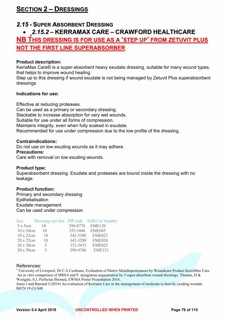
Version 5.4 April 2019 UNCONTROLLED WHEN PRINTED Page 76 of 115
SSEECCTTIIOONN 22 –– DDRREESSSSIINNGGSS 22..1155 -- SSUUPPEERR AABBSSOORRBBEENNTT DDRREESSSSIINNGG
•• 22..1155..22 –– KKEERRRRAAMMAAXX CCAARREE –– CCRRAAWWFFOORRDD HHEEAALLTTHHCCAARREE NNBB TTHHIISS DDRREESSSSIINNGG IISS FFOORR UUSSEE AASS AA ‘‘SSTTEEPP UUPP’’ FFRROOMM ZZEETTUUVVIITT PPLLUUSS NNOOTT TTHHEE FFIIRRSSTT LLIINNEE SSUUPPEERRAABBSSOORRBBEERR Product description: KerraMax Care® is a super-absorbent heavy exudate dressing, suitable for many wound types, that helps to improve wound healing. Step up to this dressing if wound exudate is not being managed by Zetuvit Plus superabsorbent dressings Indications for use: Effective at reducing proteases. Can be used as a primary or secondary dressing. Stackable to increase absorption for very wet wounds. Suitable for use under all forms of compression. Maintains integrity, even when fully soaked in exudate. Recommended for use under compression due to the low profile of the dressing. Contraindications: Do not use on low exuding wounds as it may adhere. Precautions: Care with removal on low exuding wounds. Product type: Superabsorbent dressing. Exudate and proteases are bound inside the dressing with no leakage. Product function: Primary and secondary dressing Epithelialisation Exudate management Can be used under compression Size Dressings per box PIP code NHS Cat Number 5 x 5cm 10 398-0778 EME120 10 x 10cm 10 352-3446 EME045 10 x 22cm 10 342-3308 EME023 20 x 22cm 10 342-3290 EME024 20 x 30cm 5 352-3453 EME025 20 x 50cm 5 398-0786 EME121 References: * University of Liverpool, Dr C.A.Cochrane, Evaluation of Matrix Metalloproteinases by Woundcare Product KerraMax Care. An in vitro comparison of MRSA and P. aeruginosa sequestration by 5 super-absorbent wound dressings. Thomas, H & Westgate, S.J. Perfectus Biomed, EWMA Poster Presentation 2016. Jones J and Barraud J (2014) An evaluation of Kerramx Care in the management of moderate to heavily exuding wounds. BJCN 19 (3) S48
Version 5.4 April 2019 UNCONTROLLED WHEN PRINTED Page 77 of 115
SSEECCTTIIOONN 33 -- SSPPEECCIIAALLIISSTT PPRROODDUUCCTTSS THIS SECTION IS FOR PRODUCTS WHICH ARE SPECIFIC TO A PARTICULAR CLINICAL AREA, OR FOR CHALLENGING WOUNDS. NOT FOR GENERAL WOUND MANANAGEMENT OR ROUTINE PRESCRIBING. TVN’S CAN OFFER SUPPORT AND GUIDANCE IF REQUIRED. PRODUCTS IN THIS SECTION DO NOT REQUIRE A NON FORMULARY REQUEST FORM
33..11 –– AANNTTIIMMIICCRROOBBIIAALL EENNZZYYMMEE AALLGGIINNOOGGEELL
•• 33..11..22 –– FFLLAAMMIINNAALL HHYYDDRROO // FFLLAAMMIINNAALL FFOORRTTEE –– FFLLEENN HHEEAALLTTHH UUKK
MMOODDEE OOFF AACCTTIIOONN An Alginate based gel containing two antimicrobial enzymes – glucose oxidase and lactoperoxidase, which provide antimicrobial activity without damaging healthy skin cells. Debrides and desloughs the wound, manages moisture balance and protects the wound edges. There is no limit to the length of time flaminal can be used to treat a wound. Can be used directly onto bone, tendon or in sinus tracts. IINNDDIICCAATTIIOONNSS Flaminal is indicated on a wide range of wounds where there is infection or critical colonisation present, or the potential for an acquired infection. It can be used as an alternative to silver and iodine dressings. Flaminal Hydro is indicated for wounds that are dry, necrotic or lightly exuding. Flaminal Forte is indicated for wounds that are moderately or highly exuding as it contains more alginate fibres
CCOONNTTRRAAIINNDDIICCAATTIIOONNSS Flaminal should not be used on patients with a known allergy to one of its components. Cannot be used on the eyelids or eye. MMEETTHHOODD OOFF UUSSEE Clean the wound ( if necessary) and apply flaminal to the whole wound bed applying a thick layer of at least 5mm, avoiding placing right up to wound edge. Cover with secondary dressing such as a film for necrotic wounds or atraumann and secondary foam dressing. A sterile syringe can be used to introduce flaminal to a sinus tract. May be left in place for up to 4 days, depending on the amount of exudate. Change when the gel structure has dispersed. After use, replace the cap as the product remains sterile until its expiry date. NN..BB.. If the exudates level is too low for flaminal forte, white alginate flakes will appear on the wound edges as the alginate is not being used. Do Not remove the flakes as they will prevent wound border maceration. Step down to Flaminal Hydro and as the moisture balance is restored the flakes will disappear.
SSIIZZEESS AAVVAAIILLAABBLLEE TTHHRROOUUGGHH FFOORRMMUULLAARRYY 15G TUBES – PACK SIZE 5 50G TUBE – SINGLE REFERENCES: White R. Flaminal: a novel approach to wound bioburden control.(2006) Wounds UK 2(3) De Smet K et al. (2009) Pre clinical evaluation of a new antimicrobial enzyme for the control of wound bio burden. Wounds, 21 (3): 65-73
Version 5.4 April 2019 UNCONTROLLED WHEN PRINTED Page 78 of 115
Section 3 - Specialist Products
33..22 -- DDEERRMMAATTOOLLOOGGYY SSEECCTTIIOONN
•• 33..22..11 –– SSIILLFFLLEEXX -- AADDVVAANNCCIISS MMEEDDIICCAALL
Silflex is an atraumatic , soft silicone wound contact layer designed to prevent secondary dressins adhering to fragile skin and delicate wound beds. MMOODDEE OOFF AACCTTIIOONN Silflex creates a low adherence to healthy tissue surrounding a wound and not wound bed, to allow healing of blisters and other superficial wounds and skin tears. The perforations allow passage of exudate. IINNDDIICCAATTIIOONNSS It is particularly useful in large blistered areas in dermatological conditions such as bullous pemphgoid . To drss skin tears. It can also be used to line wound beds before application of TNP. MMEETTHHOODD OOFF UUSSEE Ensure surrounding periwound skin clean and dry. Apply silflex directly onto the affected area. Dress wound with appropriate secondary dressings eg hydrogel, aquacel etc depending on wound presentation. Top tip Wet your hands/gloves before application so that the Silflex doesn't stick when applying. FFRREEQQUUEENNCCYY Can be left in place for up to 14 days depending on the wound condition, however it is recommended that a 180 degree rotation is undertaken to prevent over granulation. Steroid creams, secondary dressings can be changed more frequently without disturbing the silflex dressing. CCOONNTTRRAA-- IINNDDIICCAATTIIOONNSS Known sensitivity to the dressing. SSIIZZEESS AAVVAAIILLAABBLLEE TTHHRROOUUGGHH FFOORRMMUULLAARRYY Sizes Pack sizes product code 5cmx7cm 10 CR3922 8cm x 10cm 10 CR3923 12cm x 15cm 10 CR3924 20cmx30cm 10 CR3925 35cm x 60cm 5 – Drug tariff only CR4006 REFERENCES Stephen Haynes J, Greenwood M (2014) Wound Care Today 1(1): 58–64 Edwards, J (2009) The use of Silflex in burn wound management. British Journal of Community Nursing. 14 (9) Kumal R. The use of a soft silicone wound contact dressing (Silflex) under topical negative pressure. Kings College Hospital, London.
Version 5.4 April 2019 UNCONTROLLED WHEN PRINTED Page 79 of 115
SSEECCTTIIOONN 33 -- SSPPEECCIIAALLIISSTT PPRROODDUUCCTTSS
33..22 -- DDEERRMMAATTOOLLOOGGYY SSEECCTTIIOONN •• 33..22..22 -- UURRGGOOTTUULL AABBSSOORRBB BBOORRDDEERR –– UURRGGOO MMEEDDIICCAALL
Urgotul absorb border is an absorbent foam dressing with a silicone adhesive border and shower proof backing in one foam self adhesive dressing with a soft silicone wound contact layer which absorbs and retains exudates and maintains a moist wound environment. MMOODDEE OOFF AACCTTIIOONN The dressing combines an absorbent polyurethane foam pad with an absorbent layer. It is also based on the exclusive and patened TLC (technology Lipido-colloid) Healing Matrix. This matrix forms a gel on contact with wound exudates. Encourages new tissue formation, through fibroblast proliferation. Reduces wound bed trauma, bleeding and pain during dressing changes. Note -This dressing should only be used in specific situations and not where a simple foam dressing would suffice
IINNDDIICCAATTIIOONNSS Low to moderate exuding wounds Leg and pressure ulcers Traumatic wounds resulting in skin loss Under compression bandaging Wounds with compromised or fragile surrounding skin CCOONNTTRRAA –– IINNDDIICCAATTIIOONNSS NONE KNOWN Note - Should not be used on heavily exuding wounds Dressing pad should not be cut as it will affect the absorbent layer. The silicone border can be cut if required
MMEETTHHOODD OOFF UUSSEE IF CLINICALLY INDICATED CLEANSE WOUND AND DRY SURROUNDING SKIN Remove the protective wings from the dressing and apply the soft adherent side of Urgotul Absorb Border to the wound. The adhesive silicone border should be at least 1 cm away from the wound. Gently smooth down the dressing over the wound, minimising creasing where possible. For removal gently lift one corner and peel back dressing FFRREEQQUUEENNCCYY OOFF DDRREESSSSIINNGG CCHHAANNGGEE Maybe left in place for several days on clean and uninfected wounds, for up to 7 days Dressing should be changed when exudate is present at the pad edges.
SSIIZZEESS AAVVAAIILLAABBLLEE TTHHRROOUUGGHH FFOORRMMUULLAARRYY Dressing Sizes Pad sizes Pack sizes Product code 6.5 x 10cm 3cm x 6.5cm 10 551431 8cm x 8cm 4.5cm x 4.5cm 10 550866 8cmx 15cm 3.4cm x 10.4cm 10 551432 10cm x 10cm 5.9cm x 5.9cm 10 551014 10cm x 25cm 4.6cm x 19.6cm 10 551433 13cm x 13cm 8.4cm x 8.3cm 10 550867 15cm x 20cm 9.7cm x 14.7cm 10 550868 REFERENCES : White, R., Cowan, T., Glover, D. Supporting evidence-based practice: a clinicalreview of TLC healing matrix (2nd edition). MA Healthcare Ltd, London, 2015.
Version 5.4 April 2019 UNCONTROLLED WHEN PRINTED Page 80 of 115
Specialist Foam
•• 33..22..33-- AAQQUUAACCEELL FFOOAAMM AADDHHEESSIIVVEE DDRREESSSSIINNGG -- CCOONNVVAATTEECC This dressing is a combination of an Aquacel contact layer with a gentle silicone border. It comprises a protective top layer, an upper soft absorbent foam pad and an integral hydrofiber wound contact layer with a silicone border. The dressing is water/shower proof. MMOODDEE OOFF AACCTTIIOONN It can be used alone to manage shallow exuding wounds and for wounds of any depth used in conjunction with other primary dressings. It provides a moist wound environment, locks in wound exudates, microcontours to the wound bed and the silicone layer helps to reduce pain on removal of dressing. The aquacel layer absorbs wound fluid and creates a soft gel, maintaining a moist wound environment. Locks in exudates through vertically wicking, reducing the risk of maceration. IINNDDIICCAATTIIOONNSS Aquacel foam is suitable for a wide range of acute and chronic wounds producing high exudate, regardless of the tissue within the wound bed. Suitable for all exuding wounds. AQUACEL® Foam dressing is a more cost effective alternative to the combination of AQUACEL® Extra™ and an alternative foam dressing Aquacel foam dressing should not be used as an alternative to a simple foam dressing when this will suffice. CCOONNTTRRAA –– IINNDDIICCAATTIIOONNSS Not for use if patient is allergic to any of the ingredients. Not for use on dehydrated or dry necrotic wounds FFRREEQQUUEENNCCYY OOFF DDRREESSSSIINNGG CCHHAANNGGEE Maximum recommended wear time is 7 days but should be changed as determined by the wound exudate. NB if dressing requires changing every day then an alternative dressing requires to be considered. SSIIZZEESS AAVVAAIILLAABBLLEE TTHHRROOUUGGHH FFOORRMMUULLAARRYY Size Pack size Product Code 8x 8cm 10 420804 10 x10cm 10 420680 12.5x12.5cm 10 420619 10cmx20cm 10 421150 References Bishop, S*, Helen S,; David P et al (2012 ) A Comparison of the in vitroBio-Physical PerformanceCharacteristics of SiliconeFoam Dressings Used in Wound Management Global clinical Case Study compendium (2012) clinical and quality of life benefits from next generation foam dressings – aquacel Foam
Version 5.4 April 2019 UNCONTROLLED WHEN PRINTED Page 81 of 115
SSEECCTTIIOONN 33 -- SSPPEECCIIAALLIISSTT PPRROODDUUCCTTSS
33..44 -- TTHHEE FFOOUURR LLAAYYEERR BBAANNDDAAGGEE SSYYSSTTEEMM MMOODDEE OOFF AACCTTIIOONN Patients with venous leg ulceration often have dry and itchy skin with dermatitis present in some cases. These patients may also become sensitive to elements of their treatment at any time. Due to this, products which commonly cause skin sensitivities such as latex are often avoided. Choose the simplest dressing possible for the ulcer and avoid mixing dressings, lotions and creams, as this is likely to increase the likelihood of developing sensitivities. IINNDDIICCAATTIIOONNSS The four layer bandage system is used for treatment of venous leg ulcers, venous dermatitis, oedema and weeping cellulites. A full assessment should be made, taking into account medical history, vascular history and recording the ankle brachial Pressure Index (ABPI) to exclude arterial disease. CCOONNTTRRAA--IINNDDIICCAATTIIOONNSS The four layer bandage system should only be applied by nurses competent in this procedure following an assessment of the patient’s arterial supply (ABPI) or under the guidance of Consultant Surgeon or Community Tissue Viability Service/ Wound Management Specialist Nurses. MMEETTHHOODD OOFF UUSSEE The primary dressing may be held in place with a layer of cotton stockinette, although this is not essential. Layer 1:- Wool padding – ROBINSONS ULTRA SOFT - COMMUNITY URGO K SOFT- ACUTE · Protects bony prominences and tendons in the foot / ankle area · Prevents damage to vulnerable skin · Pads out narrow gaiter area · Absorbs exudate · Promotes patient comfort, by providing cushion over ulcer Application is from base of toes to knee, in a spiral, with 50% overlap. Extra layers can be added to the gaiter area if narrow. Apply minimum to the underside of the foot to allow ease of application of footwear. Layer 2:- 10cm crepe bandage – ROBINSONS ULTRA LITE - COMMUNITY HOSPICREPE CREPE BANDAGE- ACUTE · Doesn’t provide any significant compression · Smooths out wool layer · Flat surface to apply compression layers Application is from base of toes to knee, in a spiral, with 50% overlap. Cut off any excess bandage and secure with tape. Layer 3:- 10cm elastic conformable compression bandage – ROBINSONS ULTRA PLUS · Provides compression of 14 – 27 mmHg at the ankle
Version 5.4 April 2019 UNCONTROLLED WHEN PRINTED Page 82 of 115
SSEECCTTIIOONN 33 -- SSPPEECCIIAALLIISSTT PPRROODDUUCCTTSS
33..44 ((CCOONNTT)) -- TTHHEE FFOOUURR LLAAYYEERR BBAANNDDAAGGEE SSYYSSTTEEMM Application 50% extension in figure of eight, from base of toes to below knee. This layer is often referred to as “modified” when it is applied in a spiral to reduce the compression a little. Cut off any excess bandage and secure with tape. Layer 4:- 10cm cohesive bandage – ROBINSONS ULTRA FAST · Light compression 14 – 27 mmHg · Adheres to itself · Adds durability to the system · Prevents slippage Application is from base of toes to knee, in a spiral, with 50% overlap 50% extension. Cut off any excess bandage and secure with tape. Ensure the bandage system is comfortable and that the patient is able to move their ankle freely. FFRREEQQUUEENNCCYY OOFF DDRREESSSSIINNGG CCHHAANNGGEESS This multi-layer bandage system may be worn for up to 7 days. PPAATTIIEENNTT IINNFFOORRMMAATTIIOONN Patient should be given a telephone number to contact if concerned about bandaging. Also advised to remove top layer (cohesive bandage) if feels bandage is too tight or uncomfortable and call Nurse if this does not help, the next layer could be removed. REFERENCES: 120 Management of Chronic Venous Leg Ulcers (2010) – a national clinical guideline. Scottish Intercollegiate Guidelines Network (SIGN).
Version 5.4 April 2019 UNCONTROLLED WHEN PRINTED Page 83 of 115
33..44..11 TTUUBBUULLAARR BBAANNDDAAGGEE –– CCLLIINNIIFFAASSTT TTUUBBUULLAARR BBAANNDDAAGGEESS ––
CCLLIINNIISSUUPPPPLLIIEESS DDEESSCCRRIIPPTTIIOONN
• Clinifast Tubular Bandages are specially designed from a light breathable fabric, allowing complete freedom of movement. They are used for dressing retention, wet and dry wrapping and for patch wrapping atopic eczema. They are simple and easy to apply, and come in five colour-codes that are available in different lengths for community and hospital use, with a double stripe for easy identification. They are also cost-effective, as they are washable and re-usable
INDICATIONS • As a protective layer over Co-hesive bandage to prevent adherence to bed clothes and
clothing. For dressing retention on various parts of the body including legs, arms, head, wet and dry wrapping or patch wrapping for atopic eczema
• PREPARATIONS AVAILABLE Tubular bandages
SIZES AVAILABLE
Small limb (red line) - 3.5cm x 1m; Medium limb (green line) - 5cm x 1m, 5cm x 3m, 5cm x 5m; Large limb (blue line) - 7.5cm x 1m, 7.5cm x 3m, 7.5cm x 5m; Child trunk (yellow line) - 10.75cm x 1m, 10.75cm x 3m, 10.75cm x 5m; Adult trunk (beige line) - 17.5cm x 1m
Version 5.4 April 2019 UNCONTROLLED WHEN PRINTED Page 84 of 115
Section 3 - Specialist Products
33..55 -- LLAARRVVAAEE TTHHEERRAAPPYY May be referred to by different names, either as, Larval Debridement Therapy, Biosurgery or Maggot Therapy. PPRROODDUUCCTT DDEESSCCRRIIPPTTIIOONN Sterile larvae (maggots) of the common greenbottle blowfly are used for cleansing/debriding of devitalized tissue and slough from wounds. Its use is advocated in chronic infected wounds due to the rising challenges posed by multi-resistant bacteria. (Thomas S 2010; Fleischmann W et al 2004) MMOODDEE OOFF AACCTTIIOONN Larvae produce powerful proteolytic enzymes which breakdown sloughy and soft necrotic tissue, which they ingest as a source of nutrient. When first applied to a wound they are only 2-3mm long but under favourable conditions they increase in size rapidly, reaching 8-10mm when fully grown. It is claimed that they combat odour and infection by ingesting and killing bacteria in the wound (Thomas. et.al. 1994). The treatment must be fully explained to the patient, verbal consent given and documented in medical/nursing records. A patient information leaflet should also be given. Availability as free range (roaming) or Bio-bags. Amounts depending on size of wound area. 100 Larvae (4.0 x 5.0cms) 200 Larvae (5.0 x 6.0cms) 300 Larvae (6.0 x 12.0cms) Delivery – No service on a sunday, orders before 1400 hrs are delivered the next day, same day service is available at increased cost.
IINNDDIICCAATTIIOONNSS Can be used in the treatment of most types of sloughy, infected wounds, including leg ulcers, (both venous and arterial), pressure ulcers, burns and ulcerated areas on the feet of diabetics. CCOONNTTRRAA--IINNDDIICCAATTIIOONNSS Should not be applied to wounds that have a tendency to bleed easily, or be introduced into wounds that communicate with the body cavity or any internal organs. In addition it is recommended that they should not be applied close to any large blood vessels, they are not recommended for wounds covered in thick adherent necrotic material. WWAARRNNIINNGGSS AANNDD PPRREECCAAUUTTIIOONNSS On rare occasions it has been reported that the use of Larvae have caused a wound to bleed, for this reason it is recommended that the dressing is inspected daily, if bleeding has occurred the larvae should be removed and the wound re-assessed. Increased odour and exudates are expected in the initial stages of treatment. If exudates production is excessive, the secondary dressing may be changed daily whilst leaving the primary dressing undisturbed. Occlusive dressings or film dressings should not be used as these will cause the larvae to suffocate, for this reason care must also be taken to ensure that the wound area is free from pressure.
Version 5.4 April 2019 UNCONTROLLED WHEN PRINTED Page 85 of 115
Wounds must be cleansed with tepid tap water prior to the application of larvae as previous wound preparations including some hydrogels are known to adversely react with the larvae and therefore it is recommended that such preparations are removed twenty four hours prior to commencement. PPRROOCCEEDDUURREE FFOORR OOBBTTAAIINNIINNGG MMAAGGGGOOTTSS In the Acute Sector, TVN’s can help assess the wound for suitability of larvae therapy. If the medical staff and Patient are in agreement, larvae are ordered from the pharmacy department, progress can be monitored by the Tissue Viability Service and further advice given as required. Maximum recommended duration of treatment is 4 days (biofoam larvae), subsequent applications can then be applied depending on the status of the wound. In the community the Tissue Viability Nurses assess the wound for suitability, then liaise with the GP/Patient/District Nurse, larvae are then ordered by prescription In both instances the wound area is measured to estimate how many larvae are required. Disposal within the Acute Hospital and clinic areas, following removal from the wound, larvae should be doubled bagged and disposed of as per the clinical waste policy procedure manual. Within the Community, larvae should be double bagged and disposed of as per domestic waste. IINNFFOORRMMAATTIIOONN LLEEAAFFLLEETTSS should be distributed to patients/families. It is anticipated that staff education will be provided if necessary prior to commencement of treatment. Reference Thomas S, Andrews AM, Hay NP, Bourgoise S (1999) The Anti-microbial activity of maggot secretions: results of a preliminary study Journal of Tissue Viability 9 (4): 127-132 Thomas S, (2010) Maggot Therapy; Surgical Dressings and Wound Management, 563-632. Fleischmann W et al (2004) Maggot therapy. A Handbook of Maggot Assisted Wound Healing. Thieme . ISBN 3-13-136811 – X (GTV) Pagnamenta F (2013) Using Maggots to clean wounds – a clinical review. Wound Essentials Vol 8 No 1
Version 5.4 April 2019 UNCONTROLLED WHEN PRINTED Page 86 of 115
SSEECCTTIIOONN 33 -- SSPPEECCIIAALLIISSTT PPRROODDUUCCTTSS
33..55..11 -- LLAARRVVAAEE TTHHEERRAAPPYY -- PPAATTIIEENNTT IINNFFOORRMMAATTIIOONN LLEEAAFFLLEETT May be referred to by different names, such as Larvae Therapy, Biotherapy or Maggot Therapy. Sterile larvae (maggots) of the common greenbottle fly can be used for cleansing infected or dirty wounds. They also help to combat odour from the wound. They are placed onto your wound and kept in place with layers of dressings. The nurses in the ward or Community nurses will change the outer dressings every day and monitor progress of larvae treatment. The larvae produce enzymes which breakdown the yellow material (slough) in your wound. When first applied to the wound they are only 2-3mm long, but increase in size, reaching 8-10mm after 2-5 days, they are then removed. You should not be aware of the larvae on the wound, although occasionally patients have reported a tingling sensation. If the pain or discomfort does increase, ask your doctor or nurse for advice.
TTRREEAATTMMEENNTT OOBBJJEECCTTIIVVEESS Combat odour from the wound Breakdown yellow matter and dead tissue Promote wound healing IINNDDIICCAATTIIOONNSS FFOORR UUSSEE Can be used in the treatment of most types of sloughy, infected wounds including leg ulcers, pressure ulcers, burns and ulcerated areas on the feet. On rare occasions it has been reported that the use of Larvae have caused a wound to bleed, for this reason it is recommended that the dressing is inspected daily, this will be done by nurses in the ward or the Community nurse if you are at home. If bleeding has occurred the nurse will remove the larvae and the re-dress the wound. Increased odour and soakage from the wound are expected in the initial stages of treatment. If soakage from the wound is excessive, the outer dressing may be changed daily whilst leaving the primary dressing undisturbed. TTHHIINNGGSS YYOOUU CCAANN DDOO TTOO HHEELLPP Do not: • sit too close to a fire, larvae need moisture to survive • put direct pressure on the wound, if possible, as this could damage the larvae • get your dressings wet in the bath or shower, the larvae may drown or have difficulty breathing • panic if any larvae escape from your dressing, simply place them in a rubbish bag and dispose of in your rubbish bin. If you have any questions about your treatment ask your doctor or nurse for advice
Version 5.4 April 2019 UNCONTROLLED WHEN PRINTED Page 87 of 115
SSEECCTTIIOONN 33 -- SSPPEECCIIAALLIISSTT FFOORRMMUULLAARRYY 33..66 TTOOPPIICCAALL SSTTEERROOIIDD PPRREEPPAARRAATTIIOONNSS
33..66..11 DDEERRMMOOVVAATTEE -- CCLLOOBBEETTAASSOOLL PPRROOPPIIOONNAATTEE A PGD for this product is available on the NHS Forth Valley Intranet for Tissue Viability/Dermatology specialist nurses. Otherwise this product must be prescribed by a doctor or independent prescriber. A VERY POTENT topical corticosteroid preparation, also known as CLOBETASOL PROPIONATE. INDICATIONS Treatment of choice in patients with severe resistant inflammatory skin disorders unresponsive to less potent corticosteroids. CONTRA-INDICATIONS Should not be used when there is a known sensitivity to clobetasol propionate. or other corticosteroids. Long term use should be avoided, Adrenal suppression can occur. Atrophic changes may occur in the skin with prolonged use. METHOD OF USE Ensure the area to be treated is washed thoroughly with warm water in between applications. A thin layer to be applied to the area of skin as prescribed ( refer to finger tip unit chart for exact dose). Once applied occlude the area with Atrauman dressing for a more concentrated penetration. Wait 5-10 minutes before applying emollients to the area. The emollient should be smoothed gently into the skin following the lie of the hair to prevent folliculitis. NOTE: Emollients dilute the efficacy of the steroid. In acutely inflamed conditions omit the emollient for the first few days. Ointments are preferable to creams in inflammatory skin conditions associated with dry plaques. They have a deeper, more prolonged effect and increase the penetration of the steroid. They are also less likely to cause irritation as they do not contain preservatives. FREQUENCY OF USE Initial application should be 1-2 times daily for 7 days and a reducing regime implemented thereafter to reduce the risk of a ‘rebound’ effect. APPLICATION Refer to Finger Tip Unit chart for accurate dose application. PRESENTATION Available as cream and ointment. 30g and 100g.
References: British journal of Community Nursing (2011) 16, (7), pg 329 Cameron.J. (2007) Dermatological changes associated with venous leg ulcers. 2. pg 60-66
Version 5.4 April 2019 UNCONTROLLED WHEN PRINTED Page 88 of 115
SSEECCTTIIOONN 33 -- SSPPEECCIIAALLIISSTT FFOORRMMUULLAARRYY
33..66 –– TTOOPPIICCAALL SSTTEERROOIIDD PPRREEPPAARRAATTIIOONNSS •• 33..66..22 -- EELLOOCCOONN OOIINNTTMMEENNTT -- MMOOMMEETTAASSOONNEE FFUURROOAATTEE 00..11%%..
A PGD for this product is available on the NHS Forth Valley Intranet for Tissue Viability/Dermatology specialist nurses. Otherwise this product must be prescribed by a doctor or independent prescriber. A POTENT topical corticosteroid ointment also known as MOMETASONE FUROATE 0.1%. INDICATIONS Treatment of choice in patients with leg ulcers, with associated moderate to severe contact sensitivity or stasis dermatitis. Applied to the surface of the skin to reduce the redness and itchiness caused by certain skin problems, including psoriasis and dermatitis. If skin infection present Fucibet cream should be used in place of Elocon. CONTRA-INDICATIONS Should not be used when there is a known sensitivity to mometasone furoate or other corticosteroids. Long term use should be avoided, Adrenal suppression can occur. Atrophic changes may occur in the skin in prolonged use. Not recommended for children under the age of 2. METHOD OF USE A thin layer to be applied to the affected area of skin,(refer to Finger tip unit chart for exact dose). Once applied occlude with Atrauman dressing for more concentrated penetration. Wait 5-10mins before applying emollients to the area. NOTE: Emollients dilute the efficacy of the steroid. Ointments are preferable to creams as they have a deeper, more prolonged emollient effect and increase the penetration of steroid. They are also less likely to cause irritation as they do not contain preservatives FREQUENCY OF USE Initial application should be daily for 7 days and a reducing regime implemented thereafter to minimise the risk of a ‘rebound’ effect. Twice a week when compression bandages are being used, increased to three times a week if no improvement noted. APPLICATION Refer to finger Tip Unit chart for accurate dose application. PRESENTATION Available as cream and ointment 30g and 100g tubes References: MeReC Bulletin,NPC, Topical Corticosteroids in General Practice (1999) (10) 6 Pg 21-24 BNF 62 September (2011)pg 728 WWW.Dermnetnz.org
Version 5.4 April 2019 UNCONTROLLED WHEN PRINTED Page 89 of 115
SSEECCTTIIOONN 33 -- SSPPEECCIIAALLIISSTT FFOORRMMUULLAARRYY 3.6.3 FLUDROXYCORTIDE CREAM and OINTMENT- 0.0125% w/w- previously known as Haelan. Product description: A moderately potent corticosteroid cream or ointment for topical application. Creams are suitable for moist or weeping lesions whereas ointments are generally chosen for dry or scaly lesions. Indications: Adults and children: Eczema and dermatitis of all types including childhood and adult atopic eczema, photodermatitis, primary irritant and allergic dermatitis, lichen planus, lichen simplex, prurigo nodularis, discoid lupus erythematosus, necrobiosis lipoidica, pretibial myxoedema and erthroderma. Overgranulated tissue (unlicensed). Application and frequency of use: Apply thinly to the affected area 1-2 times daily. In Overgranulation an anecdotal reducing regime of daily application is recommended over a period of approximately 4 weeks. Contra-indications: Tuberculosis of the skin,facial rosacea, acne vulgaris, perioral dermatitis, perianal and genital pruritis, dermatoses in infancy including eczema, dermatitis napkin eruption, bacterial (impetigo), viral hypersensitivity to any components in the preparation. Potent corticosteroids are contra indicated in wide spread plaque psoriasis. Special warnings and Precautions for use: Not intended for Opthalmic use. Long term continuous therapy should be avoided in all patients irrespective of age. Please also refer to the BNF and TYPHARM GROUP for exhaustive list. Side Effects: The following local adverse reactions are reported infrequently with topical corticosteroids but may occur more frequently with the use of occlusive dressings. These reactions are listed in an approximate decreasing order of occurrence; burning, itching, irritation, dryness, folliculitis, hypertrichosis, acne-form eruptions, hypopigmentation, perioral dermatitis, allergic contact dermatitis, maceration of the skin, secondary infection, skin atrophy, miliaria, striae and thinning and dilatations of superficial blood vessels producing telangiectasia. Prolonged use of large doses to extensive areas can result in sufficient systemic absorption to produce generalised manifestations of steroid toxicity and may result in depression of HPA function on discontinuing treatment. Manifestations of Cushing’s syndrome, hyperglycaemia and glycosuria have occurred in some patients. Manifestations of adrenal suppression in children include linear growth retardation, delayed weight gain, low plasma cortisol levels and absence of response to ACTH stimulation. Intracranial hypertension including bulging fontanelles, headaches and bilateral papilloedema have also been reported in children receiving topical corticosteroids. Infected skin lesions, viral, bacterial or fungal may be substantially exacerbated by topical steroid therapy. Wound healing is significantly retarded.
Version 5.4 April 2019 UNCONTROLLED WHEN PRINTED Page 90 of 115
Hypersensitivity reactions may occur. Presentation: Available as a cream or ointment – 60g aluminium tube with screw cap. 33..66..44 -- FFLLUUDDRROOXXYYCCOORRTTIIDDEE TTAAPPEE ((HHAAEELLAANN TTAAPPEE)) A MODERATELY POTENT occlusive topical corticosteroid preparation, impregnated with 4 micrograms fludroxycortide per square centimetre. INDICATIONS Occlusive topical steroid. Adjunctive therapy for chronic, localised, recalcitrant dermatoses that may respond to topical corticosteroids. CONTRA INDICATIONS Chicken pox; vaccinia; tuberculosis of the skin; hypersensitivity to any components of the tape; facial rosacea; acne vulgaris; perioral dermatitis; perianal and genital pruritus; dermatoses in infancy; dermatitic napkin eruption, bacterial, viral and fungal infections. METHOD OF USE Haelan Tape should be applied to clean, dry skin which is free of hair. The tape need only remain in place for 12 out of 24 hours, but clinical practice has shown that 24-hour use is more advantageous.Corners should be rounded off to prevent excess pressure and rolling of the edges. The tape is cut 5mm larger than the treatment area (it is very easy to prepare a variety of dressing shapes including ‘key hole’ techniques). Remove the paper backing and apply the adhesive side of the tape to the skin. Stroke the back of the tape gently from the middle outwards to ensure contact between the tape and the skin, avoiding excessive tension as this could result in skin stripping on removal. If longer strips are to be applied, the lining paper should be removed progressively. If irritation or infection develops, remove tape and consult a physician. FREQUENCY OF USE Long-term continuous therapy should be avoided in all patients irrespective of age. Application under occlusion should be restricted to dermatoses in very limited areas. If used on the face, courses should be limited to five days and occlusion should not be used. This is not a problem when treating overgranulation as treatment lasts a maximum of seven days. PRESENTATION In a cardboard dispensing box 7.5cmx20cm or 7.5x50cm SIDE EFFECTS: SEE ABOVE UNDER HAELAN CREAM/OINTMENT References: Johnson S (2007) Haelan Tape for the treatment of overgranulation tissue. Wounds UK November 2016 Volume 12 Issue 4
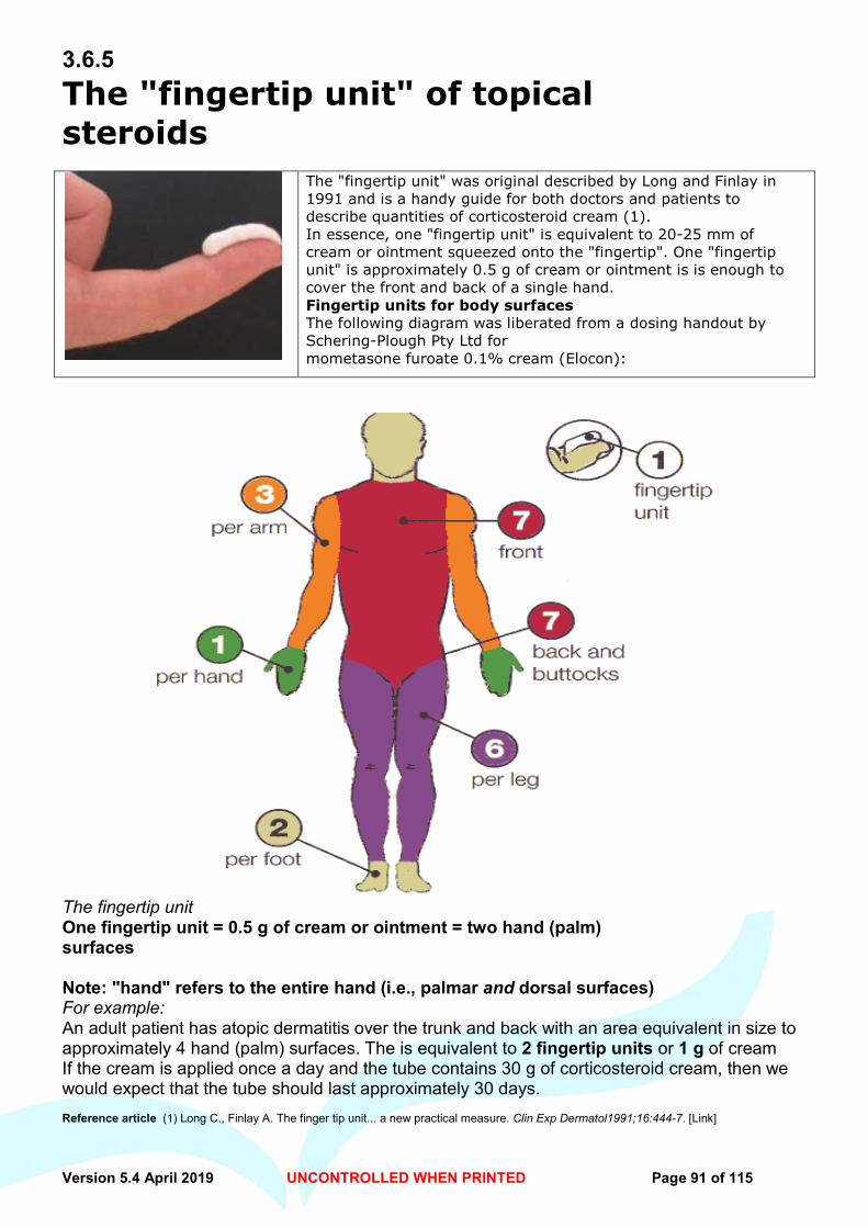
Version 5.4 April 2019 UNCONTROLLED WHEN PRINTED Page 91 of 115
3.6.5
The "fingertip unit" of topical steroids
The "fingertip unit" was original described by Long and Finlay in 1991 and is a handy guide for both doctors and patients to describe quantities of corticosteroid cream (1). In essence, one "fingertip unit" is equivalent to 20-25 mm of cream or ointment squeezed onto the "fingertip". One "fingertip unit" is approximately 0.5 g of cream or ointment is is enough to cover the front and back of a single hand. Fingertip units for body surfaces The following diagram was liberated from a dosing handout by Schering-Plough Pty Ltd for mometasone furoate 0.1% cream (Elocon):
The fingertip unit One fingertip unit = 0.5 g of cream or ointment = two hand (palm) surfaces Note: "hand" refers to the entire hand (i.e., palmar and dorsal surfaces) For example: An adult patient has atopic dermatitis over the trunk and back with an area equivalent in size to approximately 4 hand (palm) surfaces. The is equivalent to 2 fingertip units or 1 g of cream If the cream is applied once a day and the tube contains 30 g of corticosteroid cream, then we would expect that the tube should last approximately 30 days. Reference article (1) Long C., Finlay A. The finger tip unit... a new practical measure. Clin Exp Dermatol1991;16:444-7. [Link]
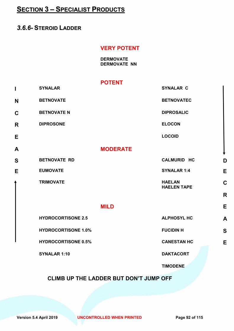
Version 5.4 April 2019 UNCONTROLLED WHEN PRINTED Page 92 of 115
SSEECCTTIIOONN 33 –– SSPPEECCIIAALLIISSTT PPRROODDUUCCTTSS
33..66..66-- SSTTEERROOIIDD LLAADDDDEERR
VERY POTENT
DERMOVATE DERMOVATE NN
POTENT
I SYNALAR SYNALAR C
N BETNOVATE BETNOVATEC
C BETNOVATE N DIPROSALIC
R DIPROSONE ELOCON
E LOCOID
A MODERATE
S BETNOVATE RD CALMURID HC D
E EUMOVATE SYNALAR 1:4 E
TRIMOVATE HAELAN HAELEN TAPE
C
R
MILD E
HYDROCORTISONE 2.5 ALPHOSYL HC A
HYDROCORTISONE 1.0% FUCIDIN H S
HYDROCORTISONE 0.5% CANESTAN HC E
SYNALAR 1:10 DAKTACORT
TIMODENE
CLIMB UP THE LADDER BUT DON’T JUMP OFF
Version 5.4 April 2019 UNCONTROLLED WHEN PRINTED Page 93 of 115
Section 3 - Specialist Products
33..77-- TTOOPPIICCAALL NNEEGGAATTIIVVEE PPRREESSSSUURREE
Topical negative pressure (TNP) is a specialised system of wound management. Tissue Viability Service must be alerted to the fact that a patient is commencing this therapy. If required the Tissue Viability Nurses can be contacted to assess the suitability of the patient and wound, prior to ordering or applying this therapy or throughout the course of treatment including on discharge home. MMOODDEE OOFF AACCTTIIOONN There is limited trial based evidence to support the use of TNP in clinical practice, but anecdotal evidence and case studies from clinical papers, suggest it is an effective system that applies controlled, localised sub-atmospheric pressure to the wound site to promote wound healing. This treatment should only be used by staff trained and competent in its use. Objectives of TNP treatment are: · Removes excess exudates whilst maintaining a moist wound healing environment. · Reduces bacterial count · Promotes granulation · Draws the wound margins closer together · Increases blood flow to the wound May be portable or static. Consists of an internal pump with easily adjustable controls to meet wound specific needs. The canister filter system is easily removable. The treatment must be fully explained to the patient, verbal consent given and documented in medical/nursing records. An information leaflet should also be offered to patient and their family/carers..(Overleaf Section 3.5.1) IINNDDIICCAATTIIOONNSS Acute and traumatic wounds Sub-acute wounds (i.e. Dehisced incisions) Chronic wounds, diabetic or pressure ulcers Meshed grafts (pre and post) Skin flaps CCOONNTTRRAA--IINNDDIICCAATTIIOONNSS Necrotic tissue with eschar present Osteomyelitis (untreated) Malignancy in the wound Active bleeding/ Difficult wound haemostasis Patients on anticoagulants Care must be taken with respect to weakened, irradiated or sutured blood vessels or organs. TNP therapy must not be used (unless indicated by surgical consultants) over, or in close proximity to exposed blood vessels or organs or fistulae to organs or body cavities. Manufacturers guidelines should also be consulted prior to use.
Version 5.4 April 2019 UNCONTROLLED WHEN PRINTED Page 94 of 115
SSEECCTTIIOONN 33 -- SSPPEECCIIAALLIISSTT PPRROODDUUCCTTSS
33..77 ((CCOONNTT))-- TTOOPPIICCAALL NNEEGGAATTIIVVEE PPRREESSSSUURREE Use of TNP therapy should only be used in conjunction with Tissue Viability Service or on the recommendation of medical staff. Costs can be expensive but vary between manufacturers. TNP should only be used by nurses competent in its use. To order TNP the unit administrator /community nurse completes order form (appendix 4) and an order placed with CSD for the pump and consumables. Delivery is normally within 24-48 hours and made directly to the ward or health centre. When treatment is complete CSD and TVS must be contacted immediately to uplift the pump and avoid further charges, this is done by the ward, or by the unit administrator. If TNP therapy is required within the community please contact the Tissue Viability Service for further information. If patient being discharged home from hospital with Vac therapy – please follow guidelines on discharge protocol – Appendix 5 MMEETTHHOODD OOFF UUSSEE TNP therapy should be applied by nursing staff competent in its use. Tissue Viability Service can assist with first dressing initiatives and also help with further training and guidance in the therapy and dressings, to nursing staff, if required. Clinical Representatives from the various companies can also provide training and support to staff when required. 1. Cut the foam/gauze to fit the size and shape of the wound, including tunnels and undermined areas. Do not pack tightly into the wound. NB GAUZE MUST BE PREMOISTENED BEFORE INSERTION INTO THE WOUND. 2. Trim the drape to cover the foam, plus 3-5cm border of intact skin. 3. Cut a small hole in the centre of the drape, apply on top of foam. Place the TRAC pad
over this small hole. 4. With the final dressing in place switch therapy on. 5. Further film dressing can be applied to seal any wrinkles or gaps in the dressing if
required. 6. Inspect the dressing frequently throughout each day to ensure foam/gauze is collapsed
indicating negative pressure is active. Also monitor the surrounding skin and exudate for signs of infection or other complications. This should be documented in the wound assessment/treatment form.
7. TV Service can offer supervision and support, during treatment if required. 8. Dressings should be completely removed and re-applied every two days or sooner if TNP
fails to meet the required suction pressure (normally 125 mm Hg for black foam and 75-80 mmHg for gauze)
9. Infected wounds may require the dressing changed every 12 to 24 hours. 10. If removal of the dressing is difficult, soak with normal saline solution for 15-30 mins prior
to removal. If adherence is a problem consider a single layer non-adherent dressing on the wound prior to the next application, such as mepitel.
References Safety Action Notice : Pre-vacuumed wound drainage catheters: riskmof tissue necrosis SAN (SC) 04/22 10 June 2004 Scottish Healthcare Supplies Edinburgh Evidence note 5 - Vacuum assisted closure (V.A.C) for wound healing.November 2003 NHS QIS Glasgow
Version 5.4 April 2019 UNCONTROLLED WHEN PRINTED Page 95 of 115
SSEECCTTIIOONN 33 -- SSPPEECCIIAALLIISSTT PPRROODDUUCCTTSS
33..77 -- TTOOPPIICCAALL NNEEGGAATTIIVVEE PPRREESSSSUURREE TTHHEERRAAPPYY
•• 33..77..11 -- PPAATTIIEENNTT IINNFFOORRMMAATTIIOONN LLEEAAFFLLEETT Topical negative pressure is a specialised system of wound management. The therapy consists of a dressing applied to the wound either foam or gauze and connected with tubing to a small pump. This delivers a controlled, localised, vacuum to the wound site 24 hours a day to promote wound healing. Medical or specialist nursing staff will have recommended this treatment to help heal your wound. A nurse can explain the treatment to you in more detail if required and advise you to consider it and discuss with family, prior to agreeing to the treatment. It can be used in the hospital or home setting. The therapy consists of a dressing to suit the size and depth of the wound. It is inserted into the wound with a clear film dressing applied on top. The tubing is connected to this, which in turn is connected to the pump. The pump is switched on. You may experience a “pulling” sensation initially. This should subside within a short time. Any pain or discomfort should be reported to the Nurse. Although the nurse will check your dressing at least once each day in hospital, it should be completely removed and re-applied every 2 - 3 days, sometimes more frequently depending on circumstances. The leakage from the wound is collected in a canister situated within the pump. The canister can be changed and disposed of in the same way as any other dressing or medical waste. In hospital there will be no need for you to touch the dressing or pump, nursing staff will attend to this for you. If you are at home the District Nurses and Community Tissue Viability service will advise you on the therapy and give any instruction necessary should the therapy have to be switched off or dressing resealed. TTRREEAATTMMEENNTT OOBBJJEECCTTIIVVEESS
• To remove heavy soakage from the wound and surrounding skin • Reduce the level of infection in the wound • Promote healing • Increase blood flow to the area
Your treatment will be supervised by specially qualified staff who will monitor progress of the treatment and decide when it should be stopped and traditional dressings used instead. This is normal progress, as Topical negative pressure therapy is not intended for long term use. Free telephone helpline – your nurse will give you the company telephone number where advisors can assist with any problems out of office hours. If you have any questions about your treatment, ask your Doctor or Nurse for advice.
Version 5.4 April 2019 UNCONTROLLED WHEN PRINTED Page 96 of 115
SSEECCTTIIOONN 33 -- SSPPEECCIIAALLIISSTT PPRROODDUUCCTTSS
33..77 -- TTOOPPIICCAALL NNEEGGAATTIIVVEE PPRREESSSSUURREE TTHHEERRAAPPYY
•• 33..77..22 –– TTNNPP PPRROOCCEEDDUURREE FFOORR OORRDDEERR//CCAANNCCEELLLLAATTIIOONN To order An order can be placed at Central Stores Department using the TNP order form (appendix 1) and sent electronically. ( If electronic copy is required, please contact Tissue Viability Service – [email protected] )
• Email [email protected] cc: [email protected] • Provide details of place of delivery and patient’s CHI No and Initials
Delivery normally within 24-48 hours To cancel When the treatment is complete contact central stores department immediately to arrange uplift. Ensure pump and charger are placed in the case prior to collection,
• Email [email protected] cc: [email protected] • Provide pump number (ATV Number) and place of collection
Contact can be made to Tissue Viability Service for advice/support. 01324-673747
Version 5.4 April 2019 UNCONTROLLED WHEN PRINTED Page 97 of 115
SSEECCTTIIOONN 33 -- SSPPEECCIIAALLIISSTT FFOORRMMUULLAARRYY 33..88 -- WWOOUUNNDD BBEEDD PPRREEPPAARRAATTIIOONN
•• 33..88..11 -- PPRROONNTTOOSSAANN IIRRRRIIGGAATTIIOONN SSOOLLUUTTIIOONN AANNDD GGEELL –– BB BBRRAAUUNN PPRROODDUUCCTT DDEESSCCRRIIPPTTIIOONN Ready to use solution or gel containing polyhexanide (PHMB) a powerful antimicrobial agent and betaine, a surface active solution that helps in removing difficult wound coatings. IINNDDIICCAATTIIOONNSS Suitable for wound bed preparation to remove biofilm and absorb wound odours. CCOONNTTRRAA-- IINNDDIICCAATTIIOONNSS Should not be used if it is known or suspected that patient may be allergic to one of the ingredients. Should not be used on the Central Nervous System or the meninges, in the middle or inner ear or in the eyes. AAPPPPLLIICCAATTIIOONN Irrigate the wound with prontosan solution and apply as a gauze soak for a minimum of 10 minutes. If applying gel apply thin layer film over wound bed at least 3mm thick. Prontosan gel – fluid consistency - suitable for deep wounds and cavities where a little is required. Prontodan Gel X – Highly viscous, use when a thicker consistency required or to cover larger surface area FFRREEQQUUEENNCCYY OOFF UUSSEE Should be carried out at each dressing change. Prontosan is for individual patient use. Should be disposed of 8 weeks after opening. PPRREESSEENNTTAATTIIOONN Available as Wound Irrigation Solution – 40ml ampoules 350ml bottles GEL – 30ml cartridge bottles GEL X – 250g GEL X – 50g Use of combined biguanide polyhexanide and betaine surfactant solution and gel to aid delayed healing in complex, recalcitrant chronic wounds. REFERENCES: Cairns SA, Minhas U, Riddell AD, Leaper DJ, Harding KG (2009) Department of wound Healing, Cardiff University UK Burnett CL, Bergfeld WF, Belsito DV et al (2012) Final report of the Cosmetic Ingredient Review Expert Panel on the safety assessment of cocamidopropyl betaine (CAPB). Int J Toxicol 3(4 Suppl): 77S–111S Bradbury S, Fletcher J (2011) Prontosan Made Easy . Wounds International 2 (2) Vowden P, Vowden K, Carville K (2011) Antimicrobial dressings Made Easy. Wounds International 2(1).
Version 5.4 April 2019 UNCONTROLLED WHEN PRINTED Page 98 of 115
SSEECCTTIIOONN 33 -- SSPPEECCIIAALLIISSTT FFOORRMMUULLAARRYY 33..88 -- WWOOUUNNDD BBEEDD PPRREEPPAARRAATTIIOONN
•• 33..88..22 –– MMEECCHHAANNIICCAALL DDEEBBRRIIDDEEMMEENNTT PPRROODDUUCCTTSS
UUCCSS DDEEBBRRIIDDEEMMEENNTT CCLLOOTTHH –– MMEEDDII Description
UCS debridement is a sterile, pre-moistened single-use cloth that can be used to debride wounds, as well as cleansing periwound skin. Its active ingredients include aloe vera barbadensis leaf juice, allantoin and poloxamer. It is a class 2B medical device and is available on prescription in the UK. The mild cleansing solution within the cloth enables it to moisten and soften the skin without damaging healthy cells, while effectively removing soft sloughy tissue and debris from the wound bed, thereby accelerating the healing process. Also effective in softening and subsequent removal of dry skin plaques and hyperkeratosis. Indications and contraindications Indications
• Chronic and acute wounds • Ulcers of all types • Pressure ulcers • First and second-degree burns • Peri-stomal skin • Ports of entry of catheters, PEG/PEJ • To remove encrusted bandages from wounds • Hyperkeratosis. • Interdigitally
Contraindications
• If there is a high risk of haemorrhage, the wound bed should not be touched.
How to Use Tear open the sterile UCS packet to access the cloth Use each area of the cloth to gently work into or around the wound to allow the fluid to penetrate the skin, enabling debris to be removed. Both sides of the cloth can be used UCS is gentle, soft and effective debridement.
Individual sterile foil packs
10 per box Box code: DT500
Version 5.4 April 2019 UNCONTROLLED WHEN PRINTED Page 99 of 115
DDEEBBRRIISSOOFFTT DDEEBBRRIIDDEEMMEENNTT PPAADD// LLOOLLLLYY . Debrisoft is made of soft, polyester fibres that are secured and knitted together. These fibres are cut at a special angle, length and thickness to effectively cleanse/debride skin and the wound bed. Debrisoft is a rapid, highly effective, safe and easy method of debridement for superficial wounds containing loose slough and debris and also including biofilm. Debrisoft® can also be used to clear the wound bed in advance of the initial wound assessment or to aid accurate pressure ulcer categorisation Debrisoft can also be effective in removal of hyperkeratosis from the skin. Debrisoft ® Lolly Comprises of monofilament polyester fibres with a blue X-ray detectable polypropylene thread. The Debrisoft ® Lolly is intended for the debridement of deep – including surgically invasive – to superficial wounds for wound bed preparation. It is used to absorb exudates, debris and keratoses during debridement. Indications
• Leg ulcers. Debrisoft gently removes debris and superficial slough from leg ulcers thus removing bacteria and helping promote healing.
• Pressure ulcers. Assessment, categorisation and treatment of pressure ulcers can be improved with Debrisoft.
• Diabetic foot ulcers. ... • Hyperkeratosis. ... • Acute wounds. • Chronic wounds
Contraindications/precautions
• Debrisoft should not be used on patients with known sensitivity to any components of the product (it is 100% polyester)
• Debrisoft is not a wound dressing so should not be used as such • If emollients have been used, skin should be washed before using Debrisoft • Debrisoft may not effectively debride dry, necrotic material or tethered, adherent slough. In such
cases, treatment with autolytic debridement before using Debrisoft may improve its efficacy. On very sloughy wounds or thick hyperkeratosis more than one treatment with Debrisoft may be required to achieve complete removal of debris and dead tissue
How to use
1. If emollient has been applied, wash the area to remove it, as skin that is well-impregnated with emollient may cause the Debrisoft to glide across the surface of the wound/skin without removing the debris (Gray et al, 2011).
2. Open the Debrisoft single use, sterile pack. 3. Fully moisten the soft fleecy side of Debrisoft with tap water or saline – 30mls for pad, 15mls for
Lolly 4. With light pressure and using a circular motion, use the soft fleecy side of Debrisoft to gently
debride the wound and/or skin for approximately 2–4 minutes. 5. Use a new piece of Debrisoft for each separate wound/area of skin. 6. Dispose of the used Debrisoft in normal clinical waste. 7. It may be necessary to use Debrisoft on more one than one occasion to achieve optimal results.
Version 5.4 April 2019 UNCONTROLLED WHEN PRINTED Page 100 of 115
Debrisoft® Ordering information Pad 10cm x 10cm -5 pack - ref code 31222, PIP code 358-1287, Hospital code ELZ354Lolly – 5cm x 2.7cm – 5 pack – ref code 33224, pip code398-5124, hospital code elz728
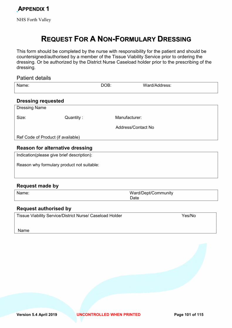
Version 5.4 April 2019 UNCONTROLLED WHEN PRINTED Page 101 of 115
AAPPPPEENNDDIIXX 11 NHS Forth Valley
RREEQQUUEESSTT FFOORR AA NNOONN--FFOORRMMUULLAARRYY DDRREESSSSIINNGG
This form should be completed by the nurse with responsibility for the patient and should be countersigned/authorised by a member of the Tissue Viability Service prior to ordering the dressing. Or be authorized by the District Nurse Caseload holder prior to the prescribing of the dressing.
Patient details Name: DOB: Ward/Address:
Dressing requested Dressing Name Size: Quantity : Manufacturer: Address/Contact No Ref Code of Product (if available)
Reason for alternative dressing Indication(please give brief description): Reason why formulary product not suitable:
Request made by Name: Ward/Dept/Community Date
Request authorised by Tissue Viability Service/District Nurse/ Caseload Holder Yes/No Name

Version 5.4 April 2019 UNCONTROLLED WHEN PRINTED Page 102 of 115
AAPPPPEENNDDIIXX 22 Assessment Chart for Wound Management For multiple wounds complete formal wound assessment for each wound. Add inserts as needed.
Factors which could delay healing: (Please tick relevant box) Immobility Poor Nutrition Diabetes Incontinence Respiratory/Circulatory Anaemia Medication Wound Infection Disease Inotropes Anti-Coagulants Oedema Steroids Chemotherapy Other………………… Allergies & Sensitivities………………………
Body Diagram Front Back
Mark location with ‘X’ and number each wound Type of Wound Total number & duration of each type of wound
Leg Ulcer ………………………………….. …… Surgical Wound ………………………………. Diabetic Ulcer ………………………….…….. Pressure Ulcer - specify grade……………….. Other, specify …………………………………
Feet Diagram
Right Left
Mark location with ‘X’ and number each wound Date referred to:
TVN …………….Physiotherapist……………. Podiatrist………………Dietician……………... Other (please specify)…………………………. Assessors signature: ……………………….. Date: ………………………..…………………...
WRITE, IMPRINT OR ATTACH LABEL Surname ……………….... CHI No ….………….. Forenames ………………… Sex………………….. DoB ……………….. Location……………………………………………………
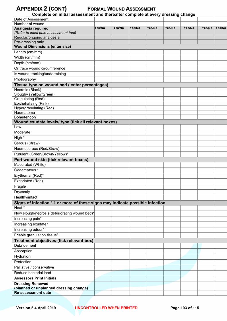
Version 5.4 April 2019 UNCONTROLLED WHEN PRINTED Page 103 of 115
AAPPPPEENNDDIIXX 22 ((CCOONNTT)) FFOORRMMAALL WWOOUUNNDD AASSSSEESSSSMMEENNTT Complete on initial assessment and thereafter complete at every dressing change
Date of Assessment
Number of wound
Analgesia required (Refer to local pain assessment tool)
Yes/No Yes/No Yes/No Yes/No Yes/No Yes/No Yes/No Yes/No
Regular/ongoing analgesia Pre-dressing only Wound Dimensions (enter size) Length (cm/mm) Width (cm/mm) Depth (cm/mm) Or trace wound circumference Is wound tracking/undermining Photography Tissue type on wound bed ( enter percentages) Necrotic (Black)
Sloughy (Yellow/Green)
Granulating (Red)
Epithelialising (Pink)
Hypergranulating (Red)
Haematoma
Bone/tendon
Wound exudate levels/ type (tick all relevant boxes) Low Moderate High * Serous (Straw) Haemoserous (Red/Straw) Purulent (Green/Brown/Yellow)* Peri-wound skin (tick relevant boxes) Macerated (White) Oedematous * Erythema (Red)* Excoriated (Red) Fragile Dry/scaly Healthy/intact Signs of Infection * 1 or more of these signs may indicate possible infection Heat * New slough/necrosis(deteriorating wound bed)* Increasing pain* Increasing exudate* Increasing odour* Friable granulation tissue* Treatment objectives (tick relevant box) Debridement Absorption Hydration Protection Palliative / conservative Reduce bacterial load Assessors Print Initials Dressing Renewed (planned or unplanned dressing change)
Re-assessment date
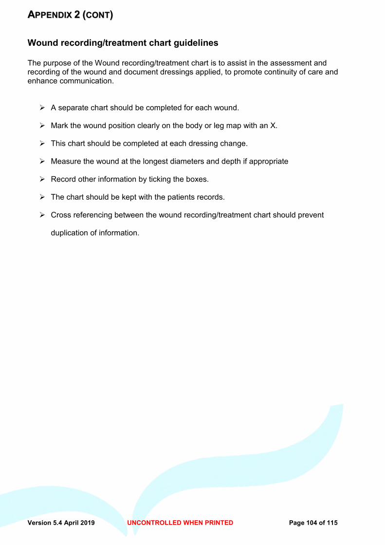
Version 5.4 April 2019 UNCONTROLLED WHEN PRINTED Page 104 of 115
AAPPPPEENNDDIIXX 22 ((CCOONNTT))
Wound recording/treatment chart guidelines The purpose of the Wound recording/treatment chart is to assist in the assessment and recording of the wound and document dressings applied, to promote continuity of care and enhance communication. A separate chart should be completed for each wound.
Mark the wound position clearly on the body or leg map with an X.
This chart should be completed at each dressing change.
Measure the wound at the longest diameters and depth if appropriate
Record other information by ticking the boxes.
The chart should be kept with the patients records.
Cross referencing between the wound recording/treatment chart should prevent
duplication of information.
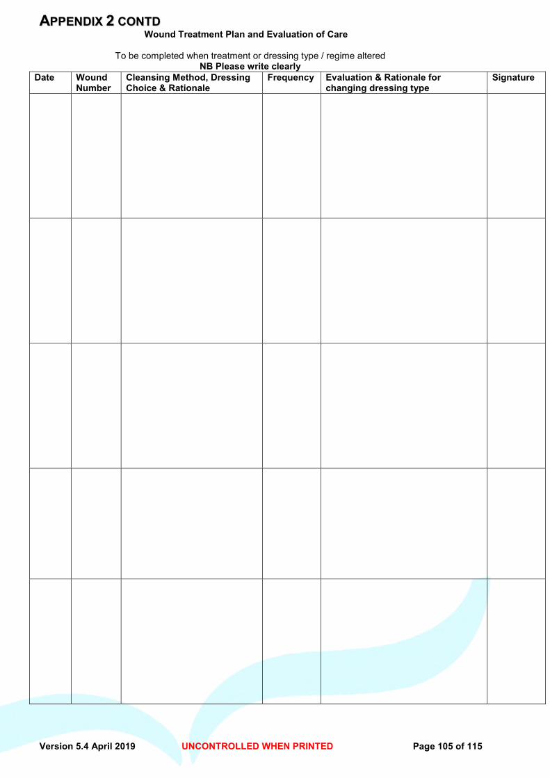
Version 5.4 April 2019 UNCONTROLLED WHEN PRINTED Page 105 of 115
AAPPPPEENNDDIIXX 22 CCOONNTTDD Wound Treatment Plan and Evaluation of Care
To be completed when treatment or dressing type / regime altered
NB Please write clearly Date Wound
Number Cleansing Method, Dressing Choice & Rationale
Frequency
Evaluation & Rationale for changing dressing type
Signature
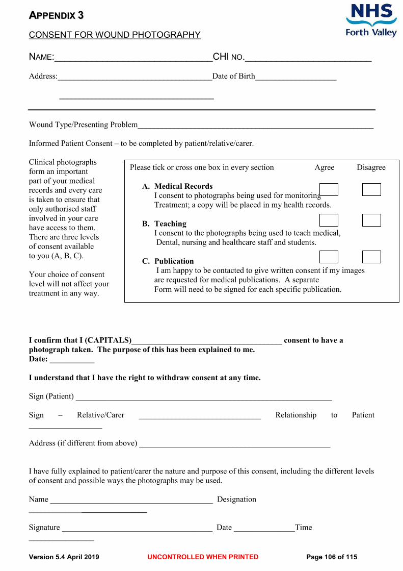
Version 5.4 April 2019 UNCONTROLLED WHEN PRINTED Page 106 of 115
AAPPPPEENNDDIIXX 33 CONSENT FOR WOUND PHOTOGRAPHY
NAME:______________________________CHI NO.________________________ Address:______________________________________Date of Birth____________________ ______________________________________ Wound Type/Presenting Problem__________________________________________________________ Informed Patient Consent – to be completed by patient/relative/carer. Clinical photographs form an important part of your medical records and every care is taken to ensure that only authorised staff involved in your care have access to them. There are three levels of consent available to you (A, B, C). Your choice of consent level will not affect your treatment in any way. I confirm that I (CAPITALS)_____________________________________ consent to have a photograph taken. The purpose of this has been explained to me. Date: ___________ I understand that I have the right to withdraw consent at any time. Sign (Patient) _______________________________________________________________ Sign – Relative/Carer ______________________________ Relationship to Patient __________________ Address (if different from above) _______________________________________________ I have fully explained to patient/carer the nature and purpose of this consent, including the different levels of consent and possible ways the photographs may be used. Name ________________________________________ Designation _____________________________ Signature _____________________________________ Date _______________Time ________________
Please tick or cross one box in every section Agree Disagree
A. Medical Records I consent to photographs being used for monitoring Treatment; a copy will be placed in my health records.
B. Teaching
I consent to the photographs being used to teach medical, Dental, nursing and healthcare staff and students.
C. Publication I am happy to be contacted to give written consent if my images
are requested for medical publications. A separate Form will need to be signed for each specific publication.
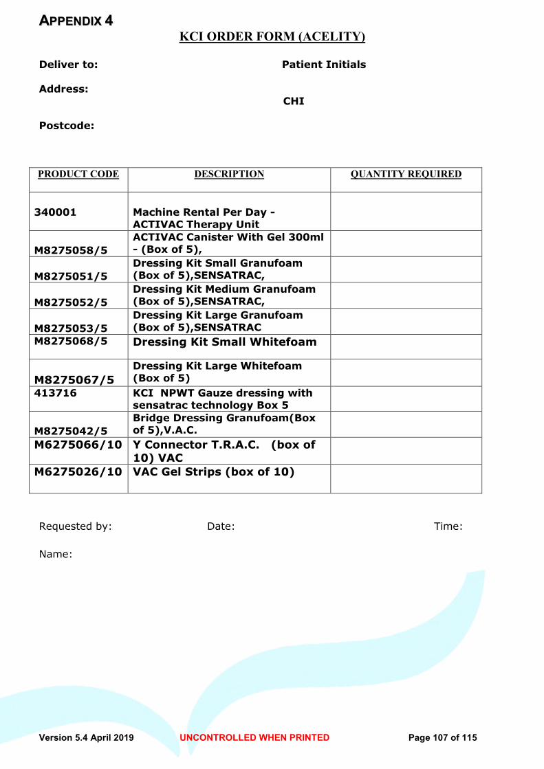
Version 5.4 April 2019 UNCONTROLLED WHEN PRINTED Page 107 of 115
AAPPPPEENNDDIIXX 44 KCI ORDER FORM (ACELITY)
Deliver to: Patient Initials Address: CHI Postcode: PRODUCT CODE DESCRIPTION QUANTITY REQUIRED
340001
Machine Rental Per Day - ACTIVAC Therapy Unit
M8275058/5
ACTIVAC Canister With Gel 300ml - (Box of 5),
M8275051/5
Dressing Kit Small Granufoam (Box of 5),SENSATRAC,
M8275052/5
Dressing Kit Medium Granufoam (Box of 5),SENSATRAC,
M8275053/5
Dressing Kit Large Granufoam (Box of 5),SENSATRAC
M8275068/5 Dressing Kit Small Whitefoam
M8275067/5
Dressing Kit Large Whitefoam (Box of 5)
413716 KCI NPWT Gauze dressing with sensatrac technology Box 5
M8275042/5
Bridge Dressing Granufoam(Box of 5),V.A.C.
M6275066/10 Y Connector T.R.A.C. (box of 10) VAC
M6275026/10 VAC Gel Strips (box of 10)
Requested by: Date: Time: Name:
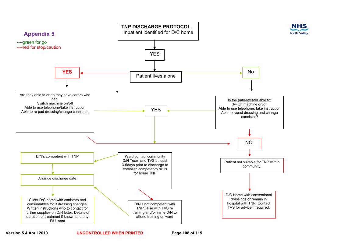
Version 5.4 April 2019 UNCONTROLLED WHEN PRINTED Page 108 of 115
TNP DISCHARGE PROTOCOL Inpatient identified for D/C home
Patient lives alone
----green for go ----red for stop/caution
YES
Are they able to or do they have carers who can:
Switch machine on/off Able to use telephone/take instruction
Able to re pad dressing/change cannister.
No
Is the patient/carer able to: Switch machine on/off
Able to use telephone, take instruction Able to repad dressing and change
cannister?
NO
YES
Patient not suitable for TNP within community.
Ward contact community D/N Team and TVS at least 3-5days prior to discharge to establish competency skills
for home TNP
D/N’s competent with TNP
D/N’s not competent with TNP,liaise with TVS re
training and/or invite D/N to attend training on ward
Arrange discharge date
Client D/C home with canisters and consumables for 3 dressing changes. Written instructions who to contact for
further supplies on D/N letter. Details of duration of treatment if known and any
F/U appt
YES
D/C Home with conventional dressings or remain in
hospital with TNP. Contact TVS for advice if required.
Appendix 5
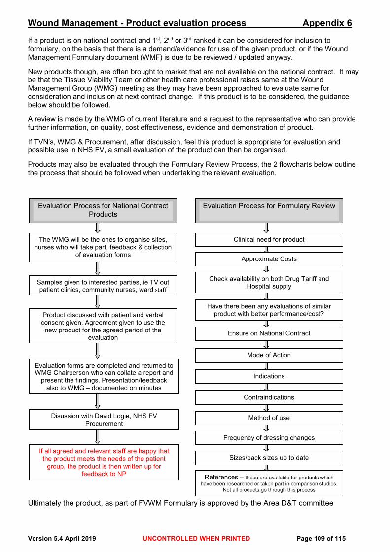
Version 5.4 April 2019 UNCONTROLLED WHEN PRINTED Page 109 of 115
Wound Management - Product evaluation process Appendix 6 If a product is on national contract and 1st, 2nd or 3rd ranked it can be considered for inclusion to formulary, on the basis that there is a demand/evidence for use of the given product, or if the Wound Management Formulary document (WMF) is due to be reviewed / updated anyway. New products though, are often brought to market that are not available on the national contract. It may be that the Tissue Viability Team or other health care professional raises same at the Wound Management Group (WMG) meeting as they may have been approached to evaluate same for consideration and inclusion at next contract change. If this product is to be considered, the guidance below should be followed. A review is made by the WMG of current literature and a request to the representative who can provide further information, on quality, cost effectiveness, evidence and demonstration of product. If TVN’s, WMG & Procurement, after discussion, feel this product is appropriate for evaluation and possible use in NHS FV, a small evaluation of the product can then be organised. Products may also be evaluated through the Formulary Review Process, the 2 flowcharts below outline the process that should be followed when undertaking the relevant evaluation. Ultimately the product, as part of FVWM Formulary is approved by the Area D&T committee
Evaluation Process for National Contract Products
The WMG will be the ones to organise sites, nurses who will take part, feedback & collection
of evaluation forms
Disussion with David Logie, NHS FV Procurement
If all agreed and relevant staff are happy that the product meets the needs of the patient
group, the product is then written up for feedback to NP
Samples given to interested parties, ie TV out patient clinics, community nurses, ward staff
Product discussed with patient and verbal consent given. Agreement given to use the
new product for the agreed period of the evaluation
Evaluation forms are completed and returned to WMG Chairperson who can collate a report and
present the findings. Presentation/feedback also to WMG – documented on minutes
Evaluation Process for Formulary Review
Clinical need for product
Approximate Costs
Check availability on both Drug Tariff and Hospital supply
Have there been any evaluations of similar product with better performance/cost?
Ensure on National Contract
Mode of Action
Indications
Contraindications
Method of use
Frequency of dressing changes
Sizes/pack sizes up to date
References – these are available for products which have been researched or taken part in comparison studies.
Not all products go through this process
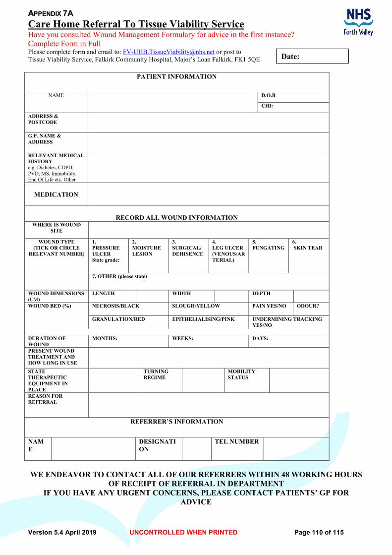
Version 5.4 April 2019 UNCONTROLLED WHEN PRINTED Page 110 of 115
Date:
AAPPPPEENNDDIIXX 77AA Care Home Referral To Tissue Viability Service Have you consulted Wound Management Formulary for advice in the first instance? Complete Form in Full Please complete form and email to: [email protected] or post to Tissue Viability Service, Falkirk Community Hospital, Major’s Loan Falkirk, FK1 5QE
PATIENT INFORMATION
NAME D.O.B
CHI:
ADDRESS & POSTCODE
G.P. NAME & ADDRESS
RELEVANT MEDICAL HISTORY e.g. Diabetes, COPD, PVD, MS, Immobility, End Of Life etc. Other
MEDICATION
RECORD ALL WOUND INFORMATION
WHERE IS WOUND SITE
WOUND TYPE (TICK OR CIRCLE
RELEVANT NUMBER)
1. PRESSURE ULCER State grade:
2. MOISTURE LESION
3. SURGICAL/ DEHISENCE
4. LEG ULCER (VENOUS/ARTERIAL)
5. FUNGATING
6. SKIN TEAR
7. OTHER (please state)
WOUND DIMENSIONS (CM)
LENGTH WIDTH DEPTH
WOUND BED (%)
NECROSIS/BLACK SLOUGH/YELLOW PAIN YES/NO ODOUR?
GRANULATION/RED
EPITHELIALISING/PINK UNDERMINING TRACKING YES/NO
DURATION OF WOUND
MONTHS: WEEKS: DAYS:
PRESENT WOUND TREATMENT AND HOW LONG IN USE
STATE THERAPEUTIC EQUIPMENT IN PLACE
TURNING REGIME
MOBILITY STATUS
REASON FOR REFERRAL
REFERRER’S INFORMATION NAME
DESIGNATION
TEL NUMBER
WE ENDEAVOR TO CONTACT ALL OF OUR REFERRERS WITHIN 48 WORKING HOURS
OF RECEIPT OF REFERRAL IN DEPARTMENT IF YOU HAVE ANY URGENT CONCERNS, PLEASE CONTACT PATIENTS’ GP FOR
ADVICE
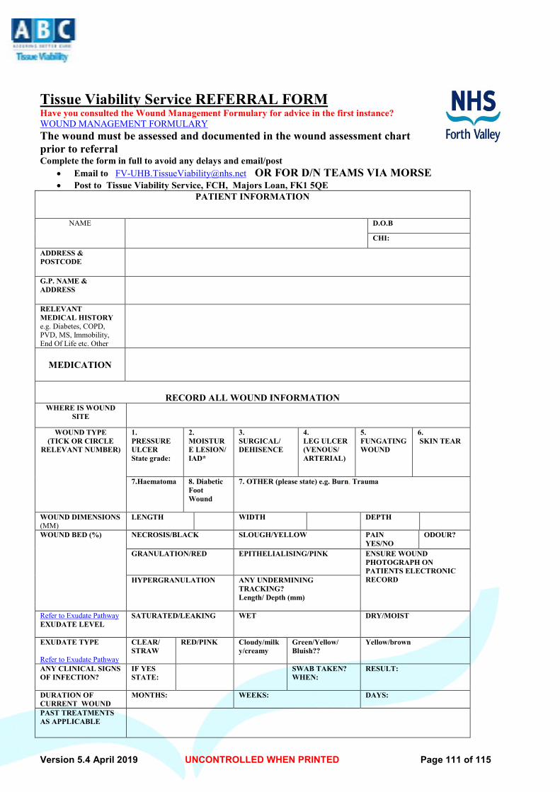
Version 5.4 April 2019 UNCONTROLLED WHEN PRINTED Page 111 of 115
Tissue Viability Service REFERRAL FORM Have you consulted the Wound Management Formulary for advice in the first instance? WOUND MANAGEMENT FORMULARY The wound must be assessed and documented in the wound assessment chart prior to referral Complete the form in full to avoid any delays and email/post
• Email to [email protected] OR FOR D/N TEAMS VIA MORSE • Post to Tissue Viability Service, FCH, Majors Loan, FK1 5QE
PATIENT INFORMATION
NAME D.O.B
CHI:
ADDRESS & POSTCODE
G.P. NAME & ADDRESS
RELEVANT MEDICAL HISTORY e.g. Diabetes, COPD, PVD, MS, Immobility, End Of Life etc. Other
MEDICATION
RECORD ALL WOUND INFORMATION
WHERE IS WOUND SITE
WOUND TYPE (TICK OR CIRCLE
RELEVANT NUMBER)
1. PRESSURE ULCER State grade:
2. MOISTURE LESION/ IAD*
3. SURGICAL/ DEHISENCE
4. LEG ULCER (VENOUS/ ARTERIAL)
5. FUNGATING WOUND
6. SKIN TEAR
7.Haematoma 8. Diabetic Foot Wound
7. OTHER (please state) e.g. Burn, Trauma
WOUND DIMENSIONS (MM)
LENGTH WIDTH DEPTH
WOUND BED (%)
NECROSIS/BLACK SLOUGH/YELLOW PAIN YES/NO
ODOUR?
GRANULATION/RED
EPITHELIALISING/PINK ENSURE WOUND PHOTOGRAPH ON PATIENTS ELECTRONIC RECORD
HYPERGRANULATION ANY UNDERMINING TRACKING? Length/ Depth (mm)
Refer to Exudate Pathway EXUDATE LEVEL
SATURATED/LEAKING WET DRY/MOIST
EXUDATE TYPE Refer to Exudate Pathway
CLEAR/ STRAW
RED/PINK Cloudy/milky/creamy
Green/Yellow/ Bluish??
Yellow/brown
ANY CLINICAL SIGNS OF INFECTION?
IF YES STATE:
SWAB TAKEN? WHEN:
RESULT:
DURATION OF CURRENT WOUND
MONTHS: WEEKS: DAYS:
PAST TREATMENTS AS APPLICABLE
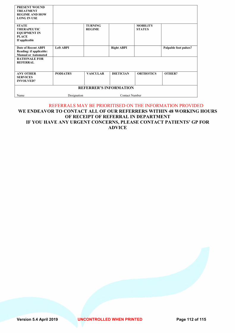
Version 5.4 April 2019 UNCONTROLLED WHEN PRINTED Page 112 of 115
PRESENT WOUND TREATMENT REGIME AND HOW LONG IN USE
STATE THERAPEUTIC EQUIPMENT IN PLACE If applicable
TURNING REGIME
MOBILITY STATUS
Date of Recent ABPI Reading: if applicable: Manual or Automated
Left ABPI Right ABPI Palpable foot pulses?
RATIONALE FOR REFERRAL
ANY OTHER SERVICES INVOLVED?
PODIATRY VASCULAR DIETICIAN ORTHOTICS OTHER?
REFERRER’S INFORMATION Name Designation Contact Number
REFERRALS MAY BE PRIORITISED ON THE INFORMATION PROVIDED WE ENDEAVOR TO CONTACT ALL OF OUR REFERRERS WITHIN 48 WORKING HOURS
OF RECEIPT OF REFERRAL IN DEPARTMENT IF YOU HAVE ANY URGENT CONCERNS, PLEASE CONTACT PATIENTS’ GP FOR
ADVICE
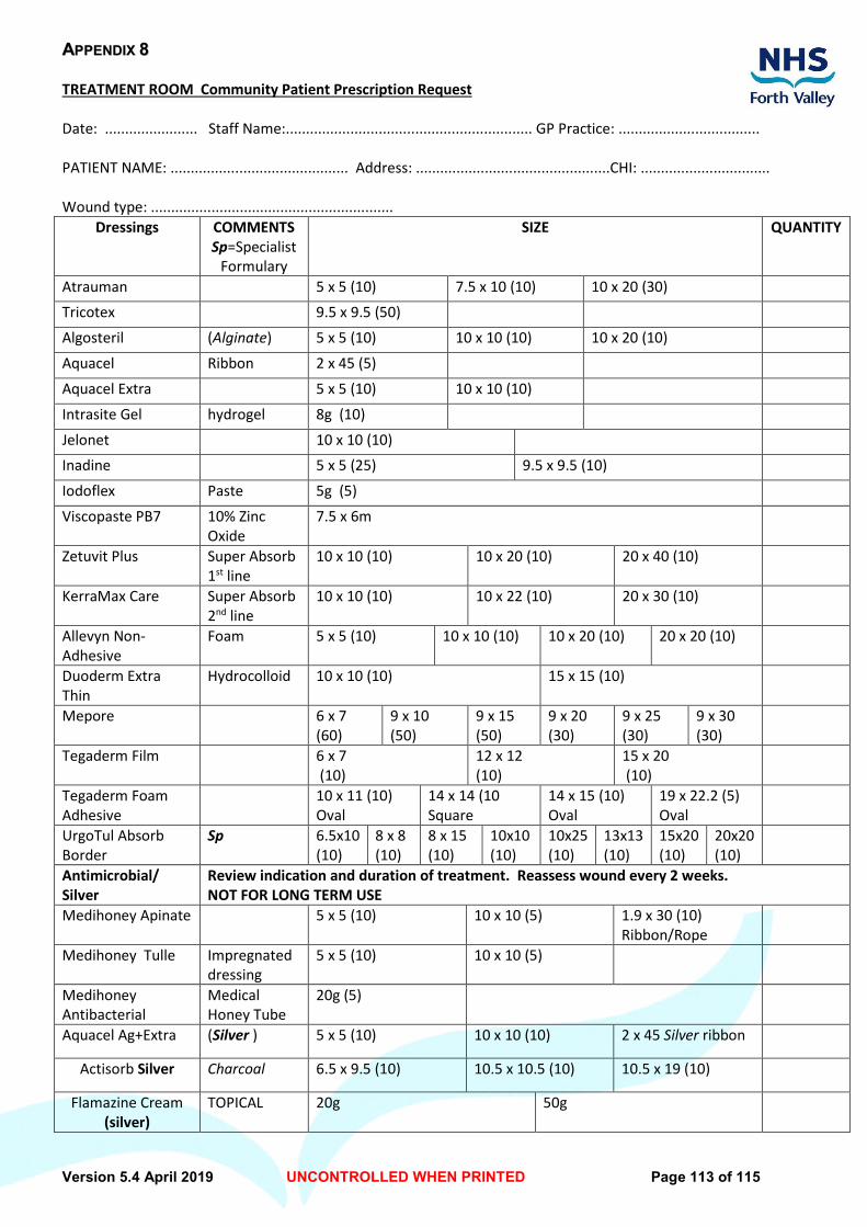
Version 5.4 April 2019 UNCONTROLLED WHEN PRINTED Page 113 of 115
AAPPPPEENNDDIIXX 88 TREATMENT ROOM Community Patient Prescription Request Date: ....................... Staff Name:............................................................. GP Practice: ................................... PATIENT NAME: ............................................ Address: ................................................CHI: ................................ Wound type: ............................................................
Dressings COMMENTS Sp=Specialist
Formulary
SIZE QUANTITY
Atrauman 5 x 5 (10) 7.5 x 10 (10) 10 x 20 (30) Tricotex 9.5 x 9.5 (50)
Algosteril (Alginate) 5 x 5 (10) 10 x 10 (10) 10 x 20 (10)
Aquacel Ribbon 2 x 45 (5)
Aquacel Extra 5 x 5 (10) 10 x 10 (10)
Intrasite Gel hydrogel 8g (10)
Jelonet 10 x 10 (10) Inadine 5 x 5 (25) 9.5 x 9.5 (10)
Iodoflex Paste 5g (5)
Viscopaste PB7 10% Zinc Oxide
7.5 x 6m
Zetuvit Plus Super Absorb 1st line
10 x 10 (10) 10 x 20 (10) 20 x 40 (10)
KerraMax Care Super Absorb 2nd line
10 x 10 (10) 10 x 22 (10) 20 x 30 (10)
Allevyn Non- Adhesive
Foam 5 x 5 (10) 10 x 10 (10) 10 x 20 (10) 20 x 20 (10)
Duoderm Extra Thin
Hydrocolloid 10 x 10 (10) 15 x 15 (10)
Mepore 6 x 7 (60)
9 x 10 (50)
9 x 15 (50)
9 x 20 (30)
9 x 25 (30)
9 x 30 (30)
Tegaderm Film
6 x 7 (10)
12 x 12 (10)
15 x 20 (10)
Tegaderm Foam Adhesive
10 x 11 (10) Oval
14 x 14 (10 Square
14 x 15 (10) Oval
19 x 22.2 (5) Oval
UrgoTul Absorb Border
Sp 6.5x10 (10)
8 x 8 (10)
8 x 15 (10)
10x10 (10)
10x25 (10)
13x13 (10)
15x20 (10)
20x20 (10)
Antimicrobial/ Silver
Review indication and duration of treatment. Reassess wound every 2 weeks. NOT FOR LONG TERM USE
Medihoney Apinate 5 x 5 (10) 10 x 10 (5) 1.9 x 30 (10) Ribbon/Rope
Medihoney Tulle Impregnated dressing
5 x 5 (10) 10 x 10 (5)
Medihoney Antibacterial
Medical Honey Tube
20g (5)
Aquacel Ag+Extra (Silver ) 5 x 5 (10) 10 x 10 (10) 2 x 45 Silver ribbon
Actisorb Silver Charcoal 6.5 x 9.5 (10) 10.5 x 10.5 (10) 10.5 x 19 (10)
Flamazine Cream (silver)
TOPICAL 20g 50g
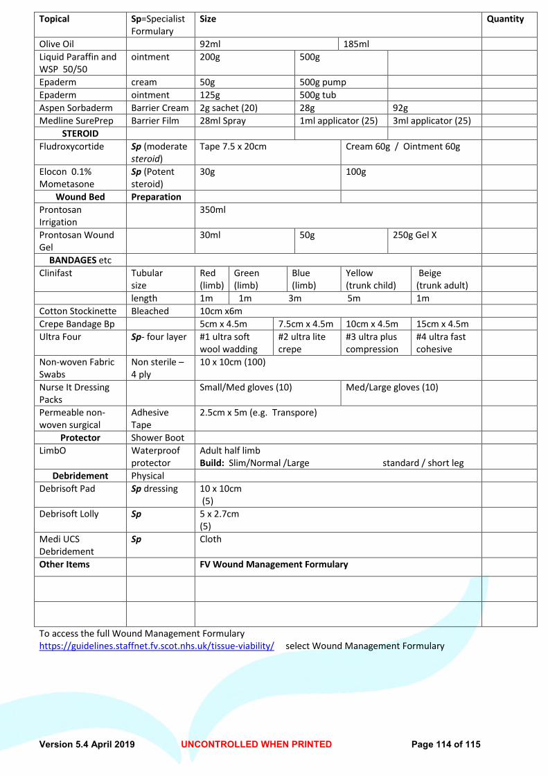
Version 5.4 April 2019 UNCONTROLLED WHEN PRINTED Page 114 of 115
Topical Sp=Specialist Formulary
Size Quantity
Olive Oil 92ml 185ml Liquid Paraffin and WSP 50/50
ointment 200g 500g
Epaderm cream 50g 500g pump Epaderm ointment 125g 500g tub Aspen Sorbaderm Barrier Cream 2g sachet (20) 28g 92g Medline SurePrep Barrier Film 28ml Spray 1ml applicator (25) 3ml applicator (25)
STEROID Fludroxycortide Sp (moderate
steroid) Tape 7.5 x 20cm Cream 60g / Ointment 60g
Elocon 0.1% Mometasone
Sp (Potent steroid)
30g 100g
Wound Bed Preparation Prontosan Irrigation
350ml
Prontosan Wound Gel
30ml 50g 250g Gel X
BANDAGES etc Clinifast Tubular
size Red (limb)
Green (limb)
Blue (limb)
Yellow (trunk child)
Beige (trunk adult)
length 1m 1m 3m 5m 1m Cotton Stockinette Bleached 10cm x6m Crepe Bandage Bp 5cm x 4.5m 7.5cm x 4.5m 10cm x 4.5m 15cm x 4.5m Ultra Four Sp- four layer #1 ultra soft
wool wadding #2 ultra lite crepe
#3 ultra plus compression
#4 ultra fast cohesive
Non-woven Fabric Swabs
Non sterile – 4 ply
10 x 10cm (100)
Nurse It Dressing Packs
Small/Med gloves (10) Med/Large gloves (10)
Permeable non-woven surgical
Adhesive Tape
2.5cm x 5m (e.g. Transpore)
Protector Shower Boot LimbO Waterproof
protector Adult half limb Build: Slim/Normal /Large standard / short leg
Debridement Physical Debrisoft Pad Sp dressing 10 x 10cm
(5)
Debrisoft Lolly Sp 5 x 2.7cm (5)
Medi UCS Debridement
Sp Cloth
Other Items FV Wound Management Formulary
To access the full Wound Management Formulary https://guidelines.staffnet.fv.scot.nhs.uk/tissue-viability/ select Wound Management Formulary
Version 5.4 April 2019 UNCONTROLLED WHEN PRINTED Page 115 of 115
Publications in Alternative Formats NHS Forth Valley is happy to consider requests for publications in other language or formats such as large print. To request another language for a patient, please contact 01324 590886. For other formats contact 01324 590886, text 07990 690605, fax 01324 590867 or e-mail [email protected]