next generation sequencing in leukemia: from … dna symposiu… · next generation sequencing in...
TRANSCRIPT
No conflict of interest to disclose
Keyur Pravinchandra Patel MD PhD ([email protected])
Medical Director, Molecular Diagnostics Laboratory (Hematologic Malignancies)
Assistant Professor, Department of Hematopathology,
Division of Pathology and Laboratory Medicine The University of Texas M.D. Anderson Cancer Center
Next Generation Sequencing in Leukemia: From Discovery to Clinical Care

The Clinical Molecular Diagnostics Laboratory (MDL)

Overview
• NGS as a genotyping platform • Implementation of NGS in clinical diagnostics • NGS as a discovery tool in clinical care • Future directions

2009
Voelkerding KV et al, J Mol Diag 2010, 10:539-551

Next Generation Sequencing as a Discovery Tool
NEJM 2010, 363;2424-33 NEJM 2009, 361;1058-66
NEJM 2013, 368;1781-90 NEJM 2011, 364; 2305-15

NGS-Based Discoveries in Hematologic Malignancies
Blood Cancer Journal (2013) 3, e127; doi:10.1038/bcj.2013.26

Mutations in Hematologic Malignancies
• Integral part of diagnostic work-up for myeloid neoplasms (AML, MDS, MPN, MDS/MPN)
• Diagnostic, prognostic and predictive value • Growing list of mutated genes with clinical utility:
NPM1, FLT3, RAS (KRAS, NRAS), KIT, CEBPA, WT1, IDH1, IDH2, DNMT3A, EZH2, JAK2, MPL, several new genes in lymphoid tumors
• New associations of known mutations: BRAF in hairy cell leukemia

Mutation Profiling of AML/MDS
NEJM, 364;26 June 30, 2011
NEJM, 366;12 March 22, 2012
MDS
AML

Hematology Diagnostics Timeline
Blood Cancer Journal (2013) 3, e127; doi:10.1038/bcj.2013.26

CAP/CLIA-Certified Hematologic Molecular Testing at MDL
2004 2005 2006 2007 2008 2010 2012 2013
ABL Seq CEBPA Luminex LTx
KIT exon 17 MPL IDH1
Fluidigm Leukemia Panel CMS53
FIP1L1-PDGFRA RT JHF
ABL p.T315I NPM1 IDH2 DNMT3A ATM
SH (CLL) JAK2 TCRB aCGH TP53
New Clinical Tests by Year
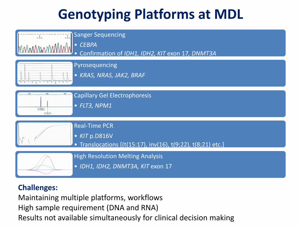
Genotyping Platforms at MDL Sanger Sequencing • CEBPA • Confirmation of IDH1, IDH2, KIT exon 17, DNMT3A
Pyrosequencing • KRAS, NRAS, JAK2, BRAF
Capillary Gel Electrophoresis • FLT3, NPM1
Real-Time PCR • KIT p.D816V • Translocations [(t(15:17), inv(16), t(9;22), t(8;21) etc.]
High Resolution Melting Analysis • IDH1, IDH2, DNMT3A, KIT exon 17
Challenges: Maintaining multiple platforms, workflows High sample requirement (DNA and RNA) Results not available simultaneously for clinical decision making

Sequencing Technologies
Platform Sensitivity (for clinical
use)
Sample Requirement
Multiplexing Capability
Throughput Type of mutations
Quantitative
Sanger 20% High (ug) None Low Point, indel
No
Pyro- 5% Intermediate None Low Point Yes
Sequenom/ ABI
SNaPshot
10% Intermediate Intermediate Medium Point Yes
NGS 5% Low (ng) High (amplicons
and samples)
High Point, indel,
translocation, copy number change
Yes
NGS= next-generation sequencing

Next Generation Sequencing • 2nd generation or massively parallel sequencing • Simultaneous amplification and sequencing of a large
number of amplicons
Advantages High throughput Better sensitivity Efficient use of limited tissue Consolidation of platforms Wider range of mutation detection
Challenges for Clinical Implementation Selection of test platform Evaluation of multi-gene panels Establishment of assay characteristics Validation on different sample types Results interpretation and reporting Legal and ethical issues Billing and compliance
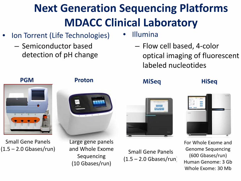
Next Generation Sequencing Platforms MDACC Clinical Laboratory
• Ion Torrent (Life Technologies) – Semiconductor based
detection of pH change
MiSeq
• Illumina – Flow cell based, 4-color
optical imaging of fluorescent labeled nucleotides
PGM Proton
Small Gene Panels (1.5 – 2.0 Gbases/run)
Small Gene Panels (1.5 – 2.0 Gbases/run)
Large gene panels and Whole Exome
Sequencing (10 Gbases/run)
For Whole Exome and Genome Sequencing
(600 Gbases/run) Human Genome: 3 Gb Whole Exome: 30 Mb
HiSeq

Merker JD et al, Ther Adv Hematol 2012, 3(6) 333-339
Simplified NGS Workflow

Wells :
Ion Torrent: Semiconductor based sequencing
(3.5 µM Diameter)
Metal-Oxide Semiconductor
Sequencing Wells
DNA immobilized on Ion Spheres
(2.0 µM Diameter)
316 Chip (6 million)
314 Chip (1.2 million) 318 Chip
(11 million) Proton 1
(165 million)

Ion PGM Ampliseq Cancer Panel Work Flow
Quantitation by Qubit Fluorometer
10ng Genomic DNA as Template
196 Amplicons (Hotspots in 46 genes)
Adapter & Barcode Ligation
DNA extraction (3 hrs for
PB&BM/15 hrs for FFPE)
Clonal Amplification on to ionspheres By Emulsion PCR (17 hrs)
ISP
Load on Ion Semiconductor Chip (1 hr)
3 Day Turn Around Time
Data Analysis (1 hr)
Sequencing 100bp 2.5 hr

Specific Base Cleavage (Read1)
Cluster Generation For Sequencing (Illumina)
• Hybridization of amplicon templates to a ‘lawn’ of oligos
•Synthesis of complementary strands
• Copying of immobilized strands by ‘Bridge Amplification’.
• Clonal amplification of a single strand into dense clusters
• Removal of strands attached by P7 adaptor & blocking of 3’ ends
• Sequencing by hybridizing sequencing primer
Specific Base Cleavage (Read2)

Template Clusters in the Flowcell Lane
( ~ 1000 DNA strands/cluster)
TEMPLATE STRAND COMPLAMENTARY
STRAND SYNTHESIS
LANE SURFACE
Image of Clusters in the lane
(0.8 – 1.0 million clusters/mm2)
Optical Imaging of Fluorescence from Clusters
•DNA strands in the cluster act as templates for DNA polymerization
• Sequencing based synthesis using reversible terminator chemistry
• Incorporation of each fluorescently labeled nucleotide detected
using optical imaging
• True paired end sequencing
Illumina Platform: Sequencing By Synthesis: SBS

Quantitation by Qubit Fluorometer
250ng Genomic DNA as Template
DNA extraction (3 hrs for PB, BM)
Load on Flow Cell
4 Day Turn Around Time
MiSeq TruSeq Cancer Panel Work Flow
Cluster Generation for Sequencing (9 hrs)
TEMPLATE STRAND COMPLAMENTARY
STRAND SYNTHESIS LANE
SURFACE
Adapter & Barcode Ligation
Data Analysis
Sequencing and base calling (27 hrs)
(1 hr)

Integrative Genome Browser (IGV, Broad Institute)
Coverage Depth
Chromosomal Position
Sequencing Reads
Gene
COSMIC

4b Ins
WT 4bp Ins
Mutation Detection by NGS
Sanger Sanger Capillary GE
Point Mutation Insertion Deletion
NGS
Co
nven
tiona
l

Detection of Internal Tandem Duplications (Using Pindel)
FLT3 In-Tandem Duplication of 18 bp
WT
ITD

Missing Pieces For Clinical Use • Adequacy assessment for all amplicons • Sample annotation system • Easy visualization of results • Inclusion or exclusion of variants for reporting • Ability to monitor frequency of a specific mutation in our
samples • Conversion of base calls into standardized nomenclature • Direct linking to genomic databases (COSMIC, dbSNP etc.) • Internal annotation system for variants detected for
future reference • Generation of clinical reports

Variant Caller 2.0 Output (Ion PGM
• Chromosome • Genomic position • Gene symbol • Target id • Type • Ploidy • Reference nucleotide • Variant nucleotide • Variant frequency • P-value • Coverage • Reference coverage • Variant coverage • Hotspot id (COSMIC)
• Requires high performance servers for data analysis • Variant caller file does not contain amino acid and coding sequence
information

MiSeq Reporter Output • Performs the data analysis
• Requires high performance computers • Variant caller file contains amino acid and coding sequence correlates of mutations
• Sample id • Sample name • Manifest file • Clusters PF • Clusters aligned R1 • Clusters aligned R2 • Mismatch • No call • Coverage • HetSNPs • HomSNPs • Insertions • Deletions

Base Calling
DAT Files Processing
Alignment
MiSeq Reporter
Output: BAM & FASTQ/SFF
Output: VCF
-Sample annotation -Suboptimal coverage filtering -Knowledgebase mapping (eg. COSMIC) -Intersample comparison for germline/somatic variants -Population Analysis -Amplicon viewer with the integrated Genomics Viewer (IGV)
DAT Files Server
MDACC Informatics Pipeline for Clinical Reporting
Courtesy: Mark Routbort, MD, PhD

Practical Challenges
• Re-engineering of workflow • Reporting of larger scale genomic information • Billing and compliance

Reporting of the Mutations
• Standardized nomenclature added to allow portability and inter-laboratory comparison
• BRAF mutation analysis: - Mutation detected in codon 600, exon 15 (GTG to GAG) of the BRAF gene that would change the encoded amino acid from Valine to Glutamate (p.Val600Glu) - Standardized nomenclature for this mutation: NM_004333.4(BRAF):c.1799T>A p.V600E

• Reporting of all versus selected genes • Mutations in unordered genes • Germline variants • Integration in clinical management
Medico-Legal and Ethical Issues

Billing and Compliance
Old Codes (pre-2013)
• Stacking codes • Procedure based e.g.
DNA extraction, sequencing, interpretation etc.
• Easy to implement for a new gene
New Codes (2013)
• Gene-specific codes • Limited number of
genes on the current code list
• NO codes for gene panels
• Lengthy application process for new genes
• Billing based on AMA CPT codes

III. Clinical Experience with NGS
• 53 gene panel live since October, 2012 – Total unique patients: 871 – Total samples: 906 – Average Turnaround time: <6 business days
• 28 gene panel live since August 19, 2013

Clinical Utility of Test Results
• Diagnostic work-up • Prognosis • Monitoring of minimal residual disease • Comparison of two lesions in the same patient • Identification of targetable markers • Detection of novel biomarkers • Insights into tumor biology

Thank You