next generation sequencing in a large cohort of patients
TRANSCRIPT

RESEARCH Open Access
Next generation sequencing in a largecohort of patients presenting withneuromuscular disease before or at birthEmily J. Todd1, Kyle S. Yau1, Royston Ong1, Jennie Slee2, George McGillivray3, Christopher P. Barnett4,Goknur Haliloglu5, Beril Talim6, Zuhal Akcoren6, Ariana Kariminejad7, Anita Cairns8, Nigel F. Clarke9,10,Mary-Louise Freckmann11, Norma B. Romero12, Denise Williams13,14, Caroline A Sewry13,14, Alison Colley15,Monique M. Ryan16, Cathy Kiraly-Borri17, Padma Sivadorai18, Richard J.N. Allcock19, David Beeson20,Susan Maxwell20, Mark R. Davis18, Nigel G. Laing1,18 and Gianina Ravenscroft1*
Abstract
Background: Fetal akinesia/hypokinesia, arthrogryposis and severe congenital myopathies are heterogeneousconditions usually presenting before or at birth. Although numerous causative genes have been identified for eachof these disease groups, in many cases a specific genetic diagnosis remains elusive. Due to the emergence of nextgeneration sequencing, virtually the entire coding region of an individual’s DNA can now be analysed through“whole” exome sequencing, enabling almost all known and novel disease genes to be investigated for disorderssuch as these.
Methods: Genomic DNA samples from 45 patients with fetal akinesia/hypokinesia, arthrogryposis or severecongenital myopathies from 38 unrelated families were subjected to next generation sequencing. Clinical featuresand diagnoses for each patient were supplied by referring clinicians. Genomic DNA was used for either wholeexome sequencing or a custom-designed neuromuscular sub-exomic supercapture array containing 277 genesresponsible for various neuromuscular diseases. Candidate disease-causing variants were investigated andconfirmed using Sanger sequencing. Some of the cases within this cohort study have been published previously asseparate studies.
Results: A conclusive genetic diagnosis was achieved for 18 of the 38 families. Within this cohort, mutations werefound in eight previously known neuromuscular disease genes (CHRND, CHNRG, ECEL1, GBE1, MTM1, MYH3, NEB andRYR1) and four novel neuromuscular disease genes were identified and have been published as separate reports(GPR126, KLHL40, KLHL41 and SPEG). In addition, novel mutations were identified in CHRND, KLHL40, NEB and RYR1.Autosomal dominant, autosomal recessive, X-linked, and de novo modes of inheritance were observed.
Conclusions: By using next generation sequencing on a cohort of 38 unrelated families with fetal akinesia/hypokinesia, arthrogryposis, or severe congenital myopathy we therefore obtained a genetic diagnosis for 47 % offamilies. This study highlights the power and capacity of next generation sequencing (i) to determine the aetiologyof genetically heterogeneous neuromuscular diseases, (ii) to identify novel disease genes in small pedigrees orisolated cases and (iii) to refine the interplay between genetic diagnosis and clinical evaluation and management.
Keywords: Fetal hypokinesia, Arthrogryposis, Next generation sequencing, Congenital myopathy, Nemalinemyopathy
* Correspondence: [email protected] Perkins Institute of Medical Research and the Centre for MedicalResearch, University of Western Australia, QQ Block, 6 Verdun Street,Nedlands 6009, WA, AustraliaFull list of author information is available at the end of the article
© 2015 Todd et al. Open Access This article is distributed under the terms of the Creative Commons Attribution 4.0International License (http://creativecommons.org/licenses/by/4.0/), which permits unrestricted use, distribution, andreproduction in any medium, provided you give appropriate credit to the original author(s) and the source, provide a link tothe Creative Commons license, and indicate if changes were made. The Creative Commons Public Domain Dedication waiver(http://creativecommons.org/publicdomain/zero/1.0/) applies to the data made available in this article, unless otherwise stated.
Todd et al. Orphanet Journal of Rare Diseases (2015) 10:148 DOI 10.1186/s13023-015-0364-0

BackgroundFetal akinesia/hypokinesiaFetal akinesia deformation sequence (FADS) or PenaShokeir syndrome, characterized by intrauterine growthretardation, contractures, craniofacial anomalies, limbanomalies, pulmonary hypoplasia and polyhydramnios,results from reduced movement in utero [1, 2]. A num-ber of other fetal akinesia syndromes overlap phenotyp-ically with FADS. These include the lethal congenitalcontracture syndromes, multiple pterygium syndromes,and arthrogryposis multiplex congenita [3], in which theclinical findings are dependent upon the time of onset ofthe dyskinesia, earlier onset being associated with amore severe phenotype [2]. It’s thought that more than50 % of all causes of fetal akinesia are of neuromuscularorigin [4]; at least 30 causative genes have been identi-fied, involving all points along the neuromuscular axis(motor neurons, peripheral nerves, neuromuscular junc-tion and the skeletal muscle regulatory and contractileapparatus) [5–7].
ArthrogryposisArthrogryposis refers to non-progressive congenital jointcontractures in >1 area of the body, and has been de-scribed in more than 300 specific disorders [6, 8].Arthrogryposis is thought to result from reduced fetalmovement, and affects approximately 1 in 3,000 livebirths [8, 9]. There is a range of disease severity: severecases present with arthrogryposis multiplex congenita,which is lethal prior to or at birth, while milder caseswith a longer life expectancy may have predominantlydistal involvement [8, 9]. The distal arthrogryposes are agroup of disorders with contractures primarily involvingthe extremities of the body, often associated with camp-todactyly, hypoplastic or absent flexion creases, and tali-pes equinovarus [10, 11]. There are ten distinct subtypesof distal arthrogryposis, for which seven causative geneshave been identified: ECEL1 (OMIM 605896), MYH3(OMIM 160720), MYH8 (OMIM 160741), PIEZO2(OMIM 613692), TNNI2 (OMIM 191043), TNNT3(OMIM 600692) and TPM2 (OMIM 190990) [11–13].
Congenital myopathiesThe congenital myopathies are a diverse group of disor-ders, characterised by skeletal muscle dysfunction (mostoften weakness and hypotonia), with specific morpho-logical features on skeletal muscle biopsies [14, 15].Three distinct major groups are recognized based uponthe presence of one or more major histopathological fea-tures: centronuclear myopathy, core myopathy andnemaline myopathy (NEM), although there is extensiveoverlap in both genotype and phenotype within and be-tween these groups [16, 17]. While muscle biopsy re-mains critical for diagnosis, there can be overlap in the
morphological abnormalities seen in these conditions,and marked variability in their clinical progression andseverity [14, 15]. The clinical spectrum of the congenitalmyopathies ranges from severe fetal akinesia to adult-onset progressive weakness. Typical features of theseconditions include proximal weakness, respiratory insuf-ficiency, facial weakness, skeletal deformities such as hipdislocation and deformities of the feet, feeding difficul-ties, hypotonia and delayed motor milestones [17], how-ever hypertonic cases are also encountered [15, 18].More than 15 disease genes are known to cause
congenital myopathies. However, many cases remaingenetically unresolved, suggesting further heterogen-eity [5, 7, 12, 19–21]. This study aimed to assess thepotential of next generation sequencing technologiesto identify causative genes in small families or iso-lated probands presenting with fetal hypokinesia,arthrogryposis or a severe congenital myopathy.
MethodsSubject information and study ethics approvalInformed consent was given for participation in thisstudy, which was approved by the Human ResearchEthics Committee of the University of WesternAustralia, Perth, Western Australia, Australia.
Exome sequencingExome sequencing for this study was performed at theLotterywest State Biomedical Facility Genomics Node(LSBFG) in Perth, Australia. Exome sequencing wasperformed on the 5500XL SOLiD™ system (AppliedBiosystems), as described elsewhere [20, 22–24], and theIon Proton™ (Ampliseq chemistry, Life Technologies)(Family 16 and 38). For AmpliSeq exome sequencing,100 ng of DNA from the probands was amplified in 12PCR pools and sequencing adaptors ligated. The librarywas then purified using AMPure beads (BeckmanCoulter), and amplified using Platinum® High-FidelityTaq Polymerase. The amplified library was again purifiedwith AMPure beads and analysed on a 2100 Bioanalyser(Agilent Technologies Genomics). Libraries were dilutedto 18-26pM and attached to Ion Sphere™ Particles usingan Ion Proton™ Template 200 v3 kit and sequenced on aP1 sequencing chip on an Ion Proton sequencer™ (IonSequencing 200 kit v3) in pools of two.
Targeted capture and sequencing of neuromusculardisease genes by next generation sequencingNeuromuscular sub-exomic sequencing (NSES) was alsoperformed at the LSBFG. The NSES panel comprisedthose genes listed within the December 2012 freeze ofNeuromuscular Disorders gene table [25] in which thedisease-causing mutations could be identified by nextgeneration sequencing, some unpublished candidate
Todd et al. Orphanet Journal of Rare Diseases (2015) 10:148 Page 2 of 14

disease genes identified by our group and others and 59cardiomyopathy genes. NSES analysis was performed onDNA from the probands using the Ion Proton™ sequen-cer (Life Technologies), as previously described [26]. ForNSES, 2 μg of DNA was captured in pools of 16 DNAsamples using a custom TargetSeq™ (Life Technologies)capture system, enriching for the 336 known and candi-date neuromuscular and cardiomyopathy disease genes.These captured pools were then sequenced in batches of16 using an Ion P1 200 V2 sequencing kit (LifeTechnologies) for 520 flows.
BioinformaticsVariant calling was performed against the GRCh37 hu-man reference genome, using LifeScope™ 2.5 (exome se-quencing) and Torrent Suite V 3.6.2 (NSES) (LifeTechnologies). Data was filtered using an ANNOVARannotation software suite. Variants were annotated usingthe EncodeGencode gene annotation set. Variants werefiltered against the 1000 Genomes database (2012 re-lease, [27]) and the dbSNP137 common database, andvariants with a frequency of >0.5 % were excluded. Vari-ants were then filtered against an in-house commonvariant list and were checked against the HGMD profes-sional database to identify any known disease-causingmutations. The frequencies of candidate disease variantsin the 1000 Genomes Project, Exome Variant Server(http://evs.gs.washington.edu/EVS/) and ExAC Browser(http://exac.broadinstitute.org) were also determined.Pathogenicity predictions were made using online pre-diction software programs: SIFT, PolyPhen [28], andMutationTaster [29].The LSBFG has a cut-off of 90 % of on-target regions
covered to 20-fold or greater for the neuromuscularpanel (NSES) and 80 % covered to 20-fold or greater forexome sequencing, however some samples, especiallyearly samples, did not achieve these cut-offs (Additionalfile 1: Table S1). There was no significant difference inthe average coverage (mean ± SEM) of exome sequen-cing data for genetically resolved (80 ± 14-fold; n = 15)versus unresolved cases (70 ± 8-fold; n = 23). For theNSES panel, average coverages were 220 ± 23-fold (n = 6)for resolved cases versus unresolved cases (195 ± 13-fold,n = 9). Thus coverage is unlikely to contribute to the lackof a genetic diagnosis in most cases.
Sanger confirmation and co-segregation studiesPCR amplification and Sanger sequencing was per-formed to verify potential mutations identified by nextgeneration sequencing. Co-segregation was also verifiedfor all existing family members where available. Primerswere based on genomic and cDNA sequences obtainedfrom the UCSC Human Genome Browser (http://geno-me.ucsc.edu/) and Ensembl (http://www.ensembl.org/).
Primer sequences and conditions are available upon re-quest. Sanger sequencing data was processed by LSBFGand results viewed using CodonCode Aligner software.
Functional studies of the CHRND missense substitutionThe mutation CHRND p.Cys257Arg was directly intro-duced into the wild-type human delta subunit cDNA inthe vector pcDNA3.1/hygro (−) by site-directed muta-genesis (QuikChange® Site-Directed Mutagenesis Kit,Stratagene, Amsterdam, The Netherlands). Primer se-quences can be obtained on request. To confirm thepresence of the introduced mutation, and to rule outany errors, the construct was subjected to Sangersequencing.Wild-type and mutant human AChR δ-subunits
cDNAs in the vector pcDNA3.1/hygro (−) (LifeTechnologies, V875–20) were used for transfectionstudies.Wild-type and mutant AChR δ-subunit cDNAs, in
combination with wild-type α-, β-and ɛ-subunit cDNAs,were transfected into HEK 293 cells grown on six-welltissue culture plates using polyethyleneimine. SurfaceAChR expression was determined 2 days post-transfection by incubating cells in 10 nM 125I-α-bun-garotoxin (125I-α-BuTx) with 1 mg/ml BSA for 30minutes. Cells were washed three times with PBS andextracted in 1 % Triton X-100, in 10 mM Tris–HCl(pH 7.4), 100 mM NaCl, 1 mM EDTA and 125I-α-BuTxbinding determined using a gamma counter.
Results and discussionA total of 45 subjects from 38 families (including tenconsanguineous pedigrees) diagnosed with FADS,arthrogryposis, or a severe congenital myopathy were in-cluded in this study. Of these seven probands were sub-mitted for NSES only, eight families had probandssequenced using both NSES and exome sequencing,and 23 families underwent only exome sequencing(Additional file 1: Table S1). Families were groupedinto three disease entities: FADS (n = 9), arthrogrypo-sis (n = 13), and severe congenital myopathies (n = 16).Clinical details for the genetically resolved familiesare summarized in Table 1.A conclusive genetic diagnosis was achieved for 18/38
families (47 %, Table 2). This included two kindreds withFADS, six with arthrogryposis and 10 presenting with acongenital myopathy. From these results, autosomaldominant (n = 1), autosomal recessive (n = 15), de novo(n = 1) and X-linked (n = 1) modes of inheritance wereidentified. Mutations were identified in eight previouslyknown neuromuscular disease genes. As part of this co-hort study, four then novel disease genes were initiallyidentified from five families (Families 3, 4, 5, 12 and 38)in the cohort and these families have been previously
Todd et al. Orphanet Journal of Rare Diseases (2015) 10:148 Page 3 of 14
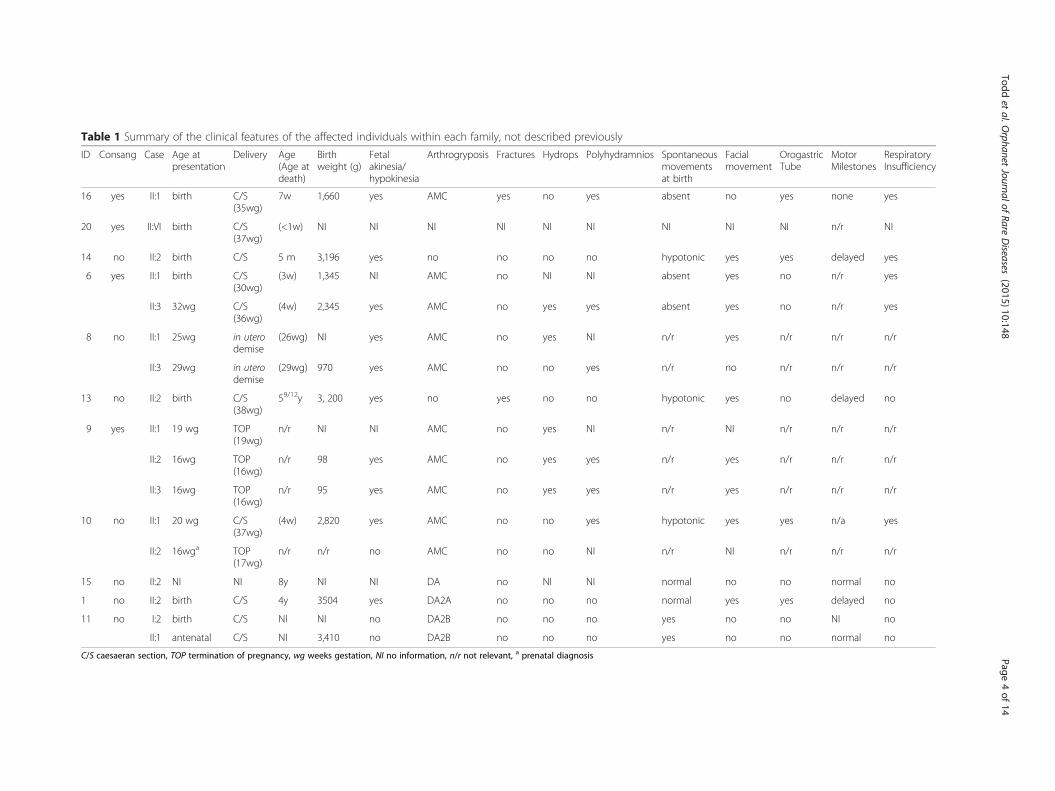
Table 1 Summary of the clinical features of the affected individuals within each family, not described previously
ID Consang Case Age atpresentation
Delivery Age(Age atdeath)
Birthweight (g)
Fetalakinesia/hypokinesia
Arthrogryposis Fractures Hydrops Polyhydramnios Spontaneousmovementsat birth
Facialmovement
OrogastricTube
MotorMilestones
RespiratoryInsufficiency
16 yes II:1 birth C/S(35wg)
7w 1,660 yes AMC yes no yes absent no yes none yes
20 yes II:VI birth C/S(37wg)
(<1w) NI NI NI NI NI NI NI NI NI n/r NI
14 no II:2 birth C/S 5 m 3,196 yes no no no no hypotonic yes yes delayed yes
6 yes II:1 birth C/S(30wg)
(3w) 1,345 NI AMC no NI NI absent yes no n/r yes
II:3 32wg C/S(36wg)
(4w) 2,345 yes AMC no yes yes absent yes no n/r yes
8 no II:1 25wg in uterodemise
(26wg) NI yes AMC no yes NI n/r yes n/r n/r n/r
II:3 29wg in uterodemise
(29wg) 970 yes AMC no no yes n/r no n/r n/r n/r
13 no II:2 birth C/S(38wg)
59/12y 3, 200 yes no yes no no hypotonic yes no delayed no
9 yes II:1 19 wg TOP(19wg)
n/r NI NI AMC no yes NI n/r NI n/r n/r n/r
II:2 16wg TOP(16wg)
n/r 98 yes AMC no yes yes n/r yes n/r n/r n/r
II:3 16wg TOP(16wg)
n/r 95 yes AMC no yes yes n/r yes n/r n/r n/r
10 no II:1 20 wg C/S(37wg)
(4w) 2,820 yes AMC no no yes hypotonic yes yes n/a yes
II:2 16wga TOP(17wg)
n/r n/r no AMC no no NI n/r NI n/r n/r n/r
15 no II:2 NI NI 8y NI NI DA no NI NI normal no no normal no
1 no II:2 birth C/S 4y 3504 yes DA2A no no no normal yes yes delayed no
11 no I:2 birth C/S NI NI no DA2B no no no yes no no NI no
II:1 antenatal C/S NI 3,410 no DA2B no no no yes no no normal no
C/S caesaeran section, TOP termination of pregnancy, wg weeks gestation, NI no information, n/r not relevant, a prenatal diagnosis
Toddet
al.Orphanet
JournalofRare
Diseases
(2015) 10:148 Page
4of
14

published: GPR126 (Family 3) [30], KLHL40 (Family 10and 17; OMIM 615340) [24], KLHL41 (Patient ID: D12-203; OMIM 607701) [22] and SPEG (Patient ID: P3;OMIM 615950) [20].
Mutations in fetal hypokinesia and congenital myopathygenesKLHL40Since our initial publication of KLHL40 as a novel NEMgene, two further families within our cohort were shownto have mutations in KLHL40 (Families 16 and 20). A
previously-unpublished homozygous nonsense mutationin KLHL40 (exon 1, c.46C>T, p.Gln16*) was identified ina proband from consanguineous parents (Family 16,Fig. 1a). This proband was born by emergency Caesareansection at 35/40 weeks gestation and presented with se-vere arthrogryposis, congenital fractures, respiratory in-sufficiency and complete akinesia. An initial clinicaldiagnosis of spinal muscular atrophy type 0 was made,but both light and electron microscopy of the child’smuscle biopsy demonstrated miliary nemaline bodies(Fig. 2), adding to the body of evidence suggesting that
Table 2 Mutations identified via next generation sequencing
Family Chr Position Gene Exon/(Intron) Transcript c. DNA change Amino acid change Alleles in ExAC
3a,WES 3:42728042 KLHL40 1 NM_152393 c.932G>T p.Arg311Leu Not found
3:42730455 4 c.1516A>C p.Thr506Pro A:7/C:121308
4a,WES 3:42730521 KLHL40 4 c.1582G>A p.Glu528Lys G:8/A:120018
3:42730521 4 c.1582G>A p.Glu528Lys G:8/A:120018
16WES 3:42727156 KLHL40 1 c.46C>T p.Gln16* Not found
3:42727156 1 c.46C>T p.Gln16* Not found
20NSES 3:42728041 KLHL40 1 c.931C>A p.Arg311Ser C:6/A:121368
3:42728041 1 c.931C>A p.Arg311Ser C:6/A:121368
5a,WES 2:170382132_9 KLHL41 6 NM_006063 c.1748_1755del8 p.Lys583Thrfs*7 Not found
2:170382132_9 6 c.1748_1755del8 p.Lys583Thrfs*7 Not found
12a,WES 2:220331929_30 SPEG 12 NM_005876 c.2915_2916del2insA p.Ala972Aspfs*79 Not found
2:220331929_30 37 c.8270G>T p.Gly2757Val Not found
38a,WES, NSES 6:142729324 GPR126 16 NM_198569 c.2306T>A p.Val769Glu Not found
6:142729324 16 c.2306T>A p.Val769Glu Not found
14NSES 23:149809808 MTM1 8 NM_000252 c.595C>T p.Pro199Ser Not found
6WES 19:38951109 RYR1 20 NM_000540 c.2455C>T p.Arg819* C:1/T:121396
19:38980890 36 c.5989G>A p.Glu1997Lys Not found
8WES 19:38987106 RYR1 41 c.6721C>T p. Arg2241* C:20/T:120468
19:39071143 101 c.14645C>T p.Thr4882Met C:2/T:121280
13NSES 19:38946103 RYR1 15 c.1589G>A p.Arg530His G:8/A:121410
19:39071143 101 c.14645C>T p.Thr4882Met C:2/T:121280
9WES 2:152539199 NEB 29 NM_001164508 c.2920C>T p.Arg974* Not found
2:152539199 29 c.2920C>T p.Arg974* Not found
2a,WES 3:81698005 GBE1 (5) NM_000158 c.691+2T>C ? T:113/C:98680
3:81691968 7 c.956A>G p.His319Arg Not found
10WES 2:233394798 CHRND 7 NM_000751 c.769T>C p.Cys257Arg Not found
2:233398996 11 c.1315delG p.Val439Trpfs*11 Not found
15NSES 2:233406191_2 CHRNG 5 NM_005199 c.459dupA p.Val154Serfs*24 CA:32/C:121406
2:233406191_2 5 c.459dupA p.Val154Serfs*24 CA:32/C:121406
1WES 17:10544634 MYH3 18 NM_002470 c.2015G>A p.Arg672His Not found
11NSES 17:10549042 MYH3 12 c.1123G>A p.Glu375Lys Not found
7a,WES 2:233347865 ECEL1 9 NM_004826 c.1531G>A p.Gly511Ser Not found
2:233346560 (12) c.1797-1G>A ? Not foundaDenotes families published previously. Type of next-generation sequencing performed for each family is also noted in this table
Todd et al. Orphanet Journal of Rare Diseases (2015) 10:148 Page 5 of 14

Fig. 1 Pedigrees for families in which mutations were identified from next generation sequencing of a proband. Pedigrees and segregation ofthe mutation/s identified within each family is shown for pedigrees not previously described elsewhere. Probands denoted by arrowheads. (a)Family 16 and (b) Family 20 with homozygous KLHL40 mutations; (c) Family 14: X-linked MTM1 mutation; (d) Family 6, (e) Family 8 and (f) Family13 with compound heterozygous mutations of RYR1; (g) Family 9: homozygous NEB mutation; (h) Family 10: compound heterozygous mutationof CHRND; i Family 15: homozygous mutation of CHRNG; (j) Family 1: de novo mutation of MYH3; (k) Family 11: dominantly-inherited mutation ofMYH3. Pedigrees for Family 223, 3-424, 522, 757, 1220 and 3830 are published previously
Fig. 2 Evolutionary conservations of substituted residues in three families harbouring novel missense substitutions. Evolutionary conservation ofthe substituted amino acid in KLHL40 in Family 20 (a), RYR1 in Family 6 (b) and CHRND in Family 10 (c)
Todd et al. Orphanet Journal of Rare Diseases (2015) 10:148 Page 6 of 14

miliary nemaline bodies are a good indicator suggestingKLHL40 as the causative gene.The proband in Family 20 was born to consanguineous
parents (Fig. 1b) by Caesarean section at 37 weeks gesta-tion. He had profound hypotonia, an absent gag reflex,myopathic facies, and was ventilated from birth, but sur-vived only a few days. His muscle biopsy showed numer-ous nemaline bodies. The family history included twoprevious miscarriages, two neonatal deaths and a siblingwho died at seven months of age with suspected NEM(light microscopy indicated rods, but electron micros-copy was not performed). No mutations were found onSanger sequencing of ACTA1, but NSES showed a novelhomozygous missense mutation in KLHL40 (exon 1,c.931C>A, p.Arg311Ser) affecting the same highly con-served amino acid residue as that in Family 3 (Fig. 3a).
MTM1The second male child of a non-consanguineous family(Family 14, Fig. 1c) was born after an uncomplicatedpregnancy, by emergency Caesarean section for failureto progress. The baby was weak and hypotonic at birth,was very long (reported >90th percentile for length withweight 10-25th percentile), had advanced bone age, andinitially required intubation. By age 5 months the infant’sstrength and spontaneous movement improved mark-edly, but he had significant residual weakness and bulbardysfunction. NSES identified a known missense muta-tion (exon 8, c.595C>T, p.Pro199Ser) in the myotubu-larin gene (MTM1; OMIM 300415) [31] associated withmyotubular myopathy. A muscle biopsy taken at10 weeks of age revealed hypoplastic myofibres, somewith internal nuclei, typical features of myotubular my-opathy (OMIM 310400) [32]. However, enzyme stainingshowed reduced central staining in some myofibres,while electron microscopy showed foci of sarcomericdissolution, suggestive of cores. IHC for myosin con-firmed the preservation of type II/fast myofibres and nu-merous small type I myofibres. Thus a diagnosis ofcongenital myopathy with fibre-type disproportion andoccasional minicores had been suggested (Fig. 2d-e).This highlights that MTM1 cases can present with con-genital weakness and muscle biopsies displaying featuresof fibre type disproportion and minicores.
RYR1The proband and affected sibling of Family 6 (Fig. 1d),were born to consanguineous parents. The proband wasborn at 30 weeks gestation with profound hypotonia, fa-cial weakness, dysmorphic features and ambiguous geni-talia, after a pregnancy complicated by fetal hypokinesia.He died at 3 weeks of age. A subsequent pregnancy witha female sibling was complicated by polyhydramnios. Atbirth there was minimal limb movement, respiratory
distress necessitating mechanical ventilation, subcutane-ous oedema, contractures of the hips and knees andcamptodactyly of the fingers. She died at 4 weeks of age.Maternal testing for myotonic dystrophy (DM1) wasnegative. Vastus lateralis biopsies from both babiesshowed non-specific abnormalities of myofibre typing,with type II myofibre predominance and numeroussmall myofibres. Occasional minicores and cores wereseen in the proband but not his sibling. Neither hadnemaline bodies or histologic features of myotubularmyopathy. Exome sequencing performed on the probandrevealed two mutations in the ryanodine receptor gene(RYR1, OMIM 180901): a novel heterozygous missensemutation affecting a highly conserved amino acid(Fig. 3b) (exon 36, c.5989G>A, p.Glu1997Lys) and a het-erozygous previously-reported nonsense mutation (exon20, c.2455C>T, p.Arg819* [33]). Sanger sequencing con-firmed these mutations and showed co-segregation withdisease. Thus in this instance, the consanguinity doesnot appear to be a contributing factor in the siblings’disease. The nonsense mutation was previously identi-fied in a 49-year old ambulant patient with a moderateform of slowly-progressive myopathy with cores [33].That patient also harboured a previously identified het-erozygous missense mutation (p.Arg4558Gln) [33, 34].Thus the same nonsense mutation, in combination withdifferent missense mutations, can result in variable phe-notypes, from fetal hypokinesia and death in the peri-natal period, to a mild delay in motor milestones andnormal life expectancy.The proband in non-consanguineous Family 8 (Fig. 1e)
presented with non-immune hydrops fetalis and arthro-gryposis, and was stillborn at 26 weeks gestation.Autopsy showed multiple contractures and reducedmuscle bulk. Microscopically, there were marked dys-trophic changes in all muscles examined (Fig. 2g). Thecontactin-1 gene (CNTN1, OMIM 600016) was Sangersequenced but no mutations were identified. A subse-quent pregnancy with a male fetus was complicated bypolyhyhydramnios, contractures, and in utero fetal de-mise at 29 weeks gestation. Both affected individualswere diagnosed with FADS and congenital muscular dys-trophy. Exome sequencing of the proband identified twopreviously reported heterozygous mutations in the RYR1gene; a nonsense mutation (exon 41, c.6721C>T,p.Arg2241* [35]) and a missense mutation (exon 101,c.14645C>T, p.Thr4882Met [36]) associated with multi-minicore disease and core rod disease, respectively.Sanger sequencing confirmed compound heterozygosityin both affected individuals, and showed that both par-ents were carriers and that the unaffected sibling did notharbour either mutation.The affected individual in Family 13 was born to non-
consanguineous Turkish parents (Fig. 1f ) after reports of
Todd et al. Orphanet Journal of Rare Diseases (2015) 10:148 Page 7 of 14

reduced intrauterine movement. He was delivered atterm by Caesarean section, due to poor positioning. Atbirth bilateral humeral fractures were noted. He had adiagnosis of osteogenesis imperfecta, and followed upwith alendronate treatment. He was referred to pediatricneurology outpatient clinic at the age of 19 monthswhen the parents had concerns in terms of hypotoniaand delay in motor developmental milestones. At thetime, he remained hypotonic with a myopathic face andhigh-arched palate. He had axial and vertical hypotonia,head lag, facial weakness, and absence of deep tendonreflexes. He could sit but not stand. The muscle biopsyshowed muscle tissue embedded in fibro-adipose tissuewith severe non-specific myopathic changes (Fig. 2f ).There were hypertrophic and atrophic myofibres, centralnuclei, type II myofibre predominance and some core-likeregions on oxidative enzyme stains. Exome sequencing of
the proband revealed two pathogenic missense mutationsin the RYR1 gene, (exon 15, c.1589G>A, p.Arg530His [37],exon 101, c.14645C>T, p.Thr4882Met [36]) which hadpreviously been associated with central core disease/ma-lignant hyperthermia (MH) and core rod myopathy, re-spectively. The p.Arg530His substitution was inheritedpaternally, thus the presence of this MH (OMIM 145600)susceptibility mutation in both the proband and asymp-tomatic father changes their clinical management.Thus, affected individuals in three families (Family 6,
8, and 13), harbored compound heterozygous mutationsin RYR1. Disease severity was much greater in the twofamilies possessing a nonsense (null) mutation as well asa missense mutation (Family 6 and 8), resulting in deathat or soon after birth. The affected individual in the thirdRYR1 family, (Family 13), possessed two missense muta-tions, and survived infancy, albeit with severe muscle
Fig. 3 Histology of muscle biopsies from four families with mutations identified in the proband. Family 16 (a-c): h&e indicating variation inmyofibre diameter (a) and Gomori trichrome staining showing dark purple regions suggesting nemaline bodies (arrows) (b). Electron micrograph,arrows indicate miliary nemaline bodies (c). (d) H&E stain of muscle from the proband in Family 14, indicating variation in myofibre size, centraland internal nuclei. (e) Staining for NADH-TR in muscle from the proband in Family 14 with arrows indicating reduced central staining indicativeof minicores. (f) H&E staining of muscle from the proband in Family 13 showing muscle tissue embedded in fibro-adipose tissue, with severemyopathic, non-specific changes. (g) H&E staining of muscle from the proband in Family 8, demonstrating a severe non-specific picture
Todd et al. Orphanet Journal of Rare Diseases (2015) 10:148 Page 8 of 14

weakness and motor delay. He had a rather static improv-ing course with physiotherapy. These findings mirrorthose of recent publications expanding the phenotypes as-sociated with recessive RYR1 disease to include arthrogry-posis multiplex congenita and fetal akinesia [9, 38, 39].Despite RYR1 originally being described as a disease genefor central core disease and minicore disease, cores areseen in only a minority of recessive RYR1 cases, and areless likely to be seen in cases with hypomorphic (null)muations [38, 39]. In this study, cores were not a promin-ent feature in two of the recessive RYR1 families, both ofwhich harboured a hypomorphic mutation.
NEBA consanguineous family (Family 9) presented early inpregnancy with monoamniotic male twins (Fig. 1g) anda history of a previous fetus therapeutically aborted dueto hydrops fetalis at 19 weeks gestation. Ultrasoundscanning revealed severe hydrops in both fetuses, andthe pregnancy was terminated at 16 weeks gestation.Post-mortem analysis of both twins showed bilateraljoint contractures, bilateral talipes, multiple pterygia,hypertelorism and cystic hygromas. Muscle biopsieswere not taken. A diagnosis of fetal akinesia with lethalmultiple pterygia syndrome was made. Karyotypingshowed a normal 46XY karyotype, with no apparentgenomic imbalance. Exome sequencing was performedon one twin, and a novel homozygous nonsense muta-tion (exon 29, c.2920C>T, p.Arg974*) in the nebulingene (NEB; OMIM 161650) was identified. Sanger se-quencing confirmed that both twins were homozygousfor this mutation and that each parent was a carrier(Fig. 1). This mutation was included in the recent NEBmutation update [40]. Although this case was diagnosedas FADS/lethal multiple pterygia syndrome, recessivemutations in the NEB gene are a known cause of NEM,which in severe cases can have a FADS phenotype [41].Without a muscle biopsy however, it cannot be deter-mined whether these cases had nemaline myopathy.In three additional families, diagnosed with NEM pre-
senting with fetal akinesia, single heterozygous patho-genic mutations were identified in NEB by either exomesequencing or NSES (Table 3). In Family 17 a knownsplice-site mutation (intron 5, c.78+1G > A, [42]) wasidentified, and in Family 19 a known frameshift mutation(exon 55, c.7523_7526del4, p.Ile2508Thrfs*14, [43]), wasidentified, both of which are associated with NEM. In
Family 18, a previously unpublished nonsense mutation(exon 29, c.2864G>A, p.Trp955*) was identified. A com-mon deletion of exon 55 of NEB, originating within theAshkenazi Jewish population, is known to cause a severeNEM phenotype [44]. A heterozygous deletion of thisexon would not be identifiable through next generationsequencing techniques. Deletion analysis was performedon the affected individuals of Family 17 and Family 18,which confirmed they did not have a deletion of this exon.The proband in Family 19 could not have harboured a de-letion of exon 55, since the exon 55 variant identified inthis proband was heterozygous. Although only single het-erozygous mutations were identified in these three severeNEM cases, given their severity and the absence of likelypathogenic variants in the other known NEM genes, it islikely that they are harbouring a second pathogenic NEBvariant that was not identified by next generation sequen-cing. In support of AR NEM, Family 18 and 19 both had apreviously affected fetus. In further support that thesecases (three of nine NEM families, 33 %) are harbouringan additional pathogenic NEB variant, only one truncatingNEB variant was identified by next generation sequencingin non-NEM cases, of which we have sequenced and ana-lysed in excess of >500 probands (~0.2 %). Due to thehighly repetitive nature of exons 83–105 of NEB, nextgeneration sequencing is unable to accurately sequenceand map this region; in addition, next generation sequen-cing data is not reliable for the detection of small CNVs.However a targeted NEB array CGH has been developedas an adjunct to overcome these limitations [45] and hasrecently identified a recurrent CNV within this triplicatedrepeat [46].Therefore, of the nine NEM cases in our cohort, five
cases had mutations in the newly described genesKLHL40 and KLHL41, and an additional three cases arethought likely to harbour a second pathogenic mutationin NEB. It is likely that many undiagnosed NEM casesare due to mutations in NEB, however due to its size ithas not been routinely screened. With the introductionof next generation sequencing techniques, more NEB-re-lated NEM cases are beginning to be identified. Thismay mean that there are not as many new NEM genesto find as might have been thought.
GBE1A non-consanguineous family (Family 2) presented withrecurrent fetal akinesia and multiple pterygium syndrome
Table 3 Single heterozygous mutations identified in NEB in three families presenting with fetal hypokinesia-NEM
Family Exon/(Intron) c. DNA change NM_001164507.1 Amino acid change Comments
17 (5) c.78+1G>A
18 29 c.2864G>A p.Trp955* Also present in affected sib
19 55 c.7523_7526del4 p.Ile2508Thrfs* Affected sib (no DNA available)
Todd et al. Orphanet Journal of Rare Diseases (2015) 10:148 Page 9 of 14

[23]. We identified compound heterozygous mutations inthe gene GBE1, a known splice site mutation (intron 5,c.691+2T>C) associated with a non-lethal neonatal glyco-genosis type IV, and a missense mutation (exon 7,c.956A>G, p.His319Arg). This report extended the pheno-typic spectrum of GBE1 disease to include lethal multiplepterygium syndrome [23].
Mutations in known disease genes for arthrogryposesCHRNDThe proband in Family 10 was the first child to non-consanguineous parents, born following an IVF preg-nancy, (Fig. 1h). A routine 20-week ultrasound identi-fied bilateral fetal talipes. Chromosome microarraywas normal. The fetal phenotype evolved with polyhy-dramnios, fetal micrognathia and an absence of handmovements noted at 32 weeks. The polyhydramniosrequired three amnioreduction procedures. The maleinfant was delivered by elective Caesarian section forplacenta praevia at 37 weeks gestation and weighed2.82 kg. He was intubated and ventilated at 10 mi-nutes for apnoea and poor respiratory effort afterAPGARS of 51, 65 and 710. He had micrognathia,cryptorchidism, a left single palmar crease, bilateraltalipes, moderate large joint contractions, hypotonia,an absent gag/suck, and paucity of movement. He de-veloped a weak suck and infrequent antigravity move-ment of the fingers after a week. Prader-Willisyndrome, SMA and myotonic dystrophy were ex-cluded. Endocrine and metabolic investigations werenormal as was the ophthalmologic examination. BrainMRI showed a right MCA infarct in the context ofpositive maternal serology for SLE. Multiple attemptsto extubate the patient to CPAP failed. Ongoing ven-tilatory support was considered futile and was with-drawn at 4 weeks of age. Exome sequencing wasperformed and two novel heterozygous mutationswere identified in CHRND (OMIM 100720) that en-codes the delta-subunit of the acetylcholine receptor(AChR) [47]. A missense mutation (c.769T>C) inexon 7 that resulted in substitution of a highly con-served amino acid (p.Cys257Arg, Fig. 3c) and aframeshift mutation in exon 11 (c.1315delG,p.Val439Trpfs*11). To our knowledge neither of thesemutations have been previously reported and are notlisted in the CHRND locus-specific database (http://www.dmd.nl/nmdb/home.php?select_db=CHRND).Sanger sequencing confirmed the presence of the mu-tations in the affected individual and showed eachparent was a carrier of one of the variants. Themother conceived a second time, naturally. Prenataldiagnosis was performed and the fetus had both vari-ants. The pregnancy was terminated.
Studies in HEK cells, found that cell surface expressionlevels of AChRs harbouring the δC257R subunit to beapproximately 20 % of wild-type (Fig. 4). This result isconsistent with the c.769T>C mutation (in combinationwith c.1315delG, p.Val439Trpfs*11 on the second allele)underlying a congenital myasthenic syndrome due toAChR deficiency. The mother is currently pregnant andis approaching term with a healthy fetus following PGD.Mutations of CHNRD typically result in congenital my-asthenic syndromes (OMIM 608930 (fast-channel) and601462 (slow-channel) [48, 49]). but have also more re-cently been associated with lethal multiple pterygiumsyndrome [50]. In two families presenting with recurrentlethal multiple pterygium syndrome, resulting in termi-nations during the second trimester of pregnancy, nullmutations of CHRND were identified (one consanguin-eous family with a homozygous p.Trp57* mutation andone with compound heterozygous p. Phe74Leu andp.Arg464* mutations). Substitutions of amino acids inclose proximity to Cys257 have been shown to causecongenital myasthenia and impaired channel function(p.Pro250Gln [51] and p.Ser268Phe [48]).
CHRNGThe affected female individual in Family 15 was born tounrelated parents (Fig. 1i). At birth there was arthrogry-posis with distinctive shin dimples. The clinical pictureof this patient is presented in Hall et al., (Patient 10)[52]. NSES was performed on the proband and revealeda known frequent homozygous frameshift mutation(exon 5, c.459dupA, p. Val154Serfs*24) in the gene en-coding the gamma-subunit of the AChR (CHRNG;
Fig. 4 Expression of wild-type (αβδε) and mutant (αβδC257Rε)acetylcholine receptors (AChR) in HEK 293 cells. AChR expressionwas determined through the binding of 125I α-Bungarotoxin (125I α-BuTx) to AChR on the cell surface (n = 6). Note: numbering of themutation includes the pre-peptide sequence
Todd et al. Orphanet Journal of Rare Diseases (2015) 10:148 Page 10 of 14
OMIM 100730) [53, 54]. Sanger sequencing confirmedthe presence of the mutation in the affected individual,as well as showing each parent had the mutation in theheterozygous state. Given the unique presentation ofarthrogryposis with shin dimples in this case and othersharbouring CHRNG mutations [54], CHRNG should beconsidered in individuals presenting with this particularphenotype.
MYH3The proband in Family 1 was born from unaffectedparents and has an unaffected sibling (Fig. 1j). Hepresented with Freeman-Sheldon Syndrome (DA2A,OMIM 193700) [55] and on examination at 2 yearsof age he showed some facial features and proximalweakness. Exome sequencing of the proband demon-strated heterozygosity for a mutation in MYH3 (exon18, c.2015G>A, p.Arg672His; OMIM 160720 [55];)previously associated with Freeman-Sheldon syn-drome. Sanger sequencing confirmed the presence ofthis mutation in the proband as well as its absence inthe unaffected sibling and both unaffected parents,confirming the mutation was de novo.The male proband from Family 11, was born from an
unaffected mother, but affected father (Fig. 1k). Bothproband and the father were born with a very typicalSheldon-Hall distal arthrogryposis (DA2B) phenotype.Sheldon-Hall syndrome can be caused by autosomaldominant or de novo mutations in a number of genes. Inthis kindred, screening of TPM2 and TNNI2 identifiedno mutations. On NSES, however, a known heterozygousMYH3 mutation (exon 12, c.1123G>A, p.Glu375Lys[55]) previously associated with Freeman-Sheldon syn-drome, was identified. Sanger sequencing confirmed themutation in both the proband and his affected father,confirming autosomal dominant inheritance.
ECEL1Non-consanguineous Family 7, previously described in[56], was also part of this cohort study. The probandwas born from an uncomplicated pregnancy with ex-tended hips, multiple arthrogrypotic features, multiplepterygium, adducted thumbs and bilateral ptosis. Thecouple presented when pregnant again, and on ultra-sound at 20 weeks the fetus appeared to have similarfeatures to those of the proband. The pterygia and ptosisled to consideration of multiple pterygium syndrome(Table 1). Exome sequencing revealed compound hetero-zygous mutations in ECEL1 (OMIM 605896), a missensesubstitution (c.1531G>A, p.Gly511Ser) and a essentialsplice-site mutation (c.1797-1G>A). Mutations in ECEL1are associated with distal arthrogryposis type 5D (OMIM615065), and the clinical presentation was in keeping with
those recently described for DA5D [13, 57], although pte-rygia was a more prominent feature in this family.In another cohort study, Laquerriere et al. identified
two novel genes (CNTNAP1 and ADCY6) for severearthrogryposis multiplex congenita (AMC) by exome se-quencing, and achieved a genetic diagnosis for 24 of 31multiplex and/or consanguineous AMC families studied(>75 %). This highlights the importance of working withwell-phenotyped cohorts [9]. Mutations in CNTNAP1were identified in four of their 31 families, suggestingthat mutations in this gene underlie a significant propor-tion of recessive AMC cases.Results from our study, and that of Laquerriere et al.,
suggest that there are further arthrogryposis diseasegenes to be identified [9]. ADCY6 and CNTNAP1 areboth involved in axonal function [9], as is ECEL1 [57].GPR126 is critical for myelination of peripheral nerves[58] and we identified AMC patients with loss-of-function mutations in GPR126 [30]. Genes involved inaxonal function should therefore be considered as candi-dates for arthrogryposis, in addition to skeletal musclecontractile proteins.Our study highlights the widening spectrum of phe-
notypes associated with mutations in known fetalakinesia, arthrogryposis and myopathy genes, as is in-creasingly demonstrated for other neuromuscular dis-orders [26, 59, 60]. As sequencing of targeted genepanels or exome sequencing becomes the mainstay ofgenetic diagnostics [61, 62], it is likely that there willbe greater broadening of genotype-phenotype correla-tions for neuromuscular diseases. With the over-whelming amount of genetic information obtained vianext generation sequencing, the reliability of meticu-lously curated locus-specific databases, the availabilityof large exome datasets from ethnically matched ref-erence populations and appropriate functional and/orprotein studies will be critical to obtaining accurategenetic diagnosis. Given that numerous novel diseasegenes and mutations are being described in non-Caucasian inbred populations [63] and genetic isolates[64], there is a real need for exome sequencing ofhealthy individuals within these populations.Within our cohort, three novel disease genes were ini-
tially identified by exome sequencing of single probands(GPR126, KLHL41 and SPEG). The success of disease genediscovery in NEM (KLHL40, KLHL41, LMOD3, MYO18B)and centronuclear myopathies (SPEG) is likely due to theability to identify patients with a very similar presentation(clinically and based upon very specific muscle biopsyfindings) such as to enable screening of candidate genes inpatients with the same disease [21, 65].For fetal hypokinesia and arthrogryposis cases, it is
more difficult to deeply phenotype the patients, due inmany cases to the poor preservation of fetal tissue and the
Todd et al. Orphanet Journal of Rare Diseases (2015) 10:148 Page 11 of 14

lack of specific pathological hallmarks from biopsy or aut-opsy material. A recent study describes exome sequencingof 143 multiplex consanguineous families, in which 33novel candidate neurogenic disease genes were identified[63], highlighting the value of studying consanguineousfamilies. As a comparison, only three of the 20 (15 %)genetically-unresolved cases were consanguineous whereasseven of 18 of the genetically-diagnosed cases were consan-guineous (39 %, Additional file 1: Table S1), thus one is 2.5-times more likely to identify the causative disease gene inconsanguineous families. A genetic diagnosis was achievedin ten of 16 congenital myopathy cases (63 %) and six of 13arthrogryposis cases (46 %) but only 22 % of fetal akinesiacases (two of nine). It is also possible that the cause of dis-ease, in some of the isolated cases (particularly those diag-nosed with fetal akinesia), is not due to a monogenicdisorder but may be environmental and/or polygenic. Infamilies with multiple affected siblings and normal CGH ar-rays, we will pursue whole genome sequencing and/orRNA-seq of target tissue cDNA to try to identify novel dis-ease genes and/or mechanisms.
ConclusionsIn summary, this study highlights the use of next generationsequencing to genetically diagnose 47 % of cases within aheterogeneous severe neuromuscular disease cohort. Thestudy has also resulted in the identification of four novelneuromuscular disease genes, and has led to the identifica-tion of a novel mechanism of sarcomere assembly andmuscle dysfunction involving KLHL40, KLHL41 andLMOD3 [21, 66, 67]. Finally, this study has contributed toextending the phenotypic spectrum of CHRNG, ECEL1,GBE1 and RYR1.
Additional file
Additional file 1: Table S1. For each family the affected individualssequenced are shown as well as the type of NGS that was performed foreach patient. The disease group and consanguinity for each family is alsoindicated as are the coverage statistics for each sample and NGS.(DOCX 21 kb)
AbbreviationsAMC: Arthrogryposis multiplex congenita; DA: Distal arthrogryposis;FADS: Fetal akinesia deformation sequence; LSBFG: Lotterywest statebiomedical facility genomics node; NEM: Nemaline myopathy;NSES: Neuromuscular sub-exomic sequencing.
Competing interestsThe authors declare that they have no competing interests.
Authors’ contributionsGR and NGL conceived and coordinated the project. EJT, KSY, RO, PS, RJNA,SM, DB, MRD and GR performed the experiments and analysed data. JS, GM,CPB, GH, ZA, AK, AC, NFC, M-LF, DW, CAS, AC, MMR and CK-B cared for thepatients and co-ordinated patient enrolment in the study. BT, NBR and CASperformed histology and immunohistochemistry. EJT, CAS, MMR, NGL andGR drafted the manuscript. All authors read and approved the manuscript.
AcknowledgementsWe are grateful to the patients and their families for their participation in thisresearch. This research was supported by the National Health and MedicalResearch Council of Australia (Early Career Researcher Fellowship #1035955to GR, Research Fellowship APP1002147 to NGL and Project GrantAPP1022707; EU Collaborative grant APP1055295); the Association Francaisecontre les Myopathies (#15734) and a UWA Collaborative Research Award.EJT and KSY were supported by University Postgraduate Awards.
Author details1Harry Perkins Institute of Medical Research and the Centre for MedicalResearch, University of Western Australia, QQ Block, 6 Verdun Street,Nedlands 6009, WA, Australia. 2Genetic Services of Western Australia, KingEdward Memorial Hospital, Perth 6000, WA, Australia. 3Victorian ClinicalGenetics Services, Murdoch Children’s Research Institute, The Royal Children’sHospital, Parkville 3052, VIC, Australia. 4Paediatric and Reproductive GeneticsUnit, South Australia Clinical Genetics Service, Women’s and Children’sHospital, North Adelaide 5006, SA, Australia. 5Department of PediatricNeurology, Hacettepe University Children’s Hospital, Ankara 06100, Turkey.6Pediatric Pathology Unit, Hacettepe University Children’s Hospital, Ankara06100, Turkey. 7Kariminejad-Najmabadi Pathology and Genetics Centre,Tehran 14656, Iran. 8Royal Children’s Hospital, Herston Road, Herson 4029,QLD, Australia. 9Institute for Neuroscience and Muscle Research, TheChildren’s Hospital at Westmead, Sydney 2145, NSW, Australia. 10Discipline ofPaediatrics and Child Health, University of Sydney, Sydney 2006, NSW,Australia. 11Sydney Children’s Hospital, High Street, Randwick 2031, NSW,Australia. 12Unitè de Morphologie Neuromusculaire, Institut de Myologie,Institut National de la Santè et de la Recherche Mèdicale, Paris 75651, France.13Dubowitz Neuromuscular Centre, UCL Institute of Child Health, LondonWC1N 1EH, UK. 14Wolfson Centre for Neuromuscular Disorders, RJAHOrthopaedic Hospital, Oswestry SY10 7AG, UK. 15Department of ClinicalGenetics, South Western Sydney Local Health District, Liverpool 1871, NSW,Australia. 16Department of Neurology, The Royal Children’s Hospital,Melbourne 3000, VIC, Australia. 17Genetic Services of Western Australia,Princess Margaret Hospital for Children and King Edward Memorial Hospitalfor Women, Subiaco 6008, WA, Australia. 18Department of DiagnosticGenomics, Pathwest, QEII Medical Centre, Nedlands 6009, WA, Australia.19Lotterywest State Biomedical Facility Genomics and School of Pathologyand Laboratory Medicine, University of Western Australia, Perth 6000, WA,Australia. 20Nuffield Department of Clinical Neurosciences, WeatherallInstitute of Molecular Medicine, University of Oxford, Oxford OX3 9DS, UK.
Received: 20 August 2015 Accepted: 2 November 2015
References1. Hall JG. Analysis of Pena Shokeir phenotype. Am J Med Genet.
1986;25:99–117.2. Hall JG. Pena-Shokeir phenotype (fetal akinesia deformation sequence)
revisited. Birth Defects Res A Clin Mol Teratol. 2009;85:677–94.3. Hall JG. Arthrogryposis multiplex congenita: etiology, genetics,
classification, diagnostic approach, and general aspects. J PediatrOrthop B. 1997;6:159–66.
4. Quinn CM, Wigglesworth JS, Heckmatt J. Lethal arthrogryposis multiplexcongenita: a pathological study of 21 cases. Histopathology. 1991;19:155–62.
5. Ravenscroft G, Sollis E, Charles AK, North KN, Baynam G, Laing NG. Fetalakinesia: review of the genetics of the neuromuscular causes. J Med Genet.2011;48:793–801.
6. Filges I, Hall JG. Failure to identify antenatal multiple congenitalcontractures and fetal akinesia–proposal of guidelines to improve diagnosis.Prenat Diagn. 2013;33:61–74.
7. Wilbe M, Ekvall S, Eurenius K, Ericson K, Casar-Borota O, Klar J, et al. MuSK: anew target for lethal fetal akinesia deformation sequence (FADS). J MedGenet. 2015;52(3):195–202.
8. Haliloglu G, Topaloglu H. Arthrogryposis and fetal hypomobility syndrome.Handb Clin Neurol. 2013;113:1311–9.
9. Laquerriere A, Maluenda J, Camus A, Fontenas L, Dieterich K, Nolent F, et al.Mutations in CNTNAP1 and ADCY6 are responsible for severe arthrogryposismultiplex congenita with axoglial defects. Hum Mol Genet. 2014;23:2279–89.
10. Bamshad M, Jorde LB, Carey JC. A revised and extended classification of thedistal arthrogryposes. Am J Med Genet. 1996;65:277–81.
Todd et al. Orphanet Journal of Rare Diseases (2015) 10:148 Page 12 of 14

11. Bamshad M, Van Heest AE, Pleasure D. Arthrogryposis: a review and update.J Bone Joint Surg Am. 2009;91 Suppl 4:40–6.
12. McMillin MJ, Beck AE, Chong JX, Shively KM, Buckingham KJ,Gildersleeve HI, et al. Mutations in PIEZO2 cause Gordon syndrome,Marden-Walker syndrome, and distal arthrogryposis type 5. Am J HumGenet. 2014;94:734–44.
13. McMillin MJ, Below JE, Shively KM, Beck AE, Gildersleeve HI, Pinner J, et al.Mutations in ECEL1 cause distal arthrogryposis type 5D. Am J Hum Genet.2013;92:150–6.
14. Romero NB, Clarke NF. Congenital myopathies. Handb Clin Neurol.2013;113:1321–36.
15. Ravenscroft G, Laing NG, Clarke NF. Chapter 28-Congenital/ultrastructuralmyopathies. In: Hilton-Jones D, editor. Oxford Textbook of NeuromuscularDisorders. 2014.
16. Maggi L, Scoto M, Cirak S, Robb SA, Klein A, Lillis S, et al. Congenitalmyopathies–clinical features and frequency of individual subtypesdiagnosed over a 5-year period in the United Kingdom. NeuromusculDisord. 2013;23:195–205.
17. Nance JR, Dowling JJ, Gibbs EM, Bonnemann CG. Congenital myopathies:an update. Curr Neurol Neurosci Rep. 2012;12:165–74.
18. Jain RK, Jayawant S, Squier W, Muntoni F, Sewry CA, Manzur A, et al.Nemaline myopathy with stiffness and hypertonia associated with anACTA1 mutation. Neurology. 2012;78:1100–3.
19. Majczenko K, Davidson AE, Camelo-Piragua S, Agrawal PB, Manfready RA, LiX, et al. Dominant mutation of CCDC78 in a unique congenital myopathywith prominent internal nuclei and atypical cores. Am J Hum Genet.2012;91:365–71.
20. Agrawal PB, Pierson CR, Joshi M, Liu X, Ravenscroft G, Moghadaszadeh B,et al. SPEG Interacts with Myotubularin, and Its Deficiency CausesCentronuclear Myopathy with Dilated Cardiomyopathy. Am J Hum Genet.2014;95:218–26.
21. Yuen M, Sandaradura SA, Dowling JJ, Kostyukova AS, Moroz N, Quinlan KG,et al. Leiomodin-3 dysfunction results in thin filament disorganization andnemaline myopathy. J Clin Invest. 2014;124:4693–708.
22. Gupta VA, Ravenscroft G, Shaheen R, Todd EJ, Swanson LC, Shiina M, et al.Identification of KLHL41 Mutations Implicates BTB-Kelch-MediatedUbiquitination as an Alternate Pathway to Myofibrillar Disruption inNemaline Myopathy. Am J Hum Genet. 2013;93:1108–17.
23. Ravenscroft G, Thompson EM, Todd EJ, Yau KS, Kresoje N, Sivadorai P, et al.Whole exome sequencing in foetal akinesia expands the genotype-phenotype spectrum of GBE1 glycogen storage disease mutations.Neuromuscul Disord. 2013;23:165–9.
24. Ravenscroft G, Miyatake S, Lehtokari VL, Todd EJ, Vornanen P, Yau KS, et al.Mutations in KLHL40 Are a Frequent Cause of Severe Autosomal-RecessiveNemaline Myopathy. Am J Hum Genet. 2013;93:6–18.
25. Kaplan JC. The 2012 version of the gene table of monogenicneuromuscular disorders. Neuromuscul Disord. 2011;21:833–61.
26. Cabrera-Serrano M, Ghaoui R, Ravenscroft G, Johnsen RD, Davis MR, CorbettA, et al. Expanding the phenotype of GMPPB mutations. Brain.2015;138:836–44.
27. Clarke L, Zheng-Bradley X, Smith R, Kulesha E, Xiao C, Toneva I, et al. The1000 Genomes Project: data management and community access. NatMethods. 2012;9:459–62.
28. Flanagan SE, Patch AM, Ellard S. Using SIFT and PolyPhen to predict loss-of-function and gain-of-function mutations. Genet Test Mol Biomarkers.2010;14:533–7.
29. Schwarz JM, Cooper DN, Schuelke M, Seelow D. MutationTaster2: mutationprediction for the deep-sequencing age. Nat Methods. 2014;11:361–2.
30. Ravenscroft G, Nolent F, Rajagopalan S, Meireles AM, Paavola KJ, Gaillard D,et al. Mutations of GPR126 Are Responsible for Severe ArthrogryposisMultiplex Congenita. Am J Hum Genet. 2015;96:955–61.
31. Flex E, De Luca A, D’Apice MR, Buccino A, Dallapiccola B, Novelli G. Rapidscanning of myotubularin (MTM1) gene by denaturing high-performanceliquid chromatography (DHPLC). Neuromuscul Disord. 2002;12:501–5.
32. Laporte J, Hu LJ, Kretz C, Mandel JL, Kioschis P, Coy JF, et al. A genemutated in X-linked myotubular myopathy defines a new putative tyrosinephosphatase family conserved in yeast. Nat Genet. 1996;13:175–82.
33. Monnier N, Marty I, Faure J, Castiglioni C, Desnuelle C, Sacconi S, et al. Nullmutations causing depletion of the type 1 ryanodine receptor (RYR1) arecommonly associated with recessive structural congenital myopathies withcores. Hum Mutat. 2008;29:670–8.
34. Kossugue PM, Paim JF, Navarro MM, Silva HC, Pavanello RC, Gurgel-GiannettiJ, et al. Central core disease due to recessive mutations in RYR1 gene: is itmore common than described? Muscle Nerve. 2007;35:670–4.
35. Zhou H, Lillis S, Loy RE, Ghassemi F, Rose MR, Norwood F, et al. Multi-minicore disease and atypical periodic paralysis associated with novelmutations in the skeletal muscle ryanodine receptor (RYR1) gene.Neuromuscul Disord. 2010;20:166–73.
36. von der Hagen M, Kress W, Hahn G, Brocke KS, Mitzscherling P, Huebner A,et al. Novel RYR1 missense mutation causes core rod myopathy. Eur J Neurol.2008;15:e31–2.
37. Robinson R, Carpenter D, Shaw MA, Halsall J, Hopkins P. Mutations in RYR1 inmalignant hyperthermia and central core disease. Hum Mutat. 2006;27:977–89.
38. Amburgey K, Bailey A, Hwang JH, Tarnopolsky MA, Bonnemann CG, MedneL, et al. Genotype-phenotype correlations in recessive RYR1-relatedmyopathies. Orphanet J Rare Dis. 2013;8:117.
39. Bharucha-Goebel DX, Santi M, Medne L, Zukosky K, Dastgir J, Shieh PB, et al.Severe congenital RYR1-associated myopathy: the expandingclinicopathologic and genetic spectrum. Neurology. 2013;80:1584–9.
40. Lehtokari VL, Kiiski K, Sandaradura SA, Laporte J, Repo P, Frey JA, et al.Mutation update: the spectra of nebulin variants and associatedmyopathies. Hum Mutat. 2014;35:1418–26.
41. Wallgren-Pettersson C, Donner K, Sewry C, Bijlsma E, Lammens M, Bushby K,et al. Mutations in the nebulin gene can cause severe congenital nemalinemyopathy. Neuromuscul Disord. 2002;12:674–9.
42. Lehtokari VL, Pelin K, Sandbacka M, Ranta S, Donner K, Muntoni F, et al.Identification of 45 novel mutations in the nebulin gene associated withautosomal recessive nemaline myopathy. Hum Mutat. 2006;27:946–56.
43. Lehtokari VL, Greenleaf RS, DeChene ET, Kellinsalmi M, Pelin K, Laing NG,et al. The exon 55 deletion in the nebulin gene–one single foundermutation with world-wide occurrence. Neuromuscul Disord. 2009;19:179–81.
44. Anderson SL, Ekstein J, Donnelly MC, Keefe EM, Toto NR, LeVoci LA, et al.Nemaline myopathy in the Ashkenazi Jewish population is caused by adeletion in the nebulin gene. Hum Genet. 2004;115:185–90.
45. Kiiski K, Laari L, Lehtokari VL, Lunkka-Hytonen M, Angelini C, Petty R, et al.Targeted array comparative genomic hybridization–a new diagnostic toolfor the detection of large copy number variations in nemaline myopathy-causing genes. Neuromuscul Disord. 2013;23:56–65.
46. Kiiski, K., Lehtokari, V.L., Loytynoja, A., Ahlsten, L., Laitila, J., Wallgren-Pettersson, C., and Pelin, K. (2015). A recurrent copy number variation of theNEB triplicate region: only revealed by the targeted nemaline myopathyCGH array. Eur J Hum Genet. Jul 22. doi:10.1038/ejhg.2015.166. [Epub aheadof print].
47. Brownlow S, Webster R, Croxen R, Brydson M, Neville B, Lin JP, et al.Acetylcholine receptor delta subunit mutations underlie a fast-channelmyasthenic syndrome and arthrogryposis multiplex congenita. J Clin Invest.2001;108:125–30.
48. Gomez CM, Maselli RA, Vohra BP, Navedo M, Stiles JR, Charnet P, et al. Noveldelta subunit mutation in slow-channel syndrome causes severe weaknessby novel mechanisms. Ann Neurol. 2002;51:102–12.
49. Muller JS, Baumeister SK, Schara U, Cossins J, Krause S, von der Hagen M,et al. CHRND mutation causes a congenital myasthenic syndrome byimpairing co-clustering of the acetylcholine receptor with rapsyn. Brain.2006;129:2784–93.
50. Michalk A, Stricker S, Becker J, Rupps R, Pantzar T, Miertus J, et al.Acetylcholine receptor pathway mutations explain various fetal akinesiadeformation sequence disorders. Am J Hum Genet. 2008;82:464–76.
51. Shen XM, Ohno K, Fukudome T, Tsujino A, Brengman JM, De Vivo DC, et al.Congenital myasthenic syndrome caused by low-expressor fast-channelAChR delta subunit mutation. Neurology. 2002;59:1881–8.
52. Hall JG. Pretibial linear vertical creases or indentations (shin dimples)associated with arthrogryposis. Am J Med Genet A. 2013;161A:737–44.
53. Morgan NV, Brueton LA, Cox P, Greally MT, Tolmie J, Pasha S, et al.Mutations in the embryonal subunit of the acetylcholine receptor (CHRNG)cause lethal and Escobar variants of multiple pterygium syndrome.Am J Hum Genet. 2006;79:390–5.
54. Robinson KG, Viereck MJ, Margiotta MV, Gripp KW, Abdul-Rahman OA, AkinsRE. Neuromotor synapses in Escobar syndrome. Am J Med Genet A.2013;161A:3042–8.
55. Toydemir RM, Rutherford A, Whitby FG, Jorde LB, Carey JC, Bamshad MJ.Mutations in embryonic myosin heavy chain (MYH3) cause Freeman-Sheldon syndrome and Sheldon-Hall syndrome. Nat Genet. 2006;38:561–5.
Todd et al. Orphanet Journal of Rare Diseases (2015) 10:148 Page 13 of 14

56. Barnett CP, Todd EJ, Ong R, Davis MR, Atkinson V, Allcock R, et al. Distalarthrogryposis type 5D with novel clinical features and compoundheterozygous mutations in ECEL1. Am J Med Genet A. 2014;164A:1846–9.
57. Dieterich K, Quijano-Roy S, Monnier N, Zhou J, Faure J, Smirnow DA, et al.The neuronal endopeptidase ECEL1 is associated with a distinct form ofrecessive distal arthrogryposis. Hum Mol Genet. 2013;22:1483–92.
58. Monk KR, Oshima K, Jors S, Heller S, Talbot WS. Gpr126 is essential forperipheral nerve development and myelination in mammals. Development.2011;138:2673–80.
59. Chong JX, McMillin MJ, Shively KM, Beck AE, Marvin CT, Armenteros JR, et al.De Novo Mutations in NALCN Cause a Syndrome Characterized byCongenital Contractures of the Limbs and Face, Hypotonia, andDevelopmental Delay. Am J Hum Genet. 2015;96:462–73.
60. Rossor AM, Oates EC, Salter HK, Liu Y, Murphy SM, Schule R, et al.Phenotypic and molecular insights into spinal muscular atrophy due tomutations in BICD2. Brain. 2015;138:293–310.
61. Xue, Y., Ankala, A., Wilcox, W.R., and Hegde, M.R. (2014). Solving themolecular diagnostic testing conundrum for Mendelian disorders in the eraof next-generation sequencing: single-gene, gene panel, or exome/genomesequencing. Genet Med. Jun;17 (6):444-51. doi:10.1038/gim.2014.122. Epub2014 Sep 18.
62. Ankala A, Da Silva C, Gualandi F, Ferlini A, Bean LJ, Collins C, et al. Acomprehensive genomic approach for neuromuscular diseases gives a highdiagnostic yield. Ann Neurol. 2015;77:206–14.
63. Alazami AM, Patel N, Shamseldin HE, Anazi S, Al-Dosari MS, Alzahrani F, etal. Accelerating novel candidate gene discovery in neurogenetic disordersvia whole-exome sequencing of prescreened multiplex consanguineousfamilies. Cell Rep. 2015;10:148–61.
64. Tan-Sindhunata, M.B., Mathijssen, I.B., Smit, M., Baas, F., de Vries, J.I., van derVoorn, J.P., Kluijt, I., Hagen, M.A., Blom, E.W., Sistermans, E., et al. (2014).Identification of a Dutch founder mutation in MUSK causing fetal akinesiadeformation sequence. Eur J Hum Genet. Sep;23 (9):1151-7. doi:10.1038/ejhg.2014.273. Epub 2014 Dec 24.
65. Alazami AM, Kentab AY, Faqeih E, Mohamed JY, Alkhalidi H, Hijazi H, et al. Anovel syndrome of Klippel-Feil anomaly, myopathy, and characteristic faciesis linked to a null mutation in MYO18B. J Med Genet. 2015;52:400–4.
66. Garg A, O’Rourke J, Long C, Doering J, Ravenscroft G, Bezprozvannaya S,et al. KLHL40 deficiency destabilizes thin filament proteins and promotesnemaline myopathy. J Clin Invest. 2014;124:3529–39.
67. Cenik BK, Garg A, McAnally JR, Shelton JM, Richardson JA, Bassel-Duby R,et al. Severe myopathy in mice lacking the MEF2/SRF-dependent geneleiomodin-3. J Clin Invest. 2015;125:1569–78.
Submit your next manuscript to BioMed Centraland take full advantage of:
• Convenient online submission
• Thorough peer review
• No space constraints or color figure charges
• Immediate publication on acceptance
• Inclusion in PubMed, CAS, Scopus and Google Scholar
• Research which is freely available for redistribution
Submit your manuscript at www.biomedcentral.com/submit
Todd et al. Orphanet Journal of Rare Diseases (2015) 10:148 Page 14 of 14