nexafs and xps of p-aminobenzoic acid polymorphs: the ... · nexafs and xps of p-aminobenzoic acid...
TRANSCRIPT

This content has been downloaded from IOPscience. Please scroll down to see the full text.
Download details:
IP Address: 155.198.8.192
This content was downloaded on 07/11/2016 at 15:17
Please note that terms and conditions apply.
You may also be interested in:
The Structure of p-Aminobenzoic Acid in Water: Studies Combining UV-Vis, NEXAFS and RIXS
Spectroscopies
A Gainar, J S Stevens, E Suljoti et al.
4-N-pyridin-2-yl-benzamide nanotubes compatible with mouse stem cell and oral deliveryin Drosophila
Jhillu S Yadav, Madugula P Lavanya, Pragna P Das et al.
Organic dyes based on fluorene and its derivatives
I V Kurdyukova and Aleksandr A Ishchenko
Modification of carbon nanotubes and synthesis of polymeric composites involving the nanotubes
E R Badamshina, M P Gafurova and Yakov I Estrin
NEXAFS and XPS of p-Aminobenzoic Acid Polymorphs: The Influence of Local Environment
View the table of contents for this issue, or go to the journal homepage for more
2016 J. Phys.: Conf. Ser. 712 012133
(http://iopscience.iop.org/1742-6596/712/1/012133)
Home Search Collections Journals About Contact us My IOPscience

NEXAFS and XPS of p-Aminobenzoic Acid Polymorphs: The Influence of Local Environment
J S Stevens1, A Gainar1, C Jaye2, D A Fischer2, S L M Schroeder3,4 1School of Chemical Engineering and Analytical Science, The University of Manchester, Oxford Road, Manchester, M13 9PL, UK 2Material Measurement Laboratory, National Institute of Standards and Technology, Gaithersburg, MD, 20899, USA 3School of Chemical and Process Engineering, University of Leeds, Leeds, LS2 9JT, UK 4DIAMOND Light Source Ltd., Chilton, Didcot, OX11 0QX, UK
E-mail: [email protected]; [email protected]
Abstract. Nitrogen K-edge XPS and NEXAFS of the two polymorphic forms of para-aminobenzoic acid (PABA) are significantly different reflecting variation in hydrogen bonding. Alteration in hydrogen bonding at the amino group leads to a shift to high energy for both the XPS N 1s core level and the 3π* NEXAFS resonance with β-PABA. Participation of the amine group in the aromatic system causes the 1π* resonance to be sensitive to the nature of the intermolecular bonding at the para-carboxylic acid group, and a shift to low energy for α-PABA is observed due to hydrogen-bonded carboxylic acid dimer formation. FEFF calculations also successfully reproduce both the energy and intensity variations observed for the σ* shape resonance associated with the C−N bond, with the majority of the decrease in energy observed for β-PABA arising from the longer C−N bond.
1. Introduction The local 3-dimensional environment and interactions of a molecule in the solid-state can have a substantial influence on physical and chemical properties, such as stability, solubility, melting temperature, and reactivity [1]. The ability to characterize and identify such forms (polymorphs) with identical chemical composition but variations in crystal structure (structural packing, interactions, and bonding) is thus extremely important. Understanding or insight into the electronic nature of these materials could provide the basis for investigating how they form in solution, and therefore ultimately how to control the outcome of crystallization [2]. Near-edge X-ray absorption fine structure (NEXAFS) spectroscopy and X-ray photoelectron spectroscopy (XPS) are ideal for this purpose, as they intrinsically probe the local environment of X-ray absorbing atoms. The effects of the local environment and interactions were initially investigated for para-aminobenzoic acid (PABA), an intermediate for folic acid synthesis with pharmaceutical applications, by nitrogen NEXAFS and ab initio density functional theory (DFT) [3]. Comparison of the crystalline α-form and an isolated molecule revealed substantial differences for the extreme case of removing all effects of the surroundings, and this was especially notable for the calculated N K-edge NEXAFS [3]. XPS and NEXAFS of the nitrogen moiety in PABA have therefore been collected for the two different crystalline polymorphs (α- and β-, Figure 1) [4,5] to investigate their sensitivity to more subtle
16th International Conference on X-ray Absorption Fine Structure (XAFS16) IOP PublishingJournal of Physics: Conference Series 712 (2016) 012133 doi:10.1088/1742-6596/712/1/012133
Content from this work may be used under the terms of the Creative Commons Attribution 3.0 licence. Any further distributionof this work must maintain attribution to the author(s) and the title of the work, journal citation and DOI.
Published under licence by IOP Publishing Ltd 1

variations in local environment and intermolecular interactions. This is complemented by investigation into the influence of relatively minor alteration in bond length with FEFF calculations.
2. Methods
2.1. Materials α-PABA was used as supplied (>99% purity, Sigma-Aldrich UK) and β-PABA was formed through an aqueous slurry of α-PABA at 5°C. Powder samples were used for analysis.
2.2. X-ray Photoelectron Spectroscopy (XPS) N 1s XP spectra were recorded with a Kratos Axis Ultra instrument, employing a monochromatic Al Kα source (1486.69 eV) operating at 180 W (15 kV and 12 mA) [3]. Samples were fixed using double-sided tape and measurements were obtained in constant analysis energy mode below 10−8 mbar, with a pass energy of 20 eV, 0.1 eV steps, and 500 ms dwell time per data point. Analysis of the data was carried out with CasaXPS software using a Shirley background and GL(30) lineshape [6]. Samples were referenced to the C 1s photoemission C=C at 284.8 eV.
2.3. Near Edge X-ray Absorption Fine Structure (NEXAFS) Spectroscopy N K-edge partial electron yield (PEY) NEXAFS measurements were performed on the U7a beamline of the National Synchrotron Light Source (NSLS) at Brookhaven National Laboratory, NY with the sample at 54.7°, a 600 l/mm monochromator grating, and an entrance grid bias of -150 V [3]. Spectra were normalized by the simultaneously recorded I0, and the monochromator energy scale was calibrated using the 400.6 eV 1st π* of a titanium nitride grid using the Athena software [7,8]. Repeatability of resonance positions is <0.1 eV.
2.4. FEFF calculations FEFF8.2 [9] was used to simulate the σ* shape resonance in the N K-edge NEXAFS for a molecule from the crystal structure of α- and β-PABA following geometry optimization of the crystal structure with CASTEP [3]. The SCF (7.0), XANES (4.0), and FMS (10.0) cards were used, and the calculated data were rigid shifted by +2.3 eV. To identify the influence of the C–N bond length, calculations were also run in which the average C–N bond length of α-PABA was increased by 0.035Å relative to the equilibrium value in the crystal structure so that it matched that of β-PABA (1.399 Å).
3. Results and Discussion The nitrogen XPS and NEXAFS for the α- and β-PABA polymorphs yield characteristic spectra (Figures 1 and 2), with NEXAFS clearly differentiating between the two forms. For N 1s XPS, a small yet reproducible increase of around 0.2 eV in binding energy is observed for β-PABA (Figure 1). This follows the change in intermolecular hydrogen bonding with the additional hydrogen-acceptor effect at the amine nitrogen in β-PABA (Figure 1), reducing the electron density on nitrogen.
Figure 1. Nitrogen 1s XPS and intermolecular interactions of the α- and β-PABA polymorphs.
16th International Conference on X-ray Absorption Fine Structure (XAFS16) IOP PublishingJournal of Physics: Conference Series 712 (2016) 012133 doi:10.1088/1742-6596/712/1/012133
2
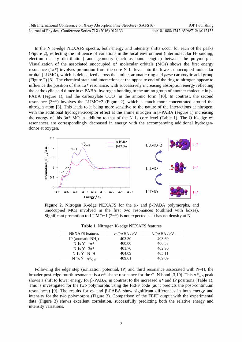
In the N K-edge NEXAFS spectra, both energy and intensity shifts occur for each of the peaks (Figure 2), reflecting the influence of variations in the local environment (intermolecular H-bonding, electron density distribution) and geometry (such as bond lengths) between the polymorphs. Visualization of the associated unoccupied π* molecular orbitals (MOs) shows the first energy resonance (1π*) involves promotion from the core N 1s level into the lowest unoccupied molecular orbital (LUMO), which is delocalized across the amine, aromatic ring and para-carboxylic acid group (Figure 2) [3]. The chemical state and interactions at the opposite end of the ring to nitrogen appear to influence the position of this 1π* resonance, with successively increasing absorption energy reflecting the carboxylic acid dimer in α-PABA, hydrogen bonding to the amino group of another molecule in β-PABA (Figure 1), and the carboxylate COO− in the anionic form [10]. In contrast, the second resonance (3π*) involves the LUMO+2 (Figure 2), which is much more concentrated around the nitrogen atom [3]. This leads to it being more sensitive to the nature of the interactions at nitrogen, with the additional hydrogen-acceptor effect at the amine nitrogen in β-PABA (Figure 1) increasing the energy of this 3π* MO in addition to that of the N 1s core level (Table 1). The O K-edge π* resonances are correspondingly decreased in energy with the accompanying additional hydrogen-donor at oxygen.
Figure 2. Nitrogen K-edge NEXAFS for the α- and β-PABA polymorphs, and unoccupied MOs involved in the first two resonances (outlined with boxes). Significant promotion to LUMO+1 (2π*) is not expected as it has no density at N.
Table 1. Nitrogen K-edge NEXAFS features
NEXAFS features α-PABA / eV β-PABA / eV IP (aromatic NH2) 403.30 403.60
N 1s → 1π* 400.00 400.58 N 1s → 3π* 401.70 402.30 N 1s → N−H 404.09 405.11 N 1s → σ*C-N 409.61 409.09
Following the edge step (ionization potential, IP) and third resonance associated with N−H, the
broader post-edge fourth resonance is a σ* shape resonance for the C−N bond [3,10]. This σ*C-N peak shows a shift to lower energy for β-PABA, in contrast to the increased π* and IP positions (Table 1). This is investigated for the two polymorphs using the FEFF code (as it predicts the post-continuum resonances) [9]. The results for α- and β-PABA show significant differences in both energy and intensity for the two polymorphs (Figure 3). Comparison of the FEFF output with the experimental data (Figure 3) shows excellent correlation, successfully predicting both the relative energy and intensity variations.
16th International Conference on X-ray Absorption Fine Structure (XAFS16) IOP PublishingJournal of Physics: Conference Series 712 (2016) 012133 doi:10.1088/1742-6596/712/1/012133
3

The dependence of this σ* resonance on the C−N bond was further investigated through variation of the C−N bond length for α-PABA. Lengthening of the C−N bond to that for β-PABA (+0.035 Å), leads to a decrease in σ* energy relative to the IP, accounting for the majority of the energy and intensity shifts between α- and β-PABA (Figure 3).
Figure 3. Comparison of experimental (solid line) and FEFF-calculated (dashed line) nitrogen K-edge NEXAFS for the two polymorphs of PABA, and the impact of variation in C−N bond length.
4. Conclusions Insights into the electronic and structural nature of the two polymorphs of PABA are obtained with nitrogen K-edge NEXAFS and XPS. A small shift in core level N 1s energy is attributed to the variation in intermolecular hydrogen bonding. Significantly different, highly characteristic NEXAFS spectra arise from the changes in local surroundings and bonding, with the π* shifts related to the nature of the MOs and local environment, and the σ* shape resonance primarily influenced by the C−N bond length. This demonstrates the sensitivity of XPS and NEXAFS to the relatively minor changes in local environment despite their identical chemical composition.
Acknowledgements Use of the NSLS at BNL was supported by U.S. Department of Energy under Contract No. DE-AC02-98CH10886. JSS, AG and SLMS acknowledge support by EPSRC Critical Mass Grant EP/I013563/1.
References [1] Cartensen J T Pharmaceutical Principles of Solid Dosage Forms 1993 (Lancaster, PA:
Technomic Publishing Co.) [2] Davey R. J., Schroeder S L M, and ter Horst J H 2103 Angew. Chem., Int. Ed. 52 2166-2179 [3] Stevens J S, Seabourne C R, Jaye C, Fischer D A, Scott A J and Schroeder S L M 2014 J. Phys.
Chem. B 118 12121-12128 [4] Lai T F and Marsh R E 1967 Acta Crystallogr. 22 885-893 [5] Gracin S and Fischer A 2005 Acta Crystallogr., Sect. E: Struct. Rep. Online 61 o1242. [6] Fairley N and Carrick A 2005 The Casa Cookbook - Part 1: Recipes for XPS Data Processing.
Acolyte Science (Knutsford, Cheshire: Acolyte Science) [7] Ravel B and Newville M 2005 J. Synchrotron Radiat. 12 537-541 [8] Newville M 2001 J. Synchrotron Radiat. 8 322-324 [9] Ankudinov A L, Ravel B, Rehr J J and Conradson S D, 1998 Phys. Rev. B: Condens. Matter
Mater. Phys. 58 7565-7576 [10] Stevens J S, Gainar A, Suljoti E, Xiao J, Golnak R, Aziz E F and Schroeder S L M 2015 Chem.
- Eur. J. 21 7256-7263
16th International Conference on X-ray Absorption Fine Structure (XAFS16) IOP PublishingJournal of Physics: Conference Series 712 (2016) 012133 doi:10.1088/1742-6596/712/1/012133
4