newsletter - afwg · this issue of the afwg newsletter focuses on this fungal infection that ... an...
TRANSCRIPT

NEWSLETTER 2017•Issue 2
This year we celebrate the 8th year of AFWG: 8 years of pursuing excellence in medical mycology throughout the region; 8 years of sharing expertise and encouraging like-minded professionals to join us in our mission. We are happy to once again share some educational articles from our experts and keep you updated on our activities through this issue.
Deep dermatophytosis may be a rare skin infection, but late diagnosis or ineffective treatment may lead to mortality in some cases. This issue of the AFWG newsletter focuses on this fungal infection that usually occurs in immunosuppressed individuals. Dr Pei-Lun Sun takes us through the basics of deep dermatophytosis, presenting data from published studies, and emphasizes the importance of treating superficial tinea infections before starting immunosuppressive treatment. Dr Ruojun Wang and Professor Ruoyu Li share a case of deep dermatophytosis caused by Trichophyton rubrum.
In this issue, we also feature a new fungus, Fereydounia khargensis, first discovered in 2014. Ms Ratna Mohd Tap and Dr Fairuz Amran present 2 cases of F. khargensis and show how PCR sequencing is crucial to correct identification of this uncommon yeast. We also provide a convenient guide on itraconazole, prepared by Dr Atul Patel. Finally, read up on recent and exciting developments on our official website, www.AFWGonline.com.
Thank you for being part of the AFWG community for 8 years. We are hopeful that, with our contribution, the coming years will continue to see a change in the landscape of medical mycology in Asia.
Editors’ welcome Dr Mitzi M Chua Adult Infectious Disease SpecialistAssociate ProfessorDepartment of Microbiology & ParasitologyCebu Institute of MedicineCebu City, Philippines
Dr Ariya ChindampornAssociate ProfessorDepartment of MicrobiologyFaculty of MedicineChulalongkorn UniversityBangkok, Thailand
Deep dermatophytosisDeep dermatophytosis: A case reportFereydounia khargensis: A new and uncommon opportunistic yeast from Malaysia
Itraconazole: A quick guide for clinicians
page 2
page 4
page 5
page 6
Visit us at AFWGonline.com and sign up for updates

2
Dermatophytes are fungi that can infect human skin, hair and nails, and can cause different types of tinea. These diseases can be treated effectively with antifungal agents. In rare circumstances, they can invade deep into the dermis by direct extension from ruptured infected hair follicles, or by direct invasion and traumatic implantation of fungi from an infected epidermis. Majocchi’s granuloma and dermatophytic pseudomycetoma fall under the former type of dermal invasion. Those without discernable follicular origin or with extensive dermal infiltration by fungal elements are collectively termed ‘deep dermatophytosis’. They are also known by other names, such as tinea profunda, invasive dermatophytosis, disseminated dermatophytosis or dermatophyte abscess. When skin is the only site of infection, the disease is categorized as deep dermatophytosis; when skin and lymph nodes and/or other organs are involved, the infection is considered invasive dermatophytosis. This article will review the clinical presentations, pathologic characteristics, underlying disease and treatment outcome of patients with deep dermatophytosis reported in published studies.
Clinical presentationsBased on a search done primarily on PubMed, there were 79 reported cases of deep dermatophytosis between 1975 and 2016. The age of the patients ranged from 8 to 83 years old (39.8±16.6). Teens to patients in their 60s were most commonly affected (Figure 1). The male to female ratio was 2.43:1.
According to published studies, the clinical presentation of deep dermatophytosis was variable, including solid red nodules, plaques, papuloplaques, large ulcers, cystic nodules and masses. Some patients had associated lymphadenopathy. Upon examination of the skin biopsy, fungal elements were seen in the dermis, which could be enhanced by periodic acid-Schiff (PAS) or Gomori methenamine silver (GMS) stains. The number of fungal elements in the tissue was small, but cases with massive fungal hyphae in tissue have been reported. Angio-invasion has also been reported, indicating evidence of hematogenous dissemination of dermatophytes.
Risk factorsThe risk factors of deep dermatophytosis include:
• Chronic tinea on other body sites;• Diabetes, hepatitis, liver cirrhosis, lymphoma, leukemia,
HIV infection, hereditary hemochromatosis, end-stage renal disease, atopic dermatitis;
• Immunosuppressive treatments for underlying diseases, such as solid organ transplantation, myasthenia gravis, rheumatoid arthritis; and
• Immunodeficiency, such as plasma factor deficiency, decreased T-cell activity, and Caspase recruitment domain-containing protein 9 (CARD9) gene mutation.
Deep dermatophytosisDr Pei-Lun SunAssistant Professor Department of DermatologyChang Gung Memorial HospitalLinkou Branch, Taoyuan, Taiwan
18
16
14
12
10
8
6
4
2
00–9 10–19 20–29 30–39 40–49
Age (years)
Num
ber o
f cas
es
50–59 60–69 70–79 80–89 ND
Figure 1. Age distribution of cases of deep dermatophytosis
ND, not determined

3
Based on the published cases, immune deficiency was the most common risk factor (Figure 2). In 18 patients with immune dysfunction, 12 had an underlying CARD9 deficiency. CARD9 is an important protein in the immune signaling pathway against fungal pathogens; its roles in invasive fungal infection have been extensively investigated. The second most important risk factor is solid organ transplantation. In 13 patients receiving organ transplantation, 8 had renal transplantation (62%), 2 had heart transplantation, 1 had liver transplantation, 1 had kidney and liver transplantation, and 1 had heart and lung transplantation.
Causative fungi and pathogenesisThe causative fungi were isolated and identified in 73 cases, and included almost all common pathogenic dermatophytes. Trichophyton rubrum was the most common (n=40), followed by T. violaceum (n=10) and Microsporum canis (n=8). Two cases had combined infections of T. rubrum and T. violaceum, and 2 others had combined infections of T. rubrum and T. verrucosum. Other pathogens were T. mentagrophytes, T. tonsurans, T. schoenleinii, M. audouinii and Epidermophyton floccosum.
The pathogenesis of deep dermatophytosis is still not fully understood. Based on the published cases of deep dermatophytosis, most had a superficial dermatophytosis on the body (eg, tinea corporis, tinea pedis, onychomycosis), which served as a source of infection.
There are 2 ways of dermal invasion by dermatophytes. One is by passive introduction of dermatophytes into the dermis through a ruptured infected hair follicle or scratching trauma from the overlying superficial tinea. The other is direct invasion from epidermis down to the dermis. The invading dermatophyte can usually be detected and eradicated by host immune cells, but when patients have impaired immunity, either congenital or iatrogenic, pathogens can proliferate freely and infection in deep tissues ensues.
TreatmentCurrently, there are no treatment guidelines available for deep dermatophytosis. But considering the depth of invasion, systemic antifungal agents should always be used. The most commonly used drugs were fluconazole, itraconazole and griseofulvin, followed by terbinafine and amphotericin B. Ketoconazole, voriconazole and posaconazole were used in a few cases. Of the published cases, 75% received antifungal monotherapy, and others were treated with 2 or more different antifungal drugs. Ten cases had their lesions removed with surgery. For 47 patients with deep dermatophytosis limited to the skin (no organ involvement), all survived or died from causes unrelated to dermatophytes.
In the 32 cases with invasive deep dermatophytosis and internal organ and/or lymph node involvement, the prognosis was quite different: 7 patients had complete remission of their disease; and 11 patients had partially resolved lesions or were stable after treatment. The disease recurred in 2 patients, and 9 died of invasive dermatophytosis. The mortality rate was as high as 28%. Three cases were lost to follow-up or the outcome was undocumented.
SummaryIn conclusion, deep dermatophytosis is a rare and invasive form of dermatophyte infection, which may lead to mortality. An accurate diagnosis relies on the combination of skin biopsy for histopathology examination and fungal culture to identify the pathogen. The host immune status plays a major role in disease pathogenesis, extent and prognosis. Systemic antifungal treatment is always necessary. Superficial tinea should be properly managed before starting immunosuppressive treatment, because it may become a source of invasive fungal infection. In patients who require extensive application of immunosuppressive therapy, clinicians should be on high alert for these potential threats from the skin surface.
20
16
12
8
4
0
Immun
e defi
cienc
y
Hemato
logic
maligna
ncy
Hemato
logic
disorder
Autoim
mune
Diabete
s melli
tus Skin
Liver
cirrho
sis
Solid or
gan t
ransp
lantat
ion
Risk factor
Num
ber o
f cas
es
Figure 2. Risk factors of deep dermatophytosis
4
Deep dermatophytosis: A case reportDr Ruojun Wang and Professor Ruoyu LiDepartment of DermatologyPeking University First HospitalBeijing, China
Case detailsA 70-year-old woman presented with chronic ulcerative nodules on her lower extremities at the outpatient clinic. A tender red nodule developed on her left anterior tibial region a month ago, which gradually enlarged; spontaneous ulceration of the nodule and occasional pain were reported. The lesions did not respond to topical antibiotics, and more nodular lesions appeared in the mid-calf area and spread to her toes. Her medical history included kidney transplantation 6 years ago, chronic tinea pedis and onychomycosis.
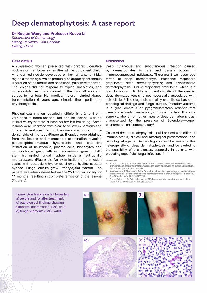
Physical examination revealed multiple firm, 2 to 4 cm, verrucose to dome-shaped, red nodular lesions, with an infiltrative erythematous base on her left lower leg. Some lesions were ulcerated with clear to yellow exudations and crusts. Several small red nodules were also found on the dorsal side of the toes (Figure a). Biopsies were obtained from the lesions and microscopic examination revealed pseudoepitheliomatous hyperplasia and extensive infiltration of neutrophils, plasma cells, histiocytes and multinucleated giant cells in the dermis (Figure c). PAS stain highlighted fungal hyphae inside a neutrophilic microabscess (Figure d). An examination of the lesion scales with potassium hydroxide showed hyaline septate hyphae. Fungal culture grew Trichophyton rubrum. The patient was administered terbinafine 250 mg twice daily for 11 months, resulting in complete remission of the lesions (Figure b).
DiscussionDeep cutaneous and subcutaneous infection caused by dermatophytes is rare and usually occurs in immunosuppressed individuals. There are 3 well-described forms of deep dermatophyte infections: Majocchi’s granuloma; deep dermatophytosis; and disseminated dermatophytosis.1 Unlike Majocchi’s granuloma, which is a granulomatous folliculitis and perifolliculitis of the dermis, deep dermatophytosis is not necessarily associated with hair follicles.2 The diagnosis is mainly established based on pathological findings and fungal culture. Pseudomycetoma is a granulomatous or pyogranulomatous reaction that usually surrounds dermatophytic fungal hyphae. It shows some variations from other types of deep dermatophytosis, characterized by the presence of Splendore-Hoeppli phenomenon on histopathology.3
Cases of deep dermatophytosis could present with different immune status, clinical and histological presentations, and pathological agents. Dermatologists must be aware of this heterogeneity of deep dermatophytosis, and be alerted to the possibility of this disease, especially in patients with preceding superficial fungal infections.2
References1. Su H, Li L, Cheng B, et al. Trichophyton rubrum infection characterized by Majocchi’s
granuloma and deeper dermatophytosis: case report and review of published literature. Mycopathologia 2017;182:549-554.
2. Kershenovich R, Sherman S, Reiter O, et al. A unique clinicopathological manifestation of fungal infection: a case series of deep dermatophytosis in immunosuppressed patients. Am J Clin Dermatol 2017;18:697-704.
3. Castro-Echeverry E, Fiala K, Fernandez MP. Dermatophytic pseudomycetoma of the scalp. Am J Dermatopathol 2017;39:e23-e25.
Figure. Skin lesions on left lower leg (a) before and (b) after treatment; (c) pathological findings showing extensive inflammation (PAS, x40); (d) fungal elements (PAS, ×400).
(a) (b)
(c) (d)

5
Fereydounia khargensis represents a new lineage in the order Urocystales, subphylum Ustilaginomycotina. It was first discovered in Iran in 2014;1 in 2016, RM Tap et al reported 2 invasive infections caused by F. khargensis in immunocompromised patients in Malaysia, which are discussed below.2
The first case of F. khargensis was from an HIV-positive patient. He was admitted because of episodes of fever associated with chills and rigors for 2 weeks. The patient was initially treated with amphotericin B, but the clinical condition did not improve. Antifungal treatment was changed to itraconazole and he was discharged upon improvement.
The second case was a hepatitis B carrier with hypertension, diabetes mellitus and end-stage renal failure on continuous ambulatory peritoneal dialysis (CAPD). He was admitted because of a dislodged distal connector of his Tenckhoff catheter caused by a fall in the toilet. After the incident, the dislodged connector was reconnected to the Tenckhoff catheter and used for CAPD. Fluconazole was started following yeast growth from the peritoneal fluid. The clinical condition of the patient improved after fluconazole treatment.
Both cases grew yeast-like colonies. Figures 1 and 2 show the macro- and microscopic examinations. Results from API 20C AUX and VITEK 2 identification system were Cryptococcus neoformans (98% probability) and Cryptococcus laurentii (89% probability), respectively. Internal transcribed spacer
(ITS) and D1/D2 region in large subunit (LSU) of rRNA gene were amplified using universal primers and sequenced. Both F. khargensis isolates matched 99.7% (ITS) and 100.0% (LSU) to the reference strain, IBRC-M 30116.
In vitro susceptibility testing showed that itraconazole and voriconazole have good activity against the yeast with minimum inhibitory concentration (MIC) ranging from 0.032 to 2.000 µg/mL, but MIC was slightly higher for fluconazole (8.000 µg/mL). On the other hand, both isolates showed resistance to amphotericin B, caspofungin and anidulafungin with the MIC more than 32.000 µg/mL.
In summary, F. khargensis is a new and uncommon opportunistic yeast. The incidence of rare fungal pathogens is rapidly increasing due to the expanding population of immunocompromised patients and advanced identification techniques. In this report, low CD4 count (case 1) and complicated medical conditions (case 2) are the risk factors that predisposed the patients to F. khargensis infection. Observation of macroscopic and microscopic characteristics provides clues to their atypical features. Correct identification is crucial and can be made possible by polymerase chain reaction (PCR) sequencing. F. khargensis exhibited resistance to polyenes and echinocandin but was sensitive to azoles.
References1. Nasr S, Soudi MR, Fazeli SAS, et al. Expanding evolutionary diversity in the
Ustilaginomycotina: Fereydouniaceae fam. nov. and Fereydounia gen. nov., the first urocystidalean yeast lineage. Mycol Prog 2014;13:1217-1226.
2. Tap RM, Ramli NY, Sabaratnam P, et al. First two cases of fungal infections associated with multi-drug resistant yeast, Fereydounia khargensis. Mycopathologia 2016;181:531-537.
Fereydounia khargensis: A new and uncommon opportunistic yeast from MalaysiaMs Ratna Mohd Tap and Dr Fairuz AmranMycology LaboratoryInfectious Diseases Research CentreInstitute for Medical ResearchKuala Lumpur, Malaysia
Figure 1. F. khargensis colonies on Sabouraud dextrose agar (SDA) after 48 h incubation at 30°C presented as cream-colored colonies, dry and wrinkled (a). However, after 72 h of incubation, the colonies started producing melanin-like pigmentation, which turned even darker after 120 h (b).
Figure 2. Elongated and irregular shape of F. khargensis yeast cells from 48 h SDA plate. The length of the cells ranged from 5.63 to 18.34 µm. Magnification at x40 (a) and x100 (b).
(a) (b) (a) (b)

6
Itraconazole: A quick guide for cliniciansDr Atul K Patel, MD, FIDSAChief Consultant and Director Infectious Diseases Clinic Vedanta Institute of Medical Sciences Ahmedabad, India
Itraconazole is a first-generation azole drug that became available in the 1990s. Itraconazole and other azoles disrupt the integrity of fungal cell membranes by interfering with ergosterol synthesis, leading to fungal cell death.
Itraconazole exhibits minimal activity against the Fusarium species, and it has no activity against most Mucorales.
IndicationsYeast: Mucosal candidiasis, cryptococcal infection (not as a primary agent, but it can be used as an alternative agent for chronic suppressive therapy).
Dimorphic fungi: For mild-to-moderate disease, itraconazole can be used as initial therapy. For severe diseases, amphotericin B is recommended for initial therapy, followed by itraconazole.
Mycelial fungi: As second-line treatment of invasive Aspergillus infection. Itraconazole is commonly used for chronic pulmonary aspergillosis and allergic bronchopulmonary aspergillosis treatment.1
Itraconazole is used for empiric treatment of fungal infection in neutropenic patients.2
It is also used successfully in the treatment of infections caused by Entomophthorales (basidiobolomycosis, conidiobolomycosis).3
Dosage and formsFor most systemic fungal infections, itraconazole 200–400 mg per day is given, except for life-threatening infections, where 200 mg 3 times/day (ie, 600 mg) is given as a loading dose for 3 days, followed by 400 mg/day.
Itraconazole is available in 2 oral preparations, as capsules and as an oral solution. Intravenous itraconazole is currently not available. Only the capsule form is available in India.
PharmacologyThe absorption and bioavailability of these 2 oral formulations are different. The oral bioavailability of the capsule formulation is approximately 55% and is improved with gastric acidity and food intake.4 It is generally recommended to be taken with an acidic beverage (such as cola) and food for better absorption. Antacids, including proton-pump inhibitors and H2 blockers, should be avoided as concomitant use significantly reduces absorption of itraconazole capsules.
The oral solution has a higher oral bioavailability of 80%, and its absorption is not affected by gastric acidity or food intake. This formulation has less interpatient variability, and patients achieve 30% higher serum concentrations than with the capsule.
Therapeutic drug monitoring for patients receiving itraconazole and its active hydroxyl itraconazole metabolite is required because of unpredictable absorption. Clinical studies have correlated itraconazole serum levels and therapeutic response for a variety of fungal infections.5 Itraconazole levels >0.5 μg/mL for antifungal prophylaxis and 1–2 μg/mL for treatment are associated with successful outcomes.6
Once-daily administration of itraconazole is generally adequate because of its long half-life of 25–64 hours. However, divided dosage is recommended for better absorption when used 400 mg or higher daily.
Spectrum of activity
Yeasts Dimorphic fungi Mycelial fungi
Most Candida spp., with higher MICs for C. glabrata and C. krusei
Blastomyces dermatitidis, Histoplasma capsulatum, Coccidioides spp., Paracoccidioides spp.
Aspergillus spp., including A. fumigatus, A. flavus, A. nidulans and A. terreus
Cryptococcus neoformans Talaromyces marneffei (formerly Penicillium marneffei), Sporothrix schenckii
Entomophthorales, eg, Conidiobolus and Basidiobolus
Currently, itraconazole clinical use is limited to: cutaneous fungal infections; allergic bronchopulmonary aspergillosis; histoplasmosis
(after induction therapy with amphotericin B); and Entomophthorales infection
7
ToxicityIn general, itraconazole is fairly well tolerated. The most common side effects include rash, headache, gastrointestinal upset, transaminitis and, rarely, liver failure. Monitoring of liver chemistry tests during its use is recommended.
Drug-drug interactionsThe triazoles have the highest potential for serious drug-drug interactions among antifungal agents. They are substrates and inhibitors of various hepatic CYP450 metabolic enzymes (CYP3A4, CYP2C9, CYP2C19).8 Remember that all triazoles (itraconazole, fluconazole, voriconazole) are inhibitors of CYP450 enzymes and impair the metabolism of co-administered drugs, resulting in increased exposure, higher levels and the risk of toxicity. As substrates of the pathway, the concentrations of the triazoles are also affected by concomitant use of medications that inhibit or induce these enzymes.
Be careful while prescribing itraconazole to patients receiving the following classes of medicines: anti-tuberculosis, anti-HIV, anticoagulants, sedative-antidepressants, anti-arrhythmics, antipsychotics, immunosuppressants, anti-epileptics, statins, and oral hypoglycemic agents, among others. Always check possible drug interactions using available guides, such as in mobile apps.
References1. Stevens DA, Schwartz HJ, Lee JY, et al. A randomized trial of itraconazole in allergic
bronchopulmonary aspergillosis. N Engl J Med 2000;342:756-762.2. Nucci M, Biasoli I, Akiti T, et al. A double-blind, randomized, placebo-controlled trial of
itraconazole capsules as antifungal prophylaxis for neutropenic patients. Clin Infect Dis 2000;30:300-305.
3. El-Shabrawi MH, Kamal NM. Gastrointestinal basidiobolomycosis in children: an overlooked emerging infection? J Med Microbiol 2011;60:871-880.
4. Lange D, Pavao JH, Wu J, Klausner M. Effect of a cola beverage on the bioavailability of itraconazole in the presence of H2 blockers. J Clin Pharmacol 1997;37:535-540.
5. Denning DW, Tucker RM, Hanson LH, Stevens DA. Treatment of invasive aspergillosis with itraconazole. Am J Med 1989;86(6 Pt 2):791-800.
6. Denning DW, Tucker RM, Hanson LH, et al. Itraconazole therapy for cryptococcal meningitis and cryptococcosis. Arch Intern Med 1989;149:2301-2308.
7. Heykants J, Van Peer A, Van de Velde V, et al. The clinical pharmacokinetics of itraconazole: an overview. Mycoses 1989;32 Suppl 1:67-87.
8. Brüggemann RJ, Alffenaar JW, Blijlevens NM, et al. Clinical relevance of the pharmacokinetic interactions of azole antifungal drugs with other coadministered agents. Clin Infect Dis 2009;48:1441-1458.
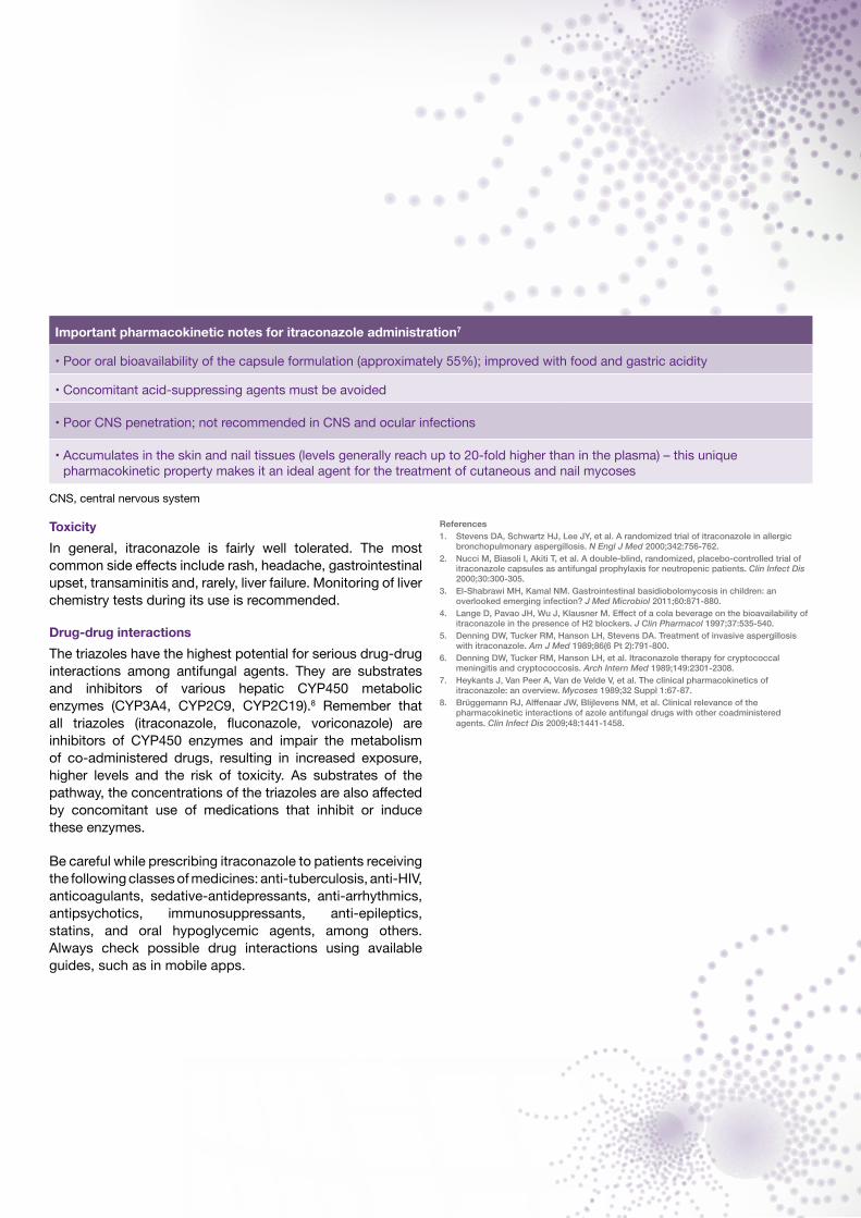
Important pharmacokinetic notes for itraconazole administration7
• Poor oral bioavailability of the capsule formulation (approximately 55%); improved with food and gastric acidity
• Concomitant acid-suppressing agents must be avoided
• Poor CNS penetration; not recommended in CNS and ocular infections
• Accumulates in the skin and nail tissues (levels generally reach up to 20-fold higher than in the plasma) – this unique pharmacokinetic property makes it an ideal agent for the treatment of cutaneous and nail mycoses
CNS, central nervous system

Copyright © 2017 Asia Fungal Working Group.This newsletter is made available to the medical profession by the Asia Fungal Working Group. The Asia Fungal Working Group of the International Society for Human and Medical Mycology is supported by an independent educational grant from Pfizer. Editorial development by Weber Shandwick Worldwide and the Asia Fungal Working Group. The opinions expressed in this publication are not necessarily those of the editor, publisher or sponsor; any liability or obligation for loss or damage, howsoever arising, is hereby disclaimed.
AFWGonline
Visit us at AFWGonline.com and sign up for updatesThank you for your valuable support!
Visit us at
A comprehensive medical mycology resource for both researchers and healthcare professionals in Asia
www.AFWGonline.com
We continue to update our official website frequently with the latest and most relevant content in medical mycology. Do you know what to do if you encounter Candida auris cases? Do you want to download lecture presentations from the recent Medical Mycology Training Network (MMTN) Conference? All these are available at AFWGonline.com.
Join the AFWG communityBe part of the AFWG community and share with us your thoughts and suggestions. Follow us on:
AFWGonline @afwgonline AFWGonline
Apply for the AFWG-sponsored laboratory courses“I joined the course because I want to establish a mycology laboratory in my country”, said Dr Batac, who was the first to complete the AFWG Laboratory Skills Enhancement Course. Our sponsored Laboratory Foundation Training and Skills Enhancement courses are now open for registration. Visit the ‘Laboratory Courses’ page to find out more and apply online. You can also read about the experience of Dr Batac in our ‘Articles’ page.
Register for our online education modules Did you know that AFWG offers free education modules on a variety of important fungal topics? All it takes is a few minutes to register on the ‘Online Education Modules’ page and you can instantly access all 6 of the educational modules prepared by our international experts.