newly formed excitatory pathways provide a substrate for ... filenewly formed excitatory pathways...
TRANSCRIPT

Newly Formed Excitatory PathwaysProvide a Substrate for Hyperexcitabilityin Experimental Temporal Lobe Epilepsy
MONIQUE ESCLAPEZ, JUNE C. HIRSCH, YEZEKIEL BEN-ARI,
AND CHRISTOPHE BERNARD*INSERM Unite 29, 75014 Paris, France
ABSTRACTTemporal lobe epilepsy (TLE) in humans and animals is associated with axonal sprouting
of glutamatergic neurons and neosynaptogenesis in the hippocampal formation. We examinedwhether this plasticity of excitatory pathways contributes to an increased level of glutamater-gic excitation in the CA1 region of rats experiencing chronic spontaneous limbic seizuresfollowing kainic acid or pilocarpine treatment. In chronic cases, we report an extensive axonalsprouting of CA1 pyramidal neurons, with many axonal branches entering the pyramidal celllayer and stratum radiatum, regions that are not innervated by axonal collaterals of CA1pyramidal neurons in control animals. Concurrently with this anatomical reorganization, alarge increase of the spontaneous glutamatergic drive is observed in the dendrites and somataof CA1 pyramidal cells. Furthermore, electrical activation of the reorganized CA1 associa-tional pathway evokes epileptiform bursts in CA1 pyramidal cells. These findings suggest thatreactive plasticity could contribute to the hyperexcitability of CA1 pyramidal neurons and tothe propagation of seizures in these two models of TLE. J. Comp. Neurol. 408:449–460, 1999.r 1999 Wiley-Liss, Inc.
Indexing terms: rat; hippocampus; CA1; pilocarpine; kainate; plasticity
Pyramidal cells in the hippocampus and neocortex giverise to networks of axonal recurrent collaterals (associa-tional pathways) that spread excitation to neighboringneurons. Based on this morphological specificity, a perma-nent increase of recurrent glutamatergic excitation onother excitatory cells has been proposed as one of the keyfactors responsible for hyperexcitability in various formsof epilepsy and particularly in temporal lobe epilepsy(TLE). Increase of recurrent excitation can be achieved byseveral factors: (1) A deficit of gamma-aminobutyric acid(GABA)-ergic inhibition: although pharmacological block-ade of fast inhibition results in hyperexcitability spreadingthrough associational pathways in control tissue (Trauband Wong, 1982; Miles and Wong, 1983; Chagnac-Amitaiand Connors, 1989; Jefferys and Whittington, 1996, Meierand Dudek, 1996), recent studies have indicated that fastinhibition is still operative (Esclapez et al., 1997a,b;Rempe et al., 1997) or even increased (Buckmaster andDudek, 1997; Prince et al., 1997; Nusser et al., 1998) inepilepsy. (2) The formation of new recurrent excitatoryconnections: morphological evidence has indicated thatglutamatergic axons sprout and establish new, sometimesaberrant, synapses in human TLE and its animal models.Although best documented for the hippocampal mossyfibers (Nadler et al., 1980a,b; Tauck and Nadler, 1985;
Represa et al., 1987; DeLanerolle et al., 1989; Represa andBen-Ari, 1992; Cronin et al., 1992), this has also beendescribed for the axons of CA1 pyramidal neurons (Perezet al., 1996), suggesting that axonal sprouting and glutama-tergic neosynapse formation may be a general property ofepileptic circuits (see also Salin et al., 1995; McKinney etal., 1997). However, the link between epilepsy and the‘‘too-well-connected brain’’ (Prince, 1997) is not knownbecause the functionality of the newly formed glutamater-gic synapses and their contribution to seizure generationhave not been established.
To address these issues, we performed patch clamprecordings and morphological analysis of CA1 pyramidalneurons in hippocampal slices from kainic acid- or pilocar-pine-treated rats with spontaneous chronic limbic sei-zures, two models of human TLE. Because of the crucialrole of dendrites in controlling cell excitability, we also
Grant sponsor: INSERM; Grant sponsor: Simone and Cino del DucaFoundation; Grant sponsor: French Foundation for Epilepsy Research.
*Correspondence to: Dr. C. Bernard, INSERM Unite 29, 123 Boulevardde Port-Royal, 75014 Paris, France. E-mail: [email protected]
Received 27 February 1998; Revised 5 January 1999; Accepted 2 March1999
THE JOURNAL OF COMPARATIVE NEUROLOGY 408:449–460 (1999)
r 1999 WILEY-LISS, INC.

examined the long-term consequences of seizures in thedendrites in addition to more conventional recordings ofsomata of pyramidal neurons. Preliminary data of thisstudy have been presented in abstract form (Esclapez etal., 1997a,b).
MATERIALS AND METHODS
Animals
Young adult male Wistar rats (180–200 g) were treatedwith pilocarpine or kainic acid (KA) according to estab-lished procedures (Nadler et al., 1980c; Turski et al., 1983;Obenaus et al., 1993; Bernard and Wheal, 1996). Briefly,pilocarpine (325–340 mg/kg) was injected intraperitonealy30 minutes after the administration of a low dose of thecholinergic antagonist methyl scopolamine nitrate (1 mg/kg, i.p.) to minimize peripheral cholinergic effects. KA (0.5µg in a volume of 0.5 µl of phosphate buffer, pH 7.4) wasinfused, over a 30-minute period, into the left lateralventricule of the rats under chloral hydrate (350 mg/kg)anesthesia. Following drug injection, 80% of the animalsin both models showed robust behavioral seizures for 3–4hours or longer. Sixty percent of the pilocarpine-treatedanimals and more than 90% of the KA-treated rats sur-vived this period of acute seizures. These rats were thenobserved periodically (at least three hours a day) in thevivarium for the occurrence of spontaneous seizures.Amongthese animals, 90% of the pilocarpine-treated and 70% ofthe KA-treated rats developed spontaneous seizures, witha frequency of one to four seizures per week. Only seizuresof grade 3 or greater on the Racine (1972) scale were scored(i.e., forelimb clonus 6 rearing 6 falling). The onset of thespontaneous seizure occurrence was 4–6 weeks for pilocar-pine rats and 2–8 months for KA animals. Twenty KA-treated and 14 pilocarpine-treated rats which had dis-played spontaneous limbic seizures were included in thisstudy. The time between drug treatment and perfusion forelectrophysiological and morphological studies was 2–3months for pilocarpine animals and 2–12 months for KArats. Any animal that had displayed a seizure within thelast 24 hours before the final experiment was excluded.Fifteen age-matched rats from the same litters were usedfor control experiments. All animal-use protocols con-formed to NIH guidelines and the French Public HealthService Policy on the use of laboratory animals.
Electrophysiology
The animals were intracardially perfused, under chloralhydrate (350 mg/kg, i.p.) anesthesia, with modified artifi-cial cerebrospinal fluid (ACSF), and 400-µm-thick hippo-campal slices were prepared with a Leica VT 1000E tissueslicer as previously described (Hirsch et al., 1996). ACSFcontained (in mM) 124 NaCl, 3 KCl, 1.25 KH2PO4, 26NaHCO3, 1.3 MgSO4-7H2O, 2 CaCl2, and 10 D-glucose andwas continuously aerated with 95% O2 and 5% CO2. Thetemperature in the submerged recording chamber wasmaintained at 30–32°C. In all experiments, before electro-physiological recordings, the slices were recut at the levelof the subiculum to isolate the hippocampal formation (thedentate gyrus and the hippocampus) from adjacent corti-cal regions. In addition, in some experiments the CA1 areawas surgically isolated from the CA3 area by a knife cutperformed in the CA1 area close to the CA3 border. Evokedand spontaneous postsynaptic currents and potentialswere recorded from somata or dendrites of CA1 pyramidal
neurons with tight-seal whole-cell patch clamp pipettes. Micro-electrodes had a resistance of 4–12 MV, and internal solutionsof the following compositions were used (in mM): (1) forcurrent clamp recordings: 135 K-gluconate, 10 MgCl2, 0.1CaCl2, 1 EGTA, 2 Na2 adenosine triphosphate, 10 HEPES,0.5% biocytin, pH 7.25; (2) for voltage clamp recordings:K-gluconate was replaced by Cs-gluconate. The osmolarity ofthe internal solutions was 265–275 mOsm. Each of the 235recorded pyramidal cells was identified post hoc. Spontaneousexcitatory currents were measured at the reversal potentialfor GABAergic events (around 260 mV) according to estab-lished procedure (Esclapez et al., 1997a,b). D-2-amino-5-phosphonovaleric acid (D-APV) and 6-cyano-7-nitroquinoxa-line-2,3-dione (CNQX) were applied at the end of theexperiments to verify that the currents were indeed glutama-tergic. CNQX and D-APV were a gift from Novartis (Basel,Switzerland). Bicuculline, KA, pilocarpine, methyl scopol-amine nitrate, and biocytin were obtained from Sigma (St.Louis, MO), and tetrodotoxin (TTX) was obtained from La-toxan (Rosans, France). Signals were fed to an Axopatch200A,B amplifier (Axon Instruments, Inc., Foster City, CA).All data were digitized (10 kHz) with a Labmaster interfacecard attached to a personal computer and analyzed withAcquis1 program (G. Sadoc, 1994, Biologic, Grenoble, France).The mean values and corresponding standard errors(S.E.M.s) were calculated for all the experimental param-eters (measures) and for each animal group. For eachparameter, the mean values obtained in control, pilocar-pine-, and KA-treated rats were compared with a mixedmodel analysis of variance (ANOVA) and Student’s t-test.
Morphology
Techniques for the detection of biocytin-filled neu-
rons. After electrophysiological recordings, slices werefixed overnight at 4°C in a solution containing 4% parafor-maldehyde in 0.1 M phosphate buffer (PB; pH 7.4). Afterfixation, slices were rinsed in PB, cryoprotected in sucrose,and quickly frozen on dry ice. After these steps, threeprotocols were used to visualize the biocytin-filled neu-rons. (1) To study the morphological features of CA1pyramidal cells, detection of labeled neurons was per-formed on resectioned slices. Briefly, 60-µm-thick sectionswere cut on a cryostat. To neutralize endogenous peroxi-dase, sections were pretreated for 30 minutes in 1% H2O2.After several rinses in 0.1 M phosphate buffered saline (pH7.4; PBS), sections were incubated overnight at 4°C inavidin-biotinylated peroxidase complex (1:200; Vector Labo-ratories, Burlingame, CA) diluted in PBS. After 30-minuterinses in PBS, sections were processed with 0.06% 3,38-diaminobenzidine tetrahydrochloride (DAB; Sigma) and0.006% H2O2 diluted in PBS. Biocytin-labeled neuronsthat displayed complete dendritic and axonal arboriza-
TABLE 1. Number of CA1 Pyramidal Neurons Detected With EachMorphological Method for Each Animal Group*
ABC onresectioned
slices1
ABC onunsectioned
slices
FITC–avidine Don unsectioned
slices
Totalnumberof cells
Control 37 (19) 8 22 67Pilocarpine 25 (15) 8 36 69KA ipsilateral side 26 (14) 4 15 45KA contralateral side 32 (17) 5 17 54
*FITC, fluorescein isothiocyanate; KA, kainic acid; ABC, avidin-biotinylated peroxidasecomplex.1The number of pyramidal neurons that exhibited, in addition to somatic and dendriticstaining, a complete and uniform labeling of the axon is indicated in parentheses.
450 M. ESCLAPEZ ET AL.

tions were reconstructed from serial adjacent sections withthe Neurolucida system (Microbrightfield, Inc., Colchester,VT). (2) To investigate putative synaptic contacts betweenCA1 pyramidal cells, we conducted experiments in whichthree to five neurons were injected with biocytin in thesame 250-µm-thick slice (two slices for control animals and
four slices for pilocarpine- and KA-treated rats). Thedetection of the biocytin-filled neurons was performed onunsectioned slices. After the pretreatement step in 1%H2O2 and several rinses in PBS, slices were incubated for24 hours at 4°C in 1:100 avidin-biotinylated peroxidasecomplex diluted in PBS containing 0.3% Triton X-100.
Fig. 1. Neuronal cell loss in the hippocampus of chronic pilocarpine(B) and kainic acid (KA)-treated (C,D) animals illustrated by sectionsstained with cresyl violet. A: In control specimens, neuronal cell bodiesof pyramidal neurons are highly concentrated and form a continousband in the CA3 and CA1 fields, and numerous neurons are present inthe hilus (H) of the dentate gyrus. B: In a pilocarpine-treated rat withspontaneous seizures (three months after injection), a marked reduc-tion in the number of neurons is present in the hilus. In addition, aslight neuronal loss is observed in some regions of CA3 (arrows). Incontrast, the CA1 pyramidal cell layer is well preserved. C: In achronic KA-treated animal (three months after injection), the hippo-campus ipsilateral to the cerebroventricular injection of KA displays a
very extensive cell loss of hilar neurons and of CA3 pyramidal cells. Areduction in the number of CA1 pyramidal neurons is also observed.D: In contrast, the hippocampus contralateral to the KA injectionexhibits very little neuronal loss in the CA3 and CA1 regions.E–F: High magnification photomicrographs of CA1 pyramidal celllayers from the same specimens illustrated in C and B, respectively.E: Despite extensive neuronal loss (see C), many CA1 pyramidal cellbodies remain in the hippocampus ipsilateral to the cerebroventricu-lar injection of a chronic KA-treated animal (three months afterinjection). F: In a pilocarpine-treated rat (three months after injec-tion), numerous well-preserved CA1 pyramidal cells are observed inthe hippocampus. Scale bars 5 400 µm in A–D, 200 µm in E,F.
NEW FUNCTIONAL GLUTAMATERGIC NETWORK IN EPILEPSY 451

Figure 2
452 M. ESCLAPEZ ET AL.

DAB and H2O2 were used as chromogens. (3) Some sliceswere processed for detection of biocytin with a fluorescentmarker to find the trace of the patch electrode. The sliceswere incubated for 24 hours in fluorescein isothiocyanateand avidin-D (Vector Laboratories, Inc.) diluted in PBScontaining 0.3% Triton X-100. All sections and slices weremounted on gelatin-coated slides and coverslipped inaqueous media (Crystal or Gel/Mount, Biomeda Corp.,Foster City, CA). Table 1 summarizes the number of CA1pyramidal neurons detected with each method for eachanimal group and indicates (in parentheses) the number ofpyramidal neurons that exhibited, in addition to somaticand dendritic staining, a complete and uniform labeling ofthe axon.
Quantitative analysis of the axonal arborization
and associated varicosities. Among the 65 biocytin-filled CA1 pyramidal neurons that displayed uniformlabeling of the axonal and dendritic trees (Table 1), 14neurons from control, KA-, and pilocarpine-treated ani-mals were selected for quantitative analysis of axonalarborization. These neurons were selected according to thefollowing criteria. Neurons had to exhibit extensive label-ing of the axon. We were able to reconstruct the entirecourse of the axon from the cell body. For these neurons, inaddition to the dendritic and axon arbors, the varicosities(putative boutons) along axonal processes were markedduring the acquisition with the computer software pro-gram Neurolucida. The total length of the axon running inthe alveus, stratum oriens, and stratum radiatum (KA andpilocarpine rats; see Results) in the CA1 region, thenumbers of axonal branches, and varicosites per brancheswere automatically calculated by the Neurolucida soft-ware. The axonal branch originating from the soma or aproximal dendrite was called the first-order axonal seg-ment and ended at the first branching point. From the firstbranching point emerged two second-order axonal seg-ments, and so on. Thus, an nth-order axonal branch isbounded by a (n-1)th (or the soma) and an nth (or the endtip of the axonal branch) branching point.
Quantitative estimation of the complexity of the axonalarborization and putative synaptic contacts was deter-mined by calculating the mean numbers of total axonallength, of segment order per axon, and of varicosities per100 µm of axon for each nth-order axonal branch. All
values are given as means 6 S.E.M. Statistical analysis ofdifferences in the number of nth-order axonal segment, thetotal axonal length, and the number of varicosities amongcontrol and pilocarpine- or KA-treated animals were per-formed with a mixed model ANOVA and Student’s t-test.
Digital processing of figures
Figures 2–6 were assembled digitally. The computer-generated images of the four-color photomicrographs (Fig.3) and of the black-and-white photomicrographs associ-ated with electrophysiological traces (Fig. 4) or withneuron drawings (Fig. 2) were performed as follows. Colorpositive (Fig. 3: Ektachrome 160T, Kodak, Rochester, NY)or black-and-white negative (Fig. 4: TMAX 3200 ASA,Kodak; Fig. 2: AGFAPAN 25 ASA, AGFA, Mortsel, Bel-gium) films of histological preparations were taken with aNikon photomicroscope. Each film was digitized with aNikon Film Scanner LS-1000 (300 dpi) driven by AdobePhotoshop, version 4.0, running on a Power Macintosh7500/100. No modification or alteration of the initialimages was done. The electrophysiological traces (Fig. 4)were obtained after acquisition of the data with Acquis 1software (G. Sadoc) installed on a PC and were transferredas HPLG or ASCII files to a Macintosh using Claris Draw,version 1.03. The drawings of biocytin-filled pyramidalneurons and axograms were generated with Neurolucida,version 3, on a PC and were exported as EPS files to aMacintosh using Adobe Photoshop. The montages of thedigitized images were made with Quark XPress, version3.31, for Macintosh and printed with a Tektronic Phaser IISDX printer.
RESULTS
General neuronal loss within thehippocampal formation of pilocarpine-
or KA-treated animals
Cresyl-violet-stained sections were studied to evaluatethe extent of the neuronal loss in the two models of chroniclimbic seizures. In pilocarpine-treated animals with spon-taneous recurrent seizures (2–3 months after drug injec-tion), a marked and consistent cell loss was observed in thehilus of the dendate gyrus. In addition, these animalsdisplayed variable amounts of neuronal loss in the CA3pyramidal cell layer, but this loss was seldom extensiveand many neuronal cell bodies remained in this layer (Fig.1B; compare with the control shown in Fig. 1A). Thedentate granule cell and the CA1 pyramidal cell layerswere relatively well preserved (Fig. 1B,F). A similar pat-tern of cell loss has been described previously in this model(Turski et al., 1986; Mello et al., 1993; Obenaus et al., 1993;Liu et al., 1994). In all KA-treated animals, a massive cellloss of hilar neurons and CA3 pyramidal cells was ob-served in the hippocampus ipsilateral to the intracerobro-ventricular injection of KA (Fig. 1C; compare with thecontrol shown in Fig. 1A). A variable amount of neuronalloss was observed in the CA1 pyramidal cell layer in theipsilateral hippocampus; the extent of loss increased withtime after drug injection. Whereas many neuronal cellbodies remained in the CA1 pyramidal cell layer threemonths after drug injection (Fig. 1E), almost all of themhad degenerated 12 months after KA treatment. In con-trast, the hippocampus contralateral to the KA injectiondisplayed only little damage that was observed mainly in
Fig. 2. Pyramidal neuron with exuberant axonal sprouting in theCA1 region of pilocarpine-treated animals. A,B: Neurolucida recon-struction of the axonal (gray) and dendritic arborizations of biocytin-filled pyramidal neurons from control (A) and pilocarpine-treated (B)rats. A: In the control animal, the axon of a CA1 pyramidal cell arisesfrom the soma and gives off one axonal branch running toward thefimbria and four branches directed toward the subiculum (S). Theseaxonal branches running in stratum oriens and the alveus displayonly few collaterals within the CA1 region. B: In a pilocarpine-treatedrat, the axon of the CA1 pyramidal neurons originates from a proximalbasal dendrite, ascends to the alveus, but gives rise to an aberrantdense axonal plexus in stratum oriens (O), with many branchesentering the pyramidal layer (P) and stratum radiatum (R).C: Photograph of an axonal collateral running throughout the pyrami-dal cell layer of the CA1 region outlined in B. This axonal collateraldisplays one or two varicosities (arrowheads) making putative con-tacts on an unlabeled proximal dendrite and somata of pyramidal cells(*); somata are outlined by dashed lines. D: Axogram of the pyramidalneuron showed in A. E: Axogram of the pyramidal neuron shown in B.Note the numerous branching points and axonal segments of the axonfrom the pyramidal cell in the pilocarpine-treated rat. Scale bars 5100 µm in A,B, 10 µm in C.
NEW FUNCTIONAL GLUTAMATERGIC NETWORK IN EPILEPSY 453

the CA3 pyramidal layer, whereas dentate granule cellsand pyramidal neurons of CA1 were well preserved (Fig.1D). A similar pattern of cell loss has been reportedpreviously in this model (Nadler et al., 1980c; Lancasterand Wheal, 1982).
Morphological alterations of CA1pyramidal cells
In slices from control animals, the main axonal branchesof CA1 pyramidal cells (n 5 67) displayed rare, thin, andshort axonal collaterals in the stratum oriens (Fig. 2A)that did not enter the stratum radiatum (Tamamaki andNojyo, 1990; Perez et al., 1996). In contrast, in one-fourthof the CA1 pyramidal cells (42 of 168) from animals withspontaneous seizures, the axon gave rise very quickly to a
large number of thin collaterals that spread throughoutthe basal dendrites field (Figs. 2B, 3A). In keeping with theobservations of Perez et al. (1996), the main axonalbranches running in stratum oriens and the alveus dis-played numerous collaterals in stratum oriens. In addi-tion, we now report that collaterals often invaded stratapyramidale and radiatum in the CA1 region (Figs. 2A,C,3D). Quantitative analysis of the numbers of axonal branchorder and total length of the axonal arborization measuredin all strata including the alveus further demonstrated theincrease in the complexity of the axon in epileptic versuscontrol animals. The mean number of segment order peraxon was increased from 3.5 6 2.5 in control (n 5 4; Fig.2D) to 14.0 6 5.3 in chronic (n 5 10; P , 0.001; Fig. 2E)animals. The mean total axon length was significantly
Fig. 3. Photomicrographs of biocytin-labeled CA1 pyramidal cellsdetected in 250-µm-thick slices from a kainic-acid-treated rat withspontaneous seizures. A: The main axonal branch of a CA1 pyramidalneuron gives rise to many thin collaterals (arrowheads), with numer-ous varicosities that correspond to putative en passant boutons.B: High magnification of A, showing two varicosities (arrowheads) ofthe thin collateral making putative synaptic contacts with a basal
dendrite of the same pyramid cell. C: An axonal collateral (arrow-heads) of the biocytin-labeled pyramid shown in A surrounds a basaldendrite of a neighboring labeled CA1 pyramid. At least four varicosi-ties are in close apposition with this dendrite. D: An axonal collateralof the CA1 pyramidal neurons shown in A, running through stratumpyramidale (P) and stratum radiatum (R), displays many varicosities(arrowheads). Scale bars 5 10 µm in B,C, 50 µm in A,D.
454 M. ESCLAPEZ ET AL.

longer in epileptic animals (7,511 6 850 µm vs. 3,732 6963 µm in control animals; P , 0.001). CA1 pyramidalneurons from pilocarpine and KA-treated animals exhib-ited small varicosities along all axonal branches includingcollaterals entering in stratum radiatum (Fig. 3D). Thesevaricosities seemed to establish synaptic contacts withsomata and dendritic processes of pyramidal neurons(Figs. 2C, 3A–C), including putative autapses (Fig. 3C).The mean number of varicosities per 100 µm of axon foreach segment order was not significantly different betweencontrol and epileptic animals (P . 0.05): 4.02 6 1.5 and4.31 6 1.33, respectively, for first-order segments; 10.04 62.98 and 9.15 6 2.56 for second-order segments; 14.78 63.42 and 13.51 6 5.36 for third-order segments; 12.34 62.6 and 10.04 6 3.57 for fourth-order segments. The meandensity of varicosities for the following order segments inpilocarpine- and KA-treated rats was 10.62 6 5.44. De-spite similar linear densities of varicosities in epileptic andcontrol animals, the total number of these putative enpassant boutons was higher in chronic animals because ofthe increased number of axonal branches.
We then determined whether these pathological alter-ations could represent a morphological substrate for elec-trophysiological modifications in the apical dendrites andsomata of CA1 pyramidal cells. In particular, we examinedwhether the reorganized CA1 associational pathway (1)could trigger epileptiform activity when activated and (2)results in an enhanced glutamatergic excitatory drive.
Epileptiform activity in the CA1 area
Patch clamp recordings from somata and apical den-drites of CA1 pyramidal cells were performed (control, n 567; pilocarpine, n 5 69; KAipsi, n 5 45; KAcontra, n 5 54).A dent on the biocytin-filled apical dendrite (Fig. 4A)indicated that the recording site was 375 6 55 µm from thesoma (KA, n 5 21; pilocarpine, n 5 24; control, n 5 13;same average distance for each group). Bulk electricalstimulations applied to stratum radiatum evoked gradedepileptiform discharges in the apical dendrites (Fig. 4B–C)and the somata (Fig. 4E–F) of 98% of the pyramidalneurons (n 5 164) in chronic animals. Spontaneous parox-ysmal discharges (Fig. 4D) also occurred in somata (n 567) and apical dendrites (n 5 20). In contrast, suprathresh-old stimulations applied to control slices evoked only asingle action potential in somata, as described by Esclapezet al. (1997a,b), and apical dendrites (not shown). Sponta-neous and evoked discharges were blocked by the glutama-tergic antagonists CNQX (10 µM) and D-APV (50 µM; Fig.4E). Therefore, in slices from chronic animals, epileptiformdischarges are present in somata and apical dendrites ofCA1 pyramidal cells.
Selective activation of the CA1 associational pathway byfocal electrical stimulation applied to stratum oriens (n 52), the alveus close to the subiculum (n 5 2), or thesubiculum itself (n 5 8) evoked glutamatergic gradedbursts of action potentials in both somata and apicaldendrites of CA1 pyramidal cells in chronic animals (Fig.4F). These bursts were blocked by CNQX (10 µM) andD-APV (50 µM; not shown). In contrast, stimulationsapplied at similar locations in control slices evoked anexcitatory/inhibitory sequence or an antidromic actionpotential (Fig. 4G; n 5 5). Therefore, activation of thereorganized CA1 associational pathway triggers epilepti-form discharges in chronic animals.
To estimate the frequency of connections between cells,we made paired pyramidal cell recordings in the presenceof bicuculline (10 µM) in the surgically isolated CA1 areain slices of control (n 5 10 pairs) and chronic (n 5 18 pairs)animals. None of these 28 pairs was monosynapticallyconnected. In the CA3 area, which possesses abundantrecurrent collateral fibers, when GABAA receptor-medi-ated inhibition is blocked, a high-frequency train of actionpotentials generated in a single pyramidal neuron bydepolarizing steps triggers epileptiform activity in anothersimultaneously recorded pyramidal cell and in the entireCA3 network (Miles and Wong, 1983). However, a similarparadigm applied to the CA1 area did not trigger polysyn-aptic epileptiform discharges in the other cells in control(n 5 10, not shown) or chronic (n 5 18, not shown) animals.Therefore, although the reorganized CA1 associationalpathway in epileptic animals shares morphological fea-tures with that of CA3, the amount of functional intercon-nectivity between CA1 pyramidal cells in chronic animalsremains below that of control CA3 neurons.
Increased spontaneous glutamatergicactivity in somata and apical dendrites
in chronic animals
If the sprouting process gives rise to new functionalglutamatergic synapses, the excitatory drive provided byspontaneous postsynaptic excitatory currents (sEPSCs)should be increased because many slices from chronicanimals displayed spontaneous epileptiform discharges.In control pyramidal cells, somatic and dendritic sEPSCswere composed of small amplitude currents (range 5 50pA; Table 2, Fig. 5A), with means 6 S.E.M. of 16 6 9 pAand 15 6 8 pA, respectively. The frequency of sEPSCs wasstatistically lower in the soma than in the apical dendrites(Table 2; P , 0.001). The frequency of sEPSCs was similarin pilocarpine- and KA-treated rats (3.34 6 0.6, n 5 13;3.51 6 0.73, n 5 18; respectively). However, in thesechronic animals, the frequency of sEPSCs was dramati-cally increased in both somata (520% of control; P , 0.001;Table 2, Fig. 5) and apical dendrites (306% of control; P ,0.001; Table 2, Fig. 5) as compared with control animals.The difference between dendrites and somata in chronicanimals was also statistically significant (P , 0.001). Inchronic animals, the amplitude distribution of sEPSCswas similar to that of controls (mean 6 S.E.M.: 15 6 10 pAfor somata, 16 6 9 pA for dendrites; Fig. 6A), with theexception of a small population of very large sEPSCs thatwas not observed in control animals (Fig. 6A–B, Table 2).These large amplitude sEPSCs had the same time courseas the smaller ones, suggesting their monosynaptic origin(Fig. 6B). In chronic and control rats, the Na1 channelblocker TTX (1 µM) abolished most of the sEPSCs in thesomata and apical dendrites (.80%; Table 2, Fig. 5),suggesting that sEPSC activity is mainly action potentialdependent. The increased sEPSC activity is not due to atonic reduction of the inhibitory drive because, whenbicuculline (10 µM) was applied to the surgically isolatedCA1 area of slices from control and chronic animals, thefrequency of sEPSCs in pyramidal cells was not increased(Table 2). Therefore, in epileptic animals the axonal sprout-ing of CA1 pyramidal neurons could subserve a substratefor the dramatically increased frequency of action-potential-dependent sEPSCs and the occurrence of large amplitudesEPSCs in both somata and dendrites.
NEW FUNCTIONAL GLUTAMATERGIC NETWORK IN EPILEPSY 455
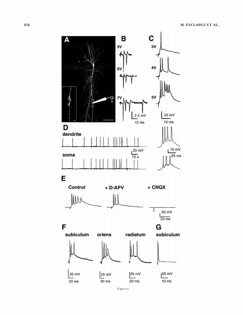
Figure 4
456 M. ESCLAPEZ ET AL.

DISCUSSION
Our results suggest that, in two animal models of TLE,(1) glutamatergic axons sprout and establish novel connec-tions on the somata and dendrites of pyramidal neuronsand (2) these new connections (putative synapses) arefunctional and could participate in the propagation ofparoxysmal activities.
Morphological reorganizationof the CA1 area
Intracerebroventricular administration of KA leads tomassive death of CA3 pyramidal cells in the hippocampusipsilateral to the lesion (Nadler et al., 1980a–c), which isfollowed by a rapid decrease of the number of asymmetricsynapses in stratum radiatum of the CA1 area (Nadler etal., 1980b; Phelps et al., 1991). The number of asymmetricsynapses then slowly recovers, suggesting that a reactivesynaptogenesis has taken place (Nadler et al., 1980b;Phelps et al., 1991). These newly formed synapses mayoriginate from surviving CA3 (mainly commissural fibers),CA1 (associational pathway) pyramidal cells, entorhinalcortex (perforant path), or the septum but not from mossyfibers, which sprout in the dentate gyrus and the CA3 areabut do not invade the CA1 area (Nadler et al., 1980a,b;Represa and Ben-Ari, 1992). The favored hypothesis isthat the CA1 associational pathway sprouts. In experimen-tal models of epilepsy, morphologically identified reactivesynaptogenesis often involves local recurrent pathways in
the dentate gyrus (Nadler et al., 1980a,b), the CA3 area(McKinney et al., 1997), or the neocortex (Salin et al.,1995). In the CA1 area, two indirect electrophysiologicaldata support this hypothesis. (1) In chronic but not incontrol animals, N-methyl-D-aspartic acid (NMDA) recep-tors directly participate in normal synaptic transmission(Turner and Wheal, 1991; Hirsch et al., 1996). In controlrats, this NMDA component does not exist in the Schaffer/commissural pathway (Andreasen et al., 1989) but ispresent in the CA1 associational pathway (Thomson andRadpour, 1991; Deuchars and Thomson, 1996). Therefore,it has been hypothetized that the NMDA receptor compo-nent seen in epileptic animals originates from the sproutedCA1 associational pathway (Turner and Wheal, 1991;Perez et al., 1996). (2) Epileptiform discharges are moreeasily triggered in the isolated CA1 area of KA-treatedrats than of control animals (Meier and Dudek, 1996).More direct evidence has been presented in the report thatthe CA1 associational pathway sprouts in KA-treated rats(Perez et al., 1996). However, new axonal collaterals weredescribed in stratum oriens only, leaving unresolved theorigin of the newly formed synapses along the apicaldendrites. In the present study, we alleviated this contra-diction in part because we report a massive sprouting, notonly in stratum oriens but also in strata pyramidale andradiatum. The quantitative data reflect the profuse axonalsprouting because the number of branching order wasconsiderably increased in chronic animals (14 vs. 3.5 incontrol) and the total axonal length increased from 3,700µm to 7,500 µm. These values, including the control ones,are higher than those reported previously (Perez et al.,1996). In the study by Perez et al., the number of branchesincreased from 1.5 to 3 and the axonal length from 1,500µm to 2,000 µm in control and chronic animals, respec-tively. The discrepancies with the study by Perez et al.(1996) may stem from the fact that our animals were killedafter they had developed spontaneous recurrent seizures.
There was no clear-cut relationship between the hippo-campal damage and the extent of the sprouting of the CA1associational pathway. Whereas in KA-treated rats theCA3 pyramidal neurons were totally destroyed in thehippocampus ipsilateral to the KA injection, the amount ofaxonal sprouting was comparable to that observed in thecontralateral side and in the pilocarpine model, in whichfew CA3 pyramidal cells disappeared. Therefore, thesprouting of the CA1 associational pathway alone does notseem to account for the recovery of 72% of asymmetricsynapses reported in the KA model (Nadler et al., 1980b),
Fig. 4. Graded bursts in apical dendrites from pyramidal neuronsin the CA1 region of epileptic rats. A: Identified dendritic recordingsite in a pyramidal neuron filled with biocytin (detected by fluores-cence). Inset at higher magnification shows the trace left by the patchelectrode on the apical dendrite (pilocarpine-treated rat, 2.5 monthsafter injection). B,C: Graded bursts recorded in the dendrites of kainicacid (KA)-treated rats in the cell-attached (B, 12 months postinjection)and the whole cell (C, three months postinjection) configurations.D: Spontaneous bursts of action potentials recorded simultaneously inthe apical dendrite and the soma of two pyramidal neurons. The lastburst is shown expanded on the right (12 months after KA injection).E–G: Somatic recordings. E: The evoked epileptiform burst is sup-pressed by glutamate antagonists (KA-treated rat, three months).F: This neuron responded with a burst to different loci of stimulation(KA-treated rat, three months). G: Control pyramidal neuron thatresponded with an antidromic action potential to stimulation of thesubiculum. All recordings were obtained in the current clamp mode atresting membrane potential. D-APV, D-2-amino-5-phosphonovalericacid; CNQX, 6-cyano-7-nitroquinoxaline-2,3-dione. Scale bars 5 100µm in A, 1 µm in inset.
TABLE 2. Distribution of Spontaneous Postsynaptic Excitatory Current (sEPSC) Peak Amplitudes and Frequencies in Somata and Dendritesof CA1 Pyramidal Cells From Control and Epileptic Animals*
Amplitudes (%) Recording site (n) 0–50 pA 51–60 pA 61–90 pA
Control Soma (16) 99.5 0.5 0.0Epileptic Soma (31) 98.5 0.5 1.5Control Dendrite (13) 99.4 0.6 0.0Epileptic Dendrite (9) 98.0 1.5 0.0Frequencies (Hz) Soma (n) Soma 1 TTX (n) Dendrite (n) Dendrite 1 TTX (n)Control 0.66 6 0.26 (16) 0.13 6 0.04 (5) 1.79 6 0.37 (13) 0.30 6 0.03 (11)Epileptic 3.43 6 0.52 (31) 0.17 6 0.03 (8) 5.48 6 1.71 (9) 0.66 6 0.12 (8)Frequencies (Hz) Soma in cut CA1 (n) Bicuculline (n)Control 0.77 6 0.17 (10) 0.57 6 0.16 (10)Epileptic 5.49 6 1.29 (10) 3.48 6 0.75 (10)
*Dramatic change in the frequency but not the amplitude of sEPSCs in the somata and dendrites of CA1 pyramidal neurons in experimental animals. The sEPSCs were measured atthe reversal potential for GABAergic events (around 260 mV) according to a well-established procedure (see Materials and Methods for details) and divided arbitrarily into threeamplitude groups. For each cell, the frequency of sEPSCs was estimated in 60-second epochs. The effects of tetrodotoxin (TTX) in controls and temporal lobe epilepsy (TLE) neuronswere similar: a decrease of the frequency of sEPSCs, indicating that sEPSCs are generated by action potentials. Adding bicuculline, to reduce tonic inhibition, did not modify thefrequency of sEPSCs in the somata of pyramidal neurons recorded in the surgically isolated CA1 area of control and epileptic rats.
NEW FUNCTIONAL GLUTAMATERGIC NETWORK IN EPILEPSY 457

suggesting that other glutamatergic fibers yet to be charac-terized also contribute to the reactive synaptogenesis.
Functionality of the new excitatoryconnections in CA1
In keeping with previous studies (Tamamaki and Nojyo,1990; Deuchars and Thomson, 1996; Perez et al., 1996), we
report that in control animals the CA1 associationalpathway is restricted to stratum oriens. In chronic ani-mals, because it is not yet possible to distinguish between
Fig. 5. The frequency of glutamatergic spontaneous postsynapticexcitatory currents recorded from the somata and apical dendrites ofpyramidal neurons is considerably increased in slices from experimen-tal animals with temporal lobe epilepsy (Exp TLE) as opposed to thatof naive rats. Somatic recording from a kainic-acid-treated rat (fivemonths). Dendritic recording from the neuron illustrated in Figure 4A.In both soma and the apical dendrite of the pyramidal neuron from theepileptic animals, the frequency of spontaneous currents is higherthan that in the control animals. For all the recordings, the membranewas voltage clamped at the reversal potential for GABAergic currents(around 260 mV). Tetrodotoxin (TTX) blocked all spontaneous cur-rents from control and epileptic animals. ACSF, modified artificialcerebrospinal fluid.
Fig. 6. Distribution of spontaneous postsynaptic excitatory current(sEPSC) amplitude in somata and dendrites of pyramidal neuronsfrom control and experimental animals. A: Superimposed frequencyhistograms of peak amplitudes (bin 1 pA) of sEPSCs recorded in thesomata of neurons from control (dotted line) and epileptic (solid line)animals. There was no significant difference between neurons fromcontrol and epileptic animals for either the somata or the dendrites.However, for pyramidal neurons in experimental rats, the longer tailof the histogram leaned toward large amplitude sEPSCs in the somataand to a lesser degree in the dendrites. B: Somatic recording of sEPSCs.Averages of 15 large (*) and small (x) EPSCs are shown together scaled tothe same amplitude (1 1 2), demonstrating identical kinetics (kainicacid, six months after injection). TLE, temporal lobe epilepsy.
458 M. ESCLAPEZ ET AL.

old and new branches in stratum oriens, newly formedsynapses cannot be identified in this region. In contrast,the axonal collaterals that enter strata pyramidale andradiatum in chronic animals are newly formed. All CA1pyramidal cell axonal recurrent collaterals were coveredwith numerous varicosities in chronic animals, includingthe branches crossing the pyramidal cell layer and enter-ing stratum radiatum. Because varicosities are generallyassociated with synaptic contacts (Sik et al., 1993; Deucharsand Thomson, 1996; Vida et al., 1998), it is likely that thesprouted CA1 associational pathway forms new synapsesalong the apical dendrites of CA1 pyramidal cells. Al-though paired recordings associated with electron micros-copy studies are required, the following observations ob-tained in hippocampal slices isolated from surroundingcortical inputs suggest that the increased glutamatergicactivity observed in epileptic animals is generated withinthe CA1 region by the newly formed glutamatergic axoncollaterals: (1) The glutamatergic activity is mainly actionpotential dependent because TTX abolished most sEPSCsin the somata and dendrites. (2) The neurons generatingthis activity, i.e., the pyramidal neurons (provided thatthere are no excitatory interneurons) are located withinthe CA1 area because this glutamatergic activity re-mained the same when the CA1 area was surgicallyisolated from the CA3 area in epileptic animals. (3) As aconsequence, the increased frequency of sEPSCs in theapical dendrites of CA1 pyramidal neurons from chronicanimals originates from the newly formed recurrent collat-erals of pyramidal cells that now enter stratum radiatumbecause in control tissue CA1 recurrent collaterals do notcontact the apical dendrites. (4) Due to cable properties,the sEPSCs recorded in somata and apical dendrites areprobably generated in the vicinity of the recording site(Hirsch et al., 1998).
Increased interconnectivityand hyperexcitability
Extensive physiological data suggest that brain areas inwhich principal cells are interconnected by a dense net-work of excitatory recurrent colaterals are particularlyprone to seizure. In lesioned animals, the sprouting ofassociational pathways should increase the excitatoryconnections between principal cells (McKinney et al.,1997). As a consequence of this hypothesis, the excitatorydrive should be increased in principal cells and its propaga-tion facilitated. Our results clearly showed an increase inthe baseline excitatory synaptic drive in CA1 pyramidalcells (frequency and amplitude of sEPSCs) in both KA- andpilocarpine-treated animals. Similar findings have beenreported in granule cells of the dentate gyrus of KA-treated animals (Wuarin and Dudek, 1998). Furthermore,we report that, in epileptic slices recorded in physiologicalconditions, stimulation of the associational pathway trig-gers epileptiform activity in the CA1 region, demonstrat-ing that the reorganized and more extensive associationalpathway can participate in the generation and spread ofparoxysmal discharges. These results directly support thehypothesis that the connectivity between principal cells isfunctionallly increased in reorganized networks (Salin etal., 1995; Meier and Dudek, 1996; McKinney et al., 1997;Wuarin and Dudek, 1998). The extent of this increasedconnectivity remains to be quantified. For example, theprobability of finding connected pyramidal pairs should behigher in chronic animals because our morphological data
demonstrated in pyramidal cells a large increase in thenumber of axonal recurrent collaterals. However, in keep-ing with a previous study (Nakajima et al., 1991), such anincrease was not shown by paired recordings of CA1pyramidal cells. The reason may be statistical because theinterconnectivity between CA1 pyramidal cells in thecontrol animals was very low, less than 1% (Deuchars andThomson, 1996), suggesting that recording from hundredsof pairs will be required to estimate the number of newlyformed synaptic contacts.
Epilepsy and the ‘‘too-well-connected brain’’
The relationship between sprouting and hyperexcitabil-ity does not seem straightforward in epilepsy. Sproutedmossy fibers in the dentate gyrus in experimental models(but see Longo and Mello, 1997) can provide a morphologi-cal substrate for hyperexcitability when inhibition is re-moved (Patrylo and Dudek, 1998; Wuarin and Dudek,1998). However, in chronic animals recorded in physiologi-cal conditions, inhibition in the dentate gyrus is enhancedby (1) a long-term potentiation at GABAergic synapses ongranule cells (Nusser et al., 1998) and (2) an increasedexcitatory drive on inhibitory interneurons (Buhl at al.,1996; Buckmaster and Dudek, 1997). In contrast to thedentate gyrus, the CA1 area is hyperexcitable in TLE andin its experimental models, and inhibition does not seem tobe functionally affected in this region (Esclapez et al.,1997a,b), thus supporting the hypothesis that sprouting ofthe associational pathway plays an active role. Although itis not yet possible to determine whether sprouting isepileptogenic per se, the remodeled CA1 area will mostlikely facilitate the propagation of epileptiform activitiesto the subiculum and from there to the neocortex. Thecritical issue for temporal lobe epilepsy may not be wherethe epileptogenic area is located but how epileptic dis-charges propagate from the hippocampus to the neocortex.The CA1 and CA3 areas, because they can easily propa-gate epileptiform activity in experimental models (Meierand Dudek, 1996; Smith et al., 1998), are ideally locatedfor that purpose. It remains to be determined whether thesprouting of the CA1 and CA3 associational pathways alsooccurs in human TLE and results in an increased excita-tory drive and hyperexcitability through the formation ofnew functional glutamatergic connections as our experi-mental data suggest. Our results also provide the experi-mental basis for an alternative to the classic boost ofinhibition in the drug treatment of TLE. Glutamatergicsynapses seem to be a promising target.
ACKNOWLEDGMENTS
We thank D. Diabira for providing lesioned animals andI. Jorquera for technical assistance.
LITERATURE CITED
Andreasen M, Lambert J, Jensen M. 1989. Effects of the new non-N-methyl-D-aspartate antagonists on synaptic transmission in the in vitro rathippocampus. J Physiol (Lond) 414:317–336.
Bernard C, Wheal HV. 1996. A role for synaptic and network plasticity incontrolling epileptiform activity in CA1 in the kainic acid-lesioned rathippocampus in vitro. J Physiol (Lond) 495:127–142.
Buckmaster PS, Dudek FE. 1997. Neuron loss, granule cell axon reorgani-zation, and functional changes in the dentate gyrus of epileptic kainate-treated rats. J Comp Neurol 385:385–404.
NEW FUNCTIONAL GLUTAMATERGIC NETWORK IN EPILEPSY 459
Buhl EH, Otis TS, Mody I. 1996. Zinc-induced collapse of augmentedinhibition by GABA in temporal lobe epilepsy model. Science 271:369–373.
Chagnac-Amitai Y, Connors BW. 1989. Horizontal spread of synchronizedactivity in neocortex and its control by GABA-mediated inhibition. JNeurophysiol 61:747–758.
Cronin J, Obenaus A, Houser CR, Dudek FE. 1992. Electrophysiology ofdentate granule cells after kainate-induced synaptic reorganization ofthe mossy fibers. Brain Res 573:305–310.
DeLanerolle N, Kim J, Robbins R, Spencer D. 1989. Hippocampal interneu-ron loss and plasticity in human temporal lobe epilepsy. Brain Res495:387–395.
Deuchars J, Thomson AM. 1996. CA1 pyramid–pyramid connections in rathippocampus in vitro: dual intracellular recordings with biocytin filling.Neuroscience 74:1009–1018.
Esclapez M, Hirsch JC, Agassandian C, Ben-Ari Y, Bernard C. 1997a.Enhanced glutamatergic excitatory drive in the dendrites and somataof CA1 pyramidal neurons in epilepsy. Soc Neurosci Abstr 2158:838.
Esclapez M, Hirsch JC, Khazipov R, Ben-Ari Y, Bernard C. 1997. OperativeGABAergic inhibition in hippocampal CA1 pyramidal neurons inexperimental epilepsy. Proc Natl Acad Sci USA 94:1251–1256.
Hirsch JC, Quesada O, Esclapez M, Gozlan H, Ben-Ari Y, Bernard C. 1996.Enhanced NMDAR-dependent epileptiform activity is controlled byoxidizing agents in a chronic model of temporal lobe epilepsy. JNeurophysiol 76:4185–4189.
Hirsch JC, Esclapez M, Cannon RC, Wheal HV, Ben-Ari Y, Bernard C. 1998.Heterogeneous distribution of spontaneous and miniature GABAergicand glutamatergic synaptic currents along the somato-dendritic axis ofneurones. Soc Neurosci Abstr 919:362.
Jefferys JGR, Whittington MA. 1996. Review of the role of inhibitoryneurons in chronic epileptic foci induced by intracerebral tetanus toxin.Epilepsy Res 26:59–66.
Lancaster B, Wheal HV. 1982. A comparative histological and electrophysi-ological study of some neurotoxins in the rat hippocampus. J CompNeurol 211:105–114.
Liu Z, Nagao T, Desjardins GC, Gloor P, Avoli M. 1994. Quantitativeevaluation of neuronal loss in the dorsal hippocampus in rats withlong-term pilocarpine seizures. Epilepsy Res 17:237–247.
Longo BM, Mello LEAM. 1997. Blockade of pilocarpine- or kainate-inducedmossy fiber sprouting by cycloheximide does not prevent subsequentepileptogenesis in rats. Neurosci Lett 226:163–166.
McKinney RA, Debanne D, Gahwiler BH, Thompson SM. 1997. Lesion-induced axonal spouting and hyperexcitability in the hippocampus invitro: implications for the genesis of post-traumatic epilepsy. NatureMed 3:990–996.
Meier CL, Dudek FE. 1996. Spontaneous and stimulation-induced synchro-nized burst afterdischarges in the isolated CA1 of kainate-treated rats.J Neurophysiol 76:2231–2239.
Mello LEAM, Cavalheiro EA, Tan AM, Kupfer WR, Pretorius JK, Babb TL,Finch DM. 1993. Circuit mechanisms of seizures in the pilocarpinemodel of chronic epilepsy: cell loss and mossy fiber sprouting. Epilepsia34:985–995.
Miles R, Wong RKS. 1983. Single neurones can initiate synchronizedpopulation discharges in the hippocampus. Nature 306:371–373.
Nadler JV, Perry BW, Gentry C, Cotman CW. 1980a. Loss and reacquisitionof hippocampal synapses after selective destruction of CA3–CA4 affer-ents with kainic acid. Brain Res 191:387–403.
Nadler JV, Perry BW, Cotman CW. 1980b. Selective reinnervation ofhippocampal area CA1 and fascia dentata after destruction of CA3–CA4 afferents with kainic acid. Brain Res 182:1–9.
Nadler JV, Perry BW, Gentry C, Cotman CW. 1980c. Degeneration ofhippocampal CA3 pyramidal cells induced by intraventricular kainicacid. J Comp Neurol 192:333–359.
Nakajima S, Franck JE, Bilkey D, Schwartzkroin PA. 1991. Local circuitsynaptic interactions between CA1 pyramidal cells and interneurons inthe kainate-lesioned hyperexcitable hippocampus. Hippocampus 1:67–78.
Nusser Z, Hajos N, Somogyi P, Mody I. 1998. Increased number of synapticGABA (A) receptors underlies potentiation at hippocampal inhibitorysynapses. Nature 395:172–177.
Obenaus A, Esclapez M, Houser CR. 1993. Loss of glutamate decarboxylasemRNA-containing neurons in the rat dentate gyrus following pilocar-pine-induced seizures. J Neurosci 13:4470–4485.
Patrylo PR, Dudek FE. 1998. Physiological unmasking of new glutamater-gic pathways in the dentate gyrus of hippocampal slices from kainate-induced epileptic rats. J Neurophysiol 79:418–429.
Perez Y, Morin F, Jutras I, Beaulieu C, Lacaille J-C. 1996. Axonal sproutingof CA1 pyramidal cells in hyperexcitable hippocampal slices of kainate-treated rats. Eur J Neurosci 8:736–748.
Phelps S, Mitchell J, Wheal HV. 1991. changes to synaptic ultrastruture infield CA1 of the rat hippocampus following intracerebroventricularinjection of kainic acid. Neuroscience 40:687–699.
Prince DA. 1997. Epilepsy and the too-well-connected brain. Nature Med3:957–958.
Prince DA, Jacobs KM, Salin PA, Hoffman S, Parada I. 1997. Chronic focalneocortical epileptogenesis: does disinhibition play a role? Can JPhysiol Pharmacol 75:500–507.
Racine RJ. 1972. Modification of seizure activity by electrical simulation: II.Motor seizure. Electroencephalogr Clin Neurophysiol 32:281–294.
Rempe DA, Bertram EH, Williamson JM, Lothman EW. 1997. Interneuronsin area CA1 stratum radiatum and stratum oriens remain functionallyconnected to excitatory synaptic input in chronically epileptic animals.J Neurophysiol 78:1504–1515.
Represa A, Ben-Ari Y. 1992. Epilepsy is associated with the formation ofnovel mossy fiber synapses in the CA3 region. Exp Brain Res 92:69–78.
Repressa A, Tremblay E, Ben-Ari Y. 1987. Kainate binding sites in thehippocampal mossy fibers: localization and plasticity. Neuroscience20:739–748.
Salin P, Tseng GF, Hoffman S, Parada I, Prince DA. 1995. Axonal sproutingin layer V pyramidal neurons of chronically injured cerebral cortex. JNeurosci 15:8234–8245.
Sik A, Tamamaki N, Freund TF. 1993. Complete axon arborization of asingle CA3 pyramidal cell in the rat hippocampus, and its relationshipwith postsynaptic parvalbumin-containing interneurons. Eur J Neuro-sci 5:1719–1728.
Smith KL, Lee CL, Swann JW. 1998. Local circuit abnormalities inchronically epileptic rats after intrahippocampal tetanus toxin injectionin infancy. J Neurophysiol 79:106–116.
Tamamaki N, Nojyo Y. 1990. Disposition of slab-like modules formed byaxon branches originating from single CA1 pyramidal neurons in therat hippocampus. J Comp Neurol 291:509–519.
Tauck DL, Nadler JV. 1985. Evidence of functional mossy fiber sprouting inhippocampal formation of kainic acid treated rats. J Neurosci 5:1016–1022.
Thomson A, Radpour S. 1991. Excitatory connections between CA1 pyrami-dal cells revealed by spike triggered averaging in slices of rat hippocam-pus are partially NMDA receptor mediated. Eur J Neurosci 3:587–601.
Traub RD, Wong RK. 1982. Cellular mechanism of neuronal synchroniza-tion in epilepsy. Science 216:745–747.
Turner DA, Wheal HV. 1991. Excitatory synaptic potentials in kainicacid-denervated rat CA1 pyramidal neurons. J Neurosci 11:2786–2794.
Turski WA, Cavalheiro EA, Schwarz M, Czuczwar SJ, Kleinrok Z, Turski L.1983. Limbic seizures produced by pilocarpine in rats: a behavioural,electroencephalographic and neuropathological study. Behav Brain Res9:315–336.
Turski L, Cavalheiro EA, Sieklucka-Dziuba M, Ikonomidou-Turski C,Czuczwar SJ, Turski WA. 1986. Seizures produced by pilocarpine:neuropathological sequelae and activity of glutamate decarboxylase inthe rat forebrain. Brain Res 398:37–48.
Vida I, Halasy K, Szinyei C, Somogyi P, Buhl EH. 1998. Unitary IPSPsevoked by interneurons at stratum radiatum-stratum lacunosum-moleculare border in the CA1 area of the rat hippocampus in vitro. JPhysiol (Lond) 506:755–773.
Wuarin JP, Dudek FE. 1998. Evidence from in vitro whole-cell patch-clamprecordings that baseline excitatory synaptic input is abnormally high ingranule cells with mossy fiber sprouting from kainate-induced epilepticrats. Soc Neurosci Abstr 851:2141.
460 M. ESCLAPEZ ET AL.