newborn screening for spinal muscular atrophysma is the leading genetic cause of death among infants...
TRANSCRIPT

National Center for Environmental Health
Division of Laboratory Sciences
APHL Newborn Screening SymposiumNew Orleans, LA, September 13, 2017
ORISE Fellow
Newborn Screening Translational Research Initiative
Newborn Screening and Molecular Biology Branch
Kristina Mercer, MPH, PhD
Newborn screening for spinal muscular atrophy

Clinical Features of Spinal Muscular Atrophy (SMA)
Type I: Birth – 6 mos.Type II: 6 mos. – 2 years
Type III: 18 mos. – 3+ years
A neuromuscular disease resulting in the progressive degeneration of motor neurons
Symptoms include loss of normal motor function and respiratory failure; can result in death
3 clinical types based on age of onset and severity

SMA is the leading genetic cause of death among infants
The birth prevalence of SMA is approx. 1 in 10,000
Type I (infantile-onset) is the most common form
The majority of children with Type I SMA do not survive beyond 2 years without effective therapy
FDA approved therapy exists
SMA has been nominated for inclusion on the Recommended Uniform Screening Panel (RUSP)

Genetic characterization of SMA
Autosomal recessive inheritance
Approximately 96% of SMA cases are caused by mutations in
the SMN1 gene
SMN1 encodes for survival of motor neuron (SMN) protein
Among the SMN1 mutations, most involve the loss of SMN1
exon 7 (on both chromosomes) by deletion or gene
conversion
Loss of this gene region results in a non-functional SMN protein
SMN2, a paralog of SMN1, may moderate the disease severity
• SMN2 can only produce 10% of the SMN protein
produced by SMN1
Wirth B., Human Mutation (2000), 15:228-37

Several different molecular assays have been used to detect SMA
Restriction Fragment Length Polymorphism (RFLP) test
High Resolution Melting (HRM) analysis
Multiplex Ligation-Dependent Probe Amplification (MLPA)
Luminex Genotyping
DNA sequencing
Quantitative (qPCR)/ Real-time PCR (RT-PCR)

Real-time PCR is one method that can be used to screen newborns for SMA
Real-time PCR allows for high throughput screening
Most state newborn screening labs are already using this method
Labs are equipped with the necessary instrumentation
Staff is familiar with procedure
Reactions can be multiplexed
Reduced cost to include a new assay
May not require added labor cost to run

What are some challenges associated with using real-time PCR to screen for SMA?

Challenge #1:
Exons 1 -6 Intron 6 Exon 8Intron 7Exon 7
G C A A G SMN1
SMN2A T G G A
There are only 5 nucleotide differences between the two genes
For real-time PCR, it is important to avoid detecting SMN2 when trying
to identify the loss of SMN1
SMN1 has a paralog, the SMN2 gene, which has nearly identical genomic sequence

LNA (locked nucleic acid) nucleotides can be used to distinguish single nucleotide differences
Astakhova K., Chemosensors (2014), 2(3):196-206
LNAs can be incorporated into primers and probes to discern single nucleotide differences between SMN1 and SMN2
This would allow for discriminatory amplification and/or signal detection of SMN1 only

Initial SMA assay developed at CDC
Taylor, J. et al., Clin. Chem, (2015), 61 (2): 412-9
The loss of SMN1 intron 7 was detected using a LNA probe (in green)
LNA substitutions underlined
The LNA probe was designed to selectively bind SMN1 by discriminating
between the mismatch nucleotides of SMN1 and SMN2
SMN1 nucleotide (A) and SMN2 nucleotide (G)
Forward and reverse primers (in yellow) will amplify both SMN1 and
SMN2 sequences

Exon 7
Exon 7 Exon 8
C
T
A
G
SMN1
SMN2Intron 7
Intron 7 Exon 8
Exon 7
Exon 7
C
T A
False positive
False negative Exon 8
Intron 7 Exon 8
Intron 7
G
8/120,000 (< 0.01%)*
Cases identified**; unknown prevalence
*Yin-Hsiu C. et al., The Journal of Pediatrics (2017); **Hahnen, E. et al., Am. J. Hum. Genet., (1996), 59: 1057-1065
Challenge #2:
Recombination between SMN1 and SMN2 can result in a hybrid genotype

Maranda, B. et al., Clin. Biochem., (2012), 45(1-2): 88-91
Revised SMA Assay – Part 1
We modified the previous assay to target exon 7 and reduce the possibility of false positive or false negative results due to hybrid genotypes
The LNA probe was designed to selectively bind SMN1 by discriminating
between the mismatch nucleotides of SMN1 (C) and SMN2 (T)
Forward and reverse primers (in yellow) will amplify both SMN1 and SMN2
sequences

Assay gives non-specific amplification some of the time when testing samples derived from
SMA patients
Non-specificsignal from SMN2
SMA patient samples
The LNA probe designed to recognize SMN1 only can bind the SMN2 amplicon, producing non-specific signal in SMA patient samples
RPP30
No signal

We replaced the original, reverse primer with an SMN1-specific LNA primer (in blue) to eliminate SMN2 amplification
Revised SMA Assay – Part 2

Assay Revision Part 1
Assay specificity improves by adding LNA primer
Assay Revision Part 2
SMA patient samples
SMA patient samples
Non-specificsignal from SMN2
No signal from SMN2
RPP30
RPP30

Possible reasons for reduced assay efficiency:
Sensitive to DNA extraction method
Sensitive to type of Taqman master mix
Sensitive to temperature fluctuations > 1 degree
Celsius
Further method improvement was needed
Technical concern:
Assay did not perform as expected in all environments

LNA probe was redesigned to make the assay more robust
Factors important in the design of LNA probe for mismatch discrimination: Length of the probe
• short (10-12 nucleotides)
Location of mismatch in the probe
• center position within probe
Modification pattern
• LNA substitution in triplet at site of mismatch
Identity of the mismatch
• pyrimidine (C or T) at mismatch site within probe
(discrimination is poor for G-T mismatches)
You, Y. et al., Nucleic Acids Research, (2006), 34(8)

The Current Assay utilizes an SMN1-specific LNA probe with forward strand sequence
We do not observe any non-specific signal in SMN1 null samples even when challenged with an excess of SMN2 sequence
0.025
SMN2 synthetic gene fragment(equivalent to 1,000 copies/cell)
RPP30
SMN1

( + SMA assay reagents)
cycles
1
Flu
ore
scen
ce (
dR
n)
0.01
0.1
( - SMA assay reagents)
cycles
1
Flu
ore
scen
ce (
dR
n)
0.01
0.1
This assay can also be multiplexed with primers and probes for RNase P (RPP30) and TREC
TRECCq=30.0
RPP30Cq=22.5
TRECCq=29.8
RPP30Cq=22.5
SMN1
RPP30
TREC
Cq values for RNase P and TREC are unaffected by the addition of reagents for SMA

The Current SMA Assay works at a range of temperatures from 60-65 degrees Celsius
Patient samples are SMA test positive (no SMN1 signal) at temperatures ranging from 60-65 degrees Celsius
Don’t need to worry about variations in instrument temperature affecting the results
Patient (60o)Fl
uo
resc
ence
(d
Rn
)
RPP30TREC
cycles20 22 24 26 28 30 32 34 36 38 40
Patient (65o)
cycles20 22 24 26 28 30 32 34 36 38 40
SMN1
RPP30TREC
SMN1

Flu
ore
scen
ce (
dR
n)
Unaffected (65o)Unaffected (60o)
cycles20 22 24 26 28 30 32 34 36 38 4020 22 24 26 34 36 38 40
TRECSMN1
RPP30
cycles28 30 32
There is no observed effect of temperature on the Cq values for RNase P and TREC
May not need to change the temperature of current TREC assay
SMN1 amplification in not negatively affected
The Current SMA Assay works at a range of temperatures from 60-65 degrees Celsius

SMA patients are correctly identified from dried blood spots when using the current assay
Assay Results Clinical Category
Sample Number Cq- SMN1 Exon 7 SMN1 Result SMA Status
1 24.69 Present Unaffected/ Carrier
2 No Cq Absent Affected
3 26.43 Present Unaffected/ Carrier
4 No Cq Absent Affected
5 No Cq Absent Affected
6 25.67 Present Unaffected/ Carrier
7 No Cq Absent Affected
8 24.28 Present Unaffected/ Carrier
9 24.23 Present Unaffected/ Carrier
10 No Cq Absent Affected
11 24.15 Present Unaffected/ Carrier
12 25.19 Present Unaffected/ Carrier
13 25.21 Present Unaffected/ Carrier
14 28.15 Present Unaffected/ Carrier
15 No Cq Absent Affected
16 24.49 Present Unaffected/ Carrier
17 24.78 Present Unaffected/ Carrier
18 No Cq Absent Affected
19 26.31 Present Unaffected/ Carrier
20 23.81 Present Unaffected/ Carrier
21 22.99 Present Unaffected/ Carrier
22 No Cq Absent Affected
23 No Cq Absent Affected
24 22.32 Present Unaffected/ Carrier
25 No Cq Absent Affected
26 No Cq Absent Affected
27 23.35 Present Unaffected/ Non-Carrier
28 23.46 Present Unaffected/ Non-Carrier
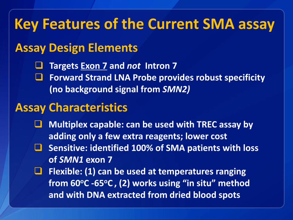
Key Features of the Current SMA assay
Targets Exon 7 and not Intron 7 Forward Strand LNA Probe provides robust specificity
(no background signal from SMN2)
Assay Design Elements
Multiplex capable: can be used with TREC assay by adding only a few extra reagents; lower cost
Sensitive: identified 100% of SMA patients with loss of SMN1 exon 7
Flexible: (1) can be used at temperatures ranging from 60oC -65oC , (2) works using “in situ” method and with DNA extracted from dried blood spots
Assay Characteristics

Additional Key Points
Both the LNA primer and forward strand (FS) probe improve specificity in detecting loss of SMN1 at exon 7
Current Assay using FS probe is comparatively more robust and cost effective
Use of a PCR clamp to suppress SMN2 amplification has also been developed, which can add an additional layer of specificity in a second tier assay for samples that are inconclusive
Droplet digital PCR can be used to determine copy number of SMN1 and SMN2

Pre assay development consultation
Providing sequence for SMA assay primers and probe
Integrating SMA into current TREC assay
Reference materials for assay development and validation
Individual training at CDC
Performing real-time PCR assay
Preparation of QC materials
CDC can provide consultation and technical support to labs interested in screening for SMA

Acknowledgments CDC scientists:
Francis LeeGolriz YazdanpanahSophia WinchesterRobert VogtHan PhanCarla Cuthbert
Taiwan Collaborators:
Yin-Hsiu Chien
Shu-Chuan ChiangWuh-Liang Hwu
State lab collaborators:
Minnesota Berta Warman, Carrie Wolf
New JerseyAlyssa MacMillan
WisconsinMei Baker, Sean Mochal
MassachusettsAnne Comeau, Lan Ji

For more information please contact Centers for Disease Control and Prevention
1600 Clifton Road NE, Atlanta, GA 30333
Telephone: 1-800-CDC-INFO (232-4636)/TTY: 1-888-232-6348
Visit: www.cdc.gov | Contact CDC at: 1-800-CDC-INFO or www.cdc.gov/info
The findings and conclusions in this report are those of the authors and do not necessarily represent the official position of the Centers for Disease Control and Prevention.
National Center for Environmental Health
Division of Laboratory Sciences
Use of trade names and commercial sources in this presentation is for identification only and does not imply endorsement by the Division of Laboratory Sciences, National Center for Environmental Health, Centers for Disease Control and Prevention, the Public Health Service, or the U.S. Department of Health and Human Services.
Thank you for your attention!