new york knee injury medical treatment guidelines, second edition
TRANSCRIPT
New York
Knee Injury
Medical Treatment Guidelines©
Second Edition
January 14, 2013
Corrected February 22, 2013
Effective March 1, 2013
This Treatment Guideline is adopted, with modification, from the State of Colorado’s Lower Extremity Medical Treatment Guideline, with supplementation from ACOEM’s Occupational Medicine Treatment Guidelines© and the State of Washington’s Medical and Surgical Treatment Guidelines.
© Copyright 2008, 2007, 2004, 1997 by the American College of Occupational and Environmental Medicine (ACOEM). Commercial reproduction or other use beyond fair use prohibited without explicit ACOEM permission.

New York State Workers’ Compensation Board New York Knee Injury Medical Treatment Guidelines
Second Edition, January 14, 2013 i
TABLE OF CONTENTS
A GENERAL GUIDELINE PRINCIPLES ...................................................................... 1
MEDICAL CARE ................................................................................................... 1 A.1
A.2 RENDERING OF MEDICAL SERVICES .............................................................. 1
A.3 POSITIVE PATIENT RESPONSE ......................................................................... 1
A.4 RE-EVALUATE TREATMENT ............................................................................. 1
A.5 EDUCATION ......................................................................................................... 1
A.6 DIAGNOSTIC TIME FRAMES .............................................................................. 2
A.7 TREATMENT TIME FRAMES .............................................................................. 2
A.8 SIX-MONTH TIME FRAME ................................................................................. 2
A.9 DELAYED RECOVERY ......................................................................................... 2
A.10 ACTIVE INTERVENTIONS ............................................................................... 3
A.11 ACTIVE THERAPEUTIC EXERCISE PROGRAMS .............................................. 3
A.12 DIAGNOSTIC IMAGING AND TESTING PROCEDURES ............................... 3
A.13 SURGICAL INTERVENTIONS .......................................................................... 4
A.14 PRE-AUTHORIZATION .................................................................................... 4
A.15 PERSONALITY/ PSYCHOLOGICAL/ PSYCHOSOCIAL EVALUATIONS ....... 4
A.16 PERSONALITY/ PSYCHOLOGICAL/ PSYCHOSOCIAL INTERVENTION ..... 5
A.17FUNCTIONAL CAPACITY EVALUATION (FCE) ................................................ 5
A.18 RETURN TO WORK .......................................................................................... 6
A.19 JOB SITE EVALUATION ................................................................................... 6
A.20 GUIDELINE RECOMMENDATIONS AND MEDICAL EVIDENCE ................ 7
A.21 EXPERIMENTAL TREATMENT ....................................................................... 7
A.22 INJURED WORKERS AS PATIENTS ................................................................ 7
New York State Workers’ Compensation Board New York Knee Injury Medical Treatment Guidelines
Second Edition, January 14, 2013 ii
A.23 SCOPE OF PRACTICE ....................................................................................... 7
B INTRODUCTION TO KNEE INJURY ........................................................................ 7
HISTORY TAKING AND PHYSICAL EXAMINATION ........................................ 7 B.1
B.1.a History of Present Injury ............................................................................. 7
B.1.b Past History ................................................................................................. 8
B.1.c Physical Examination .................................................................................. 8
B.1.d Red Flags ..................................................................................................... 9
RADIOGRAPHIC IMAGING (X-ray) .................................................................... 9 B.2
LABORATORY TESTING .................................................................................... 10 B.3
FOLLOW-UP DIAGNOSTIC IMAGING AND TESTING PROCEDURES .......... 11 B.4
C DIAGNOSTIC STUDIES ........................................................................................... 12
IMAGING STUDIES ............................................................................................ 12 C.1
C.1.a Magnetic Resonance Imaging (MRI) ........................................................ 12
C.1.b Computed Axial Tomography (CT) ........................................................... 12
C.1.c Lineal Tomography ................................................................................... 12
C.1.d Bone Scan (Radioisotope Bone Scanning) ................................................ 13
C.1.e Other Radionuclide Scanning ................................................................... 13
C.1.f Arthrograms .............................................................................................. 13
C.1.g Diagnostic Arthroscopy ............................................................................. 13
OTHER TESTS .................................................................................................... 14 C.2
C.2.a Electrodiagnostic Testing .......................................................................... 14
C.2.b Doppler Ultrasonography/Plethysmography ........................................... 14
C.2.c Venogram/Arteriogram ............................................................................. 14
OTHER PROCEDURES ...................................................................................... 14 C.3
C.3.a Joint Aspiration ......................................................................................... 14

New York State Workers’ Compensation Board New York Knee Injury Medical Treatment Guidelines
Second Edition, January 14, 2013 iii
D SPECIFIC KNEE INJURY DIAGNOSES, TESTING, AND TREATMENT .............. 15
CHONDRAL DEFECTS (Cartilage or Cartilage and Bone Defects) ................... 15 D.1
D.1.a Description/Definition .............................................................................. 15
D.1.b Mechanism of Injury ................................................................................. 15
D.1.c Specific Physical Findings ......................................................................... 15
D.1.d Diagnostic Testing Procedures .................................................................. 15
D.1.e Non-Operative Treatment ......................................................................... 15
D.1.f Surgical Indications/Operative Treatment ............................................... 16
D.1.g Autologous Chondrocyte Implantation (ACI) Exclusion Criteria ............ 16
D.1.h Post-Operative Therapy.............................................................................. 17
AGGRAVATED OSTEOARTHRITIS ................................................................... 21 D.2
D.2.a Description/Definition .............................................................................. 21
D.2.b Mechanism of Injury ................................................................................. 21
D.2.c Specific Physical Findings ......................................................................... 21
D.2.d Diagnostic Testing Procedures .................................................................. 21
D.2.e Non-Operative Treatment ......................................................................... 21
D.2.f Surgical Indications/Operative Treatment ............................................... 21
D.2.g Post-Operative Therapy............................................................................. 21
COLLATERAL LIGAMENT INJURY .................................................................. 21 D.3
D.3.a Description/Definition .............................................................................. 21
D.3.b Mechanism of Injury ................................................................................. 22
D.3.c Specific Physical Findings ......................................................................... 22
D.3.d Diagnostic Testing Procedures .................................................................. 22
D.3.e Non-Operative Treatment ......................................................................... 22
D.3.f Surgical Indications/Operative Treatment ............................................... 22
New York State Workers’ Compensation Board New York Knee Injury Medical Treatment Guidelines
Second Edition, January 14, 2013 iv
ANTERIOR CRUCIATE LIGAMENT (ACL) INJURY ........................................ 22 D.4
D.4.a Description/Definition .............................................................................. 22
D.4.b Mechanism of Injury ................................................................................. 22
D.4.c Specific Physical Findings ......................................................................... 22
D.4.d Diagnostic Testing Procedures .................................................................. 23
D.4.e Non-Operative Treatment ......................................................................... 23
D.4.f Surgical Indications/Operative Treatment ............................................... 23
D.4.g Post-Operative Therapy............................................................................. 23
POSTERIOR CRUCIATE LIGAMENT (PCL) INJURY ...................................... 24 D.5
D.5.a Description/Definition .............................................................................. 24
D.5.b Mechanism of Injury ................................................................................. 24
D.5.c Specific Physical Findings ......................................................................... 24
D.5.d Diagnostic Testing Procedures .................................................................. 24
D.5.e Non-Operative Treatment ......................................................................... 24
D.5.f Surgical Indications ................................................................................... 24
D.5.g Operative Treatment ................................................................................. 24
D.5.h Post-Operative Therapy............................................................................. 24
MENISCUS INJURY ........................................................................................... 24 D.6
D.6.a Description/Definition .............................................................................. 24
D.6.b Mechanism of Injury ................................................................................. 25
D.6.c Specific Physical Findings ......................................................................... 25
D.6.d Diagnostic Testing Procedures .................................................................. 25
D.6.e Non-Operative Treatment ......................................................................... 25
D.6.f Surgical Indications/ Operative Treatment Meniscectomy/Meniscus Repair and Meniscal Allograft Transplantation. ................................................... 25
New York State Workers’ Compensation Board New York Knee Injury Medical Treatment Guidelines
Second Edition, January 14, 2013 v
D.6.g Post-Operative Therapy............................................................................. 25
MENISCAL ALLOGRAFT TRANSPLANTATION EXCLUSION CRITERIA ..... 28 D.7
PATELLAR SUBLUXATION ............................................................................... 28 D.8
D.8.a Description/Definition .............................................................................. 28
D.8.b Mechanism of Injury ................................................................................. 28
D.8.c Specific Physical Findings ......................................................................... 29
D.8.d Diagnostic Testing Procedures .................................................................. 29
D.8.e Non-Operative Treatment ......................................................................... 29
D.8.f Surgical Indications ................................................................................... 29
D.8.g Operative Treatment ................................................................................. 29
D.8.h Post-Operative Therapy............................................................................. 29
RETROPATELLAR PAIN SYNDROME (CHONDROMALACIA PATELLA) ..... 29 D.9
D.9.a Description/Definition .............................................................................. 29
D.9.b Mechanism of Injury ................................................................................. 30
D.9.c Specific Physical Findings ......................................................................... 30
D.9.d Diagnostic Testing Procedures .................................................................. 30
D.9.e Non-Operative Treatment ......................................................................... 30
D.9.f Surgical Indications ................................................................................... 30
D.9.g Operative Treatment ................................................................................. 30
D.9.h Post-Operative Therapy............................................................................. 31
TENDINITIS/TENOSYNOVITIS ..................................................................... 31 D.10
D.10.a Description/Definition ........................................................................... 31
D.10.b Mechanism of Injury .............................................................................. 31
D.10.c Specific Physical Findings ...................................................................... 32
D.10.d Diagnostic Testing Procedures .............................................................. 32
New York State Workers’ Compensation Board New York Knee Injury Medical Treatment Guidelines
Second Edition, January 14, 2013 vi
D.10.e Non-Operative Treatment...................................................................... 32
D.10.f Surgical Indications ............................................................................... 32
D.10.g Operative Treatment .............................................................................. 32
D.10.h Post-Operative Therapy ......................................................................... 32
BURSITIS ......................................................................................................... 32 D.11
D.11.a Description/Definition ........................................................................... 32
D.11.b Mechanism of Injury .............................................................................. 32
D.11.c Specific Physical Findings ...................................................................... 32
D.11.d Diagnostic Testing Procedures .............................................................. 33
D.11.e Non-Operative Treatment...................................................................... 33
D.11.f Surgical Indications ................................................................................... 33
D.11.g Operative Treatment .............................................................................. 33
D.11.h Post-Operative Therapy ......................................................................... 33
E THERAPEUTIC PROCEDURES, NON-OPERATIVE .............................................. 33
ACUPUNCTURE ................................................................................................. 34 E.1
BIOFEEDBACK ................................................................................................... 35 E.2
INJECTIONS-THERAPEUTIC ........................................................................... 35 E.3
E.3.a Soft tissue and Joint Injections ................................................................. 35
E.3.b Trigger Point Injections ............................................................................. 35
E.3.c Prolotherapy (also known as sclerotherapy)............................................. 35
E.3.d Protein Rich Plasma (PRP) ....................................................................... 35
E.3.e Intra-Capsular Acid Salts .......................................................................... 36
MEDICATIONS ................................................................................................... 36 E.4
E.4.a Acetaminophen .......................................................................................... 36
E.4.b Minor Tranquilizer/Muscle Relaxants ...................................................... 37
New York State Workers’ Compensation Board New York Knee Injury Medical Treatment Guidelines
Second Edition, January 14, 2013 vii
E.4.c Narcotics .................................................................................................... 37
E.4.d Nonsteroidal Anti-Inflammatory Drugs (NSAIDs) .................................. 38
E.4.e Topical Drug Delivery ................................................................................ 40
E.4.f Tramadol.................................................................................................... 41
ORTHOTICS AND PROSTHETICS .................................................................... 41 E.5
E.5.a Fabrication/Modification of Orthotics ...................................................... 41
E.5.b Orthotic/Prosthetic Training .................................................................... 42
E.5.c Splints or Adaptive Equipment ................................................................. 42
RETURN TO WORK ........................................................................................... 42 E.6
E.6.a Establishment of Activity Level Restrictions ............................................ 43
E.6.b Compliance with Activity Restrictions ...................................................... 43
THERAPY-ACTIVE ............................................................................................. 43 E.7
E.7.a Activities of Daily Living (ADL)\ ............................................................... 44
E.7.b Functional Electrical Stimulation ............................................................. 44
E.7.c Gait Training .............................................................................................. 44
E.7.d Neuromuscular Re-education ................................................................... 45
E.7.e Therapeutic Exercise ................................................................................. 45
E.7.f Wheelchair Management and Propulsion ................................................ 45
THERAPY-PASSIVE ........................................................................................... 46 E.8
E.8.a Continuous Passive Movement (CPM) ..................................................... 46
E.8.b Contrast Baths ........................................................................................... 47
E.8.c Electrical Stimulation (Unattended) ......................................................... 47
E.8.d Fluidotherapy ............................................................................................ 47
E.8.e Infrared Therapy ....................................................................................... 47
E.8.f Iontophoresis ............................................................................................. 47

New York State Workers’ Compensation Board New York Knee Injury Medical Treatment Guidelines
Second Edition, January 14, 2013 viii
E.8.g Manipulation ............................................................................................. 48
E.8.h Manual Electrical Stimulation .................................................................. 48
E.8.i Massage, Manual or Mechanical ............................................................... 48
E.8.j Mobilization (Joint) ................................................................................... 49
E.8.k Mobilization (Soft Tissue) ......................................................................... 49
E.8.l Paraffin Bath .............................................................................................. 49
E.8.m Superficial Heat and Cold Therapy ........................................................ 49
E.8.n Short-wave Diathermy .............................................................................. 50
E.8.o Traction ...................................................................................................... 50
E.8.p Transcutaneous Electrical Nerve Stimulation (TENS) ............................. 50
E.8.q Ultrasound ................................................................................................. 51
E.8.r Vasopneumatic Devices ............................................................................. 51
E.8.s Whirlpool ................................................................................................... 51
E.8.t Kinesiotaping, Taping or Strapping .......................................................... 51
THERAPY: ONGOING MAINTENANCE CARE ................................................ 52 E.9
F THERAPEUTIC PROCEDURES, OPERATIVE ........................................................ 53
KNEE FUSION .................................................................................................... 53 F.1
F.1.a Description/Definition .............................................................................. 53
F.1.b Diagnostic Testing Procedures .................................................................. 53
F.1.c Non-Operative Treatment ......................................................................... 53
F.1.d Surgical Indications ................................................................................... 53
F.1.e Operative Treatment ................................................................................. 54
F.1.f Post-Operative Therapy............................................................................. 54
TOTAL KNEE REPLACEMENT (TKR) .............................................................. 54 F.2
AMPUTATION .................................................................................................... 55 F.3

New York State Workers’ Compensation Board New York Knee Injury Medical Treatment Guidelines
Second Edition, January 14, 2013 ix
F.3.a Description/Definition .............................................................................. 55
F.3.b Mechanism of Injury ................................................................................. 55
F.3.c Specific Physical Findings ......................................................................... 55
F.3.d Diagnostic Testing Procedures .................................................................. 55
F.3.e Non-Operative Treatment ......................................................................... 55
F.3.f Surgical Indications ................................................................................... 55
F.3.g Operative Treatment ................................................................................. 55
F.3.h Post-Operative Therapy............................................................................. 55
MANIPULATION UNDER ANESTHESIA (MUA) ............................................. 55 F.4
F.4.a Description/Definition .............................................................................. 55
F.4.b Mechanism of Injury ................................................................................. 55
F.4.c Specific Physical Findings ......................................................................... 56
F.4.d Diagnostic Testing Procedures .................................................................. 56
F.4.e Non-Operative Treatment ......................................................................... 56
F.4.f Surgical Indications ................................................................................... 56
F.4.g Operative Treatment ................................................................................. 56
F.4.h Post-Operative Therapy............................................................................. 56
BURSECTOMY .................................................................................................... 56 F.5
F.5.a Description/Definition .............................................................................. 56
F.5.b Mechanism of Injury ................................................................................. 56
F.5.c Specific Physical Findings ......................................................................... 56
F.5.d Diagnostic Testing Procedures .................................................................. 56
F.5.e Non-Operative Treatment ......................................................................... 57
F.5.f Surgical Indications ................................................................................... 57
F.5.g Operative Treatment ................................................................................. 57
New York State Workers’ Compensation Board New York Knee Injury Medical Treatment Guidelines
Second Edition, January 14, 2013 x
F.5.h Post-Operative Therapy............................................................................. 57
OSTEOTOMY ...................................................................................................... 57 F.6
F.6.a Description/Definition .............................................................................. 57
F.6.b Mechanism of Injury ................................................................................. 57
F.6.c Specific Physical Findings ......................................................................... 57
F.6.d Diagnostic Testing Procedures .................................................................. 57
F.6.e Non-Operative Treatment ......................................................................... 57
F.6.f Surgical Indications ................................................................................... 57
F.6.g Operative Treatment ................................................................................. 58
F.6.h Post-Operative Therapy............................................................................. 58
HARDWARE REMOVAL .................................................................................... 58 F.7
F.7.a Description/Definition .............................................................................. 58
F.7.b Mechanism of Injury ................................................................................. 58
F.7.c Specific Physical Findings ......................................................................... 58
F.7.d Diagnostic Testing Procedures .................................................................. 58
F.7.e Non-Operative Treatment ......................................................................... 58
F.7.f Surgical Indications ................................................................................... 58
F.7.g Operative Treatment ................................................................................. 58
F.7.h Post-Operative Therapy............................................................................. 58
RELEASE OF CONTRACTURE .......................................................................... 59 F.8
F.8.a Description/Definition .............................................................................. 59
F.8.b Mechanism of Injury ................................................................................. 59
F.8.c Specific Physical Findings ......................................................................... 59
F.8.d Diagnostic Testing Procedures .................................................................. 59
F.8.e Non-Operative Treatment ......................................................................... 59
New York State Workers’ Compensation Board New York Knee Injury Medical Treatment Guidelines
Second Edition, January 14, 2013 xi
F.8.f Surgical Indications ................................................................................... 59
F.8.g Operative Treatment ................................................................................. 59
F.8.h Post-Operative Therapy............................................................................. 59
MENISCECTOMY ............................................................................................... 59 F.9
F.9.a Description/Definition .............................................................................. 59
F.9.b Evaluation and Management .................................................................... 59
LIGAMENT REPAIR ........................................................................................ 60 F.10
F.10.a Description/Definition ........................................................................... 60
F.10.b Evaluation and Management ................................................................. 60
INDEX .............................................................................................................................. 61
New York State Workers’ Compensation Board New York Knee Injury Medical Treatment Guidelines
Second Edition, January 14, 2013 1
A GENERAL GUIDELINE PRINCIPLES
The principles summarized in this section are key to the intended application of the New York State Medical Treatment Guidelines
M edi c a l Car e
MEDICAL CARE A.1
Medical care and treatment required as a result of a work-related injury should be focused on restoring functional ability required to meet the patient’s daily and work activities and return to work, while striving to restore the patient’s health to its pre-injury status in so far as is feasible.
A.2 RENDERING OF MEDICAL SERVICES
Any medical provider rendering services to a workers compensation patient must utilize the Treatment Guidelines as provided for with respect to all work related injuries and or illnesses.
A.3 POSITIVE PATIENT RESPONSE
Positive results are defined primarily as functional gains which can be objectively measured. Objective functional gains include, but are not limited to, positional tolerances, range of motion, strength, and endurance, activities of daily living, cognition, psychological behavior, and efficiency/velocity measures which can be quantified. Subjective reports of pain and function should be considered and given relative weight when the pain has anatomic and physiologic correlation.
A.4 RE-EVALUATE TREATMENT
If a given treatment or modality is not producing positive results, the provider should either modify or discontinue the treatment regime. The provider should evaluate the efficacy of the treatment or modality 2 to 3 weeks after the initial visit and 3 to 4 weeks thereafter. Reconsideration of diagnosis should also occur in the event of poor response to a rational intervention.
E d u ca t i o n
A.5 EDUCATION
Education of the patient and family, as well as the employer, insurer, policy makers and the community should be a primary emphasis in the

New York State Workers’ Compensation Board New York Knee Injury Medical Treatment Guidelines
Second Edition, January 14, 2013 2
treatment of work related injury or illness. Practitioners must develop and implement effective educational strategies and skills. An education-based paradigm should always start with communication providing reassuring information to the patient. No treatment plan is complete without addressing issues of individual and/or group patient education as a means of facilitating self-management of symptoms and prevention of future injury.
T i m e Fr am es
A.6 DIAGNOSTIC TIME FRAMES
Diagnostic time frames for conducting diagnostic testing commence on the date of injury. Clinical judgment may substantiate the need to accelerate or decelerate the time frames discussed in this document.
A.7 TREATMENT TIME FRAMES
Treatment time frames for specific interventions commence once treatments have been initiated, not on the date of injury. Obviously, duration may be impacted by disease process and severity, patient compliance, as well as availability of services. Clinical judgment may substantiate the need to accelerate or decelerate the time frames discussed in this document.
A.8 SIX-MONTH TIME FRAME
Since the prognosis drops precipitously for returning an injured worker to work once he/she has been temporarily totally disabled for more than six months, the emphasis within these guidelines is to move patients along a continuum of care and return to work within a six-month time frame, whenever possible.
A.9 DELAYED RECOVERY
For those patients who are failing to make expected progress 6-12 weeks after an injury, reexamination in order to confirm the accuracy of the diagnosis should be made. Thereafter, consideration of an alternate treatment program should be made. This may include an interdisciplinary rehabilitation program and may also include a psychosocial evaluation.

New York State Workers’ Compensation Board New York Knee Injury Medical Treatment Guidelines
Second Edition, January 14, 2013 3
T r ea tm e nt Ap p r o ac h es
A.10 ACTIVE INTERVENTIONS
Active interventions emphasizing patient responsibility, such as therapeutic exercise and/or functional treatment, are generally emphasized over passive modalities, especially as treatment progresses. Generally, passive and palliative interventions are viewed as a means to facilitate progress in an active rehabilitation program with concomitant attainment of objective functional gains.
A.11 ACTIVE THERAPEUTIC EXERCISE PROGRAMS
Active therapeutic exercise program goals should incorporate patient strength, endurance, flexibility, range of motion, coordination, and education. This includes functional application in vocational or community settings.
A.12 DIAGNOSTIC IMAGING AND TESTING PROCEDURES
Clinical information obtained by history taking and physical examination should be the basis for selection and interpretation of imaging procedure results. All diagnostic procedures have variable specificity and sensitivity for various diagnoses.
When a diagnostic procedure, in conjunction with clinical information, provides sufficient information to establish an accurate diagnosis, a second diagnostic procedure will be redundant if it is performed only for diagnostic purposes. At the same time, a subsequent diagnostic procedure (that may be a repeat of the same procedure, when the rehabilitation physician, radiologist or surgeon documents the study was of inadequate quality to make a diagnosis) can be a complementary diagnostic procedure if the first or preceding procedures, in conjunction with clinical information, cannot provide an accurate diagnosis.
It is recognized that repeat imaging studies and other tests may be warranted by the clinical course and to follow the progress of treatment in some cases. It may be of value to repeat diagnostic procedures (e.g. imaging studies) during the course of care to reassess or stage the pathology when there is progression of symptoms or findings, prior to surgical interventions and therapeutic injections when warranted, and post-operatively to follow the healing process. Regarding CT examinations, it must be recognized that repeat procedures result in an increase in cumulative radiation dose and associated risks.

New York State Workers’ Compensation Board New York Knee Injury Medical Treatment Guidelines
Second Edition, January 14, 2013 4
A.13 SURGICAL INTERVENTIONS
Contemplation of surgery should be within the context of expected functional outcome. The concept of "cure" with respect to surgical treatment by itself is generally a misnomer. All operative interventions must be based upon positive correlation of clinical findings, clinical course and imaging and other diagnostic tests. A comprehensive assimilation of these factors must lead to a specific diagnosis with positive identification of pathologic condition(s). For surgery to be performed to treat severe pain, there should be clear correlation between the pain symptoms and objective evidence of its cause
A.14 PRE-AUTHORIZATION
All diagnostic imaging, testing procedures, non-surgical and surgical therapeutic procedures within the criteria of the medical treatment guidelines and based on a correct application of the medical treatment guidelines are considered authorized, with the exception of following procedures: Lumbar Fusion, Artificial Disc Replacements, Vertebroplasty, Kyphoplasty, Electrical Bone Growth Stimulators, Spinal Cord Stimulators, Osteochondral Autograft (OATS), Autologous Chondrocyte Implantation (ACI) , Meniscal Allograft Transplantation and Knee Arthroplasty (Total or Partial Knee Joint Replacement). These are not included on the list of pre-authorized procedures. Providers who want to perform one of these procedures must request pre-authorization from the carrier before performing the procedure.
A.15 PERSONALITY/ PSYCHOLOGICAL/ PSYCHOSOCIAL EVALUATIONS
In select patients, diagnostic testing procedures may be useful when there is a discrepancy between diagnosis, signs, symptoms, clinical concerns or functional recovery. Psychological testing should provide differentiation between pre-existing depression versus injury-caused depression, as well as post-traumatic stress disorder, and other psychosocial issues that may include work or non-work related issues.
For those patients who fail to make expected progress 6-12 weeks after an injury and whose subjective symptoms do not correlate with objective signs and tests, re-examination in order to confirm the accuracy of the diagnosis should be made. Formal psychological or psychosocial evaluation may be considered.
A professional fluent in the primary language of the patient is strongly preferred. When such a provider is not available, services of a professional language interpreter must be provided.

New York State Workers’ Compensation Board New York Knee Injury Medical Treatment Guidelines
Second Edition, January 14, 2013 5
Frequency: One time visit for evaluation. If psychometric testing is indicated by findings in the initial evaluation, time for such testing should not exceed an additional two hours of professional time.
A.16 PERSONALITY/ PSYCHOLOGICAL/ PSYCHOSOCIAL INTERVENTION
Following psychosocial evaluation, when intervention is recommended, such intervention should be implemented as soon as possible. This can be used alone or in conjunction with other treatment modalities.
Time to produce effect: 2 to 8 weeks
Optimum duration: 6 weeks to 3 months
Maximum duration: 3 to 6 months. Counseling is not intended to delay but to enhance functional recovery. For select patients, longer supervision may be required, and if further counseling is indicated, documentation of the nature of the psychological factors, as well as projecting a realistic functional prognosis, should be provided by the authorized treating practitioner every 4 to 6 weeks during treatment.
R e t ur n t o W or k
A.17 FUNCTIONAL CAPACITY EVALUATION (FCE)
Functional capacity evaluation is a comprehensive or more restricted evaluation of the various aspects of function as they relate to the patient’s ability to return to work. Areas such as endurance, lifting (dynamic and static), postural tolerance, specific range-of-motion, coordination and strength, worker habits, employability, as well as psychosocial, cognitive, and sensory perceptual aspects of competitive employment may be evaluated. Components of this evaluation may include: (a) musculoskeletal screen; (b) cardiovascular profile/aerobic capacity; (c) coordination; (d) lift/carrying analysis; (e) job-specific activity tolerance; (f) maximum voluntary effort; (g) pain assessment/psychological screening; (h) non-material and material handling activities; (i) cognitive; (j) visual; and (k) sensory perceptual factors.
In most cases, the question of whether a patient can return to work can be answered without an FCE.

New York State Workers’ Compensation Board New York Knee Injury Medical Treatment Guidelines
Second Edition, January 14, 2013 6
A.18 RETURN TO WORK
For purposes of these guidelines, return to work is defined as any work or duty that the patient is able to perform safely. It may not be the patient’s regular work. Ascertaining a return to work status is part of medical care, should be included in the treatment and rehabilitation plan, and normally addressed at every outpatient visit. A description of patient’s status and task limitations is part of any treatment plan and should provide the basis for restriction of work activities when warranted. Early return to work should be a prime goal in treating occupational injuries given the poor return to work prognosis for a patient who has been out of work for more than six months.
A.19 JOB SITE EVALUATION
The treating physician may communicate with the employer or his designee, either in person or by telephone, to obtain information regarding the demands of the patient’s pre-injury job, including a description of the exertional demands of the job, the need for repetitive activities, load lifting, static or awkward postures, or any other factors that would pose a risk of re-injury or impedance of convalescence. When return to work at the patient’s previous job task/setting is not feasible, given the clinically determined restrictions on the patient’s activities, inquiry should also be made about modified duty work settings, and a similar set of questions should be posed by the physician about work activities/demands in modified duty jobs.
Ideally, the physician would gain the most information from an on-site inspection of the job settings and activities; but it is recognized that this may not be feasible in most cases. If job videos/CDs/DVDs are available from the employer, these can contribute valuable information.
Frequency: 1 or 2 calls
1st call: Patient is in a functional state where the patient can perform some work.
2nd call: Patient has advanced to state where the patient is capable of enhanced functional demands in a work environment
The physician shall document the conversation on a form prepared by the Workers’ Compensation Board.

New York State Workers’ Compensation Board New York Knee Injury Medical Treatment Guidelines
Second Edition, January 14, 2013 7
O t he r
A.20 GUIDELINE RECOMMENDATIONS AND MEDICAL EVIDENCE
The Workers Compensation Board [the Department and its Advisors including medical and other professionals] have not independently evaluated or vetted the scientific medical literature used in support of the guidelines, but have relied on the methodology used by the developers of various guidelines utilized and referenced in these Guidelines.
A.21 EXPERIMENTAL TREATMENT
Medical treatment that is experimental and not approved for any purpose, application or indication by the FDA is not permitted under these Guidelines.
A.22 INJURED WORKERS AS PATIENTS
In these Guidelines, injured workers are referred to as patients recognizing that in certain circumstances there is no doctor- patient relationship.
A.23 SCOPE OF PRACTICE
These Guidelines do not address scope of practice or change the scope of practice.
B INTRODUCTION TO KNEE INJURY
HISTORY TAKING AND PHYSICAL EXAMINATION B.1
History taking and physical examination establish the foundation/basis for and dictate subsequent stages of diagnostic and therapeutic procedures. When findings of clinical evaluations and those of other diagnostic procedures are not consistent with each other, the objective clinical findings should have preference. The medical records should reasonably document the following.
B.1.a History of Present Injury
B.1.a.i Mechanism of injury: This includes details of symptom onset and progression, and symptoms that may arise from postural or functional accommodation to the knee injury;

New York State Workers’ Compensation Board New York Knee Injury Medical Treatment Guidelines
Second Edition, January 14, 2013 8
B.1.a.ii Relationship to work: This includes a statement of the probability that the illness or injury is work-related;
B.1.a.iii Prior occupational and non-occupational injuries to the same area including specific prior treatment and any prior bracing devices;
B.1.a.iv History of locking, clicking, giving way, acute or chronic swelling, crepitating, pain while ascending or descending stairs, or popping;
B.1.a.v Ability to perform job duties and activities of daily living; and
B.1.a.vi Exacerbating and alleviating factors for symptoms; not limited to the knee.
B.1.b Past History
B.1.b.i Past medical history includes, but is not limited to, neoplasm, gout, arthritis, and diabetes;
B.1.b.ii Review of systems includes, but is not limited to, symptoms of rheumatologic, neurologic, endocrine, neoplastic, and other systemic diseases;
B.1.b.iii Smoking history;
B.1.b.iv Vocational and recreational pursuits;
B.1.b.v Prior imaging studies; and
B.1.b.vi Past surgical history.
B.1.c Physical Examination
Examination of a joint should include the joint above and below the affected area. Physical examinations should include accepted tests and exam techniques applicable to the joint or area being examined, including:
B.1.c.i Visual inspection;
B.1.c.ii Palpation;
B.1.c.iii Range of motion/quality of motion;
B.1.c.iv Strength;

New York State Workers’ Compensation Board New York Knee Injury Medical Treatment Guidelines
Second Edition, January 14, 2013 9
B.1.c.v Joint stability;
B.1.c.vi Examination for a displaced or abnormally displaceable patella;
B.1.c.vii If applicable to injury, integrity of distal circulation, sensory, and motor function; and
B.1.c.viii If applicable, full neurological exam including muscle atrophy and gait abnormality.
B.1.d Red Flags
Certain findings, “Red Flags”, raise suspicion of potentially serious medical conditions. Assessment (history and physical examination) should include evaluation for red flags. In the knee these findings or indicators may include: fracture, dislocations, and ligamentous tears; infection or inflammation; and neurological or vascular compromise including compartment syndrome. Further evaluation/ consultation or urgent/ emergency intervention may be indicated, and the Knee Guidelines incorporate changes in clinical management triggered by the presence of “red flags.”
RADIOGRAPHIC IMAGING (X-ray) B.2
Radiographic imaging should not be routinely performed. The mechanism of injury and specific indications for the radiograph should be listed on the request form to aid the radiologist and x-ray technician. Indications include:
B.2.a.i The inability to transfer weight for four steps at the time of the initial visit, regardless of limping;
B.2.a.ii History of significant trauma, especially blunt trauma or fall from a height;
B.2.a.iii Age over 55 years;
B.2.a.iv Unexplained or persistent pain over two weeks. (Occult fractures, especially stress fractures, may not be visible on initial x-ray. A follow-up radiograph and/or bone scan may be required to make the diagnosis);

New York State Workers’ Compensation Board New York Knee Injury Medical Treatment Guidelines
Second Edition, January 14, 2013 10
B.2.a.v History or exam suggestive of intravenous drug abuse or osteomyelitis; and
B.2.a.vi Pain with swelling and/or range of motion (ROM) limitation localizing to an area of prior fracture, internal fixation, or joint prosthesis.
LABORATORY TESTING B.3
Laboratory tests are rarely indicated at the time of initial evaluation, unless there is suspicion of systemic illness, infection, neoplasia, connective tissue disorder, or underlying arthritis or rheumatologic disorder based on history and/or physical examination. Laboratory tests can provide useful diagnostic information. Tests include, but are not limited to:
B.3.a.i Complete Blood Count (CBC) with differential can detect infection, blood dyscrasias, and medication side effects;
B.3.a.ii Erythrocyte sedimentation rate (ESR), rheumatoid factor (RF), Antinuclear Antigen (ANA), Human Leukocyte Antigen (HLA), and C-reactive protein (CRP), among others, can be used to detect evidence of a rheumatologic, infection, or connective tissue disorder;
B.3.a.iii Serum calcium, phosphorous, uric acid, alkaline phosphatase, and acid phosphatase can detect metabolic bone disease;
B.3.a.iv Liver and kidney function may be performed for prolonged anti-inflammatory use or other medications requiring monitoring; and
B.3.a.v Analysis of joint aspiration for bacteria, white cell count, red cell count, fat globules, crystalline birefringence and chemistry to evaluate joint effusion.

New York State Workers’ Compensation Board New York Knee Injury Medical Treatment Guidelines
Second Edition, January 14, 2013 11
FOLLOW-UP DIAGNOSTIC IMAGING AND TESTING B.4PROCEDURES
One diagnostic imaging procedure may provide the same or distinctive information as obtained by other procedures. Therefore, prudent choice of procedure(s) for a single diagnostic procedure, a complementary procedure in combination with other procedures(s), or a proper sequential order in multiple procedures will ensure maximum diagnostic accuracy; minimize adverse effect to patients and promote cost effectiveness by avoiding duplication or redundancy.
All diagnostic imaging procedures have a significant percentage of specificity and sensitivity for various diagnoses. None is specifically characteristic of a certain diagnosis. Clinical information obtained by history taking and physical examination should be the basis for selection and interpretation of imaging procedure results.
Magnetic resonance imaging (MRI), myelography, or computed axial tomography (CT) scanning following myelography may provide useful information for many knee disorders. When a diagnostic procedure, in conjunction with clinical information, provides sufficient information to establish an accurate diagnosis, the second diagnostic procedure will be redundant if it is performed only for diagnostic purposes. At the same time, a subsequent diagnostic procedure (that may be a repeat of the same procedure, when the rehabilitation physician, radiologist or surgeon documents that the study was of inadequate quality to make a diagnosis) can be a complementary diagnostic procedure if the first or preceding procedures, in conjunction with clinical information, cannot provide an accurate diagnosis. Usually, preference of a procedure over others depends upon availability, a patient’s tolerance, and/or the treating practitioner’s familiarity with the procedure.
It is recognized that repeat imaging studies and other tests may be warranted by the clinical course and to follow the progress of treatment in some cases. It may be of value to repeat diagnostic procedures (e.g. imaging studies) during the course of care to reassess or stage the pathology when there is progression of symptoms or findings, prior to surgical interventions and therapeutic injections when warranted, and post-operatively to follow the healing process. Regarding CT examinations, it must be recognized that repeat procedures result in an increase in cumulative radiation dose and associated risks.
When indicated, the following additional imaging studies can be utilized for further evaluation of the lower extremity, based upon the mechanism of injury, symptoms, and patient history. The studies below are listed in frequency of use, not importance.

New York State Workers’ Compensation Board New York Knee Injury Medical Treatment Guidelines
Second Edition, January 14, 2013 12
C DIAGNOSTIC STUDIES
IMAGING STUDIES C.1
C.1.a Magnetic Resonance Imaging (MRI)
Magnetic Resonance Imaging (MRI) provides a more definitive visualization of soft tissue structures, including ligaments, tendons, joint capsule, menisci and joint cartilage structures, than x-ray or Computed Axial Tomography in the evaluation of traumatic or degenerative injuries. The addition of intravenous or intra-articular contrast can enhance definition of selected pathologies.
In general, the high field, conventional, MRI provides better resolution. A lower field scan may be indicated when a patient cannot fit into a high field scanner or is too claustrophobic despite sedation. Inadequate resolution on the first scan may require a second MRI using a different technique. A subsequent diagnostic MRI may be a repeat of the same procedure, when the rehabilitation physician, radiologist or surgeon says the study was of inadequate quality to make a diagnosis. All questions in this regard should be discussed with the MRI center and/or radiologist.
Ferrous material/metallic objects present in the tissues is a contraindication for the performance of an MRI.
C.1.b Computed Axial Tomography (CT)
Computed Axial Tomography (CT) provides excellent visualization of bone and is used to further evaluate bony masses and suspected fractures not clearly identified on radiographic window evaluation. Instrument scatter-reduction software provides better resolution when metallic artifact is of concern. When ferrous/metallic materials are present in the tissues, CT should be ordered rather than MRI. CT examinations entail exposure to ionizing radiation, with associated radiation-related risks.
C.1.c Lineal Tomography
Not recommended.

New York State Workers’ Compensation Board New York Knee Injury Medical Treatment Guidelines
Second Edition, January 14, 2013 13
C.1.d Bone Scan (Radioisotope Bone Scanning)
99MTechnecium diphosphonate uptake reflects osteoblastic activity and may be useful in metastatic/primary bone tumors, stress fractures, osteomyelitis, and inflammatory lesions, but cannot distinguish among these entities.
It is useful for the investigation of trauma, infection, stress fracture, occult fracture, Charcot joint, Complex Regional Pain Syndrome, and suspected neoplastic conditions of the lower extremity.
C.1.e Other Radionuclide Scanning
Indium and gallium scans are procedures usually used to help diagnose lesions seen on other diagnostic imaging studies. 67Gallium citrate scans are used to localize tumor, infection, and abscesses. 111Indium-labeled leukocyte scanning is utilized for localization of infection or inflammation.
C.1.f Arthrograms
Arthograms may be useful in the evaluation of internal derangement of a joint, only when MRI or other tests are contraindicated or not available. Potential complications of this more invasive technique include pain, infection, and allergic reactions.
C.1.g Diagnostic Arthroscopy
Refer to Table 1.
Table 1: Criteria for Diagnostic Arthroscopy IF the diagnosis is supported by
AND this has been done (if recommended)
The following may be appropriate
CLINICAL FINDINGS SUBJECTIVE OBJECTIVE IMAGING
CONSERVATIVE CARE
PROCEDURE
Pain and functional limitations continue despite conservative care
Imaging is inconclusive
Medications AND/OR Physical therapy
Diagnostic Arthroscopy
New York State Workers’ Compensation Board New York Knee Injury Medical Treatment Guidelines
Second Edition, January 14, 2013 14
OTHER TESTS C.2
The studies below are listed by frequency of use, not importance.
C.2.a Electrodiagnostic Testing
Electrodiagnostic testing for the knee includes, but is not limited to, Electromyography (EMG) and Nerve Conduction Studies (NCS). Evaluation of Somatosensory Evoked Potentials (SSEP) is not recommended for conditions of the knee. Electrodiagnostic studies have limited use with knee disorders. It is recommended and preferred that EDS in the out-patient setting be performed and interpreted by physicians board-certified in Neurology or Physical Medicine and Rehabilitation.
C.2.b Doppler Ultrasonography/Plethysmography
Doppler Ultrasonography/Plethysmography is useful in establishing the diagnosis of arterial and venous disease in the lower extremity and should be considered prior to the more invasive venogram or arteriogram study. Doppler is less sensitive in detecting deep-vein thrombosis in the calf muscle area. If the
test is initially negative, an ultrasound should be repeated 7 days post initial symptoms to rule out popliteal thrombosis. It is also useful for the diagnosis of popliteal mass when MRI is not available or contraindicated.
C.2.c Venogram/Arteriogram
Venogram/Arteriogram is useful for investigation of vascular injuries or disease, including deep-venous thrombosis. Potential complications may include pain, allergic reaction, and deep-vein thrombosis.
OTHER PROCEDURES C.3
C.3.a Joint Aspiration
Joint Aspiration is a procedure used when specifically indicated and performed by individuals properly trained in these techniques. This is true at the initial evaluation when history and/or physical examination are of concern for a septic joint or bursitis. Aspiration should not be performed through an infected area.
New York State Workers’ Compensation Board New York Knee Injury Medical Treatment Guidelines
Second Edition, January 14, 2013 15
Particularly at the knee, aspiration of a large effusion can help to decrease pain and speed functional recovery. Persistent or unexplained effusions may be examined for evidence of infection, rheumatologic, or inflammatory processes. The presence of fat globules in the effusion strongly suggests occult fracture. A large hemorrhagic effusion should prompt suspicion that a fracture or ligament tear may be present.
D SPECIFIC KNEE INJURY DIAGNOSES, TESTING, AND TREATMENT
CHONDRAL DEFECTS (Cartilage or Cartilage and Bone D.1Defects)
D.1.a Description/Definition
Cartilage or cartilage and bone defect at the articular or meniscal surface of a joint.
D.1.b Mechanism of Injury
Usually caused by a traumatic knee injury.
D.1.c Specific Physical Findings
Knee effusion, pain in joint.
D.1.d Diagnostic Testing Procedures
MRI may show bone bruising, osteochondral lesion, or possibly articular cartilage injury. Radiographs and CT may also be used. Following an acute injury an MRI usually shows bone bruising.
D.1.e Non-Operative Treatment
Limited indications. The size and extent of the injury should be determined first. This form of therapy is reserved for non-displaced, stable lesions. Immobilization (for acute injury), active and/or passive therapy.

New York State Workers’ Compensation Board New York Knee Injury Medical Treatment Guidelines
Second Edition, January 14, 2013 16
D.1.f Surgical Indications/Operative Treatment
Osteochondral Autograft and Autologous Chrondrocyte Implantation (ACI) are not included on the list of pre-authorized procedures. Providers who want to perform one of these procedures must request pre-authorization from the carrier before performing the procedure. Refer to Table 3 for criteria.
If a non-operative treatment approach is initially recommended, surgery may be indicated after the failure of conservative management. The patient must continue to exhibit the designated objective findings, subjective symptoms and (where applicable) imaging findings. Refer to Table 3.
D.1.g Autologous Chondrocyte Implantation (ACI) Exclusion Criteria
ACI is not a covered procedure in any of the following circumstances:
Lesion that involves any portion of the patellofemoral articular cartilage, bone, or is due to osteochondritis dissecans.
A “kissing lesion” or Modified Outerbridge Grade II, III, or IV exists on the opposite tibial surface.
Mild to severe localized or diffuse arthritic condition that appears on standing x-ray as joint space narrowing, osteophytes, or changes in the underlying bone.
Unhealthy cartilage border; the synovial membrane in the joint may be used as a substitute border for up to ¼ of the total circumference.
Prior total meniscectomy of either compartment in the affected knee. Must have at least 1/3 of the posterior meniscal rim.

New York State Workers’ Compensation Board New York Knee Injury Medical Treatment Guidelines
Second Edition, January 14, 2013 17
History of anaphylaxis to gentamycin or sensitivity to materials of bovine origin.
Chondrocalcinosis is diagnosed during the cell culture process.
Table 2: Modified Outerbridge Classification
I Articular cartilage softening
II Chondral fissures or fibrillation < 1.25 cm in diameter
III Chondral fibrillation > 1.25 cm in diameter (“crabmeat changes”)
IV Exposed subchondral bone
D.1.h Post-Operative Therapy
May include restricted weight-bearing, bracing, active and/or passive therapy. Continuous passive movement is suggested after microfracture.

New York State Workers’ Compensation Board New York Knee Injury Medical Treatment Guidelines
Second Edition, January 14, 2013 18
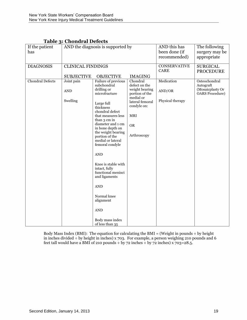
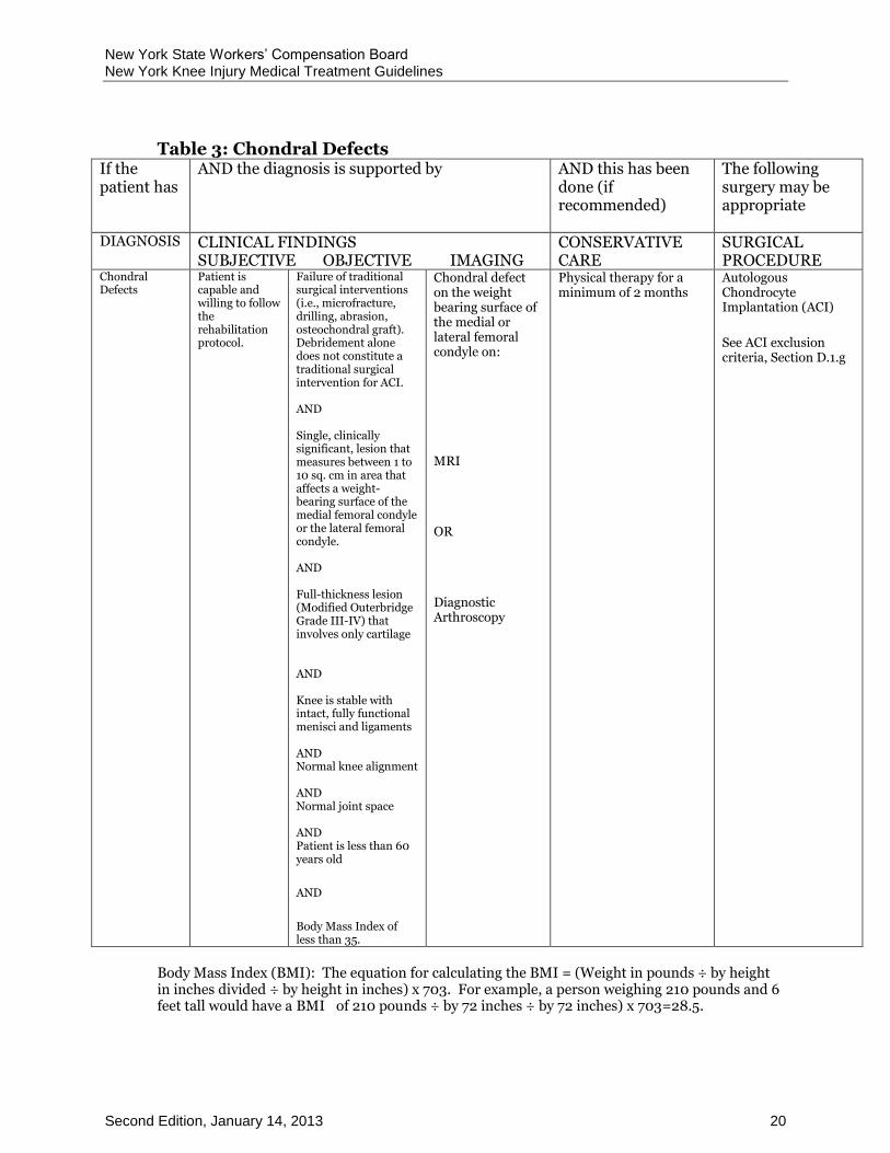
Table 3: Chondral Defects If the patient has
AND the diagnosis is supported by
AND this has been done (if recommended)
The following surgery may be appropriate
DIAGNOSIS CLINICAL FINDINGS SUBJECTIVE OBJECTIVE IMAGING
CONSERVATIVE CARE
SURGICAL PROCEDURE
Chondral Defects
Joint pain AND Swelling
Effusion OR Crepitus OR Limited ROM
Medication AND/OR Physical therapy
Chondroplasty (Shaving or debridement of an articular surface)
Chondral Defects
Joint pain AND Swelling
Small full thickness chondral defect on the weight bearing portion of the medial or lateral femoral condyle AND Knee is stable with intact, fully functional menisci and ligaments AND Normal joint space AND Ideal age 45 or younger
Chondral defect on the weight bearing portion of the medial or lateral femoral condyle on: MRI OR Diagnostic Arthroscopy
Medication AND/OR Physical therapy
Subchondral drilling OR Micro-Fracture
New York State Workers’ Compensation Board New York Knee Injury Medical Treatment Guidelines
Second Edition, January 14, 2013 19
Table 3: Chondral Defects If the patient has
AND the diagnosis is supported by
AND this has been done (if recommended)
The following surgery may be appropriate
DIAGNOSIS CLINICAL FINDINGS SUBJECTIVE OBJECTIVE IMAGING
CONSERVATIVE CARE
SURGICAL PROCEDURE
Chondral Defects Joint pain
AND
Swelling
Failure of previous subchondral drilling or microfracture
Large full thickness chondral defect that measures less than 3 cm in diameter and 1 cm in bone depth on the weight bearing portion of the medial or lateral femoral condyle
AND
Knee is stable with intact, fully functional menisci and ligaments
AND
Normal knee alignment
AND
Body mass index of less than 35
Chondral defect on the weight bearing portion of the medial or lateral femoral condyle on:
MRI
OR
Arthroscopy
Medication
AND/OR
Physical therapy
Osteochondral Autograft (Mosaicplasty Or OARS Procedure)
Body Mass Index (BMI): The equation for calculating the BMI = (Weight in pounds ÷ by height in inches divided ÷ by height in inches) x 703. For example, a person weighing 210 pounds and 6 feet tall would have a BMI of 210 pounds ÷ by 72 inches ÷ by 72 inches) x 703=28.5.
New York State Workers’ Compensation Board New York Knee Injury Medical Treatment Guidelines
Second Edition, January 14, 2013 20
Table 3: Chondral Defects If the patient has
AND the diagnosis is supported by
AND this has been done (if recommended)
The following surgery may be appropriate
DIAGNOSIS CLINICAL FINDINGS SUBJECTIVE OBJECTIVE IMAGING
CONSERVATIVE CARE
SURGICAL PROCEDURE
Chondral Defects
Patient is capable and willing to follow the rehabilitation protocol.
Failure of traditional surgical interventions (i.e., microfracture, drilling, abrasion, osteochondral graft). Debridement alone does not constitute a traditional surgical intervention for ACI. AND Single, clinically significant, lesion that measures between 1 to 10 sq. cm in area that affects a weight-bearing surface of the medial femoral condyle or the lateral femoral condyle. AND Full-thickness lesion (Modified Outerbridge Grade III-IV) that involves only cartilage AND Knee is stable with intact, fully functional menisci and ligaments AND Normal knee alignment AND Normal joint space AND Patient is less than 60 years old
AND
Body Mass Index of less than 35.
Chondral defect on the weight bearing surface of the medial or lateral femoral condyle on:
MRI
OR
Diagnostic Arthroscopy
Physical therapy for a minimum of 2 months
Autologous Chondrocyte Implantation (ACI)
See ACI exclusion criteria, Section D.1.g
Body Mass Index (BMI): The equation for calculating the BMI = (Weight in pounds ÷ by height in inches divided ÷ by height in inches) x 703. For example, a person weighing 210 pounds and 6 feet tall would have a BMI of 210 pounds ÷ by 72 inches ÷ by 72 inches) x 703=28.5.

New York State Workers’ Compensation Board New York Knee Injury Medical Treatment Guidelines
Second Edition, January 14, 2013 21
AGGRAVATED OSTEOARTHRITIS D.2
D.2.a Description/Definition
Swelling and/or pain in a joint due to an aggravating activity in a patient with pre-existing degenerative change in a joint.
D.2.b Mechanism of Injury
May be caused by repetitive activity or constant position.
D.2.c Specific Physical Findings
Increased pain and swelling in a joint.
D.2.d Diagnostic Testing Procedures
Radiographs
D.2.e Non-Operative Treatment
NSAIDS , ice, bracing, active and/or passive therapy, therapeutic injections, which may include hyaluronate therapy, restricted activity.
D.2.f Surgical Indications/Operative Treatment
Symptoms not responsive to conservative therapy.
Debridement with or without removal of loose bodies. Arthroscopic joint lavage is not recommended.
For symptoms not responsive to conservative measures, treatment may involve total joint. Refer to Table 8.
D.2.g Post-Operative Therapy
Active and/or passive therapy.
COLLATERAL LIGAMENT INJURY D.3
D.3.a Description/Definition
Sprain/strain or rupture of the medial or lateral collateral ligament. Injury of the medial collateral ligament may also be associated with a concomitant medial meniscus injury.
New York State Workers’ Compensation Board New York Knee Injury Medical Treatment Guidelines
Second Edition, January 14, 2013 22
D.3.b Mechanism of Injury
Valgus or varus trauma force applied to the knee.
D.3.c Specific Physical Findings
Medial-lateral instability (knee should be tested in slight flexion), tenderness over medial or lateral collateral ligament which increases with valgus or varus force applied to the knee.
D.3.d Diagnostic Testing Procedures
MRI may be indicated for suspected Grade II or Grade III tears.
D.3.e Non-Operative Treatment
Isolated Grade I collateral ligament tears and many Grade II tears have been shown to heal with excellent results without surgical intervention. When accompanying cruciate or meniscus injuries are ruled out, the patient can be treated non-operatively. Conservative management with casting, orthotics and rehabilitation may be indicated.
D.3.f Surgical Indications/Operative Treatment
A complete Grade III collateral ligament tear should be referred to an orthopedic surgeon.
ANTERIOR CRUCIATE LIGAMENT (ACL) INJURY D.4
D.4.a Description/Definition
Rupture or partial rupture of the anterior cruciate ligament; may be associated with other internal derangement of the knee.
D.4.b Mechanism of Injury
May be caused by virtually any traumatic force to the knee but most often caused by a twisting or a hyperextension force.
D.4.c Specific Physical Findings
Findings on physical exam include effusion or hemarthrosis, instability, Lachman’s test, pivot shift test, and anterior drawer test.
New York State Workers’ Compensation Board New York Knee Injury Medical Treatment Guidelines
Second Edition, January 14, 2013 23
D.4.d Diagnostic Testing Procedures
MRI. Radiographs may show avulsed portion of tibial spine but this is a rare finding.
D.4.e Non-Operative Treatment
Active and/or passive therapy, bracing.
D.4.f Surgical Indications/Operative Treatment
If a non-operative treatment approach is initially recommended, surgery may be indicated after the failure of conservative management. The patient must continue to exhibit the designated objective findings, subjective symptoms and (where applicable) imaging findings. Refer to Table 4.
D.4.g Post-Operative Therapy
Active and/or passive therapy, bracing.
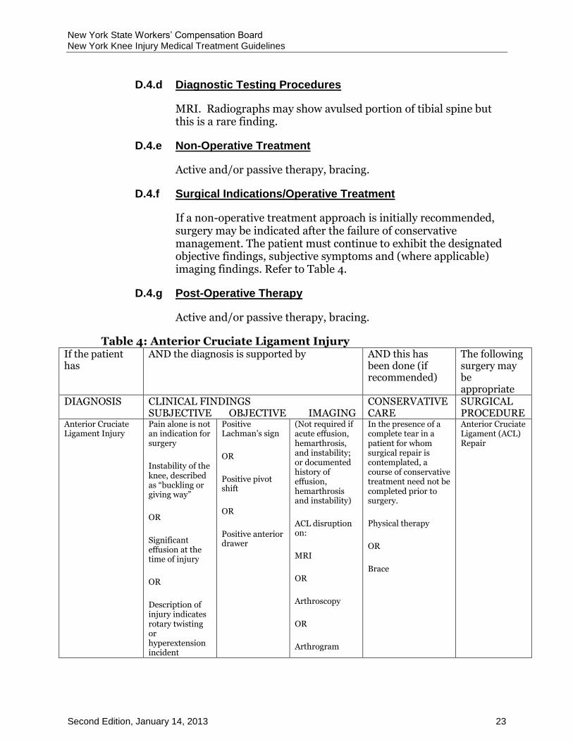
Table 4: Anterior Cruciate Ligament Injury If the patient has
AND the diagnosis is supported by
AND this has been done (if recommended)
The following surgery may be appropriate
DIAGNOSIS CLINICAL FINDINGS SUBJECTIVE OBJECTIVE IMAGING
CONSERVATIVE CARE
SURGICAL PROCEDURE
Anterior Cruciate Ligament Injury
Pain alone is not an indication for surgery
Instability of the knee, described as “buckling or giving way”
OR
Significant effusion at the time of injury
OR
Description of injury indicates rotary twisting or hyperextension incident
Positive Lachman’s sign
OR
Positive pivot shift
OR
Positive anterior drawer
(Not required if acute effusion, hemarthrosis, and instability; or documented history of effusion, hemarthrosis and instability)
ACL disruption on:
MRI
OR
Arthroscopy
OR
Arthrogram
In the presence of a complete tear in a patient for whom surgical repair is contemplated, a course of conservative treatment need not be completed prior to surgery.
Physical therapy
OR
Brace
Anterior Cruciate Ligament (ACL) Repair

New York State Workers’ Compensation Board New York Knee Injury Medical Treatment Guidelines
Second Edition, January 14, 2013 24
POSTERIOR CRUCIATE LIGAMENT (PCL) INJURY D.5
D.5.a Description/Definition
Rupture of PCL; may have concurrent ACL rupture.
D.5.b Mechanism of Injury
Most often caused by a posterior directed force to flexed knee.
D.5.c Specific Physical Findings
Findings on physical exam include acute effusion, instability, reverse Lachman’s test, reverse pivot shift, posterior drawer test.
D.5.d Diagnostic Testing Procedures
MRI, radiographs may reveal avulsed bone.
D.5.e Non-Operative Treatment
Active and/or passive therapy, bracing.
D.5.f Surgical Indications
Complaints of instability. Carefully consider the patients’ normal daily activity level before initiation of surgical intervention. Most commonly done when the PCL rupture is accompanied by multiligament injury.
D.5.g Operative Treatment
Autograft or allograft reconstruction.
D.5.h Post-Operative Therapy
Active and/or passive therapy, bracing.
MENISCUS INJURY D.6
D.6.a Description/Definition
A tear, disruption, or avulsion of medial or lateral meniscus tissue.
New York State Workers’ Compensation Board New York Knee Injury Medical Treatment Guidelines
Second Edition, January 14, 2013 25
D.6.b Mechanism of Injury
Trauma to the menisci from rotational, shearing, torsion, and/or impact injuries.
D.6.c Specific Physical Findings
Patient describes a popping, tearing, or catching sensation. Findings on physical exam may include joint line tenderness, locked joint, or occasionally, effusion.
D.6.d Diagnostic Testing Procedures
Radiographs including standing Posterior/Anterior (PA), lateral, tunnel, and skyline views. MRI is the definitive imaging test MRI is sensitive and specific for meniscal tear. However, meniscal MRI is frequently abnormal in asymptomatic injuries. Clinical correlation with history and physical exam findings specific for meniscus injury is critically important.
Providers planning treatment should therefore consider the patient's complaints and presence of arthritis on MRI carefully, knowing that not all meniscus tears in the middle aged and older population are related to the patients’ complaints of pain.
MRI arthrograms may be approved to diagnose recurrent meniscal tears particularly after previous surgery.
D.6.e Non-Operative Treatment
Active and/or passive therapy, bracing. Trial of Manipulation may be attempted for a locked knee. Clinical response should be seen within 2-3 treatments.
D.6.f Surgical Indications/ Operative Treatment Meniscectomy/Meniscus Repair and Meniscal Allograft Transplantation.
Meniscal Allograft Transplantation is not included on the list of pre-authorized procedures. Providers who want to perform one of these procedures must request pre-authorization from the carrier before performing the procedure. Refer to Table 5.
D.6.g Post-Operative Therapy
Active and/or passive therapy, bracing.
New York State Workers’ Compensation Board New York Knee Injury Medical Treatment Guidelines
Second Edition, January 14, 2013 26
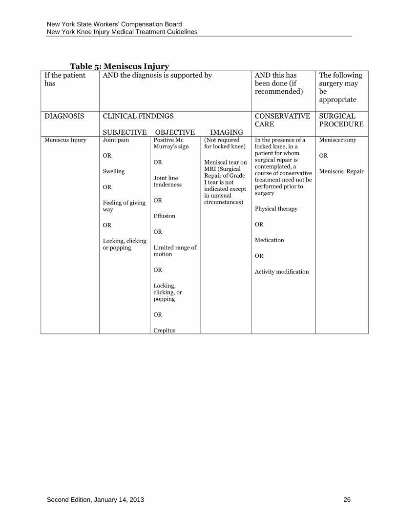
Table 5: Meniscus Injury If the patient has
AND the diagnosis is supported by
AND this has been done (if recommended)
The following surgery may be appropriate
DIAGNOSIS CLINICAL FINDINGS SUBJECTIVE OBJECTIVE IMAGING
CONSERVATIVE CARE
SURGICAL PROCEDURE
Meniscus Injury Joint pain
OR
Swelling
OR
Feeling of giving way
OR
Locking, clicking or popping
Positive Mc Murray’s sign
OR
Joint line tenderness
OR
Effusion
OR
Limited range of motion
OR
Locking, clicking, or popping
OR
Crepitus
(Not required for locked knee)
Meniscal tear on MRI (Surgical Repair of Grade I tear is not indicated except in unusual circumstances)
In the presence of a locked knee, in a patient for whom surgical repair is contemplated, a course of conservative treatment need not be performed prior to surgery
Physical therapy
OR
Medication
OR
Activity modification
Meniscectomy
OR
Meniscus Repair

New York State Workers’ Compensation Board New York Knee Injury Medical Treatment Guidelines
Second Edition, January 14, 2013 27
Table 5: Meniscus Injury If the patient has
AND the diagnosis is supported by
AND this has been done (if recommended)
The following surgery may be appropriate
DIAGNOSIS CLINICAL FINDINGS SUBJECTIVE OBJECTIVE IMAGING
CONSERVATIVE CARE
SURGICAL PROCEDURE
Meniscus Injury Capable and willing to follow the rehabilitation protocol
AND
Knee pain that has not responded to conservative treatment
Previous meniscectomy with at least two-thirds of the meniscus removed
AND
If Modified Outerbridge Scale Graft III then debridement must first produce an articular surface sufficiently free of irregularities to maintain the integrity of the transplanted meniscus. (See Table 6 for Modified Outerbridge Classification)
AND
Stable knee with intact ligaments, normal alignment, and normal joint space.
AND Ideal age 20-45 years (too young for total knee)
AND Body Mass Index of less than 35
Articular cartilage in the affected compartment demonstrates a chondrosis classified by the Modified Outerbridge Scale as Grade I, Grade II or Grade III
Physical therapy
OR
NSAID
OR
Activity modification
Meniscal Allograft Transplantation
See meniscal allograft transplantation exclusion criteria next page

New York State Workers’ Compensation Board New York Knee Injury Medical Treatment Guidelines
Second Edition, January 14, 2013 28
MENISCAL ALLOGRAFT TRANSPLANTATION EXCLUSION D.7CRITERIA
Meniscal Allograft Transplantation is not a covered procedure in any of the following circumstances:
a. Mild to severe localized or diffuse arthritic condition that appears on standing x-ray as joint space narrowing, osteophytes or changes in the underlying bone.
b. Articular cartilage in the affected compartment demonstrates a chondrosis classified by the Modified Outerbridge Scale as Grade III that has not undergone debridement; Grade III with debridement that has not produced an articular surface that can maintain the integrity of the transplanted meniscus; or Grade IV.
Table 6: Modified Outerbridge Classification
I Articular cartilage softening
II Chondral fissures or fibrillation < 1.25 cm in diameter
III Chondral fibrillation > 1.25 cm in diameter (“crabmeat changes”)
IV Exposed subchondral bone
Body Mass Index (BMI): The equation for calculating the BMI = (Weight in pounds ÷ by height in inches divided ÷ by height in inches) x 703. For example, a person weighing 210 pounds and 6 feet tall would have a BMI of 210 pounds ÷ by 72 inches ÷ by 72 inches) x 703=28.5.
PATELLAR SUBLUXATION D.8
D.8.a Description/Definition
An incomplete subluxation or dislocation of the patella. Recurrent episodes can lead to subluxation syndrome that can cause frank dislocation of the patella.
D.8.b Mechanism of Injury
Primarily associated with contusion, lateral force direct contact. Secondary causes associated with shearing forces on the patella.

New York State Workers’ Compensation Board New York Knee Injury Medical Treatment Guidelines
Second Edition, January 14, 2013 29
D.8.c Specific Physical Findings
Patient may report buckling sensation. Findings on physical exam may include retinacular weakness, swelling, effusion, marked pain with patellofemoral tracking/compression and glides. In addition, other findings include atrophy of muscles, positive patellar apprehension test, patella alta.
D.8.d Diagnostic Testing Procedures
Radiographs including Merchant views, Q-angle versus congruents.
D.8.e Non-Operative Treatment
Active and/or passive therapy, bracing, therapeutic injection.
D.8.f Surgical Indications
Fracture, recurrent subluxation or recurrent effusion, or symptoms not responsive to conservative therapy
D.8.g Operative Treatment
Open reduction internal fixation with fracture. Following a patellar dislocation, surgical consultation no sooner than 4-6 months of conservative therapy. Retinacular release, quadriceps reefing, and patellar tendon transfer should only be considered after a minimum of 4 to 5 months of conservative therapy.
D.8.h Post-Operative Therapy
Active and/or passive therapy, bracing.
RETROPATELLAR PAIN SYNDROME (CHONDROMALACIA D.9PATELLA)
D.9.a Description/Definition
A retropatellar pain syndrome lasting over three months. Retropatellar pathologies are associated with resultant weakening instability, and pain of the patellofemoral mechanism. Can include malalignment, persistent quadriceps tendinitis, distal patellar tendinitis, patellofemoral arthrosis, and symptomatic plica syndrome.
New York State Workers’ Compensation Board New York Knee Injury Medical Treatment Guidelines
Second Edition, January 14, 2013 30
D.9.b Mechanism of Injury
May be associated with contusion, repetitive patellar compressive forces, shearing articular injuries associated with subluxation or dislocation of patella, fractures, infection, and connective tissue disease.
D.9.c Specific Physical Findings
Patient complains of pain, instability and tenderness that interfere with daily living and work functions. Findings on physical exam may include retinacular tenderness, pain with patellar compressive ranging, positive patellar glide test, atrophy of quadriceps muscles, positive patellar apprehensive test. Associated anatomical findings may include increased Q angle; rotational lower extremity joints; ligament laxity, and effusion.
D.9.d Diagnostic Testing Procedures
Radiographs including tunnel, Merchant, or Laurin views. MRI rarely identifies pathology. Occasionally CT or bone scan.
D.9.e Non-Operative Treatment
Active and/or passive therapy, bracing, orthotics, therapeutic injections.
D.9.f Surgical Indications
Patellar tendon disruption, quadriceps tendon rupture/avulsion, fracture, or symptoms not responsive to conservative therapy. There is very limited data on long term outcomes of surgical treatment for anterior knee pain. Surgical intervention should be considered after failure of a comprehensive rehabilitation program that has included quadriceps strengthening.
D.9.g Operative Treatment
Arthroscopic debridement of articular surface, plica, synovial tissue, loose bodies, arthrotomy, open reduction internal fixation with fracture, patellar button (prosthesis) with grade III-IV osteoarthritis (modified Outerbridge classification ) and possible patellectomy. Retinacular release, quadriceps reefing, and tibial transfer procedures should only be considered after 6 to 9 months of conservative therapy. Refer to Table 7.

New York State Workers’ Compensation Board New York Knee Injury Medical Treatment Guidelines
Second Edition, January 14, 2013 31
D.9.h Post-Operative Therapy
Active and/or passive therapy; bracing.
Table 7: Retropatellar Pain Syndrome If the patient has
AND the diagnosis is supported by
AND this has been done (if recommended)
The following surgery may be appropriate
DIAGNOSIS CLINICAL FINDINGS SUBJECTIVE OBJECTIVE IMAGING
CONSERVATIVE CARE
SURGICAL PROCEDURE
RETROPATELLAR PAIN SYNDROME (CHONDROMALACIA PATELLA)
Knee pain with sitting
OR
Pain with patellar/femoral movement
OR
Recurrent dislocations
Lateral tracking of the patella
OR
Recurrent effusion
OR
Patellar apprehension
OR
synovitis with or without crepitus
OR
Increased Q angle > 15 degrees
Abnormal patellar tilt on: x-ray or MRI
Physical therapy
(not required for acute patellar dislocation with associated intra-articular fracture)
OR
Medications
Lateral Retinacular Release
OR
Patellar Tendon Realignment
OR
Maquet Procedure
TENDINITIS/TENOSYNOVITIS D.10
D.10.a Description/Definition
Inflammation of the lining of the tendon sheath or of the enclosed tendon. Usually occurs at the point of insertion into bone or a point of muscular origin. Can be associated with bursitis, or calcium deposits or systemic connective diseases.
D.10.b Mechanism of Injury
May be caused by extreme or repetitive trauma, strain, or excessive unaccustomed exercise or work.
New York State Workers’ Compensation Board New York Knee Injury Medical Treatment Guidelines
Second Edition, January 14, 2013 32
D.10.c Specific Physical Findings
Involved tendons may be visibly swollen with possible fluid accumulation and inflammation; popping or crepitus; and decreased range of motion.
D.10.d Diagnostic Testing Procedures
Rarely indicated.
D.10.e Non-Operative Treatment
Active and/or passive therapy, including ergonomic changes at work station(s), NSAIDs, therapeutic injections.
D.10.f Surgical Indications
Suspected avulsion fracture or severe functional impairment unresponsive to conservative therapy.
D.10.g Operative Treatment
Rarely indicated and only after extensive conservative therapy.
D.10.h Post-Operative Therapy
Active and/or passive therapy.
BURSITIS D.11
D.11.a Description/Definition
Inflammation of bursa tissue. Can be precipitated by tendinitis, bone spurs, foreign bodies, gout, arthritis, muscle tears, or infection.
D.11.b Mechanism of Injury
May be caused by sudden change in work habits, frequent repetitive motions in non-routine work profile, postural changes, contusion, frequent climbing, soft tissue trauma, fracture, continuous work on uneven surfaces, sustained compression force.
D.11.c Specific Physical Findings
Palpable, tender and enlarged bursa, decreased range of motion, warmth. May have increased pain with range of motion.

New York State Workers’ Compensation Board New York Knee Injury Medical Treatment Guidelines
Second Edition, January 14, 2013 33
D.11.d Diagnostic Testing Procedures
Bursal fluid aspiration with testing for connective tissue, rheumatic disease, and infection. Radiographs, CT, MRI are rarely indicated.
D.11.e Non-Operative Treatment
Active and/or passive therapy, ice, therapeutic injection, treatment of an underlying infection, if present.
D.11.f Surgical Indications
Bursa excision after failure of conservative therapy.
D.11.g Operative Treatment
Surgical excision of the bursa.
D.11.h Post-Operative Therapy
Active and/or passive therapy.
E THERAPEUTIC PROCEDURES, NON-OPERATIVE
Before initiation of any therapeutic procedure, the authorized treating provider, employer and insurer must consider these important issues in the care of the injured worker.
First, patients undergoing therapeutic procedure(s) should be released or returned to modified, restricted, or full duty during their rehabilitation at the earliest appropriate time.
Second, cessation and/or review of treatment modalities should be undertaken when no further significant subjective or objective improvement in the patient’s condition is noted. If patients are not responding within the recommended duration periods, alternative treatment interventions, further diagnostic studies or consultations should be pursued.
Third, providers should provide and document education to the patient. No treatment plan is complete without addressing issues of individual and/or group patient education as a means of facilitating self-management of symptoms.
In unusual cases where a patient is unable to attend an outpatient center, home therapy may be necessary. Home therapy may include active and passive therapeutic procedures as well as other modalities to assist in alleviating pain, swelling, and abnormal muscle tone. Home therapy is usually of short duration.
New York State Workers’ Compensation Board New York Knee Injury Medical Treatment Guidelines
Second Edition, January 14, 2013 34
The following procedures are listed in alphabetical order.
ACUPUNCTURE E.1
Acupuncture is a procedure used for the relief of pain and inflammation and there is some scientific evidence to support its use. The exact mode of action is only partially understood. Western medicine studies suggest that acupuncture stimulates the nervous system at the level of the brain, promotes deep relaxation, and affects the release of neurotransmitters. Acupuncture is commonly used as an alternative or in addition to traditional Western pharmaceuticals. While it is commonly used when pain medication is reduced or not tolerated, it may be used as an adjunct to physical rehabilitation and/or surgical intervention to hasten the return of functional activity. Moxibustion and other complementary integrative medicine techniques are often combined with acupuncture , but have no demonstrated efficacy. No additional reimbursement should be provided for acupuncture combined with moxibustion or other similar adjunctive procedures. Acupuncture must be performed by a professional who is authorized under the Workers’ Compensation Laws and duly certified in New York State to provide acupuncture services.
Acupuncture with or without electrical stimulation) is the insertion and removal of filiform needles to stimulate acupoints (acupuncture points), with or without the use of electrical current (micro-amperage or milli-amperage) on the needles at the acupuncture site. Needles may be inserted, manipulated and retained for a period of time. Acupuncture can be used to reduce pain, reduce inflammation, increase blood flow, increase range of motion, decrease the side effect of medication-induced nausea, promote relaxation in an anxious patient, and reduce muscle spasm. Indications include joint pain, joint stiffness, soft tissue pain and inflammation, paresthesia, post-surgical pain relief, muscle spasm, and scar tissue pain.
Time to produce effect: 3 to 6 treatments
Frequency: 1 to 3 times per week
Optimum duration: 1 month
Maximum duration: 10 treatments
Total Time Frames for Acupuncture and Acupuncture with Electrical Stimulation: Time frames are not meant to be applied to each of the above sections separately. The time frames are to be applied to all acupuncture treatments regardless of the type or combination of therapies being provided.

New York State Workers’ Compensation Board New York Knee Injury Medical Treatment Guidelines
Second Edition, January 14, 2013 35
BIOFEEDBACK E.2
Not recommended.
INJECTIONS-THERAPEUTIC E.3
Description: Therapeutic injections involve the delivery of anesthetic and/or anti-inflammatory medications to the painful structure. Therapeutic injections have many potential benefits. Ideally, a therapeutic injection will: (a) reduce inflammation in a specific target area; (b) relieve secondary muscle spasm; (c) allow a break from pain; and (d) support therapy directed to functional recovery. Diagnostic and therapeutic injections should be used early and selectively to establish a diagnosis and support rehabilitation. If injections are overused or used outside the context of a monitored rehabilitation program, they may be of significantly less value.
Contraindications: General contraindications include local or systemic infection, bleeding disorders, allergy to medications used and patient refusal. Specific contraindications may apply to individual injections.
E.3.a Soft tissue and Joint Injections
Soft tissue and joint injections may be performed as analgesic or anti-inflammatory procedures. Injections into the tendon are not recommended.
Frequency: Not more than 2 to 3 times annually. Usually 1 or 2 injections adequate. A minimum of 3 weeks interval between injections is recommended.
Time to produce effect: Immediate with local anesthetic, or within 3 days with corticosteroids.
Optimum/maximum duration: Limited to 3 injections annually to the same site.
E.3.b Trigger Point Injections
Not recommended.
E.3.c Prolotherapy (also known as sclerotherapy)
Not recommended.
E.3.d Protein Rich Plasma (PRP)
Not recommended.

New York State Workers’ Compensation Board New York Knee Injury Medical Treatment Guidelines
Second Edition, January 14, 2013 36
E.3.e Intra-Capsular Acid Salts
Intra-Capsular Acid Salts (also known as Viscosupplementation) is a form of treatment for osteoarthritis or degenerative changes in the knee joint. It is recommended that these injections be considered a therapeutic alternative in patients who have failed non-pharmacological and analgesic treatment, and particularly, if non-steroidal anti-inflammatory drug treatment is contraindicated or surgery is not an option. The utility of viscosupplementation in severe osteoarthritis and its efficacy beyond 6 months is not well known.
Time to produce effect: One series of injections, per product instructions.
Frequency: If the first use is associated with decreased symptoms and increased function, repeat use may be considered after 6 months if symptoms recur.
Optimum/maximum duration: Varies. Efficacy beyond 6 months is not well known.
MEDICATIONS E.4
Medication use in the treatment of knee injuries is appropriate for controlling acute pain and inflammation. Use of medications will vary widely due to the spectrum of injuries.
All drugs should be used according to patient needs. A thorough medication history, including use of alternative and over- the- counter medications should be performed at the time of the initial visit and updated periodically. Treatment for pain control is initially accomplished with Acetaminophen and/or NSAIDS. The patient should be educated regarding the interaction with prescription and over-the-counter medications as well as the contents of over-the-counter herbal products.
The following are listed in alphabetical order.
E.4.a Acetaminophen
Acetaminophen is an effective analgesic with antipyretic but not anti-inflammatory activity. Acetaminophen is generally well tolerated, causes little or no gastrointestinal irritation, and is not associated with ulcer formation. Acetaminophen has been associated with liver toxicity when the recommended daily dose is exceeded or in patients who chronically use alcohol. Patients may not realize that many over-the-counter preparations may contain

New York State Workers’ Compensation Board New York Knee Injury Medical Treatment Guidelines
Second Edition, January 14, 2013 37
Acetaminophen. The total daily dose of Acetaminophen should not exceed 4 grams per 24-hour period from all sources, including narcotic-Acetaminophen combination preparations. Patients who consume three or more alcoholic drinks per day are at greater risk for liver toxicity, and consideration should be given to the use of other analgesics or limiting the Acetaminophen dose to 2 grams per 24-hour period from all sources. Monitoring liver function via blood testing for use beyond 10 days is advisable.
Recommendations:
Acetaminophen is a reasonable alternative to NSAIDs, although evidence suggests it is modestly less efficacious.
Acetaminophen is recommended for treatment of knee pain, particularly for those with contraindications for NSAIDs.
Optimum Duration: 7 to 10 days.
Maximum Duration: Chronic use as indicated on a case-by-case basis.
E.4.b Minor Tranquilizer/Muscle Relaxants
Not recommended.
E.4.c Narcotics
Narcotics should be primarily reserved for the treatment of severe knee pain. In mild-to-moderate cases of knee pain, narcotic medication should be used cautiously on a case-by-case basis. Adverse effects include respiratory depression, the development of physical and psychological dependence, and impaired alertness. This medication has physically addictive properties and withdrawal symptoms may follow abrupt discontinuation.
Narcotic medications should be prescribed with strict time, quantity, and duration guidelines, and with definitive cessation parameters.
Pain is subjective in nature and should be evaluated using a scale to rate effectiveness of the narcotic prescribed. Any use beyond the maximum should be documented and justified based on the diagnosis and/or invasive procedures.
New York State Workers’ Compensation Board New York Knee Injury Medical Treatment Guidelines
Second Edition, January 14, 2013 38
Optimum Duration: 3 to 7 days.
Maximum Duration: 2 weeks. Use beyond two weeks is acceptable in appropriate cases.
Any use beyond the maximum should be documented and justified based on the diagnosis and/or invasive procedures.
E.4.d Nonsteroidal Anti-Inflammatory Drugs (NSAIDs)
Nonsteroidal Anti-Inflammatory Drugs (NSAIDS) are useful for pain and inflammation. In mild cases, they may be the only drugs required for analgesia. There are several classes of NSAIDS, and the response of the individual patient to a specific medication is unpredictable. For this reason, a range of NSAIDS may be tried in each case with the most effective preparation being continued. Patients should be closely monitored for adverse reactions. The US Food and Drug Administration advises that many NSAIDS may have an increased risk of serious cardiovascular thrombotic events, myocardial infarction, stroke, which can be fatal and increased risk of serious adverse GI events including bleeding, ulceration and perforation of the stomach and intestines.
Generally, older generation (COX-1, non-selective) NSAIDS are recommended as first-line medications. Second-line medications should generally include one of the other COX-1 medications. While COX-2 selective agents generally have been recommended as either third- or fourth-line medications to use when there is a risk of gastrointestinal complications, misoprostol, sucralfate, histamine 2 blockers and proton pump inhibitors are also gastro-protective. COX-2 selective agents may still be used for those with contraindications to other medications, especially those with a history of gastrointestinal bleeding or past history of peptic ulcer disease.
Selective COX-2 inhibitors should be used with great caution in patients with ischemic heart disease and/or stroke and avoided in patients with risk factors for coronary heart disease. Patients with known cardiovascular disease or multiple risk factors for cardiovascular disease should have the risks and benefits of NSAID therapy for pain discussed. In these patients, it appears to be safest to use Acetaminophen or aspirin as the first-line therapy. If needed, NSAIDS that are non-selective are preferred over COX-2 specific drugs. Even a relative lack of COX-2 selectivity does not completely eliminate the risk of cardiovascular events, and in that regard, all drugs in the NSAID spectrum should

New York State Workers’ Compensation Board New York Knee Injury Medical Treatment Guidelines
Second Edition, January 14, 2013 39
only be prescribed after thorough consideration of risk benefit balance. Patients who receive COX-2 inhibitors should take the lowest effective dose for the shortest time necessary to control symptoms. In patients receiving low-dose aspirin for primary or secondary cardiovascular disease prevention, to minimize the potential for the NSAID to counteract the beneficial effects of aspirin, aspirin should be taken 2 hours before or at least 8 hours after the NSAID. (Antman 07).
Recommendations:
NSAIDs are recommended for the treatment of acute, subacute, chronic, or post-operative knee pain. Over-the counter (OTC) agents may suffice and may be tried first.
Frequency/Duration: In most patients with acute knee pain, scheduled dosage, rather than as needed, is generally preferable. As needed (PRN) prescriptions may be reasonable for mild, moderate or chronic knee pain. Discontinuation: Resolution of symptoms, lack of efficacy, or development of adverse effects that necessitate discontinuation.
Those patients at substantially increased risk for gastrointestinal bleeding, who also have indications for NSAIDs, should be considered for concomitant prescriptions of cytoprotective medications, particularly if longer term treatment is contemplated.
Individuals considered being at elevated risk include history of prior gastrointestinal bleeding, elderly, diabetics, and cigarette smokers. There are four commonly used cytoprotective classes of drugs: Misoprostol, sucralfate, histamine type 2 receptor blockers (famotidine, ranitidine, cimetadine, etc.), and proton pump inhibitors (esomeprazole, lansoprazole, omeprazole, pantoprazole, rabeprazole). There is not believed to be substantial differences in efficacy for prevention of gastrointestinal bleeding. There also are combination products of NSAIDs/misoprostol (e.g., arthrotec).
Frequency/Duration: Frequency as recommended.
Discontinuation: Intolerance, development of adverse effects, or discontinuation of the NSAID.

New York State Workers’ Compensation Board New York Knee Injury Medical Treatment Guidelines
Second Edition, January 14, 2013 40
E.4.e Topical Drug Delivery
Topical drug delivery may be an alternative treatment of localized musculoskeletal disorders. It is necessary that all topical agents be used with strict instructions for application as well as maximum number of applications per day to obtain the desired benefit and avoid potential toxicity. As with all medications, patient selection must be rigorous to select those patients with the highest probability of compliance.
E.4.e.i Topical Salicylates and Nonsalicylates
Topical Salicylates and Nonsalicylates have been shown to be effective in relieving pain in acute and chronic musculoskeletal conditions. Topical salicylates and nonsalicylates achieve tissue levels that are potentially therapeutic, at least with regard to COX inhibition. Other than local skin reactions, the side effects of therapy are minimal, although not nonexistent, and the usual contraindications to use of these compounds need to be considered. Local skin reactions are rare and systemic effects even less common. Their use in patients receiving warfarin therapy may result in alterations in bleeding time. Overall, the low level of systemic absorption can be advantageous, allowing the topical use of these medications when systemic administration is relatively contraindicated such as is the case in patients with hypertension, cardiac failure, or renal insufficiency.
Optimal duration: 1-2 weeks to determine effectiveness.
Continued use should be evaluated every 3 months.
E.4.e.ii Capsaicin
Capsaicin is another medication option for topical drug use in lower extremity injury. Capsaicin offers a safe and effective alternative to systemic NSAID therapy. Although it is quite safe, effective use of capsaicin is limited by the local stinging or burning sensation that typically dissipates with regular use, usually after the first 7 to 10 days of treatment. Patients should be advised to apply the cream on the affected area with a plastic glove or cotton applicator and to avoid inadvertent contact with eyes and mucous membranes.

New York State Workers’ Compensation Board New York Knee Injury Medical Treatment Guidelines
Second Edition, January 14, 2013 41
Optimal duration: 1-2 weeks to determine effectiveness.
Continued use should be evaluated every 3 months. individual cases.
E.4.f Tramadol
Tramadol is useful in relief of lower extremity pain and has been shown to provide pain relief equivalent to that of commonly prescribed NSAIDS. Although Tramadol may cause impaired alertness, it is generally well tolerated, does not cause gastrointestinal ulceration, or exacerbate hypertension or congestive heart failure. Tramadol should be used cautiously in patients who have a history of seizures or who are taking medication that may lower the seizure threshold, such as MAO inhibitors, SSRIs, and tricyclic antidepressants. This medication has physically addictive properties and withdrawal may follow abrupt discontinuation and is not recommended for patients with prior opioid addiction.
Maximum duration: 2 weeks. Use beyond 2 weeks is acceptable in appropriate cases.
ORTHOTICS AND PROSTHETICS E.5
E.5.a Fabrication/Modification of Orthotics
Fabrication/Modification of Orthotics would be used when there is need to normalize weight-bearing, facilitate better motion response, stabilize a joint with insufficient muscle or proprioceptive/reflex competencies, to protect subacute conditions as needed during movement, and correct biomechanical problems.
Time to produce effect: 1 to 3 sessions (includes wearing schedule evaluation).
Frequency: 1 to 2 times per week.
Optimum/maximum duration: 4 sessions of evaluation, casting, fitting, and re-evaluation.
New York State Workers’ Compensation Board New York Knee Injury Medical Treatment Guidelines
Second Edition, January 14, 2013 42
E.5.b Orthotic/Prosthetic Training
Orthotic/Prosthetic Training is the skilled instruction (preferably by qualified providers) in the proper use of orthotic devices and/or prosthetic limbs including stump preparation, donning and doffing limbs, instruction in wearing schedule and orthotic/prosthetic maintenance training. Training can include gait, mobility, transfer and self-care techniques.
Time to produce effect: 2 to 6 sessions.
Frequency: 3 times per week.
Optimum/maximum duration: 2 to 4 months.
E.5.c Splints or Adaptive Equipment
Design, fabrication and/or modification indications include the need to control neurological and orthopedic injuries for reduced stress during functional activities and modify tasks through instruction in the use of a device or physical modification of a device, which reduces stress on the injury. Equipment should improve safety and reduce risk of re-injury. This includes high and low technology assistive options such as workplace modifications, computer interface or seating, crutch or walker training, and self-care aids.
Time to produce effect: Immediate.
Frequency: 1 to 3 sessions or as indicated to establish independent use.
Optimum/maximum duration: 1 to 3 sessions.
RETURN TO WORK E.6
Communication is essential between the patient, employer and physician to determine appropriate restrictions and return to work dates. It is the responsibility of the physician to provide clear, concise restrictions, and it is the employer’s responsibility to determine if temporary duties can be provided within the restrictions.

New York State Workers’ Compensation Board New York Knee Injury Medical Treatment Guidelines
Second Edition, January 14, 2013 43
E.6.a Establishment of Activity Level Restrictions
For lower extremity injuries, the following should be addressed when describing the patient’s activity level:
Lower body postures such as squatting, kneeling, crawling, stooping, or climbing should include duration and frequency.
Ambulatory level for distance, frequency and terrain should be specified.
Standing duration and frequency with regard to balance issues.
Use of adaptive devices or equipment for proper ergonomics to enhance capacities can be included.
E.6.b Compliance with Activity Restrictions
In some cases, compliance with restriction of activity levels may require a complete jobsite evaluation, a functional capacity evaluation (FCE), or other special testing
THERAPY-ACTIVE E.7
Most of the following active therapies have some evidence and are based on the philosophy that therapeutic exercise and/or activity are beneficial for restoring flexibility, strength, endurance, function, range of motion, and can alleviate discomfort. Active therapy requires an internal effort by the individual to complete a specific exercise or task. This form of therapy requires supervision from a therapist or medical provider such as verbal, visual and/or tactile instruction(s). At times, the provider may help stabilize the patient or guide the movement pattern but the energy required to complete the task is predominately executed by the patient.
Patients should be instructed to continue active therapies at home as an extension of the treatment process in order to maintain improvement levels. Home exercise can include exercise with or without mechanical assistance or resistance and functional activities with assistive devices.
The following active therapies are listed in alphabetical order.

New York State Workers’ Compensation Board New York Knee Injury Medical Treatment Guidelines
Second Edition, January 14, 2013 44
E.7.a Activities of Daily Living (ADL)\
Activities of Daily Living are instruction, active-assisted training and/or adaptation of activities or equipment to improve a person's capacity in normal daily activities such as self-care, work re-integration training, homemaking and driving.
Time to produce effect: 4 to 5 treatments.
Frequency: 2 to 3 times per week.
Optimum duration: 2 to 3 weeks.
Maximum duration: 3 weeks.
E.7.b Functional Electrical Stimulation
Functional Electrical Stimulation is the application of electrical current to elicit involuntary or assisted contractions of atrophied and/or impaired muscles. Indications include muscle atrophy, weakness, and sluggish muscle contraction secondary to pain, injury, neuromuscular dysfunction or peripheral nerve lesion or where the potential for atrophy exists. May be an appropriate treatment in conjunction with an active exercise program.
Time to produce effect: 2 to 6 treatments.
Frequency: 3 times per week.
Optimum duration: 8 weeks.
Maximum duration: 8 weeks.
E.7.c Gait Training
Gait Training is crutch walking, cane or walker instruction to a person with lower extremity injury or surgery. Indications include the need to promote normal gait pattern with assistive devices; instruct in the safety and proper use of assistive devices; instruct in progressive use of more independent devices (i.e., platform-walker, to walker, to crutches, to cane); instruct in gait on uneven surfaces and steps (with and without railings) to

New York State Workers’ Compensation Board New York Knee Injury Medical Treatment Guidelines
Second Edition, January 14, 2013 45
reduce risk of fall, or loss of balance; and/or instruct in equipment to limit weight-bearing for the protection of a healing injury or surgery.
Time to produce effect: 3 to 4 treatments.
Frequency: 2 to 3 times per week.
Optimum duration: 2 weeks.
Maximum duration: 2 weeks.
E.7.d Neuromuscular Re-education
Not recommended.
E.7.e Therapeutic Exercise
Therapeutic Exercise, with or without mechanical assistance or resistance, may include isoinertial, isotonic, isometric and isokinetic types of exercises. Indications include the need for cardiovascular fitness, reduced edema, improved muscle strength, improved connective tissue strength and integrity, increased bone density, promotion of circulation to enhance soft tissue healing, improvement of muscle recruitment, increased range of motion and are used to promote normal movement patterns. Can also include complementary/ alternative exercise movement therapy.
Time to produce effect: 2 to 6 treatments.
Frequency: 3 to 5 times per week.
Optimum duration: 4 to 8 weeks.
Maximum duration: 8 weeks.
E.7.f Wheelchair Management and Propulsion
Wheelchair Management and Propulsion is the instruction and training of self-propulsion and proper use of a wheelchair. This includes transferring and safety instruction. This is indicated in

New York State Workers’ Compensation Board New York Knee Injury Medical Treatment Guidelines
Second Edition, January 14, 2013 46
individuals who are not able to ambulate due to bilateral lower extremity injuries, inability to use ambulatory assistive devices, and in cases of multiple traumas.
Time to produce effect: 2 to 6 treatments.
Frequency: 2 to 3 times per week.
Optimum duration: 2 weeks.
Maximum duration: 2 weeks.
THERAPY-PASSIVE E.8
Therapy-Passive includes those treatment modalities that do not require energy expenditure on the part of the patient. They are principally effective during the early phases of treatment and are directed at controlling symptoms such as pain, inflammation and swelling. They should be use adjunctively with active therapies to help control swelling, pain and inflammation during the rehabilitation process. They may be used intermittently as deemed appropriate or regularly if there are specific goals with objectively measured functional improvements during treatment.
While protocols for specific diagnoses and post-surgical conditions may warrant durations of treatment beyond those listed as "maximum,” factors such as exacerbation of symptoms, re-injury, interrupted continuity of care, and co-morbidities may extend durations of care. Having specific goals with objectively measured functional improvement during treatment can support extended durations of care. It is recommended that if after 3 to 5 visits no treatment effect is observed, alternative treatment interventions, further diagnostic studies or further consultations should be pursued.
The following passive therapies and modalities are listed in alphabetical order with the exception of kinesiotaping, taping or strapping, which has been placed last to preserve the original numbering of the other therapies.
E.8.a Continuous Passive Movement (CPM)
Continuous Passive Movement) is a form of passive motion using specialized machinery that acts to move a joint and may also pump blood and edema fluid away from the joint and periarticular tissues. CPM is effective in preventing the development of joint stiffness if applied immediately following surgery. It should be continued until the swelling that limits motion of the joint no

New York State Workers’ Compensation Board New York Knee Injury Medical Treatment Guidelines
Second Edition, January 14, 2013 47
longer develops. Range of motion for the joint begins at the level of patient tolerance and is increased twice a day as tolerated. Use of this equipment may require home visits.
Time to produce effect: Immediate.
Frequency: Up to 4 times a day
Optimum duration: Up to 3 weeks post surgical.
Maximum duration: 3 weeks.
E.8.b Contrast Baths
Not recommended.
E.8.c Electrical Stimulation (Unattended)
Not recommended. For the purposes of these guidelines, unattended means that the physician or therapist is not physically present with the patient on a 1:1 basis when treatment is being administered.
E.8.d Fluidotherapy
Fluidotherapy employs a stream of dry, heated air that passes over the injured body part. The injured body part can be exercised during the application of dry heat. Indications include the need to enhance collagen extensibility before stretching, reduce muscle guarding, or reduce inflammatory response.
Time to produce effect: 1 to 4 treatments.
Frequency: 1 to 3 times per week.
Optimum duration: 4 weeks.
Maximum duration: 1 month.
E.8.e Infrared Therapy
Not recommended.
E.8.f Iontophoresis
Not recommended.
New York State Workers’ Compensation Board New York Knee Injury Medical Treatment Guidelines
Second Edition, January 14, 2013 48
E.8.g Manipulation
Manipulation is manual therapy that moves a joint beyond the physiologic range of motion but not beyond the anatomic range of motion. It is indicated for locked knee, contracture, or pain and loss of range of motion due to adhesions or contractures.
Time to produce effect: Immediate or up to 10 treatments.
Frequency: 1 to 5 times per week as indicated by the severity of involvement and the desired effect.
Optimum duration: 10 treatments.
Maximum duration: 10 treatments.
E.8.h Manual Electrical Stimulation
Manual Electrical Stimulation is used for peripheral nerve injuries or pain reduction that requires continuous application, supervision, or involves extensive teaching. Indications include muscle spasm (including TENS), atrophy, decreased circulation, osteogenic stimulation, inflammation, and the need to facilitate muscle hypertrophy, muscle strengthening, muscle responsiveness in Spinal Cord Injury/Brain Injury (SCI/BI), and peripheral neuropathies.
Time to produce effect: Variable, depending upon use.
Frequency: 3 to 7 times per week.
Optimum duration: 8 weeks.
Maximum duration: 2 months.
E.8.i Massage, Manual or Mechanical
Not recommended.

New York State Workers’ Compensation Board New York Knee Injury Medical Treatment Guidelines
Second Edition, January 14, 2013 49
E.8.j Mobilization (Joint)
Mobilization is passive movement, which may include passive range of motion performed in such a manner (particularly in relation to the speed of the movement) that it is, at all times, within the ability of the patient to prevent the movement if they so choose. It may include skilled manual joint tissue stretching. Indications include the need to improve joint play, improve intracapsular arthrokinematics, or reduce pain associated with tissue impingement/maltraction.
Time to produce effect: 6 to 9 treatments.
Frequency: 3 times per week.
Optimum duration: 10 treatments.
Maximum duration: 10 treatments.
E.8.k Mobilization (Soft Tissue)
Mobilization (Soft Tissue) is the skilled application of manual techniques designed to normalize movement patterns through the reduction of soft tissue pain and restrictions. Indications include muscle spasm around a joint, trigger points, adhesions, and neural compression.
Time to produce effect: 2 to 3 weeks.
Frequency: 2 to 3 times per week.
Optimum duration: 10 treatments.
Maximum duration: 10 treatments.
E.8.l Paraffin Bath
Not recommended.
E.8.m Superficial Heat and Cold Therapy
Superficial heat and cold therapies are thermal agents applied in various manners that lower or raise the body tissue temperature for the reduction of pain, inflammation, and/or effusion resulting from injury or induced by exercise. It may be used acutely with compression and elevation. Indications include acute pain, edema and hemorrhage, need to increase pain threshold, reduce muscle

New York State Workers’ Compensation Board New York Knee Injury Medical Treatment Guidelines
Second Edition, January 14, 2013 50
spasm and promote stretching/flexibility. Includes portable cryotherapy units and application of heat just above the surface of the skin at acupuncture points. May be performed in conjunction with other active therapy, or may be self-administered by the patient.
Time to produce effect: Immediate.
Frequency: 2 to 5 times per week.
Optimum duration: 3 weeks as primary, or up to 2 months if used intermittently as an adjunct to other therapeutic procedures.
Maximum duration: 2 months.
E.8.n Short-wave Diathermy
Not recommended.
E.8.o Traction
Not recommended.
E.8.p Transcutaneous Electrical Nerve Stimulation (TENS)
Transcutaneous Electrical Nerve Stimulation (TENS) treatment should include at least one instructional session for proper application and use. Indications include muscle spasm, atrophy and control of concomitant pain in the office setting. Minimal TENS unit parameters should include pulse rate, pulse width and amplitude modulation. Consistent, measurable, functional improvement must be documented and determination of the likelihood of chronicity prior to the provision of a home unit. TENS treatment should be used in conjunction with active Physical therapy.
Time to produce effect: Immediate.
Frequency: Variable.
Optimum duration: 3 sessions.
Maximum duration: 3 sessions. Purchase or provide with home unit if effective.
New York State Workers’ Compensation Board New York Knee Injury Medical Treatment Guidelines
Second Edition, January 14, 2013 51
E.8.q Ultrasound
Ultrasound uses sonic generators to deliver acoustic energy for therapeutic thermal effects. Indications include scar tissue, adhesions, contractures and muscle spasm, and the need to extend muscle tissue or accelerate the soft tissue healing.
Time to produce effect: 6 to 15 treatments.
Frequency: 3 times per week.
Optimum duration: 4 to 8 weeks.
Maximum duration: 2 months.
E.8.r Vasopneumatic Devices
Not recommended.
E.8.s Whirlpool
Whirlpool is conductive exposure to water at temperatures that best elicits the desired effect (cold vs. heat). It generally includes massage by water propelled by a turbine or Jacuzzi jet system and has the same thermal effects as hot packs if higher than tissue temperature. It has the same thermal effects as cold application if comparable temperature water used. Indications include the need for analgesia, relaxing muscle spasm, reducing joint stiffness, enhancing mechanical debridement and facilitating and preparing for exercise.
Time to produce effect: 2 to 4 treatments.
Frequency: 3 to 5 times per week.
Optimum duration: 3 weeks as primary, or up to 2 months if used intermittently as an adjunct to other therapeutic procedures.
Maximum duration: 2 months.
E.8.t Kinesiotaping, Taping or Strapping
Other than for acute joint immobilization (for example, acute ankle sprain), kinesiotaping, taping or strapping are not recommended for acute, subacute or chronic pain.
New York State Workers’ Compensation Board New York Knee Injury Medical Treatment Guidelines
Second Edition, January 14, 2013 52
THERAPY: ONGOING MAINTENANCE CARE E.9
A maintenance program of PT or OT may be indicated in certain situations, after the determination of MMI, when tied to maintenance of functional status.
Although the current body of scientific evidence as reviewed does not support the routine use of this intervention, maintenance therapy modalities may be indicated in certain situations in order to maintain functional status, without which an objective deterioration of function has been previously observed and documented in the medical record.
Specific objective goals should be identified and measured in order to support the need for ongoing maintenance care.
Progressively longer trials of therapeutic withdrawal are to be attempted to ascertain whether therapeutic goals can be maintained in the absence of clinical interventions.
Within a year and annually thereafter, a trial without maintenance treatment should be instituted.
The care of chronic knee symptoms should include an ongoing patient self -management program performed by the patient regularly and a self -directed pain management program initiated as indicated:
o An ongoing clinically appropriate self-management program, typically independent, home-based and self-directed, developed jointly by the provider and patient, should be implemented to encourage physical activity and/or work activities despite residual pain, with the goal of preserving function.
o In addition to the self-management program, a self-directed pain management plan should be developed which can be initiated by the patient in the event that symptoms worsen and function decreases.
If deterioration of ability to maintain function is documented, reinstatement restitution of ongoing maintenance may be acceptable.
Frequency: Maximum up to 10 visits/year, after the determination MMI, according to objectively documented maintenance of functional status. No variance from the maximum frequency is permitted.
New York State Workers’ Compensation Board New York Knee Injury Medical Treatment Guidelines
Second Edition, January 14, 2013 53
F THERAPEUTIC PROCEDURES, OPERATIVE
All operative interventions must be based upon positive correlation of clinical findings, clinical course and diagnostic tests. A comprehensive assimilation of these factors must lead to a specific diagnosis with positive identification of pathologic condition(s). It is imperative to rule out non-physiologic modifiers of pain presentation or non-operative conditions mimicking radiculopathy or instability (e.g., peripheral neuropathy, piriformis syndrome, myofascial pain, scleratogenous or sympathetically mediated pain syndromes, sacroiliac dysfunction, psychological conditions, etc.) prior to consideration of elective surgical intervention.
In addition, operative treatment is indicated when the natural history of surgically treated lesions is better than the natural history for non-operatively treated lesions. All patients being considered for surgical intervention should first undergo a comprehensive neuromusculoskeletal examination to identify mechanical pain generators that may respond to non-surgical techniques or may be refractory to surgical intervention.
Structured rehabilitation interventions should be strongly considered post-operative in any patient not making expected functional progress within three weeks post-operative.
KNEE FUSION F.1
F.1.a Description/Definition
Surgical fusion of femur to tibia at the knee joint.
F.1.b Diagnostic Testing Procedures
Radiographs, MRI, gallium scan (R/O infection). Lab work as indicated.
F.1.c Non-Operative Treatment
Active and/or passive therapy for weight-sharing braces; NSAIDs.
F.1.d Surgical Indications
All reasonable conservative measures have been exhausted and other reasonable surgical options have been seriously considered or implemented.
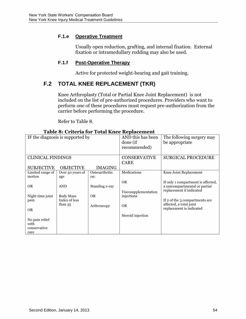
New York State Workers’ Compensation Board New York Knee Injury Medical Treatment Guidelines
Second Edition, January 14, 2013 54
F.1.e Operative Treatment
Usually open reduction, grafting, and internal fixation. External fixation or intramedullary rodding may also be used.
F.1.f Post-Operative Therapy
Active for protected weight-bearing and gait training.
TOTAL KNEE REPLACEMENT (TKR) F.2
Knee Arthroplasty (Total or Partial Knee Joint Replacement) is not included on the list of pre-authorized procedures. Providers who want to perform one of these procedures must request pre-authorization from the carrier before performing the procedure.
Refer to Table 8.
Table 8: Criteria for Total Knee Replacement IF the diagnosis is supported by
AND this has been done (if recommended)
The following surgery may be appropriate
CLINICAL FINDINGS SUBJECTIVE OBJECTIVE IMAGING
CONSERVATIVE CARE
SURGICAL PROCEDURE
Limited range of motion
OR
Night time joint pain
OR
No pain relief with conservative care
Over 50 years of age
AND
Body Mass Index of less than 35
Osteoarthritis on:
Standing x-ray
OR
Arthroscopy
Medications
OR
Viscosupplementation injections
OR
Steroid injection
Knee Joint Replacement
If only 1 compartment is affected, a unicompartmental or partial replacement if indicated
If 2 of the 3 compartments are affected, a total joint replacement is indicated

New York State Workers’ Compensation Board New York Knee Injury Medical Treatment Guidelines
Second Edition, January 14, 2013 55
AMPUTATION F.3
F.3.a Description/Definition
Surgical removal of a portion of the lower extremity.
F.3.b Mechanism of Injury
Usually secondary to post-traumatic bone, soft tissue, vascular or neurologic compromise of part of the extremity.
F.3.c Specific Physical Findings
Non-useful or non-viable portion of the lower extremity.
F.3.d Diagnostic Testing Procedures
Radiographs, vascular studies.
F.3.e Non-Operative Treatment
None
F.3.f Surgical Indications
Non-useful or non-viable portion of the extremity.
F.3.g Operative Treatment
Amputation
F.3.h Post-Operative Therapy
Active and/or passive therapy for prosthetic fitting, construction and training, protected weight-bearing.
MANIPULATION UNDER ANESTHESIA (MUA) F.4
F.4.a Description/Definition
Passive range of motion of a joint under anesthesia.
F.4.b Mechanism of Injury
Joint stiffness that usually results from a traumatic injury, compensation related surgery, or other treatment.
New York State Workers’ Compensation Board New York Knee Injury Medical Treatment Guidelines
Second Edition, January 14, 2013 56
F.4.c Specific Physical Findings
Joint stiffness in both active and passive modes.
F.4.d Diagnostic Testing Procedures
Radiographs.
F.4.e Non-Operative Treatment
Active and/or passive therapy for active and passive range of motion exercises.
F.4.f Surgical Indications
Is indicated in cases of intractable restriction and may be performed by a duly qualified surgeon. Consider if routine therapeutic modalities, including Physical therapy and/or dynamic bracing, do not restore the degree of motion that should be expected after a reasonable period of time, usually at least 12 weeks.
F.4.g Operative Treatment
Not applicable.
F.4.h Post-Operative Therapy
Active and/or passive therapy for active and passive range of motion.
BURSECTOMY F.5
F.5.a Description/Definition
Surgical removal of peri-articular bursa.
F.5.b Mechanism of Injury
Usually a traumatic local injury or repetitive minor local irritation.
F.5.c Specific Physical Findings
Swelling, tenderness over the bursa.
F.5.d Diagnostic Testing Procedures
Radiographs.
New York State Workers’ Compensation Board New York Knee Injury Medical Treatment Guidelines
Second Edition, January 14, 2013 57
F.5.e Non-Operative Treatment
Active and/or passive therapy for splinting, rest, NSAIDS, steroid injection.
F.5.f Surgical Indications
Persistent pain, swelling despite treatment.
F.5.g Operative Treatment
Surgical removal of the bursa.
F.5.h Post-Operative Therapy
Active and/or passive therapy for graduated range of motion exercises.
OSTEOTOMY F.6
F.6.a Description/Definition
A reconstructive procedure involving the surgical cutting of bone for realignment and is useful in patients that would benefit from realignment in lieu of total joint replacement.
F.6.b Mechanism of Injury
Post-traumatic arthritis or deformity.
F.6.c Specific Physical Findings
Painful decreased range of motion and/or deformity.
F.6.d Diagnostic Testing Procedures
Radiographs, MRI scan, CT scan.
F.6.e Non-Operative Treatment
Active and/or passive therapy for activity modification, bracing, NSAIDS.
F.6.f Surgical Indications
Failure of non-surgical treatment. Avoidance of total joint arthroplasty desirable.

New York State Workers’ Compensation Board New York Knee Injury Medical Treatment Guidelines
Second Edition, January 14, 2013 58
F.6.g Operative Treatment
Peri-articular opening or closing wedge of bone, usually with grafting and internal or external fixation.
F.6.h Post-Operative Therapy
Active and/or passive therapy for protected weight-bearing, progressive range of motion.
HARDWARE REMOVAL F.7
F.7.a Description/Definition
Surgical removal of internal or external fixation device.
F.7.b Mechanism of Injury
Usually following healing of a post-traumatic injury that required fixation or reconstruction using instrumentation.
F.7.c Specific Physical Findings
Local pain to palpation, swelling, erythema.
F.7.d Diagnostic Testing Procedures
Radiographs, tomography, CT scan, MRI.
F.7.e Non-Operative Treatment
Active and/or passive therapy for local modalities, activity modification, NSAIDS.
F.7.f Surgical Indications
Persistent local pain, irritation around hardware.
F.7.g Operative Treatment
Removal of instrumentation. Some instrumentation may be removed in the course of standard treatment without local irritation.
F.7.h Post-Operative Therapy
Active and/or passive therapy for progressive weight-bearing, range of motion.
New York State Workers’ Compensation Board New York Knee Injury Medical Treatment Guidelines
Second Edition, January 14, 2013 59
RELEASE OF CONTRACTURE F.8
F.8.a Description/Definition
Surgical incision or lengthening of contracted tendon or peri-articular soft tissue.
F.8.b Mechanism of Injury
Usually following a post-traumatic injury.
F.8.c Specific Physical Findings
Shortened tendon or stiff joint.
F.8.d Diagnostic Testing Procedures
Radiographs, CT scan, MRI scan.
F.8.e Non-Operative Treatment
Active and/or passive therapy for stretching, range of motion exercises.
F.8.f Surgical Indications
Persistent shortening or stiffness associated with pain and/or altered function.
F.8.g Operative Treatment
Surgical incision or lengthening of involved soft tissue.
F.8.h Post-Operative Therapy
Active and/or passive therapy for stretching, range of motion exercises.
MENISCECTOMY F.9
F.9.a Description/Definition
The surgical excision of a meniscus.
F.9.b Evaluation and Management
See Table 5.
New York State Workers’ Compensation Board New York Knee Injury Medical Treatment Guidelines
Second Edition, January 14, 2013 60
LIGAMENT REPAIR F.10
F.10.a Description/Definition
The surgical reattachment of torn anterior, or posterior cruciate ligaments or medial or lateral collateral ligaments.
F.10.b Evaluation and Management
See Table 4.
Sources:
This Treatment Guideline is adopted, with modification, from the State of Colorado’s Lower Extremity Medical Treatment Guideline, with supplementation from ACOEM’sP0F0F0F
1P Occupational Medicine Treatment Guidelines and the State of
Washington’s Medical and Surgical Treatment Guidelines.
1 American College of Occupational and Environmental Medicine
Copyright 2008, 2007, 2004, 1997 by the American College of Occupational and Environmental Medicine (ACOEM). Commercial reproduction or other use beyond fair use prohibited without explicit ACOEM permission.
New York State Workers’ Compensation Board New York Knee Injury Medical Treatment Guidelines
Second Edition, January 14, 2013 61
INDEX
Acetaminophen, 36, 37, 38
ACI, 4, 16, 20
ACL, 22, 23, 24
Active Interventions, 3
Active Therapeutic Exercise Program, 3
Activities of Daily Living. See ADL
Activity Level Restrictions, 43
Acupuncture, 34, 50
Adaptive Equipment, 42
ADL, 44
Aggravated Osteoarthritis, 21
Amputation, 55
ANA, 10
Anterior Cruciate Ligament. See ACL
Antinuclear Antigen. See ANA
Arthrograms, 13, 25
Arthroscopy, 13, 19, 23, 54
Diagnostic, 13, 18, 20
Autologous Chondrocyte Implantation. See ACI
Biofeedback, 35
BMI, 19, 20, 28
Body Mass Index. See BMI
Bone Defects, 15
Bone Scan, 9, 13, 30
Bursectomy, 56
Bursitis, 14, 31, 32
Capsaicin, 40
Cartilage and Bone Defects, 15
CBC, 10
Chondral Defects, 15, 18, 19, 20
Chondromalacia Patella, 29, 31
Chondroplasty, 18
Collateral Ligament, 21, 22, 60
Complete Blood Count. See CBC
Computed Axial Tomography. See CT
Continuous Passive Movement. See CPM
Contrast Baths, 47
CPM, 17, 46
C-reactive protein. See CRP
CRP, 10
CT, 3, 11, 12, 15, 30, 33, 57, 58, 59
Delayed Recovery, 2
Description/Definition, 15, 21, 22, 24, 28, 29, 31, 32, 53, 55, 56, 57, 58, 59, 60

New York State Workers’ Compensation Board New York Knee Injury Medical Treatment Guidelines
Second Edition, January 14, 2013 62
Diagnostic Imaging, 3, 4, 11, 13
Diagnostic Studies, 12, 33, 46
Diagnostic Time Frames, 2
Doppler Ultrasonography/Plethysmography, 14
Education, 1, 2, 3, 33
Electrical Stimulation, 34, 44, 47, 48
Electrodiagnostic Testing, 14
Electromyography. See EMG
EMG, 14
Erythrocyte Sedimentation Rate. See
ESR
ESR, 10
Experimental Treatment, 7
Fabrication/Modification of Orthotics, 41
FCE, 5, 43
Fluidotherapy, 47
Functional Capacity Evaluation. See FCE
Gait Training, 44, 54
Hardware Removal, 58
History, 3, 7, 8, 9, 11, 14, 17, 23, 25, 36, 38, 39, 41, 53
HLA, 10
HumanLeukocyte Antigen. See HLA
Imaging Studies, 3, 8, 11, 12
Infrared Therapy, 47
Injections, 3, 11, 21, 30, 32, 35, 36, 54
Injured Workers As Patients, 7
Intra-Capsular Acid Salts, 36, 54
Introduction To Knee Injury, 7
Iontophoresis, 47
Job Site Evaluation, 6
Joint Aspiration, 10, 14
Kinesiotaping, Taping or Strapping, 51
Knee
Arthroplasty, 4, 54, 57
Evaluation, 5, 7, 9, 10, 11, 13
Fusion, 53
Total Knee Replacement, 4, 21, 54, 57
Laboratory Testing, 10
Lineal Tomography, 12
Magnetic Resonance Imaging. See MRI
Manipulation, 25, 48
Manipulation Under Anesthesia. See
MUA
Massage, 48, 51
Medical Care, 1, 6
Medications, 10, 13, 31, 35, 36, 37, 38, 39, 40, 54
Meniscal Allograft Transplantation, 4, 25, 27, 28

New York State Workers’ Compensation Board New York Knee Injury Medical Treatment Guidelines
Second Edition, January 14, 2013 63
Meniscus, 21, 22, 24, 25, 26, 27, 28, 59
Meniscus Injury, 24, 26, 27
Minor Tranquilizer, 37
Mobilization (Joint), 49
Mobilization (Soft Tissue), 49
MRI, 11, 12, 13, 14, 15, 18, 19, 20, 22, 23, 24, 25, 26, 30, 31, 33, 53, 57, 58, 59
MUA, 55
Muscle Relaxants, 37
Myelography, 11
Narcotics, 37
NCS, 14
Nerve Conduction Studies. See NCS
Neuromuscular Re-education, 45
Nonsalicylates, 40
Nonsteroidal Anti-Inflammatory Drugs. See NSAIDs
NSAIDs, 21, 32, 36, 37, 38, 39, 41, 53, 57, 58
OATS, 4
Ongoing Maintenance Care, 52
Orthotic/Prosthetic Training, 42
Orthotics And Prosthetics, 41
Osteochondral Autograft. See OATS
Osteotomy, 57
Other Procedures, 11, 14
Other Radionuclide Scanning, 13
Outerbridge Classification, 17, 27, 28, 30
Paraffin Bath, 49
Patellar Subluxation, 28
PCL, 24, 60
Personality/Psychological/Psychosocial Evaluations, 4
Personality/Psychological/Psychosocial Intervention, 5
Physical Examination, 8
Physical Therapy, 13, 18, 19, 20, 23, 26, 27, 31, 50, 56
Positive Patient Response, 1
Posterior Crucite Ligament. See PCL
Pre-Authorization, 4, 16, 25, 54
Prolotherapy, 35
Prosthetics. See Orthotics and Prosthetics
Protein Rich Plasma. See PRP
PRP, 35
Radiographic Imaging. See X-ray
Radioisotope Bone Scanning, 13
Recommendations and Medical Evidence, 7
Red Flags, 9
Re-Evaluate Treatment, 1
Release Of Contracture, 59

New York State Workers’ Compensation Board New York Knee Injury Medical Treatment Guidelines
Second Edition, January 14, 2013 64
Rendering Of Medical Services, 1
Retropatellar Pain Syndrome, 29, 31
Return To Work, 1, 2, 5, 6, 42
Sclerotherapy. See Prolotherapy
Scope Of Pratice, 7
Short-wave Diathermy, 50
Six-Month Time Frame, 2
Somatosensory Evoked Potentials. See
SSEP
Specific Knee Injury Diagnoses, Testing, And Treatment, 15
Splints, 42
SSEP, 14
Superficial Heat and Cold Therapy, 49
Surgical Interventions, 3, 4, 20
Tendinitis, 29, 31, 32
Tenosynovitis, 31
TENS, 48, 50
Tests, 3, 4, 8, 10, 11, 13, 14, 53
Therapeutic Exercise, 45
Therapeutic Procedures, Non-Operative, 33
Therapeutic Procedures, Operative, 53
Therapy
Active, 43
Ongoing Maintenance Care, 52
Passive, 46
TKR. See Total Knee Replacement
Topical Drug Delivery, 40
Topical Salicylates, 40
Traction, 50
Tramadol, 41
Transcutaneous Electrical Nerve Stimulation. See TENS
Treatment Time Frames, 2
Ultrasound, 51
Vasopneumatic Devices, 51
Venogram/Arteriogram, 14
Viscosupplementation. See Intra-Capsular
Acid Salts
Wheelchair Management and Propulsion, 45
Whirlpool, 51
X-ray, 9, 12, 16, 28, 31, 54