new technologies for dna analysis – a review of the … · new biotechnology volume 33,number 3...
TRANSCRIPT
New Biotechnology �Volume 33, Number 3 �May 2016 REVIEW
New technologies for DNA analysis – areview of the READNA ProjectSteven McGinn1, David Bauer19, Thomas Brefort17, Liqin Dong19, Afaf El-Sagheer6,7,8,Abdou Elsharawy4,5, Geraint Evans10, Elin Falk-Sorqvist13, Michael Forster4,Simon Fredriksson27, Peter Freeman2, Camilla Freitag24, Joachim Fritzsche25,Spencer Gibson2, Mats Gullberg27, Marta Gut28,29, Simon Heath28,29,Isabelle Heath-Brun28,29, Andrew J. Heron20, Johannes Hohlbein10, Rongqin Ke9,13,Owen Lancaster2, Ludovic Le Reste10, Giovanni Maglia20, Rodolphe Marie16,Florence Mauger1, Florian Mertes3, Marco Mignardi9,13, Lotte Moens13,Jelle Oostmeijer15, Ruud Out15, Jonas Nyvold Pedersen16, Fredrik Persson24,Vincent Picaud18, Dvir Rotem20, Nadine Schracke17, Jennifer Sengenes1,Peer F. Stahler17, Bjorn Stade4, David Stoddart20, Xia Teng15, Colin D. Veal2,Nathalie Zahra2, Hagan Bayley20, Markus Beier17, Tom Brown6,7, Cees Dekker11,Bjorn Ekstrom27, Henrik Flyvbjerg16, Andre Franke4, Simone Guenther21,Achillefs N. Kapanidis10, Jane Kaye14, Anders Kristensen16, Hans Lehrach3,Jonathan Mangion21, Sascha Sauer3, Emile Schyns26, Jorg Tost1,Joop M.L.M. van Helvoort15, Pieter J. van der Zaag12, Jonas O. Tegenfeldt23,Anthony J. Brookes2, Kalim Mir19, Mats Nilsson9,13, James P. Willcocks22 andIvo G. Gut28,29
1CEA – Centre National de Genotypage, 2, rue Gaston Cremieux, 91057 Evry Cedex, France2University of Leicester, University Road, Leicester LE1 7RH, UK3Max Planck Institute for Molecular Genetics, Ihnestrasse 73, 14195 Berlin, Germany4 Institute of Clinical Molecular Biology, Christian-Albrechts-University (CAU), Am Botanischen Garten 11, D-24118 Kiel, Germany5 Faculty of Sciences, Division of Biochemistry, Chemistry Department, Damietta University, New Damietta City, Egypt6 School of Chemistry, University of Southampton, Highfield, Southampton SO17 1BJ, UK7Department of Chemistry, University of Oxford, Chemistry Research Laboratory, 12 Mansfield Rd, Oxford OX1 3TA, UK8Chemistry Branch, Department of Science and Mathematics, Faculty of Petroleum and Mining Engineering, Suez University, Suez 43721, Egypt9 Science for Life Laboratory, Department of Biochemistry and Biophysics, Stockholm University, Box 1031, Se-171 21 Solna, Sweden10 Biological Physics Research Group, Clarendon Laboratory, Department of Physics, Parks Road, Oxford OX1 3PU, UK11Department of Bionanoscience, Kavli Institute of Nanoscience, Delft University of Technology, Lorentzweg 1, 2628 CJ Delft, The Netherlands12 Philips Research Laboratories, High Tech Campus 11, 5656 AE Eindhoven, The Netherlands13Department of Immunology, Genetics, and Pathology, Science for Life Laboratory, Uppsala University, Sweden14HeLEX – Centre for Health, Law and Emerging Technologies, Nuffield Department of Population Health, University of Oxford, Old Road Campus,Oxford OX3 7LF, UK15 FlexGen BV, Galileiweg 8, 2333 BD Leiden, The Netherlands
Review
Corresponding author: Gut, I.G. ([email protected])
http://dx.doi.org/10.1016/j.nbt.2015.10.003 www.elsevier.com/locate/nbt1871-6784/� 2015 Published by Elsevier B.V. 311

16DTU Nanotech, Oerstedsplads Building 345 East, 2800, Kongens Lyngby, Denmark17Comprehensive Biomarker Center GmbH, Im Neuenheimer Feld 583, D-69120 Heidelberg, Germany18CEA-Saclay, Bat DIGITEO 565 – Pt Courrier 192, 91191 Gif-sur-Yvette Cedex, France19 The Wellcome Trust Centre for Human Genetics, University of Oxford, Oxford OX3 7BN, UK20Department of Chemistry, University of Oxford, Chemistry Research Laboratory, Mansfield Road, Oxford OX1 3TA, England, UK21 Thermo Fisher Scientific Frankfurter Straße 129B, 64293 Darmstadt, Germany22Oxford Nanopore Technologies, Edmund Cartwright House, 4 Robert Robinson Avenue, Oxford Science Park, Oxford OX4 4GA, UK23Division of Solid State Physics and NanoLund, Lund University, Box 118, 22100 Lund, Sweden24Department of Physics, University of Gothenburg, SE-412 96 Gothenburg, Sweden25Department of Applied Physics, Chalmers University of Technology, Kemivagen 10, 412 96 Goteborg, Sweden26 PHOTONIS France S.A.S. Avenue Roger Roncier, 19100 Brive B.P. 520, 19106 BRIVE Cedex, France27Olink AB, Dag Hammarskjolds vag 52A, 752 37 Uppsala, Sweden28Centro Nacional de Analisis Genomico (CNAG-CRG), Center for Genomic Regulation, C/Baldiri Reixac 7, 08028 Barcelona, Spain29Universitat Pompeu Fabra (UPF), Barcelona, Spain
The REvolutionary Approaches and Devices for Nucleic Acid analysis (READNA) project received funding
from the European Commission for 4 1/2 years. The objectives of the project revolved around
technological developments in nucleic acid analysis. The project partners have discovered, created and
developed a huge body of insights into nucleic acid analysis, ranging from improvements and
implementation of current technologies to the most promising sequencing technologies that constitute
a 3rd and 4th generation of sequencing methods with nanopores and in situ sequencing, respectively.
Contents
Introduction . . . . . . . . . . . . . . . . . . . . . . . . . . . . . . . . . . . . . . . . . . . . . . . . . . . . . . . . . . . . . . . . . . . . . . . . . . . . . . . . . . . . . . 313
Duration, funding & no. partners . . . . . . . . . . . . . . . . . . . . . . . . . . . . . . . . . . . . . . . . . . . . . . . . . . . . . . . . . . . . . . . . . . . . 313
Objectives & context . . . . . . . . . . . . . . . . . . . . . . . . . . . . . . . . . . . . . . . . . . . . . . . . . . . . . . . . . . . . . . . . . . . . . . . . . . . . . 313
Project highlights & achievements. . . . . . . . . . . . . . . . . . . . . . . . . . . . . . . . . . . . . . . . . . . . . . . . . . . . . . . . . . . . . . . . . . . . . . 313
Introduction. . . . . . . . . . . . . . . . . . . . . . . . . . . . . . . . . . . . . . . . . . . . . . . . . . . . . . . . . . . . . . . . . . . . . . . . . . . . . . . . . . . . 313
Discovery of ‘TUF’ DNA . . . . . . . . . . . . . . . . . . . . . . . . . . . . . . . . . . . . . . . . . . . . . . . . . . . . . . . . . . . . . . . . . . . . . . . . . . . 313
Enrichment methods . . . . . . . . . . . . . . . . . . . . . . . . . . . . . . . . . . . . . . . . . . . . . . . . . . . . . . . . . . . . . . . . . . . . . . . . . . . . . 314
Enrichment based on selectors . . . . . . . . . . . . . . . . . . . . . . . . . . . . . . . . . . . . . . . . . . . . . . . . . . . . . . . . . . . . . . . . . . . . 314
Enrichment based on long DNA fragments . . . . . . . . . . . . . . . . . . . . . . . . . . . . . . . . . . . . . . . . . . . . . . . . . . . . . . . . . . . 315
MegaPlex PCR . . . . . . . . . . . . . . . . . . . . . . . . . . . . . . . . . . . . . . . . . . . . . . . . . . . . . . . . . . . . . . . . . . . . . . . . . . . . . . . . 316
SIMLY targeted enrichment . . . . . . . . . . . . . . . . . . . . . . . . . . . . . . . . . . . . . . . . . . . . . . . . . . . . . . . . . . . . . . . . . . . . . . 316
Benchmarking of enrichment methods. . . . . . . . . . . . . . . . . . . . . . . . . . . . . . . . . . . . . . . . . . . . . . . . . . . . . . . . . . . . . . 316
Metrics for the assessment of an enrichment experiment . . . . . . . . . . . . . . . . . . . . . . . . . . . . . . . . . . . . . . . . . . . . . . . . 316
Mass spectrometry based DNA analysis methods . . . . . . . . . . . . . . . . . . . . . . . . . . . . . . . . . . . . . . . . . . . . . . . . . . . . . . . . . 317
Ribo-PCR MS sequencing . . . . . . . . . . . . . . . . . . . . . . . . . . . . . . . . . . . . . . . . . . . . . . . . . . . . . . . . . . . . . . . . . . . . . . . . 317
Ribo-PAP-PCR genotyping . . . . . . . . . . . . . . . . . . . . . . . . . . . . . . . . . . . . . . . . . . . . . . . . . . . . . . . . . . . . . . . . . . . . . . . 318
Risk profiling of multifactorial diseases by MALDI mass spectrometry . . . . . . . . . . . . . . . . . . . . . . . . . . . . . . . . . . . . . . . 318
Array based Dynamic Allele-Specific Hybridization (Array-DASH). . . . . . . . . . . . . . . . . . . . . . . . . . . . . . . . . . . . . . . . . . . . . 318
Manipulating and visualizing individual DNA molecules . . . . . . . . . . . . . . . . . . . . . . . . . . . . . . . . . . . . . . . . . . . . . . . . . . . 318
Single molecule mapping, molecule rescue and next generation sequencing . . . . . . . . . . . . . . . . . . . . . . . . . . . . . . . . . . 318
Extracting, handling & imaging ultra-long DNA and obtaining sequence information in a long-range context. . . . . . . . . 319
Sequencing chemistry, sensors . . . . . . . . . . . . . . . . . . . . . . . . . . . . . . . . . . . . . . . . . . . . . . . . . . . . . . . . . . . . . . . . . . . . . . 319
Ligation-based sequencing . . . . . . . . . . . . . . . . . . . . . . . . . . . . . . . . . . . . . . . . . . . . . . . . . . . . . . . . . . . . . . . . . . . . . . . 320
Click chemistry – enzyme-free ligation based sequencing . . . . . . . . . . . . . . . . . . . . . . . . . . . . . . . . . . . . . . . . . . . . . . . . 320
Nanopore sequencing . . . . . . . . . . . . . . . . . . . . . . . . . . . . . . . . . . . . . . . . . . . . . . . . . . . . . . . . . . . . . . . . . . . . . . . . . . . . . 320
Base distinction including 5meC . . . . . . . . . . . . . . . . . . . . . . . . . . . . . . . . . . . . . . . . . . . . . . . . . . . . . . . . . . . . . . . . . . 320
The exonuclease nanopore . . . . . . . . . . . . . . . . . . . . . . . . . . . . . . . . . . . . . . . . . . . . . . . . . . . . . . . . . . . . . . . . . . . . . . . 321
Arrays of nanopores: protein and solid state . . . . . . . . . . . . . . . . . . . . . . . . . . . . . . . . . . . . . . . . . . . . . . . . . . . . . . . . . . 321
Strand sequencing . . . . . . . . . . . . . . . . . . . . . . . . . . . . . . . . . . . . . . . . . . . . . . . . . . . . . . . . . . . . . . . . . . . . . . . . . . . . . 321
In situ DNA analysis methods . . . . . . . . . . . . . . . . . . . . . . . . . . . . . . . . . . . . . . . . . . . . . . . . . . . . . . . . . . . . . . . . . . . . . . . 321
In situ genotyping and sequencing . . . . . . . . . . . . . . . . . . . . . . . . . . . . . . . . . . . . . . . . . . . . . . . . . . . . . . . . . . . . . . . . . 322
In situ expression profiling . . . . . . . . . . . . . . . . . . . . . . . . . . . . . . . . . . . . . . . . . . . . . . . . . . . . . . . . . . . . . . . . . . . . . . . 323
Improvement of protocols . . . . . . . . . . . . . . . . . . . . . . . . . . . . . . . . . . . . . . . . . . . . . . . . . . . . . . . . . . . . . . . . . . . . . . . . . 324
DNA-seq . . . . . . . . . . . . . . . . . . . . . . . . . . . . . . . . . . . . . . . . . . . . . . . . . . . . . . . . . . . . . . . . . . . . . . . . . . . . . . . . . . . . 324
RNA-Seq . . . . . . . . . . . . . . . . . . . . . . . . . . . . . . . . . . . . . . . . . . . . . . . . . . . . . . . . . . . . . . . . . . . . . . . . . . . . . . . . . . . . 324
Optimization of whole genome methylation pipelines . . . . . . . . . . . . . . . . . . . . . . . . . . . . . . . . . . . . . . . . . . . . . . . . . . 324
REVIEW New Biotechnology � Volume 33, Number 3 �May 2016
312 www.elsevier.com/locate/nbt
Review

Other applications of nucleic acid analysis – ProteinSeq . . . . . . . . . . . . . . . . . . . . . . . . . . . . . . . . . . . . . . . . . . . . . . . . . 325
Software development . . . . . . . . . . . . . . . . . . . . . . . . . . . . . . . . . . . . . . . . . . . . . . . . . . . . . . . . . . . . . . . . . . . . . . . . . . . . 325
Alignment of 2nd generation sequencing reads. . . . . . . . . . . . . . . . . . . . . . . . . . . . . . . . . . . . . . . . . . . . . . . . . . . . . . . . 325
Variant calling . . . . . . . . . . . . . . . . . . . . . . . . . . . . . . . . . . . . . . . . . . . . . . . . . . . . . . . . . . . . . . . . . . . . . . . . . . . . . . . . 326
RNA-Seq . . . . . . . . . . . . . . . . . . . . . . . . . . . . . . . . . . . . . . . . . . . . . . . . . . . . . . . . . . . . . . . . . . . . . . . . . . . . . . . . . . . . 327
DNA methylation software, MeDIP . . . . . . . . . . . . . . . . . . . . . . . . . . . . . . . . . . . . . . . . . . . . . . . . . . . . . . . . . . . . . . . . . 327
ELSI aspects of next generation nucleic acid sequence data . . . . . . . . . . . . . . . . . . . . . . . . . . . . . . . . . . . . . . . . . . . . . . . . . 327
Perspectives – what remains to be done? What are future challenges? . . . . . . . . . . . . . . . . . . . . . . . . . . . . . . . . . . . . . . . . . . . 327
Conclusion . . . . . . . . . . . . . . . . . . . . . . . . . . . . . . . . . . . . . . . . . . . . . . . . . . . . . . . . . . . . . . . . . . . . . . . . . . . . . . . . . . . . . . . 328
Acknowledgements . . . . . . . . . . . . . . . . . . . . . . . . . . . . . . . . . . . . . . . . . . . . . . . . . . . . . . . . . . . . . . . . . . . . . . . . . . . . . . . . . 328
References. . . . . . . . . . . . . . . . . . . . . . . . . . . . . . . . . . . . . . . . . . . . . . . . . . . . . . . . . . . . . . . . . . . . . . . . . . . . . . . . . . . . . . . . 328
IntroductionDuration, funding & no. partnersThe REvolutionary Approaches and Devices for Nucleic Acid
analysis (READNA) project received 12 million s funding under
the European Union Framework Programme 7 from 1st June 2008
to 30th November 2012. The 19 project partners from both acade-
mia and industry from in total 7 countries had a project budget of
16 Ms with which they have discovered, created and developed a
huge body of insights into nucleic acid analysis. Results have been
presented widely in publications and in innumerous public pre-
sentations. Results have been moved to spin-offs such as the Olink
enrichment kits (now sold by Agilent as Haloplex) and are finding
their way to the market, such as the Oxford Nanopore MinIon
sequencer that was first released to early-access user sites in the
summer of 2014.
Objectives & contextThe READNA consortium collaborated in a wide range of devel-
opments in nucleic acid analysis methods to accelerate new break-
through DNA sequencing technologies, to enhance existing
analysis methods, and advance nucleic analysis methods to the
benefit of patients and society. Participants had diverse and com-
plementary expertise from a wide range of scientific disciplines.
The interdisciplinary nature of the consortium allowed the explo-
ration of novel concepts, ease the use and broaden nucleic acid
analysis technologies, establish best practices and standards for
research and diagnostics, accelerate and simplify methods and
create a toolbox for precision medicine. One of the declared goals
was to progress technologies enabling sequencing of an entire
human genome for 1000s in less than one day.
The project was organized into five research work packages, and
a dissemination and management work package.
The five research work packages were, WP1: Near Term Innova-
tions: Ancillary elements for 2nd generation DNA sequencers;
WP2: Near Term Innovations: Improvement and extension of
existing methods; WP3: Fluorescence-based Single Molecule Se-
quencing; WP4: Nanopore Sequencing; and WP5: New Genotyp-
ing Challenges.
Project highlights & achievementsIntroductionProgress has been made through all of the fronts that the five work
packages have been focusing on towards the overall objectives of
READNA. The results have been presented in over 100 scientific
publications, many of which are in very high impact factor jour-
nals [1–139], including a number of review papers [1–4]. Here we
highlight a few selected discoveries and developments.
Discovery of ‘TUF’ DNAMethods for genome analysis must contend with the fact that
some chromosome regions are extremely difficult to assay, pro-
ducing weak or no signals and low data accuracy. This behavior
is not easily explained by mere differences in C + G content or
secondary structures from region to region, as the problem varies
in size between DNA samples. We investigated this phenome-
non via a series of experiments, and thereby uncovered a novel
mechanistic basis for amplification and assay differences be-
tween samples and between genome regions [5]. In short, we
found that difficult to assay regions contain tiny (<<1 kb)
stretches that are extremely resistant to DNA denaturation (a
key step in almost all DNA analysis methods), even up to
�1308C. Assay efficiencies are reduced in surrounding zones
that can extend several hundreds of kbp from such a site. We
call these stretches ‘Thermodynamically Ultra-Fastened’ (TUF)
regions. The ‘non-melting’ sections within TUF regions are
extremely high in C + G content and are made up of elements
whose particular arrangements probably enables them to co-
operate in holding the two DNA strands together (Fig. 1). Long
DNA stretches that flank the non-melting loci are able to dena-
ture but remain in close proximity since they are tethered
together at the non-melted anchor regions. As such they can
very rapidly re-anneal and thereby hinder access by primers or
probes, so preventing assays from working over extended
domains. The discovery of TUF DNA allows us to explain the
inter-sample variability of assay efficiency in these troublesome
regions. Specifically, some DNA samples will contain a greater
degree of randomly distributed nicking than others, and upon
heating these nicks will allow the denatured flanking regions to
diffuse away from the non-denatured core TUF domains – such
that highly intact DNAs will be most severely affected and assay
the least well. A major practical consequence of all of this is that
inter-region and inter-sample variability can be largely over-
come by employing routine fragmentation methods (e.g. soni-
cation or restriction enzyme digestion) prior to sample
interrogation, even though this runs counter to traditional best
practice.
New Biotechnology �Volume 33, Number 3 �May 2016 REVIEW
www.elsevier.com/locate/nbt 313
Review

Enrichment methodsSince 2nd generation DNA sequencing emerged its cost has de-
creased, at first dramatically, then for several years was stable with
sequencing of a human genome at 30� coverage costing roughly
5000$ in reagents, and then only recently dropped to�1000$ with
Illumina’s HiSeq X system. For high value experiments these costs
are acceptable, however, particularly for experiments that involve
the analysis of many samples costs become prohibitive. Moreover,
for some applications 30� read depth is far from sufficient, for
example for application in cancer diagnostics where the tumor cell
content may be low, and where sequencing needs to be done to a
considerable depth (>1000�) to enable accurate detection of
somatic mutations in a 10% cancer cell population. Since very
early on, enrichment approaches for focusing only on the regions
of interest have been developed. The technical solutions available
are very broad, diverse and have different sweet spots. They range
from simple PCR for the isolation of a very small fraction of a
genome to hybridization-based methods by which several percent
of a genome can be enriched. An overview of the different methods
for targeted enrichment is depicted in Fig. 2. Within READNA we
have developed several enrichment methods (see below). We have
also carried out benchmarking of different methods and have
developed a concise set of descriptors for an enrichment experi-
ment that precisely captures the essence of an enrichment experi-
ment and method.
Enrichment based on selectors
Prior to the READNA project a novel enrichment technique
entitled ‘Selectors’ was developed to specifically select large
sets of DNA sequences for parallel amplification by PCR using
REVIEW New Biotechnology � Volume 33, Number 3 �May 2016
[(Figure_1)TD$FIG]
FIGURE 1
A putative TUF region is illustrated for two DNA samples that are either (a) high quality (low degree of nicking, so long fragments per strand) or (b) low quality
(high degree of nicking, so short fragments per strand). Upon denaturing the DNA strands are held together at the core TUF sequences that strongly resist melting.
For (a), the entire TUF region and flanks are held together, whereas in (b) sections of single and double strandedmaterial are disconnected from each other and socan diffuse apart. Upon imposing conditions for primer or probe annealing, in (a) the complimentary DNA strands that were held together at core TUF sites rapidly
and fully re-anneal, preventing access by primers or probes. Conversely, in (b) many single stranded portions now exist to which the primers and probes can
anneal, enabling efficient amplification or detection [though regions very close to the core TUF sequences remain inaccessible]. The overall efficiency of DNA
amplification or probe hybridization of target sites within a TUF regionwill therefore be influenced by the general amount of DNA nicking present and the averageper target degree of physical distance from the core TUF sequences. The counter-intuitive result of this is that stronger assay and amplification signals will be
produced by lower quality DNAs.
314 www.elsevier.com/locate/nbt
Review

target-specific oligonucleotide constructs called selectors. Selec-
tors are oligonucleotide duplexes with single-stranded target-com-
plementary end-sequences that are linked by a common sequence
motif. A pool of selectors is combined with denatured restriction
digested DNA and each selector hybridizes to its respective target,
forming individual circular complexes. The common sequence
that is introduced into the circularized fragments allows the
products to be PCR amplified in parallel using a universal primer
pair [6]. The technique was rapidly adapted to next generation
sequencing (NGS) instruments and in particular the 454/Roche
instrument highlighting the potential of the technique to com-
bine highly multiplexed enrichment with NGS [7]. During the
READNA project the Selector technology was developed further to
adapt it to NGS instruments of higher throughput, such as the
Illumina and SOLiD systems. Initially, and due to the short read-
length of these systems at the time, this was done by developing a
platform independent Multiple Displacement Amplification
(MDA) based version, where the sequencing reads were initiated
randomly across the enriched DNA. Moreover, selector probe
libraries were amplified from programmable DNA microarrays,
substantially reducing probe cost. The modified protocol was
validated on 28 genes frequently mutated in common solid tumors
[8]. Due to the development of longer reads on particularly the
Illumina HiSeq and MiSeq instruments, a PCR-based version was
introduced that enables integration of the enrichment with the
sequencing library construction into a one-step procedure. This
version is now commercially available through Agilent Technolo-
gies under the name of HaloplexTM. In addition the protocol can
be used on FFPE samples for cancer diagnostics applications [9,10].
Enrichment based on long DNA fragments
Anticipating improvements in the lengths of fragments that could
be sequenced on NGS platforms, we explored a novel solution-
phase DNA enrichment approach that used multiple, co-operat-
ing, non-overlapping DNA baits per target region, and novel
blocking reagents to suppress non-specific hybridization. Addi-
tionally, we identified key procedural improvements that: (i)
enable bait pools (sets of harvested oligonucleotides from pro-
grammable array synthesis) to be massively amplified with high
fidelity using solution-phase PCR (rather than emulsion PCR) to
give quantities needed for hundreds of enrichment reactions, and
(ii) facilitate highly robust long-fragment library preparation.
Combining these innovations into a complete procedure yielded
combined levels of enrichment power and enrichment evenness
New Biotechnology �Volume 33, Number 3 �May 2016 REVIEW
[(Figure_2)TD$FIG]
FIGURE 2
Commonly used targeted enrichment techniques. (1) Hybrid capture targeted enrichment either on solid supports such as microarrays (a) or beads (b). A shot-gun
fragment library is prepared and hybridized against a library containing the target sequence. After hybridization (and bead coupling) non-target sequences arewashed away, the enriched sample can be eluted and further processed for sequencing. (2) Enrichment by Molecular Inversion Probes (MIPs) which are composed
of a universal sequence (blue) flanked by target-specific sequences. MIPs are hybridized to the region of interest, followed by a gap filling reaction and ligation to
produce closed circles. The classical MIPs are hybridized to mechanically sheared DNA (a), the Selector Probe technique uses a cocktail of restriction enzymes to
fragment the DNA and the probes are adapted via the restriction sites (b). (3) Targeted enrichment by PCR-based approaches. Typical PCR with single-tube perfragment assay (a), multiplex PCR assay with up to 50 fragments (b) and RainDancemicro droplet PCRwith up to 20,000 unique primer pairs (c) utilized for targeted
enrichment.
www.elsevier.com/locate/nbt 315
Review

that match or exceed the levels achieved by all alternative solu-
tion-phase methods.
MegaPlex PCR
A simple approach to target DNA enrichment would involve direct
PCR amplification of the regions of interest. However, this theo-
retically attractive option is confounded by the fact that high-
plexity PCR reactions fail due to excessive inter-primer reactions
and off-target priming initiated by the many primers available in
the solution. We sought to address this limitation by placing each
intended primer pair at a different physical location on a surface
within such a reaction, thereby preventing unwanted primer–
primer interactions or off-target priming. To make this new ‘Mega-
Plex PCR’ [11] reaction format work well, we also established: (i)
how to enable surface based PCR priming events to occur efficient-
ly and specifically, and (ii) how to enable all the resulting ampli-
cons to leave the surface carrying common primer sequences, so
that they could together be uniformly amplified by a single primer
pair in a final solution phase PCR, generating enough product for
DNA sequencing.
SIMLY targeted enrichment
Targeted enrichment by hybrid capture has proven to be a highly
efficient method (e.g. [12]), particularly if large genomic target
regions as well as many samples need to be analyzed. Herein RNA
probes are hybridized in solution to specifically bind and enrich
the region of interest. The RNA probes are transcribed from a large
pool of synthesized DNA template oligonucleotides. As part of the
project the goal was to make targeted enrichment by in-solution
capture more cost efficient, especially for very large sample num-
bers. This was achieved by applying emulsion PCR for DNA
template library amplification virtually immortalizing a once
generated DNA template library and therefore allowing the re-
current synthesis of RNA bait libraries for targeted enrichment
experiments. The efficacy of the developed protocol matched the
results obtained from commercial kits, such as the SureSelect
system commercialized by Agilent Technologies, and further-
more demonstrated that emulsion PCR can be used for bias free
amplification of DNA template libraries. The developed ‘short
immortalized DNA template library’ (SIMLY) method for targeted
enrichment is a straightforward example for significantly reduc-
ing costs for NGS applications which are anticipated to grow
rapidly especially in molecular diagnostics within the near future
[12].
Benchmarking of enrichment methods
To bring more efficiency to the sequencing process and enable large-
scale genomic investigations for complex diseases, we have con-
ducted many studies to evaluate the true capabilities and perfor-
mance of many upfront targeted and whole-exome approaches
using different NGS technology platforms, tested and standardized
different scenarios of sample multiplexing to bring down the cost
per sample, and examined the applicability of whole-genome am-
plified (WGA) materials, instead of original gDNA, for NGS to
preserve preciouspatient samples. Theresults of our studies revealed
that pre-enrichment sample multiplexing using hybridization-
based methods is technically preferable over PCR-based approaches
[14,15]. Indexed NGS libraries, instead of gDNA were successfully
enriched using one selected microarray-based capture method [14].
Reproducible coverage profiles (R2 up to 0.99) for the target regions
were achieved across most of the different multiplexed samples,
enriching human exonic as well as intronic regions with less than
10% strand bias. In addition, several experimental alternatives were
performed in the PCR-based study [15] to investigate the effect of
using WGA gDNA material and different pooling strategies on the
performance of selective recovery of genomic subregions of interest
and on variant detection. The results confirm that WGA can be
combined with NGS with no substantial bias in enrichment effi-
ciency. The results of these experiments further support the possi-
bility to process non-pooled and pooled samples (before or after an
emulsion PCR (emPCR) step) without substantial bias, and reveal
that pooling pre-emPCR can reduce the cost per sample while
maintaining good performance [15]. When exome kits became
commercially available on different platforms, we performed a
multi-platform benchmark of various exome NGS approaches.
The results were surprising, showing divergent enrichment efficien-
cies and low overlap of single nucleotide variant (SNV) calls (Fig. 3a).
When we used Sanger sequencing to validate 765 SNV-calls of
potential clinical relevance that were concordant between two
different kit/sequencer/bioinformatics platforms, only 57-97% con-
cordance was seen in HapMap individual NA12752 (data derived by
Sanger sequencing (Fig. 3b)).
The results presented here emphasize that current whole-exome
and targeted NGS (T-NGS) methods are not yet optimal, and the
long term utility of these methods will depend increasingly on
progress towards evenness and enrichment power improvements
and also on new and better strategies [16,17]. Taken together, we
do expect that even if the cost of routine sequencing of the human
exome and whole-exome becomes affordable in the near future,
multiplexed targeted NGS will serve alongside as a downstream
validation and diagnostic tool to reach sufficient coverage with
less sequencing and cost, enabling the investigation of a higher
number of samples in parallel [18,19].
Metrics for the assessment of an enrichment experiment
With the wide variety of methods available for targeted enrich-
ment of selected genomic regions for NGS, comparing results
among them, and even between experiments using the same
enrichment technology, is difficult, as no universal terminology
for reporting the quality of target enrichment is in use. In the
literature various sets of parameters reporting the results are used,
and even if similar parameters seem to be reported, their definition
may be different. To deal with this issue, a universal set of Target
Enrichment Sequencing Descriptors (TESD) metrics has been de-
veloped including clear mathematical formulas to define the key
parameters. These TESD metrics enable a complete and unique
description of target enrichment experimental results, irrespective
of the underling enrichment technology. This set of TES-descrip-
tors uses the following 7 parameters [20]:
1. Size of the Region of Interest, ROI
2. Weight of the input DNA requirement, W
3. Average read depth across the ROI, DROI
4. Fraction sufficiently covered at a read depth of x, Fx
5. Specificity, S
6. Enrichment factor, EF
7. Evenness, E
REVIEW New Biotechnology � Volume 33, Number 3 �May 2016
316 www.elsevier.com/locate/nbt
Review

Of these parameters the first two (ROI, W) give the input
requirements, while the remaining five are a measureable report
on either the resulting sequencing (DROI, Fx) or on the enrichment
(S, EF, E). Via these seven parameters a complete description can be
given of the performance of any target enrichment method.
Adopting this methodology has the following two advantages.
Firstly, within a given enrichment method, of say target capture by
hybridization arrays, one can directly compare whether a certain
adaption of the method is an improvement over the existing
method. Consequently results obtained in the literature could
be consistently reported and meaningfully compared to each
other. Secondly, the strength of different methodologies (e.g.
in-solution enrichment methods versus PCR amplification based
enrichment) can be compared to determine, given a certain ROI,
what would be the optimal and most cost-effective method for
enriching a certain ROI. This could be relevant for future clinical
application when choosing the optimal method for T-NGS. To this
end a comparison and overview has been made between the
various, currently used enrichment methods using these seven
TESD parameters [20].
Mass spectrometry based DNA analysis methodsPrior to the READNA project, DNA genotyping and mitochondrial
DNA re-sequencing MS methods were developed [21,22]. These
methods take advantage of a novel class of thermostable DNA
polymerases that incorporate ribonucleotides and the high-
throughput capacity of MALDI-TOF MS. The basic method
involves a PCR reaction, followed by a ribo-extension reaction
for the preparation of single-stranded RNA/DNA chimera. The
chimera is then cleaved after each ribonucleotide base by sodium
hydroxide treatment. Cleaved fragments are then desalted by
cation exchange resin charged with H+ and analyzed by MALDI-
TOF MS in negative ion mode. Within READNA, DNA sequencing
and allele-specific multiplex genotyping MS methods were devel-
oped. They are based on the same concept but, in these newly
developed methods a double-stranded RNA/DNA chimera is syn-
thesized to reduce the number of reaction steps. Software was also
created to analyze mass spectra for high-throughput analyzes.
Ribo-PCR MS sequencing
A simple ribonucleotide-PCR (ribo-PCR) MS sequencing method
has been established [23]. The ribo-PCR method requires only
genomic DNA and a PCR mastermix for the preparation of dou-
ble-stranded RNA/DNA chimera which contains three deoxynu-
cleotides and the fourth nucleotide in its ribonucleotide form.
Therefore, the number of steps is reduced as no ribo-extension
reaction is required: ribo-PCR, cleavage, desalting, and MS analy-
sis. The mass fingerprint is used to find deviations from the
reference sequence and to identify variations (polymorphisms,
deletionsandinsertions) in the sequencebeingstudied.Themethod
takes advantage of the complementary information of both strands
of the RNA/DNA chimera in a single reaction. Rare, frequent and
unknown polymorphisms can be identified easily with this facile,
rapid and accurate method. Ribo-PCR MS sequencing is well suited
New Biotechnology �Volume 33, Number 3 �May 2016 REVIEW
[(Figure_3)TD$FIG]
FIGURE 3
Benchmark of different exome solutions based on DNA from HapMap individual NA12752. (a) Concordant overlaps of SNVs from different exome solutions with
each other and with the high quality HapMap 3 SNPs. Concordant overlaps are defined as shared genomic position and shared genotype. (b) Validation rate bySanger sequencing for 765 representative SNVs selected for potential clinical interest.
www.elsevier.com/locate/nbt 317
Review

for screening DNA sequences of limited regions of interest in a large
number of individuals. Software was established to automatically
analyze mass spectra, calculate fragment masses, compare mass
spectra with those of the reference sequence and identify the
sequence of each individual. It contains several steps: loading, file
format conversion, peak picking, computational kernel and the
control of the candidate sequence by the user.
Ribo-PAP-PCR genotyping
A single-tube allele-specific multiplex genotyping and sequencing
method was also developed [24]. It is based on the ribo-PCR
method but in this case ribonucleotide analog pyrophosphoroly-
sis-activated polymerization (ribo-PAP PCR) is used for multiplex
genotyping. The allele specificity of the ribo-PAP PCR is estab-
lished with block 30-PO4 primers and a new thermostable DNA
polymerase. All allele-selective primers also contain a 50 repetitive
motif where each motif has a distinct mass upon reverse copying
and alkali fragmentation. Masses of complementary motifs iden-
tify primers that were recruited in the ribo-PAP PCR reaction and
the genotype of the individual can be determined. This method
enables resequencing and multiplex genotyping in a single reac-
tion and in a large number of individuals. It thus constitutes the
simplest genotyping protocol with multiplex analysis.
Risk profiling of multifactorial diseases by MALDI mass
spectrometry
Mass spectrometry has become an indispensable tool in the life
sciences and in diagnostics. Due to the direct measurement of a
physical property, the molecular mass to charge ratio, mass spec-
trometry enables reliable detection of various biomolecules in-
cluding nucleic acids and proteins. Therefore, mass spectrometry
can be applied with great flexibility for integrated diagnostic
analyses of complex diseases by making use of the combined
information gained from the simultaneous detection of nucleic
acids and proteins – instead of detecting only a very limited
number of causative or surrogate markers. We could show that
the analysis of DNA markers such as SNPs and protein patterns
allowed for precise diagnosis to better characterize the disease risk
profiles compared to previous state-of-the-art methods [25–27].
This approach was successfully applied for microbial applications
[28]. Due to standardized high-throughput workflows the method
is now being introduced in many clinical microbiology laborato-
ries replacing conventional biochemical methods [29].
Array based Dynamic Allele-Specific Hybridization (Array-DASH)Sequence-specific hybridization has been a cornerstone of DNA
analysis methods for decades. Most powerfully, it has been applied
in single stringency micro-arrays (providing high plexity of target
sequence analysis) and in high-resolution dynamic melting pro-
cedures (for exquisite target resolution and accuracy). Building on
our previous invention of Dynamic Allele-Specific Hybridization
(DASH) [30,31] which was conducted on the walls of microtiter
plate wells and on plastic membranes, we exploited this core
reaction principle in a completely new way that combines all
the above assay advantages. Specifically, we established a robust
technology for conducting high-resolution dynamic melting of
target-probe DNA duplexes on micro-arrays, in a method we call
Array-DASH (manuscript in preparation). Array-DASH is simple
but powerful. It entails complete hybridization of target DNAs
(any of a vast range of sources, complexities, and length ranges) to
microarrays comprising up to many tens of thousands of interro-
gation probes, and then tracking simultaneously the melting of
each of the resulting target-probe duplexes in real-time, as the
array surface is steadily heated. Convenient, standard run condi-
tions are applied regardless of the assay particulars, and the melt-
curves so produced unambiguously resolve and identify all single
base and short indel variations in all tested sequence contexts
(other than the most extremely C + G rich elements), with quan-
titative precision that allows allelic alternatives to be detected at
ratios as low as 1:99 (cf., Sanger sequencing requires ratios greater
than 30:70). Multiple similar alleles are also fully resolved per array
feature. We have shown array-DASH to be suitable for genotyping,
mutation scanning, genome fingerprinting, and DNA re-sequenc-
ing, either within separate experiments or simultaneously on
single arrays. Target concentrations work over �1000 fold range,
with fragment sizes from 50b to at least 2 kb. The method is
therefore simple to run and yet far more universal, precise and
quantitative than traditional static stringency microarrays. Specif-
ic applications so far explored with various domain experts include
comprehensive profiling of (circulating) miRNAs, detecting
emerging drug resistant HIV species, medicinal plant identifica-
tion and purity testing, and detection of low level mutations in
cancer biopsies. However many other uses can also be imagined,
such as prenatal diagnostics using fetal DNA in maternal plasma,
identification and drug resistance scoring of microorganisms, and
mutation scanning or scoring in genetic disease – all on a single
device with no requirement for assay specific optimization of run
conditions.
Manipulating and visualizing individual DNA moleculesThe contiguity of genome sequences spans the length of chromo-
somes which in humans range in length from 50 to 250 Mbp.
Sequencing technologies tend to break genomic sequence into
small packets that are approximately a million fold shorter, com-
prising single reads or paired reads that lack coordinates in the
genome. Individual reads can be as short as Complete Genomics
36 bases to Illumina’s reads which have increased from 35 to 150
bases. Technologies with longer reads, 454 and Pacific Biosciences
have failed to scale into competitive human genome-sequencing
technologies. With no upfront information on the location of
short reads in the genome, de novo genome assembly is challenging
due to the non-randomness of genome sequences and particular
sequence elements that are enriched in genomes, such as repetitive
elements. Instead, sequencing reads must be aligned to a reference
genome, thus, missing the long-range structural context of reads
in an individual genome [1].
Single molecule mapping, molecule rescue and next generation
sequencing
READNA set out to integrate sequence information with its long-
range context. We chose to do this through direct, single molecule
analysis because this would allow sequence to be in haplotype
phase [32–52]. As a chromosome prior to replication is a single
molecule, if the length of DNA in a chromosome can be kept
intact, long-range phase and structure of sequence information
can be retained.
REVIEW New Biotechnology � Volume 33, Number 3 �May 2016
318 www.elsevier.com/locate/nbt
Review

We found individual metaphase chromosomes – which are
compact and isolatable packets of DNA to be effective vehicles
for delivering whole, by definition in-phase, DNA, into a nano/
microfluidic device. The chromosomal DNA could then be sepa-
rated from chromosomal proteins before being stretched [32–34].
At this stage the naked chromosomal DNA retains the canonical X-
shape, even though the proteins were digested [32,33]. Once
naked DNA was obtained, we found effective means to manipulate
and stretch megabases of its length in a microfluidic device. One
approach to stretching megabase DNA provided close to 100%
stretching, required no nanolithography and is thus amenable to
inexpensive mass production [35,36]. A second approach was able
to display the entire length of a S. pombe chromosome within the
field of view of a single CCD array (Fig. 4) with the potential to
scale to whole human chromosomes in the future [37].
Extracting, handling & imaging ultra-long DNA and obtaining
sequence information in a long-range context
We next developed three approaches for obtaining sequence
information in its long-range context. In the first approach, we
chose to provide long-range context to current generation se-
quence and demonstrated that this could be done effectively by
denaturation-renaturation (DR) mapping of the DNA [32,36,38–
41]. We found that a single molecule viewed in a chip could be
rescued from the chip, amplified and then sequenced on an
Illumina Genome Analyzer as well as used to probe a metaphase
chromosome spread. Consequently, this allowed long-range struc-
ture to be reconciled with short range sequence [36]. In order to
view the barcode the DNA must be stretched. We investigated local
thermophoretic stretching as an easy alternative to flow stretching
[39–41]. The decision on which molecule to stretch may be based
on their length and topology. Sorting molecules according to
length and topology was investigated [42]. In the second approach
we used a competitive binding assay to barcode the genomic DNA
[43]. In the third approach we captured sequence reads directly on
long lengths of DNA stretched in nanofluidic channels. Passiv-
ation with lipids proved to be an effective means to enable a
plethora of enzymatic reactions to be conducted in the chips
[44]. With such passivation we showed that template-directed
extension, incorporating fluorescent nucleotides could be carried
out within nanochannels (Dong et al.; manuscript in press), open-
ing the way to applying fluorescent sequencing-by-synthesis
approaches in nanofluidic channels. Finally, in order to ultimately
obtain a high-density of sequence information along stretched
DNA, we investigated super resolution microscopy and algorithms
[45–47].
Sequencing chemistry, sensorsThis part of the project focused on developing technologies for
fluorescence-based single-molecule sequencing in various con-
texts [52–55]. One approach involved the use of labeled DNA or
RNA polymerases as sequencing engines, with different values of
fluorescence intensities and fluorescence ratios providing the
information needed to assign bases in a real-time sequencing
platform [53]. To enable such an assay, we selected a high-fidelity
DNA polymerase (specifically, the Klenow fragment of DNA poly-
merase I) and used it to develop strategies to monitor distances and
conformational changes within various fluorescently labeled po-
lymerase derivatives and inextensible DNA substrates [56–59], this
work has recently been extended to extensible DNA substrates,
allowing real-time incorporation of individual bases. Furthermore,
we explored the use of dark quenchers (non-fluorescent chromo-
phores) as acceptors for single-molecule FRET [60], this ability
would be useful for several DNA-sensing assays that require high
concentration of an interactant but would be desirable to imple-
ment without sophisticated nanofabrication (such as zero-mode
waveguides). To increase the spatial and temporal resolution of
FRET-based sequencing assays, we provided means to predict and
New Biotechnology �Volume 33, Number 3 �May 2016 REVIEW
[(Figure_4)TD$FIG]
FIGURE 4
Microfluidic chip design, denaturation-renaturationmapping, and detection of structural variation in a single DNAmolecule. (a) The chip is loadedwith cell extractenriched in metaphase chromosomes. Stained DNA is partially denatured and renatured, creating a fluorescence pattern (D-R map). (b) The inlet ports of the chipconnect to 5–10-mm-deep microchannels for DNA handling, which feed into an 85-nm shallow nanoslit. This nanoslit effectively confines DNA molecules to 2Dand stretches them by opposing fluid flows from a second, perpendicular nanoslit. A megabase pair-long DNA fragment is (b) flow-stretched and (c) imaged, and
(d) its D-R map is compared locally to a reference genome; chromosomal origin and structural variations are assessed as good versus poor matches.
www.elsevier.com/locate/nbt 319
Review
evaluate single-molecule FRET efficiencies within biomolecules
while immobilized on solid supports [61], to monitor several
distances within a single molecule or molecular complex
[62,63], to detect single-molecule FRET in gel matrices [64] and
to detect several sequence-dependent protein-based operations on
DNA substrates [65–68], even in cases where single molecules of
the relevant proteins (DNA polymerase and DNA ligase) were
performing their functions in single living bacterial cells [69];
the latter possibility may allow the development of in vivo DNA
sequencing assays in the future. We also provided tools that enable
several biosensing assays that detect DNA-binding proteins, RNA
and DNA. Specifically, we offered novel routes for probing chro-
mosomes with high-resolution by introducing a new algorithm for
crowded-field single-molecule super-resolution imaging [70]; we
also introduced new ways for detecting and manipulating DNA-
binding proteins at the single-molecule level [45,71,72].
Ligation-based sequencing
We developed a ligation-based sequencing-by-synthesis approach
using DNA/RNA chimeric oligonucleotides in which only the base
immediately following the ligation junction was coded and fol-
lowing detection the non-coded nucleotides/label were removed
by RNAses before each next cycle. Compared to a concurrently
developed ligation sequencing approach (ABI SOLiDTM), our
method is less complex not requiring color-space decoding, and
as it does not require proprietary oligonucleotide chemistry, it is
easily implemented by others using off-the shelf reagents [73].
Click chemistry – enzyme-free ligation based sequencing
In sequencing by synthesis or ligation, the complementary strand
of DNA could in principle be assembled by a template mediated
chemical reaction instead of a polymerase or ligase enzyme. How-
ever, for this approach to be viable the chemistry must be very
efficient, compatible with aqueous buffers, and should proceed
with similar sequence-fidelity to that of current enzymatic meth-
ods. Assuming this demanding set of criteria can be met, chemical
sequencing methods that utilize fluorescence or other reporter
methodologies [74] might have a number of advantages, including
low cost. With this in mind we have investigated several chemical
ligation reactions including the Diels-Alder reaction [75], the
CuAAC reaction [76] and the SPAAC reaction [77]. Extensive work
on the Sharpless-Meldal Cu(I)-catalysed alkyne-azide cycloaddi-
tion reaction (CuAAC reaction) in the nucleic acid context [78] led
us to design an inter-nucleoside triazole linkage that can be read
through by DNA and RNA polymerase enzymes, albeit with the
omission of a single nucleotide at the site of chemical ligation
[76,79]. Nevertheless this was a highly promising advance and
further optimization yielded a DNA triazole linkage that can be
read through accurately by DNA and RNA polymerase enzymes
[80,81]. Surprisingly DNA containing this linkage is functional in
bacterial and mammalian cells [81] and the structural basis of this
has been recently established [82]. The triazole linkage is a phos-
phodiester mimic, and when present in a DNA template, its ring
nitrogen atoms can form hydrogen bonds with polymerase
enzymes [82]. This linkage was also able to support the activity
of a hammerhead ribozyme when positioned close to the cleavage
site of the substrate, and has been used for the chemical synthesis of
large RNA constructs [83]. The CuAAC reaction has one potential
drawback; the use of Cu(I) leads to DNA damage unless the reaction
conditions are carefully controlled (exclusion of oxygen). The pro-
cedure might not prove to be robust in the hands of inexperienced
operators, and might also be difficult to automate. Although the
biocompatibility of the triazole linkage is fascinating from a theo-
retical point of view, it is not essential in the context of DNA
sequencing by chemical methods. To circumvent the potential
problems caused by the presence of Cu(I) we then focused on the
Strain-Promoted Alkyne Azide Cycloaddition reaction (SPAAC re-
action) which does not require metal ion catalysis. This reaction has
proved tobehighlyefficient inaDNAtemplatedcontext [77,84] and
we are encouraged by the finding that chemical ligation by the
SPAAC reaction is inhibited by the presence of mismatched base
pairs [77]. Our work on chemical ligation of DNA and RNA has
recently been reviewed [85]. Chemical methods of DNA sequencing
are likely to be more flexible than the current enzyme-based meth-
ods, and could accommodate a wider rangeof substrates. This would
be an advantage when sequencing small RNAs and other modified
nucleic acids.
Nanopore sequencingThe initial concept of using the measured ionic conductance of
nanoscale pores for sequencing DNA was conceived over twenty
years ago. Nevertheless, prior to the start of the READNA project,
significant challenges remained: one being the discrimination of
single nucleotides within a nanopore, including modifications to
the standard bases, the other being a method of controlling the
translocation speed as DNA is drawn electrophoretically through a
nanopore. A third challenge, that of providing a system for inter-
rogating many nanopores in parallel in clinically useful devices,
whilst primarily drawn from commercial considerations, also
presented significant technical hurdles. Three projects within
READNA were therefore designed to investigate methods and
techniques to overcome these challenges. The first project aimed
to explore the utility of direct sensing by nanopores not only to
enable the detection and discrimination of single nucleotides, but
also to enable the detection of modifications to DNA bases [86–
101]. Modifications to nucleotides, such as cytosine methylation
play an important role in biological function and malfunction, so
such analysis may be vital for a true description of DNA. The
second project aimed to develop a novel chemistry for sequencing,
wherein an exonuclease enzyme would be coupled to a nanopore
and digestion of single stranded DNA by the enzyme would
produce a series of individual monophosphate nucleosides in a
controlled manner that would pass sequentially through a nano-
pore detector appropriately modified for recognition of nucleo-
sides [102–108]. The third project focused on critical steps towards
the development of high density arrays of individually addressable
nanopores to enable scale up of sequencing power, as well as
reducing the hardware requirement to a minimum size for porta-
bility [109,110]. Overall, the collaborative effort between Oxford
Nanopore Technologies, University of Oxford and Delft University
of Technology enabled many key steps to be taken in achieving the
goals, with Nanopore Sequencing now a commercial reality.
Base distinction including 5meC
With the unique ability to directly sense chemical structure or
composition, nanopore detection has the potential to provide
REVIEW New Biotechnology � Volume 33, Number 3 �May 2016
320 www.elsevier.com/locate/nbt
Review

DNA modification analysis. Key mutations to the alpha hemolysin
pore ultimately showed that individual bases on intact strands of
DNA could be resolved and discriminated when held stationary
inside the pore. Through careful optimization, it was demonstrat-
ed that not only could the ‘standard’ four bases be determined, but
also 5-methylcytosine (5-mC) and 5-hydroxymethylcytosine (5-
hmC) without the need for conversion or labeling. Using this
ability during a nanopore sequencing process could radically
improve the workflow and accuracy for measurement of DNA
modifications in biological systems [102]. These developments
to detect DNA were then subsequently shown to be transferable
to RNA, where it was demonstrated that there too, nanopores have
the unique ability to resolve ‘normal’ and ‘modified’ RNA bases on
a static strand [103]. In a different approach to base discrimina-
tion, it was also shown that a cyclic adaptor attached in the interior
of a protein nanopore could detect monophosphate nucleosides in
solution. This was achieved after a thorough investigation of the
method and site of attachment of a cyclodextrin adaptor at a single
point within the pore. In order to achieve recognition of the bases
passing through the protein pore a-hemolysin, a particular mutant
was shown to be superior [104]. Refinement of nanopore-based
measurement techniques were also used to show that not only was
this mutant pore capable of accurate recognition and discrimina-
tion of the ‘standard’ four bases of DNA, but it was also capable of
distinguishing 5-mC and 5-hmC.
The exonuclease nanopore
The concept of using an exonuclease enzyme to provide individual
nucleotides via digestion of a single strand of DNA that are
subsequently detected in series by a suitable detector is not
new, and was originally conceived in the mid-1980s. However,
the original designs suffered from the requirement to label every
base as well as to position the enzyme a significant distance from
the site of detection, leading to errors from mislabeling and
diffusion-driven jumbling of the nucleotide series. The exonucle-
ase-nanopore approach by comparison would remove the need for
labels given the ability of the nanopore to identify small molecules
directly, and would enable nanometer-scale localization of the
enzyme next to the detector through the coupling of the two
proteins components together. However, ionic flux measurements
in nanopores typically use non-physiological conditions and are
incompatible with most enzyme activity. Therefore, it became
important to discover suitable enzymes active under nanopore
measurement conditions, and once this was achieved, to attach
the enzyme to the pore whilst retaining the activities of both
enzyme and pore. Many enzymes and mutants were screened
before one group was selected for attachment. Novel chemical
coupling techniques were developed to enable the efficient place-
ment of the preferred enzymes such that the active site was�1 nm
from the entrance to the pore protein. It was discovered that
although enzyme component activity remained after attachment
and in the presence of the buffers used in the nanopore measure-
ments, the application of the applied potential across the nano-
pore, vital for both capture and measurement of the individual
bases of DNA, destabilized the enzyme after a relatively short
period of time (seconds to minutes) resulting in deactivation.
Furthermore, even in the cases where enzyme lifetime was long
enough for digestion of 100–200-mer fragments of DNA under an
applied potential, no DNA bases were confirmed as detected by the
nanopore. Although it was difficult to be certain as to how many
bases translocated through the nanopore after digestion, it was
likely that the binding and/or detection by the cyclodextrin-
modified pore was too inefficient. Further work is required in a
number of areas if this approach is to become scientifically and
commercially viable [104].
Arrays of nanopores: protein and solid state
One approach taken towards building robust arrays of nanopores
was to integrate protein nanopores into apertures drilled into solid
state materials. To achieve this, mutant forms of a-hemolysin were
conjugated to 3 kbp of double stranded DNA via a 12-base oligonu-
cleotide linker.This complex was driven into a solid state aperture of
2–4 nm diameter fabricated in silicon nitride using an applied
potential to create an electrophoretic force. Once localized, the
electrical properties of the bionanopore hybrid were analyzed
and shown to have similar behavior to hemolysin in lipid bilayer
systems. The functionality of the hemolysin was probed by adding
ssDNA to the cis side of the bionanopore hybrid and demonstrating
the translocation of these strands through the hybrid pore, giving
similar characteristics of blockade of current flow and time duration
of the block previously observed with hemolysin suspended in lipid
bilayers [109]. Another approach for achieving robust, scalable
arrays of nanopores was pursued that made use of the novel finding
that droplets of water in oil/lipid mixtures can form bilayers at the
interface between two adjacent droplets. It was shown that these
droplet-interface bilayers are capable of very robustly supporting a
nanopore protein and when connected to electrodes, high quality
measurements can be made of analytes interacting with the nano-
pore sensors, even in the presence of complex enzymatic or sub-
cellular activity, such asamplificationor transcription. Furtherwork
explored the connection of multiple droplets via interfaces with
imbedded nanopore channels, and demonstrated that these multi-
somes are capable of complex connectivity that may provide a path
for bottom-up synthesis of cellular systems [110].
Strand sequencing
Critical for the development of devices capable of using these novel
nanopore array systems, Oxford Nanopore developed the first high-
ly parallel integrated electronic read-out circuit in silicon chip
format. Combining these chips with proprietary nanopore arrays
as well as technology to control and measure intact strands of DNA
during translocation led ultimately to the presentation at the
Advances in Genome Biology and Technology conference in Feb-
ruary 2012 of the first nanopore-based sequenced genomes, PhiX
and Lambda phages, as well as the unveiling of the company’s first
product, the small, portable, USB-connectivityenabled device (Min-
ION TM). The core nanopore array architecture is designed to scale
and at The American Society of Human Genetics in October 2014
the company displayed its more powerful desktop version (Pro-
methION TM) with nearly 150,000 nanopore arrays. The MinION
nanopore sequencers are currently being used in many laboratories
around the world ((Fig. 5) a screenshot of a MinION run).
In situ DNA analysis methodsMost high-throughput molecular analysis techniques use tissue
homogenates as substrates. By doing so, an average molecular state
New Biotechnology �Volume 33, Number 3 �May 2016 REVIEW
www.elsevier.com/locate/nbt 321
Review

is measured providing information about the molecular composi-
tion of the average cell in the tissue. The problem with this
approach is that it does not make sense in tissues composed of
different cell types, which is the case for almost all tissues in a
mammalian organism, except on the DNA level. Some of these
tissue cell-types will have common molecular components, while
others will have unique ones. And regardless of these qualitative
differences or similarities, there will be quantitative differences
between cells of different developmental origin, and in similar
cells differences may occur due to differences in the local envi-
ronment. The only way to decompose this mix of different mo-
lecular states is to do cellular resolved analysis, such as single-cell
analysis, and histology based in situ analyses, also linking measure-
ments of abundance of molecules with spatial information. In
READNA, we have developed in situ techniques that enable NGS in
situ, a contextual sequencing approach that may be referred to as
4th generation sequencing [111–123].
In situ genotyping and sequencing
A technique that enables in situ genotyping and sequencing of
transcripts in fixed cells and tissues has been developed. The
approach is based on target-primed Rolling Circle Amplification
(RCA) of padlock probes and can be used to read barcodes in the
probes that correspond to a certain mRNA sequence or sequence
variants, or read short stretches of native mRNA sequence. The
RCA is used to generate clonally amplified sequencing substrates
of specifically circularized padlock probes, and the cDNA strand is
used as primer for this amplification to preserve the localization of
the amplification product. Thus, it is important that there is a free
30end close to the padlock probe binding site. A strategy to cut the
target strand in a site specific manner using a combination of
MutY endonuclease and AP-lyase activities has been developed
[112]. The basic general method to detect mRNA sequences and
single nucleotide variants thereof was presented by Larsson et al.
[113]. We have also developed assays to detect the seven most
common mutations in codons 12 and 13 of the KRAS transcript, as
well as mutations in the EGFR and TP53 genes [114]. This ap-
proach to detect somatic mutations in tumor tissue could be very
attractive to clinical diagnostics use since it can be performed
directly in the tissue section used for pathology examination and
scoring (Fig. 6).
We have further developed the in situ genotyping approach to
allow in situ sequencing. To achieve sequence information from
the in situ synthesized cDNA, we replaced the regular padlock
probe with a gap-fill padlock probe that introduces cDNA sequence
into the RCA products (Fig. 7). The RCA products were then
sequenced by ligation, essentially according to the protocol pub-
lished by Shendure et al. [115]. We have successfully sequenced
four bases in the human and mouse b-actin transcript (ACTB) in
cell lines and also HER2 and ACTB transcripts in a HER2 positive
breast cancer tissue [116]. This is the first study that succeeds with
sequencing RNA directly in intact tissue sections, linking
sequences to the location of the molecules with micrometer
resolution. We also demonstrated by sequencing codon 12 of
the KRAS transcript, that five KRAS mutant cells could be detected
in the background of 5000 KRAS wild-type cells.
REVIEW New Biotechnology � Volume 33, Number 3 �May 2016
[(Figure_5)TD$FIG]
FIGURE 5
Screenshot of an Oxford Nanopore MinIon during a sequencing run. At the moment of this screenshot 75% of the nanopores were sequencing. Readlengths ofseveral 10 kbs were achieved. Squiggles of all nanopores are displayed. Squiggles are then converted into sequences by submission to a cloud server running the
Metrichor software.
322 www.elsevier.com/locate/nbt
Review

In situ expression profiling
The in situ sequencing approach described was also applied to
readout multiplex padlock probe reactions to achieve expression
profiles in situ. We designed padlock probes targeting 39 tran-
scripts to study their expression and localization in tissue sections
with microscopic resolution. The transcripts were selected to be
expressed in breast cancer tissue and included 21 transcripts that
are used in a breast cancer prognostic expression panel (OncoType
DX). We applied all probes in one reaction and determined the
expression pattern of each transcript by sequencing their unique
four-base long barcodes in situ (Fig. 8). Gene expression profiling
was performed on three HER2 positive fresh frozen breast cancer
tissue sections that were fixed on microscope slides. After filtering
for base-calling quality, we were able to extract reliable expression
data from 24 transcripts, requiring a frequency of detection that
was higher than the most abundant unexpected barcode read to
New Biotechnology �Volume 33, Number 3 �May 2016 REVIEW
[(Figure_6)TD$FIG]
FIGURE 6
In situ detection of EGFR L858R point mutation in FFPE lung cancer tissue, visualizing intra-tumoral genetic heterogeneity. Fluorescence microscopy images fromnine fields of views (20�magnification) are shown displaying the results of a padlock probe and RCA based in situ scoring of a point mutation in the EGFR gene.
Probes for the wild-type transcript generate green fluorescence signals, while probes for the mutant transcript generate red signals. Nuclear staining and auto
fluorescence are displayed in grey. The top left image shows normal lung tissue, while the other images containmore or less cancer tissue. Green arrows highlightsareas with predominantly wild-type transcripts, yellow arrows mixed staining, and red arrows predominantly mutant transcripts.
www.elsevier.com/locate/nbt 323
Review
consider it reliable. The location of the sequencing reads correlated
well with the expected expression patterns of the corresponding
genes, and overall the number of in situ sequencing reads correlat-
ed exceptionally well with the number of RNA sequencing reads
from breast-cancer derived cell lines for the HER2 positive cells and
with normal breast tissue for the vimentin positive cells, defining
the cells surrounding the cancer cells [116].
Improvement of protocolsMany different protocols have been proposed since 2nd genera-
tion sequencers were introduced. However, many of the presented
protocols have shortcomings. Within READNA we re-worked and
optimized many protocols and set them up for potential commer-
cialization.
DNA-seq
We noticed that the commercially offered sample preparation
procedures for Illumina sequencing had strong GC-bias, with good
representation of sequences in the medium GC-composition
range, while representation of high and low GC-content reads
was weak. This was so marked that we found entire genes that were
not sufficiently covered in human whole genome sequencing for
reliable variant or somatic mutation calling, for example,
NOTCH1 gene (Fig. 9). Upon investigation of this, we developed
a PCR-free sample preparation that was combined with a bead-
based affinity purification procedure [137].
RNA-Seq
Another development within READNA has been the modification
of the RNA-Seq protocol to determine the polarity of transcripts,
important for correct annotation of novel genes and providing
essential information about the potential function of the gene.
The simple modification involves incorporation of deoxy-UTP
during the second strand cDNA synthesis and subsequent destruc-
tion of the uridine-containing strand in the sequencing library for
correct identification of the orientation of transcripts [119].
Optimization of whole genome methylation pipelines
5-Methyl-cytosine analysis is achieved by converting non-meth-
ylated cytosines to uracile with bisulphite followed by sequencing.
REVIEW New Biotechnology � Volume 33, Number 3 �May 2016
[(Figure_7)TD$FIG]
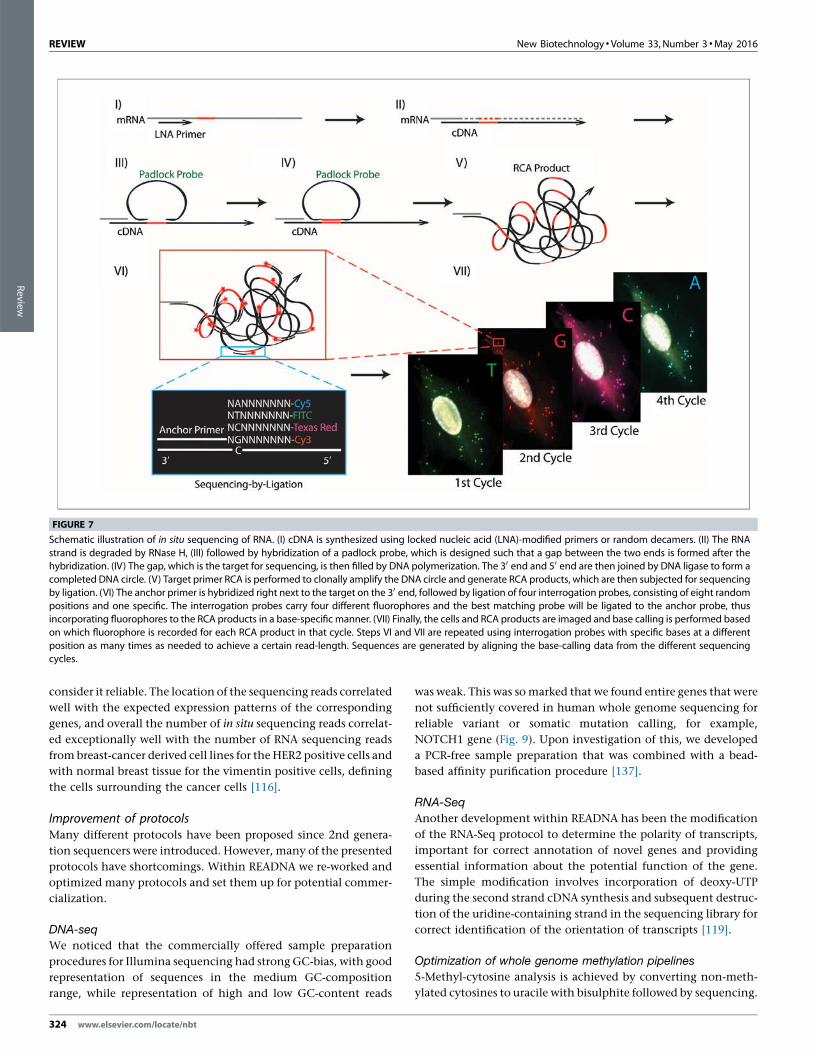
FIGURE 7
Schematic illustration of in situ sequencing of RNA. (I) cDNA is synthesized using locked nucleic acid (LNA)-modified primers or random decamers. (II) The RNA
strand is degraded by RNase H, (III) followed by hybridization of a padlock probe, which is designed such that a gap between the two ends is formed after thehybridization. (IV) The gap, which is the target for sequencing, is then filled by DNA polymerization. The 30 end and 50 end are then joined by DNA ligase to form a
completed DNA circle. (V) Target primer RCA is performed to clonally amplify the DNA circle and generate RCA products, which are then subjected for sequencing
by ligation. (VI) The anchor primer is hybridized right next to the target on the 30 end, followed by ligation of four interrogation probes, consisting of eight randompositions and one specific. The interrogation probes carry four different fluorophores and the best matching probe will be ligated to the anchor probe, thus
incorporating fluorophores to the RCA products in a base-specific manner. (VII) Finally, the cells and RCA products are imaged and base calling is performed based
on which fluorophore is recorded for each RCA product in that cycle. Steps VI and VII are repeated using interrogation probes with specific bases at a different
position as many times as needed to achieve a certain read-length. Sequences are generated by aligning the base-calling data from the different sequencingcycles.
324 www.elsevier.com/locate/nbt
Review

We developed this into a process for whole genome 5-methyl-
cytosine analysis with 2nd generation sequencing [138,139]. The
process is based on preparing a sequencing library, followed by the
inclusion of a conversion control spike-in, bisulphite conversion,
library quality control system, a sequencing procedure that allows
balancing the uneven response of the four bases after bisulphite
conversion in Illumina sequencing instruments. We integrated
the pipeline with a data analysis pipeline in which all spike-ins are
tracked automatically. This pipeline is applied in the EU-funded
project of the International Human Epigenome Project BLUE-
PRINT.
Other applications of nucleic acid analysis – ProteinSeq
READNA has developed a method entitled ProteinSeq that mea-
sures in parallel candidate protein biomarkers in many samples. A
multiplex proximity ligation assay (PLA) is performed and the
readout is done using realtime PCR or DNA sequencing (Protein-
Seq). We demonstrate improved sensitivity over conventional
sandwich assays for simultaneous analysis of sets of 35 proteins
in 5 ml of blood plasma. Importantly the method can be used with
multiplexing as background signal remains low. The level of
multiplexing that is possible will be investigated further. Protein-
Seq was used to analyze proteins in plasma samples from cardio-
vascular disease (CVD) patient cohorts and matched controls.
Three proteins, namely P-selectin, Cystatin-B and Kallikrein-6,
were identified as putative diagnostic biomarkers for CVD. The
latter two have not been previously reported in the literature and
their potential roles in CVD will be validated in larger patient
cohorts. ProteinSeq has a potential for screening large numbers
of proteins and samples while the technology can provide a
much-needed platform for validation of diagnostic markers in
biobank samples and in clinical use [121,122]. In a recent publica-
tion [123] Proximity Ligation Assay (PLA) has been proven to be a
robust protein detection method. The technique is characterized
by high sensitivity and specificity, but the assay precision is
probably limited by the PCR readout. To investigate this potential
limitation and to improve precision, we developed a digital PLA for
protein measurement in fluids based on amplified single molecule
detection (ASMD). The assay showed significant improvements in
precision, and thereby also detection sensitivity, over the conven-
tional real-time PCR readout. The assay is complementary to the
ProteinSeq method also developed within READNA.
Software developmentThe sequence output of 2nd generation sequencers puts huge
strain on computation. The main reasons are that the sequencing
reads are short and that it is easy to produce huge numbers of short
reads with 2nd generation sequencers. Within READNA we have
developed software to respond to several challenges for the analy-
sis of NGS data.
Alignment of 2nd generation sequencing reads
Sequence alignment is one of the first steps that needs to be
executed in a data analysis pipeline of NGS data. The millions
of reads generated in a sequencing run need to be positioned on a
reference sequence. Matching sequences that are identical to the
reference and are unique in the genome does not pose particular
challenges. However, the objective is to capture small differences
to the reference sequence. Another problem of many genomes is
that in genomes, Ts, Cs, Gs, and As are not randomly distributed,
New Biotechnology �Volume 33, Number 3 �May 2016 REVIEW
[(Figure_8)TD$FIG]
FIGURE 8
In situ gene expression profiling in a breast cancer tumor section. A 0.6 mm2 sized area of a breast cancer tumor tissue section was analyzed using a pool of
padlock probes targeting 39 different transcripts. The picture shows the distribution of the sequencing reads from the 30 most abundant transcripts, listed to the
right of the picture.
www.elsevier.com/locate/nbt 325
Review

but have many repetitive elements, duplications, similar
sequences in different regions of the genome. This substantially
complicates finding the best place to position a read. Exhaustive
searches are computationally very expensive. A common feature of
many aligners is the use shortcuts from exhaustive searching, by
taking random decisions. With GEM a deterministic aligner was
developed that explores the entire search space exhaustively while
on the other hand being computationally very efficient and accu-
rate [124].
Variant calling
After alignment, genomic variants are called from the consensus of
aligned sequences. As implied by the alignment difficulties de-
scribed above, the determination of true variants is not always
straightforward. The development of two new variant calling
tools/approaches was primarily motivated by our evaluation of
alignment and variant calling tools, which revealed five important
observations. Firstly, variant detection in enriched regions – even
at high coverage – depends strongly on the chosen tools and their
settings. Secondly, the tested variant callers do not seem to be well-
trained to handle targeted NGS (T-NGS) data, leading to a signifi-
cant fraction of false positive and negative SNV calls [14]. Thirdly,
when analysing T-NGS sequences, the conventional (or one-step
whole-genome) mapping and variant-calling approach is not op-
timal in terms of time and computer resources [14]. Fourthly,
depending on sequencing platforms, up to 60% of relevant var-
iants were undetected due to coverage gaps, or were misidentified
[125]. Fifthly, when comparing two samples (e.g. healthy versus
affected tissue), reliance on the derived variant lists instead of the
primary BAM file will not allow detection of significant changes of
allelic balance in heterozygous variants, unless the read counts per
allele are included in the variant lists. The results of our studies
showed that lack of evenness of coverage and inadequate enrich-
ment and/or coverage can prevent the detection of real nucleotide
variants, leading to higher false negative rates, particularly for
heterozygotes [14]. These observations and analysis challenges
emphasize that the costs, speed, and variant detection accuracy
should be considerably adapted and improved in order to be
useful in a diagnostic setting and affordable to a larger group of
researchers. To address these challenges, we have developed two
novel software tools, namely ‘backmapping’ and ‘pibase’. The
backmapping pipeline [14] (http://www.ikmb.uni-kiel.de/
tngs-backmapping) is a two-step mapping approach, target-region
mapping (to rescue false negative calls) with subsequent
REVIEW New Biotechnology � Volume 33, Number 3 �May 2016
[(Figure_9)TD$FIG]
FIGURE 9
Sequence coverage of 30�whole genome sequencing across the NOTCH1 gene (panel (a)). Three different protocols were used, standard Illumina protocol in red,
a modified version of the Illumina protocol that enhances GC-rich sequences in green, and a no PCR protocol in blue. Even coverage like in the blue protocol
dramatically facilitates variant calling. In panel (b), (c) and (d) are the Bioanalyzer traces and normalized GC representations for the three sample preparation
protocols, corresponding to the coverage plots in panel (a).
326 www.elsevier.com/locate/nbt
Review

read-backmapping of only the reads that cover a detected variant,
to the whole genome. In an exome sequencing benchmark, our
backmapping approach achieved two-fold faster mapping and
increased sensitivity, compared to the conventional approach.
The second novel software package, pibase (http://www.ikmb.
uni-kiel.de/pibase), was developed to help distinguish between
true variants, artifacts, and sequence coverage gaps. This is a
tertiary in silico analysis tool, for interrogating BAM files, re-typing
SNPs, and accurate comparison of variant data [125]. Using pibase,
we demonstrated highly specific (99.97–100.00%) genotype calls
with publicly available 1000 Genomes Project BAM files. We also
reported that the false detection rate of variant differences in pairs
of monozygotic twins is 10-fold lower using pibase’s Fisher’s exact
test, than using a genotype-based comparison method [125]. As
large-scale manual inspection of T-NGS or exome data is not
feasible and prone to subjective errors, we recommend the two-
step mapping approach; backmapping; [14] and validations with
the pibase tool [125]. As pseudo-random technical artifacts may
occur during library preparation, enrichment or sequencing, we
also recommend, if cost allows, duplicating samples and validating
computed variants by checking the concordance between these
technical replicates.
RNA-Seq
The community opinion is that RNA profiling using arrays or
sequencing is not robust and experiments carried out in different
laboratories are not comparable and even experiments done in the
same laboratory at different times or by different people are prone
to variation. We proved that an RNA sequencing project can be
distributed across several laboratories as long as a standard oper-
ating procedure is adhered to and the same lots of reagents are
used. Having an RNA-Seq dataset with 500 samples in hand to
work with has allowed the establishment of many different data
analytical techniques for RNA-Seq [126,127].
DNA methylation software, MeDIP
DNA methylation occurring on cytosines in the context of the
dinucleotide sequence CpG forms one of the multiple layers of
epigenetic mechanisms which constitute the regulatory landscape
of a cell. Aberrant DNA methylation changes have been detected
in several diseases, particularly cancer and autoimmune diseases
leading to the development of a large number of technologies for
the analysis of DNA methylation patterns. Enrichment of the
methylated fraction of the genome by Methylated DNA Immuno-
Precipitation (MeDIP) and subsequent NGS sequencing provides a
cost-effective alternative for genome-wide DNA methylation anal-
ysis and is not confounded as WGBS by the presence of hydro-
xymethylation as the antibodies are specific for the respective
cytosine modifications. However, the exact nature of the immu-
noprecipitated products has never been investigated. A novel
method for the analysis of the DNA methylation state of the
immunoprecipitated population of molecules at single nucleotide
resolution was developed [128]. A small part of the immunopre-
cipitated fraction was treated with sodium bisulfite thereby trans-
lating epigenetic differences into sequence differences which were
then quantitatively read-out by pyrosequencing, demonstrating
that MeDIP was highly specific for methylated molecules. The
assay presents a rapid and cost-effective method for the quality
control of MeDIP prior to NGS. As no complete bioinformatics
solution for the processing of MeDIP-seq data was available, a
complete pipeline for quality control, pre-processing, mapping,
estimation of DNA methylation levels and calling of differentially
methylated regions was developed for both single end and paired-
end reads [129]. The freely available pipeline permits to streamline
the data analysis process and enables researchers to concentrate on
the biological interpretation of MeDIP-seq data.
ELSI aspects of next generation nucleic acid sequence dataInnovations in sequencing mean that more detailed and richer
sequence information can be generated in larger quantities, at a
cheaper cost and faster than ever before. One sample of DNA that is
sequenced using NGS can provide information potentially on a
number of conditions at once rather than separate tests having to
be done for each condition. This raises a number of new ethical,
legal and social issues (ELSI) as this technology starts to be more
widely used in research and rolled out to the clinic. For example,
the possibility of identifying an incidental finding of clinical
significance that was not the object of the original research and
diagnostic investigation increases, because of the quality and
quantity of the information generated through NGS [130,131].
It is also increasingly difficult to guarantee the privacy of such
information for the proband and their relatives because of the
comprehensive nature of the data, the fact that DNA is a unique
identifier and this technology is likely to be accessible to more
people [132]. Obtaining consent for research is also difficult if data
has a number of possible uses that cannot always be anticipated at
the time of recruitment into research or when presenting for a
particular condition [133]. One question which arises as a result of
this is whether an individual should be informed of all of the
potential health consequences connected with the variations in
their genome and how this should be done. For this technology to
have greater clinical application, it will also need to become more
reliable and to be more easily replicated [134,135].
Perspectives – what remains to be done? What arefuture challenges?READNA has made major contributions to many aspects of nucleic
acid analysis and its applications. Many of the developments
reached maturity already during the course of READNA and are
now used in different contexts. We have shown many concepts
that can now be translated for research and application. Interest-
ing developments are in nanopore sequencing, single cell nucleic
acid applications and the work on devices for handling and
analyzing long DNA molecules, and in NGS data analysis. Howev-
er, there are many aspects that do not yet have a final solution.
Extending the length of DNA sequences is critical. The more we
refine the tools for genome analysis, the more we find that
structures of genomes are more diverse than we thought. We
are discovering that the concept of a single reference for a species
is not entirely correct. Technologies to analyze long DNA mole-
cules at high resolution need further development. In general,
technologies to carry out nucleic acid analyses at very low con-
centration still require further development. In the ultimate limit
this applies to single cell analysis at DNA, RNA, DNA modifications
and DNA protein interaction level. Understanding the functioning
of the genome requires such techniques, as stochastic effects of
New Biotechnology �Volume 33, Number 3 �May 2016 REVIEW
www.elsevier.com/locate/nbt 327
Review
doing these analyses with DNA from many cells results in a loss of
molecular context. A further opportunity that would be very
beneficial and would advance the integration of these technolo-
gies into routine is the development of fully integrated cartridge-
based systems in which procedures can be done far more reliably,
reproducibly and with a dramatically reduced risk of contamina-
tion. Sequencing with nanopores is very enticing. Many aspects of
nanopore sequencing still require resolution, such as the robust-
ness of the pores, software for deconvoluting signal and software
to deal with long reads. However, the community-based tool
development initiated by Oxford Nanopore Technologies for their
MinION nucleic acid sequencer is very innovative. 4th generation
sequencing for which READNA has provided a first proof of
concept still has a great need for further development. Currently,
this technology can be used for in situ RNA analysis, but evidently
techniques for measuring somatic mutations on the level of the
DNA in situ of many different targets simultaneously would be of
interest and being able to determine the interaction of regulatory
elements in situ would allow studying genome regulation at un-
precedented resolution [1]. There is also a great need for standard-
ization and benchmarking of 2nd generation sequencing and data
analysis for this technology to achieve robustness and get the
deserved uptake into clinical practice. Care needs to be taken that
good standards of data analysis are installed in common practice.
An eye needs to be kept on computational efficiency of analysis
and storage as large-scale projects are initiated.
ConclusionREADNA has contributed substantially to many aspects of the
revolution that nucleic acid sequencing has experienced in recent
years, has opened many new lines of work and provided proofs-of-
concepts. The body of work stands as a lasting legacy.
AcknowledgementsThe review was written and funded through the European
Community’s Seventh Framework Programme (FP7/2007-2013)
under grant agreement no. [HEALTH-F4-2008-201418] entitled
READNA. We would like to thank Professor Sunney Xie, Professor
Stephan Beck, Professor Wilhelm Ansorge and Professor George
Church, distinguished members of the READNA Scientific
Advisory Board for their critical review of the READNA project.
Finally we would like to thank Dr. Tomasz Dylag project officer at
the European Commission for his support throughout the
duration of the project.
References
[1] Mir KU. Sequencing genomes: from individuals to populations. Brief FunctGenomic Proteomic 2009;8(5):367–78.
[2] Parkhomchuk D, Amstislavskiy V, Soldatov A, Ogryzko V. Use of high through-put sequencing to observe genome dynamics at a single cell level. Proc NatlAcad Sci U S A 2009;106(49):20830–35.
[3] McGinn S, Gut IG. DNA sequencing – spanning the generations. New Bio-technol 2013;30(4):366–72.
[4] Veal CD, Xu H, Reekie K, Free R, Hardwick RJ, McVey D, et al. Automated designof paralogue ratio test assays for the accurate and rapid typing of copy numbervariation. Bioinformatics 2013;29(16):1997–2003.
[5] Veal CD, Freeman PJ, Jacobs K, Lancaster O, Jamain S, Leboyer M. A mecha-nistic basis for amplification differences between samples and between genomeregions. BMC Genomics 2012;13:455.
[6] Dahl F, Gullberg M, Stenberg J, Landegren U, Nilsson M. Multiplex amplifica-tion enabled by selective circularization of large sets of genomic DNA frag-ments. Nucleic Acids Res 2005;33(8):e71.
[7] Dahl F, Stenberg J, Fredriksson S, Welch K, Zhang M, Nilsson M, et al. Multigeneamplification and massively parallel sequencing for cancer mutation discovery.Proc Natl Acad Sci U S A 2007;104(22):9387–92.
[8] Johansson H, Isaksson M, Sorqvist EF, Roos F, Stenberg J, Sjoblom T, et al.Targeted resequencing of candidate genes using selector probes. Nucleic AcidsRes 2011;39(2):e8.
[9] Zieba A, Grannas K, Soderberg O, Gullberg M, Nilsson M, Landegren U.Molecular tools for companion diagnostics. Nat Biotechnol 2012;29(6):634–40.
[10] Moens LN, Falk-Sorqvist E, Ljungstrom V, Mattsson J, Sundstrom M, La Fleur L,et al. HaloPlex targeted resequencing for mutation detection in clinical forma-lin-fixed paraffin-embedded (FFPE) tumor samples. Mol Diagn 2015;17(6):729–39.
[11] Meuzelaar LS, Lancaster O, Pasche JP, Kopal G, Brookes AJ. MegaPlex PCR: astrategy for multiplex amplification. Nat Methods 2007;4(10):835–7.
[12] Querfurth R, Fischer A, Schweiger MR, Lehrach H, Mertes F. Creation andapplication of immortalized bait libraries for targeted enrichment and next-generation sequencing. Biotechniques 2012;52(6):375–80.
[13] Mir KU, Qi H, Salata O, Scozzafava G. Sequencing by Cyclic Ligation andCleavage (CycLiC) directly on a microarray captured template. Nucleic AcidsRes 2009;37(1):e5.
[14] Elsharawy A, Forster M, Schracke N, Keller A, Thomsen I, Petersen BS, et al.Improving mapping and SNP-calling performance in multiplexed targetednext-generation sequencing. BMC Genomics 2012;13:417.
[15] ElSharawy A, Warner J, Olson J, Forster M, Schilhabel MB, Link DR, et al.Accurate variant detection across non-amplified and whole genome amplifiedDNA using targeted next generation sequencing. BMC Genomics 2012;13:500.
[16] Mertes F, Elsharawy A, Sauer S, van Helvoort JM, van der Zaag PJ, Franke A, et al.Targeted enrichment of genomic DNA regions for next-generation sequencing.Brief Funct Genomics 2011;10(6):374–86.
[17] Ku CS, Cooper DN, Polychronakos C, Naidoo N, Wu M, Soong R. Exomesequencing: dual role as a discovery and diagnostic tool. Ann Neurol 2012;71(1):5–14.
[18] Fairfield H, Gilbert GJ, Barter M, Corrigan RR, Curtain M, Ding Y, et al.Mutation discovery in mice by whole exome sequencing. Genome Biol2011;12(9):R86.
[19] Melum E, May S, Schilhabel MB, Thomsen I, Karlsen TH, Rosenstiel P, et al. SNPdiscovery performance of two second-generation sequencing platforms in theNOD2 gene region. Hum Mutat 2010;31(7):875–85.
[20] Nilsson, M., et al., Target Enrichment Sequencing Descriptors (TESD) – descrip-tors for reporting target enrichment in DNA sequencing experiments, manu-script in preparation.
[21] Mauger F, Jaunay O, Chamblain V, Reichert F, Bauer K, Gut IG, et al. SNPgenotyping using alkali cleavage of RNA/DNA chimeras and MALDI time-of-flight mass spectrometry. Nucleic Acids Res 2006;34(3):e18.
[22] Mauger F, Bauer K, Calloway CD, Semhoun J, Nishimoto T, Myers TW, et al.DNA sequencing by MALDI-TOF MS using alkali cleavage of RNA/DNA chi-meras. Nucleic Acids Res 2007;35(8):e62.
[23] Mauger F, Bauer K, Semhoun J, Myers TW, Gelfand DH, Gut IG. Ribo-polymer-ase chain reaction – a facile method for the preparation of chimeric RNA/DNAapplied to DNA sequencing. Hum Mutat 2012;33(6):1010–5.
[24] Mauger F, Gelfand DH, Gupta A, Bodepudi V, Will SG, Bauer K, et al. High-specificity single-tube multiplex genotyping using Ribo-PAP PCR, tag primers,alkali cleavage of RNA/DNA chimeras and MALDI-TOF MS. Hum Mutat2013;34(1):266–73.
[25] Sauer S, Freiwald A, Maier T, Kube M, Reinhardt R, Kostrzewa M, et al.Classification and identification of bacteria by mass spectrometry and compu-tational analysis. PLoS One 2008;3(7):e2843.
[26] Freiwald A, Sauer S. Phylogenetic classification and identification of bacteria bymass spectrometry. Nat Protoc 2009;4(5):732–42.
[27] Freiwald A, Mao L, Kodelja V, Kliem M, Schuldt D, Schreiber S, et al. Differentialanalysis of Crohn’s disease and ulcerative colitis by mass spectrometry.Inflamm Bowel Dis 2011;17(4):1051–2.
[28] Sauer S, Kliem M. Mass spectrometry tools for the classification and identifica-tion of bacteria. Nat Rev Microbiol 2010;8(1):74–82.
[29] Kliem M, Sauer S. The essence on mass spectrometry based microbial diag-nostics. Curr Opin Microbiol 2012;15(3):397–402.
[30] Howell WM, Jobs M, Gyllensten U, Brookes AJ. Dynamic allele-specific hybrid-ization. A new method for scoring single nucleotide polymorphisms. NatBiotechnol 1999;17(1):87–8.
[31] Jobs M, Howell WM, Stromqvist L, Mayr T, Brookes AJ. DASH-2: flexible, low-cost, and high-throughput SNP genotyping by dynamic allele-specific hybrid-ization on membrane arrays. Genome Res 2003;13(5):916–24.
[32] Rasmussen KH, Marie R, Lange JM, Svendsen WE, Kristensen A, Mir KU. Adevice for extraction, manipulation and stretching of DNA from single humanchromosomes. Lab Chip 2011;11(8):1431–3.
[33] Bauer DL, Marie R, Rasmussen KH, Kristensen A, Mir KU. DNA catenationmaintains structure of human metaphase chromosomes. Nucleic Acids Res2012;40(22):11428–34.
[34] Eriksen J, Thilsted AH, Marie R, Luscher CJ, Nielsen LB, Svendsen WE, et al.Dynamic in situ chromosome immobilisation and DNA extraction using
REVIEW New Biotechnology � Volume 33, Number 3 �May 2016
328 www.elsevier.com/locate/nbt
Review
localized poly(N-isopropylacrylamide) phase transition. Biomicrofluidics2011;5(3): 31101–311014.
[35] Marie R, Kristensen A. Nanofluidic devices towards single DNA moleculesequence mapping. J Biophotonics 2012;5(8):673–86.
[36] Marie R, Pedersen JN, Bauer DL, Rasmussen KH, Yusuf M, Volpi E, et al.Integrated view of genome structure and sequence of a single DNA moleculein a nanofluidic device. Proc Natl Acad Sci U S A 2013;110(13):4893–8.
[37] Freitag C, Noble C, Fritzsche J, Persson F, Reiter-Schad M, Nilsson AN, et al.Visualizing the entire DNA from a chromosome in a single frame. Biomicro-fluidics 2015;9:044114.
[38] Reisner W, Larsen NB, Silahtaroglu A, Kristensen A, Tommerup N, TegenfeldtJO. Single-molecule denaturation mapping of DNA in nanofluidic channels.Proc Natl Acad Sci U S A 2010;107(30):13294–99.
[39] Thamdrup LH, Pedersen JN, Flyvbjerg H, Larsen NB, Kristensen A. Nanoim-printed polymer chips for light induced local heating of liquids in micro- andnanochannels. Proc SPIE 2010;7764. 77640I-77640I-13.
[40] Thamdrup LH, Larsen NB, Kristensen A. Light-induced local heating forthermophoretic manipulation of DNA in polymer micro- and nanochannels.Nano Lett 2010;10(3):826–32.
[41] Pedersen JN, Luscher CJ, Marie R, Thamdrup LH, Kristensen A, Flyvbjerg H.Thermophoretic forces on DNA measeured with a single-molecule springbalance. Phys Rev Lett 2014;113:268301.
[42] Mikkelsen MB, Reisner W, Flyvbjerg H, Kristensen A. Pressure-driven DNA innanogroove arrays: complex dynamics leads to length- and topology-depen-dent separation. Nano Lett 2011;11(4):1598–602.
[43] Nyberg LK, Persson F, Berg J, Bergstrom J, Fransson E, Olsson L, et al. A single-step competitive binding assay for mapping of single DNA molecules. BiochemBiophys Res Commun 2012;417(1):404–8.
[44] Persson F, Fritzsche J, Mir KU, Modesti M, Westerlund F, Tegenfeldt JO. Lipid-based passivation in nanofluidics. Nano Lett 2012;12(5):2260–5.
[45] Lymperopoulos K, Crawford R, Torella JP, Heilemann M, Hwang LC, Holden SJ,et al. Single-molecule DNA biosensors for protein and ligand detection. AngewChem Int Ed Engl 2010;49(7):1316–20.
[46] Persson F, Westerlund F, Tegenfeldt JO. Fluorescence nanoscopy of single DNAmolecules by using stimulated emission depletion (STED). Angew Chem Int EdEngl 2011;50(24):5581–3.
[47] Mortensen KI, Churchman LS, Spudich JA, Flyvbjerg H. Optimized localizationanalysis for single-molecule tracking and super-resolution microscopy. NatMethods 2010;7(5):377–81.
[48] Persson F, Westerlund F, Tegenfeldt JO. Fluorescence microscopy of nano-channel-confined DNA. Methods Mol Biol 2011;783:159–79.
[49] Hannestad JK, Brune R, Czolkos I, Jesorka A, El-Sagheer AH, Brown T, et al.Kinetics of diffusion-mediated DNA hybridization in lipid monolayer filmsdetermined by single-molecule fluorescence spectroscopy. ACS Nano 2013;7(1):308–15.
[50] Persson F, Tegenfeldt JO. DNA in nanochannels – directly visualizing genomicinformation. Chem Soc Rev 2010;39(3):985–99.
[51] Werner E, Persson F, Westerlund F, Tegenfeldt JO, Mehlig B. Orientationalcorrelations in confined DNA. Phys Rev E Stat Nonlin Soft Matter Phys2012;86(4 Pt 1):041802.
[52] Yusuf M, Bauer DL, Lipinski DM, MacLaren RE, Wade-Martins R, Mir KU, et al.Combining M-FISH and Quantum Dot technology for fast chromosomalassignment of transgenic insertions. BMC Biotechnol 2011;11:121.
[53] Hohlbein J, Gryte K, Heilemann M, Kapanidis AN. Surfing on a new wave ofsingle-molecule fluorescence methods. Phys Biol 2010;7(3):031001.
[54] Richardson JA, Gerowska M, Shelbourne M, French D, Brown T. Six-colourHyBeacon probes for multiplex genetic analysis. Chembiochem 2010;11(18):2530–3.
[55] Rindermann JJ, Akhtman Y, Richardson J, Brown T, Lagoudakis PG. Gaugingthe flexibility of fluorescent markers for the interpretation of fluorescenceresonance energy transfer. J Am Chem Soc 2011;133(2):279–85.
[56] Hohlbein J, Aigrain L, Craggs TD, Bermek O, Potapova O, Shoolizadeh P, et al.Conformational landscapes of DNA polymerase I and mutator derivativesestablish fidelity checkpoints for nucleotide insertion. Nat Commun2013;4:2131.
[57] Santoso Y, Joyce CM, Potapova O, Le Reste L, Hohlbein J, Torella JP, et al.Conformational transitions in DNA polymerase I revealed by single-moleculeFRET. Proc Natl Acad Sci U S A 2010;107(2):715–20.
[58] Santoso Y, Torella JP, Kapanidis AN. Characterizing single-molecule FRETdynamics with probability distribution analysis. Chemphyschem 2010;11(10):2209–19.
[59] Torella JP, Holden SJ, Santoso Y, Hohlbein J, Kapanidis AN. Identifyingmolecular dynamics in single-molecule FRET experiments with burst varianceanalysis. Biophys J 2011;100(6):1568–77.
[60] Le Reste L, Hohlbein J, Gryte K, Kapanidis AN. Characterization of darkquencher chromophores as nonfluorescent acceptors for single-moleculeFRET. Biophys J 2012;102(11):2658–68.
[61] Holden SJ, Uphoff S, Hohlbein J, Yadin D, Le Reste L, Britton OJ, et al. Definingthe limits of single-molecule FRET resolution in TIRF microscopy. Biophys J2010;99(9):3102–11.
[62] Uphoff S, Gryte K, Evans G, Kapanidis AN. Improved temporal resolution andlinked hidden Markov modeling for switchable single-molecule FRET. Chem-physchem 2011;12(3):571–9.
[63] Uphoff S, Holden SJ, Le Reste L, Periz J, van de Linde S, Heilemann M.Monitoring multiple distances within a single molecule using switchable FRET.Nat Methods 2010;7(10):831–6.
[64] Santoso Y, Kapanidis AN. Probing biomolecular structures and dynamics ofsingle molecules using in-gel alternating-laser excitation. Anal Chem2009;81(23):9561–70.
[65] Cordes T, Santoso Y, Tomescu AI, Gryte K, Hwang LC, Camara B, et al. SensingDNA opening in transcription using quenchable Forster resonance energytransfer. Biochemistry 2010;49(43):9171–80.
[66] Finan K, Torella JP, Kapanidis AN, Cook PR. T7 RNA polymerase functions invitro without clustering. PLoS One 2012;7(7):e40207.
[67] Pinkney JN, Zawadzki P, Mazuryk J, Arciszewska LK, Sherratt DJ, Kapanidis AN.Capturing reaction paths and intermediates in Cre-loxP recombinationusing single-molecule fluorescence. Proc Natl Acad Sci U S A 2012;109(51):20871–76.
[68] Robb NC, Cordes T, Hwang LC, Gryte K, Duchi D, Craggs TD, et al. Thetranscription bubble of the RNA polymerase-promoter open complex exhibitsconformational heterogeneity and millisecond-scale dynamics: implicationsfor transcription start-site selection. J Mol Biol 2013;425(5):875–85.
[69] Uphoff S, Reyes-Lamothe R, Garza de Leon F, Sherratt DJ, Kapanidis AN. Single-molecule DNA repair in live bacteria. Proc Natl Acad Sci U S A 2013;110(20):8063–8.
[70] Holden SJ, Uphoff S, Kapanidis AN. DAOSTORM: an algorithm for high-densitysuper-resolution microscopy. Nat Methods 2011;8(4):279–80.
[71] Crawford R, Erben CM, Periz J, Hall LM, Brown T, Turberfield AJ, et al. Non-covalent single transcription factor encapsulation inside a DNA cage. AngewChem Int Ed Engl 2013;52(8):2284–8.
[72] Crawford R, Kelly DJ, Kapanidis AN. A protein biosensor that relies on bendingof single DNA molecules. Chemphyschem 2012;13(4):918–22.
[73] Ding F, Manosas M, Spiering MM, Benkovic SJ, Bensimon D, Allemand JF.Single-molecule mechanical identification and sequencing. Nat Methods2012;9(4):367–72.
[74] Ranasinghe RT, Brown T. Ultrasensitive fluorescence-based methods fornucleic acid detection: towards amplification-free genetic analysis. ChemCommun (Camb) 2011;47(13):3717–35.
[75] El-Sagheer AH, Cheong VV, Brown T. Rapid chemical ligation of oligonucleo-tides by the Diels-Alder reaction. Org Biomol Chem 2011;9(1):232–5.
[76] El-Sagheer AH, Brown T. Synthesis and polymerase chain reaction amplifica-tion of DNA strands containing an unnatural triazole linkage. J Am Chem Soc2009;131(11):3958–64.
[77] Shelbourne M, Chen X, Brown T, El-Sagheer AH. Fast copper-free click DNAligation by the ring-strain promoted alkyne-azide cycloaddition reaction.Chem Commun (Camb) 2011;47(22):6257–9.
[78] El-Sagheer AH, Brown T. Click chemistry with DNA. Chem Soc Rev 2010;39(4):1388–405.
[79] Dierckx A, Diner P, El-Sagheer AH, Kumar JD, Brown T, Grotli M, et al.Characterization of photophysical and base-mimicking properties of a novelfluorescent adenine analogue in DNA. Nucleic Acids Res 2011;39(10):4513–24.
[80] El-Sagheer AH, Brown T. Efficient RNA synthesis by in vitro transcription of atriazole-modified DNA template. Chem Commun (Camb) 2011;47(44):12057–58.
[81] El-Sagheer AH, Sanzone AP, Gao R, Tavassoli A, Brown T. Biocompatibleartificial DNA linker that is read through by DNA polymerases and is functionalin Escherichia coli. Proc Natl Acad Sci U S A 2011;108(28):11338–43.
[82] Dallmann A, El-Sagheer AH, Dehmel L, Mugge C, Griesinger C, Ernsting NP.Structure and dynamics of triazole-linked DNA: biocompatibility explained.Chemistry 2011;17(52):14714–17.
[83] El-Sagheer AH, Brown T. New strategy for the synthesis of chemically modifiedRNA constructs exemplified by hairpin and hammerhead ribozymes. Proc NatlAcad Sci U S A 2010;107(35):15329–34.
[84] Shelbourne M, Brown Jr T, El-Sagheer AH, Brown T. Fast and efficient DNAcrosslinking and multiple orthogonal labelling by copper-free click chemistry.Chem Commun (Camb) 2012;48(91):11184–86.
[85] El-Sagheer AH, Brown T. Click nucleic acid ligation: applications in biology andnanotechnology. Acc Chem Res 2012;45(8):1258–67.
[86] Japrung D, Henricus M, Li Q, Maglia G, Bayley H. Urea facilitates the translo-cation of single-stranded DNA and RNA through the alpha-hemolysin nano-pore. Biophys J 2010;98(9):1856–63.
[87] Rotem D, Jayasinghe L, Salichou M, Bayley H. Protein detection by nanoporesequipped with aptamers. J Am Chem Soc 2012;134(5):2781–7.
[88] Sapra KT, Bayley H. Lipid-coated hydrogel shapes as components of electricalcircuits and mechanical devices. Sci Rep 2012;2:848.
[89] Schneider GF, Dekker C. DNA sequencing with nanopores. Nat Biotechnol2012;30(4):326–8.
[90] Schneider GF, Kowalczyk SW, Calado VE, Pandraud G, Zandbergen HW,Vandersypen LM, et al. DNA translocation through graphene nanopores. NanoLett 2010;10(8):3163–7.
[91] Dekker C, Kowalczyk S, Kapinos L, Lim RY. In vitro measurements of single-molecule transport across an individual biomimetic nuclear pore complex.Biophys J 2011;100(3):521a.
[92] Hall AR, Keegstra JM, Duch MC, Hersam MC, Dekker C. Translocation of single-wall carbon nanotubes through solid-state nanopores. Nano Lett 2011;11(6):2446–50.
[93] Hall AR, van Dorp S, Lemay SG, Dekker C. Electrophoretic force on a protein-coated DNA molecule in a solid-state nanopore. Nano Lett 2009;9(12):4441–5.
[94] Kowalczyk SW, Blosser TR, Dekker C. Biomimetic nanopores: learning fromand about nature. Trends Biotechnol 2011;29(12):607–14.
[95] Kowalczyk SW, Dekker C. Measurement of the docking time of a DNA moleculeonto a solid-state nanopore. Nano Lett 2012;12(8):4159–63.
New Biotechnology �Volume 33, Number 3 �May 2016 REVIEW
www.elsevier.com/locate/nbt 329
Review
[96] Kowalczyk SW, Grosberg AY, Rabin Y, Dekker C. Modeling the conductanceand DNA blockade of solid-state nanopores. Nanotechnology 2011;22(31):3151–61.
[97] Kowalczyk SW, Hall AR, Dekker C. Detection of local protein structures alongDNA using solid-state nanopores. Nano Lett 2010;10(1):324–8.
[98] Kowalczyk SW, Kapinos L, Blosser TR, Magalhaes T, van Nies P, Lim RY, et al.Single-molecule transport across an individual biomimetic nuclear pore com-plex. Nat Nanotechnol 2011;6(7):433–8.
[99] Kowalczyk SW, Wells DB, Aksimentiev A, Dekker C. Slowing down DNAtranslocation through a nanopore in lithium chloride. Nano Lett 2012;12(2):1038–44.
[100] Smeets RM, Kowalczyk SW, Hall AR, Dekker NH, Dekker C. Translocation ofRecA-coated double-stranded DNA through solid-state nanopores. Nano Lett2009;9(9):3089–96.
[101] Song B, Schneider GF, Xu Q, Pandraud G, Dekker C, Zandbergen H. Atomic-scale electron-beam sculpting of near-defect-free graphene nanostructures.Nano Lett 2011;11(6):2247–50.
[102] Wallace EV, Stoddart D, Heron AJ, Mikhailova E, Maglia G, Donohoe TJ, et al.Identification of epigenetic DNA modifications with a protein nanopore.Chem Commun (Camb) 2010;46(43):8195–7.
[103] Ayub M, Bayley H. Individual RNA base recognition in immobilized oligonu-cleotides using a protein nanopore. Nano Lett 2012;12(11):5637–43.
[104] Clarke J, Wu HC, Jayasinghe L, Patel A, Reid S, Bayley H. Continuous baseidentification for single-molecule nanopore DNA sequencing. Nat Nanotech-nol 2009;4(4):265–70.
[105] Hansen AS, Thalhammer A, El-Sagheer AH, Brown T, Schofield CJ. Improvedsynthesis of 5-hydroxymethyl-20-deoxycytidine phosphoramidite using a 20-deoxyuridine to 20-deoxycytidine conversion without temporary protectinggroups. Bioorg Med Chem Lett 2011;21(4):1181–4.
[106] Stoddart D, Heron AJ, Klingelhoefer J, Mikhailova E, Maglia G, Bayley H.Nucleobase recognition in ssDNA at the central constriction of the alpha-hemolysin pore. Nano Lett 2010;10(9):3633–7.
[107] Stoddart D, Maglia G, Mikhailova E, Heron AJ, Bayley H. Multiple base-recognition sites in a biological nanopore: two heads are better than one.Angew Chem Int Ed Engl 2010;49(3):556–9.
[108] Thalhammer A, Hansen AS, El-Sagheer AH, Brown T, Schofield CJ. Hydroxyl-ation of methylated CpG dinucleotides reverses stabilisation of DNA duplexesby cytosine 5-methylation. Chem Commun (Camb) 2011;47(18):5325–7.
[109] Hall AR, Scott A, Rotem D, Mehta KK, Bayley H, Dekker C. Hybrid poreformation by directed insertion of alpha-haemolysin into solid-state nano-pores. Nat Nanotechnol 2010;5(12):874–7.
[110] Villar G, Heron AJ, Bayley H. Formation of droplet networks that function inaqueous environments. Nat Nanotechnol 2011;6(12):803–8.
[111] Conze T, Goransson J, Razzaghian HR, Ericsson O, Oberg D, Akusjarvi G, et al.Single molecule analysis of combinatorial splicing. Nucleic Acids Res 2010;38(16):e163.
[112] Howell WM, Grundberg I, Faryna M, Landegren U, Nilsson M. Glycosylasesand AP-cleaving enzymes as a general tool for probe-directed cleavage of ssDNAtargets. Nucleic Acids Res 2010;38(7):e99.
[113] Larsson C, Grundberg I, Soderberg O, Nilsson M. In situ detection andgenotyping of individual mRNA molecules. Nat Methods 2010;7(5):395–7.
[114] Grundberg I, Kiflemariam S, Mignardi M, Imgenberg-Kreuz J, Edlund K, MickeP. In situ mutation detection and visualization of intratumor heterogeneity forcancer research and diagnostics. Oncotarget 2013;4(12):2407–18.
[115] Shendure J, Porreca GJ, Reppas NB, Lin X, McCutcheon JP, Rosenbaum AM,et al. Accurate multiplex polony sequencing of an evolved bacterial genome.Science 2005;309(5741):1728–32.
[116] Ke R, Mignardi M, Pacureanu A, Svedlund J, Botling J, Wahlby C, et al. In situsequencing for RNA analysis in preserved tissue and cells. Nat Methods2013;10(9):857–60.
[117] Goransson J, Wahlby C, Isaksson M, Howell WM, Jarvius J, Nilsson M. A singlemolecule array for digital targeted molecular analyses. Nucleic Acids Res2009;37(1):e7.
[118] Goransson J, Ke R, Nong RY, Howell WM, Karman A, Grawe J, et al. Rapididentification of bio-molecules applied for detection of biosecurity agentsusing rolling circle amplification. PLoS One 2012;7(2):e31068.
[119] Parkhomchuk D, Borodina T, Amstislavskiy V, Banaru M, Hallen L, Krobitsch S,et al. Transcriptome analysis by strand-specific sequencing of complementaryDNA. Nucleic Acids Res 2009;37(18):e123.
[120] Weibrecht I, Lundin E, Kiflemariam S, Mignardi M, Grundberg I, Larsson C,et al. In situ detection of individual mRNA molecules and protein complexes orpost-translational modifications using padlock probes combined with the insitu proximity ligation assay. Nat Protoc 2013;8(2):355–72.
[121] Darmanis S, Nong RY, Vanelid J, Siegbahn A, Ericsson O, Fredriksson S, et al.ProteinSeq: high-performance proteomic analyses by proximity ligation andnext generation sequencing. PLoS One 2011;6(9):e25583.
[122] Nong RY, Gu J, Darmanis S, Kamali-Moghaddam M, Landegren U. DNA-assistedprotein detection technologies. Expert Rev Proteomics 2012;9(1):21–32.
[123] Ke R, Nong RY, Fredriksson S, Landegren U, Nilsson M. Improving precision ofproximity ligation assay by amplified single molecule detection. PLoS One2013;8(7):p. e69813.
[124] Marco-Sola S, Sammeth M, Guigo R, Ribeca P. The GEM mapper: fast, accurateand versatile alignment by filtration. Nat Methods 2012;9(12):1185–8.
[125] Forster M, Forster P, Elsharawy A, Hemmrich G, Kreck B, Wittig M, et al. Fromnext-generation sequencing alignments to accurate comparison and valida-tion of single-nucleotide variants: the pibase software. Nucleic Acids Res2013;41(1):e16.
[126] Lappalainen T, Sammeth M, Friedlander MR, ’t Hoen PA, Monlong J, Rivas MA,et al. Transcriptome and genome sequencing uncovers functional variation inhumans. Nature 2013;501(7468):506–11.
[127] ’t Hoen PA, Friedlander MR, Almlof J, Sammeth M, Pulyakhina I, Anvar SY,et al. Reproducibility of high-throughput mRNA and small RNA sequencingacross laboratories. Nat Biotechnol 2013;31(11):1015–22.
[128] Sengenes J, Daunay A, Charles MA, Tost J. Quality control and single nucleo-tide resolution analysis of methylated DNA immunoprecipitation products.Anal Biochem 2010;407(1):141–3.
[129] Huang J, Renault V, Sengenes J, Touleimat N, Michel S, Lathrop M, et al. MeQA:a pipeline for MeDIP-seq data quality assessment and analysis. Bioinformatics2012;28(4):587–8.
[130] Knoppers BM, Zawati MH, Kirby ES. Sampling populations of humans acrossthe world: ELSI issues. Annu Rev Genomics Hum Genet 2012;13:395–413.
[131] van El CG, Cornel MC, Borry P, Hastings RJ, Fellmann F, Hodgson SV, et al.Whole-genome sequencing in health care. Recommendations of the EuropeanSociety of Human Genetics. Eur J Hum Genet 2013;21 Suppl 1:S1–5.
[132] Chadwick R. Personal genomes: no bad news? Bioethics 2011;25(2):62–5.[133] Peppercorn J, Shapira I, Deshields T, Kroetz D, Friedman P, Spears P, et al.
Ethical aspects of participation in the database of genotypes and phenotypes ofthe National Center for Biotechnology Information: the Cancer and LeukemiaGroup B Experience. Cancer 2012;118(20):5060–8.
[134] Tabor HK, Berkman BE, Hull SC, Bamshad MJ. Genomics really gets personal:how exome and whole genome sequencing challenge the ethical framework ofhuman genetics research. Am J Med Genet A 2011;155A(12):2916–24.
[135] Desai AN, Jere A. Next-generation sequencing: ready for the clinics? Clin Genet2012;81(6):503–10.
[136] Westerlund F, Persson F, Kristensen A, Tegenfeldt JO. Fluorescence enhance-ment of single DNA molecules confined in Si/SiO2 nanochannels. Lab Chip2010;10(16):2049–51.
[137] Puente XS, Bea S, Valdes-Mas R, Villamor N, Gutierrez-Abril J, Martin-Subero JI,et al. Non-recurrent mutations in chronic lymphocytic leukaemia. Nature2015. doi: 10.1038/nature14666.
[138] Kulis M, Heath S, Bibikova M, Queiros AC, Navarro A, Clot G, et al. Epigenomicanalysis detects widespread gene-body DNA hypomethylation in chroniclymphocytic leukemia. Nat Genet 2012;44(11):1236–42.
[139] Kulis M, Merkel A, Heath S, Queiros AC, Schuyler RP, Castellano G, et al.Whole-genome fingerprint of the DNA methylome during human B celldifferentiation. Nat Genet 2015;47(7):746–56.
REVIEW New Biotechnology � Volume 33, Number 3 �May 2016
330 www.elsevier.com/locate/nbt
Review