new surface technologies for cell culture - cultek s.l.u · pdf filenew surface technologies...
TRANSCRIPT

New Surface Technologiesfor Cell Culture
Ute Vespermann

Talk Outline• Overview of cell culture surface treatments• Corning® CellBIND® Surface
– Physical and Chemical properties of the surface– Cell growth performance on Corning® CellBIND® Surface
• Adherence of difficult cell lines• Low or no serum conditions
• How does Corning® CellBIND® Surface benefit my work?
• Ultra Low Attachment Coating– ULA designed for…
• Q&A

Your cells work hard for you!
Don’t they deserve the
best?
Corning® CellBIND®
Surface
The First New Surface for Cell Culture in over 20 years

Traditional Culture Vessel SurfacesSurface Treatments:
• Corona Discharge (1960s)• Vacuum Plasma (RF) – O2 (1970s)• Vacuum Plasma (RF) -NH4 (1980s; BD Primaria™)• Corning® CellBIND® Surface (2003)
• Collagen (1956)• Fibronectin• Laminin• Gelatin• Matrigel• P-D- & P-L-Lysine• Metals
Biological
Surface Coatings:

Corning offers 5 culture surfaces
For attachment dependent:• Traditional TCT surface
– either plasma or corona treated• New Corning® CellBIND® Surface• Poly-D-lysine-coated microplates
For suspended cells:• Ultra Low Attachment surfaces
– dishes and microplates• Untreated surfaces
– dishes and microplates
To get more from your cells, give them
the best surface!

Why do we “treat” culture vessels?
Untreated polystyrene has an uncharged hydrophobic surfaceUntreated Polystyrene
Repels water
TC-treated polystyrene has a negatively charged, hydrophilic surface to which many proteins and cells stickTC-treated Polystyrene
Attracts water and proteins- - - - - - - - - - - - - - - - - - - - - -

Treatment modifies polystyrene
HH H H
HH H HHH
H
H
C C C C CC C CC
HH H H
HH H HHH
H H
H
H
C C C C CC C CCHydrophobic
H HH
O HH H
O
OHH
OC C C C C C C CC
HH
O OH HH
H H
O
OH
OH
HO
H
OC C C C C C C CCHydrophilic
Surface treatment

Corona discharge
Conveyor belt
High energy oxygen ionsCorona Discharge Plate
• For dishes, flasks, plates

Door
Plasma generator
Vacuum pump
Vacuum plasma chamber
High energy oxygen ions
Plasma generator
• For roller bottles

What is the Corning® CellBIND® Surface?• Patented non-biological surface treatment for cell
culture
• N2O plasma in the presence of microwave energy to increase reactive functional groups on polystyrene surfaces
• A novel surface treatment that does not require any special storage conditions!

Analytical Techniques Utilized
Surface Morphology:AFM – Atomic Force Microscopy
Nano Indentation
Surface Chemistry:ESCA-Electron Spectroscopy for Chemical Analysis
Surface Energy/Wettability:Water Contact Angle

Surface Morphology - AFM (Atomic Force Microscopy)
Corning® CellBIND® SurfaceCorona Surface

Surface Chemistry
Oxygen content and depth of treatment of Corning®
CellBIND®
Surface vs TCT Treated Vessels
10
15
20
25
30
35
50 60 70 80 90 100 110 120Depth (Angstroms)
% O
xyge
n
CellBIND FlaskFlask (TCT, Corona)CellBIND Roller BottleRoller Bottle (TCT, RF)CellBIND Microplate Microplate (TCT, Corona)
• ESCA (Electron Spectroscopy for Chemical Analysis)

Corning® CellBIND® Surface treatment increases Oxygen-containing functional groups
%O %C-O %C=O %O-C=O %O-C-OO
25%
20%
15%
10%
5%
0%
Corning® CellBIND® Surface
TCT Surface
ESCA Data post wetting

• Corning CellBIND Surface is more wettable• And more stable after liquid contact
CellBINDLow Contact Angle -
High Wettability
Standard TCTHigh Contact Angle -
Low Wettability
Corning® CellBIND® Surface Review - Contact angle
Pre-Wash Post-Wash22° 24°
Pre-Wash Post-Wash 43° 58°

Corning® CellBIND® SurfaceAnalytical summary
• More textured surface
• More oxygen containing functional groups added to the surface
• Increased depth of surface modification
• Increased wettability
• Increased stability

Corning® CellBIND® Surface vs. TCT: Growth StudiesObjectives:Compare Corning® CellBIND® surface with traditional TCT surface using four cell lines:• HEK-293 - transformed human embryonic kidney• WS1 - transfected HEK-293• CHO – transfected Chinese hamster ovary• LNCaP – human prostate cancer

HEK-293 yields in reduced (1%) FBS
TCT Corning® CellBIND®
SurfaceInitial seeding density of 1.8x10⁶ cell/ T-75 flask. Cells were grown 6 days in 10% serum prior to seeding into to medium containing 1% serum. Data represents the average count + SE from six flasks from two separate experiments for each condition tested. 49.5% higher yield in Corning® CellBIND® surface.
TCT Surface
Corning®CellBIND®
Surface
Num
ber o
f Cel
ls
0
5.0x10⁶
2.5x10⁷
3.5x10⁷
3.0x10⁷
2.0x10⁷
1.5x10⁷
1.0x10⁷

WS-1 yields were ~ 3-fold higher for Corning® CellBIND® Surface
Initial seeding density of 1.7 x 10⁷ cells/roller bottle in medium containing 10% FBS. Data represents the average count + SE from six bottles from two different studies for each condition tested after 6 days.
TCT Corning® CellBIND® Surface
Num
ber o
f Cel
ls
TCT Surface
Corning®CellBIND®
Surface
Initial Seeding
0
8x10⁷
6x10⁷
4x10⁷
2x10⁷
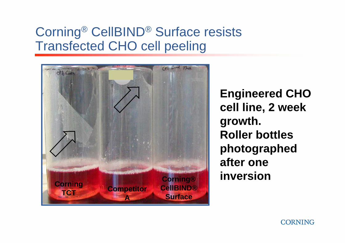
Corning® CellBIND® Surface resists Transfected CHO cell peeling
Engineered CHO cell line, 2 week growth. Roller bottles photographed after one inversion
Competitor A
Corning® CellBIND®
Surface
Corning TCT Competitor
A
Corning® CellBIND®
Surface
Corning TCT

LNCaP Cells Attach Faster and Cell Yields are Higher on Corning® CellBIND® Surface
0
0.2
0.4
0.6
0.8
1
Input TCT Surface
CellBIND Surface
Cel
l# (x
106 )
24 hr Initial Attachment 7 Day Growth
0
2
4
6
8
Input TCT Surface
CellBIND Surface
Cel
l# (x
106 )
After 7 Days Growth
Cell attachment at 24 hrs
Corning®
CellBIND®
Surface
Tissue Culture Treated
Unattached cells (n=4)
0
1
2
3
4
24 72 168Time after thawing (hr)
TCTCellBIND
Cel
l# (x
105 )

Cell Lines Tested on Corning® CellBIND®
SurfaceIn House• 3T3• 39SK• A431• AD-293• CaCo• CAE• CEF• CHO• CPA-47• HepG2• HEK293• HUVEC• LNCaP• MRC5• MDCK• MEF (feeder cells) and rtNHeps• Neuro2a• NG108• NRK• PC12• Primary HEKa (Human Epidermal Keratinocytes)• Primary hepatocytes• SF 9• WSS-1 (HEK 293 dve.)
Customers• BAE• HUVEC• CD34• Human chondrocytes• Human prostate cancer cells: LNCaP. • HeLa• Transfected HEK293• Transfected CHO• Human stem cells• Rat stem cells• OVCAR3--ovarian epithelial • MCF7 (breast cancer epithelial).• Preadipocytes• Primary osteoblasts• Primary chick embryo fibroblasts• Primary prostate• Primary Keratinocytes• Primary Hair follicle cells• Primary Human bronchial epithelial• Primary Macrophage

Where has Corning® CellBIND® Surface made a difference? (fraction of successes)
• Low serum conditions for transfected HEK lines, CHO and LNCaP• Difficult to adhere lines in roller bottles for human and veterinary biologicals and
vaccines• HTS cell-based assays
– Better adherence and retention during processing– Eliminates bubbles in 384-well plates– GPCR mediated Ca2+ Flux assay in low FBS conditions (10% > 0.5%)
• Equivalent, sometimes better growth for most neuronal cell lines:– PC-12, Neuro-2A, NG-108-15
• Proliferation assay– More equal growth in the wells– LAPC4 cells: 40% higher proliferation
• Primary melanoma cells are prevented from Apoptosis• HUVEC cells always better• LNCaP – replacement of PDL coating; better cell recovery after freezing• CHO: better Cell Morphology and Attachment from Guava Technology• Avoid half moon effect in 6, 12 or 24 wl plates

Preliminary Cell Health Analysis -
Corning® CellBIND® Surface andUltra Low Attachment Plates
Keith Olson, Ph.D.
Guava Confidential Materials © 2001-2005

Guava's Personal Cell Analysis systems, the Guava PCA, the Guava PCA-96 and the new Guava EasyCyte, consist of:
• A compact instrument• Software for data acquisition, analysis and archiving • Optimized reagents

Cell Morphology in Different Corning® Microplates
• Images captured at same magnification 4 hours after plating of CHO cells• Red arrows indicate positions of strongly adherent cells• Ultra Low Attachment Plate showed no attachment even the next morning
Ultra Low Attachment Plate Corning® CellBIND® Plate

Comparison of Three Corning® Microplate Formats – Unattached Cells
Row1
Row2
Row3
Row4
Row5
Row6
Row7
Row8
0.0E+00
1.0E+04
2.0E+04
3.0E+04
4.0E+04
5.0E+04
6.0E+04
Corning CellBIND®
Plates
TC Treated Plates
Ultra Low Attachment Plates
Cell # left in suspension
Guava® ViaCount assay
Increased cell adhesion decreases unattached cells

Comparison of Cell Attachment in Corning®
CellBIND and TCT Microplates
Row1
Row2
Row3
Row4
Row5
Row6
Row7
Row8
0.0E+00
1.0E+04
2.0E+04
3.0E+04
4.0E+04
5.0E+04
6.0E+04
Corning CellBIND®
Plates
TC Treated Plates
Attached Cell #
Guava® ViaCount assay
Increased cell adhesion decreases unattached cells

Time (s) Time (s)
SDF-1
SDF-1
Method: Calcium flux (Fluo-4, Invitrogen) was measured in MDA-MB-231 cells induced by G-protein coupled receptor CXCR4-ligand, SDF-1. Cells were seeded on day 1 in 10% FBS and switched to 0.5% FBS on day 2. Calcium flux was measured on day 3. Results: On the tissue culture treated surface cell washes and addition of agonist caused loss of cells, whereas cells were retained well on the CellBIND surface, resulting in a high quality assay.Data Courtesy of Dr. Hyunsuk Shim, Winship Cancer Institute, Emory University
96-well Tissue Culture Treated
96-well Corning CellBIND Surface
Corning® CellBIND® Surface Enables GPCR Calcium Flux Assay
45000
46000
47000
48000
49000
50000
51000
52000
53000
0.0 5.0 10.0 15.0 20.0 25.0 30.022000
24000
26000
28000
30000
32000
0.0 5.0 10.0 15.0 20.0 25.0 30.0

LAPC4 Cells Show > 40% Higher Proliferation on CellBIND vs. TCT
Data generated using the Roche Cell Proliferation Kit (MTT, Cat. # 1465007) using recommended protocol. 3 Replicates seeded at 100,000 cells/flaskData kindly provided by Justin Hutzley, University of Pittsburgh Medical Center
Corning® CellBIND® Surface Test Analysis
00.10.20.30.40.50.60.70.8
Corning TCT
Surface
Corning CellBIND Surface
48 Hrs
168 HrsM
TT A
bsor
banc
e @
595
nm
Corning TCT
Surface
Corning CellBIND Surface

Corning CellBIND Surface 3.6 30.5
7.94
58%Viable
Annexin V
DA
PI
Method: Primary melanoma cells were seeded in Corning CellBIND Surface /Tissue Culture treated flasks for 48 h. Cells were then detached and stained with DAPI/Annexin-V for 15 min and analyzed using FACS
Data Courtesy of Yuru Ruth Meng, Ph.D, University of Chicago
Tissue Culture Treated2.25 14.4
5.08
78.2 %Viable
Annexin V
DA
PI
Corning® CellBIND® Surface Protects Primary Melanoma Cells From Apoptosis
38.44%Apoptotic
19.48% Apoptotic
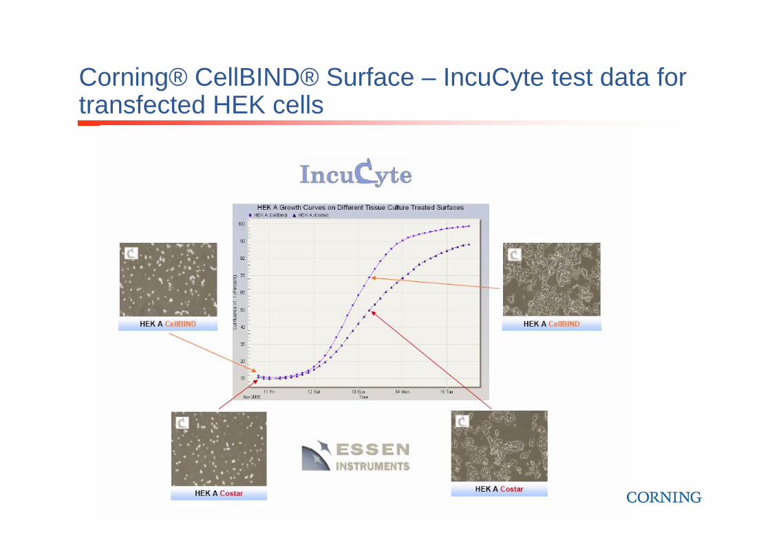
Corning® CellBIND® Surface – IncuCyte test data for transfected HEK cells

Corning® CellBIND® Surface – IncuCyte test data for transfected HEK cells

Haichen Song, Nina Santi, Øystein Evensen, and Vikram N. Vakharia: Molecular Determinants of Infectious Pancreatic Necrosis Virus Virulence and Cell Culture Adaptation; JOURNAL OF VIROLOGY, Aug. 2005, p. 10289–10299 Vol. 79, No. 16“I have so far used your CellBIND products only for one published assay. In this assay we compare the cell
culture infectivity of two different virus isolates by comparing the number of IFAT positive cells at different time points after infection. We were asked to perform this assay as a part of the revision of our paper before publishing. Since I was short of time, I had no time to do repeated experiments. I set up quite a large experiment, looking at different concentrations of virus and several different time points.
The advantage of using CellBIND compared to conventional cell culture products was that the risk of loosing cells after washing was greatly reduced. This was very important to me, since I was calculating the ratio of infected to non-infected cells, and cell loss could lead to wrong results. The CellBIND material worked very well, I do not think I lost a single cell during the staining procedures. I used the same medium and serum amount as I use for other assays, as it was only the increased binding I was after in this assay.
Since we became aware of the benefits of the CellBIND surface, we have decided to try it for certain cell lines that are not very well adapted to culture (derived from fish tissue) as well. The results so far are promising. It looks like the cells thrive better on the CellBIND surface. Lately, we have also used this surface when we isolate adherent leucocytes (macrophages) directly from the blood of Atlantic salmon brood fish to test if they are virus carriers. This also seems to work very well. I am sure we will find other applications in the future too.
I am very satisfied with your product and would not hesitate to recommend it to other scientists.”

Lucia Lazorova, Department of Pharmacy, Uppsala UniversityApplication:•to thaw adherent cell lines: Caco-2, Saos-2, HEK 293, 2/4/A1, HepG2, Calu-3 etc.
Growth conditions: We have used the same receipt as usual, thus unmodified media. Example:•A) for Caco-2: DMEM -high glucose; 10% fetal calf serum, 1% non-essential amino acids•B) 2/4/A1 medium containing only 5% fetal calf serum in DMEM/F12 medium also at thawing.
Results: cells “revived” quicker in the CellBIND flasks than compared to a standard flask (TCT). Previously we had to wait up to one week in order for the first “passage”. I do not know if this is due to the cells attaching quicker to the surface or due to faster cell proliferation. I would suspect that it is the first option as I do not see as many dead cells in the media during the media change. If this is the case than it is REALLY good as we also need the presence of more sensitive cells in our cell population in order not to select just strong survivors after every thaw as this might lead to changing the property of the cell line after a number of thaws.•Email: [email protected]

Some successes…

Can Corning® CellBIND® Surface replace biological coatings?
• Maybe, IF you use coatings only to improve cell attachment– More likely if coating is fibronectin, collagen or
gelatin
– Less likely if coating is ECM, Matrigel, or laminin
• Unlikely if you use coatings to affect cell behavior

Corning® CellBIND® Surface issues: will it change my cells, my experiments…?
The answer is maybe. Why?• There are thousands of unique cell lines• Each cell line has 100s of unique characteristics• These characteristics constantly change in
response to the environment, the medium, from evolution and from aging
HOWEVER!• Many customers have switched and found only
positive impact on their cultures.• It is a better surface but not a different surface

Corning® CellBIND® SurfaceThe new standard in cell culture
A premium surface treatment!No special handling or storage requirementsImproves attachment of many cell typesHelps when using reduced or serum-free culturesNo change in cell harvesting proceduresConsistently better surface treatment for premium cell performance
For a premium performance,
give your cells the best!
Corning CellBIND Surface

For more information...
Please check theCorning web site:www.corning.com/lifesciences
Ask for ourCell Culture Toolsbinder
Corning Guide for Identifying andCorrecting Common Cell Growth Problems

Any questionsabout
Corning® CellBIND® Surface?
Any questionsabout
Corning® CellBIND® Surface?

Talk Outline• Overview of cell culture surface treatments• Corning® CellBIND® Surface
– Physical and Chemical properties of the surface– Cell growth performance on Corning® CellBIND® Surface
• Adherence of difficult cell lines• Low or no serum conditions
– Will Corning® CellBIND® Surface benefit my cell culture work?
• Ultra Low Attachment Coating– ULA designed for…
• Q&A

Ultra Low Products: Designed for…• Maintaining stem cells in a suspended state
• Studying tissue-specific functions of certain cancer cells (i.e. MCF-7 breast cancer cells)
• Selective culture of tumor cells and virally transformed cells (normal cells do not grow unattached)
• studying activation and inactivation mechanisms of macrophages and other phagocytic cells
6 well plate 24 well plate 96 well plate
Cell Culture Dishes in 60 mm and 100 mm sizes
NEW!

Ultra Low Attachment
NEW!
Ultra Low Attachment Culture Dishes available in 60 mm and 100 mm sizes
• Covalently bonded hydrogelsurface – non-biological
• Minimizes cell attachment,protein absorption andenzyme activation.
• Surface is non-cytotoxic,biologically inert andnon-degradable.

Polystyrene surfaces
Untreated polystyrene has an uncharged hydrophobic surface
TC-treated polystyrene has a charged, hydrophilic surface to which many proteins and cells stick
Ultra Low Attachment surface has a neutral, hydrophilic coating to which almost no proteins or cells stick
Untreated Polystyrene
Polystyrene with ULA Surface
+ + + + + + + + + + + + + + +TC-treated Polystyrene
Repels water
Attracts water and proteins
Attracts water but not proteins

Cell attachment in Ultra Low versus standard tissue culture plates
60
50
40
30
20
10
0Ultra Low
Attachment SurfaceStandard Tissue Culture Surface
Vero
Cel
l cou
nt
per w
ell x
106

Glioma Cell Growth on Ultra Low
C6 rat glioma cell colony (arrow) on the Corning Ultra Low Attachment surface
Glioma cell colonies on Ultra Low Attachment surface (left panel) and on a tissue culture treated polystyrene surface (right panel)

Curing Cancer with the Corning Ultra Low Attachment (ULA) Surface
• A Corning customer is developing a new vaccine to fight cancer using cell therapy
– Cells from a patient’s immune system are taken from a blood sample and grown in tissue culture
– The cells are grown on the ULA surface and “taught” how to attack specific cancer cells
– The immune system cells are injected back into the patient where they target cancer cells
• The Corning ULA surface plays a vital role in recovering the immune system cells from culture
Immune System (Dendritic) Cells used in cancer vaccine

Improving Stem Cell Research with the Corning Ultra Low Attachment (ULA) Surface
ULA Surface enables tight, well-defined, undifferentiated embryoid body formation (murine ES cells)
TCT Surface embryoid bodies attach to surface and show poor morphology and randomly differentiating cells (murine ES cells)
Conventional TCT Surface Ultra Low Attachment Surface

Improving Stem Cell Research with the Corning Ultra Low Attachment (ULA) Surface
Frequency of Hematopoietic colonies from murine EBs formed in Methylcellulose
0
0.1
0.2
0.3
0.4
0.5
0.6
0.7
Study #1 Methylcellulose cultures Study #2 Methylcellulose cultures Study #3 Methylcellulose cultures
Study ID
Hem
Col
onie
s/EB
ULA CulturesGriener CulturesNunc CulturesNT
• Internal work with CCE ES murine cell line• Data from Suspension Cultures Pending

Improving Stem Cell Research with the Corning Ultra Low Attachment (ULA) Surface• Lab. of Neural Stem Cell Biology
(Dr. Kokaia), Stem Cell Center, Lund University– ULA flasks prevent
attachment to a higher degree and thus generate bigger spheres in both rat and human neural stem cell cultures.
– “if you have any questions, please, do not hesitate to contact us!” - Therése Kallurand Henrik Ahlenius
Human Neurospheres on TCT flasks
Human Neurospheres on ULA flasks

References for ULA• The Effects of Surface Chemistry and Adsorbed Proteins on
Monocyte/macrophage adhesion to chemically Modified Polystyrene Surfaces (Shen and Horbett, 2001)– Inhibits the attachment and activation of Macrophages and
Neutrophils
• In vitro propagation and transcriptional profiling of human mammary stem/progenitor cells (Dontu et al., Genes and Development, 2003)
• Nina Wolmer at Cellular and Molecular Immunology, Department of Medicine, Karolinska University Hospital– “We used Ultra Low Attachment for proliferation assays of sphere
forming human neural stem and precursor cells. Normal cell culture treated plastic cannot be used since the cells readily stick to the surface and the spheres are disrupted. With Ultra Low Attachmenthowever, the cells can be cultured for up to one week without adhering at all and neither sphere formation or proliferation rate seem to be otherwise affected. This has proven to be a useful tool in my research and I am pleased with the performance of the product.”

Overall conclusions:Corning® CellBIND® surface• Improved cell attachment • Better consistency• Potential savings of serum
Ultra Low Attachment• Low protein binding: to maintain cells (e.g. stem
cells) in a suspended state• non-biological, non-cytotoxic, biologically inert

2006 Corning Scientific Seminar Series Online
The 2006 Scientific Seminar Series free webinars, hosted by Corning Life Sciences, provide novel tips, best practices, and proven techniques to help you with your research needs. From Cell Culture to Microarrays, these online seminars will include information regarding the latest innovations and applications being utilized by leading researchers worldwide to maximize their results.
Participating is easySimply register by visiting www.corning.com/lifesciences and follow the registration link. New seminars will be periodically added, so check
back often. All seminars are powered by Interwise®, the latest in webcast presentation technology so you can participate in the comfort of your own lab or office or even from home. Webinars will be recorded and posted for you to download and play at any time.
Seminar 17:00 - 18:00 GMT 13:00 - 14:00 GMT 18:00 - 19:00 CET 14:00 - 15:00 CET
The Impact of Microplate Attributes and Instruments on Assay Performance 7 March 9 March
Using DNA Microarrays to Study Genome Structure 4 April 6 April
Achieving More In Vivo-like Cell Cultures Using Permeable Supports 9 May 11 May
Identifying and Correcting Common Cell Growth and Attachment Problems 6 June 8 June
What attendees had to say about past seminars:"Despite having been involved in cell culture for 16 years, I still learned a number of new things from your seminar.""I use your seminars as training for new employees and estimate they save my company more than €24,000 a year in
training costs."

Further Information……
• Dr. Ute Vespermann, Field Applications Scientist• [email protected]• Cell: +49-170-935 00 17

Thanks for your participation
Any questions?Any questions?