new mechanism of hepatic fibrogenesis: hepatitis c virus...
TRANSCRIPT

New Mechanism of Hepatic Fibrogenesis: Hepatitis C Virus InfectionInduces Transforming Growth Factor �1 Production through Glucose-Regulated Protein 94
Min Hyeok Jee,a Ka Young Hong,a Ji Hoon Park,a Jae Seung Lee,b Hee Sun Kim,b Song Hee Lee,a Sung Key Janga
POSTECH Biotech Center, Department of Life Sciences, Pohang University of Science and Technology, Pohang, Kyungbuk, Republic of Koreaa; Division of IntegrativeBiosciences and Biotechnology, Pohang University of Science and Technology, Pohang, Kyungbuk, Republic of Koreab
ABSTRACT
Hepatitis C virus (HCV) is one of the leading causes of chronic liver inflammatory disease (hepatitis), which often leads to moresevere diseases, such as liver fibrosis, cirrhosis, and hepatocellular carcinoma. Liver fibrosis, in particular, is a major pathogenicconsequence of HCV infection, and transforming growth factor �1 (TGF-�1) plays a key role in its pathogenesis. Several HCVproteins have been suggested to either augment or suppress the expression of TGF-�1 by HCV-infected cells. Here, we reportthat TGF-�1 levels are elevated in HCV-infected hepatocytes cultured in vitro and in liver tissue of HCV patients. Notably, thelevel of TGF-�1 in media from in vitro-cultured HCV-infected hepatocytes was high enough to activate primary hepatic stellatecells isolated from rats. This indicates that TGF-�1 secreted by HCV-infected hepatocytes is likely to play a key role in the liverfibrosis observed in HCV patients. Moreover, we showed that HCV E2 protein triggers the production of TGF-�1 by HCV-in-fected cells through overproduction of glucose-regulated protein 94 (GRP94).
IMPORTANCE
Hepatic fibrosis is a critical step in liver cirrhosis caused by hepatitis C virus infection. It is already known that immune cells,including Kupffer cells, mediate liver fibrosis. Recently, several papers have suggested that HCV-infected hepatocytes also signif-icantly produce TGF-�1. Here, we provide the first examination of TGF-�1 levels in the hepatocytes of HCV patients. Using anHCV culture system, we showed that HCV infection increases TGF-�1 production in hepatocytes. Furthermore, we confirmedthat the amount of TGF-�1 secreted by HCV-infected hepatocytes was sufficient to activate primary hepatic stellate cells. To un-derstand the molecular basis of TGF-�1 production in HCV-infected hepatocytes, we used HCV replicons and various stable celllines. Finally, we elucidated that HCV E2 triggered TGF-�1 secretion via GRP94 mediated NF-�B activation. This study contrib-utes to the understanding of liver fibrosis by HCV and suggests a new potential target (GRP94) for blocking liver cirrhosis inHCV patients.
Hepatitis C virus (HCV) is one of the leading causes of chronicinflammatory liver disease (hepatitis) (1). According to
World Health Organization reports, about 170 million peoplecurrently suffer from HCV infection worldwide (2). ChronicHCV infection often results in the development of hepatic fibro-sis, cirrhosis, and hepatocellular carcinoma (HCC) (3).
HCV is a positive-sense, single-stranded RNA virus whose9.6-kb genome encodes at least 10 viral proteins, including fourstructural and six nonstructural proteins (4). Error-prone RNA-dependent RNA polymerase (RDRP) generates a wide variety ofmutations (5), which complicates development of HCV vaccines(6). Several therapeutic agents have recently been developed forHCV-related chronic hepatitis, including direct-acting antivirals(DAAs) that target HCV viral proteins (7, 8).
Liver fibrosis is known to be tightly linked with HCV infectionand ultimately leads to liver dysfunction and cirrhosis (9). Liverfibrosis is involved in the process of healing wounds caused bychronic inflammation (one of the prominent features of HCVinfection-related pathogenesis) and the death of HCV-infectedhepatocytes (10). In fibrotic states, liver tissue shows abnormalaccumulation of extracellular matrix (ECM) and changes in ECMcomposition (11, 12).
Generally, liver fibrosis progression is promoted by activatedhepatic stellate cells (HSCs) (13). In the normal liver, HSCs residein the sinusoidal space in a quiescent state (14), but upon activa-
tion by cytokines, they secrete large amounts of ECM (9). Trans-forming growth factor �1 (TGF-�1), a multifunctional cytokinethat controls growth, adhesion, migration, apoptosis, and differ-entiation of cells (15), is the most important cytokine involved infibrogenesis (16). During liver fibrosis, TGF-�1 induces trans-differentiation of quiescent HSCs to myofibroblasts, which, inturn, produce other cytokines and matrix proteins (17).
It has been suggested that Kupffer cells, activated by the con-tinuous liver injury caused by HCV infection, are the main sourceof TGF-�1 (18). However, there have been several reports suggest-ing that HCV-infected hepatocytes themselves secrete TGF-�1(19–21). Elevation of reactive oxygen species (ROS) in HCV-in-fected cells was suggested as a mechanism for the TGF-�1 produc-
Received 29 November 2015 Accepted 23 December 2015
Accepted manuscript posted online 30 December 2015
Citation Jee MH, Hong KY, Park JH, Lee JS, Kim HS, Lee SH, Jang SK. 2016. Newmechanism of hepatic fibrogenesis: hepatitis C virus infection inducestransforming growth factor �1 production through glucose-regulated protein 94.J Virol 90:3044 –3055. doi:10.1128/JVI.02976-15.
Editor: J.-H. J. Ou
Address correspondence to Sung Key Jang, [email protected].
Copyright © 2016, American Society for Microbiology. All Rights Reserved.
crossmark
3044 jvi.asm.org March 2016 Volume 90 Number 6Journal of Virology
on May 29, 2018 by guest
http://jvi.asm.org/
Dow
nloaded from

tion in HCV-infected cells (21), and HCV core protein was shownto be responsible for TGF-�1 secretion (19, 20). In contrast, sev-eral reports have shown that HCV viral protein(s) repressesTGF-�1 signaling and/or TGF-�1 gene expression (22, 23). Forinstance, NS5A protein was shown to inhibit TGF-�1 signaling(22). Moreover, NS5A protein was suggested to inhibit the func-tion of activating protein 1 (AP-1), a transcriptional activator re-quired for TGF-�1 gene expression (24), by perturbing Ras-ERKsignaling pathway (23).
In this study, we investigated the molecular basis of the fibro-genesis induced by HCV infection. Particularly, we sought toidentify HCV protein(s), if it exists, that augments TGF-�1 pro-duction by HCV-infected cells. Using molecular biological andbiochemical approaches, we found that HCV-infected hepato-cytes secrete TGF-�1 proteins which are sufficiently high to acti-vate primary HSCs. Moreover, high level of TGF-�1 was observedfrom HCV-infected liver cells of HCV patients. Our investigationof the molecular basis of TGF-�1 production in HCV-infectedcells revealed that HCV core protein and envelope protein 2 (E2)participate in elevated TGF-�1 production. We also demon-strated that overproduction of glucose-regulated protein 94(GRP94) by HCV E2 triggers the signal transduction cascades thatresult in the elevated TGF-�1 production.
MATERIALS AND METHODSCell culture. Huh-7 and Huh-7.5.1 cells were cultured at 37°C in Dulbec-co’s modified Eagle medium (DMEM; Gibco) supplemented with antibi-otics (100 U/ml of penicillin and 10 �g/ml of streptomycin) and 10% fetalbovine serum (FBS; HyClone) in a humidified 5% CO2 environment.Huh-7 cells containing HCV replicons were grown under the same con-ditions, with the additional inclusion of the antibiotic G418 (500 �g/ml;AG Scientific) (25). Huh-7 cells expressing HCV E2 or GRP94 were cul-tured under the same conditions, with the additional inclusion of theantibiotic hygromycin B (300 �g/ml; AG Scientific) (25, 26).
Western blotting. Cells were lysed in passive lysis buffer (Promega).Proteins in lysates were resolved by sodium dodecyl sulfate-polyacryl-amide gel electrophoresis (SDS-PAGE) followed by transfer to nitro-cellulose membranes (Whatman). Antibodies against GRP94, NS5B,and �-smooth muscle actin (�-SMA; Santa Cruz), �-actin (ICN), andTGF-�1 (BD Biosciences) were used as primary antibodies. Themonoclonal anti-E2 antibody H52 was a kind gift from Jean Dubuis-son (University of Lille), and an antibody against core protein waskindly provided by Ralf Bartenschlager (University of Heidelberg).Horseradish peroxidase (HRP)-conjugated donkey anti-rabbit IgG, sheepanti-mouse IgG (GE Healthcare), and donkey anti-goat IgG (Santa Cruz)were used as secondary antibodies. Chemiluminescence detection wasperformed using WEST-ZOL plus (iNtRON).
siRNA transfection. Knockdown of GRP94 was performed by trans-fection with a specific small interfering RNA (siRNA) duplex targetingGRP94 (5=-UGAUGUGGAUGGUACAGUA-3=; Bioneer) using Oligo-fectamine (Invitrogen) (26). Cells were incubated for 4 h, after whichserum-free medium was replaced with DMEM containing 10% FBS.
qRT-PCR. Total RNA was isolated with TRI-solution (BioscienceTechnology). cDNA synthesis was performed by reverse transcriptionreagent (Promega). Quantitative reverse transcription-PCR (qRT-PCR) was performed with the TaKaRa SYBR Premix EX Taq II proto-col using an IQ5 multicolor real-time PCR detection system (Bio-Rad). Sequences of primer pairs for qRT-PCR are as follows: TGF-�1,5=-GGC GAT ACC TCA GCA ACC G-3 and 5=-CTA AGG CGA AAGCCC TCA AT-3=; HCV, 5=-TCT GCG GAA CCG GTG AGT A-3= and5=-TCA GGC AGT ACC ACA AGG C-3=; and glyceraldehyde-3-phos-phate dehydrogenase (GAPDH), 5=-AGG GCT GCT TTT AAC TCTGGT-3= and 5=-CCC CAC TTG ATT TTG GAG GGA-3=.
Quantification of TGF-�1 by ELISA. Huh-7 cells were seeded in a100-mm dish and cultured for 12 h. After three washings with phosphate-buffered saline (PBS), fresh serum-free medium was added. Cells werethen incubated for 24 h, after which culture media were collected andfiltered through a 0.2-�m Millipore filter. The amount of secretedTGF-�1 in the culture medium was determined using a human TGF-�1enzyme-linked immunosorbent assay (ELISA) kit (R&D Systems) accord-ing to the manufacturer’s protocol.
Immunofluorescence assays. The subcellular localization of TGF-�1and HCV core protein was monitored using anti-TGF-�1 (BD Biosci-ences) and anti-core (Affinity Bioreagents) antibodies, respectively. Forimmunocytochemistry, HCV-inoculated and uninoculated Huh-7.5.1cells were fixed with 4% paraformaldehyde (10 min) and then incubatedfor 0.5 h in blocking solution containing 1% bovine serum album (BSA)and 0.1% Tween 20 in PBS to permeabilize cells and block nonspecificbinding of antibodies. Cells were then washed with PBS and incubated atroom temperature for 1 h with primary antibodies. After a washing withPBS, cells were incubated with secondary antibodies for 1 h.
For immunohistochemistry, paraffin-embedded liver tissue speci-mens were deparaffinized and rehydrated with xylene and ethanol. Anti-genic epitopes of samples were exposed by treatment with 10 mM citratebuffer and heating in a microwave oven. Samples were incubated in block-ing solution containing 5% horse serum and 0.02% Triton X-100 in Tris-buffered saline (TBS) at room temperature for 2 h and then incubatedovernight at 4°C with primary antibodies. After a washing with TBS con-taining 0.01% Triton X-100, the samples were incubated with secondaryantibodies (Invitrogen; Jackson ImmunoResearch) for 2 h. Immuno-stained samples were observed under an Olympus FV1000 confocal laserscanning microscope.
Quantification of the imaging data. Images were analyzed usingMetaMorph software. The data from immunocytochemical study of 134cells and the data from immunohistochemical study of 57 cells were ana-lyzed using the software. The fluorescence intensities (TGF-�1, red; HCVcore, green; and Hoechst, blue) of cells were measured by the linescan toolin MetaMorph software. To calculate the level of protein expression incells, the sum of fluorescence intensities (TGF-�1, red, and HCV core,green) was divided by the sum of Hoechst intensities in the correspondingcells. The average value of fluorescence intensity in each group was calcu-lated by dividing the sum of protein level in the group by the number ofcells belonging to the group. The version of MetaMorph used is 7.04r4.
Virus infection and production. In vitro transcription of HCV RNA(derived from JFH-1) and transfection of RNAs were performed as de-scribed previously (27). Infectious HCV particles were collected from theculture media of Huh-7.5.1 cells 3 days after transfection with HCV RNA.The levels of TGF-�1 in the media of HCV-infected Huh-7.5.1 cells weremeasured 3 weeks after HCV infection using a TGF-�1 ELISA kit.
Isolation of HSCs. Primary HSCs were isolated from the livers of maleSprague-Dawley rats according to an established method (28). Briefly, ratlivers were perfused with Ca2�- and Mg2�-free Hanks’ balanced salt so-lution (HBSS) containing 0.025% collagenase B. The resulting liver sus-pension was incubated at 37°C for 20 min, and HSCs were separated bycentrifugation with an 11.9% Histodenz (Sigma) cushion. The purity ofHSC isolates was greater than 90%, as assessed by fluorescence of ananti-GFAP (glial fibrillary acidic protein) antibody (Abcam) under UVexcitation (29). Rats were maintained under specific-pathogen-free con-ditions. All animal procedures were approved by the Animal Care Com-mittee of POSTECH Biotech Center.
Detection of HSC activation. HSCs were cultured in DMEM contain-ing 3% FBS. After 3 days of incubation, HSCs were transferred to 6-wellplates and cultured for 12 h. The cells were further cultured with serum-free DMEM for 24 h. The culture media were replaced with the incubationmedia of various cells with or without pretreatment of antibodies for 4 h.After being cultured for an additional 10 h, HSCs were collected and lysedwith passive lysis buffer (Promega). A recombinant human TGF-�1 pro-tein (R&D Systems) was used as a positive control for activation of HSCs.
HCV E2 Enhances TGF-�1 Secretion
March 2016 Volume 90 Number 6 jvi.asm.org 3045Journal of Virology
on May 29, 2018 by guest
http://jvi.asm.org/
Dow
nloaded from

Liver tissue samples. Liver tissue samples were donated by liver cancerpatients who provided informed consent, and the utilization of specimensfor this research was authorized by the institutional review boards of theuniversities which provided the samples. Tissue sample number 06-30414N (a nontumor region of a liver cancer patient who was not infectedwith HCV), numbers N1 and N2 (nontumor regions of hepatitis B virus[HBV] patients), and numbers 375N, 388N, 02-27024N, 03-10521N, and06-9008N (nontumor regions of HCV patients) were provided by theLiver Cancer Specimen Bank, supported by National Research ResourceBank Program of the Korea Science and Engineering Foundation in theMinistry of Science and Technology. Tissue sample numbers s10-6410-Band s09-20494 (nontumor regions of HCV patients) were provided byEwha Womans University.
RESULTSHCV infection increases TGF-�1 secretion by hepatocytes. Todetermine whether HCV infection affects the secretion of TGF-�1by hepatocytes, we measured the amount of TGF-�1 in the culturemedia of Huh-7.5.1 cells after HCV (JFH-1) infection using aTGF-�1 ELISA. The level of TGF-�1 in the culture media fromHCV-infected cells (3 weeks after infection with HCV at a multi-plicity of infection [MOI] of 0.1) was increased �2-fold comparedwith that in culture media from mock-infected cells (Fig. 1A).HCV infection was monitored by Western blotting of cell extractswith an antibody against HCV core protein (Fig. 1A).
We also investigated the acute effect of HCV infection onTGF-�1 gene expression by inoculation with a high titer of HCV(an MOI of 5, ensuring infection of more than 99% of the cells).Surprisingly, both TGF-�1 mRNA and pro-TGF-�1 protein (aprecursor of TGF-�1) were increased �4-fold in the HCV-in-fected cells at 2 days after infection (Fig. 1B and C, respectively).The results indicated that HCV infection stimulated the produc-tion and secretion of TGF-�1 by hepatocytes.
TGF-�1 protein levels are high in HCV-infected cells and inneighboring cells. TGF-�1 levels in individual hepatocytes in-fected with JFH-1 were investigated using fluorescence immuno-cytochemistry. HCV-infected cells were detected using an anti-body against HCV core protein, and intracellular TGF-�1 wasstained using a specific anti-TGF-�1 antibody. Much higher levelsof TGF-�1 (red) were observed in HCV-infected cells (green)than in uninfected cells (compare Fig. 2A-1 and A-2). In order tocompare the levels of TGF-�1 in individual cells in the HCV-inoculated sample, we further analyzed the immunocytochemis-try data shown in Fig. 2A-2 using MetaMorph software (Fig. 2B).We classified HCV-inoculated cells into three groups: (i) HCV-infected cells, which showed high levels of HCV core protein (Fig.2B-1); (ii) uninfected neighboring cells, which showed undetect-able levels of HCV core protein but were in direct contact withHCV-infected cells (Fig. 2B-2); and (iii) uninfected nonneighbor-ing cells, which did not show HCV core protein and were not incontact with HCV-infected cells (Fig. 2B-3). The intensity of sig-nals emitted by HCV core (green), TGF-�1 (red), and Hoechst(blue) along transects through individual cells, indicated by redlines in the left images in Fig. 2B, are depicted in the traces in theright images in Fig. 2B. These data indicate that high levels ofTGF-�1 proteins were present in the cytoplasm of HCV-infectedcells and that TGF-�1 protein was not colocalized with HCV coreproteins even though both proteins were found in the cytoplasm.Statistical analyses of TGF-�1 protein in individual cells (Tables 1and 2) showed that the average amount of TGF-�1 protein inHCV-infected cells was more than 20-fold higher than that in
uninoculated cells (Table 1) and that the average amounts ofTGF-�1 protein in uninfected cells neighboring HCV-infectedcells and in uninfected nonneighboring cells were 5.6- and 2.8-fold higher than that in mock-inoculated cells, respectively (Table1). Many HCV-infected cells showed more than 30-fold-higherrelative TGF-�1 levels (RTLs) (Table 2), whereas none of the un-
FIG 1 HCV infection induces TGF-�1 secretion. (A) TGF-�1 levels in theculture media of Huh-7.5.1 cells, infected with HCV (JFH-1) or mock infected,were examined by ELISA. Huh-7.5.1 cells were infected with HCV (MOI, 0.1)and maintained for 3 weeks; then the same numbers of HCV-infected cells andmock-infected cells (7 � 105) were seeded on 100-mm dishes and cultured for12 h. The media were changed to DMEM without FBS, and the cells werecultured for an additional 24 h. Culture media were collected and analyzed byELISA after being filtered through a 0.2-�m-pore filter. Data are presented asmeans and standard deviations (bars and error bars, respectively) from threeindependent experiments. HCV infections were confirmed by Western blot-ting for HCV core protein. The extracts from mock-infected (lane 1) andHCV-infected (lane 2) Huh-7.5.1 cells were resolved by SDS-PAGE on 12%gels and analyzed by Western blotting with antibodies against HCV core pro-tein and �-actin (used as a loading control). (B) HCV infection inducesTGF-�1 mRNA production. Huh-7.5.1 cells were inoculated with HCV (JC1E2-Flag; MOI, 5) and cultivated for 2 days. The amounts of TGF-�1 mRNA inthe HCV-infected (lane 2) and mock-infected cells were measured by qRT-PCR using the total RNAs isolated by TRI-solution method (Bioscience Tech-nology). The amount of TGF-�1 mRNA was normalized by that of GAPDHmRNA. The relative amounts of TGF-�1 mRNA in the HCV-infected cells andthe mock-infected cells are depicted. The experiments were performed threetimes, and the means and standard deviations are depicted by bars and errorbars, respectively. (C) HCV infection augments production of pro-TGF-�1 (aprecursor of TGF-�1) and GRP94. Huh-7.5.1 cells were inoculated with HCV(JC1 E2-Flag; MOI, 5) and cultivated for 2 days (lane 2) or 4 days (lane 4).Mock-infected cells were cultivated for 2 days (lane 1) or 4 days (lane 3) asnegative controls. The amounts of HCV E2, pro-TGF-�1, GRP94, and �-actinproteins were monitored by Western blotting analyses using correspondingantibodies. Asterisks in the figures indicate P values: *, P � 0.05; **, P � 0.001.
Jee et al.
3046 jvi.asm.org March 2016 Volume 90 Number 6Journal of Virology
on May 29, 2018 by guest
http://jvi.asm.org/
Dow
nloaded from

infected cells in the HCV-inoculated sample showed RTLs greaterthan 20-fold (Table 2). The relationship between HCV infectionand TGF-�1 production was evaluated by calculating the Pearsoncorrelation coefficient from the MetaMorph analysis results (RTL
values) shown in Table 2. The Pearson correlation coefficientvalue (0.88) indicates that HCV infection and TGF-�1 productionare highly correlated. The biochemical and immunocytochemicalresults indicate that TGF-�1 expression was augmented in HCV-
FIG 2 TGF-�1 expression is augmented in HCV-infected cells and their neighboring cells. (A) Huh-7.5.1 cells were inoculated or mock inoculated with HCV(JFH-1; MOI, 0.1) and then cultivated for 3 days. The uninoculated (A-1) and HCV-inoculated (A-2) samples were stained with Hoechst 33342 (blue) andimmunostained with antibodies against HCV core (green) and TGF-�1 (red) proteins. Representative fluorescence microscopic images of TGF-�1, HCV coreprotein (Core), and nuclei (Hoechst) are shown. Merged images are also presented. Scale bar 10 �m. (B) Quantitative analyses of fluorescence intensities ofTGF-�1 and HCV core protein signals in HCV-infected cells (B-1), uninfected neighboring cells (B-2), and nonneighboring cells (B-3) in HCV-inoculatedsamples and uninfected cells in uninoculated samples (B-4) were performed using MetaMorph software.
HCV E2 Enhances TGF-�1 Secretion
March 2016 Volume 90 Number 6 jvi.asm.org 3047Journal of Virology
on May 29, 2018 by guest
http://jvi.asm.org/
Dow
nloaded from

infected cells. Moreover, the elevated RTL of uninfected non-neighboring cells compared with the RTL of uninoculated, unin-fected cells suggests that one or more cytokines secreted fromHCV-infected cells stimulated expression of TGF-�1 in HCV-uninfected cells in the same culture dish.
HCV-infected liver cells of HCV patients express high levelsof TGF-�1. We next investigated whether HCV infection resultsin overproduction of TGF-�1 protein in the liver cells of HCVpatients as it does in the in vitro culture system. For this purpose,we performed Western blot analysis with nontumor regions ofliver tissues from liver cancer patients with (Fig. 3A, lanes 2 to 6)or without (lane 1 in Fig. 3A) HCV infection. Clinical back-grounds of the specimens are summarized in Materials and Meth-ods. TGF-�1 protein was highly expressed in the nontumor re-gions of liver tissues of HCV patients. On the other hand, TGF-�1protein was not detectable from the nontumor region of a livertissue of a liver cancer patient who was not infected with HCV(Fig. 3A, compare lane 1 with lanes 2 to 6).
In order to investigate TGF-�1 expression in the individualcells of HBV and HCV patients’ liver tissues, we performed im-munohistochemical studies using nontumor regions of liver tis-sues of HBV and HCV patients (Fig. 3C). To monitor HCV-in-fected cells and TGF-�1 levels, we used antibodies against HCVcore protein (green) and TGF-�1 (red), respectively. As expected,core-positive signals were detected in HCV patients’ tissues (Fig.3C, panels j, n, and r) but not in HBV patients’ tissues (Fig. 3C,panels b and f). Consistent with in vitro HCV culture system re-sults, HCV-infected cells in liver tissue from patients expressedhigh levels of TGF-�1 (Fig. 3C, panels i to t). In contrast, TGF-�1signals were much weaker in cells negative for HCV core protein(noninfected cells) (Fig. 3C, panels i to t) and were very weak incells from HBV patients’ tissues (Fig. 3C, panels a to h). We esti-mated the amounts of TGF-�1 and HCV core proteins in theindividual cells of HBV and HCV patients using the immunohis-tochemical data shown in Fig. 3C with MetaMorph software sim-ilarly to the cases with Fig. 2B and Tables 1 and 2. In this analysis,we classified the liver cells into three groups: group 1 consisted ofHBV-infected cells, which were in the nontumor regions of HBVpatients’ liver tissues; group 2 consisted of HCV-infected cells,which were in the nontumor regions of HCV patients’ liver tissuesand showed high levels of HCV core protein; and group 3 con-sisted of uninfected cells which were in the nontumor regions ofHCV patients’ liver tissues but did not show HCV core protein.
The average amount of TGF-�1 protein in group 2 was �14-foldhigher than that in group 1. We further investigated the relation-ship between HCV infection and TGF-�1 production by calculat-ing the Pearson correlation coefficient using the estimatedamounts of TGF-�1 and HCV core proteins in the immunohisto-chemical data. The Pearson correlation coefficient (0.94) indicatesthat HCV infection and TGF-�1 production were very highly cor-related. These results suggest that one or more HCV proteinsstimulated the production and/or stabilization of TGF-�1 proteinin the HCV-infected cells of liver tissue from HCV patients.
HCV core and E2 proteins enhance TGF-�1 secretion byHCV replicon-containing cells. Results from our in vitro HCVculture system and our investigation of patients’ liver tissuesshowed that HCV-infected hepatocytes produced more TGF-�1protein than uninfected cells (Fig. 1 to 3). To understand the mo-lecular basis of the elevated TGF-�1 expression in HCV-infectedcells, we sought to identify the HCV protein(s) responsible for theoverproduction of TGF-�1. In order to mimic HCV infection, weused various HCV replicons expressing various structural pro-teins of HCV (core, E1, and E2). The replicons Core(UAA),E1(UAA), and E2(UAA) direct expression of core, core plus E1,
TABLE 2 Numbers of cells showing various levels of TGF-�1 in thedifferent groupsa
Sample type Cell categoryRelative TGF-�1level (RTL) Cell no.
HCV-inoculatedsample
HCV-infected cells Subtotal 8740 � RTL 530 � RTL � 40 1220 � RTL � 30 2610 � RTL � 20 295 � RTL � 10 11RTL � 5 4
Uninfected neighboringcells
Subtotal 1240 � RTL 030 � RTL � 40 020 � RTL � 30 010 � RTL � 20 15 � RTL � 10 5RTL � 5 6
Uninfected nonneighboringcells
Subtotal 2540 � RTL 030 � RTL � 40 020 � RTL � 30 010 � RTL � 20 05 � RTL � 10 1RTL � 5 24
Uninoculated Uninfected cells Subtotal 1040 � RTL 030 � RTL � 40 020 � RTL � 30 010 � RTL � 20 05 � RTL � 10 0RTL � 5 10
Total 134a The amounts of TGF-�1 proteins in the cells were estimated by MetaMorph softwareusing the immunocytochemical data shown in Fig. 2B and data not shown (total of 134cells). The relative amount of TGF-�1 proteins (RTL) in the cells was calculated bysetting the average amount of TGF-�1 proteins in the uninoculated, uninfected cells as1.
TABLE 1 Relative amounts of TGF-�1 protein in various groups ofcellsa
Sample type Cell category
Relative amtof TGF-�1(avg)
HCV-inoculatedsample
HCV-infected cells 21.8Uninfected neighboring
cells5.6
Uninfected nonneighboringcells
2.8
Uninoculatedsample
Uninfected cells 1.0
a The amounts of TGF-�1 proteins in the cells were estimated by MetaMorph softwareusing the immunocytochemical data shown in Fig. 2B and immunocytochemical datanot shown (total of 134 cells). The amount of TGF-�1 in the uninoculated, uninfectedcells was set to 1 in normalization.
Jee et al.
3048 jvi.asm.org March 2016 Volume 90 Number 6Journal of Virology
on May 29, 2018 by guest
http://jvi.asm.org/
Dow
nloaded from

and core plus E1 plus E2, respectively, together with nonstructuralproteins NS3 to NS5A, as described by Lee et al. (25) (Fig. 4A); thereplicon �2 C399T expresses only nonstructural proteins. Thereplicon �2 C399T showed reduced levels of TGF-�1 protein inthe culture media compared to the culture media of controlHuh-7 cells (Fig. 4B, compare bar 2 with bar 1). This indicates thatnonstructural protein(s) of HCV inhibits expression of TGF-�1by host cells (see Discussion). Huh-7 cells containing repliconsCore(UAA) and E1(UAA) restored TGF-�1 levels to values com-
parable to those in control Huh-7 cells (Fig. 4B, compare bars 3and 4 with bar 1). This result indicates that HCV core contributesto secretion of TGF-�1. This result is consistent with a previousreport suggesting that the HCV core augments TGF-�1 throughROS generation (21). Interestingly, TGF-�1 levels in the mediawere greatly increased by the replicon E2(UAA), which producesHCV E2 (Fig. 4B, lane 5). This suggests that HCV E2 augmentsTGF-�1 expression via an unknown mechanism. To determinewhether HCV E2 is capable of enhancing TGF-�1 expression
FIG 3 HCV-infected liver cells of HCV patients express high levels of TGF-�1 protein. (A) Protein levels of HCV core, TGF-�1, GRP94, and �-actin wereanalyzed by Western blotting using nontumor regions of liver tissues from liver cancer patients with (lanes 2 to 6) or without (lane 1) infection of HCV. (B)Pearson correlation coefficients of protein levels of HCV core, TGF-�1, and GRP94 in the liver tissues were calculated by using the protein band intensities shownin panel A. (C) Nontumor regions of liver tissues from HBV patients (N1 and N2) and HCV patients (s09-20494 and s10-6410-B) were stained with Hoechst33342 (blue) or immunostained with antibodies against HCV core (green) and TGF-�1 (red) proteins. Representative fluorescence microscopic images ofTGF-�1, HCV core protein (Core), and nuclei (Hoechst) are shown. Merged images are also presented. Scale bar 10 �m.
HCV E2 Enhances TGF-�1 Secretion
March 2016 Volume 90 Number 6 jvi.asm.org 3049Journal of Virology
on May 29, 2018 by guest
http://jvi.asm.org/
Dow
nloaded from
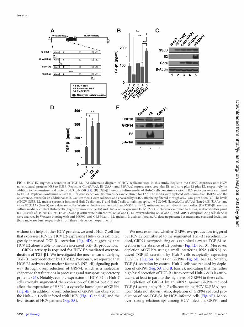
without the help of other HCV proteins, we used a Huh-7 cell linethat expresses HCV E2. HCV E2-expressing Huh-7 cells exhibitedgreatly increased TGF-�1 secretion (Fig. 4D), suggesting thatHCV E2 alone is able to mediate increased TGF-�1 production.
GRP94 activity is required for HCV E2-mediated overpro-duction of TGF-�1. We investigated the mechanism underlyingTGF-�1 overproduction by HCV E2. Previously, we reported thatHCV E2 activates the nuclear factor B (NF-B) signaling path-way through overproduction of GRP94, which is a molecularchaperone that functions in processing and transporting secretoryproteins (26). Notably, ectopic expression of HCV E2 in Huh-7cells strongly augmented the expression of GRP94 but did notaffect the expression of HSP90, a cytosolic homologue of GRP94(Fig. 4E). In addition, overproduction of GRP94 was observed inthe Huh-7.5.1 cells infected with HCV (Fig. 1C and 5E) and theliver tissues of HCV patients (Fig. 3A).
We next examined whether GRP94 overproduction triggeredby HCV E2 contributed to the augmented TGF-�1 secretion. In-deed, GRP94-overproducing cells exhibited elevated TGF-�1 se-cretion in the absence of E2 protein (Fig. 4D, bar 3). Moreover,depletion of GRP94 using a small interfering RNA (siRNA) re-duced TGF-�1 secretion by Huh-7 cells ectopically expressingHCV E2 (Fig. 5A, bar 4) or GRP94 (Fig. 5B, bar 4). Notably,TGF-�1 secretion by control Huh-7 cells was reduced by deple-tion of GRP94 (Fig. 5A and B, bars 2), indicating that the ratherhigh basal secretion of TGF-�1 from control Huh-7 cells is attrib-utable, at least in part, to the high level of GRP94 in these cells.
Depletion of GRP94 by an siRNA against GRP94 reducedTGF-�1 secretion by Huh-7 cells containing HCV E2(UAA) rep-licon (data not shown). Also, depletion of GRP94 reduced pro-duction of pro-TGF-�1 by HCV-infected cells (Fig. 5E). More-over, strong relationships among HCV infection, GRP94, and
FIG 4 HCV E2 augments secretion of TGF-�1. (A) Schematic diagram of HCV replicons used in this study. Replicon �2 C399T expresses only HCVnonstructural proteins NS3 to NS5B. Replicons Core(UAA), E1(UAA), and E2(UAA) express core, core plus E1, and core plus E1 plus E2, respectively, inaddition to the nonstructural proteins NS3 to NS5B (25). (B) TGF-�1 levels in culture media of Huh-7 cells containing various HCV replicons were examinedby ELISA. Replicon-containing cells (7 � 105) were seeded on 100-mm dishes and cultured for 12 h. The media were replaced with serum-free DMEM, and thecells were cultured for an additional 24 h. Culture media were collected and analyzed by ELISA after being filtered through a 0.2-�m-pore filter. (C) The levelsof HCV NS5B, E2, and core proteins in control Huh-7 cells (lane 1) and Huh-7 cells containing replicon �2 C399U (lane 2), Core(UAA) (lane 3), E1(UAA) (lane4), or E2(UAA) (lane 5) were determined by Western blotting analyses with anti-NS5B, anti-E2, anti-core, and anti-�-actin antibodies. (D) TGF-�1 levels inculture media of control Huh-7 cells (hygromycin-selected cells) and Huh-7 cells expressing HCV E2 or GRP94 were examined by ELISA, as described for panelB. (E) Levels of HSP90, GRP94, HCV E2, and �-actin proteins in control cells (lane 1), E2-overproducing cells (lane 2), and GRP94-overproducing cells (lane 3)were analyzed by Western blotting with anti-HSP90, anti-GRP94, anti-E2, and anti-�-actin antibodies. All data are presented as means and standard deviations(bars and error bars, respectively) from three independent experiments.
Jee et al.
3050 jvi.asm.org March 2016 Volume 90 Number 6Journal of Virology
on May 29, 2018 by guest
http://jvi.asm.org/
Dow
nloaded from

TGF-�1 in the human liver are also indicated by the high Pearsoncorrelation coefficients among these items (Fig. 3B). Taken to-gether, these data strongly suggest that HCV E2 triggers expres-sion of TGF-�1 through overproduction of GRP94.
We further investigated the role of GRP94 activity in TGF-�1secretion using 17-allylamino-demethoxygeldamycin (17-AAG),an inhibitor of GRP94 that blocks the activities of GRP94 andHSP90. Treatment with 17-AAG (250 nM) reduced secretion ofTGF-�1 protein by control Huh-7 cells as well as Huh-7 cellsectopically expressing either E2 or GRP94 (Fig. 5F, bars 2, 4, and6). Moreover, treatment of 17-AAG (250 nM) reduced TGF-�1secretion by Huh-7 cells containing the HCV E2(UAA) replicon(data not shown). These results indicate that GRP94 activity isrequired for high levels of TGF-�1 secretion by HCV-infectedcells.
Activation of NF-�B is required for the enhanced secretionof TGF-�1 protein by HCV-infected hepatocytes. Previously,we reported that GRP94 overproduction by HCV infection en-hances the expression of anti-apoptotic proteins in an NF-B-dependent manner (26). Moreover, Lin et al. reported thatTGF-�1 production is increased by HCV infection via NF-Bactivation (21). On the basis of these previous observations, weinvestigated whether the augmented secretion of TGF-�1 pro-teins by HCV E2 is mediated by NF-B activation. To this end,we examined the effect of the IKK�/� inhibitor Bay 11-7082 onTGF-�1 secretion by Huh-7 cells overproducing HCV E2 orGRP94 (Fig. 6). Treatment with Bay 11-7082 decreasedTGF-�1 secretion by both E2- and GRP94-overproducing cells(Fig. 6). Moreover, treatment with Bay 11-7082 decreasedTGF-�1 secretion by Huh-7 cells containing the HCV
FIG 5 Overproduction of GRP94 is required for E2-dependent TGF-�1 pro-duction. (A) Effect of GRP94 knockdown on the secretion of TGF-�1 fromcontrol Huh-7 cells (bars 1 and 2) and Huh-7 cells ectopically expressing HCVE2 (bars 3 and 4). Cells were seeded in 12-well plates at 4 � 104 cells/well,cultured for 12 h, and then transfected with an siRNA against GRP94 usingOligofectamine and cultured for 48 h. After culturing of cells in serum-freemedia for an additional 24 h, TGF-�1 levels in culture media were examined byELISA. (B) The effect of GRP94 knockdown on the secretion of TGF-�1 fromthe Huh-7 cells ectopically expressing GRP94 (bars 3 and 4) was determined asdescribed for panel A. (C and D) GRP94 siRNA knockdown efficiencies weremonitored by Western blotting analyses. (C) Control (lanes 1 and 2) and HCVE2-overproducing (lanes 3 and 4) Huh-7 cells described for panel A wereanalyzed by Western blotting with anti-GRP94, anti-HCV E2, and anti-�-actin antibodies. (D) Control (lanes 1 and 2) and GRP94-overproducing(lanes 3 and 4) Huh-7 cells described for panel B were analyzed by Westernblotting with anti-GRP94 and anti-�-actin antibodies. (E) Effect of GRP94knockdown on the production of pro-TGF-�1 by HCV-infected cells. Huh-7.5.1 cells were inoculated with HCV (JC1; MOI, 0.1) and cultivated for 4weeks. HCV-infected and mock-infected cells were seeded on 12-well plates (5� 104 cells/well) and cultivated for 12 h, and then the cells were transfectedwith an siRNA against GRP94 using Oligofectamine. The cells were furthercultivated for 72 h, and then the levels of HCV core, GRP94, pro-TGF-�1, and�-actin proteins in the cells were examined by Western blotting using corre-sponding antibodies. (F) Effect of the GRP94 inhibitor 17-AAG on the pro-duction of TGF-�1 by Huh-7 cells ectopically expressing GRP94 or HCV E2.Cells were seeded on 12-well plates at 5 � 104 cells/well and cultured for 12 h.After treatment with 17-AAG (250 nM) in DMEM containing 10% FBS for 12
h, cells were cultured in serum-free DMEM with 17-AAG (250 nM) for anadditional 24 h. The culture media were collected, filtered through a 0.2-�m-pore filter, and analyzed by ELISA. All data are presented as means and stan-dard deviations (bars and error bars, respectively) from three independentexperiments.
FIG 6 Activation of NF-B is required for E2-dependent TGF-�1 production.Effects of the NF-B inhibitor Bay 11-7082 on the production of TGF-�1 byHuh-7 cells ectopically expressing HCV E2 or GRP94 were analyzed. Cellswere seeded on 12-well plates at 5 � 104 cells/well and cultured for 12 h. Mediawere replaced with DMEM containing 10% FBS and Bay 11-7082 (5 �M), andcells were incubated for 12 h. After culturing of cells in serum-free mediacontaining Bay 11-7082 (5 �M) for an additional 24 h, TGF-�1 levels in cul-ture media were examined by ELISA. Data are presented as means and stan-dard deviations (bars and error bars, respectively) from three independentexperiments.
HCV E2 Enhances TGF-�1 Secretion
March 2016 Volume 90 Number 6 jvi.asm.org 3051Journal of Virology
on May 29, 2018 by guest
http://jvi.asm.org/
Dow
nloaded from

E2(UAA) replicon (data not shown). These results indicate thatNF-B activation is involved in HCV E2-dependent TGF-�1secretion, although additional research, currently ongoing inour laboratory, will be necessary to establish the mechanisticdetails.
TGF-�1 protein secreted by HCV-infected cells activates pri-mary HSCs. Activated HSCs play a key role in liver fibrosis, andTGF-�1 is the major cytokine involved in activating HSCs.Accordingly, we investigated whether TGF-�1 protein secretedby HCV-infected cells and HCV E2-overproducing cells is suf-ficient to activate HSCs. For this purpose, we isolated rat pri-mary HSCs using a perfusion and collagenase digestionmethod (28). Using these HSCs, we examined the effects ofculture media from HCV-infected cells or HCV E2-overpro-ducing cells on HSC activation by Western blotting for�-smooth muscle actin (�-SMA), an indicator of HSC activa-tion (30). Treatment with culture media (5-fold diluted) fromHCV-infected Huh-7.5.1 cells increased �-SMA in primary
HSCs about 3-fold compared with that in primary HSCstreated with control uninfected Huh-7.5.1 cell culture media(Fig. 7A, compare lane 2 with lane 1). The level of primary HSCactivation was comparable with the activation of the cells bystimulation with TGF-�1 protein at a final concentration of 2ng/ml (Fig. 7A, lane 3). Treatment with culture media fromHuh-7 cells expressing HCV E2 (5-fold diluted) also activatedprimary HSCs (Fig. 7A, lanes 4 to 6).
In order to test whether TGF-�1 protein secreted by Huh-7cells overproducing HCV E2 is the major cytokine that activatesHSCs, we examined the effect of a TGF-�1-neutralizing antibody.Addition of a TGF-�1-neutralizing antibody to culture mediafrom both control cells and HCV E2-overproducing cells reducedthe levels of �-SMA in primary HSCs (Fig. 7B). Importantly, thelevel of �-SMA was reduced to the same level as in the control cellsafter treatment of the TGF-�1 antibody (Fig. 7B and C). Thisclearly demonstrates that TGF-�1 is the key cytokine activatingHSCs by the HCV E2. Taken together, these results indicate that
FIG 7 TGF-�1 secreted by HCV E2-overproducing cells activates primary HSCs. (A) HSCs isolated from Sprague-Dawley rats were incubated in DMEMcontaining 3% FBS for 3 days. HSCs were then seeded on 6-well plates at 8 � 104 cells/well and cultured for 12 h. After culturing of cells for 24 h in serum-freemedia, media were replaced with culture media from control Huh-7.5.1 cells (lane 1) or HCV-infected Huh-7.5.1 cells (lanes 2) or with serum-free mediasupplemented with 2 ng/ml of recombinant human TGF-�1 (lane 3), and HSCs were incubated for an additional 10 h. HSCs were then harvested, and levels of�-SMA (a marker of HSC activation) and eukaryotic initiation factor 3B (eIF3B; control) were monitored by Western blotting. Similar experiments wereperformed using culture media from control Huh-7 cells (lane 4), HCV E2-overproducing cells (lane 5), and serum-free media supplemented with 2 ng/ml ofrecombinant human TGF-�1 (lane 6). The relative amounts of �-SMA in cells normalized to those of eIF3B are depicted at the top. (B) Isolated HSCs wereincubated in DMEM containing 3% FBS for 3 days, after which they were seeded on 6-well plates at 5 � 104 cells/well and cultured for 12 h. After culturing ofcells for 24 h in serum-free media, media were replaced with culture media from control Huh-7 cells (lanes 2 and 4) or HCV E2-overproducing cells (lanes 1 and3), and cells were incubated for an additional 10 h. Culture media were pretreated with either a control mouse antibody (lane 1 and 2) or neutralizinganti-TGF-�1 antibody (lanes 3 and 4) at 4°C for 4 h before culturing of HSCs. The cells were harvested, and levels of �-SMA and eIF3B were monitored byWestern blotting. The relative amounts of �-SMA in cells normalized to those of eIF3B are depicted on top of the panel. The same experiment was performedindependently two times. (C) Relative amount of �-SMA in HSCs from panel B, depicted graphically.
Jee et al.
3052 jvi.asm.org March 2016 Volume 90 Number 6Journal of Virology
on May 29, 2018 by guest
http://jvi.asm.org/
Dow
nloaded from

the amount of TGF-�1 protein produced by HCV infection issufficient to activate HSCs.
DISCUSSION
HCV infection is closely associated with liver fibrosis, a major riskfactor related to fatal liver diseases, such as liver cirrhosis and HCC(31). TGF-�1 is the most important cytokine involved in trigger-ing liver fibrosis. It has been suggested that Kupffer cells and liver-infiltrating lymphocytes are the major sources of TGF-�1 protein(32, 33). However, a number of reports have suggested that HCV-infected hepatocytes could be the original source of TGF-�1 pro-tein (21, 34, 35). In this study, we monitored the effect of HCVinfection on the production of TGF-�1 in individual hepatocytesfor the first time using an immunocytochemical method. Wefound that HCV-infected cells expressed the highest levels ofTGF-�1 but that cells neighboring HCV-infected cells also pro-duced high levels of TGF-�1 (Fig. 2 and Tables 1 and 2). More-over, TGF-�1 levels in uninfected, nonneighboring cells on thesame culture dish were higher than those in cells on an uninocu-lated culture dish. These results indicate that one or more HCVprotein(s) triggers the production of TGF-�1 in HCV-infectedcells and further suggest that protein(s) secreted from HCV-in-fected cells acts in a paracrine manner to induce TGF-�1 produc-tion in uninfected cells on the same dish (see below). Importantly,high levels of TGF-�1 protein were also observed in HCV-infectedhepatocytes in liver tissue from HCV patients (Fig. 3), suggestingthat the strong expression of TGF-�1 in HCV-infected hepato-cytes occurs under physiological conditions.
Elevated production of TGF-�1 mRNA and pro-TGF-�1 pro-tein was observed as early as 2 days after infection with HCV (Fig.1B and C). On the other hand, a detectable increase of TGF-�1proteins secreted in the media was observed only at 3 weeks afterinfection with HCV (Fig. 1A). The delayed secretion of TGF-�1may be attributable in part to the complex processes of proteolyticcleavage and secretory pathway of TGF-�1 (36, 37). Interestingly,it was reported that HCV infection reduces the expression of pro-protein convertase subtilisin/kexin type 9 (PCSK9) (38), which isalso known to mediate the proteolytic processing of TGF-�1 (39).Therefore, it is likely that the secretion of TGF-�1 is modulatedafter the production of pro-TGF-�1 by HCV infection. The regu-latory mechanism of TGF-�1 secretion from the HCV-infectedcells remains to be elucidated.
In an effort to understand the molecular basis of TGF-�1 pro-duction by HCV infection, we analyzed the effects of HCV struc-tural proteins on TGF-�1 production. To minimize artifacts re-sulting from ectopic production of a heterologous gene in a cellline, we used various HCV replicon-containing cells that mimicthe HCV infection state of hepatocytes. These replicons directexpression of nonstructural proteins NS3 to NS5B and a limitednumber of HCV structural proteins (none, core, core plus E1, orcore plus E1 plus E2) (Fig. 4A). To our surprise, TGF-�1 produc-tion by Huh-7 cells was reduced in the presence of the subgenomicreplicon expressing only the nonstructural proteins NS3 to NS5B(Fig. 4B). This indicates that one or more of the HCV nonstruc-tural proteins inhibit TGF-�1 production. We speculate thatNS5A is a potential candidate inhibitory viral protein, since itinhibits expression of the transcription activator AP-1 (23), whichis responsible for promoting TGF-�1 expression. NS5A may alsocontribute to TGF-�1 reduction by inhibiting TGF-� receptorfunction through protein-protein interactions (22).
The reduced production of TGF-�1 by HCV nonstructuralprotein(s) was restored to the original level by coexpression ofnonstructural proteins with core protein or core plus E1 (Fig. 4B).This indicates that HCV core protein augments TGF-�1 produc-tion, consistent with previous reports by Shin et al. (19) and Tani-guchi et al. (20). The generation of ROS by core protein could bethe underlying mechanism of TGF-�1 production by the coreprotein (21).
Notably, we found that HCV E2 further enhanced the secretionof TGF-�1 from the cells containing the HCV replicon E2(UAA),which expresses E2 in addition to core and E1 proteins and non-structural proteins (Fig. 4B). This suggests that HCV E2 mediatedthe enhanced TGF-�1 production by HCV-infected cells. To testthis, we generated a Huh-7 cell line ectopically expressing only E2and found that expression of HCV E2 is sufficient to trigger over-production of TGF-�1. To elucidate the mechanism underlyingE2-mediated TGF-�1 production, we investigated the role ofGRP94, since E2 is known to trigger overproduction of GRP94(26). Several lines of evidence indicated that GRP94 is responsiblefor the E2-mediated TGF-�1 production: (i) ectopic expression ofGRP94 in Huh-7 cells induced production of TGF-�1 (Fig. 4D),(ii) knockdown of GRP94 reduced production of TGF-�1 by bothcontrol Huh-7 cells and E2-expressing Huh-7 cells (Fig. 5A), (iii)TGF-�1 production was reduced by a chemical inhibitor (17-AAG) of GRP94 (Fig. 5F), (iv) depletion of GRP94 reduced pro-duction of pro-TGF-�1 by HCV-infected cells (Fig. 5E), and (v)the levels of GRP94 and TGF-�1 were highly correlated in thehuman liver tissues (Fig. 3A and B). On the basis of these obser-vations, we conclude that HCV E2 augments TGF-�1 productionby triggering overproduction of GRP94.
It is not fully understood how the overproduction of GRP94induces TGF-�1 expression. However, we previously showed thatoverproduction of GRP94 activates the NF-B signaling pathway,which results in production of anti-apoptotic proteins (26). In thisstudy, using an inhibitor of NF-B signal transduction, we dem-onstrated that NF-B activation is involved in mediating TGF-�1expression, providing a clue to the molecular mechanism linkingGRP94 to TGF-�1 production (Fig. 6). Consistent with this result,it has been shown that NF-B activation is involved in TGF-�1production in hepatocytes (21). Moreover, Chiao and colleagueshave shown that NF-B activation enhances AP-1 activation inmurine embryonic fibroblasts (MEF) (40), and Sullivan andcoworkers have suggested that increased c-Fos expression andc-Jun phosphorylation triggered by tumor necrosis factor al-pha (TNF-�) constitute the mechanism underlying TNF-�–induced TGF-�1 expression in lung fibroblasts (24). c-Jun andc-Fos proteins are components of AP-1 (24), which is a transcrip-tion factor that enhances TGF-�1 expression (41). Indeed, we alsoobserved that HCV E2-mediated overproduction of GRP94 trig-gers TNF-� secretion through the activation of NF-B (unpub-lished data) and that treatment of Huh-7 cells with TNF-� inducesTGF-�1 production (data not shown). Moreover, we have foundthat TGF-�1 secretion by Huh-7 cells expressing E2 or GRP94 isdecreased by addition of a TNF-�-neutralizing antibody to theculture media (data not shown). Therefore, we speculate that E2in HCV-infected cells indirectly induces secretion of TNF-�, andthis cytokine, in turn, triggers TGF-�1 production by HCV-in-fected cells and neighboring cells in an autocrine or paracrinemanner.
What would be the physiological consequence of TGF-�1 pro-
HCV E2 Enhances TGF-�1 Secretion
March 2016 Volume 90 Number 6 jvi.asm.org 3053Journal of Virology
on May 29, 2018 by guest
http://jvi.asm.org/
Dow
nloaded from

duction by HCV-infected hepatocytes apart from its role in thepathogenesis of hepatic fibrosis? Studies have revealed that pa-tients with chronic HCV have higher levels of regulatory T (Treg)cells—which play important roles in immunological toleranceand viral persistence (42, 43)—in the peripheral blood comparedwith healthy control subjects or patients whose HCV infectionspontaneously resolves (44, 45). Moreover, Treg cells have beenshown to suppress the proliferation of HCV-specific cytotoxic Tlymphocytes during persistent HCV infection (46, 47). There havealso been reports that HCV-infected hepatocytes directly inducethe development of Treg cells, which contribute to the impairedhost T-cell responses (34, 48). Considering that TGF-� plays apivotal role in the generation of Treg cells from precursor cells(49), we speculate that TGF-�1 produced by HCV core and E2proteins contributes to the induction of Treg cells and the induc-tion of immune tolerance against the HCV proteins.
In conclusion, as a result of our efforts to elucidate the molec-ular basis of the pathogenesis of hepatic fibrosis caused by HCVinfection, we have uncovered a novel mechanism of TGF-�1 in-duction. We found that the cellular protein GRP94, which directlyinteracts with HCV E2, plays an important role in TGF-�1 induc-tion, suggesting that GRP94 is a potential target for the develop-ment of drugs that prevent hepatic fibrosis caused by HCV infec-tion. Moreover, such a GRP94-inhibiting drug would also likelyboost immunity against HCV infection by blocking the inductionof Treg cells, which direct the immune tolerance against HCV.
ACKNOWLEDGMENTS
We thank Jean Dubuisson (University of Lille) for kindly gifting HCV E2antibody and Ralf Bartenschlager (University of Heidelberg) for provid-ing antibody against HCV core protein.
This research was supported by the Bio & Medical Technology Devel-opment Program of the NRF funded by the Korean government (MISP[2012M3A9A9054974]) and by a grant from the TEPIK, a part of theKorea Healthcare Technology R&D Project, funded by the Ministry ofHealth & Welfare, Republic of Korea (grant no. A103001).
FUNDING INFORMATIONBio & Medical Technology Development Program of NRF (Koreangovernment) provided funding to Min Hyeok Jee, Ka Young Hong, JiHoon Park, Jae Seung Lee, Hee Sun Kim, and Sung Key Jang under grantnumber 2012M3A9A9054974. A grant from TEPIK, a part of the KoreaHealthcare Technology R&D Project (Ministry of Health & welfare, Re-public of Korea) provided funding to Min Hyeok Jee, Ka Young Hong, JiHoon Park, Jae Seung Lee, Hee Sun Kim, and Sung Key Jang under grantnumber A103001.
REFERENCES1. Zeremski M, Petrovic LM, Talal AH. 2007. The role of chemokines as
inflammatory mediators in chronic hepatitis C virus infection. J ViralHepat 14:675– 687.
2. Hoofnagle JH. 2002. Course and outcome of hepatitis C. Hepatology36:S21–S29. http://dx.doi.org/10.1002/hep.1840360704.
3. Saito I, Miyamura T, Ohbayashi A, Harada H, Katayama T, Kikuchi S,Watanabe Y, Koi S, Onji M, Ohta Y, Choo Q-L, Houghton M, Kuo G.1990. Hepatitis C virus infection is associated with the development ofhepatocellular carcinoma. Proc Natl Acad Sci U S A 87:6547– 6549. http://dx.doi.org/10.1073/pnas.87.17.6547.
4. Boudreau HE, Emerson SU, Korzeniowska A, Jendrysik MA, Leto TL.2009. Hepatitis C virus (HCV) proteins induce NADPH oxidase 4 expres-sion in a transforming growth factor beta-dependent manner: a new con-tributor to HCV-induced oxidative stress. J Virol 83:12934 –12946. http://dx.doi.org/10.1128/JVI.01059-09.
5. Wang S, Buchli R, Schiller J, Gao J, VanGundy RS, Hildebrand WH,
Eckels DD. 2010. Natural epitope variants of the hepatitis C virus impaircytotoxic T lymphocyte activity. World J Gastroenterol 16:1953–1969.http://dx.doi.org/10.3748/wjg.v16.i16.1953.
6. Fauvelle C, Lepiller Q, Felmlee DJ, Fofana I, Habersetzer F, Stoll-KellerF, Baumert TF, Fafi-Kremer S. 2013. Hepatitis C virus vaccines—progress and perspectives. Microb Pathog 58:66 –72. http://dx.doi.org/10.1016/j.micpath.2013.02.005.
7. Gottwein JM, Jensen SB, Li YP, Ghanem L, Scheel TK, Serre SB,Mikkelsen L, Bukh J. 2013. Combination treatment with hepatitis C virusprotease and NS5A inhibitors is effective against recombinant genotype1a, 2a, and 3a viruses. Antimicrob Agents Chemother 57:1291–1303. http://dx.doi.org/10.1128/AAC.02164-12.
8. Pawlotsky JM. 2013. Treatment of chronic hepatitis C: current and future.Curr Top Microbiol Immunol 369:321–342.
9. Bataller R, Brenner DA. 2005. Liver fibrosis. J Clin Invest 115:209 –218.http://dx.doi.org/10.1172/JCI24282.
10. Matsuzaki K, Murata M, Yoshida K, Sekimoto G, Uemura Y, Sakaida N,Kaibori M, Kamiyama Y, Nishizawa M, Fujisawa J, Okazaki K, Seki T.2007. Chronic inflammation associated with hepatitis C virus infectionperturbs hepatic transforming growth factor beta signaling, promotingcirrhosis and hepatocellular carcinoma. Hepatology 46:48 –57. http://dx.doi.org/10.1002/hep.21672.
11. Hemmann S, Graf J, Roderfeld M, Roeb E. 2007. Expression of MMPsand TIMPs in liver fibrosis—a systematic review with special emphasis onanti-fibrotic strategies. J Hepatol 46:955–975. http://dx.doi.org/10.1016/j.jhep.2007.02.003.
12. Karadeniz G, Acikgoz S, Tekin IO, Tascylar O, Gun BD, Comert M.2008. Oxidized low-density-lipoprotein accumulation is associated withliver fibrosis in experimental cholestasis. Clinics 63:531–540.
13. Safadi R, Friedman SL. 2002. Hepatic fibrosis—role of hepatic stellate cellactivation. MedGenMed 4:27.
14. Sumpter TL, Dangi A, Matta BM, Huang C, Stolz DB, Vodovotz Y,Thomson AW, Gandhi CR. 2012. Hepatic stellate cells undermine theallostimulatory function of liver myeloid dendritic cells via STAT3-dependent induction of IDO. J Immunol 189:3848 –3858. http://dx.doi.org/10.4049/jimmunol.1200819.
15. Kim IH, Kim DG, Hao P, Wang Y, Kim SH, Kim SW, Lee SO, Lee ST.2012. Anti-fibrotic effects of L-2-oxothiazolidine-4-carboxylic acid viamodulation of nuclear factor erythroid 2-related factor 2 in rats. BMB Rep45:348 –353. http://dx.doi.org/10.5483/BMBRep.2012.45.6.276.
16. Friedman SL. 2008. Mechanisms of hepatic fibrogenesis. Gastroenterol-ogy 134:1655–1669. http://dx.doi.org/10.1053/j.gastro.2008.03.003.
17. Das SK, Vasudevan DM. 2008. Genesis of hepatic fibrosis and its bio-chemical markers. Scand J Clin Lab Invest 68:260 –269. http://dx.doi.org/10.1080/00365510701668516.
18. Choi J, Ou JH. 2006. Mechanisms of liver injury. III. Oxidative stress inthe pathogenesis of hepatitis C virus. Am J Physiol Gastrointest LiverPhysiol 290:G847–G851.
19. Shin JY, Hur W, Wang JS, Jang JW, Kim CW, Bae SH, Jang SK, YangSH, Sung YC, Kwon OJ, Yoon SK. 2005. HCV core protein promotesliver fibrogenesis via up-regulation of CTGF with TGF-beta1. Exp MolMed 37:138 –145.
20. Taniguchi H, Kato N, Otsuka M, Goto T, Yoshida H, Shiratori Y,Omata M. 2004. Hepatitis C virus core protein upregulates transforminggrowth factor-beta 1 transcription. J Med Virol 72:52–59. http://dx.doi.org/10.1002/jmv.10545.
21. Lin W, Tsai WL, Shao RX, Wu G, Peng LF, Barlow LL, Chung WJ,Zhang L, Zhao H, Jang JY, Chung RT. 2010. Hepatitis C virus regulatestransforming growth factor beta1 production through the generation ofreactive oxygen species in a nuclear factor kappaB-dependent manner.Gastroenterology 138:2509 –2518.e1. http://dx.doi.org/10.1053/j.gastro.2010.03.008.
22. Choi SH, Hwang SB. 2006. Modulation of the transforming growthfactor-beta signal transduction pathway by hepatitis C virus nonstructural5A protein. J Biol Chem 281:7468 –7478. http://dx.doi.org/10.1074/jbc.M512438200.
23. Macdonald A, Crowder K, Street A, McCormick C, Saksela K, Harris M.2003. The hepatitis C virus non-structural NS5A protein inhibits activat-ing protein-1 function by perturbing ras-ERK pathway signaling. J BiolChem 278:17775–17784. http://dx.doi.org/10.1074/jbc.M210900200.
24. Sullivan DE, Ferris M, Nguyen H, Abboud E, Brody AR. 2009. TNF-alpha induces TGF-beta1 expression in lung fibroblasts at the transcrip-
Jee et al.
3054 jvi.asm.org March 2016 Volume 90 Number 6Journal of Virology
on May 29, 2018 by guest
http://jvi.asm.org/
Dow
nloaded from

tional level via AP-1 activation. J Cell Mol Med 13:1866 –1876. http://dx.doi.org/10.1111/j.1582-4934.2008.00647.x.
25. Lee SH, Kim YK, Kim CS, Seol SK, Kim J, Cho S, Song YL, Barten-schlager R, Jang SK. 2005. E2 of hepatitis C virus inhibits apoptosis. JImmunol 175:8226 – 8235. http://dx.doi.org/10.4049/jimmunol.175.12.8226.
26. Lee SH, Song R, Lee MN, Kim CS, Lee H, Kong YY, Kim H, Jang SK.2008. A molecular chaperone glucose-regulated protein 94 blocks apop-tosis induced by virus infection. Hepatology 47:854 – 866. http://dx.doi.org/10.1002/hep.22107.
27. Barua S, Chadman KK, Kuizon S, Buenaventura D, Stapley NW,Ruocco F, Begum U, Guariglia SR, Brown WT, Junaid MA. 2014.Increasing maternal or post-weaning folic acid alters gene expression andmoderately changes behavior in the offspring. PLoS One 9:e101674. http://dx.doi.org/10.1371/journal.pone.0101674.
28. Ramm GA. 1998. Isolation and culture of rat hepatic stellate cells. J Gas-troenterol Hepatol 13:846 – 851. http://dx.doi.org/10.1111/j.1440-1746.1998.tb00747.x.
29. Morini S, Carotti S, Carpino G, Franchitto A, Corradini SG, Merli M,Gaudio E. 2005. GFAP expression in the liver as an early marker of stellatecells activation. Ital J Anat Embryol 110:193–207.
30. Hellerbrand C, Stefanovic B, Giordano F, Burchardt ER, Brenner DA.1999. The role of TGFbeta1 in initiating hepatic stellate cell activation invivo. J Hepatol 30:77– 87. http://dx.doi.org/10.1016/S0168-8278(99)80010-5.
31. Pereira Tde A, Witek RP, Syn WK, Choi SS, Bradrick S, Karaca GF,Agboola KM, Jung Y, Omenetti A, Moylan CA, Yang L, Fernandez-Zapico ME, Jhaveri R, Shah VH, Pereira FE, Diehl AM. 2010. Viralfactors induce Hedgehog pathway activation in humans with viral hepa-titis, cirrhosis, and hepatocellular carcinoma. Lab Invest 90:1690 –1703.http://dx.doi.org/10.1038/labinvest.2010.147.
32. Purohit V, Brenner DA. 2006. Mechanisms of alcohol-induced hepaticfibrosis: a summary of the Ron Thurman Symposium. Hepatology 43:872– 878. http://dx.doi.org/10.1002/hep.21107.
33. Li S, Vriend LE, Nasser IA, Popov Y, Afdhal NH, Koziel MJ, SchuppanD, Exley MA, Alatrakchi N. 2012. Hepatitis C virus-specific T-cell-derived transforming growth factor beta is associated with slow hepaticfibrogenesis. Hepatology 56:2094 –2105. http://dx.doi.org/10.1002/hep.25951.
34. Hall CH, Kassel R, Tacke RS, Hahn YS. 2010. HCV� hepatocytes inducehuman regulatory CD4� T cells through the production of TGF-beta.PLoS One 5:e12154. http://dx.doi.org/10.1371/journal.pone.0012154.
35. Lee HC, Sung SS, Krueger PD, Jo YA, Rosen HR, Ziegler SF, Hahn YS.2013. Hepatitis C virus promotes T-helper (Th)17 responses through thy-mic stromal lymphopoietin production by infected hepatocytes. Hepatol-ogy 57:1314 –1324. http://dx.doi.org/10.1002/hep.26128.
36. Constam DB. 2014. Regulation of TGFbeta and related signals by precur-sor processing. Semin Cell Dev Biol 32:85–97. http://dx.doi.org/10.1016/j.semcdb.2014.01.008.
37. Zhu HJ, Burgess AW. 2001. Regulation of transforming growth factor-
beta signaling. Mo Cell Biol Res Commun 4:321–330. http://dx.doi.org/10.1006/mcbr.2001.0301.
38. Syed GH, Tang H, Khan M, Hassanein T, Liu J, Siddiqui A. 2014.Hepatitis C virus stimulates low-density lipoprotein receptor expressionto facilitate viral propagation. J Virol 88:2519 –2529. http://dx.doi.org/10.1128/JVI.02727-13.
39. Leitlein J, Aulwurm S, Waltereit R, Naumann U, Wagenknecht B,Garten W, Weller M, Platten M. 2001. Processing of immunosuppressivepro-TGF-beta 1,2 by human glioblastoma cells involves cytoplasmic andsecreted furin-like proteases. J Immunol 166:7238 –7243. http://dx.doi.org/10.4049/jimmunol.166.12.7238.
40. Fujioka S, Niu J, Schmidt C, Sclabas GM, Peng B, Uwagawa T, Li Z,Evans DB, Abbruzzese JL, Chiao PJ. 2004. NF-B and AP-1 connection:mechanism of NF-B-dependent regulation of AP-1 activity. Mol Cell Biol24:7806–7819. http://dx.doi.org/10.1128/MCB.24.17.7806-7819.2004.
41. Jiang T, Qu JJ, Nishinaka T, Zhang N. 2008. Transcription factor AP-1regulates TGF-beta(1)-induced expression of aldose reductase in culturedhuman mesangial cells. Nephrology 13:212–217. http://dx.doi.org/10.1111/j.1440-1797.2007.00913.x.
42. Li S, Gowans EJ, Chougnet C, Plebanski M, Dittmer U. 2008. Naturalregulatory T cells and persistent viral infection. J Virol 82:21–30. http://dx.doi.org/10.1128/JVI.01768-07.
43. Keynan Y, Card CM, McLaren PJ, Dawood MR, Kasper K, Fowke KR.2008. The role of regulatory T cells in chronic and acute viral infections.Clin Infect Dis 46:1046 –1052. http://dx.doi.org/10.1086/529379.
44. Cabrera R, Tu Z, Xu Y, Firpi RJ, Rosen HR, Liu C, Nelson DR. 2004.An immunomodulatory role for CD4(�)CD25(�) regulatory T lympho-cytes in hepatitis C virus infection. Hepatology 40:1062–1071. http://dx.doi.org/10.1002/hep.20454.
45. Sugimoto K, Ikeda F, Stadanlick J, Nunes FA, Alter HJ, Chang KM.2003. Suppression of HCV-specific T cells without differential hierarchydemonstrated ex vivo in persistent HCV infection. Hepatology 38:1437–1448. http://dx.doi.org/10.1016/j.hep.2003.09.026.
46. Boettler T, Spangenberg HC, Neumann-Haefelin C, Panther E, Ur-bani S, Ferrari C, Blum HE, von Weizsacker F, Thimme R. 2005. Tcells with a CD4�CD25� regulatory phenotype suppress in vitro pro-liferation of virus-specific CD8� T cells during chronic hepatitis Cvirus infection. J Virol 79:7860 –7867. http://dx.doi.org/10.1128/JVI.79.12.7860-7867.2005.
47. Rushbrook SM, Ward SM, Unitt E, Vowler SL, Lucas M, Klenerman P,Alexander GJ. 2005. Regulatory T cells suppress in vitro proliferation ofvirus-specific CD8� T cells during persistent hepatitis C virus infection. JVirol 79:7852–7859. http://dx.doi.org/10.1128/JVI.79.12.7852-7859.2005.
48. Ji XJ, Ma CJ, Wang JM, Wu XY, Niki T, Hirashima M, Moorman JP,Yao ZQ. 2013. HCV-infected hepatocytes drive CD4� CD25� Foxp3�regulatory T-cell development through the Tim-3/Gal-9 pathway. Eur JImmunol 43:458 – 467. http://dx.doi.org/10.1002/eji.201242768.
49. Wan YY, Flavell RA. 2007. ‘Yin-Yang functions of transforming growthfactor-beta and T regulatory cells in immune regulation. Immunol Rev220:199 –213. http://dx.doi.org/10.1111/j.1600-065X.2007.00565.x.
HCV E2 Enhances TGF-�1 Secretion
March 2016 Volume 90 Number 6 jvi.asm.org 3055Journal of Virology
on May 29, 2018 by guest
http://jvi.asm.org/
Dow
nloaded from