new insights into host specialization and mycoparasitic ... · micoparassita specifico di molte...
TRANSCRIPT

DISS. ETH Nr. 21129
New insights into host specialization and mycoparasitic interaction
between powdery mildew fungi and Ampelomyces quisqualis
A dissertation submitted to
ETH ZURICH
For the degree of
Doctor of Sciences
Presented by
DARIO ANGELI
Doctor of Agronomic Science and Technology, UniPD Padua
Born February 08th, 1976
Citizen of Mezzolombardo (IT)
Accepted on the recommendation of
Prof. Dr. Cesare Gessler, examiner
Prof. Dr. Bruce A. McDonald, co-examiner
Dr. Monika Maurhofer and Dr. Ilaria Pertot, co-examiners
2013


Table of contents
Abstract .......................................................................................................................................... 4
Riassunto ........................................................................................................................................ 6
General Introduction .................................................................................................................... 8
1.1 Natural antagonists of powdery mildew fungi ..................................................................... 9
1.1.1 Antagonistic fungi in nature......................................................................................... 10
1.1.2 Potential relevance for biocontrol ................................................................................ 14
1.2 Mycoparasitism by Ampelomyces quisqualis ..................................................................... 15
1.2.1 An overview of the biology and natural occurrence .................................................... 16
1.2.2 Biocontrol potential and its exploitation in sustainable agriculture............................. 17
1.3 Aim of the thesis ................................................................................................................. 18
1.4 Literature ............................................................................................................................. 20
Occurence of Erysiphe necator chasmothecia and their natural parasitization by Ampelomyces quisqualis in Northern Italy (Trentino Alto Adige region) ... ........................... 25
Abstract ..................................................................................................................................... 26
2.1 Introduction ......................................................................................................................... 26
2.2 Material and methods .......................................................................................................... 28
2.2.1 Study sites, assessment, and sampling ......................................................................... 28
2.2.2 Isolation, identification, morphological, and molecular characterization of isolates of Ampelomyces spp. ................................................................................................................. 30
2.2.3 Meteorological data and statistical analysis. ................................................................ 31
2.3 Results ................................................................................................................................. 32
2.3.1 Natural occurrence of E. necator ................................................................................. 32
2.3.2 Development of powdery mildew chasmothecia ......................................................... 35
2.3.3 Occurrence of Ampelomyces spp. in E. necator .......................................................... 36
2.3.4 Identification and morphological characterization of Ampelomyces spp ................... 39
2.4 Discussion ........................................................................................................................... 42
2.5 Acknowledgements ............................................................................................................. 44
2.6 Literature ............................................................................................................................. 44
Existence of different physiological forms within genetically diverse strains of Ampelomyces quisqualis ...................................................................................................................................... 48
Abstract ..................................................................................................................................... 49
3.1 Introduction ......................................................................................................................... 49
3.2 Materials and Methods ........................................................................................................ 52

Table of contents
3.2.1 Cultural, morphological and physiological characteristics .......................................... 56
3.2.2 Phylogenetic analysis ................................................................................................... 57
3.2.3 Clustering and statistical analysis ................................................................................ 58
3.3 Results ................................................................................................................................. 59
3.3.1 Cultural, morphological and physiological characteristics .......................................... 59
3.3.2 Phylogenetic analysis ................................................................................................... 67
3.4 Discussion ........................................................................................................................... 69
3.5 Acknowledgments............................................................................................................... 71
3.6 Literature ............................................................................................................................. 72
Is the mycoparasitic activity of Ampelomyces quisqualis strains related to phylogeny and hydrolytic enzyme production ................................................................................................... 75
Abstract ..................................................................................................................................... 76
4.1 Introduction ......................................................................................................................... 76
4.2 Materials and Methods ........................................................................................................ 79
4.2.1 Fungal strains and pathogens ....................................................................................... 79
4.2.2 Mycoparasitic activity: ability of A. quisqualis to reduce conidiation of powdery mildews in vivo ..................................................................................................................... 83
4.2.3 Mycoparasitic activity: intra-hyphal formation of A. quisqualis pycnidia within powdery mildews .................................................................................................................. 85
4.2.4 In vitro production of CWDEs by A. quisqualis culture filtrates ................................ 86
4.2.5 Data evaluation and statistical analysis........................................................................ 87
4.3 Results ................................................................................................................................. 88
4.3.1 Mycoparasitic activity: ability of A. quisqualis to reduce conidiation of powdery mildews in vivo ..................................................................................................................... 88
4.3.2 Mycoparasitic activity: intra-hyphal formation of A. quisqualis pycnidia within powdery mildews .................................................................................................................. 90
4.3.3 In vitro production of CWDEs by A. quisqualis culture filtrates ................................ 93
4.4 Discussion ........................................................................................................................... 98
4.5 Acknowledgment .............................................................................................................. 101
4.6 Literature ........................................................................................................................... 101
Stimulation of the conidial germination as a novel tool to improve the biocontrol potential of Ampelomyces quisqualis ........................................................................................................ 107
Abstract ................................................................................................................................... 108
5.1 Introduction ....................................................................................................................... 108
Table of contents
5.2 Materials and Methods .......................................................................................................... 111
5.2.1 Pathogen and microrganism culture........................................................................... 111
5.2.2 Stimulation of A. quisqualis under different conditions ............................................ 111
5.2.3 Biocontrol activity after stimulation of conidial spores of A. quisqualis .................. 113
5.2.4 Data evaluation and statistical analysis...................................................................... 114
5.3 Results ............................................................................................................................... 115
5.3.1 Stimulation of A. quisqualis under different conditions ............................................ 115
5.3.2 Biocontrol activity after stimulation of conidial spores of A. quisqualis .................. 119
5.4 Discussion ......................................................................................................................... 121
5.5 Acknowledgment .............................................................................................................. 124
5.6 Literature ........................................................................................................................... 124
General conclusions .................................................................................................................. 127
6.1 General conclusions .......................................................................................................... 128
6.2 Literature ........................................................................................................................... 133
Aknowledgements ..................................................................................................................... 134
Curriculum Vitae ...................................................................................................................... 135

Abstract
4
Abstract
Pycnidial fungi belonging to the genus Ampelomyces quisqualis are the most widespread natural
antagonists of Erysiphales. A. quisqualis is a specific mycoparasite of many species of
Erysiphales and the most studied biocontrol agent of powdery mildews. Genetically different A.
quisqualis strains are available from culture collections and one strain has been already
commercialized under the trade name of AQ10. Report data on the aggressiveness and
morphology and cultural patterns of A. quisqualis strains found in the literature are controversial
and incomplete. Screening is a crucial step in the selection of strains capable of providing highly
effective biocontrol. There is a need for further investigations aiming at the identification of
phenotypic markers that can be used to differentiate genetically distinct groups within A.
quisqualis in order to find more effective strains within A. quisqualis species, which differ
considerably with respect to their biocontrol effectiveness.
The first objective of the present work was to verify the presence of natural strains of A.
quisqualis in a wide viticulture area (Trentino Alto Adige region). We aimed to isolate and select
new strains better adapted to the local environmental conditions than commercial strain AQ10
and highly aggressive against Erysiphaceae for a potential development as biocontrol agents.
During a three-year survey, a limited amount of natural parasitism of E. necator by Ampelomyces
spp. (0.17−3.51 %) was observed. Pycnidia and conidia of Ampelomyces spp. parasitizing E.
necator chasmothecia were found both in conventional, organically grown on and untreated
vineyards. Some of the isolated A. quisqualis strains have conidia that are shaped differently than
those of the commercial A. quisqualis strain (AQ10) and are phylogenetically different from
AQ10.
Second objective of the thesis was to characterize several A. quisqualis strains from different
hosts and geographic regions and possessing different ITS rDNA sequences and investigate
whether the host or site of origin of the strains or their cultural, morphological and/or growth
characteristics are related to their phylogenetic group which would indicate an adaptation to the
host or geographic area. Strains were molecularly characterized by sequencing the ITS rDNA
and sequence polymorphisms were used to classify and group the strains. The results revealed
some significant variation among the selected strains, which provides evidence for the existence
Abstract
5
of different physiological forms within the A. quisqualis species. Phylogenetic analysis revealed
that these A. quisqualis strains can be classified into five different genetic groups, which
generally correlate with the fungal host of origin and morphological and growth characteristics.
Finally, the pathogenicity, virulence and host range of a group of A. quisqualis strains was
assessed on different powdery mildew agents. Strains were screened both for their ability to
colonize different powdery mildews (mycoparasitic activity) and for in vitro production of cell
wall degrading enzymes (CWDEs). This study showed a positive correlation between
mycoparasitic activity and production of chitobiases and proteases. A. quisqualis strains with
similar levels of mycoparasitic activity originate from the same host species and share an
identical ITS rDNA sequence.
In the 5th chapter it was investigated whether it is possible to enhance the efficiency of this
fungus in the biological control of powdery mildews by increasing the conidial germination rate
of the fungus. The obtained results revealed that some natural extracts can stimulate the
germination of A. quisqualis conidia and enhance its biocontrol ability of the powdery mildew.
This part of the thesis demonstrates that the conidial germination efficacy of A. quisqualis strains
is positively related to virulence against powdery mildew and can, therefore, be considered as
relevant factor in the selection of biocontrol agents.
The results obtained in this thesis provide a deeper understanding of the process of
mycoparasitism and a sound basis for developing new screening strategies for detecting highly
effective A. quisqualis strains for the biocontrol of powdery mildews. Moreover, we
demonstrated the existence of A. quisqualis strains well adapted to local environmental
conditions. Their discovery may be the starting point for their development as biocontrol agents
to control powdery mildew under the environmental conditions found in Northern countries.

Riassunto
6
Riassunto
I funghi che producono picnidi appartenenti al genere Ampelomyces quisqualis sono i più diffusi
antagonisti naturali degli Erysiphales, gli agenti causali degli oidi. A. quisqualis è un
micoparassita specifico di molte specie di Erysiphales e risulta essere l’agente di biocontrollo
degli oidi più noto. Alcuni isolati di A. quisqualis diversi geneticamente sono disponibili in
collezioni di colture ed uno di essi è già stato commercializzado con il nome di AQ10. I dati
presenti in letteratura risultati di test colturali, morfologici e di prove di aggressività con diversi
isolati di A. quisqualis sono piuttosto contrastanti ed incompleti. La selezione di isolati altamente
efficaci rappresenta un passo cruciale per il biocontrollo degli oidi. C’è la necessità di ulteriori
investigazioni che mirino all’identificazione di caratteri fenoticipi che possano essere impiegati
per la differenziazione di diversi gruppi genetici di A. quisqualis con lo scopo di individuare
isolati più efficaci all’interno della stessa specie che differiscono per la loro capacità di
biocontrollo.
Il primo obiettivo del presente lavoro era quello di verificare la presenza di isolati naturali di A.
quisqualis all’interno di un ampia area viticola (regione Trentino Alto Adige). Lo scopo era di
isolare e selezionare nuovi isolati meglio adattati alle caratteristiche climatiche locali rispetto
all’isolato commerciale AQ10 ed altamente aggressivi nei confronti dei funghi appartenenti alla
famiglia delle Erysiphaceae, da poter sviluppare quali nuovi agenti di controllo biologico.
Durante i tre anni di monitoraggio è stata osservata una ridotta parassitizzazione naturale di E.
necator con Ampelomcyes spp. (0.17−3.51 %). Chasmoteci di E. necator parassitizzati da picnidi
e conidi di Ampelomyces spp. sono stati individuati sia in vigneti biologici che incolti ma anche
in vigneti sottoposti ai trattamenti chimici convenzionali. Alcuni degli isolati di A. quisqualis
identificati producevano conidi di forma diversa rispetto a quelli dell’isolato commerciale
(AQ10) ed erano filogeneticamente diversi da AQ10.
Il secondo obiettivo della tesi era quello di caratterizzare isolati di A. quisqualis provenienti da
ospiti e regioni geografiche diverse e con una diversa sequenza ITS rDNA ed indagare se l’ospite
o regione di origine degli isolati o le loro caratteristiche colturali, morfologiche e/o di crescita
sono correlate al loro gruppo filogenetico indicando in tal caso un adattamento all’ospite oppure
all’area geografica. La caratterizzazione molecolare degli isolati è stata fatta seguendo la

Riassunto
7
metodologia descritta da Szentivanyi attraverso la determinazione della lunghezza dei
polimorfismi della regione amplificata rDNA-ITS. I polimorfismi di sequenza sono stati
utilizzati per classificare e raggruppare gli isolati. I risultati hanno rivelato molte variazioni
significative tra gli isolati selezionati, evidenziando in tal modo l’esistenza di diverse forme
fisiologiche all’interno della specie A. quisqualis. Le analisi filogenetiche hanno rivelato che
questi isolati di A. quisqualis possono essere classificati in cinque differenti gruppi genetici, i
quali in linea generale correlano con l’ospite fungino di origine e le caratteristiche morfologiche
e di crescita.
Infine, la patogenicità, virulenza e spettro d’ospite di un gruppo di isolati di A. quisqualis è stato
determinato su diversi agenti di oidio. Gli isolati sono stati selezionati sia per la capacità di
colonizzazione di diversi oidi (attività micoparassitica) sia per la produzione in vitro di enzimi
degradanti pareti cellulari (CWDEs). Questo studio ha mostrato una correlazione positiva tra
l’attività micoparassitica e la produzione di chitobiasi e proteasi. Isolati di A. quisqualis con
livelli simili di attività micoparassitica erano originati dalle stesse specie ospiti e mostravano una
sequenza ITS rDNA identica tra loro.
Un paragrafo addizionale è stato aggiunto per verificare la possibilità di migliorare l’efficacia di
questo fungo nel controllo biologico degli oidi attraverso un aumento del tasso di germinazione
dei conidi del fungo. I risultati attuali hanno rivelato l’esistenza di alcuni estratti naturali in grado
di stimolare la germinazione dei conidi di A. quisqualis e migliorare la sua capacità di
biocontrollo degli oidi. Questo studio ha dimostrato che la capacità di geminazione dei conidi di
A. quisqualis è legata a differenze di virulenza nei confronti dell’oidio e può perciò essere
considerata come un fattore importante nella selezione di nuovi efficaci agenti di biocontrollo.
Il lavoro presentato in questa tesi fornisce una più profonda comprensione del processo di
micoparassitismo e offre una base solida per lo sviluppo di nuove strategie di selezione per l’
individuazione di isolati di A. quisqualis altamente efficaci nel biocontrollo degli oidi. Inoltre,
noi abbiamo accertato l’esistenza di isolati di A. quisqualis bene adattati alle condizioni
ambientali locali e la loro scoperta potrebbe rappresentare il punto di partenza per il loro
sviluppo quali agenti di controllo biologico dell’oidio nei Paesi del Nord.
Chapter 1: Introduction
8
Chapter 1
General Introduction

Chapter 1: Introduction
9
1.1 Natural antagonists of powdery mildew fungi
Powdery mildews (Ascomycotina, Erysiphales) are some of the world’s most frequently
encountered plant pathogenic fungi and they are among the most significant plant diseases,
despite extensive research on their pathogenesis, epidemiology and control. They are often
conspicuous owing to the profuse production of conidia that give them their common name.
Among the economically important plants, important crops like grapevine, apple, strawberry, but
also cereals and several vegetables and ornamentals, grown in the field or greenhouses, are the
major targets of powdery mildew fungi. They infect leaves, stems, flowers, and fruits of nearly
10,000 species of angiosperms (Braun, 1987). Uncontrolled epidemics of Erysiphales may result
in yield losses, as well as a reduction in the quality of the production. During the winter, the
fungus survives either as mycelia in the dormant buds of grapevine or as chasmothecia, which
are the fruiting bodies arising from the sexual stage. In the spring, primary infections originating
from ascospores commonly appear randomly as scattered whitish and powdery spots on leaves
(1-3 mm in diameter) that mainly appear close to the trunk. The two main methods for disease
control currently available in crop production are repeated applications of fungicides and the use
of cultivars resistant or tolerant to powdery mildews. However, both methods have their own
limitations (Hewitt, 1998). Public attitude and environmental concerns towards the use of
pesticides as well as the development of powdery mildew strains resistant to different fungicides
have reduced the appeal of chemicals (Whipps & Lumsden, 2001; Bélanger & Benyagoub, 1997;
Ishii et al., 2001). Moreover, in some countries a number of fungicides effective against powdery
mildews are no longer registered for greenhouse production, due to restrictions in pesticide usage
(Jarvis & Slingsby, 1977; Menzies & Bélanger, 1996). Cultivars resistant or tolerant to powdery
mildew infections have been developed in a number of crops, but their use is limited, especially
in fruit and vegetable crops (Bélanger & Benyagoub, 1997). All these constraints have led to the
search of alternative methods to control powdery mildews. Non-fungicide products, such as
soluble silicon, oils, salts and plant extracts, inducing resistance in plants infected with powdery
mildews or acting as prophylactic and/or curative factors are in focus, especially in greenhouse
production (Bélanger & Benyagoub, 1997; Menzies & Bélanger, 1996; Pasini et al., 1997;
McGrath & Shishkoff, 1999). On the other hand, the exploitation of antagonistic trophic
interactions between plant inhabiting microorganisms offers an opportunity for their use in
biological control of plant diseases, yet few examples have reached the market. Microbial

Chapter 1: Introduction
10
biocontrol agents (BCAs) of plant pathogen, if carefully selected, may offer a valid alternative to
chemical fungicides in crop protection against weed, insects, and disease in both agriculture and
forestry (Cook & Baker, 1983; Andrews, 1992). This method is based on the knowledge of
natural interaction between pests or pathogen and their natural enemies. Some of the beneficial
organisms used in the biocontrol are mass produced and available for large-scale distribution in
crops with the aim to reduce damage population in organic farming. Microbial biocontrol agents
have several mechanisms to antagonize their hosts. Four different mechanisms for controlling
pests or diseases by BCAs are known: i) inhibition of the pathogen by the production of
metabolites produced by another organism (antibiosis); ii) consuption of the same resource
present in limited quantity, insufficient for the survival of both organisms (competition); iii)
direct attack on the pathogen and using it as nutrition source (mycoparasitism); iv) production of
several metabolites reducing damages due to the plant respons to the pathogen (induction of
resistence). Attempts have been made to use mycolytic bacteria, mycophagous arthropods and
other possible non-fungal biological control agents against powdery mildews, but these studies
have provided no promise of practical control to date (Kiss, 2003). The most promising
biological control trials have involved a number of fungi antagonistic to powdery mildews.
1.1.1 Antagonistic fungi in nature
The names of all the fungal species reported as natural antagonists of powdery mildews and/or
used in biocontrol experiments against them are presented in Table 1. Some of these fungi are
well-known natural antagonists of powdery mildews. For example, Ampelomyces spp. or
Tilletiopsis spp. were repeatedly isolated from plants infected with powdery mildew worldwide
(Kiss, 1998; Falk et al., 1995; Knudsen & Skou, 1993; Klecan et al., 1990; Urquhart et al.,
1994). Other fungi, such as Cephalosporium spp., Cladosporium spp. or Trichothecium spp. are
cited as being frequently associated with powdery mildew colonies (Braun, 1987). Furthermore,
Pseudozyma flocculosa is known worldwide and this species is considered one of the most
efficient biocontrol agent of powdery mildews (Paulitz & Bélanger, 2001). A number of species
included in Table 1, such as Aphanocladium album (Hijwegen & Buchenauer, 1984) were also
reported to inhibit naturally the sporulation and growth of powdery mildews, but available data
on their natural occurrence and/or biocontrol activity are very limited. Some of these data need
Chapter 1: Introduction
11
further confirmation. Other fungi, such as Trichoderma spp. (Elad, 2000; Elad et al., 1998),
Penicillium chrysogenum or Fusarium oxysporum (Hijwegen, 1988) were used in some
biocontrol trials against powdery mildews, but they were never found to be naturally associated
with powdery mildew colonies.
Due to the biotrophic nature of powdery mildews, fungal antagonists can act against them
through antibiosis and mycoparasitism only. Competition for nutrients and/or space, the third
major mechanism of microbial antagonism, is not feasible against powdery mildews. The niche
they occupy in the phyllosphere could be colonized only by other plant pathogens and certainly
not by biocontrol agents. Mycoparasitism of powdery mildews by Ampelomyces spp. is one of
the best known mechanisms of fungal antagonism. These intracellular mycoparasites suppress
the sporulation of the attacked powdery mildew mycelia and kill all the parasitized cells (Kiss,
1998; Falk et al., 1995; Hashioka & Nakai, 1980). When the sporulation rate of the pathogen is
high, the mycoparasites usually cannot stop the spread of powdery mildew colonies. In contrast,
other phyllosphere fungi acting through antibiosis, such as Pseudozyma spp. or Tilletiopsis spp.,
can kill powdery mildew colonies rapidly and completely, causing plasmolysis of their cells
(Hajlaoui et al., 1994; Choudbury et al., 1994). Some authors have proposed to enlarge the
possible modes of action of antagonists by considering induced resistance as a part of the
antagonistic effect (Bélanger & Labbé, 2002). The induction of plant defence might be a
plausible mechanism in cases when neither mycoparasitism nor antibiosis can explain the
efficacy of a biocontrol fungus (Elad, 2000; Elad et al., 1998). Most probably, in many
biocontrol agents the antagonistic effect is based on more than one mode of action (Cook &
Baker, 1983; Bélanger & Labbé, 2002). Verticillium lecanii, for example, was reported as a
mycoparasite of powdery mildews that penetrate their cells either directly or by means of
appressoria (Heintz & Blaich, 1990). However, according to recent studies, antibiosis also plays
an important role in this interaction (Bélanger & Labbé, 2002). However, in most antagonists,
the modes of action against powdery mildews are largely unknown.

Chapter 1: Introduction
12
Table 1. A list of fungi tested as potential biocontrol agents against powdery mildews.
Antagonist Powdery mildew Plant host Mode of action
Acremonium alternatum Sphaerotheca fuliginea Cucumis sativus Mycoparasitism ? a
A.strictum S. fuliginea C. sativus ?
A.lanosoniveum S. macularis Strawberry ?
Ampelomyces quisqualis Many species Many species Mycoparasitism
Aphanocladium album Erysiphe cichoracearum C. sativus ?
S. fuliginea C. sativus ?
S. pannosa Rosa sp. ?
Aspergillum fumigatus E. cichoracearum C. sativus ?
Cladosporium spongiosum Phyllactinia dalbergiae Dalbergia sissoo ?
Ph. corylea Morus alba ?
Cladosporium sp. S. fuliginea Cucurbita pepo ?
Leveillula taurica Capsicum annuum ?
Cephalosporium sp. S. fuliginea Citrullus lanatus ?
Calcarisporium arbuscola S. fuliginea C. sativus Antibiosis ?
Cladobotryum varium S. fuliginea C. sativus ?
Chaetomium spp. Podosphaera leucotricha Malus domestica ?
Drechslera spicifera E. cichoracearum C. maxima ?
Fusarium oxysporum E. cichoracearum C. maxima ?
Paecilomyces farinosus S. fuliginea C. sativus Antibiosis ?
E. marti Lupinus polyphyllus Antibiosis ?
L. taurica C. annum Antibiosis ?
Penicillium chrysogenum S. fuliginea C. sativus ?
P. fellutanum E. cichoracearum C. maxima ?
Peziza ostracoderma S. fuliginea C. sativus ?
Pseudozyma spp. S. fuliginea C. sativus Antibiosis ?
E. polygoni Trifolium pratense Antibiosis ?
S. pannosa Rosa sp. Antibiosis ?
Blumeria graminis Triticum aestivum Antibiosis ?
Scopulariopsis brevicaulis S. fuliginea C. sativus ?

Chapter 1: Introduction
13
Table 1. (continued)
Antagonist Powdery mildew Plant host Mode of action
Sesquicillium candelabrum S. fuliginea C. sativus ?
Sepedonium chrysospermum S. fuliginea C. sativus ?
Tilletiopsis albescens S. fuliginea C. sativus Antibiosis
B. graminis Hordeum vulgare Antibiosis
T. minor S. fuliginea C. sativus Antibiosis
E. marti L. polyphyllus Antibiosis
B. graminis H. vulgare Antibiosis
T. pallescens B. graminis Hordeum vulgare Antibiosis
S. pannosa Rosa sp. Antibiosis
S. fuliginea C. sativus Antibiosis
T. washingtonensis S. fuliginea C. sativus Antibiosis
Tilletiopsis sp. S. fuliginea C. sativus Antibiosis
Trichoderma harzianum S. fusca C. sativus Induced resistence ?
T. viride S. fuliginea C. sativus ?
Verticillium lecanii S. fuliginea C. sativus Mycoparasitism ?
E. necator Vitis sp. Mycoparasitism ?
V. fungicola S. fuliginea C. sativus ?
a The mechanism of action is unknown
Chapter 1: Introduction
14
1.1.2 Potential relevance for biocontrol
Crops are constantly under the attack of pest and pathogens. In the past harvest losses due to pest
and diseases often caused food shortage and severe famines (i.e. Phytophthora infestans on
potatoes in Ireland). It is therefore understandable how the development of chemical pesticides
were applauded in the past century. Soon after the initial enthusiasm, synthetic compounds
showed their serious drawbacks: toxicity and negative effect on human health, environmental
pollution, but also outbreaks of uncontrolled not-target pests due to the disruption of ecological
balance and loss of efficacy related to resistant strains in the pathogen population. In EU,
consumers and policy makers concerns on pesticides encouraged, through specific and stringent
regulations, the removal of highly toxic pesticides from the market. Incorporation of transgenes
to obtain genetically modified plants resistant to pests and pathogens can help in future to solve
point wise particular problems. Therefore, chemical industry is nowadays looking for new
generation pesticides, which must be safe for consumers and have a low or null impact on the
environment. The exploitation of antagonistic trophic interactions between plant inhabiting
microorganisms offers an opportunity for their use in biological control of plant diseases, yet few
examples have reached the market. Microbial biocontrol agents (BCAs) of plant pathogen, if
carefully selected, may offer a valid alternative to chemical fungicides, being them safe,
biodegradable and renewable. Many potential BCAs have been tested against several plant
pathogens and various mechanism of action have been described (competition for space and
nutrients, antibiosis, induced resistance, hyperparasitism), but they are far from being fully
understood. The most promising biological control trials have involved a number of fungi
antagonistic to powdery mildews and have resulted in the development of two biofungicide
products, AQ10 Biofungicide and Sporodex, which have been registered and commercialized in
some countries. AQ10 contains the conidia of a strain of a pycnidial fungus Ampelomyces
quisqualis (Hofstein et al., 1996) while Sporodex is based on the conidia of a basidiomycetous
yeast, Pseudozyma flocculosa (Paulitz & Bélanger, 2001). Other biocontrol fungi have also been
studied extensively for the same purpose.
Integrated management programs were also developed combining the use of some biocontrol
agents, such as A. quisqualis in grapevine and cucumber (Hofstein & Fridlender, 1994;
Sundheim, 1982) and P. flocculosa in rose (Bélanger & Benyagoub, 1997) together with a
Chapter 1: Introduction
15
reduced amount of fungicide. The results obtained with the two, already registered biocontrol
agents, as well as with other extensively studied fungal antagonists of powdery mildews such as
other Ampelomyces and Pseudozyma species, Verticillium lecanii, Tilletiopsis spp. and
Acremonium alternatum have been thoroughly reviewed (Paulitz & Bélanger, 2001; Bélanger &
Benyagoub, 1997; Elad et al., 1996; Menzies & Bélanger, 1996; Bélanger & Labbé, 2002).
However, the list of antagonistic fungi tested against powdery mildews is much longer. A
number of fungal antagonists were used in only one or two trials, in which they were reported to
be potentially useful against powdery mildews, but they were not included in any further studies.
This may be due to undisclosed limitations or to other factors. Most probably, the lack of data on
the biocontrol potential of a number of antagonists reflects only the degree of their evaluation.
1.2 Mycoparasitism by Ampelomyces quisqualis
Pycnidial fungi belonging to the genus Ampelomyces Ces. are the oldest known and the
commonest natural antagonists of powdery mildews that have been intensively studied in crop
protection practice. It is specific to the fungi belonging to the Erysiphales (Powdery mildews).
The interactions between host plants, powdery mildew fungi and Ampelomyces mycoparasites
are one of the most evident cases of tritrophic relationships in nature, because this relationship is
common world-wide and takes place exclusively on aerial plant surfaces, thus facilitating its
direct observation (Kiss, 1998). However, it has received little attention in fungal and plant
ecology, although it could be used as a model to study the significance of mycoparasitism in the
natural dynamics of plant parasitic fungi. A. quisqualis is naturally present worldwide with
seemingly a wide host range (16 spp. of Erysiphales on 27 different host plants). A. quisqualis
has been considered to be a single species for a long time, but its taxonomy has become
controversial and probably merits extensive revision (Sutton, 1980; Kranz, 1981). Variation in
the morphology of conidia, cultural characteristics, and rDNA ITS sequence of different A.
quisqualis strains has been described, suggesting that the binomial ‘A. quisqualis ’ has been
applied to a range of strains representing a species complex. In the absence of a formal
taxonomic reassessment, the use of the binomial ‘A. quisqualis’ for all the pycnidial
hyperparasites of powdery mildew fungi is technically not correct and a taxonomic revision is
clearly needed. Until recently, A. quisqualis was often confused with several other species: A.
Chapter 1: Introduction
16
quercinus, A. humuli, A. heracli and Phoma glomerata. During recent decades, the species A.
quisqualis has undergone several taxonomic reorganizations resulting in the assignment of the
fast-growing strains (in vitro radial growth of 3–4 mm day−1) to P. glomerata and other
Ampelomyces spp., the slow-growing strains (0.5–1.0 mm day−1) have been assigned to A.
quisqualis sensu stricto (Kiss, 1997; Kiss & Nakasone, 1998). Molecular analyses based on the
internal transcribed spacer (ITS) region of the nuclear ribosomal RNA gene (rDNA) have
revealed a high level of genetic diversity among A. quisqualis sensu stricto strains (Angeli et al.,
2009; Kiss, 1997; Kiss and Nakasone, 1998; Kiss et al., 2011; Liang et al., 2007; Nischwitz et
al., 2005; Sullivan and White, 2000; Szentivanyi et al., 2005). Recently, phylogenetic studies
have indicated that ITS groups could be related to the host fungus, suggesting, in most cases, a
degree of mycohost specialization, although no evidence for a strict association has been found
(Park et al., 2010; Pintye et al., 2012). Biocontrol potential of different A. quisqualis strains has
been assessed on more than 15 powdery mildew fungi (Kiss et al. 2004). There is no evidence
for host specificity, so it is current practice to consider A. quisqualis a pycnidial intracellular
mycoparasite of all powdery mildews world-wide (Kiss et al. 2004).
1.2.1 An overview of the biology and natural occurrence
A. quisqualis is a naturally occuring mycoparasite specific against different powdery mildew
agents. It can grow saprophytically, but has little chance to longer survive in natural environment
without the host. Conidia of A. quisqualis are produced in pycnidia (fruiting bodies) developed
intracellularly within powdery mildew hyphae, conidiophores (specialized spore-producing
hyphae), and chasmothecia (the closed fruiting bodies of powdery mildews). In ca. 10-20 h under
conditions of high humidity (Jarvis & Slingsby, 1977) conidia germinate and the hyphae of the
mycoparasites can then penetrate the host. The early stage of mycoparasitism is apparently
biotrophic, the later coincide with the death of the invaded cytoplasm (Hashioka & Nakai 1980,
Sundheim & Krekling, 1982). Parasitized powdery mildew colonies can continue their radial
growth, but their sporulation stops soon after A. quisqualis penetrated their mycelia. The conidia
concentration on the leaves is relevant: germination rapidly decreases above a concentration of
106 cfu ml-1 due to the production of a self inhibitor (Gu & Ko 1997). There is no indication that
in vitro toxic metabolites or allergenes are produced, however there is no report on absence of
Chapter 1: Introduction
17
toxicity and allergenicity of metabolites produced during the parasitic phase. Also the specificity
to the powdery mildew is questioned as under high inoculum dosages it may be possible for this
BCA to attack non-target fungal species and until its host range is identified, it is difficult to
determine the risk to beneficial fungi and other soil organisms (Brimner & Boland 2003).
Moreover, toxin production has not been detected (Beuther et al. 1981) in contrast to other
pycnidial mycoparasites. The presence of host fungi is recognized by A. quisqualis; a water-
soluble substance from powdery mildew conidia stimulates the germination of its conidia in vitro
(Gu & Ko 1997), and growth directed to the host hyphae has also been observed. As with
phytopathogenic fungi, penetration of the host cell wall involves both enzymatic and mechanical
processes with appressorium-like structures (Sundheim & Krekling 1982). Extracellular lytic
enzymes have been identified in liquid cultures of A. quisqualis, which may play a role in the
degradation of the powdery mildew hyphal walls during penetration.
A. quisqualis has been found on more than 64 species of powdery mildew on 256 species of
plants (Kiss, 1997; Kiss, 2003; Kiss et al, 2004). A 4-year study of the natural incidence of A.
quisqualis in the field in a total of 27 species of powdery mildew fungi infecting 41 host plant
genera showed that, in 16 out of the 27 powdery mildew species studied, pycnidia of A.
quisqualis were present. The intensity of the mycoparasitism, defined as a percentage of the
powdery mildew mycelia parasitized by A. quisqualis ranged from 0 to 65 % (Kiss, 1998). This
wide host range, combined with tolerance to a number of fungicides used against powdery
mildews, makes A. quisqualis the ideal candidate for use as a biological control agent (Falk et al.,
1995; Sundheim & Tronsmo, 1988; Sztejnberg et al., 1989).
1.2.2 Biocontrol potential and its exploitation in sustainable agriculture
Biological control, a phenomenon based on the antagonism between micro-organisms, is
considered as an alternative way to prevent or suppress powdery mildews in some crops.
Microbial biocontrol agents have several mechanisms to antagonize their hosts and
mycoparasitation is an effective tool to control plant pathogens. It is demonstrated by the fact
that an hyperparasite A. quisqualis of plant pathogen fungi was the first microbial fungicide to be
commercially developed and it is currently the most widely used, not only in organic agriculture,
but also on conventional integrated pest management (IPM). Infact, a product based on A.

Chapter 1: Introduction
18
quisqualis conidia (AQ10 Biofungicide) is registered and commercialized. A. quisqualis is one of
the most successful commercialized biocontrol agents, effective against powdery mildews on
several crops (Whipps & Lumsden, 2001). Yarwood (1932) was the first author to identify the
potential role of A. quisqualis as a biocontrol agent, although the first important efficacy trial
was reported by Jarvis and Slingsby (1977) who used a conidial suspension of the mycoparasite
to control cucumber powdery mildew in greenhouse. AQ10 is widely exploited to control
powdery mildew of various crops but report data on the effectiveness in the powdery mildew
control by AQ10 application are contradictory (Sztejnberg, 1993). In some experiments, good
control of powdery mildews of various crops was achieved but other trials showed that the
biocontrol was ineffective, although parasitism of powdery mildew colonies on the treated crops
did occur (Angeli et al., 2009; Gilardi et al., 2012). There are a number of biotic and abiotic
factors that do not seem favourable for the widespread and activity of A. quisqualis against
powdery mildew fungi and temperature and relative humidity represents two important limiting
factor in its use in biocontrol. However a number of examples of acceptable disease control have
been reported for greenhouse and field-grown vegetable crops. Repeated applications are
generally necessary, and high humidity and rainfall aid in spread to developing mycoparasite.
The use of mycoparasites, entrains the tolerance of a certain level of disease as they can only
attack established infections. Evolutionary considerations indicate also that mechanisms allowing
the host to survive must be active such as faster growth and spread than that the hyperparasite or
a mechanism that induces a reduction of aggressiveness on a weakened host. As many other
BCAs its activity is often inconsistent, due to the lack of information of its interactions with the
host and environment. A good knowledge of the mechanisms involved in mycoparasitism would
help to design formulation additives or to select strains with enhanced abilities.
1.3 Aim of the thesis
This work fulfils the task 1 and task 2 of the activity of AMPELO, a project founded by the
Autonomous Province of Trento and started in 2008. The wide objective of the project was to
provide the basis for an advanced approach for developing low impact fungicides, being them
microbial, enzymatic or chemical, through understanding the molecular mechanism of host
Chapter 1: Introduction
19
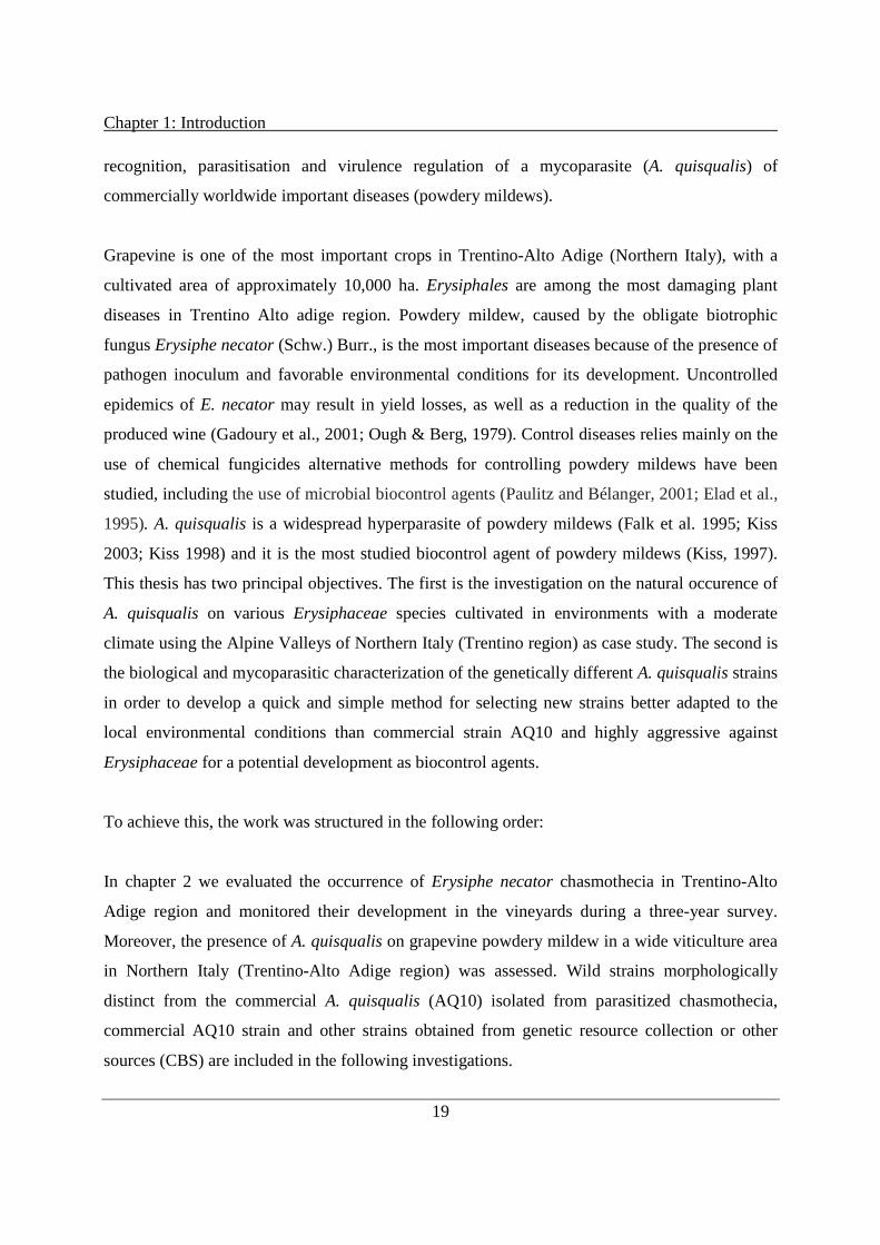
recognition, parasitisation and virulence regulation of a mycoparasite (A. quisqualis) of
commercially worldwide important diseases (powdery mildews).
Grapevine is one of the most important crops in Trentino-Alto Adige (Northern Italy), with a
cultivated area of approximately 10,000 ha. Erysiphales are among the most damaging plant
diseases in Trentino Alto adige region. Powdery mildew, caused by the obligate biotrophic
fungus Erysiphe necator (Schw.) Burr., is the most important diseases because of the presence of
pathogen inoculum and favorable environmental conditions for its development. Uncontrolled
epidemics of E. necator may result in yield losses, as well as a reduction in the quality of the
produced wine (Gadoury et al., 2001; Ough & Berg, 1979). Control diseases relies mainly on the
use of chemical fungicides alternative methods for controlling powdery mildews have been
studied, including the use of microbial biocontrol agents (Paulitz and Bélanger, 2001; Elad et al.,
1995). A. quisqualis is a widespread hyperparasite of powdery mildews (Falk et al. 1995; Kiss
2003; Kiss 1998) and it is the most studied biocontrol agent of powdery mildews (Kiss, 1997).
This thesis has two principal objectives. The first is the investigation on the natural occurence of
A. quisqualis on various Erysiphaceae species cultivated in environments with a moderate
climate using the Alpine Valleys of Northern Italy (Trentino region) as case study. The second is
the biological and mycoparasitic characterization of the genetically different A. quisqualis strains
in order to develop a quick and simple method for selecting new strains better adapted to the
local environmental conditions than commercial strain AQ10 and highly aggressive against
Erysiphaceae for a potential development as biocontrol agents.
To achieve this, the work was structured in the following order:
In chapter 2 we evaluated the occurrence of Erysiphe necator chasmothecia in Trentino-Alto
Adige region and monitored their development in the vineyards during a three-year survey.
Moreover, the presence of A. quisqualis on grapevine powdery mildew in a wide viticulture area
in Northern Italy (Trentino-Alto Adige region) was assessed. Wild strains morphologically
distinct from the commercial A. quisqualis (AQ10) isolated from parasitized chasmothecia,
commercial AQ10 strain and other strains obtained from genetic resource collection or other
sources (CBS) are included in the following investigations.

Chapter 1: Introduction
20
In chapter 3 the population of A. quisqualis present in the vineyards of Trentino region together
with AQ10 and other strains were morphologically and physiologically characterized.
Furthermore, all strains were molecularly characterized following the methodology described by
Szentivanyi (2005) determining the length polymorphisms of the amplified rDNA-ITS region
and the amplicons fully sequenced. Sequence polymorphisms were used to classify and group the
strains and the phylogenetic relationship among them was evaluated.
Chapter 4 regarded evaluation of some traits associated with strain aggressiveness and testing the
hypothesis that the ability of A. quisqualis strains in colonizing powdery mildew pathogens and
the “in vitro” production of cell wall degrading enzymes (CWDEs) secreted by the fungi are
important factor in selecting A. quisqualis strains for biocontrol. Mycoparasitic and enzymatic
assays were developed for the rapid and specific detection of new, highly effective strains for
biocontrol.
In the last chapter (5) a general protocol was derived for optimizing the production of pure, high
concentration A. quisqualis spore suspensions. We attemped to improve the efficiency of the
hyperparasite in the biological control of powdery mildews by adding a certain number of
additives to the inoculum in order to overcome humidity requirements of the fungus.
1.4 Literature
Andrews, J.H. (1992). Biological control in the phyllosphere. Annu Rev Phytopathol 30:603-635.
Angeli, D., Pellegrini, E., Micheli, S., Ress, D., Maurhofer, M., Pertot, I. and Gessler, C. (2009). Molecular
characterization of Ampelomyces spp. strains from different hosts and geographic origins and evaluation of
their potential to control powdery mildew of cucumber. IOBC/WPRS Bulletin, 43, 40-44.
Bélanger, R.R. and Benyagoub, M. (1997). Challenges and prospects for integrated control of powdery mildews in
the greenhouse.Canad J Plant Pathol 19:310-314.
Bélanger, R.R. and Labbé, C. (2002). Control of powdery mildews without chemicals: prophylactic and biological
alternatives for horticultural crops, in The powdery mildews: a comprehensive treatise, ed by Bélanger, R.R.,
Bushnell, W.R., Dik, A.J. and Carver, T.L.W., American Phytopathological Society, St Paul, USA, pp 256-
267.
Beuther, E., Philipp, W.D. and Grossmann, F. (1981). Untersuchungen zum Hyperparasitismus von Ampelomyces
quisqualis auf Gurkenmehltau (Sphaerotheca fuliginea ). Phytopathologische Zeitschrift 101, 265-270.

Chapter 1: Introduction
21
Braun, U. (1987). A monograph of the Erysiphales (powdery mildews). B. Nova Hedwigia 89: 1-700.
Brimner, T. and Boland, G. (2003). A review of the non-target effects of fungi used to biologically control plant
diseases. Agriculture, Ecosystems & Environment, 100(1), 3-16.
Choudbury, S.R., Traquair, J.A. and Jarvis, W.R. (1994). 4-Methyl-7,11-heptadecadenal and 4-methyl-7,11-
heptadecadienoic acid: new antibiotics from Sporothrix flocculosa and Sporothrix rugulosa. J Nat Prod 57:700-
704.
Cook, R.J. and Baker, K.F. (1983). The nature and practice of biological control of plant pathogens, American
Phytopathological Society, St Paul, MN.
Elad, Y., Malathrakis, N.E. and Dik, A.J. (1996). Biological control of Botrytis-incited diseases and powdery
mildews in greenhouse crops. Crop Protect 15:229-240.
Elad, Y., Kirshner, B. and Sztejnberg, A. (1998). Management of powdery mildew and gray mold of cucumber by
Trichoderma harzianum mT39 and Ampelomyces quisqualis AQ10. BioControl 43:241-251.
Elad, Y. (2000). Biological control of foliar pathogens by means of Trichoderma harzianum and potential modes of
action. Crop Protect 19:709-714.
Falk, S.P., Gadoury, D.M., Cortesi, P., Pearson, R.C. and Seem R.C. (1995). Parasitism of Uncinula necator
cleistothecia by the mycoparasite Ampelomyces quisqualis. Phytopathology 85:794-800.
Gadoury, D.M., Seem, R.C., Pearson, R.C., Wilcox, W.F. and Dunst, R.M. (2001). Effects of powdery mildew on
vine growth, yield, and quality of Concord grapes. Plant Dis. 85:137-140.
Gilardi, G., Baudino, M., Garibaldi, A., Gullino, M.L. (2012). Efficacy of biocontrol agents and natural compounds
against powdery mildew of zucchini. Phytoparasitica 40, 147-155.
Gu, Y.H. and Ko, W.H. (1997). Water agarose medium for studying factors affecting germination of conidia of
Ampelomyces quisqualis. Mycological Research 101, 422-424.
Hajlaoui, M.R., Traquair, J.A., Jarvis, W.R. and Bélanger R.R. (1994) Antifungal activity of extracellular
metabolites produced by Sporothrix flocculosa. Biocontrol Sci Technol 4:229-237.
Hashioka, Y. and Nakai, Y. 1980. Ultrastructure of pycnidial development and mycoparasitism of Ampelomyces
quisqualis parasitic on Erysiphales. Trans. Mycol. Soc. Jpn 21:329-338.
Heintz, C. and Blaich, R. (1990). Verticillium lecanii als Hyperparasit des Rebmehltaus (Uncinula necator). Vitis
29:229-232.
Hewitt, H.G. (1998). Fungicides in crop protection, CABI Publishing;Wallingford, UK.
Hijwegen, T. and Buchenauer, H. (1984). Isolation and identification of hyperparasitic fungi associated with
Erysiphaceae. Neth J Pl Path 90:79-84.

Chapter 1: Introduction
22
Hijwegen, T. (1988). Effect of seventeen fungicolous fungi on sporulation of cucumber powdery mildew. Neth J Pl
Path 94:185-190.
Hofstein, R. and Fridlender, B. (1994). Development of production, formulation and delivery systems, in Proc
Brighton Crop Protect Conf Pests Dis, BCPC, Farnham, Surrey, UK, pp 1273-1280.
Hofstein, R., Daoust, R.A. and Aeschlimann, J.P. (1996). Constraints to the development of biofungicides: the
example of ‘AQ-10’, a new product for controlling powdery mildews. Entomophaga 41:455-460.
Kiss, L. (1997). Genetic diversity in Ampelomyces isolates, hyperparasites of powdery mildew fungi, inferred from
RFLP analysis of the rDNA ITS region. Mycol. Res. 101:1073-1080.
Kiss, L. (1998). Natural occurrence of Ampelomyces intracellular mycoparasites in mycelia of powdery mildew
fungi. New Phytol. 140:709-714.
Kiss, L. and Nakasone, K. K. (1998). Ribosomal DNA internal transcribed spacer sequences do not support the
species status of Ampelomyces quisqualis, a hyperparasite of powdery mildew fungi. Curr. Gen. 33:362-367.
Kiss, L. (2003). A review of fungal antagonists of powdery mildews and their potential as biocontrol agents. Pest
Management Science, 59, 475-483.
Kiss, L., Russell, J.C., Szentivanyi, O., Xu, X., Jeffries, P. (2004). Biology and biocontrol potential of Ampelomyces
mycoparasites, natural antagonist of powdery mildew fungi. Biocontrol Science and Technology 14, 635-651.
Kiss, L., Pintye A., Kovacs G.M., Jankovics T., Fontaine M.C., Harvey N., Xu X., Nicot P.C., Bardin M., Shykoff
J.A. and Giraud T. (2011). Temporal isolation explains host-related genetic differentiation in a group of
widespread mycoparasitic fungi. Molecular Ecology, 20, 1492-1507.
Klecan, A.L., Hippe, S. and Somerville, S.C. (1990). Reduced growth of Erysiphe graminis f sp hordei induced by
Tilletiopsis pallescens. Phytopathology 80:325-331.
Knudsen, I.M.B. and Skou, J.P. (1993). The effectivity of Tilletiopsis albescens in biocontrol of powdery mildew.
Ann Appl Biol 123:173-185.
Kranz, J. (1981). Hyperparasitism of biotrophic fungi, in Microbial Ecology of the Phylloplane (Blakeman, J. P.,
Ed.). Academic Press, London, UK, pp. 327-352.
Ishii, H., Fraaije, B.A., Sugiyama, T., Noguchi, K., Nishimura, K., Takeda, T., Amano, T. and Hollomon, D.W.
(2001). Occurrence and molecular characterization of strobilurin resistance in cucumber powdery mildew and
downy mildew. Phytopathology 91:1166-1171.
Jarvis, W.R. and Slingsby, K. (1977). The control of powdery mildew of greenhouse cucumber by water sprays and
Ampelomyces quisqualis. Plant Dis Rep 61:728-730.
Liang C., Yang J., Kovacs G.M., Szentivanyi O., Li B., Xu X.M. & Kiss L. (2007). Genetic diversity of
Ampelomyces mycoparasite isolated from different powdery mildew species in China inferred from analyses of
rDNA ITS sequences. Fungal Diversity, 24, 225-240.

Chapter 1: Introduction
23
McGrath, M.T. and Shishkoff, N. (1999). Evaluation of biocompatible products for managing cucurbit powdery
mildew. Crop Protect m18:471-478.
Menzies, J.G. and Bélanger, R.R. (1996). Recent advances in cultural management of diseases of greenhouse crops.
Canad J Plant Pathol 18:186-193.
Nischwitz, C., Newcombe, G., Anderson, C.L. (2005). Host specialization of the mycoparasite Eudarluca caricis
and its evolutionary relationship to Ampelomyces. Mycological Research 109, 421-428.
Ough, C.S. and Berg, H.W. (1979). Powdery mildew sensory effect on wine. Am. J. Enol. Vit. 30:321.
Park M.J., Choi Y.J., Hong S.B. & Shin H.D. (2010). Genetic variability and mycohost association of Ampelomyces
quisqualis isolates inferred from phylogenetic analyses of ITS rDNA and actin gene sequences. Fungal
Biology, 114, 235-247.
Pasini, C., D’Aquila, F., Curir, P. and Gullino, M.L. (1997). Effectiveness of antifungal compounds against rose
powdery mildew (Sphaerotheca pannosa var rosae) in glasshouses. Crop Protect 16:251-256.
Paulitz, T.C. and Bélanger, R.R. (2001). Biological control in greenhouse systems. Annu Rev Phytopathol 39:103-
133.
Pintye, A., Bereczky, Z., Kovács, G.M., Nagy, L.G., Xu, X., Legler, S.E., Váczy, Z., Váczy, K.Z., Caffi, T., Rossi,
V., Kiss, L. (2012). No indication of strict host associations in a widespread mycoparasite: grapevine powdery
mildew (Erysiphe necator) is attacked by phylogenetically distant Ampelomyces quisqualis strains in the field.
Phytopathology 102:707-716.
Sullivan, R.F. and White, Jr. J.F. (2000). Phoma glomerata as a mycoparasite of powdery mildew. Applied and
Environmental Microbiology, 66, 425-427.
Sundheim, L. (1982). Control of cucumber powdery mildew by the hyperparasite Ampelomyces quisqualis and
fungicides. Plant Pathol 31:209-214.
Sundheim, L., Krekling, T. (1982). Host-parasite relationships of the hyperparasite Ampelomyces quisqualis and its
powdery mildew host Sphaerotheca fuliginea. I. Scanning electron microscopy. Phytopathology 104, 202-210.
Sundheim, L. and Tronsmo, A. (1988). Hyperparasites in biological control. Pages 53-69 in: Biocontrol of Plant
Diseases. Vol. I. CRC Press, Boca Raton, FL.
Sutton, B.C. (1980). The Coelomycetes. Fungi imperfecti with pycnidia, acervuli and stromata. Commonwealth
Mycological Institute, Kew, UK.
Szentiványi, O., Kiss, L., Russell, J.C., Kovács, G.M., Varga, K., Jankovics, T., Lesemann, S., Xu, X.M. and
Jeffries, P. (2005). Ampelomyces mycoparasites from apple powdery mildew identified as a distinct group
based on single-stranded conformation polymorphism analysis of the rDNA ITS region. Mycological
Research, 109, 429-438.

Chapter 1: Introduction
24
Sztejnberg, A., Galper, S., Mazar, S., and Lisker, N. (1989). Ampelomyces quisqualis for biological and integrated
control of powdery mildews in Israel. J. Phytopath. 124:285-295.
Sztejnberg, A. (1993). Ampelomyces quisqualis AQ10, CNCM I-807, for biological control of powdery mildew. US
patent no. 5190754.
Urquhart, E.J., Menzies, J.G. and Punja, Z.K. (1994). Growth and biological control activity of Tilletiopsis species
against powdery mildew (Sphaerotheca fuliginea) on greenhouse cucumber. Phytopathology 84:341-351.
Whipps, J.M. and Lumsden, R.D. (2001). Commercial use of fungi as plant disease biological control agents: status
and prospects, in Fungi as Biocontrol Agents: Progress, Problems and Potential (Butt, T.M., Jackson, C. And
Magan, N., Eds.). CABI Publishing, Wallingford, UK, pp. 9-22.
Yarwood, C.E. (1932). Ampelomyces quisqualis on clover mildew. Phytopathology 22, 31 (Abstr.).

25
Chapter 2
Occurrence of Erysiphe necator chasmothecia and their
natural parasitization by Ampelomyces quisqualis in
Northern Italy (Trentino Alto Adige region)
Published as
Angeli D., Pellegrini E., Pertot I. 2009. Occurrence of Erysiphe necator Chasmothecia and Their
Natural Parasitization by Ampelomyces quisqualis. Phytopathology, 99: 704-710.
Chapter 2: Natural parasitism by Ampelomyces quisqualis
26
Abstract
In Northern Italy, Erysiphe necator overwinters almost exclusively as chasmothecia. From 2004
to 2008, we investigated the occurrence of natural parasitism of grapevine powdery mildew
chasmothecia by Ampelomyces quisqualis in the Trentino-Alto Adige region, in northern Italy.
The survey was conducted in 18 vineyards in autumns 2004 and 2005 and in 45 vineyards in
autumns 2006 and 2007. The incidence of powdery mildew signs (white powdery mycelia and
conidia), the number of chasmothecia and their development pattern, and the incidence of
parasitism by A. quisqualis were assessed. The production of E. necator chasmothecia on leaves
is related to the incidence and severity of the disease on leaves at the end of the season and is not
correlated with the elevation of the vineyard, which is inversely related to the temperature. A
limited amount of natural parasitism of E. necator by Ampelomyces spp. (0.17−3.51 %) was
observed in all of the years of the survey. Pycnidia and conidia of Ampelomyces spp. parasitizing
E. necator chasmothecia were found both in conventional, organically grown on and untreated
vineyards. Some of the isolated Ampelomyces strains have conidia that are shaped differently
than those of the commercial A. quisqualis strain (AQ10) and are phylogenetically different from
AQ10.
2.1 Introduction
Grapevine powdery mildew, caused by the obligate biotrophic fungus Erysiphe necator (Schw.)
Burr., is one of the most important grapevine diseases in Italy, because of the presence of
pathogen inoculum and favorable environmental conditions for its development. Uncontrolled
epidemics of E. necator may result in yield losses, as well as a reduction in the quality of the
produced wine (Gadoury et al., 2001; Ough & Berg, 1979). During the winter, the fungus
survives either as mycelium in the dormant buds of grapevine or as chasmothecia, which are the
fruiting bodies arising from the sexual stage (Bulit & Lafon, 1978; Pearson & Goheen, 1988).
Mycelium preserved inside the bud is thought to give rise to so called flag shoots in spring. The
infected flag shoots are stunted, deformed and covered with white powdery mycelium and
conidia (Rumbolz & Gubler, 2005). In contrast, chasmothecia represent the main source of
primary inoculum in regions with cold winters, such as are found in northern Italy. In the spring,

Chapter 2: Natural parasitism by Ampelomyces quisqualis
27
ascosporic infections originating from chasmothecia commonly appear randomly in the vineyard
as scattered whitish and powdery spots on leaves (1 to 3 mm in diameter) that mainly appear on
leaves close to the trunk (Grove, 2004; Pearson & Gadoury, 1987; Sall & Wrysinski, 1982;
Ypema & Gubler, 2000). Grapevine is one of the most important crops in Trentino-Alto Adige
(northern Italy), with a cultivated area of approximately 10,000 ha. Chasmothecia are thought to
be the main overwintering form of E. necator in this region (Angeli et al., 2006).
Ampelomyces quisqualis Ces. is a naturally occurring mycoparasite of several powdery mildew
species (Erysiphales), including E. necator (Falk et al., 1995a; Kiss, 1998; Sundheim & Krekling
1982). A. quisqualis has been considered to be a single species for a long time, but its taxonomy
has become controversial and probably merits extensive revision (Kiss & Nakasone, 1998). The
morphology of the conidia and pycnidia of different A. quisqualis isolates and the growing
patterns of colonies of these isolates on laboratory media are highly variable (Kiss et al., 2004).
The pycnidia of Ampelomyces spp. also vary in shape depending upon the fungal structure in
which they were formed. They are pear-shaped, spindle-shaped or nearly spherical when they are
formed inside E. necator conidiophores, hyphae or chasmothecia, respectively. Pycnidia contain
cylindrical to spindle-shaped conidia, which are occasionally curved and two-spotted (Falk et al.,
1995a). Recent molecular studies have hypothesized the existence of more than one species in
the genus Ampelomyces. Analyses of the internal transcribed spacer (ITS) region of the nuclear
ribosomal RNA gene (nrDNA) of several putative A. quisqualis strains have uncovered a high
level of genetic diversity (Kiss, 1997; Kiss & Nakasone, 1998; Sullivan & White, 2000;
Szentiványi et al., 2005), which suggests that the binomial A. quisqualis should be regarded as a
species complex (Kiss & Nakasone, 1998).
A. quisqualis has been found on more than 64 species of powdery mildew on 256 species of
plants (Kiss, 1997; Kiss, 2003; Kiss et al., 2004). This wide host range, combined with tolerance
to a number of fungicides used against powdery mildews, makes A. quisqualis the ideal
candidate for use as a biological control agent (Falk et al., 1995b; Sundheim & Tronsmo, 1988;
Sztejnberg et al., 1989). An A. quisqualis strain isolated in Israel has been formulated, registered,
and commercialized in several countries under the trade name “AQ10” (Sztejnberg, 1993).
A. quisqualis can grow saprophytically for short periods of time, but has little chance of
surviving for longer periods in natural environments without parasitizing a powdery mildew

Chapter 2: Natural parasitism by Ampelomyces quisqualis
28
host. It requires water to germinate and to infect powdery mildew colonies. Infections can occur
in less than 24 h at 25 °C (Kiss et al., 2004; Sundheim & Krekling, 1982). A. quisqualis invades
and grows within powdery mildew hosts (Hashioka & Nakai, 1980; Sundheim & Krekling,
1982). Parasitized powdery mildew colonies are dull, flattened and off-white to gray in color.
Pycnidia are formed within hyphae, conidiophores, conidia and the immature chasmothecia of
powdery mildews. Once the mycoparasite has begun to produce pycnidia, the hyphae and
conidiophores swell to several times their normal diameter and the amber color of the pycnidial
walls of A. quisqualis may be visible through the cell walls of the host (Falk et al., 1995a).
Several studies have shown that A. quisqualis cannot parasitize mature E. necator chasmothecia
(Falk et al., 1995a; Kiss et al., 2004). Parasitized chasmothecia are typically dull, fawn-colored,
and flaccid and range from 64 to 130 µm in diameter (5). A. quisqualis parasitism reduces
powdery mildew sporulation, as well as the production of chasmothecia and may eventually kill
the entire mildew colony (Falk et al., 1995a; Falk et al., 1995b; Hashioka & Nakai, 1980; Kiss et
al., 2004; Sundheim & Krekling, 1982).
While a few studies have reported the presence of A. quisqualis on several powdery mildews on
several plants (Kiss, 1998; Kiss et al., 2004), no study has reported on the extent of the natural
occurrence of A. quisqualis on grapevine powdery mildew. The aims of this study were i) to
assess and quantify the presence of Ampelomyces spp. on grapevine powdery mildew in a wide
viticulture area in northern Italy (Trentino-Alto Adige region); and ii) to characterize the isolates
present in the area. We also evaluated the occurrence of chasmothecia in Trentino-Alto Adige
region and monitored their development in the vineyard.
2.2 Material and Methods
2.2.1 Study sites, assessment, and sampling
The study was carried out from 2004 to 2007 in the Trentino-Alto Adige region of northern Italy.
Sampling was carried out between 15 and 31 October in 18 vineyards from 2004 to 2007.
Additionally, 27 vineyards were sampled in 2006 and 2007, respectively. The vineyards were
randomly selected. The sampled vineyards included conventionally and organically managed
vines; abandoned (untreated) vineyards were also surveyed. The chemical fungicides used in the

Chapter 2: Natural parasitism by Ampelomyces quisqualis
29
conventionally managed vineyards included mancozeb, folpet, dimethomorph, zoxamide,
iprovalicarb, quinoxyfen, spiroxamine, copper, sulphur, acylalanines, strobilurins and triazoles.
In the organic vineyards, only sulphur and copper were used. A. quisqualis (AQ10; Ecogen,
Langhorne, PA, USA) was not used in any of the monitored vineyards during the survey period.
The size of each sampled vineyard ranged between 800 and 1200 m2. In each vineyard and year,
four replicates of 25 leaves each were randomly collected at the fifth leaf from the shoot. For
each replicate, the percentage of infected area on the upper surface of the leaf (whitish, powdery
spots) and the number of infected leaves were visually assessed. Disease severity (percentage of
infected leaf area) and incidence (percentage of infected leaves) were calculated. A disk (2 cm in
diameter) was cut from the central part of each sampled leaf. The E. necator chasmothecia on the
upper side of each leaf disk were counted under a stereomicroscope (Nikon SMZ 800, Tokyo,
Japan). To identify the optimal times for assessing parasitism by and development of
Ampelomyces spp., the ripening of chasmothecia was surveyed weekly between August and
November in an untreated experimental vineyard (cv. Schiava) in S. Michele all’Adige on a
sample of 100 randomly collected leaves. The number of chasmothecia was assessed as
described above. Chasmothecia were classified into three categories according to their color,
which reflects their development stage: yellow (young), brown (semi-mature), and black
(mature). On each leaf (100 leaves per vineyard, collected when black chasmothecia were 50 ±
10%), Ampelomyces spp. mycoparasitism of E. necator was assessed under a light microscope
(Hund Wetzlar H 600LL, Wetzlar, Germany), in terms of the presence of parasitized dull, flaccid
and fawn-colored chasmothecia, brownish intracellular pycnidia in E. necator hyphae, and/or
cylindrical, spindle-shaped, and two-spotted conidia. In detail, E. necator mycelium and
chasmothecia were transferred into Eppendorf tubes by brushing the upper surface of sampled
leaves (four tubes for each replicate of 25 leaves) and stored at 4 °C. One hundred chasmothecia
from each tube were mounted in lactophenol and observed under a light microscope to asses the
percentage of parasitized cleistothecia, as well as the presence of Ampelomyces spp. pycnidia and
conidia. Since Ampelomyces spp. infect and produce pycnidia only inside young and semi-
mature chasmothecia, as reported in several studies of parasitism of E. necator (Falk et al.,
1995a; Kiss et al., 2004), when black chasmothecia were 50 ± 10 % of the total, we selected
yellow and brown chasmothecia and excluded mature dark brown chasmothecia. Chasmothecia
were gently crushed by pressing the cover slip over the glass slide on which the sample was
Chapter 2: Natural parasitism by Ampelomyces quisqualis
30
spread, to allow the release of Ampelomyces spp. conidia and pycnidia. The possible presence of
Ampelomyces spp. pycnidia in the sample was recorded. The incidence of Ampelomyces spp. in
each vineyard was calculated as the percentage of E. necator chasmothecia parasitized by
Ampelomyces spp. During 2005 to 2008, between April and May of each year, the vineyards
were monitored weekly to check for the presence of flag shoots.
2.2.2 Isolation, identification, morphological, and molecular characterization
of isolates of Ampelomyces spp.
Ampelomyces spp. samples on parasitized chasmothecia and mycelia were initially identified by
comparing the morphological characteristics of the observed conidia and pycnidia with those
described in the literature (Falk et al., 1995a; Kiss, 1998; Kiss et al., 2004). Some of the
Ampelomyces spp. samples were isolated by transferring the conidia onto potato dextrose agar
(PDA, Oxoid, Hampshire, UK) amended with 2 % chloramphenicol (Sigma, St. Louis, MO,
USA). Morphological identification and measurements of pycnidia and conidia (length, width,
and shape) were carried out on twenty replicates (pycnidia) for each isolate. Five isolates (ITA 1,
ITA 2, ITA 3, ITA 4, and ITA 5) were selected for the genetic analysis, each representing the
different morphological shapes (fusiform or ellipsoid) found in homogeneous areas of Trentino
region (Trento North, Trento South, and Rovereto). The mycelium of each of five 20-day-old
Ampelomyces spp. isolates grown on PDA was collected from Petri dishes and freeze-dried.
DNA was extracted from 2 µg of homogenized lyophilized mycelia, using the Nucleo Spin Plant
Kit (Macherey-Nagel, Düren, Germany) according to manufacturer’s instructions. The ITS
region of the nuclear ribosomal DNA was amplified using the specific fungal primers ITS1 and
ITS4 (White et al., 1990). PCR was performed in the Gene Amp PCR System 9700 (Perkin
Elmer, Waltham, MA, USA) and the following cycling parameters were used: initial denaturing
step at 95 °C for 3 min, 35 cycles of denaturing step at 95 °C for 30 s, primer annealing at 60 °C
for 30 s, extension step at 72 °C for 30 s and a final extension at 72 °C for 7 min. PCR products
were detected by electrophoresis on 1 % agarose gel in TBE buffer supplemented with ethidium
bromide (0.5 µl/ml). PCR products were purified using the ExoSAP-IT enzymes (USB
Corporation, Staufen, Germany) and sequenced using a Big Dye Terminator v1.1 Cycle
Sequencing Kit (Applied Biosystems, Foster City, CA, USA). Both DNA strands were

Chapter 2: Natural parasitism by Ampelomyces quisqualis
31
sequenced with the primers used for PCR amplification; electrophoresis was carried out using an
ABI 3130xl Genetic Analyzer (Perkin Elmer). The consensus sequence was assembled using
Pregap4 and Gap4 (Staden et al., 2000). Searches were performed in the NCBI/GenBank
database to find the closest relatives of the sequenced isolates and the similitude percentages
through the BLAST method (Altschul et al., 1990). The ClustalW2 program, available for free
on the internet (Larkin et al., 2007), was used to construct multiple sequence alignments using
the DNA identity matrix and to visualize the evolutionary relationship between the input
sequences. The alignments were checked and edited using Bioedit 7.0.5.2. (Hall, 1999) to
generate an alignment of the same length for inferring phylogenies. Maximum-likelihood and
neighbor-joining analyses based on ITS region sequences were conducted using respectively the
programs BioEdit 7.0.5.2. and Mega4 (Tamura et al., 2007) with the Jukes-Cantor substitution
model and with rate uniformity among sites. All positions containing gaps and missing data were
eliminated. The branches of the inferred tree were tested by bootstrap analysis (Felsenstein,
1985) with 1000 replicates. The trees were visualized using TreeView 1.6.6 (Page, 1996).
2.2.3 Meteorological data and statistical analysis
Meteorological data (rain, hourly temperature and relative humidity) were collected by the local
agro-meteorological service (http://meteo.iasma.it/meteo/). Sum of hours with optimal
temperatures (from 20 to 27 °C) for powdery mildew infection and disease development and
total rain (Pearson & Goheen, 1988) were calculated from May 1 to October 31. Statistical
analyses were performed using Statistica software 6.0 (Statsoft, Tulsa, OK, USA). Disease
incidence and severity data were Arcsin-transformed to normalize the data. One-way analysis of
variance (ANOVA) was used to compare differences in the incidence and severity in the
different years. Means were separated using Tukey’s test (α = 0.05). The Chi-square test
followed by Ryan’s multiple comparison test (P ≤ 0.05) (Ryan, 1960) was used to compare
differences in the presence of Ampelomyces in the different years.

Chapter 2: Natural parasitism by Ampelomyces quisqualis
32
2.3 Results
2.3.1 Natural occurrence of E. necator
The assessment of powdery mildew infections in the sampled vineyards at the end of the
growing seasons detected increases in disease severity and incidence between 2004 and 2007
(Fig. 1). The monitoring showed both high incidences and high severity of E. necator infections
in 2006 and 2007 (34 and 45 % of average severity and 45 and 67 % of average incidence in all
vineyards, respectively) probably because of the favorable climatic conditions for powdery
mildew (Pearson & Goheen, 1988) present after veraison through the end of the growing season
(Fig. 2). In 2006 and 2007, it was cooler and rainier during the last part of the season, as
compared to the earlier seasons (data not shown). Similar to the findings concerning disease
severity and disease incidence on leaves, the populations of E. necator chasmothecia were lower
in 2004 and 2005 than they were in 2006 and 2007 (Fig. 3). There was a very high correlation
between the severity of powdery mildew infections on leaves in the vineyard (average of the four
replicates) and the number of E. necator chasmothecia per unit of leaf area (average of the four
replicates; Fig. 4). A similar high correlation was observed between powdery mildew incidence
and the number of chasmothecia per unit of leaf surface area. In 2004, 2005, 2006, and 2007, R2
values were 0.938 (y = 4.3898x – 63.046), 0.952 (y = 4.6758x – 65.763), 0.984 (y = 5.4574x –
88.023), and 0.972 (y = 6.2413x – 156.83), respectively. There was no correlation between the
number of chasmothecia per unit of leaf area and the elevation of the different vineyards, which
is inversely related to temperature. No flag shoots were seen in the vineyards in the years of the
survey and infections began (seemingly) randomly in the monitored vineyards.
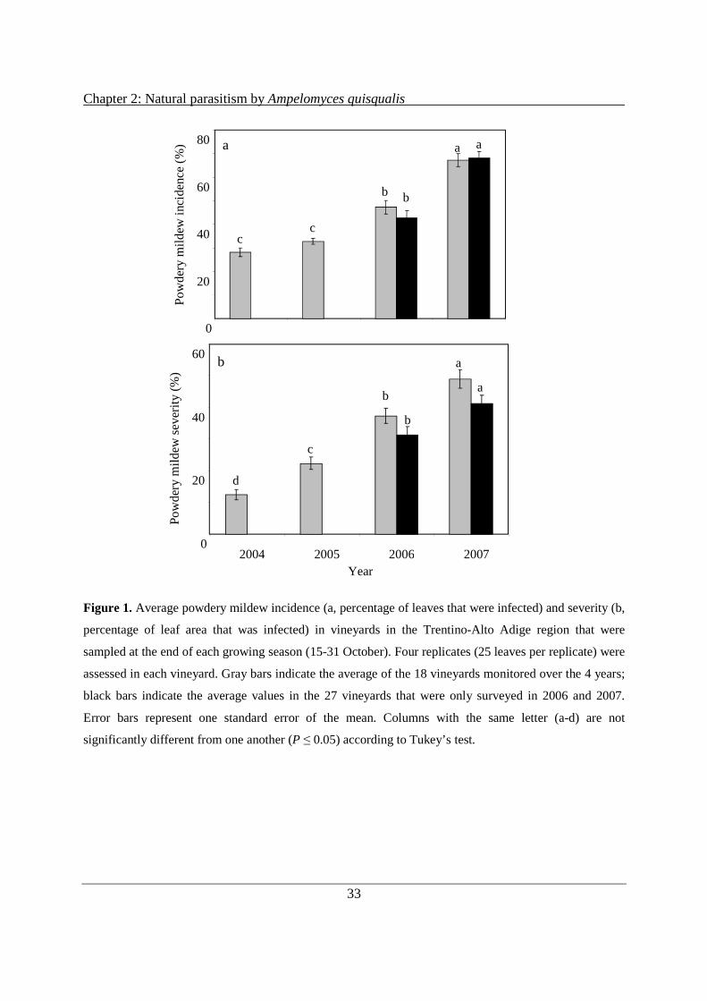
Chapter 2: Natural parasitism by Ampelomyces quisqualis
33
Figure 1. Average powdery mildew incidence (a, percentage of leaves that were infected) and severity (b,
percentage of leaf area that was infected) in vineyards in the Trentino-Alto Adige region that were
sampled at the end of each growing season (15-31 October). Four replicates (25 leaves per replicate) were
assessed in each vineyard. Gray bars indicate the average of the 18 vineyards monitored over the 4 years;
black bars indicate the average values in the 27 vineyards that were only surveyed in 2006 and 2007.
Error bars represent one standard error of the mean. Columns with the same letter (a-d) are not
significantly different from one another (P ≤ 0.05) according to Tukey’s test.
Po
wd
ery
mild
ew in
cide
nce
(%
)
0
20
40
60
80
b
c c
a
b
a a
0
20
40
60
2004 2005 2006 2007
a
a
b
b
c
d
Po
wd
ery
mild
ew s
eve
rity
(%
)
Year
b

Chapter 2: Natural parasitism by Ampelomyces quisqualis
34
Figure 2. Periods with suitable temperature for powdery mildew (sum of hours with temperature between
20 °C and 27 °C) and total rain from May 1 to October 31 of each year in S. Michele all’Adige (Trentino-
Alto Adige region).
Figure 3. Number of chasmothecia per unit of leaf area (cm2) in the sampled vineyards in the Trentino-
Alto Adige region at the end of each growing season (15-31 October). Four replicates (25 leaves per
replicate) were collected and chasmothecia were counted on leaf disk samples cut from the middle of each
1340
1360
1380
1400
1420
2004 2005 2006 2007
Tem
per
atu
re (
sum
of h
ou
rs)
0
100
200
300
400
500
Rai
n (
mm
)
Temperature Rain
Year
1440
1460
0
50
100
150
200
250
300
2004 2005 2006 2007
a
b
c
d
a
b
Year
Ch
asm
othe
cia
(N
o./
cm2 lea
f ar
ea)
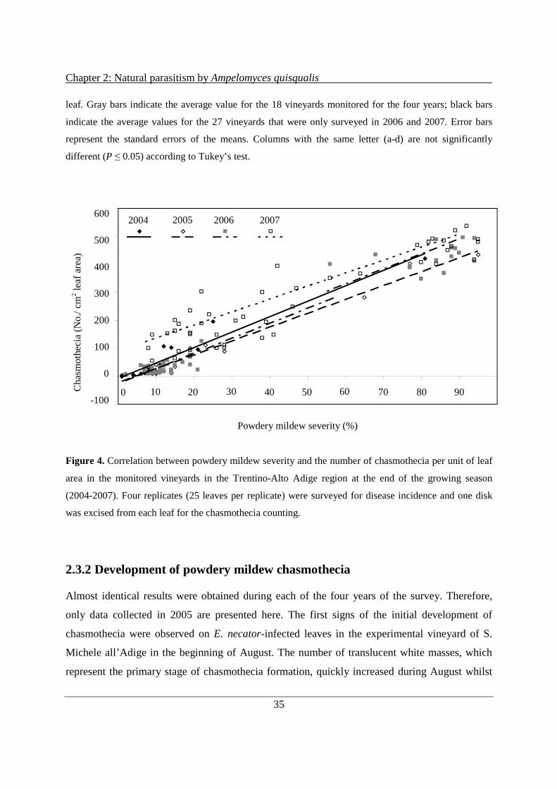
Chapter 2: Natural parasitism by Ampelomyces quisqualis
35
leaf. Gray bars indicate the average value for the 18 vineyards monitored for the four years; black bars
indicate the average values for the 27 vineyards that were only surveyed in 2006 and 2007. Error bars
represent the standard errors of the means. Columns with the same letter (a-d) are not significantly
different (P ≤ 0.05) according to Tukey’s test.
Figure 4. Correlation between powdery mildew severity and the number of chasmothecia per unit of leaf
area in the monitored vineyards in the Trentino-Alto Adige region at the end of the growing season
(2004-2007). Four replicates (25 leaves per replicate) were surveyed for disease incidence and one disk
was excised from each leaf for the chasmothecia counting.
2.3.2 Development of powdery mildew chasmothecia
Almost identical results were obtained during each of the four years of the survey. Therefore,
only data collected in 2005 are presented here. The first signs of the initial development of
chasmothecia were observed on E. necator-infected leaves in the experimental vineyard of S.
Michele all’Adige in the beginning of August. The number of translucent white masses, which
represent the primary stage of chasmothecia formation, quickly increased during August whilst
-100
0
100
200
300
400
500
600
0 10 20 30 40 50 60 70 80 90
Powdery mildew severity (%)
2004 2005 2006 2007
Cha
smo
the
cia
(No
./ cm2 le
af a
rea)
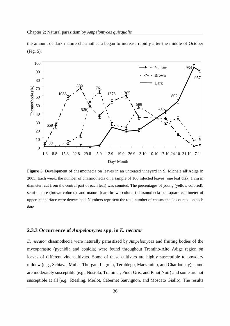
Chapter 2: Natural parasitism by Ampelomyces quisqualis
36
the amount of dark mature chasmothecia began to increase rapidly after the middle of October
(Fig. 5).
Figure 5. Development of chasmothecia on leaves in an untreated vineyard in S. Michele all’Adige in
2005. Each week, the number of chasmothecia on a sample of 100 infected leaves (one leaf disk, 1 cm in
diameter, cut from the central part of each leaf) was counted. The percentages of young (yellow colored),
semi-mature (brown colored), and mature (dark-brown colored) chasmothecia per square centimeter of
upper leaf surface were determined. Numbers represent the total number of chasmothecia counted on each
date.
2.3.3 Occurrence of Ampelomyces spp. in E. necator
E. necator chasmothecia were naturally parasitized by Ampelomyces and fruiting bodies of the
mycoparasite (pycnidia and conidia) were found throughout Trentino-Alto Adige region on
leaves of different vine cultivars. Some of these cultivars are highly susceptible to powdery
mildew (e.g., Schiava, Muller Thurgau, Lagrein, Teroldego, Marzemino, and Chardonnay), some
are moderately susceptible (e.g., Nosiola, Traminer, Pinot Gris, and Pinot Noir) and some are not
susceptible at all (e.g., Riesling, Merlot, Cabernet Sauvignon, and Moscato Giallo). The results
100
0
10
20
30
40
50
60
70
80
90
1.8 8.8 15.8 22.8 29.8 5.9 12.9 19.9 26.9 3.10 10.10 17.10 24.10 31.10 7.11
Ch
asm
oth
eci
a (
%)
Yellow
Brown
Dark
88
659
1083
800
526
761
1373 1365
688 650
802
934
957
Day/ Month

Chapter 2: Natural parasitism by Ampelomyces quisqualis
37
of this four-year monitoring program underline the generally low natural presence of
Ampelomyces spp. in the vineyards of the Trentino-Alto Adige region (Table 1).
In the first two years no differences in the percentage of parasitism in the vineyard were seen, but
the total number of chasmothecia parasitized by Ampelomyces significantly increased over the
period of the study (χ2 = 102.98; df = 3; P ≤ 0.001). The average rate of parasitism among
chasmothecia in all of the monitored vineyards ranged from 0.17 % (2004) to 3.51 % (2007).
Once Ampelomyces spp. were first found in a vineyard, it was also found in that vineyard in
following years. Ampelomyces spp. was found on leaves, mainly as conidia in parasitized E.
necator chasmothecia and, in four vineyards, as pycnidia in the powdery mildew mycelia.
Because of the limited number of E. necator samples parasitized by Ampelomyces spp. and the
limited numbers of available untreated/abandoned and organic vineyards in the area, no
relationship between treated (conventional and biological treatments) or untreated/abandoned
vineyards and A. quisqualis parasitism of powdery mildew could be identified. A weak
relationship was observed between the number of chasmothecia per unit of leaf surface area and
the percentage of chasmothecia parasitized by Ampelomyces spp. during the period 2004-2007
(R2 = 0.366, y = 7E – 0.5x2 – 0.0165 + 0.5218).
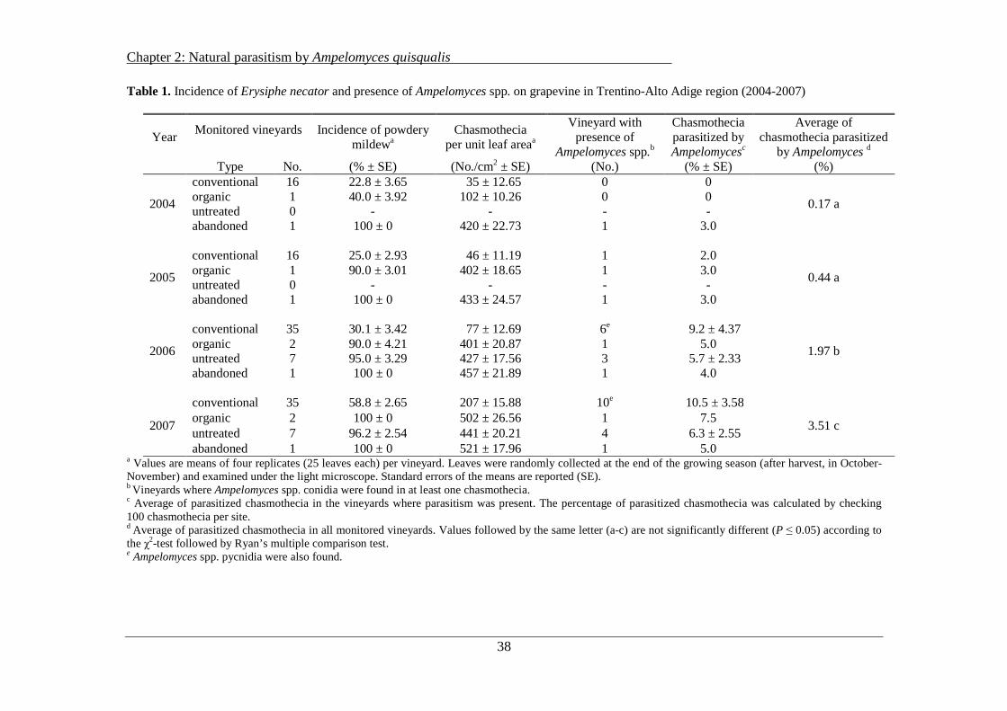
Chapter 2: Natural parasitism by Ampelomyces quisqualis
38
Table 1. Incidence of Erysiphe necator and presence of Ampelomyces spp. on grapevine in Trentino-Alto Adige region (2004-2007)
Year Monitored vineyards
Incidence of powdery
mildewa Chasmothecia
per unit leaf areaa
Vineyard with presence of
Ampelomyces spp.b
Chasmothecia parasitized by Ampelomycesc
Average of chasmothecia parasitized
by Ampelomyces d Type No. (% ± SE) (No./cm2 ± SE) (No.) (% ± SE) (%)
2004
conventional 16 22.8 ± 3.65 35 ± 12.65 0 0
0.17 a organic 1 40.0 ± 3.92 102 ± 10.26 0 0 untreated 0 - - - - abandoned 1 100 ± 0 420 ± 22.73 1 3.0
2005
conventional 16 25.0 ± 2.93 46 ± 11.19 1 2.0
0.44 a organic 1 90.0 ± 3.01 402 ± 18.65 1 3.0 untreated 0 - - - - abandoned 1 100 ± 0 433 ± 24.57 1 3.0
2006
conventional 35 30.1 ± 3.42 77 ± 12.69 6e 9.2 ± 4.37
1.97 b organic 2 90.0 ± 4.21 401 ± 20.87 1 5.0 untreated 7 95.0 ± 3.29 427 ± 17.56 3 5.7 ± 2.33 abandoned 1 100 ± 0 457 ± 21.89 1 4.0
2007
conventional 35 58.8 ± 2.65 207 ± 15.88 10e 10.5 ± 3.58
3.51 c organic 2 100 ± 0 502 ± 26.56 1 7.5 untreated 7 96.2 ± 2.54 441 ± 20.21 4 6.3 ± 2.55 abandoned 1 100 ± 0 521 ± 17.96 1 5.0
a Values are means of four replicates (25 leaves each) per vineyard. Leaves were randomly collected at the end of the growing season (after harvest, in October-November) and examined under the light microscope. Standard errors of the means are reported (SE). b Vineyards where Ampelomyces spp. conidia were found in at least one chasmothecia. c Average of parasitized chasmothecia in the vineyards where parasitism was present. The percentage of parasitized chasmothecia was calculated by checking 100 chasmothecia per site. d Average of parasitized chasmothecia in all monitored vineyards. Values followed by the same letter (a-c) are not significantly different (P ≤ 0.05) according to the χ2-test followed by Ryan’s multiple comparison test. e Ampelomyces spp. pycnidia were also found.
Chapter 2: Natural parasitism by Ampelomyces quisqualis
39
2.3.4 Identification and morphological characterization of Ampelomyces spp.
Pycnidia of the Ampelomyces spp. isolates varied in shape (ovoid, ellipsoid, or globose). They
were found only in powdery mildew mycelia. No pycnidia were observed in immature or semi-
mature chasmothecia. Moreover, the ranges of pycnidia sizes were quite variable.
Conidia of Ampelomyces spp. were found in chasmothecia and in mycelia. Conidia size was
variable within isolates. The conidia ranged from 11.5 to 14.5 µm in length (major axis) and
from 2.5 to 3.5 µm in width (minor axis). Differences in the shape (fusiform or ellipsoidal) of the
conidia were observed among isolates collected in different vineyards (Table 2). Molecular
analysis, based on ITS sequencing, showed that all of the collected isolates belong to the
Ampelomyces genus. The most similar NCBI library strains are listed in Table 3, together with
their accession numbers and similarity percentage figures. The ITS sequences of the five selected
Ampelomyces strains were analyzed together with the sequence obtained from AQ10 and with
other Ampelomyces sequences from GenBank, from different collections and from different host
species, in order to evaluate their phylogenetic diversity. A 511 nucleotides long alignment was
used to infer phylogenies. Both the maximum-likelihood (not shown) and the neighbor-joining
trees (Fig. 6) showed the same clustering of the strains. The five wild strains grouped into two
different clades with an optimal bootstrap support (100 %): the isolates named ITA 1 and ITA 2
grouped together with AQ10 and no differences were observed between these three sequences.
The fusiform-shaped conidia of ITA1 are similar to the conidia of the commercial A. quisqualis
(AQ10). The other three wild isolates, named ITA 3, ITA 4, and ITA 5 grouped together with the
sequence from the ATCC collection. There were more than 80 differences between the
sequences of these isolates and AQ10’s sequence, while these isolates differed from one another
by only one base.
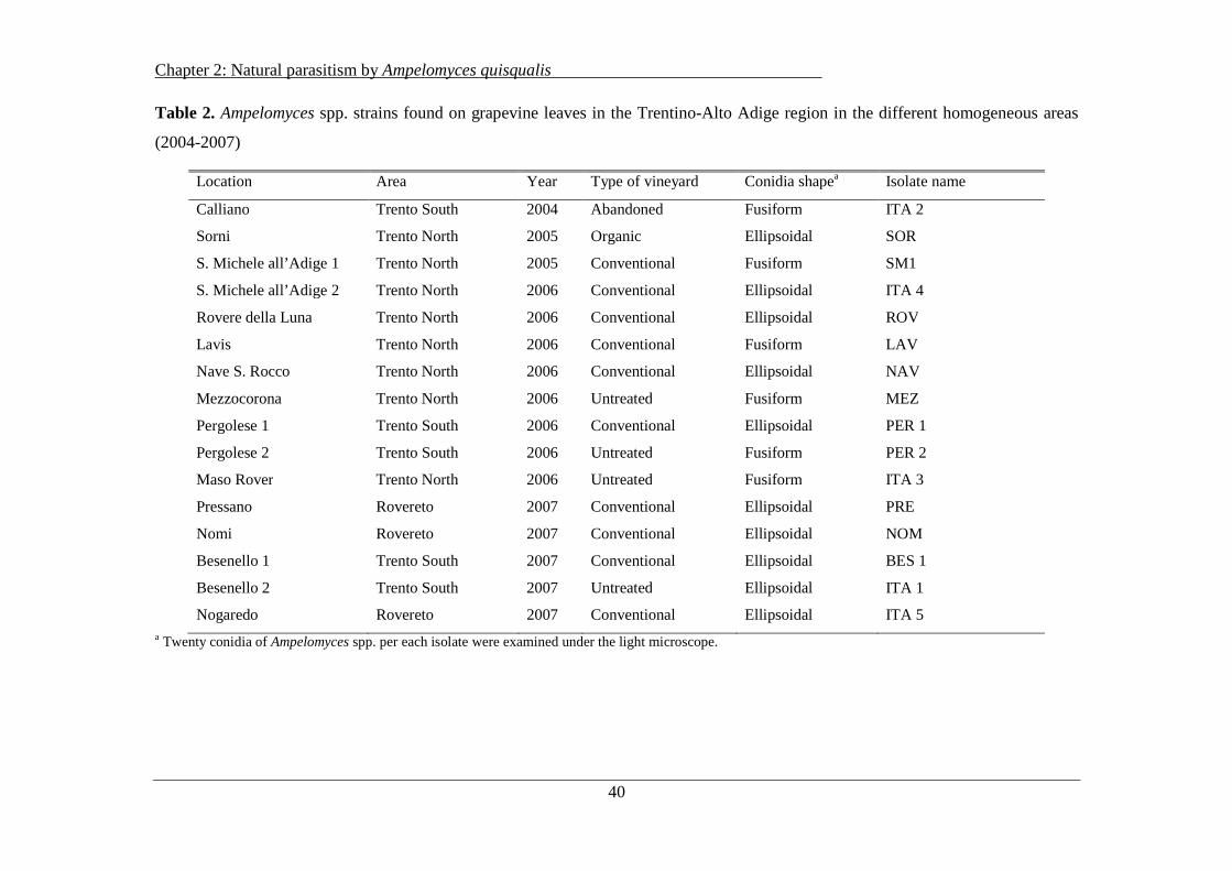
Chapter 2: Natural parasitism by Ampelomyces quisqualis
40
Table 2. Ampelomyces spp. strains found on grapevine leaves in the Trentino-Alto Adige region in the different homogeneous areas
(2004-2007)
Location Area Year Type of vineyard Conidia shapea Isolate name
Calliano Trento South 2004 Abandoned Fusiform ITA 2
Sorni Trento North 2005 Organic Ellipsoidal SOR
S. Michele all’Adige 1 Trento North 2005 Conventional Fusiform SM1
S. Michele all’Adige 2 Trento North 2006 Conventional Ellipsoidal ITA 4
Rovere della Luna Trento North 2006 Conventional Ellipsoidal ROV
Lavis Trento North 2006 Conventional Fusiform LAV
Nave S. Rocco Trento North 2006 Conventional Ellipsoidal NAV
Mezzocorona Trento North 2006 Untreated Fusiform MEZ
Pergolese 1 Trento South 2006 Conventional Ellipsoidal PER 1
Pergolese 2 Trento South 2006 Untreated Fusiform PER 2
Maso Rover Trento North 2006 Untreated Fusiform ITA 3
Pressano Rovereto 2007 Conventional Ellipsoidal PRE
Nomi Rovereto 2007 Conventional Ellipsoidal NOM
Besenello 1 Trento South 2007 Conventional Ellipsoidal BES 1
Besenello 2 Trento South 2007 Untreated Ellipsoidal ITA 1
Nogaredo Rovereto 2007 Conventional Ellipsoidal ITA 5 a Twenty conidia of Ampelomyces spp. per each isolate were examined under the light microscope.

Chapter 2: Natural parasitism by Ampelomyces quisqualis
41
Table 3. Comparison of ITS sequences of five wild isolates found in the Trentino-Alto Adige region and
other isolates from GenBank with the most similar NCBI library strains
Fungal isolate Sequence length (bp) Closest NCBI library strain Similarity (%)
ITA 1 589 Ampelomyces sp. AQ10 (AF035783) a
Ampelomyces sp.HMLAC214 (DQ490765)
100
100
ITA 2 649 Ampelomyces sp. AQ10 (AF035783) 100
ITA 3 585 A. quisqualis (AF126817) 97
ITA 4 582 Ampelomyces quisqualis (AF126817) 97
ITA 5 575 Ampelomyces quisqualis (AF126817) 97
AQ10 520 Ampelomyces sp.HMLAC214 (DQ490765) 99
CBS 128.79 587 Ampelomyces sp.CBS130.79 (U82449) 99
CABI 272851 588 Ampelomyces sp.HMLAC214 (DQ490765) 99
ATCC 250 605 A. quisqualis (AF126817) 97 a Accession number.
Figure 6. The Neighbour Joining tree of nine Ampelomyces spp. sequences as inferred with Mega4
software package (34) using the Jukes-Cantor substitution model and rate uniformity among sites. All
gaps and missing data of the 511 characters long alignment were eliminated. On the branches are
indicated the bootstrap values. The scale bar represents the number of inferred substitutions per 100 sites
(nucleotide residues).

Chapter 2: Natural parasitism by Ampelomyces quisqualis
42
2.4 Discussion
Powdery mildew caused by E. necator is an important disease of grapevine in the Trentino-Alto
Adige region, second only to downy mildew (Plasmopara viticola). If it is not controlled
(untreated or abandoned vineyards), the disease can become extremely widespread and
devastating (Table 1). Chasmothecia were formed in all the monitored vineyards in each year of
the study, confirming that the initiation and development of chasmothecia in Italian vineyards
can occur in any year in which conditions are favorable for disease development (Cortesi et al.,
1995). The high incidence of chasmothecia in the vineyards of the Trentino-Alto Adige region,
which was associated with the absence of flag shoots in the monitored vineyards, could imply
that chasmothecia are the primary inoculum and confirm previous results, which had indicated
that chasmothecia represent the main source of inoculum in the spring (Cortesi et al., 1995).
The monitoring of powdery mildew infections carried out during the course of this four-year
study led to the detection of a highly significant relationship between the level of powdery
mildew infection (incidence and severity) and the quantity of overwintering chasmothecia
formed on leaves. The amount of chasmothecia was generally low in conventional vineyards and
therefore their presence could be probably reduced by applications of an effective method of
disease control during the growing season. Even if some reports have stated that most of the
chasmothecia die during the winter without having any opportunity to release ascospores
(Cortesi et al., 1995), a reduction in the number of overwintering chasmothecia by hyperparasites
may nevertheless be useful for reducing the amount of primary inoculum present in the region.
In the Trentino-Alto Adige region, chasmothecia were formed on the surface of infected leaves
beginning in the late summer. The many studies on E. necator chasmothecia have reported
unclear and sometimes contradictory effects of environmental factors on the development of
chasmothecia (Gadoury & Pearson, 1988). Reports based on field studies have suggested that the
formation of ascocarps is triggered by severe powdery mildew infection, drought, cold, heat or
environmental conditions that are generally unfavorable for the parasite. Host nutrition and host
resistance have both been reported to affect the formation of chasmothecia (Gadoury & Pearson,
1988). Our results suggest that the concentration of chasmothecia per unit of leaf area was
mainly related to the level of disease in the vineyard, which is usually the result of complex
interactions among the amount of initial inoculum, the efficacy of disease control measures,
Chapter 2: Natural parasitism by Ampelomyces quisqualis
43
cultivar susceptibility, the physiology of the crop, and the weather conditions during the growing
season. The elevation of the vineyard, which is inversely related to temperature, was not related
to the concentration of chasmothecia.
A. quisqualis is one of the most successful commercialized biocontrol agents. It acts directly by
invading and destroying host cytoplasm (Hashioka & Nakai, 1980). The present study is the first
extensive investigation of quantitative aspects of A. quisqualis on grapevine powdery mildew in
Italy. Ampelomyces spp. were observed parasitizing colonies and chasmothecia of E. necator on
grapevine leaves in the main viticultural areas of Trentino-Alto Adige region suggesting that,
although at low levels, Ampelomyces spp. was naturally present in the region. This finding also
adds support to the claim that chasmothecia parasitism is probably widespread wherever A.
quisqualis is present (Falk et al., 1995a). In Trentino, the level of natural chasmothecia
parasitism by A. quisqualis on leaves was shown to be lower than that observed in New York
State (Falk et al., 1995a), but similar to what was reported for chasmothecia on vine bark in New
York State. The conidia size of the Italian isolates was quite variable and generally longer than
reported in literature (Falk et al., 1995a).
Even if A. quisqualis (AQ10) was not directly used in the monitored vineyards during the studied
period, it is and was used in the surrounding area. The similarity between the ITS sequences of
two of the isolates and that of AQ10 may suggest that the commercial strain could have become
established in the area. The fact that Ampelomyces was found continuously in certain vineyards
may indicate that, in some areas, it may find suitable climatic conditions for parasitism. Among
the isolated Ampelomyces spp., three strains that are probably new were identified (ITA 3/ITA 4,
ITA 2, and ITA 5). The ITS sequences of these strains were different from that of the
commercial Ampelomyces strain (AQ10). In any case, further exhaustive studies are necessary
for any inferences concerning Ampelomyces spp. population genetics.
The widespread presence of the disease at the end of the growing season did not correspond to
high levels of parasitism in the same season. At the levels of infection observed in the Trentino-
Alto Adige region, the potential for the mycoparasite to naturally reduce the levels of
overwintering inoculum available for the next growing season and/or to impede disease
development were relatively low. A few hypotheses can be raised to explain the minimal
presence of A. quisqualis on E. necator in the region. The prevailing environmental conditions

Chapter 2: Natural parasitism by Ampelomyces quisqualis
44
may not meet the requirements of the mycoparasite (Kiss et al., 2004; Sundheim & Krekling,
1982). It is also possible that the low level of powdery mildew disease before the beginning of
the survey (from 1999 to 2003) (Mescalchin et al., 2005) may also have reduced the natural
population of the mycoparasite in the area.
Numerous Ampelomyces mycoparasites are found around the world (Liang et al., 2007). Some of
the A. quisqualis strains isolated in the Trentino-Alto Adige region were different from AQ10.
Therefore, they may represent new isolates that are better adapted to local environmental
conditions and their discovery may be the starting point of their development for use as
biocontrol agents to control powdery mildew under the environmental conditions found in
northern countries. Further analyses are necessary to better characterize the aggressiveness of
these isolates against E. necator and their environmental requirements for development and
parasitism.
2.5 Acknowledgements
We thank L. Maines, D. Ress, E. Di Marino and E. Mescalchin for the technical support and
assistance. This research was supported by SafeCrop Centre, funded by Autonomous province of
Trento.
2.6 Literature
Altschul, S. F., Gish, W., Miller, W., Myers, E. W. and Lipman, D. J. (1990). Basic Local Alignment Search Tool. J.
Mol. Biol. 215: 403-410.
Angeli, D., Di Marino, E. and Mescalchin, E. (2006). Colonization of grapevine powdery mildew cleistothecia by
the mycoparasite Ampelomyces quisqualis in Trentino-Alto Adige, Italy. IOBC/WPRS bulletin 29(11):89-92.
Bulit, J. U. and Lafon, R., 1978. Powdery mildew of the vine. Pages 525-648 in: The Powdery Mildews. Academic
Press, New York.
Cortesi, P., Gadoury, D. M., Seem, R. C. and Pearson, R. C. (1995). Distribution and retention of cleistothecia of
Uncinula necator on the bark of grapevines. Plant Dis. 79:15-19.
Falk, S. P., Gadoury, D. M., Cortesi, P., Pearson, R. C. and Seem R. C. (1995a). Parasitism of Uncinula necator
cleistothecia by the mycoparasite Ampelomyces quisqualis. Phytopathology 85:794-800.

Chapter 2: Natural parasitism by Ampelomyces quisqualis
45
Falk, S. P., Gadoury, D. M., Pearson, R. C. and Seem, R. C. (1995b). Partial control of grape powdery mildew by
the mycoparasite Ampelomyces quisqualis. Plant Dis. 79:483-490.
Felsenstein, J. (1985). Confidence limits on phylogenies: An approach using the bootstrap. Evolution 39:783-791.
Gadoury, D. M. and Pearson, R. C. (1988). Initiation, development, dispersal, and survival of cleistothecia of
Uncinula necator in New York vineyards. Phytopathology 78:1413-1421.
Gadoury, D. M., Seem, R. C., Pearson, R. C., Wilcox, W. F. and Dunst, R. M. (2001). Effects of powdery mildew
on vine growth, yield, and quality of Concord grapes. Plant Dis. 85:137-140.
Grove, G. G. (2004). Perennation of Uncinula necator in vineyards of Eastern Washington. Plant Dis. 88:242-247.
Hall, T. A. (1999). BioEdit: a user-friendly biological sequence alignment editor and analysis program for Windows
95/98/NT. Nucl. Acids. Symp. Ser. 41:95-98.
Hashioka, Y. and Nakai, Y. (1980). Ultrastructure of pycnidial development and mycoparasitism of Ampelomyces
quisqualis parasitic on Erysiphales. Trans. Mycol. Soc. Jpn 21:329-338.
Kiss, L. (1997). Genetic diversity in Ampelomyces isolates, hyperparasites of powdery mildew fungi, inferred from
RFLP analysis of the rDNA ITS region. Mycol. Res. 101:1073-1080.
Kiss, L. (1998). Natural occurrence of Ampelomyces intracellular mycoparasites in mycelia of powdery mildew
fungi. New Phytol. 140:709-714.
Kiss, L. (2003). A review of fungal antagonists of powdery mildews and their potential as biocontrol agents. Pest
Manag. Sci. 59:475-483.
Kiss, L. and Nakasone, K. K. (1998). Ribosomal DNA internal transcribed spacer sequences do not support the
species status of Ampelomyces quisqualis, a hyperparasite of powdery mildew fungi. Curr. Gen. 33:362-367.
Kiss, L., Russell, J. C., Szentivanyi, O., Xu, X. and Jeffries, P. 2004. Biology and biocontrol potential of
Ampelomyces mycoparasites, natural antagonist of powdery mildew fungi. Biocontrol Sci. Technol. 14:635-
651.
Larkin, M. A., Blackshields, G., Brown, N. P., Chenna, R., McGettigan, P. A., McWilliam, H., Valentin, F.,
Wallace, I. M., Wilm, A., Lopez, R., Thompson, J. D., Gibson, T. J. and Higgins, D. G. (2007). ClustalW2 and
ClustalX version 2. Bioinformatics 23:2947-2948.
Liang, C., Yang, J., Kovacs, G. M., Szentivanyi, O., Li, B., Xu, X. M. and Kiss, L. (2007). Genetic diversity of
Ampelomyces mycoparasite isolated from different powdery mildew species in China inferred from analyses of
rDNA ITS sequences. Fung. Diversity 24:225-240.
Mescalchin, E., Di Marino E. and Angeli, D. (2005). Monitoraggio della presenza di Ampelomyces nei vigneti del
Trentino. Terra Trentina 5:32-37.
Ough, C. S. and Berg, H. W. (1979). Powdery mildew sensory effect on wine. Am. J. Enol. Vit. 30:321.
Chapter 2: Natural parasitism by Ampelomyces quisqualis
46
Page, R. D. M. (1996). Tree View: An application to display phylogenetic trees on personal computers. Comput.
Appl. Biosci. 12:357-358.
Pearson, R. C. and Gadoury, D. M. (1987). Cleistothecia, the source of primary inoculum for grape powdery mildew
in New York. Phytopathology 77:1509-1514.
Pearson, R. C. and Goheen, A. C. (1988). Compendium of Grape Diseases. American Phytopathological Society, St.
Paul, MN.
Rumbolz, J. and Gubler, W. D. (2005). Susceptibility of grapevine buds to infection by powdery mildew Erysiphe
necator. Plant Path. 54:535-548.
Ryan, T. A. (1960). Significance tests for multiple comparison of proportions, variances, and other statistics.
Psychol. Bull. 57:318-328.
Sall, M. A. and Wrysinski, J. (1982). Perennation of powdery mildew in buds of grapevine. Plant Dis. 66:678-9.
Staden, R., Beal, K. F. and Bonfield, J. K. (2000). The Staden package, 1998. Bioinform. Meth. Prot. 132:115-130.
Sullivan, R. F. and White, J. F. Jr. (2000). Phoma glomerata as a mycoparasite of powdery mildew. Appl. Environ.
Microb. 66:425-427.
Sundheim, L. and Krekling, T. (1982). Host-parasite relationships of the hyperparasite Ampelomyces quisqualis and
its powdery mildew host Sphaerotheca fuliginea. I. Scanning electron microscopy. Phytopath. Z. 104:202-210.
Sundheim, L. and Tronsmo, A. (1988). Hyperparasites in biological control. Pages 53-69 in: Biocontrol of Plant
Diseases. Vol. I. CRC Press, Boca Raton, FL.
Szentiványi, O., Kiss, L., Russell, J. C., Kovács, G. M., Varga, K., Jankovics, T., Lesemann, S., Xu, X. M. and
Jeffries, P. (2005). Ampelomyces mycoparasites from apple powdery mildew identified as a distinct group
based on single-stranded conformation polymorphism analysis of the rDNA ITS region. Mycol. Res.109:429-
438.
Sztejnberg, A. (1993). Ampelomyces quisqualis AQ10, CNCM I-807, for biological control of powdery mildew. US
patent no. 5190754.
Sztejnberg, A., Galper, S., Mazar, S. and Lisker, N. (1989). Ampelomyces quisqualis for biological and integrated
control of powdery mildews in Israel. J. Phytopath. 124:285-295.
Tamura, K., Dudley, J., Nei, M. and Kumar, S. (2007). MEGA4: Molecular Evolutionary Genetics Analysis
(MEGA) software version 4.0. Mol. Biol. Evol. 24:1596-1599.
White, T., J., Bruns, T., Lee, S. and Taylor, J. (1990). Amplification and direct sequencing of fungal ribosomal RNA
genes for phylogenetics. Pages 315-322 in: PCR Protocols: a guide to methods and applications (M. A. Innis,
D. H. Gelfand, J. J. Sninsky and T. J. White, eds), Academic Press, San Diego, California.

Chapter 2: Natural parasitism by Ampelomyces quisqualis
47
Ypema, H. L. and Gubler, W. D. (2000). The distribution of early season grapevine shoots infected by Uncinula
necator from year to year: A case study in two California vineyards. Am. J. Enol. Vit. 51:1-6.
48
Chapter 3
Existence of different physiological forms within genetically
diverse strains of Ampelomyces quisqualis
Published as
Angeli D., Maurhofer M., Gessler C., Pertot I. 2012. Existence of different physiological forms
within genetically diverse strains of Ampelomyces quisqualis. Phytoparasitica, 40 (1): 37-51.

Chapter 3: Characterization of Ampelomyces quisqualis
49
Abstract
Powdery mildew fungi are parasitized by strains of the genetically distinct Ampelomyces
quisqualis. To investigate whether differences in the phylogeny and other cultural,
morphological and physiological characteristics of these different strains are related to
differences in their geographic origins or the host species from which they were isolated, several
strains of different species of Erysiphaceae collected in different countries and possessing
different ITS rDNA sequences were selected and characterized. The results revealed some
significant variation among the selected strains, which provides evidence for the existence of
different physiological forms within the A. quisqualis species. Two groups that display
differential growth on artificial media were identified. These groups also differ in the
morphology of their mycelium, but not in the morphology of their pycnidia and conidia.
Temperature greatly affected the in vitro growth of the A. quisqualis strains and growth rate was
closely correlated to colony color. Differences in the conidial germination of distinct strains were
observed during the recognition phase of the parasitic relationship. The germination of each of
the investigated strains was greatly stimulated by all of the examined powdery mildew species
and not only by the conidia of their original hosts. An Italian strain isolated from grapevine in the
Trentino Alto-Adige region was identified as the strain that germinates the most quickly in the
presence of powdery mildew conidia. Phylogenetic analysis revealed that these A. quisqualis
strains can be classified into five different genetic groups, which generally correlate with the
fungal host of origin and morphological and growth characteristics.
3.1 Introduction
Ampelomyces quisqualis is a widespread hyperparasite of powdery mildews (Falk et al., 1995a;
Kiss, 2003; Kiss, 1998). The natural occurrence of A. quisqualis on various Erysiphaceae species
has been reported in different geographic regions (Angeli et al., 2009a; Kiss, 1997; Kiss et al.,
2004; Rankovic, 1997). Most research on A. quisqualis has focused on its potential use as a
biocontrol agent against powdery mildews of various crops (Bélanger & Labbé 2002; Paulitz &
Bélanger, 2001; Sundheim & Tronsmo, 1988; Sztejnberg et al., 1989). Its biology and biocontrol
potential were reviewed by Kiss et al. (2004).
Chapter 3: Characterization of Ampelomyces quisqualis
50
This mycoparasite invades and destroys host cytoplasm, killing the parasitized powdery mildew
cells (Falk et al., 1995ab; Hashioka & Nakai, 1980; Kiss et al., 2004). Intracellular pycnidia of A.
quisqualis are commonly found in hyphae, conidiophores and immature ascomata of powdery
mildews (Kiss et al., 2004). These pycnidia vary in shape depending upon the fungal structure in
which they are formed (Sundheim & Krekling, 1982). Pycnidia contain cylindrical to spindle-
shaped conidia, which are occasionally curved and two-spotted (Falk et al., 1995a). Recently, the
microcyclic conidiogenesis of powdery mildews has been investigated (Kiss et al., 2009). When
mildew colonies are treated with a suspension of A. quisqualis conidia, pycnidia are formed in
microcyclic conidiophores, thereby accelerating the asexual reproduction of A. quisqualis.
The concentration of A. quisqualis conidia is an important factor affecting their germination.
Germination has been shown to dramatically decrease when conidia are at a concentration of
more than 106 conidia ml-1, due to the production of self-inhibitory substances (Gu & Ko, 1997).
A. quisqualis conidia do not germinate well in sterile, distilled water. However, their germination
is significantly enhanced in the presence of conidia of powdery mildew fungi (Sundheim 1982).
The presence of host fungi is recognized by A. quisqualis and a water-soluble substance from
conidia of powdery mildew fungi has been shown to stimulate the germination of A. quisqualis
conidia in vitro (Gu & Ko, 1997). After penetration, the hyphae of the mycoparasite continue to
grow and produce their intracellular pycnidia after 5 to 8 days in the mycelia of their fungal host
(Hashioka & Nakai, 1980; Sundheim & Krekling, 1982). High relative humidity and
temperatures between 20° and 25 °C enhance the growth and sporulation of A. quisqualis, but
this mycoparasite survives and is active against powdery mildew even at temperatures below
12°C (Jarvis & Slingsby, 1977; Philipp & Cruger, 1979).
A. quisqualis is known to be a slow-growing fungus with an in vitro radial growth rate of 0.5–1.0
mm d-1 on Czapek-Dox agar supplemented with 2 % malt extract (MCzA) at 23 °C (Kiss, 1997;
Kiss & Nakasone, 1998). Until recently, A. quisqualis was often confused with several other
species: A. quercinus, A. humuli, A. heracli and Phoma glomerata. Unlike A. quisqualis, these
other powdery mildew mycoparasites do not produce intracellular pycnidia and they also grow
more quickly, with in vitro radial growth rates of 3–4 mm d-1 (Kiss et al., 2004; Sullivan &
White, 2000). In the past, scientists identified all of these fast-growing strains as A. quisqualis
(e.g., Mhaskar, 1974a; Kiss, 1997). Later, a molecular phylogenetic study of rDNA ITS
sequences (Kiss & Nakasone, 1998) revealed that the slow-growing and fast-growing strains

Chapter 3: Characterization of Ampelomyces quisqualis
51
belong to two distinct groups. The slow-growing strains are genetically more diverse; whereas
the fast-growing ones show a close phylogenetic relationship with Epicoccum nigrum, which is
known to have a pycnidial, Phoma-like state (Kiss et al., 2004). It seems likely that the fast-
growing strains are, in fact, Phoma species; whereas true A. quisqualis strains always grow
slowly in culture and produce intracellular pycnidia in powdery mildew mycelium.
Several A. quisqualis strains are available from culture collections and one strain has been
formulated, registered and commercialized under the trade name of AQ10 (Kiss, 1997;
Sztejnberg, 1993). Many molecular analyses based on the internal transcribed spacer (ITS)
region of the nuclear ribosomal RNA gene (nrDNA) have revealed considerable genetic diversity
among A. quisqualis strains (Angeli et al., 2009b; Liang et al., 2007; Park at al., 2010;
Szentiványi et al., 2005). Recently, ITS sequences and microsatellite markers have been used to
show that a set of A. quisqualis populations found in apple powdery mildew (Podosphaera
leucotricha) are quite genetically distinct from populations collected from several other powdery
mildew species infecting other plant species (Kiss et al., 2011). Although the genetic diversity
within the A. quisqualis species has already been characterized, we still do not know whether the
differences in the phenotypic characteristics of different strains of A. quisqualis are related to
their phylogeny.
The morphological and cultural characteristics of this hyperparasite on different fungal hosts and
plants have been reported to be extremely variable (Belsare et al. 1980; Shin and Kyeung 1994;
Speer 1978). Some older studies focused on the shape and size of the pycnidia and conidia on
culture media (e.g., Mhaskar, 1974a, Mhaskar & Rao, 1974b) and fungal hosts (e.g., Kiss, 1997;
Rankovic, 1997). However, all of these studies were carried out when fast-growing strains
(Phoma glomerata and Ampelomyces spp.) were still classified as A. quisqualis. Therefore, a
thorough investigation of A. quisqualis sensu strictu is lacking. More physiological and
phenotypic information is required to complete the picture of the genetic differences that have
been identified in previous studies (Liang et al., 2007; Park et al., 2010; Kiss et al., 2011).
In the present work, we selected several A. quisqualis strains from different hosts and geographic
regions, all characterized by the formation of intracellular pycnidia and slow radial growth at
room temperature in vitro (defined as Type II; Kiss, 1997), features corresponding to the
characterization of A. quisqualis sensu strictu. We investigated the cultural, morphological and
Chapter 3: Characterization of Ampelomyces quisqualis
52
growth characteristics and phylogenetic relations (ITS) of these strains. The objectives of this
study were to determine whether the host or site of origin of the strains or their cultural,
morphological and/or growth characteristics are related to their phylogenetic group which would
indicate an adaptation to the host or geographic area. Furthermore, some physiological tests were
performed as we looked for evidence of any host specificity or temperature adaptation of strains
affecting the recognition phase of the host-parasite interaction.
3.2 Material and Methods
A total of 28 A. quisqualis strains isolated from different crops, sites and powdery mildew
species were included in this study (Table 1). We selected strains isolated from grapevine (Vitis
vinifera), cucumber (Cucumis sativus) and apple (Malus domestica) in different geographic
regions, as well as strains isolated from wild and/or ornamental species. Only slow-growing
strains [e.g., Type II (0.5–1.0 mm d-1) (Kiss, 1997)] producing intracellular pycnidia within
powdery mildew mycelia were selected. The strains were obtained from culture collections
(ATCC, American Type Culture Collection, Rockville, MD, USA; CBS, Centraalbureau voor
Schimmelcultures, Baarn, Netherlands; CABI, Commonwealth Agricultural Bureaux
International, Egham, UK; DSMZ, Deutsche Sammlung von Mikroorganismen und Zellkulturen
Gmbh, Braunschweig, Germany) or provided by individual scientists (L. Kiss, PPI, Plant
Protection Institute of the Hungarian Academy of Sciences, Budapest, Hungary; D. Angeli,
FEM, Fondazione Edmund Mach, S. Michele all’Adige, Italy).
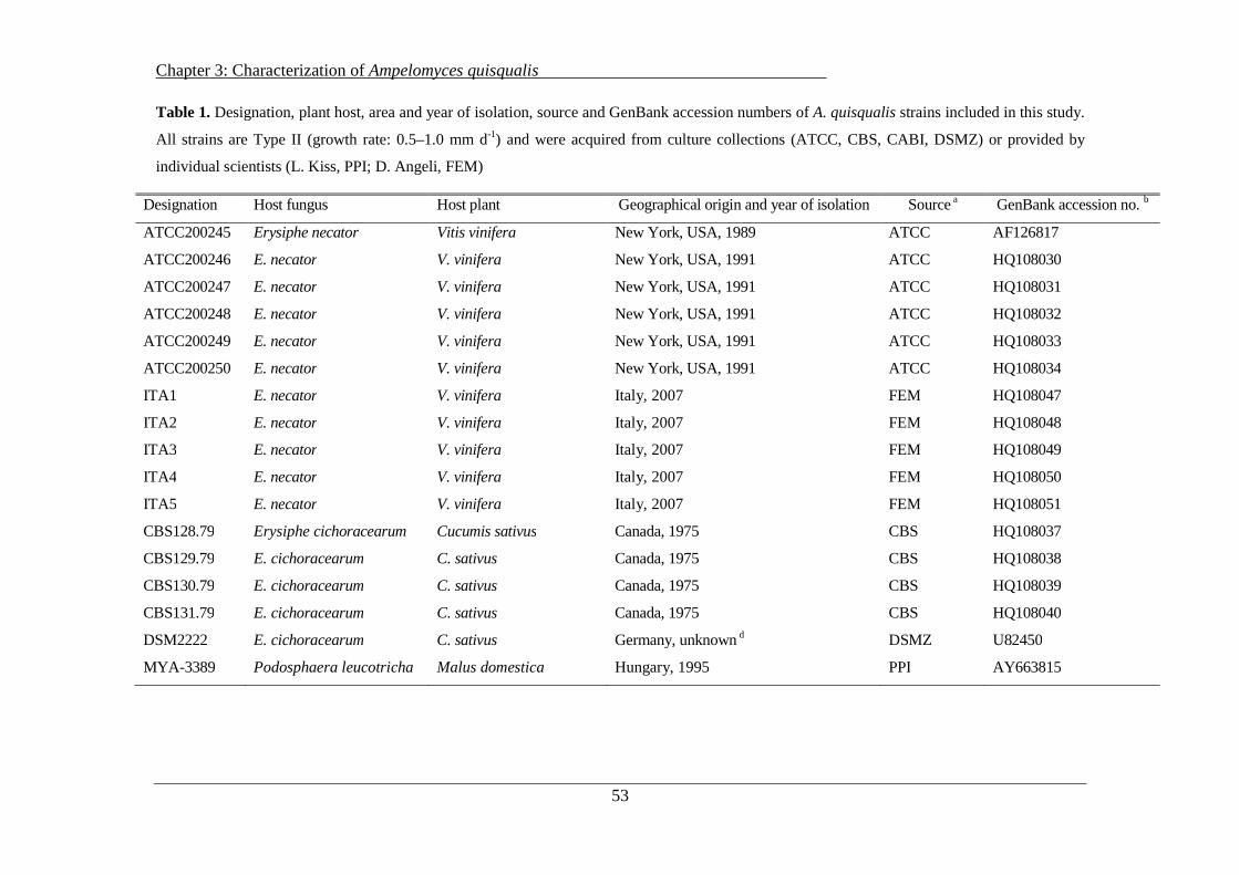
Chapter 3: Characterization of Ampelomyces quisqualis
53
Table 1. Designation, plant host, area and year of isolation, source and GenBank accession numbers of A. quisqualis strains included in this study.
All strains are Type II (growth rate: 0.5–1.0 mm d-1) and were acquired from culture collections (ATCC, CBS, CABI, DSMZ) or provided by
individual scientists (L. Kiss, PPI; D. Angeli, FEM)
Designation Host fungus Host plant Geographical origin and year of isolation Source a GenBank accession no. b
ATCC200245 Erysiphe necator Vitis vinifera New York, USA, 1989 ATCC AF126817
ATCC200246 E. necator V. vinifera New York, USA, 1991 ATCC HQ108030
ATCC200247 E. necator V. vinifera New York, USA, 1991 ATCC HQ108031
ATCC200248 E. necator V. vinifera New York, USA, 1991 ATCC HQ108032
ATCC200249 E. necator V. vinifera New York, USA, 1991 ATCC HQ108033
ATCC200250 E. necator V. vinifera New York, USA, 1991 ATCC HQ108034
ITA1 E. necator V. vinifera Italy, 2007 FEM HQ108047
ITA2 E. necator V. vinifera Italy, 2007 FEM HQ108048
ITA3 E. necator V. vinifera Italy, 2007 FEM HQ108049
ITA4 E. necator V. vinifera Italy, 2007 FEM HQ108050
ITA5 E. necator V. vinifera Italy, 2007 FEM HQ108051
CBS128.79 Erysiphe cichoracearum Cucumis sativus Canada, 1975 CBS HQ108037
CBS129.79 E. cichoracearum C. sativus Canada, 1975 CBS HQ108038
CBS130.79 E. cichoracearum C. sativus Canada, 1975 CBS HQ108039
CBS131.79 E. cichoracearum C. sativus Canada, 1975 CBS HQ108040
DSM2222 E. cichoracearum C. sativus Germany, unknown d DSMZ U82450
MYA -3389 Podosphaera leucotricha Malus domestica Hungary, 1995 PPI AY663815

Chapter 3: Characterization of Ampelomyces quisqualis
54
Table 1. (continued)
Designation Host fungus Host plant Geographical origin and year of isolation Source a GenBank accession no. b
MYA -3391 P. leucotricha M. domestica Hungary, 1995 PPI HQ108043
MYA -3394 P. leucotricha M. domestica United Kingdom, 2002 PPI HQ108044
MYA -3395 P. leucotricha M. domestica Germany, 2002 PPI AY663817
MYA -3398 P. leucotricha M. domestica United Kingdom, 2002 PPI HQ108045
AQ10 Powdery mildew c Chata edulis Israel, unknown d Ecogen Italia AF035783
DSM2225 Erisiphe heracli Daucus sp. Germany, unknown d DSMZ HQ108042
DSM4624 Sphaerotheca fuliginea Leontodon sp. Germany, unknown d DSMZ HQ108041
CABI272851 Powdery mildew c Schinus molle Ecuador, 1983 CABI HQ108036
CABI380234 Powdery mildew c Alliar ia petiolata Switzerland, 1999 CABI HQ108035
ATCC201056 Oidium hortenisae Lycium halimifolium Hungary, 1990 ATCC AF035781
MYA -3401 Oidium hortenisae Hydrangea macrophylla United Kingdom, 1999 PPI HQ108046 a ATCC, American Type Culture Collection, Rockville, MD, USA; CBS, Centraalbureau voor Schimmelcultures, Baarn, Netherlands; CABI, Commonwealth Agricultural Bureaux International, Egham, United Kingdom; DSMZ, Deutsche Sammlung von Mikroorganismen und Zellkulturen Gmbh, Braunschweig, Germany); FEM, Fondazione Edmund Mach, S. Michele all’Adige, Italy; PPI, Plant Protection Institute of the Hungarian Academy of Sciences, Budapest, Hungary. b GenBank accession numbers of the rDNA ITS sequences. c Unknown species. d Data not available.

Chapter 3: Characterization of Ampelomyces quisqualis
55
Table 2. GenBank accession numbers of the rDNA ITS sequences, plant hosts, area and year of isolation and source of published A.
quisqualis sequences included in the ITS sequence analysis
GenBank accession no. a Host fungus Host plant Geographical origin and year of isolation Source
GQ324149 b Erysiphe necator Vitis vinifera Seoul, South Korea, 2007 Park et al. (2010)
GQ324144 b E. necator Vitis flexuosa Jeju, South Korea, 2006 Park et al. (2010)
DQ490750 Podosphaera xanthii Cucurbita pepo China, 2002 Liang et al. (2007)
DQ490752 P. xanthii Cucumis moschata China, 2002 Liang et al. (2007)
DQ490755 P. xanthii Cucumis sativus China, 2002 Liang et al. (2007)
DQ490759 P. xanthii Cucurbita maxima China, 2002 Liang et al. (2007)
AY663816 Podosphaera leucotricha Malus domestica Hungary, 2000 Szentivanyi et al. (2005)
AY663818 P. leucotricha M. domestica UK, 2002 Szentivanyi et al. (2005)
AY663819 P. leucotricha M. domestica UK, 2002 Szentivanyi et al. (2005)
AY663820 P. leucotricha M. domestica Germany, 2002 Szentivanyi et al. (2005)
AY663821 P. leucotricha M. domestica Hungary, 2002 Szentivanyi et al. (2005)
GQ324095 b Erysiphe ranunculi Clematis apiifolia Seogwipo, South Korea, 2006 Park et al. (2010)
GU329997 b Golovinomyces cichoracearum Zinnia elegans Yangpyeong, South Korea, 2008 Park et al. (2010)
DQ490762 Arthrocladiella mougeotii Lycium chinense China, 2003 Liang et al. (2007)
DQ490767 Oidium sp. Castanopsis sp. China, 2002 Liang et al. (2007) a GenBank accession numbers of the rDNA ITS sequences. b Strains identified in the maximum-likelihood tree with the same isolate numbers used by Park et al. (2010).

Chapter 3: Characterization of Ampelomyces quisqualis
56
For phylogenetic comparison, 15 sequences of ITS rDNA of strains of A. quisqualis were
retrieved from GenBank (Table 2). Strains were selected according to the fungal host of origin.
The selected strains represent the four major clades reported in a recent study by Park et al.
(2010).
3.2.1 Cultural, morphological and physiological characteristics
The cultural behavior and morphological characteristics of the pycnidia and conidia of the 28
selected A. quisqualis strains were evaluated on six different types of media: potato dextrose agar
(Oxoid, Hampshire, UK, 39 g l-1 twice-distilled water), sucrose nutrient agar (50 g sucrose, 28 g
nutrient agar, twice-distilled water to 1 l), yeast malt dextrose agar (10 g dextrose, 5 g peptone, 3
g yeast extract, 3 g malt extract, 20 g agar, twice-distilled water to 1 l), nutrient agar (Oxoid, 28 g
l-1 twice-distilled water), Czapek agar (Oxoid, 33.4 g l -1 twice-distilled water) and cornmeal agar
(Oxoid, 17 g l-1 twice-distilled water).
The Petri-dish cultures of fungus were prepared with 100 µl of a suspension of sterile distilled
water containing 102 conidia ml-1. The color of the mycelia and pycnidia was assessed by visual
observation of colonies grown for 14 days at 25 °C in the dark. We measured the length and
width of 60 pycnidia and 60 conidia per strain (20 per replicate) on glass slides under a light
microscope (Hund Wetzlar H 600LL, Wetzlar, Germany). The radial growth rate of the A.
quisqualis strains was evaluated at temperatures of 10°, 15°, 20°, 25° and 30 °C on Petri plates
containing potato dextrose agar (PDA) that were inoculated as described above. Cultures were
incubated in the dark for 30 days and the radial growth of the colonies (20 per replicate) was
evaluated by measuring the diameter of each colony twice a week. All of the experiments were
conducted with three replicates (plates) per strain.
Furthermore, the recognition of the fungal host by A. quisqualis strains at different temperatures
was studied. We evaluated the effects of the conidia of four powdery mildew species,
Podosphaera xanthii (from cucumber), E. necator (from grapevine), Podosphaera aphanis (from
strawberry) and Podosphaera leucotricha (from apple), on the germination rates of five selected
A. quisqualis strains. Each of the tested strains belongs to a different genetic (ITS) group
(identified in this study) and was collected from a different host plant in a different geographic

Chapter 3: Characterization of Ampelomyces quisqualis
57
area (AQ10, ITA3, CBS129.79, MYA-3391 and MYA-3401). Each strain was paired with an
isolate of the fungal host from which it was originally isolated and three other powdery mildews.
Sterile distilled water suspensions of the conidia of each powdery mildew strain (1 × 10⁵ conidia
ml-1) together with each A. quisqualis strain (1 × 10⁵ conidia ml-1) were prepared, for a total of
20 combinations. Suspensions of only A. quisqualis conidia in sterile distilled water were used as
controls. Six drops (10 µl each) per combination (three replicates) were put onto a glass slide,
which was placed in a Petri dish (RH = 100 %) and stored in a dark incubator kept at 25 °C,
which is the temperature that Gu and Ko (1997) used in their study of the factors affecting the
germination of A. quisqualis conidia. We evaluated the germination rates and germ-tube
elongation of 150 conidia per strain (three replicates, 50 conidia per replicate) under a light
microscope after 24 h.
We also evaluated the effect of temperature on the germination of A. quisqualis conidia in the
presence of the fungal host. In this experiment, we tested two powdery mildew species (P.
xanthii from cucumber and E. necator from grapevine) and the two A. quisqualis strains that
were the most and least responsive to powdery mildew conidia in the previous experiment. We
evaluated both the germination rate and the tube elongation of those strains at three temperatures
(15°, 20° and 25 °C) using the method described above.
3.2.2 Phylogenetic analysis
The phylogenetic relations of the 28 strains analyzed in the present study and another 15 strains
selected for phylogenetic analysis were inferred from maximum-likelihood and maximum-
parsimony analyses in which the sequences of their respective ITS rDNA regions were compared
(White et al. 1990). The additional 15 sequences of ITS rDNA from A. quisqualis strains that
were analyzed by Park et al. (2010) were retrieved from GenBank (Table 2). The strains isolated
by Park et al. (2010), whose GenBank accession numbers begin with GQ and GU, were
identified here with the same isolate numbers used by Park et al. (2010), to make it easier to
recognize them across the two studies (SMKC22963 = GQ324149, SMKC22519 = GQ324144,
SMKC22511 = GQ324095, SMKC23914 = GU329997).

Chapter 3: Characterization of Ampelomyces quisqualis
58
The 28 selected strains (Table 1) were grown on PDA for 21 days at 25 °C. DNA was extracted
from approximately 200 µg of homogenized, lyophilized mycelium, using the Nucleo Spin Plant
Kit (Macherey-Nagel, Düren, Germany) according to the manufacturer’s instructions. The ITS
region of the nuclear ribosomal DNA was amplified using ITS1 and ITS4 fungal-specific
primers (Gardes and Bruns 1993). PCR was performed using the Gene Amp PCR System 9700
(Perkin Elmer, Waltham, MA, USA). PCR products were purified using ExoSAP-IT enzymes
(USB Corporation, Staufen, Germany) and sequenced using a Big Dye Terminator v1.1 Cycle
Sequencing Kit (Applied Biosystems, Foster City, CA, USA). Both DNA strands were
sequenced with the primers that had been used for PCR amplification and electrophoresis was
carried out using an ABI 3130xl Genetic Analyzer (Perkin Elmer). The consensus sequence was
assembled using Pregap4 and Gap4 (Staden et al. 2000; http://staden.sourceforge.net/). Blast
searches were performed in the NCBI/GenBank database
(http://www.ncbi.nlm.nih.gov/Genbank/) using the program Megablast to find long stretches of
alignment between very similar nucleotide sequences. The ClustalW2 program (Larkin et al.
2007; http://www.ebi.ac.uk/Tools/clustalw2/index.html) was used to construct multiple sequence
alignments and to identify evolutionary relationships among the input sequences. Maximum-
likelihood (ML) and maximum-parsimony (MP) analyses based on ITS region sequences were
conducted using the Mega4 program (Tamura et al. 2007;
http://www.megasoftware.net/mega.html) and a p-distance model (Nei and Kumar 2000) was
used to calculate within-group and between-group distances. A phylogenetic tree inferred from
the ITS rDNA region sequences was constructed using the TreeView program, version 1.6.6
(Page 1996; http://taxonomy.zoology.gla.ac.uk/rod/treeview.html). Phoma glomerata
(AF126816), A. quercinus (AF035778) and A. humuli (AF035779) were also included in the
model.
3.2.3 Clustering and statistical analysis
Morphological experiments (size of pycnidia and conidia) were performed in three replicates per
strain and medium and standard deviations of the averages were calculated. A one-way analysis
of variance (ANOVA) was performed on log-transformed and arcsin-transformed data. If

Chapter 3: Characterization of Ampelomyces quisqualis
59
significant differences were detected, treatment means (strains and media) were separated using
Tukey’s test (α = 0.05).
Growth tests involved two independent experiments with three replicates per experiment. A two-
way ANOVA was performed on log-transformed and arcsin-transformed data from two
experiments and revealed no significant Experiment × Treatment (temperature) interaction.
Therefore, cluster analyses were performed using a software package for multivariate data
analysis capable of performing K-means cluster analyses (Multi-Experiment Viewer, TMEV).
The Euclidean distance between each of the samples within a given cluster and their respective
cluster centroid was calculated. The angular coefficients (a) and the correlation coefficients (r) of
the linear regression curves were also calculated. Data from the two experiments were pooled
and averages of six replicates are presented in Table 4 and Figure 1. Statistical analyses were
performed using Statistica software 7.0 (Statsoft, Tulsa, OK, USA). Significant differences
between treatments (strains and temperatures) were separated using Scheffè’s test (α = 0.05).
Germination tests involved two independent experiments with three replicates per experiment
and 50 conidia per replicate. Two-way ANOVA was performed on both the log-transformed and
arcsin-transformed data from the two experiments and revealed no significant Experiment ×
Treatment (temperature) interaction. Therefore, data from the two experiments were pooled.
Averages of six replicates are presented in Figures 2a–b and 3a–b. Significant differences
between treatments (strains and powdery mildews) were separated using Tukey’s test (α = 0.05).
3.3 Results
3.3.1 Cultural, morphological and physiological characteristics
Cultural and morphological analyses of A. quisqualis strains indicated that two different colony
types can be distinguished after 14 days on all tested media: A. quisqualis strains that form dark-
brown colonies and those that form olive-green colonies (mycelium and pycnidia; Table 3).
Furthermore, mycelial growth and pycnidial and conidial formation were all greatly increased on
three of the tested media (potato dextrose agar, sucrose nutrient agar and yeast malt dextrose
agar), in comparison with the other media. The study on morphology of pycnidia and conidia
(Table 3) revealed differentiation in color and dimensions of the fruiting bodies of the tested
Chapter 3: Characterization of Ampelomyces quisqualis
60
strains. In all of the examined strains, the color and shape of the pycnidia varied from light to
dark (brown or green) and from ovoid to ellipsoid, with increasing age of the culture. The length
and width of the pycnidia ranged from 30 to 95 µm and from 25 to 55 µm, respectively. A great
deal of variation in width and length was observed among the pycnidia of individual strains, but
no significant differences were found between strains (P = 0.112). The conidia of the different
strains also varied in size. They ranged from 5.5 to 14.5 µm in length and from 2.3 to 3.5 µm in
width. However, there were no significant differences between strains according to Tukey’s test
(P = 0.073).
Temperature greatly affected the growth rate of A. quisqualis growth on PDA. The highest radial
growth rates for all strains were measured between 15° and 25 °C. At the extreme temperatures
of 10° and 30 °C, sharp reductions were observed in the growth rates (Fig. 1). K-means
clustering based on Euclidean distances showed that at temperatures of 15°, 20° and 25 °C, the
slow-growing Type II strains (Kiss 1997) could be further clustered into two different growth-
rate groups that were significantly different from each other. At all of these temperatures, the two
groups included the same isolates, which we refer to as slow-growing (0.70–0.82 mm d-1) and
very slow-growing (0.34–0.63 mm d-1) (Table 4). At 10° and 30 °C, K-means clustering analysis
revealed two growth-rate classes, which do not include exactly the same strains as the groups
observed at the other tested temperatures (Table 4). However, no statistically significant
differences were observed between the two classes (P = 0.09). At 10 °C, the strains grew at a rate
of 0.18–0.43 mm d-1; whereas at 30 °C, they grew at a rate of 0.07–0.33 mm d-1.
Our results indicate that, for all of the tested strains except for MYA-3401, there is a relationship
between the growth rates at 15°, 20° and 25 °C and colony color. Strains that grew fairly slowly
at these temperatures formed dark-brown colonies and strains that grew very slowly at these
temperatures formed olive-green colonies. However, even with the limited number of strains
examined, it is possible to demonstrate a relationship between growth rate and the original fungal
host. Strains isolated from apple, grapevine and cucumber plants belonged to the same growth
rate group at temperatures between 15° and 25 °C. On the other hand, no relationship between
the growth rate and the geographic origin of the strains was observed.

Chapter 3: Characterization of Ampelomyces quisqualis
61
Table 3. Morphological characteristics of the A. quisqualis strains included in this study
Designation Colony color a Length and width (µm ± SD)b
mycelia pycnidia pycnidium conidium
ATCC200245 Olive green Light/dark olive green 65±25 × 38±15 8.9±1.9 × 2.7±0.4
ATCC200246 Olive green Light/dark olive green 58±18 × 31±12 8.4±1.6 × 2.9±0.4
ATCC200247 Olive green Light/dark olive green 57±15 × 32±14 7.9±1.3 × 2.8±0.4
ATCC200248 Olive green Light/dark olive green 58±18 × 31±12 8.5±1.9 × 2.8±0.4
ATCC200249 Olive green Light/dark olive green 64±23 × 40±11 8.4±1.7 × 2.9±0.5
ATCC200250 Olive green Light/dark olive green 59±11 × 32± 8 8.5±1.8 × 2.8±0.4
ITA1 Dark brown Light/dark brown 72±23 × 42±13 8.4±1.6 × 2.8±0.4
ITA2 Dark brown Light/dark brown 67± 9 × 39±12 9.8±1.8 × 2.9±0.5
ITA3 Olive green Light/dark olive green 54±15 × 34±11 8.0±1.7 × 2.9±0.4
ITA4 Olive green Light/dark olive green 56±16 × 33±7 7.3±1.5 × 2.9±0.5
ITA5 Olive green Light/dark olive green 64±13 × 37±9 7.3±1.5 × 2.7±0.4
CBS128.79 Dark brown Light/dark brown 59±10 × 35±10 7.1±1.3 × 2.6±0.2
CBS129.79 Dark brown Light/dark brown 66±14 × 41±7 7.3±1.5 × 2.8±0.5
CBS130.79 Dark brown Light/dark brown 64±9 × 38±9 9.0±1.6 × 2.9±0.5
CBS131.79 Dark brown Light/dark brown 71±18 × 37±8 9.1±1.9 × 2.9±0.4
DSM2222 Dark brown Light/dark brown 67±16 × 41±9 7.9±1.2 × 2.7±0.4
MYA-3389 Olive green Light/dark olive green 72±20× 38±12 7.9±1.0 × 2.6±0.2
MYA-3391 Olive green Light/dark olive green 61±16 × 34±9 7.8±1.8 × 2.7±0.4
MYA-3394 Olive green Light/dark olive green 69±15 × 36±11 8.7±1.8 × 2.8±0.4
MYA-3398 Olive green Light/dark olive green 65±10 × 34±7 9.1±1.8 × 2.8±0.4
MYA-3395 Olive green Light/dark olive green 66±17 × 37±10 9.6±1.6 × 2.7±0.4
AQ10 Dark brown Light/dark brown 55±10 × 32±5 7.5±1.6 × 2.6±0.3
DSM2225 Dark brown Light/dark brown 62±15 × 36±10 9.6±1.4 × 2.9±0.4
DSM4624 Dark brown Light/dark brown 58±9 × 36±7 7.9±1.6 × 2.8±0.4
CABI272851 Dark brown Light/dark brown 66±15 × 35±11 8.5±1.6 × 2.7±0.4
CABI380234 Olive green Light/dark olive green 56±18 × 34±12 9.5±1.8 × 3.0±0.4
ATCC201056 Olive green Light/dark olive green 58±17 × 37±12 8.1±1.2 × 2.6±0.2
MYA-3401 Dark brown Light/dark brown 73±19 × 44±14 10.6±2.6 × 2.9±0.5
a Colors of mycelia and pycnidia on the six tested agar media. Three agar plates per strain were examined visually following 14 days of incubation at 25 °C in the dark. b Values are means from cultures grown on the six media (360 pycnidia and 360 conidia per strain). No significant differences between media were detected when Tukey’s tests were applied (P ≥ 0.05). Standard deviations of the means are reported (SD).

Chapter 3: Characterization of Ampelomyces quisqualis
62
Figure 1. Effect of temperature (10°, 15°, 20°, 25° and 30 °C) on the growth of A. quisqualis strains over
30 days on potato dextrose agar. The radial growth rates of 28 strains, which divided into slow-growing
and very slow-growing groups at each temperature, are presented (pooled data). K-means clustering using
the software TMEV (Multi-Experiment Viewer) was based on the Euclidean distance between each of the
strains at each temperature. Significant differences between groups were separated by ANOVA. Presented
values are means of six replicates derived from two independent experiments with three replicates per
experiment. The standard deviation of the six replicates is presented.
y = -0.139x2 + 0.803x - 0.2951
R2 = 0.9911
y = -0.0868x2 + 0.5102x - 0.1788
R2 = 0.921
0
0.2
0.4
0.6
0.8
1
10 15 20 25 30Temperature (°C)
Gro
wth
rat
e (m
m d
-1
)
Slow-growing strains
Very slow-growing strains
a
a
a
a
aa
bb
b
a
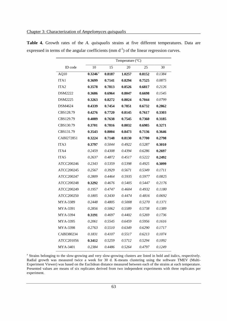
Chapter 3: Characterization of Ampelomyces quisqualis
63
Table 4. Growth rates of the A. quisqualis strains at five different temperatures. Data are
expressed in terms of the angular coefficients (mm d-1) of the linear regression curves.
Temperature (°C)
ID code 10 15 20 25 30
AQ10 0.3246 a 0.8187 1.0257 0.8152 0.1384
ITA1 0.3699 0.7141 0.8294 0.7525 0.0875
ITA2 0.3578 0.7813 0.8526 0.6817 0.2126
DSM2222 0.3686 0.6964 0.8047 0.6698 0.1545
DSM2225 0.3263 0.8272 0.8024 0.7044 0.0799
DSM4624 0.4339 0.7454 0.7851 0.6732 0.2862
CBS128.79 0.4276 0.7720 0.8145 0.7617 0.3303
CBS129.79 0.4089 0.7638 0.7545 0.7360 0.3185
CBS130.79 0.3701 0.7816 0.8032 0.6985 0.3271
CBS131.79 0.3543 0.8004 0.8473 0.7136 0.3646
CABI272851 0.3224 0.7148 0.8130 0.7700 0.2798
ITA3 0.3797 0.5044 0.4922 0.5287 0.3010
ITA4 0.2459 0.4308 0.4394 0.6286 0.2697
ITA5 0.2637 0.4872 0.4517 0.5222 0.2492
ATCC200246 0.2343 0.5359 0.5398 0.4925 0.3099
ATCC200245 0.2567 0.3929 0.5671 0.5349 0.1711
ATCC200247 0.2809 0.4464 0.5935 0.5977 0.0825
ATCC200248 0.3292 0.4676 0.5405 0.5447 0.2176
ATCC200249 0.1957 0.4747 0.4604 0.4932 0.1180
ATCC200250 0.1805 0.3430 0.4474 0.4816 0.0692
MYA-3389 0.2448 0.4805 0.5008 0.5270 0.1371
MYA-3391 0.2856 0.5062 0.5589 0.5738 0.1389
MYA-3394 0.3191 0.4697 0.4402 0.5269 0.1736
MYA-3395 0.2061 0.5545 0.6459 0.5956 0.1616
MYA-3398 0.2763 0.5510 0.6349 0.6290 0.1717
CABI380234 0.1831 0.4107 0.5517 0.6213 0.1074
ATCC201056 0.3412 0.5259 0.5712 0.5294 0.1092
MYA-3401 0.2384 0.4486 0.5264 0.4797 0.1249
a Strains belonging to the slow-growing and very slow-growing clusters are listed in bold and italics, respectively. Radial growth was measured twice a week for 30 d. K-means clustering using the software TMEV (Multi-Experiment Viewer) was based on the Euclidean distance measured between each of the strains at each temperature. Presented values are means of six replicates derived from two independent experiments with three replicates per experiment.

Chapter 3: Characterization of Ampelomyces quisqualis
64
Physiological tests with different powdery mildew species and A. quisqualis strains showed that
the germination rate and germ-tube length of A. quisqualis conidia increased greatly in the
presence of powdery mildew conidia. This stimulation of germination is not powdery mildew
species-specific. All of the tested powdery mildews increased the germination rate and tube
elongation of the A. quisqualis strains isolated from different host species (Figs. 2a–b). However,
in some cases, significant differences in the induction of germination were detected among the
different powdery mildews. For example, the conidial germination of CBS129.79 (originally
isolated from cucumber) was stimulated more strongly by grapevine powdery mildew and
cucumber powdery mildew than by strawberry powdery mildew. The conidial germination of
MYA-3391 (originally isolated from apple) was most strongly induced by grapevine powdery
mildew and was less affected by strawberry and apple powdery mildews. We observed big
differences among the conidial germination rates of the different strains, independent of the
powdery mildew species. The conidial germination of ITA3 and MYA-3401 was strongly
induced by conidia of all of the tested powdery mildew species (both in terms of germination rate
and tube elongation); whereas AQ10 was generally the least responsive.
In the second experiment, in which we investigated the effect of temperature, we again observed
that, in the absence of powdery mildew conidia, the germination rates and germ-tube elongation
of strains ITA3 and AQ10 were very poor (Figs. 3a–b). Both parameters were strongly enhanced
in the presence of powdery mildew conidia, with no statistical differences between the effects of
E. necator and those of P. xanthii. Just as in the previous experiment, ITA3 was significantly
more induced by powdery mildew conidia than AQ10 (Figs. 3a–b). We did not observe any
changes in the general behavior of the two A. quisqualis strains at the different tested
temperatures. In fact, temperature (within the tested range) had no effect on germination rate, but
did seem to affect tube length. In the presence of powdery mildew conidia, tube elongation for
both strains was always greater at the two higher temperatures (20° and 25 °C) than at 15 °C.
In these two experiments, AQ10 exhibited significantly lower germination rates and less tube
elongation in the presence of powdery mildew conidia, as compared to the strain ITA3 (Figs. 3a–
b). However, on agar plates incubated at the examined temperatures, AQ10 and ITA3 were
classified as slow-growing and very slow-growing, respectively. Thus, the growth rates of strains
of A. quisqualis on artificial media are not correlated with their germination rates and tube
elongation in the presence of a host fungus.

Chapter 3: Characterization of Ampelomyces quisqualis
65
Figures 2a–b. Effects of different powdery mildew species (Podosphaera xanthii from cucumber,
Erysiphe necator from grapevine, Podosphaera aphanis from strawberry and Podosphaera leucotricha
from apple) on the conidial germination (%) (a) and tube elongation (µm) (b) of five selected strains of
Ampelomyces quisqualis. Measurements were taken in aqueous suspensions containing mixtures of A.
quisqualis and powdery mildew conidia that had been incubated together for 24 h at 25 °C. Identical
column designs indicate exposure to the same powdery mildew species. Columns labeled with the same
letter (a-d) are not significantly different (P ≤ 0.05) according to Tukey’s test. Three replicates (50
conidia per replicate) of each strain were evaluated. Presented values are means of six replicates derived
from two independent experiments with three replicates per experiment.

Chapter 3: Characterization of Ampelomyces quisqualis
66
Figures 3a–b. Effect of temperature on the conidial germination (%) (a) and tube elongation (µm) (b) of
two selected strains of Ampelomyces quisqualis in the presence of two different powdery mildew species
(Podosphaera aphanis from cucumber and Erysiphe necator from grapevine). Measurements were taken
in aqueous suspensions containing mixtures of A. quisqualis and powdery mildew conidia that had been
incubated for 24 h at three different temperatures (15°, 20° and 25 °C). Identical column designs indicate
incubation at the same temperature. Columns labeled with the same letter (a-e) are not significantly
different according to Tukey’s test (α = 0.05). Three replicates (50 conidia per replicate) of each strain
were evaluated. Presented values are means of six replicates derived from two independent experiments
with three replicates per experiment.
Chapter 3: Characterization of Ampelomyces quisqualis
67
3.3.2 Phylogenetic analysis
In the phylogenetic analysis, the 43 A. quisqualis strains clustered into five different groups (1–
5), revealing genetic diversity among the A. quisqualis strains included in the present work (Fig.
4). A. quisqualis strains with similar ITS sequences were often isolated from the same powdery
mildew host. In fact, strains isolated from powdery mildew of apple (clade 3), powdery mildew
of cucumber (clade 1) and powdery mildew of grapevine (clade 4 and 5) with bootstrap (BS)
values between 98 and 99% mainly clustered into four different genetic groups. However,
different sequence similarity was observed among strains in clade 1 (92–100 %), clade 3 (99–
100 %), clade 4 (95–100 %) and clade 5 (97–100 %). Strains isolated from apple powdery
mildew and classified within clade 3 displayed the least ITS diversity, even though they were
collected in three different geographical areas.
Comparing clades, clade 1 containing strains isolated from cucumber had the greatest ITS
sequence diversity. In fact, these strains can be divided into two different subclades, the first
composed of strains collected in Canada (1A) and the second made up of strains collected in
China (1B). The strains within each of these subclades share exactly the same ITS sequence (100
%). Comparing strains according to their host, the strains from grapevine clearly displayed the
highest level of ITS diversity. These strains fell into two different clades: clade 4, which includes
strains isolated in China, and clade 5, which includes strains isolated in the USA and Italy.
Taking a closer look at the ITS sequence clustering in clade 5, we see that, with the exception of
ATCC200250, the Italian strains seem to differ from the American strains. The only ITS clade
which includes strains isolated from different mycohosts and in different geographical regions
was clade 2. This clade contains two strains from grapevine powdery mildew (ITA1 and ITA2),
one strain from cucumber powdery mildew (DSM2222) and nine strains isolated from several
different ornamental plants. The 12 strains in clade 2 (BS value of 83 %) were further divided
into two subgroups: subclade 2A (nine sequences) and subclade 2B (three sequences), which
were supported by BS values of 83 and 99 %, respectively, and which each had ITS-similarity
scores of 98–100 %. Among the strains included in subclade 2A, only the ITS sequence of
strains DSM2225 and DQ490767 differed from that of the commercial strain AQ10.
Interestingly, strains isolated from the same fungal host and assigned to the same ITS group
(clades 1, 3, 4 and 5) showed similar colony morphology and growth characteristics. Strains

Chapter 3: Characterization of Ampelomyces quisqualis
68
belonging to clades 3, 4 and 5 are green and very slow-growing; whereas strains belonging to
clade 1 are brown and grow more quickly. Again, clade 2 is an exception in that it includes
strains that differ in the color of their colonies and their in vitro growth rates.
ITA1
DQ490762
DSM2222
SMKC22511
CABI272851
MYA-3401
ITA2
AQ10
DSM2225
DQ490767
ATCC201056
SMKC23914
AY663821
AY663818
MYA-3398
MYA-3394
AY663820
MYA-3391
MYA-3389
AY663816
AY663819
MYA-3395
CABI380234
SMKC22519
SMKC22963
ITA5
ITA3
ITA4
ATCC200250
ATCC200247
ATCC200246
ATCC200245
ATCC200249
ATCC200248
CBS131.79
CBS128.79
CBS130.79
CBS129.79
DSM4624
DQ490750
DQ490752
DQ490759
DQ490755
P.glomerata
A.humuli
A.quercinus
85
99
99
84
74
99
87
99
83
83
99
99
80
98
99
(CA-bs)
(DE-bs)
(UK-gvs)
(UK-gvs)
(HU-gvs)
(UK-bvs)
(EC-bs)
(IT-bs)
(CH-gvs)
(US-gvs)
(DE-gvs)
(HU-gvs)
(IT-gvs)
(DE-bs)
(DE-bs)
(IL-bs)
(HU-gvs)
2A
2B
34
25
1
1A
1B
(CA-bs)
(CA-bs)
(CA-bs)
(US-gvs)
(US-gvs)
(US-gvs)
(US-gvs)
(US-gvs)
(IT-gvs)
(IT-gvs)
(CN)
(KR)
(CN)
(KR)
(UK)
(HU)
(HU)
(UK)
(DE)
(KR)
(KR)
(CN)
(CN)
(CN)
(CN)
0.1(IT-bs)
A. humuli
A. quercinus
P. glomerata
Chapter 3: Characterization of Ampelomyces quisqualis
69
Figures 4. The maximum-likelihood tree for the ITS rDNA sequences of 43 Ampelomyces quisqualis
strains. Sequences were inferred with the Mega4 software package, using the Jukes-Cantor substitution
model and rate uniformity among sites. The gaps in the 450-character-long alignment were handled as
missing characters. The bootstrap values were obtained from maximum-likelihood and maximum-
parsimony analyses. The values shown above the branches are percentages of 1000 replicates; scores
below 50 % are not shown. The scale bar represents 0.1 substitutions per nucleotide position. The five
groups (1–5) discussed in the text are indicated on the tree. Different colors are used to indicate the
different powdery mildew host species: blue = apple, green = grapevine, red = cucumber, no color = other
species). In brackets: geographic origin, colony color and growth pattern of each strain. Geographic
origin: DE = Germany, UK= United Kingdom, HU = Hungary, CA = Canada, CH = Switzerland, IT =
Italy, IL = Israel, EC = Ecuador, CN = China, KR = South Korea, US = United States of America. Colony
color and growth pattern: bs = brown, slow-growing; bvs = brown, very slow-growing; gvs = green, very
slow-growing).
3.4 Discussion
A. quisqualis strains have mostly been studied for use in the biological control of powdery
mildew on different plant species. Data on the morphology and cultural patterns of A. quisqualis
strains found in the literature are controversial and incomplete. There is a need for further
cultural and morphological investigations aiming at the identification of phenotypic markers that
can be used to differentiate genetically distinct groups (Park et al. 2010) within the true A.
quisqualis [e.g., Type II (Kiss 1997)].
With this study, we aimed to understand whether there are relationships between the origins
(host, geographic location) of different strains of A. quisqualis and their phylogeny and/or other
characteristics (cultural, growth, morphology, physiology), in order to establish the existence of
any host/geographic adaption of this mycoparasite. Our results provide solid evidence for the
existence of different physiological forms among slow-growing A. quisqualis strains [e.g., Type
II (Kiss 1997)] isolated from different fungal hosts. These physiological forms vary in terms of
their cultural, morphological and physiological characteristics. In the present study, the A.
quisqualis strains cultured on artificial media did not differ in the dimensions or shape of their
fruiting bodies (pycnidia and conidia). Thus, these morphological characters are insufficient for
accurate identification of different strains in the laboratory. Based on colony characteristics and

Chapter 3: Characterization of Ampelomyces quisqualis
70
growth rates on artificial media, two fairly homogenous groups can be clearly distinguished:
dark-brown, slow-growing strains and olive-green, very slow-growing strains. Thus, a clear
relationship was detected between the growth rate and colony color of A. quisqualis strains.
Moreover, we demonstrated the existence of a relationship between the original fungal host and
cultural and growth characteristics. In contrast, we did not identify any relationship between the
geographic origins of the different strains and their in vitro growth rates.
As expected, temperature greatly affects the growth rate of A. quisqualis. We observed
differences in the behavior of different strains at different temperatures and six strains
(DSM4624, CBS128.79, CBS129.79, CBS130.79, CBS131.79, CABI272851) grew faster than
the others at all of the tested temperatures.
Powdery mildew strongly affects the germination of A. quisqualis conidia during the recognition
phase of parasitism, but specialization is indistinct. In fact, the germination rates and tube
elongation of all of the examined A. quisqualis strains could be stimulated by all of the examined
powdery mildew species and not only by the conidia of their original fungal host. However, we
did observe statistically significant differences among the tested strains. Strain ITA3 was most
strongly stimulated and, interestingly, among all the tested strains, the commercial strain AQ10
was stimulated the least by the presence of conidia of different powdery mildew species.
Furthermore, the present study clearly shows that the growth rate of A. quisqualis on artificial
media is not related to the conidial germination or the germ-tube elongation of A. quisqualis
during the recognition phase of parasitism. Therefore, we think that a screening program based
on in vitro growth rates is not the right way to select strains of this mycoparasitic fungus for use
in biocontrol programs.
Finally, phylogenetic analysis of the ITS rDNA sequence revealed a high level of genetic
diversity among Type II strains of A. quisqualis and suggested that ITS groups could be related
to the host fungus. In most cases, a degree of mycohost specialization was present in the A.
quisqualis strains, as demonstrated by the phylogenetic studies conducted by Park et al. (2010).
Our study revealed that strains isolated from the same host species had similar cultural,
morphological and growth characteristics and grouped into the same ITS group. Moreover,
among the strains isolated from cucumber powdery mildew and grapevine powdery mildew, we
observed differentiation among A. quisqualis strains that was congruent with differences in

Chapter 3: Characterization of Ampelomyces quisqualis
71
geographic origin. Therefore, it appears that, in some cases, powdery mildew fungi and A.
quisqualis might have coevolved through the processes of specialization and adaptation without
the mycoparasite losing the ability to be stimulated by other hosts.
Our results indicate that the Type II strains of A. quisqualis examined in our study are widely
scattered among four clades and two subclades identified by Park et al. (2010). Furthermore, this
study reveals the presence of an additional clade (5) that includes the Type II strains isolated
from grapevine powdery mildew whose morphology and physiology were analyzed in this
experiment, as well as all of the American strains of grapevine powdery mildew. Clade 5 appears
homogeneous in regard to the mycohost (E. necator), as well as colony color and growth pattern.
However, the clustering of strains within clade 5 may suggest a certain tendency toward
geographic–genetic differentiation, suggesting possible local adaptations of this mycoparasite
that should be verified in wider studies. Clade 2A includes the commercial AQ10 strain, as well
as five strains (CABI272851, MYA-3401, ITA1, ITA2, SMKC22511) that were isolated after the
commercialization of AQ10 and have identical ITS sequences, suggesting that those five strains
may have originated from AQ10 treatments applied in the same area. This would indicate that
the commercial strain can spread and persist in nature. On the other hand, the strain DQ490762
belonging to Clade 2A which was isolated after the commercialization of AQ10 and having a
different nucleotide sequence, probably represents a distinct strain.
Screening is a crucial step in the selection of strains capable of providing highly effective
biocontrol. Future studies should attempt to clarify whether the conidial germination rates of A.
quisqualis strains are related to differences in virulence against powdery mildew, which would
be a relevant factor in the selection of biocontrol agents.
3.5 Acknowledgements
The authors thank Dr. Levente Kiss (Plant Protection Institute of the Hungarian Academy of
Science, Budapest, Hungary) for valuable comments and suggestions and for providing fungal
strains. We are grateful to the following colleagues for their helpful suggestions and assistance
with the data analysis: Lorenzo Tosi, Valerio Mazzoni and Alberto Pellegrini. We would also
like to thank Susanna Micheli and Denise Ress for their technical support and assistance. This

Chapter 3: Characterization of Ampelomyces quisqualis
72
work was supported by the AMPELO project (morphological and genetic analysis) and the
ENVIROCHANGE project (effect of temperature), both funded by the Autonomous Province of
Trento.
3.6 Literature
Angeli, D., Pellegrini, E. and Pertot, I. (2009a). Occurrence of Erysiphe necator chasmothecia and their natural
parasitization by Ampelomyces quisqualis. Phytopathology, 99, 704-710.
Angeli, D., Pellegrini, E., Micheli, S., Ress, D., Maurhofer, M., Pertot, I. and Gessler, C. (2009b). Molecular
characterization of Ampelomyces spp. strains from different hosts and geographic origins and evaluation of
their potential to control powdery mildew of cucumber. IOBC/WPRS Bulletin, 43, 40-44.
Bélanger, R.R. and Labbé, C. (2002). Control of powdery mildews without chemicals: prophylactic and biological
alternatives for horticultural crops. In: Bélanger RR, Bushnell WR, Dik AJ, Carver TLW (eds), The Powdery
Mildews: a Comprehensive Treatise, APS Press, St. Paul, MN, pp. 256-267.
Belsare, S.W., Moniz, L. and Deo, V.B. (1980). The hyperparasite Ampelomyces quisqualis Ces. from Maharashtra
State, India. Biovigyanam, 6, 173-176.
Falk, S.P., Gadoury, D.M., Cortesi, P., Pearson, R.C. and Seem, R.C. (1995a). Parasitism of Uncinula necator
cleistothecia by the mycoparasite Ampelomyces quisqualis. Phytopathology, 85, 794-800.
Falk, S.P., Gadoury, D.M., Pearson, R.C. and Seem, R.C. (1995b). Partial control of grape powdery mildew by the
mycoparasite Ampelomyces quisqualis. Plant Disease, 79, 483-490.
Gardes, M. and Bruns, T.D. (1993). ITS primers with enhanced specifity for basidiomycetes – application to the
identification of mycorrhizae and rusts. Molecular Ecology, 2, 113-118.
Gu, Y.H. and Ko, W.H. (1997). Water agarose medium for studying factors affecting germination of conidia of
Ampelomyces quisqualis. Mycological Research, 101, 422-424.
Hashioka, Y. and Nakai, Y. (1980). Ultrastructure of pycnidial development and mycoparasitism of Ampelomyces
quisqualis parasitic on Erysiphales. Transactions of the Mycological Society of Japan, 21, 329-338.
Jarvis, W.R. and Slingsby, K. (1997). The control of powdery mildew of greenhouse cucumber by water spray and
Ampelomyces quisqualis. Plant Disease Reporter, 61, 728-730.
Kiss, L. (1997). Genetic diversity in Ampelomyces isolates, hyperparasites of powdery mildew fungi, inferred from
RFLP analysis of the rDNA ITS region. Mycological Research, 101, 1073-1080.
Kiss, L. (1998). Natural occurrence of Ampelomyces intracellular mycoparasites in mycelia of powdery mildew
fungi. New Phytologist, 140, 709-714.

Chapter 3: Characterization of Ampelomyces quisqualis
73
Kiss, L. (2003). A review of fungal antagonists of powdery mildews and their potential as biocontrol agents. Pest
Management Science, 59, 475-483.
Kiss, L. and Nakasone, K.K. (1998). Ribosomal DNA internal transcribed spacer sequences do not support the
species status of Ampelomyces quisqualis, a hyperparasite of powdery mildew fungi. Current Genetics, 33,
362-367.
Kiss, L., Russell, J.C., Szentivanyi, O., Xu, X. and Jeffries, P. (2004). Biology and biocontrol potential of
Ampelomyces mycoparasites, natural antagonist of powdery mildew fungi. Biocontrol Science and
Technology, 14, 635-651.
Kiss, L., Pintye, A., Kovacs, G.M., Jankovics, T., Fontaine, M.C., Harvey, N., Xu, X., Nicot, P.C., Bardin, M.,
Shykoff, J.A. and Giraud, T. (2011). Temporal isolation explains host-related genetic differentiation in a group
of widespread mycoparasitic fungi. Molecular Ecology, 20, 1492-1507.
Kiss, L., Pintye, A., Zséli, G., Jankovics, T., Szentivànyi, O., Hafez, Y.M. and Cook, R.T.A. (2009). Microcyclic
conidiogenesis in powdery mildews and its association with intracellular parasitism by Ampelomyces.
European Journal of Plant Pathology, 126, 445-451.
Larkin, M.A., Blackshields, G., Brown, N.P., Chenna, R., McGettigan, P.A., McWilliam, H., Valentin, F., Wallace,
I.M., Wilm, A., Lopez, R., Thompson, J.D., Gibson, T.J. and Higgins, D.G. (2007). ClustalW2 and ClustalX
version 2. Bioinformatics, 23, 2947-2948.
Liang, C., Yang, J., Kovacs, G.M., Szentivanyi, O., Li, B., Xu, X.M. and Kiss, L. (2007). Genetic diversity of
Ampelomyces mycoparasite isolated from different powdery mildew species in China inferred from analyses of
rDNA ITS sequences. Fungal Diversity, 24, 225-240.
Mashkar, D.N. (1974a). Mycoparasite – Ampelomyces in artificial culture I. Morphology and cultural behaviour.
Mycopathologia et Mycologia applicata, 52, 55-64.
Mashkar, D.N. and Rao, V.G. (1974b). The mycoparasite Ampelomyces quisqualis Ces. in artificial culture. II.
Effect of environmental factors. Phytopathologia Mediterranea, 13, 147-154.
Nei, M. and Kumar, S. (2000). Molecular Evolution and Phylogenetics. New York, Oxford Univ. 333 p.
Page, R.D.M. (1996). Tree View: An application to display phylogenetic trees on personal computers. Computer
Applications in the Biosciences, 12, 357-358.
Paulitz, T.C. and Bèlanger, R.R. (2001). Biological control in greenhouse systems. Annual Review of
Phytopathology, 39, 103-133.
Park, M.J., Choi, Y.J., Hong, S.B. and Shin, H.D. (2010). Genetic variability and mycohost association of
Ampelomyces quisqualis isolates inferred from phylogenetic analyses of ITS rDNA and actin gene sequences.
Fungal Biology, 114, 235-247.

Chapter 3: Characterization of Ampelomyces quisqualis
74
Philipp, W.D. and Cruger, G. (1979). Parasitismus von Ampelomyces quisqualis auf echten mehltaupilzen an gurken
und anderen gemusearten. Zeitchrift fur Pflkanzenkrankheiten und Pflanzenschutz, 86, 129-142.
Rankovic, B. (1997). Hyperparasites of the genus Ampelomyces on powdery mildew fungi in Serbia.
Mycopathologia, 139, 157-164.
Shin, H.D. and Kyeung, H.Y. (1994). Isolation of hyperparasitic fungi to control powdery mildews and selection of
superior isolates for biocontrol of cucurbit powdery mildew. RDA Journal of Agricoltural Science, 36, 141-
151.
Speer, E.O. (1978). Beitrag zur morphologie von Ampelomyces quisqualis Ces. Sydowia, 31, 242-246.
Staden, R., Beal, K.F. and Bonfield, J.K. (2000). The Staden package, 1998. Methods in Molecular Biology, 132,
115-130.
Sullivan, R.F. and White, Jr. J.F. (2000). Phoma glomerata as a mycoparasite of powdery mildew. Applied and
Environmental Microbiology, 66, 425-427.
Sundheim, L. (1982). Control of cucumber powdery mildew by the hyperparasite Ampelomyces quisqualis and
fungicides. Plant Pathology, 31, 209-214.
Sundheim, L. and Krekling, T. (1982). Host-parasite relationships of the hyperparasite Ampelomyces quisqualis and
its powdery mildew host Sphaerotheca fuliginea. I. Scanning electron microscopy. Phytopathology, 104, 202-
210.
Sundheim, L. and Tronsmo, A. (1988). Hyperparasites in biological control. Biocontrol of Plant Diseases, Vol. I.
CRC Press, Boca Raton, FL, pp. 53–69.
Szentiványi, O., Kiss, L., Russell, J.C., Kovács, G.M., Varga, K., Jankovics, T., Lesemann, S., Xu, X.M. and
Jeffries, P. (2005). Ampelomyces mycoparasites from apple powdery mildew identified as a distinct group
based on single-stranded conformation polymorphism analysis of the rDNA ITS region. Mycological
Research, 109, 429-438.
Sztejnberg, A. (1993). Ampelomyces quisqualis AQ10, CNCM I-807, for biological control of powdery mildew. US
patent no. 5190754.
Sztejnberg, A., Galper, S., Mazar, S. and Lisker, N. (1989). Ampelomyces quisqualis for biological and integrated
control of powdery mildews in Israel. Journal of Phytopathology, 124, 285-295.
Tamura, K., Dudley, J., Nei, M. and Kumar, S. (2007). MEGA4: Molecular Evolutionary Genetics Analysis
(MEGA) software version 4.0. Molecular Biology and Evolution, 24, 1596-1599.
White, T.J., Bruns, T., Lee, S. and Taylor, J. (1990). Amplification and direct sequencing of fungal ribosomal RNA
genes for phylogenetics. In: Innis M.A., Gelfand D.H., Sninsky J.J., White T.J. (eds), PCR Protocols: a Guide
to Methods and Applications, Academic Press, San Diego, CA, pp. 315-322.

75
Chapter 4
Is the mycoparasitic activity of Ampelomyces quisqualis
strains related to phylogeny and hydrolytic enzyme
production?
Published as
Angeli D., Puopolo G., Maurhofer M., Gessler C., Pertot I. 2012. Is the mycoparasitic activity of
Ampelomyces quisqualis strains related to phylogeny and hydrolytic enzyme production?
Biological Control, 63 (3): 348-358.
Chapter 4: Mycoparasitic activity of Ampelomyces quisqualis
76
Abstract
The use of mycoparasites is a highly elegant method of biocontrol, as the agent is specific and
can expand its activity on its own, and it presents no risk to the environment. Ampelomyces
quisqualis best typifies the potential of mycoparasites, as it is highly specific to powdery
mildews, does not produce any toxic metabolites and can easily be mass-produced in vitro.
However, the level of parasitization varies between strains, and the cause of this variation is
unknown. In this study, twenty four selected strains isolated from different host mildews and
possessing different ITS rDNA sequences were evaluated for their ability to colonize cucumber,
grapevine and strawberry powdery mildews and for in vitro production of cell wall degrading
enzymes (CWDEs). Individual strains differed significantly in enlargement of the colonization
area by intrahyphal formation of pycnidia within powdery mildew colonies and in inhibition of
host conidiation. Pronounced differences in the in vitro activity of chitobiases, proteases and β-
1,3-glucanases were also observed between strains. We found a positive correlation between
mycoparasitic activity and chitobiases and proteases but not glucanases. Furthermore, principal
component analysis showed that A. quisqualis strains with similar levels of mycoparasitic
activity originated from the same host species and shared an identical ITS rDNA sequence.
These results provide a deeper understanding of the process of mycoparasitism and provide a
sound basis for developing new screening strategies for detecting highly effective A. quisqualis
strains in the biocontrol of powdery mildews.
4.1 Introduction
Biotrophic fungi belonging to the Erysiphaceae family are the causal agents of powdery mildews
that are among the most damaging plant diseases worldwide (Glawe, 2008). Control of powdery
mildew diseases relies mainly on the use of chemical fungicides, yet intensive use of these can
result in the accumulation of toxic compounds that are potentially hazardous to humans and the
environment. In order to reduce the use of chemical pesticides, alternative methods for
controlling powdery mildews have been studied, including the use of microbial biocontrol agents
(Paulitz & Bélanger, 2001; Elad et al., 1995). Ampelomyces quisqualis is a specific pycnidial
mycoparasite of several species of Erysiphales and has a wide distribution in tropical and

Chapter 4: Mycoparasitic activity of Ampelomyces quisqualis
77
temperate climates (Angeli et al., 2009a; Kiss et al., 2004; Rankovic, 1997). It is the most studied
biocontrol agent of powdery mildews (Kiss, 1997) and a formulation based on an A. quisqualis
strain originally isolated in Israel (CNCM I-807) has been registered and is marketed under the
trade name of AQ10 (Sztejnberg, 1993).
During recent decades, the species A. quisqualis has undergone several taxonomic
reorganizations resulting in the assignment of the fast-growing strains (in vitro radial growth of
3–4 mm day−1) to Phoma glomerata and other Ampelomyces spp., the slow-growing strains (0.5–
1.0 mm day−1) have been assigned to A. quisqualis sensu stricto (Kiss, 1997; Kiss & Nakasone,
1998). Molecular analyses based on the internal transcribed spacer (ITS) region of the nuclear
ribosomal RNA gene (rDNA) have revealed a high level of genetic diversity among A.
quisqualis sensu stricto strains (Angeli et al., 2009b; Kiss, 1997; Kiss & Nakasone, 1998; Kiss et
al., 2011; Liang et al., 2007; Nischwitz et al., 2005; Sullivan & White, 2000; Szentivanyi et al.,
2005). Recently, phylogenetic studies have indicated that ITS groups could be related to the host
fungus, suggesting, in most cases, a degree of mycohost specialization, although no evidence for
a strict association has been found (Angeli et al., 2012; Park et al., 2010; Pintye et al., 2012).
Furthermore, different forms in terms of cultural, morphological and physiological
characteristics have been observed within A. quisqualis sensu stricto strains isolated from
different powdery mildew agents (Angeli et al., 2012).
A. quisqualis invades the powdery mildew host and grows within the mycelium and fruiting
bodies. It forms pycnidia within the hyphae, conidiophores and immature chasmothecia of the
host, and its development suppresses asexual and sexual sporulation of the powdery mildew
(Hashioka & Nakai, 1980; Sundheim & Krekling, 1982). Growth of A. quisqualis germ-tubes
towards powdery mildew hyphae has also been observed (Sundheim & Krekling, 1982). A.
quisqualis recognizes the presence of the host and a water-soluble substance from powdery
mildew conidia has been shown to stimulate germination of A. quisqualis conidia in vitro (Gu &
Ko, 1997). Toxin production has not been detected in A. quisqualis (Kiss et al., 2004), in contrast
to another pycnidial mycoparasite, Coniothyrium minitans (Machida et al., 2001; McQuilken et
al., 2003).
Mycoparasitism is a complex mechanism and hydrolytic enzymes have been reported as key
factors responsible for fungal cell wall lysis and cell degradation (Cook & Baker, 1983). Several
Chapter 4: Mycoparasitic activity of Ampelomyces quisqualis
78
fungi have been shown to produce these enzymes and they play an important role in biological
control (Adams, 2004; Elad et al., 1982; Elad et al., 1985; McQuilken & Gemmell, 2004; Mucha
et al., 2006). Under in vitro conditions, exposure of phytopathogenic fungi to lytic enzymes such
as chitinases, proteases or glucanases can result in degradation of the structural matrix of fungal
cell walls (Lorito et al., 1994a; Lorito et al., 1994b; Oppenheim & Chet, 1992; Pozo et al., 2004;
Viterbo et al., 2004 Gruber & Seidl-Seiboth, 2012). The role played by lytic enzymes produced
by another mycoparasitic fungus, Trichoderma harzianum, in biological control has been
explored in depth in many studies which have demonstrated the importance of chitinases,
proteases and β-1,3-glucanases in degradation of the fungal cell wall of phytopatogenic fungi
(Geremia et al., 1993; Haran et al., 1996; Lorito et al., 1994b; Monteiro et al., 2010; Salmoski et
al., 2009).
Penetration of the host cell wall by A. quisqualis is likely to involve both enzymatic and
mechanical processes. Appressorium-like structures have been reported at the point of
penetration (Sundheim & Krekling, 1982), although little is known about lytic extracellular
enzymes of A. quisqualis. Philipp (1985) identified hydrolytic enzymes in liquid cultures of A.
quisqualis, which may play a role in degradation of powdery mildew hyphal cell walls during
penetration. At that time, however, P. glomerata and other Ampelomyces spp. (A. quisqualis fast-
growing strains) had not been separated from A. quisqualis sensu stricto (Kiss, 1997), after
therefore it is not possible to unequivocally attribute the reported activities to strains of the A.
quisqualis sensu stricto group. A few years later, Rotem et al. (1999) demonstrated that A.
quisqualis can excrete an exo-β-1,3-glucanase in culture and during mycoparasitism, and showed
that culture filtrates of A. quisqualis strain AQ10 could degrade hyphal walls of Podosphaera
xanthii in the absence of active mycelium.
Yarwood (1932) was the first author to identify the potential role of A. quisqualis as a biocontrol
agent, although the first important efficacy trial was reported by Jarvis and Slingsby (1977) who
used a conidial suspension of the mycoparasite to control cucumber powdery mildew in
greenhouse. The commercial strain AQ10 is used to control powdery mildew in various crops but
the reported data on its efficacy are contradictory (Sztejnberg, 1993). In some experiments, it
effectively controled powdery mildews in various crops; other trials resulted in unsatisfactory
levels of biocontrol, although parasitism of powdery mildew colonies on the treated crops did

Chapter 4: Mycoparasitic activity of Ampelomyces quisqualis
79
occur (Sztejnberg, 1993). Repeated applications are generally more effective, while high
humidity and rainfall help the mycoparasite to spread and develop. Some preliminary biocontrol
experiments suggest there may be different levels of efficacy among different strains of A.
quisqualis (Angeli et al., 2009b). There is still considerable interest in finding mycoparasitic
strains within A. quisqualis species that afford more effective plant protection than the existing
biofungicide strain.
The present work aims to ascertain whether i) genetically different A. quisqualis strains (isolated
from different powdery mildew hosts) have different levels of mycoparasitic activity against
powdery mildews; ii) the level of mycoparasitic activity is linked to the genotype and/or fungal
host of origin; iii) there is a correlation between mycoparasitic activity, genotype and in vitro
production of CWDEs in A. quisqualis. Our approach is based on characterizing the
mycoparasitic activity of the A. quisqualis strains in terms of their ability to inhibit conidiation in
different powdery mildew pathogens and intra-hyphal formation of their intracellular pycnidia
within powdery mildews in vivo. At the same time, production of cell wall degrading enzymes
(CWDEs) was measured under in vitro conditions. Analysis of the data indicates which factors
are important in effective mycoparasitism of A. quisqualis sensu stricto.
4.2 Materials and methods
4.2.1 Fungal strains and pathogens
A total of twenty-four genetically different strains belonging to the A. quisqualis sensu stricto
group (Kiss, 1997) isolated from different plants, sites and powdery mildew species were
included in the present study (Table 1). The strains were obtained from culture collections
(ATCC, American Type Culture Collection, Rockville, USA; CBS, Centraalbureau voor
Schimmelcultures, Baarn, Netherlands; CABI, Commonwealth Agricultural Bureaux
International, Egham, United Kingdom; DSMZ, Deutsche Sammlung von Mikroorganismen und
Zellkulturen Gmbh, Braunschweig, Germany) or were provided by individual scientists (L. Kiss,
PPI, Plant Protection Institute of the Hungarian Academy of Sciences, Budapest, Hungary). Five
strains were isolated by myself (Angeli et al. 2009b) and belonged to the collection of FEM
(Fondazione Edmund Mach, S. Michele all’Adige, Italy). Phenotypic and genetic relationships

Chapter 4: Mycoparasitic activity of Ampelomyces quisqualis
80
among these strains have already been identified (Angeli et al., 2012). During the experiments
the strains were grown on Potato Dextrose Agar (PDA, Oxoid, United Kingdom) in the dark at
25 °C (Gu & Ko, 1997; Angeli et al., 2012). For long-term preservation, the strains were kept in
glycerol 10 % at −80 °C using microbank vials (Pro-Lab Diagnostic, Richmond Hill, ON,
Canada).
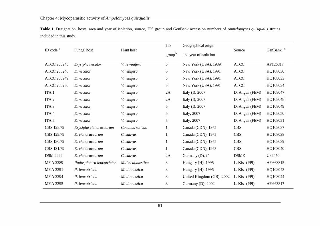
Chapter 4: Mycoparasitic activity of Ampelomyces quisqualis
81
Table 1. Designation, hosts, area and year of isolation, source, ITS group and GenBank accession numbers of Ampelomyces quisqualis strains
included in this study.
ID code a Fungal host Plant host ITS
group b
Geographical origin
and year of isolation Source GenBank c
ATCC 200245 Erysiphe necator Vitis vinifera 5 New York (USA), 1989 ATCC AF126817
ATCC 200246 E. necator V. vinifera 5 New York (USA), 1991 ATCC HQ108030
ATCC 200249 E. necator V. vinifera 5 New York (USA), 1991 ATCC HQ108033
ATCC 200250 E. necator V. vinifera 5 New York (USA), 1991 ATCC HQ108034
ITA 1 E. necator V. vinifera 2A Italy (I), 2007 D. Angeli (FEM) HQ108047
ITA 2 E. necator V. vinifera 2A Italy (I), 2007 D. Angeli (FEM) HQ108048
ITA 3 E. necator V. vinifera 5 Italy (I), 2007 D. Angeli (FEM) HQ108049
ITA 4 E. necator V. vinifera 5 Italy, 2007 D. Angeli (FEM) HQ108050
ITA 5 E. necator V. vinifera 5 Italy, 2007 D. Angeli (FEM) HQ108051
CBS 128.79 Erysiphe cichoracearum Cucumis sativus 1 Canada (CDN), 1975 CBS HQ108037
CBS 129.79 E. cichoracearum C. sativus 1 Canada (CDN), 1975 CBS HQ108038
CBS 130.79 E. cichoracearum C. sativus 1 Canada (CDN), 1975 CBS HQ108039
CBS 131.79 E. cichoracearum C. sativus 1 Canada (CDN), 1975 CBS HQ108040
DSM 2222 E. cichoracearum C. sativus 2A Germany (D), ? e DSMZ U82450
MYA 3389 Podosphaera leucotricha Malus domestica 3 Hungary (H), 1995 L. Kiss (PPI) AY663815
MYA 3391 P. leucotricha M. domestica 3 Hungary (H), 1995 L. Kiss (PPI) HQ108043
MYA 3394 P. leucotricha M. domestica 3 United Kingdom (GB), 2002 L. Kiss (PPI) HQ108044
MYA 3395 P. leucotricha M. domestica 3 Germany (D), 2002 L. Kiss (PPI) AY663817
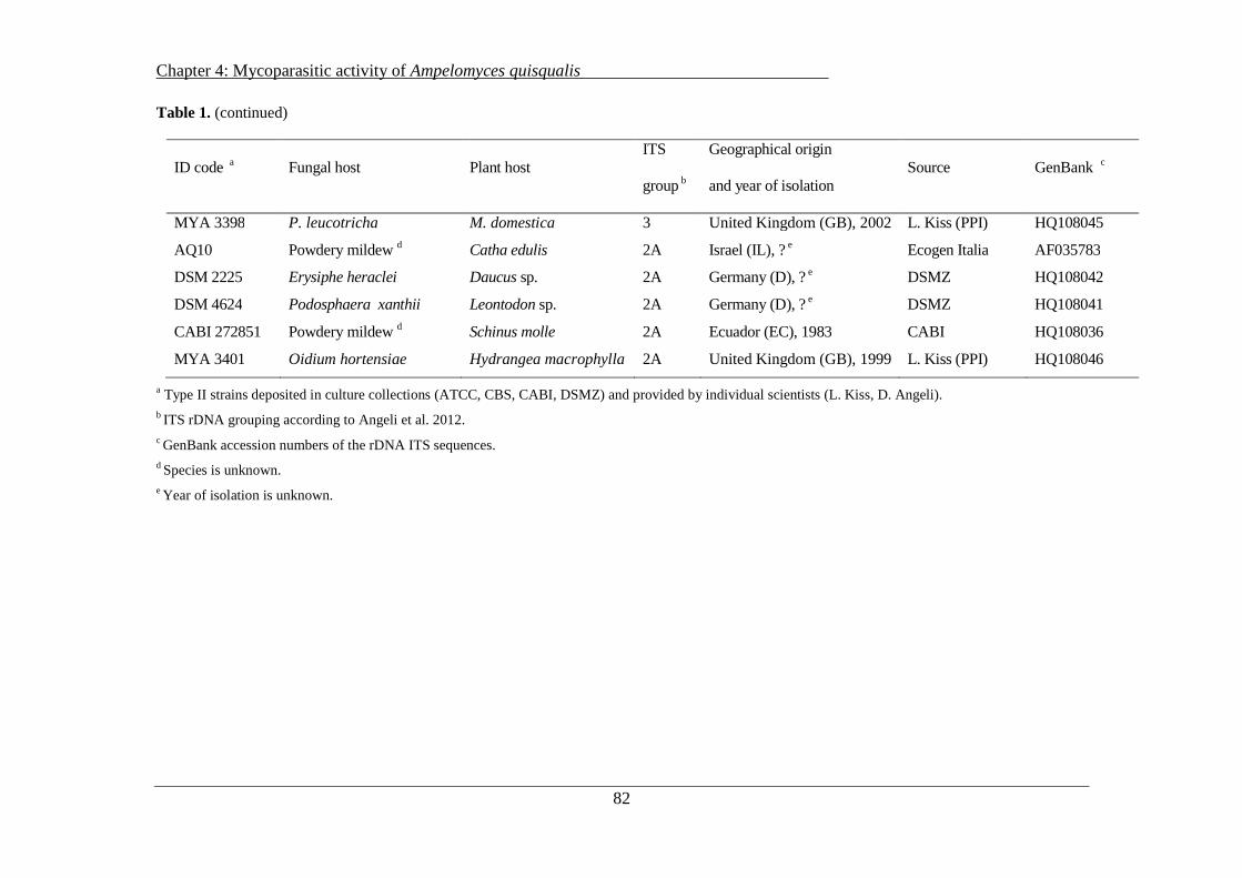
Chapter 4: Mycoparasitic activity of Ampelomyces quisqualis
82
Table 1. (continued)
ID code a Fungal host Plant host ITS
group b
Geographical origin
and year of isolation Source GenBank c
MYA 3398 P. leucotricha M. domestica 3 United Kingdom (GB), 2002 L. Kiss (PPI) HQ108045
AQ10 Powdery mildew d Catha edulis 2A Israel (IL), ? e Ecogen Italia AF035783
DSM 2225 Erysiphe heraclei Daucus sp. 2A Germany (D), ? e DSMZ HQ108042
DSM 4624 Podosphaera xanthii Leontodon sp. 2A Germany (D), ? e DSMZ HQ108041
CABI 272851 Powdery mildew d Schinus molle 2A Ecuador (EC), 1983 CABI HQ108036
MYA 3401 Oidium hortensiae Hydrangea macrophylla 2A United Kingdom (GB), 1999 L. Kiss (PPI) HQ108046
a Type II strains deposited in culture collections (ATCC, CBS, CABI, DSMZ) and provided by individual scientists (L. Kiss, D. Angeli). b ITS rDNA grouping according to Angeli et al. 2012. c GenBank accession numbers of the rDNA ITS sequences. d Species is unknown. e Year of isolation is unknown.
Chapter 4: Mycoparasitic activity of Ampelomyces quisqualis
83
The aggressiveness of the A. quisqualis strains was tested against the pathogens Erysiphe
necator, Podosphaera aphanis and P. xanthii, which were collected in the Trentino-Alto Adige
region between 2009 and 2011 from naturally infected grapevine, strawberry and cucumber
plants, respectively. Fresh colonies of powdery mildew were maintained under controlled
greenhouse conditions at 25 ± 1 °C and 70 ± 10 % relative humidity (RH) at a fixed daily
photoperiod of 16 h. Subsequent inoculations of powdery mildew were carried out every 30 days
by shaking leaves bearing sporulating mildew over three-week-old plants with at least five true
leaves (Angeli et al., 2009b).
The Trichoderma atroviride strain SC1 has been used as a positive control in enzymatic
experiments as the activity of chitinolytic, proteolytic and glycolytic enzymes in Trichoderma
spp. has already been extensively characterized (Kubicek et al., 2001; Benitez et al., 2004). T.
atroviride SC1 was isolated from decaying hazelnut in northern Italy and is currently deposited
in the CBS restricted collection (CBS 122089) in accordance with the regulations of the
Budapest Treaty.
4.2.2 Mycoparasitic activity: ability of A. quisqualis to reduce conidiation of
powdery mildews in vivo
The ability of the 24 A. quisqualis strains to inhibit conidiation of E. necator, P. aphanis and P.
xanthii on their respective host plants was assessed under controlled conditions. Two-week-old
grapevine, strawberry and cucumber plants were inoculated with their respective powdery
mildew pathogens; P. xanthii inoculation was carried out by spraying a suspension of distilled
water and Tween 80 (0.01 %) containing 1 × 10⁵ conidia/ml, while in the case of E. necator and
P. aphanis dry inoculation was effected by shaking infected leaves bearing sporulating mildew
over the plants. The amount of strawberry and grapevine powdery mildew inoculum was
determined by counting conidia on glass slides placed between the plants during dry inoculation
and was expressed as the number of conidia per cm2. The choice of different inoculation methods
was based on preliminary trials carried out to optimize the methodology (data not shown). Plants
were incubated in the greenhouse at 25 ± 1 °C and 70 ± 10 % of RH at a fixed daily photoperiod
of 16 h. As soon as powdery mildew conidiation appeared on the upper surface of the leaves (5,
7 and 12 days after infection for cucumber, strawberry and grapevine, respectively) plants were

Chapter 4: Mycoparasitic activity of Ampelomyces quisqualis
84
immediately treated with A. quisqualis (see below). Powdery mildew conidiation appeared as a
homogenous infection on cucumber and grapevine leaves, while several powdery spots appeared
on strawberry leaves.
Conidia suspensions of A. quisqualis strains were prepared as follows: A. quisqualis strains were
grown on PDA in Petri dishes for two weeks at 25 °C in the dark (ten plates per strain); conidia
were harvested by adding 20 ml of sterile distilled water and Tween 80 (0.01 %) to each plate
and concentration was adjusted to 1 × 10⁶ conidia/ml under a light microscope (Hund Wetzlar H
600LL, Wetzlar, Germany) using a Thoma-Zeiss counting chamber.
Plants bearing sporulating colonies of P. xanthii and E. necator were homogeneously treated
with the suspension containing 1 × 10⁶ conidia/ml of A. quisqualis using a hand air-sprayer. Only
two basal leaves per plant, previously infected with the relevant powdery mildew, were treated (5
ml of suspension per leaf). For strawberry, a drop of the suspension containing A. quisqualis
conidia (10 µl each) was applied to powdery mildew spots of approximately 3 mm diameter. Six
spots per leaf were treated. Plants with powdery mildew and A. quisqualis infections were kept at
25 °C and 95 % RH for 48 h, after which humidity was reduced to 70 ± 10 % RH. Control plants
with powdery mildew lesions were sprayed with distilled water and Tween 80 only.
Reduction in powdery mildew fungal conidiation was assessed by counting the number of E.
necator, P. aphanis and P. xanthii conidia produced by the pathogens on the leaf surface 10 days
after A. quisqualis treatment compared with untreated infected plants. The two basal leaves of
each grapevine and cucumber plant were collected (12 leaves per strain) and leaf disks of 1.8 cm
diameter were cut (6 disks per grapevine leaf, 18 disks per cucumber leaf). The leaf disks were
transferred to 50 ml tubes containing 5 ml (grapevine) or 15 ml (cucumber) distilled water with
Tween 80 (0.01 %). For strawberry, two leaves per plant were picked and a leaf disk of 3 mm
diameter was cut from each lesion and transferred to a 2 ml tube containing 1 ml distilled water
with 0.01 % Tween 80. In all three cases, tubes were vortexed for 1 min and for each leaf four
droplets of 20 µl were mounted on glass slides. Concentrations of powdery mildew conidia were
measured under a light microscope and conidia were counted using a Thoma-Zeiss counting
chamber. Results were expressed as the percentage reduction in the number of powdery mildew
conidia compared with leaves treated with powdery mildew only. Three independent

Chapter 4: Mycoparasitic activity of Ampelomyces quisqualis
85
experiments each with six replicates (plants with two inoculated leaves) per treatment (A.
quisqualis strain) were performed.
4.2.3 Mycoparasitic activity: intra-hyphal formation of A. quisqualis pycnidia
within powdery mildews
Intra-hyphal formation of intracellular pycnidia of the 24 A. quisqualis strains in grapevine (E.
necator), strawberry (P. aphanis) and cucumber (P. xanthii) powdery mildew colonies was
measured following Kiss’s (1998) methodology with some modifications. Potted plants were
kept in a greenhouse and their leaves inoculated with their respective powdery mildews. Leaves
with powdery mildew infection were detached from the plants and the surfaces checked for the
presence of freshly sporulating colonies of E. necator, P. aphanis and P. xanthii under a
stereomicroscope. Each sporulating powdery mildew colony (lesion), marked with a permanent
label on the lower leaf surface, was inoculated with a 2 µl droplet of an A. quisqualis conidial
suspension (1 × 10⁵ conidia/ml) spotted in the middle of the lesion. Six leaves bearing five
inoculated lesions each were examined for each strain. To assess the inoculum area of each
fungal strain, a leaf disk of 5 mm diameter was cut from each lesion from three leaves and put
onto a glass slide. The area covered by conidial suspension was measured at 40× magnification
with a laser microdissection system (Leica LDM 7600 Light Microscope, Leica, Germany). The
average expanse of the 15 inoculation areas measured was then subtracted from the total area
covered by pycnidia measured at the end of the experiment (see below). The three other
inoculated leaves were kept in Petri dishes (180 mm) and placed on plastic nets floated on water.
Petioles were submerged in water in order to guarantee leaf survival and high relative humidity.
These plates were placed in a climate chamber with 16 h daily illumination at 25 °C for 14 days.
After this period, a leaf disk of 5 mm diameter was cut from each lesion and put onto a glass
slide. The total area with A. quisqualis pycnidia in the powdery mildew mycelium was measured
at 40× magnification under the microscope. The difference between the total area covered by
pycnidia and the average surface area below the inoculation drop, determined as described
above, was calculated. Intra-hyphal enlargement of A. quisqualis strains in the powdery mildew
mycelium was expressed as µm2 day−1. Three independent experiments, each with three
replicates (leaves with five lesions) per treatment (A. quisqualis strain), were carried out.

Chapter 4: Mycoparasitic activity of Ampelomyces quisqualis
86
4.2.4 In vitro production of CWDEs by A. quisqualis culture filtrates
Glucanase activity was assessed according to the procedure established by Rotem et al. (1999) to
characterize exo-β-1,3-glucanase activity in A. quisqualis strain AQ10. The amount of µmol of
glucose liberated during the reaction was determined with a glucose hexokinase reagent (Sigma),
according to the manufacturer’s instructions. Glucanase activity was expressed as µmol of
glucose h−1 mg−1 of dried mycelium.
Protease activity was determined using the artificial substrate azocasein according to Girard and
Michaud (2002) with some modifications. Briefly, a 6 mm plug of the pycnidia-coated area was
excised from 14-day-old A. quisqualis PDA cultures. The agar plug was placed in a sterile 1.5 ml
tube containing 120 µl of an azocasein solution (1 %, w/v in 50mM Tris-HCl, pH 8.8) and
incubated for 7 days at 25 °C. The reaction was then stopped and the undigested substrate was
precipitated by the addition of 300 µl of cold trichloroacetic acid solution (10 %, w/v). After
centrifugation for 5 min at 13000 rpm, a volume of 100 µl of supernatant was transferred to 96-
multiwell plates and 85 µl of NaOH (1N) was added to each well. Absorbance was then read at
440 nm using a spectrophotometer and protease activity was expressed as the ratio of the
absorbance value per ml of supernatant.
To induce production of chitinases in A. quisqualis strains, 6 mm plugs from pycnidia-coated
PDA plates were transferred to sterile tubes containing 200 µl of a carboxymethyl-chitin-remazol
brilliant violet (CM-chitin-RBV, Sigma) solution (1 % in 50mM Tris-HCl, pH 7.5) and
incubated at 25 °C for 6 days. At the end of the incubation period, the tubes were centrifuged for
5 min at 13000 rpm and supernatants were assessed for their ability to degrade colloidal chitin
(endochitinase activity) and for their N-acetyl-β-1,4-D-glucosamminidase activity (chitobiase
activity). For endochitinase activity, a volume of 100 µl of supernatant was transferred to 96-
multiwell plates and absorbance was read at 550 nm using a spectrophotometer according to
Quecine et al. (2008). The compound 4-methylumbelliferyl-N-acetyl-β-D-glucosaminide (4-MU-
GlcNAc, Sigma), where the 4-methylumbelliferyl group is linked by β-1,4 linkage to the N-
acetyl-β-D-glucosaminide monosaccharide, was used to assess chitobiase activity (O’Brien &
Colwell, 1987). A volume of 50 µl of supernatant was mixed with 200 µl of 4-MU-Glc NAc
solution (10 µM in 100 mM sodium phosphate buffer, pH 6.0) and the reaction mixture was
incubated at 37 °C for 10 min then stopped by adding 50 µl of Na2CO3 (0.2 M). The amount of
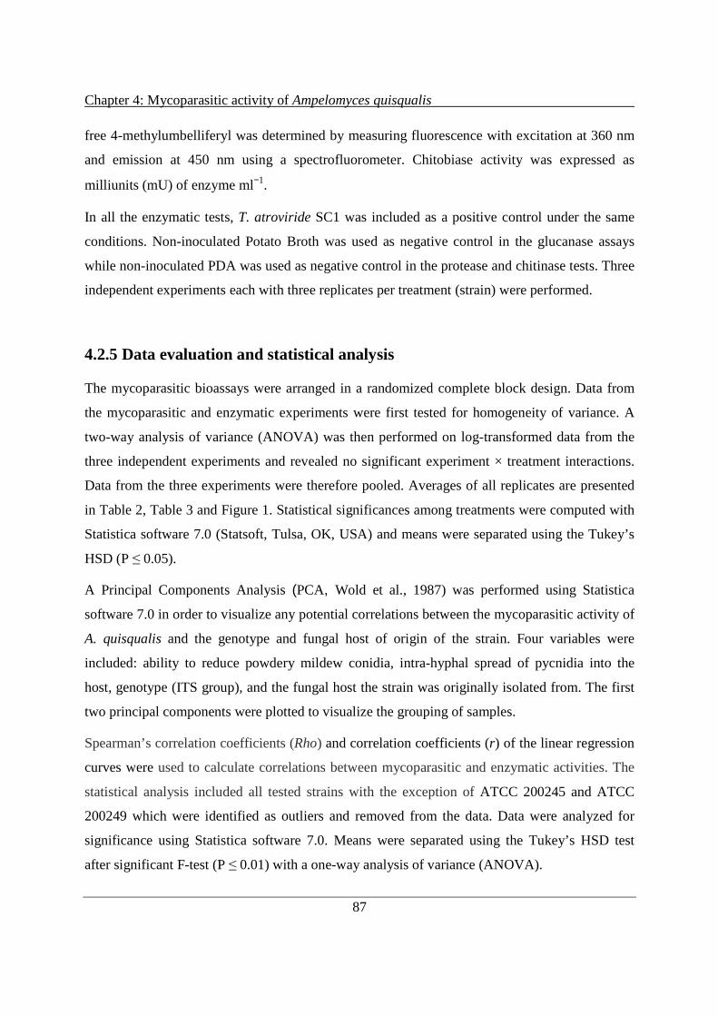
Chapter 4: Mycoparasitic activity of Ampelomyces quisqualis
87
free 4-methylumbelliferyl was determined by measuring fluorescence with excitation at 360 nm
and emission at 450 nm using a spectrofluorometer. Chitobiase activity was expressed as
milliunits (mU) of enzyme ml−1.
In all the enzymatic tests, T. atroviride SC1 was included as a positive control under the same
conditions. Non-inoculated Potato Broth was used as negative control in the glucanase assays
while non-inoculated PDA was used as negative control in the protease and chitinase tests. Three
independent experiments each with three replicates per treatment (strain) were performed.
4.2.5 Data evaluation and statistical analysis
The mycoparasitic bioassays were arranged in a randomized complete block design. Data from
the mycoparasitic and enzymatic experiments were first tested for homogeneity of variance. A
two-way analysis of variance (ANOVA) was then performed on log-transformed data from the
three independent experiments and revealed no significant experiment × treatment interactions.
Data from the three experiments were therefore pooled. Averages of all replicates are presented
in Table 2, Table 3 and Figure 1. Statistical significances among treatments were computed with
Statistica software 7.0 (Statsoft, Tulsa, OK, USA) and means were separated using the Tukey’s
HSD (P ≤ 0.05).
A Principal Components Analysis (PCA, Wold et al., 1987) was performed using Statistica
software 7.0 in order to visualize any potential correlations between the mycoparasitic activity of
A. quisqualis and the genotype and fungal host of origin of the strain. Four variables were
included: ability to reduce powdery mildew conidia, intra-hyphal spread of pycnidia into the
host, genotype (ITS group), and the fungal host the strain was originally isolated from. The first
two principal components were plotted to visualize the grouping of samples.
Spearman’s correlation coefficients (Rho) and correlation coefficients (r) of the linear regression
curves were used to calculate correlations between mycoparasitic and enzymatic activities. The
statistical analysis included all tested strains with the exception of ATCC 200245 and ATCC
200249 which were identified as outliers and removed from the data. Data were analyzed for
significance using Statistica software 7.0. Means were separated using the Tukey’s HSD test
after significant F-test (P ≤ 0.01) with a one-way analysis of variance (ANOVA).

Chapter 4: Mycoparasitic activity of Ampelomyces quisqualis
88
4.3 Results
4.3.1 Mycoparasitic activity: ability of A. quisqualis to reduce conidiation of
powdery mildews in vivo
The trials on the different pathosystems (E. necator-grapevine, P. aphanis-strawberry and P.
xanthii-cucumber) showed that all 24 A. quisqualis strains tested significantly reduced the
number of conidia on treated leaves compared with untreated controls. On strawberry (Fig. 1a),
the strains belonging to ITS group 1 isolated from P. xanthii (cucumber, Canada, Table 1) and
most strains of ITS group 2A, including strains isolated from different fungal hosts, (ITA 1 and
ITA 2 from E. necator, CABI 272851 from Schinus molle, DSM 4624 from S. fuliginea, DSM
2225 from E. heraclei, MYA 3401 from Oidium hortensiae) greatly reduced the number of
powdery mildew conidia (46–51 %). AQ10, DSM 2222 (ITS group 2A) and ITS group 3 reduced
P. aphanis conidia by 28–40 %. Very poor powdery mildew conidia reduction was obtained with
the four ATCC strains isolated from E. necator and clustering within ITS group 5 (22–27 %
reduction). Strain ITA 3 was the only strain of ITS group 5 able to greatly reduce the powdery
mildew conidiation rate (53 %). In the experiments with E. necator, all the A. quisqualis strains
tested greatly reduced the number of conidia on treated leaves compared with untreated controls
(Fig. 1b), with values ranging from 63 % (ATCC 200245) to 100 % (MYA 3401). Significant
differences between strains were, however, detected. With the exception of DSM 2225, all the
strains which performed best against strawberry powdery mildew (ITS group 1, some strains
from ITS group 2A and ITA 3) were also the most effective against E. necator. Strain MYA
3401 even produced complete inhibition of E. necator conidiation, while a significantly lower
effect was obtained with strains belonging to ITS groups 5 and 3. The greatest variation between
A. quisqualis treatments was observed (23–93 % control) with cucumber (Fig. 1c). The same
strains which were highly effective against P. aphanis and E. necator (ITS group 1, ITA 1, ITA
2, DSM 4624, CABI 272851 from ITS group 2A and ITA 3 belonging to ITS group 5) greatly
reduced the number of powdery mildew conidia (77–93 %). The A. quisqualis strains isolated
from P. xanthii (cucumber, Canada) and belonging to ITS group 1 were the best strains for
controlling cucumber powdery mildew. Strains belonging to ITS group 3 isolated from P.
leucotricha on apple plants and the strains of ITS group 5 (except ITA 3) isolated from E.
necator were found to have little efficacy .
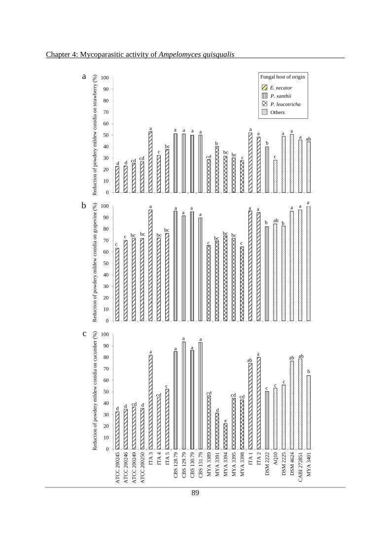
Chapter 4: Mycoparasitic activity of Ampelomyces quisqualis
89
0
10
20
30
40
50
60
70
80
90
100
AT
CC
20
0245
AT
CC
20
0246
AT
CC
20
0249
AT
CC
20
0250
ITA
3
ITA
4
ITA
5
CB
S 1
28.7
9
CB
S 1
29.7
9
CB
S 1
30.7
9
CB
S 1
31.7
9
MY
A 3
389
MY
A 3
391
MY
A 3
394
MY
A 3
395
MY
A 3
398
ITA
1
ITA
2
DS
M 2
222
AQ
10
DS
M 2
225
DS
M 4
624
CA
BI
2728
51
MY
A 3
401
Red
uct
ion
of p
ow
der
y m
ildew
co
nid
ia o
n s
traw
ber
ry (
%)
aaa a a a a a
bab
a
c
a
cd bc c
b
cdcdd
bc
d
bcc
a Fungal host of origin
E. necator
P. xanthii
P. leucotricha
Others
0
10
20
30
40
50
60
70
80
90
100
AT
CC
200
245
AT
CC
200
246
AT
CC
200
249
AT
CC
200
250
ITA
3
ITA
4
ITA
5
CB
S 1
28.7
9
CB
S 1
29.7
9
CB
S 1
30.7
9
CB
S 1
31.7
9
MY
A 3
389
MY
A 3
391
MY
A 3
394
MY
A 3
395
MY
A 3
398
ITA
1
ITA
2
DS
M 2
222
AQ
10
DS
M 2
225
DS
M 4
624
CA
BI 2
728
51
MY
A 3
401
Red
uct
ion
of p
ow
der
y m
ildew
co
nid
ia o
n g
rap
evin
e (%
)
aaa aa
aa
a
b
a
bab
a
c
bc
cbc
bcbccbc
c
bcbc
b
0
10
20
30
40
50
60
70
80
90
100
AT
CC
200
245
AT
CC
200
246
AT
CC
200
249
AT
CC
200
250
ITA
3
ITA
4
ITA
5
CB
S 1
28.7
9
CB
S 1
29.7
9
CB
S 1
30.7
9
CB
S 1
31.7
9
MY
A 3
389
MY
A 3
391
MY
A 3
394
MY
A 3
395
MY
A 3
398
ITA
1
ITA
2
DS
M 2
222
AQ
10
DS
M 2
225
DS
M 4
624
CA
BI 2
728
51
MY
A 3
401
Red
uct
ion
of p
ow
der
y m
ildew
co
nid
ia o
n c
ucu
mb
er (
%)
aab
aa
a
a
a
ab
c
b
cc
ab
cd cd cd
ddcdd
e
d
c
cd
c

Chapter 4: Mycoparasitic activity of Ampelomyces quisqualis
90
Figure 1. Reduction in the number of powdery mildew conidia of Podosphaera aphanis (a), Erysiphe
necator (b), P. xanthii (c), in comparison with untreated controls, on leaves sprayed with Ampelomyces
quisqualis strains from different host plants, fungal hosts and geographic origins. Columns with the same
design represent strains isolated from the same plant and mildew species, with the exception of the dotted
columns which represent strains isolated from different hosts (Table 1). The four ITS groups (1, 2A, 3, 5)
discussed in the text are indicated in the figure. Columns with the same letters do not significantly differ
(P ≤ 0.05, Tukey’s test). Values are means of eighteen replicates derived from three independent
experiments with six replicates per experiment.
4.3.2 Mycoparasitic activity: intra-hyphal formation of A. quisqualis pycnidia
within powdery mildews
The 24 A. quisqualis strains were rated for their ability to parasitize powdery mildew mycelia by
forming intrahyphal pycnidia. Microscopic examination revealed that intrahyphal pycnidia had
already formed outside the inoculation area for all tested strains of A. quisqualis 14 days after
inoculation. All A. quisqualis strains displayed antagonistic activity against P. xanthii, E. necator
and P. aphanis, but different levels of fungal growth within the pathogens were observed
between strains (Table 2). Among the 24 strains, six (ITA 1, ITA 2 and all strains from cucumber
powdery mildew belonging to ITS group 1) induced the largest parasitized areas on the three
powdery mildews. Their daily expansion ranged from 182 to 263 mm2 day-1. Good growth rates
were also found with strains ITA 3, CABI 272851 and DSM 4624. CBS strains clustering into
ITS group 1 displayed the highest mycoparasitic activity in vivo (191–245 mm2 day-1).
Considerably reduced pycnidial development into host cells was observed with strains clustering
into ITS group 3 (36–52 mm2 day-1) and group 5 (except ITA3) (62–93 mm2 day-1). Analysis of
the data according to ITS group showed that strains from ITS groups 1 and 2A spread
significantly faster within the powdery mildew host compared with strains belonging to ITS
groups 3 and 5 (Table 2).

Chapter 4: Mycoparasitic activity of Ampelomyces quisqualis
91
Table 2. Intra-hyphal spread of Ampelomyces quisqualis pycnidia within powdery mildew
Strains
ITS group
Intra-hyphal spread of pycnidia a
(mm2 day−1 ± SE) on
P. xanthii E. necator P. aphanis
ATCC 200245 5 43.8 ± 0.2 f 42.7 ± 0.2 g 35.4 ± 0.3 d
ATCC 200246 5 44.0 ± 0.3 f 48.9 ± 0.3 fg 36.1.0 ± 0.4 d
ATCC 200249 5 56.2 ± 0.5 ef 70.1 ± 0.4 ef 38.7 ± 0.4 d
ATCC 200250 5 88.4 ± 0.4 de 77.1 ± 0.5 e 38.8 ± 0.4 d
ITA 1 2A 240.1 ± 1.3 a 245.7 ± 1.7 a 187.7 ± 0.9 a
ITA 2 2A 228.3 ± 1.2 a 232.2 ± 0.8 ab 182.4 ± 0.8 a
ITA 3 5 202.7 ± 1.0 b 207.1 ± 1.3 b 147.4 ± 1.1 b
ITA 4 5 71.7 ± 0.6 e 90.5 ± 0.6 de 59.1 ± 0.3 cd
ITA 5 5 102.8 ± 0.4 d 111.3 ± 0.5 d 78.3 ± 0.3 c
CBS 128.79 1 253.3 ± 0.9 a 261.5 ± 1.1 a 198.5 ± 1.2 a
CBS 129.79 1 224.4 ± 1.4 a 230.6 ± 1.3 ab 184.4 ± 1.3 a
CBS 130.79 1 218.9 ± 1.1 ab 224.0 ± 1.6 ab 188.8 ± 1.2 a
CBS 131.79 1 255.0 ± 1.1 a 262.9 ± 0.9 a 193.0 ± 1.1 a
DSM 2222 2A 194.7 ± 1.3 bc 182.3 ± 0.8 c 138.2 ± 1.0 b
MYA 3389 3 49.4 ± 0.4 ef 55.5 ± 0.3 f 40.1 ± 0.3 d
MYA 3391 3 42.6 ± 0.4 f 30.4 ± 0.3 h 28.1 ± 0.3 d
MYA 3394 3 71.3 ± 0.1 e 78.2 ± 0.1 e 42.2 ± 0.2 d
MYA 3395 3 44.6 ± 0.3 f 54.5 ± 0.3 f 35.9 ± 0.2 d
MYA 3398 3 42.8 ± 0.3 f 38.9 ± 0.3 h 34.4 ± 0.3 d
AQ10 2A 180.0 ± 1.4 c 177.7 ± 1.6 c 132.0 ± 0.8 bc
DSM 2225 2A 188.6 ± 1.0 bc 183.6 ± 0.7 c 145.8 ± 1.0 b
DSM 4624 2A 214.0 ± 1.0 b 205.4 ± 0.8 b 151.3 ± 1.1 b
CABI 272851 2A 205.7 ± 1.5 b 190.4 ± 1.2 bc 172.3 ± 1.2 ab
Mean value b
1 237.9 ± 1.3 a 244.7 ± 1.4 a 191.2 ± 1.1 a
2A 205.6 ± 1.2 a 200.1 ± 1.2 a 156.2 ± 1.2 a
3 50.2 ± 0.3 b 51.5 ± 0.3 b 36.1 ± 0.1 b
5 87.1 ± 0.5 b 92.5 ± 0.5 b 61.9 ± 0.3 b

Chapter 4: Mycoparasitic activity of Ampelomyces quisqualis
92
Values are means of nine replicates derived from three independent experiments with three replicates per experiment. Standard errors of the means are reported (SE). Values with the same letter within a column are not significantly (P ≤ 0.05) different according to the Tukey’s HSD test. a Powdery mildew mycelial area covered by A. quisqualis pycnidia (mm2 per day). b Average of all A. quisqualis strains belonging to an individual ITS group. Values with the same letter within a column are not significantly (P ≤ 0.05) different according to the Tukey’s HSD test.
Assays for evaluating ability to reduce conidiation of powdery mildew and intra-hyphal spread
of A. quisqualis pycnidia within the host showed that all strains formed pycnidia and reduced
conidiation of the three powdery mildew species but with different levels of aggressiveness. A
Principal Components Analysis (PCA) based on the interaction between mycoparasitic activities
(reduction of conidiation and intra-hyphal growth), ITS group and fungal host of origin showed
that the latter two generally corresponded with the grouping based on level of aggressiveness.
Three different groups, each comprising strains isolated from the same fungal host, belonging to
the same ITS group and with similar levels of aggressiveness could be clearly recognized in the
PCA (Fig. 2). In fact, all strains isolated from P. xanthii and clustering into ITS group 1 were
highly effective against the tested powdery mildews. On the other hand, strains isolated from P.
leuchotricha and E. necator (with exception of ITA 3) and belonging to ITS groups 3 and 5,
respectively, were less effective in powdery mildew control. The only ITS group where the
strains failed to cluster together in the PCA analysis was group 2A which included strains
isolated from different hosts and displaying different levels of aggressiveness. Three strains in
this group (ITA 1, ITA 2, and DSM 2222) did not cluster with the other 2A strains.

Chapter 4: Mycoparasitic activity of Ampelomyces quisqualis
93
Figure 2. Principal Components Analysis (PCA) of mycoparasitic activities, genotype and fungal host of
origin of the Ampelomyces quisqualis strains. Four variables were included: ability to reduce powdery
mildew conidiation, intra-hyphal spread of pycnidia within the host, genotype (ITS group), and fungal
host of origin. Clusters were named according to the ITS rDNA phylogenetic group (Angeli et al., 2012).
The first two principal components were plotted to visualize the grouping of samples.
4.3.3 In vitro production of CWDEs by A. quisqualis culture filtrates
CWDE activities in vitro were determined for the 24 A. quisqualis strains and compared with
those of T. atroviride SC1. The levels of chitinase, protease and β-1,3 glucanase secreted by the
various A. quisqualis strains differed significantly (P ≤ 0.05) according to a Tukey’s multiple
range test (Table 3).
None of the A. quisqualis strains exhibited endochitinase activity in vitro while the greatest
chitobiase activity was produced by CBS strains 128.79 (43 mU ml-1) and 131.79 (37 mU ml-1),
ATCC 200245 (41 mU ml-1) and DSM 4624 (38 mU ml-1). The activity of these strains was
about three times higher than T. atroviride SC1. In general, ITS group 1 displayed the greatest
CBS 128.79CBS 129.79CBS 130.79
CBS 131.79
MYA 3389
MYA 3391MYA 3394
MYA 3395
MYA 3398
ATCC 200245ATCC 200246
ATCC 200249ATCC 200250
ITA 3
ITA 4
ITA 5ITA 1
ITA 2DSM 2222
AQ10
DSM 2225
DSM 4624
CABI 272851
MYA 3401
-4 -3 -2 -1 0 1 2 3 4
Factor 1 (60,03)
-4
-3
-2
-1
0
1
2
3
4F
acto
r 2
(31,
56)
CBS 128.79CBS 129.79CBS 130.79
CBS 131.79
MYA 3389
MYA 3391MYA 3394
MYA 3395
MYA 3398
ATCC 200245ATCC 200246
ATCC 200249ATCC 200250
ITA 3
ITA 4
ITA 5ITA 1
ITA 2DSM 2222
AQ10
DSM 2225
DSM 4624
CABI 272851
MYA 3401

Chapter 4: Mycoparasitic activity of Ampelomyces quisqualis
94
chitobiase activities with an average of 34 mU ml-1. The lowest activities were clearly measured
for A. quisqualis strains belonging to ITS group 3 (10 mU ml-1). There was a 0.5 to 1-fold
decrease in the activities of these strains when compared with the Trichoderma control strain.
Strains of A. quisqualis generally exhibited lower protease activity compared with T. atroviride
SC1 under the same conditions. Only the protease activities of ITA 1, ITA 2, ATCC 200249 and
CBS 128.79 did not statistically differ from SC1 (10.2 Abs ml-1) whereas the activities of all
other strains were significantly lower, with several strains belonging to ITS group 3 and ITS
group 5 exhibiting as much as a 5-fold decrease compared with the Trichoderma control. In
general, ITS groups 1 and 2A displayed higher enzyme proteases activities (Average = 8.1 Abs
ml-1) than group 3 (3.8 Abs ml-1) and group 5 (5 Abs ml-1) (Table 3).
Differences in activity levels between individual strains of A. quisqualis were also found for β-
1,3 glucanase. Maximum activity of β-1,3 glucanase was measured with strains ITA 1, ITA 2,
AQ10 and DSM 2225 (405–451 µmol mg-1 h-1). These values were about 2 times higher than
those for SC1 (199 µmol mg-1 h-1). The strains displaying the lowest glucanase activities (e.g. a
2-fold decrease compared with SC1) were ITA 4, MYA 3389 and MYA 3398.
Most of the strains tested exhibiting high chitobiase and protease activities (e.g. strains of ITS
groups 1 and 2A) also performed best in parasitizing the powdery mildews and in inhibiting the
formation of conidia (Figs. 1 and 3, Tables 2 and 3). Conversely, all the strains belonging to ITS
groups 3 and 5, which displayed the lowest chitobiase and protease activities, were also less
effective in colonizing powdery mildew in vivo. Correlation analysis of the mycoparasitic and
enzymatic activities of A. quisqualis (Fig. 3; Table 1S) showed the activities of these two
CWDEs to be positively correlated with intra-hyphal formation of pycnidia (Figs. 3a,c; Rho =
0.82, R2 = 0.68; Rho = 0.77, R2 = 0.67) and inhibition of conidiation in vivo (Figs. 3b,d; Rho =
0.70, R2 = 0.53; Rho = 0.71, R2 = 0.54). However, no correlation was found between exo-β-1,3
glucanase activity and the two tested mycoparasitic activities of A. quisqualis strains belonging
to different ITS groups (Figs. 3e,f; Table 1S).
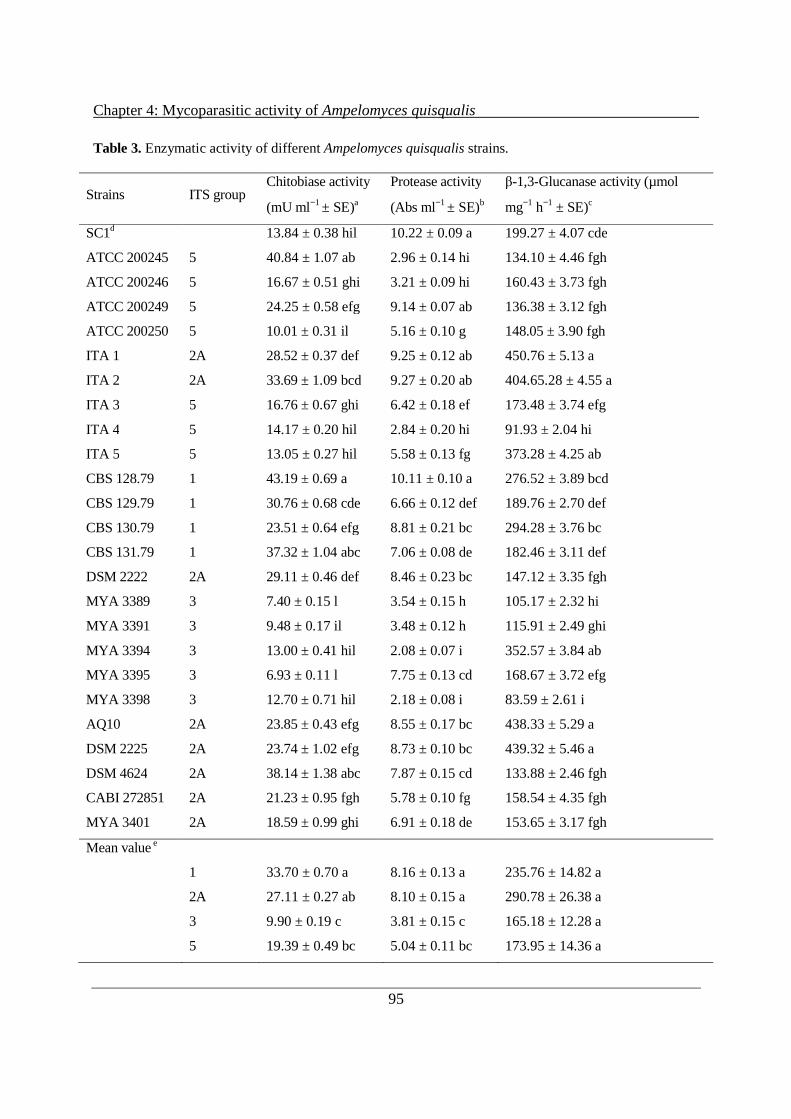
Chapter 4: Mycoparasitic activity of Ampelomyces quisqualis
95
Table 3. Enzymatic activity of different Ampelomyces quisqualis strains.
Strains ITS group Chitobiase activity
(mU ml−1 ± SE)a
Protease activity
(Abs ml−1 ± SE)b
β-1,3-Glucanase activity (µmol
mg−1 h−1 ± SE)c
SC1d 13.84 ± 0.38 hil 10.22 ± 0.09 a 199.27 ± 4.07 cde
ATCC 200245 5 40.84 ± 1.07 ab 2.96 ± 0.14 hi 134.10 ± 4.46 fgh
ATCC 200246 5 16.67 ± 0.51 ghi 3.21 ± 0.09 hi 160.43 ± 3.73 fgh
ATCC 200249 5 24.25 ± 0.58 efg 9.14 ± 0.07 ab 136.38 ± 3.12 fgh
ATCC 200250 5 10.01 ± 0.31 il 5.16 ± 0.10 g 148.05 ± 3.90 fgh
ITA 1 2A 28.52 ± 0.37 def 9.25 ± 0.12 ab 450.76 ± 5.13 a
ITA 2 2A 33.69 ± 1.09 bcd 9.27 ± 0.20 ab 404.65.28 ± 4.55 a
ITA 3 5 16.76 ± 0.67 ghi 6.42 ± 0.18 ef 173.48 ± 3.74 efg
ITA 4 5 14.17 ± 0.20 hil 2.84 ± 0.20 hi 91.93 ± 2.04 hi
ITA 5 5 13.05 ± 0.27 hil 5.58 ± 0.13 fg 373.28 ± 4.25 ab
CBS 128.79 1 43.19 ± 0.69 a 10.11 ± 0.10 a 276.52 ± 3.89 bcd
CBS 129.79 1 30.76 ± 0.68 cde 6.66 ± 0.12 def 189.76 ± 2.70 def
CBS 130.79 1 23.51 ± 0.64 efg 8.81 ± 0.21 bc 294.28 ± 3.76 bc
CBS 131.79 1 37.32 ± 1.04 abc 7.06 ± 0.08 de 182.46 ± 3.11 def
DSM 2222 2A 29.11 ± 0.46 def 8.46 ± 0.23 bc 147.12 ± 3.35 fgh
MYA 3389 3 7.40 ± 0.15 l 3.54 ± 0.15 h 105.17 ± 2.32 hi
MYA 3391 3 9.48 ± 0.17 il 3.48 ± 0.12 h 115.91 ± 2.49 ghi
MYA 3394 3 13.00 ± 0.41 hil 2.08 ± 0.07 i 352.57 ± 3.84 ab
MYA 3395 3 6.93 ± 0.11 l 7.75 ± 0.13 cd 168.67 ± 3.72 efg
MYA 3398 3 12.70 ± 0.71 hil 2.18 ± 0.08 i 83.59 ± 2.61 i
AQ10 2A 23.85 ± 0.43 efg 8.55 ± 0.17 bc 438.33 ± 5.29 a
DSM 2225 2A 23.74 ± 1.02 efg 8.73 ± 0.10 bc 439.32 ± 5.46 a
DSM 4624 2A 38.14 ± 1.38 abc 7.87 ± 0.15 cd 133.88 ± 2.46 fgh
CABI 272851 2A 21.23 ± 0.95 fgh 5.78 ± 0.10 fg 158.54 ± 4.35 fgh
MYA 3401 2A 18.59 ± 0.99 ghi 6.91 ± 0.18 de 153.65 ± 3.17 fgh
Mean value e
1 33.70 ± 0.70 a 8.16 ± 0.13 a 235.76 ± 14.82 a
2A 27.11 ± 0.27 ab 8.10 ± 0.15 a 290.78 ± 26.38 a
3 9.90 ± 0.19 c 3.81 ± 0.15 c 165.18 ± 12.28 a
5 19.39 ± 0.49 bc 5.04 ± 0.11 bc 173.95 ± 14.36 a
Chapter 4: Mycoparasitic activity of Ampelomyces quisqualis
96
Values are means of nine replicates derived from three independent experiments with three replicates per experiment. Standard errors of the means are reported (SE). Values with the same letter within a column are not significantly (P ≤ 0.05) different according to the Tukey’s HSD test. a The total chitobiase activity was measured using 4-methylumbelliferyl N-acetyl-β-D-glucosaminide (4-MU-GluNAc) as the substrate. The fluorescence of the liberated 4-MU was measured with excitation at 360 nm and emission at 450 nm. One milliunit of chitobiase activity was defined as the amount of enzyme that liberates 1 nmol 4-MU per min. b The protease activity was measured in absorbance per millilitre of substrate per reaction c The β-1,3-glucanase activity was measured in µmol glucose liberated per h per mg dry weight according to Rotem et al (1999). d Internal positive control (Trichoderma atroviride SC1). e Average of all A. quisqualis strains belonging to an individual ITS group. Values with the same letter within a column are not significantly (P ≤ 0.05) different according to the Tukey’s HSD test.

Chapter 4: Mycoparasitic activity of Ampelomyces quisqualis
97
Figure 3. Relationship between CWDE production and mycoparasitic activities in Ampelomyces
quisqualis strains. Correlation coefficients (r) of the linear regression curves were used to calculate
correlations between the two mycoparasitic activities tested (intra-hyphal formation of pycnidia and
inhibition of powdery mildew conidiation) and chitobiase (a, b), protease (c, d), and β-1,3-glucanase
activities (e, f). The four ITS groups (1, 2A, 3, 5) are indicated with different symbols.
y = 5,9955x + 7,6175R² = 0,6827
0
50
100
150
200
250
300
0 10 20 30 40 50
Intr
ahy
phal
spr
ead
of
pycn
idia
(m
m2
d-1
)
Chitobiase activity (mU ml-1)
1
2A
3
5
(a)
y = 0,9861x + 39,473R² = 0,5333
0
10
20
30
40
50
60
70
80
90
100
0 10 20 30 40 50
Re
duct
ion
of p
ow
dery
mild
ew c
onid
ia (
%)
Chitobiase activity (mU ml-1)
1
2A
3
5
(b)(b)
y = 24,329x - 14,595R² = 0,6696
0
50
100
150
200
250
300
0 2 4 6 8 10 12
Intr
ahy
pha
l spr
ead
of p
ycn
idia
(m
m2
d-1
)
Protease activity (Abs ml-1)
1
2A
3
5
(c)
y = 4,1256x + 34,93R² = 0,5435
0
10
20
30
40
50
60
70
80
90
100
0 2 4 6 8 10 12
Red
uct
ion
of
pow
der
y m
ilde
w c
onid
ia (
%)
Protease activity (Abs ml-1)
1
2A
3
5
(d)(
y = 0,2782x + 71,339R² = 0,1979
0
50
100
150
200
250
300
0 100 200 300 400 500
Intr
ahy
phal
sp
read
of
pycn
idia
(m
m2
d-1
)
β-1,3-glucanase activity (µmol mg-1 h-1)
1
2A
3
5
(e)
y = 0,0352x + 52,063R² = 0,0892
0
10
20
30
40
50
60
70
80
90
100
0 100 200 300 400 500
Re
duct
ion
of p
ow
der
y m
ildew
con
idia
(%
)
β-1,3-glucanase activity (µmol mg-1 h-1)
1
2A
3
5
(f)(
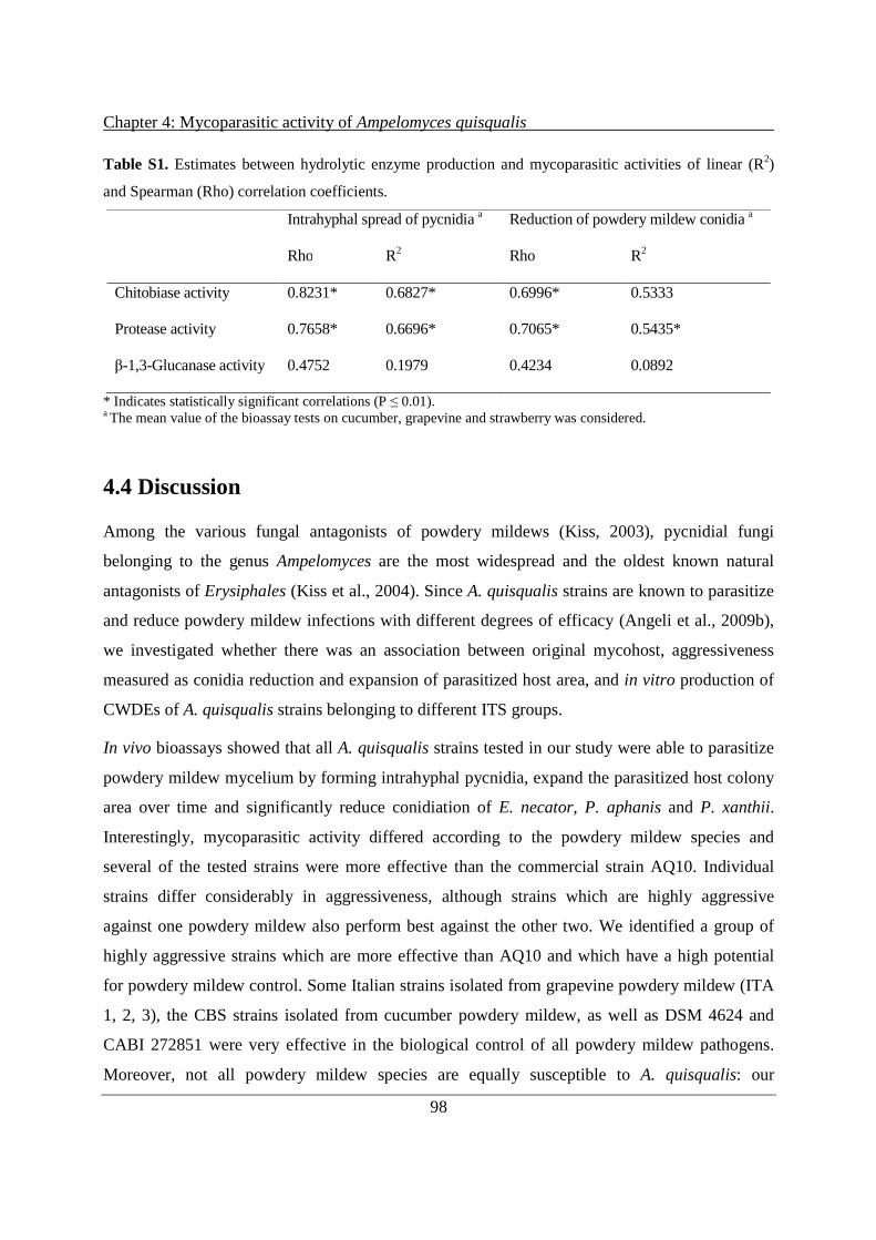
Chapter 4: Mycoparasitic activity of Ampelomyces quisqualis
98
Table S1. Estimates between hydrolytic enzyme production and mycoparasitic activities of linear (R2)
and Spearman (Rho) correlation coefficients.
Intrahyphal spread of pycnidia a Reduction of powdery mildew conidia a
Rho R2 Rho R2
Chitobiase activity 0.8231* 0.6827* 0.6996* 0.5333
Protease activity 0.7658* 0.6696* 0.7065* 0.5435*
β-1,3-Glucanase activity 0.4752 0.1979 0.4234 0.0892
* Indicates statistically significant correlations (P ≤ 0.01). a The mean value of the bioassay tests on cucumber, grapevine and strawberry was considered.
4.4 Discussion
Among the various fungal antagonists of powdery mildews (Kiss, 2003), pycnidial fungi
belonging to the genus Ampelomyces are the most widespread and the oldest known natural
antagonists of Erysiphales (Kiss et al., 2004). Since A. quisqualis strains are known to parasitize
and reduce powdery mildew infections with different degrees of efficacy (Angeli et al., 2009b),
we investigated whether there was an association between original mycohost, aggressiveness
measured as conidia reduction and expansion of parasitized host area, and in vitro production of
CWDEs of A. quisqualis strains belonging to different ITS groups.
In vivo bioassays showed that all A. quisqualis strains tested in our study were able to parasitize
powdery mildew mycelium by forming intrahyphal pycnidia, expand the parasitized host colony
area over time and significantly reduce conidiation of E. necator, P. aphanis and P. xanthii.
Interestingly, mycoparasitic activity differed according to the powdery mildew species and
several of the tested strains were more effective than the commercial strain AQ10. Individual
strains differ considerably in aggressiveness, although strains which are highly aggressive
against one powdery mildew also perform best against the other two. We identified a group of
highly aggressive strains which are more effective than AQ10 and which have a high potential
for powdery mildew control. Some Italian strains isolated from grapevine powdery mildew (ITA
1, 2, 3), the CBS strains isolated from cucumber powdery mildew, as well as DSM 4624 and
CABI 272851 were very effective in the biological control of all powdery mildew pathogens.
Moreover, not all powdery mildew species are equally susceptible to A. quisqualis: our
Chapter 4: Mycoparasitic activity of Ampelomyces quisqualis
99
experiments show that P. aphanis is in general less susceptible to all the A. quisqualis strains
than P. xanthii and E. necator.
Recently, studies have shown that the cultural and growth characteristics of these strains are
related to ITS group and host fungus and have suggested that a certain degree of mycohost
specialization may be present within the A. quisqualis species (Angeli et al., 2012; Park et al.,
2010). We have demonstrated for the first time that the ability of A. quisqualis strains to colonize
powdery mildew by forming pycnidia and inhibiting conidiation is completely independent of
the geographical origin of the strain, but appears to be related to genotype (ITS rDNA sequence)
and the original fungal host, with the exception of strains ITA 1, ITA 2 and ITA 3. It is worth
noting that the strains belonging to ITS groups 3 and 5 were the least effective in reducing
powdery mildews. These strains were isolated from apple and grapevine plants, respectively, the
natural hosts of only one powdery mildew species (M. domestica-P. leucotricha; V. vinifera-E.
necator). On the other hand, the most aggressive strains belonged to ITS groups 1 and 2A,
isolated from plants that, for the most part, are attacked by more than one Erysiphales, as in the
case of C. sativus which is the natural host of E. cichoracearum and P. xanthii (ITS group 1). In
addition, members of the plant species that hosted A. quisqualis strains of ITS group 2A are
natural hosts of at least two powdery mildews, such as Daucus sp. (E. heraclei and E. polygoni ),
H. macrophylla (E. polygoni and O. hortensiae) and Leontodon sp. (E. cichoracearum and P.
xanthii). Unfortunately, no information is available on the number of powdery mildews
occurring on C. edulis and S. molle. However, it is tempting to hypothesize that co-evolution of
A. quisqualis strains and plants which are the natural host of more than one powdery mildew
drove the selection of more aggressive genotypes. A broader range of strains belonging to
different genotypes and originating from different plant hosts will need to be investigated in the
future in order to validate this hypothesis.
Another aspect of our work regarded evaluation of the traits associated with strain
aggressiveness and testing the hypothesis that CWDEs secreted by A. quisqualis strains are an
important factor in selecting A. quisqualis strains for biocontrol. Although the mycoparasitic
activity of A. quisqualis against powdery mildews is well documented, information on the role
played by CWDEs is in fact scarce (Philipp, 1985; Rotem et al., 1999). These enzymes are
produced by mycoparasitic fungi and are involved in lysis of the cell wall through the
degradation of chitin, glucan and proteins, the main constituents of the cell walls of higher fungi
Chapter 4: Mycoparasitic activity of Ampelomyces quisqualis
100
(Cao et al., 2009; Larena & Melgarejo, 1996; Lorito et al., 1994a; Oppenheim & Chet, 1992).
The importance of the role played by CWDEs in the activity of fungal biocontrol agents has
already been demonstrated in several works. For example, the gene ech42 coding for an
endochitinase in T. harzianum strain P1 is expressed before contact between the biocontrol
fungus and the plant pathogen Rhizoctonia solani, while a null mutant lacking a functional copy
of this gene was impaired in the control of another plant pathogen Botrytis cinerea (Woo et al.,
1998; Zeilinger et al., 1999). Szekeres et al. (2004) found that T. harzianum strain T334 mutants
were able to overproduce proteases and better control Fusarium culmorum, Pythium and R.
solani in vitro. In addition, the biocontrol activity of T. virens strain GV29-8 against R. solani on
cotton seeds was much improved when the gene tvsp1, coding for an alkaline serine protease,
was constitutively expressed, showing that proteolytic activity plays a role in the mycoparasitism
of this T. virens strain (Pozo et al., 2004). The role played by β-1,3 and β-1,6 glucanases in the
same strain has been well documented (Djonovic et al., 2006, 2007). The use of derivatives of
the T. virens strain GV29-8 lacking glucanolytic activity or overproducing β-1,3 and β-1,6
glucanases in a biocontrol assay on cotton seedlings clearly showed the importance of these
enzymes. It was found that GV29-8 derivatives overproducing glucanase were able to control
plant pathogens more efficiently than wild type strains, while the null mutants lost their
biocontrol effectiveness (Djonovic et al., 2006, 2007).
Our enzymatic assays revealed in vitro β-1,3-glucanase, protease and chitobiase activity in all
tested strains, although the levels of enzymatic activity differed between genetically different A.
quisqualis strains. This is the first time that in vitro protease activity has been reported for
members of A. quisqualis sensu stricto. It is worth noting that all the strains tested displayed
chitobiase activity, but no endochitinase activity was detected using the CM-chitin-RBV method.
In agreement with Rotem et al (1999), we found that the commercial strain AQ10 produces a
high amount of extracellular β-1,3-glucanase and also displayed good protease and chitobiase
activity.
Our results suggest that the ability of some A. quisqualis strains to parasitize the host by forming
intracellular pycnidia and reducing fungal-host conidiation was associated with their intrinsic
ability to secrete proteases and with chitobiase activity in culture filtrates (Fig. 3, Table S1). This
was also illustrated by the regression analysis which showed there to be a significant positive
Chapter 4: Mycoparasitic activity of Ampelomyces quisqualis
101
correlation (Table S1) between chitobiase and protease activity with intra-hyphal formation of
pycnidia and the inhibition of conidiation in vivo, whereas exo-glucanase activity was not
statistically related to mycoparasitic activities. Therefore, we hypothize that proteases and
chitobiases play an important role in A. quisqualis mycoparasitism. However, lytic activities
alone do not fully explain the level of aggressiveness, although they are probably involved in the
biocontrol ability of A. quisqualis and of other mycoparasitic fungi (Adams, 2004; Reithner et
al., 2011; Vinale et al., 2008).
An interesting issue for future research would be to investigate the role played by these
enzymatic activities in the antagonistic properties of members of A. quisqualis sensu stricto
either by creating strains that are defective in these activities or by monitoring expression of the
genes responsible for lytic activities during the parasitization of powdery mildews. Nevertheless,
the biological characterization of the A. quisqualis strain collection carried out in this work
suggests that mycoparasitism by A. quisqualis can be explained in part by the activities of
CWDEs. This provides important knowledge for the selection of new, highly effective strains for
biocontrol.
4.5 Acknowledgements
The authors thank Dr. Levente Kiss (Plant Protection Institute of the Hungarian Academy of
Science, Budapest, Hungary) for providing the fungal strains. We would also like to thank
Carmela Sicher and Denise Ress for technical assistance. This work was supported by the
AMPELO and ENVIROCHANGE projects funded by the Autonomous Province of Trento.
4.6 Literature
Adams, D.J. (2004). Fungal cell wall chitinases and glucanases. Microbiology 150, 2029-2035.
Angeli, D., Pellegrini, E. and Pertot, I. (2009a). Occurence of Erysiphe necator chasmothecia and their natural
parasitization by Ampelomyces quisqualis. Phytopathology 99, 704-710.
Angeli, D., Pellegrini, E., Micheli, S., Ress, D., Maurhofer, M., Pertot, I. and Gessler, C. (2009b). Molecular
characterization of Ampelomyces spp. isolates from different host and geographic origin and evaluation of their
potential to control powdery mildew of cucumber. IOBC/WPRS bulletin 43, 40-44.

Chapter 4: Mycoparasitic activity of Ampelomyces quisqualis
102
Angeli D., Maurhofer M., Gessler C. and Pertot I. (2012). Existence of different physiological forms within
genetically diverse strains of Ampelomyces quisqualis. Phytoparasitica 40, 37-51.
Benitez, T., Rincon, A.M., Limon, M.C. and Codon, A.C. (2004). Bioncontrol mechanisms of Trichoderma strains.
International Microbiology 4, 249-260.
Cao, R., Liu, X., Gao, K., Mendgen, K., Kang, Z., Gao, J., Dai, Y. and Wang, X. (2009). Mycoparasistism of
endophytic fungi isolated from reed on soilborne phytopathogenic fungi and production of cell wall-degrading
enzymews in vitro. Current Microbiology 59, 584-592.
Cook, R.J. and Baker, K.F. (1983). The nature and practice of biological control of plant pathogens. American
Phytopathological Society, Minnesota p. 539.
Djonovic, S., Pozo, M.J. and Kenerley, C.M. (2006). Tv-bgn3, a beta-1,6- glucanase from the biocontrol fungus
Trichoderma virens is involved in mycoparasitism and control of Pythium ultimum. Applied Environmental
Microbiology 72, 7661-7670.
Djonovic, S., Vittone, G., Mendoza Herrera, A. and Kenerley, C.M. (2007). Enhanced bicontrol activity of
Trichoderma virens transformants constitutively coexpressing β-1,3 and β-1,6-glucanase genes. Molecular
Plant Pathology 8, 469-480.
Elad, Y., Chet, I. and Henis, Y. (1982). Degradation of plant pathogenic fungi by Trichoderma harzianum. Canadian
Journal of Microbiology 28, 719-725.
Elad, Y., Lifshitz, R. and Baker, R. (1985). Enzymatic activity of the mycoparasite Pythium nunn during interaction
with host and non-host fungi. Physiological Plant Pathology 27, 131-148.
Elad, Y., Malathrakis, N.E. and Dik, A.J. (1995). Biological control of Botrytis incited diseases and powdery
mildews in greenhouse crops. Crop Protection 15, 229-240.
Geremia, R.A., Goldman, G.H., Jacobs, D., Ardiles, W., Vila, S.B., Van Montagu, M. and Herrera-Estrella, A.
(1993). Molecular characterization of the proteinase-encoding gene, prb1, related to mycoparasitism by
Trichoderma harzianum. Molecular Microbiology 8, 603-613.
Girard, C. and Michaud, D. (2002). Direct monitoring of extracellular protease activities in microbial cultures.
Analytical Biochemistry 308, 388-391.
Glawe, D.A. (2008). The powdery mildews: a review of the world’s most familiar (yet poorly known) plant
pathogens. Annual Review of Phytopathology 46, 27–51.
Gruber, S. and Seidl-Seiboth, V. (2012). Self versus non-self: fungal cell wall degradation in Trichoderma.
Microbiology 158, 26-34.
Gu, Y.H. and Ko, W.H. (1997). Water agarose medium for studying factors affecting germination of conidia of
Ampelomyces quisqualis. Mycological Research 101, 422-424.

Chapter 4: Mycoparasitic activity of Ampelomyces quisqualis
103
Haran, S., Schickler, H. and Chet, I. (1996). Molecular mechanisms of lytic enzymes involved in the biocontrol
activity of Trichoderma harzianum. Microbiology 142, 2321-2331.
Hashioka, Y. and Nakai, Y. (1980). Ultrastructure of pycnidial development and mycoparasitism of Ampelomyces
quisqualis parasitic on Erysiphales. Transactions of the Mycological Society of Japan 21, 329-338.
Jarvis, W.R. and Slingsby, K. (1997). The control of powdery mildew of greenhouse cucumber by water spray and
Ampelomyces quisqualis. Plant Disease Reporter 61, 728-730.
Kiss, L. (1997). Genetic diversity in Ampelomyces isolates, hyperparasites of powdery mildew fungi, inferred from
RFLP analysis of the rDNA ITS region. Mycological Research 101, 1073-1080.
Kiss, L. (1998). Natural occurrence of Ampelomyces intracellular mycoparasites in mycelia of powdery mildew
fungi. The New Phytologist 140, 709-714.
Kiss, L. and Nakasone, K.K. (1998). Ribosomal DNA internal transcribed spacer sequences do not support the
species status of Ampelomyces quisqualis, a hyperparasite of powdery mildew fungi. Current Genetics 33, 362-
367.
Kiss, L. (2003). A review of fungal antagonists of powdery mildews and their potential as biocontrol agents. Pest
Management Science 59, 475-483.
Kiss, L., Russell, J.C., Szentivanyi, O., Xu, X. and Jeffries, P. (2004). Biology and biocontrol potential of
Ampelomyces mycoparasites, natural antagonist of powdery mildew fungi. Biocontrol Science and Technology
14, 635–651.
Kiss, L., Pintye, A., Kovacs, G.M., Jankovics, T., Fontaine, M.C., Harvey, N., Xu, X., Nicot, P.C., Bardin, M.,
Shykoff, J.A. and Giraud, T. (2011). Temporal isolation explains host-related genetic differentiation in a
group of widespread mycoparasitic fungi. Molecular Ecology 20, 1492-1507.
Kubicek, C.P., Mach, R.L., Peterbauer, C.K. and Lorito, M. (2001). Trichoderma: from genes to biocontrol. Journal
of Plant Pathology 83, 11-23.
Larena, I. and Melgarejo, P. (1996). Biological control of Monilinia laxa and Fusarium oxysporum f. sp. lycopersici
by a lytic enzyme-producing Penicillium purpurogenum. Biological Control 6, 361-367.
Liang, C., Yang, J., Kovacs, G.M., Szentivanyi, O., Li, B., Xu, X.M. and Kiss, L. (2007). Genetic diversity of
Ampelomyces mycoparasite isolated from different powdery mildew species in China inferred from analyses of
rDNA ITS sequences. Fungal Diversity 24, 225-240.
Lorito, M., Leterbauer, C., Hayes, C.K., Woo, S.L. and Harman, G.E. (1994a). Synergistic combination of cell wall
degrading enzymes and different antifungal compounds enhances inhibition of spore germination.
Microbiology 140, 623-629.

Chapter 4: Mycoparasitic activity of Ampelomyces quisqualis
104
Lorito, M., Hayes, C.K., Di Pietro, A., Woo, S.L. and Harman, G.E. (1994b). Purification, characterization and
synergistic activity of a glucan β-1,3-glucosidase and a N-acetyl-β-glucosaminidase from Trichoderma
harzianum. Phytopathology 84, 398-405.
Machida, K., Trifonov, L.S., Ayer, W.A., Lu, Z.X., Laroche, A., Huang, H.C., Cheng, K.J. and Zantige, J.L. (2001).
3(2H)-Benzofuranones and chromanes from liquid cultures of the mycoparasitic fungus Coniothyrium
minitans. Phytochemistry 58, 173-177.
McQuilken, M.P., Gemmell, J., Hill, R.A. and Whipps, J.M. (2003). Production of macrosphelide A by the
mycoparasite Coniothyrium minitans. FEMS Microbiology Letters 219:27-31.
McQuilken, M.P. and Gemmell, J. (2004). Enzyme production by the mycoparasite Verticillium biguttatum against
Rhizoctonia solani. Mycopathologia 157, 201-205.
Monteiro, V., do Nascimento, S.R., Steindorff, A.S., Costa, F.T., Noronha, E.F., Ricart, C.A.O., de Sousa, M.V.,
Vainstein, M.H. and Ulhoa, C.J. (2010). New insights in Trichoderma harzianum antagonism of fungal plant
pathogen by secreted protein analysis. Current Microbiology 61, 298-305.
Mucha, J., Dahm, H., Strzelczyk, E. and Werner, A. (2006). Synthesis of enzymes connected with mycoparasitism
by ectomycorrhizal fungi. Archives Microbiology 185, 69-77.
Nischwitz, C., Newcombe, G. and Anderson, C.L. (2005). Host specialization of the mycoparasite Eudarluca caricis
and its evolutionary relationship to Ampelomyces. Mycological Research 109, 421-428.
O’Brien, M. and Colwell, R.R. (1987). A rapid test for chitinase activity that uses 4-methylumbelliferyl-N-acetyl-β-
D-glucosamininide. Applied and Environmental Microbiology 53, 1718-1720.
Oppenheim, A.B. and Chet, I. (1992). Cloned chitinase in fungal pathogen control strategies. Trends in
Biotechnology 10, 392-394.
Park, M.J., Choi, Y.J., Hong, S.B. and Shin, H.D. (2010). Genetic variability and mycohost association of
Ampelomyces quisqualis isolates inferred from phylogenetic analyses of ITS rDNA and actin gene sequences.
Fungal Biology 114, 235-247.
Paulitz, T.C. and Bélanger, R.B. (2001). Biological control in greenhouse systems. Annual Review of
Phytopathology, 39, 103–133.
Philipp, W.D. (1985). Extracellular enzymes and nutritional physiology of Ampelomyces quisqualis Ces.,
hyperparasite of powdery mildew, in vitro. Phytopathologische Zeitschrift 114, 274-283.
Pintye, A., Bereczky, Z., Kovács, G.M., Nagy, L.G., Xu, X., Legler, S.E., Váczy, Z., Váczy, K.Z., Caffi, T., Rossi,
V. and Kiss, L. (2012). No indication of strict host associations in a widespread mycoparasite: grapevine
powdery mildew (Erysiphe necator) is attacked by phylogenetically distant Ampelomyces quisqualis strains in
the field. Phytopathology 102:707-716.
Chapter 4: Mycoparasitic activity of Ampelomyces quisqualis
105
Pozo, M.J., Baek, L.M., Garcia, J.M. and Kenerley, C.M. (2004). Functional analysis of tvspl, a serine protease-
encoding gene in the biocontrol agent Trichoderma virens. Fungal Genetics and Biology 41, 336-348.
Quecine, M.C., Araujo, W.L., Marcon, J., Gai, C.S., Azevedo, J.L. and Pizzirani-Kleiner, A.A. (2008). Chitinolytic
activity of endophytic Streptomyces and potential for biocontrol. Letter in Applied Microbiology 47, 486–491.
Rankovic, B. (1997). Hyperparasites of the genus Ampelomyces on powdery mildew fungi in Serbia Mycopathologia
139, 157-164.
Reithner, B., Ibarra-Laclette, E., Mach, R.L. and Herrera-Estrella, A. (2011). Identification of mycoparasistim-
related genes in Trichoderma atroviride. Applied and Environmental Microbiology 77, 4361-4370.
Rotem, Y., Yarden, O. and Sztejnberg, A. (1999). The mycoparasite Ampelomyces quisqualis expresses exgA
encoding and exo-β-1,3-glucanase in culture and during mycoparasitism. Phytopathology 89, 631-638.
Samolski, I., de Luis, A., Vizcaino, J., Monte, E. and Suarez, M.B. (2009). Gene expression analysis of the
biocontrol fungus Trichoderma harzianum in the presence of tomato plants, chitin, or glucose using a high-
density oligonucleotide microarray. BMC Microbiology 9, 217.
Sullivan, R.F. and White, J.F. (2000). Phoma glomerata as a mycoparasite of powdery mildew. Applied and
Environmental Microbiology 66, 425-427.
Sundheim, L. and Krekling, T. (1982). Host-parasite relationships of the hyperparasite Ampelomyces quisqualis and
its powdery mildew host Sphaerotheca fuliginea. I. Scanning electron microscopy. Phytopathology 104, 202-
210.
Szekeres, A., Kredics, L., Antal, Z., Kevei, F. and Manczinger, L. (2004). Isolation and characterization of protease
overproducing mutants of Trichoderma harzianum. FEMS Microbiology Letters 233, 215-222.
Szentiványi, O., Kiss, L., Russell, J.C., Kovács, G.M., Varga, K., Jankovics, T., Lesemann, S., Xu, X.M. and
Jeffries, P. (2005). Ampelomyces mycoparasites from apple powdery mildew identified as a distinct group
based on single-stranded conformation polymorphism analysis of the rDNA ITS region. Mycological Research
109, 429-438.
Sztejnberg, A. (1993). Ampelomyces quisqualis AQ10, CNCM I-807, for biological control of powdery mildew. US
patent no. 5190754.
Vinale, F., Sivasithamparam, K., Ghisalberti, E.L., Marra, R., Woo, S.L. and Lorito, M. (2008). Trichoderma-plant-
pathogen interactions. Soil Biology and Biochemistry 40, 1-10.
Viterbo, A., Harel, M. and Chet, I. (2004). Isolation of two aspartyl proteases from Trichoderma asperellum
expressed during colonization of cucumber roots. FEMS Microbiology Letters 238, 151-158.
Wold, S., Geladi, P., Esbesen, K. and Ohman, J. (1987). Multi-way principal components- and PLS-analysis. Journal
of Chemometrics 1, 41-56.
Chapter 4: Mycoparasitic activity of Ampelomyces quisqualis
106
Woo, S.L., Donzelli, B., Scala, F., Mach, R.L., Harman, G.E., Kubicek, C.P., Del Sorbo, G.and Lorito, M. (1998).
Disruption of ech42 (endochitinase-encoding) gene affects biocontrol activity in Trichoderma harzianum
strain P1. Molecular Plant–Microbe Interactions 12, 419-429.
Yarwood, C.E. (1932). Ampelomyces quisqualis on clover mildew. Phytopathology 22, 31 (Abstr.).
Zeilinger, S., Galhaup, C., Payer, K., Woo, S.L., Mach, R.L., Fekete, C., Lorito, M. and Kubicek, C.P. (1999).
Chitinase gene expression during mycoparasitic interaction of Trichoderma harzianum with its host. Fungal
Genetics and Biology 26, 131-140.

107
Chapter 5
Stimulation of conidial germination as a novel tool to
improve the biocontrol potential of Ampelomyces quisqualis
To be submitted as
Angeli D., Maurhofer M., Gessler C., Pertot I. 2013. Stimulation of conidial germination as a
novel tool to improve the biocontrol potential of Ampelomyces quisqualis.

Chapter 5: Improving biocontrol potential of Ampelomyces quisqualis
108
Abstract
Spores of the Ampelomyces quisqualis mycoparasitic fungus are unique in their ability to
actively infect and kill the causal agents of powdery mildew (Erysiphales). The potential of A.
quisqualis in the biological control of its fungal hosts is often inconsistent under field conditions
whereas the most successful biocontrol experiments using A. quisqualis were carried out in
greenhouses where temperature and relative humidity (RH) are suitable to the mycoparasite.
Clearly, the high RH requirement of A. quisqualis represents one of the major obstacles in its use
as a reliable biocontrol agent. In this study, novel tools for exploring future application of A.
quisqualis in biological control of powdery mildew fungi were investigated. For this purpose,
selection of new highly effective mycoparasitic strains and identification of specific mechanisms
to increase the biocontrol efficiency are considered to be the most appropriate method. Thus,
some attempts were made to enhance the efficacy of different strains of A. quisqualis. Conidia of
A. quisqualis germinate poorly in sterile, distilled water. A procedure to increase the
performance of A. quisqualis in the biological control of powdery mildews was developed by
increasing the conidial germination rate of the fungus before leaf application. Customized
compounds were used to stimulate conidia germination and the efficacy in the control of
different powdery mildews was increased by 20–30 %. The method may be useful for improving
the efficacy of biological control agents under limiting temperature and RH conditions.
5.1 Introduction
Pycnidial fungi belonging to the genus Ampelomyces are the most widespread natural antagonists
of Erysiphales (Kiss et al., 2004) and are the most studied biocontrol agent of powdery mildews
(Kiss, 1997). The wide host range, combined with tolerance to a number of fungicides used
against powdery mildews, makes A. quisqualis the ideal candidate for use as a biological control
agent (Falk et al., 1995a; Sundheim & Tronsmo, 1988; Sztejnberg et al., 1989). Genetically
different A. quisqualis strains are available from culture collections (Angeli et al., 2012a; Kiss et
al., 2011; Liang et al., 2007; Park et al., 2010) and one strain isolated in Israel (CNCM I–807)
has been formulated, registered and commercialized under the trade name of AQ10 (Kiss, 1997;
Sztejnberg, 1993). In general, when A. quisqualis strains are used as biocontrol agents, a certain

Chapter 5: Improving biocontrol potential of Ampelomyces quisqualis
109
level of disease has to be tolerated as they can only attack established infection of powdery
mildews (Fokkema, 1993). As they act against powdery mildews through mycoparasitism,
without producing antifungal compounds, they destroy the powdery mildew colonies only
slowly, taking 5–7 days. Recently, studies on the host-parasite interaction of the mycoparasite
and its powdery mildew host showed that the presence of host fungi is recognized by A.
quisqualis, and a water-soluble substance from powdery mildew conidia was shown to stimulate
the germination of their conidia in vitro (Gu & Ko, 1997). Directed growth of germ-tubes of A.
quisqualis towards powdery mildew hyphae has also been observed (Sundheim & Krekling,
1982). A. quisqualis invades and grows within powdery mildew hosts. The fungus formes
pycnidia within hyphae, conidiophores, conidia and the immature chasmothecia of powdery
mildews (Sundheim & Krekling, 1982). Parasitization reduces powdery mildew sporulation, as
well as the production of chasmothecia and may eventually kill the entire mildew colony
(Hashioka & Nakai, 1980; Sundheim & Krekling, 1982). In order to maintain control, the growth
and spread of their fungal hosts must be slower than that of the mycoparasite. As the artificially
released mycoparasites, together with the naturally occurring ones, destroyed a part of ascomata
of grapevine powdery mildew in autumn, which ensure the perpetuation of the disease (Gadoury
& Pearson, 1988), they contributed to a significant delay in the powdery mildew epidemic in the
next season (Falk et al., 1995b). Thus, an additional benefit of this biocontrol experiment was the
reduction of the overwintering inocula of powdery mildew. Commercial strain AQ10, based on
spores of A. quisqualis, is widely exploited to control powdery mildew of various crops. Several
studies were made to determine the potential of A. quisqualis in the biological control of powdery
mildews, a group of fungi difficult to control in both greenhouse and field crops. A number of
examples of acceptable disease control have been reported for greenhouse and field-grown
vegetable crops (Sztejnberg 1993). In most experiments, A. quisqualis was sprayed onto infected
plants as a conidial suspension and the applications were repeated several times during the
season to ensure a high level of control (Sztejnberg et al., 1989). The first significant trial using
A. quisqualis was reported by Jarvis and Slingsby (1977) who used conidial suspensions of the
mycoparasite to control cucumber powdery mildew in greenhouse trials successfully. The control
was enhanced when interspersed with water sprays. A. quisqualis requires water to germinate
and to infect powdery mildew colonies. The high relative humidity (RH) requirement of A.
quisqualis represents an important limiting factor in its use in biocontrol (Verhaar et al., 1999;

Chapter 5: Improving biocontrol potential of Ampelomyces quisqualis
110
Paulitz & Bélanger, 2001). The dry conditions that usually are suitable for the development of
powdery mildews are not conducive to the development of A. quisqualis (Kiss et al., 2004;
Sundheim & Krekling, 1982). The most successful biocontrol experiments using A. quisqualis
were carried out in greenhouses where the RH was kept high (Jarvis & Slingsby, 1977;
Sundheim, 1982), or in the field where free water was frequently available on the treated leaves
(Sztejnberg et al., 1989; Falk et al., 1995b). The efficacy of A. quisqualis always decreased
rapidly when the RH was below 90–95 % (Kiss et al., 2004; Verhaar et al., 1999). A number of
additives, such as an emulsion of 1 % paraffin oil or a 0.3 % mineral oil surfactant, were reported
to increase the biocontrol performance of A. quisqualis at lower RHs (Hofstein et al., 1996).
However, some of these additives can control powdery mildews directly (Verhaar et al., 1996;
Pasini et al., 1997). Some trials have been reported where the treatments with A. quisqualis were
ineffective, and suggested that only very limited control of powdery mildew could be expected in
the plant protection practice (Dik et al., 1998; McGrath & Shishkoff, 1999; Verhaar et al., 1999;
Shishkoff & McGrath, 2002). Experiments with A. quisqualis showed the biocontrol level was
unsatisfactory, although parasitism of powdery mildew colonies on the treated crops did occur
(Angeli et al., 2009; Gilardi et al., 2012). Recently, biocontrol experiments showed that
genetically different A. quisqualis strains reduced the sporulation of different powdery mildews
under controlled greenhouse conditions and the level of parasitization varies between different
strains but the cause of this variation is unknown (Angeli et al., 2012b). However, there is still
considerable interest in finding mycoparasitic strains within A. quisqualis species that afford
more effective plant protection than the existing strains.
This research has focused on the development of new effective strains of A. quisqualis which can
be used in the organic field to control powdery mildews. Here we aimed at enhancing the
performance of A. quisqualis in the biological control of powdery mildew under controlled
greenhouse conditions. Because of its moisture requirements, a major focus of our research is
optimizing nutritional conditions for the culture production of fungal spores focuses on
producing spores which will germinate rapidly. Thus, the present study was undertaken to
investigate on the existence of external sources affecting some cultural and physiological
parameters of the mycoparasitic fungus. The possibility of utilizing different active principles to
increase its virulence in the biocontrol of powdery mildews is also investigated.

Chapter 5: Improving biocontrol potential of Ampelomyces quisqualis
111
5.2 Material and methods
5.2.1 Pathogen and microorganism culture
Cucumber (Podosphaera xanthii), strawberry (Podosphaera aphanis) and grapevine (Erysiphe
necator) powdery mildews were used to evaluate the aggressiveness of mycoparasitic strains of
A. quisqualis. Fungal hosts P. xanthii, P. aphanis and E. necator derived from populations
collected in Trentino Alto Adige region between 2010 and 2011 on zucchini, strawberry and
grapevine plants, respectively. Fresh colonies of powdery mildew were maintained in
greenhouse controlled conditions at 25 ± 1 °C and 70 ± 10 % of relative humidity (RH) and
natural photoperiod. Subsequent inoculations of powdery mildew were carried out every 30 days
by shaking leaves with sporulating mildew on 3-week-old plants having at least five true leaves.
A. quisqualis is known to be a slow-growing fungus with an in vitro radial growth rate of 0.5–1.0
mm d-1 on Czapek-Dox agar supplemented with 2 % malt extract (MCzA) at 23 °C (Kiss, 1997;
Kiss & Nakasone, 1998) and its conidia germinate poorly in sterile, distilled water as previously
seen by Angeli et al. 2012a. Three genetically different strains of A. quisqualis showing different
level of mycoparasitic activities on different powdery mildews in preliminary experiments
(Angeli et al. 2012b) were included in this study: strain ITA 3 (HQ108049) isolated from
Erysiphe necator in Italy, displaying high mycoparastic activity, commercial AQ10 strain
(AF035783) isolated from powdery mildew of Chata Edulis in Israel displaying intermediate
biocontrol activity, and ATCC 200245 (AF126817) isolated from Erysiphe necator in USA
which is a poor biocontrol agent. Strains were obtained from Ecogen, Langhorne, PA, USA
(AQ10), from culture collections (ATCC200245, ATCC, American Type Culture Collection,
Rockville, MD, USA) or provided by D. Angeli, FEM, Fondazione Edmund Mach, S. Michele
all’Adige, Italy (ITA3). Cultures were grown and maintained on potato dextrose agar (PDA,
Oxoid, United Kingdom) solid medium at 25 °C in the dark. For long-term storage, the strains
were preserved at -80 °C using microbank vials.
5.2.2 Stimulation of A. quisqualis under different conditions
The effect of five different extracts (Table 1) on the cultural behavior and conidial germination
of three selected A. quisqualis strains was assessed in two different experiments. In order to test
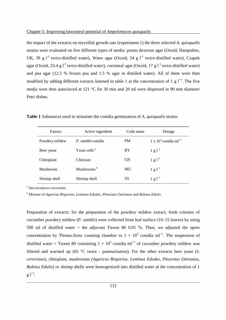
Chapter 5: Improving biocontrol potential of Ampelomyces quisqualis
112
the impact of the extracts on mycelilal growth rate (experiment 1) the three selected A. quisqualis
strains were evaluated on five different types of media: potato dextrose agar (Oxoid, Hampshire,
UK, 39 g l-1 twice-distilled water), Water agar (Oxoid, 24 g l-1 twice-distilled water), Czapek
agar (Oxoid, 33.4 g l-1 twice-distilled water), cornmeal agar (Oxoid, 17 g l -1 twice-distilled water)
and pea agar (12.5 % frozen pea and 1.5 % agar in distilled water). All of them were then
modified by adding different extracts listened in table 1 at the concentration of 1 g l−1. The five
media were then autoclaved at 121 °C for 30 min and 20 ml were dispersed in 90 mm diameter
Petri dishes.
Table 1 Substances used to stimulate the conidia germination of A. quisqualis strains
Extract Active ingredient Code name Dosage
Powdery mildew P. xanthii conidia PM 1 x 10⁵ conidia ml−1
Beer yeast Yeast cells a BY 1 g l−1
Chitoplant Chitosan CH 1 g l−1
Mushroom Mushrooms b MU 1 g l−1
Shrimp shell Shrimp shell SS 1 g l−1
a Saccaromyces cerevisiae. b Mixture of Agaricus Bisporius, Lentinus Edodes, Pleurotus Ostreatus and Boletus Edulis.
Preparation of extracts: for the preparation of the powdery mildew extract, fresh colonies of
cucumber powdery mildew (P. xanthii) were collected from leaf surface (10–15 leaves) by using
500 ml of distilled water + the adjuvant Tween 80 0.01 %. Then, we adjusted the spore
concentration by Thoma-Zeiss counting chamber to 1 × 10⁵ conidia ml−1. The suspension of
distilled water + Tween 80 containing 1 × 10⁵ conidia ml−1 of cucumber powdery mildew was
filtered and warmed up (65 °C twice - pasteurization). For the other extracts beer yeast (S.
cerevisiae), chitoplant, mushrooms (Agaricus Bisporius, Lentinus Edodes, Pleurotus Ostreatus,
Boletus Edulis) or shrimp shells were homogenized into distilled water at the concentration of 1
g l−1.
Chapter 5: Improving biocontrol potential of Ampelomyces quisqualis
113
Preparation of A. quisqualis conidia: cultures of the three A. quisqualis strains, approximately 2
weeks old, were flooded with sterile, distilled water and left for about 1 hour for the conidia to
ooze out of the pycnidia. The resulting conidial suspension was harvested and adjusted with a
Thoma-Zeiss counting chamber to 1 × 10³ conidia ml-1 (experiment 1) or 1 × 10⁵ conidia ml-1
(experiment 2).
For the first experiment Petri-dish cultures of the three fungal strains on the five media amended
with the different extracts were prepared (75 plates per strain, 15 plates per media and 3 plates
per extract) with 100 µl of a suspension of sterile distilled water containing 103 conidia ml-1.
Cultures were incubated in the dark at 25 °C for 30 days. The radial growth rate of the colonies
was estimated by measuring the diameter of each colony after 30 days. All of the experiments
were conducted with three replicates (plates) per strain. The experiment was conducted twice.
In the second experiment each strain was paired with the five selected extracts. Sterile distilled
water suspensions of the substances (1 g l−1) together with each fungal strain (1 × 10⁵ conidia ml-
1) were prepared. Suspension of only A. quisqualis conidia in sterile distilled water were used as
controls. Four drops (10 µl each) per combination (four replicates) were put onto a glass slide,
which was placed in a Petri dish (RH = 100 %) and stored in a dark incubator kept at 25 °C,
which is the temperature that Gu and Ko (1997) used in their study of the factors affecting the
germination of A. quisqualis conidia. The germination rates and germ-tube elongation of 200
conidia per strain (four replicates, 50 conidia per replicate) were evaluated under a light
microscope (Hund Wetzlar H 600LL, Wetzlar, Germany) after 12 hours. The experiment was
conducted twice
5.2.3 Biocontrol activity after stimulation of conidial spores of A. quisqualis
Cucumber (Podosphaera xanthii), strawberry (Podosphaera aphanis) and grapevine (Erysiphe
necator) powdery mildew were used as fungal hosts to evaluate the ability of A. quisqualis in
reducing mildew infections under controlled conditions. Two weeks old plants were inoculated
with powdery mildew spraying a suspension of distilled water containing 1 × 10⁵ conidia ml-1.
Once powdery mildew cultures were sporulating (four/seven days later) a suspension of distilled
water containing 1 × 10⁶ conidia ml-1 of A. quisqualis and some soluble substances was prepared.
Chapter 5: Improving biocontrol potential of Ampelomyces quisqualis
114
Fungal cultures, approximately 2 weeks old, were flooded with 20 ml of sterile, distilled water +
Tween 80 and left for about 1 hour for the conidia to ooze out of the pycnidia. Conidia of A.
quisqualis were collected from 5 plates and adjusting the concentration to 1 × 10⁶ conidia ml-1
under a light microscope (Hund Wetzlar H 600LL, Wetzlar, Germany) by using Thoma-Zeiss
counting chamber. Then, soluble extracts of powdery mildew, shrimp shell, mushroom, beer
yeast, chitoplant were prepared as described above and mixed with A. quisqualis conidia
suspensions to finally obtain 250 ml of a suspension containing 1 × 10⁶ conidia ml-1 and 1 g
extract ml-1. The extracts were added to the conidia suspensions 12 hours before inoculation
time. Plants bearing sporulating colonies of P. xanthii, E. necator and P. aphanis were
homogeneously treated with the A. quisqualis/extract suspensions using a hand air-sprayer. Only
two basal leaves per plant previously infected with the specific powdery mildew were treated (10
ml of suspension per leaf). Plants with powdery mildew and A. quisqualis infections were kept at
25 °C and 95 % of RH for 48 hours and then stored at the same temperature and 70 ± 10 % of
RH. Untreated control plants were infected with powdery mildew and then sprayed with the
natural extract dissolved into distilled water and Tween 80 only. The reduction of mycelial
fungal infection of powdery mildews was evaluated ten days after A. quisqualis inoculation.
Powdery mildew infected leaf area (severity) was evaluate by observing the presence of powdery
mildew spots on the upper leaf surface of the two basal leaves. Results were expressed as
percentage reduction of the powdery mildew infected leaf area in comparison with the powdery
mildew control.
5.2.4 Data evaluation and statistical analysis
Assays were performed on two independent experiments arranged in a randomized complete
block design with three replications. Data were first tested for the homogeneity of variances and
then subjected to the analysis of variance (ANOVA). Two-way ANOVA was performed on log-
transformed data from the two independent experiments and revealed no significant experiment
× treatment interaction. Therefore, data from the two experiments were pooled. Averages of all
replicates are presented in Table 2, Figure 1 and 2. Statistical significances among treatments
were computed with Statistica software 7.0 (Statsoft, Tulsa, OK, USA) and means were
separated using the Tukey’s HSD (P ≤ 0.05).

Chapter 5: Improving biocontrol potential of Ampelomyces quisqualis
115
5.3 Results
5.3.1 Stimulation of A. quisqualis under different conditions
The radial growth rate of the three selected A. quisqualis strains were different on the five tested
media (Tab. 2). The highest growth rates were observed on potato dextrose and pea agar plates
for all strains whereas strains grew slowest on water agar. The growth rate of commercial AQ10
strain (0.12–0.79 mm d-1) was higher than that of ITA 3 (0.11–0.59 mm d-1) and ATCC 200245
(0.09–0.54 mm d-1) on different media. Interestingly, our results indicate that the presence of
some natural extracts into different media increased the growth of A. quisqualis strains.
Significant differences in the induction of growth rate were detected among the different
extracts. A. quisqualis responded to mushroom and shrimp shell supply with increased growth
rate on different media. The effect is higher on water agar and czapek media where the growth
rate of strains was increased by 80–250 % and 50–160 %, respectively. Powdery mildew extract
significantly enhanced the growth of A. quisqualis strains by 40–120 % on all considered media.
However, the other two extracts (chitoplant and beer yeast) did not stimulate the growth on these
media.
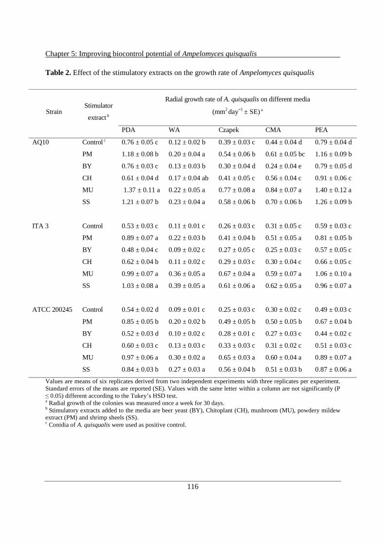
Chapter 5: Improving biocontrol potential of Ampelomyces quisqualis
116
Table 2. Effect of the stimulatory extracts on the growth rate of Ampelomyces quisqualis
Strain
Stimulator
extract b
Radial growth rate of A. quisqualis on different media
(mm2 day−1 ± SE) a
PDA WA Czapek CMA PEA
AQ10 Control c 0.76 ± 0.05 c 0.12 ± 0.02 b 0.39 ± 0.03 c 0.44 ± 0.04 d 0.79 ± 0.04 d
PM 1.18 ± 0.08 b 0.20 ± 0.04 a 0.54 ± 0.06 b 0.61 ± 0.05 bc 1.16 ± 0.09 b
BY 0.76 ± 0.03 c 0.13 ± 0.03 b 0.30 ± 0.04 d 0.24 ± 0.04 e 0.79 ± 0.05 d
CH 0.61 ± 0.04 d 0.17 ± 0.04 ab 0.41 ± 0.05 c 0.56 ± 0.04 c 0.91 ± 0.06 c
MU 1.37 ± 0.11 a 0.22 ± 0.05 a 0.77 ± 0.08 a 0.84 ± 0.07 a 1.40 ± 0.12 a
SS 1.21 ± 0.07 b 0.23 ± 0.04 a 0.58 ± 0.06 b 0.70 ± 0.06 b 1.26 ± 0.09 b
ITA 3 Control 0.53 ± 0.03 c 0.11 ± 0.01 c 0.26 ± 0.03 c 0.31 ± 0.05 c 0.59 ± 0.03 c
PM 0.89 ± 0.07 a 0.22 ± 0.03 b 0.41 ± 0.04 b 0.51 ± 0.05 a 0.81 ± 0.05 b
BY 0.48 ± 0.04 c 0.09 ± 0.02 c 0.27 ± 0.05 c 0.25 ± 0.03 c 0.57 ± 0.05 c
CH 0.62 ± 0.04 b 0.11 ± 0.02 c 0.29 ± 0.03 c 0.30 ± 0.04 c 0.66 ± 0.05 c
MU 0.99 ± 0.07 a 0.36 ± 0.05 a 0.67 ± 0.04 a 0.59 ± 0.07 a 1.06 ± 0.10 a
SS 1.03 ± 0.08 a 0.39 ± 0.05 a 0.61 ± 0.06 a 0.62 ± 0.05 a 0.96 ± 0.07 a
ATCC 200245 Control 0.54 ± 0.02 d 0.09 ± 0.01 c 0.25 ± 0.03 c 0.30 ± 0.02 c 0.49 ± 0.03 c
PM 0.85 ± 0.05 b 0.20 ± 0.02 b 0.49 ± 0.05 b 0.50 ± 0.05 b 0.67 ± 0.04 b
BY 0.52 ± 0.03 d 0.10 ± 0.02 c 0.28 ± 0.01 c 0.27 ± 0.03 c 0.44 ± 0.02 c
CH 0.60 ± 0.03 c 0.13 ± 0.03 c 0.33 ± 0.03 c 0.31 ± 0.02 c 0.51 ± 0.03 c
MU 0.97 ± 0.06 a 0.30 ± 0.02 a 0.65 ± 0.03 a 0.60 ± 0.04 a 0.89 ± 0.07 a
SS 0.84 ± 0.03 b 0.27 ± 0.03 a 0.56 ± 0.04 b 0.51 ± 0.03 b 0.87 ± 0.06 a
Values are means of six replicates derived from two independent experiments with three replicates per experiment. Standard errors of the means are reported (SE). Values with the same letter within a column are not significantly (P ≤ 0.05) different according to the Tukey’s HSD test. a Radial growth of the colonies was measured once a week for 30 days. b Stimulatory extracts added to the media are beer yeast (BY), Chitoplant (CH), mushroom (MU), powdery mildew extract (PM) and shrimp sheels (SS). c Conidia of A. quisqualis were used as positive control.
Chapter 5: Improving biocontrol potential of Ampelomyces quisqualis
117
The experiment in which we investigated the effect of stimulatory extracts on conidial
germination of A. quisqualis strains showed that germination rates and germ-tube elongation of
strains AQ10, ITA 3 and ATCC 200245 were very poor in the absence of stimulators (Figs. 1a–
b). In sterile, distilled water only a small fraction (less than 12 %) of the conidia of A. quisqualis
had germinated at the end of 12 hours incubation and the lenght of their germ tubes was less than
5 µm. Both parameters were strongly enhanced when 1 g l-1 of different extracts inducing fungal
growth on artificial media (Tab. 2) were added to the distilled water. However, significant
differences in the induction of germination were detected among the different extracts. Similarly
to the mycelial growth the conidial germination and tube elongation of all strains were stimulated
most by shrimp shell (60–87 % and 19–30 um) and mushroom extract (60–69 % and 17–25 um).
Germination was much less, but still significantly induced by powdery mildew conidia and
chitoplant extracts whilst treatments with beer yeast did not statistically differ from untreated
control, with the exception of f strain ATCC 200245 which was slightly stimulated by yeast
extract. Furthermore, we observed differences among the conidial germination rates of the
different strains, independent of the extract treatments. With the exception of beer yeast, the
germination rate and tube elongation of ITA 3 the most aggressive of the tested strains were
strongly induced by all of the tested extracts (33–87 % and 7–31 um) whereas the lower
aggressive strains were generally less responsive.

Chapter 5: Improving biocontrol potential of Ampelomyces quisqualis
118
Figure 1 Effects of beer yeast (BY), Chitoplant (CH), mushroom (MU), powdery mildew extract (PM)
and shrimp sheels (SS) on the conidial germination (%) (a) and germ tube elongation (µm) (b) of AQ10,
ITA 3 and ATCC 200245. Measurements were taken in aqueous suspensions containing mixtures of A.
quisqualis conidia and extracts that had been incubated together for 12 hours at 25 °C. Untreated conidia

Chapter 5: Improving biocontrol potential of Ampelomyces quisqualis
119
were used as control. Columns with the same design represent the same stimulator extract. Columns
labeled with the same letter (a-d) are not significantly different (P ≤ 0.05) according to Tukey’s test. Four
replicates (50 conidia per replicate) of each strain were evaluated. Presented values are means of two
independent experiments with four replicates per experiment.
5.3.2 Biocontrol activity after stimulation of conidial spores of A. quisqualis
Experiments conducted on three different fungal hosts showed that the ability to reduce mildew
infections of A. quisqualis strains can be increased by the presence of some of the tested extracts
(Fig. 2). As observed for mycelial growth and conidia germination (Table 2, Fig. 1) the
stimulating effect of mushroom and shrim shell extracts was most pronounced. In the presence of
these substances all tested strains showed a stronger reduction of infected leaf area of all tested
powdery mildews. Bear yeast and Chitoplant extracts did not have any impact on biocontrol
activity. Extracts of mildew conidia stimulated only the activity A. quisqualis on P. aphani (Fig.
2 A). However, individual A. quisqualis strains differ in their aggressiveness and ITA 3 was
more effective than ATCC 200245 and the commercial strain AQ10. Best results were obtained
with ITA 3 stimulated by shrimp shell and mushroom extracts which reduced the infected leaf
area on strawberry, grapevine and cucumber (Fig 2). Values ranged from 62 to 91 % reduction of
powdery mildew infected leaf area. The improving effects of the extracts on obtained with
AQ10, ITA 3 and ATCC 200245 were clearly higher on strawberry powdery mildew which is
less susceptible versus A. quisqualis than cucumber and grapevine. For example, mushroom and
shrimp shell extracts increased the reduction of mildew infection by more than 20 % in
comparison to ITA 3 and AQ10 treatments without extract addition (Fig. 2a).

Chapter 5: Improving biocontrol potential of Ampelomyces quisqualis
120

Chapter 5: Improving biocontrol potential of Ampelomyces quisqualis
121
Figure 2 P. aphanis (a), E. necator (b) and P. xanthii (c) infected area on leaves sprayed with AQ10,
ITA 3 and ATCC 200245 stimulated by beer yeast (BY), Chitoplant (CH), mushroom (MU), powdery
mildew extract (PM) and shrimp sheels (SS). Untreated conidia were used as positive control. Columns
with the same design represent the same stimulator extract. Columns with the same letters (a-c) do not
significantly differ (P < 0.05, Tukey’s test). Presented values are means of two independent experiments
with three replicates per experiment.
5.4 Discussion
Biological control of powdery mildews remains a challenge for future research and development.
The results obtained so far showed some promises of practical biocontrol of a number of
powdery mildew diseases but more efforts are needed to prove the efficacy of these methods in
agricultural and horticultural practice. The major constraint to the use of fungal pathogens as
biological control agents is consistent performance under field conditions. Powdery mildews are
biotrophic pathogens infecting the aerial parts of their host plants and biocontrol agents have to
perform their actions in the phyllosphere, a high dynamic environment, where biological control
of different plant pathogenic fungi is, in general, less successful. Moreover, the sporulation
capacity of powdery mildews is high and their spread very fast making their control extremely
difficult even with chemicals. AQ10, is the first commercialized biocontrol product based on
spores of A. quisqualis against powdery mildews but it seems that it cannot provide satisfactory
control in a number of crops including grape, rose and vegetables (Angeli et al., 2009; Gilardi et
al., 2012). Inconsistent performance is often attributed to a lack of free moisture which is
required by the fungal spores during process of germination and penetration of the host (Jackson,
1997). The requirement for free moisture is a major environmental constraint of A. quisqualis to
effective control under field conditions. The slow growth rate and relatively low conidial
germination and concentration is another disadvantage to its use as biocontrol agents.
Because of these constraints, one of our primary goals in producing fungal spores for use as a
biocontrol agent is to reduce the impact of the free moisture requirement by producing spores
which germinate more rapidly and form an appressorium more frequently. Here different media
containing sufficient nutrients to support microbial growth were compared to a medium
essentially free of nutrients (water agar), an approach that is useful for the detection of
Chapter 5: Improving biocontrol potential of Ampelomyces quisqualis
122
stimulators or inhibitors (Gu & Ko, 1997). The mycelium of A. quisqualis grows slowly on
artificial media in vitro. On tested media growth of colonies of A. quisqualis was greatly
enhanced by including mushroom and shrimp shell extracts in the media. These extracts seem to
have a general stimulating effect on A. quisqualis since they stimulated the growth of all three
tested strains.
Only a few studies examined the impact of added nutrients on the conidial germination of this
hyperparasite whereas there are numerous reports of the stimulatory effects of nutrients on the
germination of spores of other fungal species. Leachate from aerial plant surfaces contains
nutrients that stimulate the germination of fungal propagules of Alternaria alternata and
Epicoccum purpurascens (Tukey, 1971). Blakeman (1975) found that sugars promoted
germination of Botrytis cinerea conidia. Some aminoacids were stimulatory while others
inhibited germination. The amount of sugars and aminoacids present on leaves were sufficient to
stimulate germination of B. cinerea conidia.
Cochrane 1966 differentiated between fungi which require external nutrients for germination and
those that germinate without the addition of nutrients. Conidia of A. quisqualis fall into the first
category. Some organic molecules present in the hyphae of the host fungus and other fungi are
needed for germination. However, the addition of simple carbohydrates or aminoacids did not
promote germination of A. quisqualis (Cochrane, 1966). Recently, Gu & Ko (1997) showed that A.
quisqualis recognizes the presence of the host and a water-soluble substance from powdery
mildew conidia has been shown to stimulate germination of A. quisqualis conidia in vitro. The
stimulatory substance in powdery mildew was thermostable and its molecular weight was in the
range of 1200–1500 dalton. However, the identity of the fungal substance that stimulates
germination of A. quisqualis conidia remains to be elucidate.
The results obtained in this study confirm that the mildew host stimulates the germination of A.
quisqualis conidia but show that supplementation with external sources of nutrient precursors
present in shrimp shell and mushroom extracts even had a stonger positive effect on the
germination of conidia. We identified new extracts based on different active ingredients able to
stimulate the growth rate on plate and conidial germination of A. quisqualis and their stimulatory
effect is often higher than that of the fungal host (P. xanthii conidia). Of the tested extracts
mushroom, shrimp shell and mildew conidia extracts, but not bear yeast and Chitoplant extracts

Chapter 5: Improving biocontrol potential of Ampelomyces quisqualis
123
stimulated both growth and conidial germination of all tested Ampelomyces strains. Interestingly
conidia of ITA3 the strain displaying the highest level of biocontrol were stimulated the most by
mushroom and shrimp shell extracts.
In this work conidia of the three tested strains germinated poorly or not at all in sterile, distilled
water. Cook (1977) hypothesized that self-inhibitors in fungal spores are common and that failure
of spores to germinate is due to substances within the spores. Nutrients may cause germination by
deactivating the self-inhibitors in the spores. Other studies showed that spores of some fungi
germinate readily under dispersed conditions but when crowded they germinate poorly due to the
presence of a self-inhibitor (Allen, 1976; Lax et al., 1985). A few years later Gu & Ko (1997)
developed an in vitro method for laboratory observation of the interaction between conidia of
powdery mildews and conidia of their mycoparasite and they reported on the presence of self-
inhibitor in conidia of A. quisqualis. On water agarose media germination of A. quisqualis conidia
decreased as their concentration increased. In light of this discovery, we identified some extracts
which seem to easly overcome the self-inhibition of germination of A. quisqualis conidia.
Whether indeed, some compounds in the extracts we used interact with self-inhibitors would be
an interesting issue for investigation in future research.
Furthermore, we report on improvement of in vivo biocontrol acitivity of A. quisqualis in three
different pathosystems by the addition of stimulants. Our results show that performance of
different A. quisqualis strains isolated from different plants and fungal hosts can be increased by
the presence of extracts stimulating growth and conidial germination. Protection against powdery
mildew was clearly higher on plants treated with a suspesion of A. quisqualis ITA 3 strain
stimulated through mushroom and shrimp shell. The greatest stimulating effect was observed on
plants infected with strawberry powdery mildew which is less susceptible to A. quisqualis than
cucumber and grapevine.
In this study we demonstrated there is considerable potential for improving the efficiency of the
fungus A. quisqualis in the control of powdery mildew pathogen. These findings indicate that the
nutritional environment during culture growth can have an effect on hyphal growth pattern and,
even more relevant on spore attributes such as germination and germ tube elongation. Our
studies have demonstrated that the conidial germination ability of A. quisqualis can be positively
related to virulence against powdery mildew and can, therefore, be considered as relevant factor

Chapter 5: Improving biocontrol potential of Ampelomyces quisqualis
124
in the selection of new good biocontrol agents. We feel that by optimizing the spore attributes we
can improve the biocontrol efficacy of this agent under field conditions. This provides important
knowledge for the selection of new, highly effective mycoparasitic fungal strains for biocontrol
potential and its exploitation in sustainable agriculture.
5.5 Acknowledgements
We would like to thank Franca Micheli, Carmela Sicher and Denise Ress for technical
assistance. This work was supported by the AMPELO and ENVIROCHANGE projects funded
by the Autonomous Province of Trento.
5.6 Literature
Allen, P. J. (1976) Spore germination and its regulation. Control of spore germination and infection structure
formation in the fungi. Encyclopedia of Plant Physiology New Series Volume 4:51-85.
Angeli, D., Pellegrini, E., Micheli, S., Ress, D., Maurhofer, M., Pertot, I. and Gessler, C. (2009). Molecular
characterization of Ampelomyces spp. strains from different hosts and geographic origins and evaluation of
their potential to control powdery mildew of cucumber. IOBC/WPRS Bulletin 43:40-44.
Angeli, D., Maurhofer M., Gessler C. and Pertot I. (2012a). Existence of different physiological forms within
genetically diverse strains of Ampelomyces quisqualis. Phytoparasitica 40:37-51.
Angeli, D., Puopolo, G., Maurhofer M., Gessler C. and Pertot I. (2012b). Is the mycoparasitic activity of
Ampelomyces quisqualis biocontrol strains related to phylogeny and hydrolytic enzyme production?.
Biological control 63(3):348-358.
Blakeman, J.P. (1975). Germination of Botrytis cinerea conidia in vitro in relation to nutrient conditions on leaf
surfaces. Trans. Br. Mycol. Soc. 65:239-247.
Cochrane, V.W. (1966). Respiration and spore germination. In “The fungus spore” ed. M.F. Madelin, pp. 201-213.
Butterwortiis, London. 338 pp.
Cook, R.J. (1977). Management of the Associated Microbiota. In “Plant Disease” ed. J.G. Horsefall and E.B.
Cowling, vol. 1, pp. 145-166. Academic Press, London and New York. 465 pp.
Dik, A.J., Verhaar, M.A. and Bélanger, R.R. (1998). Comparison of three biological control agents against
cucumber powdery mildew (Sphaerotheca fuliginea) in semi-commercial-scale glasshouse trials. European
Journal of Plant Pathology 104:413-423.

Chapter 5: Improving biocontrol potential of Ampelomyces quisqualis
125
Falk, S. P., Gadoury, D. M., Cortesi, P., Pearson, R. C., and Seem R. C. (1995a). Parasitism of Uncinula necator
cleistothecia by the mycoparasite Ampelomyces quisqualis. Phytopathology 85:794-800.
Falk, S. P., Gadoury, D. M., Pearson, R. C., and Seem, R. C. (1995b). Partial control of grape powdery mildew by
the mycoparasite Ampelomyces quisqualis. Plant Dis 79:483-490.
Fokkema, N.J. (1993). Opportunities and problems of control of foliar pathogens with micro-organisms. Pesticide
Science 37:411-416.
Gadoury, D. M. and Pearson, R. C. (1988). Initiation, development, dispersal, and survival of cleistothecia of
Uncinula necator in New York vineyards. Phytopathology 78:1413-1421.
Gilardi, G., Baudino, M., Garibaldi, A., Gullino, M.L. (2012). Efficacy of biocontrol agents and natural compounds
against powdery mildew of zucchini. Phytoparasitica 40:147-155.
Gu, Y.H. and Ko, W.H. (1997). Water agarose medium for studying factors affecting germination of conidia of
Ampelomyces quisqualis. Mycological Research 101:422-424.
Hashioka, Y. and Nakai, Y. (1980). Ultrastructure of pycnidial development and mycoparasitism of Ampelomyces
quisqualis parasitic on Erysiphales. Trans. Mycol. Soc. Jpn 21:329-338.
Hofstein, R., Daoust, R.A. and Aeschlimann, J.P. (1996). Constrains to the development of biofungicides: the
example of ‘‘AQ10’’, a new product for controlling powdery mildews. Entomophaga 41:455-460.
Jackson, M.A. (1997). Optimizing nutritional conditions for the liquid culture production of effective fungal
biological control agents. Journal of Industrial Microbiology & Biotechnology 19:180-187.
Jarvis, W.R. and Slingsby, K. (1977). The control of powdery mildew of greenhouse cucumber by water sprays and
Ampelomyces quisqualis. Plant Dis Rep 61:728-730.
Kiss, L. (1997). Genetic diversity in Ampelomyces isolates, hyperparasites of powdery mildew fungi, inferred from
RFLP analysis of the rDNA ITS region. Mycological Research 101:1073-1080.
Kiss, L. and Nakasone, K. K. (1998). Ribosomal DNA internal transcribed spacer sequences do not support the
species status of Ampelomyces quisqualis, a hyperparasite of powdery mildew fungi. Curr. Gen. 33:362-367.
Kiss, L., Russell, J.C., Szentivanyi, O., Xu, X. and Jeffries, P. (2004). Biology and biocontrol potential of
Ampelomyces mycoparasites, natural antagonist of powdery mildew fungi. Biocontrol Science and Technology
14:635-651.
Kiss, L., Pintye, A., Kovacs, G.M., Jankovics, T., Fontaine, M.C., Harvey, N., Xu, X., Nicot, P.C., Bardin, M.,
Shykoff, J.A. and Giraud, T. (2011). Temporal isolation explains host-related genetic differentiation in a group
of widespread mycoparasitic fungi. Molecular Ecology 20:1492-1507.
Lax, A. R., Templeton, G. E. and Meyer, W. L. (1985). Isolation, purification, and biological activity of a self-
inhibitor from conidia of Colletotrichum gloeosporioides. Phytopathology 75(4):386-390.

Chapter 5: Improving biocontrol potential of Ampelomyces quisqualis
126
Liang, C., Yang, J., Kovacs, G.M., Szentivanyi, O., Li, B., Xu, X.M. and Kiss, L. (2007). Genetic diversity of
Ampelomyces mycoparasite isolated from different powdery mildew species in China inferred from analyses of
rDNA ITS sequences. Fungal Diversity 24:225-240.
McGrath, M.T. and Shishkoff, N. (1999). Evaluation of biocompatible products for managing cucurbit powdery
mildew. Crop Protection 18:471-478.
Park, M.J., Choi, Y.J., Hong, S.B. and Shin, H.D. (2010). Genetic variability and mycohost association of
Ampelomyces quisqualis isolates inferred from phylogenetic analyses of ITS rDNA and actin gene sequences.
Fungal Biology 114:235-247.
Pasini, C., D’Aquila, F., Curir, P. and Gullino, M.L. (1997). Effectiveness of antifungal compounds against rose
powdery mildew (Sphaerotheca pannosa var rosae) in glasshouses. Crop Protect 16:251-256.
Paulitz, T.C. and Bélanger, R.R. (2001). Biological control in greenhouse systems. Annu Rev Phytopathol 39:103-
133.
Shishkoff, N. and McGrath, M.T. (2002). AQ10 biofungicide combined with chemical fungicides or AddQ spray
adjuvant for control of cucurbit powdery mildew in detached leaf culture. Plant Disease 86:915-918.
Sundheim, L. (1982). Control of cucumber powdery mildew by the hyperparasite Ampelomyces quisqualis and
fungicides. Plant Pathol 31:209-214.
Sundheim, L., Krekling, T. (1982). Host-parasite relationships of the hyperparasite Ampelomyces quisqualis and its
powdery mildew host Sphaerotheca fuliginea. I. Scanning electron microscopy. Phytopathology 104:202-210.
Sundheim, L. and Tronsmo, A. (1988). Hyperparasites in biological control. Pages 53-69 in: Biocontrol of Plant
Diseases. Vol. I. CRC Press, Boca Raton, FL.
Sztejnberg, A., Galper, S., Mazar, S., and Lisker, N. (1989). Ampelomyces quisqualis for biological and integrated
control of powdery mildews in Israel. J Phytopath 124:285-295.
Sztejnberg, A. (1993). Ampelomyces quisqualis AQ10, CNCM I-807, for biological control of powdery mildew. US
patent no. 5190754.
Tukey, H.B. (1971). Leaching of substances from plants. In “Ecology of leaf surface micro-organisms” ed. T.F
Preece. and C.H. Dickinson, pp. 67-80. Academic Press, London and New York. 640 pp.
Verhaar, M.A., Huwegen, T. and Zadoks, J.C. (1996). Glasshouse experiments on biocontrol of cucumber powdery
mildew (Sphaerotheca fuliginea) by the mycoparasites Verticillium lecanii and Sporothrix rugulosa.
Biological Control 6:353-360.
Verhaar, M.A., Kerssies, A. and Huwegem, T. (1999). Effect of RH on mycoparasitism of rose powdery mildew
with and without treatments with mycoparasites. Zeitschrift fur Pflanzenkrankheiten und Pflanzenschutz
106:158-165.

127
Chapter 6
General conclusions

Chapter 6: General conclusions
128
6.1 General conclusions
Biological control of powdery mildews remains a challenge for future research and development.
The results obtained so far show some promises of practical biocontrol of a number of powdery
mildew diseases, especially on cucumber and rose, but more efforts are needed to prove the
efficacy of these methods in horticultural practice. A product based on P. flocculosa (Sporodex),
the most promising biocontrol alternative for use against powdery mildews, has received a
temporary registration in Canada only since May 2002, and no data about its commercial
success, are available yet. Unfortunately, it seems that AQ10 biofungicide based on spores of A.
quisqualis, the first commercialized biocontrol product against powdery mildews, cannot provide
satisfactory control in a number of crops including grape, rose and vegetables (Bélanger &
Labbé, 2002; Dik et al., 1998; McGrath & Shishkoff, 1999; Verhaar et al., 1996). Currently, its
use is recommended as a part of an integrated programme and as a preventive measure only
(Bélanger & Labbé, 2002; Paulitz & Bélanger, 2001). Another formulated A. quisqualis product
was registered in Australia in the late 1980s, but did not meet the expectations of the plant
protection practice. However, a number of possibilities for the use of A. quisqualis as an
effective biocontrol agent have yet to be exploited. Some powdery mildews can be controlled
efficiently with these mycoparasites (Kiss L., 2004). Moreover, there is a high genetic diversity
in A. quisqualis strains that offers the not yet exploited possibility of screening a large number of
different types of strains (Angeli et al., 2009b; Angeli et al., 2012a; Kiss, 1997; Kiss &
Nakasone, 1998; Kiss et al., 2011; Liang et al., 2007). Strains of V. lecanii and Tilletiopsis spp.
represent another promising source of successful biocontrol agents of powdery mildews.
However, in spite of intensive studies on their biology and biocontrol potential there have been
no initiatives to develop commercial products based on these fungi (Askary et al., 1998; Dik et
al., 1998; Urquhart et al., 1994; Urquhart & Punja, 1997; Urquhart & Punja, 2002; Verhaar et al.,
1996; Verhaar et al., 1999;). This thesis shows that there are still many unexplored possibilities
for research and development of biocontrol agents. Well-studied and little known fungal
antagonists constitute together a valuable source that will surely be used to develop biocontrol or
integrated programs to reduce the use of chemicals against powdery mildews in the foreseeable
future. Powdery mildew caused by E. necator is an important disease of grapevine in the
Trentino-Alto Adige region.

Chapter 6: General conclusions
129
The first objective of the present work was to investigate the natural occurrence of overwintering
inoculum of powdery mildew in a wide viticulture area in Northern Italy (Trentino-Alto Adige
region) and evaluate the presence of A. quisqualis on grapes (Chapter 2). Here we demonstrated
that the initiation and development of chasmothecia can occur in any year in which conditions
are favorable for disease development (Angeli et al., 2009a). The high incidence of chasmothecia
in the vineyards of the Trentino-Alto Adige region, which was associated with the absence of
flag shoots in the monitored vineyards, could imply that chasmothecia are the primary inoculum
and confirm previous results, which had indicated that chasmothecia represent the main source of
inoculum in the spring (Cortesi et al., 1995). The many studies on E. necator chasmothecia have
reported unclear and sometimes contradictory effects of environmental factors on the
development of chasmothecia (Gadoury & Pearson, 1988). Our results suggest that the
concentration of chasmothecia was mainly related to the level of disease in the vineyard, which
is usually the result of complex interactions among the amount of initial inoculum, the efficacy
of disease control measures, cultivar susceptibility, the physiology of the crop, and the weather
conditions during the growing season. Reports have stated that most of the chasmothecia die
during the winter without having any opportunity to release ascospores (Cortesi et al., 1995), but
a reduction in the number of overwintering chasmothecia by hyperparasites may nevertheless be
useful for reducing the amount of primary inoculum (Falk et al., 1995a,b). A. quisqualis is one of
the most successful commercialized biocontrol agents and it acts directly by invading and
destroying the viable chasmothecia (Hashioka & Nakai, 1980). The present study is the first
extensive investigation of quantitative aspects of A. quisqualis on grapevine powdery mildew in
Italy. A. quisqualis was observed parasitizing chasmothecia of E. necator suggesting that A.
quisqualis was naturally present in the region. More than 200 strains of A. quisqualis were
identified and some of them were genetically different from the commercial strain (AQ10). They
may represent strains better adapted to local environmental conditions than AQ10 and can be
developed as new biocontrol agents to control powdery mildew under the environmental
conditions found in Northern countries.
Further analyses (chapter 3) were performed to characterize the isolated strains and other A.
quisqualis strains derived from different regions and hosts and to investigate their environmental
requirements for development and parasitism. These investigations aimed also at the
identification of phenotypic markers that can be used to differentiate genetically distinct groups

Chapter 6: General conclusions
130
within A. quisqualis. Data on the morphology and cultural patterns of A. quisqualis strains found
in the literature are controversial and incomplete (Kiss, 1997; Park et al., 2010). We revealed
some significant variation among the selected strains, which provides evidence for the existence
of different physiological forms within the A. quisqualis species. Two groups that display
differential growth on artificial media were identified. These groups also differ in the
morphology of their mycelium, but not in the morphology of their pycnidia and conidia.
Temperature greatly affected the in vitro growth of the A. quisqualis strains and growth rate was
closely correlated to colony color. Differences in the conidial germination of distinct strains were
observed during the recognition phase of the parasitic relationship. Different powdery mildew
agents strongly affect the germination of A. quisqualis conidia but specialization is indistinct.
Phylogenetic analysis of the ITS rDNA sequence revealed a high level of genetic diversity
among A. quisqualis strains and suggested that ITS groups could be related to the fungal host of
origin and morphological and growth characteristics. It appears that powdery mildew fungi and
A. quisqualis might have coevolved through the processes of specialization and adaptation. Thus,
a degree of mycohost specialization was identified within the A. quisqualis species.
The second main objective of the thesis was to assess the mycoparasitic activity of the A.
quisqualis strains in terms of their ability to inhibit conidiation in different powdery mildew
pathogens and intra-hyphal formation of their intracellular pycnidia within powdery mildews in
vivo. At the same time, production of cell wall degrading enzymes (CWDEs) was measured
under in vitro conditions and factors important in effective mycoparasitism of A. quisqualis were
detected (Chapter 4). Results showed that individual strains differed significantly in enlargement
of the colonization area by intrahyphal formation of pycnidia within powdery mildew colonies
and in inhibition of host conidiation. Pronounced differences in the activity of CWDEs were also
observed between strains. The ability of some A. quisqualis strains to parasitize the host by
forming intracellular pycnidia and reducing fungal-host conidiation was associated with their
intrinsic ability to secrete proteases and with chitobiase activity. Finally, we have demonstrated
that the ability of A. quisqualis strains to colonize powdery mildew by forming pycnidia and
inhibiting conidiation is completely independent of the geographical origin of the strain, but
appears to be related to genotype (ITS rDNA sequence) and the original fungal host. The
biological characterization of the A. quisqualis strains carried out in this work suggests that
mycoparasitism can be explained by the genotype and in part by the activities of CWDEs. This

Chapter 6: General conclusions
131
provides important knowledge for the development of highly effective strains for biocontrol of
powdery mildews. However, the level of parasitization varies between different strains, and the
cause of this variation is unknown (Angeli et al., 2012b). There is still considerable interest in
finding mycoparasitic strains within A. quisqualis species that afford more effective plant
protection than the existing strains.
In the last chapter we aimed at enhancing the performance of A. quisqualis in the biological
control of powdery mildew. A major focus of this part of the thesis was to optimize nutritional
conditions for the culture production of fungal spores focusing on producing spores which will
germinate rapidly, form an appressorium more frequently and can, therefore, increase the
virulence of the fungus in the biocontrol of powdery mildews. Inconsistent performance is often
attributed to a lack of free moisture which is required by the fungal spores during process of
germination and penetration of the host (Gu & Ko, 1997). The requirement for free moisture is a
major environmental constraint of A. quisqualis to effective control under field conditions.
Conidia of A. quisqualis germinate poorly in sterile, distilled water but a water-soluble substance
from the powdery mildew host was shown to stimulate the germination of the conidia. Here we
identified new extracts based on different active ingredients which stimulate the growth rate and
conidial germination of A. quisqualis conidia and the stimulatory effect seem to be extract species-
specific (Chapter 5). Biocontrol assays showed that the performance of different A. quisqualis
strains can be increased by the presence of extracts stimulating growth and conidial germination.
These findings revealed that the conidial germination ability of A. quisqualis could be positively
related to virulence against powdery mildew and can, therefore, be considered as relevant factor
in the selection of new good biocontrol agents.
In conclusion, the major constraint to the use of this fungal pathogen as biological control agents
is consistent performance under field conditions. Inconsistent performance is often attributed to a
lack of free moisture which is required by the fungal spores during process of germination and
penetration of the host. These studies demonstrates that there is considerable potential for
improving powdery mildew control by A. quisqualis for example by improving germination
characteristics of conidia. We further provide important knowledge on this fungus that is not
only useful for improving the efficacy of biological control agents under limiting conditions but
also for development of new screening strategies enabling the detection of highly effective
mycoparasitic fungal strains for on optimized biocontrol in sustainable agriculture.
Chapter 6: General conclusions
132
6.2 Literature
Angeli, D., Pellegrini, E. and Pertot, I. (2009a). Occurrence of Erysiphe necator chasmothecia and their natural
parasitization by Ampelomyces quisqualis. Phytopathology, 99, 704-710.
Angeli, D., Pellegrini, E., Micheli, S., Ress, D., Maurhofer, M., Pertot, I. and Gessler, C. (2009b). Molecular
characterization of Ampelomyces spp. strains from different hosts and geographic origins and evaluation of
their potential to control powdery mildew of cucumber. IOBC/WPRS Bulletin, 43, 40-44.
Angeli D., Maurhofer M., Gessler C. and Pertot I. (2012a). Existence of different physiological forms within
genetically diverse strains of Ampelomyces quisqualis. Phytoparasitica 40, 37-51.
Angeli, D., Puopolo, G., Maurhofer M., Gessler C. and Pertot I. (2012b). Is the mycoparasitic activity of
Ampelomyces quisqualis biocontrol strains related to phylogeny and hydrolytic enzyme production?.
Biological control 63(3):348-358.
Askary, H., Carriere, Y., Bélanger, R.R. and Brodeur, J. (1998). Pathogenicity of the fungus Verticillium lecanii to
aphids and powdery mildew. Biocontrol Sci Technol 8:23-32.
Bélanger, R.R. and Labbé, C. (2002). Control of powdery mildews without chemicals: prophylactic and biological
alternatives for horticultural crops, in The powdery mildews: a comprehensive treatise, ed by Bélanger, R.R.,
Bushnell, W.R., Dik, A.J. and Carver, T.L.W., American Phytopathological Society, St Paul, USA, pp 256-
267.
Cortesi, P., Gadoury, D. M., Seem, R. C. and Pearson, R. C. (1995). Distribution and retention of cleistothecia of
Uncinula necator on the bark of grapevines. Plant Dis. 79:15-19.
Dik, A.J., Verhaar, M.A. and Bélanger, R.R. (1998). Comparison of three biological control agents against
cucumber powdery mildew (Sphaerotheca fuliginea) in semi-commercial-scale glasshouse trials. European
Journal of Plant Pathology 104:413-423.
Falk, S. P., Gadoury, D. M., Cortesi, P., Pearson, R. C. and Seem R. C. (1995a). Parasitism of Uncinula necator
cleistothecia by the mycoparasite Ampelomyces quisqualis. Phytopathology 85:794-800.
Falk, S. P., Gadoury, D. M., Pearson, R. C. and Seem, R. C. (1995b). Partial control of grape powdery mildew by
the mycoparasite Ampelomyces quisqualis. Plant Dis. 79:483-490.
Gadoury, D. M. and Pearson, R. C. (1988). Initiation, development, dispersal, and survival of cleistothecia of
Uncinula necator in New York vineyards. Phytopathology 78:1413-1421.
Gu, Y.H. and Ko, W.H. (1997). Water agarose medium for studying factors affecting germination of conidia of
Ampelomyces quisqualis. Mycological Research 101, 422-424.

Chapter 6: General conclusions
133
Hashioka, Y. and Nakai, Y. 1980. Ultrastructure of pycnidial development and mycoparasitism of Ampelomyces
quisqualis parasitic on Erysiphales. Trans. Mycol. Soc. Jpn 21:329-338.
Kiss, L. (1997). Genetic diversity in Ampelomyces isolates, hyperparasites of powdery mildew fungi, inferred from
RFLP analysis of the rDNA ITS region. Mycol. Res. 101:1073-1080.
Kiss, L. and Nakasone, K. K. (1998). Ribosomal DNA internal transcribed spacer sequences do not support the
species status of Ampelomyces quisqualis, a hyperparasite of powdery mildew fungi. Curr. Gen. 33:362-367.
Kiss, L., Russell, J.C., Szentivanyi, O., Xu, X., Jeffries, P. (2004). Biology and biocontrol potential of Ampelomyces
mycoparasites, natural antagonist of powdery mildew fungi. Biocontrol Science and Technology 14, 635-651.
Kiss, L., Pintye A., Kovacs G.M., Jankovics T., Fontaine M.C., Harvey N., Xu X., Nicot P.C., Bardin M., Shykoff
J.A. and Giraud T. (2011). Temporal isolation explains host-related genetic differentiation in a group of
widespread mycoparasitic fungi. Molecular Ecology, 20, 1492-1507.
Liang C., Yang J., Kovacs G.M., Szentivanyi O., Li B., Xu X.M. & Kiss L. (2007). Genetic diversity of
Ampelomyces mycoparasite isolated from different powdery mildew species in China inferred from analyses of
rDNA ITS sequences. Fungal Diversity, 24, 225-240.
McGrath, M.T. and Shishkoff, N. (1999). Evaluation of biocompatible products for managing cucurbit powdery
mildew. Crop Protect m18:471-478.
Paulitz, T.C. and Bélanger, R.R. (2001). Biological control in greenhouse systems. Annu Rev Phytopathol 39:103-
133.
Park M.J., Choi Y.J., Hong S.B. & Shin H.D. (2010). Genetic variability and mycohost association of Ampelomyces
quisqualis isolates inferred from phylogenetic analyses of ITS rDNA and actin gene sequences. Fungal
Biology, 114, 235-247.
Urquhart, E.J., Menzies, J.G. and Punja, Z.K. (1994). Growth and biological control activity of Tilletiopsis species
against powdery mildew (Sphaerotheca fuliginea) on greenhouse cucumber. Phytopathology 84:341-351.
Urquhart, E.J. and Punja, Z.K. (1997). Epiphytic growth and survival of Tilletiopsis pallescens, a potential
biological control agent of Sphaerotheca fuliginea, on cucumber leaves. Canad J Bot 75:892-901.
Urquhart, E.J. and Punja, Z.K. (2002). Hydrolytic enzymes and antifungal compounds produced by Tilletiopsis
species, phyllosphere yeasts that are antagonists of powdery mildew fungi. Canad J Microbiol 48:219-229.
Verhaar, M.A., Huwegen, T. and Zadoks, J.C. (1996). Glasshouse experiments on biocontrol of cucumber powdery
mildew (Sphaerotheca fuliginea) by the mycoparasites Verticillium lecanii and Sporothrix rugulosa.
Biological Control 6:353-360.
Verhaar, M.A., Kerssies, A. and Huwegem, T. (1999). Effect of RH on mycoparasitism of rose powdery mildew
with and without treatments with mycoparasites. Zeitschrift fur Pflanzenkrankheiten und Pflanzenschutz
106:158-16.

Acknowledgements
134
Acknowledgements
I am grateful to:
Cesare Gessler.
Ilaria Pertot.
Sustainable Agro-Ecosystems and Bioresources Department (DASB).
AMPELO Group.
La mia famiglia.
Tutti quelli che mi sono stati vicini sopportandomi in questi anni.

Curriculum vitae
135
Curriculum Vitae
Personal Data
Name Dario
Surname Angeli
Date of birth 08.02.1976
Place of birth Mezzolombardo, Trento
Nationality Italy
Education
2008-2013 PhD at the Institute of Integrative Biology
Swiss Federal Institute of Technology (ETH), Zürich, Switzerland
Thesis: “Opening new frontiers in plant protection by
understanding the interaction between a plant pathogen and its
mycoparasite.”
2003-2007 Research assistant at the SafeCrop Centre
Fondazione Edmund Mach (IASMA), S. Michele all’Adige,
Trento, Italy
Innovative research oriented toward the technologies for reducing
chemical inputs in agriculture and using low impact plant
protection methods.
1996-2002 Bachelor degree in “Agronomic Science and Technology”
School of Agricultural Science, University of Padua
Thesis: “Efficacy of Benzyladenine as thinning and sizing agent in
relation to bloom intensity”.
Curriculum vitae
136
Publications
Angeli, D., Pellegrini, E. and Pertot, I. (2009). Occurrence of Erysiphe necator chasmothecia
and their natural parasitization by Ampelomyces quisqualis. Phytopathology, 99, 704-710
Angeli D., Maurhofer M., Gessler C. and Pertot I. (2012). Existence of different physiological
forms within genetically diverse strains of Ampelomyces quisqualis. Phytoparasitica 40, 37-
51.
Angeli, D., Puopolo, G., Maurhofer M., Gessler C. and Pertot I. (2012). Is the mycoparasitic
activity of Ampelomyces quisqualis biocontrol strains related to phylogeny and hydrolytic
enzyme production?. Biological control 63(3):348-358.
Angeli, D., Maurhofer M., Gessler C. and Pertot I. (2013). Stimulation of conidial germination
as a novel tool to improve the biocontrol potential of Ampelomyces quisqualis. To be
submitted to BioControl.
Conferences attended
Sept 2008 IOBC/WPRS Working Group “Biological control of fungal and bacterial
plant pathogens entitled “Molecular Tools for Understanding and
Improving Biocontrol”, September 9-12, 2008, Interlaken (Switzerland).
Talk: “Molecular characterization of Ampelomyces spp. isolates from
different host and geographic origin and evaluation of their potential to
control powdery mildew of cucumber”.
Jan 2010 Mycology Symposium, Research Station Agroscope, January 22, 2010,
Wadenswil (Switzerland).
Talk: “Characterization of several Ampelomyces quisqualis isolates and
potential to control cucumber and strawberry powdery mildew”
Jun 2010 IOBC/WPRS Working Group “Biological control of fungal and bacterial
plant pathogens entitled “Climate change: Challenge or Threat to
Biocontrol”, June 7-10, 2010, Graz (Austria).

Curriculum vitae
137
Talk: “Selecting highly effective strains of Ampelomyces quisqualis for the
control of powdery mildews”.
Poster: “Influence of temperature on morphology and physiology of
different isolates of Ampelomyces quisqualis”.
Jul 2010 International Workshop on Grapevine Downy & Powdery Mildew. INRA,
July 4-9, 2010, Bordeaux (France).
Talk: “Can different isolates of the mycoparasite Ampelomyces quisqualis
efficiently control powdery mildew infections?”
Mar 2011 Symposium “Biocontrol of grapevine diseases”, May 26-27, 2011, Toulouse-
Castanet-Tolosan, (France).
Talk: “Enhancing the aggressiveness of the powdery mildew mycoparasite
Ampelomyces quisqualis”.
Mar 2012 IOBC/WPRS Working Group “Biological control of fungal and bacterial
plant pathogens entitled “Biocontrol of plant pathogens in sustainable
agricultural”, June 24-27, 2012, Reims (France).
Poster: “Increasing efficacy of Ampelomyces quisqualis against powdery
mildews”.