new imaging modalities for assessment of tavi · pdf filenew imaging modalities for...
TRANSCRIPT

New imaging modalities for assessment of TAVI procedure and results
R Dulgheru, MD
Heart Valve Clinic
CHU, Liege

Disclosure of Interest
I, Raluca Dulgheru, DO NOT HAVE a financialinterest/arrangement or affiliation with one or moreorganizations that could be perceived as a real or apparentconflict of interest in the context of the subject of thispresentation

IMAGING IN TAVI
Pre-procedure evaluation– Confirmation of AS severity
– Procedural feasibility:Aortic valve and aortic root anatomy and dimensions
Peripheral artery anatomy
– Exclusion of contraindications to TAVI
– Procedural approach selectionTransfemoral
Transapical
Other
Guiding and monitoring of the procedure
Post-procedural evaluation and follow-up

Patient selection: Confirmation of AS severity
LOW-GRADIENT AS (AVA<1.0 cm² & MG<40 mmHg): With reduced LVEF and no flow reserve
Symptomatic patient with LF-LG AS and preserved LVEF (“paradoxical”)
MSCT – aortic valve calcium score as a flow independent parameter of severity
Correlates closely with echo parameters of hemodynamic severity
Prediction of disease progression and prognosis
Severity cut-offs gender-specific : ≥2065 AU for men and ≥1274 AU for women (≥476 AU/cm2 for men and ≥292 AU/cm2 for women)
Strong predictive value for all-cause mortality (incremental value to LVEF and echo-parameters of stenosis severity)

Aortic valve calcium score: MSCT
AS severe if : AVC > 2000 AU in men and > 1200 AU in womenNot yet in the ACC/AHA nor ESC Guidelines on Management of VHD
Messika Zeitoun et al, Eur Heart J 2014Clavel et al, JACC 2013Clavel et al, Eur Heart J 2015Thaden et al, Eur Heart J 2016

Procedural feasibility – Valve sizing CT and/or 3D TEE validated tools for aortic valve and aortic
root sizing
Adequate sizing is critical to avoid :
prosthesis migration
annulus perforation
residual AR
AV conduction disturbances
What is new?

New quantification tools: CT and 3D TEENew automated tools in 3D TEE for assessment of valve diameters and sizing Good agreement with MDCT eSie Valves Software */ Auto Valve Analysis (GE) / Philips AVN prototype

Automated Measurements of Aortic valve and aortic root diameters for TAVI
Automated 3D software using 3D Dicom images Automated point selection for various landmarks Predefined multistep workflow Possibility to overlay a valve prosthesis
E Prihaidi, V Delgado, N Marsan, ESC 2016

Automated Measurements of Aortic valve and aortic root diameters for TAVI
E Prihaidi, V Delgado, N Marsan, ESC 2016
3D-TEE slightly underestimates all diameters but clinically irrelevant!

Automated Measurements of Aortic valve and aortic root diameters for TAVI
E Prihaidi, V Delgado, N Marsan, ESC 2016
excellent correlation of 3D TEE automated measurements and CT (especially Annulus area)
use of AVN software in clinical practice as alternative to CT for valve prosthesis sizing

Guiding and monitoring of the procedure – adequate valve positioning
fusion imaging - to detect the optimal imaging plane for valve positioning during the procedure
• Putting all images together to overcome limitations related to each technique and derive more info
• Can be intra-modality (echo-echo) and inter-modality ( 3D TEE+fluoroscopy, CT-fluoroscopy)

Courtesy of prof. Bergler-Klein
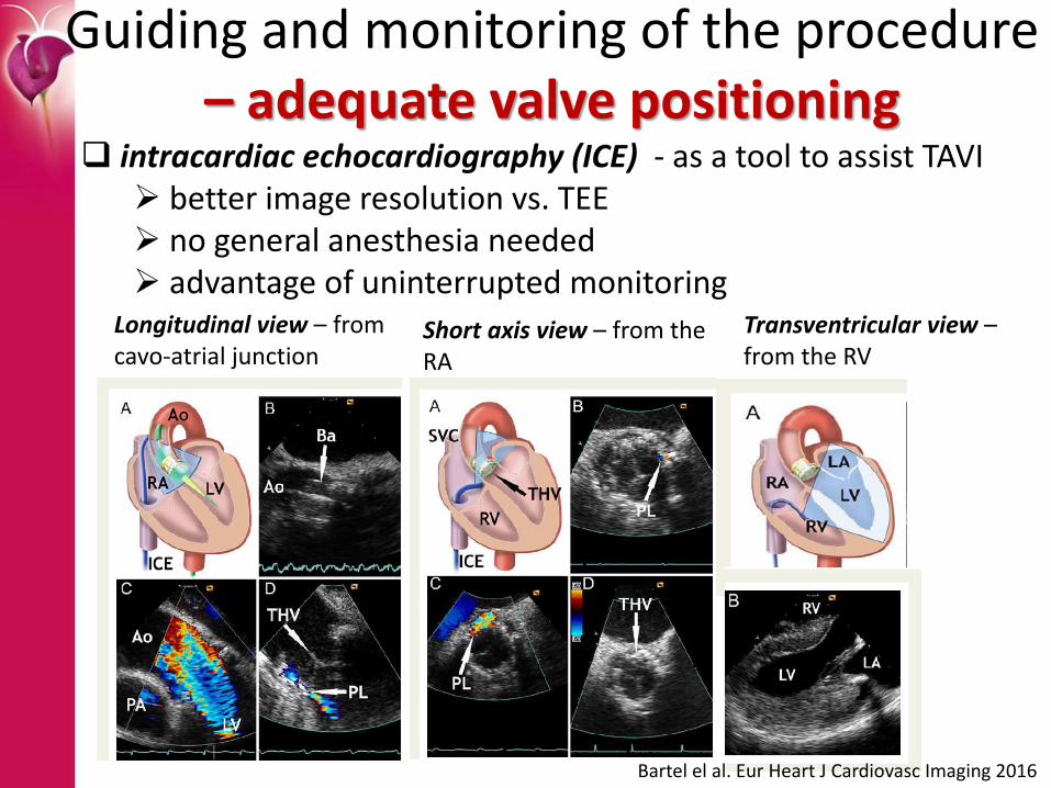
Guiding and monitoring of the procedure – adequate valve positioning
intracardiac echocardiography (ICE) - as a tool to assist TAVI better image resolution vs. TEE no general anesthesia needed advantage of uninterrupted monitoring
Longitudinal view – from cavo-atrial junction
Short axis view – from the RA
Transventricular view –from the RV
Bartel el al. Eur Heart J Cardiovasc Imaging 2016

Post procedural evaluation and results: assessment of paravalvular AR in the cathlab & beyond
Kodali et al, NEJM 2012Still an open question!

Better assessment of paravalvular AR after the procedure – echo has limitations
Para valvular AR after TAVI is difficult to quantify by echo : several regurgitation jets eccentric jets origin in different cut-planes complex regurgitant orifice
3D TEE (VC area) better than 2D TEE for native valve/post TAVI Perez de Isla et al, Int J Cardiol 2013; Goncales et al. JASE 2012

Better assessment of paravalvular ARafter the procedure – CMR?
135 pts 3 centers AR severity = (RF) by phase-contrast velocity mapping by CMR 40 days post TAVI outcomes : Mt and re-hospitalization for HF; FUP =26 months
Higher RF post-TAVR was associated with increased Mt (HR: 1.18 for each 5%increase in RF)[95% confidence interval: 1.08 to 1.30]; p < 0.001

Take home messages
Better tools to quantify severity are available in cases in which echo parameters are discordant (MSCT – AVC score)
3D TEE Automated software capable of rapid and accurate measurement and modeling of the Ao annulus and root for valve sizing (no contrast needed)
Fusion imaging and ICE to allow a better positioning of the valve and better communication between imagers and implanters
Better tools to assess residual paravalvular leak post TAVI with potential implication on treatment (early detection, timely correction, improved outcome?)