new icu eeg: encephalopathic, periodic and coma patterns · 2020. 9. 22. · - 40% of patients who...
TRANSCRIPT
ICU EEG: Encephalopathic, Periodic andComa Patterns
Christopher Newey, DO, MSStaff Physician, Cerebrovascular and Epilepsy CenterMedical Director, Akron Neurosciences ICUMedical Director, Multimodal Monitoring
Member of CCF ICU-EEG Consortium along with Stephen Hantus, MD, and Vineet Punia, MD
Disclosures: I have no relevant financial relationships or conflicts of interest to disclose
Objectives
• Recognize importance of neuromonitoring• Recognize EEG patterns and understand
prognostic significance of these patterns in the critically ill patient
• Review terminology pertaining to EEG monitoring of the critically ill
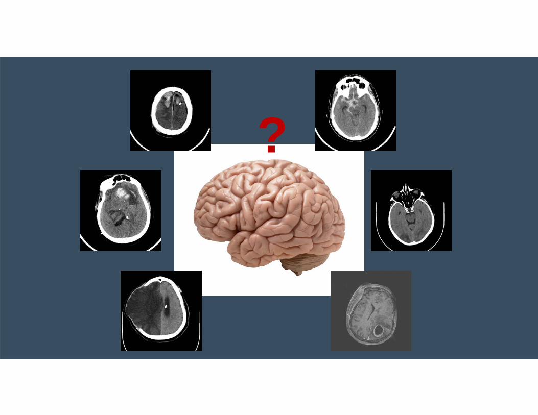
?

Encephalopathy
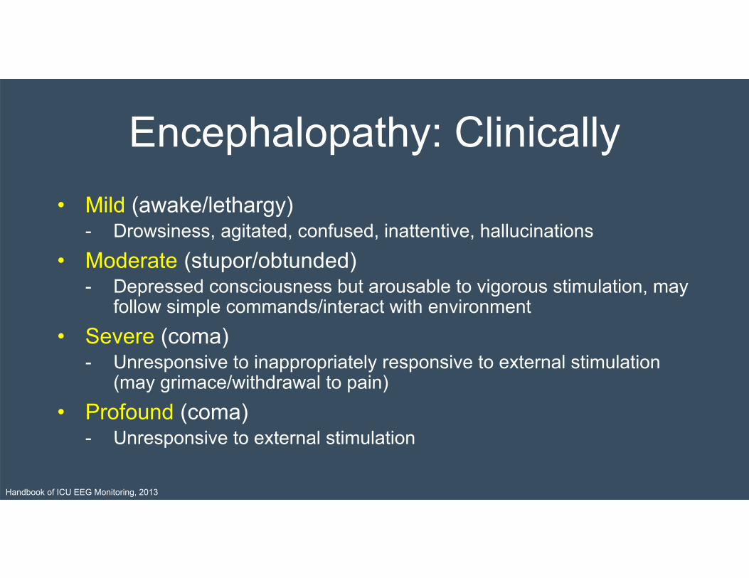
Encephalopathy: Clinically• Mild (awake/lethargy)
- Drowsiness, agitated, confused, inattentive, hallucinations• Moderate (stupor/obtunded)
- Depressed consciousness but arousable to vigorous stimulation, may follow simple commands/interact with environment
• Severe (coma)- Unresponsive to inappropriately responsive to external stimulation
(may grimace/withdrawal to pain)• Profound (coma)
- Unresponsive to external stimulation
Handbook of ICU EEG Monitoring, 2013

Encephalopathy: EEG• EEG Characteristics:
- Slow activity (rhythmic delta activity)- Periodic patterns (periodic
discharges)- Coma/stupor patterns including
medication effect- Suppression- Reactivity
• Purpose of EEG:- Assess depth of coma and severity of
cortical dysfunction - Determine etiology/depth of
depressed LOC (medication, seizure, etc)
- Assess for changes over time (worsening/improving encephalopathy, ischemia)• Need to be aware of changes in
background, reactivity, state changes, sleep architecture, etc
- Prognostication- Compliments neuroimaging to
exclude other causes (catatonia, psychogenic coma)
Handbook of ICU EEG Monitoring, 2013
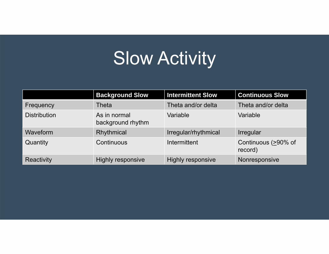
Slow Activity
Background Slow Intermittent Slow Continuous SlowFrequency Theta Theta and/or delta Theta and/or deltaDistribution As in normal
background rhythmVariable Variable
Waveform Rhythmical Irregular/rhythmical IrregularQuantity Continuous Intermittent Continuous (>90% of
record)Reactivity Highly responsive Highly responsive Nonresponsive

Mild Diffuse Encephalopathy• Background slow
- Frequency of background rhythm is lower than the normal value• 1y: < 5Hz• 4y: < 6Hz• 5y: <7Hz• 8y: <8Hz
• Diffuse theta activity, occasional delta activity
- Interpretation:• Cortical or subcortical mechanisms involved in the generation of the
background rhythm are disturbed• Nonspecific marker of diffuse dysfunction but may be related to cerebral
perfusion or metabolic/toxic etiologies
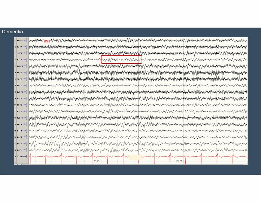
Dementia

Mild Diffuse Encephalopathy
• Intermittent Slow (Rhythmic Delta Activity)- Not caused by drowsiness- Can be generalized (may be frontal or
occipital predominant), regional, or lateralized• Interpretation:
- Marker for nonspecific functional dysfunction, especially if generalized

Meningoencephalitis
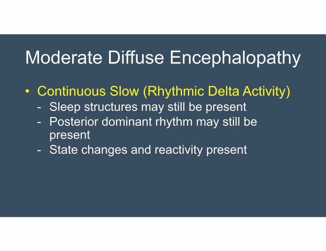
Moderate Diffuse Encephalopathy
• Continuous Slow (Rhythmic Delta Activity)- Sleep structures may still be present- Posterior dominant rhythm may still be
present- State changes and reactivity present
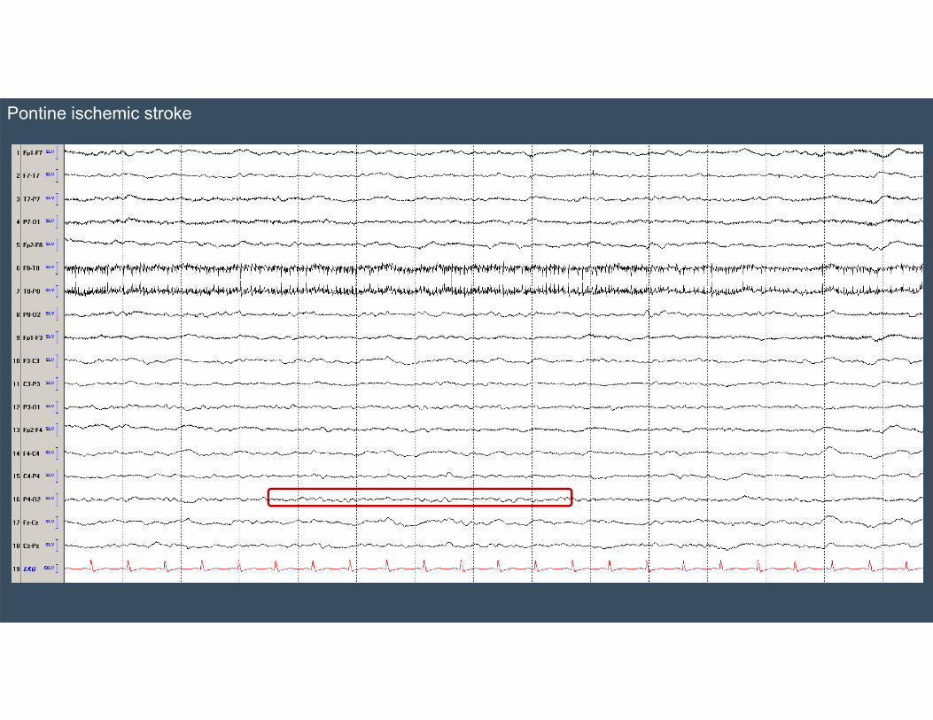
Pontine ischemic stroke
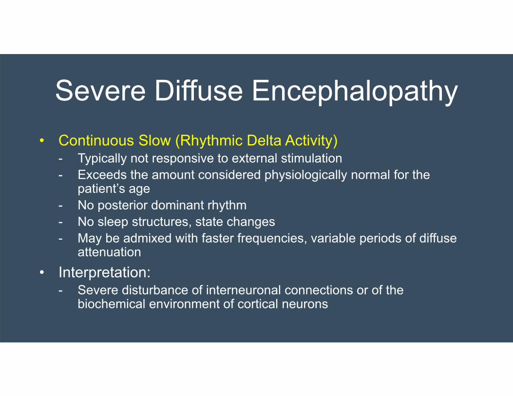
Severe Diffuse Encephalopathy• Continuous Slow (Rhythmic Delta Activity)
- Typically not responsive to external stimulation - Exceeds the amount considered physiologically normal for the
patient’s age- No posterior dominant rhythm - No sleep structures, state changes- May be admixed with faster frequencies, variable periods of diffuse
attenuation• Interpretation:
- Severe disturbance of interneuronal connections or of the biochemical environment of cortical neurons

Cardiac arrest

EEG Changes with Cerebral Blood Flow
Foreman and Claassen, 2012

AMS and worsening cardiac function

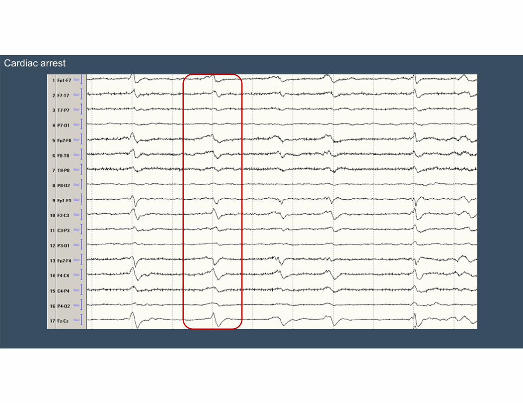
Generalized Periodic Discharges (GPDs)
• Stereotypical waveforms with a periodic rate- Generalized periodic discharge +/- triphasic morphology- Frontocentral predominant but may also be frontotemporal
midline or occipital predominant- Typically have a negative polarity- Discharge can be up to 0.5seonds long; <4 phases- Interdischarge interval should not vary by more than 50%
• Interpretation:- Indicates an acute/subacute, severe diffuse
encephalopathy
Cardiac arrest
CJD

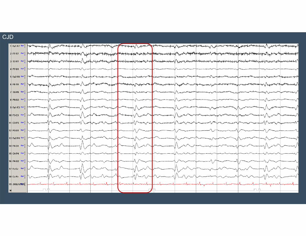
Triphasic Morphology• High amplitude sharp transients followed by low amplitude negative
wave- First negative wave generally has a lower amplitude than the negative afterwave- Typically generalized with the largest deflection in the frontal electrodes on bipolar
montage; wave duration ~300-500msec; can increase/appear on stimulation/arousal
• Interpretation:- Classically seen in metabolic diffuse encephalopathies (hepatic/renal failure), toxic
encephalopathies (baclofen overdose, lithium overdose), - Can occur in white matter diseases, atrophy, hemorrhage, stroke, anoxia,
hypoglycemia, sepsis, hypercalcemia- Typically has a lower seizure risk
Handbook of ICU EEG Monitoring, 2013; So, 2016

Triphasic Morphology
• May appear to have a delay (or lag) on bipolar montage, which is not seen on ear reference montage
Handbook of ICU EEG Monitoring, 2013; So, 2016
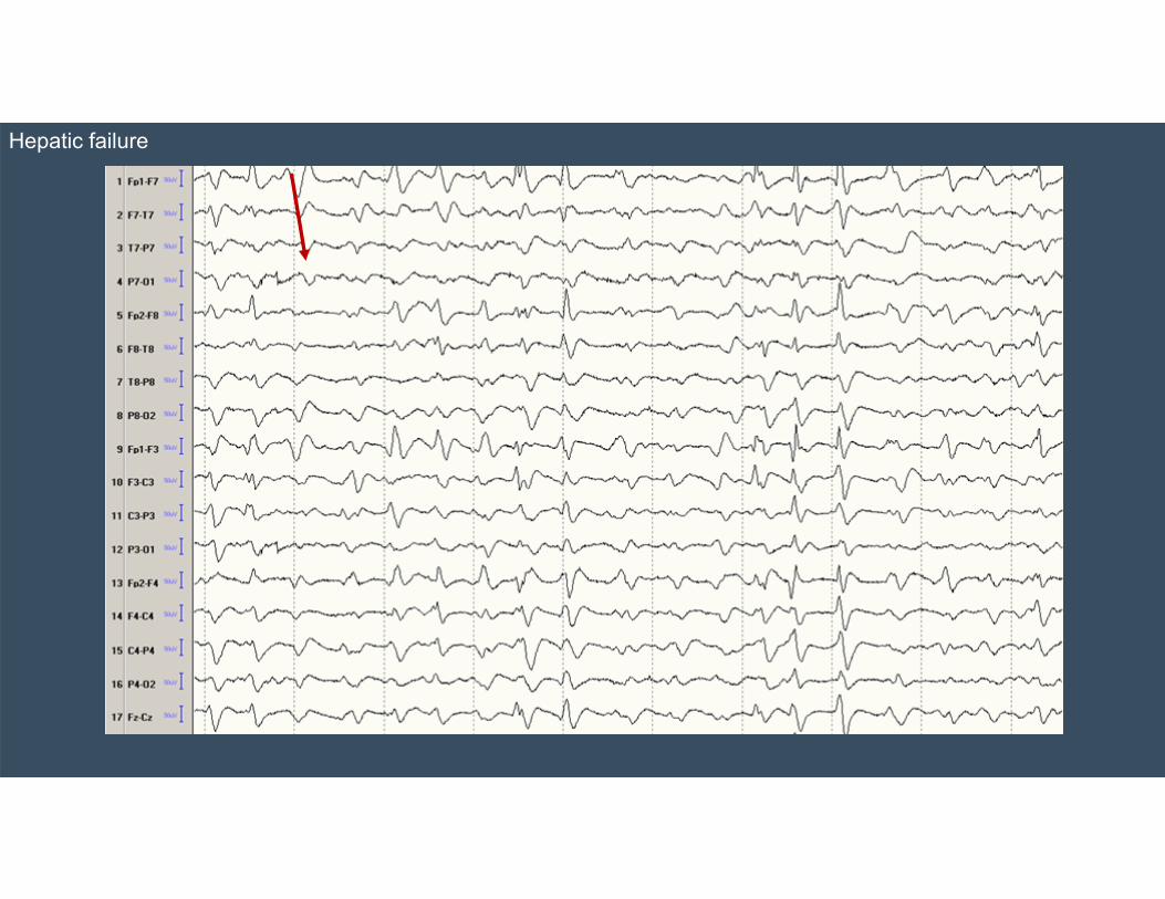
Hepatic failure
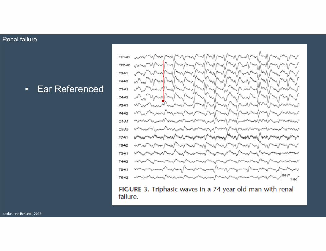
• Ear Referenced
Kaplan and Rossetti, 2016
Renal failure

Lateralized Periodic Discharges (LPDs)• Sharp transients (SWs or spikes)
that occur in a periodic fashion either regionally or lateralized- Main component is negative
• Interpretation: - Acute/subacute: severe, regionally
destructive lesion (such as ischemic, tumor, encephalitis, hemorrhage, abscess, PRES), TBI
- Chronic: tumor, remote stroke, or TBI
Lesion on imaging
Cortical Subcortical Both
Orta 23% 12% 65%
Kalamangalam 70% 23%
Orta 2009; Kalamangalm 2007; So, 2016
• Focal deficits: 60-80%• Altered LOC: 10-35%• High risk for seizures: 50-90%
(40-70% for NCSz)• Can precede or develop after
seizure• Mortality 25-40%
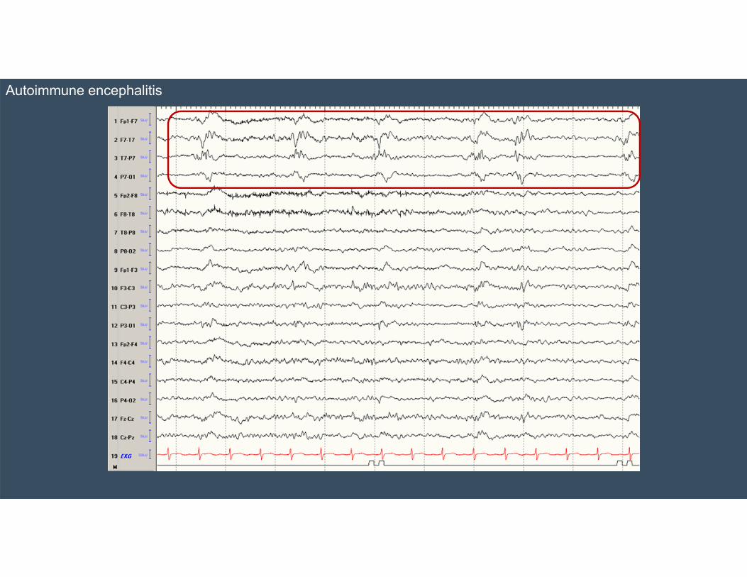
Autoimmune encephalitis
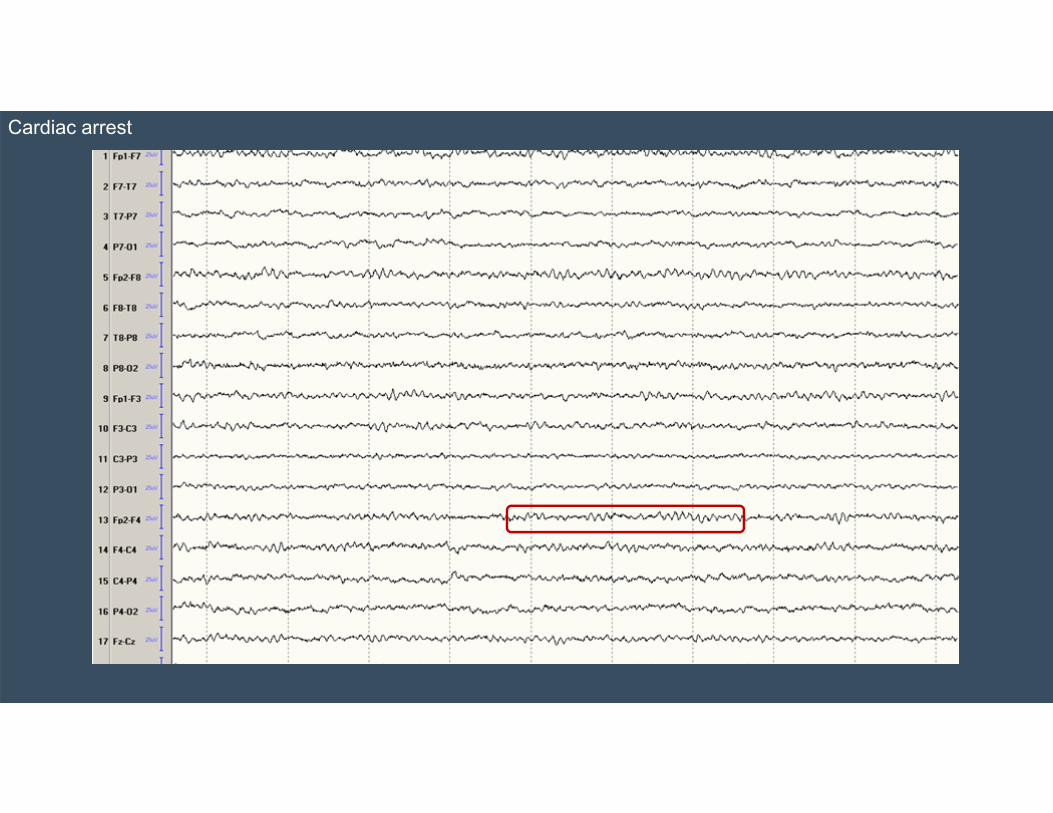
Bilateral Periodic Discharges
• Lateralized periodic discharges from two hemispheres, independent from one another
• Interpretation:- Seen in multifocal, bihemispheric, or diffuse cerebral
insults and encephalopathies (strokes, anoxia, toxic, metabolic, infection, tumor)
- May have an increased mortality risk compared to LPDs (61% vs 29%) and more likely to be comatose (72% vs 24%)
De la Paz and Brenner, 1981

Hirsch and Brenner, 2010
Cardiac arrest

Alpha Coma/Stupor
• Diffuse, invariant alpha activity (8-12Hz) in a comatose/stuporous patient
• Interpretation:- May indicate lesions of the brainstem at the
pontomesencephalic level - Also seen in anoxic brain injury and drug-induced
Cardiac arrest

Spindle Coma/Stupor• EEG showing spindle (11-14Hz) in the
comatose/stuporous patient• Interpretation:
- Typically seen in patients with lesions in the brainstem that does not impair normal sleep-generating mechanisms (i.e., caudal to the thalamus)
- Medication effect

Propofol
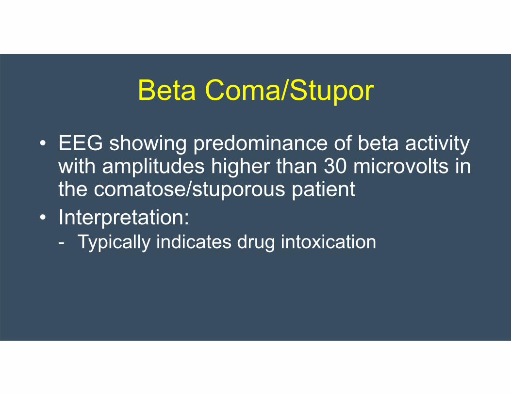
Beta Coma/Stupor
• EEG showing predominance of beta activity with amplitudes higher than 30 microvolts in the comatose/stuporous patient
• Interpretation: - Typically indicates drug intoxication
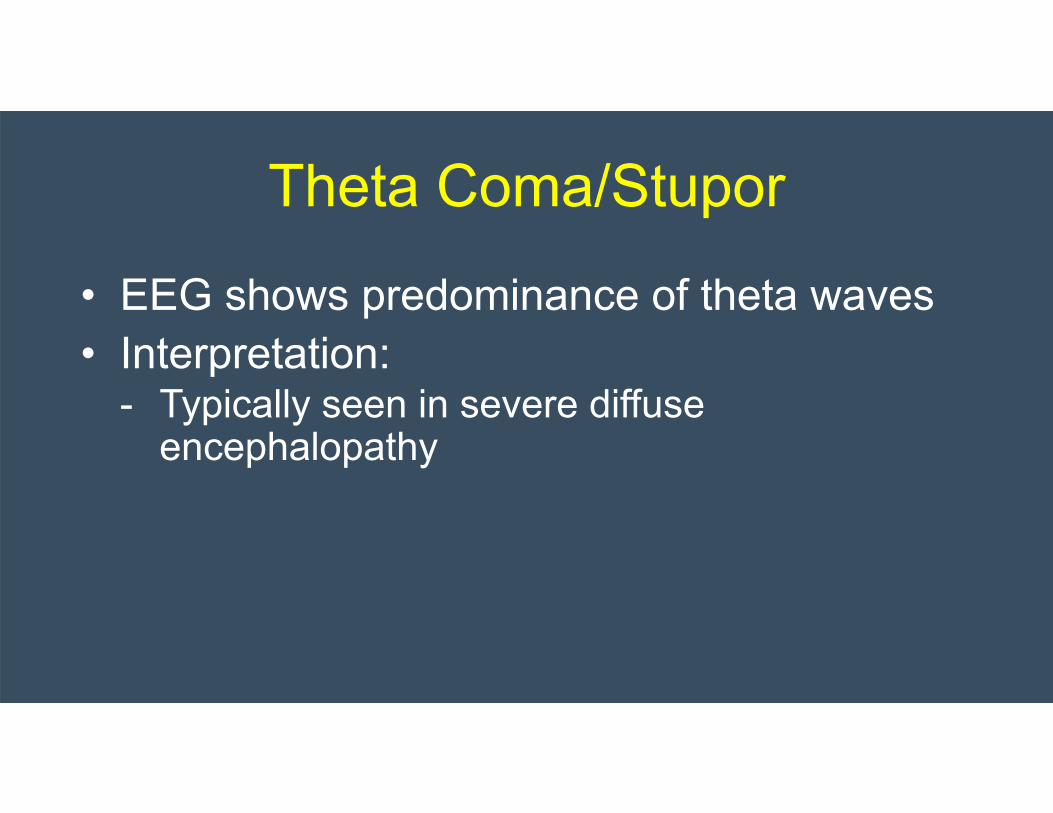
Theta Coma/Stupor
• EEG shows predominance of theta waves• Interpretation:
- Typically seen in severe diffuse encephalopathy

Cardiac arrest
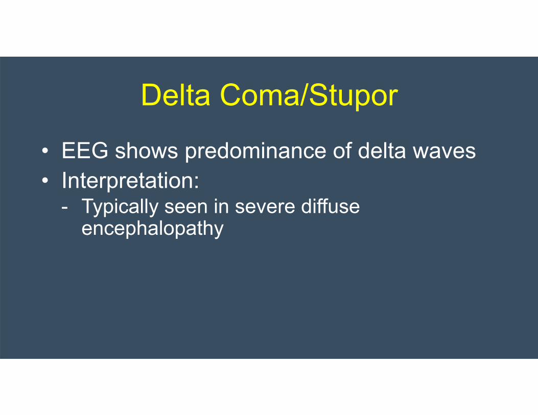
Delta Coma/Stupor
• EEG shows predominance of delta waves• Interpretation:
- Typically seen in severe diffuse encephalopathy
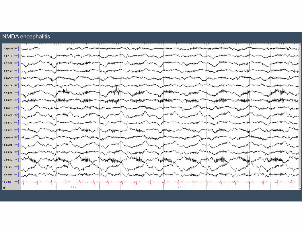
NMDA encephalitis

Excessive Fast
• At least 50% of the recording is dominated by beta activity of an amplitude of at least 50 microvolts- Refers to generalized EEG finding
• Interpretation:- Frequent finding with sedative medications
such as benzodiazepines and barbiturates

Benzodiazepine
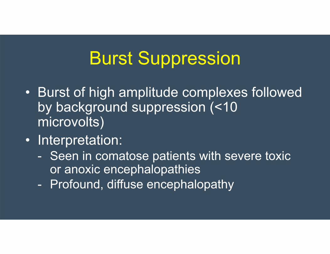
Burst Suppression
• Burst of high amplitude complexes followed by background suppression (<10 microvolts)
• Interpretation:- Seen in comatose patients with severe toxic
or anoxic encephalopathies- Profound, diffuse encephalopathy

Cardiac arrest

Background Suppression
• EEG activity of less than 10 microvolts• Interpretation:
- Profound, diffuse encephalopathy

Right MCA stroke with malignant cerebral edema
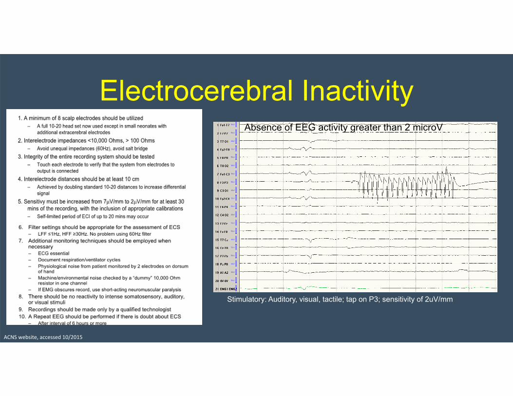
Electrocerebral Inactivity
Stimulatory: Auditory, visual, tactile; tap on P3; sensitivity of 2uV/mm
ACNS website, accessed 10/2015
Absence of EEG activity greater than 2 microV

EEG of Focal Lesions
Kaplan and Rossetti 2016
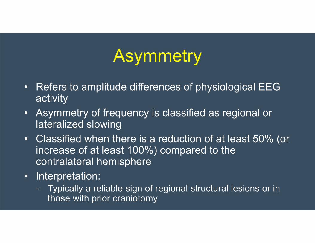
Asymmetry• Refers to amplitude differences of physiological EEG
activity• Asymmetry of frequency is classified as regional or
lateralized slowing• Classified when there is a reduction of at least 50% (or
increase of at least 100%) compared to the contralateral hemisphere
• Interpretation:- Typically a reliable sign of regional structural lesions or in
those with prior craniotomy
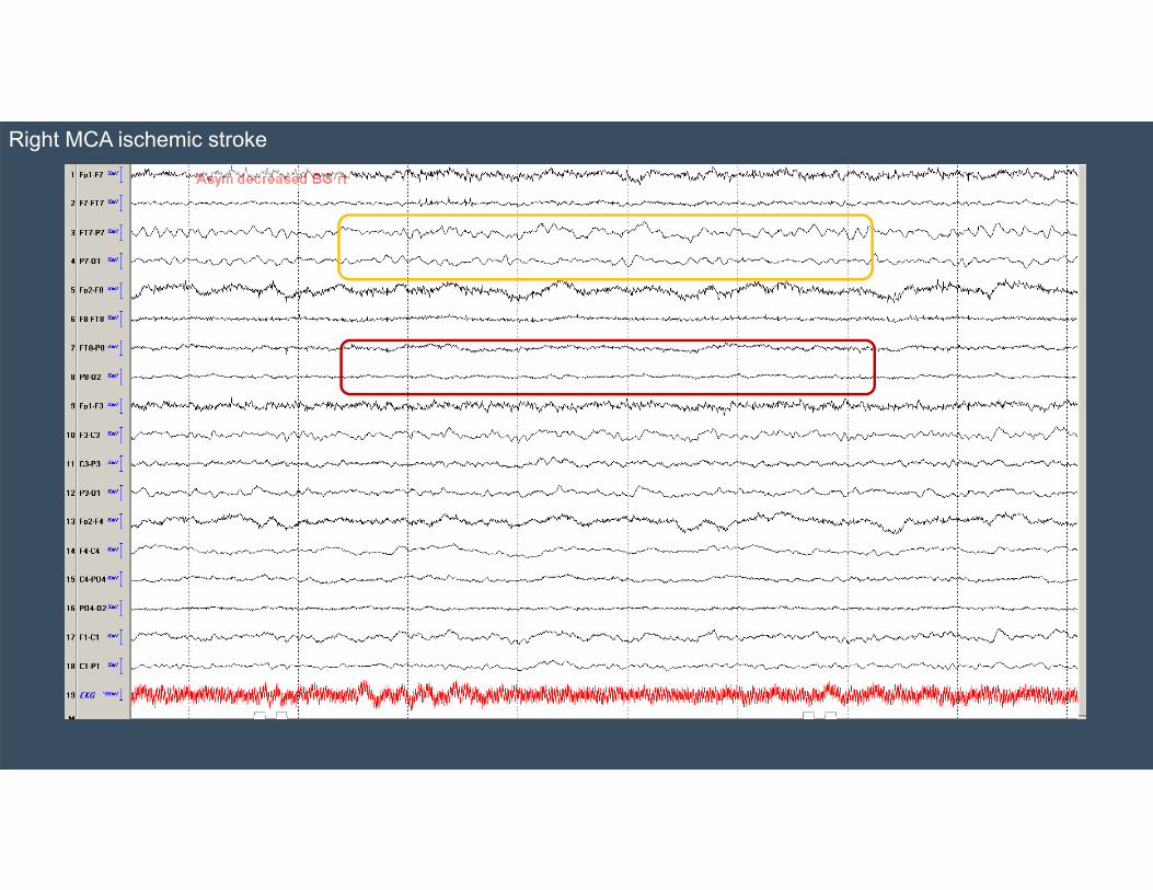
Right MCA ischemic stroke
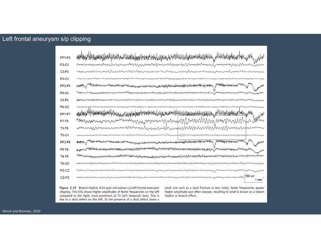
Hirsch and Brenner, 2010
Left frontal aneurysm s/p clipping

Reactivity• Types:
- Increase of background frequencies
- Brief diffuse attenuation- Decreased background
frequencies (“paradoxical reactivity”)
- Stimulus-induced rhythmic periodic, or ictal discharges (SIRPIDS)• Related to dysregulated
afferent input into hyperexcitable cortex
Kaplan and Rossetti 2016
Verbal Stimulation

Prognosis
Bricolo, 1978; Jaitly 1997; DeLorenzo 1998; Lawn 2000; Vespa 2003; Claassen et al., 2006; Claassen et al., 2007; Claassen EEG surface and depth ppt, accessed 10/2015; Tjepmkema‐Cloostermans et al., 2015; Rossetti et al., 2010 ; Azabou et al, 2018
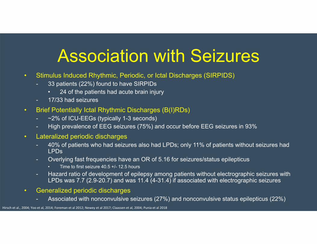
Association with Seizures• Stimulus Induced Rhythmic, Periodic, or Ictal Discharges (SIRPIDS)
- 33 patients (22%) found to have SIRPIDs• 24 of the patients had acute brain injury
- 17/33 had seizures
• Brief Potentially Ictal Rhythmic Discharges (B(I)RDs)- ~2% of ICU-EEGs (typically 1-3 seconds)- High prevalence of EEG seizures (75%) and occur before EEG seizures in 93%
• Lateralized periodic discharges- 40% of patients who had seizures also had LPDs; only 11% of patients without seizures had
LPDs- Overlying fast frequencies have an OR of 5.16 for seizures/status epilepticus
• Time to first seizure 40.5 +/- 12.5 hours
- Hazard ratio of development of epilepsy among patients without electrographic seizures with LPDs was 7.7 (2.9-20.7) and was 11.4 (4-31.4) if associated with electrographic seizures
• Generalized periodic discharges- Associated with nonconvulsive seizures (27%) and nonconvulsive status epilepticus (22%)
Hirsch et al., 2004; Yoo et al, 2014; Foreman et al 2012; Newey et al 2017; Claassen et al, 2004; Punia et al 2018

ACNS Critical Care EEG Terminology

Primary Objectives• Develop standardized
terminology for scientific investigation related to rhythmic and periodic EEG patterns (i.e., patterns of uncertain significance) seen in encephalopathic patients- Excludes patterns that most
define as seizures
• Allow collaborative, multicenter studies
• Allow comparison of results between centers
Hirsch et al, 2013; Chong and Hirsch, 2005; Handbook of ICU EEG Monitoring, 2013
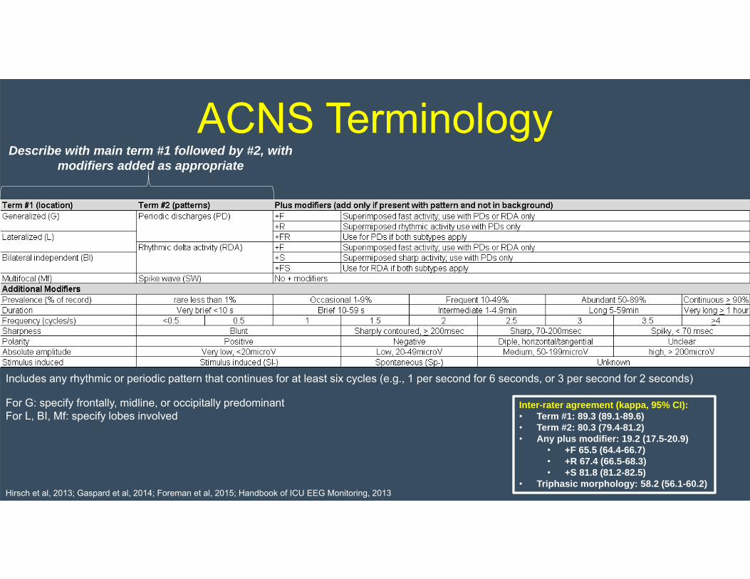
ACNS Terminology
Hirsch et al, 2013; Gaspard et al, 2014; Foreman et al, 2015; Handbook of ICU EEG Monitoring, 2013
Describe with main term #1 followed by #2, with modifiers added as appropriate
Includes any rhythmic or periodic pattern that continues for at least six cycles (e.g., 1 per second for 6 seconds, or 3 per second for 2 seconds)
For G: specify frontally, midline, or occipitally predominant For L, BI, Mf: specify lobes involved
Inter-rater agreement (kappa, 95% CI):• Term #1: 89.3 (89.1-89.6)• Term #2: 80.3 (79.4-81.2)• Any plus modifier: 19.2 (17.5-20.9)
• +F 65.5 (64.4-66.7)• +R 67.4 (66.5-68.3)• +S 81.8 (81.2-82.5)
• Triphasic morphology: 58.2 (56.1-60.2)
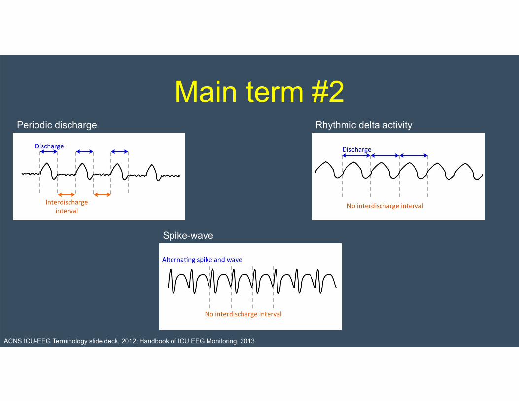
Main term #2Periodic discharge Rhythmic delta activity
Spike-wave
ACNS ICU-EEG Terminology slide deck, 2012; Handbook of ICU EEG Monitoring, 2013

TerminologyOld Term New TermTriphasic waves, most of record
Continuous GPDs at 2Hz with triphasic morphology
PLEDs LPDsBiPLEDs BiPDsGPEDs/PEDs GPDsFIRDA Occasional brief 2Hz GRDA, frontally predominantPLEDs+ LPDs+SIRPIDs with evolving RDA SI-evolving LRDALateralized seizure, delta frequency
Evolving LRDA
Semi-rhythmic delta Quasi-RDA
EEG Patterns in Coma:• RDA – GRDA, FIRDA• GPDs +/- triphasic
morphology• Low voltage, slow,
nonreactive pattern• Specific patterns (beta,
spindle, alpha, theta coma)
• Burst suppressoin
ACNS ICU-EEG Terminology slide deck, 2012; Handbook of ICU EEG Monitoring, 2013
Conclusion• CEEG is a neuromonitoring tool for critically
ill patients.• Many EEG patterns emerge in the critically
ill patient and may have prognostic implications.
• Standardizing terminology is important for better understanding of these rhythmic and periodic patterns.

Supplemental Slides

ACNS TerminologyTraining Slides
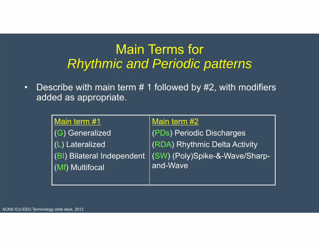
Main Terms for Rhythmic and Periodic patterns
• Describe with main term # 1 followed by #2, with modifiers added as appropriate.
Main term #1(G) Generalized (L) Lateralized (BI) Bilateral Independent(Mf) Multifocal
Main term #2(PDs) Periodic Discharges(RDA) Rhythmic Delta Activity(SW) (Poly)Spike-&-Wave/Sharp-and-Wave
ACNS ICU-EEG Terminology slide deck, 2012
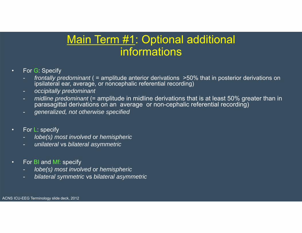
Main Term #1: Optional additional informations
• For G: Specify - frontally predominant ( = amplitude anterior derivations >50% that in posterior derivations on
ipsilateral ear, average, or noncephalic referential recording)- occipitally predominant- midline predominant (= amplitude in midline derivations that is at least 50% greater than in
parasagittal derivations on an average or non-cephalic referential recording)- generalized, not otherwise specified
• For L: specify - lobe(s) most involved or hemispheric- unilateral vs bilateral asymmetric
• For BI and Mf: specify - lobe(s) most involved or hemispheric- bilateral symmetric vs bilateral asymmetric
ACNS ICU-EEG Terminology slide deck, 2012
Main Term #1: Optional additional informations
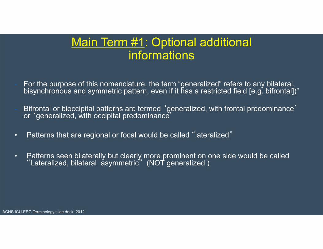
- For the purpose of this nomenclature, the term “generalized” refers to any bilateral, bisynchronous and symmetric pattern, even if it has a restricted field [e.g. bifrontal])”
- Bifrontal or bioccipital patterns are termed ‘generalized, with frontal predominance’or ‘generalized, with occipital predominance’
• Patterns that are regional or focal would be called “lateralized”
• Patterns seen bilaterally but clearly more prominent on one side would be called “Lateralized, bilateral asymmetric” (NOT generalized )
ACNS ICU-EEG Terminology slide deck, 2012
Main Term #2
• PD: Periodic Discharges
• RDA: Rhythmic Delta Activity
• SW: Spike-and-Wave, Sharp-and-Wave or Polyspike-and-Wave
ACNS ICU-EEG Terminology slide deck, 2012

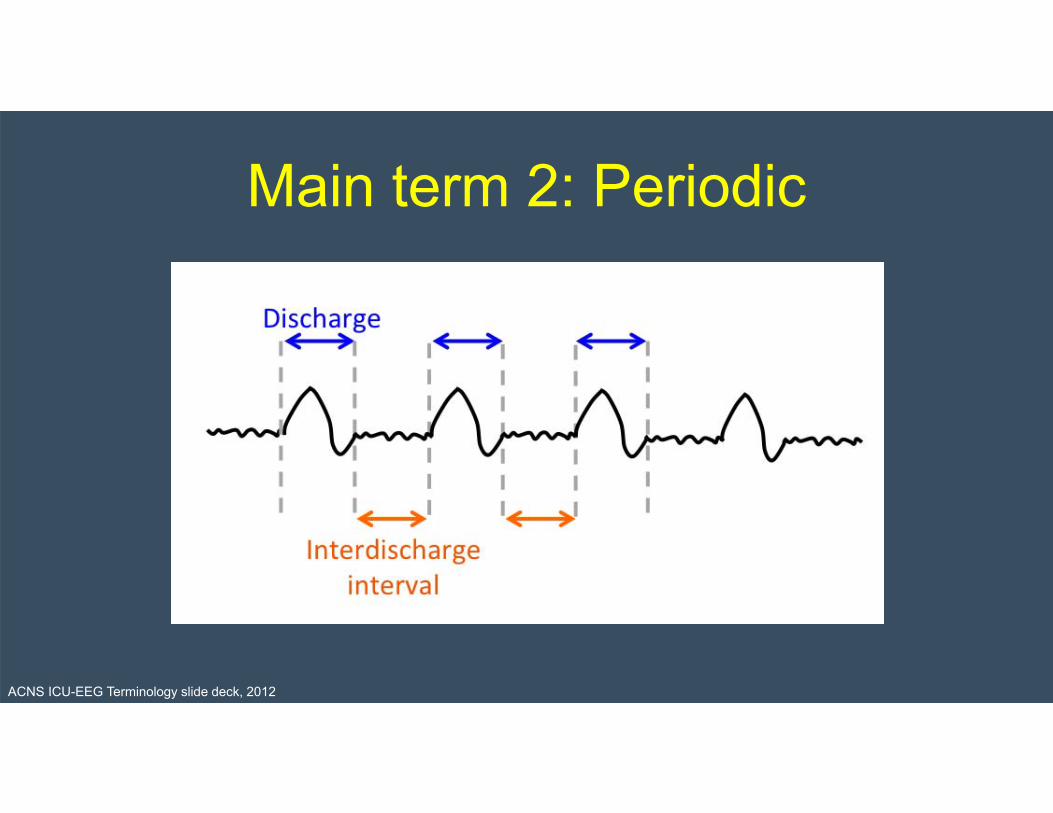
Main Term #2: Definitions•Periodic discharges= repeating waveforms/discharges with (relatively) uniform morphology at nearly regular intervals. Applies only to single discharges (must have ≤3 phases [i.e. ≤2 baseline crossings] or any discharge lasting ≤0.5 sec regardless of number of phases) and not to bursts(discharges lasting >0.5 sec and having ≥4 phases [i.e. ≥3 baseline crossings]). “Nearly regular intervals” = cycle length (period) varying by <50% from one cycle to the next in most (>50%) cycle pairs.
•Rhythmic = repetition of a waveform with relatively uniform morphology and duration and without an interval between consecutive waveforms. Duration of one cycle (the period) should vary by <50% from the duration of the subsequent cycle for the majority (>50%) of cycle pairs to qualify as a rhythmic pattern.
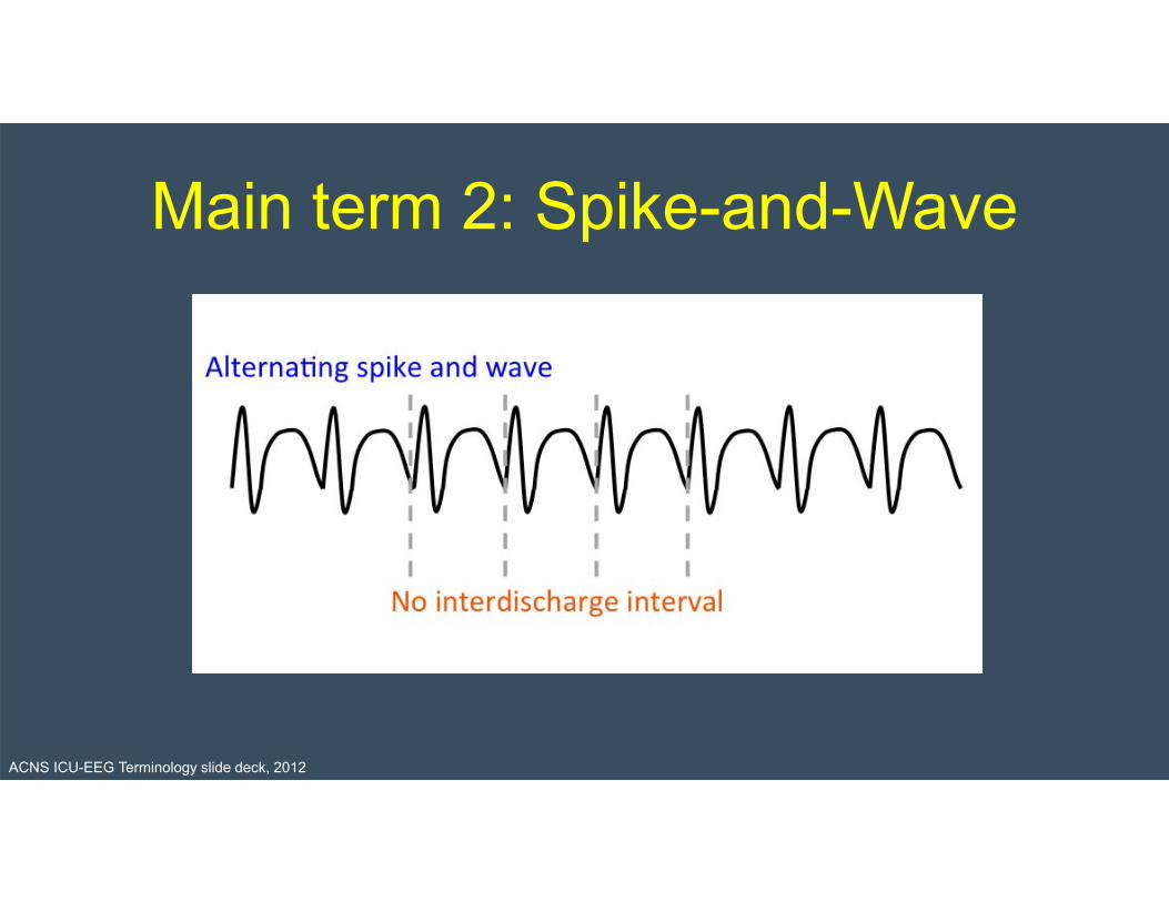
•Spike-and-wave = spike, polyspike or sharp wave consistently followed by a slow wave in a regularly repeating pattern (spike-wave-spike-wave-spike-wave), with a consistent relationship between the spike (or sharp wave) component and the slow wave.
-This terminology does not signify whether or not these patterns are ictal/related to seizures.
ACNS ICU-EEG Terminology slide deck, 2012
Main term 2: Periodic
ACNS ICU-EEG Terminology slide deck, 2012

Main term 2: Rhythmic
ACNS ICU-EEG Terminology slide deck, 2012
Main term 2: Spike-and-Wave
ACNS ICU-EEG Terminology slide deck, 2012
Main terms #1,2 cont’d….• NOTE 1: A pattern can qualify as rhythmic, periodic or spike-and-wave
as long as it continues for at least 6 cycles (e.g. 1/s for 6 seconds, or 3/s for 2 seconds).
• NOTE 2: If a pattern qualifies as both GPDs and RDA, it should be coded as GPDs+R rather than RDA+ (see slide 53 for description of “+”suffixes).
ACNS ICU-EEG Terminology slide deck, 2012
Modifiers: Prevalence• Specify:
Approximate percent of record/epoch, using the following divisions, or consistently use the suggested equivalent clinical terms:
• >90% “Continuous”• 50-89% “Abundant”• 10-49% “Frequent”• 1-9% of “Occasional”• <1% of “Rare”
ACNS ICU-EEG Terminology slide deck, 2012

Modifiers, cont’d: Duration• Specify for each pattern the typical duration of pattern (if
not continuous) using the following divisions or suggested equivalent clinical terms.
• >1 hour “Very long”• 5-59 min “Long”• 1-4.9 min “Intermediate”• 10-59 sec “Brief”• <10 sec “Very brief”
• Record total duration (over whole record or 24 hours (“daily pattern duration”; see slide 74) and longest continuous duration.
ACNS ICU-EEG Terminology slide deck, 2012
Modifiers, cont’d: FrequencySpecify for each pattern:
Rate (typical & range) to the nearest 0.5/s division :
<0.5/s, 0.5/s, 1/s, 1.5/s, 2/s, 2.5/s, 3/s, 3.5/s, or >4/s.
e.g., 1/s (typical) and 0.5-2/s (range)
ACNS ICU-EEG Terminology slide deck, 2012

Modifiers, cont’d: Phases• Number of baseline crossings of the typical
discharge (in longitudinal bipolar and in the channel in which it is most readily appreciated).
• Applies to PDs and the entire spike-and-wave or sharp-and-wave complex of SW (includes the slow wave).
• Categorize as 1, 2, 3 or >3.
• Applies to PDs and SW, not to RDA.
ACNS ICU-EEG Terminology slide deck, 2012

Modifiers, cont’d: Sharpness• Specify for predominant phase (phase with greatest amplitude) and sharpest
phase (if different). • Applies only to PDs and SW, not RDA. • If SW, specify for the spike/sharp wave only. For both phases, describe the typical
discharge.Categorize as one of the following: - Spiky (duration of phase [measured at EEG baseline] <70 ms)- Sharp (duration of phase component 70-200 ms)- Sharply contoured (having a sharp inflection at its peak or trough, or a steep upslope or
downslope (such as saw-tooth morphology), but the duration of the wave at the baseline is >200ms and thus does not qualify as sharp or spiky)
- Blunt
ACNS ICU-EEG Terminology slide deck, 2012
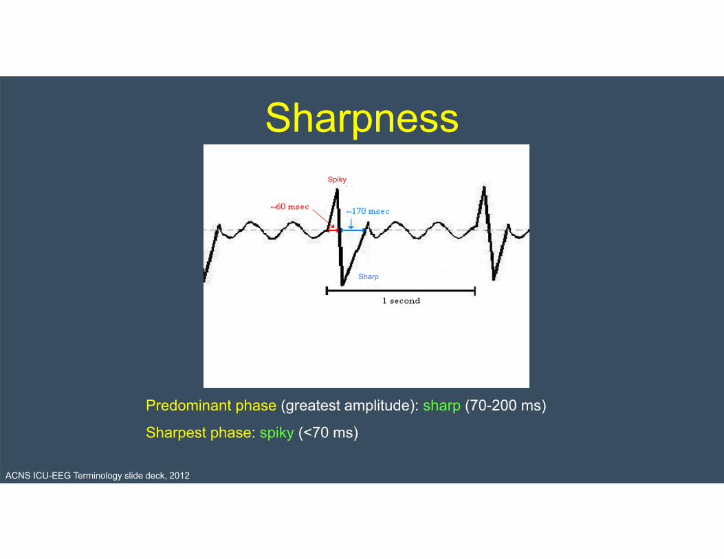
Sharpness
Sharp
Predominant phase (greatest amplitude): sharp (70-200 ms)
Sharpest phase: spiky (<70 ms)
Spiky
Sharp
ACNS ICU-EEG Terminology slide deck, 2012

Modifiers, cont’d: Amplitude• Absolute amplitude:
- Typical amplitude measured in standard longitudinal bipolar 10-20 recording in the channel where the pattern is most apparent. • For PDs, this refers to the highest amplitude component. • For SW, this refers to the spike/sharp wave.
- Measure peak to trough (i.e. positive to negative peak; not peak to baseline).
- Specify for RDA as well.
- Categorize as:• <20 uV “very low”• 20-49 uV “low”
• 50-199 uV “medium”
• >200 uV “high”
ACNS ICU-EEG Terminology slide deck, 2012
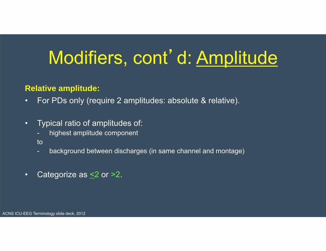
Modifiers, cont’d: AmplitudeRelative amplitude:• For PDs only (require 2 amplitudes: absolute & relative).
• Typical ratio of amplitudes of: - highest amplitude component to - background between discharges (in same channel and montage)
• Categorize as <2 or >2.
ACNS ICU-EEG Terminology slide deck, 2012
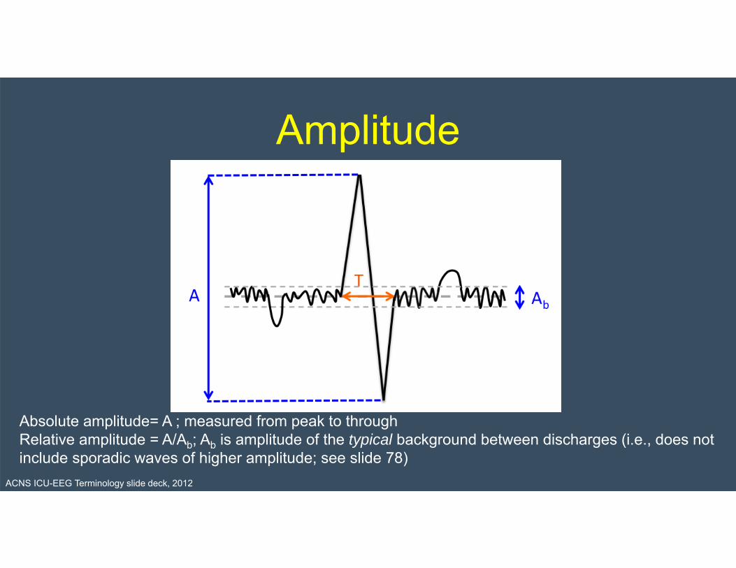
Amplitude
Absolute amplitude= A ; measured from peak to throughRelative amplitude = A/Ab; Ab is amplitude of the typical background between discharges (i.e., does not include sporadic waves of higher amplitude; see slide 78)
ACNS ICU-EEG Terminology slide deck, 2012

Modifiers, cont’d: PolaritySpecify For the predominant phase (phase with the greatest amplitude)
only. Describe the typical discharge. Applies only to PDs and spike/sharp component of SW, not to
RDA. Determined in referential montage.
Categorize as: Positive Negative Dipole, horizontal/tangential Unclear
ACNS ICU-EEG Terminology slide deck, 2012
Modifiers, cont’d: Stimulus-Induced (SI)
• Repetitively and reproducibly brought about by an alerting stimulus, with or without clinical alerting (may also occur without apparent stimulus--i.e. does not disqualify pattern as SI).
• If never clearly stimulus induced, report as spontaneous. • If unknown, unclear or untested, report as unknown.
• Specify type of stimulus (auditory, light tactile, patient care and other non-noxious stimulations, suction, sternal rub, nostril tickle or other noxious stimulations).
ACNS ICU-EEG Terminology slide deck, 2012

Modifiers, cont’d: Evolving or Fluctuating
• Both refer to changes in one of the following- Frequency, - Location, - Morphology.
• If neither term applies, report as static.
ACNS ICU-EEG Terminology slide deck, 2012
Modifiers, cont’d: EvolvingAt least 2 unequivocal, sequential changes in frequency, morphology or location defined as follows: • Frequency: ≥2 consecutive increases or decreases of ≥0.5/s, (e.g. 2 2.5 to 3/s, or 3 2 to 1.5/s;• Morphology: ≥2 consecutive changes to a novel morphology;• Location : sequentially spreading into/out of ≥2 two different standard 10-20 electrode locations.
• To qualify as evolution in frequency or location, each change must persist ≥3 cycles (e.g. 1/s for 3 seconds, or 3/s for 1 second). Thus, the following pattern would qualify as evolving: 3/s for > 1 second, then 2/s for > 1.5 seconds (the first change), then 1.5/s for > 2 seconds (the 2nd change).
• To qualify as evolution in morphology, each different morphology or each morphology plus its transitional forms must last at least 3 cycles. Thus the following examples would both qualify as evolving in morphology:- spiky 4-phase PDs for 3 cycles then sharp 2-3 phase PDs for 3 cycles then blunt diphasic PDs for 3 cycles- 1 blunt triphasic PD then 2 blunt biphasic PDs then 2 sharply contoured biphasic PDs then 2 sharp biphasic PDs then 3 sharp monophasic PDs.
• The pattern must not remains unchanged in frequency, morphology or location for more than 5 minutes. Thus, this pattern would not qualify as evolving: 3/s for 1 min 2/s for 7 min 1.5/s for 2 min
ACNS ICU-EEG Terminology slide deck, 2012

Modifiers, cont’d: FluctuatingAt least 3 changes, <1 min apart, in:
- Frequency (by ≥0.5/s), - Morphology, or - Location (by ≥1 standard inter-electrode distance), BUT not qualifying as evolving.
• Includes patterns alternating from 1 1.5 1 1.5 Hz; spreading in and out of a single electrode repeatedly; or alternating between 2 morphologies repeatedly.
• Would not qualify as fluctuating: 2/s for 30s 1.5/s for 30s 2/s for 3min 1.5/s for 30s 2/s for 5 min. (Changes are too far apart, i.e. >1 minute).
• Would qualify as fluctuating: 2/s for 10 s 2.5/s for 30s 2/s for 5s 2.5/s for 5s.
ACNS ICU-EEG Terminology slide deck, 2012
Modifiers, cont’d: Evolving and fluctuating
NOTE 1: Change in amplitude or sharpness alone would not qualify as evolving or fluctuating.
NOTE 2: For databasing, if evolving or fluctuating, specify min, max, and typical frequency (under the frequency modifier; see slide 34). For non-generalized patterns, specify degree of spread (none, unilateral, bilateral).
ACNS ICU-EEG Terminology slide deck, 2012
Modifiers, cont’d: PlusAdditional feature which renders the patter more ictal-
appearing than the same pattern without the plus:
• How to specify: - +F superimposed fast activity (theta or faster;
for PDs or RDA)- +R superimposed rhythmic or quasi rhythmic delta
activity (for PDs only)- +S “frequent” superimposed sharp waves/spikes
(frequent = >1/10s but not periodic and not SW), or sharply contoured (for RDA only)
- If both subtypes apply, PDs can have “+FR” and RDAcan have “+FS”.
- Does not apply to SW.- If absent, database as “no plus”.
ACNS ICU-EEG Terminology slide deck, 2012

Modifiers contd…• PLUS (“+”):
- NOTE 1: Re: Bilateral “+” vs. unilateral: If a pattern is bilateral and qualifies as plus on one side, but not on the other, the overall main term should include the plus (even though one side does not warrant a plus). • Example: Bilateral independent periodic discharges with fast activity
superimposed in one hemisphere only (PD on one side, and PD+F on the other) would qualify for BIPDs+F. Similarly, generalized rhythmic delta activity with superimposed spikes in one hemisphere only (RDA on one side and RDA+S on the other) would qualify for GRDA+S
- NOTE 2: Re: +F: If a pattern qualifying as RDA or PDs has superimposed continuous fast activity (theta or faster), this should be coded as +F ONLY if the fast activity is not present in the background activity when the RDA or PDs is not present. In other words, if the superimposed fast activity is part of the RDA or PD
pattern and not simply part of the background activity.
ACNS ICU-EEG Terminology slide deck, 2012
Minor modifiers(all except “Quasi-” are required for database studies; record presence or absence of each):
Quasi-: Defined as: Cycle length (period) varying by 25-50% from one cycle to the next in >50% of cycle pairs.
• Does not qualify if the cycle length varies by >50% (in which case it is not rhythmic at all) or <25% (in which case it is rhythmic, without the “quasi-”) in the majority of cycles.
• Use only when using computer-assisted analysis. Modifies rhythmic or periodic patterns, as in quasi-periodic or quasi-rhythmic.
• (Quasi preferred over pseudo- or semi-). • When not using computer analysis, quasi-periodic is coded as periodic, and quasi-rhythmic as
rhythmic.
Onset:• Sudden (preferred over paroxysmal): Progressing from absent to well-developed in <3s.• Gradual onset : Progressing from absent to well-developed in >3s.
ACNS ICU-EEG Terminology slide deck, 2012

• Triphasic morphology: Applies to PDs and SW. Either two or three phases, with each phase longer than the previous, and the positive phase of highest amplitude. If three phases, this must be negative-positive-negative in polarity; if two phases, positive-negative. Note that a biphasic waveform may be categorized as “triphasic” by this definition.
• Anterior-posterior lag or posterior-anterior lag: Applies if a consistent measureable delay of >100 ms exists from the most anterior derivation to the most posterior derivation in which it is seen ; specify typical delay in msec from anterior to posterior (negative = posterior to anterior lag) in both longitudinal bipolar and in a referential montage, preferably with an ipsilateral ear reference.
Minor modifiers: Cont’d(all except “Quasi-” are required for database studies; record presence or absence of each):
ACNS ICU-EEG Terminology slide deck, 2012
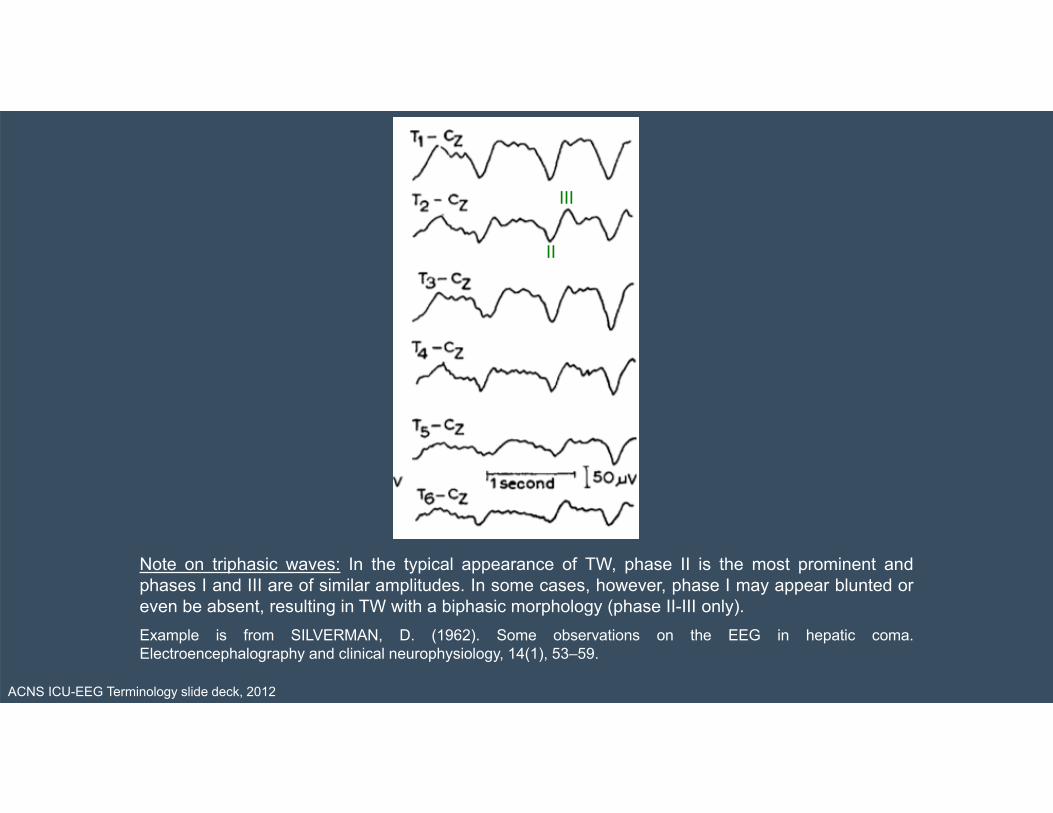
Note on triphasic waves: In the typical appearance of TW, phase II is the most prominent andphases I and III are of similar amplitudes. In some cases, however, phase I may appear blunted oreven be absent, resulting in TW with a biphasic morphology (phase II-III only).Example is from SILVERMAN, D. (1962). Some observations on the EEG in hepatic coma.Electroencephalography and clinical neurophysiology, 14(1), 53–59.
II
III
ACNS ICU-EEG Terminology slide deck, 2012

Minimal time epochs to be reported/databased separately
• First ~30 minutes (equivalent to a “routine” EEG).
• Each 24 hour period. If significant changes occur in the record during this time period, report additional epochs separately as needed.
ACNS ICU-EEG Terminology slide deck, 2012
Moving on…
• Next slides specify how to record:- Sporadic epileptiform discharges- EEG background (e.g. slowing,
posterior dominant rhythm, etc)
ACNS ICU-EEG Terminology slide deck, 2012
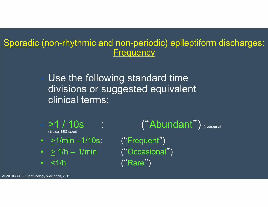
Sporadic (non-rhythmic and non-periodic) epileptiform discharges: Frequency
- Use the following standard time divisions or suggested equivalent clinical terms:
- >1 / 10s : (“Abundant”) (average ≥1 / typical EEG page)
• >1/min –1/10s: (“Frequent”) • > 1/h -- 1/min (“Occasional”)• <1/h (“Rare”)
ACNS ICU-EEG Terminology slide deck, 2012

N.B. re: Prevalence/Duration/Frequency
• Note that there are 4 distinct time-related scales:
- 1. Prevalence (continuous, abundant, frequent, occasional, rare): refers to percent of the entire record occupied by a pattern
- 2. Duration (very long, long, intermediate, brief, very brief): refers to the typical duration of a single occurrence of the pattern, regardless of whether the pattern occurs rarely or frequently.
- 3. Quantification of sporadic (aka “interictal”) epileptiform discharges (abundant, frequent, occasional, rare)
ACNS ICU-EEG Terminology slide deck, 2012
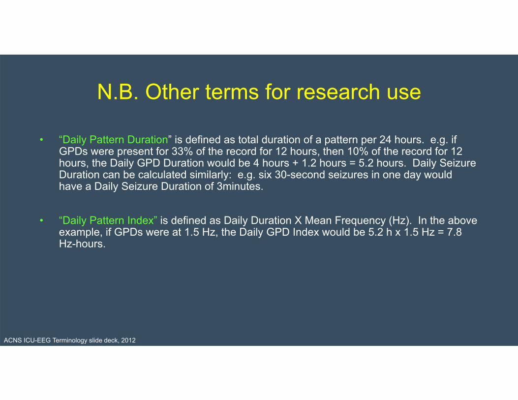
N.B. Other terms for research use
• “Daily Pattern Duration” is defined as total duration of a pattern per 24 hours. e.g. if GPDs were present for 33% of the record for 12 hours, then 10% of the record for 12 hours, the Daily GPD Duration would be 4 hours + 1.2 hours = 5.2 hours. Daily Seizure Duration can be calculated similarly: e.g. six 30-second seizures in one day would have a Daily Seizure Duration of 3minutes.
• “Daily Pattern Index” is defined as Daily Duration X Mean Frequency (Hz). In the above example, if GPDs were at 1.5 Hz, the Daily GPD Index would be 5.2 h x 1.5 Hz = 7.8 Hz-hours.
ACNS ICU-EEG Terminology slide deck, 2012

N.B. re: Amplitudes• Note that there are multiple ratings for amplitude:
- 1. Absolute amplitude of a single discharge within a pattern• Applies to all patterns (see slide 42)• Categorize as:
• <20 uV “very low”• 20-49 uV “low”
• 50-199 uV “medium”
• >200 uV “high”- 2. Relative amplitude of a single discharge to the
interdischarge amplitude during a periodic pattern• Applies to PDs only (see slide 43)• Categorize as <2 or >2.
- 3. Background amplitude (voltage)• Applies to EEG background description (see slide 79)• Categorize as:
• >20µV “normal”• <20µV but >10µV “low”• <10µV “suppressed
ACNS ICU-EEG Terminology slide deck, 2012
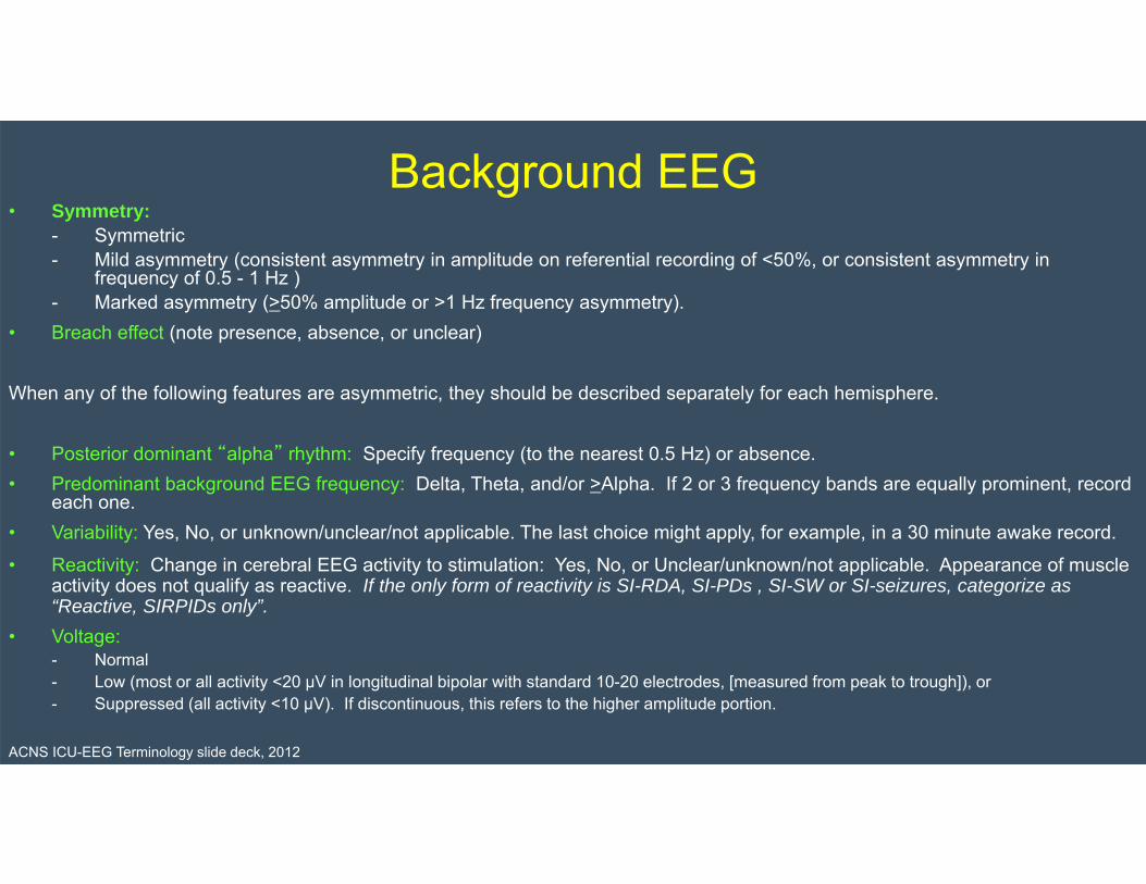
Background EEG• Symmetry:
- Symmetric- Mild asymmetry (consistent asymmetry in amplitude on referential recording of <50%, or consistent asymmetry in
frequency of 0.5 - 1 Hz )- Marked asymmetry (>50% amplitude or >1 Hz frequency asymmetry).
• Breach effect (note presence, absence, or unclear)
When any of the following features are asymmetric, they should be described separately for each hemisphere.
• Posterior dominant “alpha” rhythm: Specify frequency (to the nearest 0.5 Hz) or absence.• Predominant background EEG frequency: Delta, Theta, and/or >Alpha. If 2 or 3 frequency bands are equally prominent, record
each one.• Variability: Yes, No, or unknown/unclear/not applicable. The last choice might apply, for example, in a 30 minute awake record.
• Reactivity: Change in cerebral EEG activity to stimulation: Yes, No, or Unclear/unknown/not applicable. Appearance of muscle activity does not qualify as reactive. If the only form of reactivity is SI-RDA, SI-PDs , SI-SW or SI-seizures, categorize as “Reactive, SIRPIDs only”.
• Voltage:- Normal- Low (most or all activity <20 µV in longitudinal bipolar with standard 10-20 electrodes, [measured from peak to trough]), or- Suppressed (all activity <10 µV). If discontinuous, this refers to the higher amplitude portion.
ACNS ICU-EEG Terminology slide deck, 2012

Background EEG, cont’d.
• Anterior-posterior (AP) gradient: Present, absent or reverse. - An AP gradient is present if at any point in the epoch, there is a
clear and persistent (at least 1 continuous minute) anterior to posterior gradient of voltages and frequencies such that lower amplitude, faster frequencies are seen in anterior derivations, and higher amplitude, slower frequencies are seen in posterior derivations
- A reverse AP gradient is defined identically but with a posterior to anterior gradient of voltages and frequencies.
• Stage II sleep transients (K-complexes and spindles): - Normal (K-complexes and spindles both present and
normal), - Present (at least one) but abnormal, or - Absent (both absent).
ACNS ICU-EEG Terminology slide deck, 2012

Background EEG, cont’d.• Continuity:
- Continuous- Nearly Continuous: continuous, but with occasional (<10% of the record) periods of
attenuation or suppression. Describe typical duration of attenuation/suppression as above.• Nearly continuous with attenuation: periods of lower voltage are >10µV but <50% of
the background voltage• Nearly continuous with suppression: periods of lower voltage are <10 µV • If suppressions/attenuations are stimulus-induced, code as “nearly continuous with
SI-attenuation” or “…with SI-suppression”
- Discontinuous: 10-49% of the record consisting of attenuation or suppression, as defined above.
- Burst-attenuation/Burst-suppression: more than 50% of the record consisting of attenuation or suppression, as defined above, with bursts alternating with attenuation or suppression; specify the following:• Typical duration of bursts and interburst intervals• Sharpest component of a typical burst using the sharpness categories defined under
modifiers • Presence or absence of Highly Epileptiform Bursts (HEB): Present if multiple
epileptiform discharges (traditional definition) are seen within the majority (>50%) of bursts and occur at an average of 1/s or faster; record typical frequency (using categories above) and location (G, L, BI or Mf). Also present if a rhythmic, potentially ictal-appearing pattern occurs at 1/s or faster within the majority (>50%) of bursts; record frequency and location as well
- Suppressed: entirety of the record consisting of suppression (<10 uV, as defined above).
ACNS ICU-EEG Terminology slide deck, 2012