new developments in antibacterial chemotherapy for bacterial
TRANSCRIPT
19T. Reinhard, F. Larkin (eds.), Corneal Disease, DOI 10.1007/978-3-642-28747-3_2, © Springer-Verlag Berlin Heidelberg 2013
H. Sueke (*) • J. Shankar • R. Gilbert • S.B. Kaye St. Paul’s Eye Unit , Royal Liverpool University Hospital , 8Z Link, Prescot Street , Liverpool L7 8XP , UK e-mail: [email protected]; [email protected]; [email protected]
T.J. Neal Department of Microbiology , Royal Liverpool University Hospital , 7th fl oor, Duncan Building, Daulby street , Liverpool L69 3GA , UK e-mail: [email protected]
M. Horsburgh Institute of Integrative Biology , University of Liverpool , Crown street , Liverpool L69 7ZB , UK e-mail: [email protected]
2 New Developments in Antibacterial Chemotherapy for Bacterial Keratitis
H. Sueke , J. Shankar , T. J. Neal , M. Horsburgh , R. Gilbert , and Stephen B. Kaye
Core Messages The treatment of bacterial keratitis depends on identifying the causative • agent and selecting an appropriate antimicrobial. The initial antimicrobial that is prescribed should be selected based on•
(i) The most likely causative bacterium, from contemporaneous clinical and laboratory data (bacterial spectrum and antimicrobial studies)
(ii) Knowledge of the pharmacokinetics and pharmacodynamics of the agent Treatment is then modi fi ed based on•
(i) Actual bacterial species identi fi ed (ii) Antimicrobial susceptibility assays (iii) Clinical response
Antimicrobial combination therapy should be based on those combinations• (i) Which are likely to produce a synergistic response (ii) For which the suspected or isolated bacteria are likely to be susceptible
Antimicrobial combinations which increase the antimicrobial spectrum • but which are likely to be antagonistic or inhibitory should not be used.
20 H. Sueke et al.
Introduction
The ideal treatment of bacterial keratitis depends on identifying the causative agent and selecting an appropriate antimicrobial. The initial antimicrobial that is prescribed, however, is selected based on the most likely causative bacteria from contemporaneous clinical and laboratory data (bacterial spectrum and antimicro-bial studies) and knowledge of the pharmacokinetics and pharmacodynamics of the agent. Treatment is then modi fi ed based on the actual bacterium identi fi ed and its antimicrobial susceptibility assays, and clinical response. In addition, host and non-antimicrobial virulence factors must also be considered as they both play a crucial role in determining clinical outcome and the risk of recurrent disease.
Epidemiology
The epidemiological patterns of bacterial keratitis vary with patient population, health of the cornea, geographic location and climate. Bacteria are responsible for a larger proportion of corneal ulceration in temperate climates such as the United Kingdom and northern United States than in tropical regions such as south India, where fungal infection predominates [ 1 ] . There are approximately 6,000 cases of bacterial keratitis per year in the UK (c.150 per year for a city the size of Liverpool or Manchester).
Host and bacterial factors and their interaction must also be considered as • they both play a crucial role in determining clinical outcome and the risk of recurrent disease.
(i) Speci fi c host risk factors should be identi fi ed such as ocular surface disease.
(ii) Bacterial isolates should be kept and investigated for known virulence factors.
The relevant contribution or interaction of host and bacterial factors to the • clinical outcome may be dependent on the individual patient. Future treat-ment of recurrent disease needs to be tailored according to the relevant contribution of host and bacterial factors in the individual patient. An example might be recurrent Staphylococcus aureus corneal ulceration in a patient with chronic meibomian gland disease with nasal colonisation by PVL-producing Staphylococcus aureus . Novel antimicrobials under evaluation include besi fl oxacin, meropenem, • tigecycline and linezolid
212 New Developments in Antibacterial Chemotherapy for Bacterial Keratitis
Visual Morbidity
Bacterial keratitis leads to severe in fl ammation, thinning, distortion, vascularisation and scarring of the cornea. The severity of the infection correlates positively with increased scarring and corresponding loss of vision. Many cases require hospitalisation with pro-longed treatment periods. Bacterial keratitis accounts for approximately 8% of corneal transplants undertaken in the UK [Ocular Tissue Advisory Group to NHS BT UK]. Patients with an abnormal ocular surface from neurotrophic keratopathy, herpes simplex keratitis, Sjogrens syndrome and contact lens wearers, may have different responses to treatment despite having the same bacteria isolated from their corneal ulcer.
Documentation
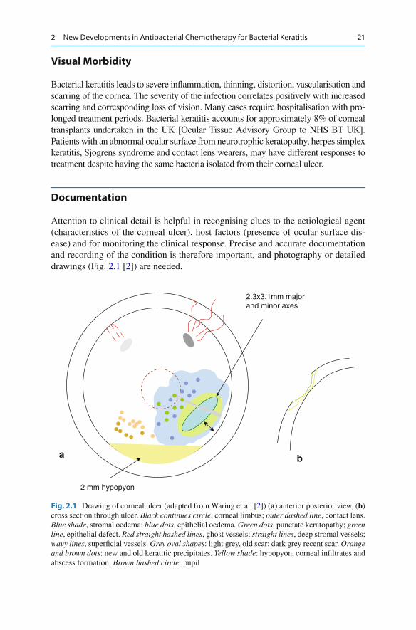
Attention to clinical detail is helpful in recognising clues to the aetiological agent (characteristics of the corneal ulcer), host factors (presence of ocular surface dis-ease) and for monitoring the clinical response. Precise and accurate documentation and recording of the condition is therefore important, and photography or detailed drawings (Fig. 2.1 [ 2 ] ) are needed.
ba
2.3x3.1mm majorand minor axes
2 mm hypopyon
Fig. 2.1 Drawing of corneal ulcer (adapted from Waring et al. [ 2 ] ) ( a ) anterior posterior view, ( b ) cross section through ulcer. Black continues circle , corneal limbus; outer dashed line , contact lens. Blue shade , stromal oedema; blue dots , epithelial oedema . Green dots , punctate keratopathy ; green line , epithelial defect. Red straight hashed lines , ghost vessels; straight lines , deep stromal vessels; wavy lines , super fi cial vessels. Grey oval shapes : light grey, old scar; dark grey recent scar. Orange and brown dots : new and old keratitic precipitates. Yellow shade : hypopyon, corneal in fi ltrates and abscess formation. Brown hashed circle : pupil

22 H. Sueke et al.
Causative Factors
Predisposing factors that facilitate successful bacterial colonisation and invasion of the cornea include trauma, contact lens wear, ocular surface disease [ 3, 4 ] and cor-neal surface abnormalities [ 5 ] . These are all signi fi cant risk factors and often are associated with recurrent disease [ 6 ] . Although the majority of patients have one or more risk factors, contact lens use is seen as the major risk factor in most studies [ 5, 6 ] . Lam et al. [ 7 ] reported that the incidence of bacterial keratitis was sixfold higher in contact lens wearers than in the general population and an increased inci-dence of Pseudomonas aeruginosa ( P. aeruginosa ) infections coincided with the increased popularity of contact lens wear. Among contact lens users, extended-wear contact lens wearers are at an increased risk of bacterial infection relative to daily disposable lens wearers [ 3 ] . Contact lens wear and correspondingly, contact lens-related keratitis is rarer in developing countries [ 4 ] .
Causative Bacteria
Of the bacteria associated with an ulcerative keratitis, P. aeruginosa , a Gram-negative bacillus, and Staphylococcus aureus , a Gram-positive coccus, are the most common bacterial pathogens. Although coagulase-negative staphylococci (CNS) account for a signi fi cant proportion of bacteria that are isolated from patients with bacterial keratitis, they are found in the conjunctival fl ora [ 8, 9 ] and their primary role in the disease is unclear. For example, a recent study has shown a positive correlation between clinical outcome and in vitro susceptibility for S. aureus and P. aeruginosa , but not for CNS [ 10 ] . Although Streptococci spp. are isolated less commonly, Strep. pneumoniae is often associated with a poor out-come and accounts for the greatest percentage of cases where eyes are lost [ 10 ] . Prompt intervention is crucial if a Strep. pneumoniae -related keratitis is suspected.
Summary for the Clinician Bacterial keratitis almost always results in a corneal scar • Good accurate documentation is required in follow up of corneal ulcers •
Summary for the Clinician In treating bacterial keratitis, host factors must be considered; they may be • indicators for recurrent disease. Risk factors include contact lens use, ocular surface disease, trauma and • Sjogrens Syndrome.

232 New Developments in Antibacterial Chemotherapy for Bacterial Keratitis
Table 2.1 Percentage of different bacterial species from patients with bacterial keratitis: compa-rable studies
Organism
Sueke [ 11 ] (UK)
Tuft [ 12 ] (UK)
Bourcier [ 5 ] (France)
Bharati [ 13 ] (India)
n = 772 n = 1,312 n = 208 n = 1,109 Gram-positive bacteria Coagulase-negative
staphylococci 26.9 N/A 48.1 17.4
Methicillin sensitive 21.7 N/A N/A N/A Methicillin resistant 5.2 N/A N/A N/A Staphylococcus aureus 13.6 33.4 7.7 3.9 Methicillin sensitive 12.4 N/A N/A N/A Methicillin resistant 1.2 N/A N/A N/A Streptococcus 12.6 19.0 9.2 42.4 Strep. pneumoniae 3.4 N/A 3.4 37.5 Other alpha-haemolytic 7.8 N/A 5.8 4.4 Beta-haemolytic a 1.4 N/A N/A 0.5 Other Gram-positive b 4.4 3.5 17.8 6.3 Gram-positives subtotal 57.4 55.9 82.8 70.1 Gram-negative bacteria Pseudomonas aeruginosa 20.9 24.8 10.1 18.0 Enterobacteriaceae 13.4 8.5 6.3 1.1 Serratia spp. 4.4 3.0 5.3 0.2 Klebsiella spp. 2.1 0.4 N/A 0.4 Citrobacter spp. 1.6 0.4 N/A 2.6 Proteus spp. 1.6 0.7 1.0 N/A E. coli 1.2 0.6 N/A N/A Enterobacter spp. 1.0 2.1 N/A 0.8 Morganella morganii 0.1 N/A N/A 0.5 Pantoea spp. 0.1 N/A N/A N/A Other 1.2 1.3 N/A 29.9 Moraxella spp. 2.6 5.9 0.5 N/A Haemophilus spp. 1.4 2.2 N/A N/A Other Gram-negatives c 4.2 2.8 N/A N/A Gram-negatives subtotal 42.5 44.2 16.9 20.9
a Lance fi eld Group A (0.3%), Group B (0.1%), Group C (0.3%), Group G (0.8%) b Corynebacterium spp. (2.6%), Bacillus spp. (1.7%), Enterococcus spp. (0.9%), Listeria spp. (0.1%) c Acinetobacter spp. (1.2%), Stenotrophomonas maltophilia (1.2%), Neisseria spp. (0.3%), Pasturella spp. (0.3%), Aeromonas spp. (0.1%), Eikenella spp. (0.1%), Agrobacterium spp. (0.1%), Alcaligines spp. (0.1%), Methylbacterium spp. (0.1%)
Table 2.1 illustrates the wide variability in the proportions of bacteria causing kera-titis between four similar studies set in different geographical locations. For exam-ple, the proportion of Gram-positive isolates varies between 56% and 83% and the proportion of Gram-negative isolates varies between 17% and 44%. Differences may re fl ect climate of the country or prevalence of risk factors such as contact lens use, trauma or co-existent ocular disease.

24 H. Sueke et al.
Investigation of Keratitis
There are various approaches to the microbiological investigation of patients with suspected keratitis. Traditional methods include the use of multiple corneal scrapes with direct inoculation onto different enrichment media. Collecting multiple scrapes, particularly from the eye of an uncooperative patient, is not always easy. Growing a minute sample in culture on an agar plate is technically dif fi cult: the inoculum might be deposited beneath the surface of the agar, a full range of fresh culture media may not be always available and the non-laboratory setting poses an increased risk of extraneous contamination of culture plates. These problems explain the reluctance of some ophthalmologists to perform a corneal scrape to reach a microbiological diagnosis. For example, McDonnell et al. [ 14 ] found that 49% of ophthalmologists treated corneal ulcers empirically without attempting to identify the causative organism. Kaye et al. [ 15 ] reported that collecting two corneal scrapes, one for a smear and the other placed in an enrichment transport medium (such as brain heart infusion broth), resulted in detection rates similar to those of direct plating with no signi fi cant loss of organisms.
The role of polymerase chain reaction (PCR) techniques has recently been evalu-ated to diagnose bacterial keratitis [ 16, 17 ] . PCR has the advantage of being a quicker and more sensitive technique than traditional culture methods; however, its high sensitivity may result in false-positive results. Although further larger studies comparing the two techniques are necessary to evaluate its place in the diagnosis of bacterial keratitis, it would seem reasonable to include PCR as part of the patient’s investigation.
Laboratory Diagnosis: Susceptibility Testing
Topical antimicrobials form the mainstay of treatment of bacterial keratitis. Despite their widespread usage, clinical decision making has rested upon suscep-tibility data derived from and for systemic infections. The relationship between bacterial susceptibility to antimicrobials and clinical outcome has only recently been demonstrated [ 10 ] . Although there are signi fi cant associations between the
Summary for the Clinician Causative bacteria and host factors can vary signi fi cantly between geo-• graphical locations. • Streptococcus pneumoniae is associated with the worst outcomes and requires prompt treatment.
252 New Developments in Antibacterial Chemotherapy for Bacterial Keratitis
minimum inhibitory concentration of the prescribed antimicrobial and the clinical outcome, the parameters of the association may be dependent on the particular bacterial species and antimicrobial.
Susceptibility and Resistance of Bacterial Isolates
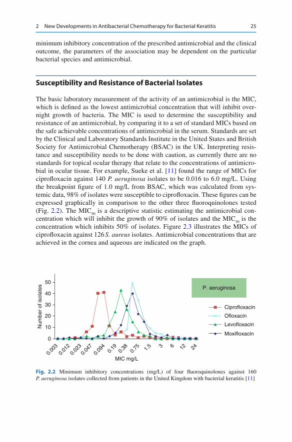
The basic laboratory measurement of the activity of an antimicrobial is the MIC, which is de fi ned as the lowest antimicrobial concentration that will inhibit over-night growth of bacteria. The MIC is used to determine the susceptibility and resistance of an antimicrobial, by comparing it to a set of standard MICs based on the safe achievable concentrations of antimicrobial in the serum. Standards are set by the Clinical and Laboratory Standards Institute in the United States and British Society for Antimicrobial Chemotherapy (BSAC) in the UK. Interpreting resis-tance and susceptibility needs to be done with caution, as currently there are no standards for topical ocular therapy that relate to the concentrations of antimicro-bial in ocular tissue. For example, Sueke et al. [ 11 ] found the range of MICs for cipro fl oxacin against 140 P. aeruginosa isolates to be 0.016 to 6.0 mg/L. Using the breakpoint fi gure of 1.0 mg/L from BSAC, which was calculated from sys-temic data, 98% of isolates were susceptible to cipro fl oxacin. These fi gures can be expressed graphically in comparison to the other three fl uoroquinolones tested (Fig. 2.2 ). The MIC
90 is a descriptive statistic estimating the antimicrobial con-
centration which will inhibit the growth of 90% of isolates and the MIC 50
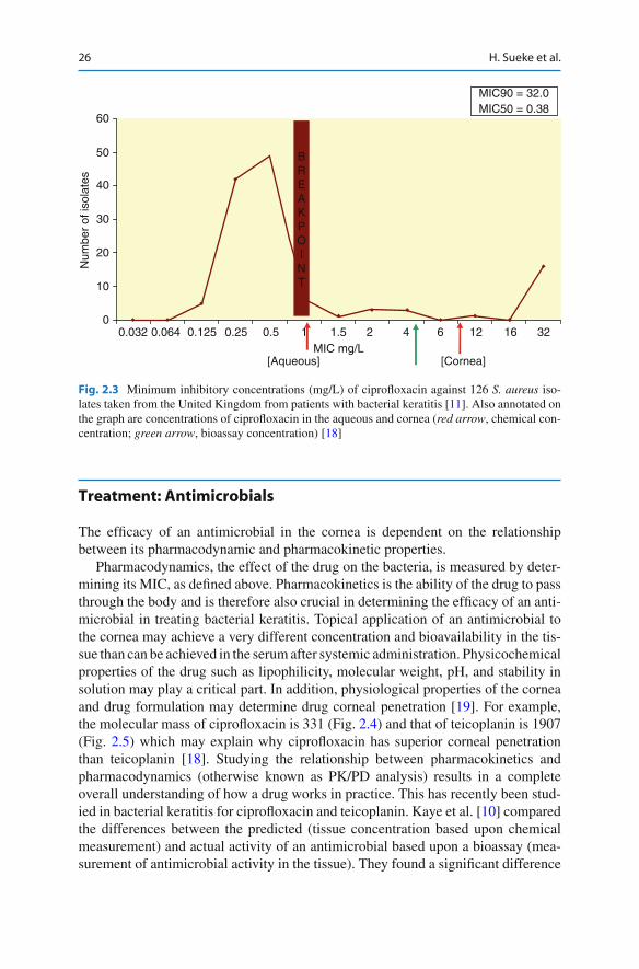
is the concentration which inhibits 50% of isolates. Figure 2.3 illustrates the MICs of cipro fl oxacin against 126 S. aureus isolates. Antimicrobial concentrations that are achieved in the cornea and aqueous are indicated on the graph.
50
40
30
20
10
0
MIC mg/L
P. aeruginosa
Ciprofloxacin
Levofloxacin
Ofloxacin
Moxifloxacin
Num
ber
of is
olat
es
0.00
30.
012
0.02
30.
047
0.09
40.
190.
380.
75 1.5 3 6 12 24
Fig. 2.2 Minimum inhibitory concentrations (mg/L) of four fl uoroquinolones against 160 P. aeruginosa isolates collected from patients in the United Kingdom with bacterial keratitis [ 11 ]
26 H. Sueke et al.
Treatment: Antimicrobials
The ef fi cacy of an antimicrobial in the cornea is dependent on the relationship between its pharmacodynamic and pharmacokinetic properties.
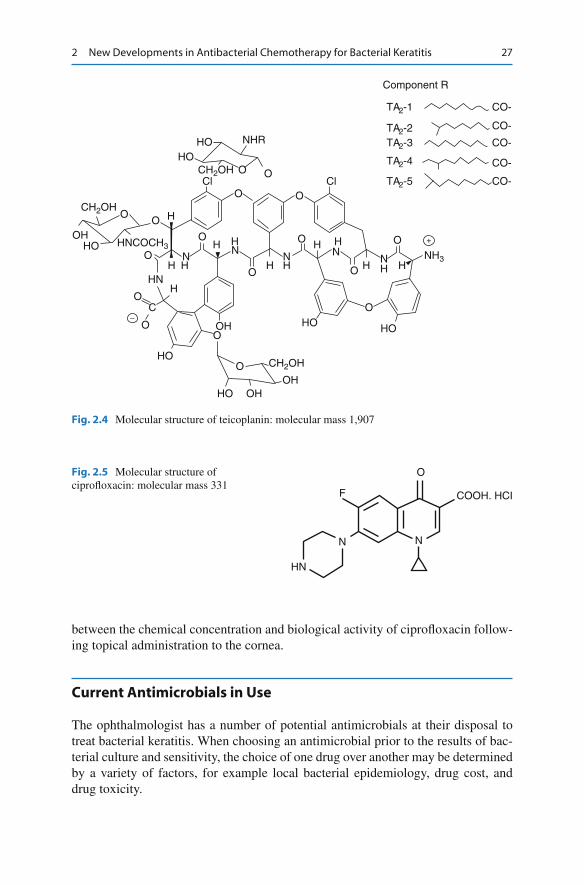
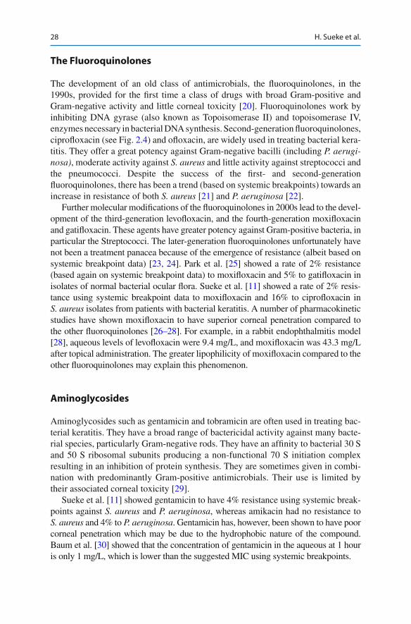
Pharmacodynamics, the effect of the drug on the bacteria, is measured by deter-mining its MIC, as de fi ned above. Pharmacokinetics is the ability of the drug to pass through the body and is therefore also crucial in determining the ef fi cacy of an anti-microbial in treating bacterial keratitis. Topical application of an antimicrobial to the cornea may achieve a very different concentration and bioavailability in the tis-sue than can be achieved in the serum after systemic administration. Physicochemical properties of the drug such as lipophilicity, molecular weight, pH, and stability in solution may play a critical part. In addition, physiological properties of the cornea and drug formulation may determine drug corneal penetration [ 19 ] . For example, the molecular mass of cipro fl oxacin is 331 (Fig. 2.4 ) and that of teicoplanin is 1907 (Fig. 2.5 ) which may explain why cipro fl oxacin has superior corneal penetration than teicoplanin [ 18 ] . Studying the relationship between pharmacokinetics and pharmacodynamics (otherwise known as PK/PD analysis) results in a complete overall understanding of how a drug works in practice. This has recently been stud-ied in bacterial keratitis for cipro fl oxacin and teicoplanin. Kaye et al. [ 10 ] compared the differences between the predicted (tissue concentration based upon chemical measurement) and actual activity of an antimicrobial based upon a bioassay (mea-surement of antimicrobial activity in the tissue). They found a signi fi cant difference
60
MIC mg/L[Aqueous] [Cornea]
BREAKPOINT
Num
ber
of is
olat
es
50
40
30
20
10
00.032 0.064 0.125 0.25 0.5 1 1.5 2 4 6 12 16 32
MIC90 = 32.0MIC50 = 0.38
Fig. 2.3 Minimum inhibitory concentrations (mg/L) of cipro fl oxacin against 126 S. aureus iso-lates taken from the United Kingdom from patients with bacterial keratitis [ 11 ] . Also annotated on the graph are concentrations of cipro fl oxacin in the aqueous and cornea ( red arrow , chemical con-centration; green arrow , bioassay concentration) [ 18 ]
272 New Developments in Antibacterial Chemotherapy for Bacterial Keratitis
between the chemical concentration and biological activity of cipro fl oxacin follow-ing topical administration to the cornea.
Current Antimicrobials in Use
The ophthalmologist has a number of potential antimicrobials at their disposal to treat bacterial keratitis. When choosing an antimicrobial prior to the results of bac-terial culture and sensitivity, the choice of one drug over another may be determined by a variety of factors, for example local bacterial epidemiology, drug cost, and drug toxicity.
HO
HN
HNCOCH3
CH2OH
CH2OH
CH2OH
OH
OH
OH
OH
HO
HO
HO
HO
HO NHR
ClCl
Component R
TA2-1 CO-
CO-
CO-
CO-
CO-
NH3
TA2-2TA2-3
TA2-4
TA2-5
HO
H H
HHH
H
H
H
H H
H HN
NNN
NH
O
O
O
O
O
OO
O
O
O
O
O
O
O
C
O
O
O
+
−
Fig. 2.4 Molecular structure of teicoplanin: molecular mass 1,907
HN
N
F
O
N
COOH. HCI
Fig. 2.5 Molecular structure of cipro fl oxacin: molecular mass 331
28 H. Sueke et al.
The Fluoroquinolones
The development of an old class of antimicrobials, the fl uoroquinolones, in the 1990s, provided for the fi rst time a class of drugs with broad Gram-positive and Gram-negative activity and little corneal toxicity [ 20 ] . Fluoroquinolones work by inhibiting DNA gyrase (also known as Topoisomerase II) and topoisomerase IV, enzymes necessary in bacterial DNA synthesis. Second-generation fl uoroquinolones, cipro fl oxacin (see Fig. 2.4 ) and o fl oxacin, are widely used in treating bacterial kera-titis. They offer a great potency against Gram-negative bacilli (including P. aerugi-nosa) , moderate activity against S. aureus and little activity against streptococci and the pneumococci. Despite the success of the fi rst- and second-generation fl uoroquinolones, there has been a trend (based on systemic breakpoints) towards an increase in resistance of both S. aureus [ 21 ] and P. aeruginosa [ 22 ] .
Further molecular modi fi cations of the fl uoroquinolones in 2000s lead to the devel-opment of the third-generation levo fl oxacin, and the fourth-generation moxi fl oxacin and gati fl oxacin. These agents have greater potency against Gram-positive bacteria, in particular the Streptococci. The later-generation fl uoroquinolones unfortunately have not been a treatment panacea because of the emergence of resistance (albeit based on systemic breakpoint data) [ 23, 24 ] . Park et al. [ 25 ] showed a rate of 2% resistance (based again on systemic breakpoint data) to moxi fl oxacin and 5% to gati fl oxacin in isolates of normal bacterial ocular fl ora. Sueke et al. [ 11 ] showed a rate of 2% resis-tance using systemic breakpoint data to moxi fl oxacin and 16% to cipro fl oxacin in S. aureus isolates from patients with bacterial keratitis. A number of pharmacokinetic studies have shown moxi fl oxacin to have superior corneal penetration compared to the other fl uoroquinolones [ 26– 28 ] . For example, in a rabbit endophthalmitis model [ 28 ] , aqueous levels of levo fl oxacin were 9.4 mg/L, and moxi fl oxacin was 43.3 mg/L after topical administration. The greater lipophilicity of moxi fl oxacin compared to the other fl uoroquinolones may explain this phenomenon.
Aminoglycosides
Aminoglycosides such as gentamicin and tobramicin are often used in treating bac-terial keratitis. They have a broad range of bactericidal activity against many bacte-rial species, particularly Gram-negative rods. They have an af fi nity to bacterial 30 S and 50 S ribosomal subunits producing a non-functional 70 S initiation complex resulting in an inhibition of protein synthesis. They are sometimes given in combi-nation with predominantly Gram-positive antimicrobials. Their use is limited by their associated corneal toxicity [ 29 ] .
Sueke et al. [ 11 ] showed gentamicin to have 4% resistance using systemic break-points against S. aureus and P. aeruginosa , whereas amikacin had no resistance to S. aureus and 4% to P. aeruginosa . Gentamicin has, however, been shown to have poor corneal penetration which may be due to the hydrophobic nature of the compound. Baum et al. [ 30 ] showed that the concentration of gentamicin in the aqueous at 1 hour is only 1 mg/L, which is lower than the suggested MIC using systemic breakpoints.

292 New Developments in Antibacterial Chemotherapy for Bacterial Keratitis
Cephalosporins
Cephalosporins have a broad spectrum of activity, including effective action against Haemophilus species. They contain a b -lactam ring similar to penicillin but have the advantage of being resistant to the penicillinases. They inhibit bacterial cell wall syn-thesis and are well tolerated topically. The fi rst-generation cephalosporins include cep-hazolin, second-generation cefuroxime and third-generation ceftazidime. Cefuroxime has often been used in combination with an aminoglycoside for the empirical treatment of suspected bacterial keratitis. Cefuroxime and ceftazidime had high MICs against S. aureus and P. aeruginosa suggesting a signi fi cant degree of antimicrobial resistance; however, systemic breakpoints were not available to formally assess this. Jenkins et al. [ 31 ] found that following topical administration to cataract surgery patients, aqueous concentrations of cefuroxime were only signi fi cant when applied once the corneal wound had been fashioned. This suggests poor corneal penetration of cefuroxime which may be explained as the cephalosporins are hydrophobic [ 31 ] .
Other Antimicrobials Used
Glycopeptides such as teicoplanin and vancomycin have activity against Gram-positive bacteria, including methicillin and penicillin-resistant staphylococci. They inhibit the biosynthesis of peptidoglycan polymers during the second stage of bacte-rial cell wall formation, at a different site of action from that of the b -lactam antimi-crobials. They also have an excellent activity against a variety of Gram-positive bacilli, but not Gram-negative bacteria which are inherently resistant. The glyco-peptides are, however, large molecules and in the intact corneal epithelium show a reduced corneal penetration, as mentioned previously [ 18 ] .
Development of Existing and New Classes of Drugs
Besi fl oxacin
Besi fl oxacin is a novel fl uoroquinolone for topical ophthalmic use, recently approved by the US Food and Drug Administration (USFDA) for the treatment of bacterial conjunctivitis [ 32 ] . Besi fl oxacin appears to have a broad spectrum of activity against
Summary for the Clinician
The fl uoroquinolones provide good activity against Gram-negative and • most Gram-positive bacteria causing an ulcerative keratitis, although resis-tance of the latter is emerging. Additional speci fi c Gram-positive coverage is offered by teicoplanin or • vancomycin.
30 H. Sueke et al.
aerobic and anaerobic bacteria, possibly due to its cyclopropyl group and chloride substituent at C-8 improving its activity against DNA gyrase and topoisomerase IV enzymes. Besi fl oxacin has been shown to be active against both Gram-positive (S . aureus , Strep. pneumoniae , Corynebacterium and Propionibacterium acnes ) and Gram-negative organisms ( H. in fl uenzae , Moraxella , Escherichia coli , Neisseria gonorrhoeae and P. aeruginosa ). Recent studies have found besi fl oxacin to have good pharmacokinetic parameters in vitro [ 33 ] as well as excellent ef fi cacy in ani-mal models of keratitis, compared to fourth-generation fl uoroquinolones [ 34, 35 ] .
Tigecycline
Tigecycline [ 36 ] is a glycylcycline with activity against most aerobic and anaerobic Gram-positive and -negative bacteria but with limited activity against P. aeruginosa. Glycylcyclines are bacteriostatic agents that inhibit protein synthesis in bacteria by reversibly binding to the 30 S ribosomal subunit. Sueke et al. [ 27 ] showed tigecy-cline to have no resistance to any Gram-positive isolates using systemic breakpoints, but complete resistance to P. aeruginosa . Corneal pharmacokinetics of tigecycline, however, have not yet been determined.
Linezolid
Linezolid [ 37 ] , the fi rst of a new class, the oxazolidinones, is a synthetic compound with activity against all the major Gram-positive groups of bacteria, but no activity against Gram-negative bacteria. Linezolid works by inhibiting bacterial ribosomal protein synthesis by binding to a site on the 50 S ribosomal subunit, thus preventing the formation of a 70 S initiation complex. Pharmacokinetic studies using animal models of keratitis have showed good corneal penetration and no recorded toxicity with linezolid [ 38, 39 ] . Sueke et al. [ 11 ] showed linezolid to have no resistance against Gram-positive isolates including methicillin-resistant S. aureus .
Meropenem
Meropenem [ 40 ] is a broad-spectrum carbapenem that is currently FDA approved to treat skin infections, intraabdominal infections and bacterial meningitis. Like other carbapenems, it is a b -lactam antimicrobial, working through bacterial cell wall inhibition. It has activity against Gram-positive and -negative pathogens, including extended-spectrum lactamases (ESBL) and AmpC-producing Enterobacteriaceae. Sueke et al. [ 11 ] showed meropenem to have wide coverage against both Gram-positive and Gram-negative microorganisms, where only one of the 772 isolates tested ( P. aeruginosa ) was resistant using systemic breakpoints. Corneal pharma-cokinetics of meropenem are not yet known; however, intravitreal meropenem in a rabbit model of endophthalmitis [ 41 ] did not show any evidence of toxicity.

312 New Developments in Antibacterial Chemotherapy for Bacterial Keratitis
Similarly, intravenous meropenem prior to cataract surgery showed penetration of the drug into the anterior chamber with no notable side effects [ 42 ] .
Developing Ophthalmic Breakpoints: Relation Between MIC and Clinical Outcome
There is good evidence demonstrating the relationship between the MIC of topi-cally applied antimicrobials and clinical outcome in bacterial keratitis [ 10 ] . This relationship is particularly well established for pathogenic bacteria such as P. aerug-inosa and S. aureus . Figure 2.6 summarises the relationship for the patients in a study by Kaye et al. [ 10 ] between a measure of clinical outcome (healing time to ulcer size: HT/UA) and the lowest MIC of the particular antimicrobial agent used. The general linear multivariate model revealed a weak but signi fi cant association between the MIC of the antimicrobial prescribed and clinical outcome de fi ned by the ratio of healing time to ulcer size. The importance of the bacterial type and anti-microbial used for treatment is indicated by the signi fi cant associations between the fl uoroquinolone MIC and clinical outcome for Pseudomonas spp., S. aureus and Enterobacteriaceae but not for Streptococcus spp. or CNS. The MIC is therefore an important measure for evaluating the potential effectiveness of topically applied antimicrobials in the treatment of bacterial keratitis.
MIC (Log g/L)
40.00
30.00
20.00
10.00
0.00
–3.00 –2.00 –1.00 0.00 1.00 2.00 3.00
Fig. 2.6 Clinical outcome and MIC: antimicrobial used and all bacterial isolates. Healing time to ulcer area (HT/UA) (days per mm 2 ), logarithm (Log) of Minimum inhibitory concentration (MIC mg/L). Hypothetical susceptible and resistant outcomes (HT/UA of 3.5 and 7 days/mm 2 ) corre-sponds to an MIC of 0.1 and 10 mg/L [ 10 ]

32 H. Sueke et al.
Combination Therapy
As opposed to single therapy, an antimicrobial combination offers a broader spec-trum of activity and may reduce selective pressures. This may be of particular impor-tance for the fl uoroquinolones, as increasing resistance has been reported in S. aureus and P. aeruginosa isolates from cases of bacterial keratitis [ 9, 21– 24 ] . An often over-looked reason for combination therapy, however, is not for providing a broader spec-trum but for an increased antimicrobial effect. In particular, combination therapy may result in synergy as occurs, for example, with the combination of penicillin and gentamicin when used in the treatment of enterococcal endocarditis [ 43, 44 ] . This synergistic effect can be explained by the increased ease of gentamicin passage into the bacterial cell, due to cell wall disruption caused by the action of penicillin. Conversely, combinations of antimicrobials may be antagonistic, as occurs with the combination of chloramphenicol and penicillin in the treatment of pneumococcal meningitis [ 45 ] . The presumed reason for this antagonism is that chloramphenicol, a bacteriostatic agent, by reducing growth prevents penicillin, which requires a divid-ing and growing organism from having its full effect on the cell wall synthesis. It is important therefore not to use combination therapy which may have inhibitory or antagonistic effects. A recent in vitro combination study [ 46 ] using isolates from patients with bacterial keratitis demonstrated that the combination of meropenem and cipro fl oxacin was predominantly additive or synergistic for both S. aureus and P. aeruginosa . Furthermore, teicoplanin combined with meropenem, cipro fl oxacin or moxi fl oxacin was also additive or synergistic against S. aureus .
Drug Delivery to the Cornea
The most commonly used route of antimicrobial delivery into the cornea is topically, in the form of drops, solutions, emulsions or suspensions. However topical administration and its resultant pharmacokinetics and pharmacodynamics remain an inef fi cient method of delivery and may in part account for the poor outcome from bacterial keratitis. Furthermore, in the acute in fl amed eye, there is increase in tearing which, together with the associated pain, makes instillation of a topical antimicrobial dif fi cult. Hospitalisation is often needed to deliver the antimicrobials at frequent intervals (15 min) through con-secutive nights. It is clear therefore that drug delivery systems need to be rethought.
Summary for the Clinician The minimum inhibitory concentration (MIC) is de fi ned as the lowest anti-• microbial concentration that will inhibit overnight growth of bacteria. A relationship has now been determined between MIC and clinical out-• come for S. aureus and P. aeruginosa.
332 New Developments in Antibacterial Chemotherapy for Bacterial Keratitis
Novel Methods of Drug Delivery to the Cornea
Alternative methods of delivery of antimicrobials to treat keratitis are currently being studied [ 47, 48 ] . Various drug delivery devices have been evaluated for the treatment of keratitis. They broadly fall into two categories: matrix and reservoir based. Matrix-based implants distribute the drug throughout a degradable poly-mer matrix, for example, Lacrisert® [ 49 ] . In a reservoir implant, the drug is stored within a reservoir made of a non-degradable substance such as collagen shields. Drug penetration into the cornea can be also enhanced by altering its physico-chemical properties with the addition of particulates such as nano-particles and other penetration enhancers, as well as using the prodrug and mucoadhesive dos-age forms.
Conclusion
It is clear that although there is a signi fi cant relationship between the MIC of the prescribed topical antimicrobial and clinical outcome, this relationship is rela-tively small; c.14%. Other bacterial and host factors play a role in the progres-sion of infection. This is underlined by the fi nding that although the majority of bacterial isolates from keratitis based upon systemic breakpoint data were reported to be susceptible to prescribed antimicrobials, the actual outcome was far worse than expected. It is clear therefore that although ophthalmic suscepti-bility data to topical antimicrobials gives a better indication of outcome, the main determinants of outcome relate to factors such as the host–bacterial interaction. This is particularly evident for infections caused by S. aureus which accounts for up to 31% of cases of keratitis with the majority of ocular surface infections occurring in patients living in the community in the United Kingdom. This nev-ertheless presents an opportunity to develop treatments aimed at interfering with the action of the bacteria on its host target. These treatments could also be deliv-ered in novel ways, reducing the dependency of instilling intensive topical antimicrobials.
Summary for the Clinician Resistance to commonly used antimicrobials in keratitis has prompted the • development of novel strategies in treating bacterial keratitis. Novel antimicrobials such as besi fl oxacin, tigecycline, meropenem and • linezolid are currently under consideration. Additional improvements to clinical outcome may arise through utilising • synergistic combinations of antimicrobials, as well as using novel corneal drug delivery systems.

34 H. Sueke et al.
References
1. Bharathi MJ et al (2007) Microbial keratitis in South India: in fl uence of risk factors, climate, and geographical variation. Ophthalmic Epidemiol 14(2):61–69
2. Waring GO, Laibson PR (1977) A systematic method of drawing corneal pathologic condi-tions. Arch Ophthalmol 95(9):1540–1542
3. Liesegang TJ (1997) Contact lens-related microbial keratitis: part I: epidemiology. Cornea 16(2):125–131
4. Sharma S et al (2003) Trends in contact lens-associated microbial keratitis in Southern India. Ophthalmology 110(1):138–143
5. Bourcier T et al (2003) Bacterial keratitis: predisposing factors, clinical and microbiological review of 300 cases. Br J Ophthalmol 87(7):834–838
6. Schaefer F et al (2001) Bacterial keratitis: a prospective clinical and microbiological study. Br J Ophthalmol 85(7):842–847
7. Lam DS et al (2002) Incidence and risk factors for microbial keratitis in Hong Kong: compari-son with Europe and North America. Eye (Lond) 16(5):608–618
8. Fahmy JA, Moller S, Bentzon MW (1974) Bacterial fl ora of the normal conjunctiva. I. Topographical distribution. Acta Ophthalmol (Copenh) 52(6):786–800
9. Fukuda M et al (2002) Methicillin-resistant Staphylococcus aureus and methicillin-resistant coagulase-negative Staphylococcus ocular surface infection ef fi cacy of chloramphenicol eye drops. Cornea 21(7 Suppl):S86–S89
10. Kaye S et al (2010) Bacterial susceptibility to topical antimicrobials and clinical outcome in bacterial keratitis. Invest Ophthalmol Vis Sci 51(1):362–368
11. Sueke H et al (2010) Minimum inhibitory concentrations of standard and novel antimicrobials for isolates from bacterial keratitis. Invest Ophthalmol Vis Sci 51(5):2519–2524
12. Tuft SJ, Matheson M (2000) In vitro antibiotic resistance in bacterial keratitis in London. Br J Ophthalmol 84(7):687–691
13. Bharathi MJ et al (2003) Epidemiology of bacterial keratitis in a referral centre in south India. Indian J Med Microbiol 21(4):239–245
14. McDonnell PJ (1996) Empirical or culture-guided therapy for microbial keratitis? A plea for data. Arch Ophthalmol 114(1):84–87
15. Kaye SB et al (2003) Simplifying collection of corneal specimens in cases of suspected bacte-rial keratitis. J Clin Microbiol 41(7):3192–3197
16. Itahashi M et al (2010) Detection and quanti fi cation of pathogenic bacteria and fungi using real-time polymerase chain reaction by cycling probe in patients with corneal ulcer. Arch Ophthalmol 128(5):535–540
17. Subrayan V et al (2010) Assessment of polymerase chain reaction in the detection of Pseudomonas aeruginosa in contact lens-induced severe infectious keratitis. Eye Contact Lens 36(4):201–203
18. Kaye SB et al (2009) Concentration and bioavailability of cipro fl oxacin and teicoplanin in the cornea. Invest Ophthalmol Vis Sci 50(7):3176–3184
19. Urtti A (2006) Challenges and obstacles of ocular pharmacokinetics and drug delivery. Adv Drug Deliv Rev 58(11):1131–1135
20. Baum J, Barza M (2000) The evolution of antibiotic therapy for bacterial conjunctivitis and keratitis: 1970–2000. Cornea 19(5):659–672
21. Goldstein MH, Kowalski RP, Gordon YJ (1999) Emerging fl uoroquinolone resistance in bacte-rial keratitis: a 5-year review. Ophthalmology 106(7):1313–1318
22. Garg P, Sharma S, Rao GN (1999) Cipro fl oxacin-resistant Pseudomonas keratitis. Ophthalmology 106(7):1319–1323
23. Moshirfar M et al (2006) Fourth-generation fl uoroquinolone-resistant bacterial keratitis after refractive surgery. J Cataract Refract Surg 32(3):515–518
24. Jhanji V et al (2007) Fourth-generation fl uoroquinolone-resistant bacterial keratitis. J Cataract Refract Surg 33(8):1488–1489
25. Park SH et al (2009) The resistance patterns of normal ocular bacterial fl ora to 4 fl uoroquinolone antibiotics. Cornea 28(1):68–72

352 New Developments in Antibacterial Chemotherapy for Bacterial Keratitis
26. Kim DH, Stark WJ, O’Brien TP (2005) Ocular penetration of moxi fl oxacin 0.5% and gati fl oxacin 0.3% ophthalmic solutions into the aqueous humor following topical administra-tion prior to routine cataract surgery. Curr Med Res Opin 21(1):93–94
27. Sugioka K et al (2009) Intraocular penetration of sequentially instilled topical moxi fl oxacin, gati fl oxacin, and levo fl oxacin. Clin Ophthalmol 3:553–557
28. Yagci R et al (2007) Penetration of second-, third-, and fourth-generation topical fl uoroquinolone into aqueous and vitreous humour in a rabbit endophthalmitis model. Eye (Lond) 21(7):990–994
29. Alfonso EC et al (1990) In vitro toxicity of gentamicin to corneal epithelial cells. Cornea 9(1):55–61
30. Baum J (1982) Treatment of bacterial ulcers of the cornea in the rabbit: a comparison of administration by eye drops and subconjunctival injections. Trans Am Ophthalmol Soc 80:369–390
31. Jenkins CD et al (1996) Comparative intraocular penetration of topical and injected cefu-roxime. Br J Ophthalmol 80(8):685–688
32. Karpecki P (2009) Besi fl oxacin ophthalmic suspension 0.6% in patients with bacterial con-junctivitis: a multicenter, prospective, randomized, double-masked, vehicle-controlled, 5-day ef fi cacy and safety study. Clin Ther 31(3):514–526
33. Proksch JW et al (2009) Ocular pharmacokinetics of besi fl oxacin following topical adminis-tration to rabbits, monkeys, and humans. J Ocul Pharmacol Ther 25(4):335–344
34. Sanders ME et al (2011) Comparison of besi fl oxacin, gati fl oxacin, and moxi fl oxacin against strains of Pseudomonas aeruginosa with different quinolone susceptibility patterns in a rabbit model of keratitis. Cornea 30(1):83–90
35. Sanders ME et al (2009) Ef fi cacy of besi fl oxacin in a rabbit model of methicillin-resistant Staphylococcus aureus keratitis. Cornea 28(9):1055–1060
36. Pankey GA (2005) Tigecycline. J Antimicrob Chemother 56(3):470–480 37. Moellering RC (2003) Linezolid: the fi rst oxazolidinone antimicrobial. Ann Intern Med
138(2):135–142 38. Ekdawi NS (2005) Topical linezolid in Streptococcus pneumoniae corneal ulcer model in rab-
bits. Invest Ophthalmol Vis Sci 46(5):4910 39. Saleh M (2010) Ocular penetration of topically applied linezolid in a rabbit model. J Cataract
Refract Surg 36(3):488–492 40. Baldwin CM, Lyseng-Williamson KA, Keam SJ (2008) Meropenem: a review of its use in the
treatment of serious bacterial infections. Drugs 68(6):803–838 41. Manav C (2004) Comparison of intravitreal ceftazidime and meropenem in treatment of experi-
mental pseudomonal posttraumatic endophthalmitis in a rabbit model. J Appl Res 4(2):337–345 42. Schauersberger J et al (1999) Penetration and decay of meropenem into the human aqueous
humor and vitreous. J Ocul Pharmacol Ther 15(5):439–445 43. Moellering RC Jr, Wennersten C, Weinberg AN (1971) Synergy of penicillin and gentamicin
against Enterococci . J Infect Dis 124(Suppl):S207–S209 44. Winstanley TG, Hastings JG (1990) Synergy between penicillin and gentamicin against
enterococci. J Antimicrob Chemother 25(4):551–560 45. Friedland IR, Klugman KP (1992) Failure of chloramphenicol therapy in penicillin-resistant
pneumococcal meningitis. Lancet 339(8790):405–408 46. Sueke H et al (2010) An in vitro investigation of synergy or antagonism between antimicrobial
combinations against isolates from bacterial keratitis. Invest Ophthalmol Vis Sci 51(8):4151–4155
47. Kearns VR, Williams RL (2009) Drug delivery systems for the eye. Expert Rev Med Devices 6(3):277–290
48. Behar-Cohen F (2002) Drug delivery systems to target the anterior segment of the eye: funda-mental bases and clinical applications. J Fr Ophtalmol 25(5):537–544
49. Luchs JI, Nelinson DS, Macy JI (2010) Ef fi cacy of hydroxypropyl cellulose ophthalmic inserts (LACRISERT) in subsets of patients with dry eye syndrome: fi ndings from a patient registry. Cornea 29(12):1417–1427

http://www.springer.com/978-3-642-28746-6