new bacterial cellulose nanocomposites - core.ac.uk · cristalinidade. por sua vez, os...
TRANSCRIPT

NEW BACTERIAL CELLULOSE NANOCOMPOSITES
PREPARED BY IN SITU RADICAL
POLYMERIZATION OF METHACRYLATE
MONOMERS
Tese apresentada à Universidade da Madeira com vista à
obtenção do grau de Mestre em Bioquímica Aplicada
Marisa Camacho Gonçalves Faria
Sob a orientação de:
Professora Doutora Nereida Maria Abano Cordeiro
Professora Doutora Carmen Sofia da Rocha Freire Barros
Faculdade de Ciências Exatas e da Engenharia
Universidade da Madeira
Funchal – Portugal
Departamento de Química e CICECO
Universidade de Aveiro
Aveiro, Portugal
2015


III
ACKNOWLEDGEMENTS
I would like to acknowledge every one that contribute in any way to the performance
of my master thesis, either at professional or/and motivational/personal level and made
possible their execution.
To my supervisor Prof. Dr. Nereida Cordeiro, for her constant support, guidance,
encouragement, patience and constructive critics which help me during my project and be
indispensable for the accomplishment of my project.
To my co-supervisor Prof. Dr. Carmen Freire for the support and all help in
recommending and understanding through my master thesis. To Dr. Armando Silvestre for
all the support and collaboration given in my project.
To University of Madeira for providing the facilities to carry out this project.
To Dr. Carla Vilela (University of Aveiro, Portugal), for performing the Infrared
Spectroscopy, NMR, X-ray diffraction, Scanning Electron Microscopy, Thermogravimetric
Analysis and Dynamic Mechanical Analysis and all support in data collection.
To Dr. Faranak Mohammadkazemi (Shahid Beheshti University, Iran) for running
the elemental chemical composition analyses.
To the laboratory technicians Paula Andrade and Paula Vieira, for availability and
support in the reagents and laboratory during my project.
To my colleagues and friends, Dina Maciel, Igor Fernandes, Micael Leça, Tomásia
Fernandes, Roberto Aguiar, Nuno Nunes, Emanuel Gouveia, Gabriel Freitas, Carla Alves,
Rogério Correia, José Rodrigues, Marisela Santos, Francisco Maciel, and Dr. Gabriel Leça
for all the motivation and help with their good dispositions and willingness through this
years.
To my family, Mom, Isabel, my sister, Elisabete, my brother, Roberto and João, and
my nephew, Afonso, for all support, encouragement and love all the time which help me to
continue and supporting me to go through.
Last but not the least, to my boyfriend, Ricardo Pascoal, for his endless support,
tireless patience and lovely sense of humor encouraging me all the time. Love you!

IV

IV

V
LIST OF PUBLICATIONS
Cordeiro, N., Ashori, A., Hamzeh, Y. & Faria, M. (2013) Effects of hot water pre-extraction
on surface properties of bagasse soda pulp. Materials Science and Engineering C, 33, 613–
617
Ashori, A., Cordeiro, N., Faria, M. & Hamzeh, Y. (2013) Effect of chitosan and cationic
starch on the surface chemistry properties of bagasse paper. International Journal of
Biological Macromolecules, 58, 343– 348
Cordeiro, N., Faria M., Abraham, E. & Pothan, L. A. (2013) Assessment of the changes in
the cellulosic surface of micro and nano banana fibres due to saponin treatment.
Carbohydrate Polymers 98, 1065– 1071
Cordeiro, N., Freitas, N., Faria, M. & Gouveia, M. (2013) Ipomoea batatas (L.) Lam.: A
Rich Source of Lipophilic Phytochemicals. J. Agric. Food Chem, 61, 12380−12384
Deepa, B., Abraham, E., Cordeiro, N., Mozetic, M., Mathew, A. P., Oksman, K., Faria, M.,
Thomas, S. & Pothan, L. A. (2015) Utilization of various lignocellulosic biomass for the
production of nanocellulose: a comparative study. Cellulose 22, 1075–1090
Castro, C., Cordeiro, N., Faria, M., Zuluaga, R., Putaux, J.L., Filpponen, I., Velez, L.,
Rojas, O.J. & Gañán, P. (2015) In situ glyoxalization during biosynthesis of bacterial
cellulose. Carbohydrate Polymers 126 (2015) 32–39
Mohammadkazemi, F., Faria, M., Cordeiro, N. In situ by«iosynthesis of bacterial
nanocellulose- CaCO3 hybrid composite: one.step process. 2015 (Submited)
VI
LECTURE
Moares, A.G.O., Faria, M., Silva, F.W., Cordeiro, N. & Amico, S.C. (2013) Efeito de
tratamentos químicos nas propriedades de superfície de fibras de carbono via cromatografia
gasosa inversa. 12° Congresso Brasileiro de Polímeros (12°CBPol) Brasil

VII
ABSTRACT
Bacterial cellulose/polymethacrylate nanocomposites have received attention in
numerous areas of study and in a variety of applications. The attractive properties of
methacrylate polymers and bacterial cellulose, BC, allow the synthesis of new
nanocomposites with distinct characteristics. In this study, BC/poly(glycidylmethacrylate)
(BC/PGMA) and BC/poly(ethyleneglycol)methacrylate (BC/PPEGMA) nanocomposites
were prepared through in situ free radical polymerization of GMA and PEGMA,
respectively. Ammonium persulphate (APS) was used as an initiator and N,N’-
methylenebisacrilamide (MBA) was used as a crosslinker in BC/PGMA. Chemical
composition, morphology, thermal stability, water absorption, mechanic and surface
properties were determined through specific characterization techniques. The optimal
polymerization was obtained at (1:2) for BC/PGMA, (1:2:0.2) ratio for BC/GMA/MBA
and (1:20) for BC/PPEGMA, with 0.5% of initiator at 60 ºC during 6 h. A maximum of 67%
and 87% of incorporation percentage was obtained, respectively, for the nanocomposites
BC/PGMA/MBA and BC/PPEGMA. BC/PGMA nanocomposites exhibited an increase
of roughness and compactation of the three-dimensional structure, an improvement in the
thermal and mechanical properties, and a decrease in their swelling ability and crystallinity.
On the other hand, BC/PPEGMA showed a decrease of stiffness of three-dimensional
structure, improvement in thermal and mechanical properties, an increase in their swelling
ability and a decrease the crystallinity. Both BC/polymethacrylate nanocomposites exhibited
a basic surface character. The acid treatment showed to be a suitable strategy to modifiy
BC/PGMA nanocomposites through epoxide ring-opening reaction mechanism.
Nanocomposites became more compact, smooth and with more water retention ability. A
decrease in the thermal and mechanical proprieties was observed.
The new nanocomposites acquired properties useful to biomedical applications
or/and removal of heavy metals due to the presence of functional groups.
Keywords: bacterial cellulose, nanocomposite, in situ radical polymerization,
glycidylmethacrylate, poly(ethyleneglycol)methacrylate, chemical treatment.

VI

VII
RESUMO
Os nanocompósitos de celulose bacteriana/polimetacrilatos têm ganho interesse em
inúmeras áreas de estudo e para diversas aplicações. As propriedades atrativas dos
monómeros metacrilatos e da celulose bacteriana, BC, possibilitam aos novos
nanocompósitos adquirirem características interessantes para diversas aplicações. No
presente estudo, os nanocompósitos de BC/poli(glicidilmetacrilato) (BC/PGMA) e
BC/poli(etilenoglicol)metacrilato (BC/PPEGMA) foram obtidos através da polimerização
radical livre in situ do GMA e PEGMA, respetivamente, dentro da rede da BC usando o
persulfato de amónia (APS) como iniciador. O N,N’-metilenobisacrilamida (MBA) foi usado
como agente reticulante nos nanocompósitos de BC/PGMA. A composição química, a
estrutura morfológica, a estabilidade química, a absorção de água e as propriedades
mecânicas e de superfície foram determinadas. A polimerização ótima foi observada para
uma razão de (1:2) para a BC/PGMA, (1:2:0.2) para a BC/PGMA/MBA e a uma razão de
(1:20) para o BC/PPEGMA com 0.5 % de iniciador a 60 °C durante 6 h. A percentagem
máxima de incorporação de 67% e 87% foi obtida para os nanocompósitos
BC/PGMA/MBA e BC/PPEGMA, respetivamente. Os nanocompósitos de BC/PGMA
demonstraram uma estrutura rugosa e compacta, um melhoramento nas propriedades
mecânicas e térmicas, uma diminuição na sua capacidade de absorção de água e
cristalinidade. Por sua vez, os nanocompósitos de BC/PPEGMA apresentaram uma
diminuição na rigidez da rede, um melhoramento nas propriedades mecânicas e térmicas,
um aumento na sua capacidade de absorção de água e uma diminuição na cristalinidade. Os
nanocompósitos de BC/polimetacrilato exibiram um caracter básico à superfície depois da
incorporação dos metacrilatos. O tratamento químico ácido demonstrou ser uma estratégia
útil para a modificação dos nanocompósitos de BC/PGMA através do mecanismo de
abertura do anel epóxi. Os nanocompósitos tornaram-se morfologicamente mais compactos
e com a superfície mais lisa. No entanto, as propriedades mecânicas e térmicas decrescem e,
a superfície tornou-menos básica. Os novos nanocompósitos apresentam propriedades uteis
para aplicações biomédicas ou para a remoção de metais pesados.
Palavras-chave: celulose bacteriana, glicidilmetacrilato, poli(etilenoglicol)metacrilato,
nanocompósitos, polimerização radical livre in situ, tratamento químico.

VIII

XV
TABLE OF CONTENTS
ACKNOWLEDGEMENTS .......................................................................................... III
LIST OF PUBLICATIONS ............................................................................................. V
ABSTRACT................................................................................................................. VII
RESUMO .................................................................................................................... VII
TABLES INDEX ........................................................................................................ XVI
LIST OF ABBREVIATIONS ................................................................................... XVIII
CHAPTER I. Overview of bacterial cellulose ................................................................... 2
1. Cellulose as Biopolymer ............................................................................................ 2
2. Bacterial Cellulose – Historical Outline ..................................................................... 4
2.1. Biochemistry of Bacterial Cellulose and Biosynthesis Pathways ........................... 4
2.2. Structure and Properties of Bacterial Cellulose .................................................... 6
2.3. Application of Bacterial Cellulose ....................................................................... 9
3. Bacterial Cellulose Nanocomposites ........................................................................ 10
3.1. BC nanocomposites through BC biosynthesis .................................................... 10
3.2. BC nanocomposites through in situ polymerization............................................ 12
3.3. Acrylate monomers used in BC nanocomposite ................................................. 14
4. Aim of the Study ..................................................................................................... 16
CHAPTER II. New bacterial cellulose/poly(glycidylmethacrylate) nanocomposites films by
in situ free radical polymerization ................................................................................... 20
1. Introduction ............................................................................................................ 21
2. Methodology .......................................................................................................... 22
2.1. Material ........................................................................................................... 22
2.2. BC nanocomposites preparation ....................................................................... 22
2.2.1. GMA and GMA/MBA polymerization ..................................................... 22
2.2.2. In situ free radical polymerization of GMA and GMA/MBA into BC ......... 23
2.3. BC nanocomposites characterization ................................................................ 24
2.3.1. Infrared Spectroscopy ................................................................................ 24

XVI
2.3.2. Energy-dispersive X-ray spectroscopy ......................................................... 24
2.3.3. Scanning electron microscopy .................................................................... 24
2.3.4. X-ray diffraction ........................................................................................ 24
2.3.5. 13Carbon Nuclear Magnetic Resonance....................................................... 25
2.3.6. Swelling ratio ............................................................................................. 25
2.3.7. Thermogravimetric analyses ....................................................................... 26
2.3.8. Dynamic mechanical analyses .................................................................... 26
3. Results and Discussion ............................................................................................ 26
3.1. In situ free radical polymerization reaction ........................................................ 26
3.2. Optimization of the in situ free radical polymerization ....................................... 28
3.3. BC nanocomposites characterization ................................................................ 30
3.3.1. Infrared and X-ray Spectroscopy ................................................................ 30
3.3.2. X-ray diffraction ........................................................................................ 32
3.3.3. 13Carbon Nuclear Magnetic Resonance....................................................... 33
3.3.4. Scanning electron microscopy .................................................................... 34
3.3.5. Swelling ratio ............................................................................................. 35
3.3.6. Thermogravimetric analyses ....................................................................... 36
3.3.7. Dynamic Mechanical properties ................................................................. 38
4. Conclusions ............................................................................................................ 39
CHAPTER III. Epoxide ring-opening of bacterial cellulose/poly(glycidylmethacrylate)
nanocomposites ............................................................................................................. 43
1. Introduction ............................................................................................................ 44
2. Methodology .......................................................................................................... 45
2.1. BC nanocomposites preparation ....................................................................... 45
2.2. Chemical modification of BC nanocomposites .................................................. 45
2.3. BC nanocomposites characterization ................................................................ 46
3. Results and Discussion ............................................................................................ 47
3.1. Structural Analysis ........................................................................................... 48
3.2. Morphological Analyses ................................................................................... 49
3.3. Thermal and Mechanical Analyses ................................................................... 50
3.4. Surface properties ............................................................................................. 52

XVII
4. Conclusions ............................................................................................................ 56
CHAPTER IV. Novel bacterial cellulose/poly(ethyleneglycol) methacrylate nanocomposite
films obtained by in situ free radical polymerization ........................................................ 59
1. Introduction ............................................................................................................ 60
2. Methodology .......................................................................................................... 61
2.1. Material ........................................................................................................... 61
2.2. BC nanocomposites preparation ....................................................................... 61
2.3. BC nanocomposites characterization ................................................................ 62
3. Results and Discussion ............................................................................................ 62
4. Conclusions ............................................................................................................ 68
CHAPTER V. Assessment of the surface properties changes of bacterial
cellulose/polymethacrylate nanocomposite films by Inverse Gas Chromatography ......... 70
1. Introduction ............................................................................................................ 71
1.1. Inverse gas chromatography ............................................................................. 71
1.1.1. Surface energy component ......................................................................... 72
1.1.2. Acid base character .................................................................................... 73
1.1.3. Isotherm measurements ............................................................................. 74
2. Experimental methodology ..................................................................................... 75
2.1. Material ........................................................................................................... 75
2.2. BC/PGMA and BC/PEGMA nanocomposites ................................................. 75
2.3. IGC measurements ........................................................................................... 76
2.4. Swelling ratio ................................................................................................... 77
2.5. Scanning electron microscopy ........................................................................... 77
2.6. Energy-dispersive X-ray spectroscopy ............................................................... 78
3. Results and Discussion ............................................................................................ 78
3.1. Dispersive component of surface energy ........................................................ 78
3.2. Heterogeneity ............................................................................................... 81
3.3. Surface area .................................................................................................. 83
3.4. Acid-base character ....................................................................................... 84
4. Conclusions ............................................................................................................ 87
XVIII
FINAL CONCLUSIONS .............................................................................................. 89
REFERENCES ............................................................................................................. 90
XIX
FIGURES INDEX
Figure 1. Molecular structure of cellulose where inter- (blue) and intra- (red) chain hydrogen
bonds dashed lines [reproduced from (14)]. ...................................................................... 3
Figure 2. Morphology characteristics of the different types of cellulose fibers [reproduced
from (15)]. ....................................................................................................................... 3
Figure 3. Schematic of the metabolic pathways of Glucanoacetobacter xylinum and the
assembly of cellulose molecules into nanofibrils [reproduced from (6)]. ............................. 6
Figure 4. A SEM of freeze-dried surface of bacterial cellulose gel [reproduced from (14)]. . 7
Figure 5. (A) X-ray patterns of BC films and (B) View along the direction 4 (i.e. [1ī0] for Iα
and [010] for Iβ) and the displacement of the hydrogen bonding sheets: blue Iα and red Iβ
[reproduced from (3, 13)]. ................................................................................................ 8
Figure 6. Different forms of BC produced by Gluconacetobacter sp. (A) Membrane, (B)
Irregular forms, (C) Sphere-like particles [reproduced from (36)]. ...................................... 8
Figure 7. Applications of bacterial cellulose: A – Tubes (blood vessel); B – Branched tube
fermented on a branched silicone tube (vascular grafts) [reproduced from (36)]; C –Dressing
(wound dressing) [reproduced from (7)]; D – Incorporation of AgNO3 nanoparticles
(antimicrobial activity); E –Paper (filter paper) and F – Colored with aniline blue dye
(bioadsorbent) [reproduced from (11)]. ............................................................................. 9
Figure 8. SEM images of surface morphology of (A) BC and (B) BC/Chitosan film in dry
form [reproduced from (41)]. ......................................................................................... 11
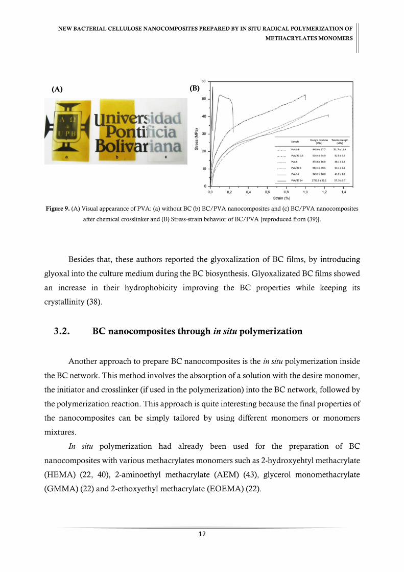
Figure 9. (A) Visual appearance of PVA: (a) without BC (b) BC/PVA nanocomposites and
(c) BC/PVA nanocomposites after chemical crosslinker and (B) Stress-strain behavior of
BC/PVA [reproduced from (39)].................................................................................... 12
Figure 10. (A) Visual appearance of BC/PAEM and all BC/PAEM/MBA nanocomposites.
(B) TGA thermograph of BC/PAEM nanocomposites. (C) Swelling of wet BC, BC/PAEM
and BC/PAEM/MBA nanocomposites [reproduced from (43)]. ..................................... 13
Figure 11. (A) Visual aspect of BC, BC/PHEMA and BC/PHEMA/PEGDA films and (B)
ADSCs proliferation in contact with BC and BC/PHEMA/PEDGA films [reproduced from
(40)]. ............................................................................................................................. 14
Figure 12. Molecular structure of glycidylmethacrylate (GMA). ..................................... 15
XX
Figure 13. Molecular structure of poly(ethylene glycol) methacrylate (PEGMA). ............ 16
Figure 14. Schematic reaction of in situ polymerization of GMA/MBA inside BC network.
..................................................................................................................................... 20
Figure 15. Scheme diagram of in situ free radical polymerization reaction of PGMA/MBA
inside of BC network. .................................................................................................... 27
Figure 16. Visual images of (a) BC, (b) BC/PGMA and (c) BC/PGMA/MBA
nanocomposites. ............................................................................................................ 28
Figure 17. Effect of reaction time (A), temperature (B), initiator (C), monomer (D) and
crosslinker (E) amount in BC in situ free radical polymerization. ..................................... 29
Figure 18. ATR-FTIR of native BC, BC/PGMA and BC/PGMA/MBA nanocomposites.
..................................................................................................................................... 30
Figure 19. XRD patterns of BC, BC/PGMA and BC/PGMA/MBA nanocomposites. ... 32
Figure 20. 13C NMR spectra of native BC, BC/PGMA and BC/PGMA/MBA
nanocomposites. ............................................................................................................ 34
Figure 21. SEM images of BC/PGMA and BC/PGMA/MBA nanocomposites. ............ 35
Figure 22. Swelling ratio in function of time of BC, BC/PGMA and BC/PGMA/MBA
nanocomposites. ............................................................................................................ 36
Figure 23. TGA and DTGA profile of BC/PGMA and BC/PGMA/MBA nanocomposites
and their controls. .......................................................................................................... 37
Figure 24. Storage modulus e tan versus temperature of BC and BC/PGMA
nanocomposites. ............................................................................................................ 38
Figure 25. Schematic diagram of the epoxide ring-opening mechanism of BC/PGMA
nanocomposites. ............................................................................................................ 47
Figure 26. 13C NMR spectra of BC/PGMA and BCP/PGMA/MBA nanocomposites before
and after chemical treatment .......................................................................................... 48
Figure 27. XRD patterns of BC/PGMA and BC/PGMA/MBA nanocomposites before and
after chemical treatment. ............................................................................................... 49
Figure 28. SEM images of BCGMA-CT and BCGMAMBA-CT nanocomposites
morphology in different perspectives. The (i), (ii), (iii) and (iv) images correspond to the
samples without chemical treatment. .............................................................................. 50

XXI
Figure 29. TGA and DTGA of BC/PGMA and BC/PGMA/MBA nanocomposites before
and after chemical treatment .......................................................................................... 51
Figura 30. Storage modulus e tan versus temperature of BCPGMA-CT nanocomposites.
..................................................................................................................................... 52
Figure 31. Heterogeneity profile with n-octane for BC/PGMA/MBA and BCPGMAMBA-
CT nanocomposites at 25 ºC. ......................................................................................... 54
Figure 32. Specific surface energy with polar probes: THF - tetrahydrofuran, DCM -
dichloromethane, ETOAc – ethyl acetate, ACN – acetonitrile of BC/PGMA/MBA and
BCPGMAMBA-CT nanocomposites at 25 ºC. ............................................................... 55
Figure 33. Schematic reaction of in situ free polymerization of PEGMA inside BC network.
..................................................................................................................................... 59
Figure 34. Schematic of in situ free radical polymerization reaction of PEGMA inside of BC
network [based on (86)]. ................................................................................................ 62
Figure 35. Effect of monomer (PEGMA) amount in BC in situ free radical polymerization.
..................................................................................................................................... 63
Figure 36. ATR–FTIR spectra of BC and BC/PPEGMA nanocomposite. ...................... 64
Figure 37. SEM images of BC and BC/PPEGMA nanocomposites morphology in different
perspectives. .................................................................................................................. 65
Figure 38. XRD patterns of BC and BC/PPEGMA nanocomposites. ............................. 66
Figure 39. Behavior of the swelling ratio in function of time of BC and BC/PPEGMA
nanocomposites. ............................................................................................................ 66
Figure 40. TGA and DTGA profile of BC and BC/PPEGMA nanocomposites. ............. 67
Figure 41. Storage modulus e tan versus temperature of BC/PPEGMA nanocomposites.
..................................................................................................................................... 68
Figure 42. Schematic representation of GC and IGC measurements (89). ....................... 72
Figure 43. SEM images of BC nanocomposites morphology (A) BC; (B) BC/PGMA; (C)
BC/PGMA/MBA and (D) BC/PPEGMA. ................................................................... 80
Figure 44. Schematic interactions of non-polar and polar probes with surface groups of
BC/PGMA/MBA nanocomposites. .............................................................................. 81
Figure 45. Heterogeneity profile with n-octane for native BC, (A) BC/PGMA and
BC/PGMA/MBA and (B) BC/PPEGMA nanocomposites at 25 ºC. ............................. 82

XXII
Figure 46. Specific surface energy of BC, BC/PGMA and BC/PGMA/MBA
nanocomposites with polar probes: THF - tetrahydrofuran, DCM - dichloromethane,
ETOAc – ethyl acetate and ACN – acetonitrile, at 25 ºC. ............................................... 85
Figure 47. Specific surface energy of BC and BC/PPEGMA nanocomposites with polar
probes: THF - tetrahydrofuran, DCM - dichloromethane, ETOAc – ethyl acetate, ACN –
acetonitrile, at 25 ºC. ..................................................................................................... 85
Figure 48. Heterogeneity profile with dichloromethane for (A) BC, BC/PGMA and
BC/PGMA/MBA and (B) BC and BC/PPEGMA nanocomposites, at 25 ºC. ................ 86
XVI
TABLES INDEX
Table 1. EDX analysis of elemental composition of BC, BC/PGMA and BC/PGMA/MBA
nanocomposites (wt %). ................................................................................................. 32
Table 2. Crystallinity index, 𝐼𝐶, and crystallite size, ACS, of BC/PGMA and
BC/PGMA/MBA nanocomposite. ................................................................................ 33
Table 3. Thermal degradation, 𝑇𝑑 (ºC), of BC/PGMA, BC/PGMA/MBA nanocomposites
and their controls in study. ............................................................................................. 38
Table 4. Thermal degradation, 𝑇𝑑 (ºC), of BC/PGMA and BC/PGMA/MBA
nanocomposites before and after chemical treatment. ..................................................... 51
Table 5. Surface properties of BC/PGMA/MBA and BCPGMAMBA-CT nanocomposites
at 25ºC. ......................................................................................................................... 53
Table 6. EDX analysis of elemental composition of BC and BC/PPEGMA nanocomposites
(wt %). .......................................................................................................................... 64
Table 7. Physical constants of all probes used in IGC experiments. ................................. 77
Table 8. Dispersive component of surface energy, 𝛾𝑠𝑑, swelling ratio, 𝑆𝑊, of BC, BC/PGMA
and BC/PPEGMA nanocomposites. ............................................................................. 79
Table 9. Surface area, 𝑆𝐵𝐸𝑇, monolayer capacity, 𝑛𝑚 and adsorption potential distribution
maximum, 𝐴𝑚𝑎𝑥., of BC, BC/PGMA, PC/PGMA/MBA and BC/PPEGMA
nanocomposites, at 25 ºC with n-octane. ........................................................................ 84
Table 10. Acid-base character, 𝐾𝐴and 𝐾𝐵 of BC, BC/PGMA BC/PGMA/MBA and
BC/PPEGMA nanocomposites, at 25 ºC. ...................................................................... 87

XVII
XVIII
LIST OF ABBREVIATIONS
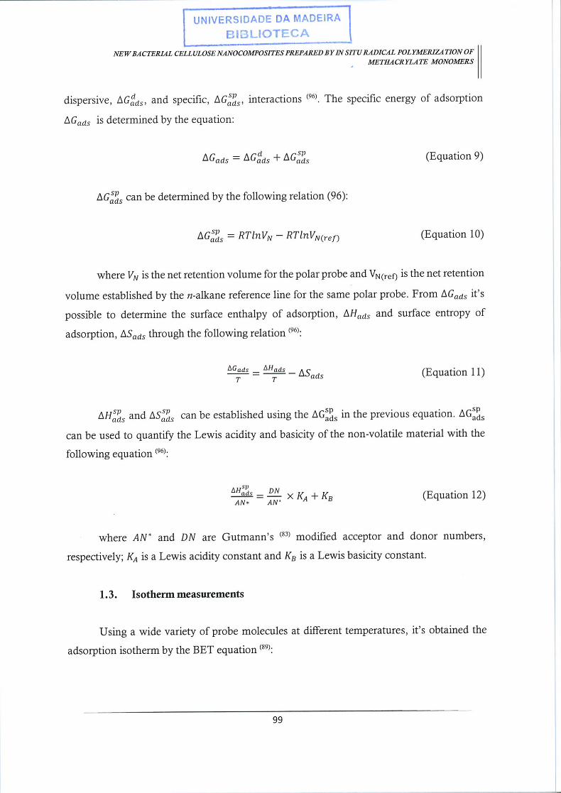
∆𝑮𝒂𝒅𝒔 – Molar energy of adsorption
∆𝑮𝒔𝒑𝒔 – Specific component of the surface energy
∆𝑯𝒂𝒅𝒔𝒔𝒑
– Specific enthalpy of adsorption
∆𝑺𝒂𝒅𝒔𝒔𝒑
– Specific entropy of adsorption
𝑉𝑁 – Net retention volume
𝑨𝑵∗- Acceptor numbers
𝑨𝒎𝒂𝒙 – Adsorption potential
𝑲𝑨 – Acid constant
𝑲𝑩 – Basic constant
𝑺𝑩𝑬𝑻 – Surface area
𝑾𝑨 – Energy of adhesion
𝒂𝒎 – Cross area
𝒏𝒎 – Monolayer capacity
𝒑° - Saturation pressure
𝜸𝑳𝒅 – Dispersive component of the surface energy of the probe molecule
𝜸𝒔 – Surface energy
𝜸𝒔𝒅 – Dispersive component of the surface energy
ADSCs – Adipose-derived stem cells
AEM – 2-Aminoethyl methacrylate
APS – Ammonium persulphate
ATR – FTIR – Attenuated Total Reflection Fourier Transform Infrared Spectroscopy
ATRP – Atom transfer radical polymerization
BC – Bacterial cellulose
BC/PPEGMA – Bacterial cellulose/poly(ethyleneglycolmethacrylate)
BC/PGMA – Bacterial cellulose/poly(glycidylmethacrylate)
BC/PGMA/MBA – Bacterial cellulose/poly(glycidylmethacrylate) crosslinker with N,N’-
methylenebisacrilamide
BCPGMA-CT – Bacterial cellulose/poly(glycidylmethacrylate) after chemical treatment

XIX
BCPGMAMBA-CT – Bacterial cellulose/poly(glycidylmethacrylate) crosslinker with N,N’-
methylenebisacrilamide after chemical treatment
BuMA – Butyl methacrylate
c-di-GMP – Cyclic diguanylic acid
CPMAS 13C NMR – Solid-state Cross-Polarization Magic Angle Spinning Carbon-13
Nuclear Magnetic Resonance
DG – Degradation profile
DMA – Dynamic Mechanical Analysis
DMAEMA – N,N–dimethylaminoethylmethacrylate
Eʹ - Storage modulus
EDX – Energy-Dispersive X-ray Spectroscopy
EOEMA – 2-Ethoxyethyl methacrylate
FE–SEM – Field Emission Scanning Electron Microscopy
Glc – 1 – P – Glucose-1-phosphate
Glc – 6 – P – Glucose-6-phosphate
GMA – Glycidylmethacrylate
GMMA – Glycerol monomethacrylate
HEMA – 2-Hydroxyehtyl methacrylate
IGC – Inverse Gas Chromatograph
MBA - N,N’-Methylenebisacrilamide
MMA – Methyl methacrylate
PAAc – Poly(acrylic acid)
PDE-A and PDE-B – c-di-GMP phosphodiesterases A and B
PEG – Poly(ethylene glycol)
PEGDA – Poly(ethylene glycol)diacrylate
PEGMA – Poly(ethyleneglycol)methacrylate
PGMA – Poly(glycidylmethacrylate)
PVA – Poly(vinyl alcohol)
RAFT – Reversible addition-fragmentation chain transfer polymerization
SW – Swelling ratio
TGA – Thermogravimetric Analysis

XX
UDPG – Uracil diphosphate glucose
UGPase – UDPG-pyrophosphorylase
XRD – X-ray diffraction
𝑫𝑵 – Donor number
𝑵 – Avogadro’s number
𝑹 – Gas constant
𝑻 – Temperature
𝑻𝒅 – Thermal degradation
𝒂 – Area occupied by probe molecule
𝒄 – Heat of sorption
𝒏 – Amount adsorbed
𝒑 – Partial pressure


CHAPTER I
Bacterial cellulose and Nanocomposites world – General
Introduction


NEW BACTERIAL CELLULOSE NANOCOMPOSITES PREPARED BY IN SITU RADICAL POLYMERIZATION OF
METHACRYLATES MONOMERS
2
CHAPTER I. Overview of bacterial cellulose
1. Cellulose as Biopolymer
Originally from Greek and employed for the first time by the Swedish chemist
Berzelius in 1833, polymers (poli, many; meros, parts) are defined as large molecules
(macromolecules) synthesized from simple molecules, monomers, linked by covalent bonds
by polymerization processes (1-3).
Cellulose is the most abundant natural polymer because it is the main component of
the plant cell walls. It can be found in large amount in wood [above 50% (w/w)] and cotton
[above 94% (w/w)] (3). Due to its versatility and properties, cellulose can be applied into
different areas such as pulp and paper production, textiles, construction materials, cosmetics,
pharmaceutical industries as excipient and as a source for biofuel production (4, 5). Through
chemical processes like e.g. methylation and acetylation, cellulose derivatives can be
produced such as cellulose ethers and cellulose esters for other’s applications (3, 5-8). The
increase interest and demand for vegetable cellulose derivatives had augmented the
consumption of wood as a raw material, being the cellulose production approximately 1.5 x
1012 tons per year. This value represents a major fraction of the total biomass produced which
contributes to the deforestation and global environmental issues (4, 9, 10).
Cellulose was described for the first time by Anselm Payen in 1836 as “a resistant fibrous
solid that remains behind after treatment of various plant tissues with acids and ammonia, and after
subsequent extraction with water, alcohol, and ether” (11). It consists in a linear homopolymer
composed by D-glucose monomer glycosidically linked covalently in a β(1-4) conformation
through acetal functions, between the hydroxyl groups of C1 and C4 carbon atoms. It is
attributed a molecular formula of (C6H10O5)n where 𝑛 is the degree of polymerization of
glucose (Figure 1) (9, 11, 12). Two adjacent structural units constitute the basic repeating unit
called disaccharide cellobiose of the linear polymer (9). The linear configuration of cellulose
is stabilized by the intra-chain hydrogen bonds between the hydroxyl groups and the oxygens
of the adjacent ring molecules. Besides that, the interaction between hydroxyls groups and
oxygens of neighboring molecules via inter-chain hydrogen bonds promote the aggregation

NEW BACTERIAL CELLULOSE NANOCOMPOSITES PREPARED BY IN SITU RADICAL POLYMERIZATION OF
METHACRYLATES MONOMERS
3
of multiple cellulose chains resulting into nanofibrils. Combining both intra- and inter-chain
hydrogen bonding, turns the polymer relatively stable and give fibrils high axial stiffness (13).
Cellulose is insoluble in water, with high mechanical resistance and with a chemical
composition of 43.6–45 % carbon, 6.0–6.5 % hydrogen and the remainder oxygen. The length
of fibers depends of the source of cellulose and can range between 100 up to 10,000 nm (4).
Figure 1. Molecular structure of cellulose where inter- (blue) and intra- (red) chain hydrogen bonds dashed lines
[reproduced from (14)].
Cellulose with different morphologies can be obtained from conventional plant fibers
following adequate procedures as pulping processes (mechanical and chemical), acid
hydrolysis, steam explosion, high-intensity ultrasonification, among others (Figure 2) (4).
Figure 2. Morphology characteristics of the different types of cellulose fibers [reproduced from (15)].

NEW BACTERIAL CELLULOSE NANOCOMPOSITES PREPARED BY IN SITU RADICAL POLYMERIZATION OF
METHACRYLATES MONOMERS
4
2. Bacterial Cellulose – Historical Outline
Adrian J. Brown discovered in 1886 a gelatinous membrane as a product of microbial
fermentation of acetic acid from the “vinegar plant “or “mother” (6, 16, 17). Afterwards,
their chemical structure resembles that of native cellulose (cell-wall) through X-ray
diffraction. It was verified a crystallography typical of cellulose I, with two cellobiose units
arranged parallel in units cell and tended to have a specific planer orientation in dried film
(14). This gelatinous membrane was denominated as bacterial cellulose, BC, and the “vinegar
plant”, as Acetobacter (nowadays Gluconacetobacter) xylinum. As an exopolysaccharide, BC was
prompted a number of studies in order to better understand its biosynthesis pathways and
properties (18-20). BC can be produced in laboratory by several genera of bacteria such as
Gluconacetobacter, Entrobacter, Rhizobium, Agrobacterium and Sarcina sp during their
biosynthesis (12). Innumerable bacteria strains have been reported as BC producers. Known
as a gram-negative, aerobic, straight, slightly bent rods or ellipsoidal with dimensions of 0.6
x 4 µm, Gluconacetobacter strains are characterized as BC producers (6). For example,
Gluconacetobacter sacchari was reported as a high efficient producer of BC for the first time by
Trovatti et al (2011) with production yields of 2.7 g/L after 96h (21).
In recent years, BC has gained interest in numerous areas of study and in a variety of
applications due to its unique and specific properties such as high degree of crystallinity (80-
90%), high water retention capacity (99% of own weight), high purity (free hemicellulose and
lignin), high degree of polymerization (above 8000 wherein sometimes getting even 16,000
or 20,000), ultrafine fibrous network, high tensile strength, water holding ability, porosity,
biocompatibility, hydrophilicity, non-toxicity and a relatively simple production and efficient
in terms of costs (9, 11, 17, 22, 23). The BC display a typical Young´s modulus in the range
of 15-35 GPa with a tensile strength ranging between 200-300 MPa (9, 11).
2.1. Biochemistry of Bacterial Cellulose and Biosynthesis Pathways
Extensive research and reviews regarding the metabolic pathways of BC, genetic and
strains of cellulose-producing of BC have been reported (3, 24-27). Gluconacetobacter xylinum
is the most commonly model bacteria strain studied due to its ability to produce cellulose

NEW BACTERIAL CELLULOSE NANOCOMPOSITES PREPARED BY IN SITU RADICAL POLYMERIZATION OF
METHACRYLATES MONOMERS
5
from a wide range of carbon/nitrogen sources, being the rate of cellulose production
proportional to that of cell growth (6, 18, 28-30). Several studies indicate that the carbon
sources present in the medium influence the yield of BC as well as the bacteria strains and
the culture conditions: static and shaking (11, 29-35).
Two metabolic pathways operate in G. xylinum for BC production: the pentose
phosphate cycle (oxidation of carbohydrates) and the citrate acid cycle (oxidation of organic
acid and related compounds). G. xylinum is unable to metabolize glucose in the absence of
oxygen due to the lack or weakly present phosphofrutose kinase, which is required for
glycolysis. Glucose is the most common monosaccharides used as a carbon source in order
to obtain cellulose. Four fundamental enzymatic steps occur (i) phosphorylation of glucose
by glucokinase, (ii) isomerization of glucose-6-phosphate (Glc-6-P) to glucose-1-phosphate
(Glc-1-P) by phosphoglucomutase, (iii) synthesis of UDP-glucose (UDPG) by UDPG-
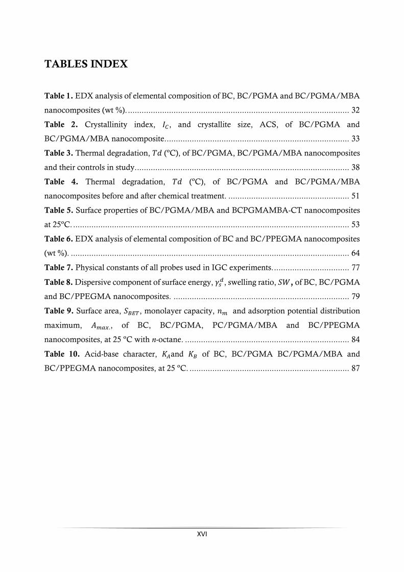
pyrophosphorylase (UGPase) and (iv) cellulose synthase reaction (Figure 3) (6, 18, 24).
UDPG is the immediate sugar nucleotide precursor of cellulose synthesis and for β(1-
4) glucan polymerization reaction. This enzyme allows an improved activity in BC producers
of c.a. 100x orders of magnitude (24). When disaccharides (sucrose or maltose) are used as
carbon source, the biosynthesis of BC initiates with the hydrolysis of disaccharides into
monosaccharides (fructose and glucose). Then, they are metabolized by the bacteria strain.
The molecular mechanisms of glucose polymerization into long and unbranched cellulose
chains are still nuclear, since the pathways of UDPG are relatively not well known. Cyclic
diguanylic acid (c-di-GMP) acts like an allosteric activator of the membrane-bound cellulose
synthase and display an important role in the biosynthesis of BC as a regulatory element (6).
The activity of c-di-GMP phosphodiesterases A and B (PDE-A and PDE-B) stopped the
activity of cellulose synthase. PDE-A cleaves the c-di-GMP to form pGpG which is
degraded, producing two molecules of 5’-GMP. The activity of PDE-A is selectively inhibited
by Ca2+ ions (24).
NEW BACTERIAL CELLULOSE NANOCOMPOSITES PREPARED BY IN SITU RADICAL POLYMERIZATION OF
METHACRYLATES MONOMERS
6
Figure 3. Schematic of the metabolic pathways of Glucanoacetobacter xylinum and the assembly of cellulose molecules into
nanofibrils [reproduced from (6)].
Cellulose is synthesized in microorganisms in two intermediary steps: (i) the
formation of β(1-4) glucan chains and (ii) the assembly and crystallization of cellulose chains.
The cellulose molecules are initially synthesized inside the bacteria and then spun through
cellulose export components to form protofibrils with a diameter c.a. of 2 – 4 nm. A ribbon
shaped microfibril of approximately 80 nm is assembled from these protofibrils. Cellulose
synthase is the catalyst for biosynthesis of cellulose, which polymerizes the glucose units into
the β(1-4) glucan chains (6).
2.2. Structure and Properties of Bacterial Cellulose
Structurally, BC possess an identical molecular formula (C6H10O5)n of the vegetable
cellulose as shown in Figure 1 (9, 11, 12). The process of biosynthesis of BC comprises two
main stages: linear polymerization of the glucose units catalyzed by the enzyme cellulose
synthase, followed by crystallization of the linear chains. The polymerization of β(1-4)
glucan starts from 300 to 10,000 glucose unit in order to form a linear polymer. This linear
polymer chain assembles with other adjacent chains to form aggregates (subfibrils) with 1.5

NEW BACTERIAL CELLULOSE NANOCOMPOSITES PREPARED BY IN SITU RADICAL POLYMERIZATION OF
METHACRYLATES MONOMERS
7
nm width, that further combine with another one to form semi-crystalline nanofibrils with
3.5 nm width. The nanofibrils combine between them, originating bundles and then the
macroscopic ribbon shapes (Figure 4) (9, 11). The size of nanofibrils and their spatial
arrangement strongly influences the BC crystallinity, which depends on two factors: source
(organism) and synthesis conditions (9, 11).
Figure 4. A SEM of freeze-dried surface of bacterial cellulose gel [reproduced from (14)].
Cellulose type I is the most common form biosynthesized and detected by X-rays
(Figure 5), consisting in two β(1-4) glucan chains oriented parallel to each other in a
monocyclic unit cell and arranged uniaxially (11). Native cellulose exhibited two different
structural crystals, cellulose Iα and Iβ. The cellulose Iα is crystallized in larger-size nanofibrils
while cellulose Iβ is formed in smaller-size nanofibrils being thermodynamically more stable
(3). Also, the allomorph forms differ in the unit cell: Iα show a triclinic unit and Iβ a
monoclinic unit cell. The ratio 𝐼𝛼 𝐼𝛽⁄ depends from species to species (3). The aggregates form
nanofibrils of width approximately 100 nm denominated fibrillary bands and these form the
tridimensional network structure. The 3-D micro and nanofibrillar structure of bacterial
cellulose influence the majority of its properties (9, 11). Bacterial cellulose, compared with
vegetal cellulose, had higher degree of polymerization, a higher degree of crystallinity and
higher size of crystallites (9, 11). These structural differences induce considerable differences
in terms of physical properties, particularly in terms of the mechanical strength (9, 11).

NEW BACTERIAL CELLULOSE NANOCOMPOSITES PREPARED BY IN SITU RADICAL POLYMERIZATION OF
METHACRYLATES MONOMERS
8
Figure 5. (A) X-ray patterns of BC films and (B) View along the direction 4 (i.e. [1ī0] for Iα and [010] for Iβ) and the
displacement of the hydrogen bonding sheets: blue Iα and red Iβ [reproduced from (3, 13)].
BC can be obtained in static and agitated culture media showing three different forms:
membrane, irregular shapes and sphere-like particles (Figure 6). BC membranes are produced
in static conditions at the air-liquid medium interface and have been commonly used for
different applications due to their suitable properties.
In contrast, irregular shapes and spheres of BC are produced under agitated culture.
Typically, BC produced in agitated culture media showed lower cellulose Iα content, lower
Young’s modulus, higher swelling and have potential to be used in food, healthcare and
materials applications (36).
Figure 6. Different forms of BC produced by Gluconacetobacter sp. (A) Membrane, (B) Irregular forms, (C) Sphere-like
particles [reproduced from (36)].
(A) (B)

NEW BACTERIAL CELLULOSE NANOCOMPOSITES PREPARED BY IN SITU RADICAL POLYMERIZATION OF
METHACRYLATES MONOMERS
9
2.3. Application of Bacterial Cellulose
BC is one of the finest examples of Nature’s art, with a singular morphology and
unique properties, and is gaining increasing interest as an excellent biopolymeric material to
be employed in a wide range of applications in different areas (Figure 7), namely in the:
(i) Biomedical and biotechnology fields: artificial skin, artificial blood vessels, artificial
cornea, heart valve prosthesis, artificial urethra, artificial bone, artificial cartilage,
artificial porcine knee menisci and delivery drug, hormone and protein,
reinforcement material for wound dressings, scaffolds for tissue engineering and soft
tissue replacement (7, 11);
(ii) Environmental and agricultural fields: dye decolorization, biadsorbent for heavy
metal removal and improvement of quality of soil with application of BC (11);
(iii) Electronic field: reinforcement transparent flat-panel (11);
(iv) Food fields: nata-de-coco manufacture (11);
(v) Industrial fields: papermaking (11);
(vi) Reinforcement as composites (11).
Figure 7. Applications of bacterial cellulose: A – Tubes (blood vessel); B – Branched tube fermented on a branched
silicone tube (vascular grafts) [reproduced from (36)]; C –Dressing (wound dressing) [reproduced from (7)]; D –
Incorporation of AgNO3 nanoparticles (antimicrobial activity); E –Paper (filter paper) and F – Colored with aniline blue
dye (bioadsorbent) [reproduced from (11)].
NEW BACTERIAL CELLULOSE NANOCOMPOSITES PREPARED BY IN SITU RADICAL POLYMERIZATION OF
METHACRYLATES MONOMERS
10
3. Bacterial Cellulose Nanocomposites
Nanocomposites were defined for the first time as “a multi-phase compound in which one
of the phases has a length scale in the nanometer range” by Roy and co-workers in 1980’s. A more
practical definition was introduced by Komarneni in 1992 that defined nanocomposites as
“composites of more than a Gibbsian solid phase where at least one-dimension is in the nanometer range
and typically all solid phases are in the 1-20 nanometer range” (37).
BC nanocomposites have been studied over time due to the excellent properties of
these materials. BC can be manipulated in order to improve mechanical performance and
biocompatibility, and can be employed in many forms from nano to macro scales for various
applications. They can be applied into plant biomimicking, biomedical, electrically
conductive materials, catalysis, optical and luminescent materials, proton conductive,
separating materials, antimicrobial materials, thermos-responsive, among many another’s
(36).
Different approaches for the preparation of BC nanocomposites have been reported
in literature (38-41), including: (i) incorporation of desire components in culture medium
during BC biosynthesis; (ii) In situ polymerization of monomers inside of the BC network and
(iii) blending with other polymeric materials. The first’s two approaches are applied in the in
situ preparation of nanocomposites and are described below.
3.1. BC nanocomposites through BC biosynthesis
One approach for the BC nanocomposites obtainment is the introduction of polymers
and/or desirable compounds into the culture medium during BC biosynthesis. This way, the
nanocomposite will be produced at the same time as the BC fibrils, through assembly of the
desire compounds. In fact, many compounds/polymers have already been incorporated in
BC culture media in order to obtain BC nanocomposites. Compounds/polymers such as
chitosan (41), carbon fibers, aloe-vera (42), glyoxal (38) or poly (vinyl alcohol) (PVA) (39).
Phisalaphong et al (2008) reported a new BC/chitosan film obtained through the
supplement of low molecular weight chitosan into the culture medium. The obtained
BC/chitosan nanocomposite exhibited a denser homogeneous fibril structure with smaller

NEW BACTERIAL CELLULOSE NANOCOMPOSITES PREPARED BY IN SITU RADICAL POLYMERIZATION OF
METHACRYLATES MONOMERS
11
pore diameter and higher surface area (Figure 8). Structurally, the introduction of chitosan
as a supplement into the culture medium didn’t change BC properties such as water vapor
transmission rates, average crystallinity index and antimicrobial ability (41).
A new nanostructured film of BC/aloe vera was reported by Saibuatong et al (2010)
through incorporation of aloe vera into the culture medium during biosynthesis of BC. This
assembly resulted in the enhancement of the mechanical properties, water absorption
capacity, water vapor transmission rate and crystallinity index. The BC/aloe vera
nanocomposite displayed an excellent compatibility and physical properties which make it a
good material with a wide range of application in medical areas (42).
Figure 8. SEM images of surface morphology of (A) BC and (B) BC/Chitosan film in dry form [reproduced from (41)].
Castro el at (2014) reported the production of BC/PVA nanocomposite films by
adding PVA to the culture media followed by chemical crosslinking. The chemical
crosslinking, after the biosynthesis, avoid the loss of the PVA matrix during the purification
steps improving the functional properties of the nanocomposites, reflected by the good
interaction between the PVA matrix and the reinforcement BC phases (Figure 9) (39).
NEW BACTERIAL CELLULOSE NANOCOMPOSITES PREPARED BY IN SITU RADICAL POLYMERIZATION OF
METHACRYLATES MONOMERS
12
Figure 9. (A) Visual appearance of PVA: (a) without BC (b) BC/PVA nanocomposites and (c) BC/PVA nanocomposites
after chemical crosslinker and (B) Stress-strain behavior of BC/PVA [reproduced from (39)].
Besides that, these authors reported the glyoxalization of BC films, by introducing
glyoxal into the culture medium during the BC biosynthesis. Glyoxalizated BC films showed
an increase in their hydrophobicity improving the BC properties while keeping its
crystallinity (38).
3.2. BC nanocomposites through in situ polymerization
Another approach to prepare BC nanocomposites is the in situ polymerization inside
the BC network. This method involves the absorption of a solution with the desire monomer,
the initiator and crosslinker (if used in the polymerization) into the BC network, followed by
the polymerization reaction. This approach is quite interesting because the final properties of
the nanocomposites can be simply tailored by using different monomers or monomers
mixtures.
In situ polymerization had already been used for the preparation of BC
nanocomposites with various methacrylates monomers such as 2-hydroxyehtyl methacrylate
(HEMA) (22, 40), 2-aminoethyl methacrylate (AEM) (43), glycerol monomethacrylate
(GMMA) (22) and 2-ethoxyethyl methacrylate (EOEMA) (22).
(A) (B)

NEW BACTERIAL CELLULOSE NANOCOMPOSITES PREPARED BY IN SITU RADICAL POLYMERIZATION OF
METHACRYLATES MONOMERS
13
Methacrylate monomers are versatile to prepare a series of BC/methacrylate
nanocomposites with different properties, due to their chemical structure variability, great
availability and easy polymerization (22).
Figueiredo et al (2015) reported the preparation of novel BC nanocomposites through
in situ polymerization of 2– aminoethyl methacrylate (AEM). Variable amounts of AEM and
N,N’-methylenebis(acrylamide) (MBA) used as a crosslinker were impregnated into the BC
membrane before the polymerization step. The BC/PAEM and BC/PAEM/MBA
nanocomposites were very homogeneous suggesting a good distribution of PAEM inside the
BC network, and were more translucent than native BC [Figure 10 (A)]. In addition, these
nanocomposites showed improved thermal stability and mechanical properties [Figure 10
(B)], a decrease in crystallinity and an increase of swelling ability [Figure 10 (C)] (43).
Furthermore, BC/PAEM nanocomposites (without crosslinker) display antibacterial
activity when in contact with a bacterial suspension of a bioluminescent Escherichia coli
demonstrating to be a good nanomaterial with properties for potential application as
antimicrobial wound dressing.
Figure 10. (A) Visual appearance of BC/PAEM and all BC/PAEM/MBA nanocomposites. (B) TGA thermograph of
BC/PAEM nanocomposites. (C) Swelling of wet BC, BC/PAEM and BC/PAEM/MBA nanocomposites [reproduced
from (43)].

NEW BACTERIAL CELLULOSE NANOCOMPOSITES PREPARED BY IN SITU RADICAL POLYMERIZATION OF
METHACRYLATES MONOMERS
14
In a different study (40), 2-hydroxyethyl methacrylate (HEMA) was used to prepare
a series of bacterial cellulose/poly(2-hydroxyethyl methacrylate) nanocomposites films by in
situ radical polymerization using poly(ethylene glycol) diacrylate (PEGDA) as a crosslinker
(40). As in the case of BC/PAEM nanocomposites, the translucency and homogeneity of the
BC/PHEMA and BC/PHEMA/PEGDA nanocomposite increased with the amount of
monomer and crosslinker polymerized inside the BC network [Figure 11 (A)]. The
nanocomposites exhibited improved thermal properties, good swelling ability and a decrease
in the storage tensile modulus. Furthermore, the nanocomposites are non-cytotoxic
providing favorable cell environment for optimal adhesion and proliferation of adipose-
derived stem cells (ADSCs) [Figure 11 (B)]. These makes them promising material for several
biomedical applications as for the design of 3D matrices with the purpose of maintaining a
cellular niche for stem-mediated tissue regeneration (40).
Figure 11. (A) Visual aspect of BC, BC/PHEMA and BC/PHEMA/PEGDA films and (B) ADSCs proliferation in
contact with BC and BC/PHEMA/PEDGA films [reproduced from (40)].
3.3. Acrylate monomers used in BC nanocomposite
Acrylate monomers such as acrylic acid, 2-ethylhexyl acrylate, 2-hydroxyethyl
methacrylate (22), methyl methacrylate (MMA), butyl methacrylate (BuMA) (44), N,N–
dimethylaminoethyl methacrylate (DMAEMA) (45) are typically used for polymerization
reaction due their high reactivity.
(B) (A)
NEW BACTERIAL CELLULOSE NANOCOMPOSITES PREPARED BY IN SITU RADICAL POLYMERIZATION OF
METHACRYLATES MONOMERS
15
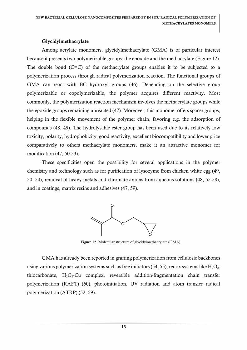
Glycidylmethacrylate
Among acrylate monomers, glycidylmethacrylate (GMA) is of particular interest
because it presents two polymerizable groups: the epoxide and the methacrylate (Figure 12).
The double bond (C=C) of the methacrylate groups enables it to be subjected to a
polymerization process through radical polymerization reaction. The functional groups of
GMA can react with BC hydroxyl groups (46). Depending on the selective group
polymerizable or copolymerizable, the polymer acquires different reactivity. Most
commonly, the polymerization reaction mechanism involves the methacrylate groups while
the epoxide groups remaining unreacted (47). Moreover, this monomer offers spacer groups,
helping in the flexible movement of the polymer chain, favoring e.g. the adsorption of
compounds (48, 49). The hydrolysable ester group has been used due to its relatively low
toxicity, polarity, hydrophobicity, good reactivity, excellent biocompatibility and lower price
comparatively to others methacrylate monomers, make it an attractive monomer for
modification (47, 50-53).
These specificities open the possibility for several applications in the polymer
chemistry and technology such as for purification of lysozyme from chicken white egg (49,
50, 54), removal of heavy metals and chromate anions from aqueous solutions (48, 55-58),
and in coatings, matrix resins and adhesives (47, 59).
Figure 12. Molecular structure of glycidylmethacrylate (GMA).
GMA has already been reported in grafting polymerization from cellulosic backbones
using various polymerization systems such as free initiators (54, 55), redox systems like H2O2-
thiocarbonate, H2O2-Cu complex, reversible addition-fragmentation chain transfer
polymerization (RAFT) (60), photoinitiation, UV radiation and atom transfer radical
polymerization (ATRP) (52, 59).

NEW BACTERIAL CELLULOSE NANOCOMPOSITES PREPARED BY IN SITU RADICAL POLYMERIZATION OF
METHACRYLATES MONOMERS
16
Poly(ethylene glycol)methacrylate
Poly(ethylene glycol) (PEG) exhibited unique advantageous properties to be exploited
in biomedical and biotechnological devices due their low-toxicity, absence of antigenicity
and immunogenicity and inherent ability to prevent protein adsorption (61). Poly(ethylene
glycol)methacrylate (PEGMA), a PEG derivative, is another attractive methacrylate
monomer due to its amphiphilic nature. This property comes from its water-soluble PEG
side chain with a pendant hydroxyl group and its hydrophobic methacrylate group (Figure
13) (62). Due to these properties, PEGMA is one of the most attractive monomers used to
prepare biomedical materials such as drug carrier (63), microspheres for transient vascular
embolization (62), protein adsorption and immobilization of polysaccharides (64). It is also,
used for heavy metals removal (65, 66).
Different approaches of polymerization of PEGMA have been reported in the
literature such as photopolymerization (63, 67) and ATRP (61).
Figure 13. Molecular structure of poly(ethylene glycol) methacrylate (PEGMA).
4. Aim of the Study
In view of all issues described above, the aim of the present study is to prepare novel
BC nanocomposites for potential biomedical application through in situ free radical
polymerization of glycidylmethacrylate (GMA) and poly(ethyleneglycol)methacrylate
(PEGMA) using N,N’-methylenebis(acrylamide) (MBA) as crosslinker.
All BC nanocomposites were characterized in terms of structure, morphology,
thermal stability, water absorption, mechanical and surface properties by Attenuated Total
Reflection Fourier Transform Infrared Spectroscopy (ATR-FTIR), CPMAS 13C NMR, X-ray
diffraction (XRD), Energy-Dispersive X-ray Spectroscopy (EDX), Field Emission Scanning
NEW BACTERIAL CELLULOSE NANOCOMPOSITES PREPARED BY IN SITU RADICAL POLYMERIZATION OF
METHACRYLATES MONOMERS
17
Electron Microscopy (FE-SEM), Swelling (SW), Thermogravimetric Analysis (TGA),
Dynamic Mechanical Analysis (DMA) and Inverse Gas Chromatography.
The present manuscript is laid out in four different chapters, each one corresponding
to scientific papers to be submitted briefly.
CHAPTER II
New bacterial cellulose/poly(glycidylmethacrylate)
nanocomposites films by in situ free radical polymerization

NEW BACTERIAL CELLULOSE NANOCOMPOSITES PREPARED BY IN SITU RADICAL POLYMERIZATION OF
METHACRYLATE MONOMERS
20
CHAPTER II. New bacterial cellulose/poly(glycidylmethacrylate)
nanocomposites films by in situ free radical polymerization
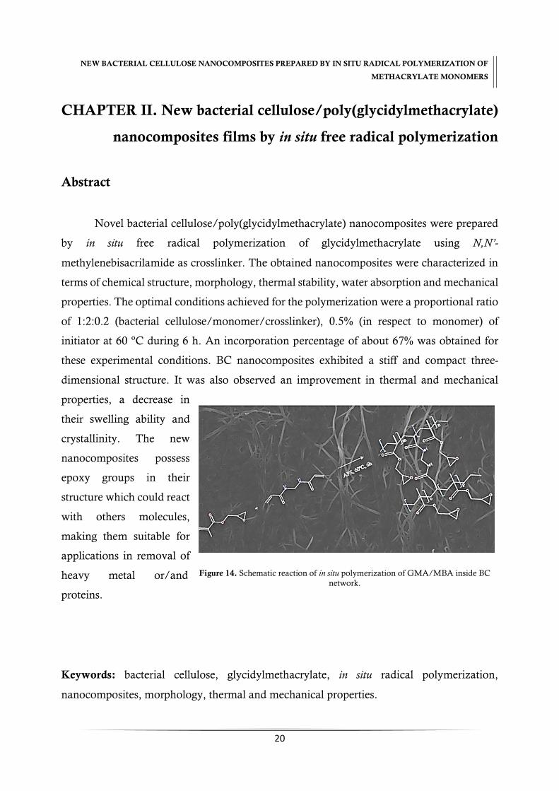
Abstract
Novel bacterial cellulose/poly(glycidylmethacrylate) nanocomposites were prepared
by in situ free radical polymerization of glycidylmethacrylate using N,N’-
methylenebisacrilamide as crosslinker. The obtained nanocomposites were characterized in
terms of chemical structure, morphology, thermal stability, water absorption and mechanical
properties. The optimal conditions achieved for the polymerization were a proportional ratio
of 1:2:0.2 (bacterial cellulose/monomer/crosslinker), 0.5% (in respect to monomer) of
initiator at 60 ºC during 6 h. An incorporation percentage of about 67% was obtained for
these experimental conditions. BC nanocomposites exhibited a stiff and compact three-
dimensional structure. It was also observed an improvement in thermal and mechanical
properties, a decrease in
their swelling ability and
crystallinity. The new
nanocomposites possess
epoxy groups in their
structure which could react
with others molecules,
making them suitable for
applications in removal of
heavy metal or/and
proteins.
Keywords: bacterial cellulose, glycidylmethacrylate, in situ radical polymerization,
nanocomposites, morphology, thermal and mechanical properties.
Figure 14. Schematic reaction of in situ polymerization of GMA/MBA inside BC
network.

NEW BACTERIAL CELLULOSE NANOCOMPOSITES PREPARED BY IN SITU RADICAL POLYMERIZATION OF
METHACRYLATE MONOMERS
21
1. Introduction
Bacterial cellulose (BC) is a pure form of cellulose produced by several bacteria
including Gluconacetobacter sacchari which are a gram-negative, rod shaped and strictly aerobic
bacteria (16, 17, 21-23). BC confers unique and specific properties such as high degree of
crystallinity (80-90%), high water retention capacity (99%), ultrafine fibrous network, high
tensile strength, biocompatibility, hydrophobicity and non-toxicity. These properties stand
out BC as a good polymer to study having a relatively simple production thus cost efficient
(17, 22, 23, 68).
However, BC possess some limitations that restrict its applications as lack of
antibacterial properties, optical transparency and stress bearing capability (10). To overcome
these limitations, BC nanocomposites have been prepared, consisting in a BC network and
reinforcement materials (6, 10, 11, 69). The reinforcement materials improve BC’s biological
and physiochemical properties (10). BC nanocomposites have been synthetized through in
situ biosynthesis (38, 42), blended with other polymeric materials (12) and by in situ radical
polymerization (40, 43).
In the present study, it is exploited the in situ radical polymerization of methacrylate
monomers into the BC network. Glycidylmethacrylate (GMA) is a methacrylate monomer
with two polymerizable groups: the epoxide and the methacrylate groups (double bonds)
which offers a wide range of industrial applications in the polymer chemistry and technology
namely for purification of lysozyme from chicken white egg (49, 50, 54), removal of heavy
metals and chromate anions from aqueous solutions (48, 55-58), in coatings, matrix resins
and adhesives (47, 59). Besides that, GMA show a relatively low toxicity, polarity,
hydrophobicity and low price which make it an attractive monomer (47, 50-53). GMA has
been reported in grafting polymerization on cellulosic backbones using various
polymerization systems such as free initiators (54, 55), redox systems like H2O2-
thiocarbonate, H2O2-Cu complex, reversible addition-fragmentation chain transfer
polymerization (60), photoinitiation, UV radiation and atom transfer radical polymerization
(52, 59).
NEW BACTERIAL CELLULOSE NANOCOMPOSITES PREPARED BY IN SITU RADICAL POLYMERIZATION OF
METHACRYLATE MONOMERS
22
This work report the development of BC nanocomposite through in situ radical
polymerization of GMA into BC network using N,N’-methylenebisacrilamide (MBA) as
crosslinker and ammonium persulphate (APS) as initiator. The optimal conditions of
polymerization were determined and the new BC nanocomposites membranes were
characterized in term of structural, morphological, water ability, thermal, and viscoelastic
properties.
2. Methodology
2.1. Material
Wet bacterial cellulose (BC) membranes produced by Gluconacetobacter sacchari (70)
using standard conditions were used. Glycidylmethacrylate (GMA, 97%, with 100 ppm of
monomethyl ether hydroquinone as inhibitor), N,N’-methylenebisacrilamide (MBA, ≥
99.5%) and ammonium persulphate (APS, 98%) were used as monomer, crosslinker and
initiator, respectively. All reagents were purchased from Sigma-Aldrich and used as received.
Distilled water was used as solvent in all steps of the procedure.
2.2. BC nanocomposites preparation
2.2.1. GMA and GMA/MBA polymerization
A solution of 500 mg of GMA (monomer) and APS (initiator) (0.5% w/w GMA) in
distilled water and N2 atmosphere was prepared. This reaction mixture was placed at 60 ºC
for 6 hours to prepare the PGMA/MBA polymer. PGMA/MBA polymer was prepared
using the same conditions used for PGMA, but with addition of 20% (𝑤𝑐𝑟𝑜𝑠𝑠𝑙𝑖𝑛𝑘𝑒𝑟 𝑤𝑚𝑜𝑛𝑜𝑚𝑒𝑟⁄ )
MBA crosslinker to the monomer and initiator solution.

NEW BACTERIAL CELLULOSE NANOCOMPOSITES PREPARED BY IN SITU RADICAL POLYMERIZATION OF
METHACRYLATE MONOMERS
23
The obtained PGMA/MBA polymer after the reaction time was washed several times
with water, while PGMA was washed with methanol, and both were dried at 40 ºC
overnight, and stored after that in a desiccator until their characterization.
2.2.2. In situ free radical polymerization of GMA and GMA/MBA into BC
The in situ free radical polymerization of GMA inside the BC network was adapted
from the procedure described by Figueiredo et al. (43). An aqueous reaction mixture
containing the monomer (𝑤 𝑤𝑑𝑟𝑦 𝐵𝐶⁄ ) in a ratio of 1:2, 0.5% initiator (𝑤 𝑤𝑚𝑜𝑛𝑜𝑚𝑒𝑟⁄ ) and 20%
crosslinker (𝑤𝑐𝑟𝑜𝑠𝑠𝑙𝑖𝑛𝑘𝑒𝑟 𝑤𝑚𝑜𝑛𝑜𝑚𝑒𝑟⁄ ) (when used in reaction) was prepared. Wet BC
membranes were weight and about 60% of their water content was drained. The drained
membranes and reaction mixture were purged in N2 for 30 min. After that the reactional
mixture was added to the BC membranes and left for 1 hour at room temperature to occur
the incorporation of the monomer (and of the initiator and crosslinker) inside the BC
network. Then, the polymerization reaction take place in an oil bath for 6 hours at 60 ºC.
The obtained nanocomposite membranes were washed with distilled water during 1 hour for
8 times, dried at 40 ºC and stored in a desiccator until their characterization.
The percentage of polymer incorporation in the BC network was determined by the
sample increasing weight after polymerization and was calculated according to Eq. (1):
% 𝐼𝑛𝑐𝑜𝑟𝑝𝑜𝑟𝑎𝑡𝑖𝑜𝑛 = 𝑤𝑚𝑜𝑑𝑖𝑓𝑖𝑒𝑑 𝐵𝐶−𝑤 𝐵𝐶
𝑤 𝐵𝐶 × 100 (Equation 1)
where 𝑊 𝐵𝐶 is the weight of dry bacterial cellulose (g) and 𝑊𝑚𝑜𝑑𝑖𝑓𝑖𝑒𝑑 𝐵𝐶 is the weight
of dry bacterial cellulose after in situ free radical polymerization (g).

NEW BACTERIAL CELLULOSE NANOCOMPOSITES PREPARED BY IN SITU RADICAL POLYMERIZATION OF
METHACRYLATE MONOMERS
24
2.3. BC nanocomposites characterization
2.3.1. Infrared Spectroscopy
The infrared spectra were obtained using Attenuated Total Reflection Fourier
Transform Infrared Spectroscopy (ATR-FTIR) using a Perkin Elmer FTIR System Spectrum
BX spectrophotometer equipped with a single horizontal Golden Gate ATR cell after 32
scans in the 4000−500 cm−1 range with a resolution of 4 cm−1.
2.3.2. Energy-dispersive X-ray spectroscopy
The semi-quantitative elemental chemical compositions of the samples were
determined by energy-dispersive X-ray spectroscopy (EDX). Semi-quantitative analyses (wt.
%) were done for elements (carbon, oxygen and nitrogen) and the 𝐶 𝑂⁄ ratio was obtained.
EDX experiments were conducted at an accelerated voltage of 5 kV in a Hitachi SU 8090
equipment.
2.3.3. Scanning electron microscopy
Scanning electron micrographs of the surface samples were obtained by Scanning
Electron Microscopy (SEM), with a HR-FESEM SU-70 Hitachi equipment operating at 1.5
kV operating in the field emission mode. Samples were deposited on a steel plate and coated
with carbon before analysis.
2.3.4. X-ray diffraction
The X-ray diffraction (XRD) measurements were carried out with a Phillips X’pert
MPD diffractometer using Cu Kα radiation. The peaks were deconvoluted using Pearson VII
peak functions (Peakfit software) for crystallinity index, 𝐼𝐶, appearance crystal size (ACS)
(38):
NEW BACTERIAL CELLULOSE NANOCOMPOSITES PREPARED BY IN SITU RADICAL POLYMERIZATION OF
METHACRYLATE MONOMERS
25
𝐼𝐶 = 1 −𝐼𝑎𝑚
𝐼002 𝑥 100 % (Equation 2)
where 𝐼𝑎𝑚 is the maximum peak intensity at 2θ around 22º, representing the
crystalline region, and 𝐼002 is the minimum peak intensity at 2θ around 18º, representing the
amorphous region. The ACS was calculated using Scherrer´s formula:
𝐴𝐶𝑆 =(0.9𝜆)
𝐹𝑊𝐻𝑀 cos 𝜃 (Equation 3)
where FWHM is the width of the peak at half the maximum height.
2.3.5. 13Carbon Nuclear Magnetic Resonance
Solid-state Cross-Polarization Magic Angle Spinning 13Carbon Nuclear Magnetic
Resonance (CPMAS 13C NMR) spectra were recorded on a Bruker Avance III 400
spectrometer operating at a B0 field of 9.4 T using 9 kHz MAS with proton 90° pulse of 3
microseconds and a time between scans of 3 seconds. 13C CPMAS NMR spectra were
acquired using a contact time of 2000 (2000) microseconds. 13C chemical shifts were
referenced with respect to glycine (C=O at 176.03 ppm).
2.3.6. Swelling ratio
The swelling ratio (𝑆𝑊) of the membranes was determined by immersing the samples
in distilled water at room temperature with a minimum of three replicas. The weight increase
was periodically measured during 48 hours. For each measurement, the samples were taken
out of the water, their wet surfaces immediately wiped dry in filter paper, weighted, and then
re-immersed. The SW was calculated using the equation:
𝑆𝑊(%) =(𝑊𝑠−𝑊𝑑)
𝑤𝑑 × 100% (Equation 4)

NEW BACTERIAL CELLULOSE NANOCOMPOSITES PREPARED BY IN SITU RADICAL POLYMERIZATION OF
METHACRYLATE MONOMERS
26
where, 𝑊𝑆 is the samples weight after swelling and 𝑊𝑑 is the weight of dry sample
before swelling.
2.3.7. Thermogravimetric analyses
Thermogravimetric analysis (TGA) were carried out with a Shimadzu TGA 50
analyzer equipped with a platinum cell. Samples were heated at a constant rate of 10 °C/min,
from room temperature to 800 °C, under a nitrogen flow of 20 mL/min.
2.3.8. Dynamic mechanical analyses
Dynamic mechanical analyses were performed using tension as deformation mode
(single strain) on a Tritec 2000 DMA (Triton Technologies). For the temperature sweeps, a
ramp rate of 2 ºC/min was used and samples were heated from –100 to 200 ºC, at a frequency
of 1 and 10 Hz, with a displacement of 0.005 mm.
3. Results and Discussion
3.1. In situ free radical polymerization reaction
Novel BC nanocomposites were obtained through in situ free radical polymerization
of GMA into the BC network using MBA as crosslinker and APS as initiator. A schematic
illustration of in situ polymerization reaction of GMA inside the BC network is illustrated in
Figure 15. The in situ radical polymerization of GMA occurs through a homolytic cleavage
via C=C, initiated by free radical provoked by the dissociation of APS (71).
The crosslinking of PGMA with MBA allows a better incorporation of polymer inside
the BC network and consequently the retention of more polymer avoiding its removal
through washing (40, 72). This incorporation can change the morphology and properties of
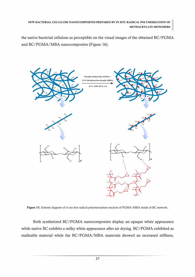
NEW BACTERIAL CELLULOSE NANOCOMPOSITES PREPARED BY IN SITU RADICAL POLYMERIZATION OF
METHACRYLATE MONOMERS
27
the native bacterial cellulose as perceptible on the visual images of the obtained BC/PGMA
and BC/PGMA/MBA nanocomposites (Figure 16).
Figure 15. Scheme diagram of in situ free radical polymerization reaction of PGMA/MBA inside of BC network.
Both synthetized BC/PGMA nanocomposites display an opaque white appearance
while native BC exhibits a milky white appearance after air drying. BC/PGMA exhibited as
malleable material while the BC/PGMA/MBA materials showed an increased stiffness,

NEW BACTERIAL CELLULOSE NANOCOMPOSITES PREPARED BY IN SITU RADICAL POLYMERIZATION OF
METHACRYLATE MONOMERS
28
being difficult to remove from the Erlenmeyer after polymerization, which predict new
thermal, morphologic and surface properties.
Figure 16. Visual images of (a) BC, (b) BC/PGMA and (c) BC/PGMA/MBA nanocomposites.
3.2. Optimization of the in situ free radical polymerization
The effects of the reaction time and temperature, and monomer, initiator and
crosslinker amount on the incorporation yield of the methacrylate polymer were studied in
order to establish the optimal conditions for the in situ free radical polymerization of GMA
and GMA/MBA inside of the BC network. The goal of this study was to obtain the
maximum incorporation of polymer and crosslinker inside BC tridimensional network.
The PGMA and PGMA/MBA incorporation percentage was calculated through
Equation 1. Figure 17 shows the results achieved for the different conditions tested. The in
situ free radical polymerizations were carried out for different reaction times, namely 60, 180
and 360 min. As shown in Figure 17 (A), the highest polymer incorporation was attained for
6 hours of reaction for BC/PGMA and BC/PGMA/MBA nanocomposites with 60.8% and
67.3% of polymer incorporation in BC network, respectively. The effect of temperature on
polymerization was evaluated at 50, 60 and 70 ºC and it was concluded that 60 ºC is the
optimum temperature in the range considered [Figure 17 (B)]. The diffusion of monomer,
initiator and crosslinking from the solution to the BC membrane influences the extension of

NEW BACTERIAL CELLULOSE NANOCOMPOSITES PREPARED BY IN SITU RADICAL POLYMERIZATION OF
METHACRYLATE MONOMERS
29
the polymerization reaction (56). Thus, it’s probable that the number of free radicals increase
with the temperature increase (56). Figures 17 (C), (D) and (E) show the effects of the amount
of initiator, BC/PGMA and BC/PGMA/MBA ratio, respectively. The best concentration
of initiator, BC/PGMA and BC/PGMA/MBA molar ratio (considering the amount of
polymer and crosslinker incorporated) tested were 0.5%, (1:2) and (1:2:0.2), respectively. The
initiator concentration is referred by other authors as a crucial factor for a successful of
polymerization due to the formation of free radicals in solution (73). A lower incorporation
percentage at lower concentration of initiator may be related with the deficit of the free
radicals and at higher concentration due to the self-combination of the free radicals, causing
a failure of chain initiator or enhanced rate of termination which balances the rate of
propagation (56).
Figure 17. Effect of reaction time (A), temperature (B), initiator (C), monomer (D) and crosslinker (E) amount in BC in
situ free radical polymerization.

NEW BACTERIAL CELLULOSE NANOCOMPOSITES PREPARED BY IN SITU RADICAL POLYMERIZATION OF
METHACRYLATE MONOMERS
30
3.3. BC nanocomposites characterization
Of all ratios of BC/PGMA and BC/PGMA/MBA nanocomposites obtained, the
ratio of (1:2) and (1:2:0.2) were chosen to be characterized as they were where the maximum
ratio polymerization was achieved.
3.3.1. Infrared and X-ray Spectroscopy
ATR–FTIR analysis was performed in order to confirm the occurrence of the
polymerization inside of the BC tridimensional network. Figure 18 shows the ATR–FTIR
spectra of BC, BC/PGMA and BC/PGMA/MBA nanocomposites.
Figure 18. ATR-FTIR of native BC, BC/PGMA and BC/PGMA/MBA nanocomposites.

NEW BACTERIAL CELLULOSE NANOCOMPOSITES PREPARED BY IN SITU RADICAL POLYMERIZATION OF
METHACRYLATE MONOMERS
31
The BC characteristic peaks are visible in all BC nanocomposites spectra at 3338,
2896, 1448, 1148, 1108 and 1060-1028 cm-1 corresponding to the free stretching vibration of
the (O–H) group, stretching vibration (C–H), in plan bending (O–H), stretching (C–O–C),
deformation (C–H), stretching (C–C) ring of cellulose and stretching (C–O), respectively (43).
The two peaks at 3296 cm-1 and 710 cm-1 correspond to the monoclinic Iα allomorph and the
peaks at 3240 cm-1 and 750 cm-1 were attributed to the triclinic Iβ form of BC (38). The
appearance of peaks at 1724, 844 and 754 cm-1, attributed to the vibrations of ester carbonyl
group (C=O) and epoxy group of the PGMA chains, respectively, confirms the incorporation
of PGMA in the BC network.
The absence of peak at 1660 cm-1 [stretching of methacrylate group (C=C)] indicates
that the in situ free radical polymerization occurred with success. The presence of the epoxy
groups of PGMA confirms the in situ polymerization of PGMA into BC only via C=C. The
addition of crosslinker, MBA, is visible by the peaks at 1650, 1450 and 1390 cm-1 that
correspond to (N–H) and (C–N) acrylamide units. Thus, the characteristic peaks of polymer
and crosslinker observed in the BC nanocomposites spectra evidences their incorporation
into BC network.
The elemental composition of the BC/PGMA and BC/PGMA/MBA
nanocomposites was determined by EDX analysis (Table 1). The major elements in all
samples were carbon (C) and oxygen (O). Based on the molecular formula, GMA and BC
have a 𝐶 𝑂⁄ ratio of 1.79 and 0.75, respectively, and MBA has a 𝐶 𝑂⁄ ratio of 3.02. It´s
expected that the C amount in BC/PGMA nanocomposite increase due to the PGMA
incorporation. According to the obtained results, an increase for 𝐶 𝑂⁄ ratio was observed in
the BC/PGMA nanocomposite. By the other hand, was observed an increase of N amount
in BC/PGMA/MBA nanocomposites with a 𝐶 𝑁⁄ ratio of 6.45. These data evidence the
success incorporation of PGMA and MBA into the BC network and are in agreement with
ATR-FTIR.

NEW BACTERIAL CELLULOSE NANOCOMPOSITES PREPARED BY IN SITU RADICAL POLYMERIZATION OF
METHACRYLATE MONOMERS
32
Table 1. EDX analysis of elemental composition of BC, BC/PGMA and BC/PGMA/MBA nanocomposites (wt %).
Samples 𝑪 𝑵 𝑶 𝑪 𝑶⁄ Ratio
BC 75.37 1.58* 23.05 3.30
BC/PGMA (1:2) 80.70 1.40* 17.90 4.50
BC/PGMA/MBA (1:2:0.2) 72.37 11.22 16.41 4.41
*impurities due to culture medium.
3.3.2. X-ray diffraction
Figure 19 presents the XRD profiles of BC, BC/PGMA and BC/PGMA/MBA
nanocomposites. Three peaks characteristic of BC were assigned in the diffraction angle of
15.4º, 17.1º and 22.9º corresponding to (1ī0), (110) and (200) crystallographic planes for
BC/PGMA and BC/PGMA/MBA. The decrease in the intensity indicates a decrease in the
crystallinity of BC with the introduction of PGMA and MBA. The peak were deconvoluted
in order to determine the crystalline dimensions and crystallinity index, 𝐼𝐶, showed in Table
2. As expected, the incorporation of the polymers into the BC network change the BC chain
organization and leads to a decrease in the crystallinity index and a decrease in the crystallite
size.
Figure 19. XRD patterns of BC, BC/PGMA and BC/PGMA/MBA nanocomposites.

NEW BACTERIAL CELLULOSE NANOCOMPOSITES PREPARED BY IN SITU RADICAL POLYMERIZATION OF
METHACRYLATE MONOMERS
33
Table 2. Crystallinity index, 𝐼𝐶, and crystallite size, ACS, of BC/PGMA and BC/PGMA/MBA nanocomposite.
Sample
ACS (nm)
𝑰𝑪 (%) (1ī0) (110) (200)
BC/PGMA 0.692 2.29 1.98 68.96
BC/PGMA/MBA 0.899 0.907 0.927 56.51
3.3.3. 13Carbon Nuclear Magnetic Resonance
Figure 20 shows the solid-state 13C NMR spectra of native BC, BC/PGMA and
BC/PGMA/MBA nanocomposites films. 13C NMR spectra of BC display the characteristic
resonances of cellulose at δ 65.2 (C-6), 71.4–74.3 (C-2,3,5), 90.0 (C-4) and 104.8 ppm (C-1).
The resonance at 15.74 ppm correspond to the α-methyl group and at 48.99 ppm to
the backbone methylene carbons atoms. The presence of these signals confirms the success
of polymerization inside BC. The methylenoxy group, methyne and methylene carbons of
the epoxy group exhibited signal at 65.49, 54.45 and 44.73 ppm, respectively. The resonance
signal at 176.99 ppm is attributed to the carbonyl ester carbons of PGMA. Similar results are
reported in the literature for PGMA (74-76).
The 13C NMR spectra of BC/PGMA nanocomposites evidence that the resonance
signals correspond of the sum of the starting components (BC and GMA). In
BC/PGMA/MBA, the absence of crosslinker resonance signal at 163.88 ppm (amide
carbonyl carbons) may be due to the lower content used in mixture reaction.
Furthermore, the absence of 13C reasonable signals typical of the monomer (GMA) at
127.75 ppm attributed to (C=C), confirming the success of the in situ polymerization of GMA
into the BC network previously indicated by ATR–FTIR spectra.

NEW BACTERIAL CELLULOSE NANOCOMPOSITES PREPARED BY IN SITU RADICAL POLYMERIZATION OF
METHACRYLATE MONOMERS
34
Figure 20. 13C NMR spectra of native BC, BC/PGMA and BC/PGMA/MBA nanocomposites.
3.3.4. Scanning electron microscopy
SEM analysis was carried out to study the effect of the incorporation of PGMA in the
morphology of BC. Figure 21 shows the SEM images of the surface and cross-section views
of BC nanocomposites. The surface morphology of BC/PGMA and BC/PGMA/MBA is
different when compared with that of native BC which confirms the change in the structural
BC network. The incorporation of the PGMA makes the BC network more compact and
smoother, although it’s still visible the entangled microfibrils characteristic of BC.
BC/PGMA/MBA surface exhibited a soft appearance with small spaces between particles
in a wide range of sizes (from sub-µm to several µm wide) and a microporous structure.
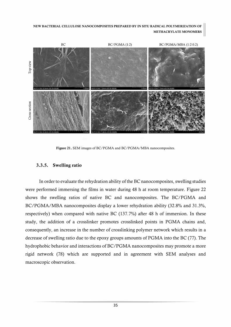
NEW BACTERIAL CELLULOSE NANOCOMPOSITES PREPARED BY IN SITU RADICAL POLYMERIZATION OF
METHACRYLATE MONOMERS
35
Figure 21. SEM images of BC/PGMA and BC/PGMA/MBA nanocomposites.
3.3.5. Swelling ratio
In order to evaluate the rehydration ability of the BC nanocomposites, swelling studies
were performed immersing the films in water during 48 h at room temperature. Figure 22
shows the swelling ratios of native BC and nanocomposites. The BC/PGMA and
BC/PGMA/MBA nanocomposites display a lower rehydration ability (32.8% and 31.3%,
respectively) when compared with native BC (137.7%) after 48 h of immersion. In these
study, the addition of a crosslinker promotes crosslinked points in PGMA chains and,
consequently, an increase in the number of crosslinking polymer network which results in a
decrease of swelling ratio due to the epoxy groups amounts of PGMA into the BC (77). The
hydrophobic behavior and interactions of BC/PGMA nanocomposites may promote a more
rigid network (78) which are supported and in agreement with SEM analyses and
macroscopic observation.
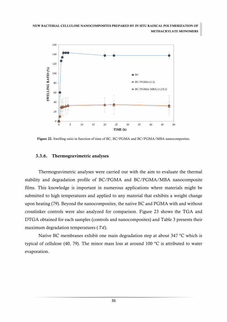
NEW BACTERIAL CELLULOSE NANOCOMPOSITES PREPARED BY IN SITU RADICAL POLYMERIZATION OF
METHACRYLATE MONOMERS
36
Figure 22. Swelling ratio in function of time of BC, BC/PGMA and BC/PGMA/MBA nanocomposites.
3.3.6. Thermogravimetric analyses
Thermogravimetric analyses were carried out with the aim to evaluate the thermal
stability and degradation profile of BC/PGMA and BC/PGMA/MBA nanocomposite
films. This knowledge is important in numerous applications where materials might be
submitted to high temperatures and applied to any material that exhibits a weight change
upon heating (79). Beyond the nanocomposites, the native BC and PGMA with and without
crosslinker controls were also analyzed for comparison. Figure 23 shows the TGA and
DTGA obtained for each samples (controls and nanocomposites) and Table 3 presents their
maximum degradation temperatures ( 𝑇𝑑).
Native BC membranes exhibit one main degradation step at about 347 ºC which is
typical of cellulose (40, 79). The minor mass loss at around 100 ºC is attributed to water
evaporation.

NEW BACTERIAL CELLULOSE NANOCOMPOSITES PREPARED BY IN SITU RADICAL POLYMERIZATION OF
METHACRYLATE MONOMERS
37
Figure 23. TGA and DTGA profile of BC/PGMA and BC/PGMA/MBA nanocomposites and their controls.
PGMA display a degradation profile with three main degradation steps at around 326,
388, 414 ºC and start their decomposition at 210 ºC. The first steps of degradation of PGMA
probably may be due to the: (i) depolymerization reaction and (ii) decomposition of esters
(72). The minor mass loss at around 414 ºC can be attributed to decomposition of units
involving side group scission of PGMA (71). The addition of crosslinker turn the polymer
more thermally stable and provokes a displacement of the degradation profile evidencing a
start decomposition at around 278 ºC and possessing a degradation profile at around 352,
398 and 408 ºC (Figure 23).
The TGA shows that incorporation of PGMA into the BC network influence the
thermal stability and degradation profile. The incorporation of PGMA and PGMA/MBA
into the BC increase their stability starting their decomposition, at around 275 ºC.
The increase of the 𝑇𝑑𝑖 in nanocomposites suggest a strong interaction and
compatibility between the PGMA and cellulose nanofibrils probably due to the establishment
of hydrogens bonds between BC nanofibrils and PGMA chains (40).

NEW BACTERIAL CELLULOSE NANOCOMPOSITES PREPARED BY IN SITU RADICAL POLYMERIZATION OF
METHACRYLATE MONOMERS
38
Table 3. Thermal degradation, 𝑇𝑑 (ºC), of BC/PGMA, BC/PGMA/MBA nanocomposites and their controls in study.
Samples 𝑻𝒅𝒊 𝑻𝒅𝒎𝒂𝒙𝟏 𝑻𝒅𝒎𝒂𝒙𝟐 𝑻𝒅𝒎𝒂𝒙𝟑
BC 266 347 --- ---
PGMA (1:0) 210 326 388 414
PGMA/MBA (1:0.2) 278 352 398 408
BC/PGMA (1:2) 273 321 363 413
BC/PGMA/MBA (1:2:0.2) 277 336 367 407
3.3.7. Dynamic Mechanical properties
The viscoelastic properties of BC/PGMA and BC/PGMA/MBA nanocomposites
were evaluated through dynamic mechanical analysis. Figure 24 shows the variation of the
storage modulus and tan 𝛿 of BC/PGMA and BC/PGMA/MBA nanocomposites.
Figure 24. Storage modulus e tan versus temperature of BC and BC/PGMA nanocomposites.
The storage modulus trace of the nanocomposites, E , shows only one transition. The
results obtained showed that the incorporation of PGMA provokes a decrease of storage

NEW BACTERIAL CELLULOSE NANOCOMPOSITES PREPARED BY IN SITU RADICAL POLYMERIZATION OF
METHACRYLATE MONOMERS
39
modulus when compared with native BC (40). This is due to the fact that PGMA is an
amorphous polymer, which decreases the crystallinity of the nanocomposites. The addition
of cross-linker leads to an increase of 11% in E ʹ and of 7 % in tan 𝛿 indicating that the cross-
linker make the nanocomposites with a tighter and higher stiffness BC network. The 𝑇𝑔
obtained from the tan 𝛿 peak also increase with the crosslinker addition from 90.2 to 96.5 ºC.
4. Conclusions
Bacterial cellulose/poly(glycidylmethacrylate) nanocomposites (BC/PGMA) were
obtained through in situ polymerization of glycidylmethacrylate using N,N’-
methylenebisacrilamide, MBA, as crosslinker and ammonium persulphate as initiator. The
maximum monomer incorporation into the bacterial cellulose occurs at (1:2) for BC/PGMA
and at (1:2:0.2) ratio for BC/PGMA/MBA, with 0.5 % of initiator, at 60 ºC during 6 h. The
incorporation of the PGMA makes the BC network more compact and smooth and allows
an improvement in their thermal and mechanical properties. It’s also observed that the
addition of a crosslinker allows a better retention of the polymer into BC network which
makes the new nanocomposite to exhibit a roughness and more compact three-dimensional
structure. Moreover, an improvement in thermal and mechanical properties was observed.
Both BC nanocomposites display a decrease in their water retention ability and in the
crystallinity degree. The new BC nanocomposites exhibited properties that can be applied for
absorbent materials for the removal of heavy metals and/or proteins due to the fact they
possess epoxy groups in their structure which could react with other molecules.


CHAPTER III
Epoxide ring-opening of bacterial
cellulose/poly(glycidylmethacrylate) nanocomposites

NEW BACTERIAL CELLULOSE NANOCOMPOSITES PREPARED BY IN SITU RADICAL POLYMERIZATION OF
METHACRYLATE MONOMERS
43
CHAPTER III. Epoxide ring-opening of bacterial
cellulose/poly(glycidylmethacrylate) nanocomposites
Abstract
New nanocomposites were prepared by epoxide ring-opening reaction mechanism of
bacterial cellulose/poly(glycidylmethacrylate) without (BC/PGMA) and with crosslinker,
N,N’-methylenebisacrilamide (BC/PGMA/MBA). To make it, the BC/PGMA and
BC/PGMA/MBA nanocomposites were subject to chemical treatment to promote epoxide
ring-opening reactions. A replacement of the epoxy groups for hydroxyls groups occurred in
an aqueous solutions at pH 3.5. After chemical treatment, the methacrylate nanocomposites,
BCPGMA-CT and BCPGMAMBA-CT, present a more compact and smoother structure. It
was also observed a decrease in thermal properties, mechanical properties, and crystallinity,
but an increase in their water retention ability. The surface became with less basic character
and occurred an increase in surface area. The reported methodology give rise to new and
interesting methacrylate nanocomposites.
Keywords: bacterial cellulose, glycidylmethacrylate, nanocomposites, chemical treatment,
epoxy-ring opening.
NEW BACTERIAL CELLULOSE NANOCOMPOSITES PREPARED BY IN SITU RADICAL POLYMERIZATION OF
METHACRYLATE MONOMERS
44
1. Introduction
Recently, bacterial cellulose (BC) has gained considerable attention as a source for the
development of new materials including nanocomposites due to its excellent properties such
as high purity, high degree of crystallinity (80-90%), high water retention capacity (99%),
ultrafine fibrous network and high tensile strength (17, 22). Combining the excellent
properties of BC with those of methacrylate monomers, such as high reactivity and
availability, bacterial cellulose/methacrylate nanocomposites have been prepared and
evaluated for different biomedical applications such as wound dressing (43).
Glycidylmethacrylate (GMA), among other methacrylate monomers, have received
particular attention because of the presence of two polymerizable groups: the epoxide and
the methacrylate groups. As polymer, it offers a wide range of industrial applications in
polymer chemistry and technology such as for purification of lysozyme from chicken white
egg (49, 50, 54), removal of heavy metals and chromate anions from aqueous solutions (48,
55-58), in coatings, matrix resins and adhesives (47, 59). Furthermore, GMA show a
relatively low toxicity, polarity, hydrophobicity and low price comparatively to other
methacrylate monomers which make them an attractive monomer for the polymer synthesis
(47, 50-53).
PGMA has been reported as grafted to different materials such as cellulose (49, 50)
via C=C, functionalizing the epoxy group, through epoxy ring-opening, with other molecules
as adsorbent material.
BC/PGMA nanocomposites, synthetized through in situ free radical polymerization
using N,N’-methylenebisacrilamide, MBA, exhibited a great retention of the polymer inside
the BC network. Without crosslinker, theses nanocomposites display a smoother and
compact network and with crosslinker, a rough, compact, dense and crosslinked network.
Both BC nanocomposites display an improvement in their thermal and mechanical
properties and, a decrease in the swelling ratio and crystallinity.
In the present study, a simple methodology was developed using the chemical
treatment that occurs in an aqueous solution at pH 3.5, to epoxide ring-opening of PGMA

NEW BACTERIAL CELLULOSE NANOCOMPOSITES PREPARED BY IN SITU RADICAL POLYMERIZATION OF
METHACRYLATE MONOMERS
45
inside BC network of BC/PGMA and BC/PGMA/MBA nanocomposites as a way to obtain
a microporous and crosslinked nanocomposite.
The obtained materials were characterized in terms of their structural and chemical
composition, crystallinity, morphology, thermal stability, water absorption, mechanical
properties and surface properties.
2. Methodology
2.1. BC nanocomposites preparation
Wet bacterial cellulose (BC) membranes produced by Gluconacetobacter sacchari (70)
using standard conditions were used. The BC/PGMA/MBA nanocomposites were obtained
through the methodology described in “Section 2.2. of Chapter II: New Bacterial
cellulose/poly(glycidylmethacrylate) nanocomposites films by in situ radical polymerization” of
present monography.
2.2. Chemical modification of BC nanocomposites
The chemical modification of BC/PGMA and BC/PGMA/MBA nanocomposites
was based in procedure described by Reis et al (2009) (80). Wet BC/PGMA and
BC/PGMA/MBA nanocomposites membranes were drained in order to remove the excess
of water and immersed in 2 mmol % HCl aqueous solution. Then, the reaction mixture was
placed at 140 ºC during 4 hours and under constant stirring (100 rpm). After this period, the
membranes were washed in distilled water until neutral pH, dried at 40 ºC and kept in a
desiccator until characterization.
Along this paper, the terminology of samples are described below: BC/PGMA–
bacterial cellulose membrane with poly(glycidylmethacrylate); BC/PGMA/MBA – bacterial
cellulose membrane poly(glycidylmethacrylate) crosslinking N,N’-methylenebisacrilamide;
BCPGMA-CT – chemical treatment of bacterial cellulose membrane with
NEW BACTERIAL CELLULOSE NANOCOMPOSITES PREPARED BY IN SITU RADICAL POLYMERIZATION OF
METHACRYLATE MONOMERS
46
glycidylmethacrylate and BCPGMAMBA-CT – chemical treatment of bacterial cellulose
membrane with glycidylmethacrylate crosslinking N,N’-methylenebisacrilamide.
2.3. BC nanocomposites characterization
The obtained BC/PGMA nanocomposite were characterized in terms of chemical
composition, morphology, thermal stability, water absorption, mechanic and surface
properties by ATR-FTIR, EDX, CPMAS 13C NMR, FE-SEM, XRD, TGA, SW, DMA and
IGC, respectively. The methodology applied for each methods is described in “Section 2.3.
of Chapter II: New Bacterial cellulose/poly(glycidylmethacrylate) nanocomposites films by in situ
radical polymerization” of present manuscript, expect the IGC analysis which is described
below.
Inverse Gas Chromatography measurements
The BC nanocomposites were packed in the standard glass silanized
(dymethyldichlorosilane; Repelcote BDH, UK) columns with 0.2 cm ID and 30 cm in length
by vertical tapping about 2 hours. The columns with the samples were conditioned 8 h at 40
ºC. After conditioning, pulse injections were carried out with a 0.25 mL gas loop. Four n-
alkanes (heptane, octane, nonane and decane) were used for measurements of the dispersive
component of the surface energy at 20, 25 and 30 ºC. Four polar probes (tetrahydrofuran,
dichloromethane, acetonitrile and ethyl acetate) were used to determine the specific
component of surface energy and acid-base character (𝐾𝐴 and 𝐾𝐵) at 25 ºC. The isotherm
experiments were undertaken with n-octane, tetrahydrofuran and dichloromethane at 25 ºC
for all samples tested. All experiments were carried out at 0% relative humidity with a helium
flow rate of 10 ml/min and at least in duplicate, producing an error less than 4%.
IGC measurements were carried out on a commercial inverse gas chromatograph
(iGC, Surface Measurements Systems, London, UK) equipped with flame ionization (FID)
and thermal conductivity (TCD) detectors. The iGC system is fully automatic with SMS iGC
Controller v1.8 control software. Data were analysed using iGC Standard v1.3 and

NEW BACTERIAL CELLULOSE NANOCOMPOSITES PREPARED BY IN SITU RADICAL POLYMERIZATION OF
METHACRYLATE MONOMERS
47
Advanced Analysis Software v1.25. The calculation methodology is described exhaustively
in Chapter V of the present manuscript.
3. Results and Discussion
BC/PGMA and BC/PGMA/MBA nanocomposites were subject to a chemical
treatment in order to promote the reaction of epoxy groups of the PGMA with hydroxyl
group of cellulose chain through epoxide ring-opening. The effect of attachment of PGMA
in structure, morphology, swelling, thermal, mechanical and surface properties was
evaluated through ATR-FTIR, 13C NMR, XRD, EDX, FE-SEM, SW, TGA, DMA and IGC
techniques.
A probably scheme illustrating the epoxide ring-opening mechanism of
BC/PGMA/MBA nanocomposites is shown in Figure 25.
Figure 25. Schematic diagram of the epoxide ring-opening mechanism of BC/PGMA nanocomposites.
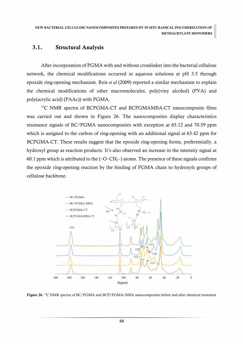
NEW BACTERIAL CELLULOSE NANOCOMPOSITES PREPARED BY IN SITU RADICAL POLYMERIZATION OF
METHACRYLATE MONOMERS
48
3.1. Structural Analysis
After incorporation of PGMA with and without crosslinker into the bacterial cellulose
network, the chemical modifications occurred in aqueous solutions at pH 3.5 through
epoxide ring-opening mechanism. Reis et al (2009) reported a similar mechanism to explain
the chemical modifications of other macromolecules, poly(viny alcohol) (PVA) and
poly(acrylic acid) (PAAc)) with PGMA.
13C NMR spectra of BCPGMA-CT and BCPGMAMBA-CT nanocomposite films
was carried out and shown in Figure 26. The nanocomposites display characteristics
resonance signals of BC/PGMA nanocomposites with exception at 65.12 and 70.59 ppm
which is assigned to the carbon of ring-opening with an additional signal at 63.42 ppm for
BCPGMA-CT. These results suggest that the epoxide ring-opening forms, preferentially, a
hydroxyl group as reaction products. It’s also observed an increase in the intensity signal at
60.1 ppm which is attributed to the (–O–CH2–) atoms. The presence of these signals confirms
the epoxide ring-opening reaction by the binding of PGMA chain to hydroxyls groups of
cellulose backbone.
Figure 26. 13C NMR spectra of BC/PGMA and BCP/PGMA/MBA nanocomposites before and after chemical treatment
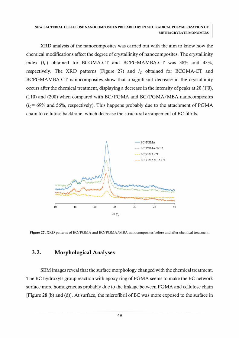
NEW BACTERIAL CELLULOSE NANOCOMPOSITES PREPARED BY IN SITU RADICAL POLYMERIZATION OF
METHACRYLATE MONOMERS
49
XRD analysis of the nanocomposites was carried out with the aim to know how the
chemical modifications affect the degree of crystallinity of nanocomposites. The crystallinity
index (𝐼𝐶) obtained for BCGMA-CT and BCPGMAMBA-CT was 38% and 43%,
respectively. The XRD patterns (Figure 27) and 𝐼𝐶 obtained for BCGMA-CT and
BCPGMAMBA-CT nanocomposites show that a significant decrease in the crystallinity
occurs after the chemical treatment, displaying a decrease in the intensity of peaks at 2θ (1ī0),
(110) and (200) when compared with BC/PGMA and BC/PGMA/MBA nanocomposites
(𝐼𝐶= 69% and 56%, respectively). This happens probably due to the attachment of PGMA
chain to cellulose backbone, which decrease the structural arrangement of BC fibrils.
Figure 27. XRD patterns of BC/PGMA and BC/PGMA/MBA nanocomposites before and after chemical treatment.
3.2. Morphological Analyses
SEM images reveal that the surface morphology changed with the chemical treatment.
The BC hydroxyls group reaction with epoxy ring of PGMA seems to make the BC network
surface more homogeneous probably due to the linkage between PGMA and cellulose chain
[Figure 28 (b) and (d)]. At surface, the microfibril of BC was more exposed to the surface in

NEW BACTERIAL CELLULOSE NANOCOMPOSITES PREPARED BY IN SITU RADICAL POLYMERIZATION OF
METHACRYLATE MONOMERS
50
both samples but more compact with chemical modification as shown in Figure 28 (a) and
(c). Thus, the chemical treatment of the BC /PGMA nanocomposites change their
morphology at surface and inside, turning it more compact and smooth which, consequently,
affect the properties of the BC nanocomposites.
Figure 28. SEM images of BCGMA-CT and BCGMAMBA-CT nanocomposites morphology in different perspectives.
The (i), (ii), (iii) and (iv) images correspond to the samples without chemical treatment.
3.3. Thermal and Mechanical Analyses
Figure 29 exhibit the TGA and DTGA achieved for nanocomposites and Table 4
show the maximum temperature of thermal degradation, 𝑇𝑑, obtained for each sample. The
minor mass loss at around 100 ºC is attributed to water evaporation of samples. BCPGMA-
CT nanocomposites display a degradation profile with three main degradation steps similar
to BC/PGMA nanocomposites. It´s observed the maximum degradation temperature of
PGMA which suggests that doesn´t occur the degradation of PGMA polymer provoked by
temperature or/and acid aqueous solution.
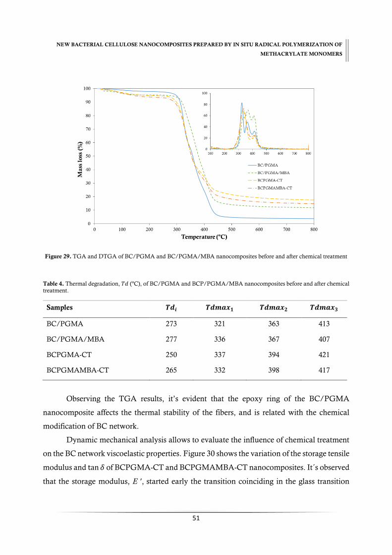
NEW BACTERIAL CELLULOSE NANOCOMPOSITES PREPARED BY IN SITU RADICAL POLYMERIZATION OF
METHACRYLATE MONOMERS
51
Figure 29. TGA and DTGA of BC/PGMA and BC/PGMA/MBA nanocomposites before and after chemical treatment
Table 4. Thermal degradation, 𝑇𝑑 (ºC), of BC/PGMA and BCP/PGMA/MBA nanocomposites before and after chemical treatment.
Samples 𝑻𝒅𝒊 𝑻𝒅𝒎𝒂𝒙𝟏 𝑻𝒅𝒎𝒂𝒙𝟐 𝑻𝒅𝒎𝒂𝒙𝟑
BC/PGMA 273 321 363 413
BC/PGMA/MBA 277 336 367 407
BCPGMA-CT 250 337 394 421
BCPGMAMBA-CT 265 332 398 417
Observing the TGA results, it’s evident that the epoxy ring of the BC/PGMA
nanocomposite affects the thermal stability of the fibers, and is related with the chemical
modification of BC network.
Dynamic mechanical analysis allows to evaluate the influence of chemical treatment
on the BC network viscoelastic properties. Figure 30 shows the variation of the storage tensile
modulus and tan 𝛿 of BCPGMA-CT and BCPGMAMBA-CT nanocomposites. It´s observed
that the storage modulus, E ʹ, started early the transition coinciding in the glass transition

NEW BACTERIAL CELLULOSE NANOCOMPOSITES PREPARED BY IN SITU RADICAL POLYMERIZATION OF
METHACRYLATE MONOMERS
52
zone in both nanocomposites. The chemical treatment increase the storage modulus
compared with BC/PGMA and BC/PGMA/MBA nanocomposites as well as the 𝑇𝑔
obtained from the tan 𝛿 peak (90.2 to 146.4 ºC and 96.5 to 99.2 ºC, respectively) (as described
in Section 3.3.6. of Chapter II). These results suggest that the reaction of epoxy group of PGMA
chain to cellulose backbone via (–OH), increase the amorphous content (supported by XRD
pattern). Moreover, these results also indicates that the nanocomposites became less rigid
than initial BC/PGMA nanocomposites.
BCPGMA-CT and BCPGMAMBA-CT exhibited distinct mechanical performance
which make these simple chemical treatment a good strategy to obtain nanocomposites for
new applications.
Figura 30. Storage modulus e tan versus temperature of BCPGMA-CT nanocomposites.
3.4. Surface properties
In order to study the effect of the chemical treatment in the nanocomposites surface,
IGC analysis was performed. This knowledge allows the prediction of its compatibility with
other materials. Parameters such as dispersive component of the surface energy, 𝛾𝑠𝑑, specific
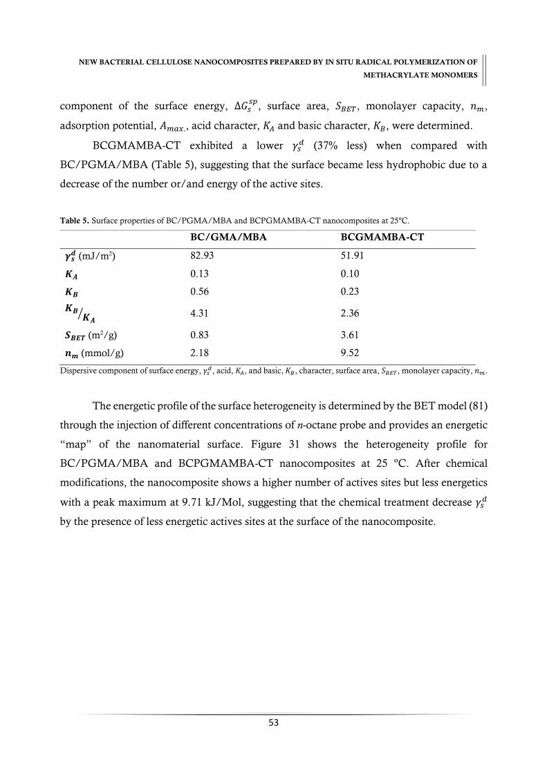
NEW BACTERIAL CELLULOSE NANOCOMPOSITES PREPARED BY IN SITU RADICAL POLYMERIZATION OF
METHACRYLATE MONOMERS
53
component of the surface energy, ∆𝐺𝑠𝑠𝑝
, surface area, 𝑆𝐵𝐸𝑇, monolayer capacity, 𝑛𝑚,
adsorption potential, 𝐴𝑚𝑎𝑥., acid character, 𝐾𝐴 and basic character, 𝐾𝐵, were determined.
BCGMAMBA-CT exhibited a lower 𝛾𝑠𝑑 (37% less) when compared with
BC/PGMA/MBA (Table 5), suggesting that the surface became less hydrophobic due to a
decrease of the number or/and energy of the active sites.
Table 5. Surface properties of BC/PGMA/MBA and BCPGMAMBA-CT nanocomposites at 25ºC.
BC/GMA/MBA BCGMAMBA-CT
𝜸𝒔𝒅 (mJ/m2) 82.93 51.91
𝑲𝑨 0.13 0.10
𝑲𝑩 0.56 0.23
𝑲𝑩𝑲𝑨
⁄ 4.31 2.36
𝑺𝑩𝑬𝑻 (m2/g) 0.83 3.61
𝒏𝒎 (mmol/g) 2.18 9.52
Dispersive component of surface energy, 𝛾𝑠𝑑, acid, 𝐾𝐴, and basic, 𝐾𝐵, character, surface area, 𝑆𝐵𝐸𝑇, monolayer capacity, 𝑛𝑚.
The energetic profile of the surface heterogeneity is determined by the BET model (81)
through the injection of different concentrations of n-octane probe and provides an energetic
“map” of the nanomaterial surface. Figure 31 shows the heterogeneity profile for
BC/PGMA/MBA and BCPGMAMBA-CT nanocomposites at 25 ºC. After chemical
modifications, the nanocomposite shows a higher number of actives sites but less energetics
with a peak maximum at 9.71 kJ/Mol, suggesting that the chemical treatment decrease 𝛾𝑠𝑑
by the presence of less energetic actives sites at the surface of the nanocomposite.
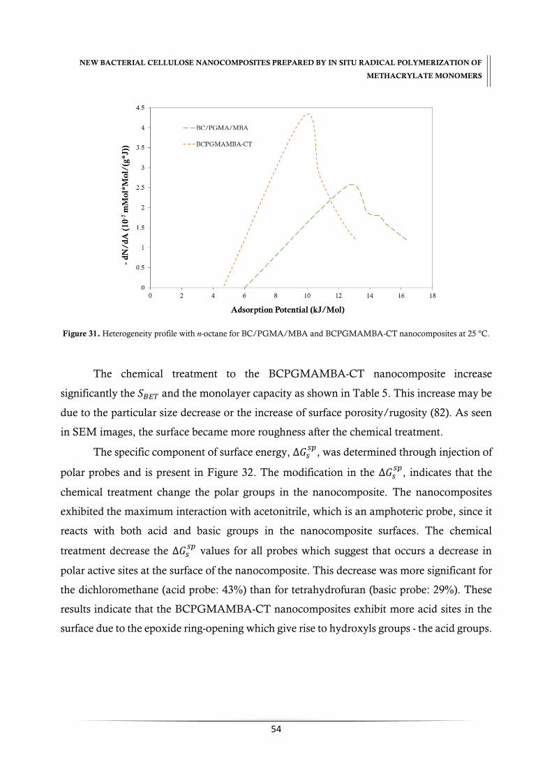
NEW BACTERIAL CELLULOSE NANOCOMPOSITES PREPARED BY IN SITU RADICAL POLYMERIZATION OF
METHACRYLATE MONOMERS
54
Figure 31. Heterogeneity profile with n-octane for BC/PGMA/MBA and BCPGMAMBA-CT nanocomposites at 25 ºC.
The chemical treatment to the BCPGMAMBA-CT nanocomposite increase
significantly the 𝑆𝐵𝐸𝑇 and the monolayer capacity as shown in Table 5. This increase may be
due to the particular size decrease or the increase of surface porosity/rugosity (82). As seen
in SEM images, the surface became more roughness after the chemical treatment.
The specific component of surface energy, ∆𝐺𝑠𝑠𝑝
, was determined through injection of
polar probes and is present in Figure 32. The modification in the ∆𝐺𝑠𝑠𝑝
, indicates that the
chemical treatment change the polar groups in the nanocomposite. The nanocomposites
exhibited the maximum interaction with acetonitrile, which is an amphoteric probe, since it
reacts with both acid and basic groups in the nanocomposite surfaces. The chemical
treatment decrease the ∆𝐺𝑠𝑠𝑝
values for all probes which suggest that occurs a decrease in
polar active sites at the surface of the nanocomposite. This decrease was more significant for
the dichloromethane (acid probe: 43%) than for tetrahydrofuran (basic probe: 29%). These
results indicate that the BCPGMAMBA-CT nanocomposites exhibit more acid sites in the
surface due to the epoxide ring-opening which give rise to hydroxyls groups - the acid groups.

NEW BACTERIAL CELLULOSE NANOCOMPOSITES PREPARED BY IN SITU RADICAL POLYMERIZATION OF
METHACRYLATE MONOMERS
55
Figure 32. Specific surface energy with polar probes: THF - tetrahydrofuran, DCM - dichloromethane, ETOAc – ethyl
acetate, ACN – acetonitrile of BC/PGMA/MBA and BCPGMAMBA-CT nanocomposites at 25 ºC.
The ∆𝐺𝑠𝑠𝑝
is converted into acid-base number using the Guttmann’s concept (83),
and the results (Table 5) show that the chemical treatment decrease the base constant, 𝐾𝐵,
with a 𝐾𝐵 𝐾𝐴⁄ ratio 55% lower than BC/PGMA/MBA. Thus, the increase in the character
exhibited in BC nanocomposites derives from the epoxide ring-opening, which decrease the
basic groups (epoxide).
The know rehydration ability of the bacterial cellulose is attributed to the presence of
hydroxyls groups. The BCGMAMBA-CT nanocomposite exhibits a swelling ratio of 227%,
a considerable increase of c.a. 7 times plus when compared with BC/PGMA/MBA
nanocomposite (31%). These results are in agreement with 𝛾𝑠𝑑 trend data discussed previously
and reinforces the epoxide ring-opening process during the chemical treatment.

NEW BACTERIAL CELLULOSE NANOCOMPOSITES PREPARED BY IN SITU RADICAL POLYMERIZATION OF
METHACRYLATE MONOMERS
56
4. Conclusions
Natural polymers are able to react with many modifiers agents such as
glycidylmethacrylate, a bifunctional compound, in order to obtain new composites. Epoxide
ring-opening reaction was a suitable strategy to modify the bacterial
cellulose/poly(glycidylmethacrylate) nanocomposites. In order to do that, the BC/PGMA
and BC/PGMA/MBA nanocomposites were subjected to chemical treatment. The
morphology of BCPGMA-CT and BCPGMAMBA-CT nanocomposites is more compact
and smooth. A decrease in thermal and mechanical properties was observed while an
increase of the amorphous content and water retention ability in the new bacterial cellulose
nanocomposites was verified. The surface of BCPGMA-CT and BCPGMAMBA-CT
nanocomposites present a less basic character and an increase of the surface area was
observed. The reported methodology gives rise to new and interesting methacrylate
nanocomposites.

CHAPTER IV
Novel bacterial cellulose/poly(ethyleneglycol)methacrylate
nanocomposite films obtained by in situ free radical
polymerization


NEW BACTERIAL CELLULOSE NANOCOMPOSITES PREPARED BY IN SITU RADICAL POLYMERIZATION OF
METHACRYLATE MONOMERS
59
CHAPTER IV. Novel bacterial cellulose/poly(ethyleneglycol)
methacrylate nanocomposite films obtained by in situ free radical
polymerization
Abstract
New bacterial cellulose/methacrylate nanocomposite were prepared by the in situ free
radical polymerization of poly(ethyleneglycol)methacrylate, (PEGMA), inside the BC
network using ammonium persulphate, (APS), as radical initiator. Chemical composition,
morphology, thermal stability, water absorption and mechanic properties were determined.
The optimal conditions achieved for polymerization were a ratio of (1:20) for bacterial
cellulose/monomer with 0.5% of initiator during 6 h at 60 ºC. The maximum incorporation
percentage achieved was 87%. With the incorporation of the polymer, films became less stiff
and displayed a translucent-yellow color. The incorporation of the PPEGMA hydrophilic
chains originates an improvement of the thermal and mechanical properties and also their
swelling ability. A decrease in the crystallinity occurred due to the increase of the structural
entropy. The new nanocomposite acquires properties able to be applied in biomedical
materials.
Keywords: bacterial cellulose, PEGMA, in situ radical polymerization, nanocomposites
Figure 33. Schematic reaction of in situ free polymerization of PEGMA inside BC network.

NEW BACTERIAL CELLULOSE NANOCOMPOSITES PREPARED BY IN SITU RADICAL POLYMERIZATION OF
METHACRYLATE MONOMERS
60
1. Introduction
Recently, several studies have been reported using bacterial cellulose as a starting
component to obtain bacterial cellulose based nanocomposites. Bacterial cellulose produced
by Gluconacetobacter sp acquires excellent properties such as high purity, degree of crystallinity
(80-90%), high water retention capacity (99%), ultrafine fibrous network and high tensile
strength (17, 22). Besides that, they are biocompatible, hydrophilic and non-toxic (11).
Bacterial cellulose/methacrylate nanocomposites have been prepared with different
methacrylate monomers which provide a wide range of materials with adjustable properties
due to their variability in chemical structure, possibility of adapting the physicochemical
properties, great availability and easy to polymerization. (22) Wound dressing (43), design
of 3D matrices (40), removal of heavy metal and chromate anions from aqueous solutions
(48, 55-58), coatings, matrix resins and adhesives (47, 59) were some applications of bacterial
cellulose based methacrylate’s nanocomposites.
Poly (ethylene glycol) (PEG) display unique properties which are advantageous to
biomedical and biotechnological applications due its low-toxicity, absence of antigenicity
and immunogenicity and inherent ability to prevent protein adsorption (61).
Poly(ethyleneglycol)methacrylate (PEGMA), one PEG derivatives, is an attractive
methacrylate monomer due to its amphiphilic nature which comes from its water-soluble
PEG side chain with a pendant hydroxyl group and its hydrophobic methacrylate group (62).
PEGMA was used for biomedical materials such as drug carrier, microspheres for transient
vascular embolization (62), protein adsorption and immobilization of polysaccharides (64)
and heavy metal removal (65, 66).
PPEGMA can be produced by different polymerization approaches such as
suspension polymerization (65, 84), UV-initiated free radical polymerization (85) and atom
transfer radical polymerization (ATRP) (61).
The present study reports the development of BC nanocomposite through in situ
radical polymerization of PEGMA into BC network using APS as initiator. The optimal
polymerization conditions were determined and the obtained BC/PPEGMA nanocomposite
characterized in terms of chemical composition, morphology, thermal stability, water
absorption and mechanic properties.
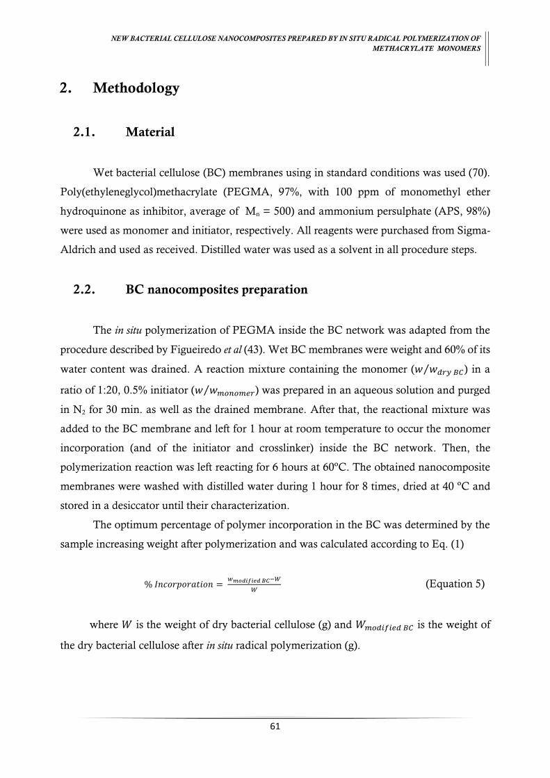
NEW BACTERIAL CELLULOSE NANOCOMPOSITES PREPARED BY IN SITU RADICAL POLYMERIZATION OF
METHACRYLATE MONOMERS
61
2. Methodology
2.1. Material
Wet bacterial cellulose (BC) membranes using in standard conditions was used (70).
Poly(ethyleneglycol)methacrylate (PEGMA, 97%, with 100 ppm of monomethyl ether
hydroquinone as inhibitor, average of Mn = 500) and ammonium persulphate (APS, 98%)
were used as monomer and initiator, respectively. All reagents were purchased from Sigma-
Aldrich and used as received. Distilled water was used as a solvent in all procedure steps.
2.2. BC nanocomposites preparation
The in situ polymerization of PEGMA inside the BC network was adapted from the
procedure described by Figueiredo et al (43). Wet BC membranes were weight and 60% of its
water content was drained. A reaction mixture containing the monomer (𝑤 𝑤𝑑𝑟𝑦 𝐵𝐶⁄ ) in a
ratio of 1:20, 0.5% initiator (𝑤 𝑤𝑚𝑜𝑛𝑜𝑚𝑒𝑟⁄ ) was prepared in an aqueous solution and purged
in N2 for 30 min. as well as the drained membrane. After that, the reactional mixture was
added to the BC membrane and left for 1 hour at room temperature to occur the monomer
incorporation (and of the initiator and crosslinker) inside the BC network. Then, the
polymerization reaction was left reacting for 6 hours at 60ºC. The obtained nanocomposite
membranes were washed with distilled water during 1 hour for 8 times, dried at 40 ºC and
stored in a desiccator until their characterization.
The optimum percentage of polymer incorporation in the BC was determined by the
sample increasing weight after polymerization and was calculated according to Eq. (1)
% 𝐼𝑛𝑐𝑜𝑟𝑝𝑜𝑟𝑎𝑡𝑖𝑜𝑛 = 𝑤𝑚𝑜𝑑𝑖𝑓𝑖𝑒𝑑 𝐵𝐶−𝑊
𝑊 (Equation 5)
where 𝑊 is the weight of dry bacterial cellulose (g) and 𝑊𝑚𝑜𝑑𝑖𝑓𝑖𝑒𝑑 𝐵𝐶 is the weight of
the dry bacterial cellulose after in situ radical polymerization (g).

NEW BACTERIAL CELLULOSE NANOCOMPOSITES PREPARED BY IN SITU RADICAL POLYMERIZATION OF
METHACRYLATE MONOMERS
62
2.3. BC nanocomposites characterization
The obtained BC/PPEGMA nanocomposite was characterized in terms of chemical
composition, morphology, thermal stability, water absorption and mechanic properties by
ATR-FTIR, XRD, EDX, FE-SEM, TGA, SW and DMA, respectively. The methodology
applied for each methods is described in “Section 2.3. of Chapter II: New Bacterial
cellulose/poly(glycidylmethacrylate) nanocomposites films by in situ radical polymerization” of
present monography.
3. Results and Discussion
BC/PPEGMA nanocomposites were prepared by the in situ free polymerization of
PEGMA incorporation into the BC network using APS as initiator. The schematic
representation of the polymerization is shown in Figure 34.
Figure 34. Schematic of in situ free radical polymerization reaction of PEGMA inside of BC network [based on
(86)].
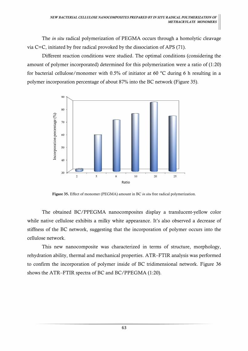
NEW BACTERIAL CELLULOSE NANOCOMPOSITES PREPARED BY IN SITU RADICAL POLYMERIZATION OF
METHACRYLATE MONOMERS
63
The in situ radical polymerization of PEGMA occurs through a homolytic cleavage
via C=C, initiated by free radical provoked by the dissociation of APS (71).
Different reaction conditions were studied. The optimal conditions (considering the
amount of polymer incorporated) determined for this polymerization were a ratio of (1:20)
for bacterial cellulose/monomer with 0.5% of initiator at 60 ºC during 6 h resulting in a
polymer incorporation percentage of about 87% into the BC network (Figure 35).
Figure 35. Effect of monomer (PEGMA) amount in BC in situ free radical polymerization.
The obtained BC/PPEGMA nanocomposites display a translucent-yellow color
while native cellulose exhibits a milky white appearance. It’s also observed a decrease of
stiffness of the BC network, suggesting that the incorporation of polymer occurs into the
cellulose network.
This new nanocomposite was characterized in terms of structure, morphology,
rehydration ability, thermal and mechanical properties. ATR–FTIR analysis was performed
to confirm the incorporation of polymer inside of BC tridimensional network. Figure 36
shows the ATR–FTIR spectra of BC and BC/PPEGMA (1:20).

NEW BACTERIAL CELLULOSE NANOCOMPOSITES PREPARED BY IN SITU RADICAL POLYMERIZATION OF
METHACRYLATE MONOMERS
64
Figure 36. ATR–FTIR spectra of BC and BC/PPEGMA nanocomposite.
The ATR–FTIR spectra of the BC/PPEGMA nanocomposites exhibited, in addition
to the characteristic peaks of native BC (described in Section 3.3.1. of Chapter II) a absorption
band at 1720 cm-1, attributed to the carboxyl groups of methacrylate ester units (O–C=O).
Additionally, the intensification of the absorption at 2894 cm-1, assigned to the vibrations of
the CH, CH2 and CH3 and at 3340 cm-1, attributed to the PEG terminal hydroxyl groups (65,
85), confirm the success of incorporation of PPEGMA into the BC network. Moreover, the
absence of the characteristic C=C stretching vibrations at 1660 cm-1 indicates that the
polymerization of PEGMA occurs into the three-dimensional network.
A decrease of 12% in the 𝐶 𝑂⁄ ratio in BC/PPEGMA nanocomposites, determined
by EDX (Table 6) evidence the increase of hydroxyl groups (–OH) and ester groups (O–C=O)
into the network of BC, arising from the presence of PPEGMA in BC network.
Table 6. EDX analysis of elemental composition of BC and BC/PPEGMA nanocomposites (wt %).
Samples 𝑪 𝑶 𝑪 𝑶⁄ Ratio
BC 75.37 23.05 3.30
BC/PPEGMA (1:20) 73.14 25.23 2.90
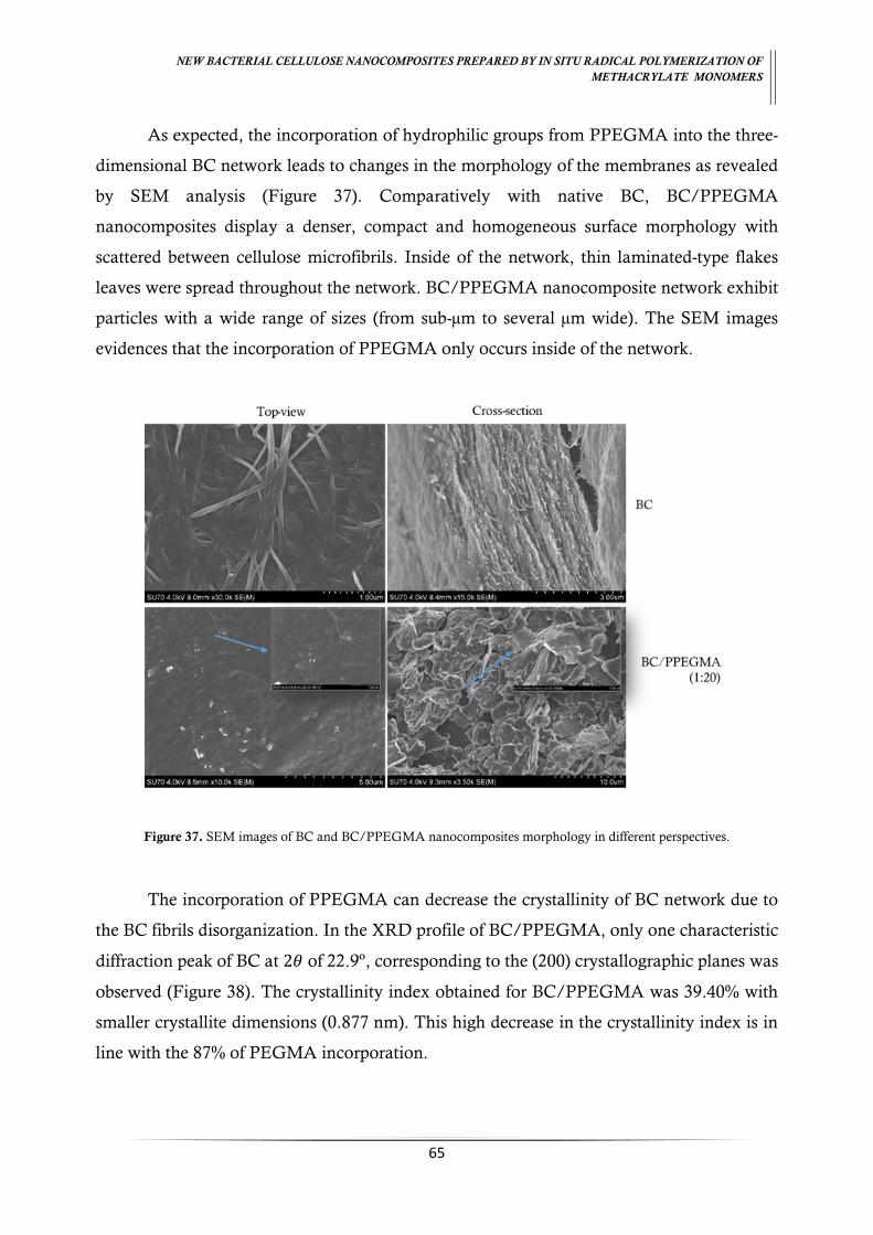
NEW BACTERIAL CELLULOSE NANOCOMPOSITES PREPARED BY IN SITU RADICAL POLYMERIZATION OF
METHACRYLATE MONOMERS
65
As expected, the incorporation of hydrophilic groups from PPEGMA into the three-
dimensional BC network leads to changes in the morphology of the membranes as revealed
by SEM analysis (Figure 37). Comparatively with native BC, BC/PPEGMA
nanocomposites display a denser, compact and homogeneous surface morphology with
scattered between cellulose microfibrils. Inside of the network, thin laminated-type flakes
leaves were spread throughout the network. BC/PPEGMA nanocomposite network exhibit
particles with a wide range of sizes (from sub-µm to several µm wide). The SEM images
evidences that the incorporation of PPEGMA only occurs inside of the network.
Figure 37. SEM images of BC and BC/PPEGMA nanocomposites morphology in different perspectives.
The incorporation of PPEGMA can decrease the crystallinity of BC network due to
the BC fibrils disorganization. In the XRD profile of BC/PPEGMA, only one characteristic
diffraction peak of BC at 2𝜃 of 22.9º, corresponding to the (200) crystallographic planes was
observed (Figure 38). The crystallinity index obtained for BC/PPEGMA was 39.40% with
smaller crystallite dimensions (0.877 nm). This high decrease in the crystallinity index is in
line with the 87% of PEGMA incorporation.
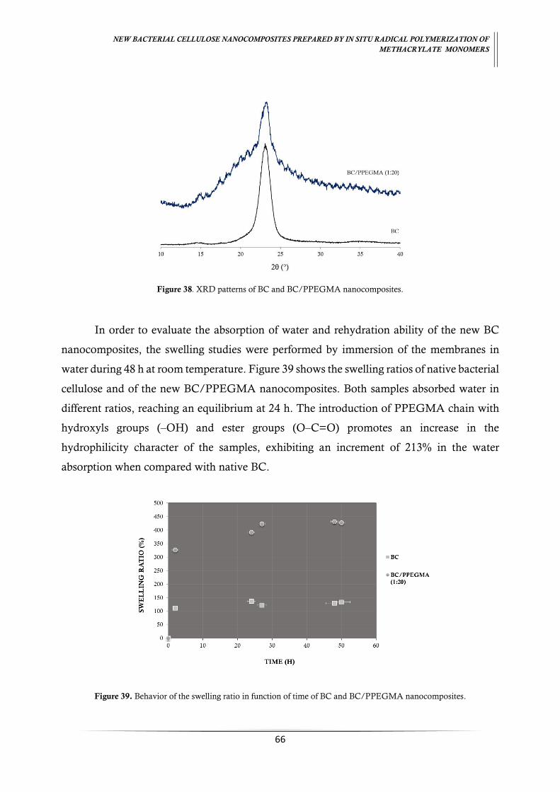
NEW BACTERIAL CELLULOSE NANOCOMPOSITES PREPARED BY IN SITU RADICAL POLYMERIZATION OF
METHACRYLATE MONOMERS
66
Figure 38. XRD patterns of BC and BC/PPEGMA nanocomposites.
In order to evaluate the absorption of water and rehydration ability of the new BC
nanocomposites, the swelling studies were performed by immersion of the membranes in
water during 48 h at room temperature. Figure 39 shows the swelling ratios of native bacterial
cellulose and of the new BC/PPEGMA nanocomposites. Both samples absorbed water in
different ratios, reaching an equilibrium at 24 h. The introduction of PPEGMA chain with
hydroxyls groups (–OH) and ester groups (O–C=O) promotes an increase in the
hydrophilicity character of the samples, exhibiting an increment of 213% in the water
absorption when compared with native BC.
Figure 39. Behavior of the swelling ratio in function of time of BC and BC/PPEGMA nanocomposites.
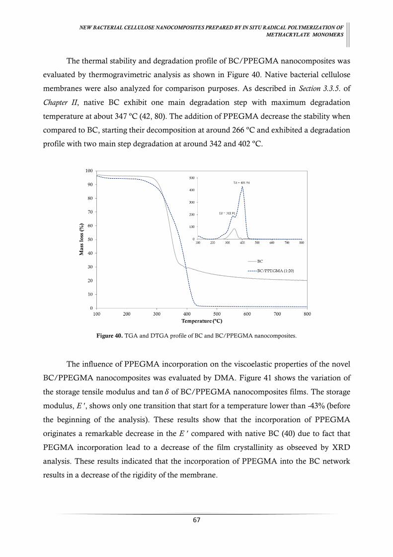
NEW BACTERIAL CELLULOSE NANOCOMPOSITES PREPARED BY IN SITU RADICAL POLYMERIZATION OF
METHACRYLATE MONOMERS
67
The thermal stability and degradation profile of BC/PPEGMA nanocomposites was
evaluated by thermogravimetric analysis as shown in Figure 40. Native bacterial cellulose
membranes were also analyzed for comparison purposes. As described in Section 3.3.5. of
Chapter II, native BC exhibit one main degradation step with maximum degradation
temperature at about 347 ºC (42, 80). The addition of PPEGMA decrease the stability when
compared to BC, starting their decomposition at around 266 ºC and exhibited a degradation
profile with two main step degradation at around 342 and 402 ºC.
Figure 40. TGA and DTGA profile of BC and BC/PPEGMA nanocomposites.
The influence of PPEGMA incorporation on the viscoelastic properties of the novel
BC/PPEGMA nanocomposites was evaluated by DMA. Figure 41 shows the variation of
the storage tensile modulus and tan 𝛿 of BC/PPEGMA nanocomposites films. The storage
modulus, E ʹ, shows only one transition that start for a temperature lower than -43% (before
the beginning of the analysis). These results show that the incorporation of PPEGMA
originates a remarkable decrease in the E ʹ compared with native BC (40) due to fact that
PEGMA incorporation lead to a decrease of the film crystallinity as obseeved by XRD
analysis. These results indicated that the incorporation of PPEGMA into the BC network
results in a decrease of the rigidity of the membrane.

NEW BACTERIAL CELLULOSE NANOCOMPOSITES PREPARED BY IN SITU RADICAL POLYMERIZATION OF
METHACRYLATE MONOMERS
68
Figure 41. Storage modulus e tan versus temperature of BC/PPEGMA nanocomposites.
4. Conclusions
New bacterial cellulose/poly(ethyleneglycol)methacrylate nanocomposite,
BC/PPEGMA was obtained by in situ radical polymerization of PEGMA inside the BC
network. BC/PPEGMA nanocomposite display a translucent yellow appearance and have
new thermal, morphologic and surface characteristics. The incorporation of the hydrophilic
PEGMA inside the BC network lead to a decrease of the stiffness of three-dimensional
structure, improve the thermal and mechanical properties, increase their swelling ability as
well as of amorphous content.

CHAPTER V
Assessment of the surface properties changes of bacterial
cellulose/polymethacrylate nanocomposite films by Inverse Gas
Chromatography

NEW BACTERIAL CELLULOSE NANOCOMPOSITES PREPARED BY IN SITU RADICAL POLYMERIZATION OF
METHACRYLATE MONOMERS
70
CHAPTER V. Assessment of the surface properties changes of
bacterial cellulose/polymethacrylate nanocomposite films by
Inverse Gas Chromatography
Abstract
Bacterial cellulose/polymethacrylate nanocomposites have been receiving attention
in the last years due to the availability and attractive properties of methacrylate monomers.
In this work, bacterial cellulose/poly(glycidylmethacrylate) and bacterial
cellulose/poly(ethylene glycol)methacrylate nanocomposites were prepared by in situ free
radical polymerization. Inverse Gas Chromatography was used to study surface properties
namely surface energy, surface area, heterogeneity and acid-base character. The results show
the successful incorporation of methacrylate polymers into the bacterial cellulose. The
incorporation of glycidylmethacrylate decrease the hydrophilicity and increase the porosity
and roughness of the bacteria cellulose network. In the case of
poly(ethyleneglycol)methacrylate incorporation, an increase of hydrophilicity and a decrease
of the porosity and roughness, was observed. Both methacrylate polymers incorporation
made the bacterial cellulose film surface more basic and consequently more reactive with the
acid compounds, thus allowing new applications.
Keywords: Nanocomposites, bacterial cellulose, poly(glycidylmethacrylate),
poly(ethyleneglycol)methacrylate, surface properties, IGC.

NEW BACTERIAL CELLULOSE NANOCOMPOSITES PREPARED BY IN SITU RADICAL POLYMERIZATION OF
METHACRYLATE MONOMERS
71
1. Introduction
Nanocomposites are defined as ‘composites of more than a Gibbsian solid phase where at
least one-dimension is in the nanometer range and typically all solid phases are in the 1-20 nanometer
range’ by Komarneni in 1992 . Recently, several studies have been reported using bacterial
cellulose as a starting component and/or the base to obtain bacterial cellulose
nanocomposites. Bacterial cellulose possesses excellent properties such as high purity, degree
of crystallinity (80-90%), high water retention capacity (99%), ultrafine fibrous network and
high tensile strength (17, 22). Furthermore, it exhibits excellent biocompatibility and non-
toxicity, being a great material to be employed in a wide range applications in different areas
(6, 11, 69).
Bacterial cellulose/poly(methacrylate) nanocomposites have been receiving attention
by researchers due to their availability and attractive properties. These properties can be
employed in nanocomposites for biomedical applications such as wound dressing (43) and
design of 3D matrices (40). Glycidylmethacrylate (GMA) and
poly(ethyleneglycol)methacrylate (PEGMA) are two attractive methacrylate due to their
properties. GMA possess two functional groups, the epoxide and the methacrylate groups
(double bonds), which offers a wide range of industrial applications in polymer chemistry
and technology (49, 50, 54). PEGMA, one poly(ethyleneglycol) derivate, confers an
amphiphilic nature, which comes from its water-soluble PEG side chain with a pendant
hydroxyl group and its hydrophobic methacrylate group (62).
In this paper, the modification of the surface properties of bacterial cellulose with the
methacrylate compounds (glycidylmethacrylate and poly(ethyleneglycol)methacrylate)
through in situ radical polymerization was evaluated. Thermodynamic and morphologic
parameters such as dispersive and specific surface energy, acid–base surface character,
surface area and heterogeneity were determined using Inverse Gas Chromatography (IGC).
1.1. Inverse gas chromatography
Physical chemical properties of the surface nanocomposites can be determinated by
IGC. This technique is simple, fast and sensitive, giving great accuracy of the results. Unlike
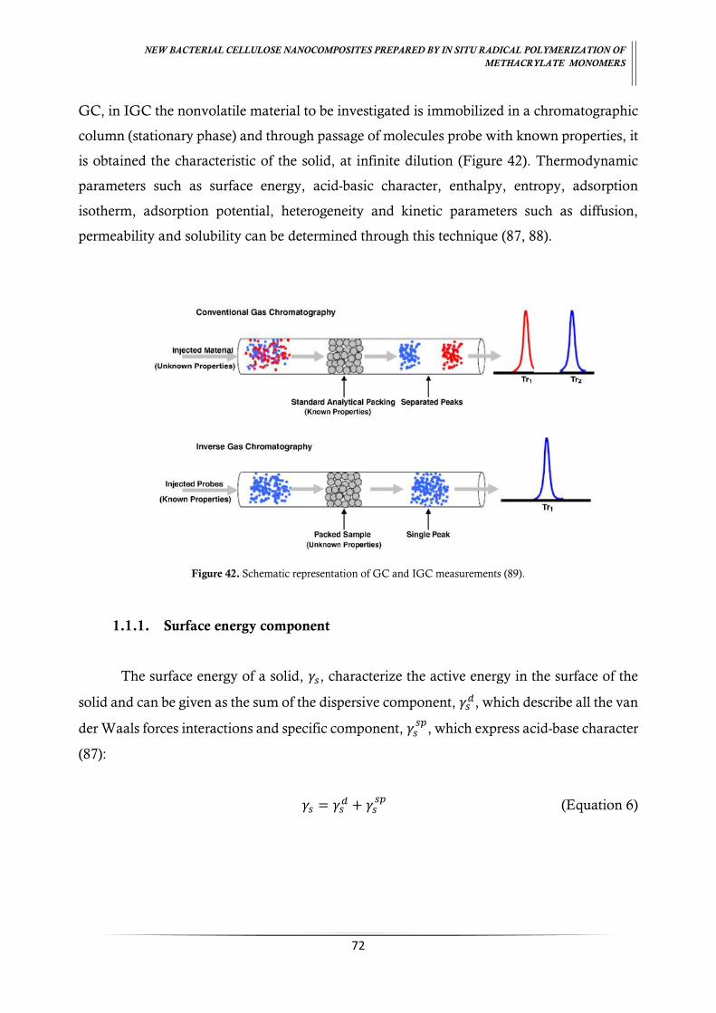
NEW BACTERIAL CELLULOSE NANOCOMPOSITES PREPARED BY IN SITU RADICAL POLYMERIZATION OF
METHACRYLATE MONOMERS
72
GC, in IGC the nonvolatile material to be investigated is immobilized in a chromatographic
column (stationary phase) and through passage of molecules probe with known properties, it
is obtained the characteristic of the solid, at infinite dilution (Figure 42). Thermodynamic
parameters such as surface energy, acid-basic character, enthalpy, entropy, adsorption
isotherm, adsorption potential, heterogeneity and kinetic parameters such as diffusion,
permeability and solubility can be determined through this technique (87, 88).
Figure 42. Schematic representation of GC and IGC measurements (89).
1.1.1. Surface energy component
The surface energy of a solid, 𝛾𝑠, characterize the active energy in the surface of the
solid and can be given as the sum of the dispersive component, 𝛾𝑠𝑑, which describe all the van
der Waals forces interactions and specific component, 𝛾𝑠𝑠𝑝
, which express acid-base character
(87):
𝛾𝑠 = 𝛾𝑠𝑑 + 𝛾𝑠
𝑠𝑝 (Equation 6)

NEW BACTERIAL CELLULOSE NANOCOMPOSITES PREPARED BY IN SITU RADICAL POLYMERIZATION OF
METHACRYLATE MONOMERS
73
The retention time obtained from the interaction between the probe molecules and the
surface of the solid allows us to obtain the volume of probe molecules used, that is directly
related to the energy of adsorption (90), ∆𝐺𝑎𝑑𝑠:
∆𝐺𝑎𝑑𝑠 = 𝑅𝑇 ln 𝑉𝑁 + 𝐾 (Equation 7)
where 𝑅 is the gas constant, 𝐾 is a constant depending on the chosen reference state,
𝑇 is the column temperature and 𝑉𝑁 is the net retention volume. According Fowkes et al,
∆Gads is related to the energy of adhesion, 𝑊𝐴, by the equation:
𝑅𝑇𝑙𝑛𝑉𝑁 = 2𝑁𝐴(𝛾𝑠𝑑)1 2⁄ 𝑎(𝛾𝐿
𝑑)1 2⁄
+ 𝐾 (Equation 8)
where 𝛾𝐿𝑑 is the dispersive component of the surface energy of the probe molecule, 𝑎 is
the area occupied by probe molecule and 𝑁 is the Avogadro’s number. 𝛾𝑠𝑑 can be calculated
from the slope of the obtained line made a plot of ∆G𝑎𝑑𝑠 versus 𝑎(𝛾𝐿𝑑)
1 2⁄ of the series of n-
alkanes.
1.1.2. Acid base character
Knowledge of the surface materials acid or basic character is important to understand
their interaction with polar molecules. The polar molecules and solid interactions involve:
dispersive, ∆𝐺𝑎𝑑𝑠𝑑 , and specific, ∆𝐺𝑎𝑑𝑠
𝑠𝑝, interactions (92). The specific energy of adsorption
∆𝐺𝑎𝑑𝑠 is determined by the equation:
∆𝐺𝑎𝑑𝑠 = ∆𝐺𝑎𝑑𝑠𝑑 + ∆𝐺𝑎𝑑𝑠
𝑠𝑝 (Equation 9)
∆𝐺𝑎𝑑𝑠𝑠𝑝
can be determined by the following relation (92):
∆𝐺𝑎𝑑𝑠𝑠𝑝 = 𝑅𝑇𝑙𝑛𝑉𝑁 − 𝑅𝑇𝑙𝑛𝑉𝑁(𝑟𝑒𝑓) (Equation 10)

NEW BACTERIAL CELLULOSE NANOCOMPOSITES PREPARED BY IN SITU RADICAL POLYMERIZATION OF
METHACRYLATE MONOMERS
74
where 𝑉𝑁 is the net retention volume for the polar probe and VN(ref) is the net retention
volume established by the n-alkane reference line for the same polar probe. From ∆𝐺𝑎𝑑𝑠 it’s
possible to determine the surface enthalpy of adsorption, ∆𝐻𝑎𝑑𝑠 and surface entropy of
adsorption, ∆𝑆𝑎𝑑𝑠 through the following relation (92):
∆𝐺𝑎𝑑𝑠
𝑇=
∆𝐻𝑎𝑑𝑠
𝑇− ∆𝑆𝑎𝑑𝑠 (Equation 11)
∆𝐻𝑎𝑑𝑠𝑠𝑝
and ∆𝑆𝑎𝑑𝑠𝑠𝑝
can be established using the ∆Gadssp
in the previous equation. ∆Gadssp
can be used to quantify the Lewis acidity and basicity of the non-volatile material with the
following equation (92):
∆𝐻𝑎𝑑𝑠𝑠𝑝
𝐴𝑁∗=
𝐷𝑁
𝐴𝑁∗ × 𝐾𝐴 + 𝐾𝐵 (Equation 12)
where 𝐴𝑁∗ and 𝐷𝑁 are Gutmann’s (83) modified acceptor and donor numbers,
respectively; 𝐾𝐴 is a Lewis acidity constant and 𝐾𝐵 is a Lewis basicity constant.
1.1.3. Isotherm measurements
Using a wide variety of probe molecules at different temperatures, it’s obtained the
adsorption isotherm by the BET equation (93):
𝑝
𝑛(𝑝°−𝑝)=
1
𝑛𝑚𝑐+
𝑐−1
𝑛𝑚𝑐×
𝑝
𝑝° (Equation 13)
where nm is the monolayer capacity; 𝑛 the amount adsorbed; 𝑝 the partial pressure;
𝑝° the saturation pressure and 𝑐 is related with the heat of sorption. Knowing the monolayer
capacity and the cross area, 𝑎𝑚, of a probe molecule, the surface area, SBET, can be calculated
by the following equation (93):
𝑆𝐵𝐸𝑇 = 𝑎𝑚𝑛𝑚𝑁𝐴 (Equation 14)
NEW BACTERIAL CELLULOSE NANOCOMPOSITES PREPARED BY IN SITU RADICAL POLYMERIZATION OF
METHACRYLATE MONOMERS
75
where 𝑁𝐴 is the Avogadro constant.
Through the adsorption isotherm, the heterogeneity of the surface can be deduced
from the adsorption potential, 𝐴 by the following equation:
𝐴 = 𝑅𝑇𝑆𝑙𝑛 (𝑝°
𝑝) (Equation 15)
2. Experimental methodology
2.1. Material
Wet bacterial cellulose (BC) membranes in standard conditions were used (70).
Glycidyl methacrylate (GMA, 97%, with 100 ppm of monomethyl ether hydroquinone as
inhibitor), poly (ethylene glycol) methacrylate (PEGMA, average Mn 500, containing
900 ppm monomethyl ether hydroquinone as inhibitor), N,N’-methylenebisacrilamide
(MBA, ≥ 99.5%) and ammonium persulphate (APS, 98%) were purchase in Sigma-Aldrich
and were used as received. The chemical reagents used for IGC measurements such as non-
polar and polar molecules were GC grade (>99% purity) supplied by Sigma–Aldrich. The
methane gas (reference probe) and helium (carrier gas), of high purity (>99.99%), were
supplied by Air Liquide Company.
2.2. BC/PGMA and BC/PEGMA nanocomposites
The bacterial cellulose nanocomposites were obtained through the methodology
described in “Section 2.2. Chapter II: Bacterial cellulose/poly (glycidyl-methacrylate) nanocomposites
films by in situ radical polymerization” and Chapter IV: In situ radical polymerization of bacterial
cellulose/poly(ethyleneglycol)methacrylate nanocomposites films”
Briefly, wet membranes were drained to remove the excess of water. In a separate
Erlenmeyer, the membrane and the reaction mixture were purged in nitrogen for 30 min.
being added, posteriorly through a syringe. After 1 hour at room temperature, the reaction
NEW BACTERIAL CELLULOSE NANOCOMPOSITES PREPARED BY IN SITU RADICAL POLYMERIZATION OF
METHACRYLATE MONOMERS
76
took place in an oil bath for 6 hours at 60ºC. The BC nanocomposites prepared were washed
during 1 hour several times, dried at 40ºC and kept in a desiccator until their characterization.
The reaction mixture to the BC/PGMA was prepared in an aqueous solution with
(1:2) ratio of monomer, BC:GMA, (𝑤 𝑤⁄ ), initiator, APS, 0.5% (𝑤𝑖𝑛𝑖𝑡𝑖𝑎𝑡𝑜𝑟 𝑤𝑚𝑜𝑛𝑜𝑚𝑒𝑟⁄ ) and
cross-linking, MBA, 20% (𝑤𝑐𝑟𝑜𝑠𝑠𝑙𝑖𝑛𝑘𝑒𝑟 𝑤𝑚𝑜𝑛𝑜𝑚𝑒𝑟⁄ ) (when used).
For BC/PEGMA nanocomposites, the reaction mixture was prepared in an aqueous
solution with (1:20) ratio BC:PEGMA (𝑤 𝑤⁄ ) and initiator, 0.5% (𝑤𝑖𝑛𝑖𝑡𝑖𝑎𝑡𝑜𝑟 𝑤𝑚𝑜𝑛𝑜𝑚𝑒𝑟⁄ ).
2.3. IGC measurements
IGC measurements were carried out on a commercial inverse gas chromatograph
(iGC, Surface Measurements Systems, London, UK) equipped with flame ionization (FID)
and thermal conductivity (TCD) detectors. The iGC system is fully automatic with SMS iGC
Controller v1.8 control software. Data were analysed using iGC Standard v1.3 and
Advanced Analysis Software v1.25.
The sample were packed in the standard glass silanized (dymethyldichlorosilane;
Repelcote BDH, UK) columns with 0.2 cm ID and 30 cm in length by vertical tapping about
2 h. The columns with the samples were conditioned 8 h at 40 ºC and 2 h at the measurements
conditions, to stabilize the columns. After conditioning, pulse injections were carried out
with a 0.25 ml gas loop. Four n-alkanes (heptane, octane, nonane and decane) were used for
measurements of the dispersive component of surface energy at 20, 25 and 30 ºC. Four polar
probes (tetrahydrofuran, dichloromethane, acetonitrile and ethyl acetate) were used to
determine the specific component of surface energy and acid-base character (𝐾𝐴 and 𝐾𝐵) at
25 ºC. The physical constants for the probes used in IGC are presented in Table 7. The
isotherm experiments were undertaken with n-octane, tetrahydrofuran and dichloromethane
at 25 ºC. All experiments were carried out at 0% relative humidity with a helium flow rate of
10 ml/min at least in duplicate, producing a reproducibility error of less than 4%.

NEW BACTERIAL CELLULOSE NANOCOMPOSITES PREPARED BY IN SITU RADICAL POLYMERIZATION OF
METHACRYLATE MONOMERS
77
Table 7. Physical constants of all probes used in IGC experiments.
Probe 𝒂(10-19 m2) 𝜸𝑳𝒅(mJ/m2) 𝑫𝑵(kcal/mol) 𝑨𝑵∗(kcal/mol) 𝑫𝑵 𝑨𝑵∗⁄
n-Heptane 5.73 18.4 - - -
n-Octane 6.30 21.3 - - -
n-Nonane 6.90 22.7 - - -
n-Decane 7.50 23.4 - - -
Dichloromethane 2.45 24.5 0.0 20.4 -
Acetonitrile 2.14 27.5 14.1 4.7 3.00
Ethyl Acetate 3.30 19.6 17.1 1.5 11.4
Tetrahydrofuran 2.90 22.5 20.0 0.5 40.0
a: Cross-sectional area; γLd: surface tension; AN∗: electron acceptor number [0.288(AN − ANd)]; DN: electron donor number.
(83, 94)
2.4. Swelling ratio
The swelling ratio (𝑆𝑊) of the membranes was determined by immersing the samples
in distilled water at room temperature with a minimum of three replicates. The weight was
periodically measured during 48 hours. For each measurement the samples were taken out
of the water, their wet surfaces immediately wiped dry in filter paper, weigh, and then re-
immersed. The SW was calculated using the equation below:
𝑆𝑊(%) =(𝑊𝑠−𝑊𝑑)
𝑤𝑑 × 100% (Equation 16)
where, 𝑊𝑆 is the weight of bacterial cellulose after swelling and 𝑊𝑑 is the weight of
dry bacterial cellulose before swelling.
2.5. Scanning electron microscopy
Scanning electron microscopy (SEM) micrographs of the BC nanocomposite surfaces
were obtained on a HR-FESEM SU-70 Hitachi equipment operating at 1.5 kV in the field
emission mode. Samples were deposited on a steel plate and coated with carbon before
analysis.
NEW BACTERIAL CELLULOSE NANOCOMPOSITES PREPARED BY IN SITU RADICAL POLYMERIZATION OF
METHACRYLATE MONOMERS
78
2.6. Energy-dispersive X-ray spectroscopy
The semi-quantitative elemental chemical compositions of the samples were
determined by energy-dispersive X-ray spectroscopy (EDX). Semi-quantitative analyses (wt.
%) were done for carbon, oxygen and nitrogen and the 𝐶 𝑂⁄ ratio was obtained. EDX
experiments were conducted at an accelerated voltage of 5 kV in a Hitachi SU 8090
equipment.
3. Results and Discussion
IGC measurement allows to know the changes that occur in the materials surface
properties due to different modification processes. This data enables the knowledge of their
compatibility and behaviour with other molecules and modifications agents.
In order to study the influence of the poly(glycidylmethacrylate) (PGMA) and
poly(ethyleneglycol)methacrylate (PPEGMA), on the surface properties of the bacterial
cellulose (BC) produced by Gluconacetobacter sacchari, native BC and BC nanocomposites
were subjected to inverse gas chromatography (IGC) analysis. The dispersive component of
the surface energy, 𝛾𝑠𝑑, specific component of the surface energy, ∆𝐺𝑠
𝑠𝑝, surface area, 𝑆𝐵𝐸𝑇,
monolayer capacity, 𝑛𝑚, adsorption potential, 𝐴𝑚𝑎𝑥., acid character, 𝐾𝐴 and basic character,
𝐾𝐵 were determined.
3.1. Dispersive component of surface energy
𝛾𝑠𝑑 was determined by the injection of non-polar molecules (n-alkanes probes) and
obtained plotting 𝑅𝑇𝑙𝑛𝑉𝑛 against 𝑎(𝛾𝐿𝑑)
1/2 at different temperatures (Eq. 8). Table 8 shows
the results obtained for 𝛾𝑠𝑑 in native BC and BC methacrylate nanocomposites. The
correlation coefficient was good, ranging between 0.987 and 0.998.
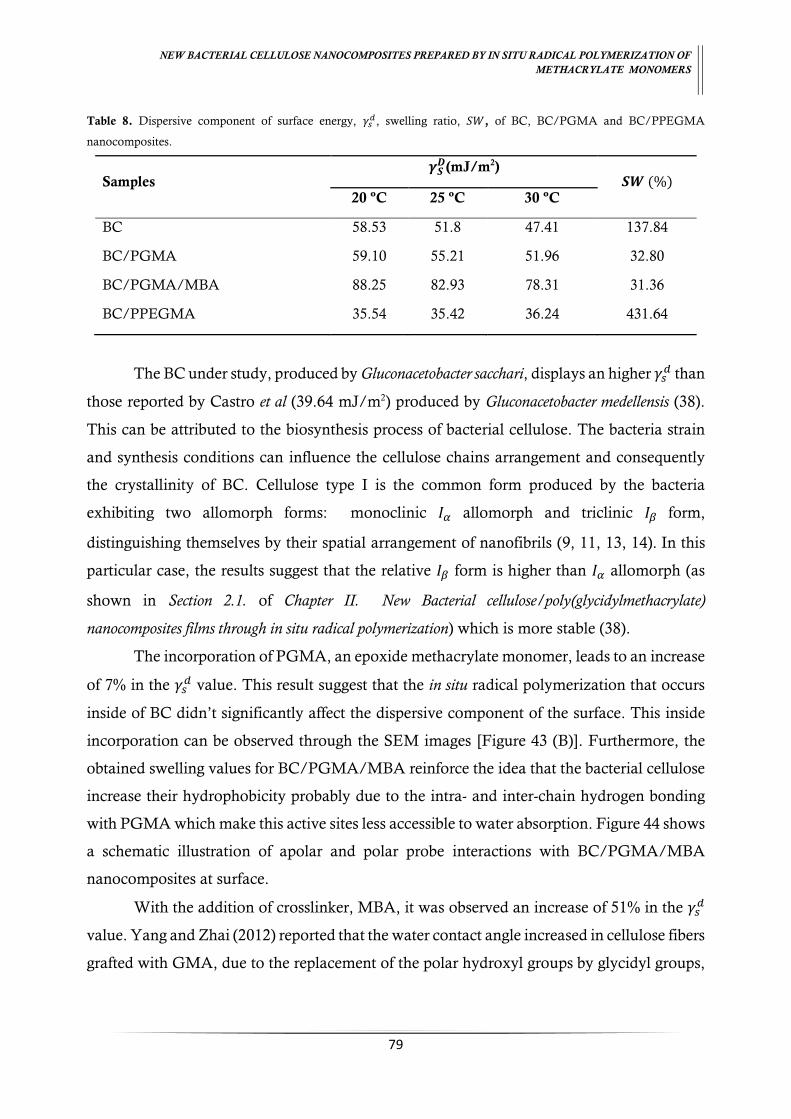
NEW BACTERIAL CELLULOSE NANOCOMPOSITES PREPARED BY IN SITU RADICAL POLYMERIZATION OF
METHACRYLATE MONOMERS
79
Table 8. Dispersive component of surface energy, 𝛾𝑠𝑑, swelling ratio, 𝑆𝑊, of BC, BC/PGMA and BC/PPEGMA
nanocomposites.
Samples 𝜸𝑺
𝑫(mJ/m2) 𝑺𝑾 (%)
20 ºC 25 ºC 30 ºC
BC 58.53 51.8 47.41 137.84
BC/PGMA 59.10 55.21 51.96 32.80
BC/PGMA/MBA 88.25 82.93 78.31 31.36
BC/PPEGMA 35.54 35.42 36.24 431.64
The BC under study, produced by Gluconacetobacter sacchari, displays an higher 𝛾𝑠𝑑 than
those reported by Castro et al (39.64 mJ/m2) produced by Gluconacetobacter medellensis (38).
This can be attributed to the biosynthesis process of bacterial cellulose. The bacteria strain
and synthesis conditions can influence the cellulose chains arrangement and consequently
the crystallinity of BC. Cellulose type I is the common form produced by the bacteria
exhibiting two allomorph forms: monoclinic 𝐼𝛼 allomorph and triclinic 𝐼𝛽 form,
distinguishing themselves by their spatial arrangement of nanofibrils (9, 11, 13, 14). In this
particular case, the results suggest that the relative 𝐼𝛽 form is higher than 𝐼𝛼 allomorph (as
shown in Section 2.1. of Chapter II. New Bacterial cellulose/poly(glycidylmethacrylate)
nanocomposites films through in situ radical polymerization) which is more stable (38).
The incorporation of PGMA, an epoxide methacrylate monomer, leads to an increase
of 7% in the 𝛾𝑠𝑑 value. This result suggest that the in situ radical polymerization that occurs
inside of BC didn’t significantly affect the dispersive component of the surface. This inside
incorporation can be observed through the SEM images [Figure 43 (B)]. Furthermore, the
obtained swelling values for BC/PGMA/MBA reinforce the idea that the bacterial cellulose
increase their hydrophobicity probably due to the intra- and inter-chain hydrogen bonding
with PGMA which make this active sites less accessible to water absorption. Figure 44 shows
a schematic illustration of apolar and polar probe interactions with BC/PGMA/MBA
nanocomposites at surface.
With the addition of crosslinker, MBA, it was observed an increase of 51% in the 𝛾𝑠𝑑
value. Yang and Zhai (2012) reported that the water contact angle increased in cellulose fibers
grafted with GMA, due to the replacement of the polar hydroxyl groups by glycidyl groups,

NEW BACTERIAL CELLULOSE NANOCOMPOSITES PREPARED BY IN SITU RADICAL POLYMERIZATION OF
METHACRYLATE MONOMERS
80
ester and epoxy groups, that exhibit a less polar character. In the present study, the in situ free
radical polymerization of GMA crosslinked with MBA inside BC network probably leads to
two effects: (i) the increase in the number of glycidyl groups exposed in the surface which
present a less polar character, making it more hydrophobic or/and (ii) the change of the
spatial arrangement of microfibrils leaving them more exposed to surface (as shown in Figure
43 (C)).
Figure 43. SEM images of BC nanocomposites morphology (A) BC; (B) BC/PGMA; (C) BC/PGMA/MBA and (D) BC/PPEGMA.
However, the incorporation of the PPEGMA, change differently the surface
properties of BC. PPEGMA has an amphiphilic nature which arises from the pendant
hydroxyl group and its hydrophobic methacrylate group. BC/PPEGMA nanocomposite
exhibited a decrease in the 𝛾𝑠𝑑 value to 36.42 mJ/m2 at 25 ºC (40% less). This result suggests
that the incorporation of PPEGMA decreases the number of non-polar active sites probably
due to the increase of ester bonds and hydroxyls groups from PPEGMA exposed at
nanocomposite surface. As shown in SEM images [Figure 43 (D)], the BC/PPEGMA
exhibited a dense surface arising of incorporation which probably favours the probes
interaction with the polar active sites. The decrease in the 𝐶 𝑂⁄ ratio (2.90) in the

NEW BACTERIAL CELLULOSE NANOCOMPOSITES PREPARED BY IN SITU RADICAL POLYMERIZATION OF
METHACRYLATE MONOMERS
81
BC/PPEGMA nanocomposites compared to 𝐶 𝑂⁄ ratio (3.30) in the native BC evidence this
supposition. Besides that, the water retention ability shows an increase of 213 % compared
with native BC which means that the BC, acquire a hydrophilic character after incorporation
of PPEGMA, in agreement with the 𝛾𝑠𝑑 decrease.
Figure 44. Schematic interactions of non-polar and polar probes with surface groups of BC/PGMA/MBA nanocomposites.
3.2. Heterogeneity
The dispersive energetic profile of the surface heterogeneity is determined by BET
model (81) injecting different concentrations of a n-octane probe. The heterogeneity profile
for BC and BC nanocomposites at 25 ºC are shown in Figure 45. BC/PGMA/MBA
nanocomposites exhibited an increase of the actives sites energy, opposite to the
BC/PPEGMA nanocomposites where occurs a decrease of active sites energy when
compared with native BC. This behaviour is aligned with the results obtained to the
dispersive component of surface energy and described previously.

NEW BACTERIAL CELLULOSE NANOCOMPOSITES PREPARED BY IN SITU RADICAL POLYMERIZATION OF
METHACRYLATE MONOMERS
82
Figure 45. Heterogeneity profile with n-octane for native BC, (A) BC/PGMA and BC/PGMA/MBA and (B)
BC/PPEGMA nanocomposites at 25 ºC.
The BC/PGMA show one peak maximum (𝐴𝑚𝑎𝑥) at 12.59 kJ/Mol with a high
number of actives sites. In the presence of the crosslinker, BC/PGMA/MBA, present two
peak maximum, (𝐴𝑚𝑎𝑥) at 12.58 and 14.76 kJ/Mol, significantly more energetics compared
with native BC [Figure 45 (A)]. The addition of crosslinker plays an important function in
retaining more monomer into the BC network, having a great effect in 𝛾𝑠𝑑, making the surface
with more energetic dispersive actives sites which increase significantly the 𝛾𝑠𝑑. This non-
polar character probably comes from the alkanes chains introduced by more polymerization
of GMA being more affordable to the nanocomposite surface (as supported by SEM images
– Figure 43).

NEW BACTERIAL CELLULOSE NANOCOMPOSITES PREPARED BY IN SITU RADICAL POLYMERIZATION OF
METHACRYLATE MONOMERS
83
In the BC/PPEGMA nanocomposites one peak maximum (𝐴𝑚𝑎𝑥) at 7.38 kJ/Mol is
found with an increase number of less energetic actives sites [Figure 45 (B)], which make the
nanocomposite have smaller 𝛾𝑠𝑑. This result suggests that the introduction of PPEGMA into
the BC network leads to more accessible the non-polar PEG groups.
3.3. Surface area
The isotherm measurements with n-octane at different concentrations BET model
(81)were performed. Symmetrical peaks were obtained and a linear adsorption isotherm
described by Henry’s Law was observed. These indicates that the interactions between
materials and the probe molecule occurs predominantly via the high-energy sites (93).
Besides that a type I isotherm was observed, which is characteristic for microporous materials
with relatively small external surfaces (96).
The surface area (𝑆𝐵𝐸𝑇 ) of bacterial cellulose range between 0.83 m2/g and 12.59 m2/g
and the monolayer capacity (𝑛𝑚) range between 2.18 µmol/g and 33.17 µmol/g (Table 9).
These results show that the polymerization with the two methacrylates had different effects
in the particle morphology. BC/PGMA and BC/PPEGMA nanocomposites increased the
𝑆𝐵𝐸𝑇 in 57 % and 219 %, respectively, when compared with native BC, which means: (i) a
significant decrease in the particles size or/and (ii) increase in its porosity or roughness (89).
SEM images (Figure 43) show that the porosity increase when both polymers were added to
BC network.
The addition of crosslinker had an opposite behaviour, decreasing significantly the
𝑆𝐵𝐸𝑇 in 79 %, which means that occurs an increase of particle size or/and a decrease in
porosity/roughness. This can be due to the more compact and dense surface formed after
incorporation of PGMA/MBA as supported by SEM images (Figure 43).
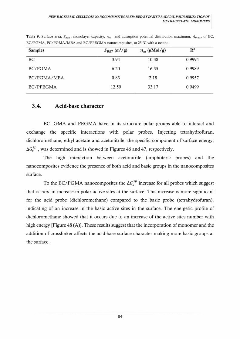
NEW BACTERIAL CELLULOSE NANOCOMPOSITES PREPARED BY IN SITU RADICAL POLYMERIZATION OF
METHACRYLATE MONOMERS
84
Table 9. Surface area, 𝑆𝐵𝐸𝑇, monolayer capacity, 𝑛𝑚 and adsorption potential distribution maximum, 𝐴𝑚𝑎𝑥, of BC,
BC/PGMA, PC/PGMA/MBA and BC/PPEGMA nanocomposites, at 25 ºC with n-octane.
Samples 𝑺𝑩𝑬𝑻 (m2/g) 𝒏𝒎 (µMol/g) R2
BC 3.94 10.38 0.9994
BC/PGMA 6.20 16.35 0.9989
BC/PGMA/MBA 0.83 2.18 0.9957
BC/PPEGMA 12.59 33.17 0.9499
3.4. Acid-base character
BC, GMA and PEGMA have in its structure polar groups able to interact and
exchange the specific interactions with polar probes. Injecting tetrahydrofuran,
dichloromethane, ethyl acetate and acetonitrile, the specific component of surface energy,
∆𝐺𝑠𝑠𝑝
, was determined and is showed in Figures 46 and 47, respectively.
The high interaction between acetonitrile (amphoteric probes) and the
nanocomposites evidence the presence of both acid and basic groups in the nanocomposites
surface.
To the BC/PGMA nanocomposites the ∆𝐺𝑠𝑠𝑝
increase for all probes which suggest
that occurs an increase in polar active sites at the surface. This increase is more significant
for the acid probe (dichloromethane) compared to the basic probe (tetrahydrofuran),
indicating of an increase in the basic active sites in the surface. The energetic profile of
dichloromethane showed that it occurs due to an increase of the active sites number with
high energy [Figure 48 (A)]. These results suggest that the incorporation of monomer and the
addition of crosslinker affects the acid-base surface character making more basic groups at
the surface.
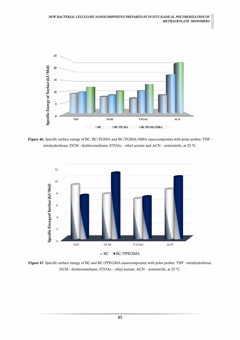
NEW BACTERIAL CELLULOSE NANOCOMPOSITES PREPARED BY IN SITU RADICAL POLYMERIZATION OF
METHACRYLATE MONOMERS
85
Figure 46. Specific surface energy of BC, BC/PGMA and BC/PGMA/MBA nanocomposites with polar probes: THF -
tetrahydrofuran, DCM - dichloromethane, ETOAc – ethyl acetate and ACN – acetonitrile, at 25 ºC.
Figure 47. Specific surface energy of BC and BC/PPEGMA nanocomposites with polar probes: THF - tetrahydrofuran,
DCM - dichloromethane, ETOAc – ethyl acetate, ACN – acetonitrile, at 25 ºC.

NEW BACTERIAL CELLULOSE NANOCOMPOSITES PREPARED BY IN SITU RADICAL POLYMERIZATION OF
METHACRYLATE MONOMERS
86
Figure 48. Heterogeneity profile with dichloromethane for (A) BC, BC/PGMA and BC/PGMA/MBA and (B) BC and BC/PPEGMA nanocomposites, at 25 ºC.
To the BC/PPEGMA nanocomposites, a higher increase in the ∆𝐺𝑠𝑠𝑝
for
dichloromethane (Figure 47) was observed. The dichloromethane energetic profile [Figure
48 (B)] show an increase in the number of active sites. This observation suggests more basic
groups at nanocomposites surface accessible for interactions.
The calculated ∆𝐺𝑠𝑠𝑝
is converted into acid-base number using the Guttmann’s
concept (83). Table 10 present the obtained values for acid-base constants, 𝐾𝐴 and 𝐾𝐵, for
bacterial cellulose nanocomposites. The surface basicity exhibited in native BC is higher than
the one found by Castro et al (2015), which reported a 𝐾𝐵 𝐾𝐴⁄ ratio of 1.80. This result suggests

NEW BACTERIAL CELLULOSE NANOCOMPOSITES PREPARED BY IN SITU RADICAL POLYMERIZATION OF
METHACRYLATE MONOMERS
87
a different arrangement of the cellulose groups at the surface with less hydroxyls groups
available to interactions. The incorporation of basic groups such as (C–O–C) in BC/PGMA
and BC/PPEGMA nanocomposites leads to an increase of the base constant, 𝐾𝐵, showing a
𝐾𝐵 𝐾𝐴⁄ ratio about four times higher.
Taking into account the energetic profile, the increase in the basic character
exhibited in BC nanocomposites derives from the increased number of more energetic basic
active sites due to the incorporation of methacrylates compounds into BC network.
Table 10. Acid-base character, 𝐾𝐴 and 𝐾𝐵 of BC, BC/PGMA BC/PGMA/MBA and BC/PPEGMA nanocomposites, at
25 ºC.
Samples 𝑲𝑨 𝑲𝑩 𝑲𝑩
𝑲𝑨⁄ R2
BC 0.10 0.23 2.27 0.9873
BC/PGMA 0.10 0.42 4.20 0.9868
BC/PGMA/MBA 0.13 0.56 4.31 0.9862
BC/PPEGMA 0.08 0.34 4.25 0.9668
4. Conclusions
The incorporation of methacrylate monomers into the BC network through in situ
radical polymerization is a suitable method to change the BC properties for the production
of new nanocomposites aiming new applications. Inverse Gas Chromatography shows that
the incorporation of glycidylmethacrylate and poly(ethyleneglycol)methacrylate modify the
bacterial cellulose which leads:
(i) in BC/poly(glycidylmethacrylate), to an increase in the dispersive component
of surface energy (decrease in hydrophilicity) and an decrease the surface area
when the crosslinked was added;
(ii) in BC/(poly(ethylene glycol)methacrylate), to a significant decrease in the in the
dispersive component of surface energy (increase of hydrophilicity) and a
decrease in surface area;
NEW BACTERIAL CELLULOSE NANOCOMPOSITES PREPARED BY IN SITU RADICAL POLYMERIZATION OF
METHACRYLATE MONOMERS
88
(iii) in both BC/methacrylate nanocomposites, to a great increase of the basic
surface character.

89
FINAL CONCLUSIONS
BC as support for nanocomposite films has been exploited for numerous applications
and their combination with methacrylate monomers allow to obtain new nanomaterials with
different properties to apply in various fields, namely biomedical applications or/and remove
heavy metals.
In situ free radical polymerization of glycidylmethacrylate, GMA, and poly (ethylene
glycol) methacrylate, PEGMA, occurred into the BC network using ammonium persulphate,
APS, as initiator and N,N’-methylenebisacrilamide, MBA, as crosslinker in BC/PGMA. The
optimal conditions determined for polymerization was (1:2) ratio for BC/PGMA, (1:2:0.2)
for BC/PGMA/MBA and (1:20) for BC/PPEGMA with 0.5 % of initiator during 6 h at 60
ºC. A maximum incorporation percentage of 60, 67 and 87 %, were obtained respectively.
Compared with native BC, the BC/PGMA nanocomposites exhibited a roughness and a
compact three-dimensional structure, an improvement in thermal and mechanical properties,
a decrease in their swelling ability and a lower crystallinity. Unlike, the BC/PPEGMA had
shown a decrease of stiffness of three-dimensional structure and an increase in their swelling
ability. However, an improvement in thermal and mechanical properties and an increase in
the amorphous content it’s observed. Both BC nanocomposites present basic surface
character.
Chemical treatment proved to be a good and simple strategy to modified BC/PGMA
nanocomposites through epoxide ring-opening reaction mechanism. The replacement of
hydroxyls groups of cellulose chains occurs for the linkage of GMA chains by epoxy group.
Nanocomposites became more compact, smooth and with more water retention ability. A
decrease in the thermal and mechanical proprieties was observed.
As future perspectives, the reactivity of epoxy group as absorbent material, it’s an
interesting as well as the biocompatibility of all BC polymethacrylate nanocomposites for
biomedical applications for application as adsorbent material for removal of heavy metal
or/and proteins.
90
REFERENCES
1. The chemistry of polymers. Second edition ed: RSC Paperbacks; 1997. 22 p.
2. Chawla P, Bajaj I, Survase S, Singhal R. Fermentative production of microbial
cellulose. Food Technol Biotechnol. 2009;47 107-24.
3. Keshk S. Bacterial cellulose production and its industrial applications. J Bioproces
Biotechniq. 2014;4:1-10.
4. Khalil H, Bhat A, Yusra A. Green composites from sustainable cellulose nanofibrils:
A review. Carbohydr Polym. 2012;87:963-79.
5. Shokri J, Adibkia K. Application of cellulose and cellulose derivatives in
pharmaceutical industries. Intech. 2013:47-66.
6. Lee K, Buldum G, Mantalaris A, Bismarck A. More than meets the eye in bacterial
cellulose: biosynthesis, bioprocessing, and applications in advanced fiber composites.
Macromol Biosci. 2014;14:10-32.
7. Fu L, Zhang J, Yang G. Present status and applications of bacterial cellulose-based
materials for skin tissue repair. Carbohydr Polym. 2013;92:1432-42.
8. Xu X, Liu F, Jiang L, Zhu J, Haagenson D, Wiesenborn D. Cellulose nanocrystals
vs. cellulose nanofibrils: a comparative study on their microstructures and effects as polymer
reinforcing agents. ACS applied materials & interfaces. 2013;5:2999-3009.
9. Vitta S, Thiruvengadam V. Multifunctional bacterial cellulose and nanoparticle-
embedded composites. Curr Sci. 2012;102:1398-405.
10. Esa F, Tasirin S, Rahman N. Overview of bacterial cellulose production and
application. Agric Agric Sci Procedia. 2014;2:113-9.
11. Mohite B, Patil S. A novel biomaterial: bacterial cellulose and its new era applications.
Biotechnol Appl Biochem. 2014;61:101-10.
12. Cai Z, Kim J. Bacterial cellulose/poly(ethylene glycol) composite: characterization
and first evaluation of biocompatibility. Cellulose. 2009;17:83-91.
13. Moon R, Martini A, Nairn J, Simonsen J, Youngblood J. Cellulose nanomaterials
review: structure, properties and nanocomposites. Chem Soc Rev. 2011;40(7):3941-94.
14. Iguchi M, Yamanaka S, Budhiono A. Bacterial cellulose - a masterpiece of nature's
arts. J Mater Sci. 2000;35:261-70.

91
15. Pinto R, Neves M, Neto C, Trindade T. Composites of cellulose and metal
nanoparticles. Intech. 2012:1-26.
16. Brown A. On an acetic ferment which form cellulose. J Chem Soc London.
1886;49:432-9.
17. Kramer F, Klemm D, Schumann D, Heßler N, Wesarg F, Fried W, et al.
Nanocellulose Polymer Composites as Innovative Pool for (Bio)Material Development.
Macromol Symp. 2006;244:136-48.
18. Ross P, Mayer R, Benziman M. Cellulose biosynthesis and function in bacteria.
Microbiol Rev. 1991;55:35-58.
19. Hestrin S, Schramm M. Synthesis of cellulose by Acetobacter xylinum. 1954;345-351.
20. Schramm M, Hestrin S. Factors affecting production of cellulose at the air/liquid
interface of a culture of Acetobacter xylinum. J gen Microbiol. 1954;11:123-9.
21. Trovatti E, Serafim L, Freire C, Silvestre A, Neto C. Gluconacetobacter sacchari: An
efficient bacterial cellulose cell-factory. Carbohydr Polym. 2011;86:1417-20.
22. Hobzova R, Duskova-Smrckova M, Michalek J, Karpushkin E, Gatenholm P.
Methacrylate hydrogels reinforced with bacterial cellulose. Polym Int. 2012;61:1193-201.
23. Leitão A, Silva J, Dourado F, Gama M. Production and characterization of a new
bacterial cellulose/poly(vinyl alcohol) nanocomposite. Materials. 2013;6:1956-66.
24. Vandamme E, Baets S, Vanbaelen V, Joris K, Wulf P. Improved production of
bacterial cellulose and its application potential. Polym Degrad Stab. 1998;59:93-9.
25. Donini I, Salvi D, Fukumoto F, Lustri W, Barud H, Marchetto R, et al. Biossíntese e
recentes avanços na produção de celulose bacteriana. Ecl Quím 2010;35:165-78.
26. Delmer D, Amor Y. Cellulose biosynthesis. The Plant cell. 1995;7:987-1000.
27. Pokalwar S, Mishra M, Manwar A. Production of Cellulose by Gluconacetobacter sp.
Recent Res Sci Technol. 2010;2:14-9.
28. Sadhu S, Maiti T. Cellulase Production by Bacteria: A Review. Br Microbiol Res J.
2013;3:235-58.
29. Krystynowicz A, Czaja W, Wiktorowska-Jezierska A, Goncalves-Miskiewicz M,
Turkiewicz M, Bielecki S. Factors affecting the yield and properties of bacterial cellulose. J
Ind Microbiol Biotechnol. 2002;29:189-95.
30. Keshk S, Sameshima K. Evaluation of different carbon sources for bacterial cellulose
production. Afr J Biotechnol. 2005;4 478-82.

92
31. Jung H, Lee O, Jeong J, Jeon Y, Park K, Kim H, et al. Production and
characterization of cellulose by Acetobacter sp. V6 using a cost-effective molasses-corn steep
liquor medium. Appl Biochem Biotechnol. 2010;162:486-97.
32. Mikkelsen D, Flanagan B, Dykes G, Gidley M. Influence of different carbon sources
on bacterial cellulose production by Gluconacetobacter xylinus strain ATCC 53524. J Appl
Microbiol. 2009;107:576-83.
33. Mohammadkazemi F, Azin M, Ashori A. Production of bacterial cellulose using
different carbon sources and culture media. Carbohydr Polym. 2015;117:518-23.
34. Oikawa T, Morino T, Ameyama M. Production of cellulose from D-Arabitol by
Acetobacter xylinum KU-1. Biosci Biotech Biochem. 1995;59:1564-5.
35. Oikawa T, Ohtori T, Ameyama M. Production of cellulose from D-Mannitol by
Acetobacter xylinum KU-1. Biosci Biotech Biochem. 1995;59:331-2.
36. Qiu K, Netravali A. A Review of Fabrication and Applications of Bacterial Cellulose
Based Nanocomposites. Polym Rev. 2014;54:598-626.
37. Knauth P, Schoonman J. Nanocomposites: ionic conducting materials and structural
spectroscopies. Tuller HL, editor2008.
38. Castro C, Cordeiro N, Faria M, Zuluaga R, Putaux J, Filpponen I, et al. In-situ
glyoxalization during biosynthesis of bacterial cellulose. Carbohydr Polym. 2015;126:32-9.
39. Castro C, Vesterinen A, Zuluaga R, Caro G, Filpponen I, Rojas O, et al. In situ
production of nanocomposites of poly(vinyl alcohol) and cellulose nanofibrils from
Gluconacetobacter bacteria: effect of chemical crosslinking. Cellulose. 2014;21:1745-56.
40. Figueiredo A, Figueiredo A, Alonso-Varona A, Fernandes S, Palomares T, Rubio-
Azpeitia E, et al. Biocompatible bacterial cellulose-poly(2-hydroxyethyl methacrylate)
nanocomposite films. BioMed research international. 2013;2013:1-14.
41. Phisalaphong M, Jatupaiboon N. Biosynthesis and characterization of bacteria
cellulose–chitosan film. Carbohydr Polym. 2008;74:482-8.
42. Saibuatong O-A, Phisalaphong M. Novo aloe vera–bacterial cellulose composite film
from biosynthesis. Carbohydr Polym. 2010;79:455-60.
43. Figueiredo A, Figueiredo A, Silva N, Barros-Timmons A, Almeida A, Silvestre A, et
al. Antimicrobial bacterial cellulose nanocomposites prepared by in situ polymerization of 2-
aminoethyl methacrylate. Carbohydr Polym. 2015;123:443-53.

93
44. Granel C, Dubois P, Jérôme R, Teyssie P. Controlled radical polymerization of
methacrylic monomers in the presence of a bis(ortho-chelated) arylnickel(II) complex and
different activated alkyl halides. Macromolecules. 1996;29:8576-82.
45. Lu G, Wu D, Fu R. Studies on the synthesis and antibacterial activities of polymeric
quaternary ammonium salts from dimethylaminoethyl methacrylate. React Funct Polym.
2007;67:355-66.
46. Devi R, Maji T, Banerjee A. Studies on dimensional stability and thermal properties
of rubber wood chemically modified with styrene and glycidyl methacrylate. J Appl Polym
Sci. 2004;93(4):1938-45.
47. Labbe A, Brocas A, Ibarboure E, Ishizone T, Hirao A, Deffieux A, et al. Selective
Ring-Opening Polymerization of Glycidyl Methacrylate: Toward the Synthesis of Cross-
Linked (Co)polyethers with Thermoresponsive Properties. Macromolecules. 2011;44:6356-
64.
48. Anirudhan T, Rejeena S. Thorium(IV) Removal and Recovery from Aqueous
Solutions using Tannin-Modified Poly(glycidylmethacrylate)-Grafted Zirconium oxide
Densified Cellulose. Ind Eng Chem Res. 2011;50:13288-98.
49. Anirudhan T, Rejeena S. Poly(acrylic acid)-modified poly(glycidylmethacrylate)-
grafted nanocellulose as matrices for the adsorption of lysozyme from aqueous solutions.
Chem Eng J. 2012;187:150-9.
50. Anirudhan T, Senan P. Adsorptive potential of sulfonated
poly(glycidylmethacrylate)-grafted cellulose for separation of lysozyme from aqueous phase:
Mass transfer analysis, kinetic and equilibrium profiles. Colloids Surf, A. 2011;377:156-66.
51. Lainé C, Bounor-Legaré V, Monnet C, Cassagnau P. Free radical polymerization of
glycidyl methacrylate in plasticized poly(vinyl chloride). Eur Polym J. 2008;44:3177-90.
52. Chauhan G, Guleria L, Sharma R. Synthesis, characterization and metal ion sorption
studies of graft copolymers of cellulose with glycidyl methacrylate and some comonomers.
Cellulose. 2005;12:97-110.
53. Ma M, Li F, Chen F, Cheng S, Zhuo R. Poly(ethylene glycol)-block-poly(glycidyl
methacrylate) with oligoamine side chains as efficient gene vectors. Macromol Biosci.
2010;10:183-91.
54. Altintas E, Denizli A. Monosize poly(glycidyl methacrylate) beads for dye-affinity
purification of lysozyme. Int J Biol Macromol. 2006;38:99-106.

94
55. Anirudhan T, Nima J, Divya P. Adsorption of chromium(VI) from aqueous solutions
by glycidylmethacrylate-grafted-densified cellulose with quaternary ammonium groups.
Appl Surf Sci. 2013;279:441-9.
56. Anirudhan T, Nima J, Sandeep S, Ratheesh V. Development of an amino
functionalized glycidylmethacrylate-grafted-titanium dioxide densified cellulose for the
adsorptive removal of arsenic(V) from aqueous solutions. Chem Eng J. 2012;209:362-71.
57. Anirudhan T, Rejeena S, Tharun A. Preparation, characterization and adsorption
behavior of tannin-modified poly(glycidylmethacrylate)-grafted zirconium oxide-densified
cellulose for the selective separation of bovine serum albumin. Colloids Surf, B. 2012;93:49-
58.
58. Bayramoğlu G, Arıca M. Ethylenediamine grafted poly(glycidylmethacrylate-co-
methylmethacrylate) adsorbent for removal of chromate anions. Sep Purif Technol.
2005;45:192-9.
59. Cañamero P, Fuente J, Madruga E, Fernández-García M. Atom Transfer Radical
Polymerization of Glycidyl Methacrylate: A Functional Monomer. Macromol Chem Phys.
2004;205:2221-8.
60. Benaglia M, Alberti A, Giorgini L, Magnoni F, Tozzi S. Poly(glycidyl methacrylate):
a highly versatile polymeric building block for post-polymerization modifications. Polym
Chem. 2013;4:124-32.
61. Brown A, Khan N, Steinbock L, Huck W. Synthesis of oligo(ethylene glycol)
methacrylate polymer brushes. Eur Polym J. 2005;41:1757-65.
62. Louguet S, Verret V, Bedouet L, Servais E, Pascale F, Wassef M, et al. Poly(ethylene
glycol) methacrylate hydrolyzable microspheres for transient vascular embolization. Acta
Biomater. 2014;10:1194-205.
63. Lee W, Lin W. Preparation and gel properties of poly[hydroxyethylmethacrylate-co-
poly(ethylene glycol) methacrylate] copolymeric hydrogels by photopolymerization. J Polym
Res. 2002;9:23-9.
64. Qiu Y, Klee D, Pluster W, Severich B, Hocker H. Surface modification of
polyurethane by plasma-induced graft polymerization of poly(ethylene glycol) methacrylate.
J Appl Polym Sci. 1996;61:2373-82.

95
65. Uguzdogan E, Denkbas E, Ozturk E, Tuncel S, Kabasakal O. Preparation and
characterization of polyethyleneglycolmethacrylate (PEGMA)-co-vinylimidazole (VI)
microspheres to use in heavy metal removal. J Hazard Mater. 2009;162:1073-80.
66. Duran A, Soylak M, Tuncel S. Poly(vinyl pyridine-poly ethylene glycol methacrylate-
ethylene glycol dimethacrylate) beads for heavy metal removal. J Hazard Mater.
2008;155:114-20.
67. Foulger S, Jiang P, Amanda C, Smith D, Ballato J. Mechanochromic Response of
Poly(ethylene glycol) Methacrylate Hydrogel Encapsulated Crystalline Colloidal Arrays.
Langmuir. 2001;17:6023-6.
68. Kurosumi A, Sasaki C, Yamashita Y, Nakamura Y. Utilization of various fruit juices
as carbon source for production of bacterial cellulose by Acetobacter xylinum NBRC 13693.
Carbohydr Polym. 2009;76:333-5.
69. Abeer M, Mohd Amin M, Martin C. A review of bacterial cellulose-based drug
delivery systems: their biochemistry, current approaches and future prospects. J Pharm Sci.
2014;66:1047-61.
70. Gomes F, Silva N, Trovatti E, Serafim L, Duarte M, Silvestre A, et al. Production of
bacterial cellulose by Gluconacetobacter sacchari using dry olive mill residue. Biomass
Bioenergy. 2013;55:205-11.
71. Zulfiqar S, Zulfiqar M, Nawaz M. Thermal degradation of
poly(glycidylmethacrylate). Polym Degrad Stab. 1990;30 195-203.
72. Martínez-Sanz M, Abdelwahab M, Lopez-Rubio A, Lagaron J, Chiellini E, Williams
T, et al. Incorporation of poly(glycidylmethacrylate) grafted bacterial cellulose nanowhiskers
in poly(lactic acid) nanocomposites: Improved barrier and mechanical properties. Eur Polym
J. 2013;49:2062-72.
73. Lv J, Fang S, Chen L. Preparation and characterization of affinity polymer basic
microsheres by soap-free emulsion polymerization. Chin J Polym Sci. 2009;27:101-8.
74. Hunter T, Price G. Glycidyl methacrylate and N-vinylpyrrolidinone copolymers:
synthesis and nuclear magnetic resonance characterization. Polymer. 1994;35:3530-4.
75. Nanjundan S, Unnithan C, Selvamalar C, Penlidis A. Homopolymer of 4-
benzoylphenyl methacrylate and its copolymers with glycidyl methacrylate: synthesis,
characterization, monomer reactivity ratios and application as adhesives. React Funct
Polym. 2005;62:11-24.
96
76. Vijayanand P, Kato S, Satokawa S, Kojima T. Copolymerization of 4-biphenyl
methacrylate with glycidyl methacrylate: Synthesis, characterization, thermal properties and
determination of monomer reactivity ratios. Polym Bull. 2007;58:861-72.
77. Mahdavinia G, Pourjavadi A, Hosseinzadeh H, Zhuriaan M. Modified chitosan 4.
Superabsorbent hydrogels from poly(acrylic acid-co-acrylamide) grafted chitosan with salt-
and pH-responsiveness properties. Eur Polym J. 2004;40:1399-407.
78. Silioc C, Maleki A, Zhu K, Kjøniksen A, Nystrom B. Effect of hydrophobic
modification on rheological and swelling features during chemical gelation of aqueous
polysaccharides. Biomacromolecules. 2007;8:719-28.
79. Gea S, Reynolds C, Roohpour N, Wirjosentono B, Soykeabkaew N, Bilotti E, et al.
Investigation into the structural, morphological, mechanical and thermal behaviour of
bacterial cellulose after a two-step purification process. Bioresour Technol. 2011;102:9105-
10.
80. Reis A, Fajardo A, Schuquel I, Guilherme M, Vidotti G, Rubira A, et al. Reaction of
glycidyl methacrylate at the hydroxyl and carboxylic groups of poly(vinyl alcohol) and
poly(acrylic acid): Is this reaction mechanism still unclear? J Org Chem 2009;74:3750-7.
81. Cordeiro N, Gouveia C, Moraes A, Amico S. Natural fibers characterization by
inverse gas chromatography. Carbohydr Polym. 2011;84:110-7.
82. Kádár K, Százdi L, Fekete E, Pukánszky B. Surface characteristics of layered silicates:
Influence on the properties of clay/polymer nanocomposites. Langmuir. 2006;22:7848-54.
83. Gutmann V. The donor acceptor approach to molecular interactions1978.
84. Kita R, Svec F, Fréchet J. Hydrophilic polymer supports for solid-phase
synthesis: preparation of poly(ethylene glycol) methacrylate polymer beads using “classical”
suspension polymerization in aqueous medium and their application in the solid-phase
synthesis of hydantoins. J Comb Chem. 2001;3:564-71.
85. Chang Y, Shih Y, Ruaan R, Higuchi A, Chen W, Lai J. Preparation of
poly(vinylidene fluoride) microfiltration membrane with uniform surface-copolymerized
poly(ethylene glycol) methacrylate and improvement of blood compatibility. J Membr Sci.
2008;309:165-74.
86. Hu W, Chen S, Yang J, Li Z, Wang H. Functionalized bacterial cellulose derivatives
and nanocomposites. Carbohydr Polym. 2014;101:1043-60.
97
87. As kin A, Yazıcı D. A study of the surface analysis of some water-soluble polymers by
inverse gas chromatography. Surf Interface Anal. 2008;40:1237-41.
88. Belgacem M, Czeremuszkin G, Sapieha S. Surface by XPS characterization and
inverse gas of cellulose fibres chromatography. Cellulose. 1995;2:145-7.
89. Cordeiro N, Ashori A, Hamzeh Y, Faria M. Effects of hot water pre-extraction on
surface properties of bagasse soda pulp. Mater Sci Eng, C. 2013;33:613-7.
90. Voelkel A, Strzemiecka B, Adamska K, Milczewska K. Inverse gas chromatography
as a source of physiochemical data. J Chromatogr A. 2009;1216:1551-66.
91. Fowkes M, Mostafa M. Acid-Base Interactions in Polymer Adsorption. Ind Eng
Chem Prod Res Dev. 1978;17:3-7.
92. Rani P, Ramanaiah S, Reddy K. Lewis acid-base properties of cellulose acetate
butyrate by inverse gas chromatography. Surf Interface Anal. 2011;43(3):683-8.
93. Thielmann F. Introduction into the characterisation of porous materials by inverse
gas chromatography. J Chromatogr A. 2004;1037:115-23.
94. Ashori A, Cordeiro N, Faria M, Hamzeh Y. Effect of chitosan and cationic starch on
the surface chemistry properties of bagasse paper. Int J Biol Macromol. 2013;58:343-8.
95. Yang G, Zhai H. Synthesis and properties of glycidylmethacrylate grafted eucalyptus
fibers. Cellul Chem Technol. 2012;46:243-8.
96. Sing K, Everett D, Haul R, Moscou L, Pierotti R, Rouquerol J, et al. Reporting
physisorption data for gas/solid systems with special reference to the determination of
surface area and porosity. Pure & App Chem. 1985;57:603-19.