neutron scattering in magnetic systems - university of...
TRANSCRIPT

Jim RhyneLujan Neutron Scattering Center
Los Alamos National Laboratory
Neutron Scattering in Magnetic Systems
2006 Spins in Solids Summer SchoolJune 19 – 23, 2006

What Can Neutrons Do?
• Diffraction (the momentum [direction] change of the neutron is measured)
– Atomic Structure via nuclear positions– Magnetic Structure(neutron magnetic moment interacts with
internal fields)– Disordered systems - radial distribution functions– Depth profile of order parameters from neutron reflectivity– Macro-scale structures from Small Angle Scattering (1 nm to
100 nm)• Inelastic Scattering (the momentum and energy change
of the neutron is measured)– Dispersive and non-dispersive phonon and magnon excitations– Density of states– Quasi-elastic scattering
Neutrons measure the space and time-dependent correlation function of atoms and spins – All the Physics!
What do we need to do neutron scattering?
• Neutron Source – produces neutrons• Diffractometer or Spectrometer
– Allows neutrons to interact with sample– Sorts out discrete wavelengths by monochromator (reactor) or by time of
flight (pulse source)– Detectors pick up neutrons scattered from sample
• Analysis methods to determine material properties• Brain power to interpret results
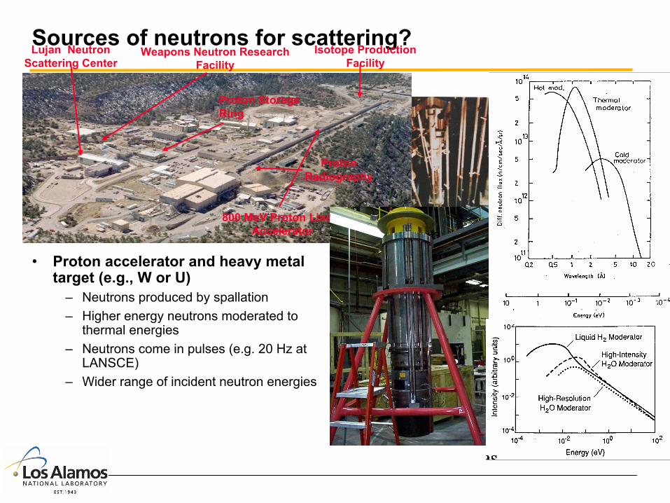
Sources of neutrons for scattering?
• Nuclear Reactor– Neutrons produced from fission of 235U– Fission spectrum neutrons moderated to
thermal energies (e.g. with D20)– Continuous source – no time structure– Common neutron energies -- 3.5 meV <
E < 200 meV
• Proton accelerator and heavy metal target (e.g., W or U)
– Neutrons produced by spallation– Higher energy neutrons moderated to
thermal energies– Neutrons come in pulses (e.g. 20 Hz at
LANSCE)– Wider range of incident neutron energies
Lujan Neutron Scattering Center
Weapons Neutron ResearchFacility
Proton Radiography
800 MeV Proton Linear Accelerator
Isotope ProductionFacility
Proton Storage Ring

High-FluxIsotope Reactor
Spallation Neutron Source (first neutrons in
May -- operational instruments late in 2006)
(1000 kW)
Intense Pulsed Neutron Source(7 kw)
Manuel Lujan Jr. Neutron Scattering
Center(100 kW)
National User National User FacilitiesFacilitiesHFIR 1966 HFIR 1966 NCNR 1969 NCNR 1969 IPNS 1981 IPNS 1981 Lujan 1985 Lujan 1985 (SNS 2006) (SNS 2006)
Local/Regional Local/Regional FacilitiesFacilities(University (University Reactors)Reactors)MITMITMissouriMissouri……
NIST Center NIST Center for Neutron for Neutron ResearchResearch
There are four National User Facilitiesfor neutron scattering in the US

Neutron scattering machines
• Spectrometers or diffractometers– typically live in a beam room – are heavily shielded to keep
background low and protect us– receive neutrons from the target (or
reactor)– correlate data with specific neutron
wavelengths by time of flight– accommodate sample
environments (high/low temperature, magnetic fields, pressure apparatus)

Neutron Scattering’s Moment in the Limelight

8
General Properties of the NeutronThe kinetic energy of a 1.8 Å neutron is equivalent to T = 293K (warm coffee!), so it is called a thermal neutron.The relationships between wavelength (Å) and the energy (meV), and the speed (m/s, mi/hr) of the neutron are:
e.g. the 1.8 Å neutron has E = 25.3 meV and v = 2200 m/s = 4900 mi/hrThe wavelength if of the same order as the atomic separation so interference occurs between waves scattered by neighboring atoms(diffraction).Also, the energy is of same order as that of lattice vibrations (phonons) or magnetic excitations (magnons) and thus creation of annihilation of a lattice wave produces a measurable shift in neutron energy (inelastic scattering).
λλ /3960 and /89.81 2 == vE

9
COMPARATIVE PROPERTIES OF X-RAY ANDNEUTRON SCATTERING
Property X-Rays Neutrons
Wavelength Characteristic line spectra such as Cu Kαλ = 1.54 Å
Continuous wavelength band, or single λ =1.1 ± 0.05 Å separated out from Maxwell spectrum by crystal monochromator or chopper
Energy for λ = 1 Å 1018 h 1013 h (same order as energy of elementary excitations)
Nature of scattering by atoms
ElectronicForm factor dependence on [sinθ]/λLinear increase of scattering amplitude with atomic number, calculable from known electronic configurations
Nuclear, Isotropic, no angular dependent factor Irregular variation with atomic number. Dependent on nuclear structure and only determined empirically by experiment
Magnetic Scattering Very weak additional scattering (≈ 10-5) Additional scattering by atoms with magnetic moments (same magnitude as nuclear scattering) Amplitude of scattering falls off with increasing [sin θ]/λ
Absorption coefficient
Very large, true absorption much larger than scattering µabs ≈ 102 - 103
increases with atomic number
Absorption usually very small (exceptions Gd, Cd, B …) and less thanscattering µabs ≈ 10-1
Method of Detection Solid State Detector, Image Plate Proportional 3He counter

10
Golden Rule of Neutron Scattering
We don’t take pictures of atoms!
Job preservation for neutron scatterers – we live in reciprocal space
Atoms in fcc crystal
Inte
nsity

11
How are neutrons scattered by atoms (nuclei)?
Short-range scattering potential:
The quantity “b” (or f) is the strength of the potential and is called the scattering length – depends on isotopic compositionThus “b” varies over N nuclei – can find average
defines coherent scattering amplitude leads to diffraction – turns on only at Bragg peaksBut what about deviations from average? This defines the incoherent scattering
Incoherent scattering doesn’t depend on Bragg diffrac. condition, thus has no angular dependence – leads to background (e.g., H)
bbcoh =b
)(2)(2
rbm
rV δπh=
( ) 2/122 bbbinc −=
12
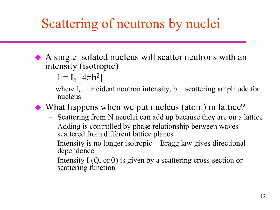
Scattering of neutrons by nuclei
A single isolated nucleus will scatter neutrons with an intensity (isotropic)– I = I0 [4πb2]
where I0 = incident neutron intensity, b = scattering amplitude for nucleus
What happens when we put nucleus (atom) in lattice?– Scattering from N neuclei can add up because they are on a lattice– Adding is controlled by phase relationship between waves
scattered from different lattice planes– Intensity is no longer isotropic – Bragg law gives directional
dependence– Intensity I (Q, or θ) is given by a scattering cross-section or
scattering function

13
Observed Coherent ScatteringIntensity of diffracted x-ray or neutron beam produces series of peaks at discrete values of 2θ [or d or K (also Q)]Note: d = λ/(2 sinθ) or K = 4πsinθ/ λ = 2π/d are more fundamental since values are independent of λ and thus characteristic only of material.
Benzine Pattern (partial)Note: Inversion of scales - 2θ ∝ f(1/d)

14
Scattering Cross-sectionThe measured scattered intensity in a diffraction experiment is proportional to a scattering function S(Q), which is proportional to a scattering cross-sectionI(2θ,d,or Q) ∝ S(Q) ∝ Ω = solid angleIn turn the cross-section ∝ |A(Q) • A*(Q)|
A = scattering anplitudeIn second Born approximation (kinematic limit) A ∝ Fourier transform of scattering length density ρ(r) = (α atom) with the sum over j atoms at position Rj
then
The scattering factor f(Q) is the fundamental quantity describing the scattering of radiation from the material
– f takes different forms depending on the type of radiation– f varies in magnitude depending on the scattering atom or magnetic spin
Ωddσ
∑ −j
jaj Rr )(rrρ
')()(factor scattering thewhere
)()()(
3'2'
232
∫
∑ ∑∫∗
∗∗
=
=−=
rderQf
eQfrdeRrQA
riQajj
j j
RiQj
riQjaj
π
ππ
ρ
ρ

15
Scattering Factors f.The Fourier transform character of the scattering factor f means that the radial extent of the scattering center density ρaj( r) will dictate its Q dependence.
– x-rays scatter from the electron cloud of dimensions comparable to λ or d (∝1/Q)
– Neutrons scatter from the nucleus ≈ 10-5 the dimension of λ or d
')()(factor scattering The 3'2'∫ ∗= rderQf riQaj
πρ

16
Scattering Factors f, cont’dFor x-rays the magntude of f is proportional to ZFor neutrons nuclear factors determine f, thus no regular with Z (different isotopes can have different f s)
Shaded (negative) --> π phase changeFor neutrons conventionally f = b (Scattering length - constant for an element)

17
What Controls the Scattering Amplitude?
I(Q) ∝ S(Q) ∝ |A(Q) • A*(Q)| [Measured scattered intensity, S = scat.func.]
where faj = atomic scattering factor [cm-1]
Magnitude of A(Q) is controlled by [|A(Q) • A*(Q)| called Structure Factor]– f values for various atoms in lattice– destructive interference of waves scattering from atoms at various lattice sites (calculation
of above sum over atoms in lattice reveals this)
fcc lattice (000, 0½½ , ½0½, ½½0) bcc lattice (000, ½½½)– A(Q) = 4 fa for hkl all odd or A(Q) = 2 fa for h+k+l even
hkl all even– A(Q) = 0 otherwise A(Q) = 0 otherwise
∑ ∗=j
RiQaj eQfQA π2)()(
18
Applications ofPowder Diffraction Methods
Scattering instruments, line shapes, resolutionRietveld Structure Refinement AnalysisMagnetic Structures and DiffractionDiffuse Scattering
19
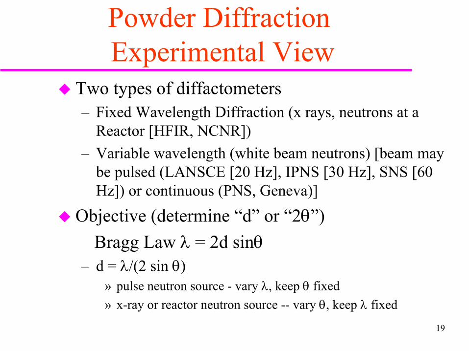
Powder DiffractionExperimental View
Two types of diffactometers– Fixed Wavelength Diffraction (x rays, neutrons at a
Reactor [HFIR, NCNR])– Variable wavelength (white beam neutrons) [beam may
be pulsed (LANSCE [20 Hz], IPNS [30 Hz], SNS [60 Hz]) or continuous (PNS, Geneva)]
Objective (determine “d” or “2θ”) Bragg Law λ = 2d sinθ
– d = λ/(2 sin θ)» pulse neutron source - vary λ, keep θ fixed» x-ray or reactor neutron source -- vary θ, keep λ fixed

20
COMPARISON OF POWDER DIFFRACTION INSTRUMENTS*** Bragg Scattering: λ = 2d sin θ [we need “d”] ***
Reactor Source -- Monochromator selects near mono-energetic neutrons, detector moves (2θ) to collect discrete•(λfixed, vary θ) diffraction data
λ= f(t) so d=λ/(2sinθ)= f(t)/(2sinθ)
short t short λ small d; long t long λ large d
•Pulse Source -- White beam of moderated neutrons used, neutron time-of-flight selects wavelength, detectors grouped in banks, no moving parts
(θ fixed, vary λ)

21
Rietveld Profile RefinementLeast Squares Fitting Procedure for Powder Data
Input Data– Powder scattering pattern data– Trial structure space group and approximate lattice parameters and
atomic positions– Line shape function and Q-dependence of resolution
Output Results– Lattice Parameters– Refined atomic positions and occupancies– Thermal parameters (Debye Waller) for each atom site– Resolution parameters– Background parameters– R factors of fit (and other measures of fit precision)– Preferential orientation, absorption, etc.
More than one phase can be separately refined

22
Magnetic Powder Diffraction
Neutron has a magnetic moment -- will interact with any magnetic fields within a solid, e.g., exchange fieldMagnetic scattering amplitude for an atom (equivalent to b)
where g = Lande “g” factor, J = total spin angular momentum, f = magnetic electrons form factorMagnetic scattering comes from polarizedspins (e.g., 3d [Fe] or 4f [RE]) not fromnucleus -- Therefore scattering amplitudeis Q-dependent (like for x-rays) via fat Q = 0 for Fe µ = gJ = 2.2 Bohr magnetonsp = 0.6 (comparable to nuclear b = 0.954)all in units of 10-12cm Refinement gives moment magnitudes oneach site and x,y,z components(if symmetry permits)
)(10269.02
122
2
cmfgJxfgJmcep −=
=
γ
Mn+2

23
Form FactorsExperimental Calculated
LessLocalizedMoment
MoreLocalizedMoment
24
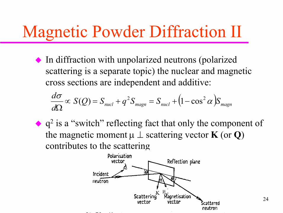
Magnetic Powder Diffraction IIIn diffraction with unpolarized neutrons (polarized scattering is a separate topic) the nuclear and magnetic cross sections are independent and additive:
q2 is a “switch” reflecting fact that only the component of the magnetic moment µ ⊥ scattering vector K (or Q) contributes to the scattering
( ) magnnuclmagnnucl SSSqSQSdd ασ 22 cos1)( −+=+=∝
Ω
K µ

25
Basic Types of Magnetic Order and Resulting Scattering
Ferromagnet (parallel spins)– Single Magnetic site (e.g., Fe, Co,
Tb) -- Scattering only at Bragg peak positions (adds to nuclear), but not necessarily all (q2 switch)
– Multi Site Ferromagnet
(e.g. Y6Fe23 (4 distinct Fe sites) -- no new peaks in scattering
Antiferromagnet (parallel spins with alternate sites reversed in direction)
– equivalent to new magnetic unit cell doubled in propagation direction of AFM
– Purely magnetic scattering peaks at half Miller index positions (e.g., 1,1,1/2)
– Overall net magnetic moment adds to 0 [job security for neutrons!!]
c
a

26
Magnetic Structures, cont’d.Other more complex antiferromagnetic cells (multi-site, but on each site Σµ = 0)
Ferrimagnet (multi-site cell)– Spins parallel on each site– Sites can reverse spin direction– Σµ ≠ 0 on each site, but overall
Σµ could = 0
– Ferromagnetic type scattering pattern --No new peaks
(111) FM Sheets
bcc siteantiparallelto corners
FM layersalong (0001)
AFM linealong (111)
A B C D
27
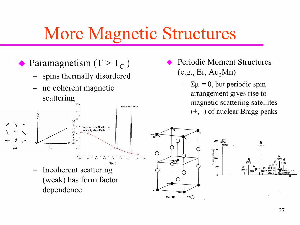
More Magnetic StructuresParamagnetism (T > TC )– spins thermally disordered– no coherent magnetic
scattering
– Incoherent scattering (weak) has form factor dependence
Periodic Moment Structures (e.g., Er, Au2Mn)– Σµ = 0, but periodic spin
arrangement gives rise to magnetic scattering satellites (+, -) of nuclear Bragg peaks

Polarized Neutron Reflectometry
Detector
SampleAl-Coil Spin Flipper
Spin Polarizing Supermirror
Specular ReflectivityIncident Polarized Neutrons
Al-Coil Spin Flipper
]n1)[4/k()z( 22i −π=ρ
22 dz)ikzexp()z()z(
iQ4)Q(r ∫
∞
∞−
ρΨπ
=
( )
φπ
λ−≈
φ±π
λ−≈
±
±±
cospN2
1n
sinpbN2
1n
2
2
m
• index of refraction: sensitive to µ
• scattering length density: used to model reflectivity
• reflectivity: measured quantity
spin-flip
non spin-flip

Ga1-xMnxAs
• Dilute ferromagnetic semiconductor• Spintronics applications• Annealing increases magnetization
& Tc• Interstitial Mn go to the surface!
– K. W. Edmonds et al., PRL, 92, 37201, (2004) - Auger
• Depth-dependence of chemical order and magnetization determined Polarized-Beam Neutron Reflectivity
• Compared similar as-grown and annealed films
– T = 13 K, H = 1 kOe (in plane)
J. Blinowski et al, Phys. Rev. B 67, 121204 (2003)

GaMnxAs1-x As-Grown & Annealedt = 110 nm, x = 0.08, TC = 50 K, 120 K
0.01 0.02 0.03 0.04 0.0510-10
10-9
10-8
Q (Å-1)
Ref
lect
ivity
x Q
4 (Å-4)
As-Grown Film measured R++ fit to R++
measured R- - fit to R- -
Annealed Film measured R++ fit to R++
measured R- - fit to R- -
0.01 0.02 0.03 0.04 0.05
-0.1
0.0
0.1
0.2
Annealed Film measured SA fit from SLD model
Q (Å-1)
Spi
n A
sym
met
ry
0.01 0.02 0.03 0.04 0.05
-0.1
0.0
0.1
0.2
Q (Å-1)
As-Grown Film measured SA fit from SLD model
Spin
Asy
mm
etry
0 200 400 600 800 1000 12000
24
68
290
300
310
320
As-Grown Film
Depth (Å)
Mavg = 17 emu cm-3
moment per Mn = 1.2 µB
ρ (µ
m-2
)
ρnuc ρmag
0
10
20
30
substratesurface
M (em
u cm-3)
0 200 400 600 800 1000 12000
5
10
15
290
300
310
320
ρnuc ρmag
Mavg = 48 emu cm-3
moment per Mn = 3.4 µB
ρ (µ
m-2)
substratesurface Depth (Å)
Annealed Film
0
20
40
60
M (em
u cm-3)
• Measured reflectivities & fits– Spin up & spin down splitting due to
sample magnetization– Spin up reflectivities are different– “Slope” at high Q different – Fits are good
• Magnetic signal: spin asymmetry– SA = (up – down) / (up + down)– Larger amplitude for annealed film– Better defined for annealed film
• SLD Models (mag. & chem.)– As-grown M doubles near surface – M increases and more uniform for
annealed film– Both films show magnetic depletion at
surface– Drastic chemical change at annealed
film’s surface– Interstitial Mn have diffused to
surface! (combined with N2 during annealing)
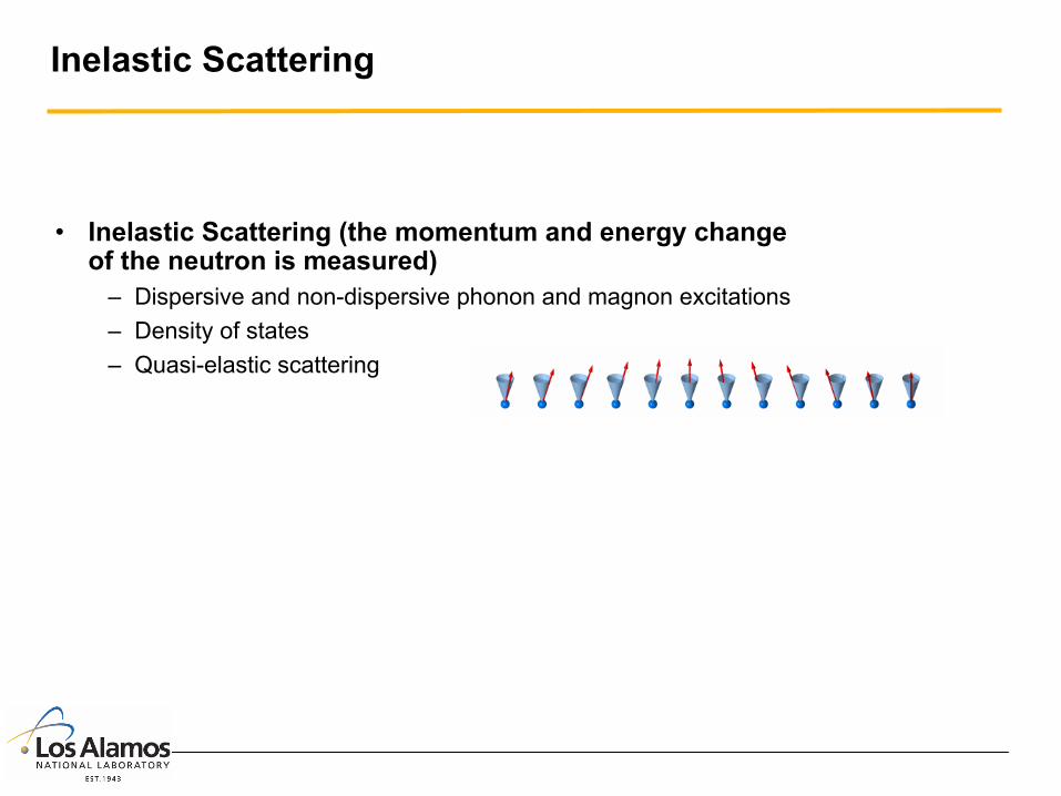
Inelastic Scattering
• Inelastic Scattering (the momentum and energy change of the neutron is measured)
– Dispersive and non-dispersive phonon and magnon excitations– Density of states– Quasi-elastic scattering

32
Triple Axis Neutron Scattering Spectrometer
Want Thermal neutronse.g., E=14mev, λ=2.4Å
λi = 2dmsin θm|ki| = 2π/λi
n
ii m
kE2
22h=
λf = 2dasinθa|kf| = 2π/λf
dEddcountsI
mk
En
ff
Ω∝
=
σ2
det
22
)(
2h
kTEE e
kTE
kTnn /2
1
02 −
=
π

33
MURR Triple Axis Neutron Spectrometer (TRIAX)
Analyzer Assembly
MonochromatorDrum
Sample Table andGoniometer
Detector Shieldand Collimator
Beam Stop (pivots withdrum and sample)

34
22
nf2
2
ni 2E
2E fi k
mk
mhh
==
( ) 2
222
ωhh−=−=∆ ifN kk
mE
Qqkk if
rrrrr≡+=− τπ2
P
0
i f
Inelastic Neutron Scattering ***
The measurement of the functional dependence of wave vector q and the energy E = hω of an elementary excitation (e.g., magnon or phonon dispersion) • Energy and Momentum Conservation Conditions: * ki = wave vector of incident neutron * kf = wave vector of scattered neutron
* Neutron energies
♦ Change of EN creates (∆EN < 0) or annihilates (∆EN > 0) an excitation of energy ω
♦Neutron - excitation system
momentum conservation: ♦ Constant q scan (Einc fixed)
Rotate P [kf,and ki] about 0
ωh

35
Physical Phenomena Studied with Inelastic Neutron Scattering *** accessible energy and momentum
transfer (Q) ranges ***
Note: Cold and hot sources at Reactors and particularly Spallation Neutron Sources have greatly extended the useful range of Q and E.
[From J.D. Axe, Neutrons: The Kinder, Gentler Probe of Condensed Matter, Matls. Res. Symp. Proc. 166, 3 (1990)].

36
0 1 2 3 40
10
20
30
40
50
60
70
80
Γ
Scat
tere
d In
tens
ity (c
ount
s/m
on.in
t.)E (meV)
0.00 0.05 0.10 0.15 0.20 0.25 0.30
1
2
3
4
5
6
qo
Ene
rgy
(meV
)
q (A-1)
Measuring Inelastic Processes • Scans are usually made with q fixed and either Ei
or Ef varying (Constant q scans)
• Can alternatively fix E and vary q under special conditions (constant E scans)
• Constant q scan:
♦ Measure intensity scattered from analyzer (and sample) as a function of Ei or Ef
• Peak of intensity
profile becomes one point on dispersion curve
Dispersion Curve (FM)
qfixed = qo

37
Magnetic Scattering Cross Section
• General form:
• The Scattering Function S(Q,ω) is the Fourier transform of the space and time correlation function G(r,t) of the spins -- All the physics!!
• S(Q,ω) can be written in terms of the expectation values of components of Heisenberg spin operators <Jα(r,t)|Jβ(0,0)> (including the Debye Waller factor W(Q) for thermal vibrations):
where Jα(Q,t) is the q-space Fourier transform of the real space spin operator Jα(r,t)

38
• The integral of the diagonal component Sαα over the Brillouin zone gives the average value of the α component of the magnetic moment:
• The diffuse (i.e. non-Bragg) scattering part of
S(Q,ω) can be related to the imaginary part of a generalized susceptibility χ(Q,ω) via the fluctuation dissipation theorem:
• The cross section then directly measures the wave
vector and energy-dependent susceptibility:

39
• The imaginary part of the generalized susceptibility χ(q,ω) can be expressed as a static susceptibility χ(q) times a Dynamic Spectral Weight Function F(q,ω) [the Fourier transform of the spin relaxation function]:
• χ(q) F(q,ω) may further be separated into a
longitudinal fluctuation part and a transverse part (latter gives rise to spin waves):
• The form of F(q,ω) can not be rigorously
determined so one of two forms are generally used
Double Lorentzian Damped Harmonic Oscillator
( )[ ] ∑ ′−−
×
−′
=
Ω
βα
αβαββααβ ωχδ
µωβωβω
γσ
,2
2
2
2
1
2
),()()(1)exp(
)[exp(
)(2exp)(21
qFqqqg
N
QWkkQgF
cme
dEdd
kkB
o
h
h
[ ] [ ] [ ]tkkkkkk qFqqFqqFq ),()(),()(),()( ωχωχωχ αβαβαβαβαβαβ
′′′ +=l

40
-5 -4 -3 -2 -1 0 1 2 3 4 50
1
2
3
4
5
6
Inte
nsity
E (meV)
-5 -4 -3 -2 -1 0 1 2 3 4 50
1
2
3
4
5
6
ΓΓ
Inte
nsity
E (meV)
Spectral Weight Function for Spin Waves • At low temperature (δ functions):
creation annihilation
• At finite temperatures linewidth (lifetime) broadening Γ
occurs (Double Lorentzian form):
• The transverse (spin wave) part of the cross
section becomes:
• Where the damping coefficient Γ (linewidth) in a Heisenberg magnet varies as T2 and q4:
][][21),( ωωδωωδω ++−= qq
t qF
Γ++Γ
+Γ+−
Γ= 2222 ][][2
1),(qq
t qFωωωωπ
ω
[ ] [ ]
Γ++Γ
+Γ+−
Γ−
×
−
=
Ω
22221
2
122
2
2
1
2
211
)(])exp(1[
)(2exp)(21
qqB
oo
t
qrgN
QWkk
QgFcm
edEd
d
ωωωωπµωβωβ
γσ
h
h
2
42 ln)(
=Γ
q
BTkqTqωh

410.0 0.1 0.2 0.3 0.4 0.5
0
20
40
60
80
100
120
[qqq]
[qq0]
[q00]
Ene
rgy
(meV
)
q (rlu)
Dispersion in a Heisenberg Ferromagnet
• At low q dispersion is quadratic (to lowest order):
E(q) = ω(q) = ∆ + D q2
D is the spin wave stiffness parameter (may be anisotropic in q direction
∆ is the gap energy (may be 0 -- reflects crystal field anisotropy energy)
• Full Brillouin zone dispersion for a single nearest neighbor exchange constant J1 is of the form:
ω(q) = ∆ + 4J1S (3 – cos qxao – cos qyao – cos qzao)
which reduces at small q (cos x ≈ 1 - x2/2) to (111):
ω(q111) ≈ 6J1Sa2q2 ≡ Dq2
Full zone dispersion for a fcc lattice
• J1 ∝ Tc In mean field approx: Tc = 4 J1S(S+1)/kB
h
h
h

42
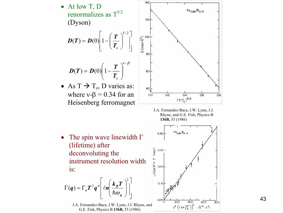
Examples of Inelastic Scattering Data
• Ferromagnetic Amorphous metallic glass Fe0.86B0.14
Constant q scans ♦ left panel -
varying T ♦ right panel -
varying q) [Rhyne, Fish, and Lynn,
JAP 53, 2316 (1982)]
• Quadratic dispersion (E
vs. q2) [J.A. Fernandez-Baca, J.W. Lynn, J.J.
Rhyne, and G.E. Fish, Physics B 136B, 53 (1986)
43
−=
2/5
1)0()(cT
TDTD
Γ=Γ
2
42)(q
Bo
TknqTq
ωhl
• At low T, D renormalizes as T5/2 (Dyson)
• As T Tc, D varies as: where ν-β = 0.34 for an
Heisenberg ferromagnet
• The spin wave linewidth Γ
(lifetime) after deconvoluting the instrument resolution width is:
βν −
−=
cTTDTD 1)0()(
J.A. Fernandez-Baca, J.W. Lynn, J.J. Rhyne, and G.E. Fish, Physics B 136B, 53 (1986)
J.A. Fernandez-Baca, J.W. Lynn, J.J. Rhyne, and G.E. Fish, Physics B 136B, 53 (1986)

44
Diffuse Scattering(Life Without Bragg Peaks)
Amorphous Material– No periodic lattice– May have short range ordering (SRO)
of atoms (e.g. Dense Random Packing (DRP) of Spheres, tetrahedral atom clusters (e.g., Ge)
– Thus no discrete Bragg Peaks, but– SRO leads to features in Intensity =
S(Q)– Study Diffraction with White Beam
(e.g., pulse source)X-ray S(Q) vs Q [K] for amorphous Fe80B20 and related metallic glasses (Wagner)

45
Amorphous Structure FactorsReciprocal Space
Single component system -- Real Space Probability Distribution of Atoms is given by Fourier Transforms of S(Q):
Multicomponent system (e.g., atoms 1 and 2) S(Q) is a superposition of partial structure factors Sij(Q) [Faber-Ziman formalism]S(Q) = <b>-2 [c1
2 b12 S11(Q) + c2
2 b22 S22(Q) + 2c1 c2 b1 b2 S12(Q) including the
scattering factor weightingsPartial structure factors can often be obtained by enhancing scattering from one atom component wrt another [e.g., by isotopic substitution (neutrons) or by combining x-ray (heavy atom sensitivity) and neutron (uniform sensitivity) data Example: Amorphous metallic glass Ni80P20 [Lamparter] -- Scattering Patterns:
– (1) Natural Ni [b = 1.03]– (2) Ni substituted by 62Ni [b = -0.87]– (3) Natural Ni substituted by null scattering mixture [b = 0]
∫∞
−=0
sin]1)([2)( QrdQQQSRGπ

46
Amorphous Ni80P20 Isotopic Substitution Patterns [Lamparter]
S(Q) with various Ni atom replacements
Yields Partial Structure Factors
Null Ni ampl.

47
Amorphous Structure Factors Real Space
Real Space -- Partial pair density distribution functions ρij(R) = #j atoms at distance R from i atom [Note: at large R --> cj ρo where ρo = average density of materialDefine partial pair correlation function Gij(R) = 4πR[ρij(R)/ cj - ρo ] where the Real Space Gij(R) corresponds to the Reciprocal Space partial structure factors Sij(Q):
The total pair correlation function is then composed of a linear superposition of the Gij(R) functions:G(R) = <b>-2 [c1
2 b12 G11(R) + c2
2 b22 G22(R) + 2c1 c2 b1 b2 G12(R)
and the peaks in this function give the interatomic distances Rij
The RDF (radial distribution function) or partial RDFij is the density of the system (or ij atom pairs) from R = 0 up to R = R1 -- RDFij = 4πR1
2ρij
∫∞
−=0
sin]1)([2)( QrdQQQSRG ijij π

48
Back to Real SpaceIdentification of 1st, 2nd nearest neighbor interatomic distances [example --amorphous YFe2 [D’Antonio, et al.]
Real space partial pair correlation functions for Ni80P20 (_____ ), Ni81P19 (…..) [Lamparter]
Relative areas give atomic coordination numbers
∫=2
1
)(R
Riji dRRRDFZ